Percutaneous Ventricular Assist Devices: New Deus Ex Machina?
Upload
independentCategory
view
1download
0
Development of DexAide Right Ventricular Assist Device: UpdateII
Kiyotaka Fukamachi*, Diyar Saeed*, Alex L. Massiello*, David J. Horvath*, HideyukiFumoto*, Tetsuya Horai*, Roula Zahr*, Shanaz Shalli*, Tomohiro Anzai*, RaymondDessoffy*, Jacquelyn Catanese*, Ji-Feng Chen*, Qun Zhou*, Stephen Benefit*, Sue Alfini†,and Leonard A.R. Golding*
*Department of Biomedical Engineering, Lerner Research Institute, Cleveland Clinic, Cleveland, OH 44195
†Minnetronix, Inc., St. Paul, MN 55108
AbstractThe DexAide right ventricular assist device (RVAD) is a magnetically and hydrodynamicallylevitated implantable centrifugal pump. Recent progress includes (1) redesign of the inflow/outflowconduits, which yielded two successful 3-month experiments, (2) development of alternative journalbearing materials, and (3) completion of an 18-month duration of in vitro endurance testing.Verification testing of the RVAD electronics has been completed, and a prototype biventricular assistdevice (BVAD) system has been tested. Acute DexAide/CorAide BVAD implantations via mediansternotomy in two calves documented BVAD control algorithms and anatomical fit. A drug-inducedcalf chronic heart failure model, currently under development in our laboratory, resulted in asuccessful BVAD implantation in a calf with heart failure. Our future plans are to complete invitro and in vivo validation of alternative bearing materials, perform preclinical DexAide in vivo andin vitro reliability studies, and obtain FDA approval for an Investigational Device Exemption toconduct a clinical pilot study. Two successful 3-month in vivo experiments and an 18-month invitro endurance test were completed. Following final bearing material selection, the DexAide designwill be “frozen” so that preclinical systems can be manufactured. BVAD experiments using a chronicheart failure model are in progress.
Keywordsventricular assist device; right ventricular failure; circulatory support
IntroductionVarious types of implantable left ventricular assist devices (LVADs) have been successfullyused for patients with end-stage heart failure as a bridge to transplantation, bridge to recovery,or destination therapy. The clinical results with LVADs are improving, especially withcontinuous-flow devices.1 However, a poor prognosis has been shown for the subset of LVAD
Reprint/Negotiation/Corresponding Author: Kiyotaka Fukamachi, MD, PhD, Department of Biomedical Engineering / ND20, TheCleveland Clinic, 9500 Euclid Avenue, Cleveland, OH 44195, Phone: (216) 445-9344; Fax: (216) 444-9198; E-mail: E-mail:[email protected] at the 54th Annual Conference of the ASAIO, San Francisco, CA, June 19–21, 2008Disclaimer: NoneDisclosures: With the exception of the human fitting studies, the work reported here was funded by Bioengineering Research Partnershipsgrant 5R01HL074896 (to K.F.) from the National Heart, Lung, and Blood Institute/NIH.
NIH Public AccessAuthor ManuscriptASAIO J. Author manuscript; available in PMC 2009 November 1.
Published in final edited form as:ASAIO J. 2008 ; 54(6): 589–593. doi:10.1097/MAT.0b013e31818a30f1.
NIH
-PA Author Manuscript
NIH
-PA Author Manuscript
NIH
-PA Author Manuscript
patients who develop concomitant right ventricular (RV) failure and require prolongedinotropic support.2 Unfortunately, the prognosis does not improve even when they aresupported with existing right ventricular assist devices (RVADs).3,4 Clinically availableRVADs are mostly extracorporeal devices and have resulted in a high mortality rate due toseveral limitations, including poor blood compatibility, high infection rates, poor long-termdurability, need for anticoagulation, need for a prolonged hospital stay, and reduced quality oflife. The current status of RVAD support is similar to that of the early days of LVAD support,when prognosis was poor because of crude extracorporeal devices and the fact that the devicewas warranted only as a life-saving measure in morbidly ill heart failure patients. Thedevelopment of a safe and effective continuous flow implantable RVAD, combined with agrowing understanding of reliable predictors of right heart failure prior to or soon after LVADimplantation, will greatly improve the clinical results for these patients.
With funding from a Bioengineering Research Partnerships grant from the National Heart,Lung, and Blood Institute, we are developing an implantable RVAD (the DexAide), amagnetically and hydrodynamically levitated centrifugal pump.5–9 This article describes ourprogress over the last 2 years in the areas of RVAD (1) mechanical systems, (2) externalelectronics, (3) computational fluid dynamics (CFD), and (4) chronic in vivo testing. We alsoreport on biventricular assist device (BVAD) in vivo testing and development of a chronic calfheart failure model to evaluate RVAD and BVAD control algorithms.
RVAD Mechanical System ProgressThe detailed design features of the DexAide RVAD have been published previously.5 Briefly,the pump consists of three subassemblies: the volute housing, the rotating assembly, and thestator assembly. Progress with the mechanical system included (1) inflow/outflow conduitredesign and testing, (2) possible replacement of fluorinated ethylene propylene copolymer(FEP)-coated titanium stator housings with zirconia ceramic materials, (3) diamond-likecarbon (DLC) coating development for the rotating assembly and stator assembly, and (4) invitro pulsatile endurance testing.
Inflow cannula obstruction caused by endocardial tissue overgrowth and associated severedepositions was a major issue with the previous RVAD calf inflow cannulae that were insertedinto the outflow portion of the RV via a left thoracotomy approach in our first 16 calves.7 Thisinflow cannula obstruction issue has been resolved primarily by inserting the cannula from thediaphragmatic surface of the RV through a right thoracotomy instead and by a new cannuladesign to accommodate this alternative RV access. Figure 1 shows the RV diaphragmaticaccess and in it the redesigned inflow cannula after a 3-month implantation, demonstrating noobstruction or deposition. We believe that the new cannula design successfully avoided thecannula deposition issue by improving blood flow direction into the cannula. Previously, thedirection of the blood flow into the cannula was almost 90 degrees at the RV outflow tract.With the new cannula inserted from the diaphragmatic surface of the RV, the cannula tip isdirected towards the tricuspid valve, and thus the blood flow from the RV into the cannula isalmost straight.
Excessive luminal deposition in the previous outflow conduit (Figure 2A), a 14-mmHemashield® collagen-impregnated woven polyester graft (Boston Scientific, Natick, MA),has been eliminated by switching to a Gore-Tex® 14-mm Standard-walled, Ringed VascularPTFE graft (W.L. Gore & Associates, Inc., Flagstaff, AZ; Figure 2B). It is interesting to notethat this excessive luminal deposition was not observed in our CorAide™ LVAD animalstudies using the same 14-mm Hemashield graft and was nonexistent in the CorAide LVADclinical trial. It is postulated that the difference in response between the RVAD and LVADsystems may be related to the differences in pressure, flow, shear stress, or oxygen contents inthe blood between the systemic and pulmonary circulations. A possible correlation can be
Fukamachi et al. Page 2
ASAIO J. Author manuscript; available in PMC 2009 November 1.
NIH
-PA Author Manuscript
NIH
-PA Author Manuscript
NIH
-PA Author Manuscript

drawn to the preference for use of PTFE grafts for vascular replacement in the pulmonarycirculation in the clinical setting.
We have extensively evaluated a medical grade zirconia ceramic material as a replacement forthe previously used FEP-coated titanium stator in both in vitro and in vivo studies.10,11 Thein vitro results showed that motor power consumption with a zirconia stator was 20% less thanthat with a titanium stator, and DexAide battery life was extended from 9 hours to over 12hours on two fully charged batteries. In vivo data comparing average motor power consumptionfor 14 previous DexAide implants using the titanium stator (3.2 ± 0.2 watts at 6.0 ± 1.9 L/minand 2,539 ± 88 rpm), vs. 5 DexAide implants using the zirconia stator at similar average pumpflows and pump speeds (2.6 ± 0.2 watts, 5.2 ± 0.4 L/min and 2,495 ± 71 rpm) demonstrates a19% reduction in motor power consumption. When comparing in vivo motor power datarecorded at maximum (3,200 rpm) and minimum (2,000 rpm) pump speeds and at averagepump flows within 3 to 5 L/min, the titanium stator pumps required 5.77 and 2.17 watts,respectively, vs. 4.70 and 1.69 watts for the zirconia stator pumps, demonstrating a 19 and 22%reduction in motor power at maximum and minimum pump speeds.
The zirconia stator was also successfully evaluated in severe start/stop bearing tests, pre- andpost-exposure of the zirconia stator to accelerated simulated biologic aging. Average in vivopump flows and speeds using a zirconia stator in eight chronic calf implants showed nostatistically significant difference to that for our previous 16 titanium stator pump calf implants,with the exception of a 19% reduction in power consumption, matching the in vitro results.Platelet aggregation tests, using adenosine diphosphate, arachidonic acid, collagen, andepinephrine, demonstrated no device-induced increase in platelet activation. Post-explantevaluation of the zirconia journal bearing surfaces showed no biologic deposition in any of thesix implants (Figure 3). In vitro hemolysis test results using human blood and in vivo plasmafree hemoglobin results were comparable for both stator types. These results showed thatzirconia ceramic can be safely and more efficiently used as a blood-contacting journal bearingmaterial in the DexAide RVAD with excellent blood compatibility.
A new development program is under way to improve and evaluate DLC bearing coatings onthe rotating assembly and titanium stator housing (a lower cost alternate to the zirconia). Invivo studies and in vitro start/stop bearing testing are being conducted using DLC/zirconia andDLC/DLC bearing pairs.
A pulsatile endurance test pump, submerged in 37 °C saline/glycerin, has achieved an 18-monthduration without change in pump performance or the bearing condition.
RVAD External Electronics DevelopmentThe Cleveland Clinic’s Fixed Flow control algorithm for the RVAD system was implementedin the RVAD portable electronic module (RPEM) and the laptop serial interface (RSI) software.RVAD pumps have been successfully run in the Fixed Flow operating mode for 42 % of thetotal calf implant duration at requested fixed flow levels of 5.0 to 6.5 L/min This algorithmuses fixed speed increments at the completion of 10-second control cycles to match the RVADpump flow to a clinician programmed target flow. The algorithm includes a method to detectventricular unloading and prevent ventricular suction based on the pump flow response to thefixed speed increments.
Minnetronix, Inc. (Saint Paul, MN) has completed the verification testing of the RVADelectronics. This included the generation and review of the test protocols, unit testing of theRVAD RPEM and RSI software, and performing the verification tests. Minnetronixmanufactured and delivered five RPEM controllers and five RSIs. A BVAD prototype system
Fukamachi et al. Page 3
ASAIO J. Author manuscript; available in PMC 2009 November 1.
NIH
-PA Author Manuscript
NIH
-PA Author Manuscript
NIH
-PA Author Manuscript
was designed and developed at Minnetronix. One complete system with BVAD controller andBVAD serial interface was delivered to the Cleveland Clinic.
Computational Fluid Dynamics StudyThe previously validated whole pump computational fluid dynamics (CFD) model providedinsight into the pump's hemodynamic operation and quantified the fluid shear and red bloodcell (RBC) residence time throughout the pump. Recent CFD efforts used the CFD model topredict indices of hemolysis (shear stress × RBC residence time) and indices of potentialthrombosis (transit time/shear stress) within the DexAide RVAD.12
RVAD Chronic In Vivo TestingAll animal studies follow NIH and institutional guidelines and are approved by our InstitutionalAnimal Care and Use Committee. Since February 2007, the DexAide RVAD was implantedbetween the RV and the pulmonary artery in 11 healthy calves at an average pump flow of 5.1± 0.4 L/min. Either 2-week or 3-month biocompatibility studies were conducted to evaluatevarious combinations of the stator and rotator materials as alternate blood-contacting bearingsurfaces. Eight implanted pumps incorporated a polished zirconia stator housing, and threeused an FEP-coated titanium stator. Nine had either an FEP-coated or FEP-sleeved rotatingassembly, and two used a DLC-coated rotating assembly. Five calf studies were completed atthe scheduled durations: three at 2 weeks and two at 3 months. The other six calves wereterminated prematurely: three on postoperative day (POD) 23, 36 and 43 because of eitherinflow or outflow conduit obstruction; two on POD 3 and 41 because of deposition on thesecondary impellers associated with uncoated secondary impeller titanium surfaces; and oneon POD 9 because of intestinal perforation and sepsis unrelated to pump performance. Eightof the 11 explanted pumps showed no biologic deposition at post-explant examination. In onecase, nonadherent pump thrombus on the primary impeller was believed to have originatedfrom deposition at the inflow cannula tip. In two cases, deposition on the secondary impellerswas associated with uncoated titanium surfaces. The pump hydrodynamic bearing was cleanin 10 of the 11 cases, including the bearing combinations of the FEP-coated, FEP-sleeved, orDLC-coated rotating assembly run on zirconia or DLC-coated stators. Bearing deposition inone case was associated with experimentation using an uncoated titanium rotating assemblyas described above. In eight of the 11 RVAD autopsies conducted, there were no signs ofischemia, infarction, or embolus in the lungs with gross examination, 1.5-cm serial sectioningand dissection of the pulmonary artery to 2-mm branches. Two cases showed a small pulmonaryembolus with no associated infarction, and another case showed large tubular pulmonaryemboli originating from thrombosis in the pulmonary artery pressure monitoring line. Theencouraging results of these biologic studies support the use of either DLC-coated, FEP-coated,or FEP-sleeved rotating assemblies on either a zirconia or DLC-coated titanium stator in theDexAide RVAD.
Development of a Bovine Chronic Heart Failure ModelWe conducted five calf experiments to evaluate a drug-induced (oral monensin) large animalheart failure model for use in evaluating chronic BVAD performance. Monensin is a polyetherionophore antibiotic produced by fermentation of streptomyces cinnamonensis. It wasapproved by the FDA to treat ruminant coccidiosis and to improve milk production efficiencyin dairy cows. However, it has a cardiac toxicity effect in high doses. We demonstrated thefeasibility of inducing and maintaining stable yet severe heart failure for up to 3 weeks withdecreases in cardiac output (from 11–13 L/min to 6–8 L/min) and mean systemic pressure(from 90–100 mm Hg to 80–90 mm Hg) and increases in left atrial pressure (from 3–5 mm Hgto 23–25 mm Hg) and central venous pressure (from 4–5 mm Hg to 10–13 mm Hg). However,it is very difficult to control monensin blood concentrations via oral administration due to
Fukamachi et al. Page 4
ASAIO J. Author manuscript; available in PMC 2009 November 1.
NIH
-PA Author Manuscript
NIH
-PA Author Manuscript
NIH
-PA Author Manuscript
variability in absorption from the alimentary tract; therefore, several animals died due to eitherfatal arrhythmia or severe heart failure. We are currently conducting a study using intravenousmonensin administration to better control blood levels and increase the reliability andreproducibility of this chronic heart failure model.
BVAD In Vivo TestingSince February 2007, two calves have been implanted with a BVAD configuration – both theCorAide LVAD and the DexAide RVAD through a median sternotomy – under an acute, open-chest biventricular support condition (Figure 4). Although a healthy calf was used for the firstexperiment, the second experiment was performed in a calf with stable, chronic heart failureinduced by oral monensin, as described above. Both pumps were operated with balancedbiventricular pump flows, demonstrating the capability to capture the total circulation at up to6.0 L/min.
DiscussionOur program has demonstrated steady progress in the development of the DexAide RVAD.The inflow/outflow conduits were redesigned, and several better blood-compatible materialshave been identified to replace the previously used FEP-coated titanium journal bearing. Wehave completed two successful 3-month in vivo experiments and an18-month duration of invitro pulsatile endurance testing. The verification testing of the RVAD electronics has beencompleted, and a BVAD prototype system has been designed and developed.
The severe inflow cannula obstruction that was a major issue with the previous calf inflowcannulae arose primarily because we used a calf model with a left thoracotomy approach,7which would not be used in the RVAD or BVAD implantation in humans. With the leftthoracotomy approach, the infundibular portion of the RV was chosen for inflow cannulation,but it was possible that the blood flow pattern at the infundibular portion of the RV may havecaused significant stagnation of blood flow at the downstream side of the cannula. This inflowcannula obstruction issue has been resolved by inserting the redesigned cannula from thediaphragmatic surface of the RV through a right thoracotomy or a median sternotomy. Bothour human intraoperative fitting studies at the time of LVAD implantation13 and humancadaver fitting studies9 demonstrated that the DexAide RVAD fit best with its inflow cannulainserted from the diaphragmatic surface of the RV through a median sternotomy.
The recent acute BVAD experiments were performed in two calves through a mediansternotomy to evaluate BVAD control algorithms and anatomical fit. Contrary to the significantconcerns of veterinary and experienced calf-device-implant surgeons, our experience usingmedian sternotomy in our drug-induced chronic heart failure model calf studies demonstratedno observable difference to a standard lateral thoracotomy in terms of calf recovery time,incidence of infection, need for pain management, postoperative bleeding, stability of the chestwall or the ability to switch from sitting to standing.14 Better surgical access to all cardiacchambers and great vessels and more room for device placement were achieved using a mediansternotomy. In all cases, at the time of autopsy, the sternum was well healed without any signof infection.
Our future plans are to select the final alternative bearing materials, perform preclinical invivo and in vitro reliability studies, and obtain FDA approval for an Investigational DeviceExemption to conduct a clinical pilot study at our institution.
Fukamachi et al. Page 5
ASAIO J. Author manuscript; available in PMC 2009 November 1.
NIH
-PA Author Manuscript
NIH
-PA Author Manuscript
NIH
-PA Author Manuscript

ConclusionWe are nearing completion of the design and development of the DexAide RVAD system withinitiating technology transfer to industry to support preclinical testing. Two successful 3-monthin vivo experiments and an 18-month in vitro endurance test were completed. In addition,BVAD control experiments using a unique heart failure model are in progress.
References1. Miller LW, Pagani FD, Russell SD, et al. Use of a continuous-flow device in patients awaiting heart
transplantation. N Engl J Med 2007;357:885–896. [PubMed: 17761592]2. Schenk S, McCarthy PM, Blackstone EH, et al. Duration of inotropic support after left ventricular
assist device implantation: risk factors and impact on outcome. J Thorac Cardiovasc Surg2006;131:447–454. [PubMed: 16434277]
3. Fukamachi K, McCarthy PM, Smedira NG, Vargo RL, Starling RC, Young JB: Preoperative risk factorsfor right ventricular failure after implantable left ventricular assist device insertion. Ann Thorac Surg1999;68:2181–2184. [PubMed: 10616999]
4. Ochiai Y, McCarthy PM, Smedira NG, et al. Predictors of severe right ventricular failure afterimplantable left ventricular assist device insertion: analysis of 245 patients. Circulation 2002;106(12Suppl 1):I198–I202. [PubMed: 12354733]
5. Fukamachi K, Horvath DJ, Massiello AL, et al. Development of a small implantable right ventricularassist device. ASAIO J 2005;51:730–735. [PubMed: 16340358]
6. Fukamachi K, Ootaki Y, Horvath DJ, et al. Progress in the development of the DexAide right ventricularassist device. ASAIO J 2006;52:630–633. [PubMed: 17117051]
7. Ootaki Y, Saeed D, Ootaki C, et al. Development of the DexAide right ventricular assist device inflowcannula. ASAIO J 2008;54:31–36. [PubMed: 18204313]
8. Saeed D, Ootaki Y, Ootaki C, et al. Acute in vivo evaluation of an implantable continuous flowbiventricular assist system. ASAIO J 2008;54:20–24. [PubMed: 18204311]
9. Ootaki Y, Ootaki C, Kamohara K, et al. Cadaver fitting study of the DexAide right ventricular assistdevice. Artif Organs 2007;31:646–648. [PubMed: 17651120]
10. Saeed D, Horvath DJ, Massiello AL, et al. In vitro evaluation of zirconia ceramic in the DexAideright ventricular assist device journal bearing. ASAIO J 2008;54:23A.(abstract).
11. Saeed D, Shalli S, Fumoto H, et al. In vivo evaluation of zirconia ceramic in the DexAide rightventricular assist device journal bearing. ASAIO J 2008;54:22A.(abstract).
12. Goodin MS, Horvath DJ, Massiello AL, Golding LA, Harrington SA, Fukamachi K. Validation of acomputational fluid dynamics model for the Cleveland Clinic DexAide right ventricular assist device.ASAIO J 2008;54:41A.(abstract).
13. Ootaki Y, Ootaki C, Massiello A, et al. Human clinical fitting study of the DexAide right ventricularassist device. Artif Organs. in press
14. Saeed D, Zahr R, Shalli S, et al. Median sternotomy approach for chronic bovine experiments. ASAIOJ. in press
Fukamachi et al. Page 6
ASAIO J. Author manuscript; available in PMC 2009 November 1.
NIH
-PA Author Manuscript
NIH
-PA Author Manuscript
NIH
-PA Author Manuscript
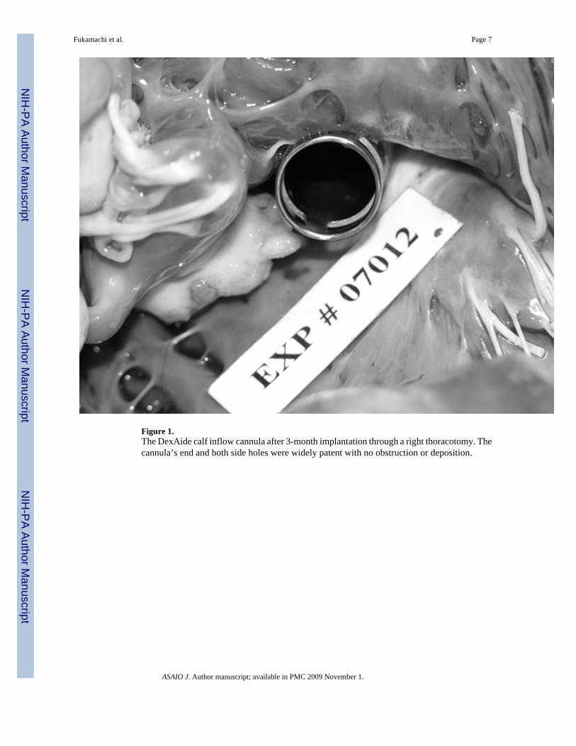
Figure 1.The DexAide calf inflow cannula after 3-month implantation through a right thoracotomy. Thecannula’s end and both side holes were widely patent with no obstruction or deposition.
Fukamachi et al. Page 7
ASAIO J. Author manuscript; available in PMC 2009 November 1.
NIH
-PA Author Manuscript
NIH
-PA Author Manuscript
NIH
-PA Author Manuscript

Figure 2.(A) A Hemashield® outflow graft after a 43-day implantation. Severe neointimal tissue growth(range 4–9 mm in thickness) was observed on the entire surface of the outflow graft, obstructingthe lumen by 25–75%. (B) A Gore-Tex® outflow graft after a 41-day implantation. Thinneointimal tissue (1–1.5 mm in thickness) was observed on the entire surface of the outflowgraft, showing no obstruction.
Fukamachi et al. Page 8
ASAIO J. Author manuscript; available in PMC 2009 November 1.
NIH
-PA Author Manuscript
NIH
-PA Author Manuscript
NIH
-PA Author Manuscript

Figure 3.A zirconia stator after 3-month implantation. There was no deposition nor any signs of bearingwear or deformation on the bearing surfaces.
Fukamachi et al. Page 9
ASAIO J. Author manuscript; available in PMC 2009 November 1.
NIH
-PA Author Manuscript
NIH
-PA Author Manuscript
NIH
-PA Author Manuscript

Figure 4.The CorAide LVAD and the DexAide RVAD implantation through a median sternotomy.LVAD, left ventricular assist device; RVAD, right ventricular assist device; LV, left ventricle;RV, right ventricle; Lt., left; Rt., right.
Fukamachi et al. Page 10
ASAIO J. Author manuscript; available in PMC 2009 November 1.
NIH
-PA Author Manuscript
NIH
-PA Author Manuscript
NIH
-PA Author Manuscript
Copyright © 2022 FDOKUMEN