Development of NanoLuc-targeting protein degraders ... - Nature
13
ARTICLE Development of NanoLuc-targeting protein degraders and a universal reporter system to benchmark tag-targeted degradation platforms Christoph Grohmann 1,2,8 , Charlene M. Magtoto 1,2,8 , Joel R. Walker 3 , Ngee Kiat Chua 1,2 , Anna Gabrielyan 1,2 , Mary Hall 3 , Simon A. Cobbold 1 , Stephen Mieruszynski 1,2 , Martin Brzozowski 1,2 , Daniel S. Simpson 1,2 , Hao Dong 1,2 , Bridget Dorizzi 1,2 , Annette V. Jacobsen 1,2 , Emma Morrish 1,2 , Natasha Silke 1,2 , James M. Murphy 1,2 , Joan K. Heath 1,2 , Andrea Testa 4 , Chiara Maniaci 5 , Alessio Ciulli 6 , Guillaume Lessene 1,2,7 , John Silke 1,2 & Rebecca Feltham 1,2 ✉ Modulation of protein abundance using tag- Targeted Protein Degrader (tTPD) systems targeting FKBP12 F36V (dTAGs) or HaloTag7 (HaloPROTACs) are powerful approaches for preclinical target validation. Interchanging tags and tag-targeting degraders is important to achieve efficient substrate degradation, yet limited degrader/tag pairs are available and side- by-side comparisons have not been performed. To expand the tTPD repertoire we developed catalytic NanoLuc-targeting PRO TACs (NanoTACs) to hijack the CRL4 CRBN complex and degrade NanoLuc tagged substrates, enabling rapid luminescence-based degradation screening. To benchmark NanoTACs against existing tTPD systems we use an inter- changeable reporter system to comparatively test optimal degrader/tag pairs. Overall, we find the dTAG system exhibits superior degradation. To align tag-induced degradation with physiology we demonstrate that NanoTACs limit MLKL-driven necroptosis. In this work we extend the tTPD platform to include NanoTACs adding flexibility to tTPD studies, and benchmark each tTPD system to highlight the importance of comparing each system against each substrate. https://doi.org/10.1038/s41467-022-29670-1 OPEN 1 The Walter and Eliza Hall Institute for Medical Research, 1G Royal Parade, Parkville, Melbourne, VIC 3052, Australia. 2 Department of Medical Biology, University of Melbourne, Parkville, VIC 3052, Australia. 3 Promega Biosciences LLC, 277 Granada Drive, San Luis Obispo, CA 93401, USA. 4 Amphista Therapeutics Ltd, Bo’Ness Road Newhouse, Glasgow ML1 5UH, UK. 5 Chemistry School of Natural and Environmental Sciences, Bedson Building, Newcastle University Edwards Walk, Newcastle NE1 8QB, UK. 6 Division of Biological Chemistry and Drug Discovery, School of Life Sciences, University of Dundee, Dow Street, Dundee DD1 5EH, UK. 7 Department of Pharmacology and Therapeutics, University of Melbourne, Parkville, VIC 3052, Australia. 8 These authors contributed equally: Christoph Grohmann, Charlene M. Magtoto. ✉ email: [email protected] NATURE COMMUNICATIONS | (2022)13:2073 | https://doi.org/10.1038/s41467-022-29670-1 | www.nature.com/naturecommunications 1 1234567890():,;
-
Upload
khangminh22 -
Category
Documents
-
view
3 -
download
0
Transcript of Development of NanoLuc-targeting protein degraders ... - Nature
ARTICLE
Development of NanoLuc-targeting proteindegraders and a universal reporter system tobenchmark tag-targeted degradation platformsChristoph Grohmann1,2,8, Charlene M. Magtoto1,2,8, Joel R. Walker3, Ngee Kiat Chua1,2, Anna Gabrielyan1,2,
Mary Hall3, Simon A. Cobbold1, Stephen Mieruszynski1,2, Martin Brzozowski1,2, Daniel S. Simpson1,2,
Hao Dong1,2, Bridget Dorizzi 1,2, Annette V. Jacobsen 1,2, Emma Morrish1,2, Natasha Silke1,2,
James M. Murphy 1,2, Joan K. Heath 1,2, Andrea Testa 4, Chiara Maniaci 5, Alessio Ciulli 6,
Guillaume Lessene 1,2,7, John Silke 1,2 & Rebecca Feltham 1,2✉
Modulation of protein abundance using tag-Targeted Protein Degrader (tTPD) systems
targeting FKBP12F36V (dTAGs) or HaloTag7 (HaloPROTACs) are powerful approaches for
preclinical target validation. Interchanging tags and tag-targeting degraders is important to
achieve efficient substrate degradation, yet limited degrader/tag pairs are available and side-
by-side comparisons have not been performed. To expand the tTPD repertoire we developed
catalytic NanoLuc-targeting PROTACs (NanoTACs) to hijack the CRL4CRBN complex and
degrade NanoLuc tagged substrates, enabling rapid luminescence-based degradation
screening. To benchmark NanoTACs against existing tTPD systems we use an inter-
changeable reporter system to comparatively test optimal degrader/tag pairs. Overall, we
find the dTAG system exhibits superior degradation. To align tag-induced degradation with
physiology we demonstrate that NanoTACs limit MLKL-driven necroptosis. In this work we
extend the tTPD platform to include NanoTACs adding flexibility to tTPD studies, and
benchmark each tTPD system to highlight the importance of comparing each system against
each substrate.
https://doi.org/10.1038/s41467-022-29670-1 OPEN
1 The Walter and Eliza Hall Institute for Medical Research, 1G Royal Parade, Parkville, Melbourne, VIC 3052, Australia. 2 Department of Medical Biology,University of Melbourne, Parkville, VIC 3052, Australia. 3 Promega Biosciences LLC, 277 Granada Drive, San Luis Obispo, CA 93401, USA. 4AmphistaTherapeutics Ltd, Bo’Ness Road Newhouse, Glasgow ML1 5UH, UK. 5 Chemistry School of Natural and Environmental Sciences, Bedson Building, NewcastleUniversity Edwards Walk, Newcastle NE1 8QB, UK. 6Division of Biological Chemistry and Drug Discovery, School of Life Sciences, University of Dundee, DowStreet, Dundee DD1 5EH, UK. 7 Department of Pharmacology and Therapeutics, University of Melbourne, Parkville, VIC 3052, Australia. 8These authorscontributed equally: Christoph Grohmann, Charlene M. Magtoto. ✉email: [email protected]
NATURE COMMUNICATIONS | (2022) 13:2073 | https://doi.org/10.1038/s41467-022-29670-1 | www.nature.com/naturecommunications 1
1234
5678
90():,;
Proteolysis targeting chimeras (PROTACs)/degraders areheterobifunctional molecules that trigger ubiquitination ofcellular targets by hijacking E3 ubiquitin ligases resulting in
rapid proteasomal degradation1. Powerful genetic tools exist toexperimentally modulate the abundance of gene products, such asCRISPR-Cas9 genome editing and RNA interference2. However,genetic technologies are limited in their ability to assess acutechanges in protein levels, and present additional challenges asprotein depletion may take days depending on protein half-lifeand protein loss is typically irreversible, which may in turn imparta selection pressure for compensatory mechanisms. Small-molecule perturbations on the other hand are popular as theycan be used to rapidly, and for the most part, reversibly assessbiological responses3. Yet, small-molecule inhibitors can be lim-ited by off-target effects, the disruption of discrete domainfunction, occupancy-driven modes of action and the cost andtime associated with their development.
Degrader-mediated degradation of proteins presents excitingopportunities to explore the not-yet-drugged proteome as themajority of human proteins, such as transcription factors, non-enzymatic proteins and scaffolding proteins lack active sitesrendering them difficult to inhibit with small-molecule inhibitors.Degraders consist of a target-binding ligand and an E3-ligase-binding ligand separated by a chemical linker. The design andoptimisation of each of these elements can be critical to achieveefficient substrate degradation as the ligand and the linker canform favourable inter- and intramolecular interactions within theE3-degrader-substrate ternary complex4. For example, varyingthe linker length can enhance the degradation of substrates byreducing steric clashes that can occur between the E3 andsubstrate5,6.
Tag-Targeted Protein Degrader (tTPD) systems direct degradercompounds to protein tags using tag-targeting heterobifunctionalmolecules (Fig. 1a)7. tTPD systems are remarkable technologiesthat permit rapid and reversible degradation of tagged substrateproteins through cell-permeable and in vivo compatibledegraders8–11. tTPD systems include the monovalent molecularglue-based auxin-inducible degron (AID)12, ligand-inducibleaffinity-directed protein missile (L-AdPROM)13, CH6-tag direc-ted specific and nongenetic inhibitors of apoptosis protein [IAP]-dependent protein erasers (SNIPERs)14, BromoTag15, FK506-binding protein 12 (FKBP12) and HaloTag targeting Cereblon(CRBN)- or Von Hippel-Lindau (VHL)-based systems. Degradermolecules that target FKBP12WT or FKBP12F36V (hereafterreferred to as FKBPF36V) and recruit the cullin-RING ligase (CRL)complexes, CRL4CRBN and CRL2VHL to an FKBP-tagged substratehave the prefix dTAG (dTAG13, dTAG48, dTAGV-1)9,10,16,17.HaloTag targeting heterobifunctional degraders have been referredto as HaloPROTACs18,19, which utilise the CRL2VHL system, orHaloTag-binding degradation inducers20, which utilise the IAPproteins.
For simplicity, we refer to all tag-targeting heterobifunctionaldegrader systems listed above as tTPD systems and have abbre-viated each degrader name and the targeted tag/E3s used in thisstudy (Table 1). We have also abbreviated all degrader names toreflect the tag and cullin substrate receptor they bind, e.g.NanoLuc-CRBN=NC (Table 1).
FKBP12 heterobifunctional degraders initially emerged to targetchimeric FKBP12WT-tagged proteins for degradation17. However,the heterobifunctional degraders dFKBP-1 and dFKBP-2 alsotargeted endogenous wild-type FKBP12. Loss of endogenous wild-type FKBP12 may lead to undesired biological side effects andconfound experimental interpretation, so heterobifunctionaldegraders that targeted an engineered mutant FKBPF36V weregenerated9,10. The F36V mutation creates a hole in the 12 kDaFKBP protein that allows 1000-fold selectivity over the wild-type
protein by a bumped synthetic FKBPF36V directed ligand21, andheterobifunctional tools to target FKBPF36V-tagged substrates forproteasomal degradation are now well established.
HaloTag-binding degradation inducers and HaloPROTACswere generated to similarly target the 33 kDa HaloTag andHaloTag7 to trigger degradation of HaloTag and HaloTag7 fusionproteins, respectively18–20. HaloTag is an enzyme engineeredfrom the bacterial dehalogenase enzyme that binds covalently toan alkyl chloride moiety22, while HaloTag7 (hereafter referred toas Halo) is a variant of the HaloTag developed to increase thestability of the enzyme23.
FKBPF36V degraders and HaloPROTACs trigger efficientdegradation of tagged substrate proteins9,10,18–20,24. However,these tag-targeting systems have some drawbacks; limited toolsexist to interrogate FKBPF36V-tagged proteins and FKBPF36V haslow genomic insertion efficiency24. Halo is a large tag relative toother protein tags and HaloPROTACs are non-catalytic in nature,i.e. they are consumed during the reaction and, therefore, cannotbe recycled and reused due to the covalent binding properties,which limits their effectivity. Non-catalytic and irreversible cova-lent binding degraders are limited in their efficiency due to theneed to achieve stoichiometric occupancy of the tagged protein totrigger complete degradation of the target protein. Degraders withnon-covalent cognate binding ligands, such as FKBPF36V-targetingdegraders are not limited in this respect as they can functioncatalytically at sub-stoichiometric concentrations.
NanoLuc (NLuc) is a 19 kDa luciferase enzyme that relies onthe substrate furimazine to produce high-intensity glow-typebioluminescence and is one of the few commercially availableluciferase enzymes25. In addition to its luminescence properties,NanoLuc is an attractive protein tag compared to other tags dueto its stability, small size and the availability of in vivo compatiblesubstrates26.
In this work, we develop and characterise catalytic NanoLuc-targeting PROTACs (NanoTACs) that hijack the CRL complexesto trigger proteasomal degradation of NanoLuc-tagged substratesto expand the tTPD repertoire. Using a universal reporter systemharbouring Halo-EGFP/Firefly-NanoLuc-FKBPF36V we bench-mark each tTPD system in identical cellular settings. We showthat all of the tTPD systems offer up the prospect of selectivelyand reversibly depleting proteins of interest at will. However, eachof the three tags offers advantages and disadvantages (Fig. 1b),and which tag to choose really hinges on the researcher’s inten-tions for their tagged protein of interest.
ResultsDevelopment of NanoTACs; a NanoLuc-targeting degradersystem. We developed heterobifunctional degraders that bindNanoLuc (NanoTACs) to trigger protein degradation ofNanoLuc-tagged substrates building upon the existing tTPDsystems that direct degraders to FKBPF36V or Halo-tagged sub-strates (Fig. 1a). While each tTPD system has specific advantages,NanoLuc is the only tag that offers luminescence properties.Additionally, NanoTACs are catalytic and can act as degradationcatalysts, unlike HaloPROTACs (Fig. 1b). Our most potentNanoTAC recruits the CRL4CRBN E3-ligase complex to triggerthe degradation of NanoLuc-tagged substrate proteins (Fig. 1c).To generate this NanoTAC (NC4) (Fig. 1d), we synthesised twoseries of NanoTAC analogues. NC1 and NC2 were generated bylinking NanoLuc inhibitor 1, a small-molecule inhibitor ofNanoLuc with a low nanomolar IC50 (31 nM) against the enzy-matic activity of NanoLuc27 (Supplementary Fig. 1a), with theCRBN ligand thalidomide using varied alkyl chain linker lengths(Supplementary Fig. 1b, c). NC3, NC4, NC5 and the structurallyrelated control analogue unable to recruit CRBN, NC*, were
ARTICLE NATURE COMMUNICATIONS | https://doi.org/10.1038/s41467-022-29670-1
2 NATURE COMMUNICATIONS | (2022) 13:2073 | https://doi.org/10.1038/s41467-022-29670-1 | www.nature.com/naturecommunications
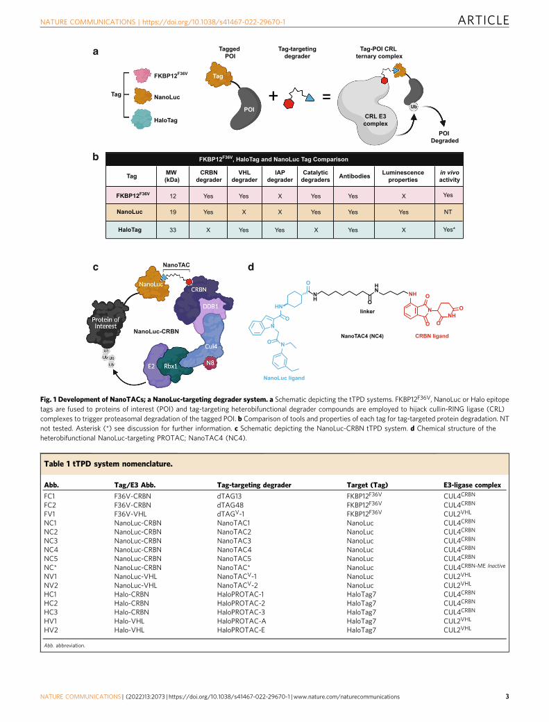
Fig. 1 Development of NanoTACs; a NanoLuc-targeting degrader system. a Schematic depicting the tTPD systems. FKBP12F36V, NanoLuc or Halo epitopetags are fused to proteins of interest (POI) and tag-targeting heterobifunctional degrader compounds are employed to hijack cullin-RING ligase (CRL)complexes to trigger proteasomal degradation of the tagged POI. b Comparison of tools and properties of each tag for tag-targeted protein degradation. NTnot tested. Asterisk (*) see discussion for further information. c Schematic depicting the NanoLuc-CRBN tTPD system. d Chemical structure of theheterobifunctional NanoLuc-targeting PROTAC; NanoTAC4 (NC4).
Table 1 tTPD system nomenclature.
Abb. Tag/E3 Abb. Tag-targeting degrader Target (Tag) E3-ligase complex
FC1 F36V-CRBN dTAG13 FKBP12F36V CUL4CRBN
FC2 F36V-CRBN dTAG48 FKBP12F36V CUL4CRBN
FV1 F36V-VHL dTAGV-1 FKBP12F36V CUL2VHL
NC1 NanoLuc-CRBN NanoTAC1 NanoLuc CUL4CRBN
NC2 NanoLuc-CRBN NanoTAC2 NanoLuc CUL4CRBN
NC3 NanoLuc-CRBN NanoTAC3 NanoLuc CUL4CRBN
NC4 NanoLuc-CRBN NanoTAC4 NanoLuc CUL4CRBN
NC5 NanoLuc-CRBN NanoTAC5 NanoLuc CUL4CRBN
NC* NanoLuc-CRBN NanoTAC* NanoLuc CUL4CRBN-ME Inactive
NV1 NanoLuc-VHL NanoTACV-1 NanoLuc CUL2VHL
NV2 NanoLuc-VHL NanoTACV-2 NanoLuc CUL2VHL
HC1 Halo-CRBN HaloPROTAC-1 HaloTag7 CUL4CRBN
HC2 Halo-CRBN HaloPROTAC-2 HaloTag7 CUL4CRBN
HC3 Halo-CRBN HaloPROTAC-3 HaloTag7 CUL4CRBN
HV1 Halo-VHL HaloPROTAC-A HaloTag7 CUL2VHL
HV2 Halo-VHL HaloPROTAC-E HaloTag7 CUL2VHL
Abb. abbreviation.
NATURE COMMUNICATIONS | https://doi.org/10.1038/s41467-022-29670-1 ARTICLE
NATURE COMMUNICATIONS | (2022) 13:2073 | https://doi.org/10.1038/s41467-022-29670-1 | www.nature.com/naturecommunications 3
prepared by coupling a derivative of the more potent NanoLucinhibitor 2, with a single-digit nanomolar IC50 (4.2 nM) againstthe enzymatic activity of NanoLuc27 (Supplementary Fig. 1d)with the CRBN ligand thalidomide using varied linker lengths(Supplementary Fig. 1e-g). Lastly, NV1 and NV2 were preparedby coupling NanoLuc inhibitor 2 to the widely used VHL ligandderivatives: VH298 and VH03218,19 (Supplementary Fig. 1h, i).
Characterisation of NanoTACs, and selection of a potentNanoTAC: NC4. To test the degradation capacity of the firstseries of analogues we cloned and stably expressed through len-tiviral transduction a versatile and universal tTPD reporter pro-tein that enabled comparative studies in HEK293T cells. Thisreporter contains all three protein tags: Halo, FKBPF36V andNanoLuc in addition to the fluorescent protein EGFP to createthe artificial recombinant fusion protein Halo-EGFP-NanoLuc-FKBPF36V (H-E-N-F) (Supplementary Fig. 2a). All graphs in thisstudy have been colour-coded corresponding to the targeted tagto ease interpretation. The F36V-CRBN degrader FC1 almostcompletely degraded the fusion protein at a concentration of100 nM as judged by Western blot while the NanoLuc-CRBNtargeting NanoTACs NC1 and NC2 led to partial degradation ofthe fusion protein at 500 nM and 1 µM (Supplementary Fig. 2b).
We next tested the second series of NanoTAC analogues. Toexplore whether NanoTAC degraders could be improved byrecruiting a different E3 ligase, we employed NV1 and NV2 thathijack the CRL2VHL complex (Supplementary Fig. 2c). NV1 andNV2 were unable to trigger degradation of the H-E-N-F fusionprotein over the wide range of tested concentrations up to 16 μM(Supplementary Fig. 2d, e). The lack of degradation was not dueto an absence of VHL activity as the F36V-VHL targetingdegrader (FV1) triggered complete degradation of the fusionprotein by Western blot (Supplementary Fig. 2d, e).
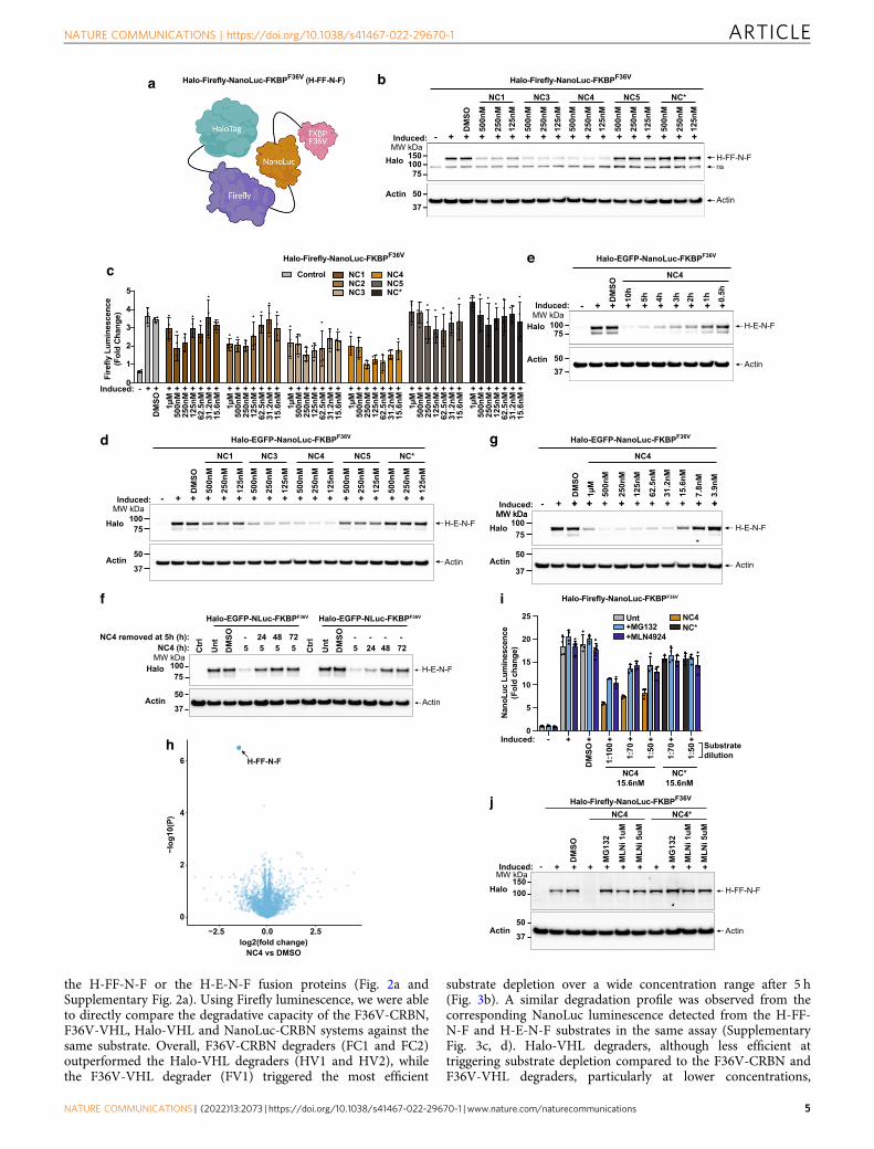
Luminescence is a higher throughput readout than Westernblotting, however, NanoTAC degraders compete with NanoLucsubstrates as they interfere with the conversion of furimazine tofurimamide (Supplementary Fig. 2f). To address this, wegenerated a fusion protein that incorporates Firefly: Halo-Firefly-NanoLuc-FKBPF36V (H-FF-N-F) (Fig. 2a). This constructallows for the direct side-by-side comparison of all tTPD systemsincluding the NanoLuc tTPD system using a Firefly luciferase,which emits a luminescence signal that can be assayedindependently from NanoLuc through the use of distinctsubstrates. Using the H-FF-N-F reporter protein we tested thesecond series of NanoLuc-CRBN targeting NanoTAC analoguesNC3, NC4, NC5 and NC* (inactive control). We observed agreater reduction in Firefly luminescence, and protein levels byWestern blot, upon NanoTAC treatment of cells expressing theH-FF-N-F fusion protein (Fig. 2b, c). Specifically, NC3 andNC4 showed a marked improvement in the extent of substratedegradation compared to the first series of analogues NC1 andNC2, particularly at lower concentrations (Fig. 2b, c). At higherconcentrations, NC4 demonstrated the hook effect (Fig. 2c),which is a characteristic feature of heterobifunctional degraderswhereby high concentrations block degradation due to ligandsaturation and inability to form a ternary complex28. Interest-ingly, NC5 which harbours a linker that is just one carbon lengthshorter than NC4 was incapable of triggering degradation of thefusion protein, possibly due to unfavourable steric interactions asa result of the decreased linker length (Fig. 2b, c). As expected, thecontrol compound NC* did not trigger degradation of the fusionprotein (Fig. 2b, c). NanoTAC degraders were equally capable oftriggering degradation of the H-E-N-F substrate protein withNC3 and NC4 consistently outperforming the first analogue NC1
(Fig. 2d). Overall, NC4 displayed the most effective degradationprofile and was therefore characterised further.
NC4 exhibited approximately a 30-fold reduced inhibition ofNanoLuc enzymatic activity compared to the parent inhibitor(NanoLuc inhibitor 2) indicating that conjugation of the CRBNligand altered the binding kinetics of NC4 (SupplementaryFig. 2g). Strikingly, even though NC4 exhibited reducedinhibition of NanoLuc enzymatic activity NC4 was still capableof rapidly reducing protein levels within 2 h and degradation wassustained over a period of 10 h (Fig. 2e). Further to this,degradation by NC4 was sustained over 24 h when stimulationwas maintained in the culture medium, and reversible by 24 hwhen stimulation was removed after 5 h of initial treatment(Fig. 2f). NC4 was also capable of triggering degradation of thefusion protein at low concentrations, ~30 nM, with a slight hookeffect observed at higher concentrations, similar to that observedin Firefly luminescence-based readouts (Fig. 2c). Importantly, theglobal proteomic analysis demonstrated that NC4 triggers specificdegradation of the target substrate with no significant off-targetdegradation observed when assayed against 5591 proteins(Fig. 2h). Notably, our proteomics analysis did not detect theThalidomide-induced CRBN neosubstrates, IKZF1 or IKZF3, andtherefore we cannot comment on whether these neosubstrates arealtered upon NC4 treatment.
To explore whether NanoLuc luminescence could be used inconjunction with NanoTAC treatment we titrated NC4 againstNC* from 2–10 h. We reasoned that the catalytic nature of NC4would allow for substrate degradation at concentrations belowthat required to inhibit NanoLuc’s enzymatic luminescenceactivity. To this extent we compared the ability of NC4 to reduceluminescence with the control compound, NC*, which lacks thedegradative capability. As predicted, we observed clear differencesin NanoLuc luminescence at 31.2 nM and 15.6 nM between NC4and NC* (Supplementary Fig. 2h). Importantly, the reduction inNanoLuc luminescence and the observed depletion of thereporter by Western blot was indicative of cullin-based protea-some-mediated degradation, as pre-treatment with the protea-some inhibitor MG132 or the NEDD8-activating enzymeinhibitor MLN4924 could rescue both the reduction in lumines-cence and protein levels (Fig. 2i, j). Notably, a slightly enhancedrescue was observed when the substrate furimazine concentrationwas increased for NanoLuc detection, consistent with thecompetitive nature of NanoLuc inhibition by NC4 (Fig. 2i).
FKBPF36V-targeting degraders outperform other tTPD sys-tems. To benchmark existing tTPD systems against NanoTACsand assess the relative degradative performance of each system,we synthesised degraders that hijack VHL and CRBN to triggerproteasomal degradation of FKBPF36V and Halo-taggedsubstrates9,10,18,19, and generated a series of Halo targetingCBRN recruiting degraders; HC1, HC2 and HC3 (SupplementaryFig. 3a). Halo-VHL degraders (HV1 and HV2) target Halo-taggedsubstrates19, while both F36V-CRBN (FC1 and FC2) degradersand the F36V-VHL (FV1) degrader target FKBPF36V-taggedsubstrates9,10 (Fig. 3a). Notably, similar Halo-CRBN moleculeshave been tested for cellular penetration using the ChloroalkanePenetration Assay (CAPA)29; however, degradation studies havenot been performed. The Halo-CRBN molecules we generatedwere unable to trigger efficient substrate degradation over a wideconcentration range (Supplementary Fig. 3b) possibly due toreduced cellular accumulation of these compounds or theinability to promote ternary complex formation.
To directly compare the NanoTAC system to the FKBPF36V
and Halo systems (Fig. 3a) we employed cells expressing either
ARTICLE NATURE COMMUNICATIONS | https://doi.org/10.1038/s41467-022-29670-1
4 NATURE COMMUNICATIONS | (2022) 13:2073 | https://doi.org/10.1038/s41467-022-29670-1 | www.nature.com/naturecommunications
the H-FF-N-F or the H-E-N-F fusion proteins (Fig. 2a andSupplementary Fig. 2a). Using Firefly luminescence, we were ableto directly compare the degradative capacity of the F36V-CRBN,F36V-VHL, Halo-VHL and NanoLuc-CRBN systems against thesame substrate. Overall, F36V-CRBN degraders (FC1 and FC2)outperformed the Halo-VHL degraders (HV1 and HV2), whilethe F36V-VHL degrader (FV1) triggered the most efficient
substrate depletion over a wide concentration range after 5 h(Fig. 3b). A similar degradation profile was observed from thecorresponding NanoLuc luminescence detected from the H-FF-N-F and H-E-N-F substrates in the same assay (SupplementaryFig. 3c, d). Halo-VHL degraders, although less efficient attriggering substrate depletion compared to the F36V-CRBN andF36V-VHL degraders, particularly at lower concentrations,
Substratedilution
NATURE COMMUNICATIONS | https://doi.org/10.1038/s41467-022-29670-1 ARTICLE
NATURE COMMUNICATIONS | (2022) 13:2073 | https://doi.org/10.1038/s41467-022-29670-1 | www.nature.com/naturecommunications 5

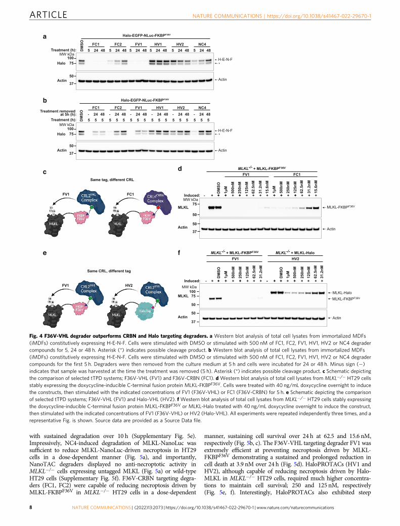
displayed slightly improved substrate degradation over 8 and 24 hat relatively low concentrations (Fig. 3c, Supplementary Fig. 3e).The NanoLuc-CRBN targeting NanoTAC, NC4, was capable oftriggering degradation of the fusion protein over a wideconcentration range and to levels comparable to that of the Haloand FKBPF36V tTPD systems displaying a calculated Dmax of 77%and a DC50 of 21.6 nM against our fusion (Fig. 3b, c). NC4,displayed a similar degradation profile to that of the HaloPRO-TACs (Fig. 3b), but reproducibly appeared to lose efficacy after24 h (Fig. 3c). Consistent with our luminescence results, weobserved reduced levels of the fusion proteins with all testedtTPD degrader systems by Western blot (Fig. 3d, SupplementaryFig. 3f), and importantly the observed reduction in luminescencecould be rescued by co-treatment with the proteasome inhibitorMG132 (Fig. 3e, Supplementary Fig. 3g). Next we compared thedurability of degradation across all tTPD systems by performingwash-out recovery experiments. We transitioned to a constitutivesystem where the H-E-N-F protein was expressed under thecontrol of a ubiquitin promoter to avoid foreseeable complica-tions with wash-out of the doxycycline in our inducible systems.Interestingly FC1 and FV1 maintained near complete degradationup to 48 h (Fig. 4a), and were capable of maintaining cleardegradation for up to 48 h when the degrader was removed fromthe culture medium at 5 h post-treatment (Fig. 4b). FC2 and NC4displayed very similar durability profiles with clear degradationseen up to 24 h (Fig. 4a), however, this was not maintained whenthe degrader was removed from the culture medium after 24 h(Fig. 4b). HaloPROTACs HV1 and HV2 were less durable in theirresponse with depletion not maintained upon removal of thedegrader from the culture medium and overall slower degrada-tion kinetics (Fig. 4a, b).
To functionally assess the impact of the different tTPDs on amodel substrate, we selected the well-characterised pro-necrop-totic pseudokinase MLKL. MLKL is the terminal effector proteinfor necroptotic cell death30 which can be induced by thecombined treatment of TNF, Smac mimetic (SM); compoundA31 and z-VAD-fmk (TSZ)32. C-terminally tagged MLKL haspreviously been shown to be fully functional33 and MLKLknockout cells cannot undergo necroptosis unless reconstitutedwith an active MLKL construct32. We therefore generatedC-terminal fusion proteins of MLKL containing either FKBPF36V,Halo or NanoLuc tags and stably expressed doxycycline-inducibleversions of these proteins in MLKL−/− HT29 cells that weregenerated by CRISPR/Cas9 targeting. The HT29 cell line is widelyused and can undergo rapid necroptosis when stimulated withTSZ32. MLKL-NanoLuc expression was independently confirmedby NanoLuc luminescence (Supplementary Fig. 4a). The taggedversions of MLKL displayed reduced expression compared to
untagged MLKL in MLKL−/− HT29 cells, with the NanoLuc tagleading to a strong reduction in expression compared to adding aHalo or FKBPF36V tag (Supplementary Fig. 4b). From this data itis unclear to what extent this will limit the application of theNanoLuc tag to other targets, and what causes the reducedexpression. Nevertheless, the expression level of MLKL-NanoLucwas sufficient as MLKL-Halo, MLKL-FKBPF36V and MLKL-NanoLuc could confer comparable sensitivity to necroptosis(Supplementary Fig. 4c).
The tagged MLKL substrates afforded us the opportunity todirectly compare the efficiency of the CRBN and VHL degradersystems against a biologically-relevant substrate with the tagspositioned at the same terminus (Fig. 4c). Interestingly the FV1degrader that recruits F36V-VHL was faster at triggeringdegradation of MLKL compared to the F36V-CRBN degraderFC1, acting within 1 h (Supplementary Fig. 4d). FV1 was alsomore effective at triggering degradation of MLKL-FKBPF36V
compared to the F36V-CRBN degrader FC1 resulting in nodetectable protein at 5 h at low nM concentrations (Fig. 4d). Todirectly compare whether the FKBPF36V or Halo epitope tagscould influence the degradation of tagged MLKL with the sameCRL system we tested the F36V-VHL and Halo-VHL systemsside-by-side (Fig. 4e). Consistent with our previous results weobserved that the F36V-VHL-recruiting degrader FV1 was themost effective compound (Fig. 4f). Interestingly, we observedcomparable degradation of murine MLKL-FKBPF36V reconsti-tuted into Mlkl−/− mouse dermal fibroblasts (MDFs) with F36V-CRBN degraders (FC1, FC2) and the F36V-VHL degrader (FV1)(Supplementary Fig. 4e), suggesting that even slight changes inthe substrate or cell type might influence the effectivity ofdegrader compounds. To explore this further, we inserted eachtag into the genomic locus of the FBL gene that encodes for theprotein Fibrillarin, which is exclusively expressed in thenucleolus. We performed concentration titrations and timecourses on Fibrillarin-NanoLuc, Fibrillarin-Halo and Fibrillarin-FKBPF36V expressing cell lines and calculated Dmax values basedon densitometry measurements (Supplementary Fig. 4f–m).Consistent with our results for MLKL and our fusion protein,we observed that FV1 was superior in degradation capacitycompared to the other tag degraders displaying a Dmax of ~95.9%after 10 h. FC1, HV1, HV2 and NC4 all demonstrated cleardegradation of FBL over 10 h with calculated Dmax values of~70%, ~93.2%, ~94.8% and ~52.9%, respectively. Fibrillarin-NanoLuc predominantly localises to the nucleus (SupplementaryFig. 5a) suggesting that NC4 was capable of degrading substratesthat primarily reside in the nucleus. To determine whethersubstrates in other cellular compartments could also be targetedby NC4 we generated a fusion construct containing NanoLuc and
Fig. 2 Identification of NC4 as a potent NanoTAC degrader. a Schematic depicting the Halo-Firefly-NanoLuc-FKBPF36V (H-FF-N-F) fusion protein.b Western blot analysis of lysates from HEK293T cells expressing H-FF-N-F, induced with 20 ng/mL doxycycline overnight, stimulated for 5 h (ns non-specific band). c Firefly luminescence from cells in b, induced with 20 ng/mL doxycycline overnight, stimulated for 5 h. d Western blot analysis of lysatesfrom HEK293T cells expressing H-E-N-F, induced with 20 ng/mL doxycycline overnight, stimulated for 5 h. e Western blot analysis of lysates from cells ind, induced with 20 ng/mL doxycycline overnight, stimulated with 125 nM of NC4 for the indicated times. f Western blot analysis of lysates fromimmortalized MDFs (iMDFs) constitutively expressing H-E-N-F or wild-type iMDFs (ctrl), cells were left untreated (Unt), stimulated with DMSO or 125 nMof NC4 for 5, 24, 48 or 72 h. NC4 was left in the culture medium for the duration of the time-course or removed from the culture medium at 5 h, and cellsincubated post degrader removal for 24, 48 or 72 h. g Western blot analysis of lysates from cells in d, induced with 20 ng/mL doxycycline overnight,stimulated with NC4 for 5 h. h Volcano plot quantifying proteins significantly downregulated in 293 T cells expressing the H-FF-N-F reporter aftertreatment with DMSO or 125 nM of NC4 for 5 h. Proteins significantly downregulated (using the q-value) with NC4 treatment when compared to DMSOtreatment are labelled and indicated with an arrow. i NanoLuc luminescence from cells in b, induced with 20 ng/mL doxycycline overnight, stimulated with10 μM MG132 or 1 μM MLN4924 for 1 h prior to treatment with 15.6 nM NC4 or NC* for 4 h. Substrate (furimazine) was diluted 1:100, 1:70 and 1:50 priorto detecting luminescence. j Western blot analysis from cells in B, induced with 20 ng/mL doxycycline overnight, stimulated with 10 μM MG132, or 1 or5 μM MLN4924 for 1 h prior to stimulation with DMSO or 125 nM of NC4 or NC* for 5 h. c, i Error bars (EB) represent mean ± SD from N= 4 technicalrepeats. All experiments were repeated independently three times and a representative Fig. is shown. Source data are provided as a Source Data file.
ARTICLE NATURE COMMUNICATIONS | https://doi.org/10.1038/s41467-022-29670-1
6 NATURE COMMUNICATIONS | (2022) 13:2073 | https://doi.org/10.1038/s41467-022-29670-1 | www.nature.com/naturecommunications

the plasma membrane transporter protein SLC38A2. Weconfirmed the location of NanoLuc-SLC38A2 in the membranecompartment (Supplementary Fig. 5b), and consistent with ourprevious results we observed a reduction in Nanoluc-SLC38A2upon treatment with NC4 (Supplementary Fig. 5c).
tTPD systems trigger physiologically relevant degradation. Tocompare the biological activity of tTPD-mediated degradation
across all tTPD platforms, we assessed whether our most potentNanoTAC could degrade substrates to a level that preventsnecroptosis to achieve a response similar to protein knockout.Consistent with our previous results, NanoTAC degraders triggeredefficient degradation of MLKL-NanoLuc, with NC4 displaying themost effective degradation profile (Supplementary Fig. 5d). Similarto the degradation of the Halo-EGFP-NanoLuc-FKBPF36V fusionprotein, NC4 triggered degradation of MLKL-NanoLuc within 2 h
Fig. 3 FKBPF36V tTPD systems outperform other tTPD systems. a Schematic representation of the Halo-VHL, F36V-CRBN, F36V-VHL and NanoLuc-CRBN tag-targeting degrader systems. b Firefly luminescence from 293 T cells stably expressing doxycycline-inducible H-FF-N-F, treated with 20 ng/mLdoxycycline overnight (induced), then stimulated with the indicated concentrations FC1 (F36V-CRBN), FC2 (F36V-CRBN), FV1 (F36V-VHL), HV1 (Halo-VHL), HV2 (Halo-VHL) or NC4 (NanoLuc-CRBN) for 5 h. c Firefly luminescence from cells used in b, treated with 20 ng/mL doxycycline overnight(induced), then stimulated with the indicated concentrations FC1, FC2, FV1, HV1, HV2 or NC4 for the indicated times. d Western blot analysis of total celllysates from cells expressing H-FF-N-F, treated with 20 ng/mL doxycycline overnight (induced), then stimulated with the indicated concentrations ofdegraders for 5 h. e Firefly luminescence from cells in b, treated with 20 ng/mL doxycycline overnight (induced), following treatment with 20 μM MG132for 1 h prior to stimulation with the indicated concentrations FC1, FC2, FV1, HV1, HV2 or NC4 for 5 h. b, c, e EB represent mean ± SD from N= 4 technicalrepeats. All experiments were repeated independently three times, and a representative Fig. is shown. Source data are provided as a Source Data file.
NATURE COMMUNICATIONS | https://doi.org/10.1038/s41467-022-29670-1 ARTICLE
NATURE COMMUNICATIONS | (2022) 13:2073 | https://doi.org/10.1038/s41467-022-29670-1 | www.nature.com/naturecommunications 7
with sustained degradation over 10 h (Supplementary Fig. 5e).Impressively, NC4-induced degradation of MLKL-NanoLuc wassufficient to reduce MLKL-NanoLuc-driven necroptosis in HT29cells in a dose-dependent manner (Fig. 5a), and importantly,NanoTAC degraders displayed no anti-necroptotic activity inMLKL−/− cells expressing untagged MLKL (Fig. 5a) or wild-typeHT29 cells (Supplementary Fig. 5f). F36V-CRBN targeting degra-ders (FC1, FC2) were capable of reducing necroptosis driven byMLKL-FKBPF36V in MLKL−/− HT29 cells in a dose-dependent
manner, sustaining cell survival over 24 h at 62.5 and 15.6 nM,respectively (Fig. 5b, c). The F36V-VHL targeting degrader FV1 wasextremely efficient at preventing necroptosis driven by MLKL-FKBPF36V demonstrating a sustained and prolonged reduction incell death at 3.9 nM over 24 h (Fig. 5d). HaloPROTACs (HV1 andHV2), although capable of reducing necroptosis driven by Halo-MLKL in MLKL−/− HT29 cells, required much higher concentra-tions to maintain cell survival; 250 and 125 nM, respectively(Fig. 5e, f). Interestingly, HaloPROTACs also exhibited steep
Fig. 4 F36V-VHL degrader outperforms CRBN and Halo targeting degraders. a Western blot analysis of total cell lysates from immortalized MDFs(iMDFs) constitutively expressing H-E-N-F. Cells were stimulated with DMSO or stimulated with 500 nM of FC1, FC2, FV1, HV1, HV2 or NC4 degradercompounds for 5, 24 or 48 h. Asterisk (*) indicates possible cleavage product. b Western blot analysis of total cell lysates from immortalized MDFs(iMDFs) constitutively expressing H-E-N-F. Cells were stimulated with DMSO or stimulated with 500 nM of FC1, FC2, FV1, HV1, HV2 or NC4 degradercompounds for the first 5 h. Degraders were then removed from the culture medium at 5 h and cells were incubated for 24 or 48 h. Minus sign (−)indicates that sample was harvested at the time the treatment was removed (5 h). Asterisk (*) indicates possible cleavage product. c Schematic depictingthe comparison of selected tTPD systems; F36V-VHL (FV1) and F36V-CRBN (FC1). d Western blot analysis of total cell lysates from MLKL−/− HT29 cellsstably expressing the doxycycline-inducible C-terminal fusion protein MLKL-FKBPF36V. Cells were treated with 40 ng/mL doxycycline overnight to inducethe constructs, then stimulated with the indicated concentrations of FV1 (F36V-VHL) or FC1 (F36V-CRBN) for 5 h. e Schematic depicting the comparisonof selected tTPD systems; F36V-VHL (FV1) and Halo-VHL (HV2). f Western blot analysis of total cell lysates from MLKL−/− HT29 cells stably expressingthe doxycycline-inducible C-terminal fusion protein MLKL-FKBPF36V or MLKL-Halo treated with 40 ng/mL doxycycline overnight to induce the construct,then stimulated with the indicated concentrations of FV1 (F36V-VHL) or HV2 (Halo-VHL). All experiments were repeated independently three times, and arepresentative Fig. is shown. Source data are provided as a Source Data file.
ARTICLE NATURE COMMUNICATIONS | https://doi.org/10.1038/s41467-022-29670-1
8 NATURE COMMUNICATIONS | (2022) 13:2073 | https://doi.org/10.1038/s41467-022-29670-1 | www.nature.com/naturecommunications
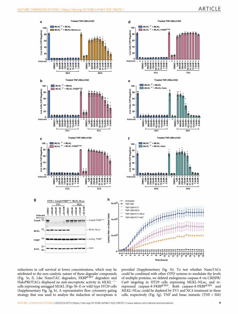
reductions in cell survival at lower concentrations, which may beattributed to the non-catalytic nature of these degrader compounds(Fig. 5e, f). Like NanoTAC degraders, FKBPF36V degraders andHaloPROTACs displayed no anti-necroptotic activity in MLKL−/−
cells expressing untagged MLKL (Figs 5b–f) or wild-type HT29 cells(Supplementary Fig. 5g, h). A representative flow cytometry gatingstrategy that was used to analyse the induction of necroptosis is
provided (Supplementary Fig. 5i). To test whether NanoTACscould be combined with other tTPD systems to modulate the levelsof multiple proteins, we deleted endogenous caspase-8 via CRISPR/Cas9 targeting in HT29 cells expressing MLKL-NLuc, and re-expressed caspase-8-FKBPF36V. Both caspase-8-FKBPF36V andMLKL-NLuc could be depleted by FV1 and NC4 treatment in thesecells, respectively (Fig. 5g). TNF and Smac mimetic (TNF+ SM)
NATURE COMMUNICATIONS | https://doi.org/10.1038/s41467-022-29670-1 ARTICLE
NATURE COMMUNICATIONS | (2022) 13:2073 | https://doi.org/10.1038/s41467-022-29670-1 | www.nature.com/naturecommunications 9
treatment triggers caspase-8 dependent apoptotic cell death that canbe switched to necroptotic cell death when caspase-8 is inhibited ordeleted from cells. Induction of MLKL-NLuc with doxycycline andconstitutive expression of caspase-8-FKBPF36V rendered this cellline susceptible to necroptotic cell death and apoptotic cell deathupon treatment with TNF+ SM (Fig. 5h). As expected, removal ofMLKL-NLuc with NC4 did not impact cell death induced byTNF+ SM treatment (TNF+ SM+NC4), owing to active apop-totic caspase-8 signalling. Impressively, removal of caspase-8-FKBPF36V with FV1 (TNF+ SM+ FV1) caused a clear reductionin TNF+ SM induced cell death that could be further reduced withMLKL-NLuc removal with NC4, but not NC*. These data indicatethat TNF+ SM induced apoptosis and necroptosis driven bycaspase-8-FKBPF36V and MLKL-NLuc can be dynamically fine-tuned in the same cell system by combining two different tTPDdegraders (Fig. 5h).
Collectively, these results suggest that efficient degradation ofsubstrates with tag-targeting degraders is system dependent.Importantly, these results highlight that it is critical to compareeach tTPD system against each substrate to identify the optimaldegrader/tag pair to achieve prolonged and sustained degradationwith minimal effective degrader concentration.
DiscussionTo add to the current toolbox of available tTPD systems and tocompare the utility of different degrader/tag combinations wedeveloped a new NanoLuc-targeted degrader system and a newreporter system. Our reporter system enables rapid comparativetesting of optimal degrader/tag combinations that will advancethe field by providing an interchangeable tag system that isamenable to a limitless number of degrader/tag pairs. We use thisreporter system to test and develop NanoTACs, demonstratingthat the reporter can facilitate the development of new tTPDsystems. We believe this reporter system will increase the easewith which the powerful tTPD approach can be adopted andextended by other laboratories.
Using our reporter system and test substrates (MLKL &Fibrillarin) we provide side-by-side comparisons of the Halo-VHL, Halo-CRBN, F36V-CRBN and F36V-VHL tTPD systems,and directly compare these to the newly developed NanoLuc-CRBN and NanoLuc-VHL systems. We highlight the importanceof comparing all tTPD systems against each substrate as eachsystem does not degrade equally, and efficient tTPD-mediateddegradation is extremely substrate-specific.
Interestingly, when we compared the degradation of humanMLKL-FKBPF36V and mouse MLKL-FKBPF36V we observed thatthe CRBN-recruiting degraders were less effective at targeting
human MLKL-FKBPF36V compared to VHL degraders, yet mouseMLKL-FKBPF36V was degraded by both systems equally. We alsoobserved that endogenously tagged-Fibrillarin, which is localisedin the nucleolus, was preferentially degraded by VHL-recruitingdegraders compared to CRBN-recruiting degraders. This effect ispotentially due to differences in the formation or stability of theternary complex between the CRBN and VHL containing E3-ligase complexes and different substrate proteins, or subcellularlocation of VHL and CRBN, which may dictate substrate speci-ficity for these E3-ligase complexes.
If a ternary complex is unstable or exhibits poor cooperativebinding then certain degraders could be less efficient at triggeringdegradation, which would be consistent with previous reportsthat detail the importance of cooperativity for degrader-mediatedprotein degradation4. Our data highlights that each substrateneeds to be assessed against all tTPD systems as it is extremelychallenging to predict which substrate-CRL complex and whichdegrader/tag pair will lead to optimal degradation of a particularsubstrate. Another important consideration that may influenceubiquitination and substrate degradation is which protein ter-minus the tag is attached to as this may alter lysine accessibility.The optimal terminus for a particular tag is ultimately substrate-specific and some substrates cannot tolerate a tag attached to aparticular terminus. However, if ubiquitin is primarily conjugatedto the tag lysine residues rather than substrate lysines then theposition of the tag would be unlikely to influence degradation butmay still impact protein function.
Importantly, we find that the newly developed NanoLuc-CRBNdegrader system functions comparably to the other tTPD systems,triggering degradation at low nM concentrations of degrader andto physiologically relevant levels. NanoTACs, however, also havesome drawbacks that are important to highlight. We currently donot have efficient NanoLuc-VHL degraders in hand that wouldpotentially increase the substrate scope of protein targets forNanoTAC degradation. NanoTACs are based on a NanoLucinhibitor warhead, therefore, at intra-cellular concentrationsapproximating the IC50, NanoTACs interfere with nanoluciferaseactivity by inhibiting the conversion of furimazine to furimamide,preventing the ability to perform high-throughput degradationassays via a luminescence readout. Notably, since NanoTACsachieve efficient degradation of NLuc-tagged proteins resulting ina low cellular DC50, nanoluciferase luminescence can still be usedto reliably read out degradation when used well below the NLucIC50. However, for this purpose, we recommend that titrations areperformed against the inactive analogue (NC*) and that criticalcontrols i.e. MG132 or MLN4924 are included to separatedegradation from NLuc inhibitory activity. Further investigation
Fig. 5 NanoTAC degraders trigger MLKL degradation and block necroptotic cell death. a MLKL−/− HT29 cells stably expressing the doxycycline-inducible untagged MLKL or the C-terminal fusion protein MLKL- NanoLuc were induced with 40 ng/mL doxycycline overnight, then stimulated with NC4(NanoLuc-CRBN) for 5 h before the addition of TNF (100 ng/mL) + Smac mimetic (compound A; 500 nM)+ caspase inhibitor; z-VAD-fmk (10 μM),for 24 h. Cell death was assessed by flow cytometric analysis of PI exclusion. N= 3 independent experiments (symbols), EB represent mean ± SD.b–d MLKL−/− HT29 cells expressing doxycycline-inducible untagged MLKL or the C-terminal fusion protein MLKL-FKBPF36V were induced with 40 ng/mLdoxycycline overnight, then stimulated with FC1, FC2 or FV1 for 5 h before the addition of TNF (100 ng/mL) + Smac mimetic (compound A;500 nM)+ caspase inhibitor; z-VAD-fmk (10 μM), for 24 h. Cell death was assessed by flow cytometric analysis of PI exclusion. N= 3 independentexperiments (symbols), EB represent mean ± SD. e, f MLKL−/− HT29 cells expressing doxycycline-inducible untagged MLKL or the C-terminal fusionprotein MLKL-Halo were treated with 40 ng/mL doxycycline overnight to induce the constructs, then stimulated with the indicated concentrations HV1 orHV2 for 5 h before the addition of TNF (100 ng/mL) + Smac mimetic (compound A; 500 nM)+ caspase inhibitor; z-VAD-fmk (10 μM), for 24 h. Celldeath was assessed by flow cytometric analysis of PI exclusion. e N= 4, f N= 3 independent experiments (symbols), EB represent mean ± SD. g Westernfrom MLKL−/−CASP8−/− HT29 cells stably expressing the doxycycline-inducible C-terminal fusion protein, MLKL-NanoLuc (MLKL-NLuc) andconstitutively expressing the C-terminal fusion protein caspase-8-FKBPF36V. Cells were induced with 40 ng/mL doxycycline overnight, then stimulatedwith FV1 and NC4 for 5 h. h Cells from g were treated with 40 ng/mL doxycycline and stimulated with 125 nM of FV1, NC4, or NC* for 5 h before theaddition of TNF (100 ng/mL) + Smac mimetic (compound A; 500 nM) for 19.5 h. Cell death was assessed using an IncuCyte and Cytotox-Red uptake.N= 3 independent experiments, one representative experiment shown, EB represent mean ± SD. Source data are provided as a Source Data file.
ARTICLE NATURE COMMUNICATIONS | https://doi.org/10.1038/s41467-022-29670-1
10 NATURE COMMUNICATIONS | (2022) 13:2073 | https://doi.org/10.1038/s41467-022-29670-1 | www.nature.com/naturecommunications
is also required to determine whether NanoTACs can be com-patible with the in vivo nanoluciferase substrates26. NanoTACsare functionally similar to FKBPF36V-targeting degraders asNanoTACs are catalytic by nature and are not consumed duringthe reaction. The catalytic property of degraders is an importantfeature as degraders that exhibit poor cellular penetration or weaktarget-binding affinity can still be highly efficient degraders. It is,therefore, intriguing that NanoTACs are less effective comparedto FKBPF36V-targeting degraders and might suggest that there aredifferences in the cellular accumulation of these molecules. Thecatalytic property of the NanoLuc/NanoTAC tTPD system is amajor advantage over non-catalytic tTPD systems and furtheroptimisation of NanoTACs to achieve improved degradationefficiency of NLuc-tagged substrates presents an excitingopportunity.
The importance of catalytic degraders for substate degradationwas highlighted by elegant studies into Bruton’s tyrosine kinase(BTK) binding ibrutinib-derived degraders, demonstrating thatreversible binding to BTK was essential to trigger BTKdegradation34,35. It was postulated that irreversible covalentdegraders such as HaloPROTACs are consumed once they engageand react with their target protein and hence cannot reachdegradation efficiency comparable to catalytic degraders. Con-versely, a number of studies argue against this theory:18–20,36–38.Furthermore, our data and those of others suggest that covalentHaloPROTACs can trigger efficient degradation of Halo-taggedsubstrates and we show the comparable activity of non-catalyticHaloPROTACs to catalytic NanoTAC degraders8,18,19,24. More-over, HaloPROTACs have high affinity and selectivity for theHalo tag and we observed that HaloPROTACs maintained sub-strate depletion over 24 h.
Overall, we find that the FKBPF36V tTPD systems are generallysuperior at triggering degradation of FKBPF36V tagged substrateswith regard to the minimal effective concentration and the timetaken to achieve protein loss respective to controls, compared tothe Halo- and NanoLuc-targeting systems, in our hands. Ourobservation that NanoTACs and HaloPROTACs degradewith slower kinetics and require higher minimal concentrationsto achieve effective degradation compared to FKBPF36V degradersmight be explained by a variety of factors that alter theintracellular accumulation of these compounds including cellpermeability, drug efflux pumps, cellular metabolism or interac-tions with intracellular proteins and other biomolecules. Anotherpossible limitation in the effectiveness of HaloPROTACs is theirpotential reactivity with strong nucleophiles present in the cell(e.g. Glutathione) due to the chloroalkyl moiety. This irreversibleside-reaction would consume the HaloPROTAC and decrease theeffective concentration of the target Halo-tagged protein. Halo-PROTACs and NanoTACs likely do not reach sufficientintracellular concentration in our necroptotic assays at lowerassay concentrations, as our data show that these degraders areunable to provide complete protection against MLKL-Haloinduced necroptosis.
The FKBPF36V system is excellent for triggering degradation ofFKBPF36V-tagged substrates, and the FKBPF36V tag is smallcompared to the other tags at just 12 kDa, having minimal impacton fusion partner proteins. If targeted protein degradation is theprimary goal, then this tTPD system is an ideal choice as theexperimenter can easily and rapidly switch between the CRBNand VHL systems through the use of different degrader com-pounds. However, if the experimenter is interested in alsounderstanding tagged protein biochemistry/biology, (cellularlocalisation, protein interactomes, tissue expression profiles ect.)then the NanoLuc (catalytic degrader available) and Halo (onlynon-catalytic degraders available) tags are superior as they offer
the best of both worlds due to the numerous commercial toolsavailable for these two protein tags.
Consistent with previous reports, our data detail that all cur-rent tag-targeting degraders trigger proteasomal degradation oftagged substrates. However, we did observe that the degradationof the reporter construct by FV1 was only partially reduced in thepresence of the proteasome inhibitor MG132. Given that we pre-incubated the cells with MG132 for 1 hour prior to the addition ofFV1, which is sufficient to block degradation triggered by theother tag-targeting degraders, it is possible that FV1 might alsotrigger degradation through alternative pathways, for example,the lysosomal degradation pathway. Alternatively, FV1 couldsimply be very efficient at triggering substrate ubiquitination anddegradation, and this degradation might precede complete pro-teasomal inhibition by MG132, even after pre-incubation.
A comprehensive analysis in animals is yet to be conducted tocompare the tTPD systems side-by-side, and in vivo pharmaco-kinetic and pharmacodynamic studies will need to be performedon the NanoTAC compounds. Regardless, studies that have testedindividual tag-targeting degraders, or similar degraders such asRC32 comprised of the FKBP12 ligand Rapamycin conjugated tothe CRBN ligand pomalidomide, in animals, detail that they arewell tolerated and can trigger efficient depletion of substrates8–11.Notably, in vivo pharmacokinetic analysis and activity studies forthe dTAG degraders FC1 and FV1 have been performed9,10, andone group has reported in vivo activity for one HaloPROTAC,HaloPROTAC-38, which was not used in our study. Tag-targetingdegrader compounds are not designed to be used therapeutically;however, tTPD systems do allow for the assessment of chemical-induced protein depletion in animal disease models, that willclosely mimic targeted protein degradation of endogenous targets.With the advancement of the tTPD systems to include NanoLucin the armamentarium, tTPD technology is leading the way forcomprehensive validation studies to be conducted on prospectivetherapeutic targets to rationally determine which targets warrantdrug discovery investment to identify ligands targeting equivalentendogenous proteins.
MethodsCell culture and maintenance. HEK293T (ATCC; CRL3216), HT29 (ATCC; HTB-38)and immortalized (SV40 large-T antigen) mouse dermal fibroblast (iMDF) cells(generated in-house from C57BL/6 mouse tails) were cultured in Dubecco’s modifiedEagle medium (DMEM) supplemented with 10% foetal bovine serum (FBS, Sigma),50U/mL penicillin (Gibco) and 50 μg/mL streptomycin (Gibco). iMDFs were isolatedfrom C57BL/6 mouse tails and transformed with SV40 Large-T antigen. All cells weremaintained at 37 °C with 10% CO2 in a humidified incubator.
Plasmid constructs and stable cell lines. The cDNA for human or mouse Mlklwas cloned from synthetic gBlock fragments (Integrated DNA Technologies) togenerate fusion proteins harbouring a Halo, FKBPF36V or NanoLuc (NLuc) tag atthe C-terminus of MLKL. The individual tags in the Halo-EGFP-NanoLuc-FKBPF36V (H-E-N-F) reporter were ordered as gBlock fragments (Integrated DNATechnologies) and made into a single reporter construct using restriction digestionand ligation cloning. Halo refers to HaloTag7, FKBPF36V refers to FKBP12F36V.Each protein in the reporter construct is separated by a 9-10 amino acid glycine-serine linker. To generate the H-FF-N-F reporter construct, the EGFP was excisedfrom the H-E-N-F reporter construct and replaced with Firefly luciferase (kind giftfrom Joan Heath) using In-Fusion cloning (Takara Bio). MLKL-/- HT29 cells weregenerated by CRISPR-Cas9 targeting using lentiviral transduction, reportedpreviously32, while Mlkl-/- mouse dermal fibroblasts (MDFs) were generated fromMlkl−/− mice described previously39. Stable cell lines were generated using theLentiviral packaging constructs (VSVg, RSV-Rev, pMDL) and the pFTRE3Gdoxycycline-inducible vector and selected with Puromycin (2 μg/mL). pX330A-FBL/PITCh was a gift from Takashi Yamamoto (Addgene plasmid # 63671; http://n2t.net/addgene:63671; RRID:Addgene_63671)40. pCRIS-PITChv2-FBL was a giftfrom Takashi Yamamoto (Addgene plasmid # 63672; http://n2t.net/addgene:63672;RRID:Addgene_63672)40. pCRIS-PITChv2-FBL was modified to contain either theNanoLuc, Halo or FKBPF36V tag sequences to enable genomic insertion of a tag-T2A-PuroR sequence at the FBL locus to produce C-terminal NanoLuc-, Halo- orFKBPF36V- tagged-Fibrillarin. For knock-in cell lines, 63671 or modified 63,672
NATURE COMMUNICATIONS | https://doi.org/10.1038/s41467-022-29670-1 ARTICLE
NATURE COMMUNICATIONS | (2022) 13:2073 | https://doi.org/10.1038/s41467-022-29670-1 | www.nature.com/naturecommunications 11
vectors were co-transfected into 293Ts with lipofectamine, stably integrated cellswere selected with puromycin (1 μg/ml).
NanoLuc and Firefly luciferase assays. For high-throughput assessment of tTPD-mediated degradation, cells expressing NanoLuc or Firefly fusion proteins wereseeded into 384-well flat bottom, clear bottom white-walled plates (Corning) at1–1.5 × 10⁴ cells/well in 50 μL of DMEM/FCS. A final concentration of 20 ng/mLand 40 ng/mL of doxycycline was used for HEK293T and HT29 cells, respectively,followed by overnight incubation (16–24 h) to induce construct expression. Cellswere then treated with either vehicle control (DMSO) or degrader compound asindicated. In compound titration experiments, all vehicle control amounts wereequivalent to the highest degrader concentration. For MG132 and MLN4924 rescueexperiments, cells were pre-treated for 30min–1 h with MG132 (10 μM, SelleckChemicals) or MLN4924 (1–5 μM; Tocris) prior to the addition of the degradercompounds. Time-course experiments were conducted as reverse time courses sothat luminescence could be detected at the same cell density. At the stated time-points, luminescence was induced using the Nano-GLO Luciferase assay system(Promega) for NanoLuc luminescence, or Nano-Glo Dual-Luciferase Reporter assaysystem (Promega) for NanoLuc/Firefly dual luminescence. Clear plate bottoms weretaped with coloured tape to prevent bleeding between wells. For IC50 measurementsrecombinant NanoLuc enzyme was prepared to a concentration of 0.2 nM, andNanoLuc LCS dilution buffer was diluted 1:30 in TBS+ 0.01% BSA. NC4 orNanoLuc inhibitor 2 were diluted in the LCS solution and combined with thediluted NanoLuc enzyme. Samples were incubated for 6 min at room temperaturebefore assaying for luminescence. Luminescence was then measured (0.1 s/well,filter 470-480) on a microplate reader (CLARIOstar Plus, BMG Labtech).
Immunoblotting. Cells were seeded in 24-well plates ±doxycycline (20 ng/mL forHEK293T, 40 ng/mL for HT29) and incubated overnight (16–24 h) prior todegrader treatments, as stated in the Fig. legends. All timepoint experiments wereperformed as reverse timepoints in order to harvest all cells at the same time. At theindicated time points cells were lysed in NuPAGE LDS lysis buffer (Invitrogen)diluted to 1× in DISC lysis buffer (20 mM Tris-HCl pH7.5, 150 mM NaCl, 2 mMEDTA pH 8.0, 1% Triton X-100, 10% glycerol, H20)) supplemented with β-Mercaptoethanol (2%), protease and phosphatase inhibitors. Lysates were runthrough polypropylene columns (Pierce) to shred DNA. Proteins were separated bySDS-PAGE on 4-12% gradient gels (Invitrogen) and transferred onto Immoblon-Epolyvinyl difluoride membranes (Merck). Membranes were blocked in 5% skimmilk (Devondale) in TBS-T (TBS, 0.1% Tween-20) for 1 h prior to immunoblottingwith primary antibodies overnight at 4 °C. Unless stated otherwise, all primaryantibodies were diluted in TBS-T (TBS, 0.1% Tween-20) containing 5% BSA(Sigma A8022) and 0.04% sodium azide: HaloTag (1:1000, Promega; G9211),NanoLuc (1:500, Promega; N7000), β-actin (1:20,000, Sigma; A-1798), MLKL(1:1000, produced in-house; 3H1 clone)39, FKBP (1:1000, R&D systems; 422513),caspase-8 (1:1000, Proteintech; 13423-1-AP), Histone H3 (1:1000, Abcam;ab10799), Cadherin (1:1000, Cell Signalling Technologies; 4068 T). Membraneswere washed 3 × 5–10 min in TBS-T (TBS, 0.1% Tween-20) prior to the addition ofthe appropriate secondary antibodies conjugated with horseradish peroxidase(Jaxon laboratories). All secondary antibodies were diluted at 1:10,000 in TBS-T(TBS, 0.1% Tween-20) containing 5% skim milk and incubated at room tem-perature for 1 h. Final washes of 4 × 5–10 min with TBS-T (TBS, 0.1% Tween-20)were conducted before ECL development (Millipore, Bio-Rad) and protein detec-tion using the ChemiDoc Touch Imaging System (Bio-Rad). All images wereprocessed using Image Lab software.
Necroptosis assays. To simulate necroptosis, cells were seeded into 96-well plates±40 ng/mL doxycycline, to induce construct expression, and treated with 100 ng/mL FLAG-TNF (recombinant human, in-house), 500 nM compound A Smacmimetic (kindly gifted by TetraLogics Pharmaceuticals) and 5 μM IDN-6556(Cayman) or 10 uM Z-VAD-fmk (Selleckchem) overnight (16–24 h). Degradercompounds or a DMSO vehicle control were added for 5 h, as stated in the Fig.legends. Cells were detached using Trypsin-EDTA (Merck) and resuspended in cellsupernatants containing 10 μg/mL propidium iodide (PI). PI exclusion analysis wasperformed using an LSR II flow cytometer (Becton Dickinson, NJ) with 10,000single-cell events per sample. Flow cytometry data were analysed using WEASELversion 2.7 software (Frank Battye).
Quantitative proteomics. Cell pellets were extracted with 5% SDS (including100mM TEAB) and processed using micro S-traps as described by the manufacturer(Protifi). For liquid chromatography-tandem mass spectrometry (LC-MS/MS) ana-lysis, approximately 200 ng of sample was injected onto an Acuity M-class UPLC(Waters) connected to a timsTOF pro II (Bruker). Peptides were separated using a112min gradient (solvent A, 0.1% formic acid; solvent B, 99.9% acetonitrile/0.1%formic acid) on a C18 analytical column (IonOpticks, Aurora Series Emitter Column,AUR2-25075C18A 25 cm× 75 µm). Data-dependent PASEF acquisition was per-formed (100–1700m/z scan range and 0.6–1.6 mobility range) and the data searchedagainst the reviewed Homo sapiens uniprot database (UP000005640) with MSfragger
(v3.1) within the Fragpipe framework (v17.0) using strict trypsin cleavage and up totwo missed cleavages. Precursor and fragment ion tolerances were both set to 20 ppmand the minimum peptide length set at seven. Peptide and protein level FDR was setat 1% and protein quantification determined using the MaxLFQ algorithm40. Datawere processed and visualised with the DEP R package, where statistical significancewas determined using the moderated t-statistic from the limma package 41. Unad-justed P-values were plotted and statistical significance was determined using thedensity-based q-value in fdrtools41.
Animal handling. C57BL/6 J mice were maintained in-house under specificpathogen-free conditions at the Walter and Eliza Hall Institute of Medical Research(WEHI), Australia. Animal rooms were maintained at approximately 21 °C ± 3 °Cat 40–70% humidity with a timed 14/10 h light-dark cycle. Wild-type C57BL/6 Jmice were bred at WEHI and/or obtained from WEHI animal supplies (Kew,Australia). None of the mice used in our experiments had been previously used forother procedures. The animals presented with a healthy status and were selectedindependently of their gender for generating MDFs. Female and male mice were atleast 6 weeks old at the time of experimentation. All procedures for this study wereapproved by the Walter and Eliza Hall Institute (WEHI) Animal Ethics Committee,Australia. All research complied with all relevant ethical regulations for animaltesting and research.
Generation of NanoTAC heterobifunctional compounds. See supplementarymethods.
Reporting summary. Further information on research design is available in the NatureResearch Reporting Summary linked to this article.
Data availabilityThe data that support this study are available from the Reagents are available uponrequest. All NanoTACs are available upon request. Uncropped Western blots areprovided in the source data file. The mass spectrometry proteomics data generated in thisstudy have been deposited in the ProteomeXchange Consortium via the PRIDE42 partnerrepository with the dataset identifier PXD031371. Source data are provided withthis paper.
Received: 22 April 2021; Accepted: 25 March 2022;
References1. Burslem, G. M. & Crews, C. M. Proteolysis-targeting chimeras as therapeutics
and tools for biological discovery. Cell 181, 102–114 (2020).2. Komor, A. C., Badran, A. H. & Liu, D. R. CRISPR-based technologies for the
manipulation of eukaryotic genomes. Cell 168, 20–36 (2017).3. Khera, N. & Rajput, S. Therapeutic potential of small molecule inhibitors. J.
Cell Biochem. 118, 959–961 (2017).4. Gadd, M. S. et al. Structural basis of PROTAC cooperative recognition for
selective protein degradation. Nat. Chem. Biol. 13, 514–521 (2017).5. Cyrus, K. et al. Impact of linker length on the activity of PROTACs. Mol.
Biosyst. 7, 359–364 (2011).6. Zorba, A. et al. Delineating the role of cooperativity in the design of potent
PROTACs for BTK. Proc. Natl Acad. Sci. USA 115, E7285–E7292 (2018).7. Yesbolatova, A., Tominari, Y. & Kanemaki, M. T. Ligand-induced genetic
degradation as a tool for target validation. Drug Discov. Today Technol. 31,91–98 (2019).
8. BasuRay, S., Wang, Y., Smagris, E., Cohen, J. C. & Hobbs, H. H. Accumulationof PNPLA3 on lipid droplets is the basis of associated hepatic steatosis. Proc.Natl Acad. Sci. USA 116, 9521–9526 (2019).
9. Nabet, B. et al. Rapid and direct control of target protein levels with VHL-recruiting dTAG molecules. Nat. Commun. 11, 4687 (2020).
10. Nabet, B. et al. The dTAG system for immediate and target-specific proteindegradation. Nat. Chem. Biol. 14, 431–441 (2018).
11. Sun, X. et al. A chemical approach for global protein knockdown from mice tonon-human primates. Cell Discov. 5, 10 (2019).
12. Nishimura, K., Fukagawa, T., Takisawa, H., Kakimoto, T. & Kanemaki, M. Anauxin-based degron system for the rapid depletion of proteins in nonplantcells. Nat. Methods 6, 917–922 (2009).
13. Simpson, L. M. et al. Inducible degradation of target proteins through atractable affinity-directed protein missile system. Cell Chem. Biol. 27,1164–1180 (2020).
14. Okitsu, K. et al. Development of a small hybrid molecule that mediatesdegradation of His-Tag fused proteins. J. Med. Chem. 61, 576–582 (2018).
ARTICLE NATURE COMMUNICATIONS | https://doi.org/10.1038/s41467-022-29670-1
12 NATURE COMMUNICATIONS | (2022) 13:2073 | https://doi.org/10.1038/s41467-022-29670-1 | www.nature.com/naturecommunications
15. Bond, A. G. et al. Development of BromoTag: a “Bump-and-Hole”-PROTACsystem to induce potent, rapid, and selective degradation of tagged targetproteins. J. Med. Chem. 64, 15477–15502 (2021).
16. Erb, M. A. et al. Transcription control by the ENL YEATS domain in acuteleukaemia. Nature 543, 270–274 (2017).
17. Winter, G. E. et al. DRUG DEVELOPMENT. Phthalimide conjugation as astrategy for in vivo target protein degradation. Science 348, 1376–1381 (2015).
18. Buckley, D. L. et al. HaloPROTACS: use of small molecule PROTACs toinduce degradation of HaloTag fusion proteins. ACS Chem. Biol. 10,1831–1837 (2015).
19. Tovell, H. et al. Rapid and reversible knockdown of endogenously taggedendosomal proteins via an optimized HaloPROTAC degrader. ACS Chem.Biol. 14, 882–892 (2019).
20. Tomoshige, S., Hashimoto, Y. & Ishikawa, M. Efficient protein knockdown ofHaloTag-fused proteins using hybrid molecules consisting of IAP antagonistand HaloTag ligand. Bioorg. Med. Chem. 24, 3144–3148 (2016).
21. Clackson, T. et al. Redesigning an FKBP-ligand interface to generate chemicaldimerizers with novel specificity. Proc. Natl Acad. Sci. USA 95, 10437–10442(1998).
22. Los, G. V. et al. HaloTag: a novel protein labeling technology for cell imagingand protein analysis. ACS Chem. Biol. 3, 373–382 (2008).
23. Ohana, R. F. et al. HaloTag7: a genetically engineered tag that enhancesbacterial expression of soluble proteins and improves protein purification.Protein Expr. Purif. 68, 110–120 (2009).
24. Caine, E. A. et al. Targeted protein degradation phenotypic studies usingHaloTag CRISPR/Cas9 endogenous tagging coupled with HaloPROTAC3.Curr. Protoc. Pharmacol. 91, e81 (2020).
25. England, C. G., Ehlerding, E. B. & Cai, W. NanoLuc: a small luciferase isbrightening up the field of bioluminescence. Bioconjug. Chem. 27, 1175–1187(2016).
26. Su, Y. et al. Novel NanoLuc substrates enable bright two-populationbioluminescence imaging in animals. Nat. Methods 17, 852–860 (2020).
27. Walker, J. R. et al. Highly potent cell-permeable and impermeable NanoLucluciferase inhibitors. ACS Chem. Biol. 12, 1028–1037 (2017).
28. Pettersson, M. & Crews, C. M. PROteolysis TArgeting Chimeras (PROTACs)—past, present and future. Drug Discov. Today Technol. 31, 15–27 (2019).
29. Foley, C. A., Potjewyd, F., Lamb, K. N., James, L. I. & Frye, S. V. Assessing thecell permeability of bivalent chemical degraders using the chloroalkanepenetration assay. ACS Chem. Biol. 15, 290–295 (2020).
30. Murphy, J. M. The killer pseudokinase mixed lineage kinase domain-likeprotein (MLKL). Cold Spring Harb. Perspect. Biol. 12, a036376 (2020).
31. Vince, J. E. et al. IAP antagonists target cIAP1 to induce TNFalpha-dependentapoptosis. Cell 131, 682–693 (2007).
32. Petrie, E. J. et al. Conformational switching of the pseudokinase domainpromotes human MLKL tetramerization and cell death by necroptosis. Nat.Commun. 9, 2422 (2018).
33. Tanzer, M. C. et al. Evolutionary divergence of the necroptosis effector MLKL.Cell Death Differ. 23, 1185–1197 (2016).
34. Guo, W. H. et al. Enhancing intracellular accumulation and target engagement ofPROTACs with reversible covalent chemistry. Nat. Commun. 11, 4268 (2020).
35. Tinworth, C. P. et al. PROTAC-mediated degradation of Bruton’s tyrosinekinaseis inhibited by covalent binding. ACS Chem. Biol. 14, 342–347 (2019).
36. Bond, M. J., Chu, L., Nalawansha, D. A., Li, K. & Crews, C. M. Targeteddegradation of oncogenic KRAS(G12C) by VHL-recruiting PROTACs. ACSCent. Sci. 6, 1367–1375 (2020).
37. Gabizon, R. et al. Correction to efficient targeted degradation via reversibleand irreversible covalent PROTACs. J. Am. Chem. Soc. 142, 11316 (2020).
38. Xue, G. et al. Protein degradation through covalent inhibitor-basedPROTACs. Chem. Commun. (Camb.) 56, 1521–1524 (2020).
39. Murphy, J. M. et al. The pseudokinase MLKL mediates necroptosis via amolecular switch mechanism. Immunity 39, 443–453 (2013).
40. Sakuma, T., Nakade, S., Sakane, Y., Suzuki, K. T. & Yamamoto, T. MMEJ-assisted gene knock-in using TALENs and CRISPR-Cas9 with the PITChsystems. Nat. Protoc. 11, 118–133 (2016).
41. Strimmer, K. fdrtool: a versatile R package for estimating local and tail area-based false discovery rates. Bioinformatics 24, 1461–1462 (2008).
42. Perez-Riverol, Y. et al. The PRIDE database resources in 2022: a hub for massspectrometry-based proteomics evidences. Nucleic Acids Res. 50, D543–D552(2022).
AcknowledgementsWork in the A.C. laboratory was funded by the European Research Council (ERC) underthe European Union’s Seventh Framework Program as a Starting Grant (ERC-2012-StG-311460 DrugE3CRLs). We thank Prof. Marc Pellegrini for providing the NanoLucantibody. R.F. is supported by The Galbraith Family Charitable Trust. We are grateful tothe NHMRC for fellowship (J.M.M. 1172929; J.S. 1107149; G.L. GNT1117089), projectgrant (G.L. GNT1067289) and infrastructure (9000653) support and the VictorianGovernment Operational Infrastructure Support Scheme. D.S.S. is supported by a phi-lanthropic PhD scholarship from the Walter and Eliza Hall Institute of Medical Research.A.V.J. was supported by an Australian Government Research Training Program (RTP)Scholarship. All schematics were generated with Biorender.com.
Author contributionsExperiments were optimised and performed by C.M.M., R.F., D.S.S, A.G., H.D. and B.D.MLKL−/− HT29 cells were generated by A.V.J. using CRISPR targeting in the laboratoryof J.M.M. J.M.M. laboratory also provided MLKL antibodies. Mlkl−/− MDF cellsexpressing MLKL-FKBPF36V were generated by E.M. and N.S. C.G. developed theNanoTAC degrader compounds in the laboratory of G.L. C.G., M.B. and J.R.W. preparedthe dTAG degrader compounds and/or HaloPROTACs based on literature reports, orNanoTACs based on C.G. protocols. J.R.W. provided NanoLuc-1 and NanoLuc-2 inhi-bitors used in the development of NanoTAC1-5, NanoTACV-1, NanoTACV-2 andNanoTAC*. N.K.C. performed the fractionation experiments. S.A.C. performed pro-teomic experiments and analysis. M.H. performed nanoluciferase binding experimentsfor IC50 calculation. A.T. and C.M. developed and generated HaloPROTACs: HV1 andHV2 in the laboratory of A.C. R.F., J.S., C.G., C.M.M. and S.M. conceived and coordi-nated the project, interpreted the results and wrote the manuscript. S.M. works in thelaboratory of J.K.H. that provided cDNA for Firefly luciferase.
Competing interestsThe A.C. Laboratory has received sponsored research support from Almirall, AmphistaTherapeutics, Boehringer Ingelheim, Eisai, Nurix Therapeutics, and Ono Pharmaceutical.A.C. is a scientific founder, shareholder and consultant of Amphista Therapeutics, acompany that is developing targeted protein degradation therapeutic platforms. J.R.W. isan employee of Promega corporation. The remaining authors declare no competinginterests.
Additional informationSupplementary information The online version contains supplementary materialavailable at https://doi.org/10.1038/s41467-022-29670-1.
Correspondence and requests for materials should be addressed to Rebecca Feltham.
Peer review information Nature Communications thanks Behnam Nabet, and the other,anonymous, reviewer for their contribution to the peer review of this work.
Reprints and permission information is available at http://www.nature.com/reprints
Publisher’s note Springer Nature remains neutral with regard to jurisdictional claims inpublished maps and institutional affiliations.
Open Access This article is licensed under a Creative CommonsAttribution 4.0 International License, which permits use, sharing,
adaptation, distribution and reproduction in any medium or format, as long as you giveappropriate credit to the original author(s) and the source, provide a link to the CreativeCommons license, and indicate if changes were made. The images or other third partymaterial in this article are included in the article’s Creative Commons license, unlessindicated otherwise in a credit line to the material. If material is not included in thearticle’s Creative Commons license and your intended use is not permitted by statutoryregulation or exceeds the permitted use, you will need to obtain permission directly fromthe copyright holder. To view a copy of this license, visit http://creativecommons.org/licenses/by/4.0/.
© Crown 2022
NATURE COMMUNICATIONS | https://doi.org/10.1038/s41467-022-29670-1 ARTICLE
NATURE COMMUNICATIONS | (2022) 13:2073 | https://doi.org/10.1038/s41467-022-29670-1 | www.nature.com/naturecommunications 13