linked immunosorbent assay for detecting antibodies against ...
Upload
independentCategory
view
0download
0
Development of a magnetic immunosorbent for on-chippreconcentration of amyloid b isoforms: Representativesof Alzheimer’s disease biomarkers
Zuzana Svobodova,1 Mohamad Reza Mohamadi,2 Barbora Jankovicova,1
Hermann Esselmann,3 Romain Verpillot,4 Markus Otto,5 Myriam Taverna,4
Jens Wiltfang,3 Jean-Louis Viovy,2 and Zuzana Bilkova1,a)
1Department of Biological and Biochemical Sciences, University of Pardubice, Pardubice,Czech Republic2UMR 168, Curie Institut/CNRS/Universite Pierre et Marie Curie, Paris, France3Department of Psychiatry and Psychotherapy, LVR-Hospital, University of Duisburg-Essen,Essen, Germany4Faculte de Pharmacie, UMR 8612-LPNSS, University of Paris Sud 11, Chatenay Malabry,France5Department of Neurology, University of Ulm, Ulm, Germany
(Received 20 February 2012; accepted 12 May 2012; published online 24 May 2012)
Determination of amyloid b (Ab) isoforms and in particular the proportion of the Ab1-42 isoform in cerebrospinal fluid (CSF) of patients suspected of Alzheimer’s disease
might help in early diagnosis and treatment of that illness. Due to the low
concentration of Ab peptides in biological fluids, a preconcentration step prior to the
detection step is often necessary. This study utilized on-chip immunoprecipitation,
known as micro-immunoprecipitation (lIP). The technique uses an immunosorbent
(IS) consisting of magnetic beads coated with specific anti-Ab antibodies organized
into an affinity microcolumn by a magnetic field. Our goal was to thoroughly describe
the critical steps in developing the IS, such as selecting the proper beads and anti-Abantibodies, as well as optimizing the immobilization technique and lIP protocol. The
latter includes selecting optimal elution conditions. Furthermore, we demonstrate the
efficiency of anti-Ab IS for lIP and specific capture of 5 Ab peptides under optimized
conditions using various subsequent analytical methods, including matrix-assisted laser
desorption/ionization time-of-flight mass spectrometry (MALDI-TOF-MS), capillary
electrophoresis, microchip electrophoresis, and immunoblotting. Synthetic Ab peptides
samples prepared in buffer and spiked in human CSF were analyzed. Finally, on-chip
immunoprecipitation of Ab peptides in human CSF sample was performed. VC 2012American Institute of Physics. [http://dx.doi.org/10.1063/1.4722588]
I. INTRODUCTION
Alzheimer’s disease (AD) is a progressive, fatal neurodegenerative disorder characterized by
deterioration in cognition and memory, progressive impairment in the ability to carry out activ-
ities of daily living, and a number of neuropsychiatric and behavioral symptoms.1 AD is giving
rise to serious socioeconomic problems, as the number of AD patients requiring long-term care
increases each year.2,3 An important step toward addressing these issues in relation to AD would
be to develop diagnostic tools for early detection of AD biomarkers in preclinical phase.4
At present, clinically relevant biomarkers used for AD diagnosis in cerebrospinal fluid (CSF)
are amyloid b (Ab) peptides 1-42,5,6 total tau, and hyperphosphorylated tau protein.2,7–9 Our work
focused on Ab peptide biomarkers. Various isoforms of the Ab peptide, usually between 37 and
43 amino acids (AA) in length, occur naturally in our body liquids and are derived by proteolysis
a)Electronic mail: [email protected].
1932-1058/2012/6(2)/024126/12/$30.00 VC 2012 American Institute of Physics6, 024126-1
BIOMICROFLUIDICS 6, 024126 (2012)

of a larger protein known as the amyloid precursor protein (APP).10 The Ab 1-42 isoform (4 kDa)
is the major species found in the senile plaques. It is regarded as a key molecule in AD pathology
and more prone to aggregation than are the shorter Ab isoforms.11
The concentration of Ab 1-42 analyzed by Ab-SDS-PAGE/immunoblot was reported by Wilt-
fang et al. to be in the range of 0.91–1.57 ng/ml in the CSF of AD patients but 1.56–2.88 ng/ml in
the CSF of controls without dementia disease.12 The decrease of Ab 1-42 level compared to other
isoforms in the CSF of AD patients is presumably due to its lower clearance from the brain into
the CSF. This might be explained by the fact that Ab 1-42 creates senile plaques in brain tissue,
and it could also be considered as a first sign of conversion from mild cognitive impairment to
AD.13 Therefore, it is important not only to determine total concentration of Ab peptide in CSF of
dementia-affected patients but also the proportions for each of the Ab isoforms.12
Differential quantification of the various Ab peptides at such low concentration in a complex
biological matrix is challenging. Various methods for preconcentration of target analyte can be
applied. Impressive results were reported in Refs. 14–16 using isotachophoresis (ITP) where fluo-
rescently labeled or unlabeled samples were enriched up to 10 000 fold from whole volume of
simple mixture of standard proteins or amino acids. It is very efficient technique applicable mainly
for pharmaceutical, food, and/or lipoprotein analysis.17 However, according to Lion et al.18 ITP
suffers from one drawback: the sample conductivity must be carefully controlled, which is not
always that easy when dealing with “real” biological samples. Therefore, we have decided in this
work to use immunoprecipitation (IP) since as reported by numerous other authors,12,19–21 IP using
specific antibodies is one of the most efficient methods for isolation and preconcentration of target
biomarkers from complex biological samples enabling its subsequent determinantion.22,23 Although
this method accomplishes in our hands lower preconcentration level compare to ITP, we can gain
especially high selectivity which is substantially desired in such cases and, moreover, the analytes
are after IP presented directly in reagent compatible with subsequent detection method. IP is very
powerful tool especially when antibody providing appropriate affinity and specificity to target anti-
gen is at hand. Recent advances in microfluidics have enabled development of on-chip immuno-
precipitation (lIP), which offers such advantages as lower sample and reagent consumption, easier
sample handling, and reduced protein loss due to surface adsorption.24
Magnetic or nonmagnetic beads can be used for creating an IP microcolumn inside a
microfluidic device. Nonmagnetic beads can be fixed mechanically by a frit or weir in the
microchannel, but this generally requires high pressures and furthermore reproducible packing
is difficult to achieve. Thus, capturing magnetic beads in a microchannel by the action of two
strong magnets has proven an efficient and convenient alternative.25 The principle of experi-
ment setup based on specific capture of target peptides on microaffinity self-assembled column
formed by magnetic anti-Ab immunosorbent (IS) which is followed by release of captured pep-
tides from the column is described here (see Fig. 1). By this method, we are able to enrich
from CSF sample the 5 Ab peptides using N-terminated monoclonal antibody immobilized on
magnetic particles.
FIG. 1. Scheme of experiment setup in macro- and micro-scales. (a) Prepared magnetic anti-Ab immunosorbent is inserted
in PDMS microchip using syringe pump where they are self-assembled into affinity microcolumn. (b) Sample containing
Ab peptides is applied into the chip and (c) the specific immunocapturing is performed. After proper washing (d) the specif-
ically captured peptides are eluted using compatible eluting reagent according to subsequent method (e).
024126-2 Svobodova et al. Biomicrofluidics 6, 024126 (2012)
In general, different immobilization techniques, either non-covalent or covalent, may be
applied for attaching antibodies onto the magnetic beads and thus for magnetic immunosorbent
preparation. Nevertheless, covalent binding provides the strongest linkages compared with such
other non-covalent immobilization methods as biospecific, ionic and/or affinity adsorption, and
leakage of ligand from the beads is thus often minimized.26 In our work, various site-groups on
magnetic beads enabling random or site-specific covalent binding of Ab were compared for
their immobilization efficiency.
Two types of specific anti-Ab antibodies were attached to beads with highest immobiliza-
tion efficiency: (i) rabbit polyclonal IgG to amyloid b (1-14 AA), and/or (ii) mouse monoclonal
IgG1 to amyloid b (1-16 AA). Both antibodies were directed against an N-terminus of Ab pep-
tide which is common for all 5 isoforms (1-37, 1-38, 1-39, 1-40, and 1-42), and hence all tested
isoforms are presumed to be immunocaptured. Proportional immunocapture of both ISs, anti-
Ab IS (pAb) or anti-Ab IS (mAb) for all 5 isoforms was investigated. When successful precon-
centration of synthetic Ab peptide mixtures on prepared ISs was confirmed, IP with spiked or
native human CSF samples of non-AD patients was performed.
Preliminary testing of the prepared magnetic IS and its use under micro-immunoprecipitation
(lIP) conditions for 3 synthetic Ab peptides with urea/tricine/tris SDS-PAGE detection had been
described in our previous work.27 Mohamadi et al. also had introduced application of such immu-
nocapture in combination with microchip electrophoresis in detecting 5 Ab isoforms.28 It was
concluded that a preconcentration step was essential for sensitive detection of such low-
abundance analytes. In the present paper, therefore, results from an extensive study on the devel-
opment of a magnetic anti-Ab IS and its characterization and validation for Ab peptides using
various analytical tools ranging from conventional western blotting to newly developed microchip
electrophoresis are presented.
II. EXPERIMENTAL SECTION
A. Materials and reagents
SiMAG-active, maghemite-core, superparamagnetic, nonporous silica beads, 1 lm in diam-
eter and with various active groups (SiMAG-PGL, SiMAG-Amino, SiMAG-Carboxyl, SiMAG-
Hydrazide, SiMAG-Thiol, SiMAG-Cyanuric, SiMAG-Oxiran and SiMAG-Hydroxyl), were
acquired from Chemicell (Berlin, Germany). Nonspecific human IgG isolated from serum and
goat anti-mouse antibody marked with horseradish peroxidase were from Sigma-Aldrich (St.
Louis, MO). Two anti-Ab antibodies were used: rabbit polyclonal IgG to amyloid b (1-14 AA)
from Abcam (Cambridge, UK) and mouse monoclonal IgG1 to amyloid b (clone 6E10, 1-16
AA) from Covance (Princeton, NJ). Synthetic Ab peptides 1-37, 1-38, 1-39, 1-40, and 1-42
were purchased from Sigma-Aldrich (St. Louis, MO), Anaspec (Fremont, CA), and/or Apronex
(Vestec, Czech Republic). Other chemicals, such as D-glyceraldehyde, sodium periodate,
acrylamide, bis-acrylamide, EDAC (N-(3-Dimethylaminopropyl)-N0-ethylcarbodiimide hydro-
chloride), 2 -(N-morpholino)ethanesulfonic acid sodium (MES), Tween 20, sulfosuccinimidyl
4-[N-maleimidomethyl]cyclohexane-1-carboxylate (SMCC) and acetonitrile, were from Sigma-
Aldrich (St. Louis, MO). a-cyano-4-hydroxycinnamic acid (CHCA) was from LaserBio Labs
(Sophia-Antipolis, France). MicrospinTM G-25 columns were from GE Healthcare (Bucking-
hamshire, UK). BCA Protein Assay Kit was from Pierce (Rockford, IL). ECL Plus western blot
detection reagents were from Amersham Biosciences (now GE Healthcare, Buckinghamshire,
UK). Trifluoroacetic acid (TFA) was from Fluka (St. Louis, MO), NH4HCO3 and urea from
Lachema (Brno, Czech Republic), and formic acid and NH4OH from Merck & Co (Whitehouse
Station, NJ). Laemmli and tricine sample buffer were from Bio-Rad (Hercules, CA). Sodium
dodecyl sulfate (SDS) was purchased from Penta (Prague, Czech Republic) and FluoProbe 488
NHS ester from Interchim (Montlucon, France). CSF samples of non-AD patients were pro-
vided by Professor Marcus Otto from the Department of Neurology at the University of Ulm
(Ulm, Germany). Collection and analysis of the CSF samples were approved by the Ethics
Committee at the University of Ulm.
024126-3 Svobodova et al. Biomicrofluidics 6, 024126 (2012)

B. Immobilization of antibodies onto SiMAG-active beads with various reactive groups
Selection of a suitable reactive group for antibody immobilization on the beads’ surface
was performed first on a model ligand using human nonspecific serum IgG (hu IgG). We used
1 mg of beads and 100 lg of hu IgG in a 1 ml reaction volume. The immobilization was carried
out according to the manufacturer’s instructions. SiMAG-PGL and/or SiMAG-Cyanuric form a
covalent bond through their terminal autoreactive polyglutaraldehyde/cyanuric groups. For
immobilization to SiMAG-Amino and SiMAG-Carboxyl, a two-step carbodiimide method was
used. SiMAG-Hydroxyl beads were activated by CNBr, and the ligand was bound through its
amino group. SiMAG-Thiol beads form a covalent bond with IgG through a sulfhydryl reactive
group using a maleimide method, and SiMAG-Oxiran beads use the epoxide groups for IgG
immobilization. SiMAG-Hydrazide beads use site-directed covalent coupling of IgG through
their carbohydrates after oxidation by sodium periodate. In this case, the supplier’s recom-
mended protocol was slightly modified. Antibodies were oxidized in 0.02 M sodium periodate,
and the reaction was stopped by addition of 0.4 ll of ethylene glycol. The unreacted reagent
was removed using G-25 micro columns. The coupling process ran overnight at RT under stir-
ring, then the IS was repeatedly washed and stored in phosphate buffered saline (PBS; pH 7.4)
with 0.05% sodium azide at 4–8 �C. Different covalent immobilization techniques were com-
pared according to the amount of immobilized IgG molecules on the beads’ surface using
SDS-PAGE and BCA assay. The difference between IgG concentration in the samples before
and after immobilization was measured. Binding capacity was expressed as the percentage of
hu IgG inputted to the reaction. When the most suitable beads were selected, the specific anti-
Ab antibodies were immobilized.
C. Batchwise immunoprecipitation
An Ab sample (1 ml) was added to the specific anti-Ab IS (1 mg) previously equilibrated
with PBS (pH 7.4). After 1.5 h incubation at RT under stirring, the unbound proteins/peptides
were removed by three washing steps. Each step consisted of 5� 1.5 ml changing 0.1 M phos-
phate buffer (pH 7.0) with three different NaCl molarities (0.2 M—1 M—0 M). Subsequently,
immunospecifically captured Ab peptides were released by adding 3� 200 ll of elution reagent
followed by 15 min incubation at RT under stirring. The elution fractions were concentrated in
a speed vac and analyzed off-line.
D. On-chip immunoprecipitation (lIP)
The single channel microchip device with one inlet and one outlet was fabricated by rapid
prototyping and PDMS (polydimethylsiloxane) technology at Institut Curie (Paris), as previously
described by Slovakova et al.29 The microchannel dimensions were 1 mm� 0.250 mm� 20 mm.
The anti-Ab immunosorbent suspension (1 mg) was injected into the channel using a syringe
pump from KD Scientific (Holliston, MA) under flow rate 2 ml/h. The suspension was passed by
two strong magnets placed along the channel in 20� angle position where the magnetic beads
were reversibly trapped in the magnetic field and formed a compact dense plug. Once a microcol-
umn was created, equilibration with PBS at pH 7.4 (100 ll, 500 ll/h) followed as described in
our previous work.27 The flow rate conditions were slightly modified. A sample containing Ab(100 ll) was pumped into the chip under flow rate 50 ll/h at RT. The unbound peptides were
removed by washing with 50 ll of 0.1 M phosphate buffer (pH 7.0) with three different NaCl
molarities (0.2 M – 1 M – 0 M) at flow rate 200 ll/h at RT. Immunospecifically captured Ab pep-
tides were eluted by 100 ll of elution reagent under flow rate 200 ll/h. The collected elution frac-
tions were concentrated in a speed vac and analyzed off-line.
E. Dot blot monitoring of lIP
The Ab concentration level during each particular step of the lIP was studied by dot blot.
Ab 1-42 was selected to simplify the experiment’s interpretation. Each 10 ll of liquid which
024126-4 Svobodova et al. Biomicrofluidics 6, 024126 (2012)

passed through the chip during the binding, washing, and elution phases was collected and
applied onto nitrocellulose membrane to determine the Ab concentration in the dots. Then, the
membrane was blocked by 1% bovine serum albumin in PBS-T (PBS, pH 7.4, with 0.05%
Tween 20) for 1 h and after a quick wash in PBS-T it was incubated overnight in a solution
containing specific monoclonal mouse anti-Ab primary Ab (1:1000). After washing in PBS-T,
it was incubated with the secondary antibody goat anti-mouse marked with horseradish peroxi-
dase (1:5000). The signal was developed using an enhanced chemiluminescence detection kit
(ECL, Roche, France) and the image was captured using an LAS-3000 mini Fuji imager (Fuji-
film Life Science, Dusseldorf, Germany). The image was processed using ImageJ and the con-
centration of Ab 1-42 in each dot of the samples and the calibration curve in concentration
range 1–50 lg/ml were determined.
F. Urea/tricine/tris SDS-PAGE of immunocaptured Ab peptides
We used a 16.5%T/3%C tricine/tris SDS-PAGE gel modified with 8 M urea for batchwise
analysis of IP Ab peptides according to Klafki et al.30 The gels of 0.75 mm thickness were run
in a Mini-PROTEAN Tetra electrophoresis cell from Bio-Rad (Hercules, CA) in tricine/tris/
SDS (0.1 M tris, 0.1 M tricine, 0.1% [w/v] SDS) buffer. Samples were mixed with tricine sam-
ple buffer (1:1) and boiled for 2 min at 100 �C.
G. Matrix-assisted laser desorption/ionization time-of-flight mass spectrometry
(MALDI-TOF-MS) of immunocaptured Ab peptides
The preconcentrated Ab samples were detected using a 4800 MALDI TOF/TOFTM
Analyzer
mass spectrometer from Applied Biosystems (Foster City, CA) in reflectron positive mode with
laser intensity 3000 and range m/z 800–5000. A 1 ll volume of each sample was spotted onto
the target MALDI-plate, air-dried, then covered with 1 ll of matrix solution (a-cyano-4-hydrox-
ycinnamic acid, 50 mg/ml in 33% acetonitrile, 0.3% TFA). The mass spectra were evaluated
using the program DATA EXPLORER (Applied Biosystems, Carlsbad, CA).
H. Capillary electrophoresis (CE)
The conditions for analyzing preconcentrated Ab samples were described in detail by Ver-
pillot et al.31 The Beckman Coulter PA 800 ProteomLab employed for off-line analysis of Absamples was equipped with UV and photodiode array detection (DAD). The uncoated silica
capillary had 50 lm inside diameter and 57 cm total length and was thermostated at 25 �C. The
samples were introduced into the capillary by hydrodynamic injection under 3.4 kPa. The pep-
tides were detected by UV detection (214 nm) or DAD (190 nm). The separations were carried
out at 30 kV with positive polarity at the inlet and using 40 mM borate buffer (pH 9) containing
3 mM of diaminobutane as the electrolyte. The data acquisition and instrument control were
directed using KARAT 7.0 software. Detection limits ranged from 300 to 500 nM depending on
the Ab peptide isoform.
I. Microchip electrophoresis (MCE)
A chip-based method for Ab profiling recently described by Mohamadi et al. was
performed.28 The electrophoresis chip has channel cross section dimensions of 100 lm
� 50 lm� 35 mm. The channel surface was coated by introducing a 0.2% solution of a home-
made copolymer PDMA-AGE in ddH2O. For separation, methylcellulose at various concentra-
tions (at least 0.2%) with 0.01% Tween 20 was added to the electrophoresis buffers. The fluo-
rescently labeled samples containing synthetic Ab 1-37 and Ab 1-42 peptides (1:1)
preconcentrated on anti-Ab IS were injected into the electrophoresis channel by pinched injec-
tion method. An Olympus IX71 inverted fluorescence microscope equipped with a 10� 0.3 NA
objective lens (Olympus, Tokyo, Japan) was used to detect fluorescent intensity of labeled Abpeptides in real time within the detection zone using a Nikon digital DS-Qi1 camera. Illumina-
tion was by 100 W mercury arc lamp.
024126-5 Svobodova et al. Biomicrofluidics 6, 024126 (2012)
J. Immunoblotting analysis
CSF aliquots of 0.5 ml from non-AD patients were received from the University of Ulm on
dry ice and then stored at �20 �C until analysis. Before analysis, the CSF sample was thawed
and diluted 1:1 (v/v) with working buffer and the batchwise IP or lIP was immediately per-
formed on anti-Ab ISs. The Ab samples for analysis were dried on a speed vac and sent at
4 �C to the University of Duisburg-Essen for analysis. The Ab-SDS-PAGE/immunoblot accord-
ing to Wiltfang et al. was used for Ab isoforms quantification.12
III. RESULTS AND DISCUSSION
A. Magnetic microbeads for lIP
Nonporous silica superparamagnetic microbeads 1 lm in diameter were selected for IS
preparation, as they were supplied with a wide range of terminal reactive groups on their surfa-
ces (hydrazide, polyglutaraldehyde, cyanuric, hydroxyl, amino, carboxyl, oxiran, and thiol). In
order to obtain high density of Ab on the bead surface, the amount of IgG immobilized on
each type of beads was determined by SDS-PAGE or BCA test. The protocols for ligand immo-
bilization differed in their process requirements, the reagents used, and the orientation of immo-
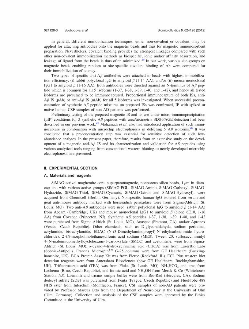
bilized ligand (Fig. 2). Therefore, the final decision as to which site-group would be used for
lIP was based upon how simple was the protocol providing random or site-specific binding of
IgG, whether the reagents were or were not harmful, and what was the IgG capacity on 1 mg of
the beads. Our results from SDS-PAGE (Fig. 3) and the bicine-choninic acid (BCA) test (Fig. S-1)32
show high binding capacity of immobilized IgG was achieved using hydrazide, cyanuric, or thiol
beads. In the end, microbeads with the hydrazide site-group were determined to be most suitable for
further experiments with specific anti-Ab IgG because they afforded a site-specific, covalent type of
IgG binding in combination with high density and reasonable binding protocol.
B. Optimization of elution step in IP of Ab peptides
Immunocaptured analytes are supposed to be released from the anti-Ab IS by using an elu-
tion reagent which ideally is compatible with the subsequent detection method. Toward this
end, various elution conditions using 0.05% TFA (pH 2.3), 0.25% formic acid (pH 2.35),
0.16% ammonium hydroxide (pH 11.0), 100 mM ammonium bicarbonate (pH 8.0), 6 M urea
(pH 8.2) and 2% SDS (pH 8.2) were tested. The determining criteria for evaluating elution effi-
ciency were the concentration of batchwise immunoprecipitated Ab peptides (1-38, 1-40, 1-42)
in elution fractions as analyzed by urea/tricine/tris SDS-PAGE (Fig. 4). Chaotropic reagents
such as urea and SDS in such concentrations were not compatible with subsequent analytical
methods, but efficient elution of all tested Ab isoforms was achieved using the other tested
reagents. Therefore, elution reagents employed for releasing of captured peptides were adapted
according to the analytical method that followed: For MALDI-TOF-MS analysis, 0.05% TFA
FIG. 2. Scheme of immobilization techniques employed for IgG on magnetic beads with various site-groups
024126-6 Svobodova et al. Biomicrofluidics 6, 024126 (2012)

was used, while for CE, chip electrophoresis and/or immunoblotting 0.16% NH4OH was pre-
ferred. For CE, moreover, 100 mM NH4HCO3 or 0.25% formic acid also was suitable.
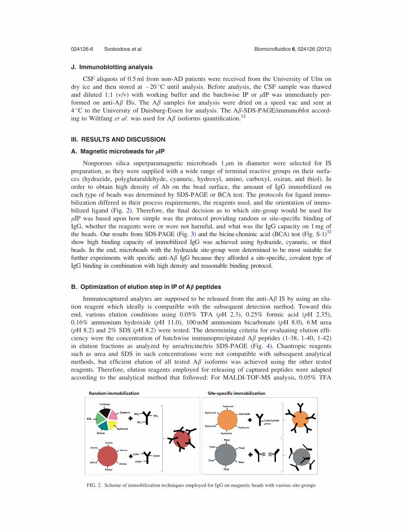
C. On-chip Ab preconcentration
On-chip IP (lIP) runs on anti-Ab IS self-organized in the channel when using two perma-
nent magnets to generate an affinity microcolumn (Fig. 5). The volume of the plug formed in
the channel was calculated from its height (0.25 mm) and width (1 mm) and length (2.33 mm).
The length may vary, thence 3 different images underwent the image analysis using software
NIS Elements AR (Nikon, Tokyo, J), where the length was measured in 5 different places of
plug and the average of the 15 measurements were calculated (2.33 6 0.44 mm). Final volume
of plug was determinated as 0.58 mm3.
Although batchwise IP and lIP include a number of the same steps, such as binding, wash-
ing, and elution, there also are some basic differences. What is considered as reaction or
incubation time in batchwise IP experiments can be transposed into flow rate in on-chip immu-
nocapture (lIP). Hence, the lIP process was monitored by dot blot to confirm that the lIP pro-
duced the same results as did conventional batchwise IP. This technique was selected because
it is a sensitive and quantitative method enabling easy monitoring of Ab 1-42 concentration at
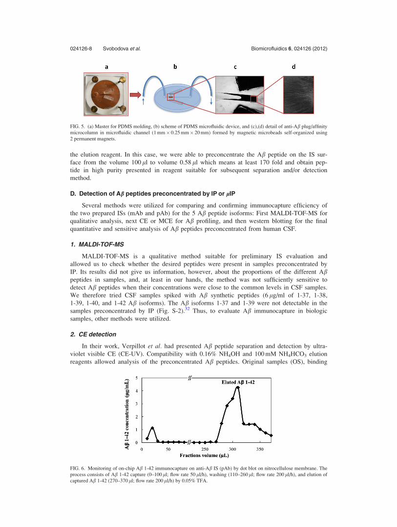
each particular step of lIP. The record of Ab 1-42 concentrations in collected fractions (10 ll)
from the chip device under optimized flow rates is presented in Fig. 6. This result clearly shows
that mostly all of the Ab 1-42 applied into the chip was biospecifically captured during the
binding step and efficiently released during the elution step.
Concentration factor of lIP setup may vary and depends on different parameters, such as
primer concentration of sample, sample volume, amount of the beads, flow rate, and volume of
FIG. 3. Binding efficiency (%) of SiMAG-active Chemicell microbeads (1 mg) with different site-groups for human serum
IgG (100 lg) immobilization as determined by SDS-PAGE (3 replicates)
FIG. 4. 16.5% urea/tricine/tris SDS-PAGE of Ab peptides (1-38, 1-40, 1-42) eluted by the various elution reagents: 0.05%
trifluoroacetic acid (TFA; pH 2.3), 0.25% formic acid (FA; pH 2.35), 0.16% ammonium hydroxide (OH; pH 11.0), and
100 mM ammonium bicarbonate (NH4HCO3; pH 8.0); molecular weight standard 1.4–26.6 kDa (MW, BioRad).
024126-7 Svobodova et al. Biomicrofluidics 6, 024126 (2012)
the elution reagent. In this case, we were able to preconcentrate the Ab peptide on the IS sur-
face from the volume 100 ll to volume 0.58 ll which means at least 170 fold and obtain pep-
tide in high purity presented in reagent suitable for subsequent separation and/or detection
method.
D. Detection of Ab peptides preconcentrated by IP or lIP
Several methods were utilized for comparing and confirming immunocapture efficiency of
the two prepared ISs (mAb and pAb) for the 5 Ab peptide isoforms: First MALDI-TOF-MS for
qualitative analysis, next CE or MCE for Ab profiling, and then western blotting for the final
quantitative and sensitive analysis of Ab peptides preconcentrated from human CSF.
1. MALDI-TOF-MS
MALDI-TOF-MS is a qualitative method suitable for preliminary IS evaluation and
allowed us to check whether the desired peptides were present in samples preconcentrated by
IP. Its results did not give us information, however, about the proportions of the different Abpeptides in samples, and, at least in our hands, the method was not sufficiently sensitive to
detect Ab peptides when their concentrations were close to the common levels in CSF samples.
We therefore tried CSF samples spiked with Ab synthetic peptides (6 lg/ml of 1-37, 1-38,
1-39, 1-40, and 1-42 Ab isoforms). The Ab isoforms 1-37 and 1-39 were not detectable in the
samples preconcentrated by IP (Fig. S-2).32 Thus, to evaluate Ab immunocapture in biologic
samples, other methods were utilized.
2. CE detection
In their work, Verpillot et al. had presented Ab peptide separation and detection by ultra-
violet visible CE (CE-UV). Compatibility with 0.16% NH4OH and 100 mM NH4HCO3 elution
reagents allowed analysis of the preconcentrated Ab peptides. Original samples (OS), binding
FIG. 5. (a) Master for PDMS molding, (b) scheme of PDMS microfluidic device, and (c),(d) detail of anti-Ab plug/affinity
microcolumn in microfluidic channel (1 mm� 0.25 mm� 20 mm) formed by magnetic microbeads self-organized using
2 permanent magnets.
FIG. 6. Monitoring of on-chip Ab 1-42 immunocapture on anti-Ab IS (pAb) by dot blot on nitrocellulose membrane. The
process consists of Ab 1-42 capture (0–100 ll; flow rate 50 ll/h), washing (110–260 ll; flow rate 200 ll/h), and elution of
captured Ab 1-42 (270–370 ll; flow rate 200 ll/h) by 0.05% TFA.
024126-8 Svobodova et al. Biomicrofluidics 6, 024126 (2012)

fraction (BF; supernatant with non-IP Ab peptides), and three successive elution fractions
(E1–3) from batch IP on anti-Ab IS (pAb) were analyzed by CE-UV (Fig. 7). Despite there
being the same initial concentration of each Ab isoform, we observed higher peak intensity for
eluted Ab 1-42 (66%) than for the shorter Ab isoforms 1-40 (23%) and 1-38 (9%). These
results were confirmed also by urea/tricine/tris SDS-PAGE, where the proportions of eluted Abpeptides were 74% for Ab 1-42, 21% for Ab 1-40, and 5% for Ab 1-38 (Table S-I).32 We thus
concluded that the anti-Ab IS (pAb) showed preferential capture of Ab 1-42. To confirm this
tendency, other methods for Ab detection were also employed.
3. MCE for comparing magnetic anti-Ab IS (mAb and pAb)
In our previous work, we presented an MCE method with fluorescent detection for profiling
of 5 Ab isoforms.28 Here, the MCE method was used for monitoring the lIP process by compar-
ing the ratio of Ab isoforms in the sample before the lIP and in the elution fraction using two
types of immunosorbents: one coated with mAb and one with pAb anti-Ab IgG (Fig. 8(a)). The
results presented in Figure 8(b) (average of 3 repetitions) show that when sample consisting of
Ab 1-37 and Ab 1-42 (Ab 1-37/Ab 1-42¼ 1.6) was immunoprecipitated, the Ab peptides were
eluted with different ratios according to the Ab-coated immunosorbents: pAb (0.6) and mAb
(0.8). In other words, the results show that both anti-Ab ISs (pAb and mAb) demonstrated
preferential capture of Ab 1-42 over Ab 1-37. Such tendency might be caused by their structural
difference and higher hydrophobicity of Ab 1-42 in comparison to the other 4 isoforms.33,34 This
feature might possibly change accessibility of the epitope for Ab, which is known as epitope
FIG. 7. CE-UV of Ab mixture of three synthetic peptides (1-38, 1-40, 1-42) from IP performed on anti-Ab IS (pAb): origi-
nal sample (OS; sample before IP), binding fraction (BF; sample with non-IP Ab peptides), and three successive elution
fractions (E1–3; fractions with IP Ab peptides eluted by 0.16% NH4OH). E1–3 show preferential capture of Ab 1-42. Ana-
lytical conditions were according to Verpillot et al. (2008).
FIG. 8. (a) Electropherogram from MCE of fluorescently labeled Ab 1-37 and 1-42 from lIP on anti-Ab IS (mAb);
FP¼FluoProbe. (b) Comparison of fluorescence intensity (FI) ratio of Ab 1-37/1-42 peptides in samples obtained from
lIPs on anti-Ab IS (pAb) and anti-Ab IS (mAb) (averages of 3 repetitions). OS¼ original sample (before IP).
024126-9 Svobodova et al. Biomicrofluidics 6, 024126 (2012)

masking. These observations were discussed by Wiltfang et al. in their study that compared their
results from Ab-SDS-PAGE/immunoblot with those of other authors determining Ab concentra-
tions in CSF.12 They reported finding that differences in Ab isoforms concentration were due to
the different types of antibodies that had been used. Thus, the origins of different antibodies, and
despite their having the same specificity, could create different reactivity with immunoprecipitated
peptides.
4. Immunoblotting of Ab captured by lIP from human CSF
Previous testing of both anti-Ab ISs was done with samples containing synthetic Ab pep-
tides or spiked CSF samples in concentration range higher than is usually found in CSF sam-
ples. The ability of both anti-Ab ISs (pAb and mAb) to capture Ab peptides from real human
CSF samples of non-AD patients was thus tested by immunoblotting since this method affords
sensitivity compatible with the concentration level of these peptides in biological samples. The
two ISs were compared to show proportional immunocapture of Ab isoforms in human CSF
samples (Fig. 9(a)). Both antibodies used were presumed to immunocapture all 5 Ab isoforms.
Significant differences between the two ISs were observed, however, as shown in the previous
results, with anti-Ab IS (pAb) having higher affinity for the Ab 1-42 isoform than for the other
Ab isoforms. The anti-Ab IS (mAb) provided a more uniform and efficient capture of all 5 Abisoforms from CSF.
The ability of anti-Ab IS (mAb) to preconcentrate the Ab isoforms from CSF on-chip was
also confirmed. The lIP of CSF sample was performed and the preconcentrated Ab peptides
were analyzed by immunoblotting (Fig. 9(b)). An IS with monoclonal antibodies showed a rep-
resentation of all Ab isoforms in proportions nearly similar to those present in the initial CSF
sample, albeit still with a slight preference for the Ab 1-42 isoform (see Table I).
FIG. 9. Ab peptides separated by Ab-SDS-PAGE/immunoblot on polyvinylidene fluoride membrane (according to Wilt-
fang et al. (2002)). (a) Batchwise IP of CSF sample on anti-Ab IS (pAb) and anti-Ab IS (mAb). CSF¼CSF sample before
IP; IP (pAb)¼ batchwise IP on anti-Ab IS (pAb); IP (mAb)¼ batchwise IP on anti-Ab IS (mAb). (b) lIP of CSF sample on
anti-Ab IS (mAb). Ab ST¼Ab standard peptides mixture corresponding to levels in CSF; lIP¼lIP on anti-Ab IS (mAb).
TABLE I. Quantification of amyloid b (Ab) isoforms in human CSF before and after lIP.
Aß isoforms CSF before lIP (ng/ml) CSF after lIP (ng/ml) CSF before lIP (%) CSF after lIP (%)
1-37 2.32 0.15 6.0 6.1
1-38 4.81 0.34 12.4 13.7
1-39 2.60 0.14 6.7 5.7
1-40 24.25 0.88 62.7 35.6
1-42 4.69 0.96 12.1 38.8
024126-10 Svobodova et al. Biomicrofluidics 6, 024126 (2012)

IV. CONCLUSION
Micro-immunoprecipitation is a suitable method for isolation and preconcentration of Abpeptide biomarkers occurring in low concentrations from CSF. Variability in elution reagents
allows releasing the target molecules in a medium compatible with the subsequent detection
method, be that performed by MALDI-TOF-MS, CE, MCE and/or immunoblotting. CE pro-
vided the best combination of sensitivity, convenience, and resolution, with a baseline resolu-
tion of the three isoforms investigated. Microchip electrophoresis, however, revealed promising
potential for on-line connection and/or integration of immunocapture and detection into a single
microfluidic device. A certain rate of Ab 1-42 preferential capture was observed for both inves-
tigated antibodies (mAb or pAb), although with anti-Ab IS (mAb) the representation of Abpeptides was much closer to the composition of the initial human CSF sample. The proportional
capture of Ab isoforms has great significance for the clinical diagnosis of AD and in research
as to the disease’s pathogenesis. Hence, this effect will be considered in our next investigation.
Our future work will also focus on developing an integrated lab-on-chip device for preconcen-
tration, separation, and detection of 5 Ab isoforms. In the aim to enhance the sensitivity of the
analysis, an additional preconcentration component is now under testing to be integrated into
the microfluidic device. Such system will be evaluated on samples from patients affected by
AD or other dementias and which might contribute in the future to rapid Ab profiling of
patients suspected of having AD and to their diagnosis.
ACKNOWLEDGMENTS
This work was supported by the European Union’s NADINE project under Contract No.
246513. Special thanks are due to H.-W. Klafki for his advice and suggestions related to IS
preparation.
1J. L. Cummings, R. Doody, and C. Clark, Neurology 69, 1622 (2007).2J. J Jalbert, L. A. Daiello, and K. L. Lapane, Epidemiol. Rev. 30, 15 (2008).3H. H. Feldman, B. Van Baelen, S. M. Kavanagh, and K. E. Torfs, Alzheimer Dis. Assoc. Discord. 19, 29 (2005).4R. Brookmeyer, S. Gray, and C. Kawas, Am. J. Public Health 88, 1337 (1998).5C. Haass, M. G. Schlossmacher, A. Y. Hung, C. Vigo-Pelfrey, A. Mellon, B. L. Ostaszewski, I. Lieberburg, E. H. Koo,D. Schenk, D. B. Teplow, and D. J. Selkoe, Nature 359, 322 (1992).
6P. Seubert, C. Vigo-Pelfrey, F. Esch, M. Lee, H. Dovey, D. Davis, S. Sinha, M. Schiossmacher, J. Whaley, C. Swindle-hurst, R. McCormack, R. Wolfert, D. Selkoe, I. Lieberburg, and D. Schenk, Nature 359, 325 (1992).
7N. Andreasen, L. Minthon, P. Davidsson, E. Vanmechelen, H. Vanderstichele, B. Winblad, and K. Blennow, Arch. Neu-rol. 58, 373 (2001).
8M. Kanai, E. Matsubara, K. Isoe, K. Urakami, K. Nakashima, H. Arai, H. Sasaki, K. Abe, T. Iwatsubo, T. Kosaka, M.Watanabe, Y. Tomidokoro, M. Shizuka, K. Mizushima, T. Nakamura, Y. Igeta, Y. Ikeda, M. Amari, T. Kawarabayashi,K. Ishiguro, Y. Harigaya, K. Wakabayashi, K. Okamoto, S. Hirai, and M. Shoji, Ann. Neurol. 44, 17 (1998).
9K. Blennow and H. Hampel, Lancet Neurol. 2, 605 (2003).10J. Kang, H. Lemaire, A. Unterbeck, J. M. Salbaum, C. L. Masters, K. Grzeschik, G. Multhaup, K. Beyreuther, and
B. Muller-Hill, Nature 325, 733 (1987).11T. Kawarabayashi and M. Shoji, Curr. Opin. Psychiatr. 21, 3 (2008).12J. Wiltfang, H. Esselmann, M. Bibl, A. Smirnov, M. Otto, S. Paul, B. Schmidt, H.-W. Klafki, M. Maler, T. Dyrks,
M. Bienert, M. Beyermann, E. Ruther, and J. Kornhuber, J. Neurochem. 81, 481 (2002).13M. J. De Leon, L. Mosconi, K. Blennow, S. DeSanti, R. Zinkowski, P. D. Mehta, D. Pratico, W. Tsui, L. A. Saint,
L. Sobanska, M. Brys, Y. Li, K. Rich, J. Rinne, and H. Rusinek, Ann. N. Y. Acad. Sci. 1097, 114 (2007).14D. Bottenus, T. Z. Jubery, Y. Ouyang, W.-J. Dong, P. Dutta, and C. F. Ivory, Lab Chip 11, 890 (2011).15T. K. Khurana and J. G. Santiago, Lab Chip 9, 1377 (2009).16M. R. Mohamadi, N. Kaji, M. Tokeshi, and Y. Baba, Anal.Chem. 79, 3667 (2007).17P. Gebauer and P. Bocek, Electrophoresis 23, 3858 (2002).18N. Lion, T. C. Rohner, L. Dayon, I. L. Arnaud, E. Damoc, N. Youhnovski, Z.-Y. Wu, CH. Roussel, J. Josserand, H. Jen-
sen, J. Rossier, M. Przybylski, and H. H. Girault, Electrophoresis 24, 3533 (2003).19N. J. Clarke, A. J. Tomlinson, Y. Ohyagi, S. Younkin, and S. Naylor, FEBS Lett. 430, 419 (1998).20V. Gelfanova, R. E. Higgs, R. A. Dean, D. M. Holtzman, M. R. Farlow, E. R. Siemers, A. Boodhoo, Y. Qian, X. He,
Z. Jin, D. L. Fisher, K. L. Cox, and J. E. Hale, Brief. Funct. Genomic. Proteomic. 6, 149 (2007).21E. Portelius, A. J. Tran, U. Andreasson, R. Persson, G. Brinkmalm, H. Zetterberg, K. Blennow, and A. Westman-Brinkmalm,
J. Proteome Res. 6, 4433 (2007).22T. Chen, J. Lei, and A. Tong, Luminescence 20, 256 (2005).23B. Kaboord and M. Perr, in Sample Preparation and Fractionation: Volume 1, edited by A. Posch (Humana, New York,
2008), p. 349.24J. Kim, M. Johnson, P. Hill, and B. K. Gale, Integr. Biol. 1, 574 (2009).25A. Le Nel, N. Minc, C. Smadja, M. Slovakova, Z. Bilkova, J. Peyrin, J. Viovy, and M. Taverna, Lab Chip 8, 294 (2008).
024126-11 Svobodova et al. Biomicrofluidics 6, 024126 (2012)

26P. Cuatrecasas, Science 184, 56 (1974).27B. Jankovicova, Z. Zverinova, M. R. Mohamadi, M. Slovakova, L. Hernychova, W. Faigle, J. Viovy, and Z. Bilkova, in
New Trends in Alzheimer and Parkinson Related Disorders, edited by A. Fisher and E. Hanin (Medimond S.r.l., Bologna,Italy, 2009), pp. 239–244.
28M. R. Mohamadi, Z. Svobodova, R. Verpillot, H. Esselmann, J. Wiltfang, M. Otto, M. Taverna, Z. Bilkova, and J. Viovy,Anal. Chem. 82, 7611 (2010).
29M. Slovakova, N. Minc, Z. Bilkova, C. Smadja, W. Faigle, C. Futterer, M. Taverna, and J. Viovy, Lab Chip 5, 935(2005).
30H.-W. Klafki, J. Wiltfang, and M Staufenbiel, Anal. Biochem. 237, 24 (1996).31R. Verpillot, M. Otto, H.-W. Klafki, and M. Taverna, J. Chromatogr. A 1214, 157 (2008).32See supplementary material at http://dx.doi.org/10.1063/1.4722588 for Fig. S-1, Fig. S-2, and Table S-I.33J. T. Jarrett, E. P. Berger, and P. T. Lansbury, Jr., Biochemistry 32, 4693 (1993).34C. Soto, M. C. Branes, J. Alvarez, and N. C. Inestrosa, J. Neurochem. 63, 1191 (1994).
024126-12 Svobodova et al. Biomicrofluidics 6, 024126 (2012)
Copyright © 2022 FDOKUMEN