Development and evaluation of NucliSens® Basic Kit NASBA for diagnosis of parainfluenza virus...
11
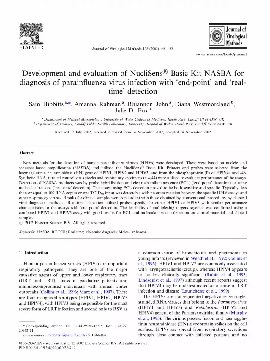
Development and evaluation of NucliSens † Basic Kit NASBA for diagnosis of parainfluenza virus infection with ‘end-point’ and ‘real- time’ detection Sam Hibbitts a, *, Amanna Rahman a , Rhiannon John a , Diana Westmoreland b , Julie D. Fox a a Department of Medical Microbiology, University of Wales College of Medicine, Heath Park, Cardiff CF14 4XN, UK b Department of Virology, Cardiff Public Health Laboratory, University Hospital of Wales, Heath Park, Cardiff CF14 4XW, UK Received 19 July 2002; received in revised form 14 November 2002; accepted 14 November 2002 Abstract New methods for the detection of human parainfluenza viruses (HPIVs) were developed. These were based on nucleic acid sequence-based amplification (NASBA) and utilised the NucliSens † Basic Kit. Primers and probes were selected from the haemagglutinin neuraminidase (HN) gene of HPIV1, HPIV2 and HPIV3, and from the phosphoprotein (P) of HPIV4a and -4b. Synthetic RNA, titrated control virus stocks and respiratory specimens (n /44) were utilised to evaluate performance of the assays. Detection of NASBA products was by probe hybridisation and electrochemiluminescence (ECL) (‘end-point’ detection) or using molecular beacons (‘real-time’ detection). The assays using ECL detection proved to be both sensitive and specific. Typically, less than or equal to 100 RNA copies or one TCID 50 input was detectable with no cross-reaction between the specific HPIV assays and other respiratory viruses. Results for clinical samples were concordant with those obtained by ‘conventional’ procedures by classical viral diagnostic methods. ‘Real-time’ detection utilised probes specific for either HPIV1 or HPIV3 with similar performance characteristics to the assays with ‘end-point’ detection. The feasibility of multiplexing targets together was confirmed using a combined HPIV1 and HPIV3 assay with good results for ECL and molecular beacon detection on control material and clinical samples. # 2002 Elsevier Science B.V. All rights reserved. Keywords: NASBA; RT-PCR; Real-time; Molecular diagnosis; Molecular beacon 1. Introduction Human parainfluenza viruses (HPIVs) are important respiratory pathogens. They are one of the major causative agents of upper and lower respiratory tract (URT and LRT) illness in paediatric patients and immunocompromised individuals with annual winter outbreaks (Collins et al., 1996; Marx et al., 1997). There are four recognised serotypes (HPIV1, HPIV2, HPIV3 and HPIV4), with HPIV3 being responsible for the most severe form of LRT infection and second only to RSV as a common cause of bronchiolitis and pneumonia in young infants (reviewed in Wendt et al., 1992; Collins et al., 1996). HPIV1 and HPIV2 are commonly associated with laryngotracheitis (croup), whereas HPIV4 appears to be less clinically significant (Rubin et al., 1993; Lindsquist et al., 1997) although recent reports suggest that HPIV4 may be underestimated as a cause of LRT infection and disease (Laurichesse et al., 1999). The HPIVs are nonsegmented negative sense single- stranded RNA viruses that belong to the Paramyxovirus (HPIV1 and HPIV3) and Rubulavirus (HPIV2 and HPIV4) genera of the Paramyxoviridae family (Murphy et al., 1995). The virions possess fusion and haemagglu- tinin neuraminidase (HN) glycoprotein spikes on the cell surface. HPIVs are spread from respiratory secretions through close contact with infected patients and no * Corresponding author. Tel.: /44-29-20742715; fax: /44-29- 20742161 E-mail address: [email protected] (S. Hibbitts). Journal of Virological Methods 108 (2003) 145 /155 www.elsevier.com/locate/jviromet 0166-0934/02/$ - see front matter # 2002 Elsevier Science B.V. All rights reserved. PII:S0166-0934(02)00268-9
-
Upload
independent -
Category
Documents
-
view
8 -
download
0
Transcript of Development and evaluation of NucliSens® Basic Kit NASBA for diagnosis of parainfluenza virus...
Development and evaluation of NucliSens† Basic Kit NASBA fordiagnosis of parainfluenza virus infection with ‘end-point’ and ‘real-
time’ detection
Sam Hibbitts a,*, Amanna Rahman a, Rhiannon John a, Diana Westmoreland b,Julie D. Fox a
a Department of Medical Microbiology, University of Wales College of Medicine, Heath Park, Cardiff CF14 4XN, UKb Department of Virology, Cardiff Public Health Laboratory, University Hospital of Wales, Heath Park, Cardiff CF14 4XW, UK
Received 19 July 2002; received in revised form 14 November 2002; accepted 14 November 2002
Abstract
New methods for the detection of human parainfluenza viruses (HPIVs) were developed. These were based on nucleic acid
sequence-based amplification (NASBA) and utilised the NucliSens† Basic Kit. Primers and probes were selected from the
haemagglutinin neuraminidase (HN) gene of HPIV1, HPIV2 and HPIV3, and from the phosphoprotein (P) of HPIV4a and -4b.
Synthetic RNA, titrated control virus stocks and respiratory specimens (n�/44) were utilised to evaluate performance of the assays.
Detection of NASBA products was by probe hybridisation and electrochemiluminescence (ECL) (‘end-point’ detection) or using
molecular beacons (‘real-time’ detection). The assays using ECL detection proved to be both sensitive and specific. Typically, less
than or equal to 100 RNA copies or one TCID50 input was detectable with no cross-reaction between the specific HPIV assays and
other respiratory viruses. Results for clinical samples were concordant with those obtained by ‘conventional’ procedures by classical
viral diagnostic methods. ‘Real-time’ detection utilised probes specific for either HPIV1 or HPIV3 with similar performance
characteristics to the assays with ‘end-point’ detection. The feasibility of multiplexing targets together was confirmed using a
combined HPIV1 and HPIV3 assay with good results for ECL and molecular beacon detection on control material and clinical
samples.
# 2002 Elsevier Science B.V. All rights reserved.
Keywords: NASBA; RT-PCR; Real-time; Molecular diagnosis; Molecular beacon
1. Introduction
Human parainfluenza viruses (HPIVs) are important
respiratory pathogens. They are one of the major
causative agents of upper and lower respiratory tract
(URT and LRT) illness in paediatric patients and
immunocompromised individuals with annual winter
outbreaks (Collins et al., 1996; Marx et al., 1997). There
are four recognised serotypes (HPIV1, HPIV2, HPIV3
and HPIV4), with HPIV3 being responsible for the most
severe form of LRT infection and second only to RSV as
a common cause of bronchiolitis and pneumonia in
young infants (reviewed in Wendt et al., 1992; Collins et
al., 1996). HPIV1 and HPIV2 are commonly associated
with laryngotracheitis (croup), whereas HPIV4 appears
to be less clinically significant (Rubin et al., 1993;
Lindsquist et al., 1997) although recent reports suggest
that HPIV4 may be underestimated as a cause of LRT
infection and disease (Laurichesse et al., 1999).
The HPIVs are nonsegmented negative sense single-
stranded RNA viruses that belong to the Paramyxovirus
(HPIV1 and HPIV3) and Rubulavirus (HPIV2 and
HPIV4) genera of the Paramyxoviridae family (Murphy
et al., 1995). The virions possess fusion and haemagglu-
tinin neuraminidase (HN) glycoprotein spikes on the cell
surface. HPIVs are spread from respiratory secretions
through close contact with infected patients and no
* Corresponding author. Tel.: �/44-29-20742715; fax: �/44-29-
20742161
E-mail address: [email protected] (S. Hibbitts).
Journal of Virological Methods 108 (2003) 145�/155
www.elsevier.com/locate/jviromet
0166-0934/02/$ - see front matter # 2002 Elsevier Science B.V. All rights reserved.
PII: S 0 1 6 6 - 0 9 3 4 ( 0 2 ) 0 0 2 6 8 - 9
vaccines are currently available to prevent infection
(Murphy et al., 1988).
Diagnosis of HPIV infection can be confirmed either
by identification of the virus in cell culture or directdetection in respiratory secretions using antigen or
nucleic acid detection methods (Downham et al., 1974;
Sarkkinen et al., 1981; Hierholzer et al., 1989; Shen et
al., 1996). Alternatively, monitoring the specific IgG and
IgM antibodies in serum can be used to confirm HPIV
infection.
In recent years, molecular assays based on reverse
transcribed-polymerase chain reaction (RT-PCR) havebeen reported for detection of HPIV-specific RNA
(Echevarria et al., 1998; Fan et al., 1998; Aguilar et
al., 2000; Kehl et al., 2001). These assays, in general,
have been shown to have good sensitivity and specifi-
city. The use of nucleic acid sequence-based amplifica-
tion (NASBA), which has certain advantages over RT-
PCR for amplification and detection of RNA sequences
(Chan and Fox, 1999), has not been applied previouslyto the diagnosis of HPIV infections. NASBA is an
isothermal nucleic acid amplification method that
amplifies RNA in a manner analogous to the replication
of retroviruses (Compton, 1991; Kievits et al., 1991).
The NASBA reaction mixture contains oligonucleotide
primers and three enzymes: avian myeloblastosis virus-
reverse transcriptase (AMV-RT), RNase H and T7
RNA polymerase for target-specific amplification (Gua-telli et al., 1990). This process occurs at 41 8C and
results in the exponential amplification of products
within 90 min, producing single-stranded RNA of
opposite sense to the original target as the major
amplification product. Detection of NASBA products
has been reported using a probe-capture hybridisation
and electrochemiluminescence (ECL) (Compton, 1991;
Chan and Fox, 1999) and, more recently, ‘real-time’detection using molecular beacons has been described
(Leone et al., 1998).
Molecular beacons are designed to have a loop region
containing a probe sequence that is complementary to
the target amplicon and a stem is formed by the
annealing of complementary arm sequences that are
located on either end of the probe sequence (Tyagi and
Kramer, 1996). A fluorophore is covalently linked toone arm and a quencher to the other. Molecular beacons
do not fluoresce when free in solution but have been
utilised for ‘real-time’ detection of amplified products.
When the probe sequence within the loop hybridises to
its target to form a rigid double helix, a conformational
change occurs that separates the quencher from the
fluorophore enabling fluorescence to occur.
We report new procedures for detection of HPIVsfrom clinical samples using the NASBA technology,
with sensitivity based on ‘end-point’ detection for both
synthetic RNA and titrated viruses. NASBA amplicon
products were detected by an ‘end-point’ procedure
(ECL) or, in the case of HPIV1 and HPIV3, also ‘real-
time’ using molecular beacons.
2. Materials and methods
2.1. Virus isolates
Cell culture isolates of reference strains were obtained
from the American Type Culture Collection (ATCC,
University Boulevard, Manassas) and used to standar-dise and develop the NASBA assays. ATCC prototype
strains utilised were HPIV1 (C-35), HPIV2 (Greer),
HPIV3 (MK9), HPIV4a (M-25) and HPIV4b (CH-
19503).
2.2. Clinical samples
Clinical samples (total n�/44) used to evaluate the
NASBA assays included throat swabs (n�/5) and
nasopharyngeal aspirate (NPA) samples (n�/39) taken
as diagnostic samples from paediatric inpatients with
respiratory symptoms between November 2000 and
April 2001. HPIV2-positive samples (n�/6) were ob-
tained from Leiden University Medical Centre (see
acknowledgements) and one sample was identified aspossibly HPIV4 positive in another laboratory (see
acknowledgements). Swab material was collected into
viral transport medium and aspirates were diluted in
phosphate-buffered saline (PBS) using standard proce-
dures prior to storage. Local ethical approval guidelines
were followed for use of clinical material and access to
diagnostic results. All throat swabs and NPA samples
were subjected to conventional culture and directantigen detection using well-established procedures
(Forghani and Hagens, 1995). Clinical samples were
inoculated into a primary monkey kidney cell line over 1
week, with alternate day analysis by haemadsorption
using human O-type red blood cells and confirmation of
positive haemadsorption by immunofluorescence using
an HPIV-specific monoclonal antibody. Two HPIV1,
six HPIV2, ten HPIV3 and one HPIV4 clinical sampleswere culture- and or antigen-positive for the respective
virus. A control-positive clinical specimen for each of
the other main respiratory virus targets was included in
the study (influenza A, influenza B, respiratory syncytial
virus (RSV) and adenovirus). Twenty-one of the clinical
specimens did not contain detectable respiratory viruses
by ‘conventional’ procedures, although they were ob-
tained during the same time frame as the HPIV clinicalpositive samples. All clinical samples were aliquoted and
stored at �/80 8C prior to testing for HPIV sequences,
retrospectively by NASBA.
S. Hibbitts et al. / Journal of Virological Methods 108 (2003) 145�/155146
2.3. Production of primary virus stocks
Culture of cell lines, propagation of viruses and
confirmation by direct antigen staining was undertakenin a similar way to that described previously (Collins et
al., 1996; Hierholzer et al., 1989; Shen et al., 1996).
Monkey African green kidney epithelial (BSC-1) cells
were used for primary viral isolation of HPIV2 and
HPIV3 (obtained from ATCC) and primary monkey
kidney cells (European Collection of Cell Cultures,
CAMR, Wiltshire, UK) for the propagation of
HPIV1, HPIV4a and HPIV4b. When a cytopathic effect(CPE) was observed or after 10 days, the monolayer was
scraped and tested for respiratory viruses using an
indirect immunofluorescence assay with HPIV-specific
monoclonal antibodies (Chemicon International, Har-
row, UK).
Virus titres were determined based on the 50% tissue
culture infective dose (TCID50) assay by infecting target
cells with serial 10-fold dilutions of each virus stock.Infected cells were incubated at 37 8C and CPE
monitored on a daily basis. The TCID50 was calculated
using the method of Reed and Muench (1938). In this
procedure, the number of infected and uninfected wells
at each dilution was compared and the dilution giving
50% infected wells was estimated. HPIV4a and -4b did
not give clear-cut CPE and therefore it was not possible
to determine end-points or calculate TCID50, and thusserial dilutions of the virus stock were evaluated in the
HPIV4-specific NASBA assay.
2.4. Primers and probes
For design of primers specific for HPIV1, HPIV2 and
HPIV3 the sequences of the HN gene obtained from
GenBank were aligned and conserved regions identified.The phosphoprotein (P) gene sequences were analysed
for HPIV4 primer design for single detection of both A
and B subtypes. Alignment was undertaken using
DNAsis software (Hitachi Software Engineering Co.
Ltd., San Francisco, CA). Molecular beacons were
designed for HPIV1 and HPIV3 based on the probe
sequence with additional sequences at either end to form
the stem structure of the molecular beacon. The stabilityand predicted structure of the beacons were analysed by
using the European MFOLD server (http://bibiserv.-
techfak.uni-bielefeld.de/mfold/). The 5? ends of the
molecular beacons were labelled with the fluorescent
dye fluorescein (FAM) and the 3? ends with the non-
fluorescent quencher 4-(4?-dimethylaminophenylazo)-
benzoic acid (DABCYL). Oligonucleotide primers and
labelled probes were synthesised and HPLC purified(Oswel DNA Services, Southampton, UK) before use in
RT-PCR or NASBA. Sequences of primers and probes
are given in Table 1.
2.5. RNA extraction
Extraction of RNA from cultured HPIVs of known
titre and clinical samples was carried out according tothe method described by Boom et al. (1990). This
procedure utilised the NucliSens† extraction kit accord-
ing to the manufacturer’s instructions (BioMerieux Ltd.,
Boxtel, The Netherlands). Briefly, 100 ml of virus stock
or clinical material (transport medium into which a
swab had been placed or aspirate material in PBS) was
added to 900 ml of lysis buffer. 50 ml silica suspension
was added to the lysis buffer to bind the nucleic acids.After washing and drying of the silica, the nucleic acids
were eluted with 50 ml of elution buffer, aliquoted and
stored at �/80 8C.
2.6. Cloning and in vitro transcription
Secondary structures were removed from the ex-
tracted RNA obtained from each of the HPIVs by
heating at 99 8C for 5 min and then immediatelyplacing on ice. The RT reaction was undertaken in a
20 ml reaction volume containing the template extract (5
ml), 25 mM Tris�/HCl (pH 8.3), 50 mM KCl, 5 mM
MgCl2, 2 mM DTT (RT buffer, BioGene Ltd., Cam-
bridge, UK), 0.25 mM each deoxynucleoside tripho-
sphate (dNTP) (Amersham�/Pharmacia, Little Chalfont,
Buckinghamshire, UK), 20 U of RNase inhibitor
(Promega, Southampton, UK), 10 U of AMV-RT(BioGene Ltd.) and 1 mM virus-specific RT reverse
primer (Table 1) at 43 8C for 1 h.
The PCR used 5 ml of the synthesised cDNA in a total
reaction volume of 50 ml that included 10 mM Tris�/HCl
(pH 8.3), 50 mM KCl, 1.5�/3 mM MgCl2 (optimised for
each target), 0.25 mM each dNTP, 2 U of Taq DNA
polymerase (all from Amersham Biosciences) with 1 mM
each virus-specific RT-PCR primers (Table 1). Sampleswere overlaid with mineral oil (Sigma Chemical Com-
pany, Poole, UK). The PCR protocol involved an initial
denaturation step at 94 8C for 4 min and then samples
were subjected to 30 cycles of amplification, each
consisting of 1 min at 94 8C, 1 min at 55�/60 8C(optimised for each target) and 2 min at 72 8C with a
final extension of 72 8C for 7 min in an MJ research
PCR machine (Genetic Research Instruments, Baintree,Essex, UK). PCR products were analysed by standard
ethidium bromide-stained agarose gel electrophoresis
(2% w/v).
PCR products obtained from the prototype strains of
HPIVs were cloned directly into pCR II-TOPO†
(Invitrogen, Inchinnan, Renfrewshire) by following the
manufacturer’s instructions. Plasmids were transformed
into high-efficiency competent cells (TOP10F?; Invitro-gen) by chemical transformation. Transformants were
‘colour’-screened on indicator plates (LB plate with 50
mg/ml ampicillin, 80 mg/ml X-gal and 0.5 mM IPTG) and
S. Hibbitts et al. / Journal of Virological Methods 108 (2003) 145�/155 147
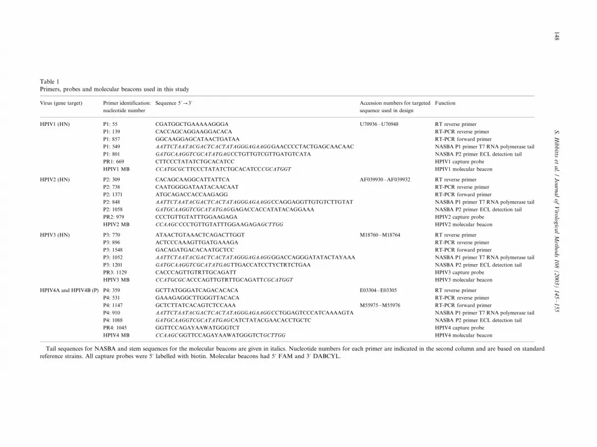
Table 1
Primers, probes and molecular beacons used in this study
Virus (gene target) Primer identification:
nucleotide number
Sequence 5?0/3? Accession numbers for targeted
sequence used in design
Function
HPIV1 (HN) P1: 55 CGATGGCTGAAAAAGGGA U70936�/U70948 RT reverse primer
P1: 139 CACCAGCAGGAAGGACACA RT-PCR reverse primer
P1: 857 GGCAAGGAGCATAACTGATAA RT-PCR forward primer
P1: 549 AATTCTAATACGACTCACTATAGGGAGAAGG GAACCCCTACTGAGCAACAAC NASBA P1 primer T7 RNA polymerase tail
P1: 801 GATGCAAGGTCGCATATGAG CCTGTTGTCGTTGATGTCATA NASBA P2 primer ECL detection tail
PR1: 669 CTTCCCTATATCTGCACATCC HPIV1 capture probe
HPIV1 MB CCATGCGC TTCCCTATATCTGCACATCCCGCATGGT HPIV1 molecular beacon
HPIV2 (HN) P2: 309 CACAGCAAGGCATTATTCA AF039930�/AF039932 RT reverse primer
P2: 738 CAATGGGGATAATACAACAAT RT-PCR reverse primer
P2: 1371 ATGCAGACCACCAAGAGG RT-PCR forward primer
P2: 848 AATTCTAATACGACTCACTATAGGGAGAAGG CCAGGAGGTTGTGTCTTGTAT NASBA P1 primer T7 RNA polymerase tail
P2: 1058 GATGCAAGGTCGCATATGAG GAGACCACCATATACAGGAAA NASBA P2 primer ECL detection tail
PR2: 979 CCCTGTTGTATTTGGAAGAGA HPIV2 capture probe
HPIV2 MB CCAAGC CCCTGTTGTATTTGGAAGAGAGCTTGG HPIV2 molecular beacon
HPIV3 (HN) P3: 770 ATAACTGTAAACTCAGACTTGGT M18760�/M18764 RT reverse primer
P3: 896 ACTCCCAAAGTTGATGAAAGA RT-PCR reverse primer
P3: 1548 GACAGATGACACAATGCTCC RT-PCR forward primer
P3: 1052 AATTCTAATACGACTCACTATAGGGAGAAGG GGACCAGGGATATACTAYAAA NASBA P1 primer T7 RNA polymerase tail
P3: 1201 GATGCAAGGTCGCATATGAG TTGACCATCCTYCTRTCTGAA NASBA P2 primer ECL detection tail
PR3: 1129 CACCCAGTTGTRTTGCAGATT HPIV3 capture probe
HPIV3 MB CCATGCGC ACCCAGTTGTRTTGCAGATTCGCATGGT HPIV3 molecular beacon
HPIV4A and HPIV4B (P) P4: 359
P4: 531
GCTTATGGGATCAGACACACA
GAAAGAGGCTTGGGTTACACA
E03304�/E03305 RT reverse primer
RT-PCR reverse primer
P4: 1147 GCTCTTATCACAGTCTCCAAA M55975�/M55976 RT-PCR forward primer
P4: 910 AATTCTAATACGACTCACTATAGGGAGAAGG CCTGGAGTCCCATCAAAAGTA NASBA P1 primer T7 RNA polymerase tail
P4: 1088 GATGCAAGGTCGCATATGAG CATCTATACGAACACCTGCTC NASBA P2 primer ECL detection tail
PR4: 1045 GGTTCCAGAYAAWATGGGTCT HPIV4 capture probe
HPIV4 MB CCAAGC GGTTCCAGAYAAWATGGGTCTGCTTGG HPIV4 molecular beacon
Tail sequences for NASBA and stem sequences for the molecular beacons are given in italics. Nucleotide numbers for each primer are indicated in the second column and are based on standard
reference strains. All capture probes were 5? labelled with biotin. Molecular beacons had 5? FAM and 3? DABCYL.
S.
Hib
bitts
eta
l./
Jo
urn
al
of
Viro
log
ical
Meth
od
s1
08
(2
00
3)
14
5�
/15
51
48
the presence of the expected inserts confirmed by PCR.
Plasmids were purified with the SNAP miniprep kit
(Invitrogen) and the plasmid copy number estimated by
UV spectroscopy at an optical density of 260 nm.
Plasmids (ca 1�/2 mg) containing the inserts from each
of the HPIVs were linearised using either EcorV
(HPIV1, HPIV2 and HPIV4) or NotI (HPIV3) and
analysed by agarose gel electrophoresis to ensure that
complete digestion had occurred. In vitro transcription
of each template utilised the SP6 MAXIscript† kit
(Ambion Europe Ltd., Huntingdon, Cambridgeshire)
with 1 mg of DNA in a final reaction volume of 20 ml
according to the manufacturer’s instructions. The RNA
copy number was estimated by UV spectroscopy at an
optical density of 260 nm, using 330 as the estimated
molecular weight of each deoxynucleotide and Avoga-
dro’s number to give copies per mole.
2.7. NASBA set-up for ‘end-point’ detection
NASBA reactions were carried out according to
Kievits et al. (1991) with some modifications. The
NucliSens† Basic Kit (BioMerieux Ltd.) was used
according to the manufacturer’s instructions. Briefly,
to the reagent sphere, 80 ml of diluent, 16 ml of molecular
grade water, 14 ml of stock KCl (final concentration 70
mM) and 5 ml of each of the required primers (5 mM
stock of each) were added (see Table 1 for sequences).
The 3? sequence of primer 1 complements the target
RNA and the 5? terminal contains a T7 RNA poly-
merase promoter sequence. The second primer is
complementary to the DNA sequence that is produced
by extension from primer 1 at the 3? end and an ECL
detection sequence is at the 5? terminal. The amplifica-
tion solution was aliquoted into 10 ml/reaction and 5 ml
of extracted nucleic acid added. The appropriate ex-
tracted HPIV (positive) and water (negative) controls
were included in each assay. The reactions were
incubated at 65 8C for 5 min to destabilise secondary
structures in RNA and then held for 5 min at 41 8C.
Subsequently, 5 ml of NASBA enzyme solution was
added to initiate amplification. Enzyme was initially
supplied in liquid form but later supply was switched to
lyophilised spheres that were re-constituted according to
manufacturer’s instructions before addition. Each assay
was re-assessed with the lyophilised enzyme and optimal
results were obtained with a final KCl concentration of
80 mM when this new preparation was utilised. The
NASBA mix was incubated for further 10 min on the
41 8C heating block. All samples were then transferred
to a 41 8C water bath for 90 min to complete
amplification by the simultaneous activity of AMV-
RT, RNAse H and T7 RNA polymerase. Products were
stored at �/80 8C until required for detection by ECL.
2.8. ECL detection of amplified NASBA products
Detection is based upon a voltage-induced oxidation�/
reduction reaction involving an immobilised NASBAproduct. Biotinylated capture probes were designed to
be specific for each of the HPIV NASBA products
(Table 1) and were coupled to streptavidin-coated beads
according to well-established methods using molecular
grade reagents supplied by Sigma Chemical Co., mag-
netic beads supplied in the Basic Kit and a magnetic
particle concentrator (Dynal A.S., Oslo, Norway).
Capture beads were hybridised to the RNA targetusing the reagents supplied in the NucliSens† Detection
Kit (BioMerieux Ltd.). The RNA target was then
captured on to the magnetic beads and the complex
immobilised on an electrode by a magnet. In the Basic
Kit format of NASBA, a generic ruthenium-labelled
detection probe (supplied in the kit) is hybridised to the
target. When the voltage is applied to the electrode, the
ECL signal generated gives an indication of hybridisa-tion and a photo-multiplier tube detects photon emis-
sion at 620 nm. The results were analysed using the
software supplied with the ECL detection machine and
the cut-off for the assay (determined experimentally
using known positive and negative samples) was set at
0.01�/ reference solution (RS) value for each assay.
2.9. NASBA with molecular beacon ‘real-time’ detection
The reaction was prepared as described in Section 2.7
with some minor modifications. The amount of water
added was reduced to 13.5 ml, and 2.5 ml of molecular
beacon (final optimised concentration of 0.1 mM) was
added to the reagent sphere along with the other
components. 10 ml of the mix was added to each tube
and 5 ml of sample added prior to incubation on a PCR
machine for 5 min at 65 8C and then 5 min at 41 8C.Enzyme was then added to each sample tube, vortexed
and amplified, and analysis of results was undertaken
using the NucliSens† Easy Q Analyser (BioMerieux
Ltd.). Development of fluorescence was monitored in
closed tubes for 90 min at 41 8C.
2.10. NASBA multiplex reactions with ‘real-time’
detection
The HPIV1 and HPIV3 multiplex reaction was
prepared as described in Sections 2.7 and 2.9 with
some minor modifications. Both sets of HPIV1 and
HPIV3 NASBA primers were premixed to give a
concentration of 5 mM of each primer in a 10 ml volume
(added to the total reaction volume of 20 ml). The
amount of water added was reduced to 13.4 ml, and 1.3ml of the HPIV1- and HPIV3-specific molecular beacons
(final concentration of 0.1 mM for each beacon) added
to the reagent sphere along with the other components.
S. Hibbitts et al. / Journal of Virological Methods 108 (2003) 145�/155 149
The master mix was aliquoted, samples added and
subsequent steps of incubation, amplification and ana-
lysis occurred as outlined in Section 2.9.
3. Results
Studies on virus stocks, synthetic RNA and clinical
samples confirmed the specificity of the individual HPIV
assays for their target sequence with no cross-reactivity
observed with adenovirus, influenza types A or B or
RSV-positive samples. Sensitivity of all assays (utilising
‘end-point’ and ‘real-time’ detection) was good.
3.1. Sensitivity of ECL assays
The sensitivity of each NASBA HPIV assay was
determined using reference virus strains. The TCID50 of
HPIV1�/3 virus stocks was determined and serial dilu-
tions equivalent to final input into NASBA of 1�/104�/
1�/10�2 TCID50 were made in guanidinium lysis bufferbefore extraction. For HPIV4a and -4b, the end-point in
TCID50 assay was not clear and so results were
expressed as dilution of the culture added into the
reaction. NASBA was performed on each of the extracts
for each assay and the cut-off point between a negative
and positive ECL signal identified.
In conjunction with experiments using stock viruses
for assessment of sensitivity, in vitro RNA transcriptswere produced for each of the HPIVs as described in
Section 2.6. RT-PCR for nucleic acid extracts from each
of the reference strains produced fragments of the
expected band sizes. These were 739, 655, 672, 638 and
638 bp for HPIV1, HPIV2, HPIV3, HPIV4a and
HPIV4b, respectively. The number of RNA copies for
each virus was calculated and serial dilutions prepared
such that 1�/104�/1�/10�2 RNA copies per reactionwere present. These dilutions of RNA were added
directly into the NASBA mix. The sensitivity of the
HPIV1, -2 and -3 assays was within the range 0.001�/1
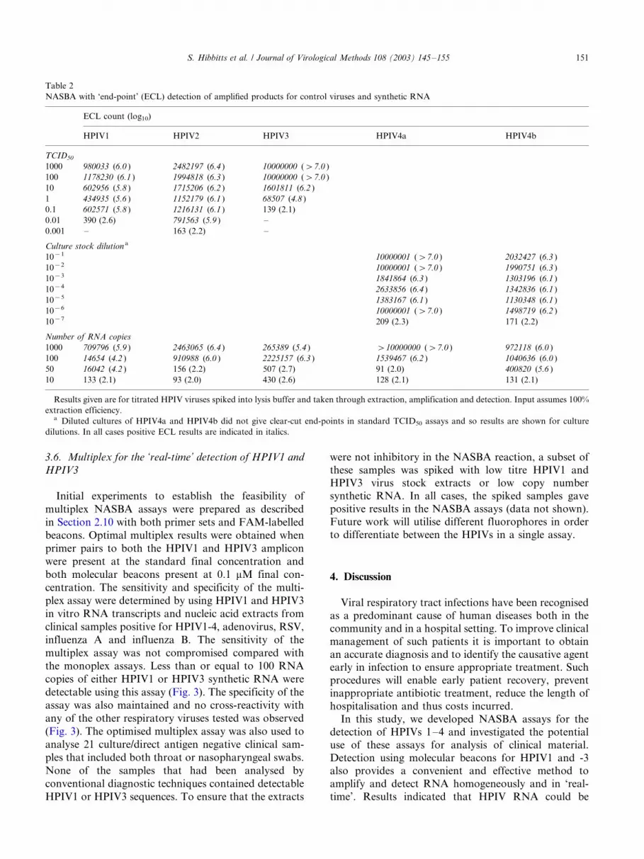
TCID50 virus input (Table 2). For HPIV4a and -4b, a
culture dilution between 1�/10�6 and 1�/10�7, prior
to extraction, gave good ECL counts and there was a
clear cut-off between positive and negative (Table 2). An
input of between 10 and 100 copies of synthetic RNAfor each of the HPIVs was sufficient to give a positive
ECL signal (Table 2).
3.2. Specificity of ECL assays
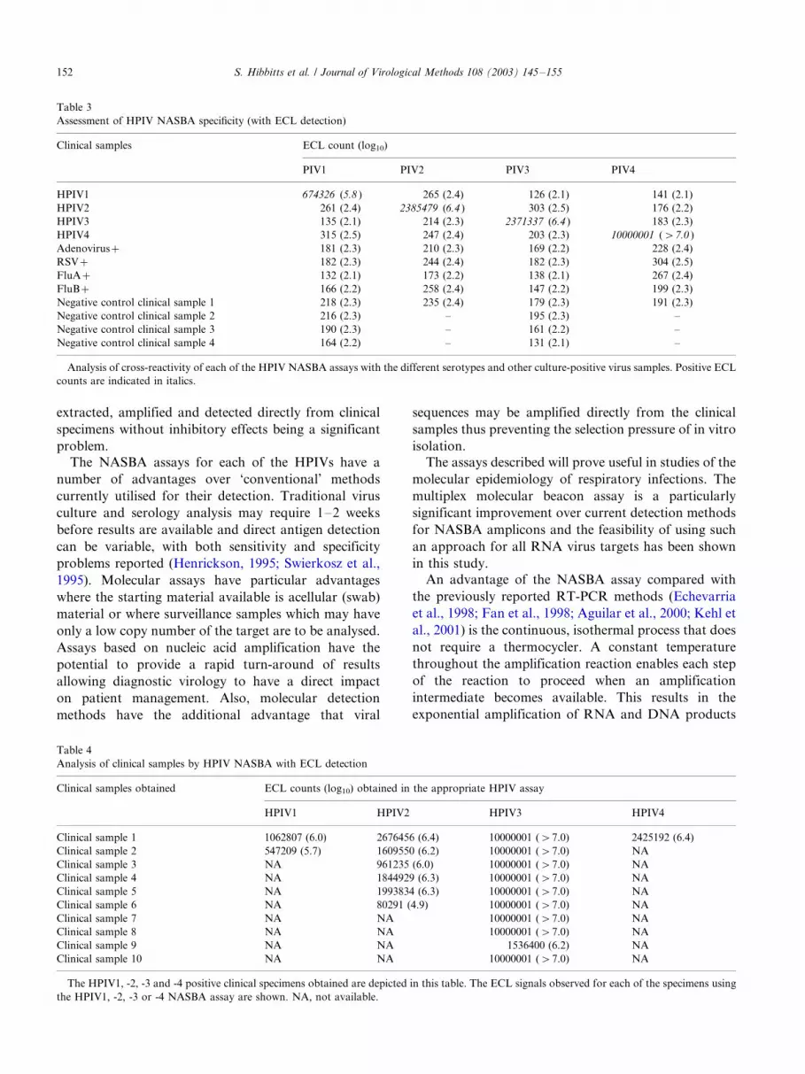
The specificity of each HPIV assay was evaluated with
diagnostic respiratory samples including adenovirus,
RSV, influenza A and influenza B positive material.No signal above background was detected for any HPIV
assay with samples containing other respiratory viruses.
Cross-reactivity of each HPIV assay with the other virus
serotypes was also assessed using high titre virus stock
preparations and each primer set and probe led to
specific detection of the HPIV type for which they had
been designed (Table 3).
3.3. NASBA with ECL detection for the analysis of
clinical samples
Respiratory samples collected from patients at the
University Hospital of Wales and Leiden University
Medical Centre (see acknowledgements) were analysed
using the developed NASBA assays. Two HPIV1, six
HPIV2 and 10 HPIV3 samples were identified that werepositive by conventional procedures. One sample, pro-
visionally identified as HPIV4 by culture and neutralisa-
tion, was also obtained (see acknowledgements). In all
cases, the HPIV-positive clinical specimens analysed in
the appropriate NASBA assay gave ECL counts clearly
above background and results are shown in Table 4.
Interestingly, two of the HPIV3 clinical samples were
throat swabs and eight were NPAs yet all of themyielded high ECL counts (�/1 536 400) irrespective of
sample type.
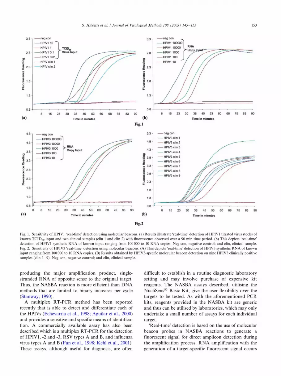
3.4. HPIV1 and HPIV3 ‘real-time’ detection using
molecular beacons
HPIV1 and HPIV3 titrated reference strains and
diluted RNA transcripts were analysed in the developedNASBA assays with ‘real-time’ detection using the
molecular beacons described in Section 2.9. The sensi-
tivity of the HPIV1 ‘real-time’ assay was found to be
within the range 0.1�/0.01 TCID50 virus input (Fig. 1A),
and between 100 and 10 copies of synthetic RNA (Fig.
1B), consistent with results obtained in the equivalent
ECL detection assay. The sensitivity of the HPIV3 ‘real-
time’ assay was within the range 10�/1 TCID50 virusinput, and between 100 and 10 copies of synthetic RNA
(Fig. 2A). The molecular beacon results for HPIV3, in
terms of virus input, may thus indicate this approach to
be slightly less sensitive than the equivalent ECL
detection assay but the difference was not thought to
be significant enough to affect clinical utility.
3.5. ‘Real-time’ detection of HPIV1 and HPIV3 clinical
samples
The clinical samples described in Section 2.2 were
analysed using the optimised HPIV1 and HPIV3
molecular beacon assays for ‘real-time’ detection. As
with the previous results obtained using the appropriate
NASBA assay with ECL detection (Section 3.3), all of
the clinical samples tested were clearly above back-ground for both HPIV1 (Fig. 1A) and HPIV3 (Fig. 2B)
and the specificity of the assays with molecular beacon
detection was confirmed.
S. Hibbitts et al. / Journal of Virological Methods 108 (2003) 145�/155150
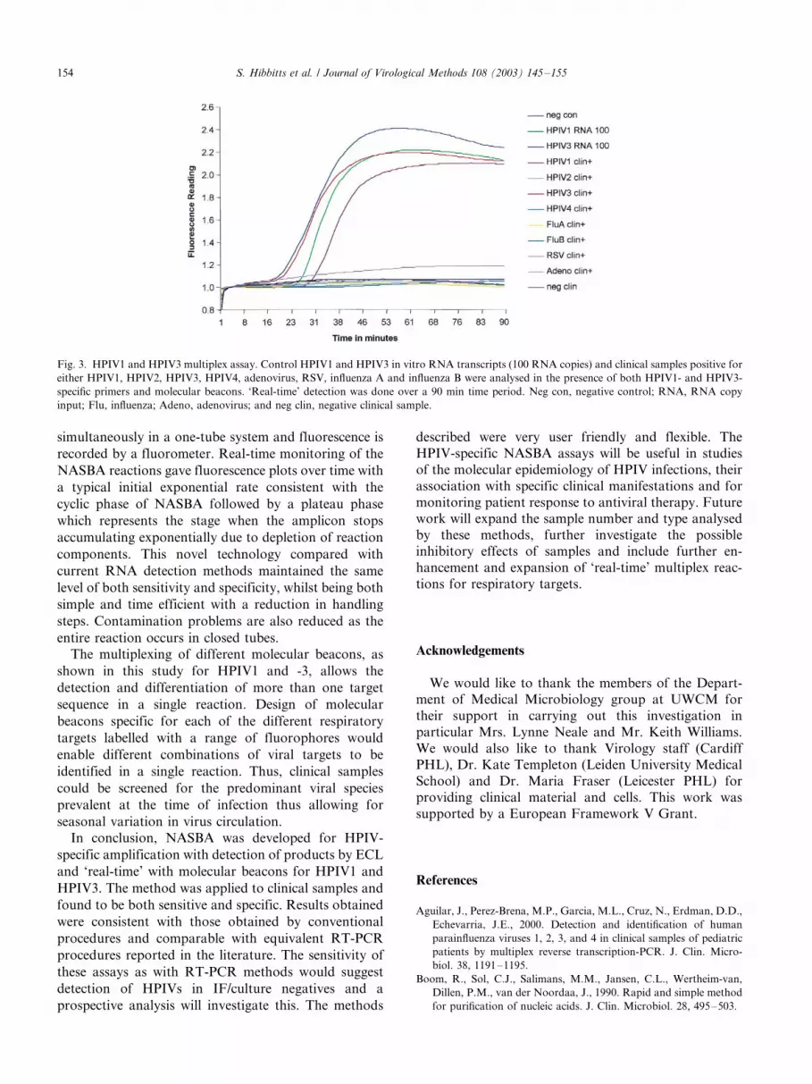
3.6. Multiplex for the ‘real-time’ detection of HPIV1 and
HPIV3
Initial experiments to establish the feasibility of
multiplex NASBA assays were prepared as described
in Section 2.10 with both primer sets and FAM-labelled
beacons. Optimal multiplex results were obtained when
primer pairs to both the HPIV1 and HPIV3 amplicon
were present at the standard final concentration and
both molecular beacons present at 0.1 mM final con-
centration. The sensitivity and specificity of the multi-
plex assay were determined by using HPIV1 and HPIV3
in vitro RNA transcripts and nucleic acid extracts from
clinical samples positive for HPIV1-4, adenovirus, RSV,
influenza A and influenza B. The sensitivity of the
multiplex assay was not compromised compared with
the monoplex assays. Less than or equal to 100 RNA
copies of either HPIV1 or HPIV3 synthetic RNA were
detectable using this assay (Fig. 3). The specificity of the
assay was also maintained and no cross-reactivity with
any of the other respiratory viruses tested was observed
(Fig. 3). The optimised multiplex assay was also used to
analyse 21 culture/direct antigen negative clinical sam-
ples that included both throat or nasopharyngeal swabs.
None of the samples that had been analysed by
conventional diagnostic techniques contained detectable
HPIV1 or HPIV3 sequences. To ensure that the extracts
were not inhibitory in the NASBA reaction, a subset of
these samples was spiked with low titre HPIV1 andHPIV3 virus stock extracts or low copy number
synthetic RNA. In all cases, the spiked samples gave
positive results in the NASBA assays (data not shown).
Future work will utilise different fluorophores in order
to differentiate between the HPIVs in a single assay.
4. Discussion
Viral respiratory tract infections have been recognised
as a predominant cause of human diseases both in the
community and in a hospital setting. To improve clinical
management of such patients it is important to obtain
an accurate diagnosis and to identify the causative agentearly in infection to ensure appropriate treatment. Such
procedures will enable early patient recovery, prevent
inappropriate antibiotic treatment, reduce the length of
hospitalisation and thus costs incurred.
In this study, we developed NASBA assays for the
detection of HPIVs 1�/4 and investigated the potential
use of these assays for analysis of clinical material.
Detection using molecular beacons for HPIV1 and -3also provides a convenient and effective method to
amplify and detect RNA homogeneously and in ‘real-
time’. Results indicated that HPIV RNA could be
Table 2
NASBA with ‘end-point’ (ECL) detection of amplified products for control viruses and synthetic RNA
ECL count (log10)
HPIV1 HPIV2 HPIV3 HPIV4a HPIV4b
TCID50
1000 980033 (6.0 ) 2482197 (6.4 ) 10000000 (�/7.0 )
100 1178230 (6.1 ) 1994818 (6.3 ) 10000000 (�/7.0 )
10 602956 (5.8 ) 1715206 (6.2 ) 1601811 (6.2 )
1 434935 (5.6 ) 1152179 (6.1 ) 68507 (4.8 )
0.1 602571 (5.8 ) 1216131 (6.1 ) 139 (2.1)
0.01 390 (2.6) 791563 (5.9 ) �/
0.001 �/ 163 (2.2) �/
Culture stock dilution a
10�1 10000001 (�/7.0 ) 2032427 (6.3 )
10�2 10000001 (�/7.0 ) 1990751 (6.3 )
10�3 1841864 (6.3 ) 1303196 (6.1 )
10�4 2633856 (6.4 ) 1342836 (6.1 )
10�5 1383167 (6.1 ) 1130348 (6.1 )
10�6 10000001 (�/7.0 ) 1498719 (6.2 )
10�7 209 (2.3) 171 (2.2)
Number of RNA copies
1000 709796 (5.9 ) 2463065 (6.4 ) 265389 (5.4 ) �/10000000 (�/7.0 ) 972118 (6.0 )
100 14654 (4.2 ) 910988 (6.0 ) 2225157 (6.3 ) 1539467 (6.2 ) 1040636 (6.0 )
50 16042 (4.2 ) 156 (2.2) 507 (2.7) 91 (2.0) 400820 (5.6 )
10 133 (2.1) 93 (2.0) 430 (2.6) 128 (2.1) 131 (2.1)
Results given are for titrated HPIV viruses spiked into lysis buffer and taken through extraction, amplification and detection. Input assumes 100%
extraction efficiency.a Diluted cultures of HPIV4a and HPIV4b did not give clear-cut end-points in standard TCID50 assays and so results are shown for culture
dilutions. In all cases positive ECL results are indicated in italics.
S. Hibbitts et al. / Journal of Virological Methods 108 (2003) 145�/155 151
extracted, amplified and detected directly from clinical
specimens without inhibitory effects being a significant
problem.
The NASBA assays for each of the HPIVs have a
number of advantages over ‘conventional’ methods
currently utilised for their detection. Traditional virus
culture and serology analysis may require 1�/2 weeks
before results are available and direct antigen detection
can be variable, with both sensitivity and specificity
problems reported (Henrickson, 1995; Swierkosz et al.,
1995). Molecular assays have particular advantages
where the starting material available is acellular (swab)
material or where surveillance samples which may have
only a low copy number of the target are to be analysed.
Assays based on nucleic acid amplification have the
potential to provide a rapid turn-around of results
allowing diagnostic virology to have a direct impact
on patient management. Also, molecular detection
methods have the additional advantage that viral
sequences may be amplified directly from the clinical
samples thus preventing the selection pressure of in vitro
isolation.
The assays described will prove useful in studies of the
molecular epidemiology of respiratory infections. The
multiplex molecular beacon assay is a particularly
significant improvement over current detection methods
for NASBA amplicons and the feasibility of using such
an approach for all RNA virus targets has been shown
in this study.
An advantage of the NASBA assay compared with
the previously reported RT-PCR methods (Echevarria
et al., 1998; Fan et al., 1998; Aguilar et al., 2000; Kehl et
al., 2001) is the continuous, isothermal process that does
not require a thermocycler. A constant temperature
throughout the amplification reaction enables each step
of the reaction to proceed when an amplification
intermediate becomes available. This results in the
exponential amplification of RNA and DNA products
Table 3
Assessment of HPIV NASBA specificity (with ECL detection)
Clinical samples ECL count (log10)
PIV1 PIV2 PIV3 PIV4
HPIV1 674326 (5.8 ) 265 (2.4) 126 (2.1) 141 (2.1)
HPIV2 261 (2.4) 2385479 (6.4 ) 303 (2.5) 176 (2.2)
HPIV3 135 (2.1) 214 (2.3) 2371337 (6.4 ) 183 (2.3)
HPIV4 315 (2.5) 247 (2.4) 203 (2.3) 10000001 (�/7.0 )
Adenovirus�/ 181 (2.3) 210 (2.3) 169 (2.2) 228 (2.4)
RSV�/ 182 (2.3) 244 (2.4) 182 (2.3) 304 (2.5)
FluA�/ 132 (2.1) 173 (2.2) 138 (2.1) 267 (2.4)
FluB�/ 166 (2.2) 258 (2.4) 147 (2.2) 199 (2.3)
Negative control clinical sample 1 218 (2.3) 235 (2.4) 179 (2.3) 191 (2.3)
Negative control clinical sample 2 216 (2.3) �/ 195 (2.3) �/
Negative control clinical sample 3 190 (2.3) �/ 161 (2.2) �/
Negative control clinical sample 4 164 (2.2) �/ 131 (2.1) �/
Analysis of cross-reactivity of each of the HPIV NASBA assays with the different serotypes and other culture-positive virus samples. Positive ECL
counts are indicated in italics.
Table 4
Analysis of clinical samples by HPIV NASBA with ECL detection
Clinical samples obtained ECL counts (log10) obtained in the appropriate HPIV assay
HPIV1 HPIV2 HPIV3 HPIV4
Clinical sample 1 1062807 (6.0) 2676456 (6.4) 10000001 (�/7.0) 2425192 (6.4)
Clinical sample 2 547209 (5.7) 1609550 (6.2) 10000001 (�/7.0) NA
Clinical sample 3 NA 961235 (6.0) 10000001 (�/7.0) NA
Clinical sample 4 NA 1844929 (6.3) 10000001 (�/7.0) NA
Clinical sample 5 NA 1993834 (6.3) 10000001 (�/7.0) NA
Clinical sample 6 NA 80291 (4.9) 10000001 (�/7.0) NA
Clinical sample 7 NA NA 10000001 (�/7.0) NA
Clinical sample 8 NA NA 10000001 (�/7.0) NA
Clinical sample 9 NA NA 1536400 (6.2) NA
Clinical sample 10 NA NA 10000001 (�/7.0) NA
The HPIV1, -2, -3 and -4 positive clinical specimens obtained are depicted in this table. The ECL signals observed for each of the specimens using
the HPIV1, -2, -3 or -4 NASBA assay are shown. NA, not available.
S. Hibbitts et al. / Journal of Virological Methods 108 (2003) 145�/155152
producing the major amplification product, single-
stranded RNA of opposite sense to the original target.
Thus, the NASBA reaction is more efficient than DNA
methods that are limited to binary increases per cycle
(Stanway, 1990).
A multiplex RT-PCR method has been reported
recently that is able to detect and differentiate each of
the HPIVs (Echevarria et al., 1998; Aguilar et al., 2000)
and provides a sensitive and specific means of identifica-
tion. A commercially available assay has also been
described which is a multiplex RT-PCR for the detection
of HPIV1, -2 and -3, RSV types A and B, and influenza
virus types A and B (Fan et al., 1998; Kehl et al., 2001).
These assays, although useful for diagnosis, are often
difficult to establish in a routine diagnostic laboratory
setting and may involve purchase of expensive kit
reagents. The NASBA assays described, utilising the
NucliSens† Basic Kit, give the user flexibility over the
targets to be tested. As with the aforementioned PCR
kits, reagents provided in the NASBA kit are generic
and thus can be utilised by laboratories, which may only
undertake a small number of assays for each individual
target.
‘Real-time’ detection is based on the use of molecular
beacon probes in NASBA reactions to generate a
fluorescent signal for direct amplicon detection during
the amplification process. RNA amplification with the
generation of a target-specific fluorescent signal occurs
Fig. 1. Sensitivity of HPIV1 ‘real-time’ detection using molecular beacons. (a) Results illustrate ‘real-time’ detection of HPIV1 titrated virus stocks of
known TCID50 input and two clinical samples (clin 1 and clin 2) with fluorescence observed over a 90 min time period. (b) This depicts ‘real-time’
detection of HPIV1 synthetic RNA of known input ranging from 100 000 to 10 RNA copies. Neg con, negative control; and clin, clinical sample.
Fig. 2. Sensitivity of HPIV3 ‘real-time’ detection using molecular beacons. (A) This depicts ‘real-time’ detection of HPIV3 synthetic RNA of known
input ranging from 100 000 to 10 RNA copies. (B) Results obtained by HPIV3-specific molecular beacon detection on nine HPIV3 clinically positive
samples (clin 1�/9). Neg con, negative control; and clin, clinical sample.
S. Hibbitts et al. / Journal of Virological Methods 108 (2003) 145�/155 153
simultaneously in a one-tube system and fluorescence is
recorded by a fluorometer. Real-time monitoring of the
NASBA reactions gave fluorescence plots over time with
a typical initial exponential rate consistent with the
cyclic phase of NASBA followed by a plateau phase
which represents the stage when the amplicon stops
accumulating exponentially due to depletion of reaction
components. This novel technology compared with
current RNA detection methods maintained the same
level of both sensitivity and specificity, whilst being both
simple and time efficient with a reduction in handling
steps. Contamination problems are also reduced as the
entire reaction occurs in closed tubes.
The multiplexing of different molecular beacons, as
shown in this study for HPIV1 and -3, allows the
detection and differentiation of more than one target
sequence in a single reaction. Design of molecular
beacons specific for each of the different respiratory
targets labelled with a range of fluorophores would
enable different combinations of viral targets to be
identified in a single reaction. Thus, clinical samples
could be screened for the predominant viral species
prevalent at the time of infection thus allowing for
seasonal variation in virus circulation.
In conclusion, NASBA was developed for HPIV-
specific amplification with detection of products by ECL
and ‘real-time’ with molecular beacons for HPIV1 and
HPIV3. The method was applied to clinical samples and
found to be both sensitive and specific. Results obtained
were consistent with those obtained by conventional
procedures and comparable with equivalent RT-PCR
procedures reported in the literature. The sensitivity of
these assays as with RT-PCR methods would suggest
detection of HPIVs in IF/culture negatives and a
prospective analysis will investigate this. The methods
described were very user friendly and flexible. The
HPIV-specific NASBA assays will be useful in studies
of the molecular epidemiology of HPIV infections, their
association with specific clinical manifestations and for
monitoring patient response to antiviral therapy. Futurework will expand the sample number and type analysed
by these methods, further investigate the possible
inhibitory effects of samples and include further en-
hancement and expansion of ‘real-time’ multiplex reac-
tions for respiratory targets.
Acknowledgements
We would like to thank the members of the Depart-ment of Medical Microbiology group at UWCM for
their support in carrying out this investigation in
particular Mrs. Lynne Neale and Mr. Keith Williams.
We would also like to thank Virology staff (Cardiff
PHL), Dr. Kate Templeton (Leiden University Medical
School) and Dr. Maria Fraser (Leicester PHL) for
providing clinical material and cells. This work was
supported by a European Framework V Grant.
References
Aguilar, J., Perez-Brena, M.P., Garcia, M.L., Cruz, N., Erdman, D.D.,
Echevarria, J.E., 2000. Detection and identification of human
parainfluenza viruses 1, 2, 3, and 4 in clinical samples of pediatric
patients by multiplex reverse transcription-PCR. J. Clin. Micro-
biol. 38, 1191�/1195.
Boom, R., Sol, C.J., Salimans, M.M., Jansen, C.L., Wertheim-van,
Dillen, P.M., van der Noordaa, J., 1990. Rapid and simple method
for purification of nucleic acids. J. Clin. Microbiol. 28, 495�/503.
Fig. 3. HPIV1 and HPIV3 multiplex assay. Control HPIV1 and HPIV3 in vitro RNA transcripts (100 RNA copies) and clinical samples positive for
either HPIV1, HPIV2, HPIV3, HPIV4, adenovirus, RSV, influenza A and influenza B were analysed in the presence of both HPIV1- and HPIV3-
specific primers and molecular beacons. ‘Real-time’ detection was done over a 90 min time period. Neg con, negative control; RNA, RNA copy
input; Flu, influenza; Adeno, adenovirus; and neg clin, negative clinical sample.
S. Hibbitts et al. / Journal of Virological Methods 108 (2003) 145�/155154
Chan, A., Fox, J.D., 1999. NASBA and other transcription based
amplification methods for research and diagnostic microbiology.
Rev. Med. Microbiol. 10, 185�/196.
Collins, P.L., Chanock, R.M., McIntosh, K., 1996. Parainfluenza
Viruses: Fields Virology, vol. 1, 3rd ed., pp. 1205�/1243.
Compton, J., 1991. Nucleic acid sequence-based amplification. Nature
350, 91�/92.
Downham, M.A.P.S., McQuillin, J., Gardener, P.S., 1974. Diagnosis
and clinical significance of parainfluenza virus infections in
children. Arch. Dis. Child. 49, 8�/15.
Echevarria, J., Erdman, D.D., Swierkosz, E.M., Holloway, B.P.,
Anderson, L.J., 1998. Simultaneous detection and identification
of human parainfluenza viruses 1, 2, and 3 from clinical samples by
multiplex PCR. J. Clin. Microbiol. 36, 1388�/1391.
Fan, J., Henrickson, K.J., Savatski, L.L., 1998. Rapid simultaneous
diagnosis of infections with respiratory syncytial viruses A and B,
influenza viruses A and B, and human parainfluenza virus types 1,
2, and 3 by multiplex quantitative reverse transcription-polymerase
chain reaction-enzyme hybridization assay (Hexaplex). Clin. Infect.
Dis. 26, 1397�/1402.
Forghani, B., Hagens, S., 1995. Diagnosis of viral infections by antigen
detection. In: Lennette, E.H., Lennette, D.A., Lennette, E.T.
(Eds.), Diagnostic Procedures for Viral, Rickettsial, and Chlamy-
dial Infections, 7th ed.. American Public Health Association, pp.
79�/96.
Guatelli, J., Whitfield, K.M., Kwoh, D.Y., Barringer, K.J., Richman,
D.D., Gingeras, T.R., 1990. Isothermal in vitro amplification of
nucleic acids by a multienzyme reaction modelled after retroviral
replication. Proc. Natl. Acad. Sci. USA 87, 7797.
Henrickson, K.J., 1995. Human parainfluenza viruses. In: Lennette,
E.H., Lennette, D.A., Lennette, E.T. (Eds.), Diagnostic Procedures
for Viral, Rickettsial, and Chlamydial Infections, 7th ed.. American
Public Health Association, pp. 481�/494.
Hierholzer, J., Bingham, P.G., Coombs, R.A., Johansson, K.H.,
Anderson, L.J., Halonen, P.E., 1989. Comparison of monoclonal
antibody time-resolved fluoroimmunoassay with monoclonal anti-
body capture-biotinylated detector enzyme immunoassay for
respiratory syncytial virus and parainfluenza virus antigen detec-
tion. J. Clin. Microbiol. 27, 1243�/1249.
Kehl, S., Henrickson, K.J., Hua, W., Fan, J., 2001. Evaluation of the
hexaplex assay for detection of respiratory viruses in children. J.
Clin. Microbiol. 39, 1696�/1701.
Kievits, T., van Gemen, B., van Strijp, D., Schukkink, R., Dircks, M.,
Adriaanse, H., Malek, L., Sooknanan, R., Lens, P., 1991. NASBA
isothermal enzymatic in vitro nucleic acid amplification optimized
for the diagnosis of HIV-1 infection. J. Virol. Methods 35, 273�/
286.
Laurichesse, H., Dedman, D., Watson, J.M., Zambon, M.C., 1999.
Epidemiological features of parainfluenza virus infections: labora-
tory surveillance in England and Wales, 1975�/1997. Eur. J.
Epidemiol. 15, 475�/484.
Leone, G., van Schijndel, H., van Gemen, B., Kramer, F.R., Schoen,
C.D., 1998. Molecular beacon probes combined with amplification
by NASBA enable homogeneous, real-time detection of RNA.
Nucleic Acids Res. 26, 2150�/2156.
Lindsquist, S.W., Darnule, A., Istas, A., Demmler, G.J., 1997.
Parainfluenza virus type 4 infections in paediatric patients. Pediatr.
Infect. Dis. J. 16, 34�/38.
Marx, A., Torok, T.J., Holman, R.C., Clarke, M.J., Anderson, L.J.,
1997. Paediatric hospitalizations for croup (laryngotracheobron-
chitis): biennial increases associated with human parainfluenza
virus 1 epidemics. J. Infect. Dis. 176, 1423�/1427.
Murphy, B., Prince, G.A., Collins, P.L., Van Wyke Coelingh, K.,
Olmsted, R.A., Spriggs, M.K., Parrott, R.H., Kim, H.W., Brandt,
C.D., Chanock, R.M., 1988. Current approaches to the develop-
ment of vaccines effective against parainfluenza and respiratory
syncytial viruses. Virus Res. 11, 1�/15.
Murphy, F.A., Fauquet, C.M., Bishops, D.H.L., Ghabrial, S.A.,
Jawis, A.W., Martelli, G.P., Mayo, M.A., 1995. Virus taxonomy.
Sixth report of the International Committee on Taxonomy of
Viruses. Arch. Virol. Suppl. 10, 1�/586.
Reed, L., Muench, H., 1938. A simple method of estimating fifty
percent endpoints. Am. J. Hyg. 27, 493�/497.
Rubin, E., Quennec, P., McDonald, J.C., 1993. Infections due to
parainfluenza virus type 4 in children. Clin. Infect. Dis. 17, 998�/
1002.
Sarkkinen, H.K., Halonen, P.E., Salmi, A.A., 1981. Type-specific
detection of parainfluenza viruses by enzyme-immunoassay and
radioimmunoassay in nasopharyngeal specimens of patients with
acute respiratory disease. J. Gen. Virol. 56, 49�/57.
Shen, K., Zhaori, G., Zweygberg-Wirgari, B., Ying, M., Grandien, M.,
Wahren, B., Linde, A., 1996. Detection of respiratory viruses in
nasopharyngeal secretions with immunofluorescence technique for
multiplex screening*/an evaluation of the chemicon assay. Clin.
Diagn. Virol. 6, 147�/154.
Stanway, G., 1990. Structure, function and evolution of picorna-
viruses. J. Gen. Virol. 71, 2483�/2501.
Swierkosz, E., Erdman, D.D., Bonnot, T., Schneiderheinze, C., Waner,
J.L., 1995. Isolation and characterization of a naturally occurring
parainfluenza 3 virus variant. J. Clin. Microbiol. 33, 1839�/1841.
Tyagi, S., Kramer, F.R., 1996. Molecular beacons: probes that
fluoresce upon hybridization. Nat. Biotechnol. 14, 303�/308.
Wendt, C., Weisdorf, D.J., Jordan, M.C., Balfour, H.H., Hertz, M.I.,
1992. Parainfluenza virus respiratory virus respiratory infection
after bone marrow transplantation. N. Engl. J. Med. 326, 921�/926.
S. Hibbitts et al. / Journal of Virological Methods 108 (2003) 145�/155 155