Developing New Strategies for the Treatment of Tuberculosis Employing Ruthnium(II)Quaterpyridyl...
10
J. Nepal Chem. Soc., Vol. 23, 2008/2009 2 Developing New Strategies for the Treatment of Tuberculosis Employing Ruthnium(II)Quaterpyridyl Compexes Megh Raj Pokhrel 1,* , Pubudu Gamage 2 , Mausam Kalita 2 , Aibin Shi 2 and Stefan H. Bossmann 2 1. Central Department of Chemistry, Tribhuvan University, Kathmandu, Nepal 2. Department of Chemistry, Kansas State University, Manhattan, KS 66506-3701, USA Abstract A series of ruthenium(II)quaterpyridyl complexes has been synthesized as prototypes for mycobacterial channel blockers. These Ru(II)complexes show distinct changes in their luminescence spectra when bound to the porin MspA from M. smegmatis, which is a non-pathogenic relative of M. tuberculosis. By using HPLC, we have determined binding constants of the Ru(II)-complexes to MspA in phosphate buffer (0.05M, pH = 6.8) ranging from 5.2 x 10 9 M -1 (Ru-C2) to 1.8 x 10 9 M -1 (Ru- C4). Our findings indicate that channel blocking is a promising treatment strategy for mycobacterial infections. It appears to be also a viable approach towards luminescent nanostructures, because MspA features extraordinary stability. Keywords: Ru(II)complexe, porin MspA, mycobacterial infection, tuberculosis. Introduction Mycobacterium tuberculosis is, according to the World Health Organization, one of the most dangerous infectious diseases; causing more deaths than any other single infection. 1 Approximately one third of the world’s population is already infected. More than 4,000,000 new cases and more than 2,000,000 deaths have to be accounted for each year. 1 Multi-resistant strains have appeared due to the discontinuing treatment of tuberculosis in many countries, threatening all countries which experience immigration during the last two decades. Therefore, entirely new strategies are required to obtain the basic knowledge, which will then enable the successful development of new anti-TB-therapies. Mycobacteria possess an especially thick outer membrane (called “cell envelope”), which acts as a hydrophobic shield against antibiotics. 2 Channel proteins (“porins”) form the main * Corresponding author
Transcript of Developing New Strategies for the Treatment of Tuberculosis Employing Ruthnium(II)Quaterpyridyl...

J. Nepal Chem. Soc., Vol. 23, 2008/2009
2
Developing New Strategies for the Treatment of Tuberculosis
Employing Ruthnium(II)Quaterpyridyl Compexes
Megh Raj Pokhrel1,*
, Pubudu Gamage2, Mausam Kalita
2,
Aibin Shi2 and Stefan H. Bossmann
2
1. Central Department of Chemistry, Tribhuvan University, Kathmandu, Nepal
2. Department of Chemistry, Kansas State University, Manhattan, KS 66506-3701, USA
Abstract
A series of ruthenium(II)quaterpyridyl complexes has been synthesized as
prototypes for mycobacterial channel blockers. These Ru(II)complexes show distinct
changes in their luminescence spectra when bound to the porin MspA from M.
smegmatis, which is a non-pathogenic relative of M. tuberculosis. By using HPLC,
we have determined binding constants of the Ru(II)-complexes to MspA in phosphate
buffer (0.05M, pH = 6.8) ranging from 5.2 x 10⁹ M
-1 (Ru-C2) to 1.8 x 10
9 M
-1 (Ru-
C4). Our findings indicate that channel blocking is a promising treatment strategy
for mycobacterial infections. It appears to be also a viable approach towards
luminescent nanostructures, because MspA features extraordinary stability.
Keywords: Ru(II)complexe, porin MspA, mycobacterial infection, tuberculosis.
Introduction
Mycobacterium tuberculosis is, according to the World Health Organization,
one of the most dangerous infectious diseases; causing more deaths than any other
single infection.1 Approximately one third of the world’s population is already
infected. More than 4,000,000 new cases and more than 2,000,000 deaths have to be
accounted for each year.1 Multi-resistant strains have appeared due to the
discontinuing treatment of tuberculosis in many countries, threatening all countries
which experience immigration during the last two decades. Therefore, entirely new
strategies are required to obtain the basic knowledge, which will then enable the
successful development of new anti-TB-therapies. Mycobacteria possess an
especially thick outer membrane (called “cell envelope”), which acts as a
hydrophobic shield against antibiotics.2 Channel proteins (“porins”) form the main
* Corresponding author
J. Nepal Chem. Soc., Vol. 23, 2008/2009
3
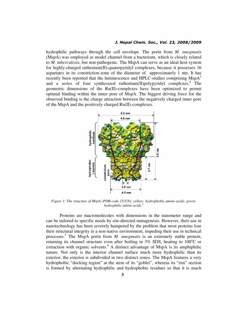
hydrophilic pathways through the cell envelope. The porin from M. smegmatis
(MspA) was employed as model channel from a bacterium, which is closely related
to M. tuberculosis, but non-pathogenic. The MspA can serve as an ideal host system
for highly-charged ruthenium(II)-quaterpyridyl complexes, because it possesses 16
aspartates in its constriction-zone of the diameter of approximately 1 nm. It has
recently been reported that the luminescence and HPLC-studies comprising MspA3
and a series of four synthesized ruthenium(II)polypyridyl complexes.4 The
geometric dimensions of the Ru(II)-complexes have been optimized to permit
optimal binding within the inner pore of MspA. The biggest driving force for the
observed binding is the charge attraction between the negatively charged inner pore
of the MspA and the positively charged Ru(II)-complexes.
Figure 1: The structure of MspA (PDB-code 1UUN): yellow: hydrophobic amino acids; green:
hydrophilic amino acids.3
Proteins are macromolecules with dimensions in the nanometer range and
can be tailored to specific needs by site-directed mutagenesis. However, their use in
nanotechnology has been severely hampered by the problem that most proteins lose
their structural integrity in a non-native environment, impeding their use in technical
processes.5 The MspA porin from M. smegmatis is an extremely stable protein,
retaining its channel structure even after boiling in 3% SDS, heating to 100oC or
extraction with organic solvents.6 A distinct advantage of MspA is its amphiphilic
nature. Not only is the interior channel surface much more hydrophilic than its
exterior, the exterior is subdivided in two distinct zones. The MspA features a very
hydrophobic “docking region” at the stem of its “goblet”, whereas its “rim” section
is formed by alternating hydrophilic and hydrophobic residues so that it is much

J. Nepal Chem. Soc., Vol. 23, 2008/2009
4
more hydrophilic. The geometric dimensions of the “docking region” are 3.7 nm in
length, and 4.9 nm in diameter as shown in Fig. 1.
Experimental Methods
The MspA was grown in laboratories using established procedures as
described elsewhere.7 The synthesis of ruthenium(II)-tris-(4,4’,2’,2’’,4’’,4’’’
quaterpyridinium) (Fig. 2) complexes is described in the literature.4 Shortly,
dimerization of 4,4´-dipyridyl on Pd/carbon in anhydrous DMF leads to
quaterpyridyl in 20 ± 2 % yield. Ru(DMSO)42+
reacts with quaterpyridyl in MeOH at
1000 psi and 100oC quantitatively to Ru(II)-tris-quaterpyridyl (Ru(II)(QP)3Cl2).
Quaternization of the exterior sp2-nitrogen functions was achieved by reacting them
with the organic bromides (R-Br) in MeOH at 1000 psi and 40oC. Purification of the
tris-homoleptic ruthenium(II) complexes and exchange of the counter-anions against
chloride is performed by anion exchange chromatography on Sephadex.
Figure 2: Ruthenium(II)-tris-(4,4’,2’,2’’,4’’,4’’’quaterpyridinium) complexes
Ru(II)-C1 to Ru(II)-C4.4
UV/Vis-Absorbance and Luminescence Experiments
Both experiments were carried out in 4.0 mL quartz-cuvettes (Helma) using
a spectrofluoro-meter (Fluoromax2) with dual monochromators and a diode array
UV-vis absorption spectrometer (HP 8453). 0.05M Phosphate buffer (pH = 6.8) was
used as solvent.
J. Nepal Chem. Soc., Vol. 23, 2008/2009
5
HPLC-determination of the binding constants of the Ru(II)quaterpyridyl complexes
to MspA
The binding constants of both of small and large gold nanoparticles bound to
MspA were measured by HPLC (Shimadzu Prominence) employing a POROS
HQ/20 anion exchange column and a flux of 0.50 ml min-1
. Two buffers were used:
AOP05 (25mM HEPES, pH 7.5, 10 mM NaCl, 0.5% OPOE) and BOP05 (25 mM
HEPES, pH 7.5, 2 M NaCl, 0.5% OPOE). A typical gradient was 100% AOP05 (0-5
min.), followed by a linear gradient to 100% BOP05 (5-35 min.). The eluent was
kept at 100% BOP05 (35-50 min.). Finally, the salt concentration is returned linearly
to 10mM (100% AOP05) (50-60min). The stop time was set at 65 min. Peak
detection was achieved using UV/Vis-absorbance(diode-array). The binding
constants were calculated according to equation (1):
KB =[Ru(II)cpx@ MspA]
([Ru(II)cpx]0 − [Ru(II)cpx@ MspA])([MspA]0 − [Ru(II)cpx@MspA])
(1
)
where, KB:binding constant, [Ru(II)cpx@MspA]:concentration (mol/L) of
the supramolecular assembly of the ruthenium(II)quaterpyridyl complexes Ru-C1 to
Ru-C4 and MspA; [MspA]0:concentration of MspA (mol/L) in the absence of NP’s;
[Ru(II)cpx]0:starting concentration of the ruthenium(II)quaterpyridyl complexes
(mol/L).
Results and Dicussion
Ruthenium(II)polypyridyl complexes
Ruthenium(II)-polypyridyl complexes possess extraordinary thermal and
photochemical stabilities. They are also kinetically stable, which means that they
usually do not show ligand-exchange reactions in the dark.8 Since they possess D
3-
geometries when three equal ligands are employed (tris-homoleptic complexes),
their geometric extensions can be estimated employing molecular modeling
methods.9 Furthermore, it should be noted that ruthenium(II)-polypyridyl complexes
absorb light in the UV- and Vis-region up to λ = 550-650 nm, depending on their
ligand structures. Finally, it should be noted that the redox potentials of Ru(III)-,
Ru(II)- and Ru(I)-complexes are easily accessible.10
Ruthenium(II)-quaterpyridinium complexes and their binding within MspA
Due to their positive charges and geometric dimensions (Table 1), Ru(II)-
quaterpyridyl complexes are the ideal guests for the MspA-pore. However, it must
J. Nepal Chem. Soc., Vol. 23, 2008/2009
6
be noted that these complexes can undergo partial deprotonation at neutral pH. This
process is able to diminish their net positive charges (maximally eight) to a certain
extent. Quantitative data on the acid/base-properties of Ru-C1 to RuC4 are not yet
available. The presence of the aspartate residues D90 and D91 provides up to 16
negative counter anions for the Ru (II)-complexes. Our results indicate a strong
charge attraction and consequent binding of the Ru (II)-complex within the pore of
MspA have been obtained (see below).
UV/Vis-absorption characteristics
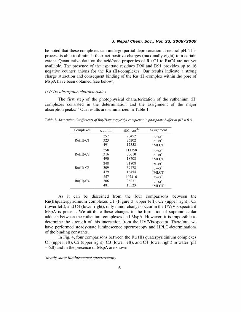
The first step of the photophysical characterization of the ruthenium (II)
complexes consisted in the determination and the assignment of the major
absorption peaks.10
Our results are summarized in Table 1.
Table 1. Absorption Coefficients of Ru(II)quaterpyridyl complexes in phosphate buffer at pH = 6.8.
Complexes λ max nm ε(M-1
cm-1
) Assignment
Ru(II)-C1
257
323
491
70452
26202
17352
π→π∗
d→π∗
3MLCT
Ru(II)-C2
258
316
490
111358
30610
18708
π→π∗
d→π∗
3MLCT
Ru(II)-C3
248
309
479
71808
39478
16454
π→π∗
d→π∗
3MLCT
Ru(II)-C4
257
306
481
107416
36231
15523
π→π∗
d→π∗
3MLCT
As it can be discerned from the four comparisons between the
Ru(II)quaterpyridinium complexes C1 (Figure 3, upper left), C2 (upper right), C3
(lower left), and C4 (lower right), only minor changes occur in the UV/Vis-spectra if
MspA is present. We attribute these changes to the formation of supramolecular
adducts between the ruthenium complexes and MspA. However, it is impossible to
determine the strength of this interaction from the UV/Vis-spectra. Therefore, we
have performed steady-state luminescence spectroscopy and HPLC-determinations
of the binding constants.
In Fig. 4, four comparisons between the Ru (II) quaterpyridinium complexes
C1 (upper left), C2 (upper right), C3 (lower left), and C4 (lower right) in water (pH
= 6.8) and in the presence of MspA are shown.
Steady-state luminescence spectroscopy

J. Nepal Chem. Soc., Vol. 23, 2008/2009
7
The luminescence data summarized in Fig. 4 shows the typical behavior for
ruthenium(II)-polypyridyl complexes which are bound by proteins.11
The
luminescence arising from the 3MLCT (metal to ligand charge transfer) of the
Ru(II)-complexes is partially quenched by H2O.12
When binding occurs, the
complex is stripped of a part of its hydrate shell. Therefore, the quantum yield of
luminescence often increases. This behavior is especially pronounced for Ru (II)-C2.
However, proton quenching can decrease the luminescence intensity and, therefore,
a significant increase cannot be observed. Apparently, this is the case for the binding
of Ru (II)-C1.
A second mechanistic criterion is the occurrence of a red shift of the
luminescence-maximum upon complex binding. In tris-homoleptic complexes,
which we exclusively use in these studies as luminescence probes, this shift is
caused by the extension of the “ligand-field” surrounding the metal center due to
supramolecular interaction with the biological structure.11
As it becomes apparent
from Figs 4 and 5, the magnitude of the red shift of luminescence increases with
increasing size of the complex.13
Figure 3: UV/Vis-Absorption Spectra of the Ruthenium (II)-tris-(4,4’,2’,2’’,4’’,4’’’quaterpyridinium)
complexes Ru(II)-C1 to Ru(II)-C4 (approx. 1.15x10⁻⁵
M in the presence (red) and absence (blue)

J. Nepal Chem. Soc., Vol. 23, 2008/2009
8
of MspA (10 µg mL-1
).
Figure 4: Luminescence Spectra of the Ruthenium(II)-tris-(4,4’,2’,2’’,4’’,4’’’quaterpyridinium)
complexes Ru(II)-C1 to Ru(II)-C4 (approx. 1.15x10⁻⁵
M in the presence (red; outer spectra except
in upper left) & absence (blue; inner spectra except in upper left) of MspA (10 µg mL-1
).
Figure 5: Indications for Binding of Ru(II)-C1 to C4 - complexes within MspA A: Luminescence
enhancement is a strong indication for the binding of Ru(II)-polypyridyl complexes.11
B: A
consecutively progressing shift in the wave-length of the emission maximum is indicative
of different binding sites of various Ru(II)-quaterpyridinium complexes of increasing
diameters (C1 > C4) within MspA.
J. Nepal Chem. Soc., Vol. 23, 2008/2009
9
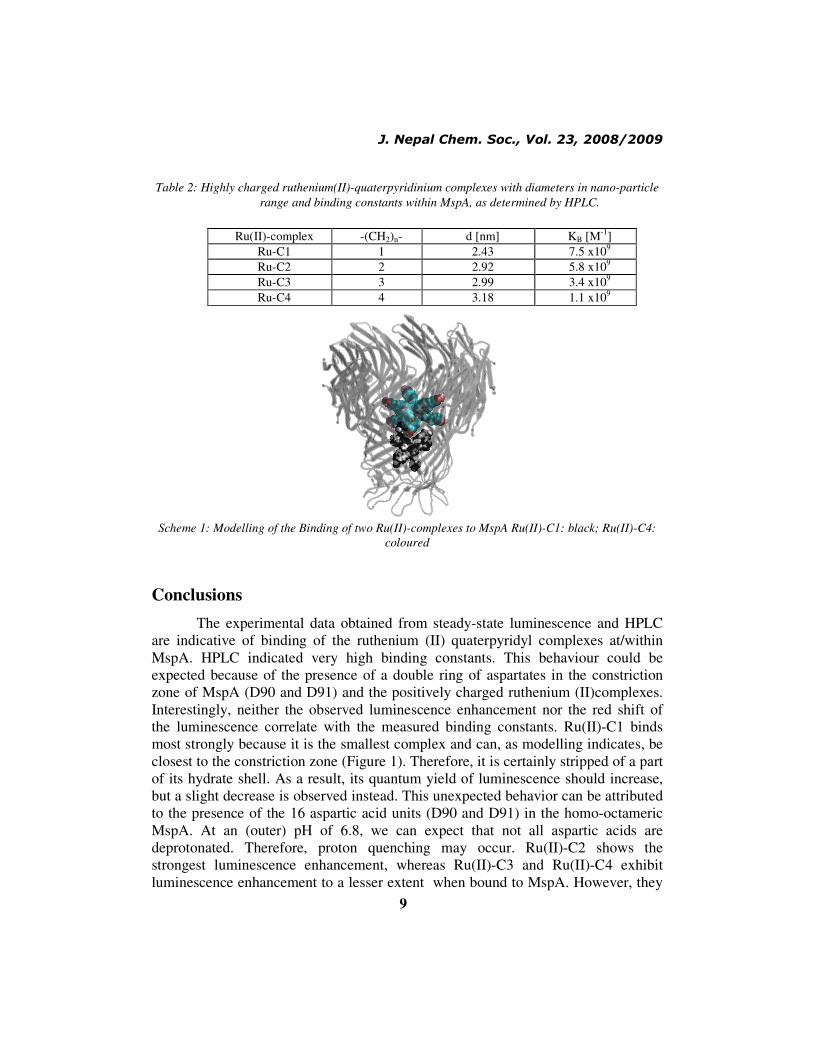
Table 2: Highly charged ruthenium(II)-quaterpyridinium complexes with diameters in nano-particle
range and binding constants within MspA, as determined by HPLC.
Ru(II)-complex -(CH2)n- d [nm] KB [M
-1]
Ru-C1 1 2.43 7.5 x10⁹
Ru-C2 2 2.92 5.8 x10⁹
Ru-C3 3 2.99 3.4 x10⁹
Ru-C4 4 3.18 1.1 x10⁹
Scheme 1: Modelling of the Binding of two Ru(II)-complexes to MspA Ru(II)-C1: black; Ru(II)-C4:
coloured
Conclusions
The experimental data obtained from steady-state luminescence and HPLC
are indicative of binding of the ruthenium (II) quaterpyridyl complexes at/within
MspA. HPLC indicated very high binding constants. This behaviour could be
expected because of the presence of a double ring of aspartates in the constriction
zone of MspA (D90 and D91) and the positively charged ruthenium (II)complexes.
Interestingly, neither the observed luminescence enhancement nor the red shift of
the luminescence correlate with the measured binding constants. Ru(II)-C1 binds
most strongly because it is the smallest complex and can, as modelling indicates, be
closest to the constriction zone (Figure 1). Therefore, it is certainly stripped of a part
of its hydrate shell. As a result, its quantum yield of luminescence should increase,
but a slight decrease is observed instead. This unexpected behavior can be attributed
to the presence of the 16 aspartic acid units (D90 and D91) in the homo-octameric
MspA. At an (outer) pH of 6.8, we can expect that not all aspartic acids are
deprotonated. Therefore, proton quenching may occur. Ru(II)-C2 shows the
strongest luminescence enhancement, whereas Ru(II)-C3 and Ru(II)-C4 exhibit
luminescence enhancement to a lesser extent when bound to MspA. However, they
J. Nepal Chem. Soc., Vol. 23, 2008/2009
10
show the largest red shifts in luminescence due to an enhanced 3MLCT-
delocalization. The observed differences in luminescence enhancement and red shift
clearly prove that each ruthenium(II)complex is bound at a different location inside
the vestibule of MspA due to increasing diameter and the funnel-shape of the inner
MspA pore. Our results indicate that channel blockers featuring high binding
constants to mycobacterial porins can be designed. Ru(II)-complexes are especially
suited for photophysical experiments, however Zn(II)-polypyridyl complexes may
be better suited for in-vivo applications due to their lower toxicity.
Acknowledgements
The authors thank Dr. Dan Higgins for the use of his instrumentation and
NSF-EPSCoR (First Award #4166) for financial support of this research.
References
1. www.who.int
2. M. Niederweis and S. H. Bossmann, Encyclopedia of Nanoscience and
Nanotechnology, 2004, 7, 851-867.
3. M. Faller, Niederweis, M. and Schulz, G. E., Science, 2004, 303, 1189-1192.
4. A. Shi, Pokhrel, M. R. and Bossmann, S. H., Synthesis, 2007, 4, 505-514.
5. H. Engelhardt, Gerbl-Rieger, S.; Krezmar, D.; Schneider-Voss, S.; Engel, A.
and Baumeister, W., J. Struct Biol., 1990, 105, 92-102.
6. M. Niederweis, Mol. Microbiol., 2003, 49, 1167-1177.
7. C. Heinz, Roth, E. and Niederweis, M., Meth. Mol. Biol., 2003, 228, 139-150.
8. H. Duerr and S. H. Bossmann, Acc. Chem. Res., 2001, 34, 905-917.
9. D. A. Lutterman, Chouai, A.; Liu, Y.; Sun, Y.; Stewart, C. D.; Dunbar, K. R.
and Turro, C., J. Am. Chem. Soc., 2008, 130, 1163-1170.
10. A. Juris, Balzani, V.; Barigelletti, F.; Campagna, S.; Belser, P. and Von
Zelewsky, A., Coord. Chem. Rev., 1988, 84, 85-277.
11. A. M. Pyle, Rehmann, J. P.; Meshoyrer, R.; Kumar, C. V.; Turro, N. J. and
Barton, J. K., J. Am. Chem. Soc., 1989, 111, 3051-3058.
12. C. Turro, Bossmann, S. H.; Leroi, G. E.; Barton, J. K. and Turro, N. J.,
Inorg. Chem., 1994, 33, 1344-1347.
13. (a) N. J. Turro, Barton, J. K. and Tomalia, D. A., Acc. Chem. Res., 1991, 24,
332-340. (b) N. J. Turro and Barton, J. K., Paradigms, Supermolecules,
electron transfer and chemistry at a distance. What's the problem? The
science or the paradigm?, JBIC, 1998, 3, 201-209.

J. Nepal Chem. Soc., Vol. 23, 2008/2009
11