Determinants of HMGB Proteins Required To Promote RAG1/2Recombination Signal Sequence Complex...
14
10.1128/MCB.25.11.4413-4425.2005. 2005, 25(11):4413. DOI: Mol. Cell. Biol. Jongbum Kwon and Reid C. Johnson Yan Dai, Ben Wong, Yi-Meng Yen, Marjorie A. Oettinger, Catalysis during V(D)J Recombination Sequence Complex Assembly and To Promote RAG1/2-Recombination Signal Determinants of HMGB Proteins Required http://mcb.asm.org/content/25/11/4413 Updated information and services can be found at: These include: REFERENCES http://mcb.asm.org/content/25/11/4413#ref-list-1 at: This article cites 67 articles, 23 of which can be accessed free CONTENT ALERTS more» articles cite this article), Receive: RSS Feeds, eTOCs, free email alerts (when new http://journals.asm.org/site/misc/reprints.xhtml Information about commercial reprint orders: http://journals.asm.org/site/subscriptions/ To subscribe to to another ASM Journal go to: on December 8, 2013 by guest http://mcb.asm.org/ Downloaded from on December 8, 2013 by guest http://mcb.asm.org/ Downloaded from
Transcript of Determinants of HMGB Proteins Required To Promote RAG1/2Recombination Signal Sequence Complex...

10.1128/MCB.25.11.4413-4425.2005.
2005, 25(11):4413. DOI:Mol. Cell. Biol. Jongbum Kwon and Reid C. JohnsonYan Dai, Ben Wong, Yi-Meng Yen, Marjorie A. Oettinger, Catalysis during V(D)J RecombinationSequence Complex Assembly andTo Promote RAG1/2-Recombination Signal Determinants of HMGB Proteins Required
http://mcb.asm.org/content/25/11/4413Updated information and services can be found at:
These include:
REFERENCEShttp://mcb.asm.org/content/25/11/4413#ref-list-1at:
This article cites 67 articles, 23 of which can be accessed free
CONTENT ALERTS more»articles cite this article),
Receive: RSS Feeds, eTOCs, free email alerts (when new
http://journals.asm.org/site/misc/reprints.xhtmlInformation about commercial reprint orders: http://journals.asm.org/site/subscriptions/To subscribe to to another ASM Journal go to:
on Decem
ber 8, 2013 by guesthttp://m
cb.asm.org/
Dow
nloaded from
on Decem
ber 8, 2013 by guesthttp://m
cb.asm.org/
Dow
nloaded from

MOLECULAR AND CELLULAR BIOLOGY, June 2005, p. 4413–4425 Vol. 25, No. 110270-7306/05/$08.00�0 doi:10.1128/MCB.25.11.4413–4425.2005Copyright © 2005, American Society for Microbiology. All Rights Reserved.
Determinants of HMGB Proteins Required To PromoteRAG1/2-Recombination Signal Sequence Complex
Assembly and Catalysis duringV(D)J Recombination
Yan Dai,1† Ben Wong,2‡ Yi-Meng Yen,2 Marjorie A. Oettinger,1*Jongbum Kwon,1§ and Reid C. Johnson2*
Department of Molecular Biology, Massachusetts General Hospital, Boston, Massachusetts 02114,1
and Department of Biological Chemistry, David Geffen School of Medicine, University ofCalifornia, Los Angeles, Los Angeles, California 90095-17372
Received 16 January 2005/Returned for modification 15 February 2005/Accepted 1 March 2005
Efficient assembly of RAG1/2-recombination signal sequence (RSS) DNA complexes that are competent forV(D)J cleavage requires the presence of the nonspecific DNA binding and bending protein HMGB1 or HMGB2.We find that either of the two minimal DNA binding domains of HMGB1 is effective in assembling RAG1/2-RSScomplexes on naked DNA and stimulating V(D)J cleavage but that both domains are required for efficientactivity when the RSS is incorporated into a nucleosome. The single-domain HMGB protein from Saccharo-myces cerevisiae, Nhp6A, efficiently assembles RAG1/2 complexes on naked DNA; however, these complexes areminimally competent for V(D)J cleavage. Nhp6A forms much more stable DNA complexes than HMGB1, and avariety of mutations that destabilize Nhp6A binding to bent microcircular DNA promote increased V(D)J cleavage.One of the two DNA bending wedges on Nhp6A and the analogous phenylalanine wedge at the DNA exit site ofHMGB1 domain A were found to be essential for promoting RAG1/2-RSS complex formation. Because the phe-nylalanine wedge is required for specific recognition of DNA kinks, we propose that HMGB proteins facilitateRAG1/2-RSS interactions by recognizing a distorted DNA structure induced by RAG1/2 binding. The resultingcomplex must be sufficiently dynamic to enable the series of RAG1/2-mediated chemical reactions on the DNA.
The site-specific V(D)J recombination reaction assemblesimmunoglobulin and T-cell receptor genes from their separatecomponent gene segments to generate the diverse repertoireof antigen-receptor specificities required by the vertebrate im-mune system (reviewed in references 7, 13, and 15). Eachcoding segment within the chromosome is flanked by a recom-bination signal sequence (RSS) that is recognized by theRAG1/2 V(D)J recombinase. The RSS is composed of con-served heptamer and nonamer elements separated by a spacerregion of conserved length (12 or 23 bp) but relatively non-conserved sequence (12 RSS or 23 RSS, respectively). EfficientV(D)J recombination requires that a pair of signals (one 12and one 23 RSS) be brought together into a synaptic complexwith RAG1 and RAG2. In vitro work indicates that the syn-aptic complex is formed by the binding of the RAG1/2 proteins
first to a single RSS followed by the capture of the second RSSby the initial single-site complex (21, 34).
Both the binding of RAG1/2 to a single RSS and formationof the 12/23 RSS synaptic complex have been shown to bestrongly enhanced by HMGB1/2 proteins (see references 13and 15 and references therein). Formation of the RAG1/2-HMGB-RSS complex is followed by the hydrolysis of onestrand of DNA, leaving a 3� hydroxyl group at the border of thecoding sequence (Fig. 1A). The free hydroxyl of this nickedDNA then attacks the 5� phosphate of its base-paired partner onthe antiparallel strand, creating a hairpin coding end and a blunt,5�-phosphorylated signal end (30). These chemical steps are be-lieved to require changes in the structure of DNA within the RSS,particularly over the heptamer region and flanking coding DNA(14). HMGB proteins are ubiquitously present in all eukaryoticorganisms and possess one or more structurally conservedHMGB DNA binding domains (for reviews see references 3, 9,16, 53, and 54). Each domain has 75 to 80 amino acids andconsists of an extended strand plus three �-helices that fold intoan L-shaped structure (Fig. 1B to D). Members of the sequence-specific class of HMGB proteins, such as LEF1 and SRY, areusually transcription factors whose expression is cell type specific.The nonsequence-specific HMGB proteins, such as HMGB1/2 inmammals and Nhp6A in Saccharomyces cerevisiae, are among themost abundant nonhistone chromatin-associated proteins. Mam-malian HMGB1 is found throughout most adult mouse tissuesexcept for regions within the brain (9, 18), whereas HMGB2 isrestricted to lymphoid tissues and the testis (44).
Upon binding, HMGB proteins bend the DNA and most
* Corresponding author. Mailing address for Reid C. Johnson: De-partment of Biological Chemistry, David Geffen School of Medicine,University of California, Los Angeles, Los Angeles, CA 90095-1737.Phone: (310) 825-7800. Fax: (310) 206-5272. E-mail: [email protected]. Mailing address for Marjorie A. Oettinger: Department ofMolecular Biology, Massachusetts General Hospital, Boston, MA 02114.Phone: (617) 726-5967. Fax: (617) 726-5949. E-mail: [email protected].
† Present address: Cancer Research Center, Boston UniversitySchool of Medicine, 715 Albany St., Boston, MA 02118.
‡ Present address: Department of Biology, Massachusetts Instituteof Technology, Cambridge, MA 02142.
§ Present address: Division of Molecular Life Sciences and Centerfor Cell Signaling Research, Ewha Womans University, Seoul 120-750,South Korea.
4413
on Decem
ber 8, 2013 by guesthttp://m
cb.asm.org/
Dow
nloaded from

FIG. 1. (A) Outline of the V(D)J recombination reaction. The 12 and 23 RSSs are depicted as triangles with the heptamer at the vertical sideof the triangle abutting the coding end. V(D)J recombination is initiated by the combined action of two lymphoid-specific proteins, RAG1 andRAG2. The RAG1-RAG2 complex binds to the RSSs and introduces a double strand break (DSB) at the border of the RSS and the coding DNA.This DSB is generated in two steps. First, a nick is introduced at the 5� end of the heptamer at the coding segment border, leaving a free 3� hydroxylon the coding DNA. In a second step, this 3� hydroxyl attacks the phosphodiester bond of the opposing strand, leaving a hairpin coding end and
4414 DAI ET AL. MOL. CELL. BIOL.
on Decem
ber 8, 2013 by guesthttp://m
cb.asm.org/
Dow
nloaded from

preferentially bind to prebent DNA structures, such as cispla-tin-cross-linked DNA, bulged DNA, and Holliday junctions(reviewed in references 54, 55, and 67). These proteins oftencontain highly charged regions outside the conserved HMGBdomain, which may vary in their location with respect to thecore domain. Mammalian HMGB1 possesses two tandemHMGB domains, referred to as domains A and B, linked by ashort basic region (Fig. 1B and C). HMGB1 domain B isfollowed by a relatively basic region and then 30 contiguousaspartate and glutamate residues. Yeast Nhp6A has a highlybasic region at its N terminus but no acidic region. The flank-ing basic or acidic regions can profoundly influence the DNAbinding affinities and functional properties of the HMGB pro-teins (8, 27, 48, 56, 63). The bent DNA within solved HMGBcomplexes conforms to the concave surface of the L-shapedfold, creating a wide and shallow minor groove and a highlycompressed major groove (Fig. 1D) (28, 29, 35, 37, 38). Local-ized DNA distortions are also introduced by one or two hy-drophobic wedges present on the binding surface of theHMGB protein. These wedges also play important but variableroles for the structure-specific binding and the functional ac-tivities of different HMGB proteins (36, 55).
Mammalian HMGB1 efficiently stimulates cleavage by theRAG1/2 proteins, while the yeast HMGB protein Nhp6A ex-hibits poor but detectable activity, and an unrelated prokary-otic DNA bending protein HU is inactive (57). The poor activityof Nhp6A could be due to (i) the presence of only a single HMGBdomain, (ii) an intrinsic difference in the DNA binding propertiesof Nhp6A and HMGB1, or (iii) the absence of a direct protein-protein interaction between the yeast protein and RAG1/2 that isimportant for HMGB1 activity in the V(D)J reaction. In supportof the last possibility, Aidinis et al. presented evidence thatHMGB1 and RAG1 interact in solution and that this interactionrequires both HMGB domains (4).
Here we show that individual domains of HMGB1 efficientlyassemble cleavage-competent RAG1/2-RSS complexes on na-ked DNA but that both domains are required for maximalactivity when the RSS is associated with a nucleosome. Thesurface-exposed phenylalanine at the DNA exit wedge ofHMGB proteins, a residue critical for recognition of a preex-isting DNA kink (19, 61), is essential for stimulating DNAbinding and cleavage by RAG1/2. Surprisingly, we find thatalthough Nhp6A efficiently stimulates the binding of RAG1/2to the RSS, these RAG1/2-RSS complexes do not support
DNA cleavage. Thus, the requirements for stimulating bindingcan be separated from those needed for promoting catalysis.Additional analysis of Nhp6A mutants revealed that thosederivatives which bind prebent DNA less stably than the wildtype are able to stimulate both RAG1/2-RSS binding andV(D)J cleavage. Taken together, these results identify thefunctionally important determinants of HMGB proteins forthe assembly of catalytically active RAG1/2-RSS complexesand indicate that a dynamic protein-DNA complex is requiredfor RAG-mediated DNA cleavage.
MATERIALS AND METHODS
Construction and purification of HMGB proteins. The construction of mutantand truncated (Fig. 2A) rat HMGB1 (12, 32) and Nhp6A (5, 29, 62, 63) geneshave been described previously. Note that the recombinant deletion forms ofHMGB1 used in this study are different from the derivatives obtained by V8protease digestion employed in an earlier study (39). Recombinant HMGBproteins were typically purified from 2 liters of LB culture after isopropyl-�-D-thiogalactopyranoside induction, using methods similar to those outlined previ-ously (63). Briefly, cells were broken using a French press, and DNA wasremoved from cleared extracts by precipitation with 0.35% polyethyleneimine(Sigma) in the presence of 0.5 M NaCl. Protein was precipitated with ammoniumsulfate (70% saturation) and applied to a column of SP-Sepharose (Pharmacia-Amersham) for Nhp6Ap or phosphocellulose (Whatman P11) for HMGB1 pro-teins, and the HMGB proteins were eluted by a salt gradient. Fractions contain-ing HMGB proteins were pooled, and contaminants were first removed byprecipitation with 2% trichloroacetic acid (TCA), followed by 10% TCA tocollect the HMGB proteins. The TCA pellet was rinsed with cold acetone,resuspended in 50 mM HEPES (pH 7.5), 0.1 M NaCl, 1 mM dithiothreitol(DTT), 0.1 M EDTA, 10% glycerol, and subjected to chromatography on anFPLC Mono S and/or a Superdex 75 column. Ion-exchange chromatography onPBE94 (Amersham-Pharmacia) was used to obtain full-length recombinantHMGB1 (1). Recombinant HMGB1 and native HMGB1 purified from calfthymus (39) were indistinguishable with respect to microcircle formation andV(D)J cleavage. In addition, our HMGB1 and Nhp6Ap preparations purifiedwith or without a TCA precipitation step had indistinguishable activities withrespect to microcircle formation.
HMGB-DNA binding assays. Gel mobility shift assays on linear substrateswere performed essentially as described previously (40, 63). 32P-labeled DNAsubstrates were purified on polyacrylamide gels after PCRs employingpRJ551-76 (for 98-bp fragments) and pRJ551-53 (for 75-bp fragments), usingeither end-labeled primers or by internal labeling with [�-32P]dATP as describedpreviously (40, 63). Substrates for microcircles were internally labeled PCRproducts that were purified using QIAGEN PCR purification columns, digestedwith 100 units of EcoRI (New England Biolabs) for 3 h at 37°C, extracted withphenol-chloroform, and ethanol precipitated. DNA microcircle ligation assayswere performed as described previously (40, 63). Remaining linear DNA sub-strates were removed by digestion with exonuclease III (New England Biolabs),and the samples were then incubated with proteinase K in the presence of 0.5%sodium dodecyl sulfate followed by extraction with phenol-chloroform. The
a blunt signal end. In vivo (and in vitro with Mg2� as the divalent metal ion) a 12/23 signal pair is required for hairpin formation. By contrast, in thepresence of Mn2� in vitro, both nicking and hairpinning can proceed on a single RSS (single-site cleavage). Both the binding of RAG1/2 to a single RSSand formation of the 12/23 RSS synaptic complex are greatly enhanced by HMGB1/2 proteins. (B) Schematic representations of full-length rat HMGB1and yeast Nhp6A. HMGB1 contains domain A (white rectangle), domain B (black rectangle), and an acidic C-terminal domain (oval). There are shortbasic regions that link domain A and domain B (solid line) and domain B with the C terminus (dashed line). Nhp6A contains a single HMGB domain(white rectangle) and a highly basic amino acid region (solid line) at its N terminus. The amino acids associated with the DNA exit wedge (Phe 38 inHMGB1 domain A, Ile 34 in HMGB1 domain B, and Phe 48 in Nhp6A) and central DNA binding wedge (Phe 15 in HMGB1 domain B and Met 29in Nhp6A) are marked by black or white (domain B) circles. (C) HMGB protein sequence alignment. Amino acid sequences of the HMGB domains ofrat HMGB1 and S. cerevisiae Nhp6A were aligned using Clustal W (UCSD Supercomputer Biology Workbench), and identical and similar residues areshown in black and gray, respectively. The residue numbering and locations of the N-terminal basic tail (dashed line), extended peptide strand at theN-terminal end of the core domain, and the �-helices (rectangles) are with respect to the Nhp6A structure. The locations of the residues comprising thecentral and exit DNA binding wedges are marked with a solid circle below the sequences. The nine solvent-exposed residues whose chemical characteris common between the three proteins, but are not involved in DNA interactions as determined from the DNA complex structures of Nhp6A, HMGB1domain A, and HMGD, are indicated by asterisks (see the text). (D) The structures of HMGB1 domain A-cisplatin-modified DNA (38), free domainB (43, 60), and Nhp6A-DNA (29). Residue side chains at the central DNA binding wedge (absent in domain A) near the beginning of helix 1 and theDNA exit wedge at the N terminus of helix 2 are denoted in each structure.
VOL. 25, 2005 HMGB DETERMINANTS FOR V(D)J RECOMBINATION 4415
on Decem
ber 8, 2013 by guesthttp://m
cb.asm.org/
Dow
nloaded from

FIG. 2. DNA binding and bending properties of HMGB1 derivatives. (A) Schematic representation of full-length and truncated derivatives ofHMGB1 used in this work. HMGB1 domains are depicted as in Fig. 1B. The amino acid residues present in the derivatives used here are full-lengthHMGB1 (1 to 215) and domains AB� (1 to 185), AB (1 to 165), A� (1 to 88), A (1 to 81), B� (89 to 215), B� (89 to 185), and B (89 to 165). (B) Gelmobility shift assay for the binding of HMGB1, domain A�, and Nhp6A to a 98-bp linear DNA fragment. The “smeary” patterns of the DNAcomplexes formed with HMGB1 and domain A� are suggestive of unstable interactions with DNA. (C) DNA microcircle formation by HMGB1and its truncated derivatives. Ligation assays on 32P-labeled 98-bp and 75-bp DNA fragments in the presence of different concentrations of HMGBproteins were performed: HMGB1 (filled triangles) and domains AB� (filled squares), AB (open squares), A� (filled diamonds), and B� (opendiamonds). The percentage of monomer circles relative to the input DNA was quantitated after exonuclease III digestion and polyacrylamide gelelectrophoresis. The percentage of monomer circles formed for truncated HMGB1 proteins was normalized with respect to the full-lengthHMGB1, whose maximum value was set to 100% and typically represented 30 to 40% of the input DNA. Averaged values from two or threeexperiments are shown.
4416 DAI ET AL. MOL. CELL. BIOL.
on Decem
ber 8, 2013 by guesthttp://m
cb.asm.org/
Dow
nloaded from

amount of monomeric microcircles formed was quantitated by phosphorimaging(ImageQuant; Amersham-Pharmacia) after electrophoresis in 7% acrylamide:bisacrylamide (59:1) gels containing 10% glycerol and Tris-borate-EDTA buffer.
For microcircle binding assays, preformed 98-bp 32P-labeled microcircles wereprepared using HMGB1 and T4 DNA ligase. Binding reactions were performedunder the same conditions as for the linear substrates. For competition assays,sufficient HMGB proteins were incubated with 98-bp microcircles in a 20-�lreaction volume containing 20 mM HEPES (pH 7.5), 40 mM NaCl, 1 mMEDTA, 0.1 mg/ml acetylated bovine serum albumin (BSA) (Roche), and 5%glycerol for 30 min at 30°C to generate at least 50% complexes. From 0 to 3.3 �gsonicated salmon sperm DNA (Sigma) was then added, and the reaction mix-tures were incubated an additional 30 min at 30°C and applied to a polyacryl-amide gel. For dissociation rate experiments, the complexes were challenged with55 �g/ml sonicated salmon sperm DNA (Pharmacia), which corresponds to approx-imately a 1,500-fold mass excess of competitor DNA, and aliquots were loaded ontoelectrophoresing gels at increasing times. Gels were dried and subjected to phos-phorimaging. Dissociation rates reflect the times required for decay of 50% of thestarting complex, as measured from the times at which the samples were applied tothe gel, and were extrapolated from graphs like those in Fig. 6B.
RAG-RSS complex assembly and DNA cleavage assays. (i) Proteins. The coreRAG2 protein was produced from vaccinia virus infection of HeLa cells as describedpreviously (30). Core RAG1 proteins (either with a histidine tag or fused to themaltose binding protein) were produced as described previously (23, 30).
(ii) Single-site cleavage. Plasmids12RSS/TP3 and 23RSS/TP2 (25) served asthe sources for the DNA substrates for single-site cleavage. 32P-labeled substrateDNA was prepared as described previously (25). The single-site cleavage reac-tions were performed in a 20-�l final volume containing 1.5 ng labeled substrateDNA, 20 ng His-tagged core RAG1, 20 ng core RAG2, 1% BSA, 21 mM HEPES(pH 7.5), 1.7 mM DTT, 62.5 mM K-glutamate, 2 mM MnCl2, and 20 ng frag-mented and boiled herring sperm DNA. The use of Mn2� as the divalent metalion permits cleavage in the presence of only a single RSS. Reaction mixtureswere incubated for 2 h at 30°C. Reaction products were separated on a 6%denaturing acrylamide:bisacrylamide (19:1) gel, visualized by autoradiography,and quantified with a phosphorimager using ImageQuant software.
(iii) Nucleosome cleavage assays. Nucleosome cleavage assays were carriedout essentially as described previously (25, 26). Nucleosomes were assembled bythe salt dilution method using bulk acetylated histones and a 152-bp DNAtemplate (12/TP3 or 23/TP2) and then purified through a 5 to 30% glycerolgradient. Equal amounts of free DNA and nucleosomes (using equal 32P activ-ities equivalent to 1 ng) were used in 20-�l cleavage reaction mixtures as de-scribed previously (25, 26).
(iv) Gel shift assay. Electrophoretic mobility shift assay reactions were carriedout as previously described (34) using Ca2� as the divalent metal ion to allow theRAG proteins to bind to but not cleave their substrate DNA. In brief, bindingreaction mixtures in a final volume of 10 �l contained 0.75 ng labeled substrateDNA, 10 ng maltose-binding protein-core RAG1, 10 ng core RAG2, 20 mMHEPES (pH 7.5), 55 mM K-glutamate, 2 mM CaCl2, 1.6 mM DTT, 1% glycerol,1% BSA, and 0.3 ng herring sperm DNA. The amount of HMGB protein presentin each reaction mixture is indicated in the figure legends. Binding was allowedto proceed at 30°C for 30 min, and the products separated on a 5% polyacryl-amide gel (29:1) (0.5� Tris-borate-EDTA and 5% glycerol) at 250 V for 3 h.
RESULTS
DNA binding activities of truncated HMGB1 proteins. Toaddress the functional importance of different segments ofHMGB1 in V(D)J recombination, a set of truncated deriva-tives (Fig. 2A) was purified. Initially, the ability of these pro-teins to bind and bend DNA independently of the RAG pro-teins was evaluated. Although, HMGB1 does not formsufficiently stable complexes with linear DNA to generate dis-crete bands in native polyacrylamide gels (Fig. 2B), it doesform discrete complexes with DNA microcircles (41, 58). On98-bp DNA circles, two complexes are formed with an appar-ent Kd of 4.5 nM (e.g., see Fig. 6A). Each of the truncatedversions of HMGB1 also formed two complexes on the prebentDNA, with the exception of domains B and B�, which exhibitonly very weak DNA interactions at high protein concentra-tions (Table 1). The individual HMGB domains containing
their C-terminal basic regions (A� and B�) displayed only amodest decrease in equilibrium binding affinity to 98-bp mi-crocircles when compared to the didomain HMGB1 proteins(Table 1). The stimulatory effect of the basic residues presenton the C-terminal end of each domain on DNA binding wasmuch greater for domain B than domain A, as the affinity ofthe domain B� peptide for DNA microcircles is over 50 timesgreater than the domain B peptide.
DNA bending activities of the HMGB1 derivatives wereevaluated using microcircle formation assays (39, 41). Ninety-eight- and 75-bp DNA fragments containing EcoRI cohesiveends were incubated with different amounts of HMGB pro-teins in the presence of DNA ligase, and the percentage ofmonomeric DNA circles was determined (Fig. 2C). Althougheach of the derivatives was active in forming microcircles, theprotein concentrations needed for reaching half-maximal lev-els of circle formation (Kcircle) differed by up to three orders ofmagnitude (Fig. 2C and Table 1). Our preparations ofHMGB1 derivatives containing both domains were more ac-tive than the single-domain derivatives, as has been notedearlier (17). Microcircle formation by domains A� and B� wasvery similar, but domain B without the C-terminal basic region(derivative B) and especially domain B� containing both thebasic and acidic C-terminal extensions exhibited low activity.The 10-fold-higher Kcircle measured for full-length HMGB1
TABLE 1. DNA binding properties of HMGB1 andNhp6A mutants
HMGB protein Kd lineara
(nM)Kd circlea
(nM)Kcircle
b
(nM)Half-lifec
(min)
HMGB1 ND 4.5 24 3Domains AB� ND 7.5 2.0 26Domains AB ND 4.5 1.5 26Domain A� ND 15 230 �2Domain A ND 65 668 �2Domain A F38A ND 120u NF NDDomain B� ND 15 210 �2Domain B ND 1,020u 2414 NDDomain B� ND ND 3482 NDNhp6A 10 1.5 35 300(2-12) 20u 2.0 55 41(2-16) 4,100u 530u 2400 NDAla(13-16) 108u 7.5 600 �2R23A R36A 105u 9.0 146 �2M29A 21 2.5 223 12F48A 19 14 NF 4M29A F48A 15 13 NF 4
a Equilibrium dissociation constants for binding to linear or circular 98-bpDNA as measured by gel mobility shift assays. Values for binding of HMGB1 orits derivatives to linear DNA or domain B� binding to microcircular DNA werenot determined (ND) since they do not form discrete complexes (e.g., see Fig.2B). Nhp6A data were obtained as part of this work or are from references 5, 29,and 63. u, complexes appeared unstable as reflected by smeary bands afterelectrophoresis.
b The concentration of protein needed to generate 50% of the maximal levelsof 98-bp monomer circles in a 30-min ligation reaction. The maximal levels ofmicrocircles formed were similar among all of the active HMGB proteins, with30 to 40% of input linear DNA converted into microcircles. NF, 98-bp micro-circles are not formed. Average values from two or more experiments weredetermined as part of this work, or values are from references 5, 29, and 63.
c Rate of dissociation of complexes preassembled on 98-bp microcircles afteraddition of 55 �g/ml competitor DNA. Averaged values were extrapolated from twoor more experiments and represent the time (min) required for decay of 50% of thestarting number of complexes. ND, not determined due to very poor binding.
VOL. 25, 2005 HMGB DETERMINANTS FOR V(D)J RECOMBINATION 4417
on Decem
ber 8, 2013 by guesthttp://m
cb.asm.org/
Dow
nloaded from

relative to HMGB1 domain AB� presumably reflects inhibitionby the acidic region at the C terminus (22, 27, 48).
Stimulation of RAG1/2-RSS binding and DNA cleavage bytruncated HMGB1 derivatives. While RAG1/2 alone can bindto an individual RSS, HMGB1 enhances this binding, promot-ing efficient formation of a complex that migrates more slowlythan the RAG1/2-RSS complex formed in the absence ofHMGB1 (for examples, see references 45 and 57 and Fig. 3A).We found that the individual HMGB domains are sufficient tostimulate formation of high levels of RAG1/2 complexes oneither the 12 or 23 RSS (Fig. 3A), although more domain B�than domain A� protein is required to generate an equivalentamount of the slower-migrating RAG1/2-HMGB-RSS com-plex (Fig. 3A). HMGB1 does not require the presence of theC-terminal charged extensions to form the RAG1/2-HMGB-RSS complex. Derivatives AB�, AB, A, and B all are capable offorming high levels of slower-migrating complexes (Fig. 3Aand data not shown). The domain B� derivative was ineffective,
consistent with its very poor DNA binding and microcircleformation activities (not shown).
The ability of the truncated HMGB1 proteins to stimulateRAG1/2-mediated cleavage of substrates containing a singleRSS was also assessed. HMGB1 and all the truncated deriva-tives (with the exception of domain B�) strongly stimulatedRSS cleavage, as measured by the accumulation of nicked andhairpinned products of both the 12 RSS (Fig. 3B) and 23 RSS(not shown) over the very low basal activity seen in the absence ofHMGB1. Domains A and A� stimulated cleavage at concentra-tions similar to those of full-length HMGB1 or domain AB� orAB, while the domain B derivatives required more protein (seelegend of Fig. 3). We conclude that both isolated domains areeffective in stimulating assembly of RAG1/2-HMGB-RSS com-plexes that mediate V(D)J cleavage but that the derivatives ofdomain B have a lower specific activity than those of domain A.
Stimulation of RAG1/2 activity on nucleosomal substratesby HMGB1 derivatives. Previous studies have shown that nu-
FIG. 3. (A) Stimulation of RAG1/2-HMGB-RSS complex assembly by mammalian HMGB1 and its truncated derivatives. Complex assembly withthe 12 RSS substrate was analyzed using fixed amounts of RAG1/2 protein and increasing amounts of the indicated HMGB derivative. Reactions werecarried out in the presence of Ca2� to permit binding, but not cleavage, to occur, and mixtures were electrophoresed on native polyacrylamide gels. Theamounts of HMGB protein per lane were as follows: HMGB1, 0, 0.05, 0.1, 0.2, 0.4, 0.8, 1.5, 3.1, 6.2, and 12.5 ng; domains AB�, AB, A�, A, and B�, 0.05to 12.5 ng; domain B, 0, 1.5, 3.2, 6.2, 12.5, 25, 50, and 100 ng. (B) Stimulation of RAG1/2-mediated cleavage and hairpin formation by HMGB1 and itstruncated derivatives. Cleavage of a 32P-labeled 150-bp DNA fragment containing a 12 RSS was analyzed in the presence of a fixed concentration ofRAG1/2 and increasing amounts of the HMGB protein. The amounts of HMGB protein per lane were as follows: HMGB1, 0, 0.012, 0.05, 0.2, 0.8, 3.2,12.5 ng; domains AB� and AB, 0.05, 0.2, 0.8, 3.2, 12.5, and 50 ng; domains A�, A, B�, and B, 0.2, 0.8, 3.2, 12.5, 50, and 100 ng. Reaction products wereseparated on 6% denaturing polyacrylamide gels. HP, hairpin; S, 12 RSS DNA substrate; N, nicked DNA.
4418 DAI ET AL. MOL. CELL. BIOL.
on Decem
ber 8, 2013 by guesthttp://m
cb.asm.org/
Dow
nloaded from
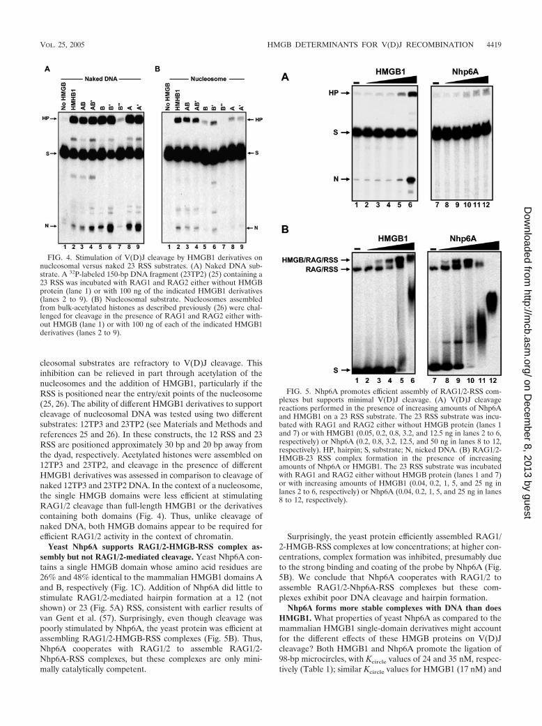
cleosomal substrates are refractory to V(D)J cleavage. Thisinhibition can be relieved in part through acetylation of thenucleosomes and the addition of HMGB1, particularly if theRSS is positioned near the entry/exit points of the nucleosome(25, 26). The ability of different HMGB1 derivatives to supportcleavage of nucleosomal DNA was tested using two differentsubstrates: 12TP3 and 23TP2 (see Materials and Methods andreferences 25 and 26). In these constructs, the 12 RSS and 23RSS are positioned approximately 30 bp and 20 bp away fromthe dyad, respectively. Acetylated histones were assembled on12TP3 and 23TP2, and cleavage in the presence of differentHMGB1 derivatives was assessed in comparison to cleavage ofnaked 12TP3 and 23TP2 DNA. In the context of a nucleosome,the single HMGB domains were less efficient at stimulatingRAG1/2 cleavage than full-length HMGB1 or the derivativescontaining both domains (Fig. 4). Thus, unlike cleavage ofnaked DNA, both HMGB domains appear to be required forefficient RAG1/2 activity in the context of chromatin.
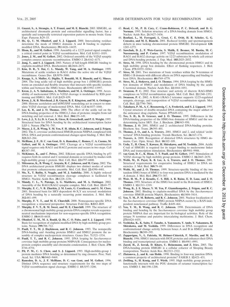
Yeast Nhp6A supports RAG1/2-HMGB-RSS complex as-sembly but not RAG1/2-mediated cleavage. Yeast Nhp6A con-tains a single HMGB domain whose amino acid residues are26% and 48% identical to the mammalian HMGB1 domains Aand B, respectively (Fig. 1C). Addition of Nhp6A did little tostimulate RAG1/2-mediated hairpin formation at a 12 (notshown) or 23 (Fig. 5A) RSS, consistent with earlier results ofvan Gent et al. (57). Surprisingly, even though cleavage waspoorly stimulated by Nhp6A, the yeast protein was efficient atassembling RAG1/2-HMGB-RSS complexes (Fig. 5B). Thus,Nhp6A cooperates with RAG1/2 to assemble RAG1/2-Nhp6A-RSS complexes, but these complexes are only mini-mally catalytically competent.
Surprisingly, the yeast protein efficiently assembled RAG1/2-HMGB-RSS complexes at low concentrations; at higher con-centrations, complex formation was inhibited, presumably dueto the strong binding and coating of the probe by Nhp6A (Fig.5B). We conclude that Nhp6A cooperates with RAG1/2 toassemble RAG1/2-Nhp6A-RSS complexes but these com-plexes exhibit poor DNA cleavage and hairpin formation.
Nhp6A forms more stable complexes with DNA than doesHMGB1. What properties of yeast Nhp6A as compared to themammalian HMGB1 single-domain derivatives might accountfor the different effects of these HMGB proteins on V(D)Jcleavage? Both HMGB1 and Nhp6A promote the ligation of98-bp microcircles, with Kcircle values of 24 and 35 nM, respec-tively (Table 1); similar Kcircle values for HMGB1 (17 nM) and
FIG. 4. Stimulation of V(D)J cleavage by HMGB1 derivatives onnucleosomal versus naked 23 RSS substrates. (A) Naked DNA sub-strate. A 32P-labeled 150-bp DNA fragment (23TP2) (25) containing a23 RSS was incubated with RAG1 and RAG2 either without HMGBprotein (lane 1) or with 100 ng of the indicated HMGB1 derivatives(lanes 2 to 9). (B) Nucleosomal substrate. Nucleosomes assembledfrom bulk-acetylated histones as described previously (26) were chal-lenged for cleavage in the presence of RAG1 and RAG2 either with-out HMGB (lane 1) or with 100 ng of each of the indicated HMGB1derivatives (lanes 2 to 9).
FIG. 5. Nhp6A promotes efficient assembly of RAG1/2-RSS com-plexes but supports minimal V(D)J cleavage. (A) V(D)J cleavagereactions performed in the presence of increasing amounts of Nhp6Aand HMGB1 on a 23 RSS substrate. The 23 RSS substrate was incu-bated with RAG1 and RAG2 either without HMGB protein (lanes 1and 7) or with HMGB1 (0.05, 0.2, 0.8, 3.2, and 12.5 ng in lanes 2 to 6,respectively) or Nhp6A (0.2, 0.8, 3.2, 12.5, and 50 ng in lanes 8 to 12,respectively). HP, hairpin; S, substrate; N, nicked DNA. (B) RAG1/2-HMGB-23 RSS complex formation in the presence of increasingamounts of Nhp6A or HMGB1. The 23 RSS substrate was incubatedwith RAG1 and RAG2 either without HMGB protein (lanes 1 and 7)or with increasing amounts of HMGB1 (0.04, 0.2, 1, 5, and 25 ng inlanes 2 to 6, respectively) or Nhp6A (0.04, 0.2, 1, 5, and 25 ng in lanes8 to 12, respectively).
VOL. 25, 2005 HMGB DETERMINANTS FOR V(D)J RECOMBINATION 4419
on Decem
ber 8, 2013 by guesthttp://m
cb.asm.org/
Dow
nloaded from

Nhp6A (38 nM) were also obtained for 75-bp microcircle forma-tion. Moreover, both Nhp6A and HMGB1 bind to 98-bp DNAmicrocircles with similar apparent equilibrium dissociation con-stants (Table 1). However, the different behaviors of Nhp6A andHMGB1 complexes with linear DNA segments upon polyacryl-amide gel electrophoresis implies that Nhp6A forms more stableDNA complexes than HMGB1 (Fig. 2B) (40).
To directly evaluate the stabilities of DNA complexes con-taining Nhp6A and HMGB1, complexes assembled on 98-bpmicrocircles were challenged with increasing amounts of son-icated salmon sperm DNA. Two Nhp6A molecules associatevery tightly with the 98-bp microcircle substrates and are re-sistant to addition of up to 5,000-fold mass excess of com-petitor DNA (Fig. 6A). By contrast the two full-lengthHMGB1 molecules readily dissociate from the microcirclesupon addition of increasing amounts of competitor DNA.
To further quantify the differences in stability of the HMGB-DNA microcircle complexes, the kinetics of dissociation weremeasured after addition of 55 �g/ml competitor DNA. Com-plexes formed with wild-type Nhp6A on 98-bp DNA micro-circles decay extremely slowly, with an estimated half-life ofabout 5 h (Fig. 6B; Table 1). By contrast, 50% of the complexesformed with full-length HMGB1 dissociated within 3 min, andessentially all of the complexes formed with the individualdomain A� and B� peptides dissociate within 3 min (Fig. 6B;Table 1). Interestingly, HMGB1 derivatives containing bothdomains but lacking the acidic C-terminal tail (HMGB1 do-mains AB� and AB) are markedly more stable than the full-length protein, with half-lives of 26 min (Fig. 6B; Table 1).However, they remain less stable than complexes formed withwild-type Nhp6A.
Mutations in Nhp6A that decrease complex stability on mi-crocircles enhance V(D)J cleavage. The results describedabove indicate that a key difference between the properties ofDNA complexes formed with Nhp6A and HMGB1 and itsderivatives is their stability. This led us to consider whetherNhp6A may be assembling RAG1/2-HMGB-RSS complexesthat are insufficiently dynamic to support DNA catalysis. Wetherefore asked if Nhp6A proteins containing mutations thatdestabilize their interaction with DNA were more effective atstimulating V(D)J cleavage. The basic N-terminal tail ofNhp6A, which wraps around the major groove on the oppositeside of DNA from the HMGB core domain (Fig. 1D), is largelyresponsible for its high-affinity binding to linear DNA (29, 63).Nhp6A (2-12) is missing the first set of basic residues (Lys 8,Lys 9, and Arg 10; Fig. 1C). This mutant is able to bind to andpromote ligation of 98-bp and 75-bp microcircles as efficientlyas wild-type Nhp6A (Table 1). Nhp6A (2-12) is much moresensitive to challenge with competitor DNA than wild-typeNhp6A and exhibits a dissociation rate from 98-bp microcirclesthat is similar to HMGB1 domains AB and AB� (Fig. 6A andB; Table 1). Nhp6A (2-16) has the entire N-terminal basicarm region deleted. It binds poorly to linear DNA but is ableto form unstable complexes on 98-bp microcircles and promoteligation of 98-bp microcircles at high protein concentrations(Table 1) (this work and reference 63). Whereas wild-typeNhp6A stimulates RAG1/2 cleavage of 12 and 23 RSS sub-strates poorly, reactions performed with either Nhp6A (2-12)or (2-16) generate substantial amounts of hairpins (Fig. 7Aand D), while supporting complex formation at levels equiva-
lent to wild-type Nhp6A (Fig. 7B and C). Similar results wereobtained in coupled-cleavage assays; whereas Nhp6A stimu-lated a small amount of coupled cleavage at the 12 and 23 RSSsites, the (2-12) and (2-16) derivatives were much moreeffective (data not shown).
Specific amino acid residue substitutions that destabilizeNhp6A-DNA interactions also result in enhanced RSS cleav-age by RAG1/2. Nhp6A Ala(13-16) has the lysines and argi-nine in the second basic patch of the N-terminal segment (Fig.1C) replaced with alanines. These substitutions result in astrong decrease in binding affinity, with most complexesformed on 98-bp microcircles dissociating within 3 min of com-petitor addition (Fig. 6B; Table 1). Like the N-terminal dele-tions, Nhp6A Ala(13-16) promotes enhanced cleavage and
FIG. 6. Stability of HMGB complexes formed on 98-bp micro-circles. (A) Nhp6A, HMGB1, and Nhp6A mutants were bound to32P-labeled 98-bp microcircles and then challenged with increasingamounts (0, 0.12, 0.37, 1.1, and 3.3 �g) of sonicated salmon spermDNA in 20-�l reaction mixtures. After an additional 30-min incuba-tion, the samples were subjected to native polyacrylamide gel electro-phoresis. The positions of the unbound (Free) 98-bp microcircles andcomplexes (C) containing one and two molecules of the HMGB pro-tein are indicated on the left. A small amount of an unstable thirdcomplex is present in the Nhp6A sample without competitor DNA.(B) Dissociation kinetics of HMGB complexes formed on 98-bp mi-crocircles after addition of 55 �g/ml sonicated salmon sperm DNA. Therelative number of complexes (percentage of the total DNA) formed byeach HMGB protein prior to the addition of competitor DNA was set to100, and the relative numbers of complexes remaining at each time pointwere scaled accordingly. The symbols for the HMGB1 and Nhp6A deriv-atives plotted are given to the right of the graph.
4420 DAI ET AL. MOL. CELL. BIOL.
on Decem
ber 8, 2013 by guesthttp://m
cb.asm.org/
Dow
nloaded from

hairpin formation at the 12 and 23 RSS (Fig. 7C and D and notshown). Alanine substitutions at residues Arg 13 plus Lys 14 orLys 15 plus Lys 16 have an intermediate defect in DNA binding(62) and, likewise, display moderately enhanced cleavage and
hairpin formation by RAG1/2 (Fig. 7D). The R23A R36Adouble mutant contains substitutions of residues within theHMGB core domain which directly interact with DNA fromthe minor groove side (29). This Nhp6A derivative binds linear
FIG. 7. Effects of mutations in Nhp6A on RAG1/2-HMGB-RSS complex formation and V(D)J cleavage. (A) Cleavage at a 12 RSS in the presenceof increasing amounts of Nhp6A and the (2-16) mutant derivative. The 12 RSS substrate was incubated with RAG1 and RAG2 without HMGB (lane1), RAG1 and RAG2 with Nhp6A (1, 2.5, 10, 50, and 100 ng in lanes 2 to 6, respectively), and (2-16) (10, 20, 40, 50, and 100 ng in lanes 7 to 11,respectively). HP, hairpin; S, substrate; N, nicked DNA. (B) Both Nhp6A and Nhp6A (2-16) support RAG1/2-RSS complex formation. The 12 RSSsubstrate was incubated with RAG1 and RAG2 without HMGB (lane 1 and lane 7), RAG1 and RAG2 with Nhp6A (0.04, 0.2, 1, 5, and 25 ng in lanes2 to 6, respectively), and Nhp6A (2-16) (0.04, 0.2, 1, 5, 25, 50, and 100 ng in lanes 8 to14, respectively). (C) RAG1/2-HMGB-12 RSS complex formation.A 150-bp 12 RSS substrate was incubated with RAG1 and RAG2 without HMGB (lane 1) or with HMGB1 (25 ng), HMGB1 domain B (50 ng), Nhp6A(1 ng), or Nhp6A mutant M29A (5 ng), M29D (5 ng), F48A (1 ng), M29A F48A (1 ng), (2-12) (5 ng), (2-16) (50 ng), K13A K14A (5 ng), K15A K16A(5 ng), Ala(13-16) (5 ng), or R23AR36A (5 ng). The amount of each protein added represents the optimal concentration for maximum RAG1/2-HMGB-RSS complex formation. (D) Single-site V(D)J cleavage. A 150-bp 32P-labeled 12 RSS substrate was incubated with RAG1 and RAG2 withoutHMGB or with HMGB1 (50 ng), HMGB1 domain B (50 ng), Nhp6A (2.5 ng), or Nhp6A mutant M29A (50 ng), M29D (50 ng), F48A (50 ng), M29AF48A (50 ng), (2-12) (50 ng), (2-16) (50 ng), R13A K14A (50 ng), K15A K16A (50 ng), Ala(13-16) (50 ng), or R23A R36A (50 ng). The amount ofeach protein added represents the optimal concentration for maximum nick and hairpin formation.
VOL. 25, 2005 HMGB DETERMINANTS FOR V(D)J RECOMBINATION 4421
on Decem
ber 8, 2013 by guesthttp://m
cb.asm.org/
Dow
nloaded from

DNA with a 10-fold-lower affinity, requires four times thewild-type amount of protein to promote equivalent microcircleformation, and dissociates from 98-bp microcircles with a �3-min half-life (Table 1) (5). Nevertheless, R23A R36A facili-tates binding and DNA catalysis by RAG1/2 with the 12 and 23RSS substrates (Fig. 7C and D and not shown). Thus, a varietyof mutations in the HMGB core or arm of Nhp6A that desta-bilize DNA binding result in enhanced RAG1/2-mediated ca-talysis of DNA relative to wild-type Nhp6A.
The DNA exit wedge generated by Nhp6A Phe 48 is requiredfor assembly of RAG1/2-HMGB-RSS complexes. Nhp6A pos-sesses two hydrophobic wedges that insert into the base stackvia the minor groove and distort the structure of the boundDNA (5, 29). Met 29 is the most important residue of thewedge, located near the center of the binding site, and Phe 48specifies the wedge at one of the DNA exit sites (Fig. 1D).Mutants with alanine substitutions at either of these residuesremain capable of forming complexes on linear DNA, butnuclear magnetic resonance analysis has revealed that bothresidues appear important in binding site selection (29). Phe 48is uniquely required for recognition of the kinked DNA struc-ture induced by cisplatin cross-linking (61). Both M29A andF48A are compromised for bending DNA, as revealed by thefailure of either mutant to form 75-bp microcircles; however,M29A remains capable of forming 98-bp microcircles. Bothmutants form complexes on 98-bp microcircles that are muchless stable than that formed by wild-type Nhp6A but morestable than those formed by individual HMGB1 domains in thepresence of competitor DNA (Fig. 6; Table 1).
The two Nhp6A wedge mutants have opposite propertieswith respect to the V(D)J reaction. Nhp6A F48A is unable toassemble RAG1/2-HMGB-RSS complexes (Fig. 7C) and thusalso fails to promote V(D)J cleavage (Fig. 7D). By contrast,Nhp6A M29A (or Nhp6A M29D) supports RAG1/2-RSS com-plex formation and stimulates V(D)J cleavage relative to wild-type Nhp6A (Fig. 7C and D). A mutant containing alaninesubstitutions at both wedges (Nhp6A M29A F48A) behaveslike Nhp6A F48A with respect to RAG1/2-HMGB-RSS com-plex formation and cleavage. Likewise, F48A and M29A F48Amutants failed to support coupled cleavage, but reactions per-formed with Nhp6A M29A or M29D exhibited enhancedcleavage at the 12 and 23 RSS sites relative to wild-type Nhp6A(data not shown). These results indicate that the Phe 48 wedgeis of critical importance for initial assembly of RAG1/2-HMGB-RSS complexes. The enhanced catalytic activity ofRAG1/2-HMGB-RSS complexes formed with Nhp6A M29A(and M29D) relative to wild-type Nhp6A presumably reflectsthe less stable binding properties exhibited by the mutant (Fig.6A and B; Table 1).
Requirement for Phe 38 of HMGB1 domain A for RAG1/2-RSS complex formation. Phe 38 in HMGB1 domain A corre-sponds to Phe 48 of Nhp6A. We therefore asked whether Phe 38in the domain A peptide was similarly required for the assemblyof RAG1/2-RSS complexes. Domain A peptide containing theF38A mutation failed to promote ligation of 98-bp microcircles,although it still was able to form unstable complexes with pre-formed 98-bp microcircles (Table 1). Similarly, domain A F38Adoes not promote assembly of RAG1/2 complexes on the 23 RSS,and therefore, as expected, no stimulation of cleavage by themutant HMGB1 derivative is detected (Fig. 8).
DISCUSSION
Previous in vitro studies on the V(D)J reaction have estab-lished that HMGB1 or HMGB2 potentiate RAG1/2 binding tothe RSS (4, 10, 45, 57; also see references 13 and 15 andreferences therein). In this report we show that either of thetwo HMGB DNA binding domains of HMGB1 can indepen-dently promote efficient assembly of catalytically competent
FIG. 8. Phe 38 of HMGB1 domain A is required for the stimula-tion of complex formation and V(D)J cleavage. (A) Complex forma-tion with a 23 RSS in the presence of increasing concentrations ofHMGB1 domain A (WT) (0.04, 0.2, 1, 5, and 25 ng in lanes 2 to 6,respectively) and the HMGB1 domain A mutant F38A (0.04, 0.2, 1, 5,and 25 ng in lanes 8 to 12, respectively) is shown. No HMGB proteinwas included in the reactions in lanes 1 and 7. (B) V(D)J cleavage inthe presence of wild-type HMGB1 domain A and HMGB1 domain Amutant F38A. The 23 RSS substrate was incubated with RAG1 andRAG2 either without HMGB (lane 1 and lane 7), with HMGB1 do-main A (WT) (25, 50, 100, 200, and 400 ng in lanes 2 to 6, respectively),or with domain A mutant F38A (25, 50, 100, 200, and 400 ng in lanes8 to 12, respectively). HP, hairpin; S, substrate; N, nicked DNA.
4422 DAI ET AL. MOL. CELL. BIOL.
on Decem
ber 8, 2013 by guesthttp://m
cb.asm.org/
Dow
nloaded from

complexes on naked DNA in vitro. However, the didomainstructure of HMGB1 is required for maximum activity whenthe RSS substrate is wrapped in a nucleosome. Analysis ofmutants of the yeast HMGB protein Nhp6A uncovered twoimportant features of HMGB proteins for stimulation ofV(D)J recombination. First, the HMGB protein must interactwith bent DNA in a relatively dynamic manner in order for thecomplexes to support DNA nicking and hairpin formation.This feature may reflect sequential changes in DNA confor-mations that are likely to be required as the RAG1/2-HMGB-RSS complex proceeds from initial binding through the twochemical steps leading to the formation of the hairpin product.Second, the hydrophobic wedge at the DNA exit site, which isspecified in Nhp6A by Phe 48, is a critical feature that isrequired to promote the initial step of RAG1/2-RSS binding.Phe 48 in Nhp6A, as well as its counterpart in HMGB1 domainA (Phe 38), is known to be a critical determinant in the rec-ognition of kinked DNA (19, 38, 61), and its importance isconsistent with HMGB proteins recognizing or stabilizing adistorted DNA structure within the RAG1/2-RSS complex (4).We discuss the biochemical properties of HMGB proteins withrespect to their stimulation of the V(D)J recombination reac-tion below.
Relationship between the DNA binding properties of re-sected derivatives of mammalian HMGB1 and their support ofRAG1/2-RSS complex assembly and cleavage activity. (i)HMGB1 domain A versus domain B. The rat HMGB1 DNAbinding domains (A and B) are 29% identical and 50% similarto each other (Fig. 1C). Our recombinant versions of domainsA� and B� containing their C-terminal basic ends function verysimilarly with respect to their abilities to form as well as toselectively bind to DNA microcircles. The similarity in DNAbending efficiencies between domains A� and B� observed herecontrasts with some other studies where domain B, with its twointercalating wedges, has been found to exhibit greater bend-ing activity (46, 52, 64). However, we note that the recombi-nant form of domain B used here lacks the lysine-rich linkerregion (amino acids 85 to 88; TKKK) separating domains Aand B, which has been reported to enhance DNA binding (17,46, 47). Our minimal-domain peptides that lack the basic res-idues at their C-terminal ends show pronounced differences inbending and binding to microcircular DNA; domain A requiresless protein than domain B for equivalent activity. We con-clude that both HMGB1 domains are effective at bendingDNA but that flanking basic residues enhance their activities,particularly for the B domain.
The DNA binding and bending properties of the isolateddomains largely parallel their activities in the V(D)J reactionon naked DNA. Whereas each of the isolated domain con-structs are able to efficiently promote RAG1/2-RSS assemblyand V(D)J cleavage, the B�- and B-domain peptides requiremore protein than the respective A�- and A-domain peptidesfor optimal activity. However, the ability to bend DNA, asevaluated by microcircle formation, is not the sole determinantof HMGB function in the V(D)J reaction, since the domain A�peptide behaved nearly indistinguishably from the didomainderivatives on naked DNA, even though 10- or 100-fold moredomain A� was required to generate equivalent numbers ofmicrocircles compared to full-length HMGB1 or domains AB/AB�, respectively. As elaborated further below, the differences
that we observe with the minimal domains are consistent withthe view that domain A of HMGB1 is more active than domainB in interacting with prebent and structured DNA (19, 22, 52,59) and that a localized DNA disruption exists within theRAG1/2-HMGB-RSS assembly (4, 10, 11, 42).
(ii) The C-terminal acidic tail. The C-terminal acidic tail hasgenerally been found to inhibit binding of HMGB1 derivativesto naked DNA, and our results are consistent with this view(22, 27, 48). Nevertheless, full-length HMGB1 appears no lessefficient at promoting RAG1/2 interactions on RSS DNA thandomain A or didomain derivatives lacking the C-terminal tail.As others also have noted, the single domain B� peptide con-taining the acidic region exhibits very poor DNA binding andbending activities, and consequently it appears essentially in-active in the V(D)J reaction. Recent nuclear magnetic reso-nance and cross-linking experiments have provided evidencethat the acidic region can specifically interact with residueswithin both domains and the connecting basic linker segment,which presumably inhibits DNA binding (22, 24).
The acidic region in the context of the didomain structurehas been reported to facilitate HMGB1 interactions with nu-cleosomes, possibly by interacting with the histone H3 N ter-minus (56). Bonaldi et al. have reported that the presence ofthe acidic region on HMGB1 enhances nucleosome sliding bythe ACF (ISWI) remodeling complex (8). We find that full-length HMGB1 is somewhat more efficient in promoting RSScleavage by RAG1/2 on nucleosomal substrates than the dido-main derivatives without the acidic region, suggesting that theacidic region could have a modest beneficial role in V(D)Jrecombination in vivo.
Nhp6A mutants reveal parameters important for assemblyof catalytically competent RAG1/2-RSS complexes. The se-quence and structure of the HMGB domain of Nhp6A aremore closely related to HMGB1 domain B (48% amino acidresidue identity, 1.2-Å root mean square deviation betweenpeptide backbones) than domain A (26% amino acid residueidentity, 2.2-Å root mean square deviation between peptidebackbones) (Fig. 1C) (5, 29). Nhp6A binds DNA much morestably than any of the HMGB1 derivatives, primarily becauseof its N-terminal basic arm. We find that the greater affinity forDNA does not improve RAG1/2-RSS complex formation.Nhp6A promotes efficient RAG1/2-RSS complex formation atprotein levels that are equivalent to HMGB1 domain A� or thedidomain constructs. However, the stable DNA binding byNhp6A strongly inhibits DNA cleavage by the RAG1/2-Nhp6A-RSS complexes. Mutations within the N-terminal tailor within the HMGB core that destabilize binding and thuslead to Nhp6A proteins whose DNA binding properties moreclosely mimic those of the HMGB1 derivatives result in cor-responding increases in V(D)J cleavage. These findings sug-gest that a dynamic association of the HMGB protein with theRAG1/2-RSS complex is critical for the DNA cleavage steps.The HMGB protein is not released from the complex, how-ever, since HMGB1 has been shown to directly enhance hair-pin formation in experiments utilizing prenicked substratesand to remain with the complex after the hairpin has beenformed (20, 33, 34, 49, 50, 57).
Nhp6A contains two hydrophobic wedges at the DNA bind-ing surface, but only one, Phe 48, is critical for stimulatingRAG1/2-RSS assembly. This residue corresponds to Phe 38 in
VOL. 25, 2005 HMGB DETERMINANTS FOR V(D)J RECOMBINATION 4423
on Decem
ber 8, 2013 by guesthttp://m
cb.asm.org/
Dow
nloaded from

domain A, which we also show to be essential for its ability topromote RAG1/2-RSS assembly, and to Ile 34 in domain B(Fig. 1D). The importance of Phe 48 and the dispensability ofthe Met 29 wedge parallel the roles of these two residues inselective binding by Nhp6A to cisplatin adducts on DNA (61).Consistent with its moderately poorer activity in the V(D)Jreaction, HMGB1 domain B has been found to be less selec-tive for binding to cisplatin adducts than domain A, in partbecause of the isoleucine in place of a phenylalanine at theDNA exit wedge (19, 22). Cisplatin forms an intrastrand cross-link at the N7 position of adjacent guanines, generating a kinkin the duplex DNA that compresses the major groove (51). AnX-ray structure of mammalian HMGB1 domain A bound to aDNA duplex containing a cisplatin cross-link revealed that thePhe 38 intercalates between the cross-linked guanines on theminor groove side (Fig. 1D) (19, 38).
Aidinis et al. have provided evidence for bending of the RSSsites by RAG1/2 by circular permutation analysis of the elec-trophoretic migrations of RAG1/2-12 or 23 RSS complexes,even in the absence of HMGB1 (4). This finding, combinedwith the importance of the phenylalanine wedge, suggests thatthe HMGB protein may be recognizing a RAG1/2-inducedDNA distortion between the nonamer and heptamer regions.Binding of the HMGB proteins may then stabilize the bentstructure, thus enhancing complex formation. Consistent withthis model, both protein-DNA cross-linking and ethylation in-terference experiments have suggested that HMGB1 is locatedin the intervening DNA segment. Ethylation interference ex-periments point to the HMGB protein contacting DNA nearthe 5� end of the nonamer on the opposite side of the duplexfrom the bound RAG1/2 (33, 49). There are also several linesof evidence suggesting that unpairing of DNA strands, partic-ularly over the heptamer region, may be important for theDNA cleavage/hairpin formation steps (10 and reviewed inreference 14). HMGB proteins induce considerable untwistingof DNA along with bending, which may further contribute totheir stimulation of RAG1/2 function.
Comparison of HMGB1 stimulation of RAG1/2 and otherDNA binding proteins. The DNA binding domain of RAG1has been proposed to contain a helix-turn-helix motif that issimilar to the Tc1/mariner family of transposases (6). Interest-ingly, a member of the Tc1/mariner family of transposons,Sleeping Beauty, has also been shown to require HMGB1 fortransposition in human cells (66). HMGB1 also facilitatesbinding of a number of transcription factors with structurallydiverse DNA binding domains (2, 54). As is the case forRAG1/2 binding, a single domain from HMGB1 has beenfound to be sufficient in stimulating DNA binding of some ofthese regulatory proteins, including the Oct1/2 and HOXD9homeodomains and Rta (31, 32, 65, 68). However, in at leastone case, the cooperative binding of the basic leucine zipperprotein Zebra to adjacent sites within the regulatory region ofan Epstein-Barr virus promoter, both linked domains ofHMGB1 are required (12). In some situations, there is evi-dence that HMGB1 interacts in solution with its binding part-ner, implying that direct protein-protein interactions, in addi-tion to effects on DNA architecture by HMGB1, appear tocontribute to binding cooperativity (reviewed in (2, 54). On theother hand, stable ternary complexes containing HMGB1 andDNA have not been observed upon polyacrylamide gel elec-
trophoresis in several systems. Therefore, the HMGB proteinappears to function in some contexts only transiently to chap-erone the transcription factor to its DNA binding site.
Aidinis et al. reported that RAG1/2 and HMGB1 interact insolution in experiments employing immobilized RAG1/2 orHMGB1 (4). Both domains A and B were required in order toobserve this interaction, leading the authors to propose thatboth domains are required for HMGB1 stimulation of RAG1/2activity. Our finding that either isolated HMGB1 domain orunstable binding mutants of yeast Nhp6A are effective in pro-moting the assembly of catalytically competent RAG1/2-HMGB-RSS complexes suggests that a specific RAG1/2–HMGB interaction may be secondary to an effect of HMGB onDNA architecture. Only nine solvent-exposed residues, whichhave similar chemical characteristics but are not directly in-volved in DNA binding, are common between the threeHMGB domains (Fig. 1C).
ACKNOWLEDGMENTS
This work was supported by USPHS grants GM38509 to R.C.J. andGM48026 to M.A.O.
REFERENCES
1. Adachi, Y., S. Mizuno, and M. Yoshida. 1990. Efficient large-scale purifica-tion of non-histone chromosomal proteins HMG1 and HMG2 by usingPolybuffer-exchanger PBE94. J. Chromatogr. 530:39–46.
2. Agrawal, A., and D. G. Schatz. 1997. RAG1 and RAG2 form a stablepostcleavage synaptic complex with DNA containing signal ends in V(D)Jrecombination. Cell 89:43–53.
3. Agresti, A., and M. E. Bianchi. 2003. HMGB proteins and gene expression.Curr. Opin. Genet. Dev. 13:170–178.
4. Aidinis, V., T. Bonaldi, M. Beltrame, S. Santagata, M. E. Bianchi, and E.Spanopoulou. 1999. The RAG1 homeodomain recruits HMG1 and HMG2to facilitate recombination signal sequence binding and to enhance theintrinsic DNA-bending activity of RAG1-RAG2. Mol. Cell. Biol. 19:6532–6542.
5. Allain, F. H., Y. M. Yen, J. E. Masse, P. Schultze, T. Dieckmann, R. C.Johnson, and J. Feigon. 1999. Solution structure of the HMG proteinNHP6A and its interaction with DNA reveals the structural determinants fornon-sequence-specific binding. EMBO J. 18:2563–2579.
6. Banerjee-Basu, S., and A. D. Baxevanis. 2002. The DNA-binding region ofRAG 1 is not a homeodomain. Genome Biol. 3:interactions 1004.1–1004.4.[Online.] http://genomebiology.com.
7. Bassing, C. H., W. Swat, and F. W. Alt. 2002. The mechanism and regulationof chromosomal V(D)J recombination. Cell 109(Suppl.):S45–S55.
8. Bonaldi, T., G. Langst, R. Strohner, P. B. Becker, and M. E. Bianchi. 2002.The DNA chaperone HMGB1 facilitates ACF/CHRAC-dependent nucleo-some sliding. EMBO J. 21:6865–6873.
9. Bustin, M. 1999. Regulation of DNA-dependent activities by the functionalmotifs of the high-mobility-group chromosomal proteins. Mol. Cell. Biol.19:5237–5246.
10. Ciubotaru, M., and D. G. Schatz. 2004. Synapsis of recombination signalsequences located in cis and DNA underwinding in V(D)J recombination.Mol. Cell. Biol. 24:8727–8744.
11. Cuomo, C. A., C. L. Mundy, and M. A. Oettinger. 1996. DNA sequence andstructure requirements for cleavage of V(D)J recombination signal se-quences. Mol. Cell. Biol. 16:5683–5690.
12. Ellwood, K. B., Y. M. Yen, R. C. Johnson, and M. Carey. 2000. Mechanismfor specificity by HMG-1 in enhanceosome assembly. Mol. Cell. Biol. 20:4359–4370.
13. Fugmann, S. D., A. I. Lee, P. E. Shockett, I. J. Villey, and D. G. Schatz. 2000.The RAG proteins and V(D)J recombination: complexes, ends, and trans-position. Annu. Rev. Immunol. 18:495–527.
14. Gellert, M. 2002. V(D)J recombination, p. 705–729. In N. L. Craig, R.Craigie, M. Gellert, and A. M. Lambowitz (ed.), Mobile DNA II. ASM Press,Washington, D.C.
15. Gellert, M. 2002. V(D)J recombination: RAG proteins, repair factors, andregulation. Annu. Rev. Biochem. 71:101–132.
16. Grasser, K. D. 2003. Chromatin-associated HMGA and HMGB proteins:versatile co-regulators of DNA-dependent processes. Plant Mol. Biol. 53:281–295.
17. Grasser, K. D., S. H. Teo, K. B. Lee, R. W. Broadhurst, C. Rees, C. H.Hardman, and J. O. Thomas. 1998. DNA-binding properties of the tandemHMG boxes of high-mobility-group protein 1 (HMG1). Eur. J. Biochem.253:787–795.
4424 DAI ET AL. MOL. CELL. BIOL.
on Decem
ber 8, 2013 by guesthttp://m
cb.asm.org/
Dow
nloaded from
18. Guazzi, S., A. Strangio, A. T. Franzi, and M. E. Bianchi. 2003. HMGB1, anarchitectural chromatin protein and extracellular signalling factor, has aspatially and temporally restricted expression pattern in mouse brain. GeneExpr. Patterns 3:29–33.
19. He, Q., U. M. Ohndorf, and S. J. Lippard. 2000. Intercalating residuesdetermine the mode of HMG1 domains A and B binding to cisplatin-modified DNA. Biochemistry 39:14426–14435.
20. Hiom, K., and M. Gellert. 1998. Assembly of a 12/23 paired signal complex:a critical control point in V(D)J recombination. Mol. Cell 1:1011–1019.
21. Jones, J. M., and M. Gellert. 2002. Ordered assembly of the V(D)J synapticcomplex ensures accurate recombination. EMBO J. 21:4162–4171.
22. Jung, Y., and S. J. Lippard. 2003. Nature of full-length HMGB1 binding tocisplatin-modified DNA. Biochemistry 42:2664–2671.
23. Kim, D. R., Y. Dai, C. L. Mundy, W. Yang, and M. A. Oettinger. 1999.Mutations of acidic residues in RAG1 define the active site of the V(D)Jrecombinase. Genes Dev. 13:3070–3080.
24. Knapp, S., S. Muller, G. Digilio, T. Bonaldi, M. E. Bianchi, and G. Musco.2004. The long acidic tail of high mobility group box 1 (HMGB1) proteinforms an extended and flexible structure that interacts with specific residueswithin and between the HMG boxes. Biochemistry 43:11992–11997.
25. Kwon, J., A. N. Imbalzano, A. Matthews, and M. A. Oettinger. 1998. Acces-sibility of nucleosomal DNA to V(D)J cleavage is modulated by RSS posi-tioning and HMG1. Mol. Cell 2:829–839.
26. Kwon, J., K. B. Morshead, J. R. Guyon, R. E. Kingston, and M. A. Oettinger.2000. Histone acetylation and hSWI/SNF remodeling act in concert to stim-ulate V(D)J cleavage of nucleosomal DNA. Mol. Cell 6:1037–1048.
27. Lee, K. B., and J. O. Thomas. 2000. The effect of the acidic tail on theDNA-binding properties of the HMG1,2 class of proteins: insights from tailswitching and tail removal. J. Mol. Biol. 304:135–149.
28. Love, J. J., X. Li, D. A. Case, K. Giese, R. Grosschedl, and P. E. Wright. 1995.Structural basis for DNA bending by the architectural transcription factorLEF-1. Nature 376:791–795.
29. Masse, J. E., B. Wong, Y. M. Yen, F. H. Allain, R. C. Johnson, and J. Feigon.2002. The S. cerevisiae architectural HMGB protein NHP6A complexed withDNA: DNA and protein conformational changes upon binding. J. Mol. Biol.323:263–284.
30. McBlane, J. F., D. C. van Gent, D. A. Ramsden, C. Romeo, C. A. Cuomo, M.Gellert, and M. A. Oettinger. 1995. Cleavage at a V(D)J recombinationsignal requires only RAG1 and RAG2 proteins and occurs in two steps. Cell83:387–395.
31. McKinney, K., and C. Prives. 2002. Efficient specific DNA binding by p53requires both its central and C-terminal domains as revealed by studies withhigh-mobility group 1 protein. Mol. Cell. Biol. 22:6797–6808.
32. Mitsouras, K., B. Wong, C. Arayata, R. C. Johnson, and M. Carey. 2002. TheDNA architectural protein HMGB1 displays two distinct modes of actionthat promote enhanceosome assembly. Mol. Cell. Biol. 22:4390–4401.
33. Mo, X., T. Bailin, S. Noggle, and M. J. Sadofsky. 2000. A highly orderedstructure in V(D)J recombination cleavage complexes is facilitated byHMG1. Nucleic Acids Res. 28:1228–1236.
34. Mundy, C. L., N. Patenge, A. G. Matthews, and M. A. Oettinger. 2002.Assembly of the RAG1/RAG2 synaptic complex. Mol. Cell. Biol. 22:69–77.
35. Murphy, E. C., V. B. Zhurkin, J. M. Louis, G. Cornilescu, and G. M. Clore.2001. Structural basis for SRY-dependent 46-X,Y sex reversal: modulationof DNA bending by a naturally occurring point mutation. J. Mol. Biol.312:481–499.
36. Murphy, F. V. T., and M. E. Churchill. 2000. Nonsequence-specific DNArecognition: a structural perspective. Structure Fold Des. 8:R83–R89.
37. Murphy, F. V. T., R. M. Sweet, and M. E. Churchill. 1999. The structure ofa chromosomal high mobility group protein-DNA complex reveals sequence-neutral mechanisms important for non-sequence-specific DNA recognition.EMBO J. 18:6610–6618.
38. Ohndorf, U. M., M. A. Rould, Q. He, C. O. Pabo, and S. J. Lippard. 1999.Basis for recognition of cisplatin-modified DNA by high-mobility-group pro-teins. Nature 399:708–712.
39. Paull, T. T., M. J. Haykinson, and R. C. Johnson. 1993. The nonspecificDNA-binding and -bending proteins HMG1 and HMG2 promote the as-sembly of complex nucleoprotein structures. Genes Dev. 7:1521–1534.
40. Paull, T. T., and R. C. Johnson. 1995. DNA looping by Saccharomycescerevisiae high mobility group proteins NHP6A/B. Consequences for nucleo-protein complex assembly and chromatin condensation. J. Biol. Chem. 270:8744–8754.
41. Pil, P. M., C. S. Chow, and S. J. Lippard. 1993. High-mobility-group 1protein mediates DNA bending as determined by ring closures. Proc. Natl.Acad. Sci. USA 90:9465–9469.
42. Ramsden, D. A., J. F. McBlane, D. C. van Gent, and M. Gellert. 1996.Distinct DNA sequence and structure requirements for the two steps ofV(D)J recombination signal cleavage. EMBO J. 15:3197–3206.
43. Read, C. M., P. D. Cary, C. Crane-Robinson, P. C. Driscoll, and D. G.Norman. 1993. Solution structure of a DNA-binding domain from HMG1.Nucleic Acids Res. 21:3427–3436.
44. Ronfani, L., M. Ferraguti, L. Croci, C. E. Ovitt, H. R. Scholer, G. G.Consalez, and M. E. Bianchi. 2001. Reduced fertility and spermatogenesisdefects in mice lacking chromosomal protein HMGB2. Development 128:1265–1273.
45. Sawchuk, D. J., F. Weis-Garcia, S. Malik, E. Besmer, M. Bustin, M. C.Nussenzweig, and P. Cortes. 1997. V(D)J recombination: modulation ofRAG1 and RAG2 cleavage activity on 12/23 substrates by whole cell extractand DNA-bending proteins. J. Exp. Med. 185:2025–2032.
46. Stros, M. 1998. DNA bending by the chromosomal protein HMG1 and itshigh mobility group box domains. Effect of flanking sequences. J. Biol.Chem. 273:10355–10361.
47. Stros, M. 2001. Two mutations of basic residues within the N-terminus ofHMG-1 B domain with different effects on DNA supercoiling and binding tobent DNA. Biochemistry 40:4769–4779.
48. Stros, M., J. Stokrova, and J. O. Thomas. 1994. DNA looping by the HMG-box domains of HMG1 and modulation of DNA binding by the acidicC-terminal domain. Nucleic Acids Res. 22:1044–1051.
49. Swanson, P. C. 2002. Fine structure and activity of discrete RAG-HMGcomplexes on V(D)J recombination signals. Mol. Cell. Biol. 22:1340–1351.
50. Swanson, P. C. 2002. A RAG-1/RAG-2 tetramer supports 12/23-regulatedsynapsis, cleavage, and transposition of V(D)J recombination signals. Mol.Cell. Biol. 22:7790–7801.
51. Takahara, P. M., A. C. Rosenzweig, C. A. Frederick, and S. J. Lippard. 1995.Crystal structure of double-stranded DNA containing the major adduct ofthe anticancer drug cisplatin. Nature 377:649–652.
52. Teo, S. H., K. D. Grasser, and J. O. Thomas. 1995. Differences in theDNA-binding properties of the HMG-box domains of HMG1 and the sex-determining factor SRY. Eur. J. Biochem. 230:943–950.
53. Thomas, J. O. 2001. HMG1 and 2: architectural DNA-binding proteins.Biochem. Soc. Trans. 29:395–401.
54. Thomas, J. O., and A. A. Travers. 2001. HMG1 and 2, and related ‘archi-tectural’ DNA-binding proteins. Trends Biochem. Sci. 26:167–174.
55. Travers, A. 2000. Recognition of distorted DNA structures by HMG do-mains. Curr. Opin. Struct. Biol. 10:102–109.
56. Ueda, T., H. Chou, T. Kawase, H. Shirakawa, and M. Yoshida. 2004. AcidicC-tail of HMGB1 is required for its target binding to nucleosome linkerDNA and transcription stimulation. Biochemistry 43:9901–9908.
57. van Gent, D. C., K. Hiom, T. T. Paull, and M. Gellert. 1997. Stimulation ofV(D)J cleavage by high mobility group proteins. EMBO J. 16:2665–2670.
58. Webb, M., D. Payet, K. B. Lee, A. A. Travers, and J. O. Thomas. 2001.Structural requirements for cooperative binding of HMG1 to DNA mi-nicircles. J. Mol. Biol. 309:79–88.
59. Webb, M., and J. O. Thomas. 1999. Structure-specific binding of the twotandem HMG boxes of HMG1 to four-way junction DNA is mediated by theA domain. J. Mol. Biol. 294:373–387.
60. Weir, H. M., P. J. Kraulis, C. S. Hill, A. R. Raine, E. D. Laue, and J. O.Thomas. 1993. Structure of the HMG box motif in the B-domain of HMG1.EMBO J. 12:1311–1319.
61. Wong, B., J. E. Masse, Y. M. Yen, P. Giannikoupolos, J. Feigon, and R. C.Johnson. 2002. Binding to cisplatin-modified DNA by the Saccharomycescerevisiae HMGB protein Nhp6A. Biochemistry 41:5404–5414.
62. Yen, Y. M., P. M. Roberts, and R. C. Johnson. 2001. Nuclear localization ofthe Saccharomyces cerevisiae HMG protein NHP6A occurs by a RAN-inde-pendent nonclassical pathway. Traffic 2:449–464.
63. Yen, Y. M., B. Wong, and R. C. Johnson. 1998. Determinants of DNAbinding and bending by the Saccharomyces cerevisiae high mobility groupprotein NHP6A that are important for its biological activities. Role of theunique N terminus and putative intercalating methionine. J. Biol. Chem.273:4424–4435.
64. Yoshioka, K., K. Saito, T. Tanabe, A. Yamamoto, Y. Ando, Y. Nakamura, H.Shirakawa, and M. Yoshida. 1999. Differences in DNA recognition andconformational change activity between boxes A and B in HMG2 protein.Biochemistry 38:589–595.
65. Zappavigna, V., L. Falciola, M. Helmer-Citterich, F. Mavilio, and M. E.Bianchi. 1996. HMG1 interacts with HOX proteins and enhances their DNAbinding and transcriptional activation. EMBO J. 15:4981–4991.
66. Zayed, H., Z. Izsvak, D. Khare, U. Heinemann, and Z. Ivics. 2003. TheDNA-bending protein HMGB1 is a cellular cofactor of Sleeping Beautytransposition. Nucleic Acids Res. 31:2313–2322.
67. Zlatanova, J., and K. van Holde. 1998. Binding to four-way junction DNA:a common property of architectural proteins? FASEB J. 12:421–431.
68. Zwilling, S., H. Konig, and T. Wirth. 1995. High mobility group protein 2functionally interacts with the POU domains of octamer transcription fac-tors. EMBO J. 14:1198–1208.
VOL. 25, 2005 HMGB DETERMINANTS FOR V(D)J RECOMBINATION 4425
on Decem
ber 8, 2013 by guesthttp://m
cb.asm.org/
Dow
nloaded from