Detection of inflammatory bowel disease: diagnostic performance of cross-sectional imaging...
10
UPDATE Detection of inflammatory bowel disease: diagnostic performance of cross-sectional imaging modalities Karin Horsthuis, 1 Pieter C. F. Stokkers, 2 Jaap Stoker 1 1 Department of Radiology, Academic Medical Center, Meibergdreef 9, 1105 AZ, Amsterdam, The Netherlands 2 Department of Gastroenterology, Academic Medical Center, Amsterdam, The Netherlands Abstract Different cross-sectional imaging techniques can be used as a diagnostic tool for the evaluation of inflammatory bowel disease (IBD). In this report the diagnostic per- formances of ultrasonography, magnetic resonance imaging and computed tomography in the detection of IBD and the evaluation of known IBD are described, together with a short update on patient preparation and imaging technique of the respective modalities discussed. Key words: Inflammatory bowel disease—Ultrasonography—Magnetic Resonance Imaging—Computed Tomography—Diagnostic performance Introduction Chronic inflammatory bowel disease (IBD) consists of two main subtypes, i.e., CrohnÕs disease (CD) and ulcerative colitis (UC). During the last decades, the incidence of CD has continued to increase worldwide, reaching incidence rates ranging from 3.1 to 14.6/100,000 in North America and from 0.7 to 9.8/100,000 in Europe [1]. Incidence rates of UC differ greatly between studies and regions, varying from 1.5 to 24.5 per 100,000 person- years [1, 2]. CrohnÕs disease can be localized in any part of the gastrointestinal tract, although the location of predilec- tion is the terminal ileum, involvement of the terminal ileum is observed in 90% of the patients with small- intestinal CD, who in turn constitute 30–40% of all CD patients. In 40–55% of the patients both ileum and colon are affected, while in a minority (15–25%) only a colonic localization is observed. The earliest change caused by CD occurs in the mu- cosa and submucosa and consists of hyperemia and edema. Tiny aphthoid or superficial ulcerations can be seen when disease progresses. In more severe disease, the disease extends transmurally with sometimes serosal involvement. In this stage, mucosal ulcerations merge to form deep longitudinal and transverse ulcerations while bowel wall thickening and narrowing of the bowel lumen can be observed due to significant mucosal edema and associated bowel spasms. In long-standing disease, chronic obstruction can develop due to scarring, luminal narrowing, and stricture formation. Extramural mani- festations of CD are fistulas, abscesses, adhesions, creeping fat, and enlargement of lymph nodes. Ulcerative colitis exclusively affects the colon with a predictable way of spreading from distal to proximal in a continuous manner; the rectum is often involved, but rectal sparing can be observed. In previous cases, small superficial erosions can be seen, whereas in more severe disease these ulcerations can be quite large. However, only in very severe disease they penetrate the muscularis layer. The mucosa is thickened because of round-cell infiltration in the lamina propria. In chronic UC, a marked hypertrophy of muscularis mucosae can be seen, causing contraction, shortening, and narrowing of the involved colon. The submucosa becomes thickened be- cause of the deposition of fat or, in acute or subacute cases, edema [3, 4]. Diagnostic modalities The gold standard examination for the small bowel tra- ditionally has been small bowel barium examination Correspondence to: Karin Horsthuis; email: [email protected] ª Springer Science+Business Media, LLC 2007 Published online: 10 July 2007 Abdominal Imaging Abdom Imaging (2008) 33:407–416 DOI: 10.1007/s00261-007-9276-3
Transcript of Detection of inflammatory bowel disease: diagnostic performance of cross-sectional imaging...
UPDATE
Detection of inflammatory bowel disease:diagnostic performance of cross-sectionalimaging modalities
Karin Horsthuis,1 Pieter C. F. Stokkers,2 Jaap Stoker1
1Department of Radiology, Academic Medical Center, Meibergdreef 9, 1105 AZ, Amsterdam, The Netherlands2Department of Gastroenterology, Academic Medical Center, Amsterdam, The Netherlands
Abstract
Different cross-sectional imaging techniques can be usedas a diagnostic tool for the evaluation of inflammatorybowel disease (IBD). In this report the diagnostic per-formances of ultrasonography, magnetic resonanceimaging and computed tomography in the detection ofIBD and the evaluation of known IBD are described,together with a short update on patient preparation andimaging technique of the respective modalities discussed.
Key words: Inflammatory boweldisease—Ultrasonography—Magnetic ResonanceImaging—Computed Tomography—Diagnosticperformance
Introduction
Chronic inflammatory bowel disease (IBD) consists oftwo main subtypes, i.e., Crohn�s disease (CD) andulcerative colitis (UC). During the last decades, theincidence of CD has continued to increase worldwide,reaching incidence rates ranging from 3.1 to 14.6/100,000in North America and from 0.7 to 9.8/100,000 in Europe[1]. Incidence rates of UC differ greatly between studiesand regions, varying from 1.5 to 24.5 per 100,000 person-years [1, 2].
Crohn�s disease can be localized in any part of thegastrointestinal tract, although the location of predilec-tion is the terminal ileum, involvement of the terminalileum is observed in 90% of the patients with small-intestinal CD, who in turn constitute 30–40% of all CD
patients. In 40–55% of the patients both ileum and colonare affected, while in a minority (15–25%) only a coloniclocalization is observed.
The earliest change caused by CD occurs in the mu-cosa and submucosa and consists of hyperemia andedema. Tiny aphthoid or superficial ulcerations can beseen when disease progresses. In more severe disease, thedisease extends transmurally with sometimes serosalinvolvement. In this stage, mucosal ulcerations merge toform deep longitudinal and transverse ulcerations whilebowel wall thickening and narrowing of the bowel lumencan be observed due to significant mucosal edema andassociated bowel spasms. In long-standing disease,chronic obstruction can develop due to scarring, luminalnarrowing, and stricture formation. Extramural mani-festations of CD are fistulas, abscesses, adhesions,creeping fat, and enlargement of lymph nodes.
Ulcerative colitis exclusively affects the colon with apredictable way of spreading from distal to proximal in acontinuous manner; the rectum is often involved, butrectal sparing can be observed. In previous cases, smallsuperficial erosions can be seen, whereas in more severedisease these ulcerations can be quite large. However,only in very severe disease they penetrate the muscularislayer. The mucosa is thickened because of round-cellinfiltration in the lamina propria. In chronic UC, amarked hypertrophy of muscularis mucosae can be seen,causing contraction, shortening, and narrowing of theinvolved colon. The submucosa becomes thickened be-cause of the deposition of fat or, in acute or subacutecases, edema [3, 4].
Diagnostic modalities
The gold standard examination for the small bowel tra-ditionally has been small bowel barium examinationCorrespondence to: Karin Horsthuis; email: [email protected]
ª Springer Science+Business Media, LLC 2007
Published online: 10 July 2007AbdominalImaging
Abdom Imaging (2008) 33:407–416
DOI: 10.1007/s00261-007-9276-3

(SBE), either by using an enteroclysis technique or byusing small-bowel follow-through [5, 6]. SBE is invasiveand burdensome, and requires an extensive bowel prep-aration (dietary restrictions, use of laxatives). Moreover,in the young population of CD patients, the ionizingradiation required for SBE limits the use of this tech-nique for follow-up of disease.
The advent of video capsule endoscopy (VCE) anddouble-balloon endoscopy (DBE) has increased thediagnostic possibilities. For VCE a capsule is swallowedafter a fasting period of up to 12 h and is propelledthrough the bowel by peristalsis. Thus, the mucosalsurface of the small bowel can be depicted in detail(Fig. 1). However, with VCE there is no facility to in-crease visibility by insufflation of air or by tissue rinsing.Moreover, tissue sampling and therapeutic interventionsare not possible. The use of VCE is contraindicated inpatients with (suspicion of) obstruction due to the risk ofcapsule retention.
For DBE, a high-resolution video-endoscope with aflexible overtube is used. By alternately inflating anddeflating two balloons attached to the overtube andendoscope the small bowel is threaded on the overtube.Both an oral and an anal approach are possible; for theoral approach no specific preparation is required, al-though patients are asked to fast for at least 6 h beforethe procedure. If the anal approach is used, bowelcleansing such as is employed for traditional colonos-copy is used. By using both the anal and oral approach,DBE allows visualization of the entire small bowel, withthe possibility of obtaining tissue for analysis and theadded advantage of the possibility of endoscopic therapy(e.g. dilatation of a stricture, cauterization of a bleedingsite). For DBE conscious sedation is a necessity.
Traditionally, ileocolonoscopy (CS) with tissuesampling is considered to be the most valuable tool fordiagnosis and follow-up of disease in the colon andterminal ileum [5, 7]. As UC solely affects the mucosa ofthe colon, CS would suffice for diagnosis of disease andevaluation of disease activity and extent. However,when strictures are present as a complication of disease,these might hamper execution of a complete examina-tion, while in severe attacks of UC CS is relativelycontra-indicated due to the increased risk of bleeding orperforation. For ileocolonic localizations of CD CSwould suffice as well, although inspection of the termi-nal ileum is reported to fail in up to 27.8% of examin-ations [8, 9].
Cross-sectional imaging modalities
The trans- and extramural extent of IBD cannot bevisualized with any of the abovementioned techniques.Much research has been directed toward the potential ofcross-sectional imaging modalities for the diagnosis andevaluation of IBD as with these techniques the bowellumen, the bowel wall and the extra-intestinal abdomenincluding the visceral fat, the lymph nodes and the vas-cular structures feeding and draining the bowel can bevisualized. An added advantage of these techniques is thefact that they are limitedly to non-invasive.
Ultrasonography (US), computed tomography (CT)and magnetic resonance imaging (MRI) are often usedfor the evaluation of the abdomen. While in the USA thetechnique of choice is CT, in Europe the focus is more onMRI and US. This inclination is reflected by the majorityof CT studies on IBD patients originating from the USA,while the majority of published studies on MRI and UShas been conducted in Europe.
Ultrasonography
Patient preparation and US technique. Patients are usu-ally asked to fast for several hours before the scan todiminish peristaltic movements and the amount of in-traluminal air; in the available literature the minimumfasting time described is 4 h, whereas overnight fasting isalso sometimes employed. Usually, no additional dietaryor cleansing measures are taken. Due to the limited pa-tient preparation necessary and the non-invasive natureof this examination, US can be considered to be a rela-tively patient-friendly and straightforward examination.
Ultrasonography is mostly performed without the useof enteral contrast medium. In two recent studies, theeffect of enteral contrast medium for diagnosis of IBD ofthe small bowel was investigated; higher sensitivity val-ues were obtained when enteral contrast medium hadbeen administered [10, 11]. In both studies the additionalvalue of enteral contrast medium permitted detectionof jejunal lesions that had escaped detection with
Fig. 1. VCE image of a 14-year-old male patient with knownCD. VCE was performed as small-bowel disease was sus-pected. Image shows severe inflammation of the small bowelwith a stenosis.
408 K. Horsthuis et al.: Cross-sectional imaging for detection of IBD
conventional US. For colonic IBD, use of enteral con-trast medium also increased accuracy [12, 13].
High-frequency transducers are preferred, such as7.5 MHz [13–16].
The use of Doppler-US might provide helpful addi-tional information on IBD, particularly on the degree ofdisease activity. Using Doppler-US the vascularity of thebowel wall can be assessed according to the intensity ofcolor signals and/or by the analysis of Doppler curves(measurement of resistive index) obtained from vesselsdetected within the bowel wall. Measurement of flowparameters of the superior and inferior mesentericarteries can also be performed.
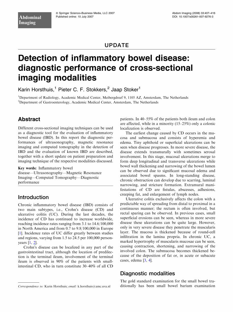
Imaging criteria. The criterion that is most extensivelyused for the diagnosis of CD is bowel wall thickening. Inmost studies, the bowel wall is considered to be thickenedwhen thickness exceeds 3 mm (Fig. 2). In a meta-analysisconducted by Fraquelli et al. [17] concerning US in CDthe respective diagnostic accuracies of different cut-offvalues were compared; sensitivity for diagnosis of CDdecreased using a cut-off value of 4 mm instead of 3 mm(from 88% to 75%), but specificity increased slightly(from 93% to 97%). Other features that are consideredcharacteristic of CD on US are the presence of a stiffbowel wall, modification or disappearance of bowel wallstratification, presence of deep ulcers (seen as interrup-tion of the submucosal hyperechoic rim by a hypoechoictract), a reduction of peristalsis and loss of haustration inthe colon. Extramural findings are fibrofatty prolifera-tion, enlarged lymph nodes, and/or the presence of anabscess or fistula.
Bowel wall thickening is considered to be a charac-teristic feature of UC as well. Mural stratification ispreserved in most UC patients due to the superficialpattern of inflammation. This feature can be used todifferentiate between CD and UC, although this was not
regarded sufficient for differentiation in all studies onthis topic [12, 13]. In long-standing UC a tubularappearance of the colon and loss of haustration can beseen.
Diagnostic accuracy of US. Most studies regardingdiagnostic accuracy of US for diagnosis and follow-up ofIBD have been conducted in the last decade. Althoughreported sensitivity and specificity values are high, withthe state-of-the-art equipment diagnostic accuracy couldpossibly be higher than that previously reported.
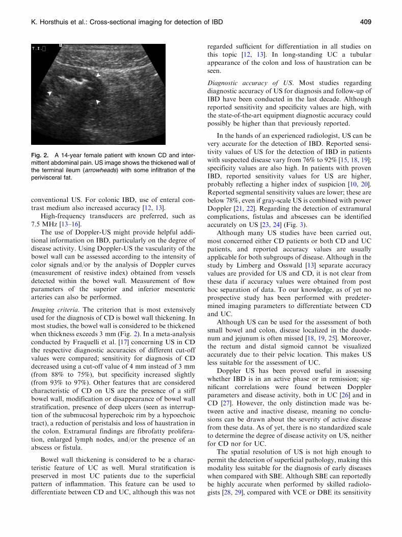
In the hands of an experienced radiologist, US can bevery accurate for the detection of IBD. Reported sensi-tivity values of US for the detection of IBD in patientswith suspected disease vary from 76% to 92% [15, 18, 19];specificity values are also high. In patients with provenIBD, reported sensitivity values for US are higher,probably reflecting a higher index of suspicion [10, 20].Reported segmental sensitivity values are lower; these arebelow 78%, even if gray-scale US is combined with powerDoppler [21, 22]. Regarding the detection of extramuralcomplications, fistulas and abscesses can be identifiedaccurately on US [23, 24] (Fig. 3).
Although many US studies have been carried out,most concerned either CD patients or both CD and UCpatients, and reported accuracy values are usuallyapplicable for both subgroups of disease. Although in thestudy by Limberg and Osswald [13] separate accuracyvalues are provided for US and CD, it is not clear fromthese data if accuracy values were obtained from posthoc separation of data. To our knowledge, as of yet noprospective study has been performed with predeter-mined imaging parameters to differentiate between CDand UC.
Although US can be used for the assessment of bothsmall bowel and colon, disease localized in the duode-num and jejunum is often missed [18, 19, 25]. Moreover,the rectum and distal sigmoid cannot be visualizedaccurately due to their pelvic location. This makes USless suitable for the assessment of UC.
Doppler US has been proved useful in assessingwhether IBD is in an active phase or in remission; sig-nificant correlations were found between Dopplerparameters and disease activity, both in UC [26] and inCD [27]. However, the only distinction made was be-tween active and inactive disease, meaning no conclu-sions can be drawn about the severity of active diseasefrom these data. As of yet, there is no standardized scaleto determine the degree of disease activity on US, neitherfor CD nor for UC.
The spatial resolution of US is not high enough topermit the detection of superficial pathology, making thismodality less suitable for the diagnosis of early diseaseswhen compared with SBE. Although SBE can reportedlybe highly accurate when performed by skilled radiolo-gists [28, 29], compared with VCE or DBE its sensitivity
Fig. 2. A 14-year female patient with known CD and inter-mittent abdominal pain. US image shows the thickened wall ofthe terminal ileum (arrowheads) with some infiltration of theperivisceral fat.
K. Horsthuis et al.: Cross-sectional imaging for detection of IBD 409
is low [30]. In this regard, comparison between US andVCE and/or DBE might be very interesting in order todetermine the accuracy of US for small lesions andaccuracy for bowel segments that are difficult to access.To our knowledge, no comparative studies have beenperformed as of yet.
Computed tomography
Patient preparation. Patients are usually asked to fast forseveral hours before the scan to diminish peristalticmovements [31–33]. In addition, in some institutions abowel-cleansing regimen is applied, as a rule consistingof mild laxatives. Dietary restrictions are also oftenapplicable. Although with this bowel preparation resid-ual feces are usually present to some degree, the muralpresentation of disease enables the identification of dis-ease even if the bowel wall is partly obscured.
There is consensus as to the indispensability ofenteral contrast medium for an abdominal CT exami-nation for IBD. The contrast medium of choice shouldbe neutral (meaning an attenuation value comparablewith water), as a neutral contrast medium allows opti-mal distinction between bowel wall and lumen. While insome institutions enteral contrast medium is adminis-tered orally (CT enterography), in other institutionscontrolled distention is achieved by inflow of contrastmedium through a nasojejunal catheter (CT-enterocly-sis). Although by some authors CT-enteroclysis ispropagated as the controlled infusion provides a moreconsistent distention of the small bowel than CT-ente-rography, especially of the jejunum [34], others reportthat with the right choice of contrast medium andcorrect timing of intake excellent distention of all smallbowel loops can be obtained after the oral administra-tion of contrast medium [31, 33]. In only one smallstudy, CT enteroclysis and CT enterography werecompared, but both the degree of luminal distentionand the diagnostic accuracy did not show significantdifferences [35].
Imaging technique. Technical developments have allowedthe widespread use of multi-slice scanners. With thesescanners volumes can be scanned in a very short breathhold, allowing the acquisition of isotropic voxels formultiplanar reformatting. Thin slices should be used topermit the detection of subtle pathology.
Before CT enterography sometimes metoclopramideis given to increase gastric emptying and peristalticmovements of the small bowel [31]. The use of antiperi-staltic drugs is not standard. In a recently publishedupdate on CT enteroclysis, Rajesh and Maglinte [32]report that in their institution all patients undergoing CTenteroclysis receive conscious sedation. Although thismost probably decreases patient discomfort to a largedegree, this can considerably increase the in-hospital timefor patients as they will have to stay in a recovery unitafter the procedure until the anesthetic effects have wornoff.
Intravenous contrast medium should be administeredfor a comprehensive CT examination of the bowel. In arecent study, the optimal timing of scanning after theadministration of intravenous contrast medium wasdetermined; Schindera et al. [36] found that peak muralenhancement of normal small-bowel wall occurs onaverage about 50 s after contrast administration or 14 safter peak aortic enhancement.
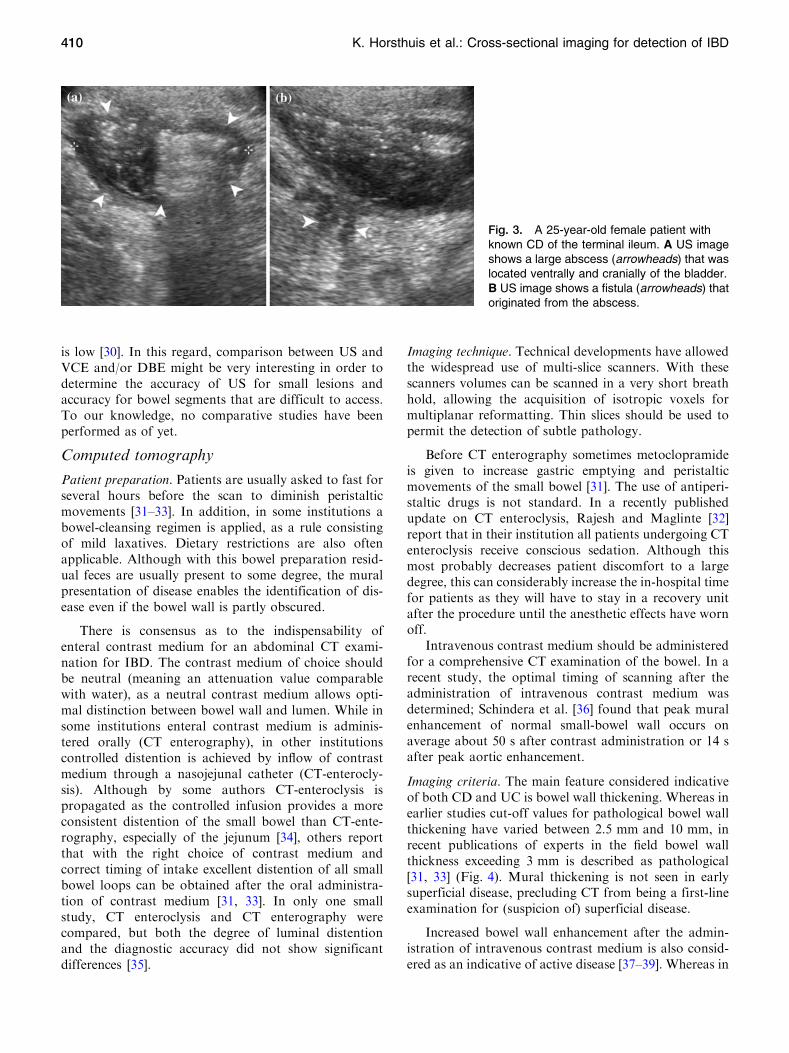
Imaging criteria. The main feature considered indicativeof both CD and UC is bowel wall thickening. Whereas inearlier studies cut-off values for pathological bowel wallthickening have varied between 2.5 mm and 10 mm, inrecent publications of experts in the field bowel wallthickness exceeding 3 mm is described as pathological[31, 33] (Fig. 4). Mural thickening is not seen in earlysuperficial disease, precluding CT from being a first-lineexamination for (suspicion of) superficial disease.
Increased bowel wall enhancement after the admin-istration of intravenous contrast medium is also consid-ered as an indicative of active disease [37–39]. Whereas in
Fig. 3. A 25-year-old female patient withknown CD of the terminal ileum. A US imageshows a large abscess (arrowheads) that waslocated ventrally and cranially of the bladder.B US image shows a fistula (arrowheads) thatoriginated from the abscess.
410 K. Horsthuis et al.: Cross-sectional imaging for detection of IBD
earlier studies the only distinction regarding enhance-ment was between pathological enhancement (i.e., hyper-enhancement) and normally enhancing bowel walls, in arecent publication the degree of mural enhancement wasfound to correlate with disease activity [38].
Bowel wall enhancement can be transmural, but alsolayered. This layered enhancement pattern, which isrepresented by a thickened intestinal wall with a middlelayer of low attenuation surrounded on each side bylayers of higher attenuation, has been termed the targetsign; this is due to the presence of edema or the deposi-tion of fat in the submucosa.
Diagnostic accuracy of CT. The accuracy of CT hasmainly been investigated for small-bowel disease. Insuspected CD sensitivity was 83% when compared withSBE [40]. When compared with ileoscopy sensitivityvalues vary from 80% to 88% [34, 35, 41]. Segmentalsensitivity of CT was somewhat lower (71.8%) in a studyby Molnar et al. [42], comparing CT with SBE and CS.
Superficial lesions (such as aphthoid lesions) are notaccurately visualized on CT, making CT less suitable as afirst-line examination for the suspicion of mild disease.This was already evident from studies comparing CTwith CS and/or SBE, but in a recent meta-analysiscomparing CT with VCE was shown that the yield of CTcompared with the yield of CE was 30% vs. 69% [43]. Nocomparative studies have been published regarding CTvs. DBE.
Extramural complications are well shown on CT, al-though the lower contrast resolution of CT makes thismodality less suitable for the detection of fistulas andabscesses than MRI in patients with CD [44] (Fig. 5). Ina recent study, the relationship between increasedattenuation of perivisceral fat and disease activity was
determined; one of the most specific markers of activedisease was increased attenuation in the perivisceral fat[37].
Hardly any studies have focused on the accuracy ofCT colonography for the detection of ileocolonic IBD.This is possibly partly due to the fact that for the rectaladministration of contrast medium the rectum is ob-scured by the rectal catheter, precluding diagnosis ofrectal IBD, specifically UC. It does seem clear that CTCis unable to detect ulcerative lesions; even diffuseinflammation with large ulcerations can be missed. CTCmight however be useful in patients with colonic stenosisor narrowing [45, 46].
Magnetic resonance imaging
Patient preparation. While in some studies on MRI aperiod of several hours of fasting was deemed sufficient,in others full bowel cleansing was performed, as thereference standard, (i.e., CS) was performed on the sameday. There is no consensus yet as to what constitutes theoptimal bowel preparation for MRI. However, as alimited bowel preparation does not seem to negativelyaffect accuracy, it might be sufficient to limit the bowelpreparation to a fasting period taking into account thepatient-friendliness of the respective preparations.
Luminal distention by means of use of enteral con-trast medium is indispensable for an adequate evaluationof the bowel as collapsed bowel can hide or mimic dis-ease. As was the case with CT, for MRI of the smallbowel contrast medium is either administrated by mouthor by enteroclysis. An advantage of MR enteroclysis overMR enterography is the fact that it allows fluoroscopicmonitoring of the inflow of contrast medium and therebyprovides functional information on bowel distensibility.An advantage of MR enterography is the fact that it canbe considered more patient-friendly and also that noionizing radiation is necessary. To our knowledge, onlyone study has been carried out in which both methods ofcontrast medium administration were compared. In thisstudy by Schreyer et al. [47] all patients (n = 21)underwent both MRI enterography and MRI enteroc-lysis; no difference in accuracy compared with SBE wasnoted by the investigators (Fig. 6).
Whereas for MR enteroclysis mostly a methylcellu-lose suspension is used, for MR enterography manydifferent contrast media have been tested. The mainsubdivision is between positive, negative, and biphasiccontrast media. A biphasic contrast medium performsbest for the identification of pathology on both T2-andT1-weighted sequences as adequate delineation betweenhypointense bowel wall and hyperintense lumen is seenon T2-weighted sequences while on T1-weighted imagesthe enhancing bowel wall can be easily discriminatedfrom the hypointense lumen. An artificial sugar-solution
Fig. 4. A 60-year old female patient who underwent CTenterography for suspected bowel obstruction. Just 1 monthearlier at ileocolonoscopy CD of the terminal ileum was dis-covered; the terminal ileum was not intubated because of thestenosis. Axial image shows the severely thickened bowelwall of the ileum (arrowheads) with only a pinpoint bowellumen remaining.
K. Horsthuis et al.: Cross-sectional imaging for detection of IBD 411
(mannitol or sorbitol) has been shown to cause gooddistention of small bowel loops with negligible side ef-fects [48, 49].
Imaging technique. Mostly, both T2-weighted and T1-weighted sequences are used for the MRI evaluation ofthe bowel. On T2-weighted images the bowel wall can beappreciated and bowel wall stratification—if pres-ent—can be well apprehended. As feces can show brightsignal intensity on T1-weighted sequences, it is importantto perform a pre-contrast T1-weighted sequence in orderto be able to determine whether high signal intensity wasalready present before intravenous contrast administra-tion, indicating the presence of stool.
Another sequence that is propagated by many au-thors is the TrueFISp sequence, a sequence that isinsensitive to motion and breathing artifacts. This se-quence, that makes use of a T2/T1 ratio, adequatelydelineates the bowel wall and the mesentery, allowingadequate assessment of disease (Fig. 7). When combin-ing a T2-weighted sequence or TrueFISP sequence and a
T1-weighted sequence, a comprehensive MRI examina-tion can be carried out in less than 30 min.
Imaging criteria. A bowel wall thickness exceeding 3 mmshould be considered as an indicative of disease. Besidesbowel wall thickening the most important criterionindicative of active IBD is pathological bowel wallenhancement after the administration of intravenousgadolinium. Bowel wall enhancement can always be seenas the bowel is a highly vascularized structure. However,in active IBD increased enhancement can be observed,due to the increased vascularization and the increasedcapillary leakage of the affected tissue (Fig. 8). In CD ithas been hypothesized that the degree of enhancementcorrelates with the degree of disease severity, but thisstatement has not been extensively corroborated [50–53].
Bowel wall stratification can be observed on T2-weighted images as a bright line within the two darkstripes of the mucosal and muscularis propria layers,likely related to the presence of fat or edema in thesubmucosal layer. On fat-suppressed T2-weighted images
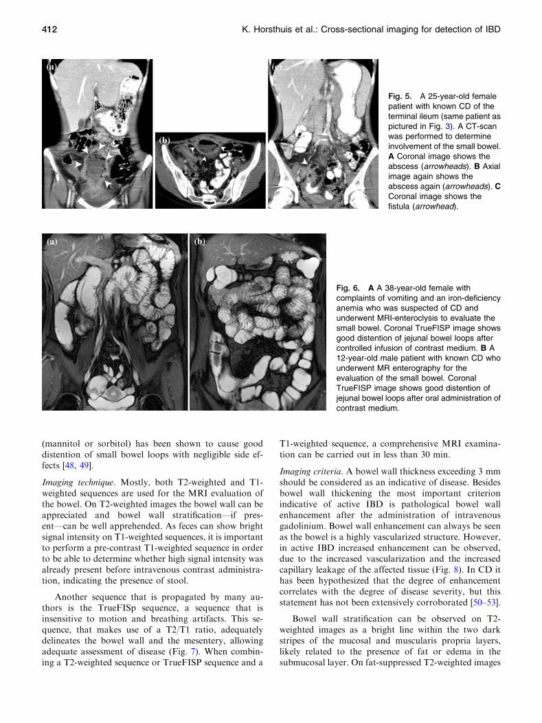
Fig. 5. A 25-year-old femalepatient with known CD of theterminal ileum (same patient aspictured in Fig. 3). A CT-scanwas performed to determineinvolvement of the small bowel.A Coronal image shows theabscess (arrowheads). B Axialimage again shows theabscess again (arrowheads). CCoronal image shows thefistula (arrowhead).
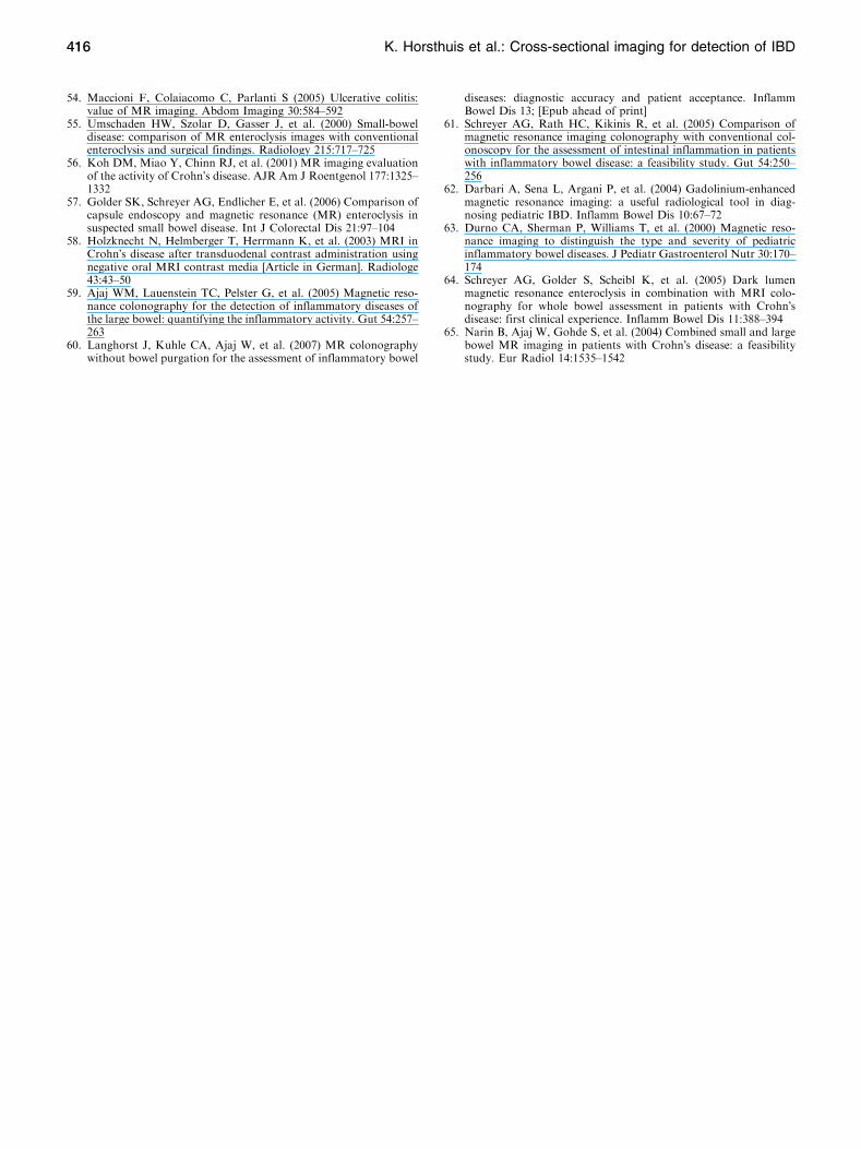
Fig. 6. A A 38-year-old female withcomplaints of vomiting and an iron-deficiencyanemia who was suspected of CD andunderwent MRI-enteroclysis to evaluate thesmall bowel. Coronal TrueFISP image showsgood distention of jejunal bowel loops aftercontrolled infusion of contrast medium. B A12-year-old male patient with known CD whounderwent MR enterography for theevaluation of the small bowel. CoronalTrueFISP image shows good distention ofjejunal bowel loops after oral administration ofcontrast medium.
412 K. Horsthuis et al.: Cross-sectional imaging for detection of IBD

it is possible to determine the nature of the bright signalas a persistent bright signal suggests the presence ofedema, whereas complete suppression of the submucosalsignal suggests fat infiltration and quiescent disease [54].
Extramural manifestations of disease that can beidentified on MRI are fistulas, abscesses, fibrofatty pro-liferation, and enlarged lymph nodes.
Diagnostic accuracy. The accuracy of MRI of the smallbowel has been extensively investigated. In many Euro-pean institutions, conventional enteroclysis is increas-ingly being replaced by MRI enteroclysis or MRIenterography as MRI has proved to be highly accurate in
both the detection of disease in patients with known IBDas in patients in whom IBD of the small bowel wassuspected [52, 55, 56]. However, the studies that havebeen performed were mostly small and concerned se-lected populations with either a high suspicion of diseaseor known CD of the small bowel. Larger studiesincluding the full spectrum of disease activity should beconducted.
As was the case with CT and US, MRI is not suitablefor the detection of superficial disease due to the limitedspatial resolution. This finding is corroborated by a studycomparing MRI and VCE in patients with CD [57].Significantly more inflammatory lesions were detectedwith VCE in the jejunum and partly in the ileum of pa-tients with CD. However, these findings had no effect onthe therapeutic approach of the individual patients. Theaccuracy of MRI has not been compared with DBE as ofyet.
As mentioned before, MRI can be used for the eval-uation of extramural disease. Due to the high contrastresolution abscesses are very conspicuous on T1-weighedfat suppressed images after the administration of intra-venous Gadolinium. MRI is also very sensitive for thedetection of fistulas [47, 52, 58] (Fig. 9).
In recent years, the accuracy of MRI for the detectionof ileocolonic IBD has been investigated by means ofMRI colonography. After administration of rectal con-trast medium the colon (and sometimes the terminalileum) was assessed for disease. Conflicting results werereported: while in one study high accuracy values werereported [59], in others segmental sensitivity values werearound 32% [60, 61].
Regarding the accuracy of MRI in differentiationbetween CD and UC conflicting results have been re-
Fig. 7. A 18-year-old female patient withknown CD. MRI-enterography was performedfor suspicion of active CD of the neoterminalileum. A Coronal TrueFISp image showsenlarged mesenteric lymph nodes(arrowheads). B Coronal TrueFISP imageshows thickened bowel wall of theneoterminal ileum (arrowheads).
Fig. 8. A 12-year-old male patient with known CD whounderwent MR-enterography for the evaluation of the smallbowel (same patient depicted in Fig. 6b). Axial T1-weightedimage shows pathological enhancement of thickened small-bowel loops after administration of intravenous contrastmedium (arrowheads). Approximately, 1 m of small bowel(terminal jejunum, proximal ileum) was shown to be affected.
K. Horsthuis et al.: Cross-sectional imaging for detection of IBD 413

ported; while some authors report that based on thelocation of inflammatory changes, the degree ofinvolvement, the continuity or discontinuity of disease,and the presence of complications it was possible todifferentiate between CD and UC [51, 62], others reporta limited value in differentiation of disease [63].
Theoretically, a whole-bowel examination would bepossible with MRI, by the administration of contrastmedium orally and rectally. This has been attempted [64,65] and was deemed feasible. More research is needed toestablish the diagnostic value of this combined approach.At the moment, MRI colonography does not seem to beable to replace CS.
Discussion
Compared with conventional imaging methods, CT, US,and MRI are accurate methods for the detection of IBDof the small bowel, both in patients suspected of diseaseas in patients with known IBD. Although subtle lesionscannot be depicted with any of these modalities, clini-cally more relevant findings can be accurately depicted.Therefore, cross-sectional imaging should be incorpo-rated in a comprehensive clinical evaluation of suspectedIBD and for follow-up of CD. The exact role cross-sec-tional imaging techniques can play for follow-up in UCshould be more extensively studied.
As US is easily accessible, widely available, andinexpensive, it is recommended to use abdominal US asfirst-line modality in patients with suspected IBD of thesmall bowel. MR enterography would be a good alter-native, especially as the assessment of the degree of dis-ease activity can be better performed on MRI than onUS. Although CT enterography is a very accurate tech-nique and is used in many institutions, its role in IBD islimited by the ionizing radiation needed, especially due tothe repetitive use for follow-up in often young individ-uals. If possible, it might be advisable to reserve this
technique for patients in whom imaging is needed at veryshort notice as CT enterography can be performed veryfast and is readily available.
Although VCE has shown to be more accurate indepicting subtle lesions in the small bowel than MRI orCT, its role should be limited as of yet as the true benefitof VCE is not clear yet. As there are presently no stan-dardized criteria for the diagnosis of CD with VCE,definitions with regard to what constitutes a positivefinding might differ between studies. Moreover, theclinical significance of finding a single mucosal break or afew superficial aphthous lesions is not clear yet. Also,specificity and positive predictive values for VCE havenot been established. At this time, it might be good toreserve VCE as a second-line modality if cross-sectionalimaging has not shown abnormalities but the suspicionof disease remains standing despite these negative find-ings.
References
1. Loftus EV Jr (2004) Clinical epidemiology of inflammatory boweldisease: incidence, prevalence, and environmental influences. Gas-troenterology 126:1504–1517
2. Farrokhyar F, Swarbrick ET, Irvine EJ (2001) A critical review ofepidemiological studies in inflammatory bowel disease. Scand JGastroenterol 36:2–15
3. Stenson WF (2004) Inflammatory bowel disease, Chap. 142 (on-lineedition). In: Goldman L, Aussiello D (eds) Cecil textbook of med-icine. 22th ed. W. B. Saunder: Philadelphia, PA
4. Friedman S, Blumberg RS (2005) Inflammatory bowel disease,Chap. 172 (on-line edition). In: Kasper DL, Fauci AS, Longo DL,Braunwald E, Hauser SL, Jameson JL (eds) Harrison�s principles ofinternal medicine. 16th ed. McGraw-Hill: New York, NY
5. Stange EF, Travis SP, Vermeire S, et al. (2006) European evidencebased consensus on the diagnosis and management of Crohn�sdisease: definitions and diagnosis. Gut 55(Suppl 1):i1–i15
6. Maglinte DD, Sandrasegaran K, Tann M (2006) Advances in ali-mentary tract imaging. World J Gastroenterol 12:3139–3145
7. Hommes DW, van Deventer SJ (2004) Endoscopy in inflammatorybowel diseases. Gastroenterology 126:1561–1573
8. Kundrotas LW, Clement DJ, Kubik CM, et al. (1994) A prospec-tive evaluation of successful terminal ileum intubation duringroutine colonoscopy. Gastrointest Endosc 40:544–546
Fig. 9. A 25-year-old female patient withknown CD of the terminal ileum (same patientas pictured in Figs. 3 and 5). A Coronal T1-weighted image clearly shows the abscess(arrowheads) that was also depicted on USand CT. B Coronal T1-weighted imageshowing a fistula
414 K. Horsthuis et al.: Cross-sectional imaging for detection of IBD

9. Marshall JB, Barthel JS (1993) The frequency of total colonoscopyand terminal ileal intubation in the 1990s. Gastrointest Endosc39:518–520
10. Pallotta N, Tomei E, Viscido A, et al. (2005) Small intestine con-trast ultrasonography: an alternative to radiology in the assessmentof small bowel disease. Inflamm Bowel Dis 11:146–153
11. Parente F, Greco S, Molteni M, et al. (2004) Oral contrast en-hanced bowel ultrasonography in the assessment of small intes-tine Crohn�s disease: a prospective comparison with conventionalultrasound, X ray studies, and ileocolonoscopy. Gut 53:1652–1657
12. Bru C, Sans M, Defelitto MM, et al. (2001) Hydrocolonic sonog-raphy for evaluating inflammatory bowel disease. AJR 177:99–105
13. Limberg B, Osswald B (1994) Diagnosis and differential diagnosisof ulcerative colitis and Crohn�s disease by hydrocolonic sonogra-phy. Am J Gastroenterol 89:1051–1057
14. Miao YM, Koh DM, Amin Z, et al. (2002) Ultrasound and mag-netic resonance imaging assessment of active bowel segments inCrohn�s disease. Clin Radiol 57:913–918
15. Rispo A, Imbriaco M, Celentano L, et al. (2005) Noninvasivediagnosis of small bowel Crohn�s disease: combined use of bowelsonography and Tc-99m-HMPAO leukocyte scintigraphy. InflammBowel Dis 11:376–382
16. Andreoli A, Cerro P, Falasco G, et al. (1998) Role of ultrasonog-raphy in the diagnosis of postsurgical recurrence of Crohn�s disease.Am J Gastroenterol 93:1117–1121
17. Fraquelli M, Colli A, Casazza G, et al. (2005) Role of US indetection of Crohn disease: meta-analysis. Radiology 236:95–101
18. Hollerbach S, Geissler A, Schiegl H, et al. (1998) The accuracy ofabdominal ultrasound in the assessment of bowel disorders. ScandJ Gastroenterol 33:1201–1208
19. Parente F, Greco S, Motelni M, et al. (2003) Role of early ultra-sound in detecting inflammatory disorders and identifying theiranatomical location within the bowel. Aliment Pharmacol Ther18:1009–1016
20. Calabrese E, La Seta F, Buccellato A, et al. (2005) Crohn�s disease:a comparative prospective study of transabdominal ultrasonogra-phy, small intestine contrast ultrasonography, and small bowelenema. Inflamm Bowel Dis 11:139–145
21. Neye H, Voderholzer W, Rickes S, et al. (2004) Evaluation of cri-teria for the activity of Crohn�s disease by power Doppler sonog-raphy. Dig Dis 22:67–72
22. Pradel JA, David XR, Taourel P, et al. (1997) Sonographicassessment of the normal and abnormal bowel wall in nondiver-ticular ileitis and colitis. Abdom Imaging 22:167–172
23. Gasche C, Moser G, Turetschek K, et al. (1999) Transabdominalbowel sonography for the detection of intestinal complications inCrohn�s disease. Gut 44:112–117
24. Maconi G, Sampietro GM, Parente F, et al. (2003) Contrastradiology, computed tomography and ultrasonography in detectinginternal fistulas and intra-abdominal abscesses in Crohn�s disease: aprospective comparative study. Am J Gastroenterol 98(7):1545–1555
25. Parente F, Maconi G, Bollani S, et al. (2002) Bowel ultrasound inassessment of Crohn�s disease and detection of related small bowelstrictures: a prospective comparative study versus x ray and intra-operative findings. Gut 50:490–495
26. Sigirci A, Baysal T, Kutlu R, et al. (2001) Doppler sonography ofthe inferior and superior mesenteric arteries in ulcerative colitis. JClin Ultrasound 29:130–139
27. Spalinger J, Patriquin H, Miron MC, et al. (2000) Doppler US inpatients with Crohn disease: vessel density in the diseased bowelreflects disease activity. Radiology 217:787–791
28. Maglinte DD, Chernish SM, Kelvin FM, et al. (1992) Crohn dis-ease of the small intestine: accuracy and relevance of enteroclysis.Radiology 184:541–545
29. Cirillo LC, Camera L, Della Noce M, et al. (2000) Accuracy ofenteroclysis in Crohn�s disease of the small bowel: a retrospectivestudy. Eur Radiol 10:1894–1898
30. Marmo R, Rotondano G, Piscopo R, et al. (2005) Meta-analysis:capsule enteroscopy vs. conventional modalities in diagnosis ofsmall bowel diseases. Aliment Pharmacol Ther 22:595–604
31. Paulsen SR, Huprich JE, Fletcher JG, et al. (2006) CT enterogra-phy as a diagnostic tool in evaluating small bowel disorders: review
of clinical experience with over 700 cases. Radiographics 26:641–662
32. Rajesh A, Maglinte DDT (2006) Multisclice CT enteroclysis:technique and clinical applications. Clin Radiol 61:31–39
33. Macari M, Megibow AJ, Balthazar EJ (2007) A pattern approachto the abnormal small bowel: observations at MDCT and CTenterography. AJR 188:1344–1355
34. Mazzeo S, Caramella D, Battolla L, et al. (2001) Crohn disease ofthe small bowel: spiral CT evaluation after oral hyperhydrationwith isotonic solution. J Comput Assist Tomogr 25:612–616
35. Wold PB, Fletcher JG, Johnson CD, et al. (2003) Assessment ofsmall bowel Crohn disease: noninvasive peroral CT enterographycompared with other imaging methods and endoscopy–feasibilitystudy. Radiology 229:275–281
36. Schindera ST, Nelson RC, DeLong DM, et al. (2007) Multi-detector row CT pf he small bowel: peak enhancement temporalwindow- initial experience. Radiology 243:438–444
37. Booya F, Fletcher JG, Huprich JE, et al. (2006) Active Crohndisease: CT findings and interobserver agreement for enteric phaseCT enterography. Radiology 241:787–795
38. Bodily KD, Fletcher JG, Solem CA, et al. (2006) Crohn disease:mural attenuation and thickness at contrast-enhanced CT ente-rography—correlation with endoscopic and histologic findings ofinflammation. Radiology 238:505–516
39. Thoeni RF, Cello JP (2006) CT imaging of colitis. Radiology240:623–638
40. Minordi LM, Vecchioli A, Guidi L, et al. (2006) Multidetector CTenteroclysis versus barium enteroclysis with methylcellulose in pa-tients with suspected small bowel disease. Eur Radiol 16:1527–1536
41. Hassan C, Cerro P, Zullo A, Spina C, et al. (2003) Computedtomography enteroclysis in comparison with ileoscopy in patientswith Crohn�s disease. Int J Colorect Dis 18:121–125
42. Molnar T, Papos M, Gyulai C, et al. (2001) Clinical value oftechnetium-99m-HMPAO-labeled leukocyte scintigraphy andspiral computed tomography in active Crohn�s disease. Am JGastroenterol 96:1517–1521
43. Triester SL, Leighton JA, Leontiadis GI (2006) A meta-analysis ofthe yield of capsule endoscopy compared to other diagnosticmodalities in patients with non-stricturing small bowel Crohn�sdisease. Am J Gastroenterol 101:954–964
44. Koelbel G, Schmiedl U, Majer MC, et al. (1989) Diagnosis offistulae and sinus tracts in patients with Crohn disease: value ofMR imaging. AJR Am J Roentgenol 152:999–1003
45. Ota Y, Matsui T, Ono H, et al. (2003) Value of virtual computedtomographic colonography for Crohn�s colitis: comparison withendoscopy and barium enema. Abdom Imaging 28:778–783
46. Biancone L, Fiori R, Tosti C, et al. (2003) Virtual colonoscopycompared with conventional colonoscopy for stricturing postop-erative recurrence in Crohn�s disease. Inflamm Bowel Dis 9:343–350
47. Schreyer AG, Geissler A, Albrich H, et al. (2004) Abdominal MRIafter enteroclysis or with oral contrast in patients with suspected orproven Crohn�s disease. Clin Gastroenterol Hepatol 2:491–497
48. Ajaj W, Goehde SC, Schneemann H, et al. (2004) Oral contrastagents for small bowel MRI: comparison of different additives tooptimize bowel distension. Eur Radiol 14:458–464
49. Lauenstein TC, Schneemann H, Vogt FM, et al. (2003) Optimiza-tion of oral contrast agents for MR imaging of the small bowel.Radiology 228:279–283
50. Shoenut JP, Semelka RC, Silverman R, et al. (1993) Magneticresonance imaging in inflammatory bowel disease. J Clin Gastro-enterol 17:73–78
51. Shoenut JP, Semelka RC, Magro CM, et al. (1994) Comparison ofmagnetic resonance imaging and endoscopy in distinguishing thetype and severity of inflammatory bowel disease. J Clin Gastro-enterol 19:31–35
52. Maccioni F, Bruni A, Viscido A, et al. (2005) MR Imaging inpatients with Crohn disease: value of T2- versus T1-weightedgadolinium-enhanced MR sequences with use of an oral super-paramagnetic contrast agent. Radiology 238:517–530
53. Florie J, Horsthuis K, Hommes DW, et al. (2005) Magnetic reso-nance imaging compared with ileocolonoscopy in evaluating dis-ease severity in Crohn�s disease. Clin Gastroenterol Hepatol3:1221–1228
K. Horsthuis et al.: Cross-sectional imaging for detection of IBD 415
54. Maccioni F, Colaiacomo C, Parlanti S (2005) Ulcerative colitis:value of MR imaging. Abdom Imaging 30:584–592
55. Umschaden HW, Szolar D, Gasser J, et al. (2000) Small-boweldisease: comparison of MR enteroclysis images with conventionalenteroclysis and surgical findings. Radiology 215:717–725
56. Koh DM, Miao Y, Chinn RJ, et al. (2001) MR imaging evaluationof the activity of Crohn�s disease. AJR Am J Roentgenol 177:1325–1332
57. Golder SK, Schreyer AG, Endlicher E, et al. (2006) Comparison ofcapsule endoscopy and magnetic resonance (MR) enteroclysis insuspected small bowel disease. Int J Colorectal Dis 21:97–104
58. Holzknecht N, Helmberger T, Herrmann K, et al. (2003) MRI inCrohn�s disease after transduodenal contrast administration usingnegative oral MRI contrast media [Article in German]. Radiologe43:43–50
59. Ajaj WM, Lauenstein TC, Pelster G, et al. (2005) Magnetic reso-nance colonography for the detection of inflammatory diseases ofthe large bowel: quantifying the inflammatory activity. Gut 54:257–263
60. Langhorst J, Kuhle CA, Ajaj W, et al. (2007) MR colonographywithout bowel purgation for the assessment of inflammatory bowel
diseases: diagnostic accuracy and patient acceptance. InflammBowel Dis 13; [Epub ahead of print]
61. Schreyer AG, Rath HC, Kikinis R, et al. (2005) Comparison ofmagnetic resonance imaging colonography with conventional col-onoscopy for the assessment of intestinal inflammation in patientswith inflammatory bowel disease: a feasibility study. Gut 54:250–256
62. Darbari A, Sena L, Argani P, et al. (2004) Gadolinium-enhancedmagnetic resonance imaging: a useful radiological tool in diag-nosing pediatric IBD. Inflamm Bowel Dis 10:67–72
63. Durno CA, Sherman P, Williams T, et al. (2000) Magnetic reso-nance imaging to distinguish the type and severity of pediatricinflammatory bowel diseases. J Pediatr Gastroenterol Nutr 30:170–174
64. Schreyer AG, Golder S, Scheibl K, et al. (2005) Dark lumenmagnetic resonance enteroclysis in combination with MRI colo-nography for whole bowel assessment in patients with Crohn�sdisease: first clinical experience. Inflamm Bowel Dis 11:388–394
65. Narin B, Ajaj W, Gohde S, et al. (2004) Combined small and largebowel MR imaging in patients with Crohn�s disease: a feasibilitystudy. Eur Radiol 14:1535–1542
416 K. Horsthuis et al.: Cross-sectional imaging for detection of IBD


![[Greeting modalities preferred by patients in pediatric ambulatory setting]](https://static.fdokumen.com/doc/165x107/6337af65d102fae1b6077daa/greeting-modalities-preferred-by-patients-in-pediatric-ambulatory-setting.jpg)