Crystal structure of human purine nucleoside phosphorylase at 2.3 Å resolution
Upload
independentCategory
view
0download
0
Bioorganic & Medicinal Chemistry 18 (2010) 7239–7251
Contents lists available at ScienceDirect
Bioorganic & Medicinal Chemistry
journal homepage: www.elsevier .com/locate /bmc
Design, synthesis, biological evaluation, and modeling of anon-carbohydrate antagonist of the myelin-associated glycoprotein
Oliver Schwardt a, Hendrik Koliwer-Brandl b, Raphael Zimmerli a, Stefanie Mesch a, Gianluca Rossato a,Morena Spreafico a, Angelo Vedani a, Sørge Kelm b, Beat Ernst a,⇑a Institute of Molecular Pharmacy, Pharmacenter, University of Basel, Klingelbergstrasse 50, CH-4056 Basel, Switzerlandb Department of Physiological Biochemistry, University of Bremen, D-28334 Bremen, Germany
a r t i c l e i n f o a b s t r a c t
Article history:Received 26 May 2010Accepted 12 August 2010Available online 17 August 2010
Keywords:Carbohydrate mimeticsSiglecsMyelin-associated glycoprotein (MAG)Binding affinityHapten inhibition assaySurface plasmon resonance (SPR)Molecular modelingKey polar groups
0968-0896/$ - see front matter � 2010 Elsevier Ltd. Adoi:10.1016/j.bmc.2010.08.027
Abbreviations: DBU, 8-diazabicyclo[5.4.0]undec-7DMAP, 4-dimethylamino-pyridine; DMF, N,N-dimethythoxypropane; FAc, 2-fluoroacetyl; IgG, immunoglobstant; MAG, myelin-associated glycoprotein; Neu5ANgR, Nogo receptor; NMR, nuclear magnetic resonancreversed phase; SAR, structure-affinity relationship;nance; TFA, trifluoroacetic acid; THF, tetrahydrofuran⇑ Corresponding author. Tel.: +41 267 15 51; fax: +
E-mail address: [email protected] (B. Ernst).
Broad modifications of various positions of the minimal natural epitope recognized by the myelin-asso-ciated glycoprotein (MAG), a blocker of regeneration of neurite injuries, produced sialosides with nano-molar affinities. However, important pharmacokinetic issues, for example, the metabolic stability of thesesialosides, remain to be addressed. For this reason, the novel non-carbohydrate mimic 3 was designedand synthesized from (�)-quinic acid. For the design of 3, previously identified beneficial modificationsof side chains of Neu5Ac were combined with the replacement of the ring oxygen by a methylene groupand the substitution of the C(4)-OH by an acetamide. Although docking experiments to a homologymodel of MAG revealed that mimic 3 forms all but one of the essential hydrogen bonds identified forthe earlier reported lead 2, its affinity was substantially reduced. Extensive molecular-dynamics simula-tion disclosed that the missing hydrogen bond of the former C(8)-OH leads to a change of the orientationof the side chain. As a consequence, an important hydrophobic contact is compromised leading to a lossof affinity.
� 2010 Elsevier Ltd. All rights reserved.
1. Introduction
Siglecs1,2 (sialic acid-binding immunoglobulin like-lectins) forma sub-group of the I-type lectins and function as cell signaling co-receptors primarily expressed on leukocytes to mediate acquiredand innate immune functions. They can be divided into two sub-sets: the first, evolutionary conserved group consists of Siglec 1,2, and 4, which show selective binding properties: Siglec 1 (alsoknown as sialoadhesin) and Siglec 4 (myelin-associated glycopro-tein, MAG) preferentially bind a(2 ? 3)-linked N-acetylneuraminicacid (Neu5Ac), whereas Siglec 2 (CD22) is highly specific fora(2 ? 6)-linked Neu5Ac. In contrast, members of the second Siglec3-related group (Siglec 3 and Siglecs 5–13) are more promiscuousin their binding, often recognizing more than one presentation ofNeu5Ac.
ll rights reserved.
-en; DCM, dichloromethane;lformamide; DMP, 2,2-dime-ulin G; KD, dissociation con-c, N-acetylneuraminic acid;
e; PDB, protein data bank; RP,SPR, surface plasmon reso-
e; p-Ts, p-tolylsulfonyl.41 267 15 52.
The most comprehensively characterized Siglecs are CD22, aregulatory protein on B lymphocytes that prevents the over-activa-tion of the immune system and the development of autoimmunediseases, and MAG, one of several inhibitor proteins that blockregeneration of injuries of the central nervous system (CNS).2–10
MAG is located on the surface of neurons and interacts with twoclasses of targets: proteins of the family of Nogo receptors(NgR)11,12 and brain gangliosides, specifically GD1a, GT1b, orGQ1ba.2,13–15 Although the relative roles of gangliosides and NgRsas MAG ligands have yet to be resolved,10,16 in some systems, MAGinhibition is completely reversed by sialidase treatment, suggest-ing that MAG uses sialylated glycans as its major axonal ligands.17
Therefore, blocking MAG with potent antagonists may be a valu-able therapeutic approach to enhance axon regeneration.18
SAR studies have revealed that the terminal tetrasaccharide epi-tope of GQ1ba (Fig. 1) shows superior binding to MAG comparedwith the terminal trisaccharide epitope present in GD1a orGT1b.19,20 Furthermore, the MAG-affinity of tetrasaccharide 1(Fig. 1), could clearly be correlated with its ability to reverseMAG-mediated inhibition of axonal outgrowth.18
To reduce the structural complexity of tetrasaccharide 1 numer-ous MAG antagonists have been synthesized.21–23 Based on struc-tural information obtained by trNOE NMR24 and STD NMR,25 theGalb(1 ? 3)GalNAc core was successfully replaced by non-carbo-hydrate linkers22 and the a(2 ? 6)-linked sialic acid by polar as

O
HO
OH
OH
OO
AcHN
O
HO
AcHN
HO
OH
OH
HO
O
O
OH
OH
OO
CO2HHO
AcHN
HO
OOH
O
CO2H
HO
AcHN
HO
OH
OH
O
OH
OH
OO
HN C17H35
O
C13H27
OHH
O
O
CO2HHO
AcHN
HO
OHOH
O
CO2H
GQ1ba
O
1 Me
NHAc
COOMeHN
O
AcHN
CO2Na
O
NH
O
3
Cl
OF
O
HO
OH
OH
OO
AcHN
O
HO
AcHN
HO
OH
OH
HO
O
CO2HHO
AcHN
HO
OHOH
O
CO2H
O
O
OHN
HO
CO2Na
O
NH
O
2
Cl
OF
FF
OHOH
2
345
6789
2
345
678
9
Figure 1. MAG antagonists; GQ1ba with the essential tetrasaccharide binding epitope highlighted, tetrasaccharide derivative 1,18 sialic acid derivative 2,31 and the targetmolecule of this communication, the non-carbohydrate mimic 3 (for better comparability, the carbon atoms in 3 are numbered in analogy to compound 2).
Figure 2. Hapten inhibition assay with sialosides 29 and 2 and non-carbohydratemimic 3. Fetuin-coated microtiter plates26,32,41 were incubated with antibody-complexed MAGd1–3-Fc in the presence of the indicated compounds at differentconcentrations. Binding and inhibition were determined as described for the hapteninhibition assay in Section 4.2.
7240 O. Schwardt et al. / Bioorg. Med. Chem. 18 (2010) 7239–7251
well as lipophilic substituents.23 A further simplification of leadstructure 1 was reported by Kelm and Brossmer who found thatsialic acid derivatives modified in the 2-, 5-, or 9-position exhibitenhanced antagonistic activity.26–28 Finally, when the best modifi-cations found for the 2-,29 4-,30 5-,31 and 9-position32 were com-bined in one molecule, we identified the nanomolar low-molecular weight antagonist 2.31
However, an important pharmacokinetic issue, namely the met-abolic stability of sialosides like 2, remains to be addressed. In gen-eral, the substrate specificity of mammalian sialidases isdetermined by the linkage type of the terminal sialic acid residue(2 ? 3, 2 ? 6, or 2 ? 8) and does not depend on the structure ofthe underlying oligosaccharide.33 Hence, a fast metabolic cleavageof sialosides of type 2 by sialidases cannot be excluded.
To solve this metabolic challenge, we replaced the pyranosecore in 2 by a cyclohexane moiety. Here, we describe design, syn-thesis, and biological evaluation of the corresponding non-carbo-hydrate lead compound 3. It still contains the beneficialmodifications identified for the 2-, 5-, and 9-position (see com-pound 2), however a simplified glycerol side chain.
2. Results and discussion
By manual docking of sialoside 2 (KD 500 nM)31 to a homologymodel of MAG23 the pharmacophores involved in this carbohy-drate-lectin interaction were identified (see Fig. 3B). The mostimportant contributions stem from a salt bridge formed by the car-boxylic acid and Arg11826,34,35 and a hydrogen bond of the C(9)-NHwith the backbone carbonyl of Thr128. Additionally, hydrogen-
bond formations of C(5)-NH with the backbone carbonyl ofGln126 and the backbone NH of Thr128 with the C(8)-OH wereidentified, whereas the C(4)- and C(7)-OH exhibit only minor con-tributions to binding. Overall, this hydrogen bond network nicelycorrelates with the SAR study published by Kelm and Schauer.28
Furthermore, considerable contributions to the binding affinity re-sult from two hydrophobic interactions: (i) the p-chlorobenzamide
O. Schwardt et al. / Bioorg. Med. Chem. 18 (2010) 7239–7251 7241
points into a hydrophobic pocket formed by Ser130 and the sidechain methylenes of Glu131 and (ii) the 2,3-difluorobenzyl agly-cone is hosted by a second hydrophobic pocket established byTrp59, Tyr60, Tyr69, and Tyr116. Most importantly, the pyranosering oxygen was not found to play a significant role in the interac-tion of 2 and MAG.
Based on these findings, the non-carbohydrate mimic 3 (Fig. 1)was designed. Since the pyranose ring oxygen in 2 does not con-tribute to binding, it was exchanged by a methylene group. In addi-tion, the hydroxyl group in the 4-position was replaced by anamide. By this modification, the H-bond with Gln126, which wasshown to exhibit a minor contribution to binding,28 is well pre-served (see Fig. 3B and D). Furthermore, the additional acyl groupallows the exploration of supplementary beneficial interactionswith the target protein. Finally, the p-chlorobenzamido-substi-tuted glycerol side chain was substituted by a simplified version,the 2-(p-chlorobenzamido)-ethoxy moiety. This modificationmaintains the essential H-bond originating from the C(9)-NH in2, but neglects the C(7)- and C(8)-OH, where at least the latterone was found to contribute to the binding to MAG.28 However,the role of this functional group was only investigated with oligo-saccharides and not in combination with other modifications inNeu5Ac derivatives,26–28 and therefore is difficult to assessquantitatively.
g)10 (R = H)
11 (R = Bz)
a)37
ORO
O O
O
f)
9
4b)
5 (R = H)
6 (R = Bn)
BocHNBzO
N3
18
k)
14
17
m)
l)
H2NBzO
N3
CO
51
BzO
N
Boc
CO2Me
OBn
BzO
HN
CO2Me
OBn
ROCO2Me
OBn
O
HOCO2Me
OBn
OTs
HO
HOCO2H
OH
OH
HO
12
345
6
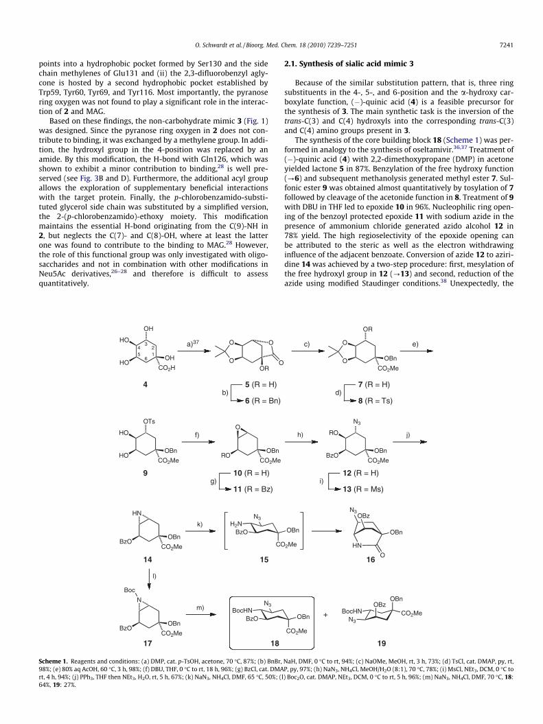
Scheme 1. Reagents and conditions: (a) DMP, cat. p-TsOH, acetone, 70 �C, 87%; (b) BnBr,98%; (e) 80% aq AcOH, 60 �C, 3 h, 98%; (f) DBU, THF, 0 �C to rt, 18 h, 96%; (g) BzCl, cat. DMArt, 4 h, 94%; (j) PPh3, THF then NEt3, H2O, rt, 5 h, 67%; (k) NaN3, NH4Cl, DMF, 65 �C, 50%; (l64%, 19: 27%.
2.1. Synthesis of sialic acid mimic 3
Because of the similar substitution pattern, that is, three ringsubstituents in the 4-, 5-, and 6-position and the a-hydroxy car-boxylate function, (�)-quinic acid (4) is a feasible precursor forthe synthesis of 3. The main synthetic task is the inversion of thetrans-C(3) and C(4) hydroxyls into the corresponding trans-C(3)and C(4) amino groups present in 3.
The synthesis of the core building block 18 (Scheme 1) was per-formed in analogy to the synthesis of oseltamivir.36,37 Treatment of(�)-quinic acid (4) with 2,2-dimethoxypropane (DMP) in acetoneyielded lactone 5 in 87%. Benzylation of the free hydroxy function(?6) and subsequent methanolysis generated methyl ester 7. Sul-fonic ester 9 was obtained almost quantitatively by tosylation of 7followed by cleavage of the acetonide function in 8. Treatment of 9with DBU in THF led to epoxide 10 in 96%. Nucleophilic ring open-ing of the benzoyl protected epoxide 11 with sodium azide in thepresence of ammonium chloride generated azido alcohol 12 in78% yield. The high regioselectivity of the epoxide opening canbe attributed to the steric as well as the electron withdrawinginfluence of the adjacent benzoate. Conversion of azide 12 to aziri-dine 14 was achieved by a two-step procedure: first, mesylation ofthe free hydroxyl group in 12 (?13) and second, reduction of theazide using modified Staudinger conditions.38 Unexpectedly, the
d)7 (R = H)
8 (R = Ts)
c) e)
CO2Me
OBn
h) j)
i)12 (R = H)
13 (R = Ms)
+N3
BocHN
OBn
CO2MeOBz
19
2Me
OBn
HN
N3
OBn
OBz
O61
BzOCO2Me
OBn
N3
RO
CO2Me
OBn
OR
O
O
NaH, DMF, 0 �C to rt, 94%; (c) NaOMe, MeOH, rt, 3 h, 73%; (d) TsCl, cat. DMAP, py, rt,P, py, 97%; (h) NaN3, NH4Cl, MeOH/H2O (8:1), 70 �C, 78%; (i) MsCl, NEt3, DCM, 0 �C to) Boc2O, cat. DMAP, NEt3, DCM, 0 �C to rt, 5 h, 96%; (m) NaN3, NH4Cl, DMF, 70 �C, 18:
7242 O. Schwardt et al. / Bioorg. Med. Chem. 18 (2010) 7239–7251
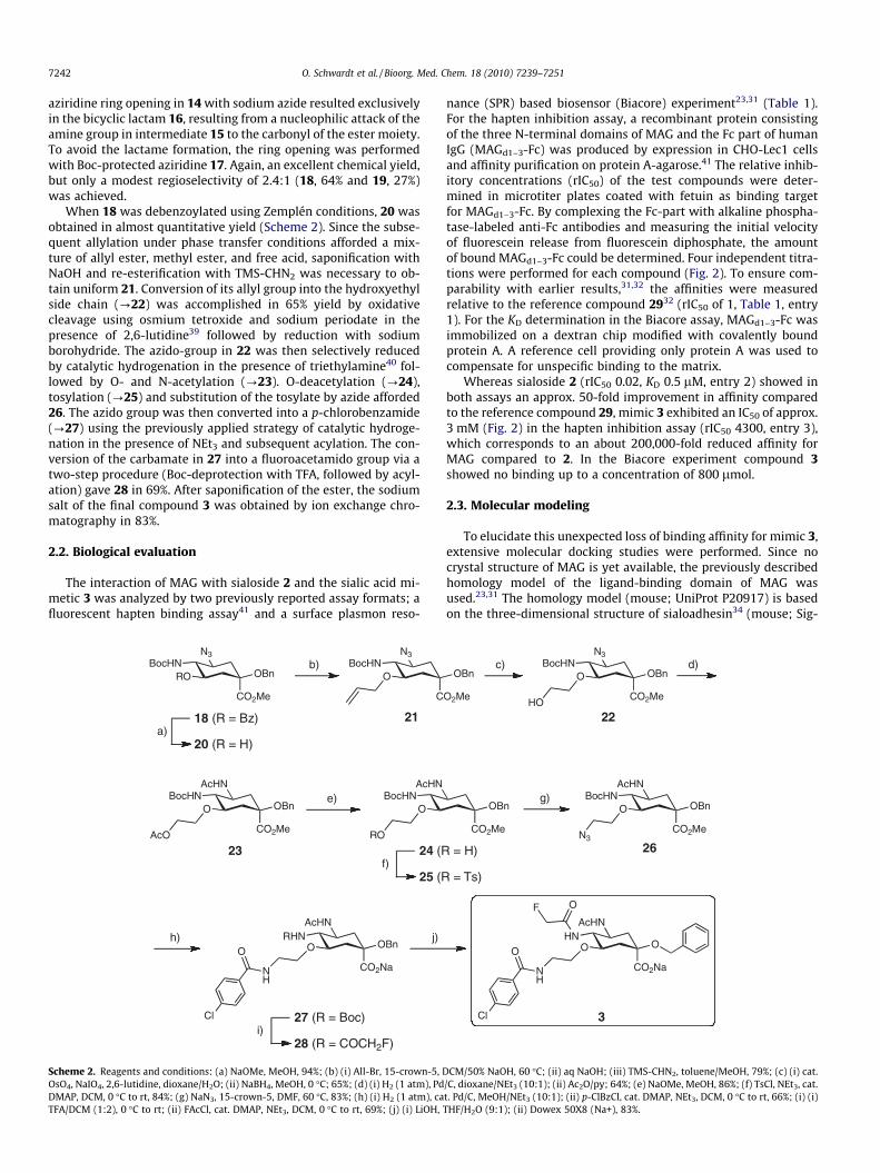
aziridine ring opening in 14 with sodium azide resulted exclusivelyin the bicyclic lactam 16, resulting from a nucleophilic attack of theamine group in intermediate 15 to the carbonyl of the ester moiety.To avoid the lactame formation, the ring opening was performedwith Boc-protected aziridine 17. Again, an excellent chemical yield,but only a modest regioselectivity of 2.4:1 (18, 64% and 19, 27%)was achieved.
When 18 was debenzoylated using Zemplén conditions, 20 wasobtained in almost quantitative yield (Scheme 2). Since the subse-quent allylation under phase transfer conditions afforded a mix-ture of allyl ester, methyl ester, and free acid, saponification withNaOH and re-esterification with TMS-CHN2 was necessary to ob-tain uniform 21. Conversion of its allyl group into the hydroxyethylside chain (?22) was accomplished in 65% yield by oxidativecleavage using osmium tetroxide and sodium periodate in thepresence of 2,6-lutidine39 followed by reduction with sodiumborohydride. The azido-group in 22 was then selectively reducedby catalytic hydrogenation in the presence of triethylamine40 fol-lowed by O- and N-acetylation (?23). O-deacetylation (?24),tosylation (?25) and substitution of the tosylate by azide afforded26. The azido group was then converted into a p-chlorobenzamide(?27) using the previously applied strategy of catalytic hydroge-nation in the presence of NEt3 and subsequent acylation. The con-version of the carbamate in 27 into a fluoroacetamido group via atwo-step procedure (Boc-deprotection with TFA, followed by acyl-ation) gave 28 in 69%. After saponification of the ester, the sodiumsalt of the final compound 3 was obtained by ion exchange chro-matography in 83%.
2.2. Biological evaluation
The interaction of MAG with sialoside 2 and the sialic acid mi-metic 3 was analyzed by two previously reported assay formats; afluorescent hapten binding assay41 and a surface plasmon reso-
BocHNRO
N3
CO2Me
OBn
e)
12
23
h) j)
b)
a)18 (R = Bz)
20 (R = H)
f)24 (R
25 (R
i)27 (R = Boc)
28 (R = COCH2F)
BocHNO
N3
C
BocHNO
AcHN
CO2Me
OBn
AcO
BocHNO
AcHN
RO
RHNO
AcHN
CO2Na
OBn
NH
O
Cl
Scheme 2. Reagents and conditions: (a) NaOMe, MeOH, 94%; (b) (i) All-Br, 15-crown-5, DOsO4, NaIO4, 2,6-lutidine, dioxane/H2O; (ii) NaBH4, MeOH, 0 �C; 65%; (d) (i) H2 (1 atm), PdDMAP, DCM, 0 �C to rt, 84%; (g) NaN3, 15-crown-5, DMF, 60 �C, 83%; (h) (i) H2 (1 atm), caTFA/DCM (1:2), 0 �C to rt; (ii) FAcCl, cat. DMAP, NEt3, DCM, 0 �C to rt, 69%; (j) (i) LiOH,
nance (SPR) based biosensor (Biacore) experiment23,31 (Table 1).For the hapten inhibition assay, a recombinant protein consistingof the three N-terminal domains of MAG and the Fc part of humanIgG (MAGd1–3-Fc) was produced by expression in CHO-Lec1 cellsand affinity purification on protein A-agarose.41 The relative inhib-itory concentrations (rIC50) of the test compounds were deter-mined in microtiter plates coated with fetuin as binding targetfor MAGd1–3-Fc. By complexing the Fc-part with alkaline phospha-tase-labeled anti-Fc antibodies and measuring the initial velocityof fluorescein release from fluorescein diphosphate, the amountof bound MAGd1–3-Fc could be determined. Four independent titra-tions were performed for each compound (Fig. 2). To ensure com-parability with earlier results,31,32 the affinities were measuredrelative to the reference compound 2932 (rIC50 of 1, Table 1, entry1). For the KD determination in the Biacore assay, MAGd1–3-Fc wasimmobilized on a dextran chip modified with covalently boundprotein A. A reference cell providing only protein A was used tocompensate for unspecific binding to the matrix.
Whereas sialoside 2 (rIC50 0.02, KD 0.5 lM, entry 2) showed inboth assays an approx. 50-fold improvement in affinity comparedto the reference compound 29, mimic 3 exhibited an IC50 of approx.3 mM (Fig. 2) in the hapten inhibition assay (rIC50 4300, entry 3),which corresponds to an about 200,000-fold reduced affinity forMAG compared to 2. In the Biacore experiment compound 3showed no binding up to a concentration of 800 lmol.
2.3. Molecular modeling
To elucidate this unexpected loss of binding affinity for mimic 3,extensive molecular docking studies were performed. Since nocrystal structure of MAG is yet available, the previously describedhomology model of the ligand-binding domain of MAG wasused.23,31 The homology model (mouse; UniProt P20917) is basedon the three-dimensional structure of sialoadhesin34 (mouse; Sig-
d)
22
g)
26
3
c)
= H)
= Ts)
O2Me
OBnBocHN
O
N3
CO2Me
OBn
HO
CO2Me
OBnBocHN
O
AcHN
CO2Me
OBn
N3
HNO
AcHN
CO2Na
O
NH
O
Cl
OF
CM/50% NaOH, 60 �C; (ii) aq NaOH; (iii) TMS-CHN2, toluene/MeOH, 79%; (c) (i) cat./C, dioxane/NEt3 (10:1); (ii) Ac2O/py; 64%; (e) NaOMe, MeOH, 86%; (f) TsCl, NEt3, cat.t. Pd/C, MeOH/NEt3 (10:1); (ii) p-ClBzCl, cat. DMAP, NEt3, DCM, 0 �C to rt, 66%; (i) (i)THF/H2O (9:1); (ii) Dowex 50X8 (Na+), 83%.
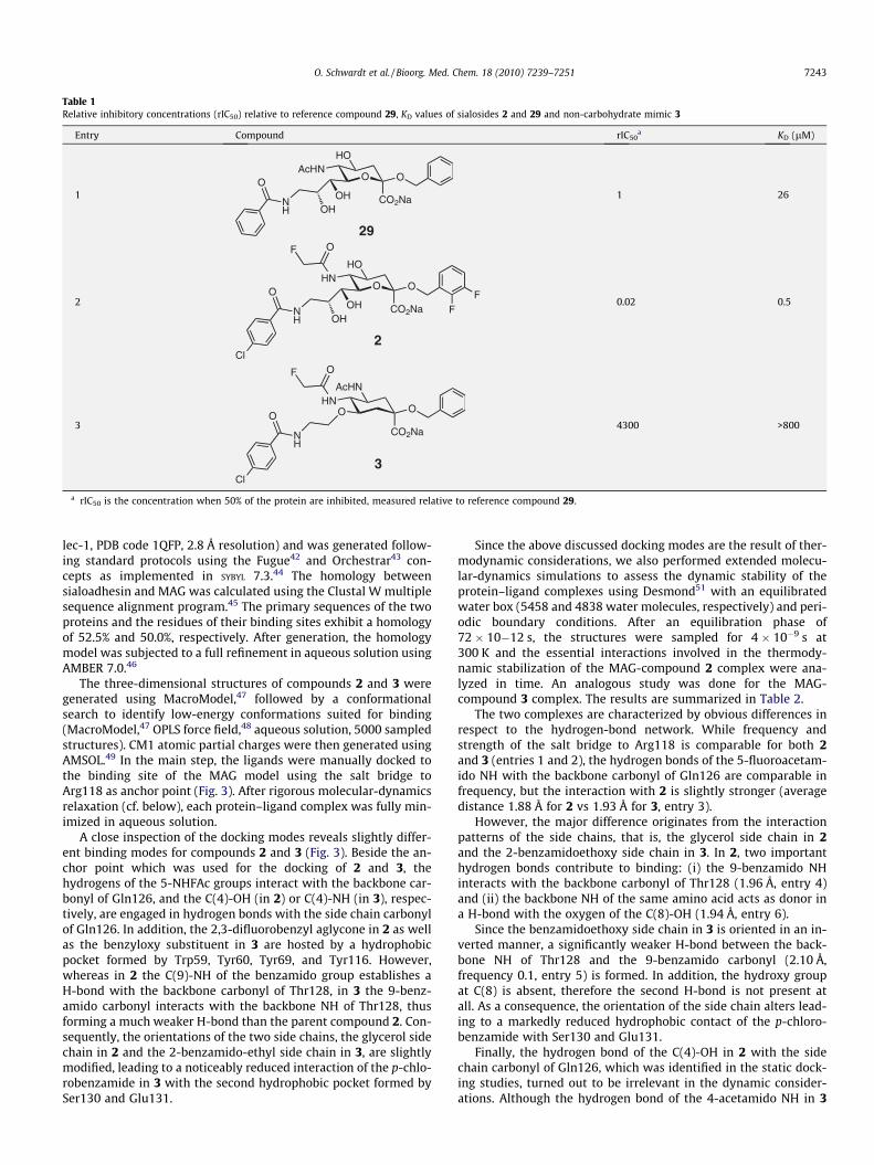
Table 1Relative inhibitory concentrations (rIC50) relative to reference compound 29, KD values of sialosides 2 and 29 and non-carbohydrate mimic 3
Entry Compound rIC50a KD (lM)
1
OAcHN
HO
CO2Na
O
NH
O
29
OHOH 1 26
2
OHN
HO
CO2Na
O
NH
O
2Cl
OF
FF
OHOH 0.02 0.5
3
HNO
AcHN
CO2Na
O
NH
O
3Cl
OF
4300 >800
a rIC50 is the concentration when 50% of the protein are inhibited, measured relative to reference compound 29.
O. Schwardt et al. / Bioorg. Med. Chem. 18 (2010) 7239–7251 7243
lec-1, PDB code 1QFP, 2.8 Å resolution) and was generated follow-ing standard protocols using the Fugue42 and Orchestrar43 con-cepts as implemented in SYBYL 7.3.44 The homology betweensialoadhesin and MAG was calculated using the Clustal W multiplesequence alignment program.45 The primary sequences of the twoproteins and the residues of their binding sites exhibit a homologyof 52.5% and 50.0%, respectively. After generation, the homologymodel was subjected to a full refinement in aqueous solution usingAMBER 7.0.46
The three-dimensional structures of compounds 2 and 3 weregenerated using MacroModel,47 followed by a conformationalsearch to identify low-energy conformations suited for binding(MacroModel,47 OPLS force field,48 aqueous solution, 5000 sampledstructures). CM1 atomic partial charges were then generated usingAMSOL.49 In the main step, the ligands were manually docked tothe binding site of the MAG model using the salt bridge toArg118 as anchor point (Fig. 3). After rigorous molecular-dynamicsrelaxation (cf. below), each protein–ligand complex was fully min-imized in aqueous solution.
A close inspection of the docking modes reveals slightly differ-ent binding modes for compounds 2 and 3 (Fig. 3). Beside the an-chor point which was used for the docking of 2 and 3, thehydrogens of the 5-NHFAc groups interact with the backbone car-bonyl of Gln126, and the C(4)-OH (in 2) or C(4)-NH (in 3), respec-tively, are engaged in hydrogen bonds with the side chain carbonylof Gln126. In addition, the 2,3-difluorobenzyl aglycone in 2 as wellas the benzyloxy substituent in 3 are hosted by a hydrophobicpocket formed by Trp59, Tyr60, Tyr69, and Tyr116. However,whereas in 2 the C(9)-NH of the benzamido group establishes aH-bond with the backbone carbonyl of Thr128, in 3 the 9-benz-amido carbonyl interacts with the backbone NH of Thr128, thusforming a much weaker H-bond than the parent compound 2. Con-sequently, the orientations of the two side chains, the glycerol sidechain in 2 and the 2-benzamido-ethyl side chain in 3, are slightlymodified, leading to a noticeably reduced interaction of the p-chlo-robenzamide in 3 with the second hydrophobic pocket formed bySer130 and Glu131.
Since the above discussed docking modes are the result of ther-modynamic considerations, we also performed extended molecu-lar-dynamics simulations to assess the dynamic stability of theprotein–ligand complexes using Desmond51 with an equilibratedwater box (5458 and 4838 water molecules, respectively) and peri-odic boundary conditions. After an equilibration phase of72 � 10�12 s, the structures were sampled for 4 � 10�9 s at300 K and the essential interactions involved in the thermody-namic stabilization of the MAG-compound 2 complex were ana-lyzed in time. An analogous study was done for the MAG-compound 3 complex. The results are summarized in Table 2.
The two complexes are characterized by obvious differences inrespect to the hydrogen-bond network. While frequency andstrength of the salt bridge to Arg118 is comparable for both 2and 3 (entries 1 and 2), the hydrogen bonds of the 5-fluoroacetam-ido NH with the backbone carbonyl of Gln126 are comparable infrequency, but the interaction with 2 is slightly stronger (averagedistance 1.88 Å for 2 vs 1.93 Å for 3, entry 3).
However, the major difference originates from the interactionpatterns of the side chains, that is, the glycerol side chain in 2and the 2-benzamidoethoxy side chain in 3. In 2, two importanthydrogen bonds contribute to binding: (i) the 9-benzamido NHinteracts with the backbone carbonyl of Thr128 (1.96 Å, entry 4)and (ii) the backbone NH of the same amino acid acts as donor ina H-bond with the oxygen of the C(8)-OH (1.94 Å, entry 6).
Since the benzamidoethoxy side chain in 3 is oriented in an in-verted manner, a significantly weaker H-bond between the back-bone NH of Thr128 and the 9-benzamido carbonyl (2.10 Å,frequency 0.1, entry 5) is formed. In addition, the hydroxy groupat C(8) is absent, therefore the second H-bond is not present atall. As a consequence, the orientation of the side chain alters lead-ing to a markedly reduced hydrophobic contact of the p-chloro-benzamide with Ser130 and Glu131.
Finally, the hydrogen bond of the C(4)-OH in 2 with the sidechain carbonyl of Gln126, which was identified in the static dock-ing studies, turned out to be irrelevant in the dynamic consider-ations. Although the hydrogen bond of the 4-acetamido NH in 3
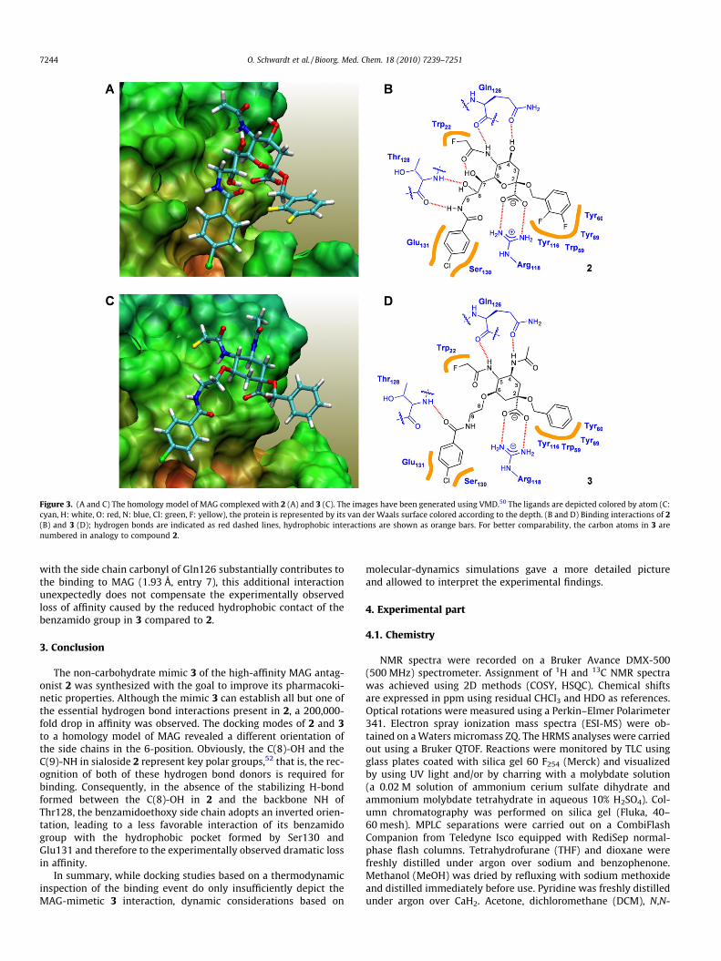
Figure 3. (A and C) The homology model of MAG complexed with 2 (A) and 3 (C). The images have been generated using VMD.50 The ligands are depicted colored by atom (C:cyan, H: white, O: red, N: blue, Cl: green, F: yellow), the protein is represented by its van der Waals surface colored according to the depth. (B and D) Binding interactions of 2(B) and 3 (D); hydrogen bonds are indicated as red dashed lines, hydrophobic interactions are shown as orange bars. For better comparability, the carbon atoms in 3 arenumbered in analogy to compound 2.
7244 O. Schwardt et al. / Bioorg. Med. Chem. 18 (2010) 7239–7251
with the side chain carbonyl of Gln126 substantially contributes tothe binding to MAG (1.93 Å, entry 7), this additional interactionunexpectedly does not compensate the experimentally observedloss of affinity caused by the reduced hydrophobic contact of thebenzamido group in 3 compared to 2.
3. Conclusion
The non-carbohydrate mimic 3 of the high-affinity MAG antag-onist 2 was synthesized with the goal to improve its pharmacoki-netic properties. Although the mimic 3 can establish all but one ofthe essential hydrogen bond interactions present in 2, a 200,000-fold drop in affinity was observed. The docking modes of 2 and 3to a homology model of MAG revealed a different orientation ofthe side chains in the 6-position. Obviously, the C(8)-OH and theC(9)-NH in sialoside 2 represent key polar groups,52 that is, the rec-ognition of both of these hydrogen bond donors is required forbinding. Consequently, in the absence of the stabilizing H-bondformed between the C(8)-OH in 2 and the backbone NH ofThr128, the benzamidoethoxy side chain adopts an inverted orien-tation, leading to a less favorable interaction of its benzamidogroup with the hydrophobic pocket formed by Ser130 andGlu131 and therefore to the experimentally observed dramatic lossin affinity.
In summary, while docking studies based on a thermodynamicinspection of the binding event do only insufficiently depict theMAG-mimetic 3 interaction, dynamic considerations based on
molecular-dynamics simulations gave a more detailed pictureand allowed to interpret the experimental findings.
4. Experimental part
4.1. Chemistry
NMR spectra were recorded on a Bruker Avance DMX-500(500 MHz) spectrometer. Assignment of 1H and 13C NMR spectrawas achieved using 2D methods (COSY, HSQC). Chemical shiftsare expressed in ppm using residual CHCl3 and HDO as references.Optical rotations were measured using a Perkin–Elmer Polarimeter341. Electron spray ionization mass spectra (ESI-MS) were ob-tained on a Waters micromass ZQ. The HRMS analyses were carriedout using a Bruker QTOF. Reactions were monitored by TLC usingglass plates coated with silica gel 60 F254 (Merck) and visualizedby using UV light and/or by charring with a molybdate solution(a 0.02 M solution of ammonium cerium sulfate dihydrate andammonium molybdate tetrahydrate in aqueous 10% H2SO4). Col-umn chromatography was performed on silica gel (Fluka, 40–60 mesh). MPLC separations were carried out on a CombiFlashCompanion from Teledyne Isco equipped with RediSep normal-phase flash columns. Tetrahydrofurane (THF) and dioxane werefreshly distilled under argon over sodium and benzophenone.Methanol (MeOH) was dried by refluxing with sodium methoxideand distilled immediately before use. Pyridine was freshly distilledunder argon over CaH2. Acetone, dichloromethane (DCM), N,N-
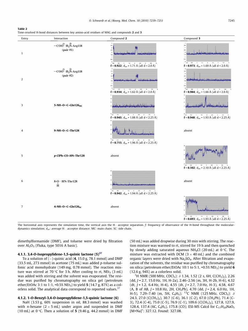
Table 2Time-resolved H-bond distances between key amino-acid residues of MAG and compounds 2 and 3
Entry Interaction Compound 2 Compound 3
1(pair #1)
COO H2N-Arg118
f = 0.922; dav = 1.71 Å (all d < 2.0 Å) f = 0.973; dav = 1.69 Å (all d < 2.0 Å)
2(pair #2)
COO H2N-Arg118
f = 0.934; dav = 1.62 Å (all d < 2.0 Å) f = 0.984; dav = 1.66 Å (all d < 2.0 Å)
3 5-NH–O@C–Gln126MC
f = 0.945; dav = 1.88 Å (all d < 2.25 Å) f = 0.948; dav = 1.93 Å (all d < 2.25 Å)
4 9-NH–O@C–Thr128 absent
f = 0.715; dav = 1.96 Å (all d < 2.25 Å)
5 p-ClPh–CO–HN–Thr128 absent
f = 0.102; dav = 2.10 Å (all d < 2.25 Å)
6 8-OH
HN-Thr128 absent
f = 0.942; dav = 1.94 Å (all d < 2.25 Å)
7 4-NH–O@C–Gln126SC absent
f = 0.693; dav = 1.93 Å (all d < 2.25 Å)
The horizontal axis represents the simulation time, the vertical axis the H� � �acceptor separation. f: frequency of observance of the H-bond throughout the molecular-dynamics simulation; dav: average H� � �acceptor distance; MC: main chain; SC: side chain.
O. Schwardt et al. / Bioorg. Med. Chem. 18 (2010) 7239–7251 7245
dimethylformamide (DMF), and toluene were dried by filtrationover Al2O3 (Fluka, type 5016 A basic).
4.1.1. 3,4-O-Isopropylidene-1,5-quinic lactone (5)37
To a solution of (�)-quinic acid (4, 15.0 g, 78.1 mmol) and DMP(33.5 mL, 273 mmol) in acetone (75 mL) was added p-toluene sul-fonic acid monohydrate (149 mg, 0.78 mmol). The reaction mix-ture was stirred at 70 �C for 3 h. After cooling to rt, NEt3 (3 mL)was added with stirring and the solvent was evaporated. The resi-due was purified by chromatography on silica gel (petroleumether/EtOAc 3:1 to 1:1, +0.5% NEt3) to yield 5 (14.7 g, 87%) as a col-orless solid. The analytical data correspond to reported values.37
4.1.2. 1-O-Benzyl-3,4-O-isopropylidene-1,5-quinic lactone (6)NaH (3.53 g, 60% suspension in oil, 88.3 mmol) was washed
with n-hexane (2 � 5 mL) under argon and suspended in DMF(10 mL) at 0 �C. Then a solution of 5 (9.46 g, 44.2 mmol) in DMF
(50 mL) was added dropwise during 30 min with stirring. The reac-tion mixture was warmed to rt, stirred for 19 h and then quenchedby slowly adding saturated aqueous NH4Cl (20 mL) at 0 �C. Themixture was extracted with DCM (3 � 40 mL) and the combinedorganic layers were dried with Na2SO4. After filtration and evapo-ration of the solvents, the residue was purified by chromatographyon silica (petroleum ether/EtOAc 10:1 to 5:1, +0.5% NEt3) to yield 6(12.6 g, 94%) as a colorless solid.
1H NMR (500 MHz, CDCl3): d 1.34, 1.52 (2 s, 6H, C(CH3)2), 2.26(dd, J = 2.7, 15.0 Hz, 1H, H-2a), 2.46–2.56 (m, 3H, H-2b, H-6), 4.32(dt, J = 1.2, 6.4 Hz, H-4), 4.55 (dt, J = 2.7, 7.0 Hz, H-3), 4.58, 4.67(A, B of AB, J = 10.8 Hz, 2H, CH2Ph), 4.70 (dd, J = 2.4, 6.0 Hz, 1H,H-5), 7.29–7.40 (m, 5H, C6H5); 13C NMR (125 MHz, CDCl3): d24.3, 27.0 (C(CH3)2), 30.7 (C-6), 36.1 (C-2), 67.0 (CH2Ph), 71.4 (C-3), 72.4 (C-4), 75.0 (C-5), 76.9 (C-1), 109.6 (C(CH3)2), 127.8, 127.9,128.4, 137.4 (6C, C6H5), 175.8 (CO); ESI-MS Calcd for C17H20NaO5
[M+Na]+: 327.12. Found: 327.08.
7246 O. Schwardt et al. / Bioorg. Med. Chem. 18 (2010) 7239–7251
4.1.3. Methyl (1R,3R,4S,5R)-1-benzyloxy-3,4,5-trihydroxy-4,5-O-isopropylidene-cyclohexanecarboxylate (7)
To a suspension of 6 (9.84 g, 32.3 mmol) in MeOH (170 mL) wasadded a freshly prepared solution of sodium (820 mg, 35.6 mmol)in MeOH (50 mL) at 0 �C under argon. The reaction mixture wasstirred for 3 h at rt, then the solution was neutralized by addingDowex 50X8 ion-exchange resin. After filtration and evaporationof the solvents, the residue was purified by chromatography on sil-ica (petroleum ether/EtOAc 2:1 to 1:8, +0.5% NEt3) to yield 7(7.96 g, 73%) as a colorless solid.
1H NMR (500 MHz, CDCl3): d 1.37, 1.48 (2 s, 6H, C(CH3)2), 1.81(dd, J = 10.9, 13.6 Hz, 1H, H-6a), 2.25–2.31 (m, 2H, H-2a, H-6b),2.54 (m, 1H, H-2b), 3.78 (m, 3H, OMe), 3.94 (t, J = 6.4 Hz, H-4),4.16 (ddd, J = 4.1, 7.0, 10.9 Hz, 1H, H-5), 4.34 (A of AB, J = 10.2 Hz,1H, CH2Ph), 4.46 (dt, J = 3.6, 5.3 Hz, H-3), 4.60 (B of AB,J = 10.2 Hz, 1H, CH2Ph), 7.27–7.39 (m, 5H, C6H5); 13C NMR(125 MHz, CDCl3): d 25.9, 28.2 (C(CH3)2), 30.9 (C-2), 37.7 (C-6),52.5 (OMe), 67.1 (CH2Ph), 68.1 (C-5), 72.9 (C-3), 79.2 (C-1), 80.1(C-4), 109.1 (C(CH3)2), 127.9, 128.2, 128.3, 138.0 (6C, C6H5), 174.1(CO); ESI-MS Calcd for C18H25O6 [M+H]+: 337.17. Found: 337.14.
4.1.4. Methyl (1S,3R,4R,5R)-1-benzyloxy-3,4-dihydroxy-3,4-O-isopropylidene-5-(p-tosyloxy)-cyclohexanecarboxylate (8)
To a solution of 7 (5.55 g, 16.5 mmol) and DMAP (190 mg) inpyridine (40 mL) was added p-tosyl chloride (6.29 g, 33.0 mmol)under argon. The solution was stirred for 19 h, then the pyridinewas evaporated and water (50 mL) was added. The aqueous phasewas extracted with DCM (3 � 50 mL), then the combined organiclayers were washed with brine (20 mL) and dried with Na2SO4.After evaporation of the solvent, the residue was purified by chro-matography on silica (petroleum ether/EtOAc 3:1 to 1:1, +0.5%NEt3) to give 8 (7.96 g, 98%) as a yellowish oil.
1H NMR (500 MHz, CDCl3): d 0.93, 1.23 (2 s, 6H, C(CH3)2), 1.91 (t,J = 12.7 Hz, 1H, H-6a), 2.16 (dd, J = 5.0, 16.1 Hz, 1H, H-2a), 2.39 (s,3H, PhCH3), 2.59 (d, J = 16.1 Hz, 1H, H-2b), 2.68 (dd, J = 3.1,13.6 Hz, 1H, H-6b), 3.76 (m, 3H, OMe), 4.01 (t, J = 6.5 Hz, H-4),4.40–4.43 (m, 2H, H-3, CH2Ph), 4.55 (B of AB, J = 10.5 Hz, 1H,CH2Ph), 4.64 (m, 1H, H-5), 7.19 (AA0 of AA0BB0, J = 8.0 Hz, 2H,C6H4), 7.31–7.37 (m, 5H, C6H5), 7.71 (BB0 of AA0BB0, J = 8.0 Hz, 2H,C6H4); 13C NMR (125 MHz, CDCl3): d 21.5 (PhCH3), 25.8, 27.3(C(CH3)2), 31.1 (C-2), 35.5 (C-6), 52.5 (OMe), 66.7 (CH2Ph), 73.2(C-3), 76.6 (C-4), 79.1, 79.2 (C-1, C-5), 109.2 (C(CH3)2), 127.4,127.5, 128.1, 128.4, 129.6, 132.9, 137.9 (12C, C6H4, C6H5), 172.6(CO); ESI-MS Calcd for C25H30NaO8S [M+Na]+: 513.16. Found:513.18.
4.1.5. Methyl (1S,3R,4R,5R)-1-benzyloxy-3,4-dihydroxy-5-(p-tosyloxy)-cyclohexanecarboxylate (9)
A suspension of 8 (7.96 g, 16.2 mmol) in 80% aqueous aceticacid (30 mL) was stirred for 2.5 h at 60 �C. After cooling to rt, thesolvents were removed by co-evaporation with toluene(3 � 50 mL). The residue was dried in high vacuum to give 9(7.13 g, 98%), which was used without further purification.
1H NMR (500 MHz, CDCl3): d 1.94 (dd, J = 3.1, 15.1 Hz, 1H, H-2a), 2.16 (dd, J = 11.9, 14.2 Hz, 1H, H-6a), 2.40 (m, 1H, H-2b),2.43 (s, 3H, PhCH3), 2.89 (dt, J = 3.4, 14.3 Hz, 1H, H-6b), 3.56(dd, J = 3.5, 9.3 Hz, H-4), 3.78 (m, 3H, OMe), 4.05 (m, 1H, H-3),4.50, 4.52 (A, B of AB, J = 10.2 Hz, 2H, CH2Ph), 4.59 (m, 1H, H-5), 7.31 (AA0 of AA0BB0, J = 8.1 Hz, 2H, C6H4), 7.33–7.39 (m, 5H,C6H5), 7.82 (BB0 of AA0BB0, J = 8.1 Hz, 2H, C6H4); 13C NMR(125 MHz, CDCl3): d 21.7 (PhCH3), 34.5 (C-6), 37.3 (C-2), 52.7(OMe), 67.6 (CH2Ph), 70.0 (C-3), 72.9 (C-4), 78.5 (C-5), 81.5 (C-1), 128.1, 128.1, 128.3, 128.7, 129.8, 133.2, 136.5, 145.0 (12C,C6H4, C6H5), 171.1 (CO); ESI-MS Calcd for C22H26NaO8S[M+Na]+: 473.12. Found: 473.15.
4.1.6. Methyl (1R,3S,4S,5R)-1-benzyloxy-3,4-epoxy-5-hydroxy-cyclohexane-1-carboxylate (10)
To a solution of 9 (7.13 g, 15.8 mmol) in THF (50 mL) was addedDBU (2.39 mL, 15.8 mmol) dropwise during 10 min at 0 �C underargon. The mixture was stirred at 0 �C for 1 h, then warmed to rtand stirred for another 17 h. The solvent was removed, and the res-idue was purified by MPLC on silica (petroleum ether/EtOAc) toyield 10 (4.23 g, 96%) as a colorless solid.
1H NMR (500 MHz, CDCl3): d 1.86 (dd, J = 9.5, 12.6 Hz, 1H, H-6a),2.13 (d, J = 15.1 Hz, 1H, H-2a), 2.23 (m, 1H, H-6b), 2.68 (ddd, J = 2.0,4.8, 15.1 Hz, 1H, H-2b), 3.35 (m, 1H, H-4), 3.44 (t, J = 4.4 Hz, 1H, H-3), 3.77 (s, 3H, OMe), 4.03 (m, 1H, H-5), 4.31, 4.49 (A, B of AB,J = 10.6 Hz, 2H, CH2Ph), 7.27–7.38 (m, 5H, C6H5); 13C NMR(125 MHz, CDCl3): d 30.3 (C-2), 36.4 (C-6), 52.5 (C-3), 52.8 (OMe),54.8 (C-4), 65.9 (C-5), 66.9 (CH2Ph), 79.5 (C-1), 127.7, 127.8,128.4, 137.4 (6C, C6H5), 173.2 (CO); ESI-MS Calcd for C15H18NaO5
[M+Na]+: 301.11. Found: 301.14.
4.1.7. Methyl (1R,3S,4S,5R)-5-benzoyloxy-1-benzyloxy-3,4-epoxy-cyclohexane-1-carboxylate (11)
To a solution of 10 (2.10 g, 7.55 mmol) in pyridine (20 mL) weresubsequently added benzoyl chloride (1.78 mL, 15.1 mmol) andDMAP (120 mg) at 0 �C under argon. The mixture was stirred for4 h at rt and then concentrated in vacuo. The residue was co-evap-orated with toluene (3 � 10 mL), dissolved in DCM (20 mL), andwashed with 1 N HCl (10 mL) and saturated aqueous NaHCO3
(10 mL). The organic phase was dried (Na2SO4), filtered and con-centrated in vacuo. The residue was purified by MPLC on silica(petroleum ether/EtOAc) to afford 11 (2.79 g, 97%) as a colorlesssolid.
[a]D �33.4 (c 0.50, CHCl3); 1H NMR (500 MHz, CDCl3): d 2.06–2.15 (m, 2H, H-2a, H-6a), 2.42 (dd, J = 4.3, 11.9 Hz, 1H, H-6b),2.85 (dd, J = 3.2, 14.9 Hz, 1H, H-2b), 3.43 (t, J = 4.5 Hz, 1H, H-3),3.47 (m, 1H, H-4), 3.84 (s, 3H, OMe), 4.35, 4.50 (A, B of AB,J = 10.9 Hz, 2H, CH2Ph), 5.39 (m, 1H, H-5), 7.28–7.35, 7.43–7.46,7.56–7.59, 8.07–8.09 (m, 10H, 2 C6H5); 13C NMR (125 MHz, CDCl3):d 31.2 (C-2), 32.2 (C-6), 51.8 (C-3), 52.7, 52.8 (C-4, OMe), 66.8(CH2Ph), 69.2 (C-5), 79.3 (C-1), 127.6, 127.8, 128.4, 128.4, 129.7,129.8, 133.3, 137.5 (12C, 2C6H5), 166.0 (COPh), 173.2 (CO2Me);ESI-MS Calcd for C22H22NaO6 [M+Na]+: 405.13. Found: 405.08.
4.1.8. Methyl (1R,3R,4S,5R)-5-azido-3-benzoyloxy-1-benzyloxy-4-hydroxy-cyclohexane-1-carboxylate (12)
To a solution of 11 (2.79 g, 7.30 mmol) in MeOH/water (8:1,54 mL) were added sodium azide (2.47 g, 37.9 mmol) and ammo-nium chloride (859 mg, 16.1 mmol), and the mixture was stirredat 70 �C for 18 h under argon. The reaction mixture was dilutedwith saturated aqueous NaHCO3 (10 mL) and water (20 mL), andthe solution was carefully evaporated to remove methanol. Theresulting aqueous phase containing an oily residue was extractedwith ethyl acetate (3 � 50 mL). The combined organic extractswere dried (Na2SO4), filtered, and concentrated in vacuo. The resi-due was purified by MPLC on silica (1% gradient of MeOH in DCM)to yield 12 (2.45 g, 78%) as a colorless oil.
[a]D �53.6 (c 0.45, CHCl3); IR (film) 3479 (m b, OH), 2107 (vs,N3), 1722 (vs, CO) cm�1; 1H NMR (500 MHz, CDCl3): d 1.87 (dd,J = 11.7, 13.8 Hz, 1H, H-6a), 2.23 (dd, J = 3.5, 15.9 Hz, 1H, H-2a),2.44 (d, J = 5.1 Hz, 1H, OH), 2.59 (m, 1H, H-6b), 2.67 (dt, J = 3.2,15.9 Hz, 1H, H-2b), 3.76–3.80 (m, 4H, H-4, OMe), 4.10 (m, 1H, H-5), 4.31, 4.38 (A, B of AB, J = 10.5 Hz, 2H, CH2Ph), 5.65 (q,J = 3.5 Hz, 1H, H-3), 7.20–7.28, 7.48–7.53, 7.90–7.93 (m, 10H,2C6H5); 13C NMR (125 MHz, CDCl3): d 32.3 (C-2), 36.8 (C-6), 52.6(OMe), 58.4 (C-5), 67.4 (CH2Ph), 70.8 (C-3), 73.7 (C-4), 79.4 (C-1),127.7, 127.8, 128.3, 128.4, 129.4, 129.9, 133.3, 137.3 (12C,2C6H5), 166.2 (COPh), 172.7 (CO2Me); ESI-MS Calcd forC22H23N3NaO6 [M+Na]+: 448.15. Found: 448.16.
O. Schwardt et al. / Bioorg. Med. Chem. 18 (2010) 7239–7251 7247
4.1.9. Methyl (1R,3R,4S,5R)-5-azido-3-benzoyloxy-1-benzyloxy-4-(methylsulfonyloxy)-cyclohexane-1-carboxylate-1,4-lactam(13)
To a solution of 12 (2.43 g, 5.70 mmol) in DCM (25 mL) at 0 �Cunder argon was added NEt3 (3.18 mL, 22.8 mmol) followed bythe addition of methanesulfonyl chloride (887 lL, 11.4 mmol).The mixture was stirred at 0 �C for 1 h and warmed to rt with stir-ring for 3 h. The reaction was quenched with MeOH (2 mL) andconcentrated in vacuo. Purification of the residue by MPLC on silica(petrol ether/EtOAc) yielded 13 (2.69 g, 94%) as a yellowish oil.
[a]D �31.0 (c 0.50, CHCl3); IR (film) 2111 (vs, N3), 1732 (vs, CO)cm�1; 1H NMR (500 MHz, CDCl3): d 2.01 (m, 1H, H-6a), 2.35 (d,J = 15.9 Hz, 1H, H-2a), 2.57–2.73 (m, 2H, H-2b, H-6b), 3.08 (s, 3H,SMe), 3.79 (s, 3H, OMe), 4.27–4.34 (m, 2H, H-5, CH2Ph-HA), 4.38(B of AB, J = 10.5 Hz, 1H, CH2Ph-HB), 4.70 (dd, J = 3.4, 9.6 Hz, 1H,H-4), 5.84 (m, 1H, H-3), 7.18–7.32, 7.53–7.55, 7.94–7.95 (m, 10H,2C6H5); 13C NMR (125 MHz, CDCl3): d 31.8 (C-2), 37.4 (C-6), 38.7(SMe), 52.7 (OMe), 55.9 (C-5), 67.4 (CH2Ph), 68.5 (C-3), 79.8 (2C,C-1, C-4), 127.8, 127.9, 128.3, 128.5, 129.4, 129.9, 133.4, 137.0(12C, 2C6H5), 166.3 (COPh), 172.5 (CO2Me); ESI-MS Calcd forC23H25N3NaO8S [M+Na]+: 526.13. Found: 526.09.
4.1.10. Methyl (1R,3R,4R,5R)-4,5-aziridino-3-benzoyloxy-1-benzyloxy-cyclohexane-1-carboxylate (14)
To a solution of 13 (2.69 g, 5.34 mmol) in THF (30 mL) at 0 �Cwas added PPh3 (1.76 g, 6.68 mmol), initially adding a third ofthe amount while cooling and then after removing the ice bathadding the remaining amount over a period of 15 min. The reactionmixture was stirred at rt for 4 h, then NEt3 (1.12 mL, 8.02 mmol)and water (2 mL) were added, and the mixture was stirred at rtfor 16 h. The reaction mixture was concentrated to remove THF,and the residue was partitioned between DCM (50 mL) and brine(20 mL). The aqueous phase was extracted with DCM(3 � 20 mL), and the combined organic extracts were dried(Na2SO4), filtered, and evaporated. The residue was purified byMPLC on silica (petrol ether/EtOAc, +1% NEt3) to give 2.20 g of a3:2-mixture of 14 and triphenylphosphine oxide, which was di-rectly used in the next step.
1H NMR (500 MHz, CDCl3): d 2.13 (dd, J = 3.7, 14.8 Hz, 1H, H-2a),2.37–2.44 (m, 3H, H-2b, H-6), 2.45–2.50 (m, 2H, H-4, H-5), 3.74 (s,3H, OMe), 4.38, 4.53 (A, B of AB, J = 11.1 Hz, 1H, CH2Ph), 5.60 (t,J = 4.9 Hz, 1H, H-3), 7.25–7.33, 7.53–7.56, 7.96–7.97 (m, 10H,2C6H5); 13C NMR (125 MHz, CDCl3): d 28.1 (C-6), 30.8 (C-2), 31.6,32.5 (C-4, C-5), 52.2 (OMe), 66.5 (CH2Ph), 68.1 (C-3), 76.9 (C-1),127.5, 128.2, 128.3, 128.5, 129.7, 132.9, 138.2 (12C, 2C6H5), 166.1(COPh), 173.1 (CO2Me); ESI-MS Calcd for C22H24NO5 [M+H]+:382.17. Found: 382.10.
4.1.11. (1R,3R,4R,5S)-4-Amino-5-azido-1-benzyloxy-3-benzoyloxy-cyclohexane-1-carboxylate-1,4-lactam (16)
To a solution of 14 (105 mg, 0.275 mmol) in DMF (3 mL) wasadded NaN3 (89.3 mg, 1.37 mmol) and NH4Cl (29.4 mg, 0.55mmol), and the mixture was heated at 65 �C under argon for23 h. The reaction mixture was cooled to rt, diluted with ethyl ace-tate (10 mL), and filtered. The filtrate was washed with brine(5 mL) and the aqueous phase was extracted with EtOAc(3 � 10 mL). The combined organic phases were dried (Na2SO4), fil-tered, and concentrated in vacuo. The residue was purified by chro-matography on silica (petrol ether/EtOAc 2:1 to 1:1) to yield 16(53.5 mg, 50%) as colorless foam.
[a]D –29.6 (c 1.24, CHCl3); IR (film) 2099 (vs, N3), 1719 (vs, CO)cm�1; 1H NMR (500 MHz, CDCl3): d 1.99 (dd, J = 5.5, 13.4 Hz, 1H, H-6a), 2.04 (dd, J = 3.8, 13.7 Hz, 1H, H-2a), 2.57 (ddd, J = 3.5, 10.8,13.3 Hz, 1H, H-6b), 2.81 (ddd, J = 3.5, 10.5, 13.7 Hz, 1H, H-2b),4.04 (m, 1H, H-4), 4.09 (m, 1H, H-5), 4.78, 4.84 (A, B of AB,J = 11.0 Hz, 2H, CH2Ph), 5.35 (m, 1H, H-3), 7.28–7.36, 7.55–7.61,
8.09–8.10 (m, 10H, 2C6H5); 13C NMR (125 MHz, CDCl3): d 34.8(C-6), 36.0 (C-2), 49.8 (C-4), 57.9 (C-5), 67.1 (CH2Ph), 70.1 (C-3),74.1 (C-1), 127.7, 128.4, 128.5, 129.3, 129.8, 133.4, 138.2 (12C,2C6H5), 166.1, 175.2 (2 CO); ESI-MS Calcd for C21H20N4NaO4
[M+Na]+: 415.14. Found: 415.16.
4.1.12. Methyl (1R,3R,4R,5R)-4,5-aziridino-3-benzoyloxy-1-benzyloxy-N-(tert-butoxycarbonyl)-cyclohexane-1-carboxylate(17)
Crude aziridine 14 (2.10 g, 3:2 mixture with PPh3O) was dis-solved in DCM (50 mL) and cooled to 0 �C under argon. ThenNEt3 (1.53 mL, 11.0 mmol), Boc2O (1.80 g, 8.25 mmol), and DMAP(10 mg) were subsequently added and the solution was stirred atrt for 5 h. The solvent was evaporated and the residue purifiedby MPLC on silica (1% gradient of EtOAc in petrol ether) to yield17 (1.66 g, 96%) as a colorless oil.
1H NMR (500 MHz, CDCl3): d 1.47 (s, 9H, C(CH3)3), 2.26 (d,J = 15.3 Hz, 1H, H-2a), 2.35 (dd, J = 5.4, 15.4 Hz, 1H, H-2b), 2.40(d, J = 15.3 Hz, 1H, H-6a), 2.52 (ddd, J = 1.6, 5.8, 15.4 Hz, 1H, H-6b), 2.83 (dd, J = 4.9, 5.7 Hz, 1H, H-5), 2.87 (m, 1H, H-4), 3.74 (s,3H, OMe), 4.31, 4.50 (A, B of AB, J = 11.0 Hz, 2H, PhCH2), 5.68 (m,1H, H-3), 7.27–7.32, 7.50–7.53, 7.93–7.95 (m, 10H, 2C6H5); 13CNMR (125 MHz, CDCl3): d 27.8 (C(CH3)3), 30.3 (C-2), 31.5 (C-6),34.8 (C-5), 38.1 (C-4), 52.3 (OMe), 66.6 (C-3), 66.7 (CH2Ph), 76.5(C-1), 81.7 (C(CH3)3), 127.4, 127.5, 128.2, 128.3, 129.8, 129.8,133.0, 137.9 (12C, 2C6H5), 161.6 (NCO), 165.8, 172.9 (2CO); ESI-MS Calcd for C27H31NNaO7 [M+Na]+: 504.20. Found: 504.19.
4.1.13. Methyl (1R,3R,4R,5S)-5-azido-1-benzyloxy-3-benzoyloxy-4-(tert-butoxycarbonylamino)-cyclohexane-1-carboxylate (18) and methyl (1R,3R,4S,5R)-4-azido-1-benzyloxy-3-benzoyloxy-5-(tert-butoxycarbonylamino)-cyclohexane-1-carboxylate (19)
To a solution of 17 (1.38 g, 2.87 mmol) in DMF (25 mL) wasadded sodium azide (1.86 g, 28.7 mmol) and ammonium chloride(1.23 g, 22.9 mmol), and the mixture was heated at 70 �C under ar-gon for 18 h. The reaction mixture was cooled to rt, diluted withethyl acetate (25 mL), and filtered. The filtrate was washed withbrine (20 mL) and the aqueous phase was extracted with EtOAc(3 � 25 mL). The combined organic phases were dried (Na2SO4), fil-tered, and concentrated in vacuo. The residue was purified byMPLC on silica (petrol ether/EtOAc) to yield 18 (956 mg, 64%)and 19 (398 mg, 27%) as colorless foams.
Data for 18: [a]D �49.0 (c 1.05, CHCl3); IR (KBr) 3385 (m, NH),2978 (m), 2100 (vs, N3), 1722 (vs, CO), 1516 (m), 1454 (m), 1391(m), 1301 (s), 1278 (vs), 1205 (m), 1157 (s), 1112 (s) cm�1; 1HNMR (500 MHz, CDCl3): d 1.27 (s, 9H, C(CH3)3), 1.75 (t,J = 12.7 Hz, 1H, H-6a), 1.92 (t, J = 12.2 Hz, 1H, H-2a), 2.79 (d,J = 12.7 Hz, 1H, H-6b), 2.87 (d, J = 11.7 Hz, 1H, H-2b), 3.56 (m, 1H,H-5), 3.85 (s, 3H, OMe), 3.91 (q, J = 10.4 Hz, 1H, H-4), 4.42, 4.47(A, B of AB, J = 10.8 Hz, 2H, PhCH2), 4.51 (d, J = 9.9 Hz, 1H, NH),5.09 (m, 1H, H-3), 7.27–7.36, 7.41–7.44, 7.55–7.58, 8.03–8.04 (m,10H, 2C6H5); 13C NMR (125 MHz, CDCl3): d 28.1 (C(CH3)3), 38.1(C-2), 38.2 (C-6), 52.9 (OMe), 58.0 (C-4), 58.9 (C-5), 67.1 (CH2Ph),70.1 (C-3), 77.9 (C-1), 80.0 (C(CH3)3), 127.6, 127.9, 128.3, 128.4,129.5, 129.9, 133.3, 137.2 (12C, 2C6H5), 155.5 (NCO), 166.2, 172.4(2CO); ESI-MS Calcd for C27H32N4NaO7 [M+Na]+: 547.22. Found:547.29.
Data for 19: 1H NMR (500 MHz, CDCl3): d 1.47 (s, 9H, C(CH3)3),2.17–2.26 (m, 2H, H-2a, H-6a), 2.56 (d, J = 13.7 Hz, 1H, H-6b),2.63 (d, J = 14.4 Hz, 1H, H-2b), 3.77 (s, 3H, OMe), 3.82 (m, 1H, H-4), 4.22 (m, 1H, H-5), 4.27, 4.39 (A, B of AB, J = 10.4 Hz, 2H, PhCH2),5.01 (br s, 1H, NH), 5.63 (m, 1H, H-3), 7.17–7.23, 7.27–7.30, 7.49–7.52, 7.94–7.95 (m, 10H, 2C6H5); 13C NMR (125 MHz, CDCl3): d 28.4(C(CH3)3), 33.1 (C-2), 36.4 (C-6), 46.8 (C-5), 52.7 (OMe), 62.8 (C-4),67.2 (CH2Ph), 69.9 (C-3), 79.5 (C-1), 80.1 (C(CH3)3), 127.6, 127.8,
7248 O. Schwardt et al. / Bioorg. Med. Chem. 18 (2010) 7239–7251
128.1, 128.3, 129.3, 129.9, 133.2, 137.3 (12C, 2C6H5), 155.1 (NCO),165.8, 173.2 (2CO); ESI-MS Calcd for C27H32N4NaO7 [M+Na]+:547.22. Found: 547.28.
4.1.14. Methyl (1R,3R,4R,5S)-5-azido-1-benzyloxy-4-(tert-butoxycarbonylamino)-3-hydroxy-cyclohexane-1-carboxylate(20)
A solution of 18 (1.01 g, 1.93 mmol) in MeOH (15 mL) was trea-ted with 1 M methanolic NaOMe (1.5 mL) under argon at rt for 3 h.The reaction mixture was neutralized with acetic acid and concen-trated. The residue was purified by MPLC on silica (petrol ether/EtOAc) to give 20 (765 mg, 94%) as a colorless foam.
[a]D +5.4 (c 1.01, CHCl3); IR (KBr) 3268 (s, OH), 2982 (m), 2101(vs, N3), 1736 (vs, CO), 1675 (vs, CO), 1551 (vs), 1455 (m), 1393 (w),1309 (vs), 1276 (m), 1199 (s), 1175 (s), 1126 (s) cm�1; 1H NMR(500 MHz, CDCl3): d 1.46 (s, 9H, C(CH3)3), 1.71 (t, J = 12.5 Hz, 1H,H-6a), 1.72 (t, J = 12.3 Hz, 1H, H-2a), 2.75 (d, J = 12.5 Hz, 1H, H-6b), 2.76 (d, J = 12.3 Hz, 1H, H-2b), 3.34 (q, J = 9.2 Hz, 1H, H-4),3.44 (m, OH), 3.50 (m, 1H, H-5), 3.62 (m, 1H, H-3), 3.77 (s, 3H,OMe), 4.41, 4.46 (A, B of AB, J = 10.8 Hz, 2H, PhCH2), 4.77 (m, 1H,NH), 7.27–7.35 (m, 5H, C6H5); 13C NMR (125 MHz, CDCl3): d 28.3(C(CH3)3), 37.7 (C-6), 40.7 (C-2), 52.6 (OMe), 58.6 (C-5), 60.8 (C-4), 67.0 (CH2Ph), 69.0 (C-3), 77.9 (C-1), 80.8 (C(CH3)3), 127.7,127.9, 128.4, 137.3 (6C, C6H5), 156.8 (NCO), 172.5 (CO); HRMSCalcd for C20H28N4NaO6 [M+Na]+: 443.1901. Found: 443.1902.
4.1.15. Methyl (1R,3R,4R,5S)-5-azido-1-benzyloxy-4-(tert-butoxycarbonylamino)-3-allyloxy-cyclohexane-1-carboxylate(21)
A solution of 20 (748 mg, 1.78 mmol) and 15-crown-5 (35.2 lL,0.178 mmol) in DCM (10 mL) was added to 50% aqueous NaOH(25 mL, w/v). Allyl bromide (770 lL, 8.90 mmol) was then added,and the resultant mixture was refluxed with vigorous stirring for16 h. The reaction mixture was concentrated to remove DCM, thenMeOH (10 mL) was added and the solution stirred for 30 min at rt.After neutralization with 4 N aqueous HCl, the aqueous solutionwas extracted with EtOAc (4 � 50 mL). The combined organic lay-ers were dried (Na2SO4) and concentrated in vacuo. The residuewas dissolved in toluene (20 mL) and MeOH (15 mL) and treatedwith TMS-CHN2 (2.5 mL, 2 M solution in hexane). After evapora-tion to dryness, the residue was purified by MPLC on silica (petrolether/EtOAc) to give 21 (644 mg, 79%) as a colorless oil.
[a]D �8.7 (c 1.02, CHCl3); IR (film) 3375 (s, NH), 3066 (w), 2977(s), 2098 (vs, N3), 1719 (vs, CO), 1522 (vs), 1455 (m), 1391 (m),1366 (s), 1300 (s), 1250 (s), 1203 (s), 1159 (vs) cm�1; 1H NMR(500 MHz, CDCl3): d 1.46 (s, 9H, C(CH3)3), 1.58 (t, J = 12.1 Hz, 1H,H-2a), 1.59 (t, J = 12.6 Hz, 1H, H-6a), 2.70 (m, 1H, H-6b), 2.80 (m,1H, H-2b), 3.25 (q, J = 10.1 Hz, 1H, H-4), 3.53 (m, 1H, H-3), 3.71(m, 1H, H-5), 3.79 (s, 3H, OMe), 3.98 (dd, J = 5.9, 12.7 Hz, 1H, H-10a), 4.12 (dd, J = 5.5, 12.7 Hz, 1H, H-10b), 4.42 (s, 2H, PhCH2),4.63 (m, 1H, NH), 5.27 (dd, J = 1.1, 10.4 Hz, 1H, H-30Z), 5.18 (dd,J = 1.5, 17.2 Hz, 1H, H-30E), 5.87 (ddt, J = 5.7, 10.5, 17.1 Hz, 1H, H-20), 7.27–7.35 (m, 5H, C6H5); 13C NMR (125 MHz, CDCl3): d 28.3(C(CH3)3), 38.2, 38.3 (C-2, C-6), 52.6 (OMe), 58.2 (C-5), 60.2 (C-4),66.9 (CH2Ph), 70.7 (C-10), 73.7 (C-3), 77.9 (C-1), 79.7 (C(CH3)3),117.4 (C-30), 127.6, 127.9, 128.4, 137.3 (6C, C6H5), 134.6 (C-20),155.4 (NCO), 172.3 (CO); HRMS Calcd for C23H32N4NaO6 [M+Na]+:483.2214. Found: 483.2217.
4.1.16. Methyl (1R,3R,4R,5S)-5-azido-1-benzyloxy-4-(tert-butoxycarbonylamino)-3-(2-hydroxyethoxy)-cyclohexane-1-carboxylate (22)
To a solution of 21 (488 mg, 1.06 mmol) in dioxane/water (3:1,10 mL) were added 2,6-lutidine (247 lL, 2.12 mmol), OsO4 (2.5% intert-butanol, 275 lL, 0.053 mmol), and NaIO4 (907 mg, 4.24 mmol)under argon. The reaction was stirred at rt for 4 h, then water
(5 mL) and DCM (20 mL) were added. The organic layer was sepa-rated, and the water phase was extracted by DCM (3 � 20 mL). Thecombined organic layers were washed with brine (20 mL) anddried over Na2SO4. The solvent was removed, and the remains werepurified by MPLC on silica (1% gradient of MeOH in DCM) to affordthe crude aldehyde intermediate (406 mg) as a yellow oil. Thealdehyde was dissolved in MeOH (8 mL) under argon and sodiumborohydride (66.4 mg, 1.76 mmol) was added in four portions at0 �C. The solution was stirred for 45 min at 0 �C and quenched bythe addition of saturated aqueous NH4Cl (10 mL). The mixturewas extracted with EtOAc (3 � 20 mL), and the combined organiclayers were washed with brine (20 mL) and dried (Na2SO4). Afterconcentration in vacuo, the residue was purified by MPLC on silica(1% gradient of MeOH in DCM) to give 22 (321 mg, 65%) as a color-less oil.
[a]D �15.4 (c 0.80, CHCl3); IR (film) 3365 (s, br, OH, NH), 2930(m), 2098 (vs, N3), 1695 (vs, CO), 1520 (m), 1455 (m), 1367 (s),1301 (s), 1251 (m), 1204 (m), 1160 (s), 1095 (m) cm�1; 1H NMR(500 MHz, CDCl3): d 1.46 (s, 9H, C(CH3)3), 1.57–1.66 (m, 2H, H-2a, H-6a), 2.26 (s br, 1H, OH), 2.72 (ddd, J = 3.0, 4.1, 12.8 Hz, 1H,H-6b), 2.82 (ddd, J = 3.0, 4.1, 12.5 Hz, 1H, H-2b), 3.34 (q,J = 9.8 Hz, 1H, H-4), 3.50–3.54 (m, 2H, H-3, H-10a), 3.60 (m, 1H,H-5), 3.70 (m, 2H, H-20), 3.74 (m, 1H, H-10b), 3.79 (s, 3H, OMe),4.42 (s, 2H, PhCH2), 4.65 (d, J = 6.8 Hz, 1H, NH), 7.27–7.36 (m, 5H,C6H5); 13C NMR (125 MHz, CDCl3): d 28.3 (C(CH3)3), 38.1 (C-2),38.2 (C-6), 52.7 (OMe), 58.3 (C-5), 59.8 (C-4), 62.0 (C-20), 67.0(CH2Ph), 71.0 (C-10), 75.7 (C-3), 77.9 (C-1), 80.2 (C(CH3)3), 127.6,127.9, 128.4, 137.2 (6C, C6H5), 155.8 (NCO), 172.4 (CO); ESI-MSCalcd for C22H32N4NaO7 [M+Na]+: 487.22. Found: 487.23.
4.1.17. Methyl (1R,3R,4R,5S)-5-acetamido-3-(2-acetoxyethoxy)-1-benzyloxy-4-(tert-butoxycarbonylamino)-cyclohexane-1-carboxylate (23)
Compound 22 (220 mg, 0.473 mmol) and Pd/C (10%, 25 mg)were suspended in dioxane/NEt3 (10:1, 11 mL) and hydrogenated(1 atm H2) at rt for 3 h. The suspension was filtered over Celiteand concentrated. To a solution of the residue in pyridine (6 mL)was added acetic anhydride (3 mL) at 0 �C under argon and themixture was stirred at rt for 16 h. The solution was co-evaporatedwith toluene (3 � 5 mL) and the residue was purified by MPLC onsilica (1% gradient of MeOH in DCM) to yield 23 (158 mg, 64%) asa colorless oil.
1H NMR (500 MHz, CDCl3): d 1.43 (s, 9H, C(CH3)3), 1.62–1.67 (m,2H, H-2a, H-6a), 1.90, 2.08 (2 s, 6H, 2COCH3), 2.67 (d, J = 12.5 Hz,1H, H-6b), 2.81 (d, J = 13.9 Hz, 1H, H-2b), 3.51 (m, 1H, H-3), 3.54(q, J = 9.8 Hz, 1H, H-4), 3.69 (ddd, J = 3.1, 6.6, 11.5 Hz, 1H, H-10a),3.79 (m, 1H, H-5), 3.81 (s, 3H, OMe), 3.82 (m, 1H, H-10b), 4.17(ddd, J = 3.2, 6.6, 12.0 Hz, 1H, H-20a), 4.24 (ddd, J = 3.1, 5.9,12.0 Hz, 1H, H-20b), 4.36, 4.50 (A, B of AB, J = 10.8 Hz, 2H, PhCH2),4.75 (d, J = 7.0 Hz, 1H, 4-NH), 6.53 (d, J = 7.4 Hz, 1H, 5-NH), 7.27–7.35 (m, 5H, C6H5); 13C NMR (125 MHz, CDCl3): d 20.8, 23.1(2COCH3), 28.2 (C(CH3)3), 37.0 (C-2), 38.5 (C-6), 48.9 (C-5), 52.6(OMe), 57.2 (C-4), 63.6 (C-20), 66.8 (CH2Ph), 67.3 (C-10), 76.3 (C-3), 78.1 (C-1), 79.8 (C(CH3)3), 127.5, 127.7, 128.3, 137.5 (6C,C6H5), 157.3 (NCO), 170.0, 170.9, 172.4 (3CO); ESI-MS Calcd forC26H38N2NaO9 [M+Na]+: 545.25. Found: 545.24.
4.1.18. Methyl (1R,3R,4R,5S)-5-acetamido-1-benzyloxy-4-(tert-butoxycarbonylamino)-3-(2-hydroxyethoxy)-cyclohexane-1-carboxylate (24)
A solution of 23 (176 mg, 0.337 mmol) in MeOH (5 mL) wastreated with 1 M methanolic NaOMe (0.5 mL) under argon at rtfor 6 h. The reaction mixture was neutralized with acetic acidand concentrated. The residue was purified by MPLC on silica (1%gradient of MeOH in DCM) to give 24 (139 mg, 86%) as a colorlessfoam.
O. Schwardt et al. / Bioorg. Med. Chem. 18 (2010) 7239–7251 7249
[a]D �19.2 (c 1.00, CHCl3); IR (film) 3328 (s, br, OH, NH), 2930(s), 1731 (s, CO), 1690 (vs, CO), 1662 (s, CO), 1537 (s), 1455 (m),1367 (s), 1305 (s), 1250 (m), 1206 (m), 1162 (s), 1099 (s) cm�1;1H NMR (500 MHz, CDCl3): d 1.42 (s, 9H, C(CH3)3), 1.64–1.69 (m,2H, H-2a, H-6a), 1.91 (s, 3H, COCH3), 2.45 (s, 1H, OH), 2.62 (d,J = 12.5 Hz, 1H, H-6b), 2.82 (d, J = 11.5 Hz, 1H, H-2b), 3.47 (dt,J = 3.9, 10.2 Hz, 1H, H-3), 3.51–3.57 (m, 2H, H-4, H-10a), 3.67 (m,2H, H-20), 3.73 (m, 1H, H-10b), 3.78 (s, 3H, OMe), 3.84 (m, 1H, H-5), 4.36, 4.47 (A, B of AB, J = 10.8 Hz, 2H, PhCH2), 5.00 (d,J = 8.0 Hz, 1H, 4-NH), 6.51 (s br, 1H, 5-NH), 7.27–7.34 (m, 5H,C6H5); 13C NMR (125 MHz, CDCl3): d 23.1 (COCH3), 28.3(C(CH3)3), 37.4 (C-2), 38.7 (C-6), 48.6 (C-5), 52.7 (OMe), 58.0 (C-4), 62.1 (C-20), 66.9 (CH2Ph), 70.9 (C-10), 76.9 (C-3), 78.1 (C-1),80.0 (C(CH3)3), 127.6, 127.8, 128.4, 137.5 (6C, C6H5), 157.4 (NCO),170.2, 172.5 (2CO); HRMS Calcd for C24H36N2NaO9 [M+Na]+:503.2364. Found: 503.2358.
4.1.19. Methyl (1R,3R,4R,5S)-5-acetamido-1-benzyloxy-4-(tert-butoxycarbonylamino)-3-(2-tosyloxyethoxy)-cyclohexane-1-carboxylate (25)
To a solution of 24 (117 mg, 0.243 mmol) in DCM were subse-quently added NEt3 (10.1 lL, 0.729 mmol), DMAP (10 mg) and p-toluenesulfonyl chloride (69.8 mg, 0.365 mmol) at 0 �C under ar-gon. The mixture was stirred at rt for 4 h and then quenched bythe addition of MeOH (0.3 mL). After evaporation of the solventsthe residue was purified by MPLC on silica (1% gradient of MeOHin DCM) to yield 25 (130 mg, 84%) as a colorless oil.
[a]D �14.2 (c 1.04, CHCl3); IR (film) 3326 (m, br, NH), 3066 (w),2926 (s), 1731 (vs, CO), 1690 (vs, CO), 1532 (s), 1455 (m), 1366 (vs),1304 (s), 1248 (m), 1206 (m), 1189 (s), 1177 (vs), 1097 (s) cm�1; 1HNMR (500 MHz, CDCl3): d 1.42 (s, 9H, C(CH3)3), 1.64–1.69 (m, 2H,H-2a, H-6a), 1.89 (s, 3H, COCH3), 2.43 (s, 3H, PhCH3), 2.61 (d,J = 12.4 Hz, 1H, H-6b), 2.72 (d, J = 11.5 Hz, 1H, H-2b), 3.44 (m, 1H,H-3), 3.49 (q, J = 9.4 Hz, 1H, H-4), 3.69 (m, 1H, H-10a), 3.76 (m,1H, H-10b), 3.78–3.81 (m, 4H, H-5, OMe), 4.12 (m, 2H, H-20), 4.33,4.46 (A, B of AB, J = 10.7 Hz, 2H, PhCH2), 4.70 (d, J = 8.0 Hz, 1H, 4-NH), 6.49 (d, J = 7.7 Hz, 1H, 5-NH), 7.27–7.34 (m, 7H, 2H of C6H4,C6H5), 7.78 (BB0 of AA0BB0, J = 8.3 Hz, 2H of C6H4); 13C NMR(125 MHz, CDCl3): d 21.6 (PhCH3), 23.2 (COCH3), 28.2 (C(CH3)3),36.8 (C-2), 38.4 (C-6), 48.9 (C-5), 52.6 (OMe), 57.1 (C-4), 66.9,67.0 (C-10, CH2Ph), 69.2 (C-20), 76.5 (C-3), 78.0 (C-1), 80.0(C(CH3)3), 127.6, 127.8, 127.9, 128.4, 129.9, 132.9, 137.5, 144.9(12C, C6H4, C6H5), 157.3 (NCO), 170.0, 172.6 (2CO); HRMS Calcdfor C31H42N2NaO10S [M+Na]+: 657.2452. Found: 657.2450.
4.1.20. Methyl (1R,3R,4R,5S)-5-acetamido-3-(2-azidoethoxy)-1-benzyloxy-4-(tert-butoxycarbonylamino)-cyclohexane-1-carboxylate (26)
To a solution of 25 (129 mg, 0.204 mmol) in DMF (5 mL) wereadded 15-crown-5 (20.1 lL, 0.102 mmol) and sodium azide(133 mg, 2.04 mmol). The resulting suspension was stirred for22 h at 60 �C under argon, then diluted with EtOAc (20 mL) and fil-tered over Celite. After co-evaporation with toluene (2 � 10 mL),the residue was purified by MPLC on silica (1% gradient of MeOHin DCM) to yield 26 (85.7 mg, 83%) as a colorless solid.
[a]D �21.0 (c 0.99, CHCl3); IR (KBr) 3363 (m, br, NH), 2926 (m),2103 (s, N3), 1732 (vs, CO), 1688 (vs, CO), 1661 (vs, CO), 1532 (m),1455 (m), 1366 (s), 1305 (vs), 1250 (m), 1206 (m), 1163 (s), 1096(m) cm�1; 1H NMR (500 MHz, CDCl3): d 1.42 (s, 9H, C(CH3)3),1.65 (t, J = 12.4 Hz, 1H, H-6a), 1.68 (t, J = 12.1 Hz, 1H, H-2a), 1.90(s, 3H, COCH3), 2.64 (d, J = 12.7 Hz, 1H, H-6b), 2.82 (d, J = 12.2 Hz,1H, H-2b), 3.35 (m, 2H, H-20), 3.49 (m, 1H, H-3), 3.57 (q,J = 9.6 Hz, 1H, H-4), 3.63 (m, 1H, H-10a), 3.78 (m, 1H, H-10b), 3.79(s, 3H, OMe), 3.83 (m, 1H, H-5), 4.36, 4.48 (A, B of AB, J = 10.8 Hz,2H, PhCH2), 4.78 (d, J = 8.1 Hz, 1H, 4-NH), 6.58 (d, J = 7.7 Hz, 1H,5-NH), 7.27–7.34 (m, 5H, C6H5); 13C NMR (125 MHz, CDCl3): d
23.1 (COCH3), 28.2 (C(CH3)3), 37.2 (C-2), 38.4 (C-6), 49.0 (C-5),51.0 (C-20), 52.6 (OMe), 57.4 (C-4), 66.9 (CH2Ph), 68.5 (C-10), 76.7(C-3), 78.1 (C-1), 79.8 (C(CH3)3), 127.6, 127.8, 128.4, 137.5 (6C,C6H5), 157.2 (NCO), 170.1, 172.6 (2CO); HRMS Calcd forC24H35N5NaO7 [M+Na]+: 528.2429. Found: 528.2430.
4.1.21. Methyl (1R,3R,4R,5S)-5-acetamido-1-benzyloxy-4-(tert-butoxycarbonylamino)-3-[2-(4-chlorobenzamido)ethoxy]-cyclohexane-1-carboxylate (27)
Compound 26 (29.0 mg, 57.5 lmol) and Pd/C (10%, 10 mg) weresuspended in MeOH (2.5 mL). NEt3 (150 lL) was added and thesuspension was hydrogenated (1 atm H2) at rt for 1 h. The mixturewas filtered over Celite and concentrated. To a solution of the res-idue in DCM (2 mL) were subsequently added NEt3 (31.8 lL,0.230 mmol), DMAP (5 mg), and p-chlorobenzoyl chloride(14.7 lL, 0.115 mmol) at 0 �C under argon. The solution was stirredat rt for 2.5 h, then NEt3 (50 lL) and MeOH (0.5 mL) were addedand stirring was continued for 15 min. The solvents were evapo-rated and the residue was purified by MPLC on silica (1% gradientof MeOH in DCM) to yield 27 (23.4 mg, 66%) as a colorless solid.
[a]D �37.9 (c 0.51, CHCl3); IR (KBr) 3319 (m, br, NH), 2926 (m),1732 (s, CO), 1690 (vs, CO), 1659 (vs, CO), 1537 (vs), 1486 (m),1454 (m), 1366 (m), 1305 (s), 1250 (m), 1206 (m), 1161 (m),1092 (s) cm�1; 1H NMR (500 MHz, CDCl3): d 1.33 (s, 9H, C(CH3)3),1.66 (t, J = 11.3 Hz, 1H, H-2a), 1.68 (t, J = 12.4 Hz, 1H, H-6a), 1.93(s, 3H, COCH3), 2.62 (ddd, J = 2.8, 3.4, 12.0 Hz, 1H, H-6b), 2.80(ddd, J = 2.6, 3.9, 11.4 Hz, 1H, H-2b), 3.43 (ddd, J = 4.4, 9.9,10.7 Hz, 1H, H-3), 3.53 (m, 2H, H-20), 3.56 (q, J = 10.0 Hz, 1H, H-4), 3.64 (m, 1H, H-10a), 3.75–3.80 (m, 4H, H-10b, OMe), 3.89 (m,1H, H-5), 4.35, 4.43 (A, B of AB, J = 10.7 Hz, 2H, PhCH2), 4.94 (d,J = 8.9 Hz, 1H, 4-NH), 6.41 (d, J = 8.1 Hz, 1H, 5-NH), 6.78 (t,J = 5.2 Hz, 1H, 20-NH), 7.24–7.31 (m, 5H, C6H5), 7.37, 7.74 (AA0,BB0 of AA0BB0, J = 8.6 Hz, 4H, C6H4); 13C NMR (125 MHz, CDCl3): d23.2 (COCH3), 28.2 (C(CH3)3), 37.6 (C-2), 38.7 (C-6), 40.1 (C-20),48.0 (C-5), 52.7 (OMe), 58.6 (C-4), 67.0 (CH2Ph), 68.4 (C-10), 76.6(C-3), 78.2 (C-1), 79.8 (C(CH3)3), 127.7, 128.0, 128.4, 128.6, 128.6,132.6, 137.1, 137.5 (12C, C6H4, C6H5), 157.2 (NCO), 166.3, 170.2,172.3 (3CO); HRMS Calcd for C31H40ClN3NaO8 [M+Na]+:640.2396. Found: 640.2400.
4.1.22. Methyl (1R,3R,4R,5S)-5-acetamido-1-benzyloxy-3-[2-(4-chlorobenzamido)ethoxy]-4-(2-fluoroacetamido)-cyclohexane-1-carboxylate (28)
A solution of 27 (30.5 mg, 49.3 lmol) was treated with DCM/TFA (2:1, 0.75 mL) for 3 h at 0 �C under argon. After concentrationin vacuo the residue was dissolved in DCM (3 mL). NEt3 (68.4 lL,0.493 mmol), DMAP (5 mg), and 2-fluoroacetyl chloride (5.6 lL,74.0 lmol) were subsequently added at 0 �C under argon and thesolution was stirred for 2.5 h at rt. Then NEt3 (20 lL) and MeOH(0.5 mL) were added and stirring was continued for 10 min. Afterevaporation to dryness the residue was purified by MPLC on silica(1% gradient of MeOH in DCM) to give 28 (19.7 mg, 69%) as a yel-lowish solid.
[a]D �38.3 (c 0.84, CHCl3); IR (KBr) 3292 (m, br, NH), 2927 (m),1733 (s, CO), 1660 (vs, CO), 1652 (vs, CO), 1555 (vs), 1487 (m),1455 (m), 1367 (m), 1310 (m), 1206 (m), 1150 (m), 1093 (s)cm�1; 1H NMR (500 MHz, CDCl3): d 1.67 (t, J = 12.1 Hz, 1H, H-2a),1.72 (t, J = 12.7 Hz, 1H, H-6a), 1.93 (s, 3H, COCH3), 2.62 (m, 1H,H-6b), 2.87 (m, 1H, H-2b), 3.44 (ddt, J = 3.9, 7.6, 12.0 Hz, 1H, H-20a), 3.54–3.59 (m, 2H, H-3, H-10a), 3.65 (m, 1H, H-20b), 3.74–3.80(m, 4H, H-10b, OMe), 3.94 (q, J = 9.8 Hz, 1H, H-4), 4.04 (m, 1H, H-5), 4.37, 4.44 (A, B of AB, J = 10.8 Hz, 2H, PhCH2), 4.47, 4.62 (A, Bof ABX, J = 14.4, 47.2 Hz, 2H, FCH2), 6.29 (d, J = 8.5 Hz, 1H, 5-NH),6.77 (t, J = 5.2 Hz, 1H, 20-NH), 6.94 (dd, J = 1.9, 8.9 Hz, 1H, 4-NH),7.26–7.35 (m, 5H, C6H5), 7.38, 7.75 (AA0, BB0 of AA0BB0, J = 8.5 Hz,4H, C6H4); 13C NMR (125 MHz, CDCl3): d 23.0 (COCH3), 37.5 (C-2),
7250 O. Schwardt et al. / Bioorg. Med. Chem. 18 (2010) 7239–7251
38.8 (C-6), 40.0 (C-20), 47.1 (C-5), 52.8 (OMe), 57.5 (C-4), 67.0(CH2Ph), 68.3 (C-10), 76.5 (C-3), 78.1 (C-1), 79.8 (d, J = 186.9 Hz,CH2F), 127.6, 128.0, 128.4, 128.6, 128.6, 132.5, 137.2, 137.7 (12C,C6H4, C6H5), 166.3, 169.4, 170.5, 172.2 (4CO); HRMS Calcd forC28H33ClFN3NaO7 [M+Na]+: 600.1883. Found: 600.1884.
4.1.23. Sodium (1R,3R,4R,5S)-5-acetamido-1-benzyloxy-3-[2-(4-chlorobenzamido)ethoxy]-4-(2-fluoroacetamido)-cyclohexane-1-carboxylate (3)
To a solution of 28 (16.5 mg, 28.5 lmol) in THF (4.5 mL) wasadded a solution of LiOH�H2O (8 mg) in water (0.5 mL) at 0 �C.The mixture was stirred for 21 h at rt, then neutralized with aceticacid and concentrated in vacuo. The crude product was purified byreversed-phase chromatography (RP-18 column, 5% gradientMeOH in water), Dowex 50X8 ion-exchange chromatography(Na+ type), and P2 size exclusion chromatography to afford 3(13.8 mg, 83%) as a colorless solid after a final lyophilization fromwater.
[a]D �19.9 (c 0.49, MeOH); IR (KBr) 3430 (vs, br, NH), 2927 (m),1645 (vs, CO), 1598 (vs, CO), 1554 (s), 1487 (m), 1454 (m), 1385(m), 1314 (m), 1093 (s), 1015 (m) cm�1; 1H NMR (500 MHz,D2O): d 1.44 (t, J = 11.8 Hz, 1H, H-2a), 1.58 (t, J = 12.5 Hz, 1H, H-6a), 1.87 (s, 3H, COCH3), 2.42 (ddd, J = 2.8, 3.6, 12.4 Hz, 1H, H-6b),2.79 (ddd, J = 2.8, 3.9, 11.9 Hz, 1H, H-2b), 3.50 (m, 2H, H-20), 3.63(dt, J = 4.4, 10.6 Hz, 1H, H-3), 3.69 (ddd, J = 4.4, 6.4, 10.6 Hz, 1H,H-10a), 3.76–3.83 (m, 2H, H-4, H-10b), 3.94 (ddd, J = 4.0, 11.1,12.4 Hz, 1H, H-5), 4.30, 4.34 (A, B of AB, J = 10.3 Hz, 2H, PhCH2),4.46, 4.63 (A, B of ABX, J = 14.4, 46.4 Hz, 2H, FCH2), 7.29–7.38 (m,5H, C6H5), 7.47, 7.69 (AA0, BB0 of AA0BB0, J = 8.6 Hz, 4H, C6H4); 13CNMR (125 MHz, D2O): d 21.8 (COCH3), 37.9 (C-2), 38.5 (C-6), 40.0(C-20), 47.7 (C-5), 56.9 (C-4), 66.8 (CH2Ph), 67.9 (C-10), 76.4 (C-3),79.3 (d, J = 182.7 Hz, CH2F), 80.3 (C-1), 128.2, 128.6, 128.6, 128.7,128.8, 131.9, 137.2, 137.7 (12C, C6H4, C6H5), 169.7, 171.0, 173.4,177.2 (4CO); HRMS Calcd for C27H30ClFN3Na2O7 [M+Na]+:608.1546. Found: 608.1546.
4.2. In vitro binding assay
Murine MAGd1–3-Fc was affinity purified from CHO-Lec 3.2.8.1cell culture supernatant as described before41, dialyzed against10 mM phosphate buffer pH 7,4, sterile filtered and stored at 4 �C.Inhibition assays for Siglecs were performed as described previ-ously.26,32,41 In brief, fetuin was immobilized in microtiter platesand binding of MAG-Fc was determined in the presence of seven toeight different concentrations for each inhibitor using alkaline phos-phatase-labeled anti-Fc antibodies. The half maximal inhibitoryconcentrations (IC50) were determined from corresponding bindingcurves. In order to compare the results from different assays com-pound 29 was included in each test and used as a reference to calcu-late the relative inhibitory concentrations (rIC50). At least fourindependent titrations were performed for each compound.
4.3. Surface plasmon resonance experiments (Biacore)31
Surface plasmon resonance experiments were performed on aBiacore 3000 machine using CM5 chips. Goat anti-human IgG anti-body (Fc specific, Sigma) was immobilized following the standardBiacore EDC/NHS immobilization procedure. For this, 2 lL of aFc-antibody solution of 2.1 mg/mL (in 50 mM phosphate buffer,pH 7.0) was added to 98 lL of 10 mM acetate buffer (pH 5.5).The immobilization yielded between 4.000 and 5.000 resonanceunits. A sample and a reference surface were prepared sequen-tially. For capturing MAGd1–3-Fc onto the sample surface,MAGd1–3-Fc in HBS-EP buffer (10 mM HEPES pH 7.4, 0.15 M NaCl,3 mM EDTA, 0.0005% surfactant P20; Biacore) was injected. Li-gands were dissolved in HBS-EP buffer. Eight ligand concentrations
of a twofold dilution series starting from 50 lM were prepared.The samples were injected in a randomized order. Five blank bufferinjections were performed before triplicate measurements and onebetween each single experiment. The flow rate was 10 lL/min forthe immobilization, 1 lL/min for the capturing of MAG, and20 lL/min for the injection of ligands. For the determination ofKD values and for the kinetics analysis, standard Biacore software,version 3.2, was used.
Acknowledgment
We thank the Volkswagen Foundation, the Swiss National Sci-ence Foundation and the German Federal Ministry for Educationand Research (BMBF, project 031632A), and the Tönjes-Vagt Foun-dation (project XXI) for their support of this work.
Supplementary data
Supplementary data (HPLC data and NMR spectra of target com-pound 3) associated with this article can be found, in the onlineversion, at doi:10.1016/j.bmc.2010.08.027.
References and notes
1. Crocker, P. R.; Paulson, J. C.; Varki, A. Nat. Rev. Immunol. 2007, 7, 255.2. Kelm, S.; Pelz, A.; Schauer, R.; Filbin, M. T.; Tang, S.; de Bellard, M. E.; Schnaar, R.
L.; Mahoney, J. A.; Hartnell, A.; Bradfield, P. Curr. Biol. 1994, 4, 965.3. Schwab, M. E.; Bandtlow, C. E. Nature 1994, 371, 658.4. Ramon y Cajal, S. Degeneration and Regeneration of the Nervous System; Oxford
Univ. Press: London, 1928.5. Schwab, M. E.; Caroni, P. J. Neurosci. 1988, 8, 2381.6. Sandvig, A.; Berry, M.; Barrett, L. B.; Butt, A.; Logan, A. Glia 2004, 46, 225.7. Filbin, M. T. Nat. Rev. Neurosci. 2003, 4, 703.8. He, Z. G.; Koprivica, V. Ann. Rev. Neurosci. 2004, 27, 341.9. Caroni, P.; Savio, T.; Schwab, M. E. Prog. Brain Res. 1988, 78, 363.
10. Quarles, R. H. Neurochem. Res. 2009, 34, 79.11. Lauren, J.; Hu, F.; Chin, J.; Liao, J.; Airaksinen, M. S.; Strittmatter, S. M. J. Biol.
Chem. 2007, 282, 5715.12. Robak, L. A.; Venkatesh, K.; Lee, H.; Raiker, S. J.; Duan, Y.; Lee-Osbourne, J.;
Hofer, T.; Mage, R. G.; Rader, C.; Giger, R. J. J. Neurosci. 2009, 29, 5766.13. Yang, L. J. S.; Zeller, C. B.; Shaper, N. L.; Kiso, M.; Hasegawa, A.; Shapiro, R. E.;
Schnaar, R. L. Proc. Natl. Acad. Sci. U.S.A. 1996, 93, 814.14. Tang, S.; Shen, Y. J.; DeBellard, M. E.; Mukhopadhyay, G.; Salzer, J. L.; Crocker, P.
R.; Filbin, M. T. J. Cell Biol. 1997, 138, 1355.15. Vinson, M.; Strijbos, P. J. L. M.; Rowles, A.; Facci, L.; Moore, S. E.; Simmons, D. L.;
Walsh, F. S. J. Biol. Chem. 2001, 276, 20280.16. Wörter, V.; Schweigreiter, R.; Kinzel, B.; Mueller, M.; Barske, C.; Böck, G.;
Frentzel, S.; Bandtlow, C. E. PLoS One 2009, 4, e5218. doi:10.1371/journal.pone.0005218.
17. Yang, L. J. S.; Lorenzini, I.; Vajn, K.; Mountney, A.; Schramm, L. P.; Schnaar, R. L.Proc. Natl. Acad. Sci. U.S.A. 2006, 103, 11057.
18. Vyas, A. A.; Blixt, O.; Paulson, J. C.; Schnaar, R. L. J. Biol. Chem. 2005, 280, 16305.19. Collins, B. E.; Kiso, M.; Hasegawa, A.; Tropak, M. B.; Roder, J. C.; Crocker, P. R.;
Schnaar, R. L. J. Biol. Chem. 1997, 272, 16889.20. Collins, B. E.; Ito, H.; Sawada, N.; Ishida, H.; Kiso, M.; Schnaar, R. L. J. Biol. Chem.
1999, 274, 37637.21. Ito, H.; Ishida, H.; Collins, B.; Fromholt, S.; Schnaar, R. L.; Kiso, M. Carbohydr.
Res. 2003, 338, 1621.22. Schwizer, D.; Gäthje, H.; Kelm, S.; Porro, M.; Schwardt, O.; Ernst, B. Bioorg. Med.
Chem. 2006, 14, 4944.23. Schwardt, O.; Gäthje, H.; Vedani, A.; Mesch, S.; Gao, G.; Spreafico, M.; von
Orelli, J.; Kelm, S.; Ernst, B. J. Med. Chem. 2009, 52, 989.24. Bhunia, A.; Schwardt, O.; Gäthje, H.; Gao, G.; Kelm, S.; Benie, A. J.; Hricovini, M.;
Peters, T.; Ernst, B. ChemBioChem 2008, 9, 2941.25. Shin, S.; Gäthje, H.; Schwardt, O.; Gao, G.; Ernst, B.; Kelm, S.; Meyer, B.
ChemBioChem 2008, 9, 2946.26. Kelm, S.; Brossmer, R.; Isecke, R.; Gross, H. J.; Strenge, K.; Schauer, R. Eur. J.
Biochem. 1998, 255, 663.27. Kelm, S.; Brossmer, R. PCT Patent WO 03/000709A2, 2003.28. Strenge, K.; Schauer, R.; Bovin, N.; Hasegawa, A.; Kiso, M.; Kelm, S. Eur. J.
Biochem. 1998, 258, 677.29. Gao, G.; Smiesko, M.; Schwardt, O.; Gäthje, H.; Kelm, S.; Vedani, A.; Ernst, B.
Bioorg. Med. Chem. 2007, 15, 7459.30. Mesch, S.; Lemme, K.; Koliwer-Brandl, H.; Strasser, D. S.; Schwardt, O.; Kelm, S.;
Ernst, B. Carbohydr. Res. 2010, 345, 1348.31. Mesch, S.; Moser, D.; Strasser, D.; Kelm, A.; Cutting, B.; Rossato, G.; Vedani, A.;
Koliwer-Brandl, H.; Wittwer, M.; Rabbani, S.; Schwardt, O.; Kelm, S.; Ernst, B. J.Med. Chem. 2010, 53, 1597.
O. Schwardt et al. / Bioorg. Med. Chem. 18 (2010) 7239–7251 7251
32. Shelke, S. V.; Gao, G. P.; Mesch, S.; Gäthje, H.; Kelm, S.; Schwardt, O.; Ernst, B.Bioorg. Med. Chem. 2007, 15, 4951.
33. (a) Bieberich, E.; Liour, S. S.; Yu, R. K. Methods Enzymol. 2000, 312B,339; (b) Kopitz, J.; Sinz, K.; Brossmer, R.; Cantz, M. Eur. J. Biochem.1997, 248, 527.
34. May, A. P.; Robinson, R. C.; Vinson, M.; Crocker, P. R.; Jones, E. Y. Mol. Cell 1998,1, 719.
35. Crocker, R. P.; Klem, S. The Siglec Family of I-type Lectins; Wiley-VCH:Weinheim, 2000, Vol. IV, p 579.
36. Kim, C. U.; Lew, W.; Williams, M. A.; Liu, H.; Zhang, L.; Swaminathan, S.;Bischofberger, N.; Chen, M. S.; Mendel, D. B.; Tai, C. Y.; Laver, G.; Stevens, R. C. J.Am. Chem. Soc. 1997, 119, 681.
37. Rohloff, J. C.; Kent, K. M.; Postich, M. J.; Becker, M. W.; Chapman, H. H.; Kelly, D.E.; Lew, W.; Louie, M. S.; McGee, L. R.; Prisbe, E. J.; Schultze, L. M.; Yu, R. H.;Zhang, L. J. Org. Chem. 1998, 63, 4545.
38. Boullanger, P.; Maunier, V.; Lafont, D. Carbohydr. Res. 2000, 324, 97.39. Yu, W.; Mei, Y.; Kang, Y.; Hua, Z.; Jin, Z. Org. Lett. 2004, 6, 3217.40. Pozsgay, V.; Ekborg, G.; Sampathkumar, S.-G. Carbohydr. Res. 2006, 341, 1408.41. Bock, N.; Kelm, S.. In Methods in Molecular Biology; Brockhausen, I., Ed.;
Humana Press Inc: Totowa, NJ, 2006; Vol. 347, p 359.42. Shi, J. Y.; Blundell, T. L.; Mizuguchi, K. J. Mol. Biol. 2001, 310, 243.
43. (a) Deane, C. M.; Kaas, Q.; Blundell, T. L. Bioinformatics 2001, 17, 541; (b) Deane,C. M.; Blundell, T. L. Protein Sci. 2001, 10, 599.
44. Tripos Inc.: St. Louis, MO, 2007.45. (a) Thompson, J. D.; Higgins, D. G.; Gibson, T. J. Nucleic Acids Res. 1994, 22, 4673;
(b) Larkin, M. A.; Blackshields, G.; Brown, N. P.; Chenna, R.; McGettigan, P. A.;McWilliam, H.; Valentin, F.; Wallace, I. M.; Wilm, A.; Lopez, R.; Thompson, J. D.;Gibson, T. J.; Higgins, D. G. Bioinformatics 2007, 23, 2947; (c) http://www.ebi.ac.uk/tools/clustalw2.
46. General: Case, D. A.; Pearlman, D. A.; Caldwell, J. W. AMBER 7; University ofCalifornia: San Francisco, 2002; AMBER* force field: Weiner, S. J.; Kollman, P.A.; Case, D. A.; Singh, U. C.; Ghio, C.; Alagona, G.; Profeta, S.; Weiner, P. J. Am.Chem. Soc. 1984, 106, 765.
47. Mohamadi, F.; Richards, N. G. J.; Guida, W. C.; Liskamp, R.; Lipton, M.; Caufield,C.; Chang, G.; Hendrickson, T.; Still, W. C. J. Comput. Chem. 1990, 11, 440.
48. Jorgensen, W. L.; Maxwell, D. S.; Tirado-Rives, J. J. Am. Chem. Soc. 1996, 118,11225.
49. Cramer, C.; Truhlar, D. J. Comput. Aided Mol. Des. 1992, 6, 629.50. Humphrey, W.; Dalke, A.; Schulten, K. J. Mol. Graphics 1996, 14, 33.51. Desmond Molecular Dynamics System, 2.0109; D. E. Shaw Research: New York,
NY, 2009.52. Lemieux, R. U. Chem. Soc. Rev. 1989, 18, 347.
Copyright © 2022 FDOKUMEN