Nucleoside conformationsXIV. Conformation of adenosine monophosphates in aqueous solution by proton...
13
Biochhnica et Biophysica Acta, 335 (1973) 1-13 Elsevier Scientific Publishing Company, Amsterdam - Printed in The Netherlands BBA 97881 NUCLEOSIDE CONFORMATIONS XIV. CONFORMATION OF ADENOSINE MONOPHOSPHATES IN AQUE- OUS SOLUTION BY PROTON MAGNETIC RESONANCE SPECTROSCOPY TRAN-DINH SON a and CLAUDE CHACHATY b aSerl,ices de Biochimie, et de bChimie Physique Centre d'Etudes Nttcl~aires de Saclay B. P. No 2-91190- Gif-sttr- Yt'ette (France) (Received July 18th, 1973) SUMMARY The conformation of the three adenosine monophosphates in aqueous solution was studied by proton magnetic resonance and by the proton-proton Overhauser effect at 60 and 250 MHz. The results show that the ribose rings of adenosine mono- phosphates are not rigid but have similar conformations. The nuclear Overhauser effect suggests a predominant syn conformation for Ado-2'-P and Ado-3'-P and anti conformation for Ado-5'-P. The effect of concentration and temperature is small on the sugar conformation but it is important on the intermolecular association. The variations of the chemical shifts suggest that the stacking geometry is head-to-tail. The population of the three possible rotamers of the exocyclic CH2OH group has been evaluated and the results indicate that the gauche-gauche conformation is preponderant for the three Ado-P's. INTRODUCTION During the last fifteen years, the structure of crystalline nucleosides and nucleo- tides has been extensively studied by X-ray diffraction, giving their configuration in the solid state. A change of conformation may be expected when these compounds are dissolved, with the release of intermolecular motions. It has been shown, by proton magnetic resonance spectroscopy (PMR) that the ribose moiety of nucleosides [1-4] and of some nucleotides such as Urd-3'-P [5] and Guo-2'-P [6] in aqueous solutions are not rigid and exist in several puckered conformations. The nuclear Overhauser effect may be used to determine the confor- mation of nucleosides [7-8]. In the case of nucleotides a recent study by nuclear Over- hauser effect has proved that the ribose ring of guanosine monophosphates is flexible and that the base rotates almost freely around the glycosidic bond [9]. However the syn and anti character, i.e. the relative position of the base and the sugar moieties are different from one isomer to the other. In neutral solutions, the syn and anti character of Guo-5'-P are nearly equivalent, whereas the syn configuration predominates in the case of Guo-2'-P and Guo-3'-P.
-
Upload
independent -
Category
Documents
-
view
1 -
download
0
Transcript of Nucleoside conformationsXIV. Conformation of adenosine monophosphates in aqueous solution by proton...
Biochhnica et Biophysica Acta, 335 (1973) 1-13 Elsevier Scientific Publishing Company, Amsterdam - Printed in The Netherlands
BBA 97881
NUCLEOSIDE CONFORMATIONS
XIV. CONFORMATION OF ADENOSINE MONOPHOSPHATES IN AQUE-
OUS SOLUTION BY PROTON MAGNETIC RESONANCE SPECTROSCOPY
T R AN-DINH SON a and CLAUDE CHACHATY b
aSerl,ices de Biochimie, et de bChimie Physique Centre d'Etudes Nttcl~aires de Saclay B. P. No 2-91190- Gif-sttr- Yt'ette (France)
(Received July 18th, 1973)
S U M M A R Y
The conformation of the three adenosine monophosphates in aqueous solution was studied by proton magnetic resonance and by the proton-proton Overhauser effect at 60 and 250 MHz. The results show that the ribose rings of adenosine mono- phosphates are not rigid but have similar conformations. The nuclear Overhauser effect suggests a predominant syn conformation for Ado-2'-P and Ado-3'-P and anti conformation for Ado-5'-P. The effect of concentration and temperature is small on the sugar conformation but it is important on the intermolecular association. The variations of the chemical shifts suggest that the stacking geometry is head-to-tail. The population of the three possible rotamers of the exocyclic CH2OH group has been evaluated and the results indicate that the gauche-gauche conformation is preponderant for the three Ado-P's.
I N T R O D U C T I O N
During the last fifteen years, the structure of crystalline nucleosides and nucleo- tides has been extensively studied by X-ray diffraction, giving their configuration in the solid state. A change of conformation may be expected when these compounds are dissolved, with the release of intermolecular motions.
It has been shown, by proton magnetic resonance spectroscopy (PMR) that the ribose moiety of nucleosides [1-4] and of some nucleotides such as Urd-3'-P [5] and Guo-2'-P [6] in aqueous solutions are not rigid and exist in several puckered conformations. The nuclear Overhauser effect may be used to determine the confor- mation of nucleosides [7-8]. In the case of nucleotides a recent study by nuclear Over- hauser effect has proved that the ribose ring of guanosine monophosphates is flexible and that the base rotates almost freely around the glycosidic bond [9]. However the syn and anti character, i.e. the relative position of the base and the sugar moieties are different from one isomer to the other. In neutral solutions, the syn and anti character of Guo-5'-P are nearly equivalent, whereas the syn configuration predominates in the case of Guo-2'-P and Guo-3'-P.
An interesting problem is to know whether a similar effect may be obser\ed in the case of adenosine monophosphates (in aqueous solutions). Such a study could be important for the understanding of the structure of di- and polynucleotides. It is indeed possible that the guanine and adenine components of such molecules may have different configurations depending on their relative position in the diester link- age.
In the present work, the conformation of adenosine monophosphates (Ado-U- P, Ado-3'-P and Ado-5'-P) has been studied by PMR at 60 and 250 MHz. Measure- ments have been made at different temperatures and nucleotide concentrations, to get information on aggregation (stacking). The conformation of the ribose ring and of the exocyclic CHzOH (or CH2-O-PO32-) group has been deduced from vicinal coupling constants. Some results of nuclear Overhauser effect experiments concerning the conformation of the adenosine monophosphates around the glycosidic bond. are also reported here.
MATERIALS A N D M E T H O D S
Adenosine monophosphates (sodium salts) were purchased from PL Biochemi- cals or Sigma. Divalent metallic ions were removed by shaking 2H20 solutions of Ado-P with chelex 100. After adjusting the p2H to 7-8 with NaO / H or 2HCI, the samples were lyophilized twice to reduce the concentration of 2HHO. Dissolved oxygen was removed by bubbling nitrogen for 15 min through the N MR tubes, which were then capped and sealed with fast epoxy glue. NMR measurements were done within the following 2 or 3 h.
The PMR spectra were recorded on Varian HA 60 (60 MHz) and CAMECA TSN 250 (250 MHz) spectrometers. The chemical shifts were measured with respect to a coaxial hexamethyl-disiloxane (HMDS) capillary. They are given, however, in tables and figures in ppm from DSS (2,2 dimethyl-2-silapental-5-sulfonate). The chemical shift difference between HMDS and an aqueous solution of DSS is measured in separate experiments as a function of temperature to take into account the variation of magnetic susceptibility. Measurements of 6 were generally made at 250 MHz with an accuracy of 0.005 ppm.
Nuclear Overhauser effect experiments were performed by recording spectra in the dispersion mode, with an RF field much larger than the line widths to eliminate the effects of fluctuations of the homogeneity of the magnetic field. The amplitude of the signals recorded under these conditions was very reproducible, variations as low as 3 ~o signifying a nuclear Overhauser effect. More detailed information about this experimental procedure has been given in a previous paper [9]. The relaxation times of protons H2 and Hs, of the order of a few seconds, were measured by Torrey's method [10].
RESULTS A N D DISCUSSION
Spectral assignments and chemical shifts The assignment of H 8 and H 2 singlets, in the region of 8.3 and 8.0 ppm,
respectively, as well as that of the H1, doublet (approx. 6.0 ppm) of adenosine mono- phosphates are given in the literature [11-12]. No attempt was made to assign Hs, and Hs,, resonance to a particular rotamer of the exocyclic group.
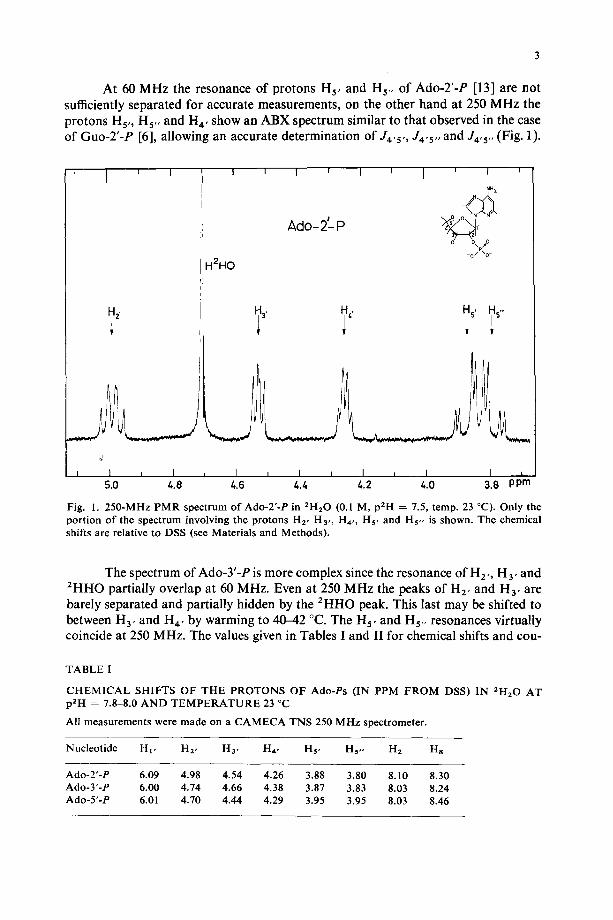
At 60 MHz the resonance of protons Hs, and Hs,, of Ado-2'-P [13] are not sufficiently separated for accurate measurements, on the other hand at 250 MHz the protons Hs,, Hs,, and H 4, show an ABX spectrum similar to that observed in the case of Guo-2'-P [6], allowing an accurate determination of J4,5,, J4'5,, and J4,5,, (Fig. 1).
a 2,
d
I 1 ~ I 5.0 4.8
I I I I I J I ' I f
NH 2 2g o O\p.mO
- 0 / "o- -.12HO
H3' it,'
I = I t I = I i I t 4.6 &.4 ~.2 &.O 3.8 ppm
Fig. 1. 250-MHz PMR spectrum of Ado-2'-P in 2H20 (0.1 M, p2H = 7.5, temp. 23 °C). Only the portion of the spectrum involving the protons H2, H3,, H4,, Hv and Hs,, is shown. The chemical shifts are relative to DSS (see Materials and Methods).
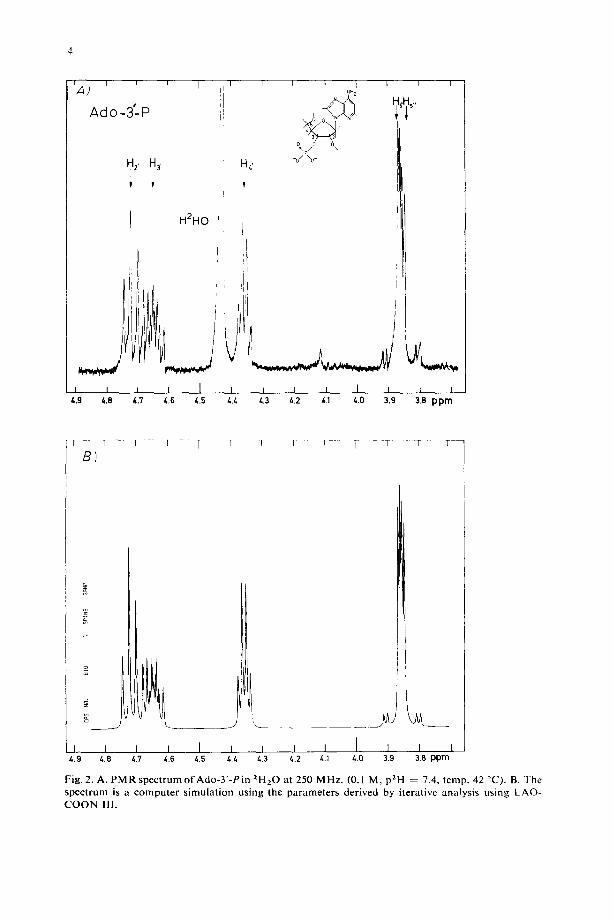
The spectrum of Ado-3'-P is more complex since the resonance of H2, , H 3, and 2HHO partially overlap at 60 MHz. Even at 250 MHz the peaks of n2, and Ha, are barely separated and partially hidden by the 2HHO peak. This last may be shifted to between H a, and H, , by warming to 40--42 °C. The H 5, and Hs,, resonances virtually coincide at 250 MHz. The values given in Tables I and II for chemical shifts and cou-
TABLE 1
CHEMICAL SHIFTS OF THE PROTONS OF Ado-Ps (IN PPM FROM DSS) IN 2H'20 AT p2H = 7.8-8.0 AND TEMPERATURE 23 °C
All measurements were made on a CAMECA TNS 250 MHz spectrometer.
Nucleotide Hi, H2, Ha, H4, Hv Hs,, H2 Hs
Ado-2'-P 6.09 4.98 4.54 4.26 3.88 3.80 8.10 8.30 Ado-3'-P 6.00 4.74 4.66 4.38 3.87 3.83 8.03 8.24 Ado-5'-P 6.01 4.70 4.44 4.29 3.95 3.95 8.03 8.46
~ A ) ' ' '
Ado -3'-- P
1 l
H2' H3'
i f
H 2 H O
; i ' !
'111 I
I I I I I /*.9 4 .8 4 3 4.6 /,.5 L,.Z,
i
i i I NH 2
% -o / o-
I I I
!
= i i 1 = = a 4.3 /,,2 /,.1 t..O 3.9 3.8 p p m
I ' B ) ' - - ~ - - T " 1 l l 1 i - T 1 ] •
J
j I I I I I I i I
4 .9 /-,.8 /-,.7 4.6 /,.5 t,./, 4.3 t~.2 I
/*.1 4.0 I I 1
3.9 3.8 p p m
Fig. 2. A. P M R s p e c t r u m o f A d o - 3 ' - P i n 2H20 at 250 MHz. (0.1 M, p2H = 7.4, temp. 42 C ) . B. The spec t rum is a compute r s imula t ion us ing the parameters derived by iterative analysis us ing LAO- C O O N I11.
5
TABLE II
COUPLING CONSTANTS (IN Hz) OF THE PROTONS OF Ado-Ps IN 2HzO AT p2H = 7.5-8.0
The coupling constants were determined either by spin-decoupling or by the use of the programme LAOCOON III. The maximal error in 0.2 Hz
N u c l e o t i d e J r - 2, J 2 ' - 3, J a , - 4, J4 , - 5, J4 , - 5', J r , - 5'" J x - pa
A d o - 2 ' - P 5 .7 5.3 3 .5 2 .7 3 .6 - - 1 2 . 2 6 .3 6 .0 b 5 .2 b 3 .4 b 6 .3 b
A d o - 3 ' - P 5 .6 5.3 3 .4 3 .0 3 .4 - - 1 2 . 1 7 .4
A d o - 5 " - P 5.3 5 .3 3 .2 3 .3 3 .3 - - 12 .0 4 .7
" X - - 2", 3 ' o r 5 ' 5 " .
b v a l u e s g i v e n i n re f . 11.
piing constants have been checked by computing the spectrum of Ado-3'-P with the LAOCOON III program [14]. Figs 2a and 2b show the experimental and simulated spectra of Ado-3'-P at 250 MHz.
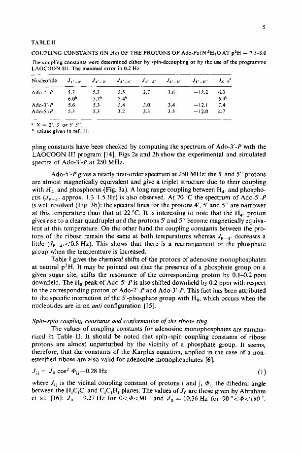
Ado-5'-P gives a nearly first-order spectrum at 250 MHz: the 5' and 5" protons are almost magnetically equivalent and give a triplet structure due to their coupling with H4, and phosphorus (Fig. 3a). A long range coupling between H4, and phospho- rus (JP-4, approx. 1.3-1.5 Hz) is also observed. At 70 °C the spectrum of Ado-5'-P is well resolved (Fig. 3b): the spectral lines for the protons 4', 5' and 5" are narrower at this temperature than that at 22 °C. It is interesting to note that the H 4 , proton gives rise to a clear quadruplet and the protons 5' and 5" become magnetically equiva- lent at this temperature. On the other hand the coupling constants between the pro- tons of the ribose remain the same at both temperatures whereas JP-4, decreases a little (Jp_4,<0.8 Hz). This shows that there is a rearrangement of the phosphate group when the temperature is increased.
Table I gives the chemical shifts of the protons of adenosine monophosphates at neutral p2H. It may be pointed out that the presence of a phosphate group on a given sugar site, shifts the resonance of the corresponding proton by 0.14).2 ppm downfield. The Ha peak of Ado-5'-P is also shifted downfield by 0.2 ppm with respect to the corresponding proton of Ado-2'-P and Ado-3'-P. This fact has been attributed to the specific interaction of the 5'-phosphate group with Ha, which occurs when the nucleotides are in an anti configuration [15].
Spin-spin coupling constants and conformation of the ribose ring The values of coupling constants for adenosine monophosphates are summa-
rized in Table II. It should be noted that spin-spin coupling constants of ribose protons are almost unperturbed by the vicinity of a phosphate group. It seems, therefore, that the constants of the Karplus equation, applied in the case of a non- esterified ribose are also valid for adenosine monophosphates [6].
Jij = Jo cos~ ~ij - 0 . 28 Hz (1)
where J~j is the vicinal coupling constant of protons i and j, ~ij the dihedral angle between the HiCiCj and C~CjHj planes. The values of Jo are those given by Abraham et al. [16]: Jo = 9.27 Hz for 0 < ~ < 9 0 ° and Jo----- 10.36 Hz for 90 °<4~<180 °
F I !i.a. b
4.9 4.8
A d o - 5 ' - P 0 .1 Id p H = 8 . 0
H 2, H 3. H~. Hs~H5.,
t ! I , L L I I 1,7 4.6 /~.5 /,/~ 4 3 z.2 /~.~ /~.0
I 3.9 ppm
i
B
A d o - 5 ' - P 0.1 M pH = 8 .0
7 0 °C
HDO
H 2, H 3. HI.
I t I I I l I ~.9 &.8 &.7 ~ 6 ~.5 4.~ 4.3
H5.. H5-
~ I I L I 4.2 t,.1 4.0 3.9 p p m
Fig . 3. P a r t i a l 250 M H z s p e c t r u m o f A d o - 5 ' - P in 2 H 2 0 , 0.1 M, p a l l ~ 8.0, A . A t t e m p . 23 °C. B. A t t e m p . 70 °C. T h e c h e m i c a l sh i f t s a r e r e l a t ive t o D S S a n d c o r r e c t e d f o r b u l k su scep t i b i l i t y .
T A B L E 11|
D I H E D R A L A N G L E S C A L C U L A T E D F R O M K A R P L U S E Q U A T I O N ( S E E T E X T )
Nucleotide ~1,- 2' ~ )2 ' - - 3 ' 1 ~ 3 ' - - 4 '
A d o - 2 ' - P 139 ° 39 ° 127 ° A d o - 3 ' - P 138 ° 39 ° 126.5 ° A d o - 5 " - P 138 ° 39 ' 126 °
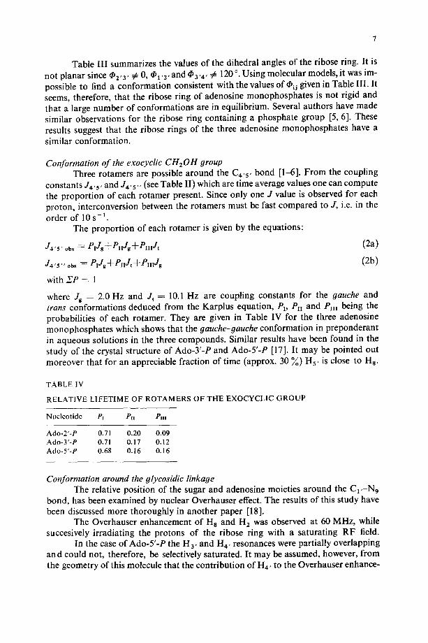
Table I l l summarizes the values of the dihedral angles of the ribose ring. It is not planar since ¢~2'3' ~ 0, ~1,2, and ~a'#' ¢: 120 °. Using molecular models, it was im- possible to find a conformation consistent with the values of ~ij given in Table III. It seems, therefore, that the ribose ring of adenosine monophosphates is not rigid and that a large number of conformations are in equilibrium. Several authors have made similar observations for the ribose ring containing a phosphate group [5, 6]. These results suggest that the ribose rings of the three adenosine monophosphates have a similar conformation.
Conformation of the exocyclic CH20H group Three rotamers are possible around the C4,5, bond [1-6]. From the coupling
constants J4'5' and J4's', (see Table II) which are time average values one can compute the proportion of each rotamer present. Since only one J value is observed for each proton, interconversion between the rotamers must be fast compared to J, i.e. in the order of 10 s - I .
The proportion of each rotamer is given by the equations:
J4'5' obs : PiJg-~-eIIJg-~-PIlIJt (2a)
: e,J + e . J t + e,,,J, (2b)
with XP = 1
where Jg : 2.0 Hz and J t : 10.1 Hz are coupling constants for the gauche and trans conformations deduced from the Karplus equation, P~, Pn and Pm being the probabilities of each rotamer. They are given in Table IV for the three adenosine monophosphates which shows that the gauche-gauche conformation in preponderant in aqueous solutions in the three compounds. Similar results have been found in the study of the crystal structure of Ado-3'-P and Ado-5'-P [17]. It may be pointed out moreover that for an appreciable fraction of time (approx. 30 ~ ) H5, is close to Ha.
TABLE IV
RELATIVE LIFETIME OF ROTAMERS OF THE EXOCYCLIC GROUP
Nucleotide Px Pn PIII
Ado-2"-P 0.71 0.20 0.09 Ado-3'-P 0.71 0.17 0.12 Ado-5"-P 0.68 0.16 0.16
Conformation around the glycosidic linkage The relative position of the sugar and adenosine moieties around the C1,-N9
bond, has been examined by nuclear Overhauser effect. The results of this study have been discussed more thoroughly in another paper [18].
The Overhauser enhancement of H8 and H2 was observed at 60 MHz, while succesively irradiating the protons of the ribose ring with a saturating RF field.
In the case of Ado-5'-P the H a, and H4, resonances were partially overlapping an d could not, therefore, be selectively saturated. It may be assumed, however, from the geometry of this molecule that the contribution of H4, to the Overhauser enhance-
ment of H 8 and H 2 r e s o n a n c e s is negligible as actually observed in the case of Ado-Y- and -3'-P.
The Overhauser enhancement is inversely proportional to the sixth power o1" the internuclear distances, therefore, allowing their relative value to be determined.
~,ij/bik = ( r i j / r i k ) - (3 )
where ~ is the enhancement of the Hi signal while irradiating Hj or Hk. The absolute distance may, however, be estimated from the following relation
[7, 16]:
r 0 0.1. 10_i i (4)
where rc is the reorientation correlation time of the whole molecule and Pi I /T , is the relaxation rate of proton i.
The different constants introduced in Eqn 4 are related to the mean molecular radius of adenosine monophosphates and to the viscosity of water at room tempera- ture.
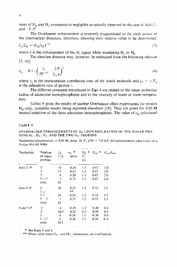
Tables V gives the results of nuclear Overhauser effect experiments for proton H 8 only, complete results being reported elsewhere [18]. They are given for 0.05 M neutral solutions of the three adenosine monophosphates. The value of r8j calculated
T A B L E V
O V E R H A U S E R E N H A N C E M E N T S OF Ha U P O N S A T U R A T I O N OF T H E S U G A R PRO- TONS HI ' , HE,, H3, A N D T H E TWO Hs, P R O T O N S
Nucleot ide concentra t ion = 0.05 M, temp. 28 °C, p2H - 7.0-8.0. All measurements were made on a Varian HA 60 MHz.
Nucleot ide Posit ion 58i r8~ * Ha * P~y, * P~n/Pa. t~ of sugar (%) (nm) Ti protons (s)
Ado-2 ' -P
Ado-3 ' -P
A do-5 ' -P 1' 2" 3" 5"+5" total
* See Eqns 3 and 4.
1" 16 0.24 1.3 0.67 2.0 2" 17 0.23 1.3 0.67 2.0 3" 6 0.28 1.3 0.67 2.0 5" k5" 3 0.32 1.3 0.67 2.0 total 42
I' 20 0.22 1.2 0.72 2.5 2 ' ~ * * 3' f 24 0.25 1.2 0.72 2.5 5 ' t 5" 1 0.35 1.2 0.72 2.5 total 45
4 0.29 1.5 0.30 0.4 20.5 0.22 1.5 0.30 0.4 9 0.26 1.5 0.30 0.4 6 0.28 1.5 0.30 0.4
39.5
** Mean value since H2,, and Ha,, resonances are overlapping.
f rom Eqn 4 are clearly inconsistent with any rigid conformation given by a molecular model [18]. It may be assumed, therefore, as in the case of guanosine monophosphates [9 ] that these distances are time averaged by rotation of the base around the glycosidic linkage and the torsional motion of the ribose ring. However, the rotation of the ade- nosine monophosphates around the CI , -N 9 bond is not entirely free and for each of the adenosine monophosphates a syn or anti character of the relative orientation of the adenine and sugar moieties may be qualitatively defined by the following equation which has been explained in a previous paper [9]
P s y n ~ l - - P a n t i = 3~a,, (5) 3~81,q-~s2,-~-~S3,
where Psy. and P,.ti are the proportions of time spent in the syn or anti conformation. In spite of the qualitative nature of the present description it is clear that both
syn and anti configurations occur, and that Ado-5'-P is appreciably more anti than the other Ado-P's. (Table V).
Effect of intermolecular association on chemical shifts The proton chemical shifts of adenosine monophosphates are dependent on
concentration and temperature. These effects are most likely related to intermolecular association [15]. We have studied the influence of these parameters on the PMR spectra of the three adenosines.
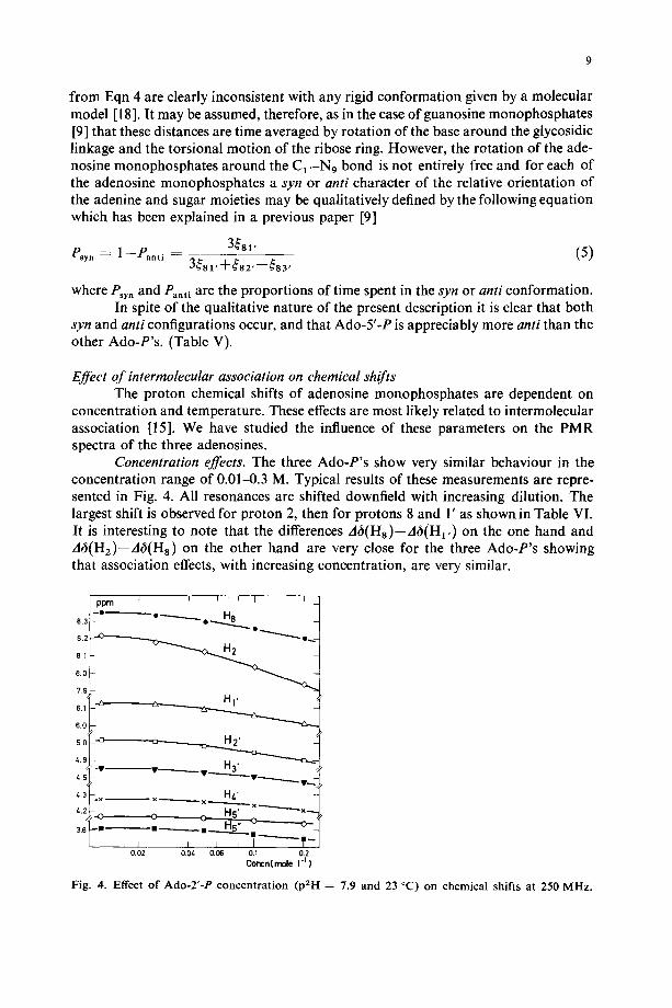
Concentration effects. The three Ado-P's show very similar behaviour in the concentration range of 0.01-0.3 M. Typical results of these measurements are repre- sented in Fig. 4. All resonances are shifted downfield with increasing dilution. The largest shift is observed for proton 2, then for protons 8 and 1' as shown in Table VI. It is interesting to note that the differences A6(Hs)--A6(H1, ) on the one hand and A~(H2)--A~(Hs) on the other hand are very close for the three Ado-P's showing that association effects, with increasing concentration, are very similar.
J f I I I I ~ p r n
8.3 ' ° ~ O ~ O ~ . . . . . . o ~
8.0 i
7.9
6.0
s.o "~ H2'
&5
/..3 -x x ._...____ x H4' X ~ x ~
i 3 8 ~ m i l ~ m _ _ _ _ ~ . ~ . _ - - -
T-------- I i J I ~ | ~ 0.02 O.0t, 0.06 0.1 0.2
C o n c h ( m o l e 1-1 )
Fig. 4. Effect of Ado-2'-P concentration (p2H = 7.9 and 23 °C) on chemical shifts at 250 MHz.
10
T A B L E VI
C O N C E N T R A T I O N E F F E C T S ON C H E M I C A L S HIFTS OF T H E P R O T O N OF Ado-P ' s (IN PPM F R O M DSS) IN 2H20 AT pZHO 7.9 A N D T E M P E R A T U R E 23 C
Nucleot ide Concentration Chemical shifts (ppm from DSS)* ( m o l e . l - t )
H~, H 2, H 3' H4, Hs, Ha,,
Ado-2 ' -P 0.01 6.126 5.014 4.450 4.270 3.870 3.806 0.20 6.010 4.905 4.476 4.206 3.838 3.750 At) 0.116 0.109 0.074 0.064 0.032 0.056
A d o - Y - P 0.01 6.094 4.798 4.700 4.418 3.890 3.870 0.20 5.910 4.672 4.610 4.334 3.830 3.794 At) 0.184 0.126 0.090 0.084 0.060 0.076
Ado-5 ' -P 0.01 6.115 4.761 4.487 4.331 3.971 0.20 5.925 4.635 4.403 4.259 3.925 At) 0.190 0.126 0.084 0.072 0.046
dt ) (Ha) - dt)(H l ') ,It)(H2) d t ) (Ha) Ado-2 ' -P 0.024 p p m 0.140 ppm Ado-3 ' -P 0.034 ppm 0.138 ppm Ado-5"-P 0.024 ppm 0.130 ppm
* See Materials and Methods.
H2 H8
8.214 8.342 7.934 8.202 0.280 0.140
8.230 8.358 7.874 8,140 0.356 0.218
8.228 8.594 7.885 8.381 0.343 0.213
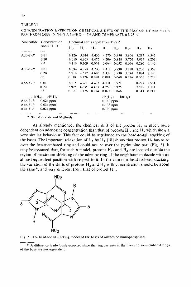
As already mentioned, the chemical shift of the proton H 2 is much more dependent on adenosine concentration than that of protons H'I and H 8 which show a very similar behaviour. This fact could be attributed to the head-to-tail stacking o f the bases. The important relaxation of H 2 by H a [18] shows that proton H2 has to be over the five-membered ring and could not be over the pyrimidine part (Fig. 5). It may be assumed that, for such a model, protons H1, and H8 are located outside the region of maximum shielding of the adenine ring of the neighbour molecule with an almost equivalent position with respect to it. In the case of a head-to-head stacking, the variation of the shifts of protons H z and H a with concentration should be about the same*, and very different from that of proton H , .
ND 2
8 8
ND 2
Fig. 5. The head-to-tail stacking model o f the bases o f adenosine monophosphates .
* A difference is obviously expected since the ring currents in the five- and six-membered rings o f the base are not equivalent.
l l
In the case of adenosine 5'-monophosphate in neutral solutions, protons 5' and 5" are magnetically equivalent below a concentration of 0.1 M, becoming slightly inequivalent as the concentration is raised, showing that the reorientation of the exo- cyclic group is also hindered by the stacking of molecules.
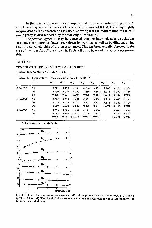
Temperature effect. It may be expected that the intermolecular associations of adenosine monophosphates break down by warming as well as by dilution, giving rise to a downfield shift of proton resonances. This has been actually observed in the case of the three Ado-P's as shown in Table VII and Fig. 6 and this variation is revers- ible.
TABLE VII
T E M P E R A T U R E EFFECTS ON CHEMICAL SHIFTS
Nucleotide concentration 0.1 M, p2H 8.0.
Nucleotide Temperature Chemical shifts (ppm from DSS)*
(o C) HI, H2, Ha, H4, Hs, Hz" H2 H8
Ado-2'-P 25 6.092 4.976 4.536 4.264 70 6.130 5.010 4.530 4.236 A6 +0.038 +0.034 --0.006 0.028
Ado-3'-P 23 6.002 4.738 4.658 4.382 70 6.052 4.758 4.700 4.354 A6 +0.050 +0.020 +0.042 --0.028
Ado-5'-P 23 6.009 4.699 4.439 4.293 70 6.088 4.736 4.480 4.320 .d6 +0.079 +0.037 +0.041 +0.027
3.878 3.800 8.100 8.304 3.864 3.786 8.252 8.334 0.014 --0.014 +0.152 +0.030
3.874 3.834 8.032 8.244 3.874 3.838 8.230 8.300 0.0 0.004 +0.198 0.056
3.950 8.029 8.463 3.992 8.200 8.512
+0.042 +0.171 +0.049
* See Materials and Methods.
ppm H a
8.1
~2 ~ . . ~ _ . ~ H'" 6.1 ~ - ' ~
~' H2, 5.0 ~ _ ~ '~
H 3 '
H z '
3 . 9 , - H 5 •
3.8 • • • • H S " . . . . . . • E m - - -
30 40 50 60 70 T~um ( * C )
Fig. 6. Effect of temperature on the chemical shifts of the protons of Ado-2'-P in 2H20 at 250 MHz (p2H = 7.9, 0.1 M). The chemical shifts are relative to DSS and corrected for bulk susceptibility (see Materials and Methods).
12
In the case of 0.1 M solutions, it has been found that between 22 and 70 C, the chemical shifts vs temperature plots of protons H8, He, H~,, are virtually superim- posable for the three adenosine monophosphates . Moreover the difference ~H8 6H2 approaches a limiting value of 0.080 ~ 0.005 ppm for Ado-2 ' -P or Ado-3'-P, and of 0.312 ppm for Ado-5 ' -P, as the temperature is raised. This last value, larger than the two others, seems to be due to the phosphate group in the 5 '-posit ion which induces an incremental shift of H8 downfield as mentiooed previously. It may be assumed more- over that the dissociation of molecular aggregates is complete at 70 ~C, since in any case, a l imiting value is obtained for ~Hs 6H2.
At room temperature (22-25 °C) for a given concentrat ion of Ado-P, less than 0.1 M, one can always find a spectrum, the relative chemical shifts of which ( taking into account the dilution, etc. ) are identical to that given by a spectrum record- ed for a 0.l M solution between room temperature and 70 °C. It must be pointed out that the chemical shift of H2 and H 8 should not depend on the relative orientat ion, syn or anti, of base with respect to the sugar moiety, since the ring current is the same in both cases. The chemical shift variat ion with temperature mainly reflects the stack- ing effects and the difference 6H8 6H2 may, therefore, be considered as a criterion of the degree of aggregation. Thus, in the case of Ado-2 ' -P for example, ,~H 8 hH z approaches a l imiting value of 0.075 ppm at 70 °C for a 0.1 M solution, whereas a 0.01 M at 23 "C gives ~5H8 6H 2 0.128 ppm showing that, at room temperature, the dissociation of molecular aggregates is far from being complete, even at high di lut ion.
ACK NOWLEDG E MENTS
We wish to thank Dr J. Thiery (Saclay) for the s imulat ion of the Ado-3 ' -P spectrum and for the use of the PDP 12 computer . We also with to thank Dr M. Gu6ron (Ecole Polytechnique, Paris) and Dr W. Guschlbauer (Saclay) for their helpful discussions and the critical appraisal of the manuscript . We are also indebted Dr Gu6ron for providing the nuclear Overhauser effect equipment .
REFERENCES
1 Hruska, F. E., Grey, A. A. and Smith, I. C. P. (1970) J. Am. Chem. Soc. 92, 4088 4094 2 Blackburn, B. J., Grey, A. A., Smith, 1. C. P. and Hruska, F. E. (1970) Can. J. Chem. 48, 2866-
2870 3 Dugas, H., Blackburn, B. J., Robins, R. K., Deslauriers, R. and Smith, 1. C. P. (1971) J. Am.
Chem. Soc. 93, 3468 3470 4 Grey, A. A., Smith, 1. C. P. and Hruska, F. E. (1971) J. Am. Chem. Soc. 93, 1765-1769 5 Schleich, T., Blackburn, B. J., Lapper, R. D. and Smith, 1. C. P. (1972) Biochemistry 11, 137-145 6 Son, T.-D., Thiery, J., Guschlbauer, W. and Dunand, J. J. (1972) Biochim. Biophys. Acta 281,
289 298 7 Hart, P. and Davis, J. (1971) J. Am. Chem. Soc. 93, 753-760 8 Schirmer, R., Davis, J., Noggle, J. and Hart, P. (1972) J. Am. Chem Soc. 94, 2561-2572 9 Son, T.-D., Guschlbauer, W. and Gueron, M. (1972) J. Am. Chem. Soc. 94, 7903-7911
10 Torrey, H. C. (1956) Phys. Rev. 104, 563-565 I 1 Jardetzky, C. D. and Jardetzky, O. (1960) J. Am. Chem. Soc. 82, 222 229 12 T'So, P. O. P., Kondo, N. S., Schweizer, M. P. and Hollis, D. P. (1969) Biochemistry 8, 997-1029 13 Jardetzky, C. D. (1962) J. Am. Chem. Soc. 84, 62-66 14 Castellano, S. and Bothner-By, A. A. (1964) J. Chem. Phys. 41, 3863-3869
13
15 Schweizer, M. P., Broom, A. D., T'So, P. O. P. aild Hollis, D. P. (1968) J. Am. Chem. Soc. 90, 1042-1055
16 Abraham: R. J., Hall, L. D., Hough, L. and Mc Lauchlan, K. A. (1962) J. Chem. Soc. 3699 17 Sundaralingam, M. (1966) Acta Crystallog. 21,495-506 18 Gueron, M., Chachaty, C. and Son, T.-D. Proceeding of the International Conference on Magne-
tic Resonance in Biological System ,New York, (1972), in the press


![Problems with a conformation assignment of aryl-substituted resorc[4]arenes](https://static.fdokumen.com/doc/165x107/6324d12685efe380f30661c8/problems-with-a-conformation-assignment-of-aryl-substituted-resorc4arenes.jpg)