Design of a water soluble 1,8-naphthalimide/3-hydroxy-4-pyridinone conjugate: Investigation of its...
11
Design of a water soluble 1,8-naphthalimide/3-hydroxy-4-pyridinone conjugate: Investigation of its spectroscopic properties at variable pH and in the presence of Fe 3þ , Cu 2þ and Zn 2þ Tânia Moniz a , Carla Queirós a , Rita Ferreira a , Andreia Leite a , Paula Gameiro a , Ana M.G. Silva a, * , Maria Rangel b, * a REQUIMTE, Departamento de Química e Bioquímica, Faculdade de Ciências, Universidade do Porto, 4169-007 Porto, Portugal b REQUIMTE, Instituto de Ciências Biomédicas Abel Salazar, Universidade do Porto, 4099-003 Porto, Portugal article info Article history: Received 9 January 2013 Received in revised form 22 February 2013 Accepted 25 February 2013 Available online 13 March 2013 Keywords: 4-Amino-1,8-naphthalimides 3-Hydroxy-4-pyridinones Microwave-assisted synthesis Fluorescence properties Fluorescent probes Quenching effect abstract The synthesis and sensing properties of a new fluorescent probe designed to have a 4-amino-1,8- naphthalimide fluorescent platform functionalized with a 3-hydroxy-4-pyridinone bidentate chelating unit at the 4-position and a terminal aliphatic dimethylamino group at the imide site, are reported. The absorption and fluorescence properties of the ligand were investigated in DMSO and in aqueous solution at variable pH and in the presence of increasing concentration of Fe 3þ , Cu 2þ and Zn 2þ . Analysis of the UVeVis spectra at variable pH allowed the determination of three pK a values (pK a1 ¼ 3.19, pK a2 ¼ 8.38, pK a3 ¼ 9.95) and establishment of the corresponding speciation diagram. Fluorescence spectra obtained in the same conditions show that the fluorescence intensity of the probe decreases with increasing pH and are off above pH 9 as a result of photo-induced electron transfer arising from the aliphatic dimethylamino group. Under physiological pH conditions, the probe shows an ab- sorption band centred at 439 nm and emits in the green at l ¼ 536 nm. Analysis of UVeVis and EPR spectra of the ligand in the presence of Fe 3þ and Cu 2þ is consistent with the formation of the corresponding metal ion complexes. The fluorescence intensity of the ligand is quenched in the presence of variable concentrations of Fe 3þ , Cu 2þ and Zn 2þ and under physiological pH conditions the fluorescence of the probe is ca 92%, 88% and 91% quenched in the presence of Fe 3þ , Cu 2þ and Zn 2þ respectively. Ó 2013 Elsevier Ltd. All rights reserved. 1. Introduction Since the early reports by Tsien and co-workers regarding fluorescent probes for calcium [1,2], the design of highly efficient fluorescent probes for sensing and monitoring various chemical species has received an increasing interest from the scientific and industrial communities. Indeed, many fluorescent probes have been developed for different applications including probes sensi- tive to pH [3], to metal ions [4], to monitor enzymatic activity [5], reactive oxygen and nitrogen species (ROS and RNS) [6] and many other analytes [7]. The most general design concept of fluorescent metal ion probes involves the use of the photoinduced electron transfer (PET) model based on the covalent linkage of a single or branched chelating architecture and a fluorophore unit. The covalent linkage which separates the two units is typically a short aliphatic spacer that minimizes any ground-state interactions. In these probes, the interaction with the analyte will cause significant changes in the emission spectra accompanied by no, or minor, changes in the absorption spectra of the probes [8]. Originally detailed by de Silva et al. [9a,b] and Czarnik et al. [9c,d], this process has been widely used in the construction of many fluorescent probes, with special emphasis for 4-amino-1,8- naphthalimide based fluorescent probes [10]. 4-Amino-1,8-naphthalimides are fluorophores that exhibit bands with an ICT (intramolecular charge transfer) character caused by the electronic conjugation of the electron donating amine and the elec- tron withdrawing imide (Fig. 1) [11]. The ICT character gives rise to a large excited-state dipole and broad absorption and emission bands typically centred at ca 450 and 550 nm, respectively, when recorded * Corresponding authors. E-mail addresses: [email protected], [email protected] (A.M.G. Silva), [email protected] (M. Rangel). Contents lists available at SciVerse ScienceDirect Dyes and Pigments journal homepage: www.elsevier.com/locate/dyepig 0143-7208/$ e see front matter Ó 2013 Elsevier Ltd. All rights reserved. http://dx.doi.org/10.1016/j.dyepig.2013.02.020 Dyes and Pigments 98 (2013) 201e211
-
Upload
independent -
Category
Documents
-
view
0 -
download
0
Transcript of Design of a water soluble 1,8-naphthalimide/3-hydroxy-4-pyridinone conjugate: Investigation of its...

at SciVerse ScienceDirect
Dyes and Pigments 98 (2013) 201e211
Contents lists available
Dyes and Pigments
journal homepage: www.elsevier .com/locate/dyepig
Design of a water soluble 1,8-naphthalimide/3-hydroxy-4-pyridinoneconjugate: Investigation of its spectroscopic properties at variable pHand in the presence of Fe3þ, Cu2þ and Zn2þ
Tânia Moniz a, Carla Queirós a, Rita Ferreira a, Andreia Leite a, Paula Gameiro a,Ana M.G. Silva a,*, Maria Rangel b,*aREQUIMTE, Departamento de Química e Bioquímica, Faculdade de Ciências, Universidade do Porto, 4169-007 Porto, PortugalbREQUIMTE, Instituto de Ciências Biomédicas Abel Salazar, Universidade do Porto, 4099-003 Porto, Portugal
a r t i c l e i n f o
Article history:Received 9 January 2013Received in revised form22 February 2013Accepted 25 February 2013Available online 13 March 2013
Keywords:4-Amino-1,8-naphthalimides3-Hydroxy-4-pyridinonesMicrowave-assisted synthesisFluorescence propertiesFluorescent probesQuenching effect
* Corresponding authors.E-mail addresses: [email protected], ana74silva
[email protected] (M. Rangel).
0143-7208/$ e see front matter � 2013 Elsevier Ltd.http://dx.doi.org/10.1016/j.dyepig.2013.02.020
a b s t r a c t
The synthesis and sensing properties of a new fluorescent probe designed to have a 4-amino-1,8-naphthalimide fluorescent platform functionalized with a 3-hydroxy-4-pyridinone bidentate chelatingunit at the 4-position and a terminal aliphatic dimethylamino group at the imide site, are reported. Theabsorption and fluorescence properties of the ligand were investigated in DMSO and in aqueous solutionat variable pH and in the presence of increasing concentration of Fe3þ, Cu2þ and Zn2þ.
Analysis of the UVeVis spectra at variable pH allowed the determination of three pKa values(pKa1 ¼ 3.19, pKa2 ¼ 8.38, pKa3 ¼ 9.95) and establishment of the corresponding speciation diagram.Fluorescence spectra obtained in the same conditions show that the fluorescence intensity of the probedecreases with increasing pH and are off above pH 9 as a result of photo-induced electron transfer arisingfrom the aliphatic dimethylamino group. Under physiological pH conditions, the probe shows an ab-sorption band centred at 439 nm and emits in the green at l ¼ 536 nm.
Analysis of UVeVis and EPR spectra of the ligand in the presence of Fe3þ and Cu2þ is consistent withthe formation of the corresponding metal ion complexes. The fluorescence intensity of the ligand isquenched in the presence of variable concentrations of Fe3þ, Cu2þ and Zn2þ and under physiological pHconditions the fluorescence of the probe is ca 92%, 88% and 91% quenched in the presence of Fe3þ, Cu2þ
and Zn2þ respectively.� 2013 Elsevier Ltd. All rights reserved.
1. Introduction
Since the early reports by Tsien and co-workers regardingfluorescent probes for calcium [1,2], the design of highly efficientfluorescent probes for sensing and monitoring various chemicalspecies has received an increasing interest from the scientific andindustrial communities. Indeed, many fluorescent probes havebeen developed for different applications including probes sensi-tive to pH [3], to metal ions [4], to monitor enzymatic activity [5],reactive oxygen and nitrogen species (ROS and RNS) [6] and manyother analytes [7].
Themost general design concept of fluorescent metal ion probesinvolves the use of the photoinduced electron transfer (PET) model
@gmail.com (A.M.G. Silva),
All rights reserved.
based on the covalent linkage of a single or branched chelatingarchitecture and a fluorophore unit. The covalent linkage whichseparates the two units is typically a short aliphatic spacer thatminimizes any ground-state interactions. In these probes, theinteraction with the analyte will cause significant changes in theemission spectra accompanied by no, or minor, changes in theabsorption spectra of the probes [8].
Originally detailed by de Silva et al. [9a,b] and Czarnik et al.[9c,d], this process has been widely used in the construction ofmany fluorescent probes, with special emphasis for 4-amino-1,8-naphthalimide based fluorescent probes [10].
4-Amino-1,8-naphthalimides are fluorophores that exhibit bandswith an ICT (intramolecular charge transfer) character caused by theelectronic conjugation of the electron donating amine and the elec-tron withdrawing imide (Fig. 1) [11]. The ICT character gives rise to alarge excited-state dipole and broad absorption and emission bandstypically centred at ca 450 and 550 nm, respectively, when recorded
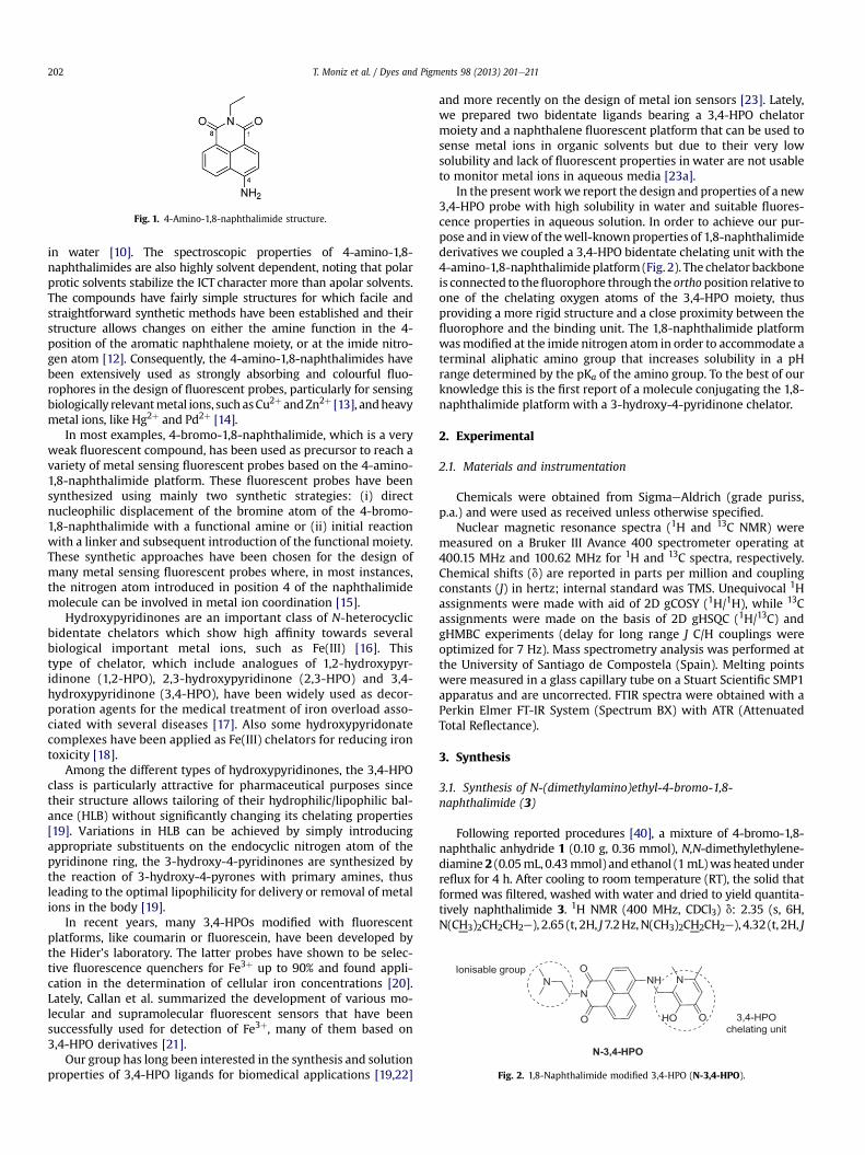
Fig. 1. 4-Amino-1,8-naphthalimide structure.
Fig. 2. 1,8-Naphthalimide modified 3,4-HPO (N-3,4-HPO).
T. Moniz et al. / Dyes and Pigments 98 (2013) 201e211202
in water [10]. The spectroscopic properties of 4-amino-1,8-naphthalimides are also highly solvent dependent, noting that polarprotic solvents stabilize the ICT character more than apolar solvents.The compounds have fairly simple structures for which facile andstraightforward synthetic methods have been established and theirstructure allows changes on either the amine function in the 4-position of the aromatic naphthalene moiety, or at the imide nitro-gen atom [12]. Consequently, the 4-amino-1,8-naphthalimides havebeen extensively used as strongly absorbing and colourful fluo-rophores in the design of fluorescent probes, particularly for sensingbiologically relevantmetal ions, suchasCu2þ andZn2þ [13], andheavymetal ions, like Hg2þ and Pd2þ [14].
In most examples, 4-bromo-1,8-naphthalimide, which is a veryweak fluorescent compound, has been used as precursor to reach avariety of metal sensing fluorescent probes based on the 4-amino-1,8-naphthalimide platform. These fluorescent probes have beensynthesized using mainly two synthetic strategies: (i) directnucleophilic displacement of the bromine atom of the 4-bromo-1,8-naphthalimide with a functional amine or (ii) initial reactionwith a linker and subsequent introduction of the functional moiety.These synthetic approaches have been chosen for the design ofmany metal sensing fluorescent probes where, in most instances,the nitrogen atom introduced in position 4 of the naphthalimidemolecule can be involved in metal ion coordination [15].
Hydroxypyridinones are an important class of N-heterocyclicbidentate chelators which show high affinity towards severalbiological important metal ions, such as Fe(III) [16]. Thistype of chelator, which include analogues of 1,2-hydroxypyr-idinone (1,2-HPO), 2,3-hydroxypyridinone (2,3-HPO) and 3,4-hydroxypyridinone (3,4-HPO), have been widely used as decor-poration agents for the medical treatment of iron overload asso-ciated with several diseases [17]. Also some hydroxypyridonatecomplexes have been applied as Fe(III) chelators for reducing irontoxicity [18].
Among the different types of hydroxypyridinones, the 3,4-HPOclass is particularly attractive for pharmaceutical purposes sincetheir structure allows tailoring of their hydrophilic/lipophilic bal-ance (HLB) without significantly changing its chelating properties[19]. Variations in HLB can be achieved by simply introducingappropriate substituents on the endocyclic nitrogen atom of thepyridinone ring, the 3-hydroxy-4-pyridinones are synthesized bythe reaction of 3-hydroxy-4-pyrones with primary amines, thusleading to the optimal lipophilicity for delivery or removal of metalions in the body [19].
In recent years, many 3,4-HPOs modified with fluorescentplatforms, like coumarin or fluorescein, have been developed bythe Hider’s laboratory. The latter probes have shown to be selec-tive fluorescence quenchers for Fe3þ up to 90% and found appli-cation in the determination of cellular iron concentrations [20].Lately, Callan et al. summarized the development of various mo-lecular and supramolecular fluorescent sensors that have beensuccessfully used for detection of Fe3þ, many of them based on3,4-HPO derivatives [21].
Our group has long been interested in the synthesis and solutionproperties of 3,4-HPO ligands for biomedical applications [19,22]
and more recently on the design of metal ion sensors [23]. Lately,we prepared two bidentate ligands bearing a 3,4-HPO chelatormoiety and a naphthalene fluorescent platform that can be used tosense metal ions in organic solvents but due to their very lowsolubility and lack of fluorescent properties in water are not usableto monitor metal ions in aqueous media [23a].
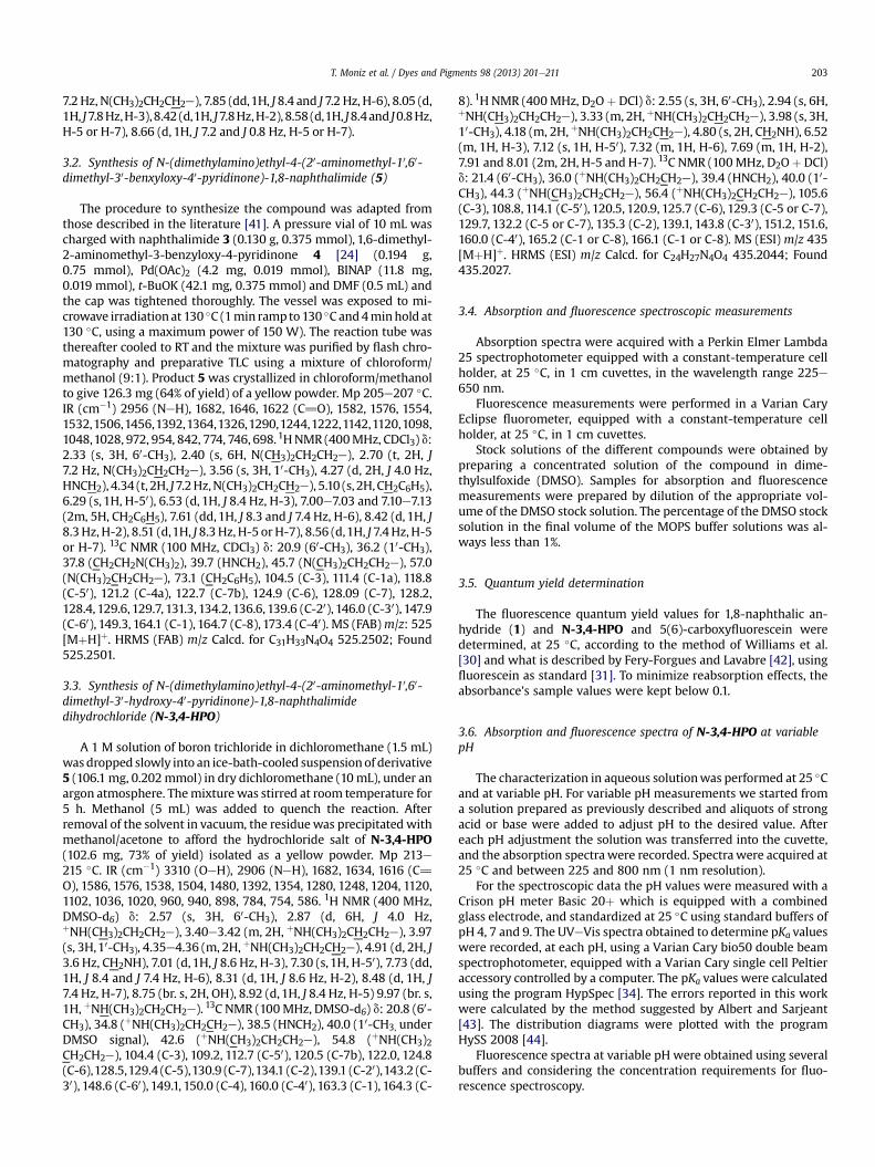
In the present workwe report the design and properties of a new3,4-HPO probe with high solubility in water and suitable fluores-cence properties in aqueous solution. In order to achieve our pur-pose and in view of thewell-knownproperties of 1,8-naphthalimidederivatives we coupled a 3,4-HPO bidentate chelating unit with the4-amino-1,8-naphthalimide platform (Fig. 2). The chelator backboneis connected to thefluorophore through the orthoposition relative toone of the chelating oxygen atoms of the 3,4-HPO moiety, thusproviding a more rigid structure and a close proximity between thefluorophore and the binding unit. The 1,8-naphthalimide platformwasmodified at the imide nitrogen atom in order to accommodate aterminal aliphatic amino group that increases solubility in a pHrange determined by the pKa of the amino group. To the best of ourknowledge this is the first report of a molecule conjugating the 1,8-naphthalimide platform with a 3-hydroxy-4-pyridinone chelator.
2. Experimental
2.1. Materials and instrumentation
Chemicals were obtained from SigmaeAldrich (grade puriss,p.a.) and were used as received unless otherwise specified.
Nuclear magnetic resonance spectra (1H and 13C NMR) weremeasured on a Bruker III Avance 400 spectrometer operating at400.15 MHz and 100.62 MHz for 1H and 13C spectra, respectively.Chemical shifts (d) are reported in parts per million and couplingconstants (J) in hertz; internal standard was TMS. Unequivocal 1Hassignments were made with aid of 2D gCOSY (1H/1H), while 13Cassignments were made on the basis of 2D gHSQC (1H/13C) andgHMBC experiments (delay for long range J C/H couplings wereoptimized for 7 Hz). Mass spectrometry analysis was performed atthe University of Santiago de Compostela (Spain). Melting pointswere measured in a glass capillary tube on a Stuart Scientific SMP1apparatus and are uncorrected. FTIR spectra were obtained with aPerkin Elmer FT-IR System (Spectrum BX) with ATR (AttenuatedTotal Reflectance).
3. Synthesis
3.1. Synthesis of N-(dimethylamino)ethyl-4-bromo-1,8-naphthalimide (3)
Following reported procedures [40], a mixture of 4-bromo-1,8-naphthalic anhydride 1 (0.10 g, 0.36 mmol), N,N-dimethylethylene-diamine 2 (0.05mL, 0.43mmol) and ethanol (1mL)was heated underreflux for 4 h. After cooling to room temperature (RT), the solid thatformed was filtered, washed with water and dried to yield quantita-tively naphthalimide 3. 1H NMR (400 MHz, CDCl3) d: 2.35 (s, 6H,N(CH3)2CH2CH2e), 2.65 (t, 2H, J7.2Hz,N(CH3)2CH2CH2e), 4.32 (t, 2H, J
T. Moniz et al. / Dyes and Pigments 98 (2013) 201e211 203
7.2 Hz, N(CH3)2CH2CH2e), 7.85 (dd,1H, J 8.4 and J 7.2 Hz, H-6), 8.05 (d,1H, J7.8Hz,H-3),8.42 (d,1H, J7.8Hz,H-2), 8.58 (d,1H, J8.4and J0.8Hz,H-5 or H-7), 8.66 (d, 1H, J 7.2 and J 0.8 Hz, H-5 or H-7).
3.2. Synthesis of N-(dimethylamino)ethyl-4-(20-aminomethyl-10,60-dimethyl-30-benxyloxy-40-pyridinone)-1,8-naphthalimide (5)
The procedure to synthesize the compound was adapted fromthose described in the literature [41]. A pressure vial of 10 mL wascharged with naphthalimide 3 (0.130 g, 0.375 mmol), 1,6-dimethyl-2-aminomethyl-3-benzyloxy-4-pyridinone 4 [24] (0.194 g,0.75 mmol), Pd(OAc)2 (4.2 mg, 0.019 mmol), BINAP (11.8 mg,0.019 mmol), t-BuOK (42.1 mg, 0.375 mmol) and DMF (0.5 mL) andthe cap was tightened thoroughly. The vessel was exposed to mi-crowave irradiationat 130 �C (1min rampto130 �C and4minhold at130 �C, using a maximum power of 150 W). The reaction tube wasthereafter cooled to RT and the mixture was purified by flash chro-matography and preparative TLC using a mixture of chloroform/methanol (9:1). Product 5 was crystallized in chloroform/methanolto give 126.3 mg (64% of yield) of a yellow powder. Mp 205e207 �C.IR (cm�1) 2956 (NeH), 1682, 1646, 1622 (C]O), 1582, 1576, 1554,1532,1506,1456,1392,1364,1326,1290,1244,1222,1142,1120,1098,1048,1028, 972, 954, 842, 774, 746, 698. 1HNMR (400MHz, CDCl3) d:2.33 (s, 3H, 60-CH3), 2.40 (s, 6H, N(CH3)2CH2CH2e), 2.70 (t, 2H, J7.2 Hz, N(CH3)2CH2CH2e), 3.56 (s, 3H, 10-CH3), 4.27 (d, 2H, J 4.0 Hz,HNCH2), 4.34 (t, 2H, J7.2Hz,N(CH3)2CH2CH2e), 5.10 (s, 2H,CH2C6H5),6.29 (s, 1H, H-50), 6.53 (d, 1H, J 8.4 Hz, H-3), 7.00e7.03 and 7.10e7.13(2m, 5H, CH2C6H5), 7.61 (dd,1H, J 8.3 and J 7.4 Hz, H-6), 8.42 (d, 1H, J8.3Hz, H-2), 8.51 (d,1H, J 8.3Hz, H-5 orH-7), 8.56 (d,1H, J 7.4Hz, H-5or H-7). 13C NMR (100 MHz, CDCl3) d: 20.9 (60-CH3), 36.2 (10-CH3),37.8 (CH2CH2N(CH3)2), 39.7 (HNCH2), 45.7 (N(CH3)2CH2CH2e), 57.0(N(CH3)2CH2CH2e), 73.1 (CH2C6H5), 104.5 (C-3), 111.4 (C-1a), 118.8(C-50), 121.2 (C-4a), 122.7 (C-7b), 124.9 (C-6), 128.09 (C-7), 128.2,128.4, 129.6, 129.7, 131.3, 134.2, 136.6, 139.6 (C-20), 146.0 (C-30), 147.9(C-60), 149.3, 164.1 (C-1), 164.7 (C-8), 173.4 (C-40). MS (FAB)m/z: 525[MþH]þ. HRMS (FAB) m/z Calcd. for C31H33N4O4 525.2502; Found525.2501.
3.3. Synthesis of N-(dimethylamino)ethyl-4-(20-aminomethyl-10,60-dimethyl-30-hydroxy-40-pyridinone)-1,8-naphthalimidedihydrochloride (N-3,4-HPO)
A 1 M solution of boron trichloride in dichloromethane (1.5 mL)was dropped slowly into an ice-bath-cooled suspensionof derivative5 (106.1 mg, 0.202mmol) in dry dichloromethane (10mL), under anargon atmosphere. Themixturewas stirred at room temperature for5 h. Methanol (5 mL) was added to quench the reaction. Afterremoval of the solvent in vacuum, the residuewas precipitated withmethanol/acetone to afford the hydrochloride salt of N-3,4-HPO(102.6 mg, 73% of yield) isolated as a yellow powder. Mp 213e215 �C. IR (cm�1) 3310 (OeH), 2906 (NeH), 1682, 1634, 1616 (C]O), 1586, 1576, 1538, 1504, 1480, 1392, 1354, 1280, 1248, 1204, 1120,1102, 1036, 1020, 960, 940, 898, 784, 754, 586. 1H NMR (400 MHz,DMSO-d6) d: 2.57 (s, 3H, 60-CH3), 2.87 (d, 6H, J 4.0 Hz,þNH(CH3)2CH2CH2e), 3.40e3.42 (m, 2H, þNH(CH3)2CH2CH2e), 3.97(s, 3H,10-CH3), 4.35e4.36 (m, 2H, þNH(CH3)2CH2CH2e), 4.91 (d, 2H, J3.6 Hz, CH2NH), 7.01 (d,1H, J 8.6 Hz, H-3), 7.30 (s, 1H, H-50), 7.73 (dd,1H, J 8.4 and J 7.4 Hz, H-6), 8.31 (d, 1H, J 8.6 Hz, H-2), 8.48 (d, 1H, J7.4 Hz, H-7), 8.75 (br. s, 2H, OH), 8.92 (d,1H, J 8.4 Hz, H-5) 9.97 (br. s,1H, þNH(CH3)2CH2CH2e). 13C NMR (100MHz, DMSO-d6) d: 20.8 (60-CH3), 34.8 (þNH(CH3)2CH2CH2e), 38.5 (HNCH2), 40.0 (10-CH3, underDMSO signal), 42.6 (þNH(CH3)2CH2CH2e), 54.8 (þNH(CH3)2CH2CH2e), 104.4 (C-3), 109.2, 112.7 (C-50), 120.5 (C-7b), 122.0, 124.8(C-6),128.5,129.4 (C-5),130.9 (C-7),134.1 (C-2),139.1 (C-20),143.2 (C-30), 148.6 (C-60), 149.1, 150.0 (C-4), 160.0 (C-40), 163.3 (C-1), 164.3 (C-
8). 1H NMR (400MHz, D2Oþ DCl) d: 2.55 (s, 3H, 60-CH3), 2.94 (s, 6H,þNH(CH3)2CH2CH2e), 3.33 (m, 2H, þNH(CH3)2CH2CH2e), 3.98 (s, 3H,10-CH3), 4.18 (m, 2H, þNH(CH3)2CH2CH2e), 4.80 (s, 2H, CH2NH), 6.52(m, 1H, H-3), 7.12 (s, 1H, H-50), 7.32 (m, 1H, H-6), 7.69 (m, 1H, H-2),7.91 and 8.01 (2m, 2H, H-5 and H-7). 13C NMR (100MHz, D2Oþ DCl)d: 21.4 (60-CH3), 36.0 (þNH(CH3)2CH2CH2e), 39.4 (HNCH2), 40.0 (10-CH3), 44.3 (þNH(CH3)2CH2CH2e), 56.4 (þNH(CH3)2CH2CH2e), 105.6(C-3), 108.8, 114.1 (C-50), 120.5, 120.9, 125.7 (C-6), 129.3 (C-5 or C-7),129.7, 132.2 (C-5 or C-7), 135.3 (C-2), 139.1, 143.8 (C-30), 151.2, 151.6,160.0 (C-40), 165.2 (C-1 or C-8), 166.1 (C-1 or C-8). MS (ESI) m/z 435[MþH]þ. HRMS (ESI) m/z Calcd. for C24H27N4O4 435.2044; Found435.2027.
3.4. Absorption and fluorescence spectroscopic measurements
Absorption spectra were acquired with a Perkin Elmer Lambda25 spectrophotometer equipped with a constant-temperature cellholder, at 25 �C, in 1 cm cuvettes, in the wavelength range 225e650 nm.
Fluorescence measurements were performed in a Varian CaryEclipse fluorometer, equipped with a constant-temperature cellholder, at 25 �C, in 1 cm cuvettes.
Stock solutions of the different compounds were obtained bypreparing a concentrated solution of the compound in dime-thylsulfoxide (DMSO). Samples for absorption and fluorescencemeasurements were prepared by dilution of the appropriate vol-ume of the DMSO stock solution. The percentage of the DMSO stocksolution in the final volume of the MOPS buffer solutions was al-ways less than 1%.
3.5. Quantum yield determination
The fluorescence quantum yield values for 1,8-naphthalic an-hydride (1) and N-3,4-HPO and 5(6)-carboxyfluorescein weredetermined, at 25 �C, according to the method of Williams et al.[30] and what is described by Fery-Forgues and Lavabre [42], usingfluorescein as standard [31]. To minimize reabsorption effects, theabsorbance’s sample values were kept below 0.1.
3.6. Absorption and fluorescence spectra of N-3,4-HPO at variablepH
The characterization in aqueous solutionwas performed at 25 �Cand at variable pH. For variable pH measurements we started froma solution prepared as previously described and aliquots of strongacid or base were added to adjust pH to the desired value. Aftereach pH adjustment the solution was transferred into the cuvette,and the absorption spectrawere recorded. Spectrawere acquired at25 �C and between 225 and 800 nm (1 nm resolution).
For the spectroscopic data the pH values were measured with aCrison pH meter Basic 20þ which is equipped with a combinedglass electrode, and standardized at 25 �C using standard buffers ofpH 4, 7 and 9. The UVeVis spectra obtained to determine pKa valueswere recorded, at each pH, using a Varian Cary bio50 double beamspectrophotometer, equipped with a Varian Cary single cell Peltieraccessory controlled by a computer. The pKa values were calculatedusing the program HypSpec [34]. The errors reported in this workwere calculated by the method suggested by Albert and Sarjeant[43]. The distribution diagrams were plotted with the programHySS 2008 [44].
Fluorescence spectra at variable pH were obtained using severalbuffers and considering the concentration requirements for fluo-rescence spectroscopy.
300 400 500 6000.0
0.2
0.4
0.6
0.8
1.0
1.2
1.4
1.6
1.8
1
Abso
rban
ce
λ / nm
N-3,4-HPO
3
A
350 400 450 500 550 600 650 700
0
100
200
300
400
500
λ / nm
FI /
a.u.
B
1 3
N-3,4-HPO
Fig. 3. (A) UVeVis spectra of 1,8-naphthalic anhydride (1) (black line), compound 3(red line) and N-3,4-HPO (blue line) in DMSO (T ¼ 25 �C) and concentration7.5 � 10�5 M; (B) fluorescence spectra of 1,8-naphthalic anhydride (1) (black line,lexc ¼ 342 nm, lem from 344 to 700 nm), compound 3 (red line, lexc ¼ 348 nm, lemfrom 350 to 700 nm) and N-3,4-HPO (blue line, lexc ¼ 437 nm, lem from 438 to700 nm) in DMSO (T ¼ 25 �C, 600 V, 5 nm slits) and concentration 7.5 � 10�5 M. (Forinterpretation of the references to colour in this figure legend, the reader is referred tothe web version of this article.)
Scheme 1. Synthesis of 1,8-naphthalimide-modified 3,4-HPO (5) and N-3,4-HPO.
T. Moniz et al. / Dyes and Pigments 98 (2013) 201e211204
3.7. Evaluation of the interaction of the probe N-3,4-HPO withFe3þ, Cu2þ and Zn2þ
The evaluation of the interaction of probe N-3,4-HPO withmetal ions was performed in DMSO and in MOPS buffer (pH 7.4,I ¼ 0.1 M NaCl) at 25 �C. Stock solutions of the different metal ionswere acquired [Fe(NO3)3, Cu(NO3)2 and Zn(NO3)2] from SigmaeAldrich and stabilized with nitrilotriacetic acid trisodium salt(NTA) at a 1:5 proportion. Concentrated solutions of N-3,4-HPO inDMSO were prepared and used as stock solutions. To prepare thesolution for fluorescence measurements, a known volume ofN-3,4-HPOwas diluted with MOPS buffer to achieve a final concentrationof, approximately, 3 mM. Increasing amounts of the metal stocksolutions were added to the 3 mM solution of the probe N-3,4-HPO,in a range of molar ratios from 1:0.01 to 1:1 (chelator:metal ion).
The UVeVis spectra were recorded after each addition using aVarian Cary bio50 double beam spectrophotometer, equipped witha Varian Cary single cell Peltier accessory controlled by a computer.Fluorescence measurements were performed using a Varian CaryEclipse fluorometer. To minimize reabsorption effects, only solu-tions with absorbance below 0.1 were used and fluorescencemeasurements were obtained above the excitation wavelength.Fluorescence intensities were always corrected for dilution.
EPR spectrawere recorded in DMSO at room temperature and infrozen solution at 100 K using an X-band (9 GHz) Bruker ELEXYSspectrometer equipped with a variable temperature unit. Thesamples were prepared by dissolution of the compound in driedDMSO and placed in a capillary which was placed in a quartz tube.
4. Results and discussion
4.1. Synthesis and characterization of 1,8-naphthalimide-modified3,4-HPO (N-3,4-HPO)
The precursor of the chemosensing ensemble, 4-bromo-1,8-naphthalimide 3 (Scheme 1), was synthesized by condensation of1,8-naphthalic anhydride 1 with an excess of N,N-dimethylethyle-nediamine 2, left under reflux in ethanol for 4 h. Then, in order toprepare the protected form of ligand N-3,4-HPO (derivative 5,Scheme 1), the BuchwaldeHartwig reaction was carried out byadding 4-bromo-1,8-naphthalimide 3 and 1,6-dimethyl-2-aminomethyl-3-benzyloxy-4-pyridinone 4 [24], in the presenceof Pd(OAc)2 as precatalyst, 2,20-bis(diphenylphosphanyl)-1,10-binaphthyl (BINAP) as ligand and t-BuOK as base, in DMF, usingconventional heating procedure (oil bath, 4 h of reflux). Afterevaporation of the solvent, the reaction components were purifiedby preparative TLC using a mixture of chloroform/methanol (9:1)and characterized byNMR. Following this protocol, derivative 5wasobtained in only 10% of yield (approximately 80% of the startingnaphthalimidewas recovered)while a by-productwas also isolated.This by-product was identified as corresponding to N-(dimethyla-mino)ethyl-4-dimethylamino-1,8-naphthalimide resulting fromthe reaction of 4-bromo-1,8-naphthalimide 3 with dimethylamine.In fact, DMF has been used as source of dimethylamine in a bignumber of substitution reactions and it has been speculated that theformation of dimethylamine arises from the decomposition of DMFandhappenswhen the solvent is heatedduring longperiods [25,26].In order to confirm this behaviour of DMF, we carried out the reac-tion of 4-bromo-1,8-naphthalimide 3 in DMF reflux for ca 10 h, andobtained exclusively N-(dimethylamino)ethyl-4-dimethylamino-1,8-naphthalimide in 10% yield (approximately 90% of the startingnaphthalimide was recovered unchanged).
In order to improve the reaction outcome to synthesize 5, tothe detriment of the synthesis of N-(dimethylamino)ethyl-4-dimethylamino-1,8-naphthalimide, some modifications were
Table 2Quantum yield (fF ) of N-3,4-HPO and fluorescein in NaOH 0.1 M (pH 14, 25 �C) andMOPS (10 mM, I¼0.1M NaCl, pH 7.4, 25 �C).
Compound NaOH MOPS
lmax (nm) 4F lmax (nm) 4F
Fluorescein 491 0.95a 491 0.93N-3,4-HPO 446 0.04 439 0.12
a Value published in reference [31].
0.4
0.5
0.6
0.7
rban
ce
pH = 1.60 pH = 2.77 pH = 3.34 pH = 4.26 pH = 5.39 pH = 6.30 pH = 7.33 pH = 8.37 pH = 9.22 pH = 10.13
300 400 500 6000.00
0.01
0.02
0.03
0.04
0.05
0.06
1
3
Abso
rban
ce
λ / nm
N-3,4-HPO
A
400 450 500 550 600 6500
100
200
300
FI /
a.u.
λ / nm
N-3,4-HPO
3
1
B
Fig. 4. (A) UVeVis spectra of 1,8-naphthalic anhydride (1) (black line), compound 3(red line) and N-3,4-HPO (blue line) in MOPS (I ¼ 0.1 M NaCl, pH ¼ 7.4, T ¼ 25 �C) andconcentration 3.0 � 10�6 M; (B) fluorescence spectra of 1,8-naphthalic anhydride (1)(in black, lexc ¼ 342 nm, lem from 344 to 650 nm), compound 3 (in red, lexc ¼ 348 nm,lem from 350 to 650 nm) and N-3,4-HPO (in blue, lexc ¼ 439 nm, lem from 441 to650 nm) in MOPS (pH ¼ 7.4, T ¼ 25 �C, 600 V, 5 nm slits) and concentration3.0 � 10�6 M. (For interpretation of the references to colour in this figure legend, thereader is referred to the web version of this article.)
T. Moniz et al. / Dyes and Pigments 98 (2013) 201e211 205
performed on the synthetic procedure. Microwave-assisted syn-thesiswas used as an alternative to the traditional oil bath in order toobtain the desired compound5 in good yield and the results showedthat the reaction timewas considerably shorter thus indicating thatan improved synthetic protocol is possible. Therefore, in a mono-mode reactor, under closed-vessel conditions (130 �C, 4 min), thereaction of 3 and a 2-fold excess of 4was performed in the presenceof Pd(OAc)2, BINAP and t-BuOK, in DMF. After chromatographicpurification, the yield of formation of derivative 5 was significantly
Table 1Photophysical properties of compounds 1, 3 and N-3,4-HPO (in MOPS buffer,I ¼ 0.1 M NaCl, pH ¼ 7.4),and DMSO (T ¼ 25 �C, 7.5 � 10�5 M).
Compound Solvent UVeVis Fluorescence
3/mol�1 dm3 cm�1 lmax/nm lem max/nm
1 DMSO 1.45 � 104 341 e
MOPS 1.04 � 105 342 e
3 DMSO 1.37 � 104 344 e
MOPS 1.41 � 105 348 411N-3,4-HPO DMSO 8.35 � 103 437 421
MOPS 1.47 � 104 439 536
improved to 64%. This result shows that, since the reaction timewassignificantly reduced to 4 min, the degradation of DMF was mini-mized and, consequently, the side-reaction of dimethylamineresulting from DMF degradation was no longer observed.
The benzyl protecting group of 5 was removed with BCl3 indichloromethane, under argon atmosphere, affording the expecteddihydrochloride salt of ligand N-3,4-HPO in 73% of yield.
4.2. NMR spectroscopy
The ligand N-3,4-HPO was isolated as þ2 charged dihydro-chloride salt; one charge is located in the nitrogen atom of thedimethylethylenediamine arm and the other is placed in the ni-trogen atom of the dihydroxypyridinium residue.
This is elucidated in the 1H NMR spectrum performed in DMSO-d6 by the resonance of the following signals: (i) the multiplet at3.40e3.42 ppm, corresponding to þNH(CH3)2CH2CH2; (ii) thedoublet at 2.87 ppm, corresponding to þNH(CH3)2CH2CH2 and (iii)the broad singlet at 9.97 ppm assigned to þNH(CH3)2CH2CH2. Theassignments of the 13C NMR spectrum have been achieved on thebasis of HSQC and HMBC experiments. The resonance peaks at 42.6and 54.8 ppm were assigned to þNH(CH3)2CH2CH2 andþNH(CH3)2CH2CH2 and these values are consistent with the pres-ence of the charge in the nitrogen atom of the dimethylethylene-diamine arm. On the other hand, the singlet corresponding to theresonance of H-50 proton, undergoes a downfield shift to 6.97 ppm,as well as the doublet corresponding to CH2NH protons, whichundergo a downfield shift to 5.02 ppm. Both H-50 and CH2NHshowed strong HMBC correlation with a signal at 143.2 ppmassigned as C-30 (CeOH). Also H-50 showed HMBC correlation witha signal at 160.0 ppm assigned to C-40 (CeOH). These values are inclose agreement with chemical shifts values found for other (3,4-HPO)s, confirming that the compound was isolated in the dihy-droxypyridinium form [23a].
250 300 350 400 450 500 5500.0
0.1
0.2
0.3
Abso
λ / nm
Fig. 5. Absorption spectra of 4.0 � 10�5 M aqueous solution of N-3,4-HPO at variablepH and 25 �C.
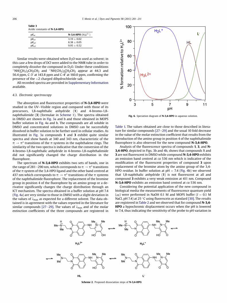
Fig. 6. Speciation diagram of N-3,4-HPO in aqueous solution.
Table 3Acidic constants of N-3,4-HPO.
pKai N-3,4-HPO (H3L2þ)
pKa1 3.19 � 0.02pKa2 8.38 � 0.05pKa3 9.95 � 0.52
T. Moniz et al. / Dyes and Pigments 98 (2013) 201e211206
Similar results were obtained when D2O was used as solvent; inthis case a few drops of DCl were added to the NMR tube in order tocompletely dissolve the compound in D2O. Under these conditionsþNH(CH3)2CH2CH2 and þNH(CH3)2CH2CH2 appear at 44.3 and56.4 ppm, C-30 at 143.8 ppm and C-40 at 160.0 ppm, confirming thepresence of the þ2 charged dihydrochloride salt.
All recorded spectra are provided in Supplementary Informationavailable.
4.3. Electronic spectroscopy
The absorption and fluorescence properties of N-3,4-HPO werestudied in the UVeVisible region and compared with those of itsprecursors, 1,8-naphthalic anhydride (1) and 4-bromo-1,8-naphthalimide (3) (formulae in Scheme 1). The spectra obtainedin DMSO are shown in Fig. 3a and b and those obtained in MOPSbuffer solution in Fig. 4a and b. The compounds are all soluble inDMSO and concentrated solutions in DMSO can be successfullydissolved in buffer solution to be further used in cellular studies. Asillustrated in Fig. 3a compounds 1 and 3 exhibit quite similarspectra and show bands at 340 and 343 nm, characteristic of thep / p* transitions of the p systems in the naphthalene rings. Thesimilarity of the two spectra is indicative that the conversion of the4-bromo-1,8-naphthalic anhydride in 4-bromo-1,8-naphthalimidedid not significantly changed the charge distribution in thefluorophore.
The spectrum of N-3,4-HPO exhibits two sets of bands, one inthe range of 281e290 nm, which corresponds to p/ p* transitionsof the p system of the 3,4-HPO ligand and the other band centred at437 nmwhich corresponds to p / p* transitions of the p systemsof the naphthalimide fluorophore. The replacement of the brominegroup in position 4 of the fluorophore by an amino group or a de-rivative significantly changes the charge distribution through anICT mechanism. The spectra obtained in a buffer solution at pH 7.4(Fig. 4a) are very similar to those in DMSOwith a slight deviation inthe values of lmax as expected for a different solvent. The data ob-tained is in agreement with the values reported in the literature forsimilar compounds [27e29]. The values of lmax and of the molarextinction coefficients of the three compounds are registered in
Scheme 2. Proposed dissocia
Table 1. The values obtained are close to those described in litera-ture for similar compounds [27e29] and the usual 10 fold decreasein the value of the molar extinction coefficient that results from theintroduction of the amino group in position 4 of the naphthalimidefluorophore is also observed for the new compound N-3,4-HPO.
Analysis of the fluorescence spectra of compounds 1, 3, and N-3,4-HPO, depicted in Figs. 3b and 4b, shows that compounds 1 and3 are not fluorescent in DMSOwhile compoundN-3,4-HPO exhibitsan emission band centred at ca 536 nm which is indicative of themodification of the fluorescent properties of compound 3 uponreplacement of the bromine atom by the amino group of the 3,4-HPO residue. In buffer solution at pH ¼ 7.4 (Fig. 4b) we observedthat 1,8-naphthalic anhydride (1) is not fluorescent at all andcompound 3 exhibits a very weak emission at 411 nm. CompoundN-3,4-HPO exhibits an emission band centred at ca 536 nm.
Considering the potential application of the new compound inbiological media the measurements of fluorescence quantum yield(4F) were performed in NaOH 0.1 M and MOPS buffer (I ¼ 0.1 MNaCl, pH 7.4) at 25 �C using fluorescein as standard [30]. The resultsare registered in Table 2 andwe observed that for compoundN-3,4-HPO a hypochromic displacement occurs when the pH is loweredto 7.4, thus indicating the sensitivity of the probe to pH variation in
tion steps of N-3,4-HPO.
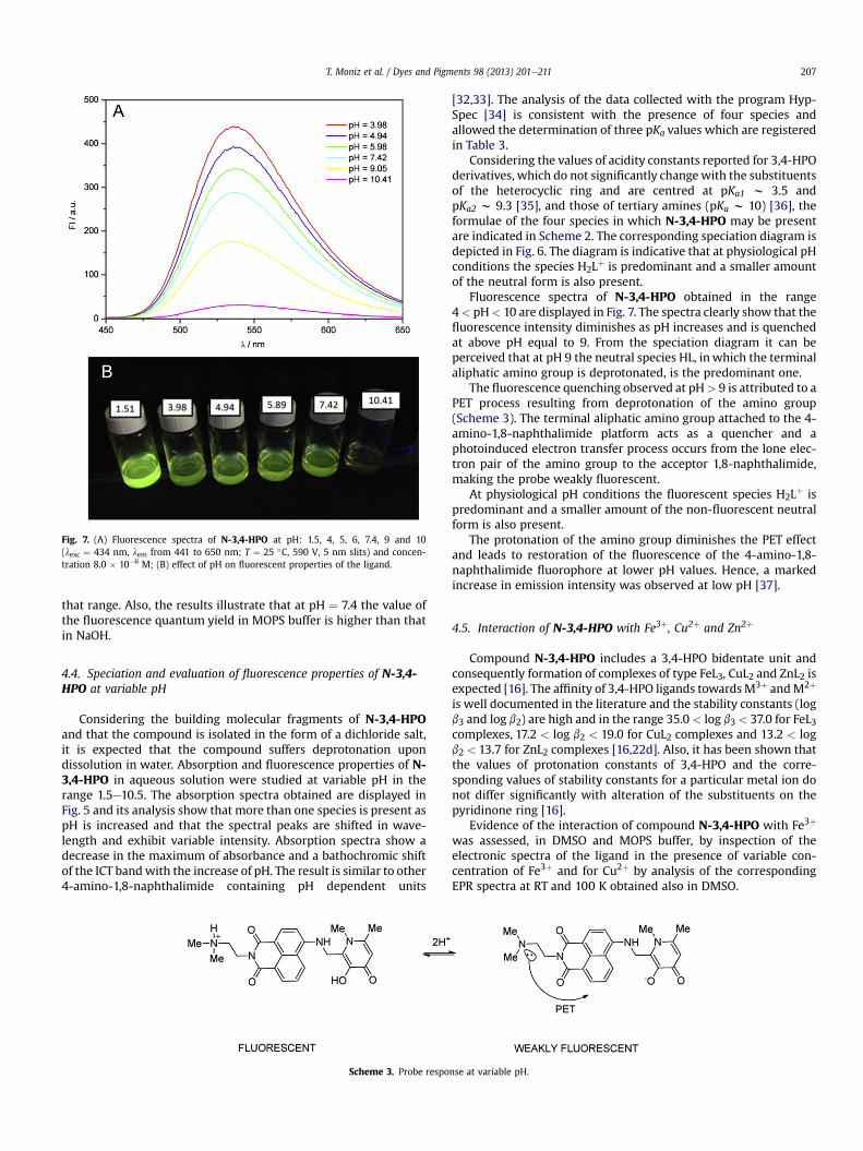
Fig. 7. (A) Fluorescence spectra of N-3,4-HPO at pH: 1.5, 4, 5, 6, 7.4, 9 and 10(lexc ¼ 434 nm, lem from 441 to 650 nm; T ¼ 25 �C, 590 V, 5 nm slits) and concen-tration 8.0 � 10�6 M; (B) effect of pH on fluorescent properties of the ligand.
T. Moniz et al. / Dyes and Pigments 98 (2013) 201e211 207
that range. Also, the results illustrate that at pH ¼ 7.4 the value ofthe fluorescence quantum yield in MOPS buffer is higher than thatin NaOH.
4.4. Speciation and evaluation of fluorescence properties of N-3,4-HPO at variable pH
Considering the building molecular fragments of N-3,4-HPOand that the compound is isolated in the form of a dichloride salt,it is expected that the compound suffers deprotonation upondissolution in water. Absorption and fluorescence properties of N-3,4-HPO in aqueous solution were studied at variable pH in therange 1.5e10.5. The absorption spectra obtained are displayed inFig. 5 and its analysis show that more than one species is present aspH is increased and that the spectral peaks are shifted in wave-length and exhibit variable intensity. Absorption spectra show adecrease in the maximum of absorbance and a bathochromic shiftof the ICT bandwith the increase of pH. The result is similar to other4-amino-1,8-naphthalimide containing pH dependent units
Scheme 3. Probe respo
[32,33]. The analysis of the data collected with the program Hyp-Spec [34] is consistent with the presence of four species andallowed the determination of three pKa values which are registeredin Table 3.
Considering the values of acidity constants reported for 3,4-HPOderivatives, which do not significantly changewith the substituentsof the heterocyclic ring and are centred at pKa1 w 3.5 andpKa2 w 9.3 [35], and those of tertiary amines (pKa w 10) [36], theformulae of the four species in which N-3,4-HPO may be presentare indicated in Scheme 2. The corresponding speciation diagram isdepicted in Fig. 6. The diagram is indicative that at physiological pHconditions the species H2Lþ is predominant and a smaller amountof the neutral form is also present.
Fluorescence spectra of N-3,4-HPO obtained in the range4< pH< 10 are displayed in Fig. 7. The spectra clearly show that thefluorescence intensity diminishes as pH increases and is quenchedat above pH equal to 9. From the speciation diagram it can beperceived that at pH 9 the neutral species HL, inwhich the terminalaliphatic amino group is deprotonated, is the predominant one.
The fluorescence quenching observed at pH> 9 is attributed to aPET process resulting from deprotonation of the amino group(Scheme 3). The terminal aliphatic amino group attached to the 4-amino-1,8-naphthalimide platform acts as a quencher and aphotoinduced electron transfer process occurs from the lone elec-tron pair of the amino group to the acceptor 1,8-naphthalimide,making the probe weakly fluorescent.
At physiological pH conditions the fluorescent species H2Lþ ispredominant and a smaller amount of the non-fluorescent neutralform is also present.
The protonation of the amino group diminishes the PET effectand leads to restoration of the fluorescence of the 4-amino-1,8-naphthalimide fluorophore at lower pH values. Hence, a markedincrease in emission intensity was observed at low pH [37].
4.5. Interaction of N-3,4-HPO with Fe3þ, Cu2þ and Zn2þ
Compound N-3,4-HPO includes a 3,4-HPO bidentate unit andconsequently formation of complexes of type FeL3, CuL2 and ZnL2 isexpected [16]. The affinity of 3,4-HPO ligands towardsM3þ andM2þ
is well documented in the literature and the stability constants (logb3 and log b2) are high and in the range 35.0 < log b3 < 37.0 for FeL3complexes, 17.2 < log b2 < 19.0 for CuL2 complexes and 13.2 < logb2 < 13.7 for ZnL2 complexes [16,22d]. Also, it has been shown thatthe values of protonation constants of 3,4-HPO and the corre-sponding values of stability constants for a particular metal ion donot differ significantly with alteration of the substituents on thepyridinone ring [16].
Evidence of the interaction of compound N-3,4-HPO with Fe3þ
was assessed, in DMSO and MOPS buffer, by inspection of theelectronic spectra of the ligand in the presence of variable con-centration of Fe3þ and for Cu2þ by analysis of the correspondingEPR spectra at RT and 100 K obtained also in DMSO.
nse at variable pH.

T. Moniz et al. / Dyes and Pigments 98 (2013) 201e211208
In order to judge the use of the new compound N-3,4-HPO tosense Fe3þ, Cu2þ and Zn2þ, the interaction of N-3,4-HPOwith Fe3þ,Cu2þ and Zn2þ was also investigated by examining the variationsobserved in the fluorescence spectra of solutions of the probe inMOPS buffer upon the addition of increasing amounts of the threemetal ions.
The absorption and emission spectra of compound N-3,4-HPOfor a 3:1 ligand:iron(III) ratio obtained in DMSO, are shown inFig. 8a and b, respectively. The absorption spectra clearly show ashift in the ICT band characteristic of the naphthalimide fluo-rophore and the appearance of a new broad and much less intenseband centred at ca 550 nm characteristic of formation of an iron(III)complex. Spectra obtained for 2:1 and 1:1 ligand:iron(III) ratios areprovided as supplementary information (Fig. S1 in SupplementaryInformation). The fluorescence spectra obtained for the same so-lutions are indicative that the fluorescence quenching occurs in thepresence of Fe3þ and that the effect is concentration dependent.Considering that compound N-3,4-HPO is isolated in the form of adichloride salt and that the NMR spectrum in DMSO confirms thatthe amino group in the imide position of the naphthalimide fluo-rophore is protonated, we conclude that fluorescence quenching isthe result of metal ion coordination to the 3,4-HPO unit, since in
300 400 500 600 7000.0
0.5
1.0
1.5
2.0
2.5
λ / nm
Abso
rban
ce
A
450 500 550 600 650 7000
100
200
300
400
λ / nm
FI /
a.u.
B
Fig. 8. (A) Absorption spectra of N-3,4-HPO at 3.0 � 10�4 M (DMSO,T ¼ 25 �C) (blackline) and in the presence of Fe3þ in a metal:ligand ratio 1:3 (red line [N-3,4-HPO] ¼ 3.0 � 10�4 M); (B) Fluorescence spectra of N-3,4-HPO at 2.5 � 10�5 M(DMSO, T ¼ 25 �C) (black line) and in the presence of Fe3þ in a metal/ligand ratio 1:3(red line, [N-3,4-HPO] ¼ 7.5 � 10�5 M). All spectra were recorded at lexc ¼ 437 nm,lem ¼ 438e700 nm, T ¼ 25 �C, 550 V, 5 nm slits. (For interpretation of the references tocolour in this figure legend, the reader is referred to the web version of this article.)
DMSO the deprotonation of the amino group does not occur andconsequently the PET effect observed in aqueous solution is notactive.
The absorption and emission spectra of a solution of N-3,4-HPOin MOPS buffer, upon addition of increasing amounts of Fe3þ aredepicted in (Fig. 9a and b). In Fig. 9a it is clearly shown that as theconcentration of Fe3þ is raised the intensity of the bands at ca 320e370 nm and the ICT absorption band at lmax ¼ 439 nm decreases.Also, the maximum of the ICT band is shifted to longer wavelengthand a less intense band emerges at ca 525 nm. These spectralchanges are a consequence of coordination of the pyridinone unit toFe3þ and are typical of naphthalimide based probes for sensingcations and anions [38,39]. In Fig. 9b the fluorescence spectra of asolution of N-3,4-HPO in MOPS buffer upon addition of increasingamounts of Fe3þ are displayed The spectra show that the intensityof the fluorescence is diminished as the concentration of Fe3þ israised and the quenching of fluorescence is ca 92% at a metal:ligandratio of 1:3. The quenching is assigned to the formation of the trisiron(III) complex with the ligand N-3,4-HPO (Scheme 4). In thepresence of Cu2þ and Zn2þ the fluorescence of the ligand is alsoquenched is ca 88% for Cu2þ and ca 91% for Zn2þ at a metal:ligandratio of 1:2 (Figs. S2 and S3 in Supplementary Information).
Confirmation of the coordination of Cu2þ with ligand N-3,4-HPO is provided by the EPR spectra obtained in DMSO at RT and
250 300 350 400 450 500 5500.00
0.02
0.04
0.06
0.08
0.10
|Fe3+| = 1 μM
|Fe3+| = 0 μM
Abso
rban
ce
λ / nm
A
500 550 600 6500
100
200
300
400
|Fe3+| = 1 µM
B
IF /
a.u
λ / nm
|Fe3+| = 0 µM
Fig. 9. (A) Absorption spectra of N-3,4-HPO at 3.0 � 10�6 M (MOPS buffer, I ¼ 0.1 MNaCl, 25 �C, pH ¼ 7.4) with increasing concentration of Fe3þ; (B) Fluorescence spectraof N-3,4-HPO at 3.0 � 10�6 M (MOPS buffer, I ¼ 0.1 M NaCl, 25 �C, pH ¼ 7.4,lexc ¼ 439 nm) with increasing concentration of Fe3þ.
Scheme 4. Probe response towards Fe3þ coordination.
T. Moniz et al. / Dyes and Pigments 98 (2013) 201e211 209
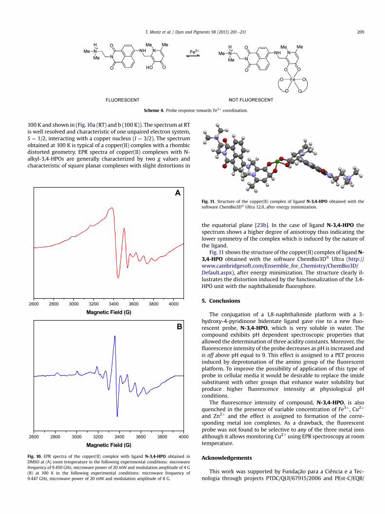
100 K and shown in (Fig.10a (RT) and b (100 K)). The spectrum at RTis well resolved and characteristic of one unpaired electron system,S ¼ 1/2, interacting with a copper nucleus (I ¼ 3/2). The spectrumobtained at 100 K is typical of a copper(II) complex with a rhombicdistorted geometry. EPR spectra of copper(II) complexes with N-alkyl-3,4-HPOs are generally characterized by two g values andcharacteristic of square planar complexes with slight distortions in
Fig. 11. Structure of the copper(II) complex of ligand N-3,4-HPO obtained with thesoftware ChemBio3D� Ultra 12.0, after energy minimization.
A
B
2600 2800 3000 3200 3400 3600 3800 4000
Magnetic Field (G)
2600 2800 3000 3200 3400 3600 3800 4000
Magnetic Field (G)
Fig. 10. EPR spectra of the copper(II) complex with ligand N-3,4-HPO obtained inDMSO at (A) room temperature in the following experimental conditions: microwavefrequency of 9.450 GHz, microwave power of 20 mW and modulation amplitude of 4 G(B) at 100 K in the following experimental conditions: microwave frequency of9.447 GHz, microwave power of 20 mW and modulation amplitude of 8 G.
the equatorial plane [23b]. In the case of ligand N-3,4-HPO thespectrum shows a higher degree of anisotropy thus indicating thelower symmetry of the complex which is induced by the nature ofthe ligand.
Fig.11 shows the structure of the copper(II) complex of ligandN-3,4-HPO obtained with the software ChemBio3D� Ultra (http://www.cambridgesoft.com/Ensemble_for_Chemistry/ChemBio3D/Default.aspx), after energy minimization. The structure clearly il-lustrates the distortion induced by the functionalization of the 3,4-HPO unit with the naphthalimide fluorophore.
5. Conclusions
The conjugation of a 1,8-naphthalimide platform with a 3-hydroxy-4-pyridinone bidentate ligand gave rise to a new fluo-rescent probe, N-3,4-HPO, which is very soluble in water. Thecompound exhibits pH dependent spectroscopic properties thatallowed the determination of three acidity constants. Moreover, thefluorescence intensity of the probe decreases as pH is increased andis off above pH equal to 9. This effect is assigned to a PET processinduced by deprotonation of the amino group of the fluorescentplatform. To improve the possibility of application of this type ofprobe in cellular media it would be desirable to replace the imidesubstituent with other groups that enhance water solubility butproduce higher fluorescence intensity at physiological pHconditions.
The fluorescence intensity of compound, N-3,4-HPO, is alsoquenched in the presence of variable concentration of Fe3þ, Cu2þ
and Zn2þ and the effect is assigned to formation of the corre-sponding metal ion complexes. As a drawback, the fluorescentprobe was not found to be selective to any of the three metal ionsalthough it allowsmonitoring Cu2þ using EPR spectroscopy at roomtemperature.
Acknowledgements
This work was supported by Fundação para a Ciência e a Tec-nologia through projects PTDC/QUI/67915/2006 and PEst-C/EQB/
T. Moniz et al. / Dyes and Pigments 98 (2013) 201e211210
LA0006/2011. T. Moniz also thanks FCT the PhD grant (SFRH/BD/79874/2011). We thank Dr. Andrea Carneiro from CeNTI, V.N.Famalicão, for making available the CEM Discover microwavereactor. The Bruker Avance II 400 spectrometer is part of the Na-tional NMR network and was purchased under the framework ofthe National Programme for Scientific Re-equipment, contractREDE/1517/RMN/2005, with funds from POCI 2010 (FEDER) and(FCT).
Appendix A. Supplementary data
Supplementary data related to this article can be found at http://dx.doi.org/10.1016/j.dyepig.2013.02.020.
References
[1] Grynkiewicz G, Poenie M, Tsien RY. A new generation of Ca2þ indicators withgreatly improved fluorescence properties. J Biol Chem 1985;260:3440e50.
[2] Tsien RY. New calcium indicators and buffers with high selectivity againstmagnesium and protons: design, synthesis, and properties of prototypestructures. Biochem 1980;19:2396e404.
[3] Han J, Burgess K. Fluorescent indicators for intracellular pH. Chem Rev2010;110:2709e28.
[4] (a) Xu Z, Yoon J, Spring DR. Fluorescent chemosensors for Zn2þ. Chem Soc Rev2010;39:1996e2006;(b) Nolan EM, Lippard SJ. Tools and tactics for the optical detection of mercuricion. Chem Rev 2008;108:3443e80.
[5] (a) Razgulin A, Ma N, Rao J. Strategies for in vivo imaging of enzyme activity:an overview and recent advances. Chem Soc Rev 2011;40:4186e216;(b) Serim S, Haedke U, Verhelst SHL. Activity-based probes for the study ofproteases: recent advances and developments. Chem Med Chem 2012;7:1146e59;(c) Mizukami S. Development of molecular imaging tools to investigate pro-tein functions by chemical probe design. Chem Pharm Bull 2011;59:1435e46.
[6] (a) Miller EW, Chang CJ. Fluorescent probes for nitric oxide and hydrogenperoxide in cell signaling. Curr Opin Chem Biol 2007;11:620e5;(b) Chen X, Tian X, Shin I, Yoon J. Fluorescent and luminescent probes fordetection of reactive oxygen and nitrogen species. Chem Soc Rev 2011;40:4783e804;(c) Hyman LM, Frank KJ. Probing oxidative stress: small molecule fluorescentsensors of metal ions, reactive oxygen species, and thiols. Coord Chem Rev2012;256:2333e56.
[7] (a) Nagano T. Development of fluorescent probes for bioimaging applications.Proc Jpn Acad Ser B 2010;86:837e47;(b) Ueno T, Nagano T. Fluorescent probes for sensing and imaging. NatMethods 2011;8:642e5.
[8] (a) Bissell RA, de Silva AP, Gunaratne HQN, Lynch PLM, Maguire GEM,Sandanayake KRAS. Molecular fluorescent signalling with ‘fluor-spacer-re-ceptor’ systems: approaches to sensing and switching devices via supramo-lecular photophysics. Chem Soc Rev 1992;21:187e95;(b) de Silva AP, Gunaratne HQN, Gunnlaugsson T, Huxley AJM, McCoy CP,Rademacher JT, et al. Signaling recognition events with fluorescent sensorsand switches. Chem Rev 1997;97:1515e66;(c) Gunnlaugsson T, Davis AP, O’Brien JE, Glynn M. Synthesis and photo-physical evaluation of charge neutral thiourea or urea based fluorescent PETsensors for bis-carboxylates and pyrophosphate. Org Biomol Chem 2005;3:48e56;(d) dos Santos CMG, Glynn M, McCabe T, Seixas de Melo JS, Burrows HD,Gunnlaugsson T. Synthesis, structural and photophysical evaluations of ureabased fluorescent PET sensors for anions. Supramol Chem 2007;20:407e18.
[9] (a) de Silva AP, Rupasinghe RADD. A new class of fluorescent pH indicatorsbased on photo-induced electron transfer. J Chem Soc Chem Commun 1985:1669e70;(b) de Silva AP, de Silva SA. Fluorescent signalling crown ethers; ‘switching on’of fluorescence by alkali metal ion recognition and binding in situ. J Chem SocChem Commun 1986:1709e10;(c) Huston ME, Haider KW, Czarnik AW. Chelation-enhanced fluorescence in9,10-bis(TMEDA)anthracene [33]. J Am Chem Soc 1988;110:4460e2;(d) Czarnik AW. Chemical communication in water using fluorescent che-mosensors. Acc Chem Res 1994;27:302e8.
[10] (a) Gunnlaugsson T, Glynn M, Tocci (née Hussey) GM, Kruger PE, Pfeffer FM.Anion recognition and sensing in organic and aqueous media using lumi-nescent and colorimetric sensors. Coord Chem Rev 2006;250:3094e117;(b) Duke RM, Veale EB, Pfeffer FM, Kruger PE, Gunnlaugsson T. Colorimetricand fluorescent anion sensors: an overview of recent developments in the useof 1,8-naphthalimide-based chemosensors. Chem Soc Rev 2010;39:3936e53;(c) Qian X, Xiao Y, Xu Y, Guo X, Qiana J, Zhua W. “Alive” dyes as fluorescentsensors: fluorophore, mechanism, receptor and images in living cells. ChemCommun 2010;46:6418e36.
[11] de Silva AP, Gunaratne HQN, Lynch PLM, Patty AJ, Spence GL. Luminescenceand charge transfer. Part 3. The use of chromophores with ICT (internal chargetransfer) excited states in the construction of fluorescent PET (photoinducedelectron transfer) pH sensors and related absorption pH sensors with ami-noalkyl side chains. J Chem Soc Trans 2 1993:1611e6.
[12] Oelgemöller M, Kramer WH. Synthetic photochemistry of naphthalimides andrelated compounds. J PhotochemPhotobiol C PhotochemRev 2010;11:210e44.
[13] For Cu2þ please see (a) Xu Z, Xiao Y, Qian X, Cui J, Cui D. Ratiometric andselective fluorescent sensor for Cu II based on internal charge transfer (ICT).Org Lett 2005;7:889e92;(b) Xu Z, Qian X, Cui J. Colorimetric and ratiometric fluorescent chemosensorwith a large red-shift in emission: Cu(II)-only sensing by deprotonation ofsecondary amines as receptor conjugated to naphthalimide fluorophore. OrgLett 2005;7:3029e32;(c) Huang J, Xu Y, Qian X. A colorimetric sensor for Cu2þ in aqueous solutionbased on metal ion-induced deprotonation: deprotonation/protonationmediated by Cu2þ-ligand interactions. Dalton Trans 2009:1761e6;(d) Huang J, Xu Y, Qian X. A red-shift colorimetric and fluorescent sensor forCu2þ in aqueous solution: unsymmetrical 4,5-diaminonaphthalimide with N-H deprotonation induced by metal ions. Org Biomol Chem 2009;7:1299e303;(e) Singh N, Kaur N, McCaughan B, Callan JF. Ratiometric fluorescent detectionof Cu(II) in semi-aqueous solution using a two-fluorophore approach. Tetra-hedron Lett 2010;51:3385e7.(f) Sahin O, YilmazM. Synthesis and fluorescencesensing properties of a new naphthalimide derivative of calix[4]arene. Tetra-hedron Lett 2012;53:2319e24;(g) Liu Z, Zhang C, Wang X, He W, Guo Z. Design and synthesis of a ratiometricfluorescent chemosensor for Cu(II) with a fluorophore hybridization approach.Org Lett 2012;14:4378e81;For Zn2þplease see (a) Gunnlaugsson T, Lee TC, ParkeshR. A highly selective andsensitive fluorescent PET (photoinduced electron transfer) chemosensor forZn(II). Org Biomol Chem 2003;1:3265e7;(b) Parkesh R, Lee TC, Gunnlaugsson T. Highly selective 4-amino-1,8-naphthalimide based fluorescent photoinduced electron transfer (PET) che-mosensors for Zn(II) under physiological pH conditions. Org Biomol Chem2007;5:310e7.
[14] For Hg2þ please see (a) Guo X, Qian X, Jia L. A highly selective and sensitivefluorescent chemosensor for Hg2þ in neutral buffer aqueous solution. J AmChem Soc 2004;126:2272e3;(b) Liu B, Tian H. A selective fluorescent ratiometric chemodosimeter formercury ion. Chem Commun 2005:3156e8;(c) Wang J, Qian X. Two regioisomeric and exclusively selective Hg(II) sensormolecules composed of a naphthalimide fluorophore and an o-phenylenedi-amine derived triamide receptor. Chem Commun 2006:109e11;(d) He C, Zhu W, Xu Y, Chen T, Qian X. Trace mercury(II) detection and sep-aration in serum and water samples using a reusable bifunctional fluorescentsensor. Anal Chim Acta 2009;651:227e33;(e) Dai H, Xu H. A water-soluble 1,8-naphthalimide-based ‘turn on’ fluores-cent chemosensor for selective and sensitive recognition of mercury ion inwater. Bioorg Med Chem Lett 2011;21:5141e4;(f) Meng Q, Zhang X, He C, Zhou P, Su W, Duan C. A hybrid mesoporousmaterial functionalized by 1,8-naphthalimide-base receptor and the applica-tion as chemosensor and absorbent for Hg2þ in water. Talanta 2011;84:53e9;(g) Fang CL, Zhou J, Liu XJ, Cao ZH, Shangguan DH. Mercury(II)-mediatedformation of imide-Hg-imide complexes. Dalton Trans 2011;40:899e903;(h) Yang R, Guo X, Wang W, Zhang Y, Jia L. Highly selective and sensitivechemosensor for Hg2þ based on the naphthalimide fluorophore. J Fluoresc2012;22:1065e71;(i) Li CY, Xu F, Li YF, Zhou K, Zhou Y. A fluorescent chemosensor for Hg2þ
based on naphthalimide derivative by fluorescence enhancement in aqueoussolution. Anal Chim Acta 2012;717:122e6;For Pd2þ please see: Duan L, Xu Y, Qian X. Highly sensitive and selective Pd2þ
sensor of naphthalimide derivative based on complexation with alkynes andthio-heterocycle Chem Commun 2008:6339e41.
[15] (a) Hanaoka K, Muramatsu Y, Urano Y, Terai T, Nagano T. Design and synthesisof a highly sensitive off-on fluorescent chemosensor for zinc ions utilizinginternal charge transfer. Chem Eur J 2010;16:568e72;(b) Xu Z, Han SJ, Lee C, Yoon J, Spring DR. Development of off-on fluo-rescent probes for heavy and transition metal ions. Chem Commun2010;46:1679e81;(c) Wang H, Yang L, Zhang W, Zhou Y, Zhao B, Li X. A colorimetric probefor copper(II) ion based on 4-amino-1,8-naphthalimide. Inorg Chim Acta2012;381:111e6.
[16] Burgess J, Rangel M. Hydroxypyranones, hydroxypyridinones and their com-plexes. Adv Inorg Chem 2008;60:167e243.
[17] (a) Cohen SM, O’Sullivan B, Raymond KN. Mixed hydroxypyridinonate ligandsas iron chelators. Inorg Chem 2000;39:4339e46;(b) Xu J, O’Sullivan B, Raymond KN. Hexadentate hydroxypyridonate ironchelators based on TREN-Me-3,2-HOPO: variation of cap size. Inorg Chem2002;41:6731e42.
[18] Raymond KN, Xu J. US patent. 5 624 901; 1997.[19] (a) Santos MA, Marques SM, Chaves S. Hydroxypyridinones as “privileged”
chelating structures for the design of medicinal drugs. Coord Chem Rev2012;256:240e59;(b) Rodríguez-Rodríguez C, Telpoukhovskaia M, Orvig C. The art of buildingmultifunctional metal-binding agents from basic molecular scaffolds for the
T. Moniz et al. / Dyes and Pigments 98 (2013) 201e211 211
potential application in neurodegenerative diseases. Coord Chem Rev2012;256:2308e32;(c) Santos MA. Hydroxypyridinone complexes with aluminium. In vitro/vivostudies and perspectives. Coord Chem Rev 2002;228:187e203.
[20] (a) Ma Y, Luo W, Quinn PJ, Liu Z, Hider RC. Design, synthesis, physicochemicalproperties, and evaluation of novel iron chelators with fluorescent sensors.J Med Chem 2004;47:6349e62;(b) Katoh A, Ogino K, Saito R. Synthesis of new 3-hydroxy-4(1H)-pyridinonedirectly attached to quinoxaline and its fluorescence property uponcomplexation to metal ions. Heterocycles 2005;65:2195e201;(c) Fakih S, Podinovskaia M, Kong X, Collins HL, Schaible UE, Hider RC. Tar-geting the lysosome: fluorescent iron(III) chelators to selectively monitorendosomal/lysosomal labile iron pools. J Med Chem 2008;51:4539e52;(d) Fakih S, Podinovskaia M, Kong X, Schaible UE, Collins HL, Hider RC.Monitoring intracellular labile iron pools: a novel fluorescent iron(III) sensoras a potential non-invasive diagnosis tool. J Pharm Sci 2009;98:2212e26.
[21] Sahoo SK, Sharma D, Bera RK, Crisponi G, Callan JF. Iron(III) selective molecularand supramolecular fluorescent probes. Chem Soc Rev 2012;41:7195e227.
[22] (a) Fernandes SS, Nunes A, Gomes AR, de Castro B, Hider RC, Rangel M, et al.Identification of a new hexadentate iron chelator capable of restricting theintramacrophagic growth of Mycobacterium avium. Microbes Infect 2010;12:287e94;(b) Nunes A, Podinovskaia M, Leite A, Gameiro P, Zhou T, Ma Y, et al. Fluo-rescent 3-hydroxy-4-pyridinone hexadentate iron chelators: intracellulardistribution and the relevance to antimycobacterial properties. J Biol InorgChem 2010;15:861e77;(c) Rangel M, Amorim MJ, Nunes A, Leite A, Pereira E, de Castro B, et al. Novel3-hydroxy-4-pyridinonato oxidovanadium(IV) complexes to investigatestructure/activity relationships. J Inorg Biochem 2009;103:496e502;(d) Moniz T, Amorim MJ, Ferreira R, Nunes A, Silva A, Queirós C, et al.Investigation of the insulin-like properties of zinc(II) complexes of 3-hydroxy-4-pyridinones: identification of a compound with glucose lowering effect inSTZ-induced type I diabetic animals. J Biol Inorg Chem 2011;105:1675e82.
[23] (a) Silva AMG, Leite A, Andrade M, Gameiro P, Brandão P, Felix V, et al. Mi-crowave-assisted synthesis of 3-hydroxy-4-pyridinone/naphthalene conju-gates. Structural characterization and selection of a fluorescent ion sensor.Tetrahedron 2010;66:8544e50;(b) Silva AMG, Leite A, Gonzalez P, Domingues MRM, Gameiro P, de Castro B,et al. Use of a porphyrin platform and 3,4-HPO chelating units to synthesizeligands with N4 and O4 coordination sites. Tetrahedron 2011;67:7821e8.
[24] Liu ZD, Kayyali R, Hider RC, Porter JB, Theobald AE. Design, synthesis, andevaluation of novel 2-substituted 3-hydroxypyridin-4-ones: structure-activityinvestigation of metalloenzyme inhibition by iron chelators. J Med Chem2002;45:631e9.
[25] Muzart J. N,N-Dimethylformamide: much more than a solvent. Tetrahedron2009;65:8313e23.
[26] Ding S, Jiao N. N,N-dimethylformamide: a multipurpose building block.Angew Chem Int Ed 2012;51:9226e37.
[27] Bojinov VB, Georgiev NI, Nikolov PS. Synthesis and photophysical propertiesof fluorescence sensing ester- and amidoamine-functionalized 1,8-naphthalimides. J Photochem Photobiol A Chem 2008;193:129e38.
[28] Alexiou MS, Tychopoulos V, Ghorbanian S, Tyman JHP, Brown RG, Brittain PI.The UV-visible absorption and fluorescence of some substituted 1,8-naphthalimides and naphthalic anhydrides. J Chem Soc Perkin Trans 21990:837e42.
[29] Li Z, Yang Q, Chang R, Ma G, Chen M, Zhang W. N-Heteroaryl-1,8-naphthalimide fluorescent sensor for water: molecular design, synthesisand properties. Dyes Pigm 2011;88:307e14.
[30] Williams ATR, Winfield SA, Miller JN. Relative fluorescence quantum yieldsusing a computer-controlled luminescence spectrometer. Analyst 1983;108:1067e71.
[31] Lakowicz JR. Principles of fluorescence spectroscopy. 3 ed. Berlin, Heidelberg:Springer; 2006.
[32] Tian Y, Su F, Weber W, Nandakumar V, Shumway BR, Jin Y, et al. A series ofnaphthalimide derivatives as intra and extracellular pH sensors. Biomaterials2010;31:7411e22.
[33] Niu CG, Zeng GM, Chen LX, Shen GL, Yu RQ. Proton “off-on” behaviour ofmethylpiperazinyl derivative of naphthalimide: a pH sensor based on fluo-rescence enhancement. Analyst 2004;129:20e4.
[34] Gans P, Sabatini A, Vacca A. Investigation of equilibria in solution. Determi-nation of equilibrium constants with the HYPERQUAD suite of programs.Talanta 1996;43:1739e53.
[35] Queiros C, Amorim MJ, Leite A, Ferreira M, Gameiro P, Castro B, et al. Nickel(II)and cobalt(II) 3-hydroxy-4-pyridinone complexes: synthesis, characterizationand speciation studies in aqueous solution. Eur J Inorg Chem 2011;1:131e40.
[36] Frenna V, Vivona N, Consiglio G, Spinelli D. Amine basicities in benzene and inwater. J Chem Soc Perkin Trans 2 1985:1865e8.
[37] Niu CG, Gui XQ, Zeng GM, Yuan XZ. A ratiometric fluorescence sensor withbroad dynamic range based on two pH-sensitive fluorophores. Analyst2005;130:1551e6.
[38] Chaves S, Canário S, Carrasco MP, Mira L, Santos MA. Hydroxy(thio)pyroneand hydroxy(thio)pyridinone iron chelators: physico-chemical properties andanti-oxidant activity. J Inorg Biochem 2012;114:38e46.
[39] Veale EB, Tocci GM, Pfeffer FM, Krugera PE, Gunnlaugsson T. Demonstrationof bidirectional photoinduced electron transfer (PET) sensing in 4-amino-1,8-naphthalimide based thiourea anion sensors. Org Biomol Chem 2009;7:3447e54.
[40] Xie L, Xu Y, Wang F, Liu J, Qian X, Cui J. Synthesis of new amonafide analoguesvia coupling reaction and their cytotoxic evaluation and DNA-binding studies.Bioorg Med Chem 2009;17:804e10.
[41] Wan Y, Alterman M, Hallberg A. Palladium-catalyzed amination of aryl bro-mides using temperature-controlled microwave heating. Synthesis 2002;11:1597e600.
[42] Forgues SF, Lavabre D. Are fluorescence quantum yields so tricky to measure?A demonstration using familiar stationery products. J Chem Educ 1999;76:1260e4.
[43] Albert A, Sergeant EP. The determination of ionization constants. 2 ed. Lon-don: Chapman & Hall; 1971.
[44] Alderighi L, Gans P, Ienco A, Peters D, Sabatini A, Vacca A. Hyperquad simu-lation and speciation (HySS): a utility program for the investigation of equi-libria involving soluble and partially soluble species. Coord Chem Rev1999;184:311e8.
![Synthesis and pharmacological assessment of diversely substituted pyrazolo[3,4-b]quinoline, and benzo[b]pyrazolo[4,3-g][1,8]naphthyridine derivatives](https://static.fdokumen.com/doc/165x107/6337be5bce400ca6980926a6/synthesis-and-pharmacological-assessment-of-diversely-substituted-pyrazolo34-bquinoline.jpg)


![1,8-Di-aza-bicyclo-[5.4.0]undec-7-en-8-ium bromido-(phthalocyaninato)zincate](https://static.fdokumen.com/doc/165x107/633691fc02a8c1a4ec024048/18-di-aza-bicyclo-540undec-7-en-8-ium-bromido-phthalocyaninatozincate.jpg)


 O12, FROM A FELSIC GRANULITE, PARRY SOUND, ONTARIO, AND A …](https://static.fdokumen.com/doc/165x107/634058530f237bc7380b4598/menzerite-y-a-new-speciesy-reeca-fe2-2mg-fe2fe3-alsi3-o12.jpg)