An update on the synthesis and reactivity of spiro-fused β ...

lable at ScienceDirect
European Journal of Medicinal Chemistry 46 (2011) 3210e3217
Contents lists avai
European Journal of Medicinal Chemistry
journal homepage: http: / /www.elsevier .com/locate/ejmech
Original article
Design and synthesis of spiro derivatives of parthenin as novel anti-cancer agents
Doma Mahendhar Reddy b, Naveed A. Qazi b, Sanghpal D. Sawant b, Abid H. Bandey b, Jada Srinivas a,Mannepalli Shankar a, Shashank K. Singh b, Monika Verma b, Gousia Chashoo b, Arpita Saxena b,Dilip Mondhe b, Ajit K. Saxena b, V.K. Sethi b, Subhash C. Taneja b, Gulam N. Qazi b,c,H.M. Sampath Kumar a,b,*aOrganic Chemistry-I, Indian Institute of Chemical Technology, Hyderabad 500007, Indiab Synthetic and Biological Chemistry, Indian Institute of Integrative Medicine, Jammu 180001, Indiac Jamia Hamdard University, New Delhi 110062, India
a r t i c l e i n f o
Article history:Received 1 February 2011Received in revised form7 April 2011Accepted 9 April 2011Available online 29 April 2011
Keywords:PartheninSpiro derivativesAnti-cancerNF-kB(P65) inhibitors
* Corresponding author. Tel.: þ91 191 2569000; faxE-mail address: [email protected] (H.M. Sam
0223-5234/$ e see front matter � 2011 Elsevier Masdoi:10.1016/j.ejmech.2011.04.030
a b s t r a c t
Several novel spiro derivatives of parthenin (1) have been synthesized by the dipolar cycloaddition usingvarious dipoles viz; benzonitrile oxides, nitrones and azides with exocyclic double bond of C ring(a-methylene-g-butyrolactone). Majority of the compounds exhibited improved anti-cancer activitycompared to the parthenin, when screened for their in vitro cytotoxicity against three human cancer celllines viz., SW-620, DU-145 and PC-3. In vivo screening of select analog revealed improved anti-canceractivity with low mammalian toxicity as compared to parthenin. The results of the cytotoxicity pattern ofthese derivatives reveals the SAR of these sesquiterpinoid lactones and possible role of a,b-unsaturatedketone of parthenin in inhibiting NF-kB. A mechanistic correlation of anti-cancer activity along within vivo and western blotting experiments has been described.
� 2011 Elsevier Masson SAS. All rights reserved.
1. Introduction
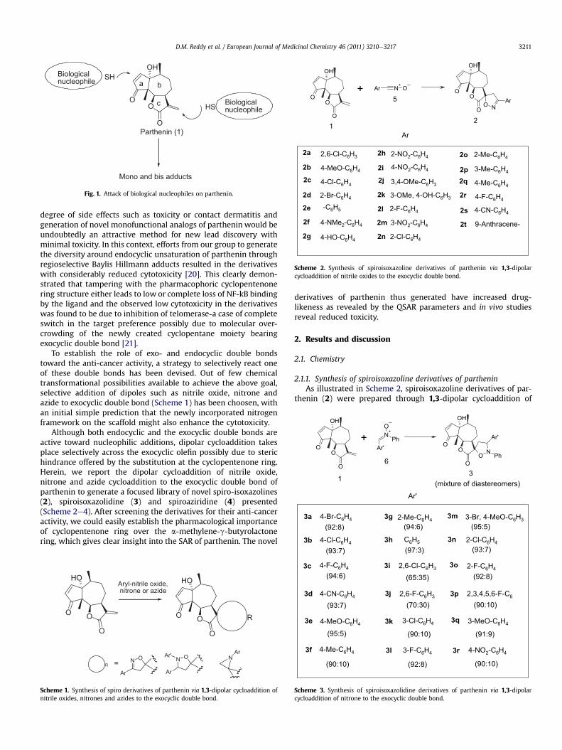
In recent years, the anti-cancer property of various sesquiter-penes has attracted a great deal of interest and extensive researchwork has been carried out to characterize the anti-cancer activity,the molecular mechanisms and the potential chemo-preventiveand chemo-therapeutic application of sesquiterpenoids [1]. Par-thenin (1), a sesquiterpene lactone (SL) isolated from Partheniumhysterophorus L [2], has found interest due to its medicinal prop-erties viz., anti-cancer, antibacterial, antiamoebic, anti-inflammatory, lipid peroxidation inhibition, and trypanocidalactivity [3e12]. Cytotoxicity, as many other biological activities ofsesquiterpene lactones, is known to be mediated by the presence ofpotential alkylant structure elements such as an a-methylene-g-lactone, an a,b-unsaturated cyclopentenone or a conjugated estercapable of reacting covalently with biological nucleophiles, espe-cially with the cysteine sulfhydryl group of proteins, in a Michael-type addition (Fig. 1), thereby inhibiting a variety of cellularfunctions which directs the cells into apoptosis [13e19].Thedifferences in activity among individual SLs are attributed to the
: þ91 191 2569333.path Kumar).
son SAS. All rights reserved.
presence of different numbers of alkylating structural elements.Apart from this, other factors such as lipophilicity, moleculargeometry and the chemical environment or the target sulfhydrylwhich may also influence the activity of these compounds.
Using helenalin and parthenolide as models, it has been wellestablished that DNA binding of NF-kB is prevented by alkylation ofcysteine in the p65/NF-kB subunit, which is considered to be thegeneral mechanism for SL bearing a,b-unsaturated carbonylstructures [19]. Despite the plethora of experimental studies foundin literature on the cytotoxicity of sesquiterpene lactones in generalincluding few reports on parthenin in particular, little is knownabout the individual role of different alkylant structures on thecytotoxicity in terms of its SAR. This however, would be animportant step in the direction of rational lead optimization.
Parthenin (1) has two active sites; the exocyclic double bond ofa-methylene-g-butyrolactone and double bond of cyclopentenonering. Various structural modifications of parthenin skeleton havebeen carried out with a view to obtain more potent analogs withlower toxicity and better activity. In spite of the improved under-standing of the role of each double bond present on the partheninskeleton through such studies, the SAR of parthenin has not beenunequivocally established so far and none of the literature reportsrevealed a rational approach to the modification of parthenin.Furthermore, monofunctional alkylants are known to possess low
OO
O
OHBiologicalnucleophile
Biologicalnucleophile
Parthenin (1)
a b
c HS
SH
Mono and bis adducts
Fig. 1. Attack of biological nucleophiles on parthenin.
OO
O
OH
OO
O
OH
O NAr
Ar N+
O
Ar
2,6-Cl-C6H3
4-MeO-C6H4
4-Cl-C6H4
2-Br-C6H4
-C6H5
4-NMe2-C6H4
4-HO-C6H4
2-NO2-C6H4
3,4-OMe-C6H3
3-OMe, 4-OH-C6H3
3-NO2-C6H4
2-Me-C6H4
3-Me-C6H4
4-Me-C6H4
4-F-C6H4
4-CN-C6H4
9-Anthracene-
2a
2b
2c
2d
2e
2f
2g
2h
2i
2j
2k
2l
2m
2n
2o
2p
2q
2r
2s
2t
12
+5
4-NO2-C6H4
2-F-C6H4
2-Cl-C6H4
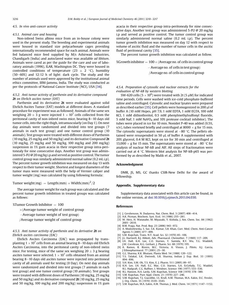
Scheme 2. Synthesis of spiroisoxazoline derivatives of parthenin via 1,3-dipolarcycloaddition of nitrile oxides to the exocyclic double bond.
OO
O
OH
N+
Ar'
O
Ph
O NOO
O
OH
Ar'
Ph
4-Cl-C6H4 C6H5
2-Me-C6H44-Br-C6H4 3-Br, 4-MeO-C6H3
2-Cl-C6H4
(92:8) (94:6) (95:5)
Ar'
3a
3b
3g
3h
3m
3n
31
+
6
(mixture of diastereomers)
D.M. Reddy et al. / European Journal of Medicinal Chemistry 46 (2011) 3210e3217 3211
degree of side effects such as toxicity or contact dermatitis andgeneration of novel monofunctional analogs of parthenin would beundoubtedly an attractive method for new lead discovery withminimal toxicity. In this context, efforts from our group to generatethe diversity around endocyclic unsaturation of parthenin throughregioselective Baylis Hillmann adducts resulted in the derivativeswith considerably reduced cytotoxicity [20]. This clearly demon-strated that tampering with the pharmacophoric cyclopentenonering structure either leads to low or complete loss of NF-kB bindingby the ligand and the observed low cytotoxicity in the derivativeswas found to be due to inhibition of telomerase-a case of completeswitch in the target preference possibly due to molecular over-crowding of the newly created cyclopentane moiety bearingexocyclic double bond [21].
To establish the role of exo- and endocyclic double bondstoward the anti-cancer activity, a strategy to selectively react oneof these double bonds has been devised. Out of few chemicaltransformational possibilities available to achieve the above goal,selective addition of dipoles such as nitrile oxide, nitrone andazide to exocyclic double bond (Scheme 1) has been choosen, withan initial simple prediction that the newly incorporated nitrogenframework on the scaffold might also enhance the cytotoxicity.
Although both endocyclic and the exocyclic double bonds areactive toward nucleophilic additions, dipolar cycloaddition takesplace selectively across the exocyclic olefin possibly due to sterichindrance offered by the substitution at the cyclopentenone ring.Herein, we report the dipolar cycloaddition of nitrile oxide,nitrone and azide cycloaddition to the exocyclic double bond ofparthenin to generate a focused library of novel spiro-isoxazolines(2), spiroisoxazolidine (3) and spiroaziridine (4) presented(Scheme 2e4). After screening the derivatives for their anti-canceractivity, we could easily establish the pharmacological importanceof cyclopentenone ring over the a-methylene-g-butyrolactonering, which gives clear insight into the SAR of parthenin. The novel
OO
O
HO
OO
O
HO
R
RN O
Ar
N O
Ar
Ar' NAr
Aryl-nitrile oxide,nitrone or azide
=
Scheme 1. Synthesis of spiro derivatives of parthenin via 1,3-dipolar cycloaddition ofnitrile oxides, nitrones and azides to the exocyclic double bond.
derivatives of parthenin thus generated have increased drug-likeness as revealed by the QSAR parameters and in vivo studiesreveal reduced toxicity.
2. Results and discussion
2.1. Chemistry
2.1.1. Synthesis of spiroisoxazoline derivatives of partheninAs illustrated in Scheme 2, spiroisoxazoline derivatives of par-
thenin (2) were prepared through 1,3-dipolar cycloaddition of
3-Cl-C6H4
4-Me-C6H4
4-F-C6H4
4-CN-C6H4
4-MeO-C6H4
2,6-Cl-C6H3
2,6-F-C6H3
3-F-C6H4
2-F-C6H4
2,3,4,5,6-F-C6
(93:7)
(94:6)
(93:7)
(95:5)
(90:10)
(97:3)
(65:35)
(70:30)
(90:10)
(92:8)
(93:7)
(92:8)
(90:10)
3c
3d
3e
3f
3i
3j
3k
3l
3o
3p
3-MeO-C6H4
(91:9)
3q
4-NO2-C6H43r
(90:10)
Scheme 3. Synthesis of spiroisoxazolidine derivatives of parthenin via 1,3-dipolarcycloaddition of nitrone to the exocyclic double bond.

OO
O
OH
NOO
O
OH
Ar''
C6H5
3-Cl-C6H4
4-Me-C6H4
3-Me-C6H4
4-MeO-C6H4
2-NO2-C6H4
2-Me-C6H4
2-MeO-C6H4
3-COOMe-C6H4
Ar''-N3
4a
4b
4c
4d
4e
4f
4g
4h
4i
4j
Ar''
7
1 4
3-NO2-C6H4
Scheme 4. Synthesis of spiroaziridine derivatives of parthenin via 1,3-dipolar cyclo-addition of azide to the exocyclic double bond.
Table 1IC50 values of various derivatives of parthenin in mM concentration.
Compound Cell lines Compound Cell lines
SW-620 DU-145 PC-3 SW-620 DU-145 PC-3
2a 43 38 56 3h’ 5.3 5.6 5.72b 41 20 35 3h’’ 6 6.5 92c 51 43 49 3i’ 3 9 2.42d 44 18 51 3i’’ 5 6.3 4.22e 45.4 39.8 46 3j’ 5 12 2.52f 5.1 4.5 6.3 3j’’ 7.2 9 2.32g 89 93 20 3k’ 6.1 7.4 8.22h 40 34 37 3k’’ 10.5 6 122i 9 7 12 3l’ 4.8 5 4.82j 86 84 25 3l’’ 5.1 8.1 92k 88 71 16 3m’ 6.8 6.3 5.72l 11 24 8.2 3m’’ 9 5.4 8.12m 49 51 55 3n’ 5.5 6 3.52n 9 19 12 3n’’ 9.2 4.1 6.32o 5 12 10 3o’ 9 4.1 2.72p 15 28 66.6 3o’’ 10.2 6 2.52q 2.5 14 14 3p’ 3 9.3 5.62r 3.6 13 27 3p’’ 5.5 6.1 52s 53 26 32 3q’ 7.5 10 122t 4.3 4.6 4.9 3q’’ 6.6 11 93a’ 6.7 5.3 5.8 3r’ 5.7 5.6 8.43a’’ 8.1 4.2 2.3 3r’’ 5.1 6 9.33b’ 7.5 7 7.8 4a 87.7 31.4 10.63b’’ 5.1 6 12 4b 48.4 39.5 86.13c’ 7.3 6.4 5.7 4c 30 78 123c’’ 6 5 12 4d 47.3 27 523d’ 4.8 5 4.8 4e 15 62.5 61.53d’’ 5 7 9.1 4f 33 28 103e’ 7.3 6.4 5.7 4g 61 30 10.13e’’ 4.2 7.1 6 4h 30 18.5 10.13f’ 8.7 8.1 8.5 4i 32 36 253f’’ 9 6.3 9.1 4j 22 30 12.53g’ 12 4.5 15 1 38.7 31.1 40.33g’’ 9 5.1 15
’ major diastereomer, ’’ minor diastereomer.
Fig. 2. DNA fragmentation assay of compound 2t in SW-620 cells. Lane-1: UntreatedCells, lane-2: treated with 5 mM of Camptothecin, lane-3: treated with 10 mM ofcompound 2t, lane-4: treated with 50 mM of compound 2t, lane-5: treated with100 mM of compound 2t.
D.M. Reddy et al. / European Journal of Medicinal Chemistry 46 (2011) 3210e32173212
various nitrile oxides (5) to parthenin [22e29]. The nitrile oxides(5) were prepared according to literature procedure in whichvarious aromatic aldehydes converted to corresponding aldoximes(syn and anti), followed by the reaction with N-chlorosuccinimidein DMF. Nitrile oxide cycloaddition was selectively done to thedouble bond adjacent to lactone ring, which is clearly confirmed bythe disappearance of the alkene protons adjacent to lactone ring.Spiroisoxazoline derivatives of parthenin were formed regiospe-cifically in all cases.
2.1.2. Synthesis of spiroisoxazolidine derivatives of partheninAs illustrated in Scheme 3, spiroisoxazolidine derivatives of
parthenin (3) were prepared through 1,3-dipolar cycloaddition ofvarious nitrones (6) to parthenin in dry benzene under refluxcondition [29e32]. Nitrones (6) were prepared according to liter-ature procedure in which nitrobenzene was reduced in the pres-ence of Zn/NH4Cl to get phenylhydroxylamine followed by thecondensation of various aromatic aldehydes. Nitrone cycloadditionwas selectively done to the double bond adjacent to lactone ring,which is clearly confirmed by the disappearance of the alkeneprotons adjacent to lactone ring. Interestingly may be due to themolecular complexicity of parthenin, two diastereomers wereformed. Each diastereomer was isolated after column chroma-tography or sometimes by preparative TLC and characterized by 1HNMR, 13C NMR and Mass spectrometry. A clear chemical shiftdeviation of benzylic proton adjacent to nitrogen atom in iso-xazolidine ring between two diastereomers was observed in 1HNMR. In major isomer this proton appears as a triplet approxi-mately at d 5, but in the minor isomer this signal shifted towardmore shielding region and appears approximately at d 4. A seriesof nitrones were reacted with parthenin and both diastereomersof spiroisoxazolidines were isolated in each case.
2.1.3. Synthesis of spiroaziridine derivatives of partheninAs illustrated in Scheme 4, spiroaziridine derivatives of par-
thenin (4) were prepared through 1,3-dipolar cycloaddition ofvarious aromatic azides (7) to parthenin in dry toulene under
refluxing condition [32]. Azide cycloaddition was selectively doneto the double bond adjacent to lactone ring, which is clearlyconfirmed by the disappearance of the alkene protons adjacent tolactone ring.
All the products synthesized were characterized by IR, 1H, 13CNMR and mass spectroscopy data.

Fig. 3. Flow cytometric analysis of compound 2t treated with SW-620 cells.
D.M. Reddy et al. / European Journal of Medicinal Chemistry 46 (2011) 3210e3217 3213
2.2. Evaluation of biological activity
2.2.1. In vitro anti-cancer activityAll derivatives of parthenin were screened for their anti-cancer
activity against three human cancer cell lines (SW-620, DU-145and PC-3). Most of the spiro derivatives of parthenin exhibitedbetter anti-cancer activity than the parent parthenin. The resultsare summarized in Table 1. In spiroisoxazoline (2) seriescompounds 2f, 2o, 2p, 2q, 2r and 2t exhibited better cytotoxicitythan parent molecule. In spiroisoxazolidine (3) series all thecompounds showed better cytotoxicity than parent partheninagainst all the cell lines. In spiroaziridine (4) series compound 4a,4c, 4f, 4e, 4g, 4h and 4j exhibited better cytotoxicity than parentparthenin. From the IC50 values it is clear that all the spiroderivatives of parthenin have anti-cancer activity and most of thecompounds are having better anti-cancer activity than the parentparthenin.
2.2.2. DNA fragmentation assayDNA fragmentation is the net result of apoptosis, and is
observed at the late stage. DNA fragmentation assay showed thatcompound 2t induced DNA fragmentation in SW-620 cells at 50and 100 mM concentrations after 24 h of incubation. Camptothecintaken as a positive control showed significant fragmentation after6 h at 5 mM concentration, however, no fragmentation wasobserved in untreated cells (Fig. 2).
2.2.3. Cell cycle analysisTo address the cell death caused by compound 2t, the extent of
apoptotic death was assessed using FACS flow cytometry through
the determination of sub-G1 cell population by propidium iodide(PI) staining. The DNA cell cycle analysis of compound 2t revealeda concentration dependent increase in the sub G1 phase of cell cyclebeing 6.58, 33.09, 33.2 and 55% at 1, 5, 10 and 50 mM respectivelyand also complete blockage of G1 phase at 50 mM concentrationafter 24 h of incubation in SW-620 cells was observed (Fig. 3).
2.2.4. In vivo study of compound 2tSolid tumor bearing mice treated with different doses of par-
thenin and its derivative compound 2t exhibited dose dependenttumor growth inhibition against EAT tumor model. A highlysignificant (p < 0.01) tumor growth inhibition up to 35.11% wasobserved in EAT bearingmice treatedwith compound 2t at 100mg/kg i.p. dosewhereas 200mg/kg i.p. dose inducedmortality of all thetest animals by fourth day of the treatment, on the contrary a highlevel of toxicity without significant antitumor activity was recordedin parthenin treated groups as it caused mortality of all the animalsby second and third day of treatment at 25 and 50 mg/kg i.p. dosesrespectively (Table 2).
In case of Ehrlich ascetic carcinoma (EAC) bearing mice samepattern of dose dependent tumor growth inhibition and toxicitywas exhibited by both parthenin and its derivative Compound 2t. At100 mg/kg, i.p. dose of 2t exhibited highly significant (p < 0.01)tumor growth inhibition up to 60% and its higher dose 200 mg/kgi.p. induced mortality of all the test animals by fourth day of thetreatment. In this experiment also the parent compound parthenininduced mortality of all the test animals by 2nd and 3rd day of thetreatment at 25 and 50mg/kg i.p. doses respectively exhibiting highlevel of toxicity to the treated animals without any significantantitumor activity (Table 3). This clearly indicate the reduced

Table 2Effect of parthenin and its compound 2t on Ehrlich ascitic tumor (EAT) bearing mice.
Test group Dose (mg/kg) Tumor weight (mg) % Tumor growthInhibition
Control 0.2 mL 1708.57 � 51.07 e
1 10 1542.86 � 35.24 9.691 25 All animals died by 4th day1 50 All animals died by 2nd day2t 10 1547.14 � 36.63 9.442t 25 1445.71 � 41.52 15.382t 50 1332.14 � 32.59* 22.032t 100 1108.57 � 8.02** 35.112t 200 All animals died by 3rd day5-FU 20 832.28 � 35.21** 51.28
p> 0.05¼ Insignificant, *¼ p< 0.05¼ Significant, **¼ p< 0.01¼Highly significant.
Fig. 4. Inhibition of NF-kB-P65 by spiro derivative of parthenin.
D.M. Reddy et al. / European Journal of Medicinal Chemistry 46 (2011) 3210e32173214
toxicity and improved anti-cancer activity of the spiro derivative ofparthenin.
2.2.5. Western blot analysis of the inhibition of NF-kB-P65expression by compound 2t
NFkB-P65 expression is completely inhibited by compound 2t. Itcan be presumed that expression is inhibited either on transcrip-tional level or translational level.
NF-kB is a central mediator of the human immune response. Inthe majority of cell types this protein is composed of a p50 anda p65 subunit. It is retained in an inactive cytoplasmic complex bybinding to IkB, its inhibitory subunit. A large variety of inflamma-tory conditions, such as bacterial and viral infections as well asinflammatory cytokines, rapidly induce NF-kB activity.
Active NF-kB is released from the cytoplasmic complex byphosphorylation, ubiquitination and degradation of the IkBsubunit. The activated factor then translocates to the nucleus whereit stimulates the transcription of its target genes. NF-kB regulatesthe transcription of various inflammatory cytokines, such as IL-1, IL-2, IL-6, IL-8 and TNF-a as well as genes encoding cyclo-oxygenase-II, nitric oxide synthase, immunoreceptors, cell adhesionmolecules,hematopoietic growth factors and growth factor receptors. Phar-macological inhibition of NF-kB in vivo may thus substantiallyattenuate inflammatory processes. The inhibition of NF-kB-P-65 bycompound 2t is shown in Fig. 4.
Western blot analysis of NFkB-p65 in compound 2t treated SW-620 cells showed decreased expression of protein with the treat-ment time. It was observed that compound 2t caused early inhi-bition of translocation of protein in to the nucleus starting from 6htreatment that was completely inhibited with prolonged exposure.Interestingly, expression of the protein was also decreased incytosolic fraction, thereby indicating that compound 2t doesn’tonly inhibit the translocation of protein from cytosol to nucleus butmay also regulate it at transcriptional levels.
Table 3Effect of Parthenin and its derivative 2t on Ehrlich ascitic carcinoma (EAC) bearingmice.
Test group Dose (mg/kg) Astic fluidvolume (mL)
Tumor cells inascetic fluid (�107)
%Tumor growthinhibition
Control 0.2 mL 4.57 � 0.39 85.00 � 4.75 e
1 10 3.92 � 0.38 74.71 � 2.77 12.011 25 All animals died by 4th day1 50 All animals died by 2nd day2t 10 4.07 � 0.35 78.42 � 2.90 7.742t 25 3.62 � 0.32 68.71 � 3.16 19.162t 50 2.85 � 0.37 61.85 � 4.20* 27.232t 100 2.07 � 0.33** 33.57 � 2.60** 60.502t 200 All animals died by 3rd day
p> 0.05¼ Insignificant, *¼ p< 0.05¼ Significant, **¼ p< 0.01¼Highly significant.
3. Conclusion
The present set of data clearly reveals that spiro derivatives ofparthenin presented here induce concentration dependentapoptosis in cancer cells. Spiroheterocyclic derivatives of parthe-nin exhibited improved cytotoxicity as indicated by the in vitrocell based assay whereas in vivo data of select derivative clearlyindicate reduced mammalian toxicity when tested in micemodels. Isoxazolidine series shown superior cytotoxicity ascompared to other two series viz. spiroisoxazoline and spiroaziridines. Select spiro derivatives of parthenin completelyinhibited the NFkB-P65 expression confirmed that these deriva-tives block p65 subunit of NFkB. SAR of parthenin could beestablished with clarity as it has been established through thepresent study that conservation of a,b-unsaturated ketonic moietyof parthenin is crucial for retaining the anti-cancer activity of theligand whereas the modification of the a-methylene-g-butyr-olactone would facilitate better protein modulation thus enablingimproved activity and bioavailability through fine tuning ofhydrophilic lipophilic balance. Furthermore, the exocyclic doublebond can be advantageously utilized to incorporate appropriatestructural entities that may enhance the cytotoxicity of the parentmolecule.
4. Experimental protocols
Melting points were recorded on Buchi Melting point apparatusD-545 and IR spectra (KBr) on Bruker Vector 22 instrument. NMRspectra were recorded on Bruker DPX500 instrument in CDCl3 withTMS as an internal standard. Chemical shift values are reported ind (ppm) and coupling constants in hertz. Mass spectra wererecorded on ESI-esquire 3000 Bruker Daltonics instrument. Theprogress of all reactions was monitored by TLC on 2 � 5 cm pre-coated silica gel 60 F254 plates of thickness 0.25 mm (Merck).The chromatograms were visualized under UV 254e366 nm andiodine.
4.1. Synthesis
4.1.1. Synthesis of spiroisoxazolines derivatives of partheninIn a typical procedure, to a solution of anthracene nitrile oxide
(0.577 g, 1.2 mmol) in THF (10 mL) stirred over a period of 10 min,maintaining the temperature between 0e5 �C, added parthenin(0.262 g, 1 mmol) and stirred the reaction mixture at sametemperature for 15 min followed by stirring at ambient tempera-ture for 2 h. The solvent was evaporated in vaccuo and the crudewas subjected for flash chromatography. The pure product (2t) wascharacterized on the basis of 1H NMR, 13C NMR and massspectrometry.
4.1.1.1. Compound 2t. Colorless solid, m.p: 162 �C; [a]D25 �27 (c 0.6,CHCl3); 1H NMR (CDCl3): d 8.54 (s, 1H), 8.15 (d, J¼ 8 Hz, 2H), 8.02 (d,J ¼ 8 Hz, 2H), 7.61e7.36 (m, 5H), 6.20 (d, J ¼ 5.7 Hz, 1H), 5.36 (d,J¼ 5.5 Hz,1H), 3.80 & 3.77 (s, together integrating to diastereomeric1H), 3.52 & 3.48 (s, together integrating to diastereomeric 1H),3.44e3.41 (dd, J ¼ 7.22, 5.51 Hz, 1H), 2.43e2.37 (p, J ¼ 6.74, 1H),2.35e2.32 (dd, J ¼ 7.04, 6.05, 1H), 1.98e1.9 (q, J ¼ 13.68, 1H),1.8e1.76 (dd, J¼ 8.2, 5.51, 1H), 1.54e1.50 (dd, J¼ 8.71, 5.89, 1H), 1.39

D.M. Reddy et al. / European Journal of Medicinal Chemistry 46 (2011) 3210e3217 3215
(s, 3H), 1.15 (d, J ¼ 7.7 Hz, 3H); 13C NMR (CDCl3): d 212.0, 168.6,164.3, 163.6, 154.2, 132.3, 131.5, 130.1, 130.4, 129.6, 128.4, 127.5,126.9, 124.0, 90.1, 86.8, 83.4, 78.1, 71.2, 60.2, 49.2, 41.2, 40.4, 31.0,22.1, 20.4, 18.3; IR (KBr): 3442, 2926, 2870, 1774, 1722, 1626,1593 cm�1; ESI MS (m/z): 482 (Mþ H)þ; Anal. calcd. for C30H27NO5:C, 74.83; H, 5.65; N, 2.91; Found. C, 74.80; H, 5.60; N, 2.89.
4.1.2. Synthesis of spiroisoxazolidine derivative of partheninIn a typical procedure, to a solution of parthenin (0.5g,1.9mmol)
in dry benzene (6 mL), was added appropriate nitrone (1.58g,2.4 mmol) in dry benzene and refluxed the reactionmixture for 8 h.The solvent was evaporated in vaccuo and the crude was subjectedfor flash chromatography. The pure products (3a’ and 3a’’) wascharacterized on the basis of 1H NMR, 13C NMR, I.R. and massspectrometry.
4.1.2.1. Compound 3a’. Pale yellow color solid; mp: 185 �C;[a]D25 �12 (c 0.9, CHCl3); 1H NMR (500 MHz, CDCl3): d 7.52e7.48(m, 3H), 7.39 (d, J ¼ 8.41 Hz, 2H), 7.21-7-17 (t, J ¼ 7.98 Hz, 2H),6.94e6.92 (t, J ¼ 7.32 Hz, 1H), 6.86 (d, J ¼ 8.20 Hz, 2H), 6,26 (d,J ¼ 5.86 Hz, 1H), 5.26 (d, J ¼ 5.59 Hz, 1H), 5.16e5.13 (t, J ¼ 8.01 Hz,1H), 3.21e3.17 (dd, J ¼ 6.51, 5.6 Hz, IH), 2.94e2.90 (dd, J ¼ 6.9,5.6 Hz, 1H), 2.5e2.45 (dd, J ¼ 9.24, 3.26 Hz, 1H), 2.37e2.31(p,J ¼ 7.35 Hz, 1H), 2.24e2.17 (td, J ¼ 6.37 Hz, 1H), 1.95e1.86 (q,J¼ 13.62 Hz,1H),1.69e1.65 (dd, J¼ 5.67, 8.49 Hz,1H),1.47e1.43 (dd,J ¼ 9.44, 5.01 Hz, 1H), 1.34 (s, 3H), 1.09 (d, J ¼ 7.79 Hz, 3H); 13C NMR(CDCl3): 211.46, 174.42, 164.06, 151.11, 140.03, 132.1, 131.57, 128.84,128.5, 122.48, 121.65,114.83, 86.54, 84.19, 79.64, 69.97, 58.99, 49.45,42.73, 39.9, 31.44, 21.74, 20.12 17.97; IR (KBr): 3451.68, 2962.07,2925.92, 1774.56, 1754.00, 1722.69, 1596.79, 1488.03, 1011.38,800.35, 754.89, 713.22 cm�1; ESI MS (m/z): 539 (M þ H)þ; Anal.Calcd for C28H28BrNO5: C, 62.46; H, 5.24; N, 2.60. Found: C, 62.40;H, 5.27; N, 2.63.
4.1.2.2. Compound 3a’’. Pale yellow colour solid; mp: 179 �C;[a]D25 �5.5 (c 0.75, CHCl3); 1H NMR (500 MHz, CDCl3): 7.51e7.48(m, 5H), 7.19e7.16 (t, J ¼ 7.55 Hz, 2H), 6.98e6.95 (t, J ¼ 7.38 Hz, 1H),6.89 (d, J ¼ 7.74 Hz, 2H), 6.21 (d, J ¼ 5.86 Hz, 1H), 5.2 (d, J ¼ 5.48 Hz,1H), 4.33e4.3 (t, J ¼ 8.21 Hz, 1H), 3.13e3.1 (dd, J ¼ 5.47, 6.2 Hz, 1H),2.92e2.88 (dd, J ¼ 9.17, 3.95 Hz, 1H), 2.69e2.65 (dd, J ¼ 7.96,5.06 Hz, 1H), 2.38e2.32 (p, J ¼ 7.35 Hz, 1H), 2.25e2.18 (td,J ¼ 6.30 Hz, 1H), 1.94e1.86 (q, J ¼ 13.51 Hz, 1H), 1.75e1.71 (dd,J ¼ 5.67, 8.42 Hz, 1H), 1.58e1.54 (dd, J ¼ 9.55, 4.55 Hz, 1H), 1.37 (s,3H), 1.1 (d, J ¼ 7.77 Hz, 3H); 13C NMR (CDCl3): 211.21, 174.55,164.001, 152.01, 140.33, 132.12, 131.42, 128.64, 128.45, 122.46,121.66, 114.63, 86.51, 84.78, 79.75, 70.09, 59.12, 49.54, 42.72, 40.21,31.68, 21.91, 20.06 18.05; IR (KBr): 3452.09, 2962.32, 2925.54,1774.66, 1754.20, 1722.69, 1596.52, 1488.0, 1012.11, 800.91, 754.9,715.23.cm�1; ESI MS (m/z): 561 (M þ Na)þ; Anal. Calcd forC28H28BrNO5: C, 62.46; H, 5.24; N, 2.60. Found: C, 62.41; H, 5.27; N,2.63.
4.1.3. Synthesis of spiroaziridine derivatives of partheninIn a typical procedure, to a solution of parthenin (0.05 g,
0.19 mmol) in dry toluene (3 mL), was added phenyl azide (0.045 g,0.4 mmol) and refluxed the reaction mixture for 15 h. The solventwas evaporated in vaccuo and the crude product was subjected forflash chromatography to afford pure product. The pure product (4a)was characterized on the basis of 1H NMR, 13C NMR, DEPT and massspectrometry.
4.1.3.1. Compound 4a. White colour solid; m.p: 245e247 �C;[a]D25 �1 (c 0.75, CHCl3); 1H NMR (500 MHz, CDCl3): d 7.48 (d,J¼ 5.36,1H,), 7.15e7.12 (t, J¼ 7.81 Hz, 2H,), 7.0e6.95 (t, J¼ 7.72, 1H),6.75 (d, J ¼ 7.66 Hz, 2H), 6.18 (d, J ¼ 5.3 Hz, 1H), 5.21 (d, J ¼ 5.66 Hz,
1H), 3.03e2.97 (td, J ¼ 6.54 Hz, 1H), 2.72 (s, 1H), 2.63 (s,1H), 2.21e2.15 (p, J ¼ 7.11, 1H), 2.12e2.08 (dd, J ¼ 7.86, 6.12, 1H),2.03e1.96 (q, J ¼ 13.68, 1H), 1.69e1.65 (dd, J ¼ 8.1, 5.48,1H), 1.52e1.48 (dd, J ¼ 8.63, 5.24, 1H), d 1.33 (s, 3H), 1.12 (d,J ¼ 7.6 Hz, 3H); 13C NMR (CDCl3): d 210.51, 162.23, 144.51, 130.5,124.03, 119.1, 116.1, 84.6, 77.82, 60.21, 49.1, 41.12, 35.27, 30.54, 22.48,20.54, 17.06; IR (KBr): 3439, 2961, 2925, 1779, 1751, 1721, 1590,1562 cm�1; ESI MS: 376 (M þ Na)þ. Anal. Calcd for C21H23NO4: C,71.37; H, 6.56; N, 3.96. Found: C, 71.30; H, 6.61; N, 4.0.
4.2. Evaluation of in vitro anti-cancer activity
The effect of spiro derivatives of parthenin on the growth ofcancer cell lines was evaluated according to the procedure adoptedby the National Cancer Institute for in vitro anti-cancer drugscreening that uses the protein-binding dye sulforhodamine B toestimate cell growth [33]. Briefly, cells in their log phase of growthwere harvested, counted and seeded (104 cells/well in 100 mLmedium) in 96-well microtitre plates. After 24 h of incubation at37 �C and 5% CO2 to allow cell attachment, cultures were treatedwith varying concentrations (0.1e100 mM) of test samples madewith 1:10 serial dilutions. Four replicate wells were set up for eachexperimental condition. Test samples were left in contact with thecells for 48 h under same conditions. Thereafter cells were fixedwith 50% chilled TCA and kept at 4 �C for 1 h, washed and air-dried.Cells were stained with sulforhodamine B dye. The adsorbed dyewas dissolved in tris-buffer and the plates were gently shaken for10 min on a mechanical shaker. The optical density (OD) wasrecorded on ELISA reader at 540 nm. The cell growthwas calculatedby subtracting mean OD value of the respective blank from themean OD value of experimental set. Percentage of growth in thepresence of test material was calculated considering the growth inthe absence of any test material as 100% and the results are re-ported in terms of IC50 values.
4.3. DNA gel electrophoresis
DNA fragmentation was determined by electrophoresis ofextracted genomic DNA from human colon cancer cell line. Cells(2 � 106/6 mL medium/60 mm tissue culture plate) were treatedwith Compound 2t at 10, 50 and 100 mM for 24 h. Cells wereharvested, washed with PBS, pellets were dissolved in lysis buffer(10 mM EDTA, 50 mM tris pH 8.0, 0.5% w/v SDS and proteinase K(0.5 mg/mL) and incubated at 50 �C for 1 h). Finally the DNA ob-tained was heated rapidly to 70 �C, supplemented with loadingdye and immediately resolved on to 1.5% agarose gel at 50 V for2e3 h.
4.4. DNA cell cycle analysis
Effect of Compound 2t on DNA content by cell cycle phasedistribution was assessed using SW-620 cells by incubating thecells 1 � 106 mL/well with Compound 2t (1, 5, 10 & 50 mM each)for 24 h. The cells were then washed twice with ice-cold PBS,harvested, fixed with ice-cold PBS in 70% ethanol and storedat �20 �C for 30 min. After fixation, these cells were incubatedwith RNase A (0.1 mg/mL) at 37 �C for 30 min, stained withpropidium iodide (50 mg/mL) for 30 min on ice in dark, and thenmeasured for DNA content using BD-LSR flow cytometer (BectonDickinson, USA) equipped with electronic doublet discrimina-tion capability using blue (488 nm) excitation from Argon laser.Data were collected in list mode on 10,000 events for FL2-A vs.FL2-W.
D.M. Reddy et al. / European Journal of Medicinal Chemistry 46 (2011) 3210e32173216
4.5. In vivo anti-cancer activity
4.5.1. Animal care and housingNon-inbred Swiss albino mice from an in-house colony were
used in the present study. The breeding and experimental animalswere housed in standard size polycarbonate cages providinginternationally recommended space for each animal. Animals werefed balanced mice feed supplied by M/s Ashirwad Industries,Chandigarh (India) and autoclaved water was available ad libitum.Animals were cared as per the guide for the care and use of labo-ratory animals (1996), ILAR, Washington DC. They were housed incontrolled conditions of temperature (23 � 2 �C), humidity(50e60%) and 12:12 h of light: dark cycle. The study and thenumber of animals used were approved by the institutional animalethics committee, IIIM-Jammu, India. The study was conducted asper the protocols of National Cancer Institute (NCI), USA [34].
4.5.2. Anti tumor activity of parthenin and its derivative compound2t on Ehrlich ascites tumor (EAT)
Parthenin and its derivative 2t were evaluated against solidEhrlich Ascites Tumor (EAT) models at different doses. A standardprocedure for experiment was as followed: Animals of the same sexweighing 20 � 3 g were injected 1 � 107 cells collected from theperitoneal cavity of non-inbred swiss mice, bearing 8e10 days oldtumor cells, into the right thigh, intramuscularly (on Day 1). On nextday animals were randomized and divided into test groups (7animals in each test group) and one tumor control group (10animals). Test groupswere treatedwith different doses of Parthenin(10mg/kg, 25mg/kg and 50mg/kg) and its derivative Compound 2t(10 mg/kg, 25 mg/kg and 50 mg/kg, 100 mg/kg and 200 mg/kg)suspension in 1% gum acacia in their respective group intra-peri-tonealy for nine consecutive days. Another test group was admin-istered5-FU@20mg/kg i.p and servedaspositive control. The tumorcontrol groupwas similarly administered normal saline (0.2mL i.p).The percent tumor growth inhibitionwas measured on day 13 withrespect to their tumorweight. Shortest and longest diameters of thetumor mass were measured with the help of Vernier caliper andtumor weight (mg) was calculated by using following formula:
Tumor weightðmgÞ ¼ LengthðmmÞ �WidthðmmÞ2=2The average tumorweight for each groupwas calculated and the
percent tumor growth inhibition in treated groups was calculatedas follows:
%Tumor Growth Inhibitor ¼ 100
�ðAverage tumor weight of control group
�Average tumor weight of test groupÞ=Average tumor weight of control group
4.5.3. Anti tumor activity of parthenin and its derivative 2t onEhrlich ascites carcinoma (EAC)
Ehrlich Ascites Carcinoma (EAC) was propagated by trans-planting 1 �107 cells from an animal bearing 8e10 days old EhrlichAscites Carcinoma, into the peritoneal cavity of non-inbred swissmice. For testing, mice of the same sex weighing 20 � 3 g bearingascites tumor were selected. 1 � 107 cells obtained from an animalbearing 8e10 days old ascites tumor were injected into peritonealcavity of all animals used for testing (0 Day). On next day animalswere randomized and divided into test groups (7 animals in eachtest group) and one tumor control group (10 animals). Test groupswere treated with different doses of Parthenin (10 mg/kg, 25 mg/kgand 50mg/kg) and its derivative compound 2t (10mg/kg, 25 mg/kgand 50 mg/kg, 100 mg/kg and 200 mg/kg) suspension in 1% gum
acacia in their respective group intra-peritonealy for nine consec-utive days. Another test group was administered 5-FU @ 20 mg/kgi.p and served as positive control. The tumor control group wassimilarly administered normal saline (0.2 mL i.p). The percenttumor growth inhibition was measured on day 12 with respect tovolume of ascitic fluid and the number of tumor cells in the asciticfluid of peritoneal cavity [35].
The percent tumor growth inhibition was calculated as follow.
%Growth inhibitor ¼ 100�ðAverageno:of cells in control group
�Averageno:of cells in test groupÞ=Averageno:of cells in control group
4.5.4. Preparation of cytosolic and nuclear extracts for theevaluation of NF-kB by western blotting
SW-620 cells (5�106) were treated with 2t (5 mM) for indicatedtime periods. Cells were washed with ice-cold phosphate-bufferedsaline and centrifuged. Cytosolic and nuclear lysates were preparedas described earlier [35]. Cell pellets were homogenized in 200 ml ofbuffer A (10 mM Hepes, pH 7.9, 1 mM EDTA, 1 mM EGTA, 100 mMKCl, 1 mM dithiothreitol, 0.5 mM phenylmethylsulfonyl fluoride,5 mM NaF, 1 mM NaVO4 and 10% protease cocktail inhibitor). Thetubes were placed in ice for 10 min. Nonidet P-40 was added (0.5%,v/v), tubes vortexed briefly and centrifuged at 8000 � g for 15 min.The cytosolic supernatants were stored at �80 �C. The pellets ob-tained were resuspended in 50 mL of buffer A supplemented with20% glycerol, 0.4 M KCl, kept on ice for 30 min and centrifuged at13,000 � g for 15 min. The supernatants were stored at �80 �C foranalysis of nuclear NF-kB and AIF. All steps of fractionation werecarried out at 4 �C. Western blot analysis for NF-kB-p65 was per-formed by as described by Malik et al., 2007.
Acknowledgment
DMR, JS, MS, GC thanks CSIR-New Delhi for the award offellowship.
Appendix. Supplementary data
Supplementary data associated with this article can be found, inthe online version, at doi:10.1016/j.ejmech.2011.04.030.
References
[1] J. Gershenzon, N. Dudareva, Nat. Chem. Biol. 3 (2007) 408e414.[2] A.K. Picman, Biochem. Syst. Ecol. 14 (1986) 255e281.[3] W. Herz, H. Watanabe, M. Miyazaki, Y. Kishida, J. Am. Chem. Soc. 84 (1962)
2601e2610.[4] B.M. Fraga, Nat. Prod. Rep. 23 (2006) 943e972.[5] A. Modzelewska, S. Sur, S.K. Kumar, S.R. Khan, Curr. Med. Chem. Anti-Cancer
Agents 5 (2005) 477e499.[6] S.M. Kupchan, Trans. N.Y. Acad. Sci. 32 (1970) 85e106.[7] J.L. Hartwell, B.J. Abbott, Adv. Pharmacol. Chemother. 7 (1969) 117e209.[8] I.H. Hall, K.H. Lee, C.O. Starnes, Y. Sumida, R.Y. Wu, T.G. Waddell,
J.W. Cocohran, K.G. Gerhart, J. Pharm. Sci. 68 (1979) 195.[9] A. Ramos, R. Rivero, M.C. Victoria, A. Visozo, J. Piloto, A.J. Garcia,
J Ethnopharmacol. 77 (2001) 25e30.[10] G.L. Sharma, K.K. Bhutani, Planta Med. 54 (1988) 120e122.[11] T.S. Talakal, S.K. Dwivedi, S.R. Sharma, Indian J. Exp. Biol. 33 (1995)
894e896.[12] S.H. Kim, S.M. Oh, T.S. Kim, E. J. Pharm. 511 (2005) 89e97.[13] K.H. Lee, I.H. Hall, E.C. Mar, C.O. Starnes, S.A. EcGebaly, T.G. Waddel,
R.L. Hadgraft, C.G. Ruffner, I. Weidner, Science 196 (1977) 533e536.[14] R.L. Hanson, H.A. Lardy, S.M. Kupchan, Science 168 (1970) 378e380.[15] S.M. Kupchan, Pure Appl. Chem. 21 (1970) 227e246.[16] S.M. Kupchan, T.J. Giacobbe, I.S. Krull, A.M. Thomas, M.A. Eakin, D.C. Fessler,
J. Org. Chem. 35 (1970) 3539e3543.[17] S.M. Kupchan, M.A. Eakin, A.M. Thomas, J. Med. Chem. 14 (1971) 1147e1152.

D.M. Reddy et al. / European Journal of Medicinal Chemistry 46 (2011) 3210e3217 3217
[18] G.A. Howie, I.K. Stamos, J.M. Cassady, J. Med. Chem. 19 (1976) 309e313.[19] J. Heilmann, R.M. Wasescha, T.J. Schmidt, Bioorg. Med. Chem. Lett. 9 (2001)
2189e2194.[20] B.A. Shah, S.C. Taneja, V.K. Sethi, P. Gupta, S.S. Andotra, S.S. Chimni, G.N. Qazi,
Tetrahedron Lett. 48 (2007) 955e960.[21] B.A. Shah, R. Kaur, P. Gupta, A. Kumar, V.K. Sethi, S.S. Andotra, J. Singh,
A.K. Saxena, S.C. Taneja, Bioorg. Med. Chem. Lett. 19 (2009) 4394e4398.[22] M. Shiro, M. Yamakawa, T. Kubota, Acta Crystallogr. B35 (1979) 712e716.[23] M. Shiro,M.Yamakawa,T.Kubota,H.Koyama,Chem.Commun. (1968)1409e1410.[24] M.M. Krayushkin, L.G. Vorontsova, M.G. Kurella, M.A. Kalik, Russ. Chem. Bull.
42 (1993) 689.[25] P.N. Confalone, E.M. Huie, Org. React. 36 (1988) 1e173.[26] K.B.G. Torsell, in: H. Feuer (Ed.), Nitrile Oxides, Nitrones and Nitronates in
Organic Synthesis, VCH Publishers, New York, 1988.
[27] A. Lachman, Org. Synth. Coll. 2 (1943) 70.[28] K.C. Liu, B.R. Shelton, R.K. Howe, J. Org. Chem. 45 (1980) 3916e3918.[29] S.R. Sandler, W. Karo, Organic Functional Group Preparations, second ed.
Academic Press, San Diego, 1989.[30] D.St.C. Black, R.F. Crozier, V.C. Davis, Synthesis (1975) 205.[31] J. Tufariello, J. Acc. Chem. Res. 12 (1979) 396e403.[32] R. Huisgen, Angew. Chem. Int. 2 (1963) 565e578.[33] P. Skehan, R. Storeng, D. Seudiero, A. Monks, J. Memahon, D. Vistica,
J.T. Warren, H. Bokesch, S. Kenney, M.R. Boyd, J. Natl. Cancer Inst. 82 (1990)1107e1112.
[34] F. Malik, A. Kumar, S. Bhushan, S. Khan, A. Bhatia, K.A. Suri, G.N. Qazi, J. Singh,Apoptosis 12 (2007) 2115e2133.
[35] R.I. Geran, N.H. Greenberg, M.M. McDonald, A.M. Schumacher, B.J. Abbott,Cancer Chemother. Rep. 3 (1972) 1e88.
Copyright © 2022 FDOKUMEN


![Synthesis and antitumor studies of novel benzopyrano-1,2,3- selenadiazole and spiro[benzopyrano]-1,3,4-thiadiazoline derivatives](https://static.fdokumen.com/doc/165x107/631b7a89a906b217b9067ba5/synthesis-and-antitumor-studies-of-novel-benzopyrano-123-selenadiazole-and-spirobenzopyrano-134-thiadiazoline.jpg)