Depression and epilepsy: epidemiologic and neurobiologic perspectives that may explain their high...
13
Review Depression and epilepsy: Epidemiologic and neurobiologic perspectives that may explain their high comorbid occurrence ☆ Andres M. Kanner ⁎, Steven C. Schachter, John J. Barry, Dale C. Hersdorffer, Marco Mula, Michael Trimble, Bruce Hermann, Alan E. Ettinger, David Dunn, Rochelle Caplan, Philippe Ryvlin, Frank Gilliam abstract article info Article history: Accepted 20 January 2012 Keywords: Major depressive episodes Peri-ictal depressive episodes Serotonin receptors Selective serotonin-reuptake inhibitors Suicidality Depression is the most frequent psychiatric comorbidity in people with epilepsy (PWE) with lifetime prevalence rates ranging between 30 and 35%. Multifactorial variables play a pathogenic role in the high comorbid occur- rence of these two disorders. These variables were critically examined during an international symposium held in Chicago in September 2010, the results of which are presented in two companion manuscripts. The first manuscript summarizes new epidemiologic data highlighting the bidirectional relation between depression and epilepsy and related methodological issues in studying this relationship. An examination of the neurobiologic aspects of primary mood disorders, mood disorders in PWE and pathogenic mechanisms of epilepsy derived from studies in animal models and humans is allowing a better understanding of the complex relation between the two conditions. In the first manuscript, we review data from animal models of epilepsy in which equivalent symptoms of depression and anxiety disorders develop and, conversely, animal models of depression in which the kindling process is facilitated. Data from structural and functional neuroimaging studies in humans provide a further understanding of potential common patho- genic mechanisms operant in depression and epilepsy that may explain their high comorbidity. The negative impact of depression on the control of seizure disorders has been documented in various studies. In this manuscript, these data are reviewed and potential mechanisms explaining this phenomenon are proposed. © 2012 Elsevier Inc. All rights reserved. 1. Introduction Depression is the most frequent psychiatric comorbidity in epi- lepsy, affecting one of every three patients with epilepsy (PWE) in population-based studies. While the term “depression” is suggestive of a single entity, it denotes a very heterogeneous psychiatric disorder with several clinical manifestations, some of which are particular to patients with epilepsy. Depression in epilepsy has been considered, for a long time, as a complication of the underlying seizure disorder. Yet, a complex relation between the two conditions has been demon- strated in research studies published in the last decade, which sug- gests that the high comorbidity of depression and epilepsy is related to the existence of common neurobiologic pathogenic mechanisms. Furthermore, depression continues to be identified as one of the most important causes of poor quality of life of PWE, which exceeds the impact of seizure frequency and severity in patients with treatment- resistant epilepsy. Yet, despite its high prevalence, depression remains underrecognized and undertreated. In September 2010, an international symposium on Epilepsy and Depressive Disorder took place in Chicago, Illinois, under the sponsor- ship of Elsevier. Over the course of 2 days, 15 national and interna- tional experts reviewed and debated some of the latest data on epidemiologic, neurobiologic, neuroimaging, clinical and therapeutic aspects of depression in PWE. The leading discussions held in this meeting are summarized in the two companion manuscripts included in this issue of Epilepsy & Behavior. 2. Epidemiologic aspects 2.1. Depression and epilepsy: historical and epidemiological aspects 2.1.1. Michael R. Trimble, MD and Dale C. Hesdorffer, PhD 2.1.1.1. Depression and epilepsy — old wine in new bottles 2.1.1.1.1. Old wine. The history of the link between depression and epilepsy goes back to ancient times [1], but modern investigations began in the 1970s when Trimble and Reynolds [2] identified the be- havioral and cognitive complications of antiepileptic drugs (AEDs), especially phenytoin and the barbiturates. These observations led on Epilepsy & Behavior 24 (2012) 156–168 ☆ The authors wish to acknowledge the invaluable contribution of DC Jackson, PhD, NM Walker BS, K Dabbs, MS, JE Jones, PhD, and M Seidenberg, PhD for this article. ⁎ Corresponding author at: Department of Neurological Sciences, Rush University Medical Center, 1653 West Congress Parkway, Chicago, IL 60612, USA. Fax: + 1 312 942 2238. E-mail address: [email protected] (A.M. Kanner). 1525-5050/$ – see front matter © 2012 Elsevier Inc. All rights reserved. doi:10.1016/j.yebeh.2012.01.007 Contents lists available at SciVerse ScienceDirect Epilepsy & Behavior journal homepage: www.elsevier.com/locate/yebeh
Transcript of Depression and epilepsy: epidemiologic and neurobiologic perspectives that may explain their high...

Epilepsy & Behavior 24 (2012) 156–168
Contents lists available at SciVerse ScienceDirect
Epilepsy & Behavior
j ourna l homepage: www.e lsev ie r .com/ locate /yebeh
Review
Depression and epilepsy: Epidemiologic and neurobiologic perspectives that mayexplain their high comorbid occurrence☆
Andres M. Kanner ⁎, Steven C. Schachter, John J. Barry, Dale C. Hersdorffer, Marco Mula, Michael Trimble,Bruce Hermann, Alan E. Ettinger, David Dunn, Rochelle Caplan, Philippe Ryvlin, Frank Gilliam
☆ The authors wish to acknowledge the invaluable coNM Walker BS, K Dabbs, MS, JE Jones, PhD, and M Seide⁎ Corresponding author at: Department of Neurolog
Medical Center, 1653 West Congress Parkway, Chicago942 2238.
E-mail address: [email protected] (A.M. Kanner).
1525-5050/$ – see front matter © 2012 Elsevier Inc. Alldoi:10.1016/j.yebeh.2012.01.007
a b s t r a c t
a r t i c l e i n f oArticle history:Accepted 20 January 2012
Keywords:Major depressive episodesPeri-ictal depressive episodesSerotonin receptorsSelective serotonin-reuptake inhibitorsSuicidality
Depression is themost frequent psychiatric comorbidity in people with epilepsy (PWE)with lifetime prevalencerates ranging between 30 and 35%. Multifactorial variables play a pathogenic role in the high comorbid occur-rence of these two disorders. These variables were critically examined during an international symposiumheld in Chicago in September 2010, the results of which are presented in two companion manuscripts. Thefirst manuscript summarizes new epidemiologic data highlighting the bidirectional relation between depressionand epilepsy and related methodological issues in studying this relationship.An examination of the neurobiologic aspects of primary mood disorders, mood disorders in PWE andpathogenic mechanisms of epilepsy derived from studies in animal models and humans is allowing a betterunderstanding of the complex relation between the two conditions. In the first manuscript, we review datafrom animal models of epilepsy in which equivalent symptoms of depression and anxiety disorders developand, conversely, animal models of depression in which the kindling process is facilitated. Data from structuraland functional neuroimaging studies in humans provide a further understanding of potential common patho-genic mechanisms operant in depression and epilepsy that may explain their high comorbidity.The negative impact of depression on the control of seizure disorders has been documented in various studies. Inthis manuscript, these data are reviewed and potential mechanisms explaining this phenomenon are proposed.
© 2012 Elsevier Inc. All rights reserved.
1. Introduction
Depression is the most frequent psychiatric comorbidity in epi-lepsy, affecting one of every three patients with epilepsy (PWE) inpopulation-based studies. While the term “depression” is suggestiveof a single entity, it denotes a very heterogeneous psychiatric disorderwith several clinical manifestations, some of which are particular topatients with epilepsy. Depression in epilepsy has been considered,for a long time, as a complication of the underlying seizure disorder.Yet, a complex relation between the two conditions has been demon-strated in research studies published in the last decade, which sug-gests that the high comorbidity of depression and epilepsy is relatedto the existence of common neurobiologic pathogenic mechanisms.Furthermore, depression continues to be identified as one of the mostimportant causes of poor quality of life of PWE, which exceeds the
ntribution of DC Jackson, PhD,nberg, PhD for this article.ical Sciences, Rush University, IL 60612, USA. Fax: +1 312
rights reserved.
impact of seizure frequency and severity in patients with treatment-resistant epilepsy. Yet, despite its high prevalence, depression remainsunderrecognized and undertreated.
In September 2010, an international symposium on Epilepsy andDepressive Disorder took place in Chicago, Illinois, under the sponsor-ship of Elsevier. Over the course of 2 days, 15 national and interna-tional experts reviewed and debated some of the latest data onepidemiologic, neurobiologic, neuroimaging, clinical and therapeuticaspects of depression in PWE. The leading discussions held in thismeeting are summarized in the two companion manuscripts includedin this issue of Epilepsy & Behavior.
2. Epidemiologic aspects
2.1. Depression and epilepsy: historical and epidemiological aspects
2.1.1. Michael R. Trimble, MD and Dale C. Hesdorffer, PhD
2.1.1.1. Depression and epilepsy — old wine in new bottles2.1.1.1.1. Old wine. The history of the link between depression and
epilepsy goes back to ancient times [1], but modern investigationsbegan in the 1970s when Trimble and Reynolds [2] identified the be-havioral and cognitive complications of antiepileptic drugs (AEDs),especially phenytoin and the barbiturates. These observations led on

157A.M. Kanner et al. / Epilepsy & Behavior 24 (2012) 156–168
to more systematic studies of the effects of AEDs on cognition andmood in patients with epilepsy and volunteers [3] and to studies ofthe clinical phenomenology of the affective disorders [4]. The first,and to date, the only published controlled trial of an antidepressantfor the treatment of depression in epilepsy was published in 1985[5]. Other studies examined the link between the laterality of epilepsyand depression and relationships between epilepsy and suicide, in-cluding the link with AEDs [6,7].
There was then a hiatus in the interest in disorders of affect, espe-cially depression and epilepsy, which has been rekindled by four fac-tors. The first factor has been the reported association betweendepression and poor quality of life in people with epilepsy, a findingconfirmed in several studies [8]. Depression is one of the main vari-ables that account for low quality of life, and in and of itself is a pre-dictor of life enjoyment. Since poor quality of life is also related toseizure frequency, depression in epilepsy also influences clinical out-come of seizure treatment [9].
Secondly, there was the introduction of several AEDs in the de-cades between 1990 and 2010. These agents, often with quite strongchemical profiles enabling seizure control to be achieved in some pre-viously treatment-intractable cases, revealed a spectrum of side ef-fects, which included psychiatric disorders, including depression [10].
Thirdly, in 2008, the FDA published results of a statistical analysisof the relationship between 11 AEDs and suicidality, leading manu-facturers to alter their data and information sheets with specificwarnings about depression and suicidal behaviors [11].
Finally, the older assumption that psychopathology in epilepsy is aconsequence of the seizures and other factors such as stigma and theprescription of AEDs has been challenged by newer epidemiologicaldata [12].
2.1.1.1.2. New bottles. In recent years, attempts to distinguish psy-chopathology that conforms to standardized diagnostic criteria (e.g.,the Diagnostic and Statistical Manual of Mental Disorders — fourthedition [DSM-IV] or the International Classification of Diseases —
tenth edition [ICD-10]) and to a clinical picture of depression thatbears a special clinical signature suggesting a direct link with the pa-thology of epilepsy have been made. This has revealed that the major-ity of comorbid depression in prevalent epilepsy is atypical, someinvestigators even having designated a specific form of depressionreferred to as interictal dysphoric disorder (IDD) [13–16]. Renewedinterest in the link between epilepsy and manic–depressive disorderhas been stimulated by recent reports of a high prevalence of bipolardisorder in epilepsy [17], which conflicts with the earlier literaturesuggesting manic depressive disorder is rare [18]. The fact thatsome AEDs are used in the management of bipolar disorders inpsychiatric practice provides an interesting link between epilepsy,psychopathology and neurochemistry, which is being activelyinvestigated.
Other investigations of depression in epilepsy have specifically ex-amined electrophysiological, biochemical, and neuroimaging variables.These studies find some consistency between depression and frontaland temporal dysfunction [19], notably hypofrontality and amygdalaenlargement [20], polytherapy, especially with barbiturate-relateddrugs, and AEDs that are GABAergic [21,22].
In addition to studies of people with existing epilepsy, depressionhas been examined as a risk factor for developing epilepsy. Suchstudies find that depression increases the risk for epilepsy [23–25],raising the possibility that depression and epilepsy share a commonpathophysiology. Further evidence of this hypothesis can be foundin studies showing that current neuropsychiatric disability anddepression are associated with a worse seizure outcome in peoplewith new-onset epilepsy [9] and that a lifetime history of psychiatricdisorder is associated with a worse seizure outcome after anteriortemporal lobectomy [26]. These observations suggest a mechanismunderlying the link between depression and epilepsy that is notaddressed solely by antiepileptic drugs. In other words, people
undergoing anterior temporal lobectomy have a long history ofunsuccessful use of antiepileptic drugs and removal of their epilepto-genic focus does not stop their seizures in the presence of a history ofdepression. This suggests that the mechanism leading to seizures inthese individuals may be interlinked with the pathophysiology oftheir depression and this may suggest different treatment strategies. Inplacebo-controlled studies of antidepressants of the SSRI or SNRI typesto treat depression, those treated with a drug had a 50% reduction inthe occurrence of seizures as adverse events, and those on placebo expe-rienced a 19-fold increased risk for developing seizures compared to thegeneral population [25]. These findings may implicate serotonergicmechanisms underlying the observed comorbidity.
In conclusion, the regeneration of interest in the associationsbetween epilepsy and depression has both research and clinicalimplications. The older studies paved the way for several lines ofinvestigation now currently underway, and with newer methodsof investigation, more about the underlying neurobiology of bothepilepsy and depression and the links between them are beinguncovered. Clinically, the identification of depressive syndromes inpeople with epilepsy has become very significant, not only becauseof the influence of depression on quality of life but also becauseof the potential for treatment of a common comorbidity which sooften remains undetected. The FDA analysis and the manufacturer'swarnings regarding suicide have placed a considerable responsibilityon those managing seizure disorders to enquire about syndromesthey may not be used to discussing and to delve into sensitive clinicalareas that, in the past, they did not.
2.2. Methodological issues in studies of epilepsy and depression
2.2.1. Dale C. Hesdorffer, PhDThere are now considerable data on the association between epi-
lepsy and depression. These data suggest that a history of depressionis associated with an increased risk for developing epilepsy[12,23,25], and for experiencing continued seizures after epilepsyonset [27] and after anterior temporal lobectomy [26] where the pre-sumed seizure focus has been excised. Findings of an increased riskfor continued seizures in people with a history of major depressionare consistent with the increased prevalence of depression in preva-lent epilepsy [28]. Thus, something about depression may be seen tolower seizure threshold, perhaps through serotonergic dysfunction[29]. Methodological issues impact studies of depression and epilepsyand must be considered when evaluating the comorbidity of epilepsyand depression. Cross-sectional studies first described the increasedprevalence of depression in epilepsy [28,30,31]. This study design ex-amines the prevalence of current depression in prevalent (i.e., exit-ing) epilepsy at one moment in time. Cross-sectional studies tend tooverrepresent survivors with continued seizures, limiting interpreta-tion of the comorbidity of depression and epilepsy to people with con-tinued seizures, rather than a complete sample of all people with adiagnosis of epilepsy whether or not they continue to have seizures.
Another problem inherent to the design of cross-sectional studies isthat it is impossible to saywhether depression is firstmanifested beforeor after epilepsy onset. All that is possible is to say that depression andepilepsy co-occur. However, as previously mentioned, studies usingother designs have found that a history of depression is associatedwith an increased risk for developing epilepsy and others have shownthat depression is a highly recurrent phenomenon in the general popu-lation [32]. Thus, episodes of depression observed in cross-sectionalstudies of prevalent epilepsy may represent recurrence of premorbiddepression, new onset depression, or both, but we are unable to tell.
The definition of depression used in studies of epilepsy is inconsis-tent across studies because there are many types of ‘depression’.Prevalence studies of depression in epilepsy have examined a diagno-sis of major depressive disorder [28], a number of depressive symp-toms [31], and a medical diagnosis of depression [28]. Studies of
158 A.M. Kanner et al. / Epilepsy & Behavior 24 (2012) 156–168
incident epilepsy have assessed past history of major depressive dis-order [23,12] and endorsement of a question on prior depression [25].Depressive symptoms are not the same as major depressive disorderalthough overlap does occur in 32% over a 15-year period [33]. Addi-tionally, among adults, between 4.6% and 19.9% of depression be-comes bipolar disorder over a 10-year period, and among children,7.7% to 36.8% of depression converts to bipolar disorder [32]. Adverseeffects of AEDs also overlap with symptoms of depression, such asfatigue, sleep disturbance, weight gain and memory problems. Thus,it is important to specify what type of depression is being studiedand how it is measured in epilepsy.
The diagnosis of epilepsy must also be considered when evaluatingstudies of epilepsy and depression. Psychogenic non-epileptic seizuresare a concern as they occur in 11% to 16% of outpatients with intractableepilepsy. To the extent that psychogenic non-epileptic seizures aremixed in with epilepsy in studies of the epilepsy–depression comorbid-ity, the extent of this comorbidity may be overestimated [34]. However,this overestimation does not appear to be large as the estimated preva-lence of psychogenic non-epileptic seizures is 0.02–0.33/1000 in thegeneral population [35] while the prevalence of epilepsy is 6–7/1000,and the estimated incidence of psychogenic non-epileptic seizures is1.4–4.9/100,000 [36,37] while the incidence of epilepsy is 50–60/100,000. These data suggest that population-based studies are leastimpacted by this misclassification of epilepsy and studies of depressionand prevalent epilepsy conducted in tertiary care centers are mostvulnerable to the inclusion of a sufficient proportion of patientswith psychogenic non-epileptic seizures to overestimate the epilepsy–depression association. This is consistent with the difference in findingson epilepsy and depression when population-based studies are com-pared to tertiary care center studies.
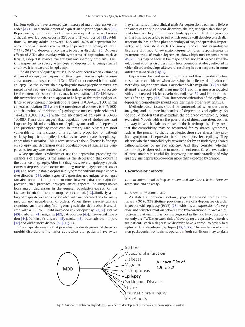
A key question is whether or not the depression preceding thediagnosis of epilepsy is the same as the depression that occurs inthe absence of epilepsy. After the diagnosis, several epilepsy-specificforms of depression can occur, including interictal dysphoric disorder[38] and acute unstable depressive syndrome without major depres-sive disorder [39]; other types of depression not unique to epilepsycan also occur. It is important to note, however, that the major de-pression that precedes epilepsy onset appears indistinguishablefrom major depression in the general population except for theincrease in suicide attempt compared to controls [12]. Similarly, a his-tory of major depression is associated with an increased risk for manymedical and neurological disorders. When these associations areexamined, an interesting finding emerges. Major depression is associ-ated with a 1.9- to 3.1-fold increased risk for epilepsy [23,12], asthma[40], diabetes [41], migraine [42], osteoporosis [43], myocardial infarc-tion [44], Parkinson's disease [45], stroke [46], traumatic brain injury[47] and Alzheimer's disease [48] (Fig. 1).
The major depression that precedes the development of these co-morbid disorders is the major depression that patients have when
Fig. 1. Association between major depression and the d
they enter randomized clinical trials for depression treatment. Beforethe onset of the subsequent disorders, the major depression that pa-tients have as they enter clinical trials appears to be homogeneousin that it is not possible to tell which person will develop which dis-order on the basis of the phenomenology of major depression. Impor-tantly, and consistent with the many medical and neurologicaldisorders that may follow major depression, drug responsiveness intreatment trials of major depression shows high non-response rates[49,50]. Thismay be because themajor depression that precedes the de-velopment of other disorders has a heterogeneous etiology reflected inwhich disorder develops afterward, resulting in poor response in someantidepressant trials (Fig. 2).
Depression does not occur in isolation and thus disorder clustersmust also be considered when assessing the epilepsy–depression co-morbidity. Major depression is associated with migraine [42], suicideattempt is associated with migraine [51], and migraine is associatedwith an increased risk for developing epilepsy [52] and for poor prog-nosis after epilepsy [53]. Thus, further consideration of the epilepsy–depression comorbidity should consider these other relationships.
Methodological issues should be contemplated when designing,analyzing and interpreting studies of depression and epilepsy. Sotoo should models that may explain the observed comorbidity beingevaluated. Models address the possibility of direct causation, such asthe way in which diabetes causes diabetic retinopathy. They positthat the comorbidity may be accounted for by shared symptoms,such as the possibility that antiepileptic drug side effects may pro-duce symptoms of depression in studies of prevalent epilepsy. Theyaddress whether comorbidity is accounted for by an underlying sharedpathophysiology or genetic etiology. And they consider whethercomorbidity is observed due to measurement error. Careful evaluationof these models is crucial for improving our understanding of whyepilepsy and depression co-occur more than expected by chance.
3. Neurobiologic aspects
3.1. Can animal models help us understand the close relation betweendepression and epilepsy?
3.1.1. Andres M. Kanner, MDAs stated in previous sections, population-based studies have
shown a 30 to 35% lifetime prevalence rate of a depressive disorderin people with epilepsy (PWE) [28], which is an expression of a veryclose and complex relation between the two conditions. In fact, a bidi-rectional relationship has been recognized in the last two decades asnot only are PWE at greater risk of developing a depressive disorder,but patients with a depressive disorder have a three- to seven-foldhigher risk of developing epilepsy [12,23,25]. The existence of com-mon pathogenic mechanisms operant in both conditions may explain
evelopment of medical and neurological disorders.
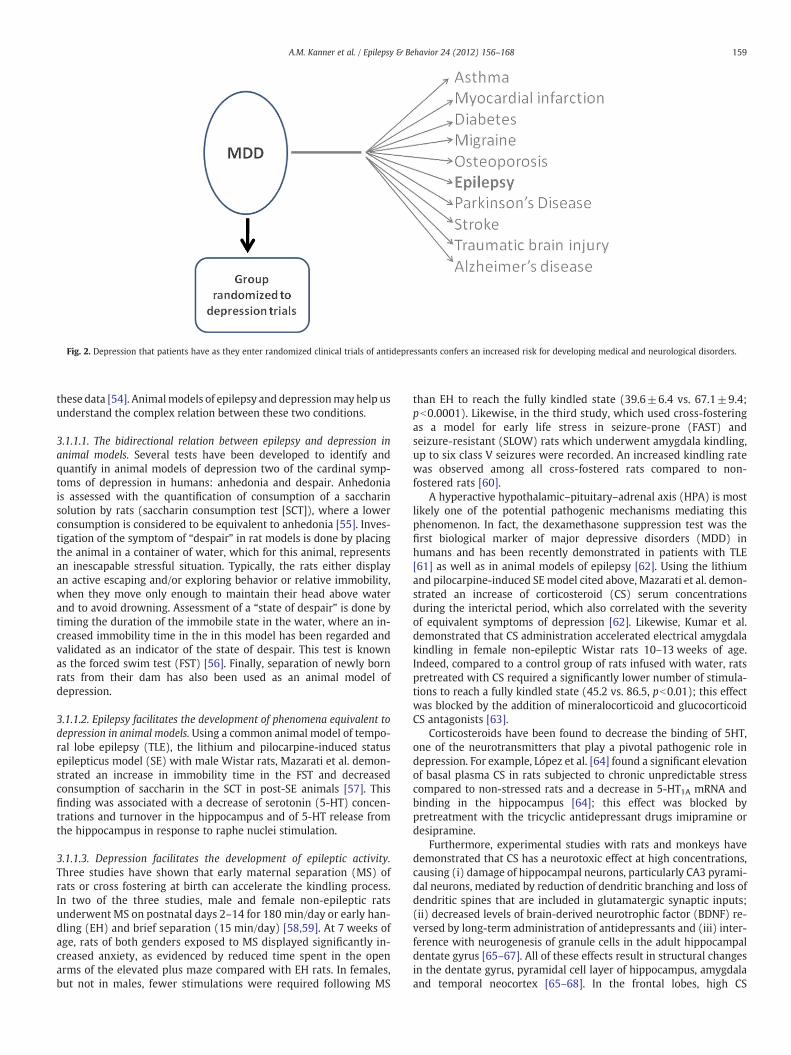
Fig. 2. Depression that patients have as they enter randomized clinical trials of antidepressants confers an increased risk for developing medical and neurological disorders.
159A.M. Kanner et al. / Epilepsy & Behavior 24 (2012) 156–168
these data [54]. Animalmodels of epilepsy and depressionmay help usunderstand the complex relation between these two conditions.
3.1.1.1. The bidirectional relation between epilepsy and depression inanimal models. Several tests have been developed to identify andquantify in animal models of depression two of the cardinal symp-toms of depression in humans: anhedonia and despair. Anhedoniais assessed with the quantification of consumption of a saccharinsolution by rats (saccharin consumption test [SCT]), where a lowerconsumption is considered to be equivalent to anhedonia [55]. Inves-tigation of the symptom of “despair” in rat models is done by placingthe animal in a container of water, which for this animal, representsan inescapable stressful situation. Typically, the rats either displayan active escaping and/or exploring behavior or relative immobility,when they move only enough to maintain their head above waterand to avoid drowning. Assessment of a “state of despair” is done bytiming the duration of the immobile state in the water, where an in-creased immobility time in the in this model has been regarded andvalidated as an indicator of the state of despair. This test is knownas the forced swim test (FST) [56]. Finally, separation of newly bornrats from their dam has also been used as an animal model ofdepression.
3.1.1.2. Epilepsy facilitates the development of phenomena equivalent todepression in animal models. Using a common animal model of tempo-ral lobe epilepsy (TLE), the lithium and pilocarpine-induced statusepilepticus model (SE) with male Wistar rats, Mazarati et al. demon-strated an increase in immobility time in the FST and decreasedconsumption of saccharin in the SCT in post-SE animals [57]. Thisfinding was associated with a decrease of serotonin (5-HT) concen-trations and turnover in the hippocampus and of 5-HT release fromthe hippocampus in response to raphe nuclei stimulation.
3.1.1.3. Depression facilitates the development of epileptic activity.Three studies have shown that early maternal separation (MS) ofrats or cross fostering at birth can accelerate the kindling process.In two of the three studies, male and female non-epileptic ratsunderwent MS on postnatal days 2–14 for 180 min/day or early han-dling (EH) and brief separation (15 min/day) [58,59]. At 7 weeks ofage, rats of both genders exposed to MS displayed significantly in-creased anxiety, as evidenced by reduced time spent in the openarms of the elevated plus maze compared with EH rats. In females,but not in males, fewer stimulations were required following MS
than EH to reach the fully kindled state (39.6±6.4 vs. 67.1±9.4;pb0.0001). Likewise, in the third study, which used cross-fosteringas a model for early life stress in seizure-prone (FAST) andseizure-resistant (SLOW) rats which underwent amygdala kindling,up to six class V seizures were recorded. An increased kindling ratewas observed among all cross-fostered rats compared to non-fostered rats [60].
A hyperactive hypothalamic–pituitary–adrenal axis (HPA) is mostlikely one of the potential pathogenic mechanisms mediating thisphenomenon. In fact, the dexamethasone suppression test was thefirst biological marker of major depressive disorders (MDD) inhumans and has been recently demonstrated in patients with TLE[61] as well as in animal models of epilepsy [62]. Using the lithiumand pilocarpine-induced SEmodel cited above, Mazarati et al. demon-strated an increase of corticosteroid (CS) serum concentrationsduring the interictal period, which also correlated with the severityof equivalent symptoms of depression [62]. Likewise, Kumar et al.demonstrated that CS administration accelerated electrical amygdalakindling in female non-epileptic Wistar rats 10–13 weeks of age.Indeed, compared to a control group of rats infused with water, ratspretreated with CS required a significantly lower number of stimula-tions to reach a fully kindled state (45.2 vs. 86.5, pb0.01); this effectwas blocked by the addition of mineralocorticoid and glucocorticoidCS antagonists [63].
Corticosteroids have been found to decrease the binding of 5HT,one of the neurotransmitters that play a pivotal pathogenic role indepression. For example, López et al. [64] found a significant elevationof basal plasma CS in rats subjected to chronic unpredictable stresscompared to non-stressed rats and a decrease in 5-HT1A mRNA andbinding in the hippocampus [64]; this effect was blocked bypretreatment with the tricyclic antidepressant drugs imipramine ordesipramine.
Furthermore, experimental studies with rats and monkeys havedemonstrated that CS has a neurotoxic effect at high concentrations,causing (i) damage of hippocampal neurons, particularly CA3 pyrami-dal neurons, mediated by reduction of dendritic branching and loss ofdendritic spines that are included in glutamatergic synaptic inputs;(ii) decreased levels of brain-derived neurotrophic factor (BDNF) re-versed by long-term administration of antidepressants and (iii) inter-ference with neurogenesis of granule cells in the adult hippocampaldentate gyrus [65–67]. All of these effects result in structural changesin the dentate gyrus, pyramidal cell layer of hippocampus, amygdalaand temporal neocortex [65–68]. In the frontal lobes, high CS

160 A.M. Kanner et al. / Epilepsy & Behavior 24 (2012) 156–168
secretion has been associated with a decrease in glial cell numbersin subgenual, cingulated and dorsolateral sections of the prefrontalcortex [69–73]. Finally, elevated levels of glucocorticoids reduce as-trocytes' activity and interfere with their function. In this mannerthey may undermine neuronal and cortical function in MDD bycausing the accumulation of excessive synaptic glutamate (seebelow) [74].
In epilepsy, the pathogenic role of the excitatory neurotransmitterglutamate has been established for a long time. In depressive disorders,the recognition of glutamate's potential pathogenic role has beensuggested in studies of experimental models of depression and inpharmacologic, neuropathologic and neuroimaging studies in humans.The available data are suggestive of a dysfunction in the regulationof glutamate neurotransmission, but the actual operant mechanismsare complex and yet to be elucidated [75,76]. First, glutamatergicand monoaminergic systems are closely interconnected as evidencedby the projection of glutamatergic neurons from the cortex to thelocus coeruleus, raphe nucleus, and substantia nigra. Likewise, seroto-nergic and noradrenergic agents can interfere with the neurotransmis-sion of glutamate. For example, chronic treatment with the SSRIfluoxetine, the SNRI reboxetine and the norepinephrine reuptakeinhibitor desipramine causes a reduction of depolarization-evokedrelease of glutamate [77,78].
Three lines of evidence support a pathogenic role of glutamateand GABA in DD: (i) dysfunction of glutamate transporter proteins,(ii) abnormal concentrations of cortical glutamate and GABA iden-tified with functional neuroimaging studies using proton magneticresonance spectroscopy (H1-MRS) and neuropathologic studies,and (iii) antidepressant effects of glutamate receptor antagonists[76,79].
In animal models of depression, glutamatergic dysfunction hasbeen suggested to be related to disturbance of glutamate transporters(vGluT1 and excitatory amino acid transporters EAAT-1, EEAT-2(found primarily in glial cells), EEAT-3 (localized principally in neu-rons) and EEAT-4 (localized in the cerebellum)) [76,80]. Glutamatetransporters play pivotal roles in the maintenance of low extracellularconcentrations of glutamate, a mechanism through which they pro-tect neurons from excitotoxic damage, and they limit the amplitudeand duration of excitatory postsynaptic currents in glutamatergicsynapses. Since there is no process by which glutamate is metabo-lized, its synaptic concentrations depend on re-uptake of these trans-porter proteins localized on glia and neurons [81]. Using an animalmodel of depression with Sprague Dawley rats, Zink et al. [80]showed a significantly suppressed expression of the glial glutamatetransporter EAAT2 in hippocampus and cerebral cortex compared tolittermates with low failure rate and asymptomatic animals [80]. Fur-ther, elevated extracellular glutamate levels, neuronal death, and ep-ilepsy were related to decreased expression and function of glialglutamate transporters in a mouse model of tuberous sclerosis(Tsc1(GFAP)CKO mice), involving inactivation of the Tsc1 gene inglial cells [82]. In this study, early treatment with ceftriaxone priorto the onset of epilepsy increased expression of glial glutamate trans-porters, decreased extracellular glutamate levels, neuronal death, andseizure frequency, and improved survival in Tsc1(GFAP)CKO mice.However, after the start of epilepsy, treatment with this drug hadno impact on seizures.
There is growing evidence suggesting antidepressant effectsof various NMDA and metabotropic antagonists (including MK-801,ketamine, mGluR5 antagonist 2-methyl-6-(phenylethynyl)-pyridine(MPEP), and the MGluR2/3 antagonists LY341495 and MGS0039) inanimal models of depression including the FST and the tail suspension-induced immobility tests, and in learned helplessness models ofdepression [75,74].
Neuro-inflammatory pathogenic mechanisms have been identi-fied in experimental models of TLE and humans with epilepsy [83]and in depressive disorders. Using the lithium pilocarpine model
of SE with Wistar rats, Mazarati et al. found that continuous blockadeof hippocampal IL-1 receptors achieved by a two week-long intrahip-pocampal infusion of human recombinant interleukin-1 receptorantagonist (hr-IL1Ra [84]) attenuated all examined behavioral, endo-crine and biochemical hallmarks of depression described above.Of note, Dunn et al. have shown that IL-1β may cause symptomsof depression by causing disturbance of the HPA axis [85]. Likewise,Vezzani et al. found that when administered into the lateralbrain ventricle, hrIL-1Ra has anticonvulsant properties in an animalmodel of TLE [83] or systematically, while hrIL-1Ra improved the pa-rameters of depression in epileptic rats selectively, without affecting re-sponses in naïve animals [86].
3.1.1.4. Are there animal models of comorbid epilepsy and depression?The genetically epilepsy-prone rat (GEPR), with its two strainsGEPR3 and GEPR9, is an animal model of epilepsy with comorbidbehaviors equivalent to depressive symptomatology manifested bydecreased saccharin consumption in the SCT and increased immobil-ity time in the FST [87]. The GEPR harbors inborn defects in pre- andpostsynaptic transmission of both serotonin (5-HT) and norepineph-rine (NE) resulting from deficient arborization of noradrenergic andserotonergic neurons arising from the locus coeruleus and raphenuclei [88]. The pathogenic role of 5HT and NE was demonstratedby the worsening of seizures with substances that interfere with thesynthesis or release of NE (reserpine and tetrabenazine, whichinactivate NE storage vesicles; α-methyl-m-tyrosine, a false NEtransmitter; α-methyl-ρ-tyrosine, a NE synthesis inhibitor) or 5-HT(ρ-chlorophenylalanine, a 5-HT synthesis inhibitor). Conversely,treatment with noradrenergic (desipramine) and serotonergic drugs(selective serotonin-reuptake inhibitors, fluoxetine and sertraline)has been shown to block seizures. Similar anticonvulsant effects ofserotonergic and noradrenergic drugs have been demonstrated inother animal models, including non-genetic animal models in therat, rabbit, cat and monkey [89–92].
Serotonin's anticonvulsant effect may also be mediated throughdirect and indirect mechanisms, the latter including inhibition ofvoltage-gated ion channels and effects on inhibitory neurotransmitterreceptors (gamma-amino butyric acid [GABA]), excitatory receptors(e.g., glutamate) and neurosteroid synthesis [93]. Of note, the seroto-nergic anticonvulsant effect appears to have an “inverted u-shaped”concentration–response effect, as suggested by a study in pilocarpine-induced seizures inwhich hippocampal perfusion of 5HTup to extracel-lular concentrations ranging between 80 and 350% of baseline levelsprotected these rats from seizures, while concentrations>900% ofbaseline worsened seizures [94]. Of note, the high extracellular 5-HTconcentrations were associated with significant increases in extracellu-lar glutamate.
The genetic absence epilepsy rats from Strasbourg (GAERS) illus-trate the existence of epilepsy and comorbid semiology equivalentto symptoms of depression and anxiety in an animal model of gener-alized epilepsy [95]. In one study, 47 GAERS and 73 non-epileptic con-trol rats (NEC) were subjected to behavioral measures of depression[the sucrose-preference test (SPT)] and anxiety [the elevated plusmaze (EPM), and the open field arena (OFA)] at 7 and 13 weeks ofage, ages prior to and after seizure onset. The GAERS exhibited signif-icantly greater levels of behaviors equivalent to symptoms of depres-sion and anxiety on these measures, including reduced consumptionof sucrose solution in the SPT (equivalent to anhedonia in humans),lower percentage of time in the open arms of the EPM and reducedexploratory activity and less time spent in the inner area of the OFA(equivalent to anxiety symptoms). These differences were evidentpreceding and following the onset of epilepsy. Clearly, increasedanxiety- and depressive-like behaviors in GAERS were not a conse-quence of seizures, but rather an expression of comorbidneurologic- and psychiatric-like conditions.

161A.M. Kanner et al. / Epilepsy & Behavior 24 (2012) 156–168
3.2. Neuroimaging of depression in epilepsy
3.2.1. Frank F. Gilliam, MD, MPHEfforts to image cerebral biomarkers of psychiatric comorbidities
in persons with epilepsy began two decades ago, and have made sig-nificant contributions to our understanding of the neurobiological un-derpinnings of depression and anxiety [96,97]. Although we do notyet have evidence to support the use of neuroimaging in the diagnosisof psychiatric disorders in individuals with epilepsy, we do have dataregarding the association of imaging abnormalities of specific brainregions with symptoms of depression and anxiety. This sectionreviews representative prior studies of each neuroimaging modalityin depression or anxiety in epilepsy.
One of the initial studies of cerebral metabolism and depression inpatients with partial seizures was performed by Bromfield et al. nearly20 years ago [98]. The investigators compared activity of 18
fluoro-2-deoxyglucose positron emission tomography (FDG-PET) in nineindividual brain regions to depression symptoms assessed by theBeck Depression Inventory (BDI). The time interval between imag-ing and depression assessment varies widely between publishedinvestigations, so it is relevant that in this study, the majority ofsubjects scored the BDI within 24 h of having the FDG-PET. All butone of the 23 subjects scored the BDI within 2 weeks of the FDG-PET. Multifactorial analysis of variance showed an interaction ofmood with non-lateralized inferior frontal regions. These resultsare consistent with prior human and other primate studies ofmood function and limbic network connectivity [99]. Using statisti-cal parametric mapping (SPM-99) FDG-PET and psychiatric assess-ment of lifetime history of epilepsy, another group of investigatorsalso found an association of orbitofrontal hypometabolism with aprior history of depression [100]. Depression has also been associat-ed with temporal lobe FDG-PET hypometabolism in subjects withtemporal lobe epilepsy [101].
Despite the extensive use of MRI in patients with epilepsy, fewstudies have evaluated an association with depression or anxiety.Hippocampal abnormalities have been variably associated withdepression in persons with epilepsy. An early study by Quiskeet al. found that subjects with hippocampal sclerosis had higherBDI scores than those with temporal lobe epilepsy with no hippo-campal abnormality [102]. However, the mean BDI scores in thesclerosis group were in the mild depression range. This findingwas replicated in another study, but the atrophy was specific tothe hippocampus contralateral to the hemisphere of seizure onset[103]. A subsequent study using a diagnosis of depression basedon the structured clinical inventory for DSM-IV (SCID) also foundan association of hippocampal atrophy with depression [103,104].Other studies have not found an association of hippocampal abnor-malities with depression [105].
Structural abnormalities in the amygdala have, interestingly, beenmore consistently associated with mood dysfunction. An early MRIstudy found an association of enlargement of the amygdala bilaterallywith dysthymia and enlargement of the left amygdala with depres-sion on the BDI [20]. This finding has been replicated in other similarstudies [105]. The FDG-PET findings in the amygdala have beeninconsistent, perhaps due to resolution limitations for a relativelysmall structure.
Other imaging modalities have offered insights into the relation-ship of epileptogenicity of limbic structures and depression. Forexample, decreased N-acetyl aspartate (NAA) on MR spectroscopyhas been associated with regions of increased spike rate and seizureonset on EEG [106,107]. A study of MR spectroscopy and depressionin 31 subjects with temporal lobe epilepsy found that the extentof voxels in the hippocampi with decreased NAA was linearlyassociated with severity of depression symptoms [108]. No othervariable was associated with depression, including seizure rate,number of seizure medications, or self-assessed social and
vocational disability. The finding suggests that chronic hyperexcitabil-ity of the hippocampus may negatively influence the limbic networktoward a depressed state.
Serotonin system dysfunction has long been associated with de-pression, which is supported by the role of serotonin reuptake inhib-itors in the treatment of depression. Several studies using serotoninreceptor ligands for PET have yielded important insights into theneurobiology of depression in temporal lobe epilepsy. A series of in-vestigations from the National Institutes of Health has found associa-tions of CWAY PET with depression, including deceased hippocampalbinding with severity of depression symptoms on the BDI [109,110].Alternatively, Swedish investigators found decreased binding in thecingulate region in subjects with epilepsy and greater depressionsymptoms [111]. Using a different serotonin ligand, MPPF, Frenchinvestigators in Lyon employed a creative strategy to compare theresults of a factor analysis of the BDI to SPM of the MPPF PET results[112]. It was found that “the total BDI score, as well as symptomsof psychomotor anhedonia and negative cognition, correlated posi-tively with [(18)F]MPPF BP in the raphe nuclei and in the insulacontralateral to seizure onset [112], whereas somatic symptomscorrelated positively with [(18)F]MPPF binding potential in the hippo-campal/parahippocampal region ipsilateral to seizure onset, theleft mid-cingulate gyrus and the inferior dorsolateral frontal cortex,bilaterally”. This unique pattern of serotonin receptor binding mayprovide greater understanding of the role of specific limbic structureswith unique patterns of mood, cognitive, and somatic symptomsof depression in temporal lobe epilepsy, which may allow forimproved treatments.
Although the associations of aspects of depression with braindysfunction in epilepsy are complex, research utilizing advanced neu-roimaging techniques is improving our understanding of affectivedisorders in persons with recurrent seizures. In fact, specific epilepsysyndromes may serve as unique models of depression to allowgreater understanding of the more general experience of depressivesymptoms [101]. Advances in our understanding of depression inepilepsy should also serve as the basis for improving treatment andclinical outcomes of depression and anxiety for persons with epilepsy.
3.3. Left orbitofrontal cortical thinning predicts depressive symptomatologyin patients with temporal lobe epilepsy
3.3.1. D.C. Jackson, PhD; N.M. Walker, BS; K. Dabbs, MS; J.E. Jones, PhD;M. Seidenberg, PhD; B.P. Hermann, PhD
As discussed throughout this review, depression in epilepsy iscommon, frequently underdiagnosed and undertreated, and tightlylinked to compromised quality of life. The etiology of depression inepilepsy has been controversial and over the years the pendulumhas swung between theories focusing on the role of psychosocialversus neurobiological causes. Psychosocial theories have focusedon the complications of epilepsy including under- and unemploy-ment, financial stresses, and social isolation and stigma. In contrast,others have suggested that the link between epilepsy and depressionis due to common or shared neurobiological substrates [23,25].In support of the neurobiological view, imaging of depression in thegeneral population has revealed structural and/or functional abnor-malities in several brain regions, including hippocampus, amygdala,and orbitofrontal and anterior cingulate cortex [113].
Abnormalities in these and other regions have also been identifiedin volumetric [104] and VBM-based [114] investigations of patientswith chronic epilepsy [115]. Further, abnormalities in cortical thick-ness have been reported. Lin et al. [116] characterized significant cor-tical thinning in patients with mesial temporal lobe epilepsy (MTLE)compared to controls in the bilateral frontal poles, frontal operculum,orbitofrontal cortex, lateral temporal and occipital regions. Bernhardtet al. [117] reported decreased cortical thickness bilaterally in thefrontal, lateral temporal, and cingulate cortex in people with MTLE

Fig. 3.
162 A.M. Kanner et al. / Epilepsy & Behavior 24 (2012) 156–168
with and without hippocampal atrophy compared to controls.Despite these well-characterized anatomic abnormalities, the rela-tionship between cortical thickness and depressive symptoms hasyet to be examined in people with epilepsy.
We examined the relationship between cortical thickness anddepressive symptoms in patients with TLE and healthy controls. Wefirst attempted to identify anatomic regions that were associatedwith depressive symptoms in patients with TLE versus controls, andthen looked within the TLE group specifically to confirm corticalthickness–depression relationships.
3.3.1.1. Method3.3.1.1.1. Participants. The study sample consisted of 53 patients
with temporal lobe epilepsy (TLE) and 52 healthy controls (HC).Selection criteria for participants with epilepsy included: a) chrono-logical age between 18 and 63 years, b) WAIS-III IQ>69, c) complexpartial seizures of definite or probable temporal lobe origin basedon consensus conference review, d) no MRI abnormalities otherthan atrophy on clinical interpretation, and e) no other neurologicaldisorder. Control participants were friends or relatives of TLEpatients. Table 1 provides baseline demographic characteristics ofthe participants and selected seizure variables for the TLE group. Allparticipants completed the Beck Depression Inventory II (BDI-2),a self-report scale assessing depressive symptoms with excellentvalidity and reliability [118]. Approval to conduct this study wasgranted by the Health Sciences Institutional Review Board at theUniversity of Wisconsin School of Medicine and Public Health.
3.3.1.1.2. MRI data acquisition and processing. Images wereobtained on a 1.5-T GE Signa MR scanner. Image acquisition foreach participant included a T1-weighted, three-dimensional SPGRacquired with the following parameters: TE=5, TR=24, flipangle=40, NEX=1, slice thickness=1.5 mm, slices=124, plane=coronal, FOV=200, matrix=256×256. Images were transferred toa Mac OSX computer for processing with the FreeSurfer image analy-sis suite which is documented and freely available for downloadonline (http://surfer.nmr.mgh.harvard.edu/). The T1 volumetric MRIscan was used for cortical reconstruction and volumetric segmenta-tion. The technical details of these procedures are found in Dabbset al. [119].
To investigate the difference in cortical thickness between groups,we performed a surface-based group analysis using Freesurfer's sta-tistical tool, QDEC 1.4. The subjects' surface data were first smoothedwith a 10-mm FWHM kernel to improve inter-subject averaging.A general linear model was then applied at each vertex to test forgroup differences in cortical thickness. To correct for multiple com-parisons, a Monte-Carlo simulation as implemented in Freesurferwas used for cluster analysis. The initial cluster forming thresholdwas set at pb0.05. Clusters were then tested against an empiricalnull distribution of maximum cluster size built using synthesized Z-distributed data across 10,000 permutations, producing cluster-wisep-values (CWP) fully corrected for multiple comparisons.
Table 1Demographics, seizure variables, and Beck Depression Inventory—II scores for temporallobe epilepsy (TLE) and control participants.
Demographics
Controls (n=53) TLE (n=54)
Age (yrs) 31.36 35.31 NsGender (%F) 67.9 74.1 NsYears of education 13.87 13.02 NsFull scale IQ 108.51 93.33 pb .001BDI score 4.23 10.23 pb .001Duration of epilepsy (mo) – 251.43 –
Age at onset (mo) – 172.02 –
3.3.1.2. Results. As anticipated, mean Beck Depression Inventory scoreswere significantly elevated in the TLE (mean=10.23; SD=8.84) com-pared to the HC (mean=4.23; SD=3.46) groups, t(103)=4.59,pb .001. The left panel of Fig. 1 depicts the regions with group differ-ences in cortical thickness as a function of self-reported depressivesymptoms, after correcting for multiple comparisons. Identified regionsincluded the left anterior cingulate and left orbitofrontal cortex. Noother area of neocortex separated the groups. Because this imageresulted from contrasting cortical thickness–depression relationshipsin the epilepsy versus control groups, the right panel of Fig. 1 specificallydepicts the relationship between depressive symptoms and corticalthickness in the TLE group only, where there is a specific association be-tween increasing depression and left orbitofrontal thinning.
The left panel of Fig. 3 shows regions significantly discriminatingtemporal lobe epilepsy (TLE) and healthy control (HC) groups forBeck Depression Inventory (BDI) by cortical thickness correlation,after correcting for multiple comparisons. The right panel of Fig. 1 de-picts regions showing significant cortical thickness relationships withthe BDI for the TLE group.
3.3.1.3. Discussion. This investigation identified anatomic areas associ-ated with self-reported depressive symptoms in patients with TLEand healthy controls. The critical regions separating the groups in-clude the left anterior cingulate gyrus and left orbitofrontal cortex(left panel of the figure). When examining TLE patients specifically,left orbitofrontal thinning is associated with increasing depressivesymptoms in TLE (right panel of the figure).
These findings suggest an anatomical correlate to depressed moodin epilepsy, consistent with the known neuroanatomy of depressionin the general population. However, our findings should be inter-preted with caution. We examined depressive symptoms and notspecific mood disorders or major depression. How these findingsmight hold for individuals with frank depressive disorders remainsto be determined. Second, the meaning of cortical thinning andits relationship to depressive symptoms remains unclear. Bothorbitofrontal and anterior cingulate cortices play a prominent rolein current theoretical formulations of the neurocircuitry of bothdepression [120] and regulation of negative affect [121]. The relation-ship observed here between cortical thinning and depressive symp-toms may be the consequence of primary pathology to left anteriorcingulate and orbitofrontal cortex, or may result from pathology indistant but related regions.
3.4. PET imaging of epilepsy and depression
3.4.1. Philippe Ryvlin, MD, PhDA large number of PET studies have been performed in major de-
pressive disorder, primarily looking at resting state glucose metabo-lism and various aspects of serotoninergic neurotransmission, before

163A.M. Kanner et al. / Epilepsy & Behavior 24 (2012) 156–168
and/or after antidepressant treatment. In contrast, only a handfulof studies are available in patients with epilepsy and comorbid de-pression, mostly investigating patients with TLE, glucose metabolismand 5-HT1A receptors. Furthermore, interpretation of depression-associated PET abnormalities is complicated in epilepsy by the pres-ence of seizure-related focal hypometabolism and decreased 5-HT1Areceptor density [122–127]. In this section, we will briefly summarizethe knowledge accumulated in the field of major depressive disorderand discuss in more detail the data obtained in epilepsy anddepression.
In major depressive disorder, resting state FDG-PET studies havedemonstrated a complex pattern where some brain regions displayhypometabolism, including the dorso- and ventro-lateral prefrontalcortex, the inferior parietal lobule, and the dorsal anterior andposterior cingulate cortices, while limbic and paralimbic structuresshow hypermetabolism, such as the subgenual cingulate cortex,the amygdala and hippocampus, and the insula [128–130]. Thiscomplex pattern suggests altered functional relations between the var-ious structures involved in the regulation of emotions andmood, whichtend to be normalized by effective antidepressant treatments[130–133]. Few FDG-PET studies have investigated comorbid depres-sion in patientswith epilepsy, typically showing an association betweenthe presence or the intensity of depressive symptoms and glucosehypometabolism in different brain regions. Such correlation wasobserved over the inferior frontal cortex [98], the orbitofrontal cortexipsilateral to the epileptogenic temporal lobe [134], and both temporallobes [101,135]. Thus, the “dysmetabolic” pattern observed in comorbiddepression partly resembles that described in major depressive disor-ders, though not reproducing all of its classic features, such astemporo-limbic and subgenual cingulate hypermetabolism. The pres-ence of atrophy and seizure-related hypometabolism over limbic struc-tures in patients with TLE might account for the lack of detectabledepression-associated hypermetabolism in these brain regions.
The serotoninergic system has been extensively investigated inpatients with major depressive disorders, using PET tracers thatlabel 5-HT synthesis, transporters or receptors. PET studies usingalpha-[11C]methyl-L-tryptophan (AMT), a precursor of 5-HT, havereported a reduction of this tracer uptake in the anterior cingulategyrus and left mesial temporal cortex in patients with major depres-sive disorder, supporting the possibility of reduced extracellular 5-HTconcentration in depression [136,137]. PET studies of serotonin trans-porter (5-HTT), using [11C]DASB or [11C]McN5652, have reportedconflicting data in patients with major depressive disorder, thoughmost series showed increased 5-HTT binding in the thalamus and lim-bic regions [138–142]. Numerous studies have also reported 5-HT1Areceptor abnormalities in depressed patients, primarily using [11C]WAY-100 635 [143–151]. Whereas the majority of studies reporteda reduction of binding potential (BP) in various limbic and neocorticalbrain regions, as well as in the raphe nuclei of untreated and treatedpatients with major depressive disorder [143–146,148,149,152], afew series found an increased BP over the same regions in patientswith major depressive disorder never exposed to antidepressants(ADs) or not recently medicated [147,151]. Similarly an increased 5-HT1A BP has been shown in patients with major depressive disorderduring sustained remission and off antidepressant medications forat least 6 months compared with healthy controls [150]. However,using [18F]MPPF, another selective antagonist of 5-HT1A receptorswith a lower affinity, which makes it more sensitive to endogenousconcentration of 5-HT, we recently confirmed the presence of de-creased 5-HT1A expression in the anterior cingulate and orbito-frontal cortex of patients with major depressive disorder [153].These abnormalities partly recovered after 6 weeks of paroxetinetreatment [153]. In contrast, [11C]WAY-100635 PET findings werenot modified by SSRI treatment [146,154,155].
In epilepsy and depression, PET investigations of the serotoniner-gic system have concentrated on 5-HT1A receptors in patients with
TLE. Using [11C]WAY-100635 and [18F]FC-WAY, two groups reportedreduced BP in the more depressed patients. This abnormality was pri-marily observed ipsilateral to the epileptogenic temporal lobe, andmore specifically over the anterior cingulate gyrus [111] and the hip-pocampus [109,110]. In a larger sample of TLE patients, Hasler et al.[156] also reported lower [18F]FCWAY binding in patients with ahistory of depression as compared to those without such history inhippocampus, temporal neocortex, anterior insula, anterior cingulateand raphe nuclei. In contrast with these findings, we observed greaterbinding potential of [18F]MPPF in the more depressed patients, partic-ularly within the insula contralateral to seizure onset as well as inthe raphe nuclei [112]. Interestingly, a different set of brain regionswas associated with each of the main dimensions explored by theBeck Depression Inventory (BDI), with the insula and RAPHE NUCLEIabnormalities being associated with symptoms of psychomotoranhedonia and negative cognition, whereas somatic symptomscorrelated with [18F]MPPF BP in the anterior cingulate gyrus andhippocampus ipsilateral to seizure onset.
Considering the opposite direction of [11C]WAY-100635 and [18F]MPPF PET abnormalities, as well as the differential sensitivity of thesetwo 5-HT1A antagonists to the extra-cellular concentration of endog-enous serotonin, we concluded that patients with TLE and depressionare most likely characterized by a combination of decreased expres-sion of 5-HT1A receptors and reduced extra-cellular concentration of5-HT, predominating over limbic brain regions and raphe nuclei[112]. However, a recent [11C]WAY-100635 PET study performed ina small population of 13 patients with TLE with or without hippocam-pal sclerosis failed to report any correlation between binding poten-tial and depression [157]. Discordance between PET studies of 5-HT1A receptors in patients with major depressive disorder or epilepsyand depression might also reflect a difference in the modelingmethods used to calculate BP [158], the choice of the reference region(inclusion of cerebellar vermis and gray matter in the reference re-gion), as well as the selected patient samples. Indeed, one shouldbear in mind that the genetic background for each subject and thegene-by-environment interaction can have a significant influence onPET findings [159,160]. For instance, [18F]MPPF and [11C]WAY-100635 BP were reported to be influenced by several genetic factors,including the triallelic 5-HT transporter gene-linked polymorphic re-gion and 5-HT1A promoter polymorphism [161–164].
Overall, PET studies of glucose metabolism and 5-HT1A receptorsin patients with TLE and depression are grossly consistent withthose performed in primary major depressive disorder, primarilyshowing hypometabolism, decreased expression of 5-HT1A receptors,and possibly decreased extra-cellular concentration of 5-HT, overbrain regions involved in the regulation of mood and emotions. Dif-ferences observed between patients with major depressive disorderand those with epilepsy and depression are difficult to interpret dueto: 1) the discordant findings reported in each group of patients,separately, preventing any firm conclusion regarding the existenceof unified patterns of PET abnormalities underlying each condition;and 2) the confounding impact of atrophy, seizures, and possibly anti-epileptic drugs on PET studies of glucose metabolism and 5-HT1Areceptors. Further studies, ideally combining MDD, depressed andnot depressed TLE patients, are warranted to address these issues.
3.5. Does a history of depression increase the risk of worse response totherapy in epilepsy?
3.5.1. Andres M. Kanner, MDDepressive disorders have been typically considered to be a com-
plication of epilepsy. Yet, as discussed in Section 2.2.1., population-based studies published in the last 15 years have suggested that notonly are people with epilepsy at greater risk of developing depressionbut patients with a history of depression have four- to seven-foldhigher risk of developing epilepsy [12,23,25,28]. This risk is not

164 A.M. Kanner et al. / Epilepsy & Behavior 24 (2012) 156–168
limited to adults but was identified when children were included in apopulation-based study done in Iceland [28]. In addition, ina retrospective cohort study of 133,440 pediatric patients (ages6–17 years) without a history of seizures or prior use of anticonvul-sant medications, McAfee et al. found a significantly lower incidencerate of seizures among children without psychiatric diagnoses (149per 100,000 person-years) than among children with psychiatricdiagnoses, other than ADHD (513 per 100,000 person-years) [165].
The more recent observations suggesting that a history of depres-sion is associated with a higher risk of worse seizure control witheither pharmacotherapy or epilepsy surgery may be another possibleexpression of this bidirectional relation. Indeed, in a study of 780patients with new-onset epilepsy, individuals with a history ofpsychiatric disorders, and particularly depression, were two-foldless likely to be seizure-free with antiepileptic drugs (AEDs) after amedian follow-up period of 79 months compared to patients withouta psychiatric history [9]. Likewise, in a prospective study of 138 pa-tients with new-onset epilepsy, those with symptoms of depressionand anxiety at the time of diagnosis of epilepsy were significantlyless likely to be seizure-free at the one-year follow-up evaluation[166].
Several studies have found a worse postsurgical seizure outcomein patients with a lifetime history of psychiatric disorders and in par-ticular depression. For example, in a study of 100 consecutive patientswith treatment-resistant temporal lobe epilepsy (TLE) who had anantero-temporal lobectomy, those with a lifetime history of depres-sion were significantly less likely to achieve complete freedomfrom auras and disabling seizures [26] than those without such a psy-chiatric history. Likewise, Koch-Stoker et al. completed a study of 384consecutive patients with TLE who underwent an antero-temporal lo-bectomy. Patients with a pre-surgical psychiatric history (includingdepressive disorders) were significantly less likely to achievefreedom from auras and disabling seizures than those without a psy-chiatric history [Koch Stocker, personal communication]. Similarly, ina study of 121 patients with TLEwho underwent a temporal lobectomy,Anhoury et al. found a significant association between a pre-surgicalpsychiatric history and a worse postsurgical seizure outcome [167].
Do depressive disorders cause neuro-chemical and/or structuralor neuropathologic abnormalities that facilitate the development ofa hyperexcitable cortex that may explain the above cited data? Or,is the worse seizure outcome associated with depressive disordersrelated to poor compliance with AEDs or the occurrence of psycho-genic non-epileptic seizures? This latter possibility was ruled out insome of the studies cited above [166]. On the other hand, there aremultiple studies conducted in experimental animal models of epilep-sy and depression as well as in humans that support the first hypoth-esis. Data on animal models were reviewed in Section 3.1.1. In thissection, we focus on data obtained from studies done in humans.
3.5.1.1. Neurochemical disturbances that may facilitate the developmentof a hyperexcitable cortex. In epilepsy, the pathogenic role of gluta-mate, the excitatory neurotransmitter “par excellence”, has beenestablished for a long time. Yet, recent data suggest that depressivedisorders are associated with a “hyper-glutamatergic state”. Thisevidence is based on data from studies that identified: (i) abnormalconcentrations of cortical glutamate with functional neuroimagingstudies using proton magnetic resonance spectroscopy (H1-MRS)and neuropathologic studies, (ii) dysfunction of glutamate transporterproteins, and (iii) antidepressant effects of glutamate receptorantagonists [76,79].
3.5.1.1.1. Abnormal concentrations of cortical glutamate. Most H1-MRS studies in humans with major depressive and bipolar disordersused the sum of intracellular and extracellular pools of glutamate,glutamine and GABA (Glx) due to their overlapping concentrationpeaks, but which reflect an overwhelming expression of intracellularpools in neurons and glia [168,169]. It has been suggested that the
abnormal Glx measured by 1H-MRS in major depressive disorders isrelated to decreased glial cell counts resulting from high cortisolserum concentrations, which are identified in about 50% of thesepatients [170]. One study demonstrated elevated glutamate concen-trations in the occipital cortex [76]. Another study of children with bi-polar depression and one conducted with unmedicated adult patientsrevealed elevated levels of Glx in the frontal lobe and basal ganglia[171] and in the cingulate gyrus, respectively [172].
3.5.1.1.2. Dysfunction of glutamate transporter proteins. The func-tion of glutamate transporter proteins (EAAT1, EAAT2, EEAT3 andEEAT4) is to “clear” glutamate from the synaptic cleft. Thus, reducedexpression of glutamate transporter proteins results in decreasedsynaptic glutamate clearance from the synaptic cleft. In a studydone in 12 patients with pharmaco-resistant TLE, decreased expres-sion of EAAT-1 and EAAT-2 was found to be associated with adecreased extracellular clearance of glutamate in CA1 of the hippo-campus by approximately 40% and 25%, respectively [173]. Likewise,decreased expression of EAAT-1 and EAAT-2 was found in frontalbrain regions of postmortem brain tissue of individuals with majordepressive disorders, while a decrease of the mRNA expression ofthe glutamate transporter proteins EAAT3 and EAAT4 was found inthe striatum [174].
3.5.1.1.3. Antidepressant effects of glutamate receptor antagonists.The NMDA antagonist ketamine has shown antidepressant effectsin two double-blind placebo-controlled studies carried out inpatients with pharmaco-resistant depression. The first study includ-ed seven patients with major depressive disorder in whom the effi-cacy of a single dose of 0.5 mg/kg was assessed in a double-blindplacebo-controlled paradigm [175]. Subjects with depression expe-rienced significant improvement in depressive symptoms within72 h after ketamine but not placebo infusion. The second study includ-ed 18 patients with treatment-resistant major depressive disorder[176]. Ketaminedemonstrated antidepressant effectswithin 2 h of a sin-gle administration of an intravenous sub-anesthetic dose in a double-blind placebo crossover study; 50% of patients met response criteriawithin 2 h and 71% by 24 h. The antidepressant effect persisted for1 to 2 weeks.
Riluzole (2-amino-6-(trifluoromethoxy) benzothiazole) has amechanism of action consisting of inhibition of glutamate receptors,but it also has effects on AMPA receptor trafficking and glutamate re-uptake. The antidepressant effects of this drug were suggested inopen trials, particularly a six-week open-label monotherapy trial car-ried out in 19 patients with treatment-resistant major depressive dis-order [177]. Riluzole has been tested in open trials as add-on therapy;in one study of eight patients with bipolar depression, combinationwith lithium for 8 weeks significantly improved depressive symp-toms [178]. Another trial of 10 patients with treatment-resistantmajor depressive disorder as add-on to antidepressant drug therapyresulted in a significant improvement in depressive symptoms aftersix to 12 weeks of treatment [179].
3.5.1.1.4. Structural abnormalities. Atrophy of mesial temporal andfrontal lobe structures has been documented with volumetric studiesin brain magnetic resonance imaging studies (MRI) of patients withprimary major depressive disorders [180,181]. Recent data suggestthat hippocampal atrophy may in fact precede the onset of primarymajor depressive disorders. Indeed, Chen et al. performed voxel-basedmorphometry analyses in brainMRI studies of 55 asymptomaticadolescent girls: 23 were considered to be at high risk for depressionas their mothers experienced recurrent episodes of depression,while the mothers of the other 32 age-matched girls (controls) hadno history of psychopathology (low-risk) [182]. Significantly lessgray matter density was found bilaterally in the hippocampal forma-tions of the girls at high-risk for depressive disorders than in thoseof the control group.
3.5.1.1.5. Neuropathologic abnormalities.A hyperactive hypothalamic–pituitary–adrenal axis resulting in high cortisol serum concentrations

165A.M. Kanner et al. / Epilepsy & Behavior 24 (2012) 156–168
can be identified in 50% of patients with major depressive disorders andhas been reported in patients with TLE [61]. In fact, neuropathologic con-sequences attributed to excessive cortisol have included: (i) decreasedglial densities and neuronal size in the cingulate gyrus, (ii) decreasedneuronal sizes and neuronal densities in layers II, III, and IV in the rostralorbito-frontal cortex resulting in a decrease of cortical thickness, (iii) asignificant decrease of glial densities in cortical layers V and VI associatedwith decreases in neuronal sizes in the caudal orbito-frontal cortex and(iiii) a decrease of neuronal and glial density and size in all cortical layersof the dorsolateral prefrontal cortex [69–71,73]. A clinical expression ofthese findings can be appreciated in a study of 48 adults withtreatment-resistant TLE that compared voxel-based morphometric ana-lyses of brain MRI among patients with (n=24) and without (n=24)major depressive episodes and a healthy control group (n=96) [183].Patients with TLE with a depressive disorder displayed a higher numberof areas of gray matter volume loss than those without depression intemporal and frontal lobe regions bilaterally and in the left thalamus.Of note, in a separate study of 165 patients with TLE, the same groupof investigators found that gray matter atrophy in patients withtreatment-resistant and remitting–relapsing epilepsy was more wide-spread than in seizure-free patients [184]. Significant differences includ-ed cortical atrophy of bilateral peri-orbital cortex, cingulum, andtemporal lobes.
In summary, the data presented here suggest the negative impactof depressive disorders on the course and response to treatmentof seizure disorders. We propose several hypotheses to explain thisphenomenon. Clearly, these hypotheses remain to be proven in futureprospective studies.
3.5.2. Concluding remarksContrary to old beliefs, the relatively high comorbidity between de-
pression and epilepsy is not only the expression of a reactive processwhereby patients face multiple obstacles associated with a life withseizures. The data presented in this review strongly suggest the exis-tence of common neurobiologic pathogenic mechanisms that mayplay a pivotal role in the high comorbidity of these two conditions.The impact of these pathogenic mechanisms is not restricted to highcomorbidity, but may play a pivotal role on the worse course and re-sponse to pharmacologic and surgical treatment of seizure disordersof patients with a lifetime history of depression.
Whether early recognition and treatment of depressive disorderswould change the impact on the higher risks that patients with de-pression have of developing epilepsy and on their response to thera-py remains to be established.
References
[1] Temkin O. The falling sickness. Baltimore: Johns Hopkins University Press; 1950.[2] Trimble MR, Reynolds EH. Anticonvulsant drugs andmental symptoms: a review.
Psychol Med 1976;6(2):169–78.[3] Trimble M. Anticonvulsant drugs, behavior, and cognitive abilities. Curr Dev
Psychopharmacol 1981;6:65–91.[4] Robertson MM, Trimble MR, Townsend HR. Phenomenology of depression in
epilepsy. Epilepsia 1987;28(4):364–72.[5] RobertsonMM, Trimble MR. The treatment of depression in patients with epilepsy.
A double-blind trial. J Affect Disord 1985;9(2):127–36.[6] Robertson MM. Suicide and epilepsy. In: Engel J, et al, editor. Epilepsy, a compre-
hensive text book. New York: Raven Press; 1996. p. 2141–52.[7] Robertson MM, Trimble MR. Depressive illness in patients with epilepsy: a review.
Epilepsia 1983;24(Supplement 2):S109–16.[8] Gilliam F, Hecimovic H, Sheline Y. Psychiatric comorbidity, health, and function
in epilepsy. Epilepsy Behav 2003;4(Suppl. 4):S26–30.[9] Hitiris N, Mohanraj R, Norrie J, Sills GJ, Brodie MJ. Predictors of pharmacoresistant
epilepsy. Epilepsy Res 2007;75(2–3):192–6.[10] Ring HA, Crellin R, Kirker S, Reynolds EH. Vigabatrin and depression. J Neurol
Neurosurg Psychiatry 1993;56(8):925–8.[11] FDA, US Department of Health and Human Services. Statistical review and
evaluation: antiepileptic drugs and suicidality; May 23 2008. Washington.[12] Hesdorffer DC, Hauser WA, Olafsson E, Ludvigsson P, Kjartansson O. Depression
and suicide attempt as risk factors for incident unprovoked seizures. Ann Neurol2005;59:35–41.
[13] Blumer D. Dysphoric disorders and paroxysmal affects: recognition and treat-ment of epilepsy-related psychiatric disorders. Harv Rev Psychiatry 2000;8:8–17.
[14] Mula M. The interictal dysphoric disorder. In: Trimble MR, Schmitz B, editors.The neuropsychiatry of epilepsy. 2nd edition. Cambridge: Cambridge UniversityPress; 2011. p. 80–7.
[15] Mendez MF, Cummings JL, Benson DF. Depression in epilepsy: significance andphenomenology. Arch Neurol 1986;43:766–70.
[16] Kanner AM. Depression and epilepsy: a new perspective on two closely relateddisorders. Epilepsy Curr 2006;6:141–6.
[17] Ettinger A, Reed M, Cramer J, et al. Depression and comorbidity in community-based patients with epilepsy or asthma. Neurology 2004;63:1008–14.
[18] Trimble MR. The psychoses of epilepsy. NY: Raven Press; 1991.[19] Schmitz EB, Moriarty J, Costa DC, et al. Psychiatric profiles and patterns of cerebral
blood flow in focal epilepsy: interactions between depression, obsessionality, andperfusion related to the laterality of the epilepsy. J Neurol Neurosurg Psychiatry1997;62:458–63.
[20] van Elst LT, Woermann FG, Lemieux L, Trimble MR. Amygdala enlargementin dysthymia—a volumetric study of patients with temporal lobe epilepsy. BiolPsychiatry 1999;46(12):1614–23.
[21] Schmitz B. The effects of antiepileptic drugs on behavior. In: Trimble MR,Schmitz B, editors. The neuropsychiatry of epilepsy. Cambridge: CambridgeUniversity Press; 2002. p. 241–55.
[22] Trimble MR. Antiepileptic drugs and suicide. In: Trimble MR, Schmitz B, editors.The neuropsychiatry of epilepsy. 2nd edition. Cambridge: Cambridge UniversityPress; 2011. p. 143–52.
[23] Hesdorffer DC, Hauser WA, Annegers JF, Cascino G. Major depression is a riskfactor for seizures in older adults. Ann Neurol 2000;47:246–9.
[24] Hesdorffer DC, Ludvigsson P, Hauser WA, Olafsson E, Kjartansson O. Co-occurrenceof major depression or suicide attempt with migraine with aura and risk forunprovoked seizure. Epilepsy Res 2007;75:220–3.
[25] Forsgren L, Nystrom L. An incident case-referent study of epileptic seizures inadults. Epilepsy Res 1990;6:66–81.
[26] Kanner AM, Byrne R, Chicharro A, Wuu J, Frey M. A lifetime psychiatric historypredicts a worse seizure outcome following temporal lobectomy. Neurology2009;72:793–9.
[27] Hitiris N, Mohanraj R, Norrie J, Sills GJ, BrodieMJ. Predictors of pharmacoresistantepilepsy. Epilepsy Res 2007;75:192–6.
[28] Tellez-Zenteno JF, Patten SB, Jette N, Williams J, Wiebe S. Psychiatric comorbidityin epilepsy: a population-based analysis. Epilepsia 2007;48:2336–44.
[29] Alper K, Schwartz KA, Kolts RL, KhanA. Seizure incidence in psychopharmacologicalclinical trials: an analysis of Food and Drug Administration (FDA) summary basis ofapproval reports. Biol Psychiatry 2007;62:345–54.
[30] Cramer JA, Blum D, Fanning K, Reed M. The impact of comorbid depressionon health resource utilization in a community sample of people with epilepsy.Epilepsy Behav 2004;5:337–42.
[31] Ettinger AB, Weisbrot DM, Nolan EE, et al. Symptoms of depression and anxietyin pediatric epilepsy patients. Epilepsia 1998;39:595–9.
[32] Kovacs M. Presentation and course of major depressive disorder duringchildhood and later years of the life span. J Am Acad Child Adolesc Psychiatry1996;35:705–15.
[33] Andreasen NC, Endicott J, Spitzer RL, Winokur G. The family history methodusing diagnostic criteria. Reliability and validity. Arch Gen Psychiatry 1977;34:1229–35.
[34] Bowman ES, Markand ON. Psychodynamics and psychiatric diagnoses of pseudo-seizure subjects. Am J Psychiatry 1996;153:57–63.
[35] Benbadis SR, Allen Hauser W. An estimate of the prevalence of psychogenic non-epileptic seizures. Seizure 2000;9:280–1.
[36] Sigurdardottir KR, Olafsson E. Incidence of psychogenic seizures in adults:a population-based study in Iceland. Epilepsia 1998;39:749–52.
[37] Duncan R. Psychogenic nonepileptic seizures: diagnosis and initial management.Expert Rev Neurother 2010;10:1803–9.
[38] Blumer D, Montouris G, Davies K. The interictal dysphoric disorder: recognition,pathogenesis, and treatment of the major psychiatric disorder of epilepsy. EpilepsyBehav 2004;5:826–40.
[39] Vaaler AE, Morken G, Iversen VC, Kondziella D, Linaker OM. Acute UnstableDepressive Syndrome (AUDS) is associated more frequently with epilepsy thanmajor depression. BMC Neurol 10:67.
[40] Jonas BS, Wagener DK, Lando JF, Feldman JJ. Symptoms of anxiety and depressionas risk factors for development of asthma. J Appl Biobehav Res 1999;4:91–110.
[41] Eaton WW, Armenian H, Gallo J, Pratt L, Ford DE. Depression and risk for onset oftype II diabetes. A prospective population-based study. Diabetes Care 1996;19:1097–102.
[42] Breslau N, Davis GC, Schultz LR, Peterson EL. Joint 1994 Wolff Award Presentation.Migraine and major depression: a longitudinal study. Headache 1994;34:387–93.
[43] Cizza G, Ravn P, Chrousos GP, Gold PW. Depression: a major, unrecognized riskfactor for osteoporosis? Trends Endocrinol Metab 2001;12:198–203.
[44] Pratt LA, Ford DE, Crum RM, Armenian HK, Gallo JJ, Eaton WW. Depression,psychotropic medication, and risk of myocardial infarction. Prospective datafrom the Baltimore ECA follow-up. Circulation 1996;94:3123–9.
[45] Schuurman AG, van den Akker M, Ensinck KT, et al. Increased risk of Parkinson'sdisease after depression: a retrospective cohort study. Neurology 2002;58:1501–4.
[46] Jonas BS, Mussolino ME. Symptoms of depression as a prospective risk factor forstroke. Psychosom Med 2000;62:463–71.
[47] Fann JR, Burington B, Leonetti A, Jaffe K, Katon WJ, Thompson RS. Psychiatricillness following traumatic brain injury in an adult health maintenance organiza-tion population. Arch Gen Psychiatry 2004;61:53–61.
166 A.M. Kanner et al. / Epilepsy & Behavior 24 (2012) 156–168
[48] Geerlings MI, Schoevers RA, Beekman AT, et al. Depression and risk of cognitivedecline and Alzheimer's disease. Results of two prospective community-basedstudies in The Netherlands. Br J Psychiatry 2000;176:568–75.
[49] Walsh BT, Seidman SN, Sysko R, Gould M. Placebo response in studies of majordepression: variable, substantial, and growing. JAMA 2002;287:1840–7.
[50] Whittington CJ, Kendall T, Fonagy P, Cottrell D, Cotgrove A, Boddington E. Selec-tive serotonin reuptake inhibitors in childhood depression: systematic review ofpublished versus unpublished data. Lancet 2004;363:1341–5.
[51] Breslau N, Davis GC, Andreski P. Migraine, psychiatric disorders, and suicideattempts: an epidemiologic study of young adults. Psychiatry Res 1991;37:11–23.
[52] Ludvigsson P, Hesdorffer D, Olafsson E, Kjartansson O, Hauser WA. Migraine withaura is a risk factor for unprovoked seizures in children. Ann Neurol 2006;59:210–3.
[53] Velioglu SK, Boz C, OzmenogluM. The impact ofmigraine on epilepsy: a prospectiveprognosis study. Cephalalgia 2005;25:528–35.
[54] Kanner AM. Depression and epilepsy: a review of multiple facets of their closerelation. Neurol Clin 2009;27:865–80.
[55] Pucilowski O, Overstreet DH, Rezvani AH, Janowsky DS. Chronic mild stress-induced anhedonia: greater effect in a genetic rat model of depression. PhysiolBehav 1993;54:1215–20.
[56] Pucilowski O, Overstreet DH. Effect of chronic antidepressant treatment on responsesto apomorphine in selectively bred rat strains. Brain Res Bull 1993;32:471–5.
[57] Mazarati AM, Siddarth P, Baldwin RA, et al. Depression after status epilepticus:behavioural and biochemical deficits and effects of fluoxetine. Brain 2008;131:2071–83.
[58] Jones NC, Kumar G, O'Brien TJ, Morris MJ, Rees SM, Salzberg MR. Anxiolyticeffects of rapid amygdala kindling, and the influence of early life experience inrats. Behav Brain Res 2009;12(203(1)):81–7.
[59] Salzberg M, Kumar G, Supit L, et al. Early postnatal stress confers enduringvulnerability to limbic epileptogenesis. Epilepsia 2007;48(11):2079–85.
[60] Gilby KL, Sydserff S, Patey AM, et al. Postnatal epigenetic influences on seizuresusceptibility in seizure-prone versus seizure-resistant rat strains. Behav Neurosci2009;123(2):337–46.
[61] Zobel A, Wellmer J, Schulze-Rauschenbach S, et al. Impairment of inhibitorycontrol of the hypothalamic pituitary adrenocortical system in epilepsy. Eur ArchPsychiatry Clin Neurosci 2004;254(5):303–11.
[62] Mazarati AM, Shin D, Kwon YS, et al. Elevated plasma corticosterone level anddepressive behavior in experimental temporal lobe epilepsy. Neurobiol Dis2009;34(3):457–61.
[63] Kumar G, Couper A, O'Brien TJ, et al. The acceleration of amygdala kindlingepileptogenesis by chronic low-dose corticosterone involves both mineralocorti-coid and glucocorticoid receptors. Psychoneuroendocrinology 2007;32(7):834–42.
[64] López JF, Chalmers DT, Little KY, Watson SJ. Regulation of serotonin1A, glucocor-ticoid, and mineralocorticoid receptor in rat and human hippocampus: implica-tions for the neurobiology of depression. Biol Psychiatry 1998;15(43(8)):547–73.
[65] Sapolsky RM. The possibility of neurotoxicity in the hippocampus in majordepression: a primer on neuron death. Biol Psychiatry 2000;15(48(8)):755–65.
[66] Sapolsky RM. Glucocorticoids and hippocampal atrophy in neuropsychiatricdisorders. Arch Gen Psychiatry 2000;57:925–35 (b).
[67] Shirayama Y, Chen AC, Nakagawa S, Russell DS, Duman RS. Brain-derived neuro-trophic factor produces antidepressant effects in behavioral models of depres-sion. J Neurosci 2002;15(22(8)):3251–61.
[68] Bowley MP, Drevets WC, Ongur D, Price JL. Low glial numbers in the amygdala inmajor depressive disorder. Biol Psychiatry 2002;52:404–12.
[69] Rajkowska G, Miguel-Hidalgo JJ, Wei J, et al. Morphometric evidence for neuro-nal and glial prefrontal cell pathology in major depression. Biol Psychiatry1999;45(9):1085–98.
[70] Cotter DR, Pariante CM, Everall IP. Glial cell abnormalities in major psychiatricdisorders: the evidence and implications. Brain Res Bull 2001;55:585–95.
[71] Cotter D, Mackay D, Landau S, Kerwin R, Everall I. Reduced glial cell density andneuronal size in the anterior cingulate cortex in major depressive disorder. ArchGen Psychiatry 2001;58:545–53.
[72] Cotter D, Mackay D, Chana G, Beasley C, Landau S, Everall IP. Reduced neuronalsize and glial cell density in area 9 of the dorsolateral prefrontal cortex insubjects with major depressive disorder. Cereb Cortex 2002;12:386–94.
[73] Öngür D, Drevets WC, Price JL. Glial reduction in the subgenual prefrontal cortexin mood disorders. Proc Natl Acad Sci U S A 1998;95:13290–5.
[74] Crossin KL, Tai MH, Krushel LA, Mauro VP, Edelman GM. Glucocorticoid receptorpathways are involved in the inhibition of astrocyte proliferation. Proc Natl AcadSci U S A 1997;94:2687–92.
[75] Machado-Vieira R, Salvadore G, Ibrahim LA, Diaz-Granados N, Zarate Jr CA.Targeting glutamatergic signaling for the development of novel therapeuticsfor mood disorders. Curr Pharm Des 2009;15(14):1595–611.
[76] Kugaya A, Sanacora G. Beyond monoamines: glutamatergic function in mooddisorders. CNS Spectr 2005;10(10):808–19.
[77] Bonanno G, Giambelli R, Raiteri L, et al. Chronic antidepressants reducedepolarization-evoked glutamate release and protein interactions favoringformation of SNARE complex in hippocampus. J Neurosci 2005;25:3270–9.
[78] Robinson R, Drafts B, Fisher J. Fluoxetine increases GABAA receptor activitythrough a novel modulatory site. J Pharmacol Exp Ther 2003;304:978–84.
[79] Brambilla P, Perez J, Barale F, Schettini G, Soares JC. GABAergic dysfunction inmood disorders. Mol Psychiatry 2003;8:721–37.
[80] Zink M, Vollmayr B, Gebicke-Haerter PJ, Henn FA. Reduced expression of gluta-mate transporters vGluT1, EAAT2 and EAAT4 in learned helpless rats, an animalmodel of depression. Neuropharmacology 2010;58(2):465–73.
[81] Zarate CA, Quiroz J, Payne J, Manji HK. Modulators of the glutamatergic system:implications for the development of improved therapeutics in mood disorders.Psychopharmacol Bull 2002;36:35–83.
[82] Zeng LH, Bero AW, Zhang B, Holtzman DM, Wong M. Modulation of astrocyteglutamate transporters decreases seizures in amousemodel of tuberous sclerosiscomplex. Neurobiol Dis 2010;37(3):764–71.
[83] Vezzani A, Balosso S, Ravizza T. The role of cytokines in the pathophysiology ofepilepsy. Brain Behav Immun 2008;22:797–803.
[84] Kalliolias GD, Liossis SN. The future of the IL-1 receptor antagonist anakinra:from rheumatoid arthritis to adult-onset Still's disease and systemic-onset juve-nile idiopathic arthritis. Expert Opin Investig Drugs 2008;17:349–59.
[85] Dunn AJ, Swiergiel AH, de Beaurepaire R. Cytokines as mediators of depression:what can we learn from animal studies? Neurosci Biobehav Rev 2005;29:891–909.
[86] Mazarati AM, ShinD, Sankar R, et al. Comorbidity between epilepsy anddepression:role of hippocampal interleukin-1β. Neurobiol Dis 2010;37:461–7.
[87] Jobe PC. Affective disorder and epilepsy comorbidity in the genetically epilepsyprone-rat (GEPR). In: Gilliam F, Kanner AM, Sheline YI, editors. Depression andbrain dysfunction. London: Taylor & Francis; 2006. p. 121–57.
[88] Jobe PC, Mishra PK, Browning RA, et al. Noradrenergic abnormalities in thegenetically epilepsy-prone rat. Brain Res Bull 1994;35:493–504.
[89] MeldrumBS, AnlezarkGM, AdamHK, et al. Anticonvulsant and proconvulsant prop-erties of viloxazine hydrochloride: pharmacological and pharmacokinetic studies inrodents and the epileptic baboon. Psychopharmacology (Berl) 1982;76:212–7.
[90] Yanagita T, Wakasa Y, Kiyohara H. Drug-dependence potential of viloxazinehydrochloride tested in rhesus monkeys. Pharmacol Biochem Behav 1980;12:155–61.
[91] Piette Y, Delaunois AL, De Shaepdryver AF, et al. Imipramine and electroshockthreshold. Arch Int Pharmacodyn Ther 1963;144:293–7.
[92] Polc P, Schneeberger J, Haefely W. Effects of several centrally active drugs on thesleep wakefulness cycle of cats. Neuropharmacology 1979;18:259–67.
[93] Ye Z, Lu Y, Sun H, Cheng H, Xu T, Zhou J. Fluoxetine inhibition of glycine receptoractivity in rat hippocampal neurons. Brain Res 2008;1239:77–84.
[94] Clinckers R, Smolders I, Meurs A, et al. Anticonvulsant action of hippocampaldopamine and serotonin is independently mediated by D2 and 5-HT1A receptors.J Neurochem 2004;89:834–43.
[95] Jones NC, Salzberg MR, Kumar G, Couper A, Morris MJ, O'Brien TJ. Elevatedanxiety and depressive-like behavior in a rat model of genetic generalizedepilepsy suggesting common causation. Exp Neurol 2008;209:254–60.
[96] Kanner AM. Depression in epilepsy: prevalence, clinical semiology, pathogenicmechanisms, and treatment. Biol Psychiatry 2003;54(3):388–98.
[97] Barry JJ, Ettinger AB, Friel P, et al. Consensus statement: the evaluation andtreatment of people with epilepsy and affective disorders. Epilepsy Behav2008;13(Suppl. 1):S1–S29.
[98] Bromfield EB, Altshuler L, Leiderman DB, et al. Cerebral metabolism and depres-sion in patients with complex partial seizures [published erratum appears inArch Neurol 1992 Sep;49(9):976]. Arch Neurol 1992;49(6):617–23.
[99] Price JL. Prefrontal cortical networks related to visceral function and mood. AnnN Y Acad Sci 1999;877:383–96.
[100] Salzberg M, Taher T, Davie M, et al. Depression in temporal lobe epilepsy surgerypatients: an FDG-PET study. Epilepsia 2006;47(12):2125–30.
[101] Gilliam FG, Santos J, Vahle V, et al. Depression in epilepsy: ignoring clinicalexpression of neuronal network dysfunction? Epilepsia 2004;45(s2):28–33.
[102] Quiske A, Helmstaedter C, Lux S, Elger CE. Depression in patients with temporallobe epilepsy is related to mesial temporal sclerosis. Epilepsy Res 2000;39(2):121–5.
[103] Finegersh A, Avedissian C, Shamim S, et al. Bilateral hippocampal atrophyin temporal lobe epilepsy: effect of depressive symptoms and febrile seizures.Epilepsia 2011;52(4):689–97.
[104] Shamim S, Hasler G, Liew C, et al. Temporal lobe epilepsy, depression, and hippo-campal volume. Epilepsia 2009;50(5):1067–71.
[105] Richardson EJ, Griffith HR, Martin RC, et al. Structural and functional neuroimagingcorrelates of depression in temporal lobe epilepsy. Epilepsy Behav 2007;10(2):242–9.
[106] Serles W, Li LM, Caramanos Z, et al. Relation of interictal spike frequency to 1H-MRSI-measured NAA/Cr. Epilepsia 1999;40(12):1821–7.
[107] Guye M, Le Fur Y, Confort-Gouny S, et al. Metabolic and electrophysiologicalalterations in subtypes of temporal lobe epilepsy: a combined protonmagnetic res-onance spectroscopic imaging and depth electrodes study. Epilepsia 2002;43(10):1197–209.
[108] Gilliam FG, Maton BM, Martin RC, et al. Hippocampal 1H-MRSI correlates with se-verity of depression symptoms in temporal lobe epilepsy. Neurology 2007;68(5):364–8.
[109] Giovacchini G, ToczekMT, Bonwetsch R, et al. 5-HT1A receptors are reduced in tem-poral lobe epilepsy after partial-volume correction. J Nucl Med 2005;46:1128–35.
[110] Theodore WH, Hasler G, Giovacchini G, et al. Reduced hippocampal 5HT1APET receptor binding and depression in temporal lobe epilepsy. Epilepsia2007;48(8):1526–30.
[111] Savic I, Lindstrom P, Gulyas B, et al. Limbic reductions of 5-HT1A receptor bindingin human temporal lobe epilepsy. Neurology 2004;62(8):1343–51.
[112] Lothe A, Didelot A, Hammers A, et al. Comorbidity between temporal lobe epi-lepsy and depression: a [18F]MPPF PET study. Brain 2008;131(Pt 10):2765–82.
[113] Davidson RJ, Jackson DC, Kalin NH. Emotion, plasticity, context, and regulation:perspectives from affective neuroscience. Psychol Bull 2000;126(6):890–909.

167A.M. Kanner et al. / Epilepsy & Behavior 24 (2012) 156–168
[114] Salgado PCB, Yasuda CL, Cender F. Neuroimaging changes in mesial temporallobe epilepsy are magnified in the presence of depression. Epilepsy Behav2010;19(3):422–7.
[115] Bell B, Lin JJ, Seidenberg M, Hermann B. The neurobiology of cognitivedisorders in temporal lobe epilepsy. Nat Rev Neurol 2011;7:154–64.
[116] Lin JJ, Salamon N, Lee AD, et al. Reduced neocortical thickness and complexitymapped in mesial temporal lobe epilepsy with hippocampal sclerosis. Cereb Cor-tex 2007;17(9):2007–18.
[117] Bernhardt BC, Bernasconi N, Concha L, Bernasconi A. Cortical thickness analysisin temporal lobe epilepsy: reproducibility and relation to outcome. Neurology2010;74:1776–85.
[118] Beck AT, Steer RA, Garbin MG. Psychometric properties of the Beck Depres-sion Inventory: twenty-five years of evaluation. Clin Psychol Rev 1988;8:77–100.
[119] Dabbs K, Jones J, Seidenberg M, Hermann B. Neuroanatomical correlates ofcognitive phenotypes in temporal lobe epilepsy. Epilepsy Behav 2009;15:445–51.
[120] Drevets WC, Price JL, Furey ML. Brain structural and functional abnormalities inmood disorders: implications for neurocircuitry models of depression. BrainStruct Funct 2008;213(1–2):83–118.
[121] McRae K, Hughes B, Chopra S, Gabrieli JD, Ochsner KN. The neural bases ofdistraction and reappraisal. J Cogn Neurosci 2010;22(2):248–62.
[122] Ryvlin P, Bouvard S, Le Bars D, et al. Clinical utility of flumazenil-PET versus [18F]fluorodeoxyglucose-PET and MRI in refractory partial epilepsy. A prospectivestudy in 100 patients. Brain 1998;121:2067–81.
[123] Toczek MT, Carson RE, Lang L, et al. PET imaging of 5-HT1A receptor binding inpatients with temporal lobe epilepsy. Neurology 2003;60:749–56.
[124] Mauguière F, Ryvlin P. The role of PET in presurgical assessment of partial epilep-sies. Epileptic Disord 2004;6:193–215.
[125] Merlet I, Ryvlin P, Costes N, et al. Statistical parametric mapping of 5-HT1Areceptor binding in temporal lobe epilepsy with hippocampal ictal onset onintracranial EEG. Neuroimage 2004;22:886–96.
[126] Merlet I, Ostrowsky K, Costes N, et al. 5-HT1A receptor binding and intracere-bral activity in temporal lobe epilepsy: an [18F]MPPF-PET study. Brain2004;127:900–13.
[127] Didelot A, Ryvlin P, Lothe A, Merlet I, Hammers A, Mauguière F. PET imaging ofbrain 5-HT1A receptors in the preoperative evaluation of temporal lobe epilepsy.Brain 2008;131:2751–64.
[128] Bench CJ, Friston KJ, Brown RG, Scott LC, Frackowiak RS, Dolan RJ. The anatomyof melancholia-focal abnormalities of cerebral blood flow in major depression.Psychol Med 1992;22:607–15.
[129] Drevets WC, Videen TO, Price JL, Preskorn SH, Carmichael ST, Raichle ME. Afunctional anatomical study of unipolar depression. J Neurosci 1992;12:3628–41.
[130] Mayberg HS. Positron emission tomography imaging in depression: a neuralsystems perspective. Neuroimaging Clin N Am 2003;13:805–15.
[131] Buchsbaum MS, Wu J, Siegel BV, et al. Effect of sertraline on regional metabolicrate in patients with affective disorder. Biol Psychiatry 1997;41:15–22.
[132] Mayberg HS, Brannan SK, Tekell JL, et al. Regional metabolic effects of fluoxetinein major depression: serial changes and relationship to clinical response. BiolPsychiatry 2000;48:830–43.
[133] Fitzgerald PB, Laird AR, Maller J, Daskalakis ZJ. A meta-analytic study of changesin brain activation in depression. Hum Brain Mapp 2008;29:683–95.
[134] Salzberg M, Taher T, Davie M, et al. Depression in temporal lobe epilepsy surgerypatients: an FDG-PET study. Epilepsia 2006;47:2125–30.
[135] Victoroff JI, Benson F, Grafton ST, Engel Jr J, Mazziotta JC. Depression in complexpartial seizures. Electroencephalography and cerebral metabolic correlates. ArchNeurol 1994;51:155–63.
[136] Rosa-Neto P, Diksic M, Okazawa H, et al. Measurement of brain regionalalpha-[11C]methyl-L-tryptophan trapping as a measure of serotonin synthesisin medication-free patients with major depression. Arch Gen Psychiatry2004;61:556–63.
[137] Berney A, Nishikawa M, Benkelfat C, Debonnel G, Gobbi G, Diksic M. An index of5-HT synthesis changes during early antidepressant treatment: alpha-[11C]methyl-L-tryptophan PET study. Neurochem Int 2008;52:701–8.
[138] Ichimiya T, Suhara T, Sudo Y, et al. Serotonin transporter binding in patients withmood disorders: a PET study with [11C](+)McN5652. Biol Psychiatry 2002;51:715–22.
[139] Meyer JH, Houle S, Sagrati S, et al. Brain serotonin transporter binding potentialmeasured with carbon 11-labeled DASB positron emission tomography: effectsof major depressive episodes and severity of dysfunctional attitudes. Arch GenPsychiatry 2004;61:1271–9.
[140] Reivich M, Amsterdam JD, Brunswick DJ, Shiue CY. PET brain imaging with[11C](+)McN5652 shows increased serotonin transporter availability in majordepression. J Affect Disord 2004;82:321–7.
[141] Parsey RV, Hastings RS, Oquendo MA, et al. Lower serotonin transporter bindingpotential in the human brain during major depressive episodes. Am J Psychiatry2006;163:52–8.
[142] Cannon DM, Ichise M, Rollis D, et al. Elevated serotonin transporter bindingin major depressive disorder assessed using positron emission tomographyand [11C]DASB; comparison with bipolar disorder. Biol Psychiatry 2007;62:870–7.
[143] Drevets WC, Frank E, Price JC, et al. PET imaging of serotonin 1A receptor bindingin depression. Biol Psychiatry 1999;46:1375–87.
[144] Drevets WC, Frank E, Price JC, Kupfer DJ, Greer PJ, Mathis C. Serotonin type-1Areceptor imaging in depression. Nucl Med Biol 2000;27:499–507.
[145] Sargent PA, Kjaer KH, Bench CJ, et al. Brain serotonin1A receptor bindingmeasured by positron emission tomography with [11C]WAY-100635: effectsof depression and antidepressant treatment. Arch Gen Psychiatry 2000;57:174–80.
[146] Bhagwagar Z, Rabiner EA, Sargent PA, Grasby PM, Cowen PJ. Persistent reduc-tion in brain serotonin1A receptor binding in recovered depressed men mea-sured by positron emission tomography with [11C]WAY-100635. MolPsychiatry 2004;9:386–92.
[147] Parsey RV, Oquendo MA, Ogden RT, et al. Altered serotonin 1A binding in majordepression: a [carbonyl-C-11]WAY100635 positron emission tomography study.Biol Psychiatry 2006;59:106–13.
[148] DrevetsWC, Thase ME, Moses-Kolko EL, et al. Serotonin-1A receptor imaging in re-current depression: replication and literature review. Nucl Med Biol 2007;34:865–77.
[149] Hirvonen J, Karlsson H, Kajander J, et al. Decreased brain serotonin 5-HT1A recep-tor availability in medication-naive patients with major depressive disorder:an in-vivo imaging study using PET and [carbonyl-11C]WAY-100635. Int JNeuropsychopharmacol 2008;11:465–76.
[150] Miller JM, Brennan KG, Ogden TR, et al. Elevated serotonin 1A binding inremitted major depressive disorder: evidence for a trait biological abnormality.Neuropsychopharmacology 2009;34:2275–84.
[151] Parsey RV, Ogden RT, Miller JM, et al. Higher serotonin 1A binding in a secondmajor depression cohort: modeling and reference region considerations. BiolPsychiatry 2010;68:170–8.
[152] Saijo T, Takano A, Suhara T, et al. Effect of electroconvulsive therapy on 5-HT1Areceptor binding in patients with depression: a PET study with [11C]WAY100635. Int J Neuropsychopharmacol 2010;13:785–91.
[153] Lothe A, Saoud M, Bouvard S, Redoute J, Lerond J, Ryvlin P. 5-HT1A receptorbinding changes in patients with major depressive disorder before and afterantidepressant treatment: a pilot [18F]MPPF positron emission tomographystudy. Psychiatry Res in press.
[154] Moses-Kolko EL, Price JC, Thase ME, et al. Measurement of 5-HT1A receptorbinding in depressed adults before and after antidepressant drug treatmentusing positron emission tomography and [11C]WAY-100635. Synapse 2007;61:523–30.
[155] Karlsson H, Hirvonen J, Kajander J, et al. Research letter: psychotherapyincreases brain serotonin 5-HT1A receptors in patients with major depressivedisorder. Psychol Med 2010;40:523–8.
[156] Hasler G, Bonwetsch R, Giovacchini G, et al. 5-HT1A receptor binding in temporallobe epilepsy patients with and without major depression. Biol Psychiatry2007;62:1258–64.
[157] Assem-Hilger E, Lanzenberger R, Savli M, et al. Central serotonin 1A receptorbinding in temporal lobe epilepsy: a [carbonyl-(11)C]WAY-100635 PET study.Epilepsy Behav 2010;19:467–73.
[158] Innis RB, Cunningham VJ, Delforge J, et al. Consensus nomenclature for in vivo im-aging of reversibly binding radioligands. J Cereb Blood Flow Metab 2007;27:1533–9.
[159] Caspi A, Sugden K, Moffitt TE, et al. Influence of life stress on depression:moderation by a polymorphism in the 5-HTT gene. Science 2003;301:386–9.
[160] Zalsman G, Huang YY, Oquendo MA, et al. Association of a triallelic serotonintransporter gene promoter region (5-HTTLPR) polymorphism with stressfullife events and severity of depression. Am J Psychiatry 2006;163:1588–93.
[161] David SP, Murthy NV, Rabiner EA, et al. A functional genetic variation of theserotonin (5-HT) transporter affects 5-HT1A receptor binding in humans. J Neu-rosci 2005;25:2586–90.
[162] Lee M, Bailer UF, Frank GK, et al. Relationship of a 5-HT transporter functionalpolymorphism to 5-HT1A receptor binding in healthy women. Mol Psychiatry2005;10:715–6.
[163] Lothe A, Boni C, Costes N, et al. Association between triallelic polymorphism ofthe serotonin transporter and [18F]MPPF binding potential at 5-HT1A receptorsin healthy subjects. Neuroimage 2009;47:482–92.
[164] Lothe A, Boni C, Costes N, et al. 5-HT1A gene promoter polymorphism and [18F]MPPF binding potential in healthy subjects: a PET study. Behav Brain Funct2010;6:37.
[165] McAfee AT, Chilcott KE, Johannes CB, Hornbuckle K, Hauser WA, Walker AM.The incidence of first unprovoked seizure in pediatric patients with and withoutpsychiatric diagnoses. Epilepsia 2007;48:1075–82.
[166] Petrovski CEI, Szoecke NC, Jones NC, et al. Neuropsychiatric symptomatology predictsseizure recurrence in newly treated patients. Neurology 2010;75:1015–21.
[167] Anhoury S, Brown RJ, Krishnamoorthy ES, Trimble MR. Psychiatric outcome aftertemporal lobectomy: a predictive study. Epilepsia 2000;41:1608–15.
[168] Walter M, Henning A, Grimm S, et al. The relationship between aberrant neuro-nal activation in the pregenual anterior cingulate, altered glutamatergic me-tabolism, and anhedonia in major depression. Arch Gen Psychiatry 2009;66(5):478–86.
[169] Capizzano AA, Jorge RE, Acion LC, Robinson RG. In vivo proton magneticresonance spectroscopy in patients with mood disorders: a technically orientedreview. J Magn Reson Imaging 2007;26(6):1378–89.
[170] Yildiz-Yesiloglu A, Ankerst DP. Review of 1H magnetic resonance spectroscopyfindings in major depressive disorder: a meta-analysis. Psychiatry Res 2006;147(1):1–25.
[171] Rosenberg DR, Mirza Y, Russell A, et al. Reduced anterior cingulate glutamatergicconcentrations in childhood OCD and major depression versus healthy controls.J Am Acad Child Adolesc Psychiatry 2004;43:1146–53.
[172] Dager SR, Friedman SD, Parow A, et al. Brain metabolic alterations in medication-free patients with bipolar disorder. Arch Gen Psychiatry 2004;61:450–8.

168 A.M. Kanner et al. / Epilepsy & Behavior 24 (2012) 156–168
[173] McCullumsmith RE, Meador-Woodruff JH. Striatal excitatory amino acidtransporter transcript expression in schizophrenia, bipolar disorder, and majordepressive disorder. Neuropsychopharmacology 2002;26:368–75.
[174] Choudary PV, Molnar M, Evans SJ, et al. Altered cortical glutamatergic andGABAergic signal transmission with glial involvement in depression. Proc NatlAcad Sci U S A 2005;102:15653–8.
[175] Berman RM, Cappiello A, Anand A, et al. Antidepressant effects of ketamine indepressed patients. Biol Psychiatry 2000;47:351–4.
[176] Zarate Jr CA, Singh JB, Carlson PJ, et al. A randomized trial of an N-methyl-D-aspartate antagonist in treatment-resistant major depression. Arch Gen Psychiatry2006;63(8):856–64.
[177] Zarate Jr CA, Payne JL, Quiroz J, et al. An open-label trial of riluzole in patientswith treatment-resistant major depression. Am J Psychiatry 2004;161:171–4.
[178] Zarate Jr CA, Quiroz JA, Singh JB, et al. An open-label trial of the glutamate-modulating agent riluzole in combination with lithium for the treatment ofbipolar depression. Biol Psychiatry 2005;57:430–2.
[179] Sanacora G, Kendell SF, Levin Y, et al. Preliminary evidence of riluzole efficacyin antidepressant-treated patients with residual depressive symptoms. BiolPsychiatry 2007;61:822–5.
[180] Sheline YI. Brain structural changes associated with depression. In: Gilliam F,Kanner AM, Sheline YI, editors. Depression and brain dysfunction. London:Taylor & Francis; 2006. p. 85–104.
[181] Bremner JD, Vythilingam M, Vermetten E, et al. Reduced volume of orbitofrontalcortex in major depression. Biol Psychiatry 2002;51(4):273–9.
[182] Chen MC, Hamilton JP, Gotlib IH. Decreased hippocampal volume in healthy girlsat risk of depression. Arch Gen Psychiatry 2010;67(3):270–6.
[183] Barioni Salgado PC, Yasuda CL, Cendes F. Neuroimaging changes in mesialtemporal lobe epilepsy are magnified in the presence of depression. EpilepsyBehav 2010;19:422–7.
[184] Bilevicius E, Yasuda CL, Silva MS, Guerreiro CAM, Lopes-Cendes I, Cendes F.Antiepileptic drug response in temporal lobe epilepsy: a clinical and MRImorphometry study. Neurology 2010;75:1695–701.