How interpersonal interactions help to explain routine dynamics

Structural Differences Explain Diverse Functions ofPlasmodium ActinsJuha Vahokoski1, Saligram Prabhakar Bhargav1., Ambroise Desfosses2., Maria Andreadaki3., Esa-
Pekka Kumpula1,4., Silvia Munico Martinez1, Alexander Ignatev1, Simone Lepper5,
Friedrich Frischknecht5, Inga Siden-Kiamos3, Carsten Sachse2, Inari Kursula1,4*
1 Faculty of Biochemistry and Molecular Medicine, University of Oulu, Oulu, Finland, 2 European Molecular Biology Laboratory, Structural and Computational Biology Unit,
Heidelberg, Germany, 3 Institute of Molecular Biology and Biotechnology, Foundation for Research and Technology – Hellas, Heraklion, Crete, Greece, 4 Centre for
Structural Systems Biology; Helmholtz Centre for Infection Research and German Electron Synchrotron, Hamburg, Germany, 5 Parasitology – Department of Infectious
Diseases, University of Heidelberg Medical School, Heidelberg, Germany
Abstract
Actins are highly conserved proteins and key players in central processes in all eukaryotic cells. The two actins of the malariaparasite are among the most divergent eukaryotic actins and also differ from each other more than isoforms in any otherspecies. Microfilaments have not been directly observed in Plasmodium and are presumed to be short and highly dynamic.We show that actin I cannot complement actin II in male gametogenesis, suggesting critical structural differences. Cryo-EMreveals that Plasmodium actin I has a unique filament structure, whereas actin II filaments resemble canonical F-actin. BothPlasmodium actins hydrolyze ATP more efficiently than a-actin, and unlike any other actin, both parasite actins rapidly formshort oligomers induced by ADP. Crystal structures of both isoforms pinpoint several structural changes in the monomerscausing the unique polymerization properties. Inserting the canonical D-loop to Plasmodium actin I leads to the formationof long filaments in vitro. In vivo, this chimera restores gametogenesis in parasites lacking actin II, suggesting that stablefilaments are required for exflagellation. Together, these data underline the divergence of eukaryotic actins anddemonstrate how structural differences in the monomers translate into filaments with different properties, implying thateven eukaryotic actins have faced different evolutionary pressures and followed different paths for developing theirpolymerization properties.
Citation: Vahokoski J, Bhargav SP, Desfosses A, Andreadaki M, Kumpula E-P, et al. (2014) Structural Differences Explain Diverse Functions of PlasmodiumActins. PLoS Pathog 10(4): e1004091. doi:10.1371/journal.ppat.1004091
Editor: Michael J. Blackman, MRC National Institute for Medical Research, United Kingdom
Received October 9, 2013; Accepted March 11, 2014; Published April 17, 2014
Copyright: � 2014 Vahokoski et al. This is an open-access article distributed under the terms of the Creative Commons Attribution License, which permitsunrestricted use, distribution, and reproduction in any medium, provided the original author and source are credited.
Funding: This study has been funded by the Academy of Finland (IK), the Sigrid Juselius Foundation (IK), the German Ministry of Education and Research (IK), anEMBL interdisciplinary postdoctoral fellowship (AD), the Human Frontier Science Program (FF), the Chica and Heinz Schaller Foundation (FF), and the EuropeanResearch Council (FF). The funders had no role in study design, data collection and analysis, decision to publish, or preparation of the manuscript.
Competing Interests: The authors have declared that no competing interests exist.
* E-mail: [email protected]
. These authors contributed equally to this work.
Introduction
Actins are the most abundant and among the most conserved
proteins in eukaryotic cells and play indispensable roles in a
plethora of key cellular events, including muscle contraction, cell
division, shape determination, transport, and cell motility [1,2].
Actins are highly conserved in opisthokonts with ,10%
divergence between yeast and man. The six mammalian actin
isoforms differ from each other by a maximum of 6% of the
sequence, and are virtually identical across species. Nevertheless,
these subtle differences are enough to determine isoform-specific
functions [3]. Common to most actins is their capacity to form
long filaments. However, in a number of phylogenetically distinct
organisms, such as Trypanosoma and Plasmodium spp., actin filaments
have not been observed [4,5]. Unlike other members of the
phylum Apicomplexa, which comprises single-celled eukaryotic
intracellular parasites, the malaria parasites have two actin
isoforms, which at the sequence level are ,80% identical with
canonical (opisthokont) actins and each other. This is a remarkable
difference, considering the near identity among canonical actins
(Fig. S1). An important question is how this divergence at the
amino-acid level translates into different structures – and how this,
in turn, influences polymerization.
Most studies on apicomplexan actins have concentrated on their
role in gliding motility, a unique mode of migration, essential for the
parasite to infect new cells. However, like in other eukaryotes,
parasite actins must have several cellular functions. Actin polymer-
ization is indispensable for gliding and likely involved in host cell
invasion and egress [6–8]. Despite evidence for this crucial role of
filamentous actin, long filaments have only been visualized in
Theileria [9], which appears not to use actin filaments for host cell
invasion [10]. The presence of regular actin filaments in Plasmodium
is uncertain [11–13]. In vitro, apicomplexan actins form short, ,100-
nm long filaments, which undergo rapid treadmilling [14–17].
Recently, specific antibodies revealed filament-like structures in
motile forms of Plasmodium [12,13]. Toxoplasma gondii actin, which is
93% identical to Plasmodium actin I, has been reported to polymerize
at concentrations 10-fold lower than canonical actins [15], and most
recently, it has been proposed to polymerize in an isodesmic manner
without a lag phase or a critical concentration, which is unique
PLOS Pathogens | www.plospathogens.org 1 April 2014 | Volume 10 | Issue 4 | e1004091
among all actins or actin homologs studied to date [18]. Yet, most of
the cellular actin is present as monomers [19], implying that
filaments occur only transiently, and polymerization is under tight
control of regulatory proteins or governed by distinct properties of
the monomer. On the other hand, it has been estimated that 2/3 of
Plasmodium actin in merozoites – the infective blood stage form,
which does not exhibit gliding motility – could be present as short
filaments [20].
Plasmodium actin I is abundant and expressed throughout the life
cycle of the parasite, whereas actin II is present only in the
gametocytes and mosquito stages [21–24], including sporozoites
[25], the highly motile form of the parasite, transmitted to the
vertebrate by the mosquito. Actin I is an indispensable part of the
parasite motor machinery responsible for the unique gliding motility
of the parasite. Actin II has at least two functions in the mosquito
stages, as revealed by reverse genetics analyses. It is required both
during male gametogenesis and in the zygote stage [23,24].
However, no clear molecular function has been assigned for actin II.
To understand the properties of the divergent Plasmodium actins,
we have determined their monomer crystal structures and
analyzed their filament assembly using electron microscopy
(EM). We show that, unlike in any other cell reported so far, the
two isoforms differ substantially from each other in their ability to
form filaments and that both oligomerize in the presence of ADP.
Their functional uniqueness is further highlighted by the finding
that Plasmodium actin II has a distinct role in male gametogenesis
that cannot be complemented by actin I. Finally, we show that a
chimera of Plasmodium actin I and canonical actin can form long
filaments and, importantly, restores the function of actin II in
gametogenesis.
Results
The two Plasmodium actins have different filamentstructures
The most peculiar property of apicomplexan actins is their
apparent inability to form long, stable filaments. This is a
fundamental difference to all actins studied so far, and in the
lack of structural information, the reasons for the poor polymeriz-
ability are not understood. It has been shown by atomic force
microscopy that the dimensions of jasplakinolide (JAS)-stabilized
P. falciparum actin I filaments, purified from merozoites, are
different with respect to their helical symmetry from canonical
actins [26]. However, the structure of actin II filaments has not
been studied before. We visualized the structures formed by both
Plasmodium actin isoforms using EM (Fig. 1). When polymerized in
the presence of ATP in high-salt conditions at room temperature
overnight, actin I forms only short, irregular structures of
approximately 100 nm in length (Fig. 1 A and F), while actin
II forms significantly longer filaments (average length 650 nm)
(Fig. 1 B,C,F). In the presence of JAS, which in vitro stabilizes
actin filaments, both actin I and II form long, rather straight
filaments (Fig. 1 D–F).
To evaluate whether the helical assemblies of the two Plasmodium
actins are different, we used cryo-EM. Filaments of both parasite
actins were embedded in vitreous ice (Fig. 2). First, we inspected the
averaged power spectra derived from segments of 330 and 56
filaments for actin I and II, respectively. These look virtually
identical because they can only be compared at 1/60 A21
resolution (Fig. 2 A). We then characterized the structure of the
filaments in real space and performed k-means classification of
helical segments [27]. Inspection of the classes allows a direct
measurement of the cross overs or half-pitch of the two-start helix,
which represent the distance the filament requires to undergo a 180urotation. For actin I and II, cross-over distances cluster around
406616 A and 364610 A, respectively (Fig. 2 B), which is
confirmed from Eigen images that represent the half-pitch of the
two-start helix (Fig. 2 C). Hence, actin II has symmetry parameters
identical to a-actin, for which a cross-over distance of 371 A has
been reported [28], while actin I possesses a significantly longer
cross-over distance, which is in agreement with the earlier work
performed on actin I using atomic force microscopy [26].
To understand whether the longer cross-over distance is a result
of a change in the helical rise and/or the helical rotation, we
determined the low-resolution 3D structure of actin I (Fig. 3 A)
using single-particle based helical reconstruction [29,30]. We
refined the helical symmetry (Fig. 3 B) and determined the low-
resolution filament structure at 25-A resolution (at FSC 0.5 cutoff,
Fig. 3 C). This showed that the cross-over distance change is mainly
due to a change of helical rotation from 2166.6u [28] to 2167.5u,which corresponds to the predicted rotation change if the helical rise
remains constant at 27.7 A. Despite the obvious difference in helical
symmetry, at the current resolution, the cryo-EM structure of the
actin I subunit looks very similar to the canonical actin filament
(Fig. 3 A). This is the first time that such large differences in the
properties of filaments have been observed for actin isoforms of any
species. Thus, higher resolution is needed to further characterize the
molecular interactions giving rise to the different polymerization
propensities and the observed helical rotation changes.
Interface regions are the most divergent in Plasmodiumactin monomers
The divergent polymerization properties of Plasmodium actins in
vivo may be partly accounted for by differences in the activities of the
actin-binding proteins, which in these parasites are also poorly
conserved and have partially divergent functions compared to
canonical counterparts [31–38]. However, differences in the actin
monomer structure must be responsible for the observed differences
in filament structure in vitro and also for interactions with regulatory
proteins and, thus, functional differences in vivo. Therefore, high-
resolution structures are required for understanding the biological
and molecular differences of the parasite actins compared to their
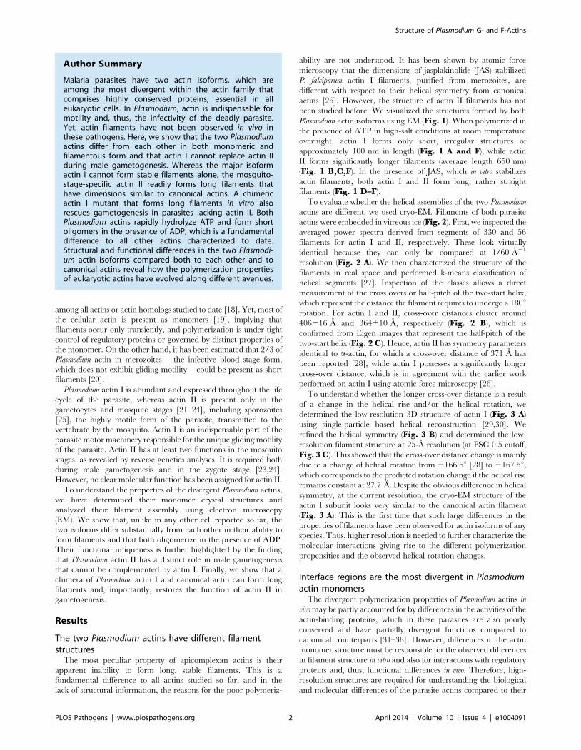
Author Summary
Malaria parasites have two actin isoforms, which areamong the most divergent within the actin family thatcomprises highly conserved proteins, essential in alleukaryotic cells. In Plasmodium, actin is indispensable formotility and, thus, the infectivity of the deadly parasite.Yet, actin filaments have not been observed in vivo inthese pathogens. Here, we show that the two Plasmodiumactins differ from each other in both monomeric andfilamentous form and that actin I cannot replace actin IIduring male gametogenesis. Whereas the major isoformactin I cannot form stable filaments alone, the mosquito-stage-specific actin II readily forms long filaments thathave dimensions similar to canonical actins. A chimericactin I mutant that forms long filaments in vitro alsorescues gametogenesis in parasites lacking actin II. BothPlasmodium actins rapidly hydrolyze ATP and form shortoligomers in the presence of ADP, which is a fundamentaldifference to all other actins characterized to date.Structural and functional differences in the two Plasmodi-um actin isoforms compared both to each other and tocanonical actins reveal how the polymerization propertiesof eukaryotic actins have evolved along different avenues.
Structure of Plasmodium G- and F-Actins
PLOS Pathogens | www.plospathogens.org 2 April 2014 | Volume 10 | Issue 4 | e1004091

canonical homologs. Such information will also aid us in evaluating
the suitability of Plasmodium actins as drug targets.
We set out to determine the crystal structures of P. falciparum
actin I and P. berghei actin II. The sequence identities between the
counterparts from P. falciparum and P. berghei are 99% for actin I
and 92% for actin II. The gelsolin G1 domain was used to stabilize
the monomers and facilitate crystallization of both actins [39,40].
The actin I structure was refined to a resolution of 1.3 A and actin
II to 2.2 A. These high-resolution structures allow for a very
detailed comparison of the Plasmodium actins with each other and
with other actins (Figs. 4, S2, and S3). Although Plasmodium lacks
a gelsolin homolog, the mammalian G1 is bound between
subdomains 1 and 3 in both Plasmodium actins, similarly to other
actin–G1 complexes [39,41] (Fig. S2 D and E). For all
comparisons, we have used canonical actin structures that have
also been determined in complex with G1, in order to rule out
structural rearrangements caused by gelsolin binding.
In most canonical actin structures, the C terminus is folded as
an a-helix, which interacts with the bottom part of subdomain 1.
This is true also for Plasmodium actin II (Fig. 4 A). In actin I,
however, the C terminus turns towards solvent and is disordered
(Fig. 4 B). The large hydrophobic cleft between subdomains 1 and
3 is defined as a ‘hotspot’ for regulatory protein binding [42]
(Fig. 4 A and B). A smaller hydrophobic patch in the direct
vicinity of the C terminus is, in addition, involved in binding at
least profilin [43]. In actin I, the large hydrophobic residues in this
smaller cleft, including Trp357, have adopted different
conformations compared to canonical actins (Fig. 4 B), possibly
influencing the binding of profilin, which we have previously
proposed to bind actin in a different manner in Plasmodium
compared to opisthokonts [31]. In actin II, these hydrophobic
residues, like the C terminus, are in the canonical conformations.
All in all, despite the obvious similarity to other actins, especially
actin I shows appreciable structural deviations, in particular in
regions involved in binding of regulatory proteins [42]. This may
provide possibilities for structure-based drug design targeted at the
Plasmodium actin–regulatory protein interfaces.
Residues involved in longitudinal contacts in F-actindiffer equally from canonical actins in both parasiteactins
A key question concerning the properties of the major
Plasmodium actin isoform, as well as other apicomplexan actins, is
why they, unlike all the extensively studied actins, form only short,
unstable filaments. Comparing the crystal structures of the
monomers to the recently determined high-resolution cryo-EM
structures of canonical actin filaments [28,44–46] can provide
clues to answer this question. In canonical F-actin, the axial
interactions, meaning the longitudinal contacts between the actin
monomers in each of the two protofilaments, are tight and mainly
electrostatic, as revealed by cryo-EM studies [28,45]. The most
important axial interactions are discussed below.
The DNase I binding (D-)loop in subdomain 2 (residues 39–61
in actin I) is one of the most important regions for polymerization
Figure 1. Electron micrographs of Plasmodium actin filaments. (A) In the absence of stabilizing agents, actin I forms only short structureslacking helical symmetry. (B,C) Actin II readily forms filaments varying from hundreds of nm to 1–2 mm in length. (D,E) In the presence of JAS, bothparasite actins form long helical filaments. (F) Length distributions of two Plasmodium actin isoforms and three actin I mutants. Note the logarithmicscale of the Y axis.doi:10.1371/journal.ppat.1004091.g001
Structure of Plasmodium G- and F-Actins
PLOS Pathogens | www.plospathogens.org 3 April 2014 | Volume 10 | Issue 4 | e1004091

and, in the filament, inserts into the hydrophobic cleft between
subdomains 1 and 3 of the neighboring monomer [28,45,47,48].
In both Plasmodium actin crystal structures, the D-loop is
disordered, and the tip of it is not visible in the electron density
maps (Figs. 4 and S3). Interestingly, the most notable changes in
the D-loop sequence concern the first and the last residues of a
segment (residues 42–49) that forms a short a-helix in some
G-actin structures with ADP bound [49] and has been modeled as
a helix also in one of the recent F-actin structures [45]. This part of
the D-loop inserts deep into the neighboring monomer in the
filament, contacting the so-called proline-rich loop (residues 109–
115). Residue 42, which is a glutamine or threonine in most other
actins, is a proline in both Plasmodium actins. A proline restricts the
conformation of the chain and, although also known as a ‘helix
Figure 2. Cryo-EM image analysis of actin I and II. (A) Electron cryo-micrographs of actin I and II (left and right, respectively). Side-by-sideaverage power spectrum of actin I and II. (B) Representative class averages from k-means clustering. Center: Histograms of measured cross-overdistances reveal a larger half pitch for actin I. (C) Eigen images 1–2 from actin I and II k-means clustering reveal a constant pitch of the one-start helix,whereas Eigen images 3–4 confirm the difference in cross-over distance of actin I and II.doi:10.1371/journal.ppat.1004091.g002
Structure of Plasmodium G- and F-Actins
PLOS Pathogens | www.plospathogens.org 4 April 2014 | Volume 10 | Issue 4 | e1004091

breaker’, is often also seen as the first residue in a-helices. At the C
terminus of this segment, residue 49, which is a glycine in
canonical actins, is a glutamate in both Plasmodium actins. These
replacements at both ends of this segment would be expected to
increase the helical propensity of the tip of the D-loop.
In canonical F-actin, Thr325 in the loop between a-helix 11
and b-strand 18 (residues 323–327; Fig. 4 A and B) in subdomain
3 of one monomer interacts with Glu242 in the loop connecting b-
strands 15 and 16 in subdomain 4 (residues 242–247; Fig. 4 Aand B) of the neighboring monomer [28]. In actin I, this loop and
the threonine side chain have turned away from the optimal
position for this interaction (Figs. 4 B and S3). In actin II, Ser325
is positioned such that hydrogen bonding to the apposing
glutamate can be easily achieved. The difference in the
conformation can be explained by the substitutions Y280F and
M284K in actin I compared to canonical actins and actin II.
The third main site contributing to axial interactions is formed
by the loop connecting helices 9 and 10 (residues 284–290 in
subdomain 3; Fig. 4 A) inserting between subdomains 2 and 4 of
the neighboring monomer. This area is almost fully conserved in
both parasite actins, the only substitution being that of Met283 by
a lysine (284) in actin I.
All in all, the largest differences in the axial contacts concentrate
to the D-loop and concern equally both Plasmodium actins. Smaller
differences in subdomain 3 may, however, partly explain the
different polymerization propensities of the two parasite actin
isoforms.
Filament lateral contact areas display the largestdifferences between the parasite actins
The lateral contacts between the two protofilaments concern
mainly interactions between subdomains 1 and 4 and the beginning
of the D-loop in subdomain 2 interacting with the so-called
hydrophobic loop (residues 263–275) (Figs. 4 A–B, S1, and S2 D–E) between subdomains 3 and 4 of the apposing monomer
[28,45,48]. In a recent high-resolution cryo-EM filament structure
[45], Arg206 in subdomain 4 interacts with Ser271 of a neighboring
monomer. In actin I, Ser271 is replaced by Ala272, and Lys207 and
Glu188 are involved in a short hydrogen bond with salt-bridge
character (Fig. 4 C). A similar interaction occurs between Arg206
and Asp187 in Latrunculin A-bound a-actin [50]. In the absence of
latrunculin, the distance between these residues in a-actin is longer
and the geometry suboptimal for a salt bridge (Fig. 4 C).
Latrunculins prevent actin polymerization, presumably by limiting
the flexibility of subdomains 2 and 4 [50,51], and the salt bridge
seen in the latrunculin-actin structure, similar to Plasmodium actin I,
may be one reason for this. The longer side chain of Glu188 in actin
I compared to Asp187 in canonical actins may facilitate the
interaction with Lys207. Actin II has Tyr187 in place of Asp187/
Glu188, which affects the orientation of Arg206. Actin II,
furthermore, has Cys272 in the place of Ser271 of canonical actins,
allowing for hydrogen bonding upon polymerization.
At the N terminus of the D-loop, Arg40 and His41 of canonical
actins are replaced by lysine and asparagine in actin I and lysine
and methionine in actin II. In the filament, Arg40 forms a salt
bridge with Glu271 [28], and the replacement to lysine can subtly
weaken this interaction. His41, in turn, interacts with Ser266 [28].
Although the asparagine in actin I can also contribute to a
hydrogen bond, the interaction may be weaker due to the shorter
side chain. In F-actin, Lys114 in the proline-rich loop (residues
109–115 in subdomain 1, following actin I numbering) forms a salt
bridge with Glu196 of a neighboring monomer [28]. Uniquely, the
neighboring residue is Gly115 in actin I (Fig. 4 D), which can
have a large effect on the mobility of the proline-rich loop. Actin II
has a non-glycine residue (threonine) at this position, similarly to
canonical actins (alanine or serine).
Figure 3. Filament structure of Plasmodium actin I compared to a-actin. (A) The cryo-EM structure of actin I filament at 25 A resolution (left)in comparison with rabbit skeletal muscle a-actin filtered to a comparable resolution (right; EM database entry EMD-5168 [28]). (B) Symmetryrefinement of actin I confirms that the change in cross-over distance is caused mainly by a change in helical rotation when compared with actin II andcanonical rabbit a-actin. (C) Fourier Shell correlation of actin I half data sets used for 3D reconstruction. The resolution can be estimated at 25 Abased on the 0.5 criterion.doi:10.1371/journal.ppat.1004091.g003
Structure of Plasmodium G- and F-Actins
PLOS Pathogens | www.plospathogens.org 5 April 2014 | Volume 10 | Issue 4 | e1004091

Taken together, especially residues involved in lateral contacts,
crucial for the stability of the filaments, are altered in Plasmodium
actin I but more conserved in actin II. The most important
differences concern subdomain 4, the D-loop, and the proline-rich
loop. The conformations of all these affect each other allosterically
and can also modulate the ATPase activity [52–54].
a-actin D-loop rescues polymerization of Plasmodiumactin I
Because of the observed differences in sequence, conformation,
and helical symmetry between Plasmodium and canonical actins
and the importance of the D-loop-mediated contacts for polymer-
ization, we constructed a chimera, in which the entire D-loop of
actin I was replaced with that of a-actin. Strikingly, this chimera in
the absence of any stabilizing agents forms filaments with an
average length of 1.6 mm, which is longer than either actin I or II
filaments (Figs. 5 A–B and 1 F). However, the appearance of the
chimera filaments is not as regular as the filaments formed by
canonical actins or actin II.
To further characterize the filaments formed by the actin I–a-
actin chimera, we analyzed electron cryo-micrographs of JAS-
stabilized chimera filaments and subjected them to the same
classification and symmetry analysis as outlined above (Fig. S4).
Based on the determined half-pitch distance and the symmetry
analysis, we conclude that the symmetry parameters of the
chimera filaments are in close agreement with the wild-type actin I
filaments. Therefore, the symmetry parameters appear to be
retained from the wild-type actin I, and thus, the D-loop of
canonical actin merely confers increased filament stability.
The crystal structure reveals that the 3D structure of the
chimera in monomeric form is very similar to that of wild-type
actin I (Figs. 5 C and S3). However, there are some notable
differences. The D-loop in the chimera is slightly more ordered. In
addition, the C-terminal helix is visible and can be superimposed
with the C terminus of actin II and most canonical actins. The
Trp357 side chain in the hydrophobic patch is not flipped as in
wild-type actin I (Figs. 4 B and 5 C). This observation is in line
with earlier reports on allostery between the conformations of the
D-loop and the C terminus [55,56] and demonstrates the ability of
even large residues in the hydrophobic core of actin to move,
which is a requirement for the rearrangements taking place in the
monomer upon polymerization.
In addition to residues already discussed, a notable difference
between the D-loop of Plasmodium actin I and canonical actins is at
position 54, which has a tyrosine in opisthokont actins and
Plasmodium actin II but phenylalanine in actin I. In the chimera,
the OH group of Tyr54 is hydrogen bonded to the main chain of
Lys51 and could also interact with the tip of Lys62 (Fig. 5 C). In
wild-type actin I, Phe54 has moved away from Lys62 and seems to
push Lys51 to a slightly different conformation. In actin II, Tyr53
(corresponding to Phe54 in actin I) is able to interact with Lys61
Figure 4. Crystal structures of Plasmodium actin I and II. (A) P. berghei actin II (PbActII; yellow) superimposed on a-actin (1eqy [39]; cyan). (B) P.falciparum actin I (PfActI; red) superimposed on a-actin. In both (A) and (B), ATP, subdomains 1–4, and several regions discussed in the text areindicated. Both N and C termini reside in subdomain 1; the N terminus is visible at the front, and the C-terminal helix is on the back side. Note that theC-terminal helix is not visible in actin I. The C-terminal part and the nearby hydrophobic cluster with Trp357 are shown in the zoomed view on theright and the region involved in intra-filament contacts in subdomain 3 in the box at the lower left corner. The blue and pink dots in (B) indicate theapproximate positions of the structural elements shown in detail in (C) and (D), respectively. (C) Lys 207 and Glu188 are at an intimate distance inactin I. A similar salt bridge is present between the corresponding residues in latrunculin-bound a-actin, but the hydrogen-bonding distance is longerwithout the drug. (D) The proline-rich loop with Gly115 in actin I superimposed on that of a-actin. Note the bending of the loop in actin I, due to themore flexible glycine residue.doi:10.1371/journal.ppat.1004091.g004
Structure of Plasmodium G- and F-Actins
PLOS Pathogens | www.plospathogens.org 6 April 2014 | Volume 10 | Issue 4 | e1004091

(Lys62 in actin I), but otherwise, this region is rather different in
conformation compared to both actin I and canonical actins. This
is due to conformational changes in subdomain 4, involving
Thr203, Arg206, and seemingly originating from the bulky side
chain of Tyr187, which is replaced by aspartate in canonical actins
and glutamate in actin I, as discussed above. According to a recent
filament structure [45], Tyr54 becomes stacked between the side
chains of Asp52 and Lys62 of the same monomer and its only
possible rotamers would allow an OH–p interaction with the
phenyl ring of Tyr170 of the neighboring monomer. The side
chain of Phe54 in actin I is not able to participate in this kind of an
interaction due to the missing hydroxyl group. Thus, the
replacement of tyrosine by phenylalanine at this position may
affect both the rigidity of the D-loop in the monomeric state as well
as the stability of the filament.
Implications for catalysisThe state of the nucleotide-binding pocket can be described
using two parameters: (i) the ‘‘phosphate clamp’’, which is the
distance between the Ca atoms of Gly15 and Asp157, and (ii) the
‘‘mouth’’, which is the distance between the Ca atoms of Gln59
and Glu207 [46]. The phosphate clamp distance in Plasmodium
actins does not differ significantly from other actins. However, the
mouth of the binding pocket is significantly more closed in both
parasite actins: 9.85 A in actin I and 9.04 A in actin II, compared
to an average of 10.8760.4 A in 9 other actin–G1 structures used
for comparison. In three recent high-resolution F-actin structures
[28,45,48], the mouth distance varies between 7.88 and 9.66 A.
Both parasite actins reach the more closed conformation by
slightly different means. In actin I, the largest differences in
conformation to canonical actins are in subdomain 4 and in actin
II, subdomain 2 (Figs. 4 and S3).
Both structures reported here have calcium and ATP bound to
the nucleotide-binding cleft between subdomains 2 and 4 (Figs. 4and 6). In the nucleotide-binding residues, there is one interesting
difference in actin I compared to canonical actins and actin II;
residue 17, which is hydrophobic (methionine/leucine) in all
canonical actins and actin II, is an asparagine in actin I (Fig. 6 Aand B) and also T. gondii actin. This side chain is close to the a-
and b-phosphates of ATP. In actin I, the distance of the Asn17
Nd2 atom to the b-phosphate O1 atom (,3.75 A) is too long for
hydrogen bonding in this conformation. However, together with
its own main chain N, that of Gly16, and Nf of Lys19, the Asn17
side chain could form an oxyanion hole to stabilize a negative
charge on the b-phosphate O1 atom (Fig. 6 A). In addition, it
could interact with the a-phosphate. The Asn17 side chain is
flexible in the crystal structure, as evident from the electron density
maps and B factors, and based on the shape of the electron density
as well as anisotropic ellipsoids, seems to move in concert with
active-site water molecules as well as the nearby Tyr338, which is
in a double conformation.
Figure 5. Plasmodium actin I–a-actin chimera forms long filaments. (A) Electron cryo-micrograph of the chimera filaments. (B) Negativelystained chimera filaments. (C) The crystal structure of the chimera (blue) resembles that of wild-type actin I (red). The zoomed views show thedifferences in the D-loop around Tyr54 (above) and the C-terminal helix and the hydrophobic residues nearby, which are in the canonical orientationin the chimera, unlike in actin I (below).doi:10.1371/journal.ppat.1004091.g005
Structure of Plasmodium G- and F-Actins
PLOS Pathogens | www.plospathogens.org 7 April 2014 | Volume 10 | Issue 4 | e1004091

A catalytic mechanism based on a nucleophilic attack of a water
molecule activated by His162 and Gln138 has been proposed for
ATP hydrolysis in actin [57]. The reaction itself is rather simple,
containing only proton transfer steps. The complication arises
from the fact that G-actin – currently the only form of which
atomic-resolution information can be achieved – is a poor catalyst,
and conformational changes upon polymerization are needed for
achieving the catalytically competent conformation of the active
site. Water 39 (actin I numbering) has been proposed to be the
nucleophile initiating the reaction, and depending on the bound
metal, the position of this water changes [57]. In actin I, this water
is 4.75 A away from the c-phosphate, and the angle between the
b-c bridging O, Pc, and water 39 is 152.4u, which is amenable for
a nucleophilic attack (Fig. 6 B). The catalytic site water structure
in actin II is different compared to actin I (Fig. 6 C), as the
presumed catalytic water (147 in the actin II structure) has moved
towards His161 and Pro109 and has weak electron density and a
high B factor. The distance of this water to Pc is 5.43 A, and it is in
an almost in-line position (162.4u). In fact, according to the
electron density and the distances to neighboring atoms, this water
may be a second conformation of water 357, which is directly
hydrogen bonded to His161. Together, these differences in the
active site architectures between the two parasite actins and
compared to other actins indicate that the catalytic activity and the
exact mechanism of ATP hydrolysis may differ in them, likely
resulting also in differences in polymerization.
Given the structural differences in the catalytic sites, we set out
to test if the Plasmodium actins differ from each other and canonical
actins in their ability to hydrolyze ATP and/or release phosphate.
We first measured phosphate release in the presence of Mg2+
(Fig. 6 D). It should be noted that, as phosphate release at least in
canonical actins is much slower than hydrolysis, this method gives
only indirect information about the hydrolysis rate. In these
conditions, both parasite actins release phosphate faster than a-
actin. Of the two parasite actins, actin I has a slightly higher rate
(,2 times higher than actin II). Curiously, the chimera with the a-
actin D-loop is approximately 3-fold more active than wild-type
actin I and 23-fold more active than a-actin. We also made two
point mutations to actin I; G115A, which we predicted to affect
the flexibility of the proline-rich loop and, thus, the rate of
hydrolysis, and F54Y, which we hypothesized might affect the
rigidity of the D-loop in monomeric state. Neither of these mutants
formed long filaments without JAS (Fig. 1 F). In the presence of
Mg2+, the F54Y mutant shows identical behavior to wild-type
actin I. G115A, however, has a reduced rate, similar to actin II.
Also the kinetics differ from each other in the different actins.
Whereas actin I and the point mutants release phosphate in a
linear way, a-actin, actin II, and the chimera display more
complex kinetics, having an initial very short, faster, non-linear
phase, followed by a linear phase.
We next measured phosphate release in the Ca2+-bound,
presumably mainly monomeric, forms (Fig. 6 D). As expected, a-
actin showed an even lower release of phosphate in the Ca2+-
bound compared to the Mg2+-bound form. Both actin I and actin
II have activities equal compared to each other and approximately
5-fold higher than a-actin. The chimera releases phosphate 3-fold
less in the presence of Ca2+ than with Mg2+, but the rate is still
significantly higher than that of muscle actin or both wild-type
parasite actins with Ca2+. Of the point mutants, F54Y has
practically no activity with Ca2+ (identical to a-actin), whereas
G115A is slightly more efficient in the presence of Ca2+ than
Mg2+. Altogether, these data show that the Plasmodium actins have
Figure 6. ATP binding sites of actin I and II. (A) The Asn17 side chain in actin I is part of a cluster formed by the Asn17 Nd and main chain Natoms as well as Nf of Lys19. Together, they could form an oxyanion hole for stabilizing a negative charge on one of the b-phosphate oxygen atomsin a reaction intermediate. (B) The active-site water structure in actin I is conserved, and W39 is in an almost inline position for a nucleophilic attack tothe ATP c-phosphate. (C) The catalytic water in actin II has moved further away from the ATP c-phosphate, is mobile, and is likely a doubleconformation of the water bound directly to His161. (D) Phosphate release rates of the wild-type Plasmodium actins in the calcium- or magnesium-bound states compared to a-actin, the actin I–a-actin chimera and the actin I mutants F54Y and G115A. Error bars represent standard deviation(n = 3).doi:10.1371/journal.ppat.1004091.g006
Structure of Plasmodium G- and F-Actins
PLOS Pathogens | www.plospathogens.org 8 April 2014 | Volume 10 | Issue 4 | e1004091

a different mechanism of ATP hydrolysis and/or subsequent
phosphate release compared to canonical actins, which are poor
catalysts in the monomeric form and adopt the catalytic
conformation only upon polymerization, which is a prerequisite
for non-equilibrium polymerization kinetics enabling directional
growth [58]. We were also able to pinpoint amino acid residues
responsible for these differences.
Plasmodium actins have a unique response to ADPIn order to evaluate the oligomeric state of the parasite actins in
the presence of ATP/ADP and different ions, we used native
PAGE. With ATP bound, actin I spontaneously forms short
polymers (from tetramers up to 11–12-mers) in ,48 h when stored
on ice (Figs. 7 and S5). Actin II stays mainly monomeric over the
same period of time, although minute amounts of oligomers
(dimers–octamers) appear. Interestingly, in the presence of ADP,
oligomerization starts instantly, and the majority of both actins is
oligomeric (dimers–10-mers) immediately after a 1-h hexokinase
treatment at 298 K to remove ATP. After incubation of the actins
at 298 K for 1 h without hexokinase, only minute amounts of
oligomers can be visualized for actin I, and no visible oligomer-
ization of actin II takes place (data not shown). After 48 h, the
proportion of larger oligomers of the ADP forms is much higher,
and monomers as well as lower oligomers are practically non-
existent. The formation of oligomers is not caused by oxidation, as
using even a large excess of the reducing agent TCEP in the
sample does not reduce the amount of oligomerization (Fig. 7 A).
Both ATP and ADP forms of a-actin remain monomeric in the
same conditions. However, short oligomers of ATP-a-actin have
been reported below the critical concentration for polymerization
[59].
Because the distribution, when separated on a gel, does not
necessarily reflect the equilibrium between different species in
solution, we also used dynamic light scattering (DLS) to visualize
the size distribution and polydispersity of the actin mono- and
oligomers in solution over time (Fig. S6). The resolution of DLS is
far from that of the native gel assay, and it is only possible to detect
size differences of approximately 5–6 fold. Therefore, e.g.
monomers, dimers, and trimers will appear as a single, polydis-
perse peak. 6 h after purification, actin I is seen in mainly two
separate peaks of average hydrodynamic radii of approximately 2
and 6 nm (Fig. S6 A). 2 nm would be very close to the expected
hydrodynamic radius of the monomer. After 11 h, nearly all of
actin I is in particles with a radius of ,5 nm (Fig. S6 B). As time
goes by, the distribution becomes divided between particles of
below 3 nm (close to a monomer) and larger oligomers with an
average radius of 11–12 nm (Fig. S6 C and D). The polydisper-
sity of the sample after 11 h is very high, indicating that the sample
contains a mixture of monomers and small oligomers, and the
polydispersity diminishes again, as the sample gains a multimodal
distribution, indicating that the smallest oligomers disappear over
time, leaving behind a pool of monomers in addition to the higher
oligomers, consistent with our native PAGE data. As seen also in
the native gels, actin II retains a higher fraction of monomers over
48 h, but also gains a fraction of significantly higher oligomers,
which are, however, infrequent and very heterogeneous in size
(Fig. S6 E–H).
In order to probe the effects of Mg2+ and K+ ions on the
oligomerization behavior, we also performed native PAGE in the
presence of two concentrations (1 and 5 mM) of MgCl2 as well as
5 mM MgCl2 and 50 mM KCl (Fig. S7). In the presence of ATP,
Mg2+ slightly reduces the amount of the short oligomers for both
actin I and II compared to the Ca2+ forms (Fig. S7 A and B).
However, some actin I is visible at the bottom of the well at the top
of the gel, which would imply filaments too long to enter the gel.
This could not be seen in the Ca2+ gels for either the Plasmodium
proteins or a-actin, but was much more pronounced for a-actin
with Mg2+. In the presence of ADP, there is a clear shift towards
longer oligomers in actin I, and after 48 h, part of actin I stays in
the well, not entering the gel in the presence of 5 mM MgCl2 both
with and without KCl (Fig. S7 C and D). Thus, Mg2+ alone
seems to be sufficient for polymerization.
Actin I cannot replace actin II in male gametogenesisWe used genetically modified parasites to address the question
whether the observed structural differences translate into different
properties of the proteins in vivo. While it is not possible to delete
actin I due to its essential functions, a knock-out of the actin2 gene
has been done, resulting in a block of male gametogenesis [23,60].
We reasoned that a replacement of actin2 with actin1 would display
the mutant phenotype if the two actin isoforms have different
biological functions, while restoration of gametocyte development
would indicate a similar function.
Male gametogenesis in the malaria parasite is a unique event,
involving the formation of flagellar gametes. This event, called
exflagellation (Fig. 8 A and Video S1), is easily scored under the
microscope, allowing us to use it as a quantitative method. In our
approach, actin1 was expressed under the control of the actin2
flanking regions. We used a recipient line, in which the complete
open reading frame (ORF) of actin2 had been deleted. Therefore,
these parasites do not exflagellate [60]. This line was separately
transfected with two constructs, both aiming at integration into the
actin2 locus. The complementation construct (act2com) restored the
actin2 ORF, which allowed expression of the cognate gene
comparable to wild type. In the replacement construct (act2rep), a
fragment corresponding to the actin1 ORF was used instead of the
actin2 ORF. The constructs were otherwise identical and were
integrated in the locus via a single crossover homologous
recombination event in the 59 flanking region of actin2 (Fig. 8B). In both cases, clonal lines were obtained. We compared the
act2com and act2rep parasite lines with wild-type parasites in the
exflagellation assay (Fig. 8 C). In the wild-type and act2com
parasites, the number of exflagellation events was similar,
indicating that complementation with actin2 restored the function
of the gene. However, while some normal exflagellation events
were detected also in the act2rep parasites, the numbers were
significantly reduced compared to the act2com parasites (Fig. 8 C),
strongly suggesting that actin II has unique functions, which actin I
cannot fulfill during male gametogenesis.
The actin I–a-actin D-loop chimera restoresgametogenesis
As it became apparent that actin I polymerization properties in
vitro could be altered by exchanging its D-loop to that of a-actin,
we decided to investigate if this modification would also have an
impact on the in vivo function of actin I. We produced transgenic
parasites using the same strategy as described above; inserting the
actin I–a-actin D-loop chimera into the actin2 locus, producing the
act1chi parasites (Fig. 8 B). Surprisingly, this revealed that the
exchange of the D-loop had a remarkable impact on exflagellation
(Fig. 8 C). In the act1chi parasites, exflagellation was significantly
increased compared to the act2rep strain and restored to values
close to the act2com strain. These data show that the D-loop has a
critical role in the function of these actins, but actin II, as shown by
the structural data, has acquired other properties that contribute
to its higher filament stability. Furthermore, and to our surprise, it
seems that the molecular function of actin II may be dependent on
the ability of the protein to form filaments.
Structure of Plasmodium G- and F-Actins
PLOS Pathogens | www.plospathogens.org 9 April 2014 | Volume 10 | Issue 4 | e1004091

Discussion
An actin cytoskeleton was long thought to be a feature unique to
eukaryotic cells, and this view was revisited only two decades ago
upon the discovery of the first bacterial actin and tubulin homologs
[61–64]. The ancient phylum Apicomplexa is likely separated from
opisthokonts by an evolutionary distance of a billion years, and the
diversion of Plasmodium spp. took place hundreds of millions of
years ago [65]. Therefore, looking at the divergent properties of
Plasmodium actins provides us with insight into the early stages of
actin evolution. The ability of actin to polymerize must have
evolved very early – before its involvement as tracks for molecular
motors [66]. This may explain some features of both the
polymerization propensity and the divergent actin-myosin motor
in Apicomplexa. Also the minimal set of actin-binding proteins in
Apicomplexa suggests that a common ancestor had a limited
polymerization propensity, and the various regulatory proteins in
higher eukaryotes have evolved as the polymerization properties of
actin itself have been fine-tuned, creating a need for additional
regulation. For the biological functions of actin in Apicomplexa, the
development of similar polymerization properties has strikingly not
been of importance.
Different polymerization properties provide evolutionarycues
The two Plasmodium actins differ in their polymerization
propensities, filament stability, and filament helical symmetry –
the hallmark of canonical F-actin. The second, stage-specific actin
isoform of Plasmodium that forms long filaments with canonical F-
actin symmetry is unique among Apicomplexa. Curiously, at the
sequence level, actin II is as divergent from Plasmodium actin I as it
Figure 7. Native PAGE analysis of the Plasmodium actins and a-actin. (A) Plasmodium actins I (lanes 1–4 and 9) and II (lanes 5–8 and 10) formsmall oligomers upon storage and after exchange of ATP to ADP. Both parasite actins were studied by native PAGE immediately and 48 h afterpurification in both ATP and ADP forms. Treatment of ADP-exchanged Plasmodium actins with a high concentration of reducing agent (10 mM TCEP)has no effect on the behavior of either of the actins. Exchange of ATP to ADP in a-actin (lanes 11–14) does not result in changes in the oligomericstate. Nt denotes the nucleotide. TCEP - and + denote either the normal 1 mM or an excessive 10 mM concentration, respectively. The approximateposition of the different oligomers, corresponding to lane 2, are given on the left. Note that actin I and II run slightly differently on the gel. (B–F) Therelative mobility vs. log MW (circles with the oligomeric state indicated on the side) and relative intensities of bands (bars) extracted from gel imagesof Coomassie-stained native PAGE gels containing ATP or ADP Plasmodium actin I (B–D) and ADP actin II (E and F) immediately or 48 h afterpurification. The dark grey bars denote the relative intensity of the bands compared to the most intense band and the light grey bars the relativeintensity of the bands compared to the sum of all band intensities.doi:10.1371/journal.ppat.1004091.g007
Structure of Plasmodium G- and F-Actins
PLOS Pathogens | www.plospathogens.org 10 April 2014 | Volume 10 | Issue 4 | e1004091

is from all other actins. It has been suggested [23], and our
structural data support the view, that the actin2 gene has arisen
only after the diversion of Plasmodium from other Apicomplexa, and
the protein seems to have gained a higher filament stability
independent of the evolution of higher eukaryotic actins.
In canonical actins, polymerization is tightly coupled to ATP
hydrolysis, such that structural rearrangements upon polymeriza-
tion enable the active site to adopt a conformation optimal for
catalysis [67]. Two key factors have been described necessary for
achieving the catalytically competent conformation upon the
transition from monomeric to filamentous state. These are: (i) a
rotation of the outer domain (subdomains 1 and 2), resulting in
flattening of the monomer and (ii) bending down of the proline-
rich loop in subdomain 1 [45,48]. The Plasmodium actins hydrolyze
ATP also in the monomeric form, releasing phosphate more
efficiently than canonical actins, and oligomerize readily in the
presence of ADP, which is a fundamental difference to all other
actins characterized, and must be a result of different atomic
structures.
Interestingly, Plasmodium actin I has a unique glycine at the end
of the proline-rich loop. This allows more flexibility for this loop,
which apparently increases the catalytic rate in the presence of
magnesium but, surprisingly, has an opposite effect in the calcium-
bound form (Fig. 6 D). Also the more closed conformations of
subdomains 2 and 4 in the parasite actins may facilitate ATP
hydrolysis but also reduce the conformational change – or
flattening – required upon insertion of the monomer into the
filament. An interesting difference that may also contribute to
catalysis is Asn17 close to the a- and b-phosphates of ATP in the
active site. Intriguingly, the bacterial actin homolog MreB [68]
shares this residue with Plasmodium actin I, whereas canonical
actins and also Plasmodium actin II have a hydrophobic residue at
this position. Thus, this asparagine may be a relict from an early,
polymerization incompetent ancestor.
The structural features described above may explain the
parasite actins’ unconventional response to ADP. Surprisingly,
the state of the nucleotide seems to determine polymerization
propensity, but not in the same way as in canonical actins. The
tight link between ATP hydrolysis and polymerization in higher
eukaryotes has probably been refined during the hundreds of
millions of years after the diversion of Apicomplexa. Our data and a
recent report proposing an isodesmic polymerization mode for
apicomplexan actins [18] suggest that the same has also happened
for allosteric regulation of conformational changes taking place
upon polymerization. However, it is clear that higher resolution
data on the Plasmodium actin filaments are needed in order to find
out what kind of conformational changes the parasite actins
undergo upon polymerization and what is the arrangement of the
protomers in the filament, leading to the altered symmetry
compared to canonical F-actin. On the other hand, the
distribution of oligomers, as seen on the native gels and DLS
(Figs. 7, S5, and S6), suggests that polymerization may involve a
nucleation step, the nucleus being either a dimer or trimer, which
are the species that disappear early in the process. Thus, we
Figure 8. The D-loop chimera but not wild-type actin I rescues the phenotype of the actin2 deletion mutant. (A) Exflagellation of a malegametocyte. The residual P. berghei gametocyte is indicated with an asterisk. The flagellar male gametes beat rapidly. The picture is the first frame ofVideo S1. (B) Schematic picture of the actin2 gene in wild-type parasites, the recipient strain act22::mCherry, and the final genotypes of the act2com,act2rep, and act1chi strains. The same experimental strategy was used for all constructs. The act1chi strain expresses a chimeric actin, where the D-loop (amino acids 39–61) has been swapped with the D-loop of a-actin (C) Exflagellation assays comparing wild-type (n = 11), act2com (n = 11),act2rep (n = 15) and act1chi (n = 14) parasites. The act2com functionally complemented the mutant, resulting in almost wild-type levels ofexflagellation. The act2rep parasites had a significantly smaller number of exflagellation events, while swapping the D-loop of actin I with that of a-actin in the act1chi mutant results in significant restoration of exflagellation. n.s. means non significant, *** stands for P,0.0001, * for P,0.05(Student’s t-test).doi:10.1371/journal.ppat.1004091.g008
Structure of Plasmodium G- and F-Actins
PLOS Pathogens | www.plospathogens.org 11 April 2014 | Volume 10 | Issue 4 | e1004091

hypothesize that the ADP state may favor nucleation, making ATP
hydrolysis a rate-limiting step for polymerization.
Importance of the D-loop for polymerizationThe D-loop plays a key role in the conformational changes
upon polymerization as well as the conformation and stability of F-
actin [28,45,47,48]. Both previous work [26] and our EM analyses
reveal differences in the helical architecture of actin I compared to
a-actin. In the crystal structures, several regions important for
intra-filament contacts in canonical actin filaments show substan-
tial differences between the parasite and opisthokont actins, and
the polymerization propensity and filament stability are overall
likely a sum of numerous atomic details in the monomers. Yet, the
sequence of the a-actin D-loop alone is sufficient to restore the
ability of actin I to form long filaments, without altering the
symmetry compared to the JAS-stabilized wild-type actin I
filaments. Thus, whereas the longitudinal contacts by the D-loop
are important for stability, the shape and symmetry of the
filaments are determined by other factors.
Actin II shows us that stability can be obtained by other means
than the D-loop, probably involving lateral interactions. In
addition to the differences we have described in the residues
involved in lateral contacts, a candidate responsible for increased
stability is residue 200, which is a glycine in Plasmodium actin I and
T. gondii actin [69] but serine or threonine in canonical actins as
well as Plasmodium actin II and Theileria actin [9], all of which form
long filaments. It has been reported that the double mutant
G200S/K270M in T. gondii actin leads to an increased filament
length when using phalloidin-labeled filaments [69]. However, we
were not able to visualize long filaments of this mutant of
Plasmodium actin I in polymerizing conditions without JAS (data
not shown), indicating that several small changes are cumulatively
responsible for the increased stability of actin II filaments.
The tip of the D-loop can adopt a helical conformation, albeit it
is disordered in the vast majority of all G-actin structures, and
appears mainly intrinsically disordered in solution in all nucleotide
states of G-actin [70]. The likely higher helical propensity of the
D-loop in Plasmodium actins may affect polymerization and
filament stability in at least two different ways. If the helical
conformation is more likely to occur in the filamentous form, this
might actually facilitate polymerization, which would be in line
with the proposed low critical concentration [15] or isodesmic
model for polymerization of parasite actins [18]. However, it has
also been proposed that the helical form occurs only transiently in
the filament or that it is favored in the ADP form and leads to
filament destabilization [54,70]. In this way, a higher helical
propensity would contribute to the lower stability of the parasite
actin filaments.
Tyrosine hydrogen bonds can contribute substantially to protein
stability [71]. Tyr54 is a phosphorylation target and plays a
regulatory role in many actins [72–74]. For Dictyostelium actin,
phosphorylation of this tyrosine increases the critical concentration
and controls cell shape changes and spore formation [72–74]. In
Mimosa pudica L., a contact sensitive plant, where actin is heavily
phosphorylated, tyrosine phosphatase inhibitors inhibit the frag-
mentation of actin filaments during leaf bending [75]. In addition
to affecting binding to other proteins, phospho-Tyr54 stabilizes the
D-loop conformation [74]. Upon polymerization, this region
undergoes a large conformational change, and it seems that the
OH group of Tyr54 may be involved in stabilizing interactions
[45] that the Phe54 side chain could not fully compensate for.
Only 11 of over 300 known actin sequences contain a
phenylalanine at this position, and no other substitutions are
known. Most of these 11 sequences are actins from Plasmodium or
Trypanosoma, both species where actin filaments have not been
observed in vivo. Despite the apparent importance of tyrosine at
this position for normal actins, a single mutation to phenylalanine
in Dictyostelium actin does not affect its polymerization properties
[74]. In line with this, we also could not observe long filaments of
the actin I F54Y mutant (Fig. 1 F). However, the large effect of
the F54Y mutation on the phosphate release rate of actin I (Fig. 6D) suggests that this residue, indeed, may significantly affect the
conformation and flexibility of the D-loop.
Together, the above described structural properties may lead to
a higher polymerization propensity but also lower filament stability
in the parasite actins by lowering the energy barrier of the
transition between monomeric and filamentous forms. Yet, the
fact that the replacement of the D-loop alone is sufficient for
stabilizing the filaments formed by actin I, while retaining their
unique symmetry, is surprising, taking into account how similar
the D-loops of the two Plasmodium actins with different stabilities
are. This implies that, starting from an unstable filament forming
ancestor, actin II has reached its present form mainly using other
means than the D-loop for gaining additional filament stability.
Roles of the two actins in vivoMale gametogenesis is a complex, rapid series of cellular events
including escape from the host cell, three mitotic divisions, and
axoneme assembly, leading to the formation of eight flagellar and
highly motile gametes from each gametocyte within 10–20 min
from activation. Both actin isoforms are present in male
gametocytes of P. berghei [23], but their function in these events
is not understood. Actin II is not expressed in the asexual blood
stages [23]. Its deletion blocks male gametogenesis, and therefore,
these mutant parasites cannot be transmitted through the
mosquito [23]. Still, it has not been possible to pinpoint the exact
role of actin II. We show that the function of actin II cannot be
complemented by actin I, proving distinct molecular functions for
the two actins and suggesting that their unique structures and the
differences in their ability to form filaments directly translate into
different functional characteristics in vivo. By generating transgenic
parasites expressing the actin I–a-actin chimera, we found that this
mutant protein was able to function almost as well as actin II in
vivo. This strongly confirms the in vitro experiments and supports
the notion that the D-loop has a significant role in determining the
polymerization properties of the parasite actins. Furthermore, we
can hypothesize that the reason two actins evolved in Plasmodium,
was the need to have actins with different propensities to
polymerize in cells lacking a large repertoire of actin-binding
proteins.
Another example of distinct general and reproductive actin
isoforms can be found in plants, where it was recently shown that
animal cytoplasmic but not muscle actins can take over the
functions of the plant vegetative actins [76]. Remarkably, also
three actins from single-celled protists could carry out the same
tasks, suggesting that the properties required for fulfilling the
cytoplasmic actin functions during spatial development in multi-
cellular organisms were present already early on in the evolution-
ary history. However, it seems that the polymerization properties
of both Plasmodium and all other actins have evolved separately,
starting from a poorly polymerizing ancestor. It would be
interesting to see if either of the Plasmodium actins can support
spatial development in either plants or animals.
The current hypothesis is that actin I in Plasmodium is required
for gliding motility, and the filaments involved need to be short
and short-lived. Our data support this, as actin I forms only very
short polymers. For the suggested role of actin I in gliding, the
formation of long, stable filament seems undesirable [69]. Actin II
Structure of Plasmodium G- and F-Actins
PLOS Pathogens | www.plospathogens.org 12 April 2014 | Volume 10 | Issue 4 | e1004091

clearly is able to form long filaments, which may be needed for
functions specific to actin II within the mosquito stages, although
such functions have not yet been specified. Intriguingly, Plasmodium
appears to be the only apicomplexan parasite that has faced the
evolutionary pressure for acquiring a second actin isoform that
forms stable, long filaments.
Concluding remarksOur data provide a structural basis for understanding the
different functional properties of the two actin isoforms of
Plasmodium spp. These structures represent the, so far, most
divergent and primitive actins characterized, and we show that
the two isoforms have the most unique biochemical properties,
structures, and biological functions of all known actin isoforms.
High-resolution structural information will serve as a starting point
for understanding these functions in detail and for evaluating the
suitability of parasite actins and actin-binding proteins as drug
targets.
Materials and Methods
Protein expression, purification, and biochemistryPurification of G1 was performed as described [40]. Endoge-
nous pig skeletal muscle a-actin was purified as described [36,77].
P. falciparum actin I (PlasmoDB PF3D7_1246200) and P. berghei
actin II (PlasmoDB PBANKA_103010) were expressed in Sf21
cells at 300 K, as described before [36]. A chimera, where residues
40–61 of the P. berghei actin I were replaced by the corresponding
residues from a-actin, was cloned into pFastBac HT A (Invitrogen)
and expressed in the same way as the wild-type actins. Two point
mutations (G115A and F54Y) were introduced to actin I by
incorporating the corresponding mutation to the 59 end of the
primers. The parental plasmid was cleaved with DpnI and
recirculated with the T4 DNA ligase. The protein coding
sequences were confirmed by DNA sequencing. The purification
of the wild-type actin–G1 complexes was performed as described
[40]. The chimera–G1 was also purified as described before for
the two wild-type actins [40], except that HEPES (pH 7.5) was
used in the lysis buffer, and size exclusion chromatography was
performed in 10 mM HEPES (pH 7.5), 50 mM NaCl, 5 mM
dithiothreitol (DTT), 0.2 mM CaCl2 and 0.5 mM ATP. Peak
fractions containing the chimera–G1 were pooled and concen-
trated to 5.6 mg ml21 for crystallization.
The purification of all the actin variants without G1 was
performed essentially as described [40] except for a few
modifications, as listed. For actin I, the lysis was carried out in
10 mM HEPES (pH 7.5), 5 mM CaCl2, 250 mM NaCl, 1 mM
ATP, 5 mM b-mercaptoethanol, 15 mM imidazole, and size
exclusion chromatography was performed in 15 mM HEPES
(pH 7.0), 0.5 mM ATP, 5 mM DTT, and 0.2 mM CaCl2. The
pH of the lysis buffer for actin II was 8.7, and size exclusion
chromatography was performed in 25 mM Tris-HCl (pH 7.5),
0.5 mM ATP, 5 mM DTT, and 0.2 mM CaCl2. For the chimera,
lysis was carried out in 20 mM HEPES (pH 7.5), 5 mM CaCl2,
250 mM NaCl, 1 mM ATP, 5 mM b-mercaptoethanol, 15 mM
imidazole, and size exclusion chromatography was performed in
15 mM HEPES (pH 7.0), 0.5 mM ATP, 5 mM DTT, and
0.2 mM CaCl2. For DLS and filament length measurements, size
exclusion chromatography was performed in 5 mM HEPES
(pH 7.5), 0.5 mM ATP, 2 mM DTT, and 0.2 mM CaCl2. DLS
was measured using a Wyatt DynaPro platereader-II and 15 or
30 ml of actin I and II at concentrations between 8.5–24 mM at
298 K. The measurements were performed in triplicate and the
samples stored at room temperature between the measurements.
ADP-actin was prepared by incubating 50 ml of 10 mM actin
with 1–2 mg of hexokinase-agarose beads (Sigma-Aldrich, #H-
2653) in 15 mM HEPES pH 7.5, 1 mM ATP, 1 mM tris(2-
carboxyethyl)phosphine (TCEP), 0.2 mM CaCl2, 2 mM D-
glucose for 1 h at 298 K. As a control reaction, Plasmodium actins
I and II were incubated in identical conditions without D-glucose
and hexokinase and subsequently run on native PAGE. The
residual ATP contamination in ADP stocks was removed by
treating them in a similar fashion.
Native PAGE was performed using a running buffer of 25 mM
Tris-HCl (pH 8.5), 195 mM glycine, 0.5 mM ATP or ADP, and
0.1 mM CaCl2 or MgCl2. The sample buffer consisted of 25 mM
Tris-HCl (pH 8.5), 195 mM glycine, 10% (v/v) glycerol (final
concentrations). Actin samples were loaded at a concentration of
6.7 mM in a volume of 10 ml. Commercial TGX 4–20% gradient
gels (Biorad) were pre-run for 30 min at 277 K, 100 V before
applying the samples. Samples were run for 7 h using the same
voltage settings and temperature, with corresponding nucleotides
and divalent cations in the running buffer. The gels were stained
the next day with Coomassie Brilliant Blue R250.
Relative mobilities were determined by measuring the distance
of the bands from the top of the image and dividing this value by
that of the monomeric band. In the absence of a reference
monomeric band in ADP-ActI (48 h), the absolute value from
ATP-ActI (0 h) was used as a reference. The absolute mobilities of
the other visible bands in these images had a difference of ,2.5%.
Gel images were processed and band intensities extracted using
ImageJ [78]. A rolling ball background subtraction was applied
before manually extracting the intensities.
Actin samples were prepared for the phosphate release assay by
treating 10–15 mM purified actin with DOWEX 1X8 to remove
nucleotides and free phosphate. After the removal of the
nucleotide and phosphate, ATP was replenished by adding a
small volume of a concentrated stock solution. Buffer controls were
treated in a similar fashion, in order to reset the level of free
phosphate and nucleotide compared to the samples. The
concentration to be used for determining the release rate was
measured from the nucleotide-free solutions in order to reduce the
effect of pipetting errors. After the DOWEX treatment, samples
were divided in triplicate wells of a UV-transparent 96-well plate
(Corning) containing reagents from the EnzChek Phosphate
Release Assay (Molecular Probes) without using the reaction
buffer, which contains MgCl2 at a final concentration of 1 mM.
For calcium measurements, the final reaction contained 1 mM
CaCl2 and 0.1 mM MgCl2. The total omission of MgCl2 was not
possible, since the coupled enzyme requires magnesium. For
magnesium measurements, the respective concentrations were
0.13 mM CaCl2 and 1 mM MgCl2. Formation of the 2-amino-6-
mercapto-7-methylpurine from the coupled reaction was mea-
sured as absorbance at 360 nm with a kinetic interval of 60 s over
a period of 5 h at 298 K. The total measurement volume was
200 ml. Phosphate release rates were calculated from linear parts
of the plot (100 to 200 min) using GraphPad PRISM 5.03.
Crystallization, diffraction data collection, structuredetermination, and refinement
Crystallization and diffraction data collection of both wild-type
actin–G1 complexes has been described [40]. The chimera–G1
complex was crystallized similarly, and the final crystallization
condition contained 100 mM Tris-HCl (pH 8.0), 8% (w/v) poly-
ethylene glycol (PEG) 20 000, and 2% (v/v) dioxane. Before flash-
cooling in liquid nitrogen, the crystal was shortly soaked in 100 mM
Tris-HCl (pH 8.5), 14% (w/v) PEG 20 000, 2% (v/v) dioxane,
0.5 mM ATP, 50 mM NaCl, 0.2 mM CaCl2, and 10% (w/v) PEG
Structure of Plasmodium G- and F-Actins
PLOS Pathogens | www.plospathogens.org 13 April 2014 | Volume 10 | Issue 4 | e1004091
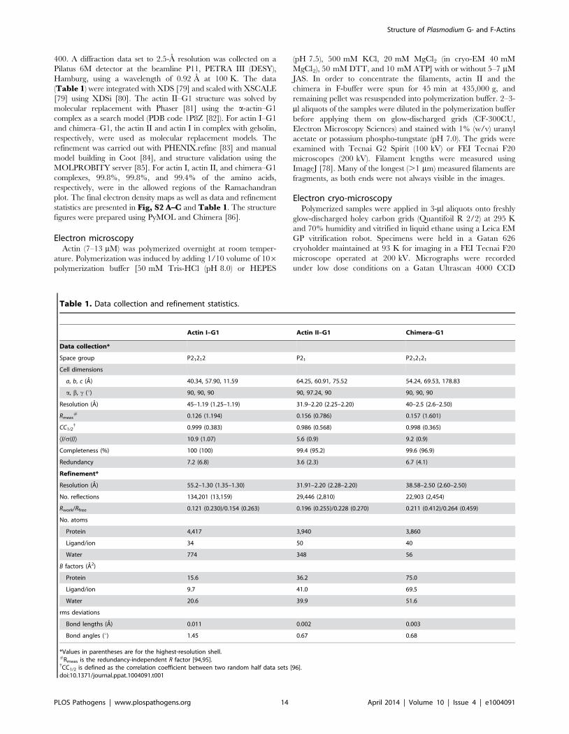
400. A diffraction data set to 2.5-A resolution was collected on a
Pilatus 6M detector at the beamline P11, PETRA III (DESY),
Hamburg, using a wavelength of 0.92 A at 100 K. The data
(Table 1) were integrated with XDS [79] and scaled with XSCALE
[79] using XDSi [80]. The actin II–G1 structure was solved by
molecular replacement with Phaser [81] using the a-actin–G1
complex as a search model (PDB code 1P8Z [82]). For actin I–G1
and chimera–G1, the actin II and actin I in complex with gelsolin,
respectively, were used as molecular replacement models. The
refinement was carried out with PHENIX.refine [83] and manual
model building in Coot [84], and structure validation using the
MOLPROBITY server [85]. For actin I, actin II, and chimera–G1
complexes, 99.8%, 99.8%, and 99.4% of the amino acids,
respectively, were in the allowed regions of the Ramachandran
plot. The final electron density maps as well as data and refinement
statistics are presented in Fig, S2 A–C and Table 1. The structure
figures were prepared using PyMOL and Chimera [86].
Electron microscopyActin (7–13 mM) was polymerized overnight at room temper-
ature. Polymerization was induced by adding 1/10 volume of 106polymerization buffer [50 mM Tris-HCl (pH 8.0) or HEPES
(pH 7.5), 500 mM KCl, 20 mM MgCl2 (in cryo-EM 40 mM
MgCl2), 50 mM DTT, and 10 mM ATP] with or without 5–7 mM
JAS. In order to concentrate the filaments, actin II and the
chimera in F-buffer were spun for 45 min at 435,000 g, and
remaining pellet was resuspended into polymerization buffer. 2–3-
ml aliquots of the samples were diluted in the polymerization buffer
before applying them on glow-discharged grids (CF-300CU,
Electron Microscopy Sciences) and stained with 1% (w/v) uranyl
acetate or potassium phospho-tungstate (pH 7.0). The grids were
examined with Tecnai G2 Spirit (100 kV) or FEI Tecnai F20
microscopes (200 kV). Filament lengths were measured using
ImageJ [78]. Many of the longest (.1 mm) measured filaments are
fragments, as both ends were not always visible in the images.
Electron cryo-microscopyPolymerized samples were applied in 3-ml aliquots onto freshly
glow-discharged holey carbon grids (Quantifoil R 2/2) at 295 K
and 70% humidity and vitrified in liquid ethane using a Leica EM
GP vitrification robot. Specimens were held in a Gatan 626
cryoholder maintained at 93 K for imaging in a FEI Tecnai F20
microscope operated at 200 kV. Micrographs were recorded
under low dose conditions on a Gatan Ultrascan 4000 CCD
Table 1. Data collection and refinement statistics.
Actin I–G1 Actin II–G1 Chimera–G1
Data collection*
Space group P21212 P21 P212121
Cell dimensions
a, b, c (A) 40.34, 57.90, 11.59 64.25, 60.91, 75.52 54.24, 69.53, 178.83
a, b, c (u) 90, 90, 90 90, 97.24, 90 90, 90, 90
Resolution (A) 45–1.19 (1.25–1.19) 31.9–2.20 (2.25–2.20) 40–2.5 (2.6–2.50)
Rmeas# 0.126 (1.194) 0.156 (0.786) 0.157 (1.601)
CC1/2{ 0.999 (0.383) 0.986 (0.568) 0.998 (0.365)
ÆI/s(I)æ 10.9 (1.07) 5.6 (0.9) 9.2 (0.9)
Completeness (%) 100 (100) 99.4 (95.2) 99.6 (96.9)
Redundancy 7.2 (6.8) 3.6 (2.3) 6.7 (4.1)
Refinement*
Resolution (A) 55.2–1.30 (1.35–1.30) 31.91–2.20 (2.28–2.20) 38.58–2.50 (2.60–2.50)
No. reflections 134,201 (13,159) 29,446 (2,810) 22,903 (2,454)
Rwork/Rfree 0.121 (0.230)/0.154 (0.263) 0.196 (0.255)/0.228 (0.270) 0.211 (0.412)/0.264 (0.459)
No. atoms
Protein 4,417 3,940 3,860
Ligand/ion 34 50 40
Water 774 348 56
B factors (A2)
Protein 15.6 36.2 75.0
Ligand/ion 9.7 41.0 69.5
Water 20.6 39.9 51.6
rms deviations
Bond lengths (A) 0.011 0.002 0.003
Bond angles (u) 1.45 0.67 0.68
*Values in parentheses are for the highest-resolution shell.#Rmeas is the redundancy-independent R factor [94,95].{CC1/2 is defined as the correlation coefficient between two random half data sets [96].doi:10.1371/journal.ppat.1004091.t001
Structure of Plasmodium G- and F-Actins
PLOS Pathogens | www.plospathogens.org 14 April 2014 | Volume 10 | Issue 4 | e1004091

camera at a magnification of 69,000 to give a final pixel size of
2.21 A.
Image processingThe contrast transfer function (CTF) of the micrographs was
determined using CTFFIND [87]. A total of 330 (actin I), 56 (actin
II) and 457 (chimera) filaments were selected using e2helixbox-
er.py from the EMAN2 suite [88]. For classification, segments
were excised using a mean step size of 30 A and an additional
random shift along the helix between -15 and 15 A to avoid high-
resolution artifacts in the class average power spectra introduced
by regularly shifted images. The segments were further corrected
for their CTF by phase flipping, and aligned to the vertical axis.
This resulted in 4,581 segments for actin I, 968 for actin II, and
8,052 for the chimera actin. Two-dimensional (2D) classification of
helical segments was performed using the SPARX k-means
algorithm [27]. The segments were iteratively classified and
aligned against a subset of class-averages chosen based on their
quality with a total of four iterations. At each cycle, multiple copies
of the chosen references were created by applying integer y-shifts
ranging from 215 A to +15 A in order to be able to reduce the Y-
shift search range during alignment to less than half of the step size
in order to avoid summation of successive images on a filament
shifted at the same axial position. The total number of class
averages used to measure the cross-over distance was 40 for actin I
and the chimera actin, and 20 for actin II. In addition, Eigen
images were calculated and the corresponding pitch distances were
measured. For 3D structure determination of actin I filaments,
2,182 segments were excised using a regular step size of 70 A,
convolved by their respective CTF and further reconstructed as
described [30] using the software SPRING [89]. In addition,
symmetry refinement was performed using the IHRSR method
[90] by systematically varying the initial helical rises and azimuthal
rotations from 26 to 30 A (step 1 A) and from 164 to 170u (step
1u), respectively. More specifically, 25 iterations of refinement
were computed with SPIDER, using a solid cylinder of 100 A in
diameter as a starting model. The symmetry parameters were
refined with the hsearch program after the second refinement
iteration, using a step size of 0.03 A for helical rise and of 0.05u for
azimuthal rotation.
Genetic replacement of actin II with actin I in Plasmodiumberghei
The actin2 complementation and replacement constructs were
made in a derivative of the pL0006 vector, which encodes human
DHFR conferring resistance to the drug WR99210 [91,92]. The
design of the constructs is described in detail elsewhere [24], and
the three different constructs were produced following the same
strategy. Briefly, 2.7 kilobase pairs of the promoter and 728 base
pairs of the 39-flanking region of the P. berghei actin2 gene were
amplified from gDNA and cloned into the vector. For the act2rep
construct, P. bergei actin I complete ORF including start and stop
codon was amplified from gDNA and cloned between the actin2
promoter and the 39 flanking region of actin2. The same strategy
was followed for the act2com construct using the P. berghei actin2
ORF and the act1chi construct. The plasmids were linearized
before transfection of the recipient act22::mCherry parasite line [60].
Parasites were cloned as described [93]. Correct integration was
verified by PCR genotyping and Southern blotting. Exflagellation
was scored after diluting blood from an infected mouse in
exflagellation medium [23] and incubating the samples for 10–
20 min at 292 K. The exflagellation events were counted under a
light microscope.
Accession numbersThe structure factors and coordinates for all three crystal
structures have been submitted to the PDB under the codes 4cbu,
4cbw, and 4cbx. The actin I EM map has been deposited to the
EMDB under the accession code EMD-2572.
Online supplemental materialFigure S1 shows an alignment of apicomplexan and canonical
actin sequences. Figure S2 shows the electron density maps
around the ATP-binding site of the Plasmodium actins and the
chimera and the gelsolin complexes for actin I and II in two
orientations. Figure S3 depicts root mean square deviations
between Plasmodium and canonical actin structures. Figure S4shows the cryo-EM analysis of the actin I–a-actin chimera
filaments. Figure S5 shows native PAGE analysis of the
Plasmodium actins in the calcium-bound form. Figure S6 shows
the DLS analysis of the oligomerization of the parasite actins over
time. Figure S7 shows native gels of the Plasmodium actins in the
magnesium-bound form. Video S1 shows an exflagellation event
of a male P. berghei gametocyte.
Supporting Information
Figure S1 Sequence alignment of selected apicomplexanand canonical actins. The following sequences were used for the
alignment: P. falciparum actin I (PfActI), P. berghei actin II (PbActII), T.
gondii actin (TgAct), Dictyostelium discoideum actin (DdAct), Saccharomyces
cerevisiae actin (ScAct), Arabidopsis thaliana actin (AtAct), Homo sapiens
skeletal muscle a-actin (HsAct_alpha_sk), H. sapiens cytoplasmic b-
actin (HsAct_beta_cp), and H. sapiens smooth muscle c-actin
(HsAct_gamma_sm). The numbering refers to, and the secondary
structure assignment is based on, P. falciparum actin I. The black coils
indicate a-helices and black arrows b-strands. Residues identical in
all sequences are colored blue, and residues in red boxes are either
identical or have similar properties. The yellow, pink, and orange
highlights denote the D-loop (residues 39–61), the proline-rich loop
(residues 109–115), and the hydrophobic loop (residues 263–275),
respectively. Residues marked with blue and green stars are those
discussed in the text as being involved in intra-protofilament or
inter-protofilament contacts, respectively. Cyan stars indicate
residues implicated in catalysis and pink ones those discussed in
the context of Tyr54 in the D-loop. Trp357 in the hydrophobic cleft
is indicated by a black star, the hinge region (prolines 333–334) with
orange stars, and Ser366, where the C terminus makes a turn in
actin I by a red triangle.
(PDF)
Figure S2 Quality of the electron density maps aroundthe ATP-binding sites and visualization of G1 binding tothe Plasmodium actins. (A) Actin I–G1, (B) actin II–G1, and
(C) chimera–G1. The electron density is contoured at 2 s. ATP
and surrounding residues are labeled. (D) Cartoon representation
of actin I in complex with G1 (red) superimposed on an a-actin-G1
complex (cyan; 1eqy [39]). Actin is above, gelsolin below, as
indicated. The right-hand panel is rotated by 90u compared to the
left panel. The hydrophobic loop is indicated in the right panel. (E)
Cartoon representation of actin II in complex with G1 (orange)
superimposed on a-actin (cyan). The orientation and labeling are
as in (D).
(TIF)
Figure S3 Root mean square deviations (rmsd) betweenPlasmodium actin and canonical actin structures. The
structures were superimposed using the Matchmaker tool in
Chimera [86]. The gray-colored ribbons have been excluded from
Structure of Plasmodium G- and F-Actins
PLOS Pathogens | www.plospathogens.org 15 April 2014 | Volume 10 | Issue 4 | e1004091

the rmsd calculation. The color panel below presents the rmsd,
which is also highlighted with the thickness of the ribbon. The
Plasmodium actin structures are compared against each other and
canonical muscle and non-muscle actin–G1 complexes (1eqy [39];
rabbit a-actin–G1 and 3cip [72]; Dictyostelium discoideum actin–G1).
(TIF)
Figure S4 Summary of the symmetry analysis of theactin I–a-actin chimera filaments. (A) Filaments embedded
in vitreous ice. (B) Histogram of half-pitch distances from
measurements of (C) class averages. (D) Eigen images. (E)
Symmetry analysis.
(TIF)
Figure S5 Native PAGE analysis. The relative mobility vs.
log MW (circles) and relative intensities of bands (bars) extracted
from gel images of Coomassie-stained native gels containing the
ATP forms of Plasmodium actin I immediately after purification (A)
and actin II 0 and 48 h after purification (B,C). The dark grey
bars denote the relative intensity of the bands compared to the
most intense band, and the light grey bars the relative intensity of
the bands compared to the sum of all band intensities.
(TIF)
Figure S6 Hydrodynamic radii and polydispersity ofactins I and II over time as measured by dynamic lightscattering. (A–D) P. falciparum actin I. (E–H) P. berghei actin II.
The average hydrodynamic radius (in nm) of each species and its
standard deviation (n = 3) are shown close to the bars in all panels.
The larger particles, for which no standard deviations are given,
were only observed in one of the triplicate measurements and
represent only a very small fraction of the total mass. Note the
different (and logarithmic) scale of the X axis in (A–D) compared
to (E–H).
(TIF)
Figure S7 Behavior of Plasmodium actins and a-actin innative PAGE in the presence of magnesium and potassi-um. Native PAGE gels showing Plasmodium actins and a-actin in
ATP (A, C) and ADP (B, D) forms 0 and 48 h after purification
with either 1 mM MgCl2, 5 mM MgCl2 and 0.5 mM EGTA, or
5 mM MgCl2, 0.5 mM EGTA, and 50 mM KCl in the sample.
(TIF)
Video S1 Exflagellation of a male gametocyte. The
flagellar P. berghei male gametes are seen beating rapidly. Images
were recorded using a light microscope at 1 frame/s, and the video
is playing at 5 frames/s.
(AVI)
Acknowledgments
We are grateful to Drs. Ross Douglas, Petri Kursula, Kitsos Louis, and
Alex Mogilner for valuable discussions on the manuscript and Dr. Juha
Kallio for help with X-ray data collection. We thank Dr. Sarah Butcher
and the National Biocenter Finland Cryo-Electron Microscopy Unit as well
as the Biocenter Oulu Electron Microscopy Core Facility for use of their
facilities and Eevakaisa Vesanen and Dr. Ilkka Miinalainen for skillful
technical assistance with preparing and imaging the EM samples. The
movie of an exflagellating male gametocyte was provided by Dr. Elena
Deligianni. We would also like to acknowledge the access to beam time for
crystallographic data collection and excellent user support at MAX-Lab
I911-3, BESSY BL14.1, and PETRA III (DESY) P11 beam lines.
Author Contributions
Conceived and designed the experiments: JV SPB AD MA EPK SL FF
ISK CS IK. Performed the experiments: JV SPB AD MA EPK AI SMM.
Analyzed the data: JV SPB AD MA EPK FF ISK CS IK. Contributed
reagents/materials/analysis tools: AI SMM SL. Wrote the paper: JV SPB
AD EPK FF ISK CS IK.
References
1. Pollard TD, Cooper JA (2009) Actin, a central player in cell shape and
movement. Science 326: 1208–1212.
2. Dominguez R, Holmes KC (2011) Actin structure and function. Annu Rev
Biophys 40: 169–186.
3. Herman IM (1993) Actin isoforms. Curr Opin Cell Biol 5: 48–55.
4. Garcıa-Salcedo JA, Perez-Morga D, Gijon P, Dilbeck V, Pays E, et al. (2004) A
differential role for actin during the life cycle of Trypanosoma brucei. EMBO J 23:
780–789.
5. Schuler H, Matuschewski K (2006) Plasmodium motility: actin not actin’ like
actin. Trends Parasitol 22: 146–147.
6. Sibley LD (1995) Invasion of vertebrate cells by Toxoplasma gondii. Trends Cell
Biol 5: 129–132.
7. Heintzelman MB (2006) Cellular and molecular mechanics of gliding
locomotion in eukaryotes. Int Rev Cytol 251: 79–129.
8. Meissner M, Ferguson DJ, Frischknecht F (2013) Invasion factors of
apicomplexan parasites: essential or redundant? Curr Opin Microbiol 16:
438–444.
9. Kuhni-Boghenbor K, Ma M, Lemgruber L, Cyrklaff M, Frischknecht F, et al.
(2012) Actin-mediated plasma membrane plasticity of the intracellular parasite
Theileria annulata. Cell Microbiol 14: 1867–1879.
10. Shaw MK (1999) Theileria parva: sporozoite entry into bovine lymphocytes is not
dependent on the parasite cytoskeleton. Exp Parasitol 92: 24–31.
11. Kudryashev M, Lepper S, Baumeister W, Cyrklaff M, Frischknecht F (2010)
Geometric constrains for detecting short actin filaments by cryogenic electron
tomography. PMC Biophys 3: 6.
12. Siden-Kiamos I, Louis C, Matuschewski K (2012) Evidence for filamentous actin
in ookinetes of a malarial parasite. Mol Biochem Parasitol 181: 186–189.
13. Angrisano F, Riglar DT, Sturm A, Volz JC, Delves MJ, et al. (2012) Spatial
localisation of actin filaments across developmental stages of the malaria
parasite. PLoS One 7: e32188.
14. Wetzel DM, Hakansson S, Hu K, Roos D, Sibley LD (2003) Actin filament
polymerization regulates gliding motility by apicomplexan parasites. Mol Biol
Cell 14: 396–406.
15. Sahoo N, Beatty W, Heuser J, Sept D, Sibley LD (2006) Unusual kinetic and
structural properties control rapid assembly and turnover of actin in the parasite
Toxoplasma gondii. Mol Biol Cell 17: 895–906.
16. Schmitz S, Grainger M, Howell S, Calder LJ, Gaeb M, et al. (2005) Malaria
parasite actin filaments are very short. J Mol Biol 349: 113–125.
17. Schuler H, Mueller AK, Matuschewski K (2005) Unusual properties ofPlasmodium falciparum actin: new insights into microfilament dynamics of
apicomplexan parasites. FEBS Lett 579: 655–660.
18. Skillman KM, Ma CI, Fremont DH, Diraviyam K, Cooper JA, et al. (2013) Theunusual dynamics of parasite actin result from isodesmic polymerization. Nat
Commun 4: 2285.
19. Dobrowolski JM, Niesman IR, Sibley LD (1997) Actin in the parasite Toxoplasma
gondii is encoded by a single copy gene, ACT1 and exists primarily in a globular
form. Cell Motil Cytoskeleton 37: 253–262.
20. Field SJ, Pinder JC, Clough B, Dluzewski AR, Wilson RJ, et al. (1993) Actin in
the merozoite of the malaria parasite, Plasmodium falciparum. Cell MotilCytoskeleton 25: 43–48.
21. Wesseling JG, de Ree JM, Ponnudurai T, Smits MA, Schoenmakers JG (1988)
Nucleotide sequence and deduced amino acid sequence of a Plasmodium falciparum
actin gene. Mol Biochem Parasitol 27: 313–320.
22. Wesseling JG, Smits MA, Schoenmakers JG (1988) Extremely diverged actin
proteins in Plasmodium falciparum. Mol Biochem Parasitol 30: 143–153.
23. Deligianni E, Morgan RN, Bertuccini L, Kooij TW, Laforge A, et al. (2011)Critical role for a stage-specific actin in male exflagellation of the malaria
parasite. Cell Microbiol 13: 1714–1730.
24. Andreadaki M, Morgan RN, Deligianni E, Kooij TW, Santos JM, et al. (2014)Genetic crosses and complementation reveal essential functions for the
Plasmodium stage-specific actin2 in sporogonic development. Cell Microbiol
[epub ahead of print]
25. Lindner SE, Swearingen KE, Harupa A, Vaughan AM, Sinnis P, et al. (2013)Total and putative surface proteomics of malaria parasite salivary gland
sporozoites. Mol Cell Proteomics 12: 1127–1143.
26. Schmitz S, Schaap IA, Kleinjung J, Harder S, Grainger M, et al. (2010) Malariaparasite actin polymerization and filament structure. J Biol Chem 285: 36577–
36585.
27. Hohn M, Tang G, Goodyear G, Baldwin PR, Huang Z, et al. (2007) SPARX, anew environment for Cryo-EM image processing. J Struct Biol 157: 47–55.
28. Fujii T, Iwane AH, Yanagida T, Namba K (2010) Direct visualization of
secondary structures of F-actin by electron cryomicroscopy. Nature 467: 724–
728.
Structure of Plasmodium G- and F-Actins
PLOS Pathogens | www.plospathogens.org 16 April 2014 | Volume 10 | Issue 4 | e1004091

29. Egelman EH (2000) A robust algorithm for the reconstruction of helicalfilaments using single-particle methods. Ultramicroscopy 85: 225–234.
30. Sachse C, Chen JZ, Coureux PD, Stroupe ME, Fandrich M, et al. (2007) High-resolution electron microscopy of helical specimens: a fresh look at tobacco
mosaic virus. J Mol Biol 371: 812–835.
31. Kursula I, Kursula P, Ganter M, Panjikar S, Matuschewski K, et al. (2008)
Structural basis for parasite-specific functions of the divergent profilin ofPlasmodium falciparum. Structure 16: 1638–1648.
32. Baum J, Tonkin CJ, Paul AS, Rug M, Smith BJ, et al. (2008) A malaria parasiteformin regulates actin polymerization and localizes to the parasite-erythrocyte
moving junction during invasion. Cell Host Microbe 3: 188–198.
33. Wong W, Skau CT, Marapana DS, Hanssen E, Taylor NL, et al. (2011)
Minimal requirements for actin filament disassembly revealed by structuralanalysis of malaria parasite actin-depolymerizing factor 1. Proc Natl Acad
Sci U S A 108: 9869–9874.
34. Wong W, Webb AI, Olshina MA, Infusini G, Tan YH, et al. (2014) A
Mechanism for actin filament severing by malaria parasite actin depolymerizingfactor 1 via a low-affinity binding interface. J Biol Chem 289: 4043–4054.
35. Sattler JM, Ganter M, Hliscs M, Matuschewski K, Schuler H (2011) Actinregulation in the malaria parasite. Eur J Cell Biol 90: 966–971.
36. Ignatev A, Bhargav SP, Vahokoski J, Kursula P, Kursula I (2012) The lassosegment is required for functional dimerization of the Plasmodium formin 1 FH2
domain. PLoS ONE 7: e33586.
37. Schuler H, Mueller AK, Matuschewski K (2005) A Plasmodium actin-
depolymerizing factor that binds exclusively to actin monomers. Mol Biol Cell
16: 4013–4023.
38. Singh BK, Sattler JM, Chatterjee M, Huttu J, Schuler H, et al. (2011) Crystalstructures explain functional differences in the two actin depolymerization
factors of the malaria parasite. J Biol Chem 286: 28256–28264.
39. McLaughlin PJ, Gooch JT, Mannherz HG, Weeds AG (1993) Structure of
gelsolin segment 1-actin complex and the mechanism of filament severing.
Nature 364: 685–692.
40. Bhargav SP, Vahokoski J, Kumpula EP, Kursula I (2013) Crystallization and
preliminary structural characterization of the two actin isoforms of the malariaparasite. Acta Crystallogr Sect F Struct Biol Cryst Commun 69: 1171–1176.
41. Matsuura Y, Stewart M, Kawamoto M, Kamiya N, Saeki K, et al. (2000)
Structural basis for the higher Ca(2+)-activation of the regulated actin-activated
myosin ATPase observed with Dictyostelium/Tetrahymena actin chimeras. J MolBiol 296: 579–595.
42. Dominguez R (2004) Actin-binding proteins–a unifying hypothesis. TrendsBiochem Sci 29: 572–578.
43. Schutt CE, Myslik JC, Rozycki MD, Goonesekere NC, Lindberg U (1993) Thestructure of crystalline profilin-beta-actin. Nature 365: 810–816.
44. Galkin VE, Orlova A, Schroder GF, Egelman EH (2010) Structural
polymorphism in F-actin. Nat Struct Mol Biol 17: 1318–1323.
45. Murakami K, Yasunaga T, Noguchi TQ, Gomibuchi Y, Ngo KX, et al. (2010)
Structural basis for actin assembly, activation of ATP hydrolysis, and delayed
phosphate release. Cell 143: 275–287.
46. Oda T, Maeda Y (2010) Multiple Conformations of F-actin. Structure 18: 761–
767.
47. Holmes KC, Popp D, Gebhard W, Kabsch W (1990) Atomic model of the actin
filament. Nature 347: 44–49.
48. Oda T, Iwasa M, Aihara T, Maeda Y, Narita A (2009) The nature of theglobular- to fibrous-actin transition. Nature 457: 441–445.
49. Otterbein LR, Graceffa P, Dominguez R (2001) The crystal structure ofuncomplexed actin in the ADP state. Science 293: 708–711.
50. Morton WM, Ayscough KR, McLaughlin PJ (2000) Latrunculin alters the actin-monomer subunit interface to prevent polymerization. Nat Cell Biol 2: 376–378.
51. Spector I, Shochet NR, Kashman Y, Groweiss A (1983) Latrunculins: novelmarine toxins that disrupt microfilament organization in cultured cells. Science
219: 493–495.
52. Strzelecka-Golaszewska H, Mossakowska M, Wozniak A, Moraczewska J,
Nakayama H (1995) Long-range conformational effects of proteolytic removal ofthe last three residues of actin. Biochem J 307: 527–534.
53. Pfaendtner J, Branduardi D, Parrinello M, Pollard TD, Voth GA (2009)Nucleotide-dependent conformational states of actin. Proc Natl Acad Sci U S A
106: 12723–12728.
54. Pfaendtner J, Lyman E, Pollard TD, Voth GA (2010) Structure and dynamics of
the actin filament. J Mol Biol 396: 252–263.
55. Khaitlina SY, Strzelecka-Golaszewska H (2002) Role of the DNase-I-binding
loop in dynamic properties of actin filament. Biophys J 82: 321–334.
56. Kuznetsova I, Antropova O, Turoverov K, Khaitlina S (1996) Conformational
changes in subdomain I of actin induced by proteolytic cleavage within theDNase I-binding loop: energy transfer from tryptophan to AEDANS. FEBS Lett
383: 105–108.
57. Vorobiev S, Strokopytov B, Drubin DG, Frieden C, Ono S, et al. (2003) The
structure of nonvertebrate actin: implications for the ATP hydrolytic
mechanism. Proc Natl Acad Sci U S A 100: 5760–5765.
58. Wegner A (1976) Head to tail polymerization of actin. J Mol Biol 108: 139–150.
59. Attri AK, Lewis MS, Korn ED (1991) The formation of actin oligomers studied
by analytical ultracentrifugation. J Biol Chem 266: 6815–6824.
60. Kooij TW, Rauch MM, Matuschewski K (2012) Expansion of experimental
genetics approaches for Plasmodium berghei with versatile transfection vectors. MolBiochem Parasitol 185: 19–26.
61. de Boer PA, Crossley RE, Rothfield LI (1992) Roles of MinC and MinD in thesite-specific septation block mediated by the MinCDE system of Escherichia coli.
J Bacteriol 174: 63–70.
62. RayChaudhuri D, Park JT (1992) Escherichia coli cell-division gene ftsZ encodes a
novel GTP-binding protein. Nature 359: 251–254.
63. Mukherjee A, Dai K, Lutkenhaus J (1993) Escherichia coli cell division protein
FtsZ is a guanine nucleotide binding protein. Proc Natl Acad Sci U S A 90:1053–1057.
64. Bork P, Sander C, Valencia A (1992) An ATPase domain common toprokaryotic cell cycle proteins, sugar kinases, actin, and hsp70 heat shock
proteins. Proc Natl Acad Sci U S A 89: 7290–7294.
65. Escalante AA, Ayala FJ (1995) Evolutionary origin of Plasmodium and other
Apicomplexa based on rRNA genes. Proc Natl Acad Sci U S A 92: 5793–5797.
66. Mitchison TJ (1995) Evolution of a dynamic cytoskeleton. Philos Trans R Soc
Lond B Biol Sci 349: 299–304.
67. Schuler H, Karlsson R, Schutt CE, Lindberg U (2006) The connection between
actin ATPase and polymerization. Advances in Molecular and Cell Biology 37:49–65.
68. van den Ent F, Amos LA, Lowe J (2001) Prokaryotic origin of the actincytoskeleton. Nature 413: 39–44.
69. Skillman KM, Diraviyam K, Khan A, Tang K, Sept D, et al. (2011)Evolutionarily divergent, unstable filamentous actin is essential for gliding
motility in apicomplexan parasites. PLoS Pathog 7: e1002280.
70. Durer ZA, Kudryashov DS, Sawaya MR, Altenbach C, Hubbell W, et al. (2012)
Structural states and dynamics of the D-loop in actin. Biophys J 103: 930–939.
71. Pace CN, Horn G, Hebert EJ, Bechert J, Shaw K, et al. (2001) Tyrosine
hydrogen bonds make a large contribution to protein stability. J Mol Biol 312:393–404.
72. Baek K, Liu X, Ferron F, Shu S, Korn ED, et al. (2008) Modulation of actinstructure and function by phosphorylation of Tyr-53 and profilin binding. Proc
Natl Acad Sci U S A 105: 11748–11753.
73. Jungbluth A, Eckerskorn C, Gerisch G, Lottspeich F, Stocker S, et al. (1995)
Stress-induced tyrosine phosphorylation of actin in Dictyostelium cells andlocalization of the phosphorylation site to tyrosine-53 adjacent to the DNase I
binding loop. FEBS Lett 375: 87–90.
74. Liu X, Shu S, Hong MS, Yu B, Korn ED (2010) Mutation of actin Tyr-53 alters
the conformations of the DNase I-binding loop and the nucleotide-binding cleft.J Biol Chem 285: 9729–9739.
75. Kameyama K, Kishi Y, Yoshimura M, Kanzawa N, Sameshima M, et al. (2000)Tyrosine phosphorylation in plant bending. Nature 407: 37.
76. Kandasamy MK, McKinney EC, Roy E, Meagher RB (2012) Plant vegetativeand animal cytoplasmic actins share functional competence for spatial
development with protists. Plant Cell 24: 2041–2057.
77. Pardee JD, Spudich JA (1982) Purification of muscle actin. Methods Enzymol 85
Pt B: 164–181.
78. Schneider CA, Rasband WS, Eliceiri KW (2012) NIH Image to ImageJ: 25
years of image analysis. Nat Methods 9: 671–675.
79. Kabsch W (2010) XDS. Acta Crystallogr D Biol Crystallogr 66: 125–132.
80. Kursula P (2004) XDSi: a graphical interface for the data processing programXDS. Journal of applied crystallography 37: 347–348.
81. McCoy AJ, Grosse-Kunstleve RW, Adams PD, Winn MD, Storoni LC, et al.
(2007) Phaser crystallographic software. J Appl Crystallogr 40: 658–674.
82. Irobi E, Burtnick LD, Urosev D, Narayan K, Robinson RC (2003) From the first
to the second domain of gelsolin: a common path on the surface of actin? FEBS
Lett 552: 86–90.
83. Afonine PV, Grosse-Kunstleve RW, Echols N, Headd JJ, Moriarty NW, et al.
(2012) Towards automated crystallographic structure refinement with phenix.refine. Acta Crystallographica Section D: Biological Crystallography 68: 352–
367.
84. Emsley P, Cowtan K (2004) Coot: model-building tools for molecular graphics.
Acta Crystallogr D Biol Crystallogr 60: 2126–2132.
85. Chen VB, Arendall WB, Headd JJ, Keedy DA, Immormino RM, et al. (2010)
MolProbity: all-atom structure validation for macromolecular crystallography.Acta Crystallogr D Biol Crystallogr 66: 12–21.
86. Pettersen EF, Goddard TD, Huang CC, Couch GS, Greenblatt DM, et al.(2004) UCSF Chimera–a visualization system for exploratory research and
analysis. J Comput Chem 25: 1605–1612.
87. Mindell JA, Grigorieff N (2003) Accurate determination of local defocus and
specimen tilt in electron microscopy. J Struct Biol 142: 334–347.
88. Tang G, Peng L, Baldwin PR, Mann DS, Jiang W, et al. (2007) EMAN2: an
extensible image processing suite for electron microscopy. J Struct Biol 157: 38–46.
89. Desfosses A, Ciuffa R, Gutsche I, Sachse C (2014) SPRING - An imageprocessing package for single-particle based helical reconstruction from electron
cryomicrographs. J Struct Biol 185: 15–26.
90. Egelman EH (2010) Reconstruction of helical filaments and tubes. Methods
Enzymol 482: 167–183.
91. de Koning-Ward TF, Fidock DA, Thathy V, Menard R, van Spaendonk RM, et
al. (2000) The selectable marker human dihydrofolate reductase enablessequential genetic manipulation of the Plasmodium berghei genome. Mol Biochem
Parasitol 106: 199–212.
92. Billker O, Dechamps S, Tewari R, Wenig G, Franke-Fayard B, et al. (2004)
Calcium and a calcium-dependent protein kinase regulate gamete formation andmosquito transmission in a malaria parasite. Cell 117: 503–514.
Structure of Plasmodium G- and F-Actins
PLOS Pathogens | www.plospathogens.org 17 April 2014 | Volume 10 | Issue 4 | e1004091

93. Janse CJ, Ramesar J, Waters AP (2006) High-efficiency transfection and drug
selection of genetically transformed blood stages of the rodent malaria parasitePlasmodium berghei. Nat Protoc 1: 346–356.
94. Diederichs K, Karplus PA (1997) Improved R-factors for diffraction data
analysis in macromolecular crystallography. Nat Struct Biol 4: 269–275.
95. Weiss MS, Hilgenfeld R (1997) On the use of the merging R factor as a quality
indicator for X-ray data. Journal of applied crystallography 30: 203–205.
96. Karplus PA, Diederichs K (2012) Linking crystallographic model and data
quality. Science 336: 1030–1033.
Structure of Plasmodium G- and F-Actins
PLOS Pathogens | www.plospathogens.org 18 April 2014 | Volume 10 | Issue 4 | e1004091
Copyright © 2022 FDOKUMEN