The ontogeny of the chin: an analysis of allometric and biomechanical scaling

RESEARCH ARTICLE
Dental Ontogeny in Pliocene and EarlyPleistocene HomininsTanya M. Smith1*‡, Paul Tafforeau2*‡, Adeline Le Cabec1,2,3, Anne Bonnin1,2,4,Alexandra Houssaye1,2,5, Joane Pouech2,6, Jacopo Moggi-Cecchi7, Fredrick Manthi8,Carol Ward9, Masrour Makaremi10, Colin G. Menter11
1 Department of Human Evolutionary Biology, Harvard University, Cambridge, Massachusetts, UnitedStates of America, 2 ESRF—The European Synchrotron, Grenoble, France, 3 Department of HumanEvolution, Max Planck Institute for Evolutionary Anthropology, Leipzig, Germany, 4 Paul Scherrer Institut,Swiss Light Source, Villigen, Switzerland, 5 Département Ecologie et Gestion de la Biodiversité, UMR 7179CNRS, Muséum National d’Histoire Naturelle, Paris, France, 6 Laboratoire de Géologie, UMR 5276 CNRS,Université Claude Bernard Lyon 1, Villeurbanne, France, 7 Dipartimento di Biologia, Università di Firenze,Firenze, Italy, 8 Department of Earth Sciences, National Museums of Kenya, Nairobi, Kenya, 9 Departmentof Pathology and Anatomical Sciences, University of Missouri, Columbia, Missouri, United States of America,10 Department of Orthodontics, University of Bordeaux II, Bordeaux, France, 11 Centre for AnthropologicalResearch, University of Johannesburg, Johannesburg, South Africa
‡ These authors contributed equally to this work.* [email protected] (TMS); [email protected] (PT)
AbstractUntil recently, our understanding of the evolution of human growth and development derived
from studies of fossil juveniles that employed extant populations for both age determination
and comparison. This circular approach has led to considerable debate about the human-
like and ape-like affinities of fossil hominins. Teeth are invaluable for understanding matura-
tion as age at death can be directly assessed from dental microstructure, and dental devel-
opment has been shown to correlate with life history across primates broadly. We employ
non-destructive synchrotron imaging to characterize incremental development, molar emer-
gence, and age at death in more than 20 Australopithecus anamensis, Australopithecusafricanus, Paranthropus robustus and South African early Homo juveniles. Long-period line
periodicities range from at least 6–12 days (possibly 5–13 days), and do not support the hy-
pothesis that australopiths have lower mean values than extant or fossil Homo. Crown for-
mation times of australopith and early Homo postcanine teeth fall below or at the low end of
extant human values; Paranthropus robustus dentitions have the shortest formation times.
Pliocene and early Pleistocene hominins show remarkable variation, and previous reports
of age at death that employ a narrow range of estimated long-period line periodicities, cus-
pal enamel thicknesses, or initiation ages are likely to be in error. New chronological ages
for SK 62 and StW 151 are several months younger than previous histological estimates,
while Sts 24 is more than one year older. Extant human standards overestimate age at
death in hominins predating Homo sapiens, and should not be applied to other fossil taxa.
We urge caution when inferring life history as aspects of dental development in Pliocene
and early Pleistocene fossils are distinct from modern humans and African apes, and recent
PLOS ONE | DOI:10.1371/journal.pone.0118118 February 18, 2015 1 / 20
a11111
OPEN ACCESS
Citation: Smith TM, Tafforeau P, Le Cabec A, BonninA, Houssaye A, Pouech J, et al. (2015) DentalOntogeny in Pliocene and Early PleistoceneHominins. PLoS ONE 10(2): e0118118. doi:10.1371/journal.pone.0118118
Academic Editor: Alistair Robert Evans, MonashUniversity, AUSTRALIA
Received: September 8, 2014
Accepted: January 6, 2015
Published: February 18, 2015
Copyright: © 2015 Smith et al. This is an openaccess article distributed under the terms of theCreative Commons Attribution License, which permitsunrestricted use, distribution, and reproduction in anymedium, provided the original author and source arecredited.
Data Availability Statement: All relevant data arewithin the paper, Supporting Information files, and inthe open access database for palaeontology hostedby the ESRF (http://paleo.esrf.eu).
Funding: This work was supported by funding fromthe National Science Foundation, BCS 1126470, T.M.S., P.T.; Radcliffe Institute for Advance Study, T.M.S.;Wenner-Gren Hunt Fellowship, T.M.S.; HarvardUniversity, T.M.S.; the National Research Foundationof South Africa, C.M.; the Italian Ministry of ForeignAffairs (Direzione Generale per la Promozione e laCooperazione Culturale, ufficio V, Archaeology),J.M-C.; the Italian Embassy and Italian Cultural
work has challenged the predictive power of primate-wide associations between hominoid
first molar emergence and certain life history variables.
IntroductionEvolutionary biologists have debated the evolution of human ontogeny for nearly a century[1–7], which continues to be fueled by discoveries of important new australopith fossils such asthe Dikika baby [8] and the Malapa juvenile [9]. Contemporary discussions of the evolution ofhuman life history largely stem from studies of dental development that began during the1980s [1, 3, 4, 6]. Tooth microstructure is an important tool for the study of growth and devel-opment as a juvenile’s age at death can be precisely determined from fossilized dentitions with-out reliance on extant reference taxa, and histological age assessments may be accurate towithin a few days or weeks [7, 10–13]. Christopher Dean and colleagues’ initial studies of incre-mental tooth growth in Plio-Pleistocene juveniles suggested that the duration of early hominindental development was more ape-like than human-like [14, 15], which was interpreted as evi-dence that these hominins had an abbreviated period of dental growth and a shorter childhoodthan extant humans [16–18]. In 1989, Holly Smith demonstrated that molar emergence ageswere correlated with aspects of life history, or the overall pace of growth and reproduction, inliving primates [19]. First molar emergence ages in a small number of early hominins were esti-mated to be similar to living great apes [14, 15, 20], lending further support to the idea thatearly hominin life histories were more rapid than living humans. Since then, numerous schol-ars have cited these studies to suggest that early hominin life history was great ape-like [3, 6,21–27], although as Dean and Lucas [24] note it is apparent that the great apes show consider-able variation in their life histories [28]. Other scholars emphasize a more mosaic-like orunique pattern of life history in australopiths [2, 4], while some suggest the possibility of amore rapid life history in early hominins than in the great apes [16, 29].
Exploring the evolution of life history is of particular significance for understanding uniqueattributes of extant humans, such as early weaning ages, long childhoods, and extended post-reproductive periods. Furthermore, the fossil record provides scant evidence to assess the rateand duration of neurological or skeletal developmental beyond the information recorded inteeth. There are no alternative methods available to age juveniles without employing extantpopulations, which leads to circular reasoning when comparing fossil to modern taxa. Thisproblem of circularity is particularly apparent in the history of debates about the Australopithe-cus africanus Taung child, which was often aged at ~3 or ~6 years according to ape and humandevelopmental standards, respectively [4, 24, 30–33]. In the following study we present preciseestimates of dental development and age at death in Pliocene and Pleistocene hominins that donot rely on parameters estimated from living taxa. We aim to establish the nature of similaritiesand differences in dental development among early hominins, extant humans, and Africanapes by applying non-destructive synchrotron imaging to a diverse sample of East and SouthAfrican fossil juveniles.
Tooth Growth in Early HomininsEnamel and dentine formation are characterized by the production of long- and short-periodincrements that represent the position of the forming front and the daily products of the secre-tory cells, respectively. Long-period features in enamel are known as Retzius lines, and areequivalent to Andresen’s lines in dentine. External manifestations of long-period lines on tooth
Dental Ontogeny in Pliocene and Early Pleistocene Hominins
PLOS ONE | DOI:10.1371/journal.pone.0118118 February 18, 2015 2 / 20
Institute in Pretoria, J.M-C.; and the EuropeanSynchrotron Radiation Facility, EC 275 & 697, T.M.S.,C.M. The funders had no role in study design, datacollection and analysis, decision to publish, orpreparation of the manuscript.
Competing Interests: The authors have declaredthat no competing interests exist.
crowns and roots are known as perikymata (enamel) and periradicular bands (dentine), respec-tively [34–36]. Short-period lines are known as cross-striations and laminations, equivalent tovon Ebner’s lines in dentine [37–40]. Cross-striations, laminations, and von Ebner’s lines showa 24-hour frequency [39, 41–43]. Cross-striations are used as a standard to determine the peri-odicity of long-period lines in both tissues because they are easier to image than von Ebner’slines [37]. The periodicity of enamel and dentine long-period lines is consistent within a singletooth and in all teeth belonging to the same individual, although this may vary within a taxon[44]. Importantly, counts and measurements of long- and short-period lines provide informa-tion on the rate and duration of enamel and dentine secretion, which may be combined to de-termine the crown formation time, rate and duration of root extension, and age at death indeveloping dentitions.
Bromage and Dean’s innovative study [14] of incremental dental development in Plioceneand early Pleistocene hominins was an important advance in our understanding of the evolu-tion of human development. They reported ages at death for six juveniles from counts of exter-nal long-period lines (perikymata) and estimates of internal growth parameters (long-periodline periodicity and cuspal enamel formation time). Their study yielded ages that were marked-ly younger than those predicted from extant human growth standards, but similar to those ofgreat apes. For A. africanus, their age at death for Sts 24 was subsequently used to infer thatTaung was also approximately 3.3 years of age, as it died at a similar developmental stage,although this has subsequently been revised to 3.7–3.9 years based on information from StW402 [45]. Assessments of Pliocene and early Pleistocene hominin enamel formation have con-tinued, and as was the case for Bromage and Dean [14], these studies have generally beenlimited to quantification of external features or those in naturally-fractured teeth due to thedestructive nature of more comprehensive traditional histological analyses [46, 47].Several of these early reports of crown formation times derived from naturally fractured teeth[48–50] tended not to specify the tooth and or cusp type, impairing comparisons as crown for-mation times vary among the cusps within a molar, as well as within a cusp type across serialmolars [51].
Although daily secretion rates (cross-striation spacing), enamel long-period line (Retziusline or perikyma) numbers, and long-period line periodicities have been documented in nu-merous Pliocene and early Pleistocene hominin teeth [52, 53], there are few secure data oncrown formation times, including initiation and completion ages, limiting estimates of age atdeath and molar emergence. Given the historic necessity of estimating missing data when his-tological sectioning is not possible [14, 21, 45, 52], fossil hominin ages at death have consider-able potential to be inaccurate [29, 54] Here we employ synchrotron virtual imaging [55] tocomprehensively characterize dental development (long-period line periodicity, cuspal enamelthickness, crown formation time, initiation age, tooth calcification stage) and age at death in adiverse sample of more than 20 Pliocene and early Pleistocene hominin juveniles. Counts oflong- and short-period incremental lines are combined to determine crown formation time,molar emergence age, and age at death in developing dentitions. A substantial advantage ofusing non-destructive virtual histology is that internal “section planes” can be precisely con-trolled [54, 55], improving the accuracy of counts and measurements of incremental features,as well as formation time and age at death determination. Assessments of age at death fromtooth microstructure based entirely on species-specific developmental variables are now possi-ble, allowing scholars to avoid the circular logic of comparing estimates of fossil hominin den-tal development that employ extant great ape and human developmental variables to thesesame reference species.
Dental Ontogeny in Pliocene and Early Pleistocene Hominins
PLOS ONE | DOI:10.1371/journal.pone.0118118 February 18, 2015 3 / 20
Materials and Methods
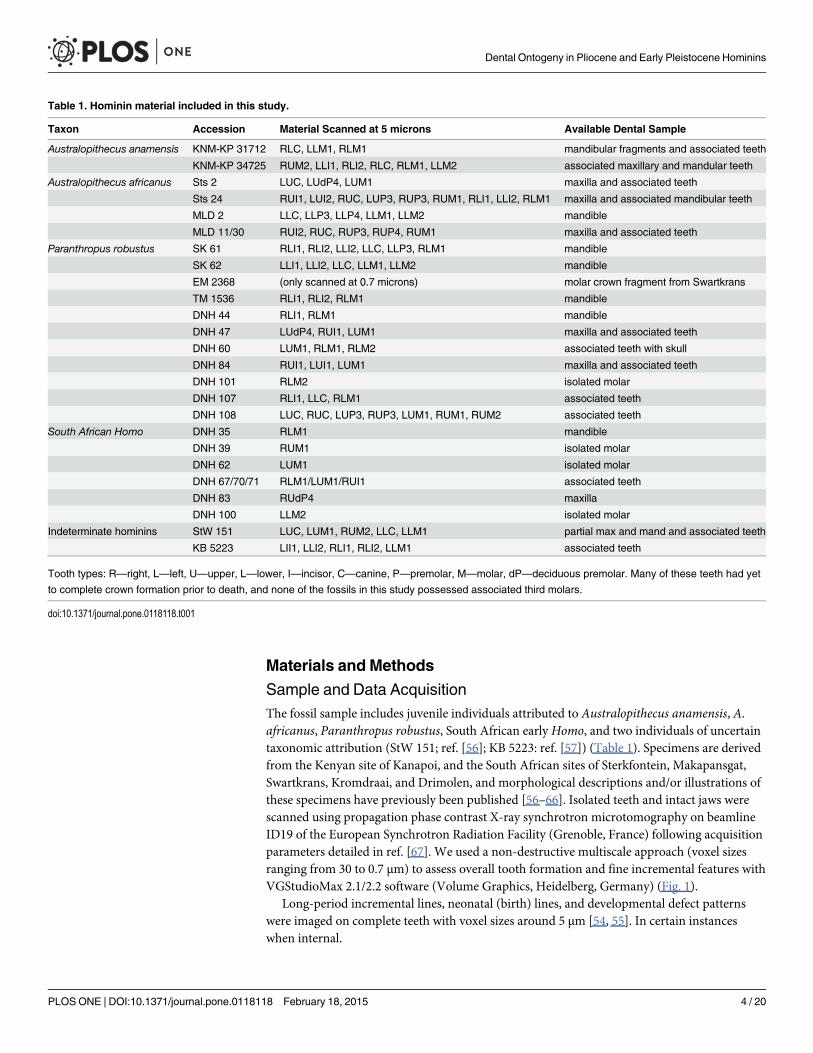
Sample and Data AcquisitionThe fossil sample includes juvenile individuals attributed to Australopithecus anamensis, A.africanus, Paranthropus robustus, South African early Homo, and two individuals of uncertaintaxonomic attribution (StW 151; ref. [56]; KB 5223: ref. [57]) (Table 1). Specimens are derivedfrom the Kenyan site of Kanapoi, and the South African sites of Sterkfontein, Makapansgat,Swartkrans, Kromdraai, and Drimolen, and morphological descriptions and/or illustrations ofthese specimens have previously been published [56–66]. Isolated teeth and intact jaws werescanned using propagation phase contrast X-ray synchrotron microtomography on beamlineID19 of the European Synchrotron Radiation Facility (Grenoble, France) following acquisitionparameters detailed in ref. [67]. We used a non-destructive multiscale approach (voxel sizesranging from 30 to 0.7 μm) to assess overall tooth formation and fine incremental features withVGStudioMax 2.1/2.2 software (Volume Graphics, Heidelberg, Germany) (Fig. 1).
Long-period incremental lines, neonatal (birth) lines, and developmental defect patternswere imaged on complete teeth with voxel sizes around 5 μm [54, 55]. In certain instanceswhen internal.
Table 1. Hominin material included in this study.
Taxon Accession Material Scanned at 5 microns Available Dental Sample
Australopithecus anamensis KNM-KP 31712 RLC, LLM1, RLM1 mandibular fragments and associated teeth
KNM-KP 34725 RUM2, LLI1, RLI2, RLC, RLM1, LLM2 associated maxillary and mandular teeth
Australopithecus africanus Sts 2 LUC, LUdP4, LUM1 maxilla and associated teeth
Sts 24 RUI1, LUI2, RUC, LUP3, RUP3, RUM1, RLI1, LLI2, RLM1 maxilla and associated mandibular teeth
MLD 2 LLC, LLP3, LLP4, LLM1, LLM2 mandible
MLD 11/30 RUI2, RUC, RUP3, RUP4, RUM1 maxilla and associated teeth
Paranthropus robustus SK 61 RLI1, RLI2, LLI2, LLC, LLP3, RLM1 mandible
SK 62 LLI1, LLI2, LLC, LLM1, LLM2 mandible
EM 2368 (only scanned at 0.7 microns) molar crown fragment from Swartkrans
TM 1536 RLI1, RLI2, RLM1 mandible
DNH 44 RLI1, RLM1 mandible
DNH 47 LUdP4, RUI1, LUM1 maxilla and associated teeth
DNH 60 LUM1, RLM1, RLM2 associated teeth with skull
DNH 84 RUI1, LUI1, LUM1 maxilla and associated teeth
DNH 101 RLM2 isolated molar
DNH 107 RLI1, LLC, RLM1 associated teeth
DNH 108 LUC, RUC, LUP3, RUP3, LUM1, RUM1, RUM2 associated teeth
South African Homo DNH 35 RLM1 mandible
DNH 39 RUM1 isolated molar
DNH 62 LUM1 isolated molar
DNH 67/70/71 RLM1/LUM1/RUI1 associated teeth
DNH 83 RUdP4 maxilla
DNH 100 LLM2 isolated molar
Indeterminate hominins StW 151 LUC, LUM1, RUM2, LLC, LLM1 partial max and mand and associated teeth
KB 5223 LII1, LLI2, RLI1, RLI2, LLM1 associated teeth
Tooth types: R—right, L—left, U—upper, L—lower, I—incisor, C—canine, P—premolar, M—molar, dP—deciduous premolar. Many of these teeth had yet
to complete crown formation prior to death, and none of the fossils in this study possessed associated third molars.
doi:10.1371/journal.pone.0118118.t001
Dental Ontogeny in Pliocene and Early Pleistocene Hominins
PLOS ONE | DOI:10.1371/journal.pone.0118118 February 18, 2015 4 / 20
long-period lines were difficult to count due to poor preservation, specimens were assessedby aligning 2D slices with 3D renderings of the tooth surface and enamel-dentine junction,which were produced with a combination of colored grazing incidence light sources followingref. [67]. This method involves capturing the phase contrast fringes at the outer enamel surfaceand enamel-dentine junction through segmentation of the structural interfaces, and generationof three-dimensional models with Phong’s algorithm and virtual lighting, which enhances sur-face topography and variation in tissue density. The 3D renderings and virtual slices employedin this study are available in the open access database for paleontology hosted by the ESRF(http://paleo.esrf.eu).
In order to characterize long-period line periodicity, selected areas of one or more teeth ofeach individual were scanned using sub-micron resolution local phase contrast X-ray synchro-tron microtomography, and virtual histological slices (typically 30 to 100 μm thick) were
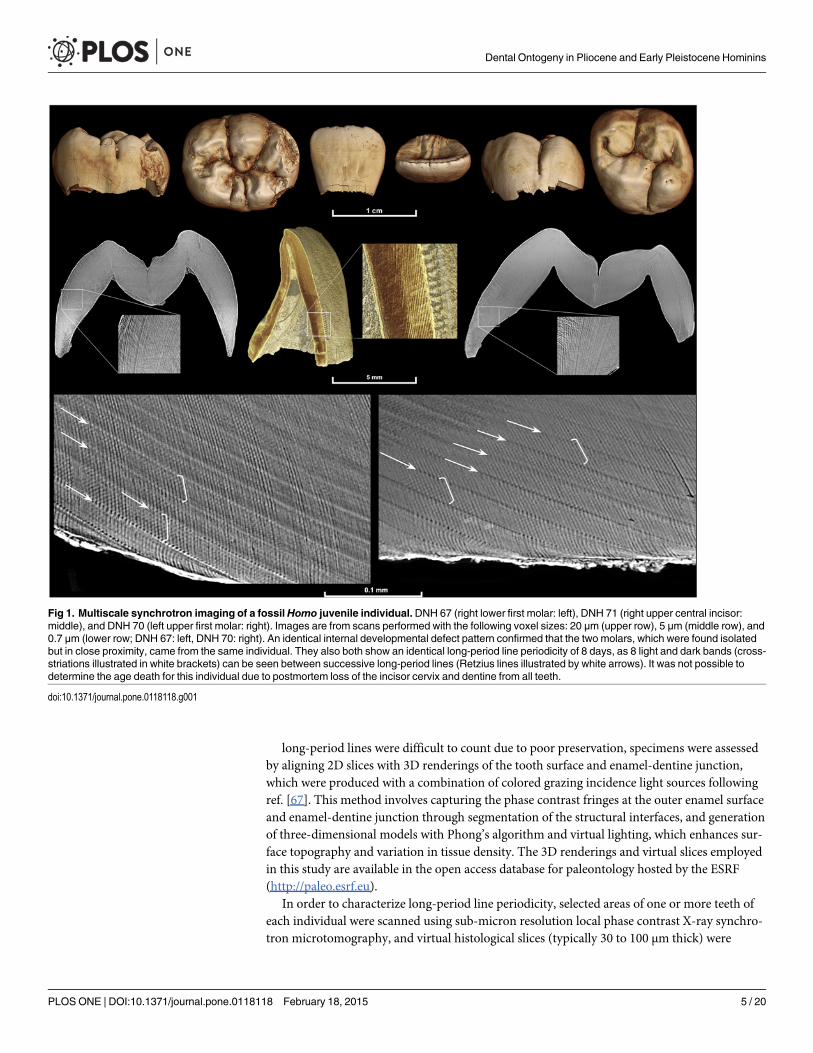
Fig 1. Multiscale synchrotron imaging of a fossilHomo juvenile individual.DNH 67 (right lower first molar: left), DNH 71 (right upper central incisor:middle), and DNH 70 (left upper first molar: right). Images are from scans performed with the following voxel sizes: 20 μm (upper row), 5 μm (middle row), and0.7 μm (lower row; DNH 67: left, DNH 70: right). An identical internal developmental defect pattern confirmed that the two molars, which were found isolatedbut in close proximity, came from the same individual. They also both show an identical long-period line periodicity of 8 days, as 8 light and dark bands (cross-striations illustrated in white brackets) can be seen between successive long-period lines (Retzius lines illustrated by white arrows). It was not possible todetermine the age death for this individual due to postmortem loss of the incisor cervix and dentine from all teeth.
doi:10.1371/journal.pone.0118118.g001
Dental Ontogeny in Pliocene and Early Pleistocene Hominins
PLOS ONE | DOI:10.1371/journal.pone.0118118 February 18, 2015 5 / 20
produced following 3D optimized orientation (Fig. 1). Temporary darkening of the enamel oc-curred in some areas scanned at high resolution; teeth were returned to their original colorwith 1–12 hours of low energy UV illumination (70 W dark light neon tube, 370 nm wave-length; following ref. [55].)
Developmental Feature QuantificationCuspal enamel thickness was measured on 2D slices with Adobe Photoshop CS5 from the den-tine horn tip to the approximate position of the first-formed long-period line (perikyma) at thecrown surface. Labio-lingual sections were cut for anterior teeth, buccal-lingual sections werecut for premolars, and buccal-lingual sections were cut for the mesial and distal cusps of molarsfollowing a standardized virtual orientation technique [54]. Long-period line periodicity wasdetermined through counts of daily growth lines between successive long-period lines in enam-el (Fig. 1). The Mann-Whitney U test was employed for comparisons of periodicity values be-tween taxa represented by four or more individuals. The fidelity of virtual histology has beendemonstrated through comparisons of virtual and histological sections [55], and long-periodline periodicity values determined using this approach have been shown to be equivalent tothose determined using light microscopy [68].
Crown formation time was calculated as the sum of cuspal and lateral formation times. Cus-pal formation time was typically calculated by dividing the linear enamel thickness of eachtooth cusp by the species-specific average cuspal daily secretion rates in ref. [53]. Daily secre-tion rates for KB 5223 were taken from ref. [57]. Direct measurements of daily secretion ratesin DNH 35 were made in the inner, middle, and outer cuspal enamel of the mesiolingual cuspfrom 0.7 μm scans. Direct measurements of cuspal daily secretion rates for other specimenswere not possible due to the time-intensive nature of quantifying cross-striation spacing in cus-pal enamel. The average value from DNH 35 (6.06 μm/day) was used to estimate cuspal enamelformation times in other South African early Homo samples. For Stw 151, which has been at-tributed to either early Homo or A. africanus [56], estimates of cuspal enamel formation timewere made using secretion rates from DNH 35 and average values from A. africanus [53], re-sulting in differences of 18–50 days depending on the tooth analyzed. Lateral enamel formationtime was calculated by multiplying the number of long-period lines (Retzius lines and/or peri-kymata) by the long-period line periodicity. Minor estimates of long-period lines were madefor worn or chipped enamel; specimens were excluded when long-period lines could not becounted on at least 90% of the total crown height. Comparative data on long-period line peri-odicity and crown formation times were chosen to minimize potential bias from methodologi-cal differences or interobserver error, and data sources are provided in thecorresponding tables.
Age at Death, Initiation, and Calcification Stage DeterminationAge at death was typically calculated by identification of the neonatal (birth) line in permanentfirst molars (M1s) or deciduous premolars, and summation of subsequent crown and root for-mation determined from incremental features. In three individuals (DNH 35, DNH 84, and KB5223) it was not possible to identify a neonatal line, and thus the M1 cusp employed was as-sumed to have initiated at birth, as species-specific initiation ages were not available for theseparticular cusps. For older juveniles, developmental defects (hypoplasias or accentuated lines)were registered across the dentition to link developmentally-overlapping teeth [10, 36, 54]. Infour individuals (MLD 11/30, SK 62, TM 1536, and DNH 44) it was not possible to registerM1s with subsequent teeth. In these cases initiation ages of anterior teeth from individuals ofthe same species were used to calculate age at death by adding crown and root formation times
Dental Ontogeny in Pliocene and Early Pleistocene Hominins
PLOS ONE | DOI:10.1371/journal.pone.0118118 February 18, 2015 6 / 20
to the tooth-specific initiation ages. Initiation ages were determined by registering synchro-nously forming teeth with developmental defects and subtracting preceding developmentaltime from the age of the defect used to register teeth. For example, an accentuated line wasidentified at 143 days of age in the mandibular M1 of KNM-KP 31712, which was also observedin the mandibular canine 110 days after initiation and 878 days before death. Thus the mandib-ular canine initiated at 33 days of age (143 days minus 110 days) and the individual died at1021 days of age (33 days of age at initiation plus 988 days of canine crown formation untildeath).
Tooth calcification was determined from virtual 2D sections of each tooth for those individ-uals whose age could be determined. The section planes employed to assess calcification stagesoften differed from those employed to assess cuspal enamel thickness. Sections were cut to op-timize developing root profiles for calcification stage determination, and a numerical score wasassigned to each tooth following the commonly employed classification system of Moorreesand colleagues [69]. Calcification standards derived from this 14 stage system [69] are moreprecise than those derived from Demirjian and colleagues’ 8 stage system [70], as crown androot formation is represented by more discrete developmental stages in the former case. Ageswere assigned to each individual tooth using an average of the extant human male and femalestandards in ref. [71]. We employed Anderson and colleagues’ calcification tables [71] as theyinclude maxillary and mandibular standards for each tooth of human children from ages 3years and up. We also collected comparative calcification data from panoramic X-rays of Euro-pean and North African human children (an expanded sample originally detailed in ref. [54]),and from recently deceased known-age West African wild chimpanzees [72].
While additional aging systems from extant human tooth calcification and/or emergencestudies are available [73, 74], the tables in ref. [71] allow discrete age calculations for homininsrepresented by partial dentitions and for whom emergence status may be unknown. Althoughscholars have noted limitations in the accuracy of this and other aging methods [73, 75], aswell as potential population-level variation in extant human tooth calcification ([76] but seerefs. [77, 78]), the aim of our study was to apply a consistent method to extant humans, chim-panzees, and juvenile hominins represented by partial dentitions in order compare the ontoge-ny of tooth calcification among these groups. Any bias in the accuracy of the calcificationsystem or aging standards we employ here applies equally to each group and does not impactconclusions based on relative patterns. Finally, in order to assess the appropriateness of chim-panzee calcification stages for hominin age prediction, we applied Kuykendall’s [79] predictivestandard (requiring the presence of all mandibular teeth except M3) to the two P. robustus ju-veniles that possessed the appropriate teeth and for which it was possible to estimate age atdeath histologically.
Results and Discussion
Long-period Line Periodicity, Enamel Thickness, and Crown FormationTimeWe find a remarkably wide range of long-period line periodicity values in 22 Pliocene andearly Pleistocene hominins, which range from at least 6–12 days, and possibly 5–13 days(Fig. 2, Table A in S1 File). In two individuals it was not possible to choose between two period-icity values (KNM-KP 34725: 5 or 6 days, KB 5223: 12 or 13 days); subsequent calculations ofcrown formation time and age at death for these individuals are given for each periodicityvalue. Periodicity values in the 11 Paranthropus robustus individuals range from 6–12 days,and do not show a significant difference when compared with Australopithecus africanus,Homo neanderthalensis, fossilHomo sapiens, extant humans, gorillas, or previously reported
Dental Ontogeny in Pliocene and Early Pleistocene Hominins
PLOS ONE | DOI:10.1371/journal.pone.0118118 February 18, 2015 7 / 20
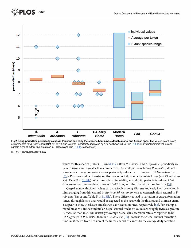
values for this species (Tables B-C in S1 File). Both P. robustus and A. africanus periodicity val-ues are significantly greater than chimpanzees. Australopiths (including P. robustus) do notshow smaller ranges or lower average periodicity values than extant or fossilHomo (contra[53]). Previous studies of australopiths have reported periodicities of 6–9 days (n = 29 individu-als) (Table B in S1 File). When considered in totality, australopith periodicity values of 6–9days are more common than values of 10–12 days, as is the case with extant humans [51].
Cuspal enamel thickness values vary markedly among Pliocene and early Pleistocene homi-nins, ranging from thin enamel in Australopithecus anamensis to extremely thick enamel in P.robustus (Fig. A and Table D in S1 File). These differences lead to variation in cuspal formationtimes, although less so than would be expected as the taxa with the thickest and thinnest enam-el appear to show the fastest and slowest daily secretion rates, respectively [53]. For example,mandibular M1 and second molar cuspal enamel thickness values are roughly twice as great inP. robustus than in A. anamensis, yet average cuspal daily secretion rates are reported to be~20% greater in P. robustus than in A. anamensis [53]. Because the cuspal enamel formationtime is estimated from division of the linear enamel thickness by the average daily secretion
Fig 2. Long-period line periodicity values in Pliocene and early Pleistocene hominins, extant humans, and African apes. Two values (5 or 6 days)are presented for A. anamensis KNM-KP 34725 due to some uncertainty (indicated by “?”), as shown in Fig. B in S1 File. Individual hominin values andsample sizes of extant taxa are given in Tables A and B in S1 File, respectively.
doi:10.1371/journal.pone.0118118.g002
Dental Ontogeny in Pliocene and Early Pleistocene Hominins
PLOS ONE | DOI:10.1371/journal.pone.0118118 February 18, 2015 8 / 20

rate, these rate differences result in formation times that differ by less than a factor of two inthis comparison. In general, our cuspal enamel thickness values are broadly similar to earlierreports of A. anamensis, P. robustus, A. africanus, and earlyHomomolars [65, 80–82], althoughdirect comparisons are limited due to methodological differences.
Hominin incisor formation times are shorter than chimpanzees (contra [6]), and are moresimilar to those of extant humans and gorillas (Table 2). This is likely due, in part, to absolute dif-ferences in tooth size among these taxa, which are most extreme in the diminutive anterior denti-tion of P. robustus. Maxillary third premolar (P3) formation times in A. africanus and P. robustusare shorter than those of extant humans and chimpanzees, while A. africanusmandibular premo-lar values exceed mean values for extant humans from South Africa (P3 buccal cusp) andchimpanzees (P4 lingual cusp). Comparisons of molar crown formation times also show a com-plex pattern, which is difficult to generalize as each molar tooth and cusp must be considered in-dependently [51]. Our new australopith molar crown formation times are broadly consistentwith previous reports of A. anamensis, Australopithecus afarensis, A. africanus, P. robustus, andHomo erectus values [15, 21, 47, 65, 82], which are shorter than those of extant humans [54, 83].In contrast, molar crown formation times for the South African earlyHomo individual DNH67/70/71 are most similar to times for both gorillas and extant humans from South Africa(Fig. 3), underscoring the importance of further recovery and histological study of this enigmatichominin group. Given the small sample sizes of the available fossil and comparative material, it ispremature to draw firm conclusions about differences in crown formation times among thesetaxa. Molar crown formation times, for example, are known to vary by ~ 6–12 months in smallsamples of African apes and extant humans [10, 54, 84], and comparable standard deviationshave also been reported for great ape and human canine formation times [85].
Age at Death and Molar EruptionThese data facilitate histologically-derived age at death estimates for 16 hominin juveniles(Table 3; Table E in S1 File). Previous histological studies reported ages at death for Sts 24 at3.3 years [14], SK 62 at 3.35–3.48 years [14], and StW 151 at 5.2–5.3 years [66]; our new agesdiffer by 0.2–1.1 years depending on the individual. Age at death estimates in other early homi-nins [14, 17, 20, 24, 29, 32, 45] should be reconsidered in light of these findings, particularlygiven newly expanded ranges of long-period line periodicity values in australopiths. For exam-ple, the A. afarensis specimen LH 2 was estimated to have died at 3.25 years of age [14] based,in part, on a count of 130 long-period lines for the mandibular central incisor and an estimatedperiodicity value of 7 days. If we assume that A. afarensis possessed a similar range of long-period line values to A. anamensis and A. africanus (minimally 6–11 days; Fig. 2), age at deathestimates would range from 130 days younger (6 day periodicity) to 520 days older (11 day pe-riodicity) than the original estimate. These potential age ranges complicate comparisons withextant taxa and underscore the necessity of comprehensive physical or virtual histological anal-yses for precise age at death estimation.
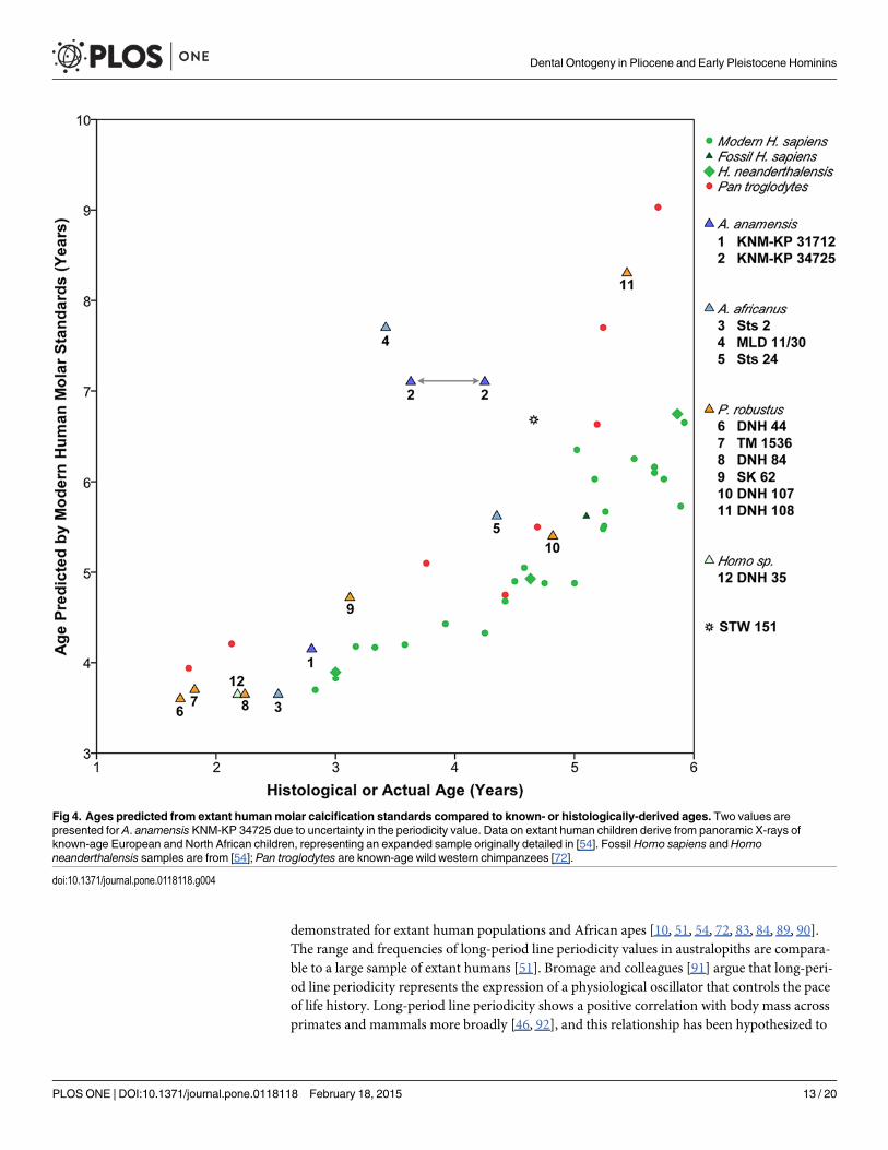
Since our age at death estimates do not rely on extant human or great ape developmental in-formation, we compared ages derived from the calcification stage of each tooth of each individ-ual (Figs. C-R in S1 File) to a sample of known-age extant humans and wild chimpanzees(Fig. S in S1 File). Because chimpanzee anterior tooth and premolar morphology differsmarkedly from fossil hominins and extant humans, we also assessed the developmental statusof individuals based solely on molar calcification (Fig. 4). Pliocene and early Pleistocene homi-nins followed an ontogenetic schedule of molar development that is more similar to chimpan-zees than to extant humans, as has been inferred from assessments of other Pliocene and earlyPleistocene hominins [14, 15, 20, 45]. However, marked differences are apparent for one A.
Dental Ontogeny in Pliocene and Early Pleistocene Hominins
PLOS ONE | DOI:10.1371/journal.pone.0118118 February 18, 2015 9 / 20

anamensis (KNM-KP 34725) and one A. africanus individual (MLD 11/30), which show par-ticularly rapid dental development that exceeds similarly-aged humans and chimpanzees. Ageat death was also predicted for two P. robustus individuals with captive chimpanzee standards[79]. The resulting age estimates were closer to the actual histological ages than the ages pre-dicted from extant human standards for only one of two cases (Table F in S1 File). In summary,radiographic developmental standards from extant humans and chimpanzees do not yield con-sistent or accurate ages at death estimates for Pliocene or early Pleistocene hominins.
None of the individuals in this study appear to have died during the initial skeletal phase ofM1 emergence (alveolar eruption). Marked wear facets are apparent on the mandibular M1 of a3.6 or 4.2 year-old A. anamensis individual (KNM-KP 34725) and a 3.4 year-old A. africanus in-dividual (MLD 11/30). More subtle minor wear facets were also observed on the maxillary andmandibular M1s of a 4.4 year-old A. africanus individual (Sts 24), implying variation in
Table 2. Average crown formation times (in days) determined by histological methods.
Tooth/cusp
H. sapiens(Eur)
H. sapiens(SA)
Pantroglodytes
Gorillagorilla
A. anamensis A. africanus P. robustus SAHomo
Stw 151 KB 5223
UI1 1582 1318 1827 1261 (2) — 1557 — — — —
UI2 1427 1324 1461 1496 — 961 — — — —
UC 1613(mixed sex)
1438(mixed sex)
2053(sex unknown)
— — 1014 818 — >1562/1580 —
UP3 buc 1407 1113 1401 — — — 770 — — —
UP3 lin 1281 972 967 — — 826 — — — —
UM1 mb 1017 981 867 (6) 784 — 544 612 — — —
UM1 ml 1159 1059 775 (5) 994 (2) — 962 775 1035 — —
UM1 dl 1088 — 684 (2) 1168 (2) — 959 785 — 952/986 —
UM2 ml 1141 1218 1012 (2) 1057 — — 806 — — —
UM2 dl 1068 — — 1136 899/1001 — — — 921/971 —
LI1 1251 1096 1581 1141 (2) 1170/1381 1220 891 (2) — — —
LI2 1293 1169 1764 1287 (2) — 1399 — — — 1296/1389
LC 2004(mixed sex)
1721(sex unknown)
2135 (f:6), 2486(m:6)
2058 (f:7),3117 (m:8)
— — >1558 — — —
LP3 buc 1454 1157 1496 — — 1223 — — — —
LP4 lin — — 823 (2) — — 1056 — — — —
LM1 mb 1097 1096 797 (6) 932 (2) — — 949 — — —
LM1 ml 948 917 633 (5) 905 (3) — — — 876 — —
LM1 db 1111 — 811 (8) 1142 (2) — 1052 — — — —
LM1 dl 939 — 662 (4) 860 (2) — 813 (2) 832 675 — 1110/1164
LM2 mb 1096 1145 1009 (5) 1241 — 1013 911 — — —
LM2 ml 904 931 847 (6) 964 (2) 754/830 996 — — — —
LM2 db — — 1160 1118 (3) — — 960 — — —
Tooth/cusp types: U—upper, L—lower, I—incisor, C—canine, P—premolar, M—molar, buc—buccal cusp, ling—lingual cusp, mb—mesiobuccal cusp,
ml—mesiolingual cusp, db—distobuccal cusp, dl—distolingual cusp. Homo sapiens European (EUR) and South African (SA) ranges and sample sizes
given in refs. 54, 83. Pan troglodytes data modified from refs. [72, 84, 85, 88, 104]; Gorilla gorilla data from refs. [10, 89, 105, 106]; samples sizes for
extant apes are given in parentheses when greater than one. Fossil hominin sample sizes are given in parentheses when greater than one; A. anamensis
values reported for periodicity of 5 or 6 days in KNM-KP 34725; StW 151 values based on cuspal daily secretion rates of 5.53 um/day (A. africanus) from
ref. [53] and 6.06 um/day (early Homo, determined from this study); KB 5223 values based on cuspal daily secretion rates from ref. [57] and long-period
line periodicity values of 12 or 13.
doi:10.1371/journal.pone.0118118.t002
Dental Ontogeny in Pliocene and Early Pleistocene Hominins
PLOS ONE | DOI:10.1371/journal.pone.0118118 February 18, 2015 10 / 20
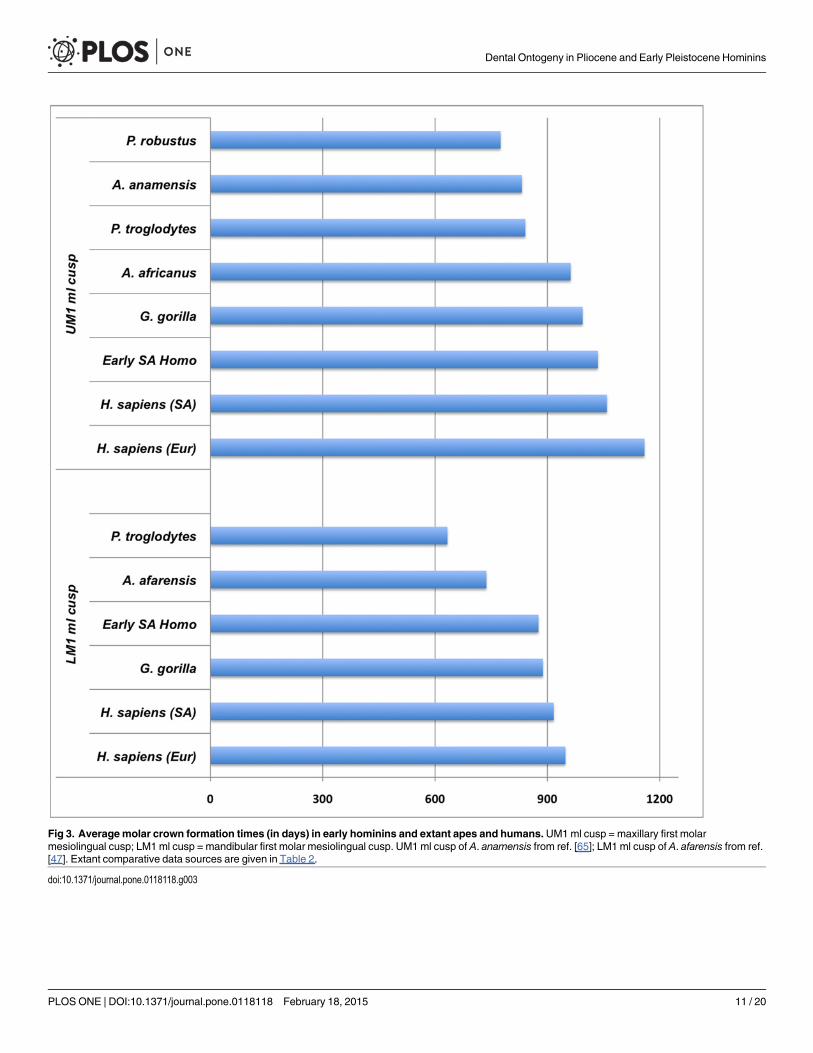
Fig 3. Averagemolar crown formation times (in days) in early hominins and extant apes and humans.UM1ml cusp = maxillary first molarmesiolingual cusp; LM1 ml cusp = mandibular first molar mesiolingual cusp. UM1 ml cusp of A. anamensis from ref. [65]; LM1 ml cusp of A. afarensis from ref.[47]. Extant comparative data sources are given in Table 2.
doi:10.1371/journal.pone.0118118.g003
Dental Ontogeny in Pliocene and Early Pleistocene Hominins
PLOS ONE | DOI:10.1371/journal.pone.0118118 February 18, 2015 11 / 20
A. africanusM1 emergence that is comparable to chimpanzees, which may erupt their first mo-lars between ~2.1–4.5 years of age [72, 84, 86–88]. Three P. robustus individuals sampled here arealso of interest for consideration of M1 emergence ages: 3.1 year-old SK 62 had not yet erupted itsM1s (although the right mandibular I1 was erupting); the mandibular M1 of DNH 107 appearedto be nearing full occlusion (with slight facets) at 4.8 years of age (Fig. T in S1 File), and the maxil-lary M1 of DNH 108 showed marked facets by 5.4 or 5.5 years of age. Remarkably, the 4.6 or 4.7year-old StW 151 individual (attributed to either earlyHomo or A. africanus; ref. [56]) appears tobe more developmentally advanced than the 4.8 year-old P. robustus individual DNH 107, whichshowed less wear on its mandibular M1 (and had yet to erupt its right mandibular I1.) The maxil-lary M1 emergence status and wear in StW 151 appears to be more similar to the 5.4 or 5.5 year-old P. robustus individual DNH 108. Although it is difficult to precisely compare emergenceamong these individuals, our results suggest that M1 emergence inA. anamensis and/or A. africa-nusmay have been somewhat earlier than in P. robustus.
ConclusionsSubstantial intraspecific variation exists in aspects of dental development quantified in thisstudy (long-period line periodicity and cuspal enamel thickness), which has also been
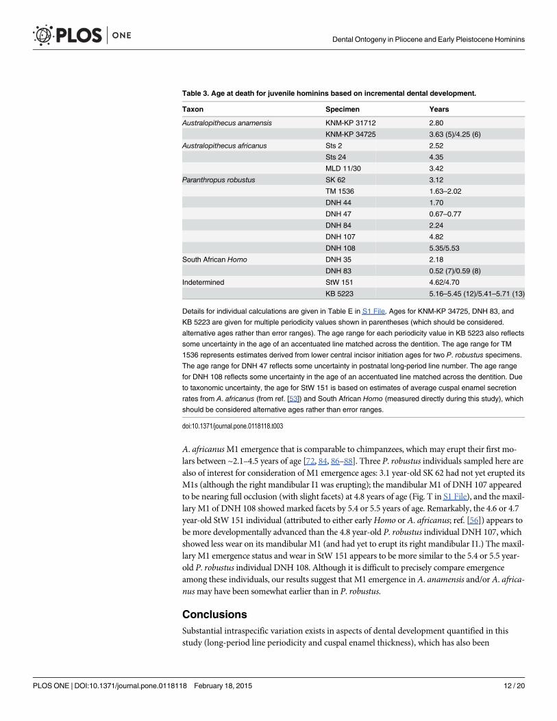
Table 3. Age at death for juvenile hominins based on incremental dental development.
Taxon Specimen Years
Australopithecus anamensis KNM-KP 31712 2.80
KNM-KP 34725 3.63 (5)/4.25 (6)
Australopithecus africanus Sts 2 2.52
Sts 24 4.35
MLD 11/30 3.42
Paranthropus robustus SK 62 3.12
TM 1536 1.63–2.02
DNH 44 1.70
DNH 47 0.67–0.77
DNH 84 2.24
DNH 107 4.82
DNH 108 5.35/5.53
South African Homo DNH 35 2.18
DNH 83 0.52 (7)/0.59 (8)
Indetermined StW 151 4.62/4.70
KB 5223 5.16–5.45 (12)/5.41–5.71 (13)
Details for individual calculations are given in Table E in S1 File. Ages for KNM-KP 34725, DNH 83, and
KB 5223 are given for multiple periodicity values shown in parentheses (which should be considered.
alternative ages rather than error ranges). The age range for each periodicity value in KB 5223 also reflects
some uncertainty in the age of an accentuated line matched across the dentition. The age range for TM
1536 represents estimates derived from lower central incisor initiation ages for two P. robustus specimens.
The age range for DNH 47 reflects some uncertainty in postnatal long-period line number. The age range
for DNH 108 reflects some uncertainty in the age of an accentuated line matched across the dentition. Due
to taxonomic uncertainty, the age for StW 151 is based on estimates of average cuspal enamel secretion
rates from A. africanus (from ref. [53]) and South African Homo (measured directly during this study), which
should be considered alternative ages rather than error ranges.
doi:10.1371/journal.pone.0118118.t003
Dental Ontogeny in Pliocene and Early Pleistocene Hominins
PLOS ONE | DOI:10.1371/journal.pone.0118118 February 18, 2015 12 / 20
demonstrated for extant human populations and African apes [10, 51, 54, 72, 83, 84, 89, 90].The range and frequencies of long-period line periodicity values in australopiths are compara-ble to a large sample of extant humans [51]. Bromage and colleagues [91] argue that long-peri-od line periodicity represents the expression of a physiological oscillator that controls the paceof life history. Long-period line periodicity shows a positive correlation with body mass acrossprimates and mammals more broadly [46, 92], and this relationship has been hypothesized to
Fig 4. Ages predicted from extant humanmolar calcification standards compared to known- or histologically-derived ages. Two values arepresented for A. anamensis KNM-KP 34725 due to uncertainty in the periodicity value. Data on extant human children derive from panoramic X-rays ofknown-age European and North African children, representing an expanded sample originally detailed in [54]. FossilHomo sapiens andHomoneanderthalensis samples are from [54]; Pan troglodytes are known-age wild western chimpanzees [72].
doi:10.1371/journal.pone.0118118.g004
Dental Ontogeny in Pliocene and Early Pleistocene Hominins
PLOS ONE | DOI:10.1371/journal.pone.0118118 February 18, 2015 13 / 20

be driven by selection on body mass [91]. However expected relationships among life historyvariables, body mass, and periodicity may not hold among closely related primates (or amongindividuals within a species). For example, lowland gorillas have significantly greater periodici-ty values than common chimpanzees (Z = -4.273, p< 0.001), yet chimpanzees have longerinterbirth intervals, and later ages at weaning, first reproduction, and first birth [93–96]. Simi-lar concerns have been raised about the widely cited relationship between hominoid molaremergence and life history [5, 86, 97, 98], which is discussed further below.
Pliocene and early Pleistocene hominin incisor, canine, and premolar crown formationtimes reveal considerable interspecific variation that warrants further study, particularly withregard to potential sex differences and constraints due to absolute tooth sizes. New molarcrown formation times are broadly comparable to previous reports of fossil hominins, and aretypically shorter than molar crown formation times in extant humans. We have also docu-mented relatively late initiation ages for certain P. robustus teeth (Table G in S1 File), whichimpact estimates of age at tooth crown formation and emergence. For example, mandibular in-cisor initiation in P. robustus occurred between 109–251 days, which is approximately one tofive months later than the modern human value employed by Bromage and Dean [14]. Previ-ous histological assessments of age at death (and molar emergence age) in early fossil homininsshould be reconsidered in light of the expanded range of long-period line periodicities, cuspalenamel thickness values, crown formation times, and initiation ages documented here.
We present histologically-derived ages at death for 16 Pliocene and early Pleistocene homi-nins, illustrating an approach that may eventually lead to the development of australopith calci-fication standards in order to estimate the age of other juveniles. Importantly, these ages weredetermined from developmental information quantified directly from the specimens them-selves and supplemented with species-specific information on daily secretion rates [53, 57] andinitiation ages from this study when necessary. This facilitates discrete developmental compari-sons with extant African apes and humans, and suggests a more nuanced interpretation of thedevelopmental affinities of Pliocene and early Pleistocene hominins. Analyses of rare histologi-cal (physical) sections posited that australopith dental development (daily secretion rate, age atM1 emergence, root extension rate) was similar to living great apes and more rapid than extanthumans, implying that early hominin life histories were great ape-like [6, 15, 21, 24]. Our com-parisons with extant humans and African apes demonstrate a complex pattern of similaritiesand differences in long-period line periodicity, crown formation time, the timing of tooth calci-fication, and molar eruption ages, prohibiting simplistic characterizations of these species as ei-ther “human-like” or “great ape-like.”While some specialists of tooth development havereached similar conclusions over the past decade [2, 4], initial determinations of age at deathfor juvenile hominins continue to be made from extant ape developmental standards (e.g., Aus-tralopithecus afarensisDikika child: ref. [8]). Moreover, this understanding is complicated byreports that dental development in later hominins (e.g., Homo erectus Narikotome KNM-WT150000) may resemble that of African apes [21, 99], which underscores the need for compre-hensive histological assessments based entirely on species-specific developmental information.
Studies of dental development have also been of great interest for reconstructing the evolu-tion of human life history, although scholars have recently begun to question the associationbetween molar emergence and life history variables among extant apes and humans [5, 7, 86,97]. For example, fundamental questions remain about the suitability of primate-wide trendsin M1 emergence for predicting weaning ages in hominoids. While it is likely that M1 emer-gence ages in most Pliocene and early Pleistocene taxa fall within African ape ranges, additionaldata are needed to discern how similar their life histories were. Future studies that aim to pre-dict hominin life history would benefit from analyses of aspects of dental development thathave been suggested to correlate with primate life history variables (e.g., molar crown
Dental Ontogeny in Pliocene and Early Pleistocene Hominins
PLOS ONE | DOI:10.1371/journal.pone.0118118 February 18, 2015 14 / 20
formation time, molar eruption age, tooth calcification timing, long-period line periodicity) insamples of wild primates with documented life histories. These approaches would also benefitgreatly from statistical approaches that control for phylogenetic relationships, as well as the po-tential confounding effect of sexual dimorphism and variation in tooth and body size.
While it is tempting to compare aspects of dental development in order to speculate on thetaxonomic identity of StW 151 or KB 5223, additional data are required from securely identi-fied Pliocene and early Pleistocene hominins. Little is known about dental development andoverall tooth morphology in earlyHomo relative to species of Australopithecus or Paranthropusdue to limited samples and uncertainty over the taxonomic identification of particular elements[56, 57, 100]. Moreover, most reports of early Homo cuspal enamel thickness and crown for-mation time involve analyses of naturally fractured teeth, which may be of limited comparativevalue due to section obliquity [15, 101], or the necessity of quantifying development from non-standardized positions on the tooth crown. Although marked differences in certain develop-mental features have been documented between chimpanzees and extant humans [51], orangu-tans and Homo erectus [102], and great apes and australopiths [52], considerable variationwithin and among fossil and extant species suggests that taxonomic interpretations of dentaldevelopment in early hominins should be made with caution, if at all, as is the case with overallmeasures of enamel thickness [103].
Supporting InformationS1 File. Contains supporting tables and figures. Fig. A. Linear cuspal enamel thickness valuesin four hominin taxa. Fig. B. Long-period line periodicity in two A. anamensis individuals.Fig. C. Developmental plate used to assess tooth calcification in A. anamensis (KNM-KP 31712).Fig. D. Developmental plate used to assess tooth calcification in A. anamensis (KNM-KP 34725).Fig. E. Developmental plate used to assess tooth calcification in A. africanus (Sts 2). Fig. F. De-velopmental plate used to assess tooth calcification in A. africanus (Sts 24). Fig. G. Developmen-tal plate used to assess tooth calcification in A. africanus (MLD 11/30). Fig. H. Developmentalplate used to assess tooth calcification in P. robustus (SK 62). Fig. I. Developmental plate used toassess tooth calcification in P. robustus (TM 1536). Fig. J. Developmental plate used to assesstooth calcification in P. robustus (DNH 44). Fig. K. Developmental plate used to assess tooth cal-cification in P. robustus (DNH 47). Fig. L. Developmental plate used to assess tooth calcificationin P. robustus (DNH 84). Fig. M. Developmental plate used to assess tooth calcification in P.robustus (DNH 107). Fig. N. Developmental plate used to assess tooth calcification in P. robustus(DNH 108). Fig. O. Developmental plate used to assess tooth calcification in earlyHomo (DNH35). Fig. P. Developmental plate used to assess tooth calcification in earlyHomo (DNH 83).Fig. Q. Developmental plate used to assess tooth calcification in StW 151. Fig. R. Developmentalplate used to assess tooth calcification in KB 5223. Fig. S. Ages at death predicted frommodernhuman calcification standards compared to known- or histologically-derived ages. Fig. T. Re-cently erupted lower right first molar of DNH 107, a 4.8 year-old P. robustus individual from Dri-molen. Table A. Long-period line periodicity (in days) for individuals in this study. Table B.Long-period line periodicity (in days) for fossil hominins, extant humans, and chimpanzees.Table C. Results of Mann-Whitney U test for comparisons of long-period line periodicity in P.robustus and A. africanus with fossil hominin taxa, extant humans, African apes, and previouslypublished values. Table D. Cuspal enamel thickness values (in microns) for fossil hominins inthis study. Table E. Age at death calculations for individual hominin specimens. Table F. Com-parison of age at death estimates for two complete P. robustusmandibular dentitions. Table G.Histologically-determined initiation ages in Pliocene and early Pleistocene hominins.(DOC)
Dental Ontogeny in Pliocene and Early Pleistocene Hominins
PLOS ONE | DOI:10.1371/journal.pone.0118118 February 18, 2015 15 / 20
AcknowledgmentsAkiko Kato, Meg Jarvi, Lazarus Kgasi, Meg Lynch, Emma Mbua, Charles Musiba, AmandaPapakyrikos, Stephany Potze, Nancy Tang, Francis Thackeray, JP Zermeno, and Bernhard Zip-fel provided invaluable research assistance, as well as the following institutions: DITSONGMu-seums of South Africa, European Synchrotron Radiation Facility, Harvard University, NationalMuseums of Kenya, South African Heritage Resources Agency, and the University of the Wit-watersrand. Shara Bailey, Kate Carter, Wendy Dirks, Daniel Green, Dan Lieberman, AnthonyOlejniczak, Donald Reid, and Matt Skinner provided helpful discussions of this research, as didAlistair Evans and four anonymous reviewers of an earlier version of this manuscript. Thispaper is dedicated to the memory of our colleague Charlie Lockwood (1970–2008).
Author ContributionsConceived and designed the experiments: TMS PT. Performed the experiments: TMS PT ALCAB AH JP JM-C FM CWMMCGM. Analyzed the data: TMS ALC AB AH JP PT. Wrote thepaper: TMS PT ALC CW.
References1. Smith BH, Tompkins RL (1995) Toward a life history of the Hominidae. Annu Rev Anthropol 24:
257–279.
2. Macho GA, Wood BA (1995) The role of time and timing in hominid dental evolution. Evol Anthropol4: 17–31
3. Anemone RL (2002) Dental development and life history in hominid evolution. In: Minugh-Purvis N,McNamara KJ, editors. Human evolution through developmental change. Baltimore: Johns HopkinsUniversity Press. pp. 249–280.
4. Kuykendall K (2003) Reconstructing australopithecine growth and development: What do we think weknow? In: Thompson JL, Krovitz GE, Nelson AJ, editors. Patterns of growth and development in thegenusHomo. Cambridge: Cambridge University Press. pp. 191–218.
5. Guatelli‐Steinberg D (2009) Recent studies of dental development in Neandertals: Implications for Ne-andertal life histories. Evol Anthropol 18: 9–20.
6. Dean MC (2010) Retrieving chronological age from dental remains of early fossil hominins to recon-struct human growth in the past. Philos Trans R Soc Lond B Biol Sci 365: 3397–3410. doi: 10.1098/rstb.2010.0052 PMID: 20855313
7. Smith TM (2013) Teeth and human life-history evolution. Annu Rev Anthropol 42: 191–208.
8. Alemseged Z, Spoor F, Kimbel WH, Bobe R, Geraads D, et al. (2006) A juvenile early hominin skele-ton from Dikika, Ethiopia. Nature 443: 296–301. PMID: 16988704
9. Berger LR, de Ruiter DJ, Churchill SE, Schmid P, Carlson KJ, et al. (2010) Australopithecus sediba: Anew species of Homo-like australopith from South Africa. Science 328: 195–204. doi: 10.1126/science.1184944 PMID: 20378811
10. Schwartz GT, Reid DJ, Dean MC, Zihlman AL (2006) A faithful record of stressful life events recordedin the dental developmental record of a juvenile gorilla. Int J Primatol 27: 1201–1219.
11. Smith TM, Reid DJ, Sirianni JE (2006) The accuracy of histological assessments of dental develop-ment and age at death. J Anat 208: 125–138. PMID: 16420385
12. Smith TM, Tafforeau P (2008) New visions of dental tissue research: tooth development, chemistry,and structure. Evol Anthropol 17: 213–226.
13. Antoine D, Hillson S, DeanMC (2009) The developmental clock of dental enamel: a test for the period-icity of prism cross‐striations in modern humans and an evaluation of the most likely sources of errorin histological studies of this kind. J Anat 214: 45–55. doi: 10.1111/j.1469-7580.2008.01010.x PMID:19166472
14. Bromage TG, Dean MC (1985) Re-evaluation of the age at death of immature fossil hominids. Nature317: 525–527. PMID: 19093314
15. DeanMC, Beynon AD, Thackeray JF, Macho GA (1993) Histological reconstruction of dental develop-ment and age at death of a juvenile Paranthropus robustus specimen, SK 63, from Swartkrans, SouthAfrica. Am J Phys Anthropol 91: 401–419. PMID: 8372933
Dental Ontogeny in Pliocene and Early Pleistocene Hominins
PLOS ONE | DOI:10.1371/journal.pone.0118118 February 18, 2015 16 / 20
16. Beynon AD, Dean MC (1987) Crown formation time of a fossil hominid premolar tooth. Arch Oral Biol32: 773–780. PMID: 3130039
17. Beynon AD, Dean MC (1988) Distinct dental development patterns in early fossil hominids. Nature335: 509–514. PMID: 3138547
18. Beynon AD (1992) Circaseptan rhythms in enamel development in modern humans and Plio-Pleistocene hominids. In: Smith P, Tchernov E, editors. Structure, function and evolution of teeth.London: Freund. pp. 295–309.
19. Smith BH (1989) Dental development as a measure of life history in primates. Evolution 43: 683–688.
20. Dean MC (1987) The dental developmental status of six East African juvenile fossil hominids. J HumEvol 16: 197–213.
21. Dean C, Leakey MG, Reid D, Schrenk F, Schwartz GT, et al. (2001) Growth processes in teeth distin-guish modern humans from Homo erectus and earlier hominins. Nature 414: 628–631. PMID:11740557
22. Hawkes K (2006) Slow life histories and human evolution. In: Hawkes K, Paine RR, editors. The evo-lution of human life history. Sante Fe: School of American Research Press. pp. 95–126.
23. Skinner MM,Wood B (2006) The evolution of modern human life history: a paleontological perspec-tive. In: Hawkes K, Paine RR, editors. The evolution of human life history. Sante Fe: School of Ameri-can Research Press. pp. 331–364.
24. Dean MC, Lucas VS (2009) Dental and skeletal growth in early fossil hominins. Ann Hum Biol 36:545–561. doi: 10.1080/03014460902956725 PMID: 19579096
25. Konner M (2010) The evolution of childhood: relationships, emotion, mind. Cambridge, BelknapPress. 943 p.
26. Hochberg Z (2012) Evo-devo of child growth. Hoboken: Wiley-Blackwell. 235 p.
27. Schwartz GT (2012) Growth, development, and life history throughout the evolution of Homo. CurrAnthropol 53: S395–S408.
28. Knott C (2001) Female reproductive ecology of the apes. In: Ellison PT, editor. Reproductive ecologyand human evolution. Hawthorne: Aldine de Gruyter. pp. 429–63.
29. Kelley J, Schwartz GT (2012) Life-history inference in the early hominins Australopithecus and Para-nthropus. Int J Primatol 33: 1332–1363.
30. Smith BH (1992) Life history and the evolution of humanmaturation. Evol Anthropol 1: 134–142.
31. Mann A (1988) The nature of Taung dental maturation. Nature 333: 123. PMID: 3130577
32. Conroy GC, Vannier MW (1991) Dental development in South African australopithecines. Part II: Den-tal stage assessment. Am J Phys Anthropol 86: 137–156.
33. McNulty KP, Frost SR, Strait DS (2006) Examining affinities of the Taung child by developmental sim-ulation. J Hum Evol 51: 274–296. PMID: 16797056
34. Dean MC (1995) The nature and periodicity of incremental lines in primate dentine and their relation-ship to periradicular bands in OH 16 (Homo habilis). In: Moggi-Cecchi J, editor. Aspects of dental biol-ogy: paleontology, anthropology and evolution. Florence: International Institute for the Study ofMan. pp. 239–265.
35. Kelley J, Smith TM (2003) Age at first molar emergence in early Miocene Afropithecus turkanensisand life-history evolution in the Hominoidea. J Hum Evol 44: 307–329. PMID: 12657519
36. Smith TM, Toussaint M, Reid DJ, Olejniczak AJ, Hublin J-J (2007) Rapid dental development in a mid-dle Paleolithic Belgian Neanderthal. Proc Natl Acad Sci U S A 104: 20220–20225. PMID: 18077342
37. Dean MC, Beynon AD, Reid DJ, Whittaker DK (1993) A longitudinal study of tooth growth in a singleindividual based on long-and short-period incremental markings in dentine and enamel. Int JOsteoarchaeol 3: 249–264.
38. Dean MC, Scandrett AE (1996) The relation between long-period incremental markings in dentineand daily cross-striations in enamel in human teeth. Arch Oral Biol 41: 233–241. PMID: 8735009
39. Smith TM (2006) Experimental determination of the periodicity of incremental features in enamel.J Anat 208: 99–113. PMID: 16420383
40. Tafforeau P, Bentaleb I, Jaeger J-J, Martin C (2007) Nature of laminations and mineralization in rhi-noceros enamel using histology and X-ray synchrotron microtomography: potential implications forpalaeoenvironmental isotopic studies. Palaeogeogr Palaeoclimatol Palaeoecol 246: 206–227.
41. Mimura F (1939) The periodicity of growth lines seen in enamel. Kobyo-shi 13: 454–455.
42. Okada M (1943) Hard tissues of animal body: highly interesting details of Nippon studies in periodicpatterns of hard tissues are described. In The Shanghai Evening Post Special Edition, Health, Recre-ation and Medical Progress, pp. 15–31.
Dental Ontogeny in Pliocene and Early Pleistocene Hominins
PLOS ONE | DOI:10.1371/journal.pone.0118118 February 18, 2015 17 / 20
43. Bromage TG (1991) Enamel incremental periodicity in the pig-tailed macaque: A polychrome fluores-cent labeling study of dental hard tissues. Am J Phys Anthropol 86: 205–214.
44. FitzGerald CM (1998) Do enamel microstructures have regular time dependency? Conclusions fromthe literature and a large-scale study. J Hum Evol 35: 371–386. PMID: 9774500
45. Lacruz RS, Rozzi FR, Bromage TG (2005) Dental enamel hypoplasia, age at death, and weaning inthe Taung child. S Afr J Sci 101: 567–569.
46. Smith TM (2008) Incremental dental development: methods and applications in hominoid evolutionarystudies. J Hum Evol 54: 205–224. PMID: 18045649
47. Lacruz RS, Ramirez Rozzi FV (2010) Molar crown development in Australopithecus afarensis. J HumEvol 58: 201–206. doi: 10.1016/j.jhevol.2009.11.007 PMID: 20044127
48. Beynon AD, Wood BA (1987) Patterns and rates of enamel growth in the molar teeth of early homi-nids. Nature 326: 493–496. PMID: 3104794
49. Ramirez-Rozzi FV (1993) Tooth development in East African Paranthropus. J Hum Evol 24:429–454.
50. Ramirez Rozzi FV, Bromage T, Schrenk F (1997) UR 501, the Plio-Pleistocene hominid fromMalawi.C R Acad Sci IIA 325: 231–234.
51. Smith TM, Reid DJ, Dean MC, Olejniczak AJ, Ferrell RJ, et al. (2007) New perspectives on chimpan-zee and human molar crown development. In: Bailey S, Hublin J-J, editors. Dental perspectives onhuman evolution: state of the art research in dental paleoanthropology. Dordrecht: Springer. pp.177–192.
52. Dean M, Reid D (2001) Perikymata spacing and distribution on hominid anterior teeth. Am J PhysAnthropol 116: 209–215. PMID: 11596000
53. Lacruz RS, Dean MC, Ramirez-Rozzi F, Bromage TG (2008) Megadontia, striae periodicity and pat-terns of enamel secretion in Plio-Pleistocene fossil hominins. J Anat 213: 148–158. PMID: 19172730
54. Smith TM, Tafforeau P, Reid DJ, Pouech J, Lazzari V, et al. (2010) Dental evidence for ontogeneticdifferences between modern humans and Neanderthals. Proc Natl Acad Sci U S A 107: 20923–20928. doi: 10.1073/pnas.1010906107 PMID: 21078988
55. Tafforeau P, Smith TM (2008) Nondestructive imaging of hominoid dental microstructure using phasecontrast X-ray synchrotron microtomography. J Hum Evol 54: 272–278. PMID: 18045654
56. Moggi-Cecchi J, Tobias PV, Beynon A (1998) The mixed dentition and associated skull fragments of ajuvenile fossil hominid from Sterkfontein, South Africa. Am J Phys Anthropol 106: 425–465. PMID:9712475
57. Lacruz RS (2007) Enamel microstructure of the hominid KB 5223 from Kromdraai, South Africa. Am JPhys Anthropol 132: 175–182. PMID: 17103424
58. Broom R (1941) Mandible of a young Paranthropus child. Nature 147: 607–608.
59. Dart RA (1948) The adolescent mandible of Australopithecus prometheus. Am J Phys Anthropol 6:391–412. PMID: 18105833
60. Broom R, Robinson JT, Schepers GWH (1950) Sterkfontein ape-man, Plesianthropus. Transvaal Mu-seumMemoir 4. Pretoria: Transvaal Museum. 117 p.
61. Robinson JT (1956) The dentition of the Australopithecinae. Transvaal MuseumMemoir 9. Pretoria:Transvaal Museum. 179 p.
62. Grine FE (1982) A new juvenile hominid (Mammalia: Primates) fromMember 3, Kromdraai Formation,Transvaal, South Africa. Ann Transvaal Museum 33: 165–239.
63. Keyser AW, Menter CG, Moggi-Cecchi J, Pickering TR, Berger LR (2000) Drimolen: a new hominid-bearing site in Gauteng, South Africa. S Afr J Sci 96: 193–197.
64. Moggi-Cecchi J, Menter C, Boccone S, Keyser A (2010) Early hominin dental remains from the Plio-Pleistocene site of Drimolen, South Africa. J Hum Evol 58: 374–405. doi: 10.1016/j.jhevol.2010.01.006 PMID: 20362324
65. Ward CV, Leakey MG, Walker A (2001) Morphology of Australopithecus anamensis from Kanapoiand Allia Bay, Kenya. J Hum Evol 41: 255–368. PMID: 11599925
66. Schwartz JH, Tattersall I (2005) The human fossil record, vol 4. New York: Wiley-Liss. 561 p.
67. Le Cabec A, Smith TM, Tang N, Tafforeau P (in review) Accessing developmental information of fossilhominin teeth using new synchrotron microtomography-based visualization techniques of dental sur-faces and interfaces. PLoS ONE doi: 10.1371/journal.pone.0115511 PMID: 25616135
68. Smith TM, Reid DJ, Olejniczak AJ, Tafforeau P, Hublin J-J, et al. (2014) Dental development and ageat death of the Scladina 1–4A juvenile Neanderthal. In: Toussaint M, Bonjean D, editors. The Scladina
Dental Ontogeny in Pliocene and Early Pleistocene Hominins
PLOS ONE | DOI:10.1371/journal.pone.0118118 February 18, 2015 18 / 20
I-4A juvenile neandertal (Andenne, Belgium) palaeoanthropology and context. Liège: Études etRecherches Archéologiques de l’Université de Liège. pp. 155–166.
69. Moorrees CF, Fanning EA, Hunt EE (1963) Age variation of formation stages for ten permanent teeth.J Dent Res 42: 1490–1502. PMID: 14081973
70. Demirjian A, Goldstein H, Tanner JM (1973) A new system of dental age assessment. Hum Biol 45:211–227. PMID: 4714564
71. Anderson D, Thompson G, Popovich F (1976) Age of attainment of mineralization stages of the per-manent dentition. J Forensic Sci 21: 191–200. PMID: 1249551
72. Smith TM, Smith BH, Reid DJ, Siedel H, Vigilant L, et al. (2010) Dental development of the Taï Forestchimpanzees revisited. J Hum Evol 58: 363–373. doi: 10.1016/j.jhevol.2010.02.008 PMID: 20416929
73. Liversidge HM, Smith BH, Maber M (2010) Bias and accuracy of age estimation using developingteeth in 946 children. Am J Phys Anthropol 143: 545–554. doi: 10.1002/ajpa.21349 PMID: 20623675
74. AlQahtani SJ, Hector MP, Liversidge HM (2010) Brief communication: the London atlas of humantooth development and eruption. Am J Phys Anthropol 142: 481–490. doi: 10.1002/ajpa.21258 PMID:20310064
75. Smith BH (1991) Dental development and the evolution of life history in Hominidae. Am J PhysAnthropol 86: 157–174.
76. Liversidge HM (2008) Timing of human mandibular third molar formation. Ann Hum Biol 35: 294–321.doi: 10.1080/03014460801971445 PMID: 18568594
77. Liversidge HM, Molleson T (2004) Variation in crown and root formation and eruption of human decid-uous teeth. Am J Phys Anthropol 123: 172–180. PMID: 14730650
78. Liversidge HM, Chaillet N, Mörnstad H, NyströmM, Rowlings K, et al. (2006) Timing of Demirjiantooth formation stages. Ann Hum Biol 33: 454–470. PMID: 17060069
79. Kuykendall KL (1996) Dental development in chimpanzees (Pan troglodytes): the timing of tooth calci-fication stages. Am J Phys Anthropol 99: 135–157. PMID: 8928716
80. Grine FE, Martin LB (1988) Enamel thickness and development in Australopithecus and Paranthro-pus. In: Grine FE, editor. Evolutionary history of the ‘‘robust” australopithecines. New York: Aldine deGruyter. pp. 3–42. doi: 10.1111/nph.13272 PMID: 25616142
81. Schwartz GT (1997) Taxonomic and functional aspects of enamel cap structure in South African Plio-Pleistocene hominids: a high-resolution computed tomographic study. Ph.D. Dissertation, Washing-ton University.
82. Lacruz RS, Rozzi FR, Bromage TG (2006) Variation in enamel development of South African fossilhominids. J Hum Evol 51: 580–590. PMID: 16999985
83. Reid DJ, Dean MC (2006) Variation in modern human enamel formation times. J Hum Evol 50:329–346. PMID: 16300817
84. Smith TM, Reid DJ, Dean MC, Olejniczak AJ, Martin LB (2007) Molar development in common chim-panzees (Pan troglodytes). J Hum Evol 52: 201–216. PMID: 17084441
85. Schwartz GT, Dean C (2001) Ontogeny of canine dimorphism in extant hominoids. Am J Phys Anthro-pol 115: 269–283. PMID: 11424078
86. Smith TM, Machanda Z, Bernard AB, Donovan RM, Papakyrikos AM, et al. (2013) First molar erup-tion, weaning, and life history in living wild chimpanzees. Proc Natl Acad Sci U S A 110: 2787–2791.doi: 10.1073/pnas.1218746110 PMID: 23359695
87. Conroy GC, Mahoney CJ (1991) Mixed longitudinal study of dental emergence in the chimpanzee,Pan troglodytes (Primates, Pongidae). Am J Phys Anthropol 86: 243–254.
88. Kelley J, Schwartz GT, Smith TM (2014) Variation in age at M1 emergence and life history in wildchimpanzees. Am J Phys Anthropol Suppl.: 58:156.
89. Kelley J, Schwartz GT (2010) Dental development and life history in living African and Asian apes.Proc Natl Acad Sci U S A 107: 1035–1040. doi: 10.1073/pnas.0906206107 PMID: 20080537
90. Reid DJ, Dean MC (2000) Brief communication: the timing of linear hypoplasias on human anteriorteeth. Am J Phys Anthropol 113: 135–139. PMID: 10954627
91. Bromage TG, Hogg RT, Lacruz RS, Hou C (2012) Primate enamel evinces long period biological tim-ing and regulation of life history. J Theor Biol 305: 131–144. doi: 10.1016/j.jtbi.2012.04.007 PMID:22542323
92. Bromage TG, Lacruz RS, Hogg R, Goldman HM, McFarlin SC, et al. (2009). Lamellar bone is an incre-mental tissue reconciling enamel rhythms, body size, and organismal life history. Calcif Tissue Int 84:388–404. doi: 10.1007/s00223-009-9221-2 PMID: 19234658
Dental Ontogeny in Pliocene and Early Pleistocene Hominins
PLOS ONE | DOI:10.1371/journal.pone.0118118 February 18, 2015 19 / 20

93. Pusey AE (1983) Mother-offspring relationships in chimpanzees after weaning. Anim Behav 31:363–377.
94. Breuer T, HockembaMB-N, Olejniczak C, Parnell RJ, Stokes EJ (2009) Physical maturation, life-history classes and age estimates of free-ranging western gorillas—insights fromMbeli Bai, Republicof Congo. Am J Primatol 71: 106–119. doi: 10.1002/ajp.20628 PMID: 19003901
95. Emery Thompson ME (2013) Reproductive ecology of female chimpanzees. Am J Primatol 75:222–237. doi: 10.1002/ajp.22084 PMID: 23015287
96. Stoinski TS, Perdue B, Breuer T, Hoff MP (2013) Variability in the developmental life history of thegenusGorilla. Am J Phys Anthropol 152: 165–172. doi: 10.1002/ajpa.22301 PMID: 23907657
97. Robson SL, Wood B (2008) Hominin life history: reconstruction and evolution. J Anat 212: 394–425.doi: 10.1111/j.1469-7580.2008.00867.x PMID: 18380863
98. Humphrey LT (2010) Weaning behaviour in human evolution. Semin Cell Dev Biol 21: 453–461. doi:10.1016/j.semcdb.2009.11.003 PMID: 19914386
99. Dean MC, Smith BH (2009) Growth and development of the Nariokotome youth, KNM-WT 15000. In:Grine FE, Fleagle JG, Leakey RE, editors. The first humans: origin and early evolution of the genusHomo. New York: Springer. pp. 101–120.
100. Ramirez Rozzi FR (1998) Can enamel microstructure be used to establish the presence of differentspecies of Plio-Pleistocene hominids from Omo, Ethiopia? J Hum Evol 35: 543–576. PMID: 9774510
101. Smith TM, Martin LB, Reid DJ, de Bonis L, Koufos GD (2004) An examination of dental developmentinGraecopithecus freybergi (=Ouranopithecus macedoniensis). J Hum Evol 46: 551–577. PMID:15120265
102. Smith TM, Olejniczak AJ, Kupczik K, Lazzari V, De Vos J, et al. (2009) Taxonomic assessment of theTrinil molars using non-destructive 3D structural and development analysis. PaleoAnthropol 2009:117–129.
103. Smith TM, Olejniczak AJ, Zermeno JP, Tafforeau P, Skinner MM, et al. (2012) Variation in enamelthickness within the genusHomo. J Hum Evol 62: 395–411. doi: 10.1016/j.jhevol.2011.12.004 PMID:22361504
104. Reid DJ, Schwartz GT, Dean C, Chandrasekera MS (1998) A histological reconstruction of dental de-velopment in the common chimpanzee, Pan troglodytes. J Hum Evol 35: 427–448. PMID: 9774504
105. Beynon AD, Dean15023064 MC, Reid DJ (1991) Histological study on the chronology of the develop-ing dentition in gorilla and orangutan. Am J Phys Anthropol 86: 189–203.
106. Green DR, Glowacka H, Schwartz GT, Reid DJ, Martin LB, et al. (2011) Developmental variation ingreat ape molar crowns. Am J Phys Anthropol Suppl. 52:148–149.
Dental Ontogeny in Pliocene and Early Pleistocene Hominins
PLOS ONE | DOI:10.1371/journal.pone.0118118 February 18, 2015 20 / 20
Copyright © 2022 FDOKUMEN