Delgado 2013
11
http://vet.sagepub.com/ Veterinary Pathology Online http://vet.sagepub.com/content/early/2013/02/06/0300985813476066 The online version of this article can be found at: DOI: 10.1177/0300985813476066 published online 6 February 2013 Vet Pathol Ferreras and V. Pérez L. Delgado, J. F. García Marín, M. Muñoz, J. Benavides, R. A. Juste, C. García-Pariente, M. Fuertes, J. González, M. C. paratuberculosis Subspecies Mycobacterium avium Doses of Pathological Findings in Young and Adult Sheep Following Experimental Infection With 2 Different Published by: http://www.sagepublications.com On behalf of: Pathologists. American College of Veterinary Pathologists, European College of Veterinary Pathologists, & the Japanese College of Veterinary can be found at: Veterinary Pathology Online Additional services and information for http://vet.sagepub.com/cgi/alerts Email Alerts: http://vet.sagepub.com/subscriptions Subscriptions: http://www.sagepub.com/journalsReprints.nav Reprints: http://www.sagepub.com/journalsPermissions.nav Permissions: What is This? - Feb 6, 2013 OnlineFirst Version of Record >> at Neiker AB Biblioteca on April 15, 2013 vet.sagepub.com Downloaded from
Transcript of Delgado 2013

http://vet.sagepub.com/Veterinary Pathology Online
http://vet.sagepub.com/content/early/2013/02/06/0300985813476066The online version of this article can be found at:
DOI: 10.1177/0300985813476066
published online 6 February 2013Vet PatholFerreras and V. Pérez
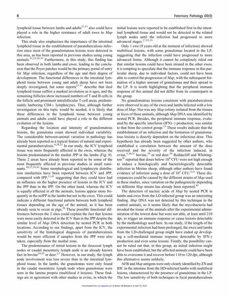
L. Delgado, J. F. García Marín, M. Muñoz, J. Benavides, R. A. Juste, C. García-Pariente, M. Fuertes, J. González, M. C. paratuberculosisSubspecies Mycobacterium avium Doses of
Pathological Findings in Young and Adult Sheep Following Experimental Infection With 2 Different
Published by:
http://www.sagepublications.com
On behalf of:
Pathologists.American College of Veterinary Pathologists, European College of Veterinary Pathologists, & the Japanese College of Veterinary
can be found at:Veterinary Pathology OnlineAdditional services and information for
http://vet.sagepub.com/cgi/alertsEmail Alerts:
http://vet.sagepub.com/subscriptionsSubscriptions:
http://www.sagepub.com/journalsReprints.navReprints:
http://www.sagepub.com/journalsPermissions.navPermissions:
What is This?
- Feb 6, 2013OnlineFirst Version of Record >>
at Neiker AB Biblioteca on April 15, 2013vet.sagepub.comDownloaded from

Pathological Findings in Young and AdultSheep Following Experimental InfectionWith 2 Different Doses of Mycobacteriumavium Subspecies paratuberculosis
L. Delgado1, J. F. Garcıa Marın1, M. Munoz1, J. Benavides1,R. A. Juste2, C. Garcıa-Pariente1, M. Fuertes1, J. Gonzalez1,M. C. Ferreras1, and V. Perez1
AbstractMycobacterium avium subsp paratuberculosis (Map) is assumed to infect young ruminants; however, little is known concerning thepossibility of adult animals becoming infected. An experimental infection was conducted to establish the effect of age and doses ofMap on susceptibility to paratuberculosis in sheep. Sixteen of twenty-four 1.5-month-old Churra lambs and 23 of 30 adult ewes(from 2–11 years old) were orally challenged with an ovine field strain of Map. Thirteen ewes and 8 lambs were infected with ahigh dose (HD) and 10 adult sheep and 8 lambs with a low dose (LD) of Map. The remaining animals were unchallenged controls.Animals were euthanized at 110 to 120 and 210 to 220 days postinfection. Histological, bacteriological, and nested polymerasechain reaction (PCR) studies were conducted in samples of intestine and related lymphoid tissue (Peyer patches, lymph nodes).Animals were classified according to their lesions. The number of granulomas was counted in 3 tissue sections from each sample.Only the HD groups showed lesions associated with paratuberculosis (92.3% of ewes and 100% of lambs). Adults had lesionscharacterized by few small demarcated focal granulomas restricted to the lymphoid tissue, whereas granulomas were morenumerous and larger, appearing in the lamina propria unrelated to lymphoid tissue, in the lambs. Only HD-infected lambs werepositive to culture, whereas nested PCR also detected positive HD ewes and some LD animals. These results suggest that adultsheep can become infected by Map, as seen by the development of lesions, but they are focal and restricted to the lymphoid tissue.
Keywordsage, granulomas, intestine, mycobacteria, sheep, paratuberculosis
Paratuberculosis or Johne disease is a chronic granulomatous
inflammation of the intestine and related lymph nodes caused
by Mycobacterium avium subsp paratuberculosis (Map) that
affects both domestic and wild ruminants. It occurs worldwide
and is responsible for heavy economic losses to domestic
livestock industries. This infectious disease is characterized
by a long incubation period, which leads to weight loss with
or without diarrhea and eventual death.6
The pathological response of the infected animals varies
widely, and different lesional classifications have been estab-
lished in ovine, caprine, and bovine species depending on the
presence, intensity, and distribution of granulomatous lesions
as well as the number of bacteria.10,19,33 Briefly, these patholo-
gical forms are divided into focal forms, characterized by small
granulomas restricted to the lymphoid tissue of the Peyer
patches; multifocal forms, where the granulomas spread into
the lamina propria related or not to the lymphoid tissue; and
diffuse forms, related to clinical signs, where wide areas of the
mucosa are severely affected. It has been proposed that these
different pathological responses represent different stages of
the disease, where the immunological status of the animal plays
an important role.10,19,33
In flocks with endemic paratuberculosis, it is common to
find animals showing focal lesions, similar to those observed
in the initial stages of experimental Map infection.21,31 This
observation raised the hypothesis that they could be considered
1 Departamento de Sanidad Animal (Anatomıa Patologica), Instituto de Gana-
derıa de Montana (CSIC-ULE), Facultad de Veterinaria, Universidad de Leon,
Leon, Spain2 Departamento de Sanidad Animal, NEIKER-Tecnalia, Bizkaia, Spain
Corresponding Author:
V. Perez, Departamento de Sanidad Animal, Instituto de Ganaderıa de Montana
(CSIC-ULE), Facultad de Veterinaria, Universidad de Leon, Campus de Vega-
zana s/n, 24071 Leon, Spain.
Email: [email protected]
Veterinary Pathology00(0) 1-10ª The Author(s) 2013Reprints and permission:sagepub.com/journalsPermissions.navDOI: 10.1177/0300985813476066vet.sagepub.com
Veterinary Pathology OnlineFirst, published on February 6, 2013 as doi:10.1177/0300985813476066
at Neiker AB Biblioteca on April 15, 2013vet.sagepub.comDownloaded from

latent lesions of adult animals infected earlier in life or as initial
lesions of recently infected adult individuals.10,19,33
The concept of ‘‘age-related’’ resistance to paratuberculosis
has been proposed in bovine25,32,43 and in red deer,28 but it has
not yet been deeply investigated in other species such as ovine.
Several studies carried out in cattle have confirmed that
animals become more resistant to the infection as they get
older,20,34,40 but bacteriological and/or histological evidence
of infection in adult cattle has also been reported.15,25,32,34
These findings would be of great importance, as one of the
main hygienic measures in paratuberculosis remains the
removal of the young animals from their dams within the first
months of life to prevent the transmission of Map within them,
and these recommendations were based on the assumption that
animals become less susceptible to infection as they age.6,43
Several doses of Map inoculum have already been used in
experimental infections, leading to disparate results. Reddacliff
and Whittington36 could not induce any detectable infection in
Merino sheep challenged with doses lower than 104 colony-
forming units (CFU) of Map, whereas 6 of 12 lambs infected with
103 CFU of Map developed detectable lesions in another study.31
An experiment was designed to study the role of the age of
infection on the pathogenesis of paratuberculosis, where young
and adult sheep were experimentally challenged. The first part
of the experiment, focusing on the differences in the peripheral
immune response, already has been reported.13 Both the
humoral and specific cell-mediated immune responses
appeared earlier and more intensely in the adult sheep than in
the lambs, probably due to previous contact with Map or other
mycobacteria, whereas the Map-challenged lambs showed a
more immature immune response, unable to control the
progression of the infection.13
The main objective of the present work is to study and char-
acterize the pathological response of Map infection in the same
experiment, by comparing the differences observed in the
pathological evolution of these experimentally infected lambs
and ewes and evaluating the severity of the lesions with a
detailed count of the granulomas formed. The effect of the
challenge inoculum dose in the establishment and developmen-
tal stage of the lesions in these animals is also assessed.
Materials and Methods
Experimental Animals
Thirty adult ewes 2 to 11 years old (73.3% of them 6–9 years
old) as well as 24 one-month-old lambs of the Churra breed
were used in this study. All of them were randomly selected
from a flock without clinical cases of paratuberculosis in the
past 5 years and in which no vaccination against paratubercu-
losis was practiced before.13
After a period of adaptation once in the experimental
facilities of the University of Leon, the animals were housed
in separate barns to prevent direct contact between the groups.
Both adult and young ovine were randomly divided into 3
groups: one infected with the high dose of Map inoculum
(HD group; n ¼ 13 ewes and 8 lambs), the second one with the
low dose (LD group; n ¼ 10 ewes and 8 lambs), and a third
group of uninfected control animals, challenged with saline
solution (n ¼ 7 ewes and 8 lambs). None of the sheep was bred
during the experiment. Animals followed a diet based on com-
mercial feed appropriate for each age.
Map Inoculum
The strain of Map used in this study was isolated from intestinal
(ileal) mucosa scrapings of 3 sheep from the same flock, all
clinically affected with paratuberculosis and shedding large
number of bacilli, using the Ratnamohan and Spencer35 tech-
nique with slight modifications consisting of the elimination
of deoxyribonuclease and lysozyme treatments. Briefly, sam-
ples of ileum were rinsed with phosphate-buffered saline
(PBS), and the mucosal surface was removed from the submu-
cosa by scraping. Then, 20 g of tissue was mixed with 2 ml of a
solution containing 150 mg/ml ampicillin (Sigma-Aldrich,
Madrid, Spain) per 5 g of mucosa and homogenized using a tis-
sue grinder. The suspension was digested with a solution with
1% trypsin (Sigma-Aldrich) in PBS (pH 7.5–8) at 23�C for
30 minutes. Finally, and after centrifugation, the sediment was
resuspended in 10 ml PBS.
The field strain of Map was then determined to be IS900 pos-
itive by polymerase chain reaction (PCR)18 and typed as an
‘‘ovine’’ strain by IS1311 PCR-restriction enzyme analysis.38
Subsequently, the inocula were subjected to culture on 7H11 and
Lowenstein-Jensen medium supplemented with mycobactin J
(Allied Monitor, Fayette, MO) as described previously,13 leading
to counts of 4 � 106 CFU Map/ml for the high-dose suspension
and 1� 102 CFU Map/ml for the lower dose. For ease of under-
standing, these 2 different doses hereafter will be referred to as the
higher dose (HD) and lower dose (LD) inoculum of Map.
Each challenged animal received a 40-ml oral dose of the
respective inoculum, divided into 4 doses of 10 ml adminis-
tered at 3-day intervals.
Experimental Design
The experimental procedures were performed in accordance
with Spanish Royal Decree 1201/2005 for the protection of
animals used for experimental and other scientific purposes.
At 110 to 120 days postinfection (dpi), 6 ewes and 3 lambs
from the HD group, 4 ewes and 3 lambs from the LD group, and
2 ewes and 3 lambs from the control group were humanely eutha-
nized by intravenous injection of a veterinary euthanasia drug
(T61; Intervet, S.A., Salamanca, Spain), followed by exsanguina-
tion. The remaining animals were euthanized at 210 to 220 dpi.
Pathological Studies
Complete necropsies were performed, and tissues were grossly
inspected with special attention to the gut and related lymph
nodes. Samples for histopathologic examination consisted of
ileocecal valve (ICV), ileum (IL) (three 5-cm samples, taken
2 Veterinary Pathology 00(0)
at Neiker AB Biblioteca on April 15, 2013vet.sagepub.comDownloaded from

20, 40, and 60 cm from the ileocecal valve), jejunum (JJ) and
jejunal Peyer patches (JPP) (at least 3 patches from each of the
proximal, medium, and distal zones), the caudal mesenteric
lymph node (MLN), 1 jejunal lymph node (JLN), and ileocecal
lymph nodes (ICLN). All these tissues were fixed in 10%neutral buffered formalin and processed to routine histological
sections that were stained with hematoxylin and eosin (HE) and
by the Ziehl-Neelsen (ZN) technique for acid-fast bacilli (AFB)
and examined by light microscopy for histological changes.
For the immunohistochemical study, selected sections hav-
ing representative granulomatous lesions were immunolabeled
using an EnVision þ HRP visualization kit (Dako North
America, Carpinteria, CA). The sections were incubated with
specific rabbit anti-Map serum at a dilution of 1/9000. This
polyclonal primary antibody was kindly gifted by Dr Balseiro,
from SERIDA (Gijon, Asturias, Spain), and previously used in
bovine3 and fallow deer.2 Technique specificity was controlled
by omission of the primary antibody and substitution by a
normal rabbit serum, omission of the EnVision polymer, and
omission of diaminobenzidine. All these controls gave negative
results. To evaluate the specificity of the anti-Map antibody,
tissue samples of ileum from a sheep with clinical paratubercu-
losis were used as positive controls.
All the lesions observed in the digestive tract were classified
following the guidelines previously proposed by Perez et al33
for paratuberculosis lesions in the ovine species, according to
the presence and location of the granulomas in the different
gut-associated lymphoid tissue compartments and the number
of AFB detected by ZN. Sections were blinded and scored by
the same observer into 5 categories, according to the amount
of AFB and Map antigens present in the lesions: 0 (no AFB
or Map antigens), þ/– (doubtful presence of bacteria), þ (soli-
tary or very few bacteria), andþþþ (high load of AFB or Map
antigens in the cytoplasm of the macrophages).
Granuloma Count
After the conventional histopathological examination, the num-
ber of granulomas per tissue section was quantified in different
areas of the intestinal tract and related lymph nodes. For this pur-
pose, 10 different intestinal sites were studied (ie, the ICV) and 3
samples of the IL, JJ, and JPP (proximal, middle, and distal).
Three different lymph nodes (MLN, JLN, and ICLN) were also
analyzed. Three tissue sections were randomly selected from each
intestinal site and 2 tissue sections from each lymph node, so that a
total of 30 intestinal and 6 lymph node tissue sections were
examined from each animal. Sections were blinded, and the mean
number of granulomas per tissue section in each site was recorded
by the same observer, distinguishing those granulomas located in
the lymphoid tissue from those in the associated lamina propria
(LP) or in the mucosa not related to lymphoid tissue.
Tissue and Fecal Culture
Fecal samples were taken at 15 and 90 dpi, as well as on the day
of the scheduled postmortem examination, from each animal.
They were frozen at –20�C until cultured for Map. Bacteriolo-
gical studies were also carried out from tissue samples of the
ICV, a 5-cm sample of the distal ileum (dIL) and of the medial
jejunum Peyer patch (mJPP), and a piece of the caudal MLN.
Two grams of tissues or feces was processed for culture onto
tubes containing Lowenstein Jensen and Middlebrook 7H11
solid medium, supplemented with mycobactin J and previously
decontaminated with hexadecylpyridinium chloride (Merck,
Darmstadt, Germany), as described by Aduriz et al.1 The
procedure is explained in detail in a previous publication.13
Nested PCR
A nested PCR method was performed from paraffin-embedded
tissues. In total, 10 mm of ICV, dIL, mJPP, and MLN tissue
sections adjacent to those used for the immunohistochemical
study was cut twice, and the nested PCR was carried out as pre-
viously described,13 using primers to detect the presence of
Map-specific IS900 DNA.
Statistical Analysis
Data on granuloma count were subjected to analysis of var-
iance using the general linear model procedure of the SAS sta-
tistical package (version 9.1; SAS Institute, Cary, NC) for the
evaluation of age, treatment, time of killing, and location main
effects and interactions. Previously, tissue section granuloma
count figures were logarithmically transformed to submit them
to normal distribution-based tests of significance. Thus, the
mean numbers of granulomas per tissue section were compared
among the groups at each time of killing using the Student t test
for pairwise comparisons with the Tukey-Kramer correction
for multiple comparisons, at the 95% significance level. Group
PCR-positive frequencies were compared and tested for signif-
icance by w2 analysis.
Results
Three animals died during the course of the experiment from
causes unrelated to Johne disease (1 ewe and 1 lamb from the
uninfected control group and 1 ewe from the LD group) and
were excluded from this study.
Pathological Findings
No clinical signs or gross lesions related to paratuberculosis
were seen in any of the animals during the whole experiment.
Microscopically, lymphoid tissue was always found in all
the ileum samples from the lambs, besides the ICV and JJP
samples. In the ewes, no lymphoid tissue was detected in the
samples taken from the ileum, at any level.
When examined histologically, granulomatous lesions typi-
cal of paratuberculosis were only found in the tissue sections
from the HD groups, regardless of the age of infection (ewes
and lambs). However, the type of lesion varied between adult
sheep and lambs.
Delgado et al 3
at Neiker AB Biblioteca on April 15, 2013vet.sagepub.comDownloaded from

Among the HD-challenged lambs, all 8 animals (100%)
showed histologic changes consistent with paratuberculosis.
Lesions were classified in several categories, according to their
location and pathological features, following the guidelines
provided by Perez et al.33 For categorizing each animal, the
final classification was based on its more severe intestinal gran-
ulomatous lesion.
Focal lesions appeared in 2 HD-infected lambs. They were
composed of small, well-defined granulomas formed by homo-
geneous macrophages with a large pale cytoplasm, large nuclei,
and scattered with a few lymphocytes within them, and they
were found exclusively restricted to the interfollicular area of
the Peyer patch lymphoid tissue (Figs. 1, 2). In some areas,
these granulomas were coalescent and partially altered the nor-
mal structure of the Peyer patches. Lesions considered multifo-
cal a were detected in 2 other HD-infected lambs. In this case,
granulomas were more numerous and larger in size, and
besides those seen in the lymphoid tissue, granulomatous
lesions were also detected in the LP always related to the Peyer
patch lymphoid tissue (Fig. 3). In the last 4 lambs, lesions were
classified as multifocal b. In them, many granulomas were also
found in the LP not related to the lymphoid tissue, invading and
thickening the apex of the villi, and in the more severely
affected areas, they partially disrupted the normal architecture,
causing the enlargement of the villi with separation of the
intestinal glands (Fig. 4). In those cases, the granulomas were
not very well demarcated from the adjacent tissue.
Among the HD-challenged ewes, 12 of 13 animals (92.3%)
showed microscopic changes consistent with paratuberculosis.
Eleven of the sheep had focal lesions, with granulomas restricted
to the intestinal lymphoid tissue of the ileocecal and jejunal
Peyer patches, mostly in its basal area. In contrast with the
lambs, the granulomas were fewer, smaller, round shaped, and
well defined from the adjacent tissue (Figs. 5, 6). No differences
were observed in the type and intensity of the lesions among the
ewes, irrespective of their age. Only 1 sheep showed multifocal a
lesions, with a few granulomas located in the mucosa adjacent to
the lymphoid tissue of the ileocecal and distal jejunal Peyer
patches (Fig. 7). This adult sheep also showed a mild granuloma-
tous lymphangitis in the serosa of the ileocecal valve.
When considering the time of euthanasia, 2 of the 3 lambs
examined at 110 to 120 dpi had multifocal b lesions, and the
remaining one was classified as multifocal a. All the sheep
showed focal forms except for 1 that had no lesion. At 210 to
220 dpi, multifocal b lesions were seen in 2 lambs, focal lesions
in 2, and a multifocal a form in the remaining animal. Among
the sheep, all but 1 (with a multifocal a lesion) showed focal
forms.
In those 4 HD-challenged lambs with the more severe intest-
inal lesions (multifocal b), granulomatous lesions were also
seen in the ICLN and MLN, especially spread throughout the
interfollicular and paracortical areas, with a multifocal distri-
bution. In these locations, granulomas were numerous and coa-
lescent, formed by 20 to 100 macrophages with a large pale
cytoplasm, morphologically similar to those observed in the
intestine.
On the contrary, the ewes and lambs with focal intestinal
lesions only occasionally showed a focal granulomatous lym-
phadenitis characterized by a few small granulomas composed
of less than 10 macrophages, scattered within the interfollicular
part of the cortical area of some MLN.
No AFB or Map antigens were detected in any of the control
or LD-challenged animals or in the HD-infected ewes. How-
ever, different grades of bacterial load were demonstrated in
the HD-infected lambs by ZN or IHC, varying from hardly any
AFB or Map antigens in the intestinal focal lesions (grade þ),
to a scant positivity by both techniques in the multifocal a
lesions (gradeþþ), and to the highest bacterial load in the mul-
tifocal b lesions (grade þþþ), especially in the mucosa of the
ileal and jejunal Peyer patches (Fig. 4). Only occasional AFB
or Map antigens were appreciated in the MLN or ICLN from
3 HD-infected lambs with multifocal b lesions.
Granuloma Counts
The mean granuloma counts corresponding to the different
intestinal compartments examined in the lambs and ewes at
110 to 120 dpi and 210 to 220 dpi are shown in Fig. 8. Varia-
bility among individuals of the same group was observed. The
statistical analysis showed that the total mean number of gran-
ulomas was significantly higher in the lambs (85.55) than in the
ewes (19.37) at 110 to 120 dpi (P < .01) and also at 210 to 220
dpi (42.05 vs 24.03, respectively) (P < .05). It is worth high-
lighting that granulomatous lesions in the LP were only
detected in the lambs, whereas in the ewes, granulomas were
only observed in the lymphoid tissue (except for 1 adult sheep).
In the lambs, a significant decrease was noted in the number of
granulomas located in the lymphoid tissue from 110 to 120 dpi
and the end of the experiment (P < .05), whereas those located
in the LP slightly increased. However, the number of granulo-
mas in the organized lymphoid tissue remained similar in the
ewes within the same period (Fig. 8).
When considering the lesions in each different intestinal site
examined (Fig. 9), the mean number of granulomas per tissue
section was always higher in the lambs compared with the
ewes, especially in the mJPP and in the IL (P < .05). Although
granulomas appeared to be slightly more numerous in the ICV
in the ewes than in the lambs (Fig. 9), differences were not sta-
tistically significant. In both adult and young animals, most of
the granulomatous lesions were associated with the lymphoid
tissue in the JPP, IPP, or the ICV (Fig. 9). Specifically, in the
ewes, the granulomas were significantly more numerous in the
ICV than in the rest of the lymphoid tissue (P < .05). Concern-
ing the lambs, the granuloma count was higher in the mJPP
compared with the remaining JPP or IPP (P < .05), but no
differences were detected with the ICV.
In addition to the lesions in the intestine, some granulomas
were also noted in the associated lymph nodes. These were
more numerous in the lambs than in the ewes (P < .05) and
were counted more frequently in the MLN, in both the adult
and young animals.
4 Veterinary Pathology 00(0)
at Neiker AB Biblioteca on April 15, 2013vet.sagepub.comDownloaded from

Figure 1. Jejunal Peyer patch, high-dose (HD)–challenged lamb. Focal lesion composed of small granulomas (*) exclusively located in the inter-follicular area of the Peyer patch lymphoid tissue. Hematoxylin and eosin (HE). Figure 2. Jejunal Peyer patch, HD-challenged lamb. Highermagnification of a focal granuloma in the interfollicular area of the Peyer patch lymphoid tissue. The granuloma is formed by homogeneousmacrophages with an abundant and pale cytoplasm and a few lymphocytes scattered within them. HE. Figure 3. Jejunal Peyer patch, HD-challenged lamb. Multifocal a lesion. Besides the severe granulomatous lesion in the lymphoid tissue, with large granulomas partially invadingthe interfollicular areas, some of the granulomas are also affecting the lamina propria related to this lymphoid tissue. HE. Figure 4. Medium
Delgado et al 5
at Neiker AB Biblioteca on April 15, 2013vet.sagepub.comDownloaded from

Fecal and Tissue Culture Results
No isolations from feces were obtained at 15 or 90 dpi. The only
positive feces cultures were isolated on the day of the postmor-
tem examination from 4 of the 8 HD-infected lambs, 1 eutha-
nized at 110 to 120 dpi and 3 at 210 to 220 dpi. Apart from 1
lamb classified as focal (euthanized at 210–220 dpi), the 3 others
had multifocal b lesions, with high bacterial load in the intestine.
All the tissue samples from the control, the LD group, and
the HD-infected ewes remained negative (Table 1). Instead,
mycobacteria were isolated, only in 7H11 medium, from the
dIL and/or ICV of 4 of the 8 HD-infected lambs: 1 multifocal
b animal, euthanized at 110 to 120 dpi, and 3 euthanized at the
end of the experiment, 1 with multifocal b lesions and the 2 oth-
ers with focal lesions. All but 1 of these animals were also pos-
itive in the fecal samples.
Nested PCR
IS900 Map-specific sequence was demonstrated by nested PCR
in all 8 HD-infected lambs (100%) from at least 2 of the 4
studied tissue sections (Table 1). The ICV was positive in all
these cases. Seven of the 8 LD-challenged lambs (87.5%), in
which no granulomatous lesions were detected in any of the
examined tissue sections, were also positive from at least 1 tissue
section. The negative lamb was euthanized at 110 to 120 dpi.
Map DNA was also detected in 12 of the 13 HD-infected
adult sheep (92.3%) in at least 1 tissue section. The negative sheep
was examined at 210 to 220 dpi and showed a focal lesion. ICV
and MLN were the most frequent tissue sites with a positive result,
although differences were not statistically significant (Table 1).
Among the LD-infected ewes, in which no histological lesions
were appreciated, the IS900 sequence was demonstrated in 7 of
the 9 animals (77.8%) from at least 1 tissue section. Negative
ewes were euthanized at 210 to 220 dpi.
All analyzed tissue sections from the uninfected lambs and
ewes were negative to PCR.
Discussion
According to the results of this study, the experimental inocu-
lation with Map was able to infect both the ewes and lambs, but
Figure 9. Total granuloma count from the tissues of the high-dose–infected lambs and ewes, regardless of the time of slaughter. Mean of thetotal number of granulomas per section and animal, according to their intestinal location. ICV, ileocecal valve; IL, ileum; JJ, jejunum; JPP, jejunalPeyer patches. d, distal; m, medium; p, proximal. Error bars: standard deviation. Lambs¼ black column. Ewes¼ white column.
Figure 8. Count of granulomas from the tissues of the high-dose–infected lambs and ewes euthanized at 110 to 120 dpi (a) and 210 to 220 dpi (b).Mean number of granulomas per animal, corresponding to their location in the different intestinal compartments. dpi, days postinfection. Error bars:standard deviation. Peyer patch (PP) lymphoid tissue¼ black column. Lamina propria (LP) associated with PP¼ grey column. LP not associated withPP¼white column.
Figure 4. (continued)ileum, HD-challenged lamb. Multifocal b lesion. Granulomas located in the lamina propia not related to the lymphoidtissue partially thicken the apex of the villi. HE. Inset: Presence of numerous acid-fast bacilli positive in the cytoplasm of the macrophages.Ziehl-Neelsen stain. Figure 5. Ileocecal Peyer patch, HD-challenged ewe. Focal lesion composed of few small well-defined granulomas (*)restricted to the basal area of the interfollicular area of the Peyer patch lymphoid tissue. HE. Figure 6. Ileocecal Peyer patch; HD-challengedewe. Focal lesion. Higher magnification of small and focal granulomas (*) composed of macrophages and some lymphocytes. Note that thegranulomas are round and well demarcated from the adjacent tissue. HE. Figure 7. Ileocecal valve, HD-challenged ewe. Multifocal a lesioncomposed of granulomas located both in the lymphoid tissue and in the associated lamina propria. HE.
6 Veterinary Pathology 00(0)
at Neiker AB Biblioteca on April 15, 2013vet.sagepub.comDownloaded from

detectable lesions and immune response13 were observed only
in those animals challenged with a high dose of the mycobac-
teria. Adult HD-infected sheep developed lesions consistent
with Map infection, with clear differences in the pathological
features when compared with the HD-infected lambs. Several
studies have reported the presence of focal lesions in adult
sheep, similar to those found in our study, considered to be
latent lesional forms originating from infections that took place
early in the life of the animals.10,17,19,33 Although this possibil-
ity cannot be completely ruled out in this study, the fact that
these focal lesions were not found in the LD-challenged ewes
or in the controls would indicate that these granulomas would
be rather the direct consequence of the dose of Map challenge
or the short duration of the examination period.
Lesions consistent with paratuberculosis were found in 12 of
the 13 (92.3%) adult sheep infected with a high dose of Map,
associated with a specific peripheral immune response.13 How-
ever, no similar lesions or an immune response were detected
in any of the ewes from the control uninfected group, suggest-
ing that adult sheep, older than 2 years of age, can get infected
with high doses of Map. In bovine, it was shown that calves
older than 4 to 6 months were more resistant to the infection
with Map,20,40 although other studies have also found that Map
was able to cause lesions in cattle experimentally infected,
older than 3 years32 and even with a 9-year-old,25 although the
lesions were more restricted than in the young calves infected
under the same conditions.25,32 These findings would be in
agreement with our results. In sheep, no experimental studies
have been carried out to date, but in a longitudinal field study,
it was shown that sheep exposed to Map as lambs had higher
odds to shed Map and to develop more severe lesions than those
exposed for the first time as adults.30
The development and features of the lesions in the
HD-challenged groups were different between lambs and ewes.
In the latter, the lesions were, in most cases, classified as focal,
always restricted to the intestinal lymphoid tissue, but small,
round shaped, and well defined, even with initial fibrosis,
suggesting a regressive character. Similar granulomas have
also been described in vaccinated animals.21 On the contrary,
in the HD-infected lambs, the granulomas were more numer-
ous, larger, and spread into areas of the mucosa related or not
(multifocal a or b lesions) to this lymphoid tissue. This fact has
been related to more advanced stages of the infection.10,19,21,33
This is confirmed in our study by the marked decrease in the
number of granulomas found in the lymphoid tissue from 110
to 120 and 210 to 220 dpi, whereas those in the LP partially
increased (Fig. 8), as well as by the high mycobacterial load,
evaluated by ZN and IHC, in the multifocal b lesions.
Payne and Rankin,32 in an experimental infection of adult
and young cattle, found that granulomatous lesions were fewer
and better demarcated in adult animals than in calves at 4 and 6
months postinfection (mpi); however, in a previous postmor-
tem examination carried out at 2 mpi, lesions were more severe
in the adult animals. It was then hypothesized that paratubercu-
losis infection could have reached its higher development in the
adults at 2 mpi, but their immune response could have been
able to react more efficiently and control the progression of the
lesions more quickly, unlike what happened in the calves.32,34
Although in our experimental design, the animals were not
examined until 4 mpi (110–120 dpi), and thus it is not possible
to compare both studies, the fact that granulomatous lesions
were focal and well demarcated in the adult sheep, together
with the earlier and more intense peripheral cell-mediated
immune response mounted by the ewes compared with the
lambs,13 would further support this hypothesis.
One feasible factor that could explain the higher resistance
of the adult animals could be the previous expositions to myco-
bacteria in the adult sheep. Sensitization experimented by the
immune system of the animals after previous and repeated
contacts with the mycobacteria, its antigens, or some other
environmental mycobacteria would induce a better specializa-
tion of the immunologic response.14,32 Animals vaccinated
against paratuberculosis develop granulomas similar to those
seen in the adult sheep and have been shown to be more resis-
tant to Map infection,21 further supporting this hypothesis. In
our study, the ewes came from a flock considered free of this
disease, although the absolute lack of mycobacteria in the
environment cannot be ensured since distribution of Map is
widespread.16 Moreover, some of the ewes used in this study
were especially old (between 9 and 11 years old), increasing
the probability of previous contacts with different mycobac-
teria, although no differences were seen in the type and inten-
sity of the lesions in comparison with the rest of the ewes.
Differences in the amount and functionality of the intestinal
Table 1. Number (%) of Animals Positive to Map Detection by Culture and by Nested PCR (n-PCR)a
Sites
HD Lambs (n ¼ 8) LD Lambs (n ¼ 8) HD Ewes (n ¼ 13) LD Ewes (n ¼ 9)
Culture n-PCR Culture n-PCR Culture n-PCR Culture n-PCR
ICV 4 (50.0) 8 (100.0) 0 4 (50.0) 0 7 (53.9) 0 3 (33.3)dIL 2 (25.0) 7 (87.5) 0 1 (12.5) 0 4 (30.8) 0 1 (11.1)mJPP 0 6 (75.0) 0 3 (37.5) 0 6 (46.2) 0 2 (22.2)MLN 0 7 (87.5) 0 4 (50.0) 0 7 (53.9) 0 4 (44.4)
All the animals, regardless of the time of sacrifice, are included. dIL, distal ileum; ICV, ileocecal valve; Map, Mycobacterium avium subsp paratuberculosis; mJPP,medium jejunal Peyer patch; MLN, mesenteric lymph node; PCR, polymerase chain reaction.aThe culture technique was carried out from fresh tissue samples and the n-PCR was performed from paraffin-embedded tissues. High-dose (HD) and low-dose(LD) infected animals are analyzed in this table.
Delgado et al 7
at Neiker AB Biblioteca on April 15, 2013vet.sagepub.comDownloaded from
lymphoid tissue between lambs and adults27,37 also could have
played a role in the higher resistance of adult ewes to Map
infection.
This study also emphasizes the importance of the intestinal
lymphoid tissue in the establishment of paratuberculosis infec-
tion since most of the granulomatous lesions were detected in
this area, as has been reported in previous studies using young
animals.21,23,24,33 Furthermore, in this study, this finding has
been observed in both lambs and ewes, leading to the conclu-
sion that the Peyer patches would be the primary portal of entry
for Map infection, regardless of the age and their degree of
development. The functional differences in the intestinal lym-
phoid tissue between young and adult sheep have not been
deeply investigated, but some reports27,37 describe that ileal
lymphoid tissue suffers a marked involution as it ages, and the
remaining follicles show increased numbers of T and B cells in
the follicle and prominent intrafollicular T-cell areas, predomi-
nantly harboring CD4þ lymphocytes. Thus, although further
investigation on this topic would be needed, it is likely that
these differences in the lymphoid tissue between young
animals and adults could have played a role in the different
evolution of the lesions.
Regarding the location and intensity of granulomatous
lesions, the granuloma count showed individual variability.
The considerable between-animal variation in pathology has
already been reported as a typical feature of natural and experi-
mental paratuberculosis.4,24,31 In our study, the ICV lymphoid
tissue was more frequently affected in the ewes, whereas the
lesions predominated in the medial JPP in the lambs (Fig. 9).
These 2 areas have already been reported to be some of the
most frequently affected in previous studies in small rumi-
nants.10,17,33,41 Some morphological and lymphocyte distribu-
tion similarities have been reported between ICV and JPP,
compared with IPP,11,41 suggesting that they could have had
an influence on the higher presence of lesions in the ICV and
the JPP than in the IPP. On the other hand, whereas the ICV
is equally affected in all the animals, lesions appear more fre-
quently in the mJPP in the lambs but not in the ewes. This could
indicate a different functional pattern between both lymphoid
tissues depending on the age of the animal, as it has been
already seen to occur in pigs.26 These possible functional dif-
ferences between the 2 sites could explain the fact that lesions
were more easily detected in the ICV than in the JPP despite the
similar level of Map DNA detection by nested PCR in both
locations. According to our findings, apart from the ICV, the
sensitivity of the histological diagnosis of paratuberculosis
would be more efficient if samples from the JPP were also
taken, especially from the medial zone.
The predominance of initial lesions in the ileocecal lymph
nodes or caudal mesenteric lymph node is an already known
fact in bovine19,25 or deer.2,7 However, in our study, the lymph
node involvement was less severe than in the intestinal lym-
phoid tissue. In the lambs, the granulomas were only found
in the caudal mesenteric lymph node when granulomas were
seen in the lamina propria (multifocal b lesions). These find-
ings are in agreement with other studies in ovine, in which the
initial lesions were reported to be established first in the intest-
inal lymphoid tissue and would not be detected in the related
lymph nodes until the infection had progressed to more
advanced stages.17,21,33
Only 1 ewe (9 years old at the moment of infection) showed
multifocal lesions, with some granulomas located in the LP,
suggesting that the infection could have progressed to more
advanced forms. Although it cannot be completely ruled out
that similar lesions could have been missed in the other ewes,
it is tempting to speculate that the immune response in this par-
ticular sheep, due to individual factors, could not have been
able to control the progression of Map, with the subsequent for-
mation of a higher amount of granulomas and their spread to
the LP. It is worth highlighting that the peripheral immune
response of this animal did not differ from its counterparts in
the group.
No granulomatous lesions consistent with paratuberculosis
were observed in any of the ewes and lambs infected with a low
dose of Map. Nor was any Map colony isolated from the tissues
or feces of these animals, although Map DNA was identified by
nested PCR. Besides, the peripheral immune response, evalu-
ated by the specific interferon (IFN)–g production, was similar
to that from the control group.13 These results indicate that the
establishment of an infection and the formation of granuloma-
tous lesions is directly dependent on the infectious dose. This
hypothesis has already been suggested by other authors, who
established a correlation between the amount of the dose
received and the severity of the infection induced, in
ovine,5,30,31 bovine,39 or red deer.29 Reddacliff and Whitting-
ton36 reported that doses below 104 CFU were not high enough
to induce a histologically and bacteriologically detectable
infection in Merino sheep, although other authors have found
evidence of infection using a dose of 103 CFU.5,31 These dis-
crepancies could be caused by the different strains of Map used
in these studies, since variation on the pathogenicity depending
on different Map strains has already been reported.42
The detection of nucleic acids of Map by nested PCR in
lambs and ewes from the LD-infected group was an interesting
finding. Map DNA was not detected by this technique in the
control animals, so it seems likely that the mycobacteria had
invaded the tissue of the animals after the experimental admin-
istration of the lowest dose but were not able, at least until 210
dpi, to trigger an immune response or cause lesions detectable
by the methodology used here. In our study, if the length of the
experimental infection had been prolonged, the ewes and lambs
from the LD-challenged group might have ended up develop-
ing a cell-mediated immune response detectable by IFN-gproduction and even some lesions. Finally, the possibility can-
not be ruled out that, in this group, an initial infection might
have been established, but the affected animals could have been
able to overcome it and recover before 110 to 120 dpi, although
this alternative seems unlikely.
AFB and Map antigens were only clearly identified by ZN and
IHC in the intestine from the HD-infected lambs with multifocal
lesions, characterized by the presence of granulomas in the LP.
The low sensitivity of both techniques in focal paratuberculosis
8 Veterinary Pathology 00(0)
at Neiker AB Biblioteca on April 15, 2013vet.sagepub.comDownloaded from

lesions, located in the lymphoid tissue, has already been reported
in previous studies and is mainly explained because of the low
amount of AFB in these types of lesions.10,17,19,22,33 It should also
be remarked that the ZN technique can only stain those AFB with
an intact cell wall and thus is unable to detect the bacillus present
in its cell wall–deficient form or as spheroplasts.9 However, this
possibility seems unlikely as the IHC labeling, which is assumed
to be able to detect degraded or previously digested forms of the
bacilli,8,12,22 did not identify any Map antigen in the focal lesions
from this study.
This study has determined that adult sheep, older than
2 years, can get infected by experimental exposure of Map,
as do 1-month-old lambs. In both cases, the infection leads to
the establishment of lesions in the intestine. However, in the
ewes, the lesions appear almost exclusively in the intestinal
lymphoid tissue and are characterized to be focal and well
demarcated, whereas in the lambs, they progress into more
advanced forms, spreading into areas of mucosa not related
to the Peyer patches and to the regional lymph nodes. Only the
oral inoculation of high levels of Map was able to induce
visible lesions in the tissues both in lambs and in ewes. Thus,
the dose of Map inoculum administered to the animals in
experimental conditions has an influence on the establishment
of the infection.
Acknowledgements
We thank J. Reyero and G. Belver for technical assistance.
Declaration of Conflicting Interests
The author(s) declared no potential conflicts of interest with respect to
the research, authorship, and/or publication of this article.
Funding
The author(s) disclosed receipt of the following financial support for
the research, authorship, and/or publication of this article: This work
was supported by grant AGL2008-05820-C02 of the Spanish Ministry
of Science and Innovation and by grant FP6-2004-FOOD-3B from the
ParaTBTools project (European Union). L. Delgado was recipient of a
predoctoral fellowship from the Spanish Ministry of Education. J.
Benavides is supported by CSIC through the JAE-Doc program,
financed in part by the European Social Fund (ESF).
References
1. Aduriz JJ, Juste RA, Cortabarria N. Lack of mycobactin depen-
dence of mycobacteria isolated on Middlebrook 7H11 from clin-
ical cases of ovine paratuberculosis. Vet Microbiol. 1995;45:
211–217.
2. Balseiro A, Garcıa-Marın JF, Solano P, et al. Histopathological
classification of lesions observed in natural cases of paratubercu-
losis in free-ranging fallow deer (Dama dama). J Comp Pathol.
2008;138:180–188.
3. Balseiro A, Prieto JM, Espı A, et al. Presence of focal and multi-
focal paratuberculosis lesions in mesenteric lymph nodes and the
ileocaecal valve of cattle positive to the tuberculin skin test. Vet J.
2003;166:210–212.
4. Begg DJ, Whittington RJ. Experimental animal infection models
for Johne’s disease, an infectious enteropathy caused by Mycobac-
terium avium subsp. paratuberculosis. Vet J. 2008;176:129–145.
5. Brotherston JG, Gilmour NJL, Samuel JM. Quantitative studies of
Mycobacterium johnei in the tissues of sheep, I: routes of infec-
tion and assay of viable M. johnei. J Comp Pathol. 1961;71:
286–299.
6. Chiodini RJ, Van Kruiningen HJ, Merkal RS. Ruminant paratu-
berculosis (Johne’s disease): the current status and future pros-
pects. Cornell Vet. 1984;74:218–262.
7. Clark RG, Griffin JF, Mackintosh CG. Johne’s disease caused by
Mycobacterium avium subsp. paratuberculosis infection in red
deer (Cervus elaphus): an histopathological grading system, and
comparison of paucibacillary and multibacillary disease. N Z Vet
J. 2010;58:90–97.
8. Coetsier C, Havaux X, Mattelard F, et al. Detection of Mycobac-
terium avium subsp. paratuberculosis in infected tissues by new
species-specific immunohistological procedures. Clin Diagn Lab
Immunol. 1998;5:446–451.
9. Condron RJ, Schroen CJ, Black CA, et al. Histological confirma-
tion of subclinical infection with M. paratuberculosis in cattle. In:
Chiodini RJ, Collins MT, Bassey EOE, eds. Proc 4th Int Colloq
Paratuberculosis. 1995;4:37–40.
10. Corpa JM, Garrido J, Garcıa Marın JF, et al. Classification of
lesions observed in natural cases of paratuberculosis in goats.
J Comp Pathol. 2000;122:255–265.
11. Corpa JM, Juste RA, Garcıa Marın JF, et al. Distribution of
lymphocyte subsets in the small intestine lymphoid tissue of
1-month-old lambs. Anat Histol Embryol. 2001;30:121–127.
12. Delgado F, Etchechoury D, Gioffre A, et al. Comparison between
two in situ methods for Mycobacterium avium subsp. paratuber-
culosis detection in tissue samples from infected cattle. Vet
Microbiol. 2009;134:383–387.
13. Delgado L, Juste RA, Munoz M, et al. Differences in the peripheral
immune response between lambs and adult ewes experimentally
infected with Mycobacterium avium subspecies paratuberculosis.
Vet Immunol Immunopathol. 2012;145:23–31.
14. Doyle TM. Johne’s disease. Vet Rec. 1956;68:869–878.
15. Doyle TM. Susceptibility to Johne’s disease in relation to age. Vet
Rec. 1953;65:363–365.
16. Eisenberg SW, Nielen M, Santema W, et al. Detection of spatial
and temporal spread of Mycobacterium avium subsp. paratuber-
culosis in the environment of a cattle farm through bio-aerosols.
Vet Microbiol. 2010;143:284–292.
17. Garcıa Marın JF, Perez V, Badiola JJ. Prevalence and type of
paratuberculosis lesions in sheep and their relation with the diag-
nosis by AGID test. In: Chiodini RJ, Kreeger JM, eds. Proceed-
ings of the Third International Colloquium on Paratuberculosis.
Providence, RI: International Association for Paratuberculosis;
1992:172–180.
18. Garrido JM, Cortabarrıa N, Oguiza JA, et al. Use of a PCR
method on fecal samples for diagnosis of sheep paratuberculosis.
Vet Microbiol. 2000;77:379–386.
19. Gonzalez J, Geijo MV, Garcıa-Pariente C, et al. Histopathological
classification of lesions associated with natural paratuberculosis
infection in cattle. J Comp Pathol. 2005;133:184–196.
Delgado et al 9
at Neiker AB Biblioteca on April 15, 2013vet.sagepub.comDownloaded from

20. Hagan WA. Age as a factor in susceptibility to Johne’s disease.
Cornell Vet. 1938;28:34–40.
21. Juste RA, Garcıa Marın JF, Peris B, et al. Experimental infection
of vaccinated and non-vaccinated lambs with Mycobacterium
paratuberculosis. J Comp Pathol. 1994;110:185–194.
22. Kheirandich R, Khodakaram Tafti A, Hosseini A. The compara-
tive evaluation of immunohistochemical and acid fast staining
with histopathological changes in naturally occurring paratuber-
culosis in sheep. Comp Clin Pathol. 2008;17:111–116.
23. Kluge JP, Merkal RS, Monlux WS, et al. Experimental paratuber-
culosis in sheep after oral, intratracheal, or intravenous inocula-
tion lesions and demonstration of etiologic agent. Am J Vet Res.
1968;29:953–962.
24. Kurade NP, Tripathi BN, Rajukumar K, et al. Sequential devel-
opment of histologic lesions and their relationship with bacterial
isolation, fecal shedding, and immune responses during progres-
sive stages of experimental infection of lambs with Mycobacter-
ium avium subsp. paratuberculosis. Vet Pathol. 2004;41:
378–387.
25. Larsen AB, Merkal RS, Cutlip RC. Age of cattle as related to
resistance to infection with Mycobacterium paratuberculosis.
Am J Vet Res. 1975;36:255–257.
26. Levast B, De Monte M, Melo S, et al. Differences in transcrip-
tomic profile and IgA repertoire between jejunal and ileal Peyer’s
patches. Dev Comp Immunol. 2010;34:102–106.
27. Lie KL, Aleksandersen M, Landsverk T. Lymphoid follicles of
different phenotype appear in ileum during involution of the
sheep ileal Peyer’s patch. Dev Comp Immunol. 2005;29:539–553.
28. Mackintosh CG, Clark RG, Thompson B, et al. Age susceptibility
of red deer (Cervus elaphus) to paratuberculosis. Vet Microbiol.
2010;143:255–261.
29. Mackintosh CG, Labes RE, Clark RG, et al. Experimental
infections in young red deer (Cervus elaphus) with a bovine and
an ovine strain of Mycobacterium avium subsp paratuberculosis.
N Z Vet J. 2007;55:23–29.
30. McGregor H, Dhand NK, Dhungyel OP, et al. Transmission of
Mycobacterium avium subsp. paratuberculosis: dose-response
and age-based susceptibility in a sheep model. Prev Vet Med.
2012;107:76–84.
31. Nisbet DI, Gilmour NJL, Brotherston JG. Quantitative studies of
Mycobacterium johnei in tissues of sheep, III: intestinal histo-
pathology. J Comp Pathol. 1962;72:80–91.
32. Payne JM, Rankin JD. A comparison of the pathogenesis of
experimental Johne’s disease in calves and cows. Res Vet Sci.
1961;2:175–179.
33. Perez V, Garcıa Marın JF, Badiola JJ. Description and classifica-
tion of different types of lesion associated with natural paratuber-
culosis infection in sheep. J Comp Pathol. 1996;114:107–122.
34. Rankin JD. The experimental infection of cattle with Mycobacter-
ium johnei, IV: adult cattle maintained in an infectious environ-
ment. J Comp Pathol. 1962;72:113–117.
35. Ratnamohan TN, Spencer TL. A technique for the purification of
Mycobacterium paratuberculosis from the ileal mucosa of
infected cattle. Aust Vet J. 1986;63:185–187.
36. Reddacliff LA, Whittington RJ. Experimental infection of weaner
sheep with S strain Mycobacterium avium subsp. paratuberculosis.
Vet Microbiol. 2003;96:247–258.
37. Reynolds JD, Morris B. The evolution and involution of Peyer’s
patches in fetal and postnatal sheep. Eur J Immunol. 1983;13:
627–635.
38. Sevilla I, Singh SV, Garrido JM, et al. Molecular typing of
Mycobacterium avium subspecies paratuberculosis strains from
different hosts and regions. Rev Sci Tech. 2005;24:1061–1066.
39. Sweeney RW, Uzonna J, Whitlock RH, et al. Tissue predilection
sites and effect of dose on Mycobacterium avium subsp. paratu-
berculosis organism recovery in a short-term bovine experimental
oral infection model. Res Vet Sci. 2006;80:253–259.
40. Taylor AW. Experimental Johne’s disease in cattle. J Comp
Pathol. 1953;63:355–367.
41. Valheim M, Storset AK, Aleksandersen M, et al. Lesions in subcli-
nical paratuberculosis of goats are associated with persistent gut-
associated lymphoid tissue. J Comp Pathol. 2002;127:194–202.
42. Verna AE, Garcıa-Pariente C, Munoz M, et al. Variations in the
immuno-pathological responses of lambs after experimental
infection with different strains of Mycobacterium avium subsp.
paratuberculosis. Zoonoses Public Health. 2007;54:243–252.
43. Windsor P, Whittington RJ. Evidence for age susceptibility to cat-
tle to Johne’s disease. Vet J. 2010;184:37–44.
10 Veterinary Pathology 00(0)
at Neiker AB Biblioteca on April 15, 2013vet.sagepub.comDownloaded from