DEGA/AMIGO2, a leucine-rich repeat family member, differentially expressed in human gastric...
12
DEGA/AMIGO-2, a leucine-rich repeat family member, differentially expressed in human gastric adenocarcinoma: effects on ploidy, chromosomal stability, cell adhesion/migration and tumorigenicity Karen E Rabenau 1 , Jennifer M O’Toole 1 , Rajiv Bassi 2 , Helen Kotanides 1 , Larry Witte 1 , Dale L Ludwig 1 and Daniel S Pereira* ,1 1 Department of Molecular and Cellular Biology, ImClone Systems Incorporated, New York, NY 10014, USA; 2 Department of Immunology, ImClone Systems Incorporated, New York, NY 10014, USA We have discovered DEGA, a novel cDNA differentially expressed in human gastric adenocarcinomas. The DEGA gene product contains a signal peptide, five leucine-rich repeat motifs and a single IgG, and transmembrane domain, suggesting its residence on the plasma membrane. Transfection of 293 cells with a DEGA-GFP fusion construct confirmed its cell surface localization. Although the cytosolic portion of the DEGA gene product does not contain known protein domains, approximately one-fifth of these residues are either a serine or a threonine, suggesting that DEGA may play a role in signal transduction. BLAST searches revealed DEGA to be an exact match to AMIGO-2, a recently identified, but functionally uncharacterized protein related to AMIGO, a leucine-rich repeat containing cell adhesion molecule implicated in axon tract development. In this report, we show that DEGA/AMIGO-2 mRNA is differentially expressed in B45% of tumor versus normal tissue from gastric adenocarcinoma patients. Stable expression of a DEGA/AMIGO-2 antisense construct in the gastric adenocarcinoma cell line, AGS, led to altered morphol- ogy, increased ploidy, chromosomal instability, decreased cell adhesion/migration, and a nearly complete abrogation of tumorigenicity in nude mice. These findings suggest a potential etiologic role for DEGA/AMIGO-2 in gastric adenocarcinoma. Oncogene (2004) 23, 5056–5067. doi:10.1038/sj.onc.1207681 Published online 26 April 2004 Keywords: tumorigenicity; ploidy; chromosomal insta- bility; leucine-rich repeat; gastric adenocarcinoma; antisense Introduction Although less prominent in North America, neoplasia of the stomach is the second most common cancer world- wide and is associated with a 5 year survival rate of less than 20% (Chan et al., 1999). Gastric adenocarcinoma accounts for 495% of these cases and the pathology differentiates the carcinoma into two distinct types: diffuse (proximal stomach) and intestinal (distal sto- mach) (Chan et al., 1999; Crookes, 2002). In general, the cancer initially spreads locally within the gastric wall serosa and then metastasizes to lymph nodes via the peritoneal cavity. Complete surgical resection at early stages offers the best chance for a cure and emphasizes the need for early diagnosis and treatment. Aside from dietary and socioeconomic factors, Helicobacter pylori infection has recently emerged from epidemiological studies as a possible environmental etiologic factor (Danesh, 1999; Ebert et al., 2000; Konturek et al., 2001; Moayyedi and Ford, 2002). With respect to molecular etiology, integrated research examining genetic and epigenetic events over the past 15 years has identified abnormalities in several genes at different stages and in different types of gastric cancer. However, the vast majority of these genes are also involved in other cancers and primarily encompass proto-oncogenes (eg K-sam (Sakamoto et al., 1986), c-met (Kuniyasu et al., 1992, 1993), HER-2/neu (Kono et al., 2000; Ross and McKenna, 2001)), tumor suppressors (eg p53 (Hollstein et al., 1996), APC (El-Rifai and Powell, 2002)), cell cycle mediators (eg cyclin D1 (Arber et al., 1996), cyclin E (Akama et al., 1995)), and cell adhesion molecules (eg E- cadherin/b-catenin (Shun et al., 2001; Woo et al., 2001), CD44 (Mayer et al., 1993; Yokozaki et al., 1994)) (for a review of genes involved in gastric cancer, see Yasui et al., 2001; El-Rifai and Powell, 2002). While char- acterization of these abnormalities has provided and should continue to provide further insight into the development and progression of gastric adenocarcino- ma, efforts to identify new and more gastric adenocar- cinoma-specific genes may be of greater importance to the understanding and possible diagnosis and/or treat- ment of stomach cancer. Received 7 November 2003; revised 25 February 2004; accepted 25 February 2004; published online 26 April 2004 *Correspondence: DS Pereira, Department of Molecular and Cellular Biology, ImClone Systems Incorporated, 180 Varick Street, New York, NY 10014, USA; E-mail: [email protected] Oncogene (2004) 23, 5056–5067 & 2004 Nature Publishing Group All rights reserved 0950-9232/04 $30.00 www.nature.com/onc
-
Upload
independent -
Category
Documents
-
view
4 -
download
0
Transcript of DEGA/AMIGO2, a leucine-rich repeat family member, differentially expressed in human gastric...

DEGA/AMIGO-2, a leucine-rich repeat family member, differentially
expressed in human gastric adenocarcinoma: effects on ploidy,
chromosomal stability, cell adhesion/migration and tumorigenicity
Karen E Rabenau1, Jennifer M O’Toole1, Rajiv Bassi2, Helen Kotanides1, Larry Witte1,Dale L Ludwig1 and Daniel S Pereira*,1
1Department of Molecular and Cellular Biology, ImClone Systems Incorporated, New York, NY 10014, USA; 2Department ofImmunology, ImClone Systems Incorporated, New York, NY 10014, USA
We have discovered DEGA, a novel cDNA differentiallyexpressed in human gastric adenocarcinomas. The DEGAgene product contains a signal peptide, five leucine-richrepeat motifs and a single IgG, and transmembranedomain, suggesting its residence on the plasma membrane.Transfection of 293 cells with a DEGA-GFP fusionconstruct confirmed its cell surface localization. Althoughthe cytosolic portion of the DEGA gene product does notcontain known protein domains, approximately one-fifthof these residues are either a serine or a threonine,suggesting that DEGA may play a role in signaltransduction. BLAST searches revealed DEGA to be anexact match to AMIGO-2, a recently identified, butfunctionally uncharacterized protein related to AMIGO,a leucine-rich repeat containing cell adhesion moleculeimplicated in axon tract development. In this report, weshow that DEGA/AMIGO-2 mRNA is differentiallyexpressed in B45% of tumor versus normal tissue fromgastric adenocarcinoma patients. Stable expression of aDEGA/AMIGO-2 antisense construct in the gastricadenocarcinoma cell line, AGS, led to altered morphol-ogy, increased ploidy, chromosomal instability, decreasedcell adhesion/migration, and a nearly complete abrogationof tumorigenicity in nude mice. These findings suggest apotential etiologic role for DEGA/AMIGO-2 in gastricadenocarcinoma.Oncogene (2004) 23, 5056–5067. doi:10.1038/sj.onc.1207681Published online 26 April 2004
Keywords: tumorigenicity; ploidy; chromosomal insta-bility; leucine-rich repeat; gastric adenocarcinoma;antisense
Introduction
Although less prominent in North America, neoplasia ofthe stomach is the second most common cancer world-wide and is associated with a 5 year survival rate of lessthan 20% (Chan et al., 1999). Gastric adenocarcinomaaccounts for 495% of these cases and the pathologydifferentiates the carcinoma into two distinct types:diffuse (proximal stomach) and intestinal (distal sto-mach) (Chan et al., 1999; Crookes, 2002). In general, thecancer initially spreads locally within the gastric wallserosa and then metastasizes to lymph nodes via theperitoneal cavity. Complete surgical resection at earlystages offers the best chance for a cure and emphasizesthe need for early diagnosis and treatment. Aside fromdietary and socioeconomic factors, Helicobacter pyloriinfection has recently emerged from epidemiologicalstudies as a possible environmental etiologic factor(Danesh, 1999; Ebert et al., 2000; Konturek et al., 2001;Moayyedi and Ford, 2002). With respect to molecularetiology, integrated research examining genetic andepigenetic events over the past 15 years has identifiedabnormalities in several genes at different stages and indifferent types of gastric cancer. However, the vastmajority of these genes are also involved in othercancers and primarily encompass proto-oncogenes (egK-sam (Sakamoto et al., 1986), c-met (Kuniyasu et al.,1992, 1993), HER-2/neu (Kono et al., 2000; Ross andMcKenna, 2001)), tumor suppressors (eg p53 (Hollsteinet al., 1996), APC (El-Rifai and Powell, 2002)), cell cyclemediators (eg cyclin D1 (Arber et al., 1996), cyclin E(Akama et al., 1995)), and cell adhesion molecules (eg E-cadherin/b-catenin (Shun et al., 2001; Woo et al., 2001),CD44 (Mayer et al., 1993; Yokozaki et al., 1994)) (for areview of genes involved in gastric cancer, see Yasuiet al., 2001; El-Rifai and Powell, 2002). While char-acterization of these abnormalities has provided andshould continue to provide further insight into thedevelopment and progression of gastric adenocarcino-ma, efforts to identify new and more gastric adenocar-cinoma-specific genes may be of greater importance tothe understanding and possible diagnosis and/or treat-ment of stomach cancer.
Received 7 November 2003; revised 25 February 2004; accepted 25February 2004; published online 26 April 2004
*Correspondence: DS Pereira, Department of Molecular and CellularBiology, ImClone Systems Incorporated, 180 Varick Street, NewYork, NY 10014, USA; E-mail: [email protected]
Oncogene (2004) 23, 5056–5067& 2004 Nature Publishing Group All rights reserved 0950-9232/04 $30.00
www.nature.com/onc

Toward this end, we utilized a differential geneexpression approach to search for genes involved in avariety of cancers and discovered DEGA, a novel geneencoding a cell surface protein differentially expressed inB45% of human gastric adenocarcinomas. Very re-cently, DEGA was found to be an exact match toAMIGO-2, a newly identified, but functionally unchar-acterized cell adhesion molecule related to AMIGO, aleucine-rich repeat (LRR) superfamily member impli-cated in axon tract development (Kuja-Panula et al.,2003). LRR motifs are generally comprised of 20–29residues that harbor a conserved 11 residue consensussegment (LXXLXLXXLXL; X can be any amino acidand L can be a leucine, valine, isoleucine, or phenyla-lanine) (Iozzo, 1997; Hocking et al., 1998; Kobe andKajava, 2001). These motifs, which have been foundrepeated up to 38 times in a given protein, are believedto mediate protein–protein interactions and do notappear to be restricted to certain protein types orprotein functions (Kobe and Deisenhofer, 1995; Kobeand Kajava, 2001). In fact, LRR superfamily membersare extremely diverse with respect to their structure,localization, and function.In this report, we have begun to characterize DEGA/
AMIGO-2 and discuss its involvement in human gastricadenocarcinoma.
Results
DEGA/AMIGO-2: cDNA cloning, sequence homology,and amino-acid analysis
In a search for novel genes differentially expressed in avariety of cancers, we started with a murine hemato-poietic stem cell subtractive cDNA library (Phillips et al.,2000). Approximately 7500 ESTs from this library wereplaced on DNA microarrays and hybridized with cDNAprepared from a variety of murine cancer cell lines andcorresponding normal tissue. One EST, S30-21616, wasidentified as being 7.6-fold differentially expressed in themurine renal cancer cell line, RAG, versus normalkidney tissue. This EST was sequenced and 522 bps wereused to BLAST the NCBI nonredundant sequencedatabases. Although S30-21616 showed no homologyto any full-length murine cDNA sequences, it showedB85% similarity to a portion of a human endometrialcancer partial cDNA clone (IMAGE:3625286) and toseveral human chromosome 12 BAC clones. Owing tothe high homology observed between S30-21616 andIMAGE:3625286, it was likely that the latter repre-sented the human homolog of S30-21616. This pre-sumption was confirmed following murine–humanchromosomal syntenic analysis using the Celera humanand murine databases. The IMAGE:3625286 DNAsequence was also used as a template to generatePCR primers for RT–PCR of the DEGA ORF, 30UTRUTR and 50UTR from A549 cells (data not shown).When these PCR products were compiled, a finalcDNA of 3769 bp was generated for DEGA (Figure 1a)that coincided with the approximately 2–3.5 kb tran-
script sizes observed in Northern blot analyses ofA549 cells (data not shown) as well as in AGS andNCI-SNU-16 gastric adenocarcinoma cells (Figure 3c).This range in transcript sizes was also observed inGenbank cDNAs expressed in testis, stomach, lung,and uterus, and are likely the result of alternative usageof multiple poly A signal sequences present in the1732 bp 30UTR of DEGA cDNA (see Figure 1a). TheORF was determined to be 1569 bp in length and thesequence encoding the initiator methionine was flankedby sequence consistent with the Kozak consensussequence (Kozak, 1987). The DEGA gene product(Figure 1b) is predicted to encode a protein of 522amino acids and is estimated to have a molecular massof 57.9 kDa and a pI of 8.40. It possesses a signalsequence and a transmembrane domain suggestingthat it may localize to the cell surface. Its putativeextracellular portion contains an Ig domain and fiveleucine-rich repeats (LRRs) flanked by cysteine residuespresent in LRR-N-terminal (LRR-NT) and LRR-C-terminal (LRR-CT) domains. Consequently, DEGAappears to belong to the LRR superfamily. Theputative cytosolic portion of DEGA contains 102 aminoacids and lacks the presence of any known proteindomains. However, approximately one-fifth or 20 ofthe cytosolic residues are either a serine or a threo-nine that may become phosphorylated and providea docking site for intermediates of signaling path-ways, thereby underscoring the possibility that DEGAmay participate in signal transduction. When ana-lysed by Scansite (Yaffe et al., 2001), an algorithm topredict which amino acid could be phosphorylatedand by what kinase, the following kinases werepredicted to phosphorylate the DEGA cytosolic serineand threonine residues shown in parentheses: proteinkinase C (T-428, S-499); GSK3 (S-442, S-504); ERK1(S-446); casein kinase II (S-454, S-504); casein kinase I(S-504, S-511); protein kinase A (S-504); and AKTkinase (S-504, S-506, S-508).During the preparation of this manuscript, BLAST
searches for sequence similarity to DEGA revealed anexact match to AMIGO-2, a recently identified, butfunctionally uncharacterized protein that is related toAMIGO, a LRR family member implicated in axontract development (Kuja-Panula et al., 2003). Conse-quently, we refer to DEGA as DEGA/AMIGO-2.
Subcellular localization of DEGA/AMIGO-2
Given that the DEGA/AMIGO-2 ORF revealed a signalpeptide and transmembrane sequence, we hypothesizedthat the protein was expressed on the plasma membrane.To determine the subcellular localization of DEGA/AMIGO-2, we engineered a fusion protein wherebyenhanced green fluorescent protein (EGFP) was fused tothe C-terminus of full-length DEGA/AMIGO-2. Thiswas accomplished by cloning the DEGA/AMIGO-2cDNA into pEGFP-N1 and stably transfecting 293 cells.The resulting cell lines showed definitive cell surfaceexpression by fluorescence microscopy (Figure 2).
DEGA/AMIGO-2 involvement in gastric adenocarcinomaKE Rabenau et al
5057
Oncogene

Figure 1 DEGA/AMIGO-2 nucleotide and amino-acid sequences/features. (a) Nucleotide and deduced amino-acid sequence ofDEGA/AMIGO-2 (cDNA¼ 3769 bp; ORF¼ 1569bp/522 amino acids) (GenBank Accession number: AY454159). The predictedinitiating methionine and surrounding sequence (ACCATAATGT) resembles the Kozak consensus sequence. Potential polyadenyla-tion sequences are underlined. Features of the DEGA/AMIGO-2 protein sequence are as follows: signal peptide sequence (bold);transmembrane domain (bold underline); putative glycosylation sites (circled amino acids); putative phosphorylated serines andthreonines (boxed amino acids). The latter are potentially phosphorylated by the following kinases: protein kinase C (T-428, S-499);GSK3 (S-442, S-504); ERK1 (S-446); casein kinase II (S-454, S-504); casein kinase I (S-504, S-511); protein kinase A (S-504); and AKTKinase (S-504, S-506, S-508). (b) Schematic of the DEGA/AMIGO-2 protein. Domains present in the DEGA/AMIGO-2 protein areshown with their amino-acid boundaries. SP¼ signal peptide; LRR¼ leucine-rich repeat; LRR-NT¼ cysteine-rich domain N-terminally flanking LRR; LRR-CT¼ cysteine-rich domain C-terminally flanking LRR; IgG¼ immunoglobulin domain; TM¼ trans-transmembrane domain; and S/T-rich¼ serine/threonine-rich domain. Black diamonds represent putative N-linked glycosylation sites
DEGA/AMIGO-2 involvement in gastric adenocarcinomaKE Rabenau et al
5058
Oncogene

Analysis of DEGA/AMIGO-2 cDNA expressionin normal and tumor tissues as well as in gastricadenocarcinoma and other cancer cell lines
To assess expression of DEGA/AMIGO-2 cDNA inhuman normal and tumor tissues, we used cancerprofiling array (CPA) I and II (BD BiosciencesClontech) (Figure 3a and b). CPA I and II containpairs of cDNA generated from tumor and correspond-ing normal tissue samples derived from individualpatients and spotted side-by-side on a nylon membrane.These arrays were probed with a C-terminal DEGA/AMIGO-2 cDNA fragment encompassing nucleotides1540–2045. With respect to differential expression intumor versus normal samples, DEGA/AMIGO-2 wasdifferentially expressed in 56% of thyroid (9/16; 5/6 inCPA I and 4/10 in CPA II), 57% of pancreatic (4/7 inCPA II), and 45% of stomach cancers (17/38; 14/28in CPA I and 3/10 in CPA II). While not exhibiting aprofile of significant differential tumor versus normalexpression, significant absolute DEGA/AMIGO-2 ex-pression levels were observed in some patient tumorcDNA isolates from breast (25%), uterus (28%), ovary
Figure 2 Expression and localization of DEGA/AMIGO-2-EGFPfusion protein. Full-length DEGA/AMIGO-2 cDNA was clonedinto pEGFP-N1 to generate a DEGA/AMIGO-2 protein with a C-terminal fusion of EGFP. Subcellular localization of the DEGA/AMIGO-2-EGFP fusion protein was assessed using fluorescencemicroscopy
Figure 3 Analysis of DEGA/AMIGO-2 mRNA expression in normal and tumor tissue as well as in gastric adenocarcinoma and othercancer cell lines. Analysis of DEGA/AMIGO-2 cDNA expression levels in individual patient tumor and normal tissue cDNA samplesusing BD Biosciences CPA (a) I and (b) II. (c) Northern blot analysis of DEGA/AMIGO-2 expression in gastric adenocarcinoma celllines. (d) cDNA dot blot analysis of DEGA/AMIGO-2 expression in nongastric adenocarcinoma cell lines
DEGA/AMIGO-2 involvement in gastric adenocarcinomaKE Rabenau et al
5059
Oncogene

(33%), colon (17%), rectum (21%), and lung (20%). Ofthe cell lines present on CPA I and II, A549 expressedhigh levels of DEGA/AMIGO-2 cDNA. K-562 alsoexpressed DEGA/AMIGO-2, but to a much lowerdegree (B10-fold less than A549). The expression ofDEGA/AMIGO-2 cDNA in Molt-4, G361, and HeLacells was minimal. With the exception of the followingnormal female reproductive tissues: breast, ovary,cervix, and uterus, expression of DEGA/AMIGO-2cDNA in normal tissue was generally minimal. Selectnormal lung, colon, and rectum samples exhibitedsignificant DEGA/AMIGO-2 expression levels; how-ever, the expression was lower in comparison to thenormal female reproductive tissues mentioned above.For example, nearly every patient showed strongexpression of DEGA/AMIGO-2 in normal breast tissue.Since B45% of the stomach cancer patient samples
(gastric adenocarcinomas) immobilized on CPA I and IIshowed differential expression of DEGA/AMIGO-2 intheir tumors and all of these patients expressedseemingly insignificant amounts of DEGA/AMIGO-2in their adjacent normal stomach tissue, we were drawnto this patient set for the possibility that DEGA/AMIGO-2 may play a pivotal role in the developmentor progression of at least a subfraction of gastricadenocarcinoma. To begin to characterize the role ofDEGA/AMIGO-2 in gastric adenocarcinoma, Northernblot analysis was used to determine DEGA/AMIGO-2expression levels in the following gastric adenocarcino-ma cell lines: AGS, NCI-N87, RF-1, KATOIII, KKVR,NCI-SNU-16, and NCI-SNU-1 (Figure 3c). AGS andNCI-SNU-16 cells were found to express DEGA/AMIGO-2, with the former demonstrating B8-foldgreater expression over the latter.To determine expression of DEGA/AMIGO-2 in
nongastric adenocarcinoma cell lines, we probed acDNA dot blot prepared from a variety of cancer lines(Cancer Cell Line Profiling Array, BD Bioscience;Figure 3d) with a DEGA/AMIGO-2 cDNA probeencompassing nucleotides 1204–1709 (numbering basedon Figure 1a). The highest expression observed was inlung NCI-H1299 and A549 cells; kidney ACHN cells;and U2-OS osteosarcoma cells. Lower, yet significant,expression was observed in the 786-O renal line, MDA-MB-231 breast cancer cells, and in the ovarian cancerline, SK-OV-3.
Stable expression of an antisense DEGA/AMIGO-2construct in the gastric adenocarcinoma cell line, AGS,leads to altered morphology, increased ploidy,chromosomal instability, and abrogated tumorigenicity
The expression profile of DEGA/AMIGO-2 suggeststhat it may play a functional role in the development orprogression of a subset of human gastric adenocarcino-mas. To begin to address this notion, we stablyexpressed an antisense DEGA/AMIGO-2 construct oran empty vector construct in the human AGS cell linethat was shown to express significant levels of DEGA/AMIGO-2 mRNA. In this way, we could establish andcompare these clones. To generate DEGA/AMIGO-2
antisense clones, a nucleotide fragment encompassingeither the entire DEGA/AMIGO-2 ORF or the 50 most608 nucleotides was cloned into pIRESPURO2 inreverse orientation and stably transfected into AGScells. Concurrently, the empty pIRESPURO2 vectorwas also stably transfected into AGS cells. Northernblot analysis of DEGA/AMIGO-2 expression revealedseveral clones demonstrating significant and stabledownregulation of DEGA/AMIGO-2 expression. Twoclones – AGS DEGA/AMIGO-2 anti-sense clone #6and #11 (five- and 14-fold reduction versus empty vectortransfectants, respectively; see Figure 4a) were selectedfor subsequent comparison with AGS empty vectorclone #15.
Figure 4 Stable expression of antisense DEGA/AMIGO-2 con-structs in AGS cells leads to an altered morphology and DNAcontent. (a) Northern blot analysis of DEGA/AMIGO-2 expres-sion in AGS DEGA/AMIGO-2 antisense clones. (b) Left panel:flow cytometric analysis of DNA content. AGS empty vector clone#15 and AGS DEGA/AMIGO-2 antisense clones #6 and #11 cellswere grown asynchronously in serum-containing conditions andstained with propidium iodide for evaluation of DNA content/cellcycle profile. Diploid, tetraploid, and polyploidy DNA content arerepresented by 2n, 4n, and 44n, respectively. Right panel:morphology of AGS Empty Vector clone #15 and AGS DEGA/AMIGO-2 antisense clones #6 and #11. Cells were grown onchamber slides, Wright–Giemsa stained and viewed using lightmicroscopy
DEGA/AMIGO-2 involvement in gastric adenocarcinomaKE Rabenau et al
5060
Oncogene
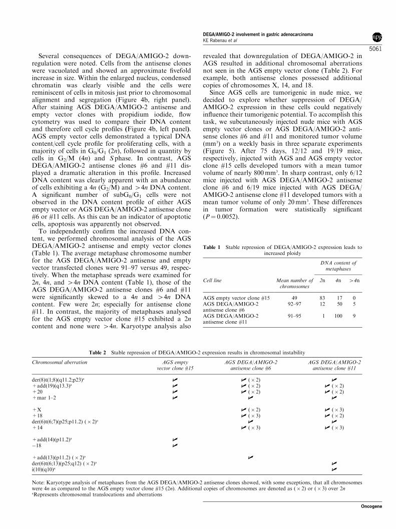
Several consequences of DEGA/AMIGO-2 down-regulation were noted. Cells from the antisense cloneswere vacuolated and showed an approximate fivefoldincrease in size. Within the enlarged nucleus, condensedchromatin was clearly visible and the cells werereminiscent of cells in mitosis just prior to chromosomalalignment and segregation (Figure 4b, right panel).After staining AGS DEGA/AMIGO-2 antisense andempty vector clones with propidium iodide, flowcytometry was used to compare their DNA contentand therefore cell cycle profiles (Figure 4b, left panel).AGS empty vector cells demonstrated a typical DNAcontent/cell cycle profile for proliferating cells, with amajority of cells in G0/G1 (2n), followed in quantity bycells in G2/M (4n) and S phase. In contrast, AGSDEGA/AMIGO-2 antisense clones #6 and #11 dis-played a dramatic alteration in this profile. IncreasedDNA content was clearly apparent with an abundanceof cells exhibiting a 4n (G2/M) and 44n DNA content.A significant number of subG0/G1 cells were notobserved in the DNA content profile of either AGSempty vector or AGS DEGA/AMIGO-2 antisense clone#6 or #11 cells. As this can be an indicator of apoptoticcells, apoptosis was apparently not observed.To independently confirm the increased DNA con-
tent, we performed chromosomal analysis of the AGSDEGA/AMIGO-2 antisense and empty vector clones(Table 1). The average metaphase chromosome numberfor the AGS DEGA/AMIGO-2 antisense and emptyvector transfected clones were 91–97 versus 49, respec-tively. When the metaphase spreads were examined for2n, 4n, and 44n DNA content (Table 1), those of theAGS DEGA/AMIGO-2 antisense clones #6 and #11were significantly skewed to a 4n and 44n DNAcontent. Few were 2n; especially for antisense clone#11. In contrast, the majority of metaphases analysedfor the AGS empty vector clone #15 exhibited a 2ncontent and none were 44n. Karyotype analysis also
revealed that downregulation of DEGA/AMIGO-2 inAGS resulted in additional chromosomal aberrationsnot seen in the AGS empty vector clone (Table 2). Forexample, both antisense clones possessed additionalcopies of chromosomes X, 14, and 18.Since AGS cells are tumorigenic in nude mice, we
decided to explore whether suppression of DEGA/AMIGO-2 expression in these cells could negativelyinfluence their tumorigenic potential. To accomplish thistask, we subcutaneously injected nude mice with AGSempty vector clones or AGS DEGA/AMIGO-2 anti-sense clones #6 and #11 and monitored tumor volume(mm3) on a weekly basis in three separate experiments(Figure 5). After 75 days, 12/12 and 19/19 mice,respectively, injected with AGS and AGS empty vectorclone #15 cells developed tumors with a mean tumorvolume of nearly 800mm3. In sharp contrast, only 6/12mice injected with AGS DEGA/AMIGO-2 antisenseclone #6 and 6/19 mice injected with AGS DEGA/AMIGO-2 antisense clone #11 developed tumors with amean tumor volume of only 20mm3. These differencesin tumor formation were statistically significant(P¼ 0.0052).
Table 1 Stable repression of DEGA/AMIGO-2 expression leads toincreased ploidy
DNA content ofmetaphases
Cell line Mean number ofchromosomes
2n 4n 44n
AGS empty vector clone #15 49 83 17 0AGS DEGA/AMIGO-2antisense clone #6
92–97 12 50 5
AGS DEGA/AMIGO-2antisense clone #11
91–95 1 100 9
Table 2 Stable repression of DEGA/AMIGO-2 expression results in chromosomal instability
Chromosomal aberration AGS emptyvector clone #15
AGS DEGA/AMIGO-2antisense clone #6
AGS DEGA/AMIGO-2antisense clone #11
der(8)t(1;8)(q11.2;p23)a | | (� 2) |+add(19)(q13.3)a | | (� 2) | (� 2)+20 | | (� 2) | (� 2)+mar 1–2 | | |
+X | (� 2) | (� 3)+18 | (� 3) | (� 2)der(6)t(6;7)(p25;p11.2) (� 2)a | |+14 | (� 3) | (� 3)
+add(14)(p11.2)a |�18 |
+add(13)(p11.2) (� 2)a |der(6)t(6;13)(p25;q12) (� 2)a |i(10)(q10)a |
Note: Karyotype analysis of metaphases from the AGS DEGA/AMIGO-2 antisense clones showed, with some exceptions, that all chromosomeswere 4n as compared to the AGS empty vector clone #15 (2n). Additional copies of chromosomes are denoted as (� 2) or (� 3) over 2naRepresents chromosomal translocations and aberrations
DEGA/AMIGO-2 involvement in gastric adenocarcinomaKE Rabenau et al
5061
Oncogene

Comparison of the cell adhesive/migratory abilitiesof AGS DEGA/AMIGO-2 antisense and empty vector-transfected cells
To compare the cell adhesive and migratory abilities ofAGS DEGA/AMIGO-2 antisense clone #11 and emptyvector-transfected cells, we utilized porous membranecell culture inserts. This assay was performed by seedingcells into the upper chamber of the insert and allowingthem to pass through the porous membrane to adhere toand migrate through the collagen-coated underside inresponse to fetal bovine serum (FBS). Figure 6b shows arepresentative experiment (n¼ 2) where migrated cellswere visualized on the underside of the collagen-coatedmembrane following exposure to 10% FBS or serum-free media. In comparison to AGS empty vector-transfected cells, AGS DEGA/AMIGO-2 antisenseclone #11 cells revealed a nearly complete abolition ofcollagen adhesion/migration in response to 10% FBS.To ensure that the lack of increase in migrated DEGA/AMIGO-2 antisense clone #11 cells was not due to theirinability to seed the upper chamber of the cell cultureinsert or their ability to pass entirely through thecollagen-coated membrane to the media in the lowerchamber, cell counts were, respectively, taken from theupper chamber after seeding under serum-free condi-tions and in the lower chamber following completion ofthe experiment. No difference in cell count was observedin either chamber between the AGS DEGA/AMIGO-2antisense and empty vector clones.
Discussion
Following a search for genes involved in cancer, weidentified DEGA, a novel gene differentially expressedin B45% of human gastric adenocarcinoma tumorsamples. Sequence analysis of the extracellular region ofthe DEGA protein revealed an IgG domain and fiveLRR motifs typically found in LRR-containing proteinspresent on the cell surface (Kobe and Deisenhofer, 1994,1995). Since LRR motifs are believed to mediateprotein–protein interactions, their presence in DEGAand other cell surface LRR proteins implied a possiblerole in cell adhesion or ligand binding. This notion isconsistent with the functions of many LRR familymembers expressed on the cell surface. For example, thetrk receptor contains several LRRs that have beenimplicated in binding to its ligand, nerve growth factorand LRR motifs are also believed to mediate plateletadhesion by platelet glycoprotein Iba and V (Kobe andDeisenhofer, 1994, 1995). Analysis of the cytosolicregion of the DEGA gene product revealed no knownprotein domains. However, 20 of the 102 cytosolicamino acids were either a serine or a threonine,suggesting a possible role for DEGA in signal transduc-tion. This notion was further supported when theScansite phosphorylation prediction algorithm (Yaffeet al., 2001) predicted seven of the 20 cytosolic serinesand threonines to be phosphorylated by either caseinkinase I and II, protein kinase A and C, as well as theGSK3, ERK1, and AKT kinases.At the time of discovery and characterization, BLAST
searches did not reveal exact matches to the novel full-length DEGA cDNA or protein sequences. While thismanuscript was in preparation, however, a repeatBLAST search uncovered an exact match to AMIGO-2, a newly identified but functionally uncharacterizedprotein related to AMIGO, a LRR family memberimplicated in axon tract development (Kuja-Panulaet al., 2003). Through the use of coimmunoprecipitationand purified protein/bead aggregation assays, Kuja-Panula et al. demonstrated that AMIGO-2 couldparticipate in homophilic and heterophilic interactionswith AMIGO family members (i.e. AMIGO andAMIGO-3). This finding, coupled with sequence ana-lyses and our cell localization and adhesive/migratorydata, suggest that DEGA/AMIGO-2 belongs to thefunctionally diverse LRR superfamily and that it mayfunction as a cell adhesion molecule involved in signaltransduction.Our studies of DEGA/AMIGO-2 expression focused
on several tumor and normal tissues. With respect to thelatter, we demonstrated DEGA/AMIGO-2 expressionin lung, colon, and rectum, but the strongest expressionobserved was in breast, ovary, uterus, and cervix,suggesting a possible role for DEGA/AMIGO-2 infemale reproductive tissues. Since we were primarilyinterested in cancer genes that were differentiallyexpressed in tumor versus normal tissue, we used cancerprofiling cDNA arrays, to assess the differential expres-sion of DEGA/AMIGO-2 in 18 human cancers. Withthe exception of thyroid and pancreatic cancer, only
Figure 5 Tumorigenicity of AGS clones stably transfected with anantisense DEGA/AMIGO-2 construct or an empty vector. Tenmillion AGS empty vector clone #15 and antisense DEGA/AMIGO-2 clones #6 and #11 were mixed with Matrigel andsubcutaneously injected in athymic (nu/nu) mice. UntransfectedAGS cells (AGS WT) were also included as a positive control.Tumor volumes were measured weekly using calipers. Threeindependent experiments were conducted and the number oftumor-bearing mice versus the total number of mice injected wereas follows: AGS WT (12/12); AGS empty vector clone #15 (19/19);AGS DEGA/AMIGO-2 antisense clone #6 (6/12); and AGSDEGA-AMIGO-2 antisense clone #11 (6/19). Tumor volumedifferences between the AGS DEGA/AMIGO-2 antisense clonesand the AGS WT and empty vector-transfected cells werestatistically significant (P¼ 0.0052)
DEGA/AMIGO-2 involvement in gastric adenocarcinomaKE Rabenau et al
5062
Oncogene

gastric adenocarcinoma exhibited increased expressionof DEGA/AMIGO-2 in approximately 45% of tumorversus normal patient tissue samples. Initially, we weresurprised that DEGA/AMIGO-2 was differentiallyexpressed in tumors from only three cancers and notin tumors from all cancers, especially given theimportance we later demonstrated for DEGA/AMI-GO-2 expression in gastric adenocarcinoma. We cameto realize, however, that this expression trend was notnecessarily uncommon for tumor-related genes. Our in-house experiences using Becton Dickinson’s cDNAcancer profiling arrays with a variety of novel andknown (well validated) tumor-related genes also demon-strated DEGA/AMIGO-2-like expression profiles. Forexample, when the well-validated cancer target, EGFR,was analysed (data not shown), it clearly showeddifferential expression in nearly all normal versus tumorbreast tissue from breast cancer patients while exhibiting
differential expression in tumor tissue derived fromseveral lung and kidney cancer patients (therapeuticindications for current anti-EGFR therapies). More-over, although EGFR was also expressed highly in sometumors from colon cancer patients on the cDNA cancerprofiling array, it was not differentially expressed intumor versus normal colon tissue from these samepatients. Aside from DEGA/AMIGO-2’s differentialtumor expression profiles in gastric, thyroid, andpancreatic tumors, we also observed significant absoluteexpression levels in a subset of patient samples and celllines representing a variety of other cancers. Thus, thepossibility that DEGA/AMIGO-2 may play a role in asubset of these cancers awaits further investigation.To determine whether DEGA/AMIGO-2 expression
was required for the proliferation/tumorigenicity ofgastric adenocarcinoma, we employed a conventionalantisense approach to stably repress DEGA/AMIGO-2
Figure 6 Comparison of the cell adhesive/migratory abilities of AGS cells stably transfected with an antisense DEGA/AMIGO-2construct or an empty vector. (a) Schematic of cell adhesion/migration assay. (i) Cells are placed in the upper chamber of a cell cultureinsert and lowered into a well of a 24-well plate containing serum-free media. The cell culture insert contains a porous membrane withcollagen coated on its underside. (ii) After 24 h, fresh media plus 10% FBS is added to the well (lower chamber) and cells pass throughthe porous membrane and adhere to and (iii) migrate through the collagen. (b) Hematoxylin and eosin staining of AGS empty vectorclone #15 and DEGA/AMIGO-2 antisense clone #11 cells that have adhered to and migrated through to the underside of the collagen
DEGA/AMIGO-2 involvement in gastric adenocarcinomaKE Rabenau et al
5063
Oncogene

expression in the gastric adenocarcinoma cell line, AGS.While several AGS clones were generated with stablyrepressed levels of DEGA/AMIGO-2 expression, twowere used to compare to AGS empty vector clones.Whiles several consequences of DEGA/AMIGO-2downregulation were noted in these two clones, it isimportant to note that these consequences were notrestricted to these clones, but were apparent in allantisense clones showing at least fivefold repression ofDEGA/AMIGO-2 expression levels (data not shown).Consequently, the phenotypes observed for the AGSDEGA/AMIGO-2 clones were specifically due to down-regulation of DEGA/AMIGO-2 expression and not theresult of clonal heterogeneity.To summarize, the consequences of DEGA/AMIGO-
2 downregulation in AGS cells were: altered morphol-ogy, increased cell size and DNA content (ploidy),chromosomal instability and nearly complete inhibitionof tumorigenicity, and cell adhesion to and migrationthrough collagen. Initially, we were surprised to observethat downregulation of the expression of a single gene,namely DEGA/AMIGO-2, could lead to an increase inDNA content and chromosomal instability. However,this observation is not unique to DEGA/AMIGO-2.Antisense inhibition of BUB1 results in a similarphenotype in normal human fibroblasts (Musio et al.,2003). In addition, overexpression of the human Ha-rasoncogene increases ploidy and genomic instability inmurine fibroblasts within one cell cycle (Denko et al.,1994). While repressed expression of BUB1 leading tochromosomal instability is perhaps logically expected,chromosomal instability as a result of overexpression ofHa-ras is not obvious due to its proximity to the plasmamembrane and to a lack of evidence suggesting that Ha-ras acts directly upon the cell’s chromosomes. Similarly,it is not obvious, yet intriguing that DEGA/AMIGO-2,a putative cell adhesion molecule, leads to chromosomalinstability upon loss of its expression. While Denko et al.do not know the mechanism by which overexpression ofHa-ras leads to chromosomal instability, they didpropose hypotheses that accommodated their data andwhich we feel also accommodate our data involvingrepression of DEGA/AMIGO-2. One hypothesis iscentered on that fact that Ha-ras is known to affectmultiple cellular pathways that could impinge upon theintegrity of chromosomal DNA. This could also applyto repression of DEGA/AMIGO-2 and its currentlyunidentified pathways. Next, they postulate that signaltransduction by overexpressed Ha-ras may induce anucleolytic activity that saturates the DNA repaircapacity. In the case of DEGA/AMIGO-2, repressionof its expression/signaling could also have the sameeffect. These hypotheses await further experimentation.When tested for their ability to form tumors in
athymic (nu/nu) mice, the two independently generatedAGS DEGA/AMIGO-2 antisense clones exhibited adramatic inhibition in comparison to the empty vectorclone (mean tumor volumes of B20 and B800mm3,respectively). Although this finding suggested a directrole for DEGA/AMIGO-2 in the maintenance of thetumorigenic state of AGS cells, it did not account for the
lack of difference we observed in in vitro proliferativepotential between the AGS DEGA/AMIGO-2 antisenseand empty vector clones. We put forth two possibleexplanations for this discrepancy. Since data presentedin this report and data from Kuja-Panula et al. (2003)demonstrate that DEGA/AMIGO-2 may function as aputative signaling cell adhesion molecule, we proposethat DEGA/AMIGO-2 may be required for tumorattachment. Consequently, downregulation of itsexpression would negatively impact the tumorigenicityof AGS cells. Precedence for this hypothesis stemsfrom antisense expression of laminin-binding protein,which causes significant inhibition of attachmentand invasion in a human colon carcinoma cell line(Mafune and Ravikumar, 1992). Other cell adhesionmolecules involved in stomach cancer include CD44(Mayer et al., 1993; Yokozaki et al., 1994) and E-cadherin/b-catenin (Shun et al., 2001; Woo et al., 2001).Interestingly, since the loss of E-cadherin expressionappears to correlate with gastric cancer progression, wehypothesize that it may be lost in gastric cancer to allowfor metastasis, while expression of DEGA/AMIGO-2may be required as an attachment factor for tumorigen-esis and possibly metastasis. Chromosomal instabilitymay be an alternative explanation for the inability ofAGS DEGA/AMIGO-2 antisense clones to formtumors. It is possible that the additional chromosomalaberrations, which arose due to chromosomal instabilityobserved in the AGS DEGA/AMIGO-2 antisenseclones, negatively affected a variety of genes requiredfor the tumorigenicity of AGS cells. For example,additional copies of the X chromosome and chromo-some 18 that carry the ING and DCC tumorsuppressors, respectively, could explain the inability ofAGS DEGA/AMIGO-2 antisense clones to formtumors in vivo. The view that chromosomal instabilitymay not always be the driving force in tumorigenesis,and in fact, that it may be detrimental, is notunprecedented and has been supported by severalinvestigations of colorectal cancer (Cahill et al., 1999;Tomlinson and Bodmer, 1999; Nowak et al., 2002;Sieber et al., 2003). Whether the difference in the in vitroand in vivo proliferation potentials of the AGS DEGA/AMIGO-2 antisense clones is attributable to celladhesion and/or chromosomal instability/aberrations,it is important to emphasis that these mechanisms maybe mutually exclusive.In summary, through the discovery of DEGA/
AMIGO-2, we have identified a novel factor that mayfunction not only as a signaling cell adhesion molecule,but that may be required for the tumorigenesis of asubset of human gastric adenocarcinomas. Through ourantisense studies using a gastric adenocarcinoma cellline, we have shown that the inhibition of DEGA/AMIGO-2 expression negatively impacts tumor growthas well as altering chromosomal ploidy and stability.Detailed genetic, molecular, and biological character-ization of DEGA/AMIGO-2 is now required todetermine its requirement and precise function/mechan-ism in gastric adenocarcinoma and possibly othercancers. Finally, it will be of interest to determine
DEGA/AMIGO-2 involvement in gastric adenocarcinomaKE Rabenau et al
5064
Oncogene

whether DEGA/AMIGO-2 will serve as a diagnosticand/or prognostic tool for neoplasia of the stomach.
Materials and methods
Cell culture
Cell lines used in this study were obtained from the AmericanType Culture Collection (ATCC; Manassas, VA, USA) andmedia used for their propagation was purchased from Gibco,Grand Island, NY, USA. Cell lines and their respective growthmedia (shown in parentheses) were as follows: AGS (F-12),293 (Dulbecco’s modified Eagle’s medium (DMEM)), KKVR(Iscove’s DMEM), RF-1 (Leibovitz’s L-15), and NCI-SNU-1,NCI-SNU-16, NCI-N87, KatoIII (RPMI). All media weresupplemented with 2mM L-Glutamine (Gibco, Grand Island,NY, USA) and 10% FBS(Hyclone, Logan, UT, USA).KatoIII and KKVR, however, were grown in 20% FBS andNCI-N87 and KKVR growth media were additionallysupplemented with 1.5 g/l sodium bicarbonate.
DEGA/AMIGO-2 cDNA cloning
RT–PCR was performed to amplify the DEGA/AMIGO-2ORF from Marathon-Ready A549 and spleen cDNA pre-parations (BD Biosciences Clontech, Palo Alto, CA, USA)using the following forward (50-ATG TCG TTA CGT GTACAC ACT CTG-30) and reverse primers (50-TTA AGT GGACGC CAC AAA AGG TGT G-30) (Bio-Synthesis Incorpo-rated, Lewisville, TX, USA); start and stop codons areunderlined. Titanium Taq polymerase (BD Biosciences Clon-tech, Palo Alto, CA, USA) was utilized with RT–PCRconditions described in the Marathon-Ready cDNA manual:941C, 30 s; 681C, 2min. The DEGA/AMIGO-2 ORF PCRproduct (1569 bp) was cloned into pCR2.1 (InvitrogenCorporation TA Cloning Kit, Carlsbad, CA, USA) andsequence verified (Molecular Genetics Instrumentation Facil-ity, University of Georgia, Athens, GA; GENEWIZ Inc.,North Brunswick, NJ, USA). To obtain DNA sequenceupstream of the DEGA/AMIGO-2 start codon, 50 RACEwas performed using A549 Marathon Ready cDNA asexplained above with few modifications. AP1, a forwardprimer (included with the Marathon Ready cDNA) and aDEGA/AMIGO-2 gene-specific reverse primer (50-AAC TCAGGT CCA GTC TCT TAA TCA G-30) produced a PCRproduct after 35 rounds of amplification (941C, 30 s; 571C, 30 s;681C, 2min.) that was subcloned into pCR2.1 and sequenceverified. Sequence representing the 30UTR of DEGA/AMI-GO-2 was initially obtained computationally from the humanchromosome 12 BAC clone, GS1-99H8 (Genbank accession#AC004010). A series of PCR primers were subsequentlydesigned to determine the longest 30UTR sequence present inthe A549 cDNA preparation. A PCR product generated by thefollowing primer set: forward primer 50-ATG TCG TTA CGTGTA CAC ACT CTG-30 (start codon underlined) and reverseprimer 50-TGG ACC TCC AAT CTT CAC CAG G-30, whichinitiates at nucleotides 3564–3585 of the DEGA/AMIGO-2ORF (Figure 1a), identified the longest 30UTR segment.
Bioinformatics
To confirm that the S30-21616 EST represented a portion ofthe murine homolog of the human DEGA/AMIGO-2 partialcDNA IMAGE:3625286 clone, murine–human chromosomalsyntenic analysis was performed with access to the Celerahuman and murine genomic databases. To determine whether
DEGA/AMIGO-2 cDNA and amino-acid sequences weresimilar to sequences present in the public domain, BLASTsearches were performed using the National Center forBiotechnology Information website (http://www.ncbi.nlm.nih.gov). Sequence representing the 30UTR of the DEGA/AMIGO-2 gene was obtained computationally from humanchromosome 12 BAC clone, GS1-99H8 (Genbank accession#AC004010) and scanned for polyadenylation sites usingpolyadq software (http://argon.cshl.org/tabaska/polyadq_form.html). Prediction of protein motifs and domains aswell as glycosylation and phosphorylation sites present inthe DEGA/AMIGO-2 gene product were accomplishedusing prediction servers at the Center for BiologicalSequence Analysis (http://www.cbs.dtu.dk/services), the Sim-ple Modular Architecture Research Tool (SMART) V3.5(http://smart.embl-heidelberg.de/) and Scansite (http://scansite.mit.edu).
Stable expression of a DEGA/AMIGO-2-EGFP fusion constructand a DEGA/AMIGO-2 antisense construct in 293 and AGScells, respectively
To determine the subcellular localization of DEGA/AMIGO-2, we engineered a fusion protein whereby an EGFP was fusedto its C-terminus. This was accomplished by PCR amplifyingthe DEGA/AMIGO-2 ORF and cloning it into XhoI/AgeIsites of pEGFP-N1 (BD Biosciences Clontech, Palo Alto, CA,USA). This PCR reaction was performed as described in theDEGA/AMIGO-2 cDNA cloning section with the exceptionthat the forward primer used (50ATC CTC GAG GCG ACCATA ATG TCG TTA CGT GTA CAC ACT 30) contained thenative Kozak sequence and a XhoI site (underlined) 50 to thestart codon shown in bold. The reverse primer used (50GATCAC CGG TGC AGT GGA CGC CAC AAA AGG TGTGTC 30) contained an AgeI site (underlined) 30 of the stopcodon shown in bold. The DEGA/AMIGO-2-EGFP constructwas stably transfected into 293 and AGS cells usingFUGENE6 transfection reagent (Roche Molecular Biochem-icals, Indianapolis, IN, USA) as outlined by the manufacturer.A 2 : 1 and 3 : 1 FUGENE6:DNA ratio was used, respectively.Transfected 293 cells were left for 2 days at 371C after whichtime they were plated in limiting dilution and subjected toselection with 800 mg/ml Geneticin (Sigma-Aldrich Corp., StLouis, MO, USA), respectively. Individual clones were isolatedby trypsinization using cloning rings and allowed to expand.To determine the subcellular localization of the DEGA/AMIGO-2-EGFP fusion protein, clones were assessed usinga Zeiss Axioskop fluorescent microscope.To generate DEGA/AMIGO-2 antisense clones, a nucleo-
tide fragment encompassing the entire ORF of DEGA/AMIGO-2 as well as one that comprised the 50 most 608nucleotides of its ORF (generated from a partial EcoRI digestof pCR2.1-DEGA/AMIGO-2) were cloned into pIRE-SPURO2 (BD Biosciences Clontech, Palo Alto, CA, USA) inreverse orientation and stably transfected into AGS cells.Concurrently, pIRESPURO2 was also stably transfected intothese cells to serve as an empty vector control. Transfectionand clone isolation was performed exactly as described for theDEGA/AMIGO-2-EGFP fusion with the exception that cellswere selected with 0.4 mg/ml puromycin (Sigma-Aldrich Corp.,St Louis, MO, USA).
cDNA dot blot analysis of DEGA/AMIGO-2 expressionin cancer patient samples
To determine expression of DEGA/AMIGO-2 in a variety oftumor and normal tissues, CPA I and II (BD Biosciences
DEGA/AMIGO-2 involvement in gastric adenocarcinomaKE Rabenau et al
5065
Oncogene

Clontech, Palo Alto, CA, USA) were probed with a cDNAfragment encompassing ORF nucleotides 1540–2045 (num-bering based on Figure 1) as per the manufacturer’srecommendations. The probe was labeled with [a-32P]dCTP(Perkin-Elmer Life Sciences, Inc., Boston, MA, USA) andisolated using the Prime-It II kit (Stratagene Cloning Systems,La Jolla, CA, USA). CPA I and II are nylon arrays con-taining normalized cDNA from 241 and 160 tumor andcorresponding normal tissues obtained from individual cancerpatients, respectively. Tissues represented by CPA I includebreast, uterus, colon, stomach, ovary, lung, kidney, rectum,thyroid, cervix, small intestine, pancreas, and prostate. Inaddition to these tissues, CPA II carries bladder, testis,and skin tissue. These arrays also contain positive and neg-ative controls as well as cDNA isolated from nine cancer celllines: HeLa, Daudi, K-562, HL-60, G-361, A549, Molt-4,SW480, and Raji. For more information regarding CPA Iand II, see the following website: http://bioinfo.clontech.com/darray.To determine DEGA/AMIGO-2 expression in nongastric
adenocarcinoma cell lines, the DEGA/AMIGO-2 probe andconditions used for CPA I and II were also used to hybridizeBD Bioscience’s Cancer Cell Line Profiling Array, whichcontains cDNA from cell lines representing cancer of the lung(A549, NCI-H-460, NCI-H1299), colon (HCT-116, HCT-15,HT-29), breast (MDA-MB4355, MDA-MB231, MCF7), ovary(SK-OV-3), cervix (HeLa), prostate (DU-145, PC-3), skin (SK-MEL-28, SK-MEL-5, SK-NSH, IMR-32, U-87MG), brain(IMR-32, U-87MG), kidney (786-O, ACHN), liver (Hep-G2),colon (Colo589), osteosarcoma (U2-OS), and epidermis(A-431).
Northern blot analysis
To determine expression of DEGA/AMIGO-2 in gastricadenocarcinoma cell lines and in AGS DEGA/AMIGO-2antisense and empty vector clones, standard Northern blottingprocedures were followed. Total RNA was isolated using theMicro-to-Midi Total RNA system (Invitrogen Corporation,Carlsbad, CA, USA) exactly as recommended by themanufacturer and 5mg was resolved on a 1.2% denaturingformaldehyde gel and transferred to Nytran SuPerChargemembranes (Schleicher & Schuell, Inc., Keene, NH,USA). Blots were hybridized with the 32P random-labeledDEGA/AMIGO-2 C-terminal probe used to probe theCPA I and II cDNA blots. Hybridizations were performedfor 1 h at 681C using ExpressHyb solution (BD BiosciencesClontech, Palo Alto, CA, USA). Three 10min room tempera-ture washes (2� SSC, 0.05% SDS) and two 20min 501Cwashes (0.1� SSC, 0.1% SDS) followed. Membranes wererinsed in 2� SSC and exposed overnight at �701C to KodakBiomax MR film (Eastman Kodak Company, Rochester,NY, USA).
Wright–Giemsa staining
To differentiate nuclear and cytoplasmic morphology of AGSDEGA/AMIGO-2 antisense clones, Giemsa staining wasperformed. Cells were grown to subconfluency on Lab-TekII chamber slides (Nalgene Nunc International, Aurora, IL,USA) and stained with Wright–Giemsa stain according to themanufacturer’s instructions (Thermo Shandon, Pittsburgh,PA, USA).
DNA content analysis
To measure DNA content, and therefore cell cycle profile, flowcytometry was utilized. AGS empty vector and DEGA/
AMIGO-2 antisense clones were grown in growth media,trypsinized, washed once in PBS and stained for 10min atroom temperature in the dark with the propidium iodidesolution provided in the DNA QC Particles kit (BectonDickinson). Cells were subsequently analysed on a BectonDickinson FACSVantage flow cytometer using parametersoutlined in the DNA QC particles kit.
Chromosome analysis
AGS empty vector #15 and DEGA/AMIGO-2 antisenseclones were grown in culture as described above. Followingconventional methods, chromosome preparations were gener-ated from logarithmically growing cells following exposure tocolcemid for 2 h. Chromosomes were trypsin-Giemsa bandedand approximately 70–110 metaphases were analysed per cellline. Chromosome abnormalities were described using stan-dard ISCN nomenclature.
Cell adhesion/migration assay
To compare the ability of AGS DEGA/AMIGO-2 antisenseand empty vector-transfected clones to adhere to and migratethrough collagen, cell culture inserts containing poroustranslucent polyethylene terephthalate track-etched mem-branes (8.0 mm pore size; Becton Dickinson Falcon) wereutilized. Just prior to the assay, the underside of the porousmembranes in the cell culture inserts were coated with collagenby aseptically removing the inserts from the packaging withsterile forceps and gently placing them into a 24-well Falconplate filled with 700 ml of Vitrogen-100-purified collagensolution (25 mg/ml; Cohesion; Palo Alto, CA, USA). Theinserts were left for at least 1 h at 41C and placed in a new24-well plate containing either 700ml serum-free media or10% FBS. Next, 6� 105 viable cells that had been serumstarved for 24 h were rinsed twice with PBS and seeded intothe upper chamber of the cell culture insert in 300mlserum-free media. Cells were then incubated another 24 h at371C to allow for adhesion to and migration through thecollagen-coated porous membrane on the underside of thecell culture insert. At the conclusion of the assay, mediawas aspirated from the inserts and washed twice withPBS. Nonmigrated cells on the upperside of the cell cultureinsert were removed with a Q-tip (dipped in PBS). Thecollagen-coated underside of the membrane containing themigrated cells was washed twice with PBS and fixed withbuffered neutral formalin (10%; VWR; West Chester, PA,USA) for 10min at RT. Following two washes with PBS,the migrated cells were permeabilized with PBS/0.1%Tween-20 for 5min at RT and their nuclei stained with700 ml Harris Modified Hematoxylin with Acetic Acid solu-tion (Fisher Scientific; Fairlawn, NJ, USA) for 10min at RT.Their cytoplasms were stained with Eosin-Y Alcoholic(Richard-Allan Scientific; Kalamazoo, MI, USA) for 2minat RT. The cell culture inserts were then allowed to air dryupside down for 15min at RT and the porous membranesremoved from the cell culture inserts with a razor blade fortransfer to a glass slide. Once mounted and cover-slipped,migrated cells on the underside of the collagen-coatedmembrane were visualized using bright field microscopy at� 100 magnification.
Tumorigenicity analysis
AGS clones were suspended in serum-free growth media,placed on ice and thoroughly mixed with Matrigel (Collabora-tive Research Biochemicals, Bedford, MA, USA) at a 1 : 1 (v/v)ratio. Matrigel is derived from the Engelbroth-Holm-Swarm
DEGA/AMIGO-2 involvement in gastric adenocarcinomaKE Rabenau et al
5066
Oncogene

mouse sarcoma, which has been found to be a rich source of theECM proteins: laminin, collagen IV, nidogen/enactin, andproteoglycan (Kleinman et al., 1982). When mixed with celllines, Matrigel enhances tumorigenesis (Terranova et al.,1986). For each 1–2-month old, gender-independent athymic(nu/nu) mouse (Charles River Laboratories, Wilmington, MA,USA), ten million cells were injected in the flank subcuta-neously and tumor volumes (mm3) measured weekly usingcalipers for 11 weeks.
Acknowledgements
We thank Drs Yaron Hadari, Jackie Doody, Nick Loizos, andHua-Quan Miao for review of this manuscript as well asShannon Mitchell for assistance with its preparation. Forkaryotype analysis, we are grateful to Dr Vundavalli VVSMurty. Appreciation is also extended to Drs Ihor Lemischka,Kateri Moore and Chris Stoeckert for development andbioinformatics support of the stem cell database.Dedication: MdLSP (ILU-RIP).
References
Akama Y, Yasui W, Yokozaki H, Kuniyasu H, Kitahara K,Ishikawa T and Tahara E. (1995). Jpn. J. Cancer Res., 86,
617–621.Arber N, Hibshoosh H, Moss SF, Sutter T, Zhang Y, Begg M,Wang S, Wenstein IB and Holt PR. (1996). Gastroenterol-ogy, 110, 669–674.
Cahill DP, Kinzler KW, Vogelstein B and Lengauer C. (1999).Trends Cell Biol., 9, M57–M60.
Chan AO, Luk JM, Hui WM and Lam SK. (1999). J.Gastroenterol. Hepatol., 14, 1150–1160.
Crookes PF. (2002). Clin. Obstet. Gynecol., 45, 892–903.Danesh J. (1999). Aliment. Pharmacol. Ther., 13, 851–856.Denko NC, Giaccia AJ, Stringer JR and Stambrook PJ.(1994). Proc. Natl. Acad. Sci. USA, 91, 5124–5128.
Ebert MP, Yu J, Sung JJ and Malfertheiner P. (2000). Eur. J.Gastroenterol. Hepatol., 12, 795–798.
El-Rifai W and Powell SM. (2002). Semin. Radiat. Oncol., 12,
128–140.Hocking AM, Shinomura T and McQuillan DJ. (1998).
Matrix Biol., 17, 1–19.Hollstein M, Shomer B, Greenblatt M, Soussi T, Hovig E,Montesano R and Harris CC. (1996). Nucleic Acids Res., 24,141–146.
Iozzo RV. (1997). Crit. Rev. Biochem. Mol. Biol., 32, 141–174.Kleinman HK, McGarvey ML, Liotta LA, Robey PG,Tryggvason K and Martin GR. (1982). Biochemistry, 21,6188–6193.
Kobe B and Deisenhofer J. (1994). Trends Biochem. Sci., 19,
415–421.Kobe B and Deisenhofer J. (1995). Curr. Opin. Struct. Biol., 5,
409–416.Kobe B and Kajava AV. (2001). Curr. Opin. Struct. Biol., 11,
725–732.Kono K, Naganuma H, Sekikawa T, Amemiya H, TakahashiA, Iizuka H and Matsumoto Y. (2000). Tumour Biol., 21,
139–144.Konturek PC, Konturek SJ, Pierzchalski P, Bielanski W, DudaA, Marlicz K, Starzynska T and Hahn EG. (2001).Med. Sci.Monit., 7, 1092–1107.
Kozak M. (1987). J. Mol. Biol., 196, 947.
Kuja-Panula J, Kiiltomaki M, Yamashiro T, Rouhiainen Aand Rauvala H. (2003). J. Cell Biol., 160, 963–973.
Kuniyasu H, Yasui W, Kitadai Y, Yokozaki H, Ito H andTahara E. (1992). Biochem. Biophys. Res. Commun., 189,227–232.
Kuniyasu H, Yasui W, Yokozaki H, Kitadai Y and Tahara E.(1993). Int. J. Cancer, 55, 72–75.
Mafune K and Ravikumar TS. (1992). J. Surg. Res., 52, 340–346.Mayer B, Jauch KW, Gunthert U, Figdor CG, Schildberg FW,Funke I and Johnson JP. (1993). Lancet, 342, 1019–1022.
Moayyedi P and Ford A. (2002). BMJ, 325, 1399–1402.Musio A, Montagna C, Zambroni D, Indino E, Barbieri O,Citti L, Villa A, Ried T and Vezzoni P. (2003). Cancer Res.,63, 2855–2863.
Nowak MA, Komarova NL, Sengupta A, Jallepalli PV, ShihIeM, Vogelstein B and Lengauer C. (2002). Proc. Natl. Acad.Sci. USA, 99, 16226–16231.
Phillips RL, Ernst RE, Brunk B, Ivanova N, Mahan MA,Deanehan JK, Moore KA, Overton GC and Lemischka IR.(2000). Science, 288, 1635–1640.
Ross JS and McKenna BJ. (2001). Cancer Invest., 19, 554–568.Sakamoto H, Mori M, Taira M, Yoshida T, Matsukawa S,Shimizu K, Sekiguchi M, Terada M and Sugimura T. (1986).Proc. Natl. Acad. Sci. USA, 83, 3997–4001.
Shun CT, Wu MS, Lin MT, Chang MC, Lin JT and ChuangSM. (2001). Oncology, 60, 339–345.
Sieber OM, Heinimann K and Tomlinson IPM. (2003). Nat.Rev. Cancer, 3, 701–708.
Terranova VP, Hujanen ES, Loeb DM, Martin GR, Thorn-burg L and Glushko V. (1986). Proc. Natl. Acad. Sci. USA,83, 465–469.
Tomlinson I and Bodmer W. (1999). Nat. Med., 5, 11–12.Woo DK, Kim HS, Lee HS, Kang YH, Yang HK and KimWH. (2001). Int. J. Cancer, 95, 108–113.
Yaffe MB, Leparc GG, Lai J, Obata T, Volinia S and CantleyLC. (2001). Nat. Biotechnol., 19, 348–353.
Yasui W, Oue N, Kuniyasu H, Ito R, Tahara E and YokozakiH. (2001). Gastric Cancer, 4, 113–121.
Yokozaki H, Ito R, Nakayama H, Kuniyasu H, Taniyama Kand Tahara E. (1994). Cancer Lett., 83, 229–234.
DEGA/AMIGO-2 involvement in gastric adenocarcinomaKE Rabenau et al
5067
Oncogene