Contourlet Transformation for Text Hiding in HSV Color Image
Upload
cpmtc-igc-ufmgCategory
view
1download
0
JOURNAL OF NEUROINFLAMMATION
Zolini et al. Journal of Neuroinflammation 2014, 11:20http://www.jneuroinflammation.com/content/11/1/20
RESEARCH Open Access
Defense against HSV-1 in a murine model ismediated by iNOS and orchestrated by theactivation of TLR2 and TLR9 in trigeminal gangliaGuilherme Pimenta Zolini1†, Graciela Kunrath Lima2†, Natália Lucinda1, Mariana Almeida Silva1,Marcela França Dias1, Natália Lima Pessoa1, Bruna Pizziolo Coura1, Christiane Teixeira Cartelle3,Rosa Maria Esteves Arantes3, Erna Geessien Kroon4 and Marco Antônio Campos1*
Abstract
Background: Herpes simplex 1 (HSV-1) causes various human clinical manifestations, ranging from simple coldsores to encephalitis. Innate immune cells recognize pathogens through Toll-like receptors (TLRs), thus initiating theimmune response. Previously, we demonstrated that the immune response against HSV-1 is dependent on TLR2and TLR9 expression and on IFN gamma production in the trigeminal ganglia (TG) of infected mice. In this work,we further investigated the cells, molecules, and mechanisms of HSV-1 infection control, especially those that areTLR-dependent.
Methods: C57BL/6 wild-type (WT), TLR2−/−, TLR9−/−, and TLR2/9−/− mice were intranasally infected with HSV-1. On theviral peak day, the TG and brains were collected from mice and TLR expression was measured in the TG and brain andinducible nitric oxide synthase (iNOS) expression was measured in the TG by real-time PCR. Immunofluorescence assayswere performed in mice TG to detect iNOS production by F4/80+ cells. Intraperitoneal macrophages nitric oxide(NO) production was evaluated by the Griess assay. WT, CD8−/−, RAG−/−, and iNOS−/− mice were intranasallyinfected in a survival assay, and their cytokine expression was measured in the TG by real-time PCR.
Results: Infected WT mice exhibited significantly increased TLR expression, compared with their respectivecontrols, in the TG but not in the brain. TLR-deficient mice had moderately increased TLR expression in the TG andbrain in compare with the non-infected animals. iNOS expression in the WT infected mice TG was higher than inthe other groups with increased production by macrophages in the WT infected mice, which did not occur in theTLR2/9−/− mice. Additionally, the intraperitoneal macrophages of the WT mice had a higher production of NOcompared with those of the TLR-deficient mice. The CD8−/−, RAG−/−, and iNOS−/− mice had 100% mortality afterthe HSV-1 infection compared with 10% of the WT mice. Cytokines were overexpressed in the iNOS−/− infectedmice, while the RAG−/− mice were nearly unresponsive to the virus.
Conclusion: TLRs efficiently orchestrate the innate immune cells, eliciting macrophage response (with NOproduction by the macrophages), thereby controlling the HSV-1 infection through the immune response in theTG of mice.
Keywords: Encephalitis, HSV-1, Innate immunity, Toll-like receptors, Host immune response
* Correspondence: [email protected]†Equal contributors1Laboratório de Imunopatologia, Imunologia de Doenças Virais, Centro dePesquisas René Rachou, Fundação Oswaldo Cruz, Fiocruz, Av. Augusto deLima 1715, Belo Horizonte, Minas Gerais 30190-002, BrazilFull list of author information is available at the end of the article
© 2014 Zolini et al.; licensee BioMed Central Ltd. This is an Open Access article distributed under the terms of the CreativeCommons Attribution License (http://creativecommons.org/licenses/by/2.0), which permits unrestricted use, distribution, andreproduction in any medium, provided the original work is properly credited.
Zolini et al. Journal of Neuroinflammation 2014, 11:20 Page 2 of 12http://www.jneuroinflammation.com/content/11/1/20
BackgroundHerpesviruses are large enveloped viruses (100–200 nm)with a double-stranded DNA genome from 120 to 230 kbpwhich encodes approximately 84 proteins [1]. An estimated70% of the world population is seropositive for humanherpes virus 1 (HHV-1), also known as herpes simplexvirus 1 (HSV-1) [1,2]. HSV-1 is the etiological agent oforolabial and cutaneous herpes, ophthalmic lesions, kera-titis, and kerato-conjunctivitis. Severe ophthalmic lesions,central nervous system involvement (encephalitis), or sys-temic infections, although rare, can occur in recently bornchildren or in immunosuppressed individuals [3]. Her-pesviruses are known for their latency/recurrence phe-nomena. HSV-1 can become latent in neuronal ganglia(especially the trigeminal ganglia) and may reactivateunder stressful conditions, sometimes causing new dis-ease episodes [1].The HSV-1 infection in humans involves the intimate
contact of a susceptible individual with someone who isshedding the virus [1]. After the primary infection,which usually occurs in the oral mucosa, the virus repli-cates in epithelial cells and then infects the neuronal ter-minal, from where the virus reaches the nerve gangliathrough retrograde axonal flow. The trigeminal gangliaharbor latent virus DNA [4,5]. Recurrences occur whenthe latent virus reactivates and is transported by antero-grade axonal flow to the primary infection site [6,7]. Insome cases, reactivated HSV-1 is transported to the cen-tral nervous system, causing encephalitis [8]. Reactiva-tion is normally associated with stress factors such ashormonal alterations, ultraviolet light exposure, and im-munosuppression [9,10]. However, a study using math-ematical models of HSV-2 (which is closely related toHSV-1) reactivation revealed that the virus is constantlyreleased from neurons in small amounts, thereby activat-ing the immune system, which in turn inhibits viral repli-cation and promotes a balance that prevents the clinicalreactivation of the disease [11].Immune responses against HSV-1 involve both innate
and acquired immunity. Innate responses are largely me-diated by leukocytes such as neutrophils, macrophages,and dendritic cells, which phagocytose pathogens and co-ordinate additional responses with the synthesis of a var-iety of inflammatory mediators and cytokines. Toll-likereceptors (TLRs) are proteins that confer ‘specificity’ tothe innate immune system, allowing the recognition ofpathogen associated molecular patterns (PAMPs) [12,13].The mRNAs from all TLR types (except TLR3) are ex-pressed by monocytes and macrophages [13]; in thecentral nervous system (CNS), while microglia expressmRNA from TLR1 to TLR9, neurons and oligodendro-cytes express only TLR3 [12]. During the last decade, theimportant roles of innate immunity responses and TLRsin HSV-1 infection control have been highlighted [14-19].
HSV-1 infection control appears to be highly dependenton the cellular immune response, including local tissuecells such as microglia and resident macrophages, as wellas inflammatory infiltration by cells such as monocytesand CD8+ T lymphocytes [19-23]. This control most likelyoccurs through the production of cytokines such as IFNgamma, especially in the trigeminal ganglia [19,24,25].IFN gamma is produced mainly by T lymphocytes [26],and one of its major actions is to increase iNOS andROS production for killing pathogens [26,27]. iNOS,IFN gamma, and other pro-inflammatory cytokines areimportant for controlling the HSV-1 infection in the tri-geminal ganglia through the action of macrophages andCD8+ T cells [19,28], although this effect in the brain iscontroversial [19,28,29].Data from our group, which uses the murine intrana-
sal infection model, have indicated that the crucial ‘spot’for immune defense organization against HSV-1 mostlikely is the trigeminal ganglia and not the brain[14,19,28,30,31]. Most C57BL/6 wild-type (WT) miceinfected with 106 p.f.u. of HSV-1 can control the infec-tion and survive [19,31]. These studies have also dem-onstrated that certain chemokines and IFN gammaexpression in trigeminal ganglia exhibit the same tem-poral profile of viral replication, with peaks of expres-sion occurring at day 5 post infection (which most likelyfacilitates controlling the virus), while mice withoutsigns of encephalitis do not harbor the virus in theirbrains [19]. Apparently this control is not as efficient inmice with deficiencies in their immune response be-cause knockout mice for MyD88, IFN gamma [15],TLR9, and TLR2/9 [19] have high mortality rates anddie with signs of encephalitis after HSV-1 infection.Additionally, the virus is found in the brains of theseanimals.The high prevalence of HSV-1 and the considerably in-
creased number of immunosuppressed individuals due todiseases or to therapeutic transplantation, heighten the im-portance of comprehending the mechanisms of immunedefense against the virus in a herpetic encephalitis model.In the present work, we demonstrate that macrophage-mediated immunity against HSV-1 occurs efficiently throughiNOS in trigeminal ganglia and appears to be organized bythe initial activation of TLR2 and TLR9 (with a possibleinterrelation of these receptors with other TLRs), which con-tributes to viral infection control.
Materials and methodsVirusHSV-1 strain EK [32], which was isolated from a humancase of recurrent oral herpes with blisters, was multipliedin Vero cells [15] and purified [33] as previously described.The virus titers obtained were 3.0 × 10⁹ p.f.u./mL.
Zolini et al. Journal of Neuroinflammation 2014, 11:20 Page 3 of 12http://www.jneuroinflammation.com/content/11/1/20
CellsVero cells (ATCC) were maintained in MEM supple-mented with 5% heat-inactivated FBS and antibiotics in5% CO2 at 37°C. These cells were used for multiplicationand titration of the virus.
MiceTLR2−/− and TLR9−/− mice were generated at OsakaUniversity (Osaka, Japan), and the TLR2/9−/− mice wereobtained by crossing the TLR2−/− and TLR9−/− mice atthe NIH (USA) and by backcrossing them to the C57BL/6background for eight generations. These mice were a kindgift of Shizuo Akira and Alan Sher, respectively. C57BL/6RAG−/−, C57BL/6 iNOS−/−, and C57BL/6 CD8−/− micewere acquired from Biotério Fiocruz/Rio. The C57BL/6(WT, control) and knockout mice were maintained in apathogen-free, barrier environment in Centro de PesquisasRené Rachou, Oswaldo Cruz Foundation (CPqRR/FIO-CRUZ) (Belo Horizonte, Minas Gerais, Brazil). Six- to10-week-old male mice were anesthetized with keta-mine (Agribrands do Brasil Ltda, Brazil), and 106 p.f.u.of the purified HSV-1 contained in 10 μL was inhaled bythe mice as described previously [34]. The control miceinhaled PBS. The mouse colonies and all of the experi-mental procedures were performed according to the in-stitutional animal care and use guidelines from CPqRR/FIOCRUZ. The project was previously approved by theEthics Committee in Animal Experimentation (CEUAfrom CPqRR/FIOCRUZ LW6/11).
� Intraperitoneal macrophages
Thioglycollate-elicited peritoneal macrophages wereobtained from either C57BL/6, TLR2−/−, TLR9−/−, orTLR2/9−/− mice by peritoneal washing, were activatedwith sub-optimal concentration of murine IFN gamma(50 U/mL) as previously described [15] and then werestimulated with HSV-1 (MOI of 10) for 24 h. The Griessreaction assay [35] was performed to evaluate nitricoxide production in the supernatants.
Griess reaction assayThioglycollate-elicited peritoneal macrophages were ob-tained from either C57BL/6, TLR2−/−, TLR9−/−, orTLR2/9−/− mice by peritoneal washing. Adherent peri-toneal macrophages were cultured in 96-well plates (2 ×10⁵ cells/well) at 37°C/5% CO2 in Dulbecco’s modifiedEagle’s medium (Life Technologies, Paisley, UK) supple-mented with 5% heat-inactivated fetal bovine serum(Life Technologies), 2 mmol/L L-glutamine (Sigma) and40 μg/mL of gentamicin (Schering do Brasil, Rio de Janeiro,Brazil). Next, the cells were stimulated with HSV-1 (multi-plicity of infection, 10), for 24 h to evaluate nitric oxideproduction in the supernatants.
RNA extractionTrigeminal ganglia and brains were aseptically removed andstored at −70°C until processing. RNA extraction was per-formed according to the procedures provided by the manu-facturer of TRIzol reagent (Invitrogen, USA). The RNAswere treated with DNase (Biolabs, USA). One microliter ofthe extracted RNA was quantified in a Nanodrop ND-1000spectrophotometer, at wavelengths of 260 and 280 nm.
Reverse transcriptionReverse transcription was performed according to theprocedures provided by the manufacturer of the M-MLVRT enzyme (Promega, USA).
Real-time PCRReal-time quantitative PCR (Applied Biosystems, USA)was performed to verify the mRNA expression in the tri-geminal ganglia and brain of mice infected with HSV-1.The reactions were performed using the Sybr GreenPCR Master Mix (Applied Biosystems, USA) in the Ap-plied Biosystems’ 7000 Sequence Detection System, at50°C, 2’; 95°C, 10’; and 40 cycles of 95°C, 15” and 60°C, 1’,followed by a final dissociation stage. The oligonucleotidesused in the reactions were specific for HPRT, F - GTTGGA TAC AGG CCA GAC TTT GTT G and R - GATTCA ACT TGC GCT CAT CTTAGG C; IP-10 (CXCL10),F -GCC GTC ATT TTC TGC CTC AT and R - GCTTCC CTA TGG CCC TCA TT; TNF alpha, F - CAT CTTCTC AAA ATT CGA GTG ACA A and R - TGG GAGTAG ACA AGG TAC AAC CC; iNOS, F - CAG CTGGGC TGTACA AAC CTTand R - CAT TGG AAG TGAAGC GTT TCG; MCP-1(CCL2), F - CTT CTG GGCCTG CTG TTC A and R - CCA GCC TAC TCA TTGGGA TCA [36]; RANTES (CCL5), F - GTC GTG TTTGTC ACT CGA AGG A and R - GAT GTA TTC TTGAAC CCA CTT CTT CTC; gp91, F - CCA ACT GGGATA ACG AGT TCA AGA C and R - AAG GCT TCAGGG CCA CAC A; p22, F - TGG CTA CTG CTG GACGTT TCA C and R - CTC CAG CAG ACA GAT GAGCAC AC; VP16 F - TTT GAC CCG CGA GAT CCT ATand R - GCT CCG TTG ACG AAC ATG AA [37]; TLR1,F - TGA TCT TGT GCC ACC CAA CA and R - GCAGGG CAT CAA AGG CAT TA; TLR2, F - TTG CTCCTG CGA ACT CCT AT and R - AGC CTG GTG ACATTC CAA GA; TLR3, F - TAG ACT GCA TCG CCTGCT AA and R - AAG CAG CCA GAA GCA GAA CT;TLR6, F - TCT GGG ATA GCC TCT GCA ACA and R -GGC GCA AAC AAA GTG GAA AC; TLR7, F - TACCAG GAC AGC CAG TTC TA and R - AGG AGC CTCTGA TGA GAC AA; and TLR9, F - ACC TCA GCC ACAACA TTC TC and R - TGC ACC TCC AAC AGT AAGTC [38]. The comparative Ct method was chosen toanalyze the data, using the arithmetic formula 2-ΔΔCt [39].The genes expression was normalized to expression of
Zolini et al. Journal of Neuroinflammation 2014, 11:20 Page 4 of 12http://www.jneuroinflammation.com/content/11/1/20
the constitutively expressed gene Hypoxanthine-guaninephosphoribosyltransferase (HPRT). All of the reactionswere replicated.
HistopathologySamples were fixed with 10% formaldehyde in phosphatebuffer, routinely processed and embedded in paraffin, aspreviously described [15].
TLR 1
C57BL/6
TLR2 -/-
TLR9 -/-
TLR2/9 -/
-
C57BL/6
TLR2 -/-
TLR9 -/-
TLR2/9 -/
-0
10
20
30
40
50
NI HSV-1
***
*****
#
###
#
Fold
incr
ease
(arb
itra
ry u
nits
)
TLR 3
C57BL/6
TLR2 -/-
TLR9 -/-
TLR2/9 -/
-
C57BL/6
TLR2 -/-
TLR9 -/-
TLR2/9 -/
-0
10
20
30
40
50
NI HSV-1
*** *** *** *
Fold
incr
ease
(arb
itra
ry u
nits
)
TLR 7
C57BL/6
TLR2 -/-
TLR9 -/-
TLR2/9 -/
-
C57BL/6
TLR2 -/-
TLR9 -/-
TLR2/9 -/
-0
10
20
30
40
50
NI HSV-1
***
*******
*
#
Fold
incr
ease
(arb
itra
ry u
nits
)
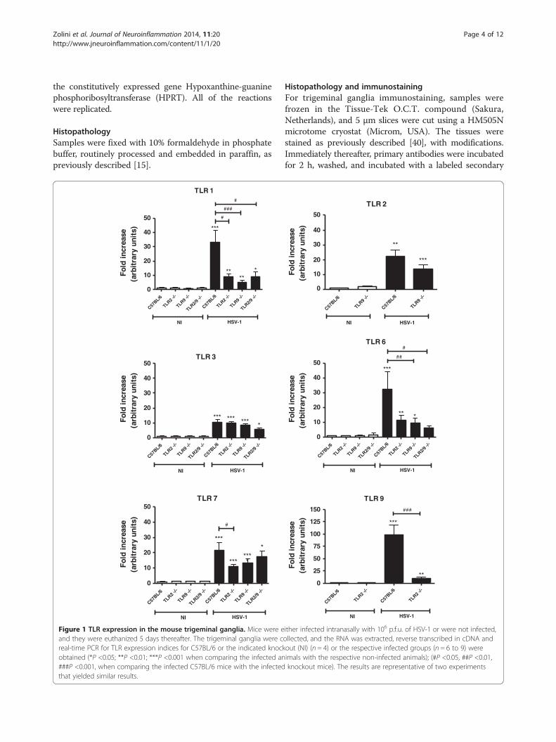
Figure 1 TLR expression in the mouse trigeminal ganglia. Mice were eand they were euthanized 5 days thereafter. The trigeminal ganglia were creal-time PCR for TLR expression indices for C57BL/6 or the indicated knockobtained (*P <0.05; **P <0.01; ***P <0.001 when comparing the infected an###P <0.001, when comparing the infected C57BL/6 mice with the infectedthat yielded similar results.
Histopathology and immunostainingFor trigeminal ganglia immunostaining, samples werefrozen in the Tissue-Tek O.C.T. compound (Sakura,Netherlands), and 5 μm slices were cut using a HM505Nmicrotome cryostat (Microm, USA). The tissues werestained as previously described [40], with modifications.Immediately thereafter, primary antibodies were incubatedfor 2 h, washed, and incubated with a labeled secondary
TLR 2
C57BL/6
TLR9 -/-
C57BL/6
TLR9 -/-
NI HSV-1
**
***
TLR 6
C57BL/6
TLR2 -/-
TLR9 -/-
TLR2/9 -/
-
C57BL/6
TLR2 -/-
TLR9 -/-
TLR2/9 -/
-
NI HSV-1
***
** *
##
#
0
10
20
30
40
50
Fold
incr
ease
(arb
itra
ry u
nits
)
0
10
20
30
40
50
Fold
incr
ease
(arb
itra
ry u
nits
)Fo
ld in
crea
se(a
rbit
rary
uni
ts)
TLR 9
C57BL/6
TLR2 -/-
C57BL/6
TLR2 -/-
0
25
50
75
100
125
150
NI HSV-1
***
**
###
ither infected intranasally with 106 p.f.u. of HSV-1 or were not infected,ollected, and the RNA was extracted, reverse transcribed in cDNA andout (NI) (n = 4) or the respective infected groups (n = 6 to 9) wereimals with the respective non-infected animals); (#P <0.05, ##P <0.01,knockout mice). The results are representative of two experiments
Zolini et al. Journal of Neuroinflammation 2014, 11:20 Page 5 of 12http://www.jneuroinflammation.com/content/11/1/20
antibody. The sections were counterstained (nuclei) byHoechst (0.2 μg/mL) (Molecular Probes, USA) andmounted in the Hydromount aqueous medium (NationalDiagnostics, USA). The primary antibody used was ratanti-F4/80, 1:50 (Serotec MCAP497, USA) or rabbit anti-iNOS, 1:5,000 (Spring Bioscience E3740, USA).The secondary antibody was Alexa Fluor 488 goat
anti-rat IgG (1:500) (Molecular Probes, USA) or goatanti-rabbit IgG (1:500) (Molecular Probes, USA). TheImmunostains were observed and photographed via anOlympus BX51 microscope (Olympus, Japan) using aMegacybernetics color digital camera and Image Pro-Express software.
TLR1
C57BL/6
TLR2 -/-
TLR9 -/-
TLR2-9 -
/-
C57BL/6
TLR2 -/-
TLR9 -/-
TLR2-9 -
/-0
10
20
30
40
NI HSV-1
##
##
**
TLR3
C57BL/6
TLR2 -/-
TLR9 -/-
TLR2-9 -
/-
C57BL/6
TLR2 -/-
TLR9 -/-
TLR2-9 -
/-0
10
20
30
40
NI HSV-1
#
TLR7
C57BL/6
TLR2 -/-
TLR9 -/-
TLR2-9 -
/-
C57BL/6
TLR2 -/-
TLR9 -/-
TLR2-9 -
/-0
10
20
30
40
NI HSV-1
#
##
##
**
Fo
ld in
crea
se
(arb
itra
ry u
nit
s)
Fo
ld in
crea
se
(arb
itra
ry u
nit
s)
Fo
ld in
crea
se
(arb
itra
ry u
nit
s)
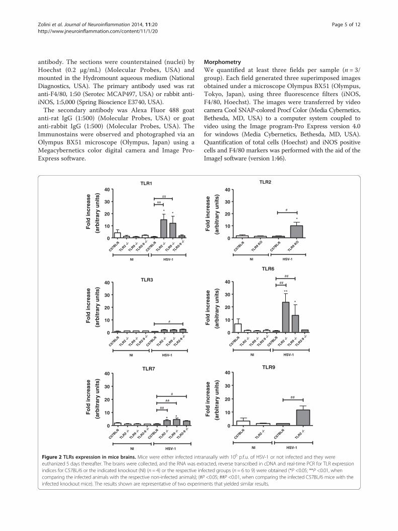
Figure 2 TLRs expression in mice brains. Mice were either infected intraeuthanized 5 days thereafter. The brains were collected, and the RNA was extindices for C57BL/6 or the indicated knockout (NI) (n = 4) or the respective infcomparing the infected animals with the respective non-infected animals); (#Pinfected knockout mice). The results shown are representative of two experim
MorphometryWe quantified at least three fields per sample (n = 3/group). Each field generated three superimposed imagesobtained under a microscope Olympus BX51 (Olympus,Tokyo, Japan), using three fluorescence filters (iNOS,F4/80, Hoechst). The images were transferred by videocamera Cool SNAP-colored Procf Color (Media Cybernetics,Bethesda, MD, USA) to a computer system coupled tovideo using the Image program-Pro Express version 4.0for windows (Media Cybernetics, Bethesda, MD, USA).Quantification of total cells (Hoechst) and iNOS positivecells and F4/80 markers was performed with the aid of theImageJ software (version 1:46).
TLR2
C57BL/6
TLR9 KO
C57BL/6
TLR9 KO
0
10
20
30
40
NI HSV-1
#
*
Fo
ld in
crea
se
(arb
itra
ry u
nit
s)
TLR6
C57BL/6
TLR2 -/-
TLR9 -/-
TLR2-9 -
/-
C57BL/6
TLR2 -/-
TLR9 -/-
TLR2-9 -
/-0
10
20
30
40
NI HSV-1
##
##
**
*
Fo
ld in
crea
se
(arb
itra
ry u
nit
s)
TLR9
C57BL/6
TLR2 -/-
C57BL/6
TLR2 -/-
0
10
20
30
40
NI HSV-1
##
Fo
ld in
crea
se
(arb
itra
ry u
nit
s)
nasally with 106 p.f.u. of HSV-1 or not infected and they wereracted, reverse transcribed in cDNA and real-time PCR for TLR expressionected groups (n = 6 to 9) were obtained (*P <0.05; **P <0.01, when<0.05; ##P <0.01, when comparing the infected C57BL/6 mice with theents that yielded similar results.
Zolini et al. Journal of Neuroinflammation 2014, 11:20 Page 6 of 12http://www.jneuroinflammation.com/content/11/1/20
Statistical analysisThe analyses were performed using the GraphPad Prism5 software for Windows (GraphPad Software, Inc., LaJolla, CA, USA). The sample groups were assessed bynon-parametric or parametric tests, according to theKolmogorov-Smirnov normality. Real-time PCR resultswere statistically analyzed using Mann–Whitney non-parametric T-tests. Macrophage results were analyzed byunpaired T-tests. For the survival curves, the statisticalanalyses were performed using the log-rank values.
Agp91
C57BL/6
TLR2 -/-
TLR9 -/-
TLR2-9 -
/-
C57BL/6
TLR2 -/-
TLR9 -/-
TLR2-9 -
/-0
20
40
60
80*
* *
*
NI HSV-1
Fo
ld in
crea
se(a
rbit
rary
un
its)
iNOS
C57BL/6
TLR2 -/-
TLR9 -/-
TLR2-9 -
/-
C57BL/6
TLR2 -/-
TLR9 -/-
TLR2-9 -
/-1
10100
100010000
1.0×1005
1.0×1006
1.0×1007
1.0×1008
1.0×1009
*
* * *
##
##
##
NI HSV-1
Fo
ld in
crea
se(a
rbit
rary
un
its)
C
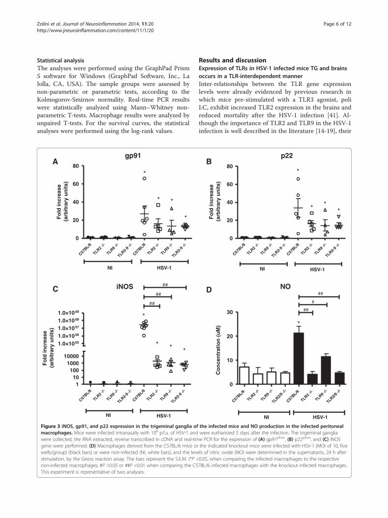
Figure 3 iNOS, gp91, and p22 expression in the trigeminal ganglia ofmacrophages. Mice were infected intranasally with 106 p.f.u. of HSV-1 andwere collected, the RNA extracted, reverse transcribed in cDNA and real-timgene were performed. (D) Macrophages derived from the C57BL/6 mice orwells/group) (black bars) or were non-infected (NI, white bars), and the levstimulation, by the Griess reaction assay. The bars represent the S.E.M. (*P <non-infected macrophages; #P <0.05 or ##P <0.01 when comparing the C5This experiment is representative of two analyses.
Results and discussionExpression of TLRs in HSV-1 infected mice TG and brainsoccurs in a TLR-interdependent mannerInter-relationships between the TLR gene expressionlevels were already evidenced by previous research inwhich mice pre-stimulated with a TLR3 agonist, poliI:C, exhibit increased TLR2 expression in the brains andreduced mortality after the HSV-1 infection [41]. Al-though the importance of TLR2 and TLR9 in the HSV-1infection is well described in the literature [14-19], their
B
C57BL/6
TLR2 -/-
TLR9 -/-
TLR2/9 -/
-
C57BL/6
TLR2 -/-
TLR9 -/-
TLR2/9 -/
-0
10
20
30
*
##
#
##
*
HSV-1NI
NO
Co
nce
ntr
atio
n (
uM
)
p22
C57BL/6
TLR2 -/-
TLR9 -/-
TLR2-9 -
/-
C57BL/6
TLR2 -/-
TLR9 -/-
TLR2-9 -
/-0
20
40
60
80*
* **
NI HSV-1
Fo
ld in
crea
se(a
rbit
rary
un
its)
D
the infected mice and NO production in the infected peritonealwere euthanized 5 days after the infection. The trigeminal gangliae PCR for the expression of (A) gp91phox, (B) p22phox, and (C) iNOSthe indicated knockout mice were infected with HSV-1 (MOI of 10, five
els of nitric oxide (NO) were determined in the supernatants, 24 h after0.05, when comparing the infected macrophages to the respective7BL/6 infected macrophages with the knockout infected macrophages.
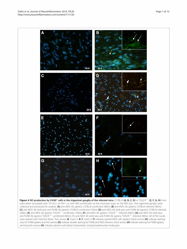
Figure 4 NO production by F4/80+ cells in the trigeminal ganglia of the infected mice. C57BL/6 (A, B, C, D) or TLR2/9−/− (E, F, G, H) micewere either inoculated with 106 p.f.u. of HSV-1 or with PBS (uninfected) via the intranasal route; on the fifth d.p.i., the trigeminal ganglia werecollected and processed for analysis. (A) anti-iNOS Ab (green), C57BL/6 uninfected (400×); (B) anti-iNOS Ab (green), C57BL/6 infected (400×);(C) anti-iNOS Ab (red) plus anti-F4/80 Ab (green), C57BL/6 uninfected (100×); (D) anti-iNOS Ab (red) plus anti-F4/80 Ab (green), C57BL/6 infected,(200×); (E) anti-iNOS Ab (green), TLR2/9−/− uninfected, (100×); (F) anti-iNOS Ab (green), TLR2/9−/− infected (400×); (G) anti-iNOS Ab (red) plusanti-F4/80 Ab (green), TLR2/9−/− uninfected (400×); (H) anti-iNOS Ab (red) plus anti-F4/80 Ab (green), TLR2/9−/− infected (400×). All of the nucleiwere stained with Hoechst (blue). Thin arrows (B, insert in B, F, insert in F) indicate stained iNOS cells (green); block arrows (D) indicate stainingonly for F4/80 (green) and thin arrows (D) indicate double staining for F4/80 and iNOS (brown). Short arrows (H) indicate staining for F4/80 (green),and long thin arrows (H) indicate stained nuclei (blue) characteristic of polymorphonuclear leukocytes.
Zolini et al. Journal of Neuroinflammation 2014, 11:20 Page 7 of 12http://www.jneuroinflammation.com/content/11/1/20
Zolini et al. Journal of Neuroinflammation 2014, 11:20 Page 8 of 12http://www.jneuroinflammation.com/content/11/1/20
inter-relationship with other TLRs has been poorly ex-plored. To investigate this inter-relationship, we ana-lyzed the expression of several TLRs in trigeminalganglia (TG) and brains of WT and knockout mice forTLR2, TLR9, and TLR2/9, after infection with HSV-1,to determine whether the deaths of TLR2/9−/− mice [19]after HSV-1 infection occur exclusively because of theabsence of these specific TLRs or whether their absencecould interfere with the expression and/or recognitionthrough other TLRs.WT, TLR2−/−, TLR9−/−, and TLR2/9−/− mice were intra-
nasally infected with 106 p.f.u. of HSV-1 [19] and were eu-thanized on the fifth day post infection (day of the TGviral peak) [19], and the TG and brains were collected toevaluate the TLR expression by real-time PCR.Data of the TLR expression in the WT infected mice re-
vealed that TLRs are highly expressed in the TG (Figure 1),but there was no increase in TLR expression in the brain(Figure 2), thereby demonstrating that the innate immun-ity against HSV-1 in this model occurs preferentially inthe TG. In vitro assays from another research group [42]have revealed the synergic action of IFN gamma and IFNalpha/beta in controlling HSV-1 infection. These cyto-kines genes are activated by phosphorylation cascadesthat, in turn, are initiated by different TLRs, and this syn-ergistic action impairs viral replication.The remaining results of this assay (Figures 1 and 2) in-
dicate that TLRs 1, 2, 3, 6, 7, and 9 are expressed in theTG of WT mice infected with HSV-1. Expression of TLR1,TLR2 (except for the TLR2−/− and TLR2/9−/− mice), TLR3,TLR6, TLR7, and TLR9 (except for the TLR9−/− and TLR2/9−/− mice) in the TG (Figure 1), was generally dimin-ished in TLR knockout mice compared with WT mice.These results suggest an interaction of different TLRsfor immune-response orchestration in the TG of WTmice. In the knockout mouse brains (Figure 2), therewas an increase or a tendency toward an increase in
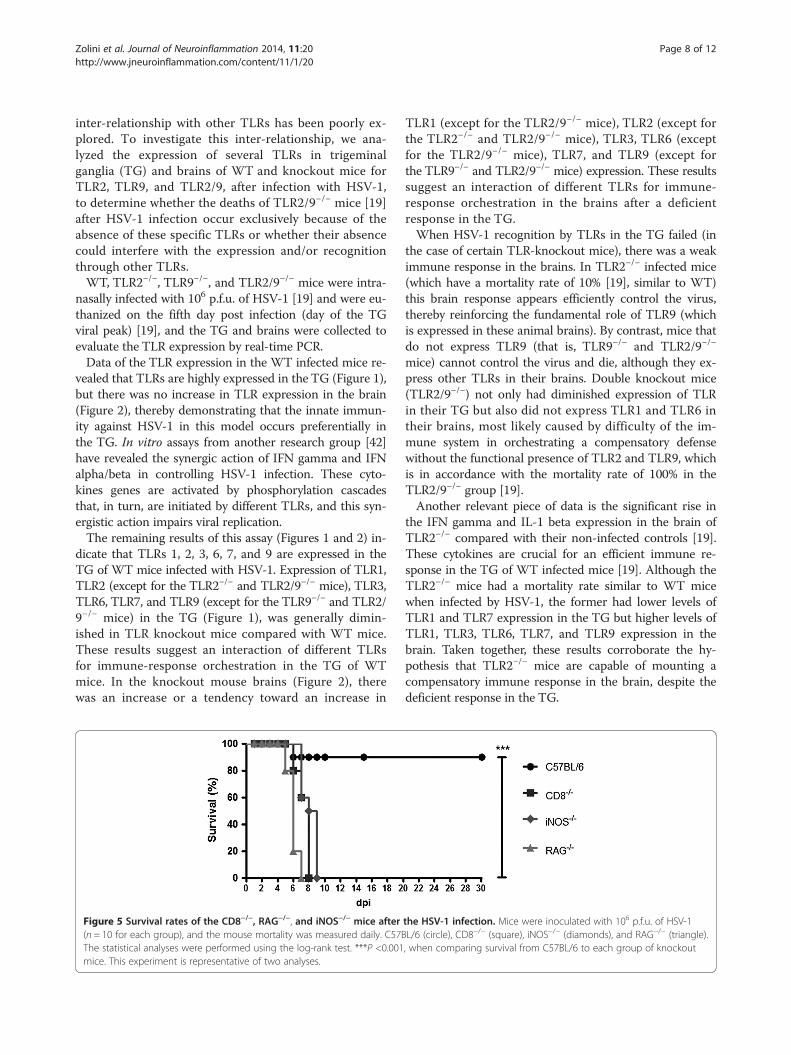
Figure 5 Survival rates of the CD8−/−, RAG−/−, and iNOS−/− mice after(n = 10 for each group), and the mouse mortality was measured daily. C57BThe statistical analyses were performed using the log-rank test. ***P <0.001mice. This experiment is representative of two analyses.
TLR1 (except for the TLR2/9−/− mice), TLR2 (except forthe TLR2−/− and TLR2/9−/− mice), TLR3, TLR6 (exceptfor the TLR2/9−/− mice), TLR7, and TLR9 (except forthe TLR9−/− and TLR2/9−/− mice) expression. These resultssuggest an interaction of different TLRs for immune-response orchestration in the brains after a deficientresponse in the TG.When HSV-1 recognition by TLRs in the TG failed (in
the case of certain TLR-knockout mice), there was a weakimmune response in the brains. In TLR2−/− infected mice(which have a mortality rate of 10% [19], similar to WT)this brain response appears efficiently control the virus,thereby reinforcing the fundamental role of TLR9 (whichis expressed in these animal brains). By contrast, mice thatdo not express TLR9 (that is, TLR9−/− and TLR2/9−/−
mice) cannot control the virus and die, although they ex-press other TLRs in their brains. Double knockout mice(TLR2/9−/−) not only had diminished expression of TLRin their TG but also did not express TLR1 and TLR6 intheir brains, most likely caused by difficulty of the im-mune system in orchestrating a compensatory defensewithout the functional presence of TLR2 and TLR9, whichis in accordance with the mortality rate of 100% in theTLR2/9−/− group [19].Another relevant piece of data is the significant rise in
the IFN gamma and IL-1 beta expression in the brain ofTLR2−/− compared with their non-infected controls [19].These cytokines are crucial for an efficient immune re-sponse in the TG of WT infected mice [19]. Although theTLR2−/− mice had a mortality rate similar to WT micewhen infected by HSV-1, the former had lower levels ofTLR1 and TLR7 expression in the TG but higher levels ofTLR1, TLR3, TLR6, TLR7, and TLR9 expression in thebrain. Taken together, these results corroborate the hy-pothesis that TLR2−/− mice are capable of mounting acompensatory immune response in the brain, despite thedeficient response in the TG.
the HSV-1 infection. Mice were inoculated with 106 p.f.u. of HSV-1L/6 (circle), CD8−/− (square), iNOS−/− (diamonds), and RAG−/− (triangle)., when comparing survival from C57BL/6 to each group of knockout
VP-16
C57BL/6
RAG -/-
iNOS -/
-
C57BL/6
RAG -/-
iNOS -/
-0
500
1000
1500
2000
100000
200000
300000
400000 *
**
NI HSV-1
Fo
ld in
crea
se(a
rbit
rary
un
its)
MCP-1
C57BL/6
RAG -/-
iNOS -/
-
C57BL/6
RAG -/-
iNOS -/
-0
50100150200500
1000
1500
2000
2500
3000
*
*
#
##
NI HSV-1
Fo
ld in
crea
se(a
rbit
rary
un
its)
iNOS
C57BL/6
RAG -/-
C57BL/6
RAG -/-
0
100
200
300
400
500
**
#
NI HSV-1
Fo
ld in
crea
se(a
rbit
rary
un
its)
IP-10
C57BL/6
RAG -/-
iNOS -/
-
C57BL/6
RAG -/-
iNOS -/
-0
100200300500
100015002000
400060008000
10000 *
##
**
NI HSV-1
Fo
ld in
crea
se(a
rbit
rary
un
its)
TNF
C57BL/6
RAG -:-
iNOS -:
-
C57BL/6
RAG -:-
iNOS -:
-0
250
500
750
1000
1250
1500
NI HSV-1
*
Fo
ld in
crea
se(a
rbit
rary
un
its)
Rantes
C57BL/6
RAG -/-
iNOS -/
-
C57BL/6
RAG -/-
iNOS -/
-0
10
20
30
40
500
1000
1500
2000
NI HSV-1
*
##
Fo
ld in
crea
se(a
rbit
rary
un
its)
Figure 6 (See legend on next page.)
Zolini et al. Journal of Neuroinflammation 2014, 11:20 Page 9 of 12http://www.jneuroinflammation.com/content/11/1/20
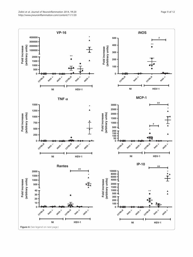
(See figure on previous page.)Figure 6 Virus transcript, inos and cytokine expression in the trigeminal ganglia of the RAG−/− and iNOS−/− mice. The C57BL/6, RAG−/−,and iNOS−/− mice were inoculated either with 106 p.f.u. of HSV-1 or with PBS (uninfected) by the intranasal route; on the fifth d.p.i., the trigeminalganglia were collected and processed for analysis to verify the expression of the viral gene VP-16 and the inos, tnf alpha, mcp-1, rantes, and ip-10genes. *P <0.05; P <0.01, when comparing the infected mice with the respective non-infected mice. #P <0.05; ##P <0.01, when comparing theC57BL/6 infected mice with the indicated infected knockout mice. The displayed results are representative of two experiments that yieldedsimilar findings.
Zolini et al. Journal of Neuroinflammation 2014, 11:20 Page 10 of 12http://www.jneuroinflammation.com/content/11/1/20
The expression of iNOS and NO production areTLR-dependent during HSV-1 infectionOur previous experiments indicated the importance ofmacrophages, CD8+ T lymphocytes and IFN gamma [19]for HSV-1 infection control in the murine model of intra-nasal infection. IFN gamma production by CD8+ T lym-phocytes could be fundamental to macrophage activationin trigeminal ganglia in these conditions, increasing the re-active oxygen species (ROS) and reactive nitrogen species(RNS) production by this type of cell [26]. Subsequently, weinvestigated which mechanisms of monocytes/macrophagesresponses are involved in HSV-1 control.To this end, the TG of WT and knockout mice were
collected 5 days after intranasal infection, and the iNOS,gp91, and p22 expression was evaluated by real-timePCR. iNOS is the enzyme responsible for NO produc-tion and, consequently, RNS [27]. gp91 phox and p22 phox
are NADPH oxidase subunits that are membrane-boundprotein components of flavocytochrome b558, which isresponsible for electron transport [43,44] and ROSproduction.The infected-mouse groups displayed a significant in-
crease in gp91phox and p22phox compared with the respect-ive non-infected controls (Figure 3A and B, respectively).Furthermore although the WT infected mice expressedthese genes with a tendency toward higher levels than inthe knockout mice, the difference between them was notstatistically significant. iNOS expression was increased inWT and knockout infected compared with non-infectedmice, with a more pronounced expression in the WT micethan in the other groups (Figure 3C). iNOS effectivelycontributes to the HSV-1 defense, and our results indicatethat TLRs are important for iNOS activation/production,while ROS apparently do not play a relevant role in thismodel’s TLR-dependent response.To confirm the TLR importance for NO production,
peritoneal macrophages of WT, TLR2−/−, TLR9−/−, andTLR2/9−/− mice were stimulated with HSV-1 (MOI = 10),and the NO production was evaluated by the Griess reac-tion (Figure 3D). The WT infected-mouse group pre-sented higher amounts of NO compared with the controlsand the knockout-infected mouse groups. This result cor-roborates publications that indicate the importance ofiNOS in the defense against HSV-1 and the ability of NOto inhibit HSV-1 replication in vitro [21,45,46]. Moreover,
this finding confirms the importance of TLR2 and TLR9in NO production.Additionally, immunofluorescence assays were per-
formed to visualize iNOS production by F4/80+ cells inthe TG. In the single-staining assays, TG slices were in-cubated with antibodies against iNOS stained with AlexaFluor 488 (green) and were counterstained with Hoechst(blue) for nuclei visualization (Figures 4A, B, E, F). Inthe double-staining assays, TG slices were incubatedwith antibodies against F4/80 stained with Alexa Fluor488 (green) and antibodies against iNOS stained withAlexa Fluor 546 (red), and then counterstained withHoechst (Figures 4C, D, G, H). The panels in (Figure 4)display the details of the ganglia cell immunofluoresce.There was no iNOS production in the single-stained cells
of the non-infected mouse TG (Figure 4A and E - green) orin the double-stained cells (Figures 4C and G - red). TheWT infected mice (Figure 4B) had thin cytoplasmic prolon-gations stained in green (insert). Most of the WT mousecells (blue nuclei) expressed the enzyme (Figure 4B), andthere was a considerable superposition of F4/80 andiNOS in the double-stained cells (green + red = brown)(Figure 4D), indicating that monocytic cells are themain producers of iNOS in the TG. Some F4/80 positivecells did not express iNOS (thick arrows, Figure 4D);however, many cells were double stained (insert, arrow).In the TLR2/9−/− infected mice, the iNOS cell staining
was modest (Figure 4F - long arrows). Although the in-flammatory infiltration was conspicuous (Additional file 1:Figure S1 and Additional file 2: Commentary on the resultsand discussion in Additional file 1: Figure S1), the cellswere predominantly polymorphonucleated, with segmentednuclei, and a few macrophages were stained with F4/80(Figure 4H - long arrows). Few mononuclear F4/80 positivecells were visible in the TG slices from the TLR2/9−/− mice(Figure 4H - insert, arrows), and iNOS-stained cells wererare (Figure 4H - selection).Altogether, the TG immunofluorescence revealed a
greater iNOS production by F4/80 positive cells in theWT infected mice (Figure 4B and D), in comparison toTLR2/9−/− (Figure 4F and H), which have a high mortalityrate when infected by HSV-1 [19]. The morphometry(Additional file 3: Figure S2 and Additional file 2: Com-mentary on the results and discussion in Additional file 3:Figure S2) did not show statistically significant difference

Zolini et al. Journal of Neuroinflammation 2014, 11:20 Page 11 of 12http://www.jneuroinflammation.com/content/11/1/20
in the production of iNOS, in the amount of macrophagesnor in the production of iNOS by macrophages betweeninfected C57BL/6 and infected TLR2/9−/− mice. However,there was a clear trend towards higher levels of iNOS, moremacrophages, and more iNOS producer macrophages ininfected C57BL/6 mice than in infected TLR2/9−/− mice.We therefore believe that this trend is biologically relevant.
Macrophages and lymphocytes are essential forcontrolling HSV-1 infectionTo confirm the importance of lymphocytes and macro-phages against the HSV-1 infection in the experimentalmodel of herpetic encephalitis, RAG−/−, iNOS−/−, CD8−/−,and WT mice (n = 10 from each group) were intranasallyinfected with 106 p.f.u. of HSV-1 [19] to evaluate the mor-tality (Figure 5), and the control mice aspirated only PBS.The animals were checked daily for encephalitis signs suchas prostration, ruffled fur, limb paralysis, and hunched pos-ture, and mice showing these signs were euthanized.All of the control mice survived, and the WT mice ex-
hibited 90% survival (Figure 5); no CD8−/−, RAG−/−, oriNOS−/− mice survived (Figure 5). These results indicatethat not only T and B lymphocytes [15,19] but also iNOSproduction is essential to control the viral infection, in ac-cordance with the results reported by other researchers[21] who have demonstrated the importance of NO forpreventing HSV-1 replication in the TG.Additionally, the WT, RAG−/− and iNOS−/− mice’s
cytokine and viral transcript expression was evaluated inthe TG by real-time PCR, 5 days after the infection. Theviral transcripts were similarly expressed in all of the in-fected mice, but the RAG−/− mice were nearly irrespon-sive to the virus in terms of their chemokines, effectorcytokine (TNF alpha) and iNOS expression (Figure 6).The chemokines MCP-1, Rantes, and IP-10, which at-tracts immune cells to the infection site, as well as theeffector cytokine TNF alpha, were over-expressed in theiNOS−/− infected mice compared with the WT mice. Be-cause all of the infected iNOS−/− mice died (Figure 5),we can assume that, although murine cells can use che-mokines to recruit immune cells to the infection site,these immune cells fail to combat the virus because theyare not able to produce nitric oxide. In the most likelycompensatory mechanism, the cells produce more che-mokines to attract additional immune cells, which pro-duce more TNF alpha; however, this process is notsufficient to control the HSV-1 infection.In summary, we conclude that, subsequent to infection
by HSV-1, TLRs efficiently organize the innate immunecells, thereby eliciting a CD8+ T cell [19] and macrophageresponse (with NO production by the macrophages) tocontrol the HSV-1 infection in the mouse TG, therebypreventing severe disease outcomes, such as encephalitisand death.
Additional files
Additional file 1: Figure S1. The animals were intranasally inoculatedwith 106 p.f.u. of HSV-1 or PBS (control). On the fifth d.p.i., the trigeminalganglia were collected, and representative sections were processed forhistological analysis (H&E stained sections). Uninfected (A) and infectedC57BL/6 mice (B); uninfected (C) and infected TLR2−/− mice (D); uninfected(E) and infected TLR9−/− mice (F); uninfected (G) and infected TLR2/9−/− mice(H). The architecture and cellularity appeared normal in the uninfectedgroups. All of the infected animals displayed inflammatory characteristics,with increased cellularity and histologically evident vascular phenomena(edema) and inflammatory changes. The higher intensity of diffuseinfiltration by polymorphonuclear cells was accompanied by degenerativeneuronal phenomena in the TLR9−/− (F) and TLR2/9−/− (H) mouse groups,compared with the other groups of mice. The lower intensity of inflammationoccurred in the C57BL/6 mouse group (B). Original magnifications, 100×;inserts 400 × .
Additional file 2: Supplementary Material. Supplementary statisticsdata from Figures 1 and 2. Commentary on the results and discussion inAdditional files 1 and 3: Figure S1 and S2.
Additional file 3: Figure S2. Morphometry. For quantification of theimmunostained cells, C57BL/6 (n = 5) or TLR2/9−/− (n = 5) mice wereinfected and after 5 days they were euthanized, the trigeminal gangliawere collected and processed for analysis and graphic comparison of thefluorescence, as stated in materials and methods. (A) anti-iNOS Ab; (B)anti-F4/80 Ab; (C) anti-iNOS plus anti-F4/80 Abs.
Competing interestsThe authors declare that they have no competing interests.
Authors’ contributionsGPZ, GKL, RMEA, EGK, and MAC conceived and designed the experiments.GPZ, GKL, RMEA, NL, MGAS, MFD, NLP, BPC, and CTC performed theexperiments. GPZ and GKL performed the statistical analyses. GPZ, GKL,RMEA, EGK, and MAC analyzed the data. RMEA, EGK and MAC contributedreagents, materials, and analysis tools. GPZ, GKL, RMEA, EGK, and MAC wrotethe paper. All the authors have read and approved the final version of themanuscript.
AcknowledgementsWork supported by the Fundação de Amparo à Pesquisa do Estado de MinasGerais (FAPEMIG, Brazil, to MAC and EGK), the Conselho Nacional deDesenvolvimento Científico e Tecnológico (CNPq, Brazil, to MAC and EGK),Instituto Nacional de Ciência e Tecnologia de Vacinas/CNPq/FAPEMIG(INCTV/CNPq/FAPEMIG; Brazil, to MAC), Coordenação de Aperfeiçoamento dePessoal de Nível Superior (CAPES, Brazil), and the Programa Estratégico dePesquisa em Saúde V/VI (PAPES)/FIOCRUZ/CNPq (Brazil, to MAC). EGK, MAC,and RMEA are Fellows from CNPq. We thank the Program for TechnologicalDevelopment in Tools for Health-PDTIS-FIOCRUZ for use of its facilities andRicardo Tostes Gazzinelli for analysis tools.
Author details1Laboratório de Imunopatologia, Imunologia de Doenças Virais, Centro dePesquisas René Rachou, Fundação Oswaldo Cruz, Fiocruz, Av. Augusto deLima 1715, Belo Horizonte, Minas Gerais 30190-002, Brazil. 2Escola deVeterinária, Universidade Federal de Minas Gerais, Belo Horizonte, MinasGerais, Brazil. 3Departamento de Patologia, Universidade Federal de MinasGerais, Belo Horizonte, Minas Gerais, Brazil. 4Departamento de Microbiologia,Universidade Federal de Minas Gerais, Belo Horizonte, Minas Gerais, Brazil.
Received: 17 June 2013 Accepted: 14 January 2014Published: 30 January 2014
References1. Roizman B, Knipe DM, Whitley RJ: Herpes Simplex Viruses. In Fields Virology.
5th edition. Edited by Knipe DM, Howley RM. Pennsylvania, PA: LippincottWilliams & Wilkins; 2007:2502–2601.
Zolini et al. Journal of Neuroinflammation 2014, 11:20 Page 12 of 12http://www.jneuroinflammation.com/content/11/1/20
2. Davison AJ, Eberle R, Ehlers B, Hayward GS, McGeoch DJ, Minson AC, Pellett PE,Roizman B, Studdert MJ, Thiry E: The order Herpesvirales. Arch Virol 2009,154:171–177.
3. Schmutzhard E: Viral infections of the CNS with special emphasis onherpes simplex infections. J Neurol 2001, 248:469–477.
4. Arduino PG, Porter SR: Herpes simplex virus type 1 infection: overview onrelevant clinico-pathological features. J Oral Pathol Med 2008, 37:107–121.
5. Preston CM: Repression of viral transcription during herpes simplex viruslatency. J Gen Virol 2000, 81:1–19.
6. Cunningham AL, Diefenbach RJ, Miranda-Saksena M, Bosnjak L, Kim M,Jones C, Douglas MW: The cycle of human herpes simplex virus infection:virus transport and immune control. J Infect Dis 2006, 194:S11–S18.
7. Hüfner K, Derfuss T, Herberger S, Sunami K, Russell S, Sinicina I, Arbusow V,Strupp M, Brandt T, Theil D: Latency of alpha herpes viruses is accompaniedby a chronic inflammation in human trigeminal ganglia but not in dorsalroot ganglia. J Neuropathol Exp Neurol 2006, 65:1022–1030.
8. Conrady CD, Drevets DA, Carr DJJ: Herpes simplex type I (HSV-1) infectionof the nervous system: is an immune response a good thing?J Neuroimmunol 2010, 220:1–9.
9. Halford WP, Gebhardt BM, Carr DJJ: Mechanisms of herpes virus type 1reactivation. J Virol 1996, 70:5051–5060.
10. Bloom DC, Giordani NV, Kwiatkowski DL: Epigenetic regulation of latentHSV-1 gene expression. Biochim Biophys Acta 2010, 1799:246–256.
11. Schiffer JT, Abu-Raddad L, Mark KE, Zhu J, Selke S, Magaret A, Wald A, Corey L:Frequent release of Low amounts of herpes simplex virus from neurons:results of a mathematical model. Sci Transl Med 2009, 1:1–9.
12. Aravalli RN, Peterson PK, Lokensgard JR: Toll-like receptors in defense anddamage of the central nervous system. J Neuroimmune Pharmacol 2007,2:297–312.
13. Takeda K, Kaisho T, Akira S: Toll-like receptors. Annu Rev Immunol 2003,21:335–376.
14. Kurt-Jones EA, Chan M, Zhou S, Wang J, Reed G, Bronson R, Arnold MM,Knipe DM, Finberg RW: Herpes simplex virus 1 interaction with Toll-likereceptor 2 contributes to lethal encephalitis. Proc Natl Acad Sci USA 2004,101:1315–1320.
15. Mansur DS, Kroon EG, Nogueira ML, Arantes RME, Rodrigues SCO, Akira S,Gazzinelli RT, Campos MA: Lethal encephalitis in myeloid differentiationfactor 88-deficient mice infected with herpes simples virus 1. Am J Pathol2005, 166:1419–1426.
16. Herbst-Kralovetz MM, Pyles RB: Toll-like receptors, innate immunity andHSV pathogenesis. Herpes 2006, 13:37–41.
17. Krug A, Luker GD, Barchet W, Leib DA, Akira S, Colonna M: Herpes simplexvirus type 1 activates murine natural interferon-producing cells throughtoll-like receptor 9. Blood 2004, 103:1433–1443.
18. Sato A, Iwasaki A: Induction of antiviral immunity requires toll-like receptorsignaling in both stromal and dendritic cell compartments. Proc Natl AcadSci USA 2004, 101:16274–16279.
19. Lima GK, Zolini GP, Mansur DS, Lima BH, Wischhoff U, Astigarraga RG, Dias MF,Das Gracas Almeida Silva M, Bela SR, Do Valle Antonelli LR, Arantes RM,Gazzinelli RT, Bafica A, Kroon EG, Campos MA: Toll-like receptor (TLR) 2 andTLR9 expressed in trigeminal ganglia are critical to viral control duringherpes simplex virus 1 infection. AJP 2010, 177:2433–2445.
20. Marques CP, Hu S, Sheng W, Lokensgard JR: Microglial cells initiatevigourous yet non-protective immune responses during HSV-1 braininfection. Virus Res 2006, 121:1–10.
21. Kodukula P, Liu T, Rooijen N, Jager MJ, Hendricks RL: Macrophage controlof herpes simplex virus type 1 replication in the peripheral nervoussystem. J Immunol 1999, 162:2895–2905.
22. Liu T, Tang Q, Hendricks RL: Inflammatory infiltration of the trigeminalganglion after herpes simplex virus type 1 infection. J Virol 1996,70:264–271.
23. Lint A, Ayers M, Brooks AG, Coles RM, Heath WR, Carbone FR: Herpessimplex virus-specific CD8+ T cells can clear established lytic infectionsfrom skin and nerves and can partially limit early spread of virus aftercutaneous inoculation. J Immunol 2004, 172:392–397.
24. Cook WJ, Kramer MF, Walker RM, Burwell TJ, Holman HA, Coen DM, Knipe DM:Persistent expression of chemokine and chemokine receptor RNAs atprimary and latent sites of herpes simplex 1 infection. Virol J 2004, 1:5.
25. Carr DJJ, Campbell IL: Herpes simplex virus type 1 induction ofchemokine production is unrelated to viral load in the cornea but not inthe nervous system. Viral Immunol 2006, 19:741–746.
26. Schroder K, Hertzog PJ, Ravasi T, Hume DA: Interferon-gamma: an overviewof signals, mechanisms and functions. J Leukoc Biol 2004, 75:163–189.
27. Flannagan RS, Cosío G, Grinstein S: Antimicrobial mechanisms of phagocytesand bacterial evasion strategies. Nat Rev Microbiol 2009, 7:355–366.
28. Lima GK: Cinética De Multiplicação Viral e Análise da Expressão de IFN gama eQuimiocinas em Infecção Murina por Vírus Herpes Simplex 1, MSc dissertation.Belo Horizonte: Universidade Federal de Minas Gerais, Instituto de CiênciasBiológicas; 2007.
29. Marques CP, Cheeran MC, Palmquist JM, Hu S, Lokensgard JR: Microglia arethe major cellular source of inducible nitric oxide synthase duringexperimental herpes encephalitis. J Neurovirol 2008, 14:229–238.
30. Mansur DS: O Papel dos Receptores do Tipo Toll na Infecção Pelo HSV-1, PhDthesis. Belo Horizonte: Universidade Federal de Minas Gerais, Instituto deCiências Biológicas; 2007.
31. Lima GK: TLR2 e TLR9: Receptores Essenciais Para o Controle da infecção porvírus Herpes Simplex do Tipo 1, PhD thesis. Belo Horizonte: UniversidadeFederal de Minas Gerais, Instituto de Ciências Biológicas; 2010.
32. Nogueira ML, Siqueira RC, Freitas N, Amorim JB, Bonjardim CA, Ferreira PC,Orefice F, Kroon EG: Detection of herpesvirus DNA by the polymerasechain reaction (PCR) in vitreous samples from patients with necrotisingretinitis. J Clin Pathol 2001, 54:103–106.
33. Campos MA, Kroon EG: Critical period of irreversible block of Vacciniavirus replication. Rev Bras Microbiol 1993, 24:104–110.
34. Boivin G, Coulombe Z, Rivest S: Intranasal herpes simplex virus type 2inoculation causes a profound thymidine kinase dependent cerebralinflammatory response in the mouse hindbrain. Eur J Neurosci 2002, 16:29–43.
35. Drapier JC, Wietzerbin J, Hibbs JB Jr: Interferon-gamma andtumor necrosisfactor induce the L-arginine-dependent cytotoxic effector mechanism inmurine macrophages. Eur J Immunol 1988, 18:1587–1592.
36. Giulietti A, Overbergh L, Valckx D, Decallonne B, Bouillon R, Mathieu C: Anoverview of real-time quantitative PCR: applications to quantify cytokinegene expression. Methods 2001, 25:386–401.
37. Broberg EK, Nygardas M, Salmi AA, Hukkanen V: Low copy numberdetection of herpes simplex virus type 1 mRNA and mouse Th1 typecytokine mRNAs by light cycler quantitative real time PCR. J VirolMethods 2003, 112:53–65.
38. Kuhlicke J, Frick JS, Morote-Garcia JC, Rosenberger P, Eltzschig HK: Hypoxiainducible factor (HIF)-1 coordinates induction of toll-like receptors TLR2and TLR6 during hypoxia. PLoS One 2007, 12:e1364.
39. Livak KJ, Schmittgen TD: Analysis of relative gene expression data usingreal-time quantitative PCR and the 2(−Delta Delta C(T)) Method. Methods2001, 25:402–408.
40. Préhaud C, Mégret F, Lafage M, Lafon M: Virus infection switches TLR3positive human neurons to become strong producers of beta interferon.J Virol 2005, 79:12893–12904.
41. Boivin N, Sergerie Y, Rivest S, Boivin G: Effect of pretreatment with Toll-likereceptor agonists in a mouse model of herpes simplex virus type 1encephalitis. JID 2008, 198:664–672.
42. Pierce AT, DeSalvo J, Foster TP, Kosinski A, Weller SK, Halford WP: Beta interferon andgamma interferon synergize to block viral DNA and virion synthesis in herpessimplex virus-infected cells. J Gen Virol 2005, 86:2421–2432.
43. Sarangi PP, Kim B, Kurt-Jones E, Rouse BT: Innate recognition network drivingherpes simplex virus-induced corneal immunopathology: role of the tollpathway in early inflammatory events in stromal keratitis. J Virol 2007,81:11128–11138.
44. Yu L, Quinn MT, Cross AR, Dinauer MC: Gp91(phox) is the heme bindingsubunit of the superoxide-generating NADPH oxidase. Proc Natl Acad Sci1998, 95:7993–7998.
45. Karupiah G, Xie Q, Buller RML, Nathan C, Duarte C, MacMicking JD:Inhibition of viral replication by interferon-gamma-induced nitric oxidesynthase. Science 1993, 261:1445–1448.
46. Karupiah G, Harris N: Inhibition of viral replication by nitric oxide and itsreversal by ferrous sulfate and tricarboxylic acid cycle metabolites. J ExpMed 1995, 181:2171–2179.
doi:10.1186/1742-2094-11-20Cite this article as: Zolini et al.: Defense against HSV-1 in a murinemodel is mediated by iNOS and orchestrated by the activation of TLR2and TLR9 in trigeminal ganglia. Journal of Neuroinflammation 2014 11:20.
Copyright © 2022 FDOKUMEN


![SAR of a series of anti-HSV-1 acridone derivatives, and a rational acridone-based design of a new anti-HSV-1 3 H-benzo[ b]pyrazolo[3,4- h]-1,6-naphthyridine series](https://static.fdokumen.com/doc/165x107/631beba7665120b3330b99e5/sar-of-a-series-of-anti-hsv-1-acridone-derivatives-and-a-rational-acridone-based.jpg)