A natural tetranortriterpenoid with immunomodulating properties as a potential anti-HSV agent
9
This article appeared in a journal published by Elsevier. The attached copy is furnished to the author for internal non-commercial research and education use, including for instruction at the authors institution and sharing with colleagues. Other uses, including reproduction and distribution, or selling or licensing copies, or posting to personal, institutional or third party websites are prohibited. In most cases authors are permitted to post their version of the article (e.g. in Word or Tex form) to their personal website or institutional repository. Authors requiring further information regarding Elsevier’s archiving and manuscript policies are encouraged to visit: http://www.elsevier.com/copyright
-
Upload
independent -
Category
Documents
-
view
6 -
download
0
Transcript of A natural tetranortriterpenoid with immunomodulating properties as a potential anti-HSV agent
This article appeared in a journal published by Elsevier. The attachedcopy is furnished to the author for internal non-commercial researchand education use, including for instruction at the authors institution
and sharing with colleagues.
Other uses, including reproduction and distribution, or selling orlicensing copies, or posting to personal, institutional or third party
websites are prohibited.
In most cases authors are permitted to post their version of thearticle (e.g. in Word or Tex form) to their personal website orinstitutional repository. Authors requiring further information
regarding Elsevier’s archiving and manuscript policies areencouraged to visit:
http://www.elsevier.com/copyright
Author's personal copy
Virus Research 141 (2009) 47–54
Contents lists available at ScienceDirect
Virus Research
journa l homepage: www.e lsev ier .com/ locate /v i rusres
A natural tetranortriterpenoid with immunomodulating propertiesas a potential anti-HSV agent
Carlos A. Buenoa, Andrea A. Barqueroa, Hernán Di Cónsoli a, Marta S. Maier b, Laura E. Alchéa,∗
a Laboratorio de Virología, Departamento de Química Biológica, Facultad de Ciencias Exactas y Naturales, Universidad de Buenos Aires,Ciudad Universitaria, Buenos Aires, Argentinab UMYMFOR (CONICET-UBA) y Departamento de Química Orgánica, Facultad de Ciencias Exactas y Naturales, Universidad de Buenos Aires,Ciudad Universitaria, Buenos Aires, Argentina
a r t i c l e i n f o
Article history:Received 29 September 2008Received in revised form 17 December 2008Accepted 19 December 2008Available online 20 January 2009
Keywords:AntiviralImmunomodulatoryNF-�BCytokineHSVMedicinal plants
a b s t r a c t
Meliacine (MA), an antiviral principle present in partially purified leaf extracts of Melia azedarach L.,prevents the development of herpetic stromal keratitis (HSK) in mice by diminishing the viral load in theeye and the severity of lesions caused by a virus-induced immunopathological reaction.
The tetranortriterpenoid 1-cinnamoyl-3,11-dihydroxymeliacarpin (CDM), obtained from MA purifica-tion, displays anti-herpetic activity and impedes nuclear factor �B (NF-�B) activation in HSV-1 infectedconjunctival cells.
To extend our understanding about CDM biological properties, we investigated its anti-HSV-1 activityas well as the effect on NF-�B activation and cytokine secretion induced by viral (HSV-1) and no-viral(LPS) stimuli, in corneal cells and macrophages.
CDM exerted a potent anti-HSV-1 effect on corneal cells and inhibited NF-�B translocation to thenucleus, leading to a decrease in IL-6 production. Besides, CDM seemed to modulate IL-6 and TNF-�responses in macrophages, whether they were infected with HSV-1 or stimulated with LPS. However,CDM did not affect NF-�B activation in these cells, suggesting that an alternative NF-�B cell signalingpathway would be involved in the modulation of cytokine production.
We conclude that, in addition to its antiviral effect, CDM would be acting as an immunomodulatingcompound which would be responsible for the improvement of murine HSK already reported.
© 2008 Elsevier B.V. All rights reserved.
1. Introduction
A great variety of ethnomedicinal plants are being studied asa source of natural products useful in the development of noveldrugs. It has been established that many of them inhibit severalsteps of the viral replication cycle of many DNA and/or RNA viruses(Chattopadhyay and Naik, 2007).
We have reported that meliacine (MA), an antiviral principlepresent in partially purified leaf extracts of Melia azedarach L.,exerts an antiviral action on the development of herpetic stro-mal keratitis (HSK) in mice by causing a significant decrease inthe viral load in the eye of Herpes simplex virus type 1 (HSV-1)infected animals, as well as in the incidence and severity of lesionsdue to a virus-induced immunopathological reaction (Pifarré et al.,
∗ Corresponding author at: Laboratorio de Virología, Depto. de Química Biológica,Facultad de Ciencias Exactas y Naturales, UBA, Pabellón II, 4to. piso, Ciudad Univer-sitaria, 1428 Buenos Aires, Argentina. Tel.: +54 11 4576/3334;fax: +54 11 4576/3342.
E-mail address: [email protected] (L.E. Alché).
2002). Bioassay guided purification of MA led to the isolation ofthe limonoid 1-cinnamoyl-3,11-dihydroxymeliacarpin (CDM) thatreduces both vesicular stomatitis virus (VSV) and HSV-1 multiplica-tion in Vero cells (Alché et al., 2003). We have found that a late stepin VSV and HSV-1 multiplication cycles is hindered by CDM sinceglycoproteins (g) B, gC and gD of HSV-1, as well as gG of VSV, areconfined to the Golgi apparatus when CDM is added after infection(Barquero et al., 2004, 2006).
HSV-1-induced ocular disease occurs as a result of a primaryinfection in the corneal epithelium and then, cells like macrophagesintervene in clearing the virus from the infected eye and inthe development of the immunologically driven stromal keratitis(Bauer et al., 2002; Biswas and Rouse, 2005). Besides, conjunctivalcells are also involved in amplifying the inflammatory processes inthe eye (Kase et al., 2004).
It has been shown that activation of nuclear factor �B (NF-�B) plays a pivotal role in triggering an immune inflammatoryresponse to a range of stimuli, including viral infections, suchas HIV-1, human T cell leukemia virus type 1, hepatitis B andinfluenza viruses. It has been reported that HSV-1 induces astrong nuclear translocation of NF-�B in human cell lines that
0168-1702/$ – see front matter © 2008 Elsevier B.V. All rights reserved.doi:10.1016/j.virusres.2008.12.013
Author's personal copy
48 C.A. Bueno et al. / Virus Research 141 (2009) 47–54
could have several functions: to promote viral replication, pre-vent virus-induced apoptosis, and mediate the immune responseto the invading pathogen (Gregory et al., 2004; Hiscott et al.,2001).
A great number of plant-derived substances, such as sesquiter-penes, di- and triterpenes, that prevent NF-�B activation, have beendescribed (Nam, 2006). We have found that CDM is able to blockHSV-1 induced activation of NF-�B by inhibiting its translocation tothe nucleus of infected human conjunctival cells (NHC), and postu-lated that CDM would be able to abolish murine HSK by controllingviral spread and the associated immunopathology as well (Barqueroet al., 2006).
The aim of the present study was to determine whether CDMdisplays an antiviral activity in infected corneal cells, the tar-get of HSV-1 multiplication in vivo, as well as its effect on thetranslocation of NF-�B to the nucleus. Since NF-�B plays a particu-larly important role as far as expression of cytokines is concerned(Santoro et al., 2003), we also evaluated the modulating effect ofCDM on the production of different cytokines in corneal cells andmacrophages.
2. Materials and methods
2.1. Cells and viruses
Human Corneal-Limbal Epithelial (HCLE) cells were kindly pro-vided by Dr Ilene K. Gipson and Dr. Pablo Argüeso (The SchepensEye Research Institute, Harvard Medical School, Boston, USA) andgrown in GIBCO Keratinocyte Serum Free Medium, supplementedwith 25 �g/ml bovine pituitary extract (BPE), 0.2 ng/ml epidermalgrowth factor (EGF), and 0.4 mM CaCl2, and maintained in low cal-cium DMEM/F12.
Murine macrophage cell line J774A.1 was kindly provided by Dr.Osvaldo Zabal (INTA–Castelar, Buenos Aires) and grown in RPMI1640 medium supplemented with 10% inactivated fetal bovineserum (FBS) (RPMI 10%) and maintained in RPMI supplementedwith 2% inactivated FBS (RPMI 2%).
Murine L929 cells and Vero cells were grown in Eagle’s minimalessential medium supplemented with 10% inactivated FBS (MEM10%), and maintained in MEM supplemented with 1.5% inactivatedFBS (MEM 1.5%).
The HSV-1 KOS strain was propagated at low multiplicity andused for in vitro experiments.
2.2. Reagents
LPS from E. coli serotype 055: B5 was obtained from Sigma.The rabbit polyclonal anti-p65 and anti-I�B� antibodies, and themouse monoclonal antibody anti-gD of HSV-1 were obtainedfrom Santa Cruz Biotechnology, USA. The monoclonal anti-calnexinantibody was obtained from Chemicon. The anti-actin (Merck)antibody was kindly provided by Dr. Viviana Castilla, Labora-tory of Virology, School of Sciences, University of Buenos Aires.Secondary goat anti-rabbit FluoroLinkTM CyTM2 and anti-mouseFluoroLinkTM CyTM3 antibodies were purchased from GE HealthcareBio-Sciences, Argentina. Peroxidase-conjugated goat anti-rabbit oranti-mouse antibodies were obtained from ICN Immunobiologi-cal.
2.3. Antiviral compound
CDM was purified from leaves of M. azedarach L., as described byAlché et al. (2003), solubilized in MEM 1.5% to a final concentrationof 1 mg/ml (1.5 mM), and stored at −20 ◦C.
2.4. Cytotoxicity assay
Cell viability in the presence of the compound was deter-mined using the cleavage of the tetrazolium salt MTT (3-(4,5-dimethylthiazol-2-yl)-2,5-diphenyltetrazolium bromide) (Sigma)by the mitochondrial enzyme succinate dehydrogenase to givea blue product (formazan) (Denizot and Lang, 1986). HCLE cellswere seeded at a concentration of 104 cells/well in 96-well platesand grown at 37 ◦C for 24 h. The culture medium was replacedby DMEM/F12 medium containing CDM at various concentra-tions by triplicate, and cells were further grown for 24 h. Afterthat, we added 0.01 ml MTT (5 mg/ml in distilled water) to cellsin culture medium. After MTT cleavage (2 h at 37 ◦C), formazanproduct was solubilized by the addition of 0.2 ml of ethanol. Theabsorbance of each well was measured on an Eurogenetics MPR-A4i microplate reader using a test wavelength of 570 nm and a refer-ence wavelength of 630 nm. Results were expressed as a percentageof absorbance of treated cell cultures with respect to untreatedones.
2.5. Antiviral activity
HCLE and J774A.1 cells grown in 96-well tissue culture plateswere infected with HSV-1 at a multiplicity of infection (m.o.i.) of0.067 PFU/cell. After 1 h adsorption at 37 ◦C, the inoculum wasremoved and medium containing different concentrations of CDMwas added, by triplicate. The plates were incubated at 37 ◦C in 4%CO2 atmosphere until 100% cell death was observed microscop-ically in untreated infected control cells, approximately at 24 hpost-infection (p.i.). After cell disruption by three cycles of freez-ing and thawing, supernatants were harvested and pooled. Virusyields were titrated by plaque assay in Vero cells and the effectiveconcentration (EC50) values were calculated as the concentration ofCDM required to reduce the yield of infectious virus by 50% relativeto the untreated virus control.
2.6. Acridine orange staining of living cells
HCLE cells grown on coverslips were treated with 40 �M CDMfor 2 h at 37 ◦C and stained with acridine orange (1 �g/ml) for 15 minat 37 ◦C. Then, cells were washed twice with cold PBS, mountedon PBS and visualized on an Olympus BX51 with epifluorescenceoptics.
2.7. Indirect immunofluorescence assay (IFI)
Subconfluent cells grown on glass coverslips in 24-well plateswere fixed with methanol for 10 min at −20 ◦C. After three washeswith PBS, the coverslips were inverted on a drop of diluted primaryantibody for 30 min at 37 ◦C, and then returned to culture dishesand subjected to three additional washes with PBS. Afterwards,cells were incubated with diluted secondary antibody for 30 min at37 ◦C.
Finally, coverslips were rinsed, mounted and photographed withan Olympus FB300 confocal microscope or an Olympus BX51 withepifluorescence optics.
2.8. Western blot analysis
Whole extracts from cells grown in 24-well plates for 24 hwere loaded on 10% sodium dodecyl sulphate-polyacrilamide gelelectrophoresis (SDS-PAGE) and transferred onto polyvinylidenefluoride (PVDF) membranes for 60 min at 75 mA. Membranes wereblocked in PBS containing 5% unfitted milk overnight and thenincubated with diluted primary antibodies for 2 h at 37 ◦C. After
Author's personal copy
C.A. Bueno et al. / Virus Research 141 (2009) 47–54 49
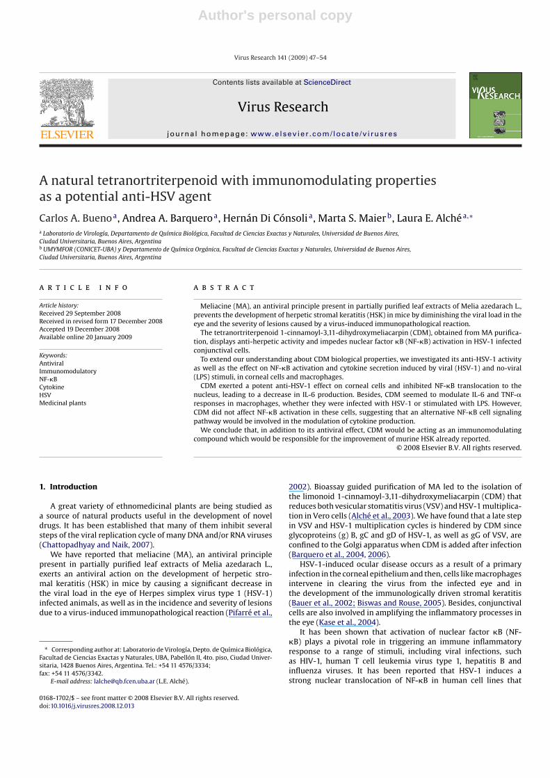
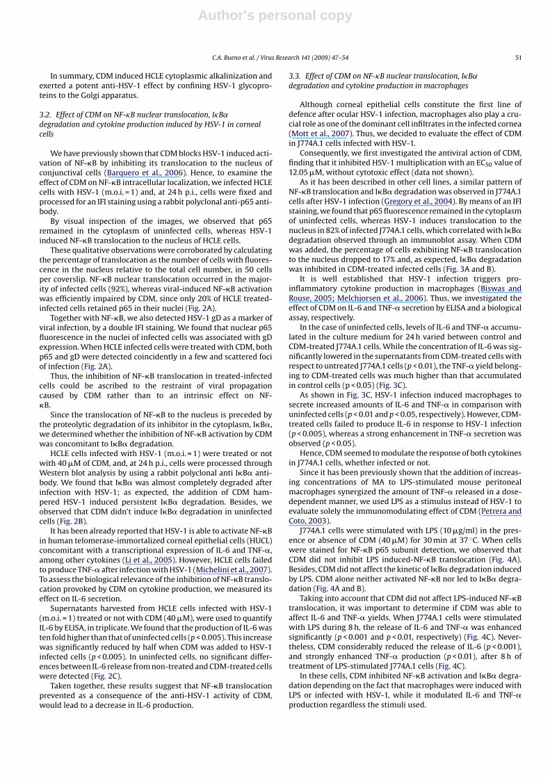
Fig. 1. Antiviral activity of CDM in HCLE cells. (A) HSV-1 infected cells were treated with different concentrations of CDM. After 24 h, supernatants were harvested and titratedby plaque assay and cytotoxicity of CDM was determined in uninfected treated cultures by the MTT assay. Data are expressed as the mean ± S.D. of two separate experiments.(B) HSV-1-infected cells were treated with CDM or not (CV) and after 13 h p.i. the intracellular localization of gD was done by IFI staining. Magnification 400×. (C) Cells grownon coverslips treated with CDM or not (CC) were stained with acridine orange and visualized on an epifluorescence microscope. Magnification 400×.
washing, membranes were incubated with diluted peroxidase-conjugated antibodies for 1.5 h at 37 ◦C. The immunoreactive bandswere visualized using an enhanced chemiluminesence system (ECL,PerkinElmer). Calnexin and actin were used as loading controls.
2.9. Cytokine determination
Cells were frozen and thawed, and then, supernatants were har-vested, centrifuged at 1000 rpm for 10 min, and cytokines werequantified by ELISA, or in a biological assay by triplicate.
Human TNF-� and IL-6, and mouse IL-6 were quantified by com-mercial ELISA sets (BD OptEIATM, Becton Dickinson, USA) accordingto manufacturer instructions.
Measurement of mouse TNF-� bioactivity was performed withthe L929 cell-based bioassay (Decker et al., 1987), with minor mod-ifications. L929 cells were grown in 96-well culture plates (2 × 10⁴
cells/well) for 24 h at 37 ◦C. Supernatants were removed and sub-stituted with the samples to be assayed for TNF-� content insuccessive twofold dilutions and incubated at 38.5 ◦C with 5 �g/mlof Actinomycin D (AcD) (Sidus, Argentina), for 22 h. Cells were fixedin 10% formaldehyde and stained with crystal violet 0.05%. To assessTNF-� activity, light absorbance at 580 nm of the eluted crystalviolet from the samples was measured and compared to a mouserecombinant TNF-� standard dilution series (Sigma). The bioas-say was specific for TNF-� since the activity was neutralized byan antibody against TNF-�.
Author's personal copy
50 C.A. Bueno et al. / Virus Research 141 (2009) 47–54
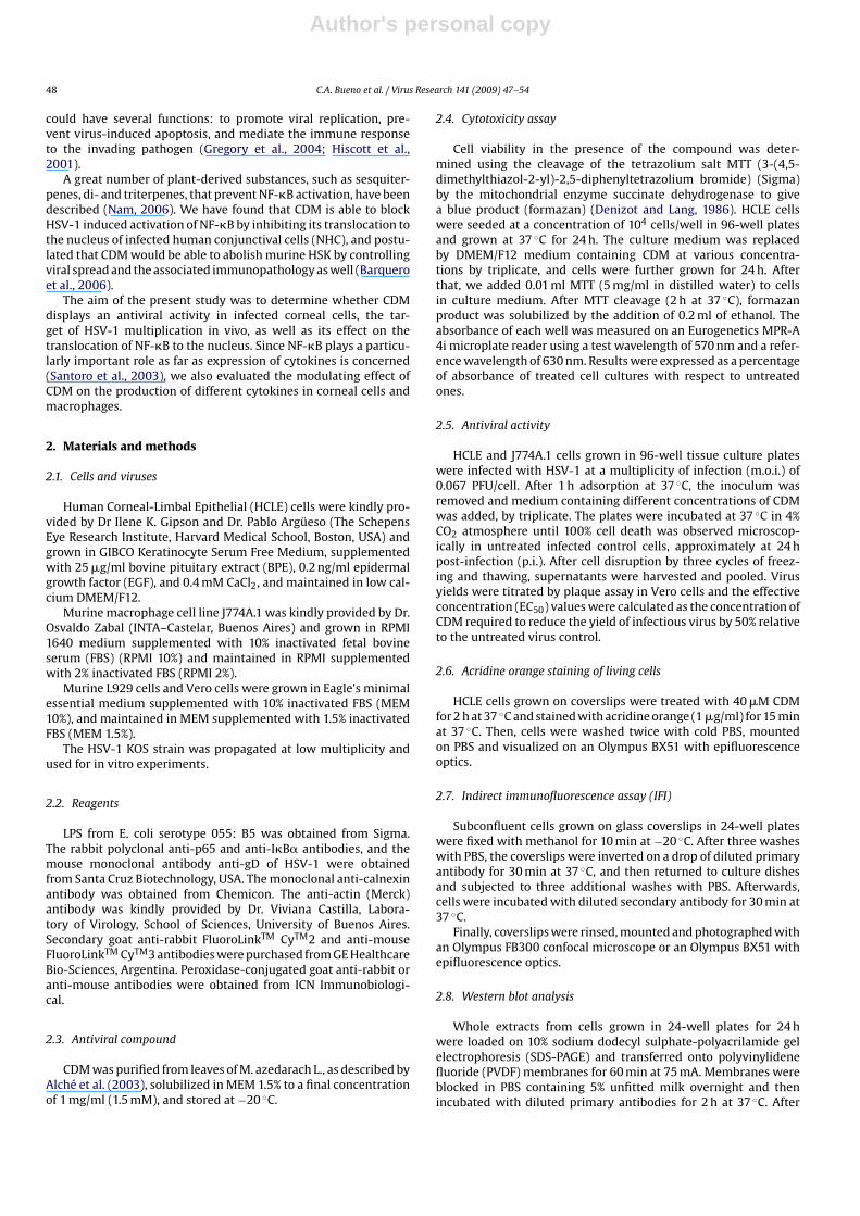
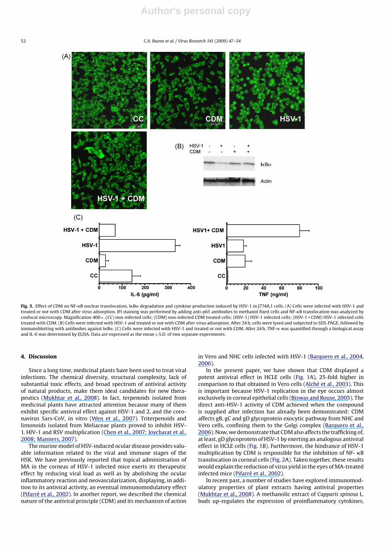
Fig. 2. Effect of CDM on NF-�B nuclear translocation, I�B� degradation and cytokine production induced by HSV-1 in HCLE cells. (A) HSV-1-infected cells were treatedwith CDM or not (CV). Double IFI staining was performed by adding anti-p65 and anti-HSV-1 gD antibodies to methanol fixed cells. NF-�B translocation and gD expressionwere analyzed by confocal microscopy. Magnification 600×. (B) HSV-1 infected cells were treated or not with CDM. After 24 h, cells were lysed and subjected to SDS-PAGE,followed by immunoblotting with antibodies against I�B�. (C) Cells were infected with HSV-1 and treated or not with CDM. After 24 h, IL-6 was determined by ELISA. Dataare expressed as the mean ± S.D. of two separate experiments.
2.10. Statistical analysis
Student’s t-test was used for statistical analysis of all data.
3. Results
3.1. Antiviral activity of CDM in a corneal cell line
It is well known that HSV-1 establishes infection usually in theepithelial layer of the cornea (Deshpande et al., 2004). Consideringthat CDM suppressed HSV-1 replication in Vero and human con-junctival cells, we decided to investigate the anti-HSV-1 effect ofCDM in HCLE cells where HSV-1 multiplied reaching a maximumviral titer of 5.3 × 10⁶ at 24 h p.i. (Alché et al., 2003; Barquero et al.,2006).
When HCLE cells infected with HSV-1 were treated with differ-ent concentrations of CDM, a dose-dependent inhibition of viralyields was observed, and an EC50 value of 0.78 �M was calculated.Besides, CDM proved to have no cytotoxic effect at all the concen-trations tested (Fig. 1A).
We have previously reported that CDM displays its antivi-ral action by affecting the trafficking of gB, gC and gD of
HSV-1 in Vero and NHC cells when supplied after infection(Barquero et al., 2006). In order to study the effect of CDMon the transport of viral glycoproteins in corneal cells, HCLEcells were infected with HSV-1 (m.o.i. = 2) and treated with CDM(40 �M). When a total IFI staining was performed using an anti-gD monoclonal antibody, we found that gD of HSV-1 exhibiteda perinuclear localization associated with the Golgi apparatus(Fig. 1B).
It has been already shown that the inhibition of viral glyco-protein transport may be related to the perturbation of the acidicpH of intracellular organelles (Sidhu et al., 1999). Although themechanism by which CDM affects the exocytic pathway is stillunraveled, we have shown that CDM provokes the basification ofthe pH of the endosomal vesicles in Vero cells (Barquero et al.,2004).
Now, we demonstrated that CDM also modified the endosomalpH of HCLE cells. The vital fluorescence microscopic study revealedthat untreated cells exhibited a bright orange punctate fluorescenceconcentrated in low-pH vesicles. Nevertheless, the pH of acidicintracellular vesicles from CDM-treated cells was markedly affectedsince only a faint granular fluorescence was observed after 2 h oftreatment (Fig. 1C).
Author's personal copy
C.A. Bueno et al. / Virus Research 141 (2009) 47–54 51
In summary, CDM induced HCLE cytoplasmic alkalinization andexerted a potent anti-HSV-1 effect by confining HSV-1 glycopro-teins to the Golgi apparatus.
3.2. Effect of CDM on NF-�B nuclear translocation, I�B˛degradation and cytokine production induced by HSV-1 in cornealcells
We have previously shown that CDM blocks HSV-1 induced acti-vation of NF-�B by inhibiting its translocation to the nucleus ofconjunctival cells (Barquero et al., 2006). Hence, to examine theeffect of CDM on NF-�B intracellular localization, we infected HCLEcells with HSV-1 (m.o.i. = 1) and, at 24 h p.i., cells were fixed andprocessed for an IFI staining using a rabbit polyclonal anti-p65 anti-body.
By visual inspection of the images, we observed that p65remained in the cytoplasm of uninfected cells, whereas HSV-1induced NF-�B translocation to the nucleus of HCLE cells.
These qualitative observations were corroborated by calculatingthe percentage of translocation as the number of cells with fluores-cence in the nucleus relative to the total cell number, in 50 cellsper coverslip. NF-�B nuclear translocation occurred in the major-ity of infected cells (92%), whereas viral-induced NF-�B activationwas efficiently impaired by CDM, since only 20% of HCLE treated-infected cells retained p65 in their nuclei (Fig. 2A).
Together with NF-�B, we also detected HSV-1 gD as a marker ofviral infection, by a double IFI staining. We found that nuclear p65fluorescence in the nuclei of infected cells was associated with gDexpression. When HCLE infected cells were treated with CDM, bothp65 and gD were detected coincidently in a few and scattered fociof infection (Fig. 2A).
Thus, the inhibition of NF-�B translocation in treated-infectedcells could be ascribed to the restraint of viral propagationcaused by CDM rather than to an intrinsic effect on NF-�B.
Since the translocation of NF-�B to the nucleus is preceded bythe proteolytic degradation of its inhibitor in the cytoplasm, I�B�,we determined whether the inhibition of NF-�B activation by CDMwas concomitant to I�B� degradation.
HCLE cells infected with HSV-1 (m.o.i. = 1) were treated or notwith 40 �M of CDM, and, at 24 h p.i., cells were processed throughWestern blot analysis by using a rabbit polyclonal anti I�B� anti-body. We found that I�B� was almost completely degraded afterinfection with HSV-1; as expected, the addition of CDM ham-pered HSV-1 induced persistent I�B� degradation. Besides, weobserved that CDM didn’t induce I�B� degradation in uninfectedcells (Fig. 2B).
It has been already reported that HSV-1 is able to activate NF-�Bin human telomerase-immortalized corneal epithelial cells (HUCL)concomitant with a transcriptional expression of IL-6 and TNF-�,among other cytokines (Li et al., 2005). However, HCLE cells failedto produce TNF-� after infection with HSV-1 (Michelini et al., 2007).To assess the biological relevance of the inhibition of NF-�B translo-cation provoked by CDM on cytokine production, we measured itseffect on IL-6 secretion.
Supernatants harvested from HCLE cells infected with HSV-1(m.o.i. = 1) treated or not with CDM (40 �M), were used to quantifyIL-6 by ELISA, in triplicate. We found that the production of IL-6 wasten fold higher than that of uninfected cells (p < 0.005). This increasewas significantly reduced by half when CDM was added to HSV-1infected cells (p < 0.005). In uninfected cells, no significant differ-ences between IL-6 release from non-treated and CDM-treated cellswere detected (Fig. 2C).
Taken together, these results suggest that NF-�B translocationprevented as a consequence of the anti-HSV-1 activity of CDM,would lead to a decrease in IL-6 production.
3.3. Effect of CDM on NF-�B nuclear translocation, I�B˛degradation and cytokine production in macrophages
Although corneal epithelial cells constitute the first line ofdefence after ocular HSV-1 infection, macrophages also play a cru-cial role as one of the dominant cell infiltrates in the infected cornea(Mott et al., 2007). Thus, we decided to evaluate the effect of CDMin J774A.1 cells infected with HSV-1.
Consequently, we first investigated the antiviral action of CDM,finding that it inhibited HSV-1 multiplication with an EC50 value of12.05 �M, without cytotoxic effect (data not shown).
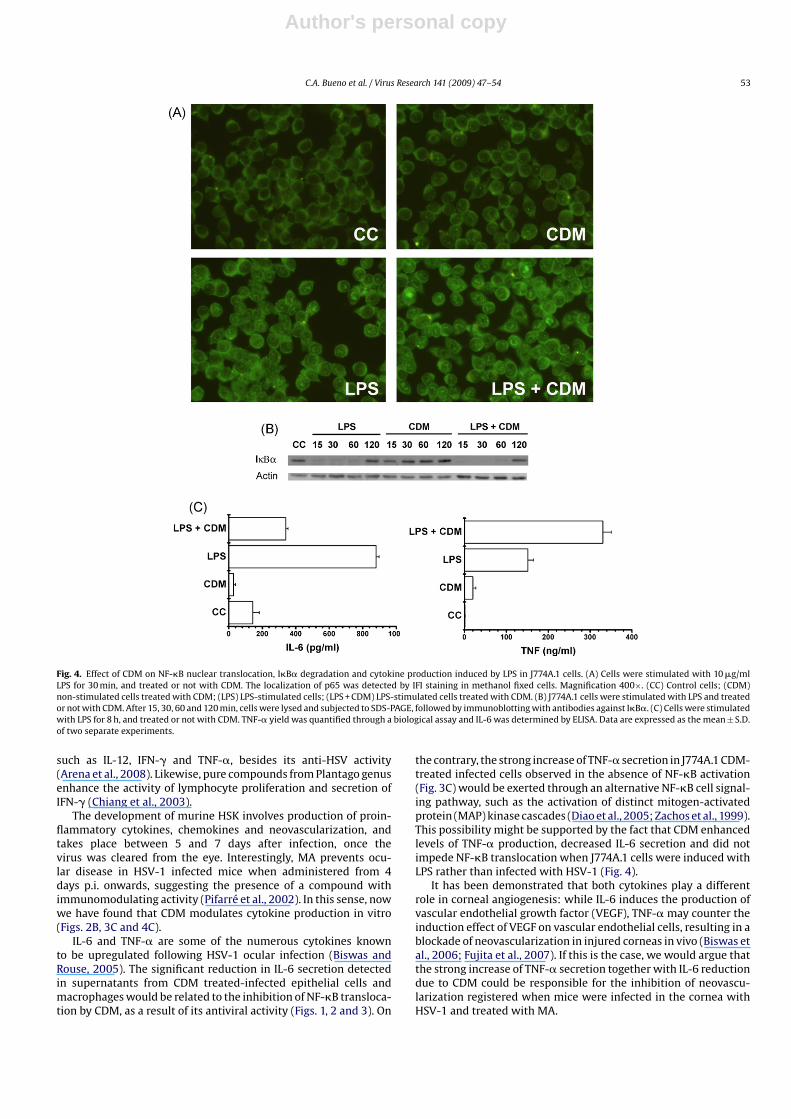
As it has been described in other cell lines, a similar pattern ofNF-�B translocation and I�B� degradation was observed in J774A.1cells after HSV-1 infection (Gregory et al., 2004). By means of an IFIstaining, we found that p65 fluorescence remained in the cytoplasmof uninfected cells, whereas HSV-1 induces translocation to thenucleus in 82% of infected J774A.1 cells, which correlated with I�B�degradation observed through an immunoblot assay. When CDMwas added, the percentage of cells exhibiting NF-�B translocationto the nucleus dropped to 17% and, as expected, I�B� degradationwas inhibited in CDM-treated infected cells (Fig. 3A and B).
It is well established that HSV-1 infection triggers pro-inflammatory cytokine production in macrophages (Biswas andRouse, 2005; Melchjorsen et al., 2006). Thus, we investigated theeffect of CDM on IL-6 and TNF-� secretion by ELISA and a biologicalassay, respectively.
In the case of uninfected cells, levels of IL-6 and TNF-� accumu-lated in the culture medium for 24 h varied between control andCDM-treated J774A.1 cells. While the concentration of IL-6 was sig-nificantly lowered in the supernatants from CDM-treated cells withrespect to untreated J774A.1 cells (p < 0.01), the TNF-� yield belong-ing to CDM-treated cells was much higher than that accumulatedin control cells (p < 0.05) (Fig. 3C).
As shown in Fig. 3C, HSV-1 infection induced macrophages tosecrete increased amounts of IL-6 and TNF-� in comparison withuninfected cells (p < 0.01 and p < 0.05, respectively). However, CDM-treated cells failed to produce IL-6 in response to HSV-1 infection(p < 0.005), whereas a strong enhancement in TNF-� secretion wasobserved (p < 0.05).
Hence, CDM seemed to modulate the response of both cytokinesin J774A.1 cells, whether infected or not.
Since it has been previously shown that the addition of increas-ing concentrations of MA to LPS-stimulated mouse peritonealmacrophages synergized the amount of TNF-� released in a dose-dependent manner, we used LPS as a stimulus instead of HSV-1 toevaluate solely the immunomodulating effect of CDM (Petrera andCoto, 2003).
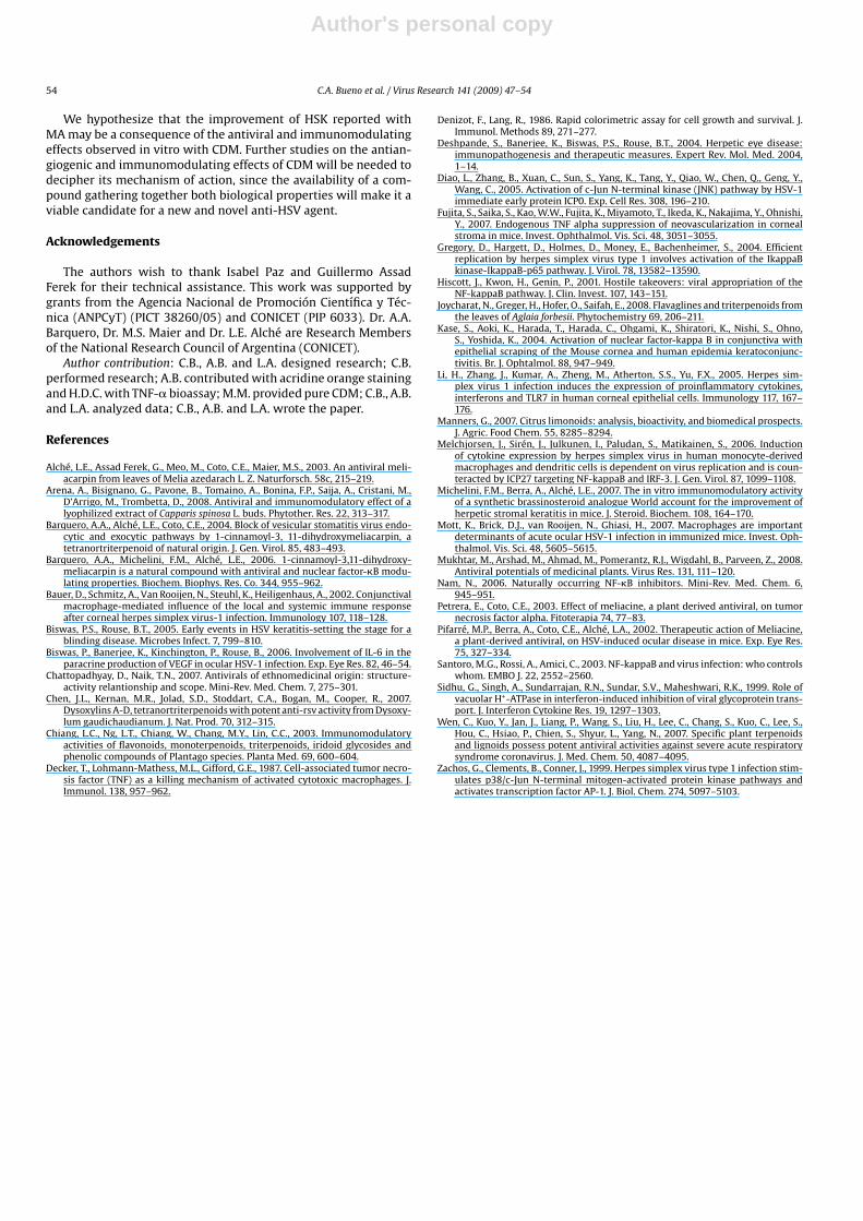
J774A.1 cells were stimulated with LPS (10 �g/ml) in the pres-ence or absence of CDM (40 �M) for 30 min at 37 ◦C. When cellswere stained for NF-�B p65 subunit detection, we observed thatCDM did not inhibit LPS induced-NF-�B translocation (Fig. 4A).Besides, CDM did not affect the kinetic of I�B� degradation inducedby LPS. CDM alone neither activated NF-�B nor led to I�B� degra-dation (Fig. 4A and B).
Taking into account that CDM did not affect LPS-induced NF-�Btranslocation, it was important to determine if CDM was able toaffect IL-6 and TNF-� yields. When J774A.1 cells were stimulatedwith LPS during 8 h, the release of IL-6 and TNF-� was enhancedsignificantly (p < 0.001 and p < 0.01, respectively) (Fig. 4C). Never-theless, CDM considerably reduced the release of IL-6 (p < 0.001),and strongly enhanced TNF-� production (p < 0.01), after 8 h oftreatment of LPS-stimulated J774A.1 cells (Fig. 4C).
In these cells, CDM inhibited NF-�B activation and I�B� degra-dation depending on the fact that macrophages were induced withLPS or infected with HSV-1, while it modulated IL-6 and TNF-�production regardless the stimuli used.
Author's personal copy
52 C.A. Bueno et al. / Virus Research 141 (2009) 47–54
Fig. 3. Effect of CDM on NF-�B nuclear translocation, I�B� degradation and cytokine production induced by HSV-1 in J774A.1 cells. (A) Cells were infected with HSV-1 andtreated or not with CDM after virus adsorption. IFI staining was performed by adding anti-p65 antibodies to methanol fixed cells and NF-�B translocation was analyzed byconfocal microscopy. Magnification 400×. (CC) non-infected cells; (CDM) non-infected CDM treated cells; (HSV-1) HSV-1 infected cells; (HSV-1 + CDM) HSV-1 infected cellstreated with CDM. (B) Cells were infected with HSV-1 and treated or not with CDM after virus adsorption. After 24 h, cells were lysed and subjected to SDS-PAGE, followed byimmunoblotting with antibodies against I�B�. (C) Cells were infected with HSV-1 and treated or not with CDM. After 24 h, TNF-� was quantified through a biological assayand IL-6 was determined by ELISA. Data are expressed as the mean ± S.D. of two separate experiments.
4. Discussion
Since a long time, medicinal plants have been used to treat viralinfections. The chemical diversity, structural complexity, lack ofsubstantial toxic effects, and broad spectrum of antiviral activityof natural products, make them ideal candidates for new thera-peutics (Mukhtar et al., 2008). In fact, terpenoids isolated frommedicinal plants have attracted attention because many of themexhibit specific antiviral effect against HSV-1 and 2, and the coro-navirus Sars-CoV, in vitro (Wen et al., 2007). Triterpenoids andlimonoids isolated from Meliaceae plants proved to inhibit HSV-1, HIV-1 and RSV multiplication (Chen et al., 2007; Joycharat et al.,2008; Manners, 2007).
The murine model of HSV-induced ocular disease provides valu-able information related to the viral and immune stages of theHSK. We have previously reported that topical administration ofMA in the corneas of HSV-1 infected mice exerts its therapeuticeffect by reducing viral load as well as by abolishing the ocularinflammatory reaction and neovascularization, displaying, in addi-tion to its antiviral activity, an eventual immunomodulatory effect(Pifarré et al., 2002). In another report, we described the chemicalnature of the antiviral principle (CDM) and its mechanism of action
in Vero and NHC cells infected with HSV-1 (Barquero et al., 2004,2006).
In the present paper, we have shown that CDM displayed apotent antiviral effect in HCLE cells (Fig. 1A), 25-fold higher incomparison to that obtained in Vero cells (Alché et al., 2003). Thisis important because HSV-1 replication in the eye occurs almostexclusively in corneal epithelial cells (Biswas and Rouse, 2005). Thedirect anti-HSV-1 activity of CDM achieved when the compoundis supplied after infection has already been demonstrated: CDMaffects gB, gC and gD glycoprotein exocytic pathway from NHC andVero cells, confining them to the Golgi complex (Barquero et al.,2006). Now, we demonstrate that CDM also affects the trafficking of,at least, gD glycoprotein of HSV-1 by exerting an analogous antiviraleffect in HCLE cells (Fig. 1B). Furthermore, the hindrance of HSV-1multiplication by CDM is responsible for the inhibition of NF- �Btranslocation in corneal cells (Fig. 2A). Taken together, these resultswould explain the reduction of virus yield in the eyes of MA-treatedinfected mice (Pifarré et al., 2002).
In recent past, a number of studies have explored immunomod-ulatory properties of plant extracts having antiviral properties(Mukhtar et al., 2008). A methanolic extract of Capparis spinosa L.buds up-regulates the expression of proinflammatory cytokines,
Author's personal copy
C.A. Bueno et al. / Virus Research 141 (2009) 47–54 53
Fig. 4. Effect of CDM on NF-�B nuclear translocation, I�B� degradation and cytokine production induced by LPS in J774A.1 cells. (A) Cells were stimulated with 10 �g/mlLPS for 30 min, and treated or not with CDM. The localization of p65 was detected by IFI staining in methanol fixed cells. Magnification 400×. (CC) Control cells; (CDM)non-stimulated cells treated with CDM; (LPS) LPS-stimulated cells; (LPS + CDM) LPS-stimulated cells treated with CDM. (B) J774A.1 cells were stimulated with LPS and treatedor not with CDM. After 15, 30, 60 and 120 min, cells were lysed and subjected to SDS-PAGE, followed by immunoblotting with antibodies against I�B�. (C) Cells were stimulatedwith LPS for 8 h, and treated or not with CDM. TNF-� yield was quantified through a biological assay and IL-6 was determined by ELISA. Data are expressed as the mean ± S.D.of two separate experiments.
such as IL-12, IFN-� and TNF-�, besides its anti-HSV activity(Arena et al., 2008). Likewise, pure compounds from Plantago genusenhance the activity of lymphocyte proliferation and secretion ofIFN-� (Chiang et al., 2003).
The development of murine HSK involves production of proin-flammatory cytokines, chemokines and neovascularization, andtakes place between 5 and 7 days after infection, once thevirus was cleared from the eye. Interestingly, MA prevents ocu-lar disease in HSV-1 infected mice when administered from 4days p.i. onwards, suggesting the presence of a compound withimmunomodulating activity (Pifarré et al., 2002). In this sense, nowwe have found that CDM modulates cytokine production in vitro(Figs. 2B, 3C and 4C).
IL-6 and TNF-� are some of the numerous cytokines knownto be upregulated following HSV-1 ocular infection (Biswas andRouse, 2005). The significant reduction in IL-6 secretion detectedin supernatants from CDM treated-infected epithelial cells andmacrophages would be related to the inhibition of NF-�B transloca-tion by CDM, as a result of its antiviral activity (Figs. 1, 2 and 3). On
the contrary, the strong increase of TNF-� secretion in J774A.1 CDM-treated infected cells observed in the absence of NF-�B activation(Fig. 3C) would be exerted through an alternative NF-�B cell signal-ing pathway, such as the activation of distinct mitogen-activatedprotein (MAP) kinase cascades (Diao et al., 2005; Zachos et al., 1999).This possibility might be supported by the fact that CDM enhancedlevels of TNF-� production, decreased IL-6 secretion and did notimpede NF-�B translocation when J774A.1 cells were induced withLPS rather than infected with HSV-1 (Fig. 4).
It has been demonstrated that both cytokines play a differentrole in corneal angiogenesis: while IL-6 induces the production ofvascular endothelial growth factor (VEGF), TNF-� may counter theinduction effect of VEGF on vascular endothelial cells, resulting in ablockade of neovascularization in injured corneas in vivo (Biswas etal., 2006; Fujita et al., 2007). If this is the case, we would argue thatthe strong increase of TNF-� secretion together with IL-6 reductiondue to CDM could be responsible for the inhibition of neovascu-larization registered when mice were infected in the cornea withHSV-1 and treated with MA.
Author's personal copy
54 C.A. Bueno et al. / Virus Research 141 (2009) 47–54
We hypothesize that the improvement of HSK reported withMA may be a consequence of the antiviral and immunomodulatingeffects observed in vitro with CDM. Further studies on the antian-giogenic and immunomodulating effects of CDM will be needed todecipher its mechanism of action, since the availability of a com-pound gathering together both biological properties will make it aviable candidate for a new and novel anti-HSV agent.
Acknowledgements
The authors wish to thank Isabel Paz and Guillermo AssadFerek for their technical assistance. This work was supported bygrants from the Agencia Nacional de Promoción Científica y Téc-nica (ANPCyT) (PICT 38260/05) and CONICET (PIP 6033). Dr. A.A.Barquero, Dr. M.S. Maier and Dr. L.E. Alché are Research Membersof the National Research Council of Argentina (CONICET).
Author contribution: C.B., A.B. and L.A. designed research; C.B.performed research; A.B. contributed with acridine orange stainingand H.D.C. with TNF-� bioassay; M.M. provided pure CDM; C.B., A.B.and L.A. analyzed data; C.B., A.B. and L.A. wrote the paper.
References
Alché, L.E., Assad Ferek, G., Meo, M., Coto, C.E., Maier, M.S., 2003. An antiviral meli-acarpin from leaves of Melia azedarach L. Z. Naturforsch. 58c, 215–219.
Arena, A., Bisignano, G., Pavone, B., Tomaino, A., Bonina, F.P., Saija, A., Cristani, M.,D’Arrigo, M., Trombetta, D., 2008. Antiviral and immunomodulatory effect of alyophilized extract of Capparis spinosa L. buds. Phytother. Res. 22, 313–317.
Barquero, A.A., Alché, L.E., Coto, C.E., 2004. Block of vesicular stomatitis virus endo-cytic and exocytic pathways by 1-cinnamoyl-3, 11-dihydroxymeliacarpin, atetranortriterpenoid of natural origin. J. Gen. Virol. 85, 483–493.
Barquero, A.A., Michelini, F.M., Alché, L.E., 2006. 1-cinnamoyl-3,11-dihydroxy-meliacarpin is a natural compound with antiviral and nuclear factor-�B modu-lating properties. Biochem. Biophys. Res. Co. 344, 955–962.
Bauer, D., Schmitz, A., Van Rooijen, N., Steuhl, K., Heiligenhaus, A., 2002. Conjunctivalmacrophage-mediated influence of the local and systemic immune responseafter corneal herpes simplex virus-1 infection. Immunology 107, 118–128.
Biswas, P.S., Rouse, B.T., 2005. Early events in HSV keratitis-setting the stage for ablinding disease. Microbes Infect. 7, 799–810.
Biswas, P., Banerjee, K., Kinchington, P., Rouse, B., 2006. Involvement of IL-6 in theparacrine production of VEGF in ocular HSV-1 infection. Exp. Eye Res. 82, 46–54.
Chattopadhyay, D., Naik, T.N., 2007. Antivirals of ethnomedicinal origin: structure-activity relantionship and scope. Mini-Rev. Med. Chem. 7, 275–301.
Chen, J.L., Kernan, M.R., Jolad, S.D., Stoddart, C.A., Bogan, M., Cooper, R., 2007.Dysoxylins A-D, tetranortriterpenoids with potent anti-rsv activity from Dysoxy-lum gaudichaudianum. J. Nat. Prod. 70, 312–315.
Chiang, L.C., Ng, L.T., Chiang, W., Chang, M.Y., Lin, C.C., 2003. Immunomodulatoryactivities of flavonoids, monoterpenoids, triterpenoids, iridoid glycosides andphenolic compounds of Plantago species. Planta Med. 69, 600–604.
Decker, T., Lohmann-Mathess, M.L., Gifford, G.E., 1987. Cell-associated tumor necro-sis factor (TNF) as a killing mechanism of activated cytotoxic macrophages. J.Immunol. 138, 957–962.
Denizot, F., Lang, R., 1986. Rapid colorimetric assay for cell growth and survival. J.Immunol. Methods 89, 271–277.
Deshpande, S., Banerjee, K., Biswas, P.S., Rouse, B.T., 2004. Herpetic eye disease:immunopathogenesis and therapeutic measures. Expert Rev. Mol. Med. 2004,1–14.
Diao, L., Zhang, B., Xuan, C., Sun, S., Yang, K., Tang, Y., Qiao, W., Chen, Q., Geng, Y.,Wang, C., 2005. Activation of c-Jun N-terminal kinase (JNK) pathway by HSV-1immediate early protein ICP0. Exp. Cell Res. 308, 196–210.
Fujita, S., Saika, S., Kao, W.W., Fujita, K., Miyamoto, T., Ikeda, K., Nakajima, Y., Ohnishi,Y., 2007. Endogenous TNF alpha suppression of neovascularization in cornealstroma in mice. Invest. Ophthalmol. Vis. Sci. 48, 3051–3055.
Gregory, D., Hargett, D., Holmes, D., Money, E., Bachenheimer, S., 2004. Efficientreplication by herpes simplex virus type 1 involves activation of the IkappaBkinase-IkappaB-p65 pathway. J. Virol. 78, 13582–13590.
Hiscott, J., Kwon, H., Genin, P., 2001. Hostile takeovers: viral appropriation of theNF-kappaB pathway. J. Clin. Invest. 107, 143–151.
Joycharat, N., Greger, H., Hofer, O., Saifah, E., 2008. Flavaglines and triterpenoids fromthe leaves of Aglaia forbesii. Phytochemistry 69, 206–211.
Kase, S., Aoki, K., Harada, T., Harada, C., Ohgami, K., Shiratori, K., Nishi, S., Ohno,S., Yoshida, K., 2004. Activation of nuclear factor-kappa B in conjunctiva withepithelial scraping of the Mouse cornea and human epidemia keratoconjunc-tivitis. Br. J. Ophtalmol. 88, 947–949.
Li, H., Zhang, J., Kumar, A., Zheng, M., Atherton, S.S., Yu, F.X., 2005. Herpes sim-plex virus 1 infection induces the expression of proinflammatory cytokines,interferons and TLR7 in human corneal epithelial cells. Immunology 117, 167–176.
Manners, G., 2007. Citrus limonoids: analysis, bioactivity, and biomedical prospects.J. Agric. Food Chem. 55, 8285–8294.
Melchjorsen, J., Sirén, J., Julkunen, I., Paludan, S., Matikainen, S., 2006. Inductionof cytokine expression by herpes simplex virus in human monocyte-derivedmacrophages and dendritic cells is dependent on virus replication and is coun-teracted by ICP27 targeting NF-kappaB and IRF-3. J. Gen. Virol. 87, 1099–1108.
Michelini, F.M., Berra, A., Alché, L.E., 2007. The in vitro immunomodulatory activityof a synthetic brassinosteroid analogue World account for the improvement ofherpetic stromal keratitis in mice. J. Steroid. Biochem. 108, 164–170.
Mott, K., Brick, D.J., van Rooijen, N., Ghiasi, H., 2007. Macrophages are importantdeterminants of acute ocular HSV-1 infection in immunized mice. Invest. Oph-thalmol. Vis. Sci. 48, 5605–5615.
Mukhtar, M., Arshad, M., Ahmad, M., Pomerantz, R.J., Wigdahl, B., Parveen, Z., 2008.Antiviral potentials of medicinal plants. Virus Res. 131, 111–120.
Nam, N., 2006. Naturally occurring NF-�B inhibitors. Mini-Rev. Med. Chem. 6,945–951.
Petrera, E., Coto, C.E., 2003. Effect of meliacine, a plant derived antiviral, on tumornecrosis factor alpha. Fitoterapia 74, 77–83.
Pifarré, M.P., Berra, A., Coto, C.E., Alché, L.A., 2002. Therapeutic action of Meliacine,a plant-derived antiviral, on HSV-induced ocular disease in mice. Exp. Eye Res.75, 327–334.
Santoro, M.G., Rossi, A., Amici, C., 2003. NF-kappaB and virus infection: who controlswhom. EMBO J. 22, 2552–2560.
Sidhu, G., Singh, A., Sundarrajan, R.N., Sundar, S.V., Maheshwari, R.K., 1999. Role ofvacuolar H+-ATPase in interferon-induced inhibition of viral glycoprotein trans-port. J. Interferon Cytokine Res. 19, 1297–1303.
Wen, C., Kuo, Y., Jan, J., Liang, P., Wang, S., Liu, H., Lee, C., Chang, S., Kuo, C., Lee, S.,Hou, C., Hsiao, P., Chien, S., Shyur, L., Yang, N., 2007. Specific plant terpenoidsand lignoids possess potent antiviral activities against severe acute respiratorysyndrome coronavirus. J. Med. Chem. 50, 4087–4095.
Zachos, G., Clements, B., Conner, J., 1999. Herpes simplex virus type 1 infection stim-ulates p38/c-Jun N-terminal mitogen-activated protein kinase pathways andactivates transcription factor AP-1. J. Biol. Chem. 274, 5097–5103.