Auditory and visual novelty processing in normally-developing Kenyan children
Upload
independentCategory
view
0download
0
BioMed CentralMalaria Journal
ss
Open AcceResearchDecorticate, decerebrate and opisthotonic posturing and seizures in Kenyan children with cerebral malariaRichard Idro*1,2, Godfrey Otieno1, Steven White3, Anderson Kahindi1, Greg Fegan1,4, Bernhards Ogutu1, Sadik Mithwani1, Kathryn Maitland1,5, Brian GR Neville6 and Charles RJC Newton1,6Address: 1Centre for Geographic Medicine Research-Coast, Kenya Medical Research, Kenya, 2Department of Paediatrics and Child Health, Mulago Hospital/Makerere University, Kampala, Uganda, 3Department of Clinical Neurophysiology, Great Ormond Street Hospital for Children London, UK, 4Infectious Disease Epidemiology Unit, Department of Infectious and Tropical Diseases, London School of Hygiene and Tropical Medicine, London, UK, 5Department of Paediatrics, Faculty of Medicine and the Wellcome Trust Centre for Tropical Medicine, Imperial College, London, UK and 6Neurosciences Unit, the Wolfson Centre, Institute of Child Health, University College London, UK
Email: Richard Idro* - [email protected]; Godfrey Otieno - [email protected]; Steven White - [email protected]; Anderson Kahindi - [email protected]; Greg Fegan - [email protected]; Bernhards Ogutu - [email protected]; Sadik Mithwani - [email protected]; Kathryn Maitland - [email protected]; Brian GR Neville - [email protected]; Charles RJC Newton - [email protected]
* Corresponding author
AbstractBackground: Abnormal motor posturing is often observed in children with cerebral malaria, but theaetiology and pathogenesis is poorly understood. This study examined the risk factors and outcome ofposturing in Kenyan children with cerebral malaria.
Methods: Records of children admitted to Kilifi district hospital with cerebral malaria from January, 1999through December, 2001 were reviewed for posturing occurring on or after admission. The clinicalcharacteristics, features of raised intracranial pressure, number of seizures and biochemical changes inpatients that developed posturing was compared to patients who did not.
Results: Of the 417 children with complete records, 163 (39.1%) had posturing: 85 on admission and 78after admission to hospital. Decorticate posturing occurred in 80, decerebrate in 61 and opisthotonicposturing in 22 patients. Posturing was associated with age ≥ 3 years (48.1 vs 35.8%, p = 0.01) and featuresof raised intracranial pressure on funduscopy (adjusted OR 2.1 95%CI 1.2–3.7, p = 0.009) but not othermarkers of severity of disease. Unlike decorticate posturing, decerebrate (adjusted OR 1.9 95%CI 1.0–3.5)and opisthotonic posturing (adjusted OR 2.9 95%CI 1.0–8.1) were, in addition, independently associatedwith recurrence of seizures after admission. Opisthotonus was also associated with severe metabolicacidosis (OR 4.2 95%CI 3.2–5.6, p < 0.001). Thirty one patients with posturing died. Of these, 19 (61.3%)had features suggestive of transtentorial herniation. Mortality and neurological deficits on discharge weregreatest in those developing posturing after admission.
Conclusion: Abnormal motor posturing is a common feature of cerebral malaria in children. It isassociated with features of raised intracranial pressure and recurrence of seizures, although intracranialhypertension may be the primary cause.
Published: 07 December 2005
Malaria Journal 2005, 4:57 doi:10.1186/1475-2875-4-57
Received: 10 June 2005Accepted: 07 December 2005
This article is available from: http://www.malariajournal.com/content/4/1/57
© 2005 Idro et al; licensee BioMed Central Ltd. This is an Open Access article distributed under the terms of the Creative Commons Attribution License (http://creativecommons.org/licenses/by/2.0), which permits unrestricted use, distribution, and reproduction in any medium, provided the original work is properly cited.
Page 1 of 10(page number not for citation purposes)
Malaria Journal 2005, 4:57 http://www.malariajournal.com/content/4/1/57
BackgroundCerebral malaria is one of the most common non-trau-matic encephalopathies affecting children worldwide andthe most severe neurological complication of falciparummalaria [1,2]. Patients present with a one to three day his-tory of fever, seizures, unarouseable coma and brainstemsigns [1,3,4]. Abnormal motor posturing (AMP), mani-festing as decorticate, decerebrate or opisthotonus, iscommon, but the aetiology and pathogenesis of thesesigns in cerebral malaria and the prognosis of each type ofposturing is poorly understood [3-9]. Raised intracranialpressure (ICP), cerebral ischaemia, hypoxia, hypoglycae-mia, and hyponatraemia are recognized causes of AMP[10,11] and raised ICP is a feature of cerebral malaria inAfrican children [6,7,12]. In a study of cerebral malaria inwhich ICP was monitored, all children in whom AMP wasdocumented had raised ICP, but in some patients, thetiming of the AMP was not directly associated with epi-sodes of severe intracranial hypertension [7]. Based oncurrent guidelines, children with similar features present-ing to emergency services in areas where malaria is absentwill receive management for raised ICP, including paraly-sis and ventilation. On the other hand, seizures have beendescribed in over 60% of patients with cerebral malariaand other acute encephalopathies associated with AMP[10,13]. The hypothesis that AMP may be caused by sei-zures has led to the use of anticonvulsants in their man-agement but there is little evidence to support thismanagement. Other treatment practices have includednon-interventional observation or therapy with mannitol.
The frequent occurrence of AMP in children with cerebralmalaria, lack of ventilation facilities in most centres thatmanage such patients and the apparent resolution in mostpatients without active intervention warrants furtherdescription of the associated risk factors. In this study,records of children admitted to a Kenyan district hospitalwith cerebral malaria were examined to determine the riskfactors associated with AMP, in particular to determineany association between AMP and features of raised ICPor seizures. The relationship between AMP and outcomewas also examined.
MethodsStudy designClinical records of all children admitted to Kilifi DistrictHospital with cerebral malaria from January, 1999through December, 2001 were examined for AMP. Thishospital is situated in a malaria-endemic area and the set-ting was described previously [14,15].
Definition of cerebral malariaCerebral malaria was defined as unarouseable coma (una-ble to localize a painful stimulus, Blantyre Coma Score ≤2) [3], at least one hour after termination of a seizure,
administration of diazepam or correction of hypoglycae-mia, with asexual forms of falciparum malaria parasiteson a Giemsa stained blood smear and cerebrospinal fluid(CSF) examination not suggestive of bacterial meningitis[2,16]. Children with epilepsy, cerebral palsy or sickle celldisease were excluded.
Definition of decorticate, decerebrate and opisthotonic posturingAbnormal motor posturing, classified as decorticate,decerebrate or opisthotonic posturing is characterised bygeneralised extension of the trunk and lower limbs withincreased muscular tone [11]. Decorticate posturing wasdefined as semi-flexion, adduction and internal rotationat the shoulders and semi-flexion or flexion at the elbows.Decerebrate posturing was defined as extension of upperlimbs, adduction and internal rotation of the shoulders,with pronation of the forearms. Opisthotonic posturingwas defined as decerebrate posturing where the neck andback are arched posteriorly [10]. For the purposes of thestudy, if a patient exhibited more than one type of AMP,the more severe type ordered as: decorticate, decerebrateand opisthotonus was assigned.
Admission proceduresEthical permission for the study was obtained from theKenya Medical Research Institute Scientific and EthicalReview Committees. At admission, standard proformawere completed detailing the medical history and physicalexamination. Emergency care and resuscitation were per-formed according to standard protocols [2,17]. Thenumber, duration and type of seizures prior to admissionwere documented. Level of coma was assessed using theBlantyre coma scale (BCS) [3]. Features of AMP were doc-umented and assigned to decorticate, decerebrate oropisthotonic categories.
Laboratory proceduresPatients had venous blood drawn for a full blood countand blood glucose, parasite density, plasma electrolytesand microbiological culture. A heparinized venous sam-ple was drawn to determine acid base status. Lumbarpunctures were performed when the level of conscious-ness improved (BCS>2) and the child did not have brain-stem signs[5].
Inpatient courseWhile in the ward, patients were closely monitored duringthe first hour. Thereafter, clinical assessments, level ofconsciousness and blood glucose measurements wererepeated every four hours by the nursing staff. Physiciansassessed the children at four and 24 hours and on a dailybasis thereafter. Patients were re-assessed if the nursingteam observed seizures, AMP, deterioration in conscious-ness or worsening vital signs. Seizures on admission and
Page 2 of 10(page number not for citation purposes)
Malaria Journal 2005, 4:57 http://www.malariajournal.com/content/4/1/57
those that developed after admission and lasted longerthan five 5 minutes were treated with intravenousdiazepam 0.3 mg/kg. Paraldehyde (0.4 ml/kg intramuscu-larly) was the second line antiepileptic drug, and pheny-toin (18 mg/kg intravenously) or phenobarbital (18 mg/kg) were the third line antiepileptic drugs. Clinical signswithin one hour of a seizure or anticonvulsant were notreported. Observed seizures were documented, timed andclassified as partial, partial with secondary generalizationor generalized tonic-clonic and recorded on a proformasheet. Direct ophthalmoscopy for retinal signs was per-formed on admission, at 4 hours and then daily until fullconsciousness was regained by the attending physicians.In the first year, some of these examinations were per-formed by an ophthalmologist who trained the other phy-sicians in the recognition of abnormal retinal features, butthere was no formal validation of the quality of fundus-copy performed by the attending physicians against thatof the ophthalmologist. AMP and other brainstem signswere documented. Treatment and nursing care wereaccording to standard protocols [2,17]. Patients weredeemed to have regained full consciousness when theyscored a BCS of 5 (or BCS 4 in children <9 months) [18].
Cerebral function analyser monitoringFifty-eight of the study patients participated in pharma-cokinetic studies of anti-epileptic drugs [19,20] and hadcontinuous monitoring with a 4-channel cerebral func-tion analyser (CFAM3c RDM, East Sussex) for the dura-tion of coma. The CFAM recordings were examined forevidence of seizure activity and changes in amplitude dur-ing episodes of AMP.
Data management and statistical analysisPatients' records were entered directly into a databaseusing FoxPro software and analysed using SPSS 11.5(SPSS Chicago Inc, 2003). Patients were divided into threegroups: those with AMP at admission, those with AMPafter admission and those without. Children who hadAMP at admission and additional episodes after admis-sion were regarded as having AMP at admission and themost severe form of AMP was assigned to the patient. Uni-variate analysis was performed to identify features associ-ated with AMP. Patients who had AMP on admission werecompared with those without AMP. A similar comparisonwas also made between patients posturing after admissionand those without AMP. Categorical variables were com-pared using Pearson's Chi square or Fisher's exact test (2-tailed) as appropriate. Continuous variables were com-pared with the student's t-test. The mean parasite densitieswere compared after logarithmic transformation and themedian and inter quartile range (IQR) used for otherskewed data. Logistic regression analysis was performed toidentify risk factors independently associated with thedevelopment of AMP.
ResultsGeneral descriptionA total of 15,506 children were admitted to KDH duringthe three-year period. Of these, 5,767 had malaria as a pri-mary diagnosis. Four hundred and twenty-six patients ful-filled the criteria for cerebral malaria, of whom nine hadincomplete records and were excluded. Of the remainingfour hundred and seventeen, 215 (51.6%) were males.The median age was 27 (IQR 15–41) months and medianduration of fever prior to admission was two (IQR one –three) days. Overall, 163 children (39.1%) had AMP: 85(20.4%) presented with AMP and an additional 78(18.7%) developed AMP after admission. Of the 85patients who had AMP on admission, 63 (74%) had fur-ther episodes after admission. Most AMP occurred spon-taneously and manifested as decorticate posturing in 80(49.1%), decerebrate posturing in 61 (37.4%) andopisthotonus in 22 (13.5%).
Presentation and risk factors for abnormal motor posturing at admissionAdmission clinical characteristics of patients with AMP atadmission, during admission or without AMP are summa-rized in Table 1 (and clinical features of patients with andwithout AMP are in the attached Additional file 1). Over-all, patients with AMP were older: 48.1% of those threeyears or older had AMP compared to only 35.8% of chil-dren under three years, p = 0.01. No association wasobserved between AMP and duration of illness, history ofseizures or parasite density. No relationship was alsoobserved between AMP and other markers of severity ofillness at admission such as a history of multiple convul-sions, profound coma, deep breathing, hypotension,hypoglycaemia, severe anaemia, hyponatraemia or hyper-parasitaemia. No significant differences were found in theCSF white blood cell count, protein, sugar or CSF: bloodsugar ratio. Using multiple logistic regression analysis, agethree years or older was independently associated with achild presenting with AMP (adjusted OR 2.0 95% CI 1.7 –2.4, p < 0.001). Unlike decorticate or decerebrate postur-ing, opisthotonic posturing was also independently asso-ciated with severe acidosis, (adjusted OR 4.2 95% CI 3.2–5.6, p < 0.001).
Abnormal motor posturing after admissionOf the 78 children who developed posturing after admis-sion, 38 (49%) had decorticate, 30 (38%) decerebrate and10 (13%) opisthotonic posturing. This distribution wassimilar to the overall distribution of AMP. Apart frombeing younger, no differences in admission characteristicsand in particular, the duration of illness or coma, historyof seizures, level of coma, parasite density, blood glucose,electrolytes, acid base status or haemodynamic state wereobserved when compared to those who presented withAMP (Table 1).
Page 3 of 10(page number not for citation purposes)
Malaria Journal 2005, 4:57 http://www.malariajournal.com/content/4/1/57
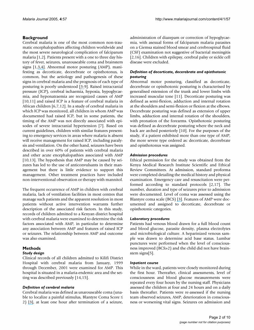
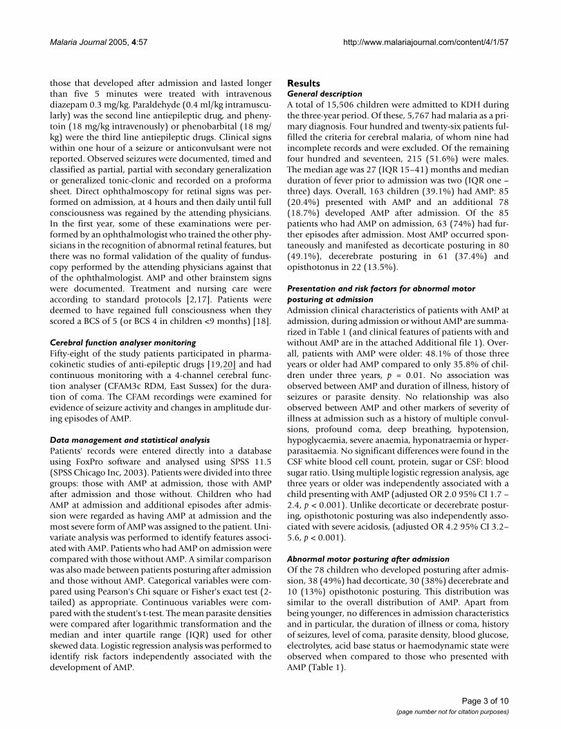
The median time from admission to the time of AMP was4.0 (IQR 1.9 – 8.8) hours. Fifty-two patients developedposturing within six hours and the remaining 26 patientshad posturing after six hours (Figure 1). Two childrendeveloped opisthotonus 32 and 55 hours after admission.No significant age, sex or clinical differences wereobserved between those who developed posturing beforeor after six hours. However, most of those who developed
opisthotonic posturing after admission did so after morethan six hours of admission: of the 26 patients who devel-oped AMP after 6 hours, 6 (23.3%) had opisthotonic pos-turing compared to only four out of 52 (7.7%) with AMPwithin six hours of admission, (OR 2.0 95% CI 1.1 – 3.8).Features of RICP on funduscopy (congested retinal veins,blurred disc margins or overt papilloedema) were the onlyclinical feature predictive for the development of postur-
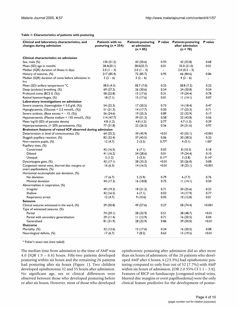
Table 1: Characteristics of patients with posturing
Clinical and laboratory characteristics, and changes during admission
Patients with no posturing (n = 254)
Patients posturing at admission
(n = 85)
P value Patients posturing after admission
(n = 78)
P value
Clinical characteristics on admissionSex, male (%) 130 (51.2) 43 (50.6) 0.93 42 (53.8) 0.68Mean (SD) age in months 28.4(20.1) 38.0(25.7) 0.01 35.0 (21.0) 0.01Median (IQR) duration of illness in days 2.0 (1 – 3) 2.0 (1 – 3) - 2.0 (0.3 – 3) -History of seizures, (%) 217 (85.4) 72 (85.7) 0.95 66 (84.6) 0.86Median (IQR) duration of coma before admission in hrs
3 (2 – 6) 3 (2 – 6) - 4 (2 – 6) -
Mean (SD) axillary temperature °C 38.0 (4.3) 38.7 (7.0) 0.25 38.8 (7.2) 0.22Deep (acidotic) breathing, (%) 69 (27.2) 26 (30.6) 0.54 24 (30.8) 0.54Profound coma (BCS 0, (%)) 58 (22.8) 15 (17.6) 0.31 19 (24.4) 0.78Retinal haemorrhages, (%) 18 (7.1) 15 (17.6) 0.01 11 (14.1) 0.07Laboratory investigations on admissionSevere anaemia, (haemoglobin < 5.0 g/dl, (%)) 54 (22.3) 17 (20.5) 0.73 14 (18.4) 0.47Hypoglycaemia, (Glucose < 2.2 mmol/L, (%)) 51 (21.3) 14 (17.7) 0.50 17 (23.3) 0.71Severe acidosis, (Base excess < – 15, (%)) 56 (24.6) 19 (25.3) 0.89 23 (32.9) 0.17Hyponatraemia, (Plasma sodium < 135 mmol/L, (%)) 114 (47.7) 39 (51.3) 0.58 32 (43.8) 0.56Mean log10 (SD) of parasite density 4.8 (1.2) 4.8 (1.2) 0.77 4.7 (1.3) 0.39Hyperparasitaemia, (> 20% parasitaemia, (%)) 77 (31.8) 22 (26.5) 0.36 24 (31.6) 0.97Brainstem features of raised ICP observed during admissionDeterioration in level of consciousness, (%) 64 (25.2) 39 (45.9) <0.01 43 (55.1) <0.001Sluggish pupillary reaction, (%) 82 (32.4) 37 (43.5) 0.06 30 (38.5) 0.32Non-reactive pupils, (%) 12 (4.7) 3 (3.5) 0.77* 4 (5.1) 1.00*Pupillary sizes, (%)
Constricted 42 (16.5) 6 (7.1) 0.03 8 (10.3) 0.18Dilated 41 (16.2) 24 (28.6) 0.01 19 (24.4) 0.10Unequal 3 (1.2) 3 (3.5) 0.17* 3 (3.8) 0.14*
Dysconjugate gaze, (%) 42 (17.1) 28 (33.3) <0.01 20 (26.0) 0.08Congested retinal veins, blurred disc margins or overt papilloedema, (%)
16 (6.3) 14 (16.5) <0.01 18 (23.1) <0.001
Horizontal oculocephalic eye deviation, (%)No deviation 17 (6.7) 5 (5.9) 0.79 6 (7.7) 0.76Minimal deviation 44 (17.3) 16 (18.8) 0.75 11 (14.1) 0.50
Abnormalities in respiration, (%)Irregular 49 (19.3) 18 (21.2) 0.71 20 (25.6) 0.23Shallow 42 (16.5) 6 (7.1) 0.03 14 (17.9) 0.77Respiratory arrest 12 (4.7) 9 (10.6) 0.05 10 (12.8) 0.01
SeizuresClinical seizures witnessed in the ward, (%) 29 (50.8) 49 (57.6) 0.27 58 (74.4) <0.001Type of witnessed seizures, (%)
Partial 74 (29.1) 28 (32.9) 0.51 38 (48.7) <0.01Partial with secondary generalisation 29 (11.4) 11 (12.9) 0.71 16 (20.5) 0.04Generalised 81 (31.9) 28 (32.9) 0.86 39 (50.0) <0.01
OutcomeMortality, (%) 32 (12.6) 15 (17.6) 0.24 16 (20.5) 0.08Neurological deficits, (%) 17 (6.7) 7 (8.2) 0.63 15 (19.5) <0.01
* Fisher's exact test (two tailed)
Page 4 of 10(page number not for citation purposes)
Malaria Journal 2005, 4:57 http://www.malariajournal.com/content/4/1/57
ing after admission, (adjusted OR 1.9 95% CI 1.7 – 2.2, p< 0.001).
Clinical events after admissionDeterioration in consciousness observed during the fourhourly assessments, recurrence of seizures and brainstemfeatures of raised ICP were the most significant clinicalevents after admission. The level of consciousness deteri-orated in over 50% of patients with AMP, within 24 hoursof admission and often, just before further posturing epi-sodes (Table 1).
Abnormal motor posturing and features of intracranial hypertensionSeveral brainstem signs consistent with different stages ofraised ICP were observed. Abnormalities of pupillary sizeand reaction, eye movements, disorders of conjugate gaze,and changes on funduscopy were the most common fea-tures (Table 1). Motor abnormalities of tone and reflexeswere also seen. Raised ICP was associated with all threetypes of posturing (Table 2), although patients with deco-rticate posturing had earlier funduscopic features such ascongested retinal veins, unlike those with decerebrate pos-turing who had papilloedema more frequently.
Thirty-two patients thought to have severe intracranialhypertension usually with papilloedema or clinical signscompatible with progressive herniation (23 of whom hadAMP), were given mannitol (0.5 g/kg infused over 20minutes up to a maximum of three doses). No further epi-sodes of AMP were observed in 12 (52.2%) after theadministration of mannitol. Fifteen of the 32 patients(46.9%) died and five (15.6%) showed severe neurologi-cal deficits. Fourteen of the 15 deaths occurred in patientswith AMP.
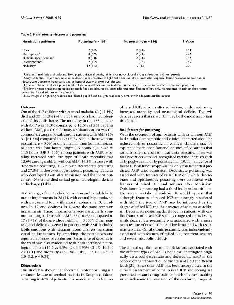
Herniation syndromesPatients were assessed for features of transtentorial herni-ation based upon the Plum and Posner criteria adaptedfor children with cerebral malaria [5,11]. Forty-eightpatients (11.5%) fulfilled a criterion for herniation (Table3). Thirty three (68.8%) of these died compared to only30 (8.1%) of those without features of herniation, (OR12.4 95% CI 7.1 – 21.4, p < 0.001). Most patients withAMP who died had features of herniation (19/31), withmost deaths following a respiratory arrest.
Seizures after admissionClinical seizures were observed in 107/163 (65.6%)patients with AMP compared to 129/254 (50.8%) ofthose without AMP (Table 1). Most patients with AMPhad multiple short seizures, often lasting one to threeminutes and 20% had five or more seizures witnessedover the duration of admission. Seizures were particularlycommon among those who developed AMP after admis-sion, often involving the mouth, face and limbs and weretwice as common as that in patients without AMP. Anassociation was observed between the type of AMP andrecurrence of seizures in the ward; no posturing 50.8%,decorticate posturing, 60.0%, decerebrate, 68.9% andopisthotonus, 77.3% (χ2 = 11.4, p = 0.01 for trend). Themedian number of witnessed seizures among patientswith opisthotonus was three compared to one in patientswith either decerebrate or decorticate posturing.
Thirty five of the 85 patients with AMP at admissionreceived intravenous phenytoin. AMP recurred in 22/35(62.9%) of those who received phenytoin compared to41/50 (82.0%) of those who did not receive phenytoin,but this difference was not statistically significant.
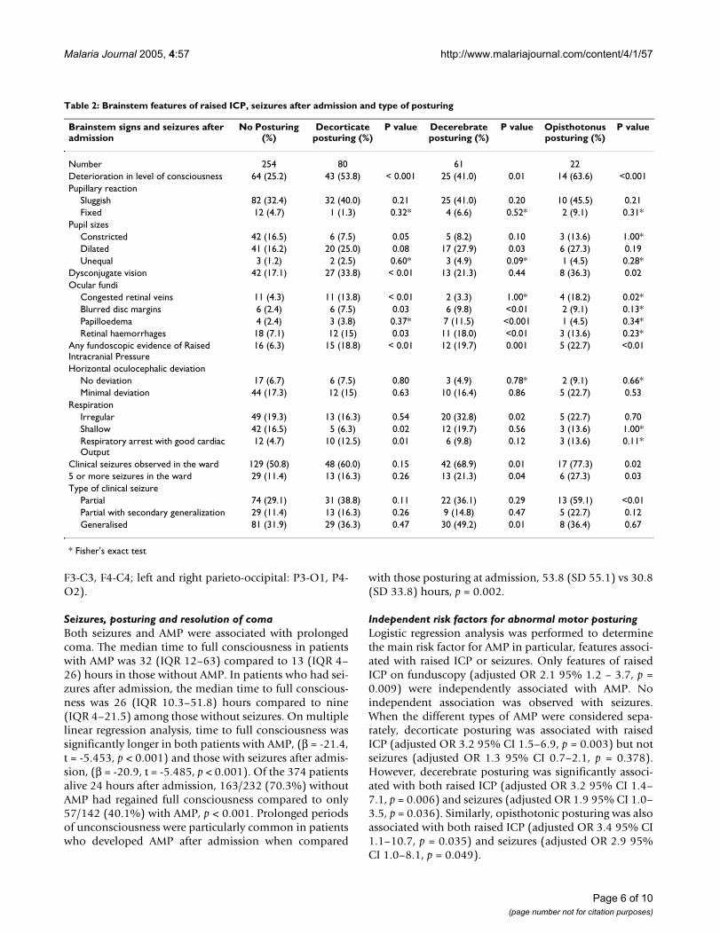
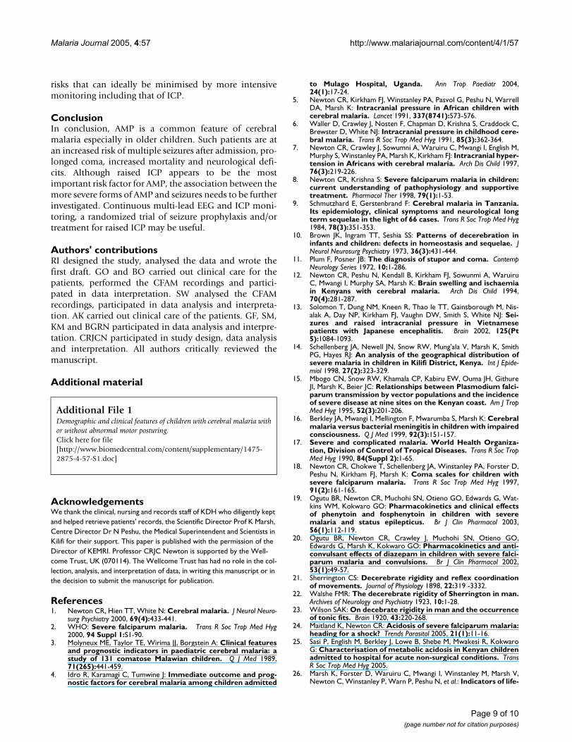
Abnormalities on cerebral function analyser monitoringTwenty-one of the 58 patients (36%) monitored with theCFAM had AMP, a proportion similar to that seen inwhole group. Recordings were examined for evidence ofseizure activity and changes in background electrical activ-ity during AMP. Patients with AMP had elevated peaks inthe amplitude of the background electrical activity, whichwere not seen in those without AMP. During posturing,transient increases in the amplitude of the electroen-cephalographic (EEG) background activity were observed(Figure 2). These were most marked with opisthotonicposturing and more pronounced with decerebrate thanwith decorticate posturing. The mean peak-to-peak ampli-tude in patients with decorticate posturing increased tojust below 100 µV (CFAM voltage readings are in a loga-rithmic scale starting at 1, 10 and 100 µV), while in decer-ebrate and opisthotonic posturing it exceeded 100 µV.There was no evidence of seizure activity during AMP inthe areas monitored by EEG (left and right fronto-central:
Time from admission to onset of posturingFigure 1Time from admission to onset of posturing.
Time to posturing after admission (hours)
0 6 12 18 24 30 36
Nu
mb
er o
f p
atie
nts
0
2
4
6
8
10
12
14
16
18
20
22
54 60
Page 5 of 10(page number not for citation purposes)
Malaria Journal 2005, 4:57 http://www.malariajournal.com/content/4/1/57
Table 2: Brainstem features of raised ICP, seizures after admission and type of posturing
Brainstem signs and seizures after admission
No Posturing (%)
Decorticate posturing (%)
P value Decerebrate posturing (%)
P value Opisthotonus posturing (%)
P value
Number 254 80 61 22Deterioration in level of consciousness 64 (25.2) 43 (53.8) < 0.001 25 (41.0) 0.01 14 (63.6) <0.001Pupillary reaction
Sluggish 82 (32.4) 32 (40.0) 0.21 25 (41.0) 0.20 10 (45.5) 0.21Fixed 12 (4.7) 1 (1.3) 0.32* 4 (6.6) 0.52* 2 (9.1) 0.31*
Pupil sizesConstricted 42 (16.5) 6 (7.5) 0.05 5 (8.2) 0.10 3 (13.6) 1.00*Dilated 41 (16.2) 20 (25.0) 0.08 17 (27.9) 0.03 6 (27.3) 0.19Unequal 3 (1.2) 2 (2.5) 0.60* 3 (4.9) 0.09* 1 (4.5) 0.28*
Dysconjugate vision 42 (17.1) 27 (33.8) < 0.01 13 (21.3) 0.44 8 (36.3) 0.02Ocular fundi
Congested retinal veins 11 (4.3) 11 (13.8) < 0.01 2 (3.3) 1.00* 4 (18.2) 0.02*Blurred disc margins 6 (2.4) 6 (7.5) 0.03 6 (9.8) <0.01 2 (9.1) 0.13*Papilloedema 4 (2.4) 3 (3.8) 0.37* 7 (11.5) <0.001 1 (4.5) 0.34*Retinal haemorrhages 18 (7.1) 12 (15) 0.03 11 (18.0) <0.01 3 (13.6) 0.23*
Any fundoscopic evidence of Raised Intracranial Pressure
16 (6.3) 15 (18.8) < 0.01 12 (19.7) 0.001 5 (22.7) <0.01
Horizontal oculocephalic deviationNo deviation 17 (6.7) 6 (7.5) 0.80 3 (4.9) 0.78* 2 (9.1) 0.66*Minimal deviation 44 (17.3) 12 (15) 0.63 10 (16.4) 0.86 5 (22.7) 0.53
RespirationIrregular 49 (19.3) 13 (16.3) 0.54 20 (32.8) 0.02 5 (22.7) 0.70Shallow 42 (16.5) 5 (6.3) 0.02 12 (19.7) 0.56 3 (13.6) 1.00*Respiratory arrest with good cardiac Output
12 (4.7) 10 (12.5) 0.01 6 (9.8) 0.12 3 (13.6) 0.11*
Clinical seizures observed in the ward 129 (50.8) 48 (60.0) 0.15 42 (68.9) 0.01 17 (77.3) 0.025 or more seizures in the ward 29 (11.4) 13 (16.3) 0.26 13 (21.3) 0.04 6 (27.3) 0.03Type of clinical seizure
Partial 74 (29.1) 31 (38.8) 0.11 22 (36.1) 0.29 13 (59.1) <0.01Partial with secondary generalization 29 (11.4) 13 (16.3) 0.26 9 (14.8) 0.47 5 (22.7) 0.12Generalised 81 (31.9) 29 (36.3) 0.47 30 (49.2) 0.01 8 (36.4) 0.67
* Fisher's exact test
F3-C3, F4-C4; left and right parieto-occipital: P3-O1, P4-O2).
Seizures, posturing and resolution of comaBoth seizures and AMP were associated with prolongedcoma. The median time to full consciousness in patientswith AMP was 32 (IQR 12–63) compared to 13 (IQR 4–26) hours in those without AMP. In patients who had sei-zures after admission, the median time to full conscious-ness was 26 (IQR 10.3–51.8) hours compared to nine(IQR 4–21.5) among those without seizures. On multiplelinear regression analysis, time to full consciousness wassignificantly longer in both patients with AMP, (β = -21.4,t = -5.453, p < 0.001) and those with seizures after admis-sion, (β = -20.9, t = -5.485, p < 0.001). Of the 374 patientsalive 24 hours after admission, 163/232 (70.3%) withoutAMP had regained full consciousness compared to only57/142 (40.1%) with AMP, p < 0.001. Prolonged periodsof unconsciousness were particularly common in patientswho developed AMP after admission when compared
with those posturing at admission, 53.8 (SD 55.1) vs 30.8(SD 33.8) hours, p = 0.002.
Independent risk factors for abnormal motor posturingLogistic regression analysis was performed to determinethe main risk factor for AMP in particular, features associ-ated with raised ICP or seizures. Only features of raisedICP on funduscopy (adjusted OR 2.1 95% 1.2 – 3.7, p =0.009) were independently associated with AMP. Noindependent association was observed with seizures.When the different types of AMP were considered sepa-rately, decorticate posturing was associated with raisedICP (adjusted OR 3.2 95% CI 1.5–6.9, p = 0.003) but notseizures (adjusted OR 1.3 95% CI 0.7–2.1, p = 0.378).However, decerebrate posturing was significantly associ-ated with both raised ICP (adjusted OR 3.2 95% CI 1.4–7.1, p = 0.006) and seizures (adjusted OR 1.9 95% CI 1.0–3.5, p = 0.036). Similarly, opisthotonic posturing was alsoassociated with both raised ICP (adjusted OR 3.4 95% CI1.1–10.7, p = 0.035) and seizures (adjusted OR 2.9 95%CI 1.0–8.1, p = 0.049).
Page 6 of 10(page number not for citation purposes)
Malaria Journal 2005, 4:57 http://www.malariajournal.com/content/4/1/57
OutcomeOut of the 417 children with cerebral malaria, 63 (15.1%)died and 39 (11.0%) of the 354 survivors had neurologi-cal deficits at discharge. The mortality in the 163 patientswith AMP was 19.0% compared to 12.6% of 254 patientswithout AMP, p = 0.07. Primary respiratory arrest was thecommonest cause of death among patients with AMP (19/31 [61.3%] compared to 12/32 [37.5%] in those withoutposturing, p = 0.06) and the median time from admissionto death was four hours longer (15 hours IQR 3–48 vs11.5 hours IQR 3–106) among patients with AMP. Mor-tality increased with the type of AMP: mortality was12.6% among children without AMP, 16.3% in those withdecorticate posturing, 19.7% with decerebrate posturingand 27.3% in those with opisthotonic posturing. Patientswho developed AMP after admission had the worst out-come; 40% either died or had gross neurological deficitsat discharge (Table 1).
At discharge, of the 39 children with neurological deficits,motor impairments in 28 (18 with central hypotonia, sixwith paresis and four with ataxia), aphasia in 13, blind-ness in 12 and deafness in 6 were the most commonimpairments. These impairments were particularly com-mon among patients with AMP: 22 (16.7%) compared to17 (7.7%) of those without AMP, p = 0.009). Other neu-rological deficits observed in patients with AMP includedlabile emotions with frequent mood changes, persistentvisual hallucinations, lip smacking, choreoathetosis andrepeated episodes of confusion. Recurrence of seizures inthe ward was also associated with both increased neuro-logical deficits (16.6 vs 4.3%, OR 4.4 95% CI 1.9–10.2, p< 0.001) and mortality (18.2 vs 11.0%, OR 1.8 95% CI1.0–3.2, p = 0.04).
DiscussionThis study has shown that abnormal motor posturing is acommon feature of cerebral malaria in Kenyan children,occurring in 40% of patients. It is associated with features
of raised ICP, seizures after admission, prolonged coma,increased mortality and neurological deficits. The evi-dence suggests that raised ICP may be the most importantrisk factor.
Risk factors for posturingWith the exception of age, patients with or without AMPhad similar demographic and clinical characteristics. Thereduced risk of posturing in younger children may beexplained by an open fontanel or uncalcified sutures thatcan dissipate increases in intracranial pressure. There wasno association with well recognised metabolic causes suchas hypoglycaemia or hyponatraemia [10,11]. Evidence ofraised ICP on funduscopy was the only risk factor that pre-dicted AMP after admission. Decorticate posturing wasassociated with features of raised ICP only while decere-brate and opisthotonic posturing were associated withfeatures of raised ICP and seizures after admission.Opisthotonic posturing had a third independent risk fac-tor, severe metabolic acidosis. It would appear thatalthough features of raised ICP are strongly associatedwith AMP, the type of AMP may be influenced by thedegree of raised ICP and the presence of seizures or acido-sis. Decorticate posturing developed in patients with ear-lier features of raised ICP such as congested retinal veinswhile decerebrate posturing was associated with a moreovert feature of raised ICP, papilloedema, and with recur-rent seizures. Opisthotonic posturing was independentlyassociated with features of raised ICP, recurrent seizuresand severe metabolic acidosis.
The clinical significance of the risk factors associated withthe different types of AMP is not clear. Sherrington origi-nally described decorticate and decerebrate AMP in thecontext of the trans-section of the brain of a cat at differentlevels[21]. Since then, AMP has been incorporated in theclinical assessment of coma. Raised ICP and coning arepresumed to cause compression of the brainstem resultingin an ischaemic trans-section of the cerebrum, "separat-
Table 3: Herniation syndromes and posturing
Herniation syndromes Posturing (n = 163) No posturing (n = 254) P Value
Uncal1 2 (1.2) 2 (0.8) 0.64Diencephalic2 8 (4.9) 2 (0.8) 0.02Midbrain/upper pontine3 0 (0.0) 2 (0.8) 0.52Lower pontine4 2 (1.2) 1 (0.4) 0.56Medullary5 19 (11.7) 12 (4.7) 0.01
1 Unilateral mydriasis and unilateral fixed pupil, unilateral ptosis, minimal or no oculocephalic eye deviation and hemiparesis2 Cheynes-Stokes respiration, small or midpoint pupils reactive to light, full deviation of oculocephalic response, flexor response to pain and/or decorticate posturing, hypertonia and or hypereflexia with extensor planters3 Hyperventilation, midpoint pupils fixed to light, minimal oculocephalic deviation, extensor response to pain or decerebrate posturing4 Shallow or ataxic respiration, midpoint pupils fixed to light, no oculocephalic response, flexion of legs only, no response to pain or decorticate posturing, flaccid with extensor planters5 Slow irregular or gasping respirations, dilated pupils fixed to light, respiratory arrest with adequate cardiac output
Page 7 of 10(page number not for citation purposes)

Malaria Journal 2005, 4:57 http://www.malariajournal.com/content/4/1/57
ing" it from the brainstem [11,22,23]. This possibly maybe the predominant mechanism in the causation of AMPin children with cerebral malaria, given the considerableclinical evidence for raised ICP observed in these patients.
Abnormal motor posturing, seizures and raised ICPAMP was associated with recurrence of seizures afteradmission, mostly as short multiple seizures, but not sei-zures before admission. CFAM recordings showedincreases in the mean peak-to-peak amplitude of the EEGbackground activity, most marked during opisthotonicposturing. These amplitude increases may possibly reflectacute elevations of ICP. There was no indication of anyepileptiform activity during AMP recorded from the fourleads of the CFAM. It is however possible that these leadswere not able to detect localized discharges.
The association between the more severe forms of AMP –decerebrate and opisthotonic posturing – and seizuresmay arise because seizures increase ICP, PCO2 and cere-bral blood flow, which then leads to AMP. Increased met-abolic demands and cerebral blood flow during seizuresmay aggravate the critical perfusion state. It is less plausi-ble that AMP itself represents an ictal phenomenon, sincethere is no evidence of a temporal association between sei-zure activity on CFAM and the occurrence of posturing.Continuous ICP and EEG monitoring would allow furtherinvestigation of the relationship between electrical sei-zures and AMP.
Posturing and outcomeIn many encephalopathies, AMP is associated with a poorprognosis [3,10]. A similar trend was observed in thisstudy. In addition to being a sign of lower brainstem com-pression, the particularly high mortality in patients withopisthotonic posturing may be a result of the combinedeffects of a more severe encephalopathy with raised ICP,repeated seizures and severe metabolic acidosis. Severemetabolic acidosis in African children with malaria hasbeen associated with shock/hypovolaemia, anaemia, lac-tic acidosis and other ketones [24,25]. Those with cerebralmalaria and concurrent severe metabolic acidosis have thehighest severe malaria mortality [26]. The association ofopisthotonic posturing with severe acidosis suggests thatthis type of abnormal posturing develops in very sickpatients with the highest risk of death.
The median time to death in patients with AMP was fourhours longer than that in patients without AMP and mor-tality was highest in those who developed AMP after sixhours of admission. The majority of deaths had a respira-tory arrest with initially an adequate cardiac output, aphenomenon associated with medullary herniation syn-drome [5,7,11]. Herniation syndromes could be recog-nized in two-thirds of deaths in patients with posturingand over 50% of all deaths [13]. Effective treatment ofintracranial hypertension may, therefore, be able toreduce the mortality of cerebral malaria in African chil-dren especially in patients who develop posturing afteradmission. The additional four hours in the median timeto death provide an opportunity for the use of measuresaimed at lowering ICP. Patients that develop AMP afteradmission may be the best group to target.
Posturing and therapy for raised ICPNo guidelines exist for the use of mannitol in cerebralmalaria [2], but it was administered to 32 patients in thisseries. Despite mannitol administration, 15 patients diedand another five developed severe neurological deficits.Uncontrolled studies of mannitol in cerebral malariashow that it reduces ICP but may not prevent subsequentdevelopment of severe intracranial hypertension [5]. Theuse of mannitol or other osmotic diuretics requires furtherinvestigation. Most district hospitals across sub-SaharanAfrica that also treat the majority of patients with cerebralmalaria are unable to offer ventilation to patients withraised ICP [27]. Since most patients develop AMP severalhours before death, posturing may be used as an earlymarker for clinicians to start initial treatment for raisedICP: patient positioning, adequate oxygenation and per-fusion, temperature and seizure control, and possiblyoffer mannitol therapy. This can be done with the hopethat at this stage, patients still have mild or moderatelyelevated intracranial pressures that may respond to man-nitol [7]. However, the use of osmotic diuretics may have
Cerebral function analyser monitoring tracings before and during opisthotonic posturing in a child with cerebral malariaFigure 2Cerebral function analyser monitoring tracings before and during opisthotonic posturing in a child with cerebral malaria.
Before posturing During posturing: the peak-peak amplitude is
elevated above threshold.
Page 8 of 10(page number not for citation purposes)
Malaria Journal 2005, 4:57 http://www.malariajournal.com/content/4/1/57
risks that can ideally be minimised by more intensivemonitoring including that of ICP.
ConclusionIn conclusion, AMP is a common feature of cerebralmalaria especially in older children. Such patients are atan increased risk of multiple seizures after admission, pro-longed coma, increased mortality and neurological defi-cits. Although raised ICP appears to be the mostimportant risk factor for AMP, the association between themore severe forms of AMP and seizures needs to be furtherinvestigated. Continuous multi-lead EEG and ICP moni-toring, a randomized trial of seizure prophylaxis and/ortreatment for raised ICP may be useful.
Authors' contributionsRI designed the study, analysed the data and wrote thefirst draft. GO and BO carried out clinical care for thepatients, performed the CFAM recordings and partici-pated in data interpretation. SW analysed the CFAMrecordings, participated in data analysis and interpreta-tion. AK carried out clinical care of the patients. GF, SM,KM and BGRN participated in data analysis and interpre-tation. CRJCN participated in study design, data analysisand interpretation. All authors critically reviewed themanuscript.
Additional material
AcknowledgementsWe thank the clinical, nursing and records staff of KDH who diligently kept and helped retrieve patients' records, the Scientific Director Prof K Marsh, Centre Director Dr N Peshu, the Medical Superintendent and Scientists in Kilifi for their support. This paper is published with the permission of the Director of KEMRI. Professor CRJC Newton is supported by the Well-come Trust, UK (070114). The Wellcome Trust has had no role in the col-lection, analysis, and interpretation of data, in writing this manuscript or in the decision to submit the manuscript for publication.
References1. Newton CR, Hien TT, White N: Cerebral malaria. J Neurol Neuro-
surg Psychiatry 2000, 69(4):433-441.2. WHO: Severe falciparum malaria. Trans R Soc Trop Med Hyg
2000, 94 Suppl 1:S1-90.3. Molyneux ME, Taylor TE, Wirima JJ, Borgstein A: Clinical features
and prognostic indicators in paediatric cerebral malaria: astudy of 131 comatose Malawian children. Q J Med 1989,71(265):441-459.
4. Idro R, Karamagi C, Tumwine J: Immediate outcome and prog-nostic factors for cerebral malaria among children admitted
to Mulago Hospital, Uganda. Ann Trop Paediatr 2004,24(1):17-24.
5. Newton CR, Kirkham FJ, Winstanley PA, Pasvol G, Peshu N, WarrellDA, Marsh K: Intracranial pressure in African children withcerebral malaria. Lancet 1991, 337(8741):573-576.
6. Waller D, Crawley J, Nosten F, Chapman D, Krishna S, Craddock C,Brewster D, White NJ: Intracranial pressure in childhood cere-bral malaria. Trans R Soc Trop Med Hyg 1991, 85(3):362-364.
7. Newton CR, Crawley J, Sowumni A, Waruiru C, Mwangi I, English M,Murphy S, Winstanley PA, Marsh K, Kirkham FJ: Intracranial hyper-tension in Africans with cerebral malaria. Arch Dis Child 1997,76(3):219-226.
8. Newton CR, Krishna S: Severe falciparum malaria in children:current understanding of pathophysiology and supportivetreatment. Pharmacol Ther 1998, 79(1):1-53.
9. Schmutzhard E, Gerstenbrand F: Cerebral malaria in Tanzania.Its epidemiology, clinical symptoms and neurological longterm sequelae in the light of 66 cases. Trans R Soc Trop Med Hyg1984, 78(3):351-353.
10. Brown JK, Ingram TT, Seshia SS: Patterns of decerebration ininfants and children: defects in homeostasis and sequelae. JNeurol Neurosurg Psychiatry 1973, 36(3):431-444.
11. Plum F, Posner JB: The diagnosis of stupor and coma. ContempNeurology Series 1972, 10:1-286.
12. Newton CR, Peshu N, Kendall B, Kirkham FJ, Sowunmi A, WaruiruC, Mwangi I, Murphy SA, Marsh K: Brain swelling and ischaemiain Kenyans with cerebral malaria. Arch Dis Child 1994,70(4):281-287.
13. Solomon T, Dung NM, Kneen R, Thao le TT, Gainsborough M, Nis-alak A, Day NP, Kirkham FJ, Vaughn DW, Smith S, White NJ: Sei-zures and raised intracranial pressure in Vietnamesepatients with Japanese encephalitis. Brain 2002, 125(Pt5):1084-1093.
14. Schellenberg JA, Newell JN, Snow RW, Mung'ala V, Marsh K, SmithPG, Hayes RJ: An analysis of the geographical distribution ofsevere malaria in children in Kilifi District, Kenya. Int J Epide-miol 1998, 27(2):323-329.
15. Mbogo CN, Snow RW, Khamala CP, Kabiru EW, Ouma JH, GithureJI, Marsh K, Beier JC: Relationships between Plasmodium falci-parum transmission by vector populations and the incidenceof severe disease at nine sites on the Kenyan coast. Am J TropMed Hyg 1995, 52(3):201-206.
16. Berkley JA, Mwangi I, Mellington F, Mwarumba S, Marsh K: Cerebralmalaria versus bacterial meningitis in children with impairedconsciousness. Q J Med 1999, 92(3):151-157.
17. Severe and complicated malaria. World Health Organiza-tion, Division of Control of Tropical Diseases. Trans R Soc TropMed Hyg 1990, 84(Suppl 2):1-65.
18. Newton CR, Chokwe T, Schellenberg JA, Winstanley PA, Forster D,Peshu N, Kirkham FJ, Marsh K: Coma scales for children withsevere falciparum malaria. Trans R Soc Trop Med Hyg 1997,91(2):161-165.
19. Ogutu BR, Newton CR, Muchohi SN, Otieno GO, Edwards G, Wat-kins WM, Kokwaro GO: Pharmacokinetics and clinical effectsof phenytoin and fosphenytoin in children with severemalaria and status epilepticus. Br J Clin Pharmacol 2003,56(1):112-119.
20. Ogutu BR, Newton CR, Crawley J, Muchohi SN, Otieno GO,Edwards G, Marsh K, Kokwaro GO: Pharmacokinetics and anti-convulsant effects of diazepam in children with severe falci-parum malaria and convulsions. Br J Clin Pharmacol 2002,53(1):49-57.
21. Sherrington CS: Decerebrate rigidity and reflex coordinationof movements. Journal of Physiology 1898, 22:319 -3332.
22. Walshe FMR: The decerebrate rigidity of Sherrington in man.Archives of Neurology and Psychiatry 1923, 10:1-28.
23. Wilson SAK: On decebrate rigidity in man and the occurrenceof tonic fits. Brain 1920, 43:220-268.
24. Maitland K, Newton CR: Acidosis of severe falciparum malaria:heading for a shock? Trends Parasitol 2005, 21(1):11-16.
25. Sasi P, English M, Berkley J, Lowe B, Shebe M, Mwakesi R, KokwaroG: Characterisation of metabolic acidosis in Kenyan childrenadmitted to hospital for acute non-surgical conditions. TransR Soc Trop Med Hyg 2005.
26. Marsh K, Forster D, Waruiru C, Mwangi I, Winstanley M, Marsh V,Newton C, Winstanley P, Warn P, Peshu N, et al.: Indicators of life-
Additional File 1Demographic and clinical features of children with cerebral malaria with or without abnormal motor posturing.Click here for file[http://www.biomedcentral.com/content/supplementary/1475-2875-4-57-S1.doc]
Page 9 of 10(page number not for citation purposes)

Malaria Journal 2005, 4:57 http://www.malariajournal.com/content/4/1/57
Publish with BioMed Central and every scientist can read your work free of charge
"BioMed Central will be the most significant development for disseminating the results of biomedical research in our lifetime."
Sir Paul Nurse, Cancer Research UK
Your research papers will be:
available free of charge to the entire biomedical community
peer reviewed and published immediately upon acceptance
cited in PubMed and archived on PubMed Central
yours — you keep the copyright
Submit your manuscript here:http://www.biomedcentral.com/info/publishing_adv.asp
BioMedcentral
threatening malaria in African children. N Engl J Med 1995,332(21):1399-1404.
27. Tomlinson RJ, Morrice J: Does intravenous mannitol improveoutcome in cerebral malaria? Arch Dis Child 2003,88(7):640-641.
Page 10 of 10(page number not for citation purposes)
Copyright © 2022 FDOKUMEN