Plexin-A4 Mediates Cytotoxic T-cell Trafficking and Exclusion ...

Original article
Cytotoxic activity of new neodymium (III) complexes of bis-coumarins
Irena Kostova a,*, Ilia Manolov b, Georgi Momekov c
a Department of Chemistry, Faculty of Pharmacy, Medical University, 2 Dunav St., Sofia 1000, Bulgariab Department of Organic Chemistry, Faculty of Pharmacy, Medical University, 2 Dunav St., Sofia 1000, Bulgaria
c Department of Pharmacology and Toxicology, Faculty of Pharmacy, Medical University, 2 Dunav St., Sofia 1000, Bulgaria
Received 9 January 2004; received in revised form 28 May 2004; accepted 1 June 2004
Available online 23 July 2004
Abstract
Complexes of neodymium (III) with bis-coumarins: 3,3′-benzylidene-bis(4-hydroxy-2H-1-benzopyran-2-one); bis(4-hydroxy-2-oxo-2H-chromen-3-yl)-piridin-2-yl-methane; bis(4-hydroxy-2-oxo-2H-chromen-3-yl)-piridin-4-yl-methane; bis(4-hydroxy-2-oxo-2H-chromen-3-yl)-(1H-pyrazol-3-yl)-methane were synthesized by reaction of neodymium (III) salt and the ligands, in amounts equal to metal:ligand molarratio of 1:2. The complexes were prepared by adding an aqueous solution of neodymium (III) salt to an aqueous solution of the ligandsubsequently raising the pH of the mixture gradually to ca. 5.0 by adding dilute solution of sodium hydroxide. The neodymium (III) complexeswith bis-coumarins were characterized by different physicochemical methods—elemental analysis, IR-, 1H- and 13C-NMR-spectroscopiesand mass-spectral data. The spectral data of neodymium (III) complexes were interpreted on the basis of comparison with the spectra of thefree ligands. This analysis showed that in the Nd (III) complexes the ligands coordinated to the metal ion through both deprotonated hydroxylgroups. On the basis of the m(C=O) red shift observed, participation of the carbonyl groups in the coordination to the metal ion was alsosuggested. Cytotoxic screening by MTT assay was carried out. The complexes were tested on HL-60, HL-60/Dox and SKW-3 cell lines. Theoverall results from the preliminary screening program revealed that all of the new Nd (III) complexes reach 50% inhibition of the malignantcells proliferation and thus could be considered as biologically active. On the basis of the IC50 values obtained compounds Nd(L1)(OH).H2Oand Nd(L3)(OH).2H2O were found to exert superior activity in comparison to the remaining complexes.© 2004 Elsevier SAS. All rights reserved.
Keywords: Bis-coumarins; Neodymium (III) complexes; IR- and NMR-spectra; Cytotoxic activity
1. Introduction
Coumarin is used widely as a therapeutic agent and isadministered clinically in the treatment of certain lymphede-mas and malignancies. 7-hydroxy- and 4-hydroxycoumarinsare naturally occurring substances with a variety of biologi-cal activities, e.g. antitumoral action.
The antitumor activities of coumarin and its known me-tabolite 7-hydroxy-coumarin were tested in several humantumor cell lines by Steffen et al. [1]. Both compounds inhib-ited cell proliferation of a gastric carcinoma cell line, acolon-carcinoma cell line (Caco-2), a hepatoma-derived cellline (Hep-G2) and a lymphoblastic cell line (CCRF cem).Egan et al. [2] have synthesized, characterized and deter-
mined cytostatic and cytotoxic nature of 8-nitro-7-hydroxycoumarin using both human (including K-562 andHL-60) and animal cell lines grown in vitro. Coumarin andits 4-hydroxy and 7-hydroxy derivatives, as well as o-, m-and p-coumaric acid were tested against P-815 and P-388tumor cells in vitro. All compounds were more or less cyto-toxic against tumor cells [3]. The effect of warfarin on tumorcell growth was studied [4]. Warfarin inhibits metastasis ofMtln3 rat mammary carcinoma without affecting primarytumor growth. Seven known coumarins, showing significantcytotoxic activities on P388 cell lines, were isolated from theroots of Angelica gigas (Umbelliferae) [5]. Akman et al. [6]had investigated synergistic cytotoxicity between menadioneand the related anticoagulant dicumarol, inhibited growth ofmurine leukemia L1210 in liquid suspension culture. Thecytotoxicity of 22 natural and semisynthetic simple cou-marins was evaluated in GLC4, a human small cell lung
* Corresponding author.E-mail address: [email protected] (I. Kostova).
European Journal of Medicinal Chemistry 39 (2004) 765–775
www.elsevier.com/locate/ejmech
0223-5234/$ - see front matter © 2004 Elsevier SAS. All rights reserved.doi:10.1016/j.ejmech.2004.06.002
carcinoma cell line, and in COLO 320, a human colorectalcancer cell line, using the microculture tetrazolium (MTT)assay [7].
A number of 4-hydroxycoumarin derivatives have beenstudied as to their HIV integrase inhibitory potency [8]. Themain purpose was to simplify the large structure of thecompounds while maintaining their potency. It was foundthat the minimum active pharmacophore consists of a cou-marin dimer containing an aryl substituent on the centrallinker, methylene. The addition of 4- and 7-hydroxy substitu-ents in the coumarin rings improved the potency of thecompounds. Among the systems studied, the 3,3′-benzyli-dene-bis(4-hydroxycoumarin) has been tested as a HIV inte-grase inhibitor and has shown significant activity [8]. Thecomplexation ability of the 3,3′-benzylidene-bis(4-hydro-xycoumarin) with lanthanides was not reported so far. It isexpected that the complexes with this ligand and with similarligands will retain or improve its biological activity as in thecase of other lanthanide complexes with hydroxycoumarinderivatives.
The complexes of rare earth ions have aroused muchinterest. Lanthanides are a subject of increasing interest inbioinorganic and coordination chemistry [9,10].
Nowadays, a lot of studies report complexes of coumarinderivatives with rare earth metals, which possess biologicalactivity. Thus, lanthanide complexes of 3-sulfo-4-hydroxy-coumarin [11] and bis-(4-hydroxy-3-coumarinyl)-acetic acid[12] have been synthesised and characterised. The complexeshave revealed good anticoagulant action.
Lanthanium chloride manifests an antitumor activity. Fur-thermore, literature data show that the coumarins have alsothese properties. These previous data from literature are inaccordance with our investigations. They give our reason tosuppose that complexes of coumarins with lanthanides couldpresent interesting metalorganic compounds with antitumoractivity. As a result from our earlier work the cytotoxicprofile of some complexes of mendiaxon, warfarin, cou-machlor and niffcoumar with lanthanides against P3HR1,K-562 and THP-1 cell lines was proved [13–18]. The com-plexes of cerium, lanthanum and neodymium with thesecoumarin ligands induced approximately 30% reduction ofthe survival of P3HR1 Burkitt lymphoma cells at concentra-tions 100 and 400 µM. The cerium and lanthanum complexesof mendiaxon and niffcoumar induce similar low cytotoxiceffect on AML derived THP-1 myeloleukemia cells. With therelatively resistant CML derived erythroleukemic K-562 cellline we obtained very interesting in vitro results. It is note-worthy that the lanthanum and neodymium complexes withniffcoumar exert more pronounced cytotoxic effects in com-parison to cerium complex. They have a strong cell prolifera-tion inhibiting effects (only about 30% of the cells survived).This means that the resistant tumor cells may be inhibitedwell with lanthanide complexes. This means also that thespectrum of cytotoxicity of these complexes is different fromcis-DDP (II) and from Pt (II) complexes. These results are of
some interest as a possibility to influence of resistant tumors.The corresponding lanthanide salts are found to be of verylow or missing activity. So far we can conclude that thestructure of metal-ligand determines the antitumor spectrumof the newly formed complexes. Those in vitro effects are notso clearly expressed as it is in the case of cis-DDP (II).Nevertheless their study is interesting in connection withother cell lines and tumors in order to find out the differencesin their spectrum of activity.
Unfortunately, little is known about the complexing abil-ity of neodymium (III) with coumarins. A survey of theliterature reveals that no work has been done on the reactionsof neodymium (III) with 3,3′-benzylidene-bis(4-hydroxy-2H-1-benzopyran-2-one) and its derivatives. It was, there-fore, considered worthwhile to study the complexation and inthe first place the objective of this study was to determinewhether the new complexes were active as cytotoxic agents.
In the present study, we perform investigation of the coor-dination ability of 3,3′-benzylidene-bis(4-hydroxy-2H-1-benzopyran-2-one); bis(4-hydroxy-2-oxo-2H-chromen-3-yl)-piridin-2-yl-methane; bis(4-hydroxy-2-oxo-2H-chro-men-3-yl)-piridin-4-yl-methane; bis(4-hydroxy-2-oxo-2H-chromen-3-yl)-(1H-pyrazol-3-yl)-methane in complexationreaction with neodymium (III). The obtained Nd (III) com-plexes with these coumarin ligands was characterized byelemental analysis, physicochemical methods, mass-, NMR-and IR-spectroscopy. The complicated vibrational spectra ofneodymium (III) complexes were interpreted on the basis ofcomparison with the vibrational spectra of the free ligands.The most sensitive to coordination modes of the ligands havebeen assigned and discussed.
We observed that Nd (III) possess a cytotoxic activity andliterature data show that the coumarins have also these prop-erties. That is why our synthesis of complexes of Nd (III) istaken into consideration with cytotoxic screening and furtherpharmacological study.
2. Chemistry
The compounds used for preparing the solutions wereMerck products, p.a. grade: Nd(NO3)3.6H2O. 3,3′-benzyli-dene-bis(4-hydroxy-2H-1-benzopyran-2-one), bis(4-hydroxy-2-oxo-2H-chromen-3-yl)-piridin-2-yl-methane, bis(4-hy-droxy-2-oxo-2H-chromen-3-yl)-piridin-4-yl-methane andbis(4-hydroxy-2-oxo-2H-chromen-3-yl)-(1H-pyrazol-3-yl)-methane were used for the preparation of metal complexes asligands (Scheme 1). These ligands were synthesized by con-densation of 4-hydroxycoumarin and aromatic or heterocy-clic aldehyde in ethanol medium at reflux and stirring untilcrystals appeared to us.
The complexes of neodymium (III) with 3,3′-benzyli-dene-bis(4-hydroxy-2H-1-benzopyran-2-one) (H2L1); bis-(4-hydroxy-2-oxo-2H-chromen-3-yl)-piridin-2-yl-methane(H2L2); bis(4-hydroxy-2-oxo-2H-chromen-3-yl)-piridin-4-yl-methane (H2L3); bis(4-hydroxy-2-oxo-2H-chromen-3-
766 I. Kostova et al. / European Journal of Medicinal Chemistry 39 (2004) 765–775
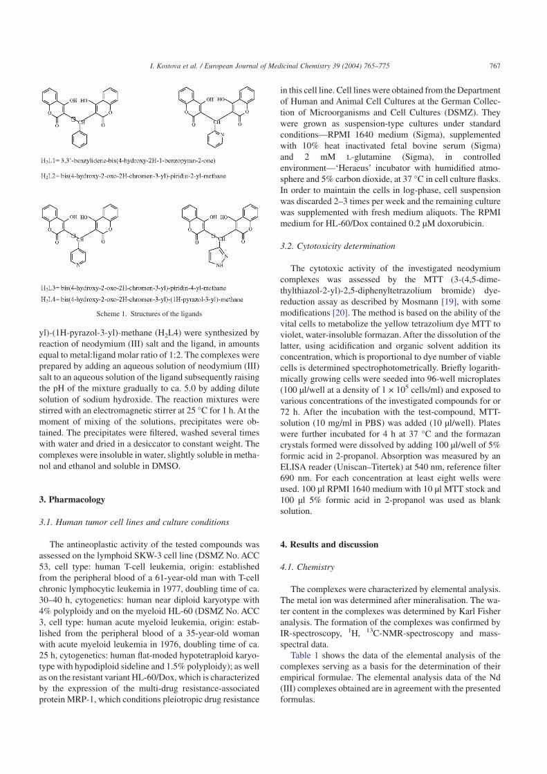
yl)-(1H-pyrazol-3-yl)-methane (H2L4) were synthesized byreaction of neodymium (III) salt and the ligand, in amountsequal to metal:ligand molar ratio of 1:2. The complexes wereprepared by adding an aqueous solution of neodymium (III)salt to an aqueous solution of the ligand subsequently raisingthe pH of the mixture gradually to ca. 5.0 by adding dilutesolution of sodium hydroxide. The reaction mixtures werestirred with an electromagnetic stirrer at 25 °C for 1 h. At themoment of mixing of the solutions, precipitates were ob-tained. The precipitates were filtered, washed several timeswith water and dried in a desiccator to constant weight. Thecomplexes were insoluble in water, slightly soluble in metha-nol and ethanol and soluble in DMSO.
3. Pharmacology
3.1. Human tumor cell lines and culture conditions
The antineoplastic activity of the tested compounds wasassessed on the lymphoid SKW-3 cell line (DSMZ No. ACC53, cell type: human T-cell leukemia, origin: establishedfrom the peripheral blood of a 61-year-old man with T-cellchronic lymphocytic leukemia in 1977, doubling time of ca.30–40 h, cytogenetics: human near diploid karyotype with4% polyploidy and on the myeloid HL-60 (DSMZ No. ACC3, cell type: human acute myeloid leukemia, origin: estab-lished from the peripheral blood of a 35-year-old womanwith acute myeloid leukemia in 1976, doubling time of ca.25 h, cytogenetics: human flat-moded hypotetraploid karyo-type with hypodiploid sideline and 1.5% polyploidy); as wellas on the resistant variant HL-60/Dox, which is characterizedby the expression of the multi-drug resistance-associatedprotein MRP-1, which conditions pleiotropic drug resistance
in this cell line. Cell lines were obtained from the Departmentof Human and Animal Cell Cultures at the German Collec-tion of Microorganisms and Cell Cultures (DSMZ). Theywere grown as suspension-type cultures under standardconditions—RPMI 1640 medium (Sigma), supplementedwith 10% heat inactivated fetal bovine serum (Sigma)and 2 mM L-glutamine (Sigma), in controlledenvironment—‘Heraeus’ incubator with humidified atmo-sphere and 5% carbon dioxide, at 37 °C in cell culture flasks.In order to maintain the cells in log-phase, cell suspensionwas discarded 2–3 times per week and the remaining culturewas supplemented with fresh medium aliquots. The RPMImedium for HL-60/Dox contained 0.2 µM doxorubicin.
3.2. Cytotoxicity determination
The cytotoxic activity of the investigated neodymiumcomplexes was assessed by the MTT (3-(4,5-dime-thylthiazol-2-yl)-2,5-diphenyltetrazolium bromide) dye-reduction assay as described by Mosmann [19], with somemodifications [20]. The method is based on the ability of thevital cells to metabolize the yellow tetrazolium dye MTT toviolet, water-insoluble formazan. After the dissolution of thelatter, using acidification and organic solvent addition itsconcentration, which is proportional to dye number of viablecells is determined spectrophotometrically. Briefly logarith-mically growing cells were seeded into 96-well microplates(100 µl/well at a density of 1 × 10⁵ cells/ml) and exposed tovarious concentrations of the investigated compounds for or72 h. After the incubation with the test-compound, MTT-solution (10 mg/ml in PBS) was added (10 µl/well). Plateswere further incubated for 4 h at 37 °C and the formazancrystals formed were dissolved by adding 100 µl/well of 5%formic acid in 2-propanol. Absorption was measured by anELISA reader (Uniscan–Titertek) at 540 nm, reference filter690 nm. For each concentration at least eight wells wereused. 100 µl RPMI 1640 medium with 10 µl MTT stock and100 µl 5% formic acid in 2-propanol was used as blanksolution.
4. Results and discussion
4.1. Chemistry
The complexes were characterized by elemental analysis.The metal ion was determined after mineralisation. The wa-ter content in the complexes was determined by Karl Fisheranalysis. The formation of the complexes was confirmed byIR-spectroscopy, 1H, 13C-NMR-spectroscopy and mass-spectral data.
Table 1 shows the data of the elemental analysis of thecomplexes serving as a basis for the determination of theirempirical formulae. The elemental analysis data of the Nd(III) complexes obtained are in agreement with the presentedformulas.
Scheme 1. Structures of the ligands
767I. Kostova et al. / European Journal of Medicinal Chemistry 39 (2004) 765–775

The suggested formulas were further confirmed by mass-spectral fragmentation analysis. As it is seen from Table 2,the first peaks in the Nd (III) complexes spectra (althoughwith low intensity) correspond to the mass-weight of thecomplex formation and the next ones to that of the ligands.The results thus obtained are in agreement with metal:ligandratio 1:1 in the investigated complexes. The data of mass-spectral fragmentation of the ligands and of the complexesare presented in Table 2.
4.2. IR spectra of the complexes
The mode of bonding of the ligands to Nd (III) waselucidated by recording the IR spectra of the complexes ascompared with this of the free ligands.
IR-spectra of the compounds were recorded on solid statein Nujol in the range 3800–400 cm–1. The data of the IR
spectra of 3,3′-benzylidene-bis(4-hydroxy-2H-1-benzo-pyran-2-one) (H2L1); bis(4-hydroxy-2-oxo-2H-chromen-3-yl)-piridin-2-yl-methane (H2L2); bis(4-hydroxy-2-oxo-2H-chromen-3-yl)-piridin-4-yl-methane (H2L3); bis(4-hydroxy-2-oxo-2H-chromen-3-yl)-(1H-pyrazol-3-yl)-methane(H2L4) and of the neodymium complexes with these ligandsare presented in Table 3.
4.2.1. IR-spectrum of the complex of 3,3′-benzylidene-bis(4-hydroxy-2H-1-benzopyran-2-one) (H2L1)
The bands appear in the IR spectrum of 3,3′-benzylidene-bis(4-hydroxy-2H-1-benzopyran-2-one) (H2L1) at 3074,3032; 1660, 1617; 1605, 1568; 1496, 1182, 1160, 1092,1074 cm–1. The bands at 1660 and 1617 cm–1 can be attrib-uted to the stretching vibrations of the carbonyl groups of thelacton rings. Bands at 1605 and 1568 cm–1 can be related tothe stretching vibrations of the conjugated olefinic system.The vibrations at 1496 cm–1 correspond to the aromaticsystems.
A broad band, characteristic of mOH of coordinated waterwas observed in the range 3300–3400 cm–1 in the spectrumof the complex. The weak bands observed at 3074 and3032 cm–1 in the spectrum of the free ligand is missing in thespectrum of the complex. A comparison of the infrared spec-tra of the ligand and of the complex reveals the disappearanceof absorption bands observed in the free ligand at 3074,3032 cm–1 and 1345, 1336 cm–1 associated with the stretch-ing and deformation OH of the phenolic groups, indicatingthe loss of phenolic protons on complexation, thus forming
Table 1Elemental analysis data for Nd (III) complexes with bis-coumarins
Experimental/calculatedComplex %C %H %N %H2O %NdNd(L1)(OH).H2O 51.27 3.24 – 3.5 24.18
50.93 2.89 – 3.06 24.45Nd(L2)(OH).2H2O 47.6 3.42 2.64 6.38 23.45
47.37 2.96 2.3 5.92 23.68Nd(L3)(OH).2H2O 47.44 3.28 2.55 6.29 23.36
47.37 2.96 2.3 5.92 23.68Nd(L4)(OH).2H2O 44.24 3.05 5.11 6.3 23.97
44.22 2.85 4.69 6.03 24.12
L1 = C25H14O62–; L2 = C24H13NO6
2–; L3 = C24H13NO62–;
L4 = C22H12N2O62–.
Table 2Mass-spectral data of bis-coumarins and their Nd (III) complexes
Ligand m/z (%) Complex m/z (%)H2L1=C25H16O6 412 8 Nd(L1)(OH).H2O 589 2
249 100 410 1221 17 307 68162 20 176 100120 37
H2L2=C24H15NO6 413 7 Nd(L2)(OH).2H2O 607 2395 2 573 4252 7 552 1162 30 411 1120 28 307 4592 38 176 100
H2L3=C24H15NO6 413 0 Nd(L3)(OH).2H2O 604 4252 18 573 5250 50 552 6162 62 410 4120 74 307 4492 86 176 100
H2L4=C22H14N2O6 402 0 Nd(L4)(OH).2H2O 600 1241 16 579 3240 100 562 1162 72 410 1120 74 307 4892 98 176 100
768 I. Kostova et al. / European Journal of Medicinal Chemistry 39 (2004) 765–775

metal–oxygen bonds which appear as bands in the far IRregion.
The mC=O bands at 1660 and 1617 cm–1 exhibits a shift of30–40 cm–1 to lower wavenumber values on complexationwhich may be taken as evidence for the participation of theC=O groups in coordination.
The C–C and C–O stretch and the C–O–C band are allshifted in the complex. Similar frequency shifts are observedfor the other complexes and are attributed to complexation ofthe positive ion with the carbonyl oxygen [21].
4.2.2. IR-spectrum of the complex of bis(4-hydroxy-2-oxo-2H-chromen-3-yl)-piridin-2-yl-methane (H2L2)
The bands appear in the IR spectrum of bis(4-hydroxy-2-oxo-2H-chromen-3-yl)-piridin-2-yl-methane (H2L2)at 3122, 3060; 1696, 1635; 1608, 1539; 1489, 1181,1164, 1111, 1039 cm–1. The bands at 1696 and 1635 cm–1
can be attributed to the stretching vibrations of the carbo-nyl groups of the lacton rings. Bands at 1608 and1539 cm–1 can be related to the stretching vibrations ofthe conjugated olefinic system. The vibrations at1489 cm–1 correspond to the aromatic systems. Bands at
1620, 1559, 1505, 1410 cm–1 can be attributed to thestretching vibrations of pyridine and they remain almostthe same in the complex.
A broad band, characteristic of mOH of coordinated waterwas observed in the range 3300–3400 cm–1 in the spectrumof the complex. The weak bands observed at 3122 and3060 cm–1 in the spectrum of the free ligand is missing in thespectrum of the complex. A comparison of the infrared spec-tra of the ligand and of the complex reveals the disappearanceof absorption bands observed in the free ligand at 3122,3060 cm–1 and 1350, 1332 cm–1 associated with the stretch-ing and deformation OH of the phenolic groups, indicatingthe loss of phenolic protons on complexation, thus formingmetal–oxygen bonds which appear as bands in the far IRregion.
The mC=O bands at 1696 and 1635 cm–1 exhibits a shift of30–40 cm–1 to lower wavenumber values on complexationwhich may be taken as evidence for the participation of theC=O groups in coordination.
The C–C and C–O stretch and the C–O–C band are allshifted in the complex. Similar frequency shifts are observedfor the other complexes and are attributed to complexation ofthe positive ion with the carbonyl oxygen [21].
Table 3Selected experimental IR frequencies of the ligands and their Nd (III) complexes (cm–1)
Compound mOH/H2O m(C=O) m(C=C) m(Py) m(Ar) d(COH) m(C–O)1182m
H2L1=C25H16O6 3074m 1660s 1605s – 1496m 1345m 1160m 7723032m 1617s 1568s 1336m 1092s 750
1074m1192w
Nd(L1)(OH).H2O 3391br 1625sh 1505s – 1450m – 1150w 7571599s 1109m
1094w1620 1181m
H2L2=C24H15NO6 3122m 1696s 1608s 1559 1489m 1350m 1164m 7703060m 1635s 1539s 1505 1332m 1111s 751
1410 1039m1622 1212w
Nd(L2)(OH).2H2O 3400br 1652sh 1522s 1558 1436m – 1150w 7591598s 1506 1109m
1418 1078w1620 1181m
H2L3=C24H15NO6 3180m 1699s 1610s 1558 1498m 1340m 1155m 7703120m 1635s 1538s 1520 1315m 1107s 750
1405 1037m1622 1186w
Nd(L3)(OH).2H2O 3382br 1653sh 1520s 1558 1436 m – 1150w 7601600s 1516 1109m
1418 1075w1620 1187m
H2L4=C22H14N2O6 3139m 1669s 1610s 1559 1496m 1360m 1150m 7703070m 1635s 1539s 1507 1300m 1110s 748
1417 1044m1622 1195w
Nd(L4)(OH).2H2O 3375br 1653sh 1520s 1559 1460m – 1145w 7581599s 1505 1108m
1420 1054wa br-broad, s-strong, m-medium, sh-shoulder, w-weak.
769I. Kostova et al. / European Journal of Medicinal Chemistry 39 (2004) 765–775

4.2.3. IR-spectrum of the complex of bis(4-hydroxy-2-oxo-2H-chromen-3-yl)-piridin-4-yl-methane (H2L3)
The bands appear in the IR spectrum of bis(4-hydroxy-2-oxo-2H-chromen-3-yl)-piridin-4-yl-methane (H2L3) at3180, 3120; 1699, 1635; 1610, 1538; 1498, 1181, 1155,1107, 1037 m–1. The bands at 1699 and 1635 m–1 can beattributed to the stretching vibrations of the carbonyl groupsof the lacton rings. Bands at 1610 and 1538 m–1 can berelated to the stretching vibrations of the conjugated olefinicsystem. The vibrations at 1498 cm–1 correspond to the aro-matic systems. Bands at 1620, 1558, 1520, 1405 cm–1 can beattributed to the stretching vibrations of pyridine and theyremain almost the same in the complex.
A broad band, characteristic of mOH of coordinated waterwas observed in the range 3300–3400 cm–1 in the spectrumof the complex. The weak bands observed at 3180 and3120 cm–1 in the spectrum of the free ligand is missing in thespectrum of the complex. A comparison of the infrared spec-tra of the ligand and of the complex reveals the disappearanceof absorption bands observed in the free ligand at 3180,3120 cm–1 and 1340, 1315 cm–1 associated with the stretch-ing and deformation OH of the phenolic groups, indicatingthe loss of phenolic protons on complexation, thus formingmetal–oxygen bonds which appear as bands in the far IRregion.
The mC=O bands at 1699 and 1635 cm–1 exhibits a shift of30–40 cm–1 to lower wavenumber values on complexationwhich may be taken as evidence for the participation of theC=O groups in coordination.
The C–C and C–O stretch and the C–O–C band are allshifted in the complex. Similar frequency shifts are observedfor the other complexes and are attributed to complexation ofthe positive ion with the carbonyl oxygen [21].
4.2.4. IR-spectrum of the complex of bis(4-hydroxy-2-oxo-2H-chromen-3-yl)-(1H-pyrazol-3-yl)-methane (H2L4)
The bands appear in the IR spectrum of bis(4-hydroxy-2-oxo-2H-chromen-3-yl)-(1H-pyrazol-3-yl)-methane (H2L4)at 3139, 3070; 1669, 1635; 1610, 1539; 1496, 1187, 1150,1110, 1044 cm–1. The bands at 1669 and 1635 cm–1 can beattributed to the stretching vibrations of the carbonyl groupsof the lacton rings. Bands at 1610 and 1539 cm–1 can berelated to the stretching vibrations of the conjugated olefinicsystem. The vibrations at 1496 cm–1 correspond to the aro-matic systems. Bands at 1620–1417 cm–1 can be attributed tothe stretching vibrations of pyrazol and they remain almostthe same in the complex.
A broad band, characteristic of mOH of coordinated waterwas observed in the range 3300–3400 cm–1 in the spectrumof the complex. The weak bands observed at 3139 and3070 cm–1 in the spectrum of the free ligand is missing in thespectrum of the complex. A comparison of the infrared spec-tra of the ligand and of the complex reveals the disappearanceof absorption bands observed in the free ligand at 3139,
3070 cm–1 and 1360, 1300 cm–1 associated with the stretch-ing and deformation OH of the phenolic groups, indicatingthe loss of phenolic protons on complexation, thus formingmetal–oxygen bonds which appear as bands in the far IRregion.
The mC=O bands at 1669 and 1635 cm–1 exhibits a shift of20–30 cm–1 to lower wavenumber values on complexationwhich may be taken as evidence for the participation of theC=O groups in coordination.
The C–C and C–O stretch and the C–O–C band are allshifted in the complex. Similar frequency shifts are observedfor the other complexes and are attributed to complexation ofthe positive ion with the carbonyl oxygen [21].
IR-spectra of the compounds were recorded on solid statein Nujol in the range 700–220 cm–1. The spectrum of thecomplex shows new bands, in comparison with this of thefree ligand, which have been assigned to the rocking, wag-gling and metal–oxygen stretching vibrations.
4.3. 1H- and 13C-NMR spectra of the ligands and their Nd(III) complexes
Metal ion coordination with ligand by means of oxygenatoms of C=O groups and of the deprotonated hydroxylgroups was shown owing to data of 1H- and 13C-NMR spec-tra.
Proton spectra of the compounds recorded at 250 MHz inDMSO-d6, confirmed the formation of the complex. Thetypical chemical shifts of the 1H-NMR spectra in DMSO-d6
are presented in Table 4. As it is seen from Table 4, chemicalshifts to higher ppm were observed in the complexes and theywere attributed to coordination of ligands to Nd (III).
13C-NMR spectra of the ligands and of the complexeswere recorded at 62.9 MHz in DMSO-d6. The results of
Table 41H NMR spectral shifts, d (ppm) of the ligands and their Nd (III) complexes(250 MHz, DMSO-d6)
Compound d (ppm)H5–H8
a H9a H2′–H6′
a
H2L1=C25H16O6 7.11–7.39 6.37 7.56–7.92Nd(L1)(OH).H2O 7.47–7.60 6.64 7.88–8.16H2L2=C24H15NO6 7.24–7.58 6.54 7.80–8.64Nd(L2)(OH).2H2O 7.20–7.75 6.3 8.35–8.81H2L3=C24H15NO6 7.22–7.58 6.46 7.80–8.68Nd(L3)(OH).2H2O 7.20–7.80 6.72 8.40–8.80H2L4=C22H14N2O6 7.23–7.55 6.36 7.83–8.15Nd(L4)(OH).2H2O 7.18–7.80 5.89 8.02–8.48
770 I. Kostova et al. / European Journal of Medicinal Chemistry 39 (2004) 765–775

13C-NMR spectra of the compounds in d (ppm) are presentedin Table 5.
The ligand 3,3′-benzylidene-bis(4-hydroxy-2H-1-benzo-pyran-2-one) (H2L1) showed seven signals in the 13C-NMRspectra resonating at d 131.91, 128.08, 126.70, 125.58,123.92, 123.76 and 115.95 ppm for 13 methine carbons(Table 5). In agreement with literature data, the peaks at d131.91, 123.92, 123.76 and 115.95 ppm were related to C-7,C-5, C-6 and C-8 (the atom numbering is in agreement withthe scheme in Table 4 carbons, respectively of the coumarinmoieties. The signals at d 128.08, 126.70 and 125.58 ppmwere assigned to C-3′ (and C-5′), C-4′ and C-2′ (and C-6′)carbons of the phenyl ring. The chemical shifts at d 165.36,164.87, 152.23, 139.94, 117.96 and 104.13 ppm are due tothe C-2, C-4, C-8a, C-1′, C-4a and C-3 quaternary carbons,respectively. Due to electron transfer from the hydroxyl andcarbonyl oxygen atoms to Nd (III), a difference in chemicalshifts was observed for the neighboring C-4, C-3 and C-2
carbon atoms of the complex and they confirmed the ex-pected coordination of the ligand through both deprotonatedhydroxyl and carbonyl oxygen atoms. The other carbon at-oms were only slightly affected from the coordination of themetal. Similar chemical shifts were observed for the otherligands and their complexes (Table 5). On the basis of theresults thus obtained, it was suggested that the ligands act astetradentate ones in the Nd (III) complex formation.
4.4. Pharmacology
The in vitro screening data for the investigated com-pounds are presented in Tables 6–9 and Figs. 1–12. Theinvestigations carried out enabled the construction of dose–response curves with consequent calculation of the IC50
values for all compounds under investigation. The data forthe cytotoxic efficacy of the tested compounds on HL-60cells indicate that Nd(L1)(OH).H2O proved to be the most
Table 513C NMR spectral shifts, d (ppm) of the ligands and their Nd(III) complexes (62.9 MHz, DMSO-d6)
Atom d (ppm)H2L1=C25H16O6 Nd(L1)(OH).H2O H2L2=C24H15NO6 Nd(L2)(OH).2H2O H2L3=C24H15NO6 Nd(L3)(OH).2H2O H2L4=C22H14N2O6 Nd(L4)(OH).2H2O
C-2 165.3 167.7 168.6 164.8 168.2 164.7 167.9 169.1C-4 164.9 164.6 164.0 161.9 164.9 157.0 163.9 157.6C-8a 152.2 152.5 157.6 157.28 164.2 155.8 152.7 157.1C-1′ 139.9 142.3 152.9 152.58 152.8 148.3 150.5 152.6C-7 131.9 130.9 146.5 148.3 141.0 146.2 134.3 142.1C-3′ 128.1 127.6 141.9 135.8 131.7 134.5 131.6 132.8C-5′ 128.1 127.6 141.9 135.8 131.7 134.5 – –C-4′ 126.7 126.6 131.9 130.3 – – – –C-6′ 125.6 124.8 125.9 124.2 125.3 129.8 – –C-2′ 125.6 124.8 – – 125.3 129.8 106.1 129.9C-5 123.9 124.1 124.4 122.4 124.3 126.2 124.3 122.1C-6 123.8 122.9 123.4 120.4 123.3 121.9 123.3 120.1C-4a 117.9 119.9 119.3 117.6 119.5 117.1 119.5 117.6C-8 115.9 115.4 115.9 115.5 115.9 116.8 115.8 117.4C-3 104.1 103.4 100.5 103.5 101.5 102.7 101.4 103.8C-9 35.9 36.5 36.7 38.5 37.9 34.4 30.1 38.5
Table 6Spectrophotometric data from MTT assay concerning the cytotoxic activity of the investigated Nd complexes on HL-60 cells after 72 h incubation
Compound MTT-formazan absorption at 580 nmUntreated control 31.25 (µM) 62.5 (µM) 125 (µM) 250 (µM) 500 (µM)
Nd(L1)(OH).H2O 0.6047 ± 0.0351 0.489 ± 0.0576 0.335 ± 0.0137 0.2658 ± 0.0337 0.233 ± 0.0103 0.1548 ± 0.0076Nd(L2)(OH).2H2O 0.6047 ± 0.0351 0.592 ± 0.0176 0.511 ± 0.0303 0.3228 ± 0.0189 0.252 ± 0.0187 0.2133 ± 0.0047Nd(L3)(OH).2H2O 0.6047 ± 0.0351 0.5893 ± 0.0646 0.5995 ± 0.0058 0.4863 ± 0.0504 0.2143 ± 0.0172 0.1778 ± 0.6387Nd(L4)(OH).2H2O 0.6047 ± 0.0351 0.5913 ± 0.0187 0.5325 ± 0.0239 0.468 ± 0.0422 0.3028 ± 0.0170 0.22 ± 0.0117
Table 7Spectrophotometric data from MTT assay concerning the cytotoxic activity of the investigated Nd complexes on HL-60/Dox cells after 72 h incubation
Compound MTT-formazan absorption at 580 nmUntreated control 31.25 (µM) 62.5 (µM) 125 (µM) 250 (µM) 500 (µM)
Nd(L1)(OH).H2O 1.1204 ± 0.0650 0.8293 ± 0.0163 0.4068 ± 0.0415 0.2233 ± 0.0125 0.1755 ± 0.0185 0.1888 ± 0.0199Nd(L2)(OH).2H2O 1.1204 ± 0.0650 1.244 ± 0.0360 1.3575 ± 0.0699 0.8335 ± 0.1546 0.333 ± 0.0614 0.2198 ± 0.0269Nd(L3)(OH).2H2O 1.1204 ± 0.0650 1.2153 ± 0.0277 1.2888 ± 0.0716 1.229 ± 0.2140 0.6348 ± 0.0311 0.1823 ± 0.0222Nd(L4)(OH).2H2O 1.1204 ± 0.0650 1.1178 ± 0.0641 0.9735 ± 0.1152 0.8013 ± 0.1005 0.8318 ± 0.0349 0.3663 ± 0.0627
771I. Kostova et al. / European Journal of Medicinal Chemistry 39 (2004) 765–775

active compound with IC50 value 90.1 µM, whereas the othercompunds could be ranged according to the decrease inpotency as follows: Nd(L2)(OH).2H2O (IC50 = 161.64 µM)> Nd(L3)(OH).2H2O (IC50 = 209.4 µM) > Nd(L4)(OH).2H2O (IC50 = 255.24 µM). The most active compounds
Nd(L1)(OH).H2O and Nd(L2)(OH).2H2O also caused themost prominent maximal efficacy at the higher concentrationinvestigated (500 µM)—about 25% of the cells were viable.The other complexes investigated caused less pronouncedproliferation inhibition with approximately 35% vital cells atthe highest concentration. The results obtained for the effectson the resistant HL-60/Dox cells revealed that Nd(L1)(OH).H2O and Nd(L2)(OH).2H2O were the most active com-pounds with IC50 values 51.4 and 161.85 µM, respectively.These data suggest that there is no cross resistance to bothcomplexes in HL-6/Dox and even collateral sensitivity toNd(L1)(OH).H2O was discovered, since its IC50 value in theresistant sub-line is almost twice smaller than that in thesensitive line HL-60. Interestingly we found that HL-60/Doxwas quite less sensitive to the other compounds under inves-
Table 8Spectrophotometric data from MTT assay concerning the cytotoxic activity of the investigated Nd complexes on SKW-3 cells after 72 h incubation
Compound MTT-formazan absorption at 580 nmUntreated control 31.25 (µM) 62.5 (µM) 125 (µM) 250 (µM) 500 (µM)
Nd(L1)(OH).H2O 0.2772 ± 0.0114 0.2973 ± 0.0220 0.1785 ± 0.0214 0.1455 ± 0.0068 0.1073 ± 0.0119 0.082 ± 0.0095Nd(L2)(OH).2H2O 0.2772 ± 0.0114 0.3173 ± 0.008 0.2995 ± 0.0244 0.137 ± 0.0244 0.1215 ± 0.0019 0.1103 ± 0.0078Nd(L3)(OH).2H2O 0.2772 ± 0.0114 0.3045 ± 0.0121 0.2625 ± 0.0443 0.1398 ± 0.0121 0.1158 ± 0.0102 0.089 ± 0.0116Nd(L4)(OH).2H2O 0.2772 ± 0.0114 0.267 ± 0.0102 0.244 ± 0.0161 0.1905 ± 0.0094 0.0958 ± 0.0213 0.0995 ± 0.0099
Table 9IC50 values of the investigated Nd complexes on HL-60, HL-60/Dox andSKW-3 cells after 72 h incubation
Compound IC50 value (µM)HL-60 HL-60/Dox SKW-3
Nd(L1)(OH).H2O 90.01 51.4 131.59Nd(L2)(OH).2H2O 161.64 161.85 123.1Nd(L3)(OH).2H2O 209.4 290.61 130.8Nd(L4)(OH).2H2O 255.4 396.7 192.4
Fig. 1. Cytotoxic activity of Nd(L1)(OH).H2O on HL-60 cells after 72 hincubation as assessed by MTT assay. Each data point represents the arith-metic mean of at least six independent experiments.
Fig. 2. Cytotoxic activity of Nd(L2)(OH).2H2O on HL-60 cells after 72 hincubation as assessed by MTT assay. Each data point represents the arith-metic mean of at least six independent experiments.
Fig. 3. Cytotoxic activity of Nd(L3)(OH).2H2O on HL-60 cells after 72 hincubation as assessed by MTT assay. Each data point represents the arith-metic mean of at least six independent experiments.
Fig. 4. Cytotoxic activity of Nd(L4)(OH).2H2O on HL-60 cells after 72 hincubation as assessed by MTT assay. Each data point represents the arith-metic mean of at least six independent experiments.
772 I. Kostova et al. / European Journal of Medicinal Chemistry 39 (2004) 765–775

tigation Nd(L3)(OH).2H2O and Nd(L4)(OH).2H2O with in-dices of resistance 1.4 and 1.55, respectively. The maximalefficacy of Nd(L2)(OH).2H2O, Nd(L1)(OH).H2O andNd(L3)(OH).2H2O did not differ significantly with approxi-mately 16, 17 and 20% vital cells at 500 µM, respectively,
whereas Nd(L4)(OH).2H2O caused less pronounced cell sur-vival inhibition with almost 33% viable cells at the highestconcentration applied. The antineoplastic efficacy of the in-dividual tested compounds on the lymphoid cell line SKW-3did not differ to the extent, found for HL-60 and HL-60/Dox.
Fig. 5. Cytotoxic activity of Nd(L1)(OH).H2O on HL-60/Dox cells after72 h incubation as assessed by MTT assay. Each data point represents thearithmetic mean of at least six independent experiments.
Fig. 6. Cytotoxic activity of Nd(L2)(OH).2H2O on HL-60/Dox cells after72 h incubation as assessed by MTT assay. Each data point represents thearithmetic mean of at least six independent experiments.
Fig. 7. Cytotoxic activity of Nd(L3)(OH).2H2O on HL-60/Dox cells after72 h incubation as assessed by MTT assay. Each data point represents thearithmetic mean of at least six independent experiments.
Fig. 8. Cytotoxic activity of Nd(L4)(OH).2H2O on HL-60/Dox cells after72 h incubation as assessed by MTT assay. Each data point represents thearithmetic mean of at least six independent experiments.
Fig. 9. Cytotoxic activity of Nd(L1)(OH).H2O on SKW-3 cells after 72 hincubation as assessed by MTT assay. Each data point represents the arith-metic mean of at least six independent experiments.
Fig. 10. Cytotoxic activity of Nd(L2)(OH).2H2O on SKW-3 cells after 72 hincubation as assessed by MTT assay. Each data point represents the arith-metic mean of at least six independent experiments.
773I. Kostova et al. / European Journal of Medicinal Chemistry 39 (2004) 765–775

On the basis of the IC50 values obtained Nd(L2)(OH).2H2Owas found to be the most active compound (IC50 123.1 µM),whereas the IC50 values for Nd(L1)(OH).H2O andNd(L3)(OH).2H2O were approximately 131 µM and forNd(L4)(OH).2H2O IC50 value of 192.4 µM was calculated.As the concentration–response curves indicate, however,there were no large differences in the maximal efficacyof Nd(L3)(OH).2H2O, Nd(L4)(OH).2H2O and Nd(L2)(OH).2H2O with 32.1, 35.9 and 39.8% vital cells, respec-tively, whereas Nd(L1)(OH).H2O caused more than 70%inhibition of malignant cell proliferation at the highest con-centration investigated.
Cytotoxicity determination by MTT assay shows that theinorganic salt neodymium nitrate did not show any signifi-cant activity [13–18] but the complexes with Nd (III) werefound to be cytotoxic.
5. Conclusions
The coordination ability of the ligands has been proved incomplexation reaction with neodymium (III) ion. 1H-, 13CNMR- and IR-spectral analysis of the ligands and their Nd
(III) complexes confirmed the suggested coordination of theligands through both the hydroxyl and carbonyl oxygen at-oms.
The overall results from the preliminary screening pro-gram revealed that all of the novel Nd complexes reach 50%inhibition of the malignant cells proliferation and thuscould be considered as biologically active. On the basis of theIC50 values obtained compounds Nd(L1)(OH).H2O andNd(L3)(OH).2H2O were found to exert superior activity incomparison to the remaining complexes. These findingsas well as the practical lack of cross-resistance to theseagents in HL-60/Dox give us reason to conclude thatNd(L1)(OH).H2O and Nd(L3)(OH).2H2O should undergofurther thorough pharmacological and toxicological investi-gations.
According to our expectations the complexes of neody-mium (III) possess a cytotoxic activity and their in vitroeffects are clearly expressed. These results confirmed ourprevious observations on the cytotoxicity of neodymium (III)complexes.
6. Experimental protocols
6.1. Chemistry
The carbon, hydrogen and nitrogen contents of the com-pounds were determined by elemental analysis. The watercontent was determined by Metrohn Herizall E55 Karl Fishertitrator. IR spectra (Nujol) were recorded on a IR-spectrometer FTIR-8101M Shimadzu (3800–400 cm–1) andon a IR-spectrometer Perkin–Elmer GX Auto image system(700–200 cm–1). 1H-NMR spectra were recorded at roomtemperature on Brucker WP 250 (250 MHz) spectrometer inDMSO-d6. Chemical shifts are given in ppm. 13C-NMRspectra were recorded at ambient temperature on Brucker250 WM (62.9 MHz) spectrometer in DMSO-d6. Chemicalshifts are given in ppm, downfield from TMS. Mass spectrawere recorded on a Jeol JMS D 300 double focusing massspectrometer coupled to a JMA 2000 data system. The com-pounds were introduced by direct inlet probe, heated from50 to 400 °C at a rate of 100 °C/min. The ionization currentwas 300 mA, the accelerating voltage 3 kV and the chambertemperature 150 °C.
6.2. General method of synthesis
The complexes of neodymium (III) with 3,3′-benzyli-dene-bis(4-hydroxy-2H-1-benzopyran-2-one) (H2L1); bis-(4-hydroxy-2-oxo-2H-chromen-3-yl)-piridin-2-yl-methane(H2L2); bis(4-hydroxy-2-oxo-2H-chromen-3-yl)-piridin-4-yl-methane (H2L3); bis(4-hydroxy-2-oxo-2H-chromen-3-yl)-(1H-pyrazol-3-yl)-methane (H2L4) were synthesized byreaction of neodymium (III) salt and the ligand, in amountsequal to metal:ligand molar ratio of 1:2. The complexes wereprepared by adding an aqueous solution of neodymium (III)
Fig. 11. Cytotoxic activity of Nd(L3)(OH).2H2O on SKW-3 cells after 72 hincubation as assessed by MTT assay. Each data point represents the arith-metic mean of at least six independent experiments.
Fig. 12. Cytotoxic activity of Nd(L4)(OH).2H2O on cells after 72 h incuba-tion as assessed by MTT assay. Each data point represents the arithmeticmean of at least six independent experiments.
774 I. Kostova et al. / European Journal of Medicinal Chemistry 39 (2004) 765–775

salt to an aqueous solution of the ligand subsequently raisingthe pH of the mixture gradually to ca. 5.0 by adding dilutesolution of sodium hydroxide. The reaction mixtures werestirred with an electromagnetic stirrer at 25 °C for 1 h. At themoment of mixing of the solutions, precipitates were ob-tained. The precipitates were filtered, washed several timeswith water and dried in a desiccator to constant weight.
6.3. Pharmacology
All of the procedures concerning cell culture mainte-nance, solution preparation and treatment were carried out ina laminar flow cabinet ‘Heraeus’. Stock solutions of thetested compounds were freshly prepared in analytical gradeDMSO at a concentration of 50 mM and were thereafterdiluted with RPMI-1640 medium to yield the desired finalconcentrations. For the cell viability assessment MTT-formazan absorption was measured using Uniskan–TitertekELISA reader at 580 nm.
References
[1] U.S. Steffen, B. Weber, C. Siegers Res. Commun. Mol. Pathol. Phar-macol. 99 (1998) 193–206.
[2] D. Egan, P. James, D. Cooke, R. O’Kennedy Cancer Lett. 118 (1997)201–211.
[3] F. Rosskopt, J. Kraus, G. Franz Pharmazie 47 (1992) 139–142.[4] P. Mc Culloch, W.D. George Brit. J. Cancer 59 (1989) 179–183.[5] H. Itokawa, J.S. Yun, H. Morita, K. Takeya Nat. Med. 48 (1994)
334–335.[6] S.A. Akman, J.H. Doroshow, M.F. Dietrich, R.T. Chlebowski,
J.S. Block J. Pharmacol. Exp.Ther. 240 (1987) 486–491.[7] H. Kolodziej, O. Kayser, H.J. Woerdenbag, W. Van Uden, N. Pras Z.
Naturforsch C. 52 (1997) 240–244.[8] H. Zhao, N. Neamati, H. Hong, A. Mazumder, S. Wang, S. Sunder,
G.W.A. Milne, Y. Pommier, T.R. Burke J. Med. Chem. 40 (1997)242–248.
[9] Y.M. Issa, M.M. Omar, B.A. Sabrah, S.K. Mohamed J. Indian Chem.Soc. 69 (1992) 186–189.
[10] C.C. Bisi, O. Carugo Inorg. Chim. Acta 159 (1989) 157–161.[11] D. Jiang, R. Deng, J. Wu Wuji Huaxue 5 (1989) 21–28.[12] R. Deng, J. Wu, L. Long Bull. Soc. Chim. Belg. 101 (1992) 439–443.[13] I. Kostova, I. Manolov, I. Nicolova, S. Konstantinov, M. Karaivanova
Eur. J. Med. Chem. 36 (2001) 339–347.[14] I. Kostova, I. Manolov, S. Konstantinov, M. Karaivanova Eur. J. Med.
Chem. 34 (1999) 63–68.[15] I. Manolov, I. Kostova, S. Konstantinov, M. Karaivanova Eur. J. Med.
Chem. 34 (1999) 853–858.[16] I. Manolov, I. Kostova, T. Netzeva, S. Konstantinov, M. Karaivanova
Arch. Pharm. Pharm. Med. Chem. 333 (2000) 93–98.[17] I. Kostova, I. Manolov, I. Nicolova, N. Danchev Il Farmaco 56 (2001)
707–713.[18] I. Manolov, I. Kostova, M. Karaivanova Arch. Pharm. Pharm. Med.
Chem. 334 (2001) 157–162.[19] T. Mosmann J. Immunol. Methods 65 (1983) 5563.[20] S.M. Konstantinov, H. Eibl, M.R. Berger Br. J. of Haematol. 107
(1999) 365–374.[21] F.D. Lewis, S.V. Barancyk J. Am. Chem. Soc. 111 (1989) 8653–8661.
775I. Kostova et al. / European Journal of Medicinal Chemistry 39 (2004) 765–775
Copyright © 2022 FDOKUMEN


![Thermal [4 + 2] Cycloadditions of 3Acetyl, 3Carbamoyl, and 3-Ethoxycarbonyl-Coumarins with 2,3Dimethyl1,3-butadiene under Solventless Conditions: A Structural Study](https://static.fdokumen.com/doc/165x107/631436e25cba183dbf077646/thermal-4-2-cycloadditions-of-3acetyl-3carbamoyl-and-3-ethoxycarbonyl-coumarins.jpg)