Cytotherapy with naive rat umbilical cord matrix stem cells significantly attenuates growth of...
18
Cytotherapy with naïve rat umbilical cord matrix stem cells significantly attenuates growth of murine pancreatic cancer cells and increases mouse survival in syngeneic mice Chiyo Doi, Dharmendra Kumar Maurya, Marla M. Pyle, Deryl Troyer, and Masaaki Tamura * Department of Anatomy & Physiology, Kansas State University, College of Veterinary Medicine, Manhattan, KS 66506 Abstract Background—Pancreatic cancer, sometimes called a “silent killer,” is one of the most aggressive human malignancies, with a very poor prognosis. It is the fourth leading cause of cancer-related morbidity and mortality in the United States. Methods—A mouse peritoneal model was used to test the ability of un-engineered rat umbilical cord matrix derived stem cells (UCMSCs) to control growth of pancreatic cancer. In vivo results are supported by various in vitro assays such as MTT, direct cell count, [ 3 H] thymidine uptake, and soft agar colony assays. Results—Co-culture of rat UCMSCs with PAN02 murine pancreatic carcinoma cells (UCMSCs:PAN02, 1:6 and 1:3) caused G0/G1 arrest and significantly attenuated the proliferation of PAN02 tumor cells as monitored by MTT assay, direct cell counts, and [ 3 H] thymidine uptake assay. Rat UCMSCs also significantly reduced PAN02 colony size and number as measured by soft agar colony assay. The in vivo mouse studies showed that rat UCMSCs treatment significantly decreased the peritoneal PAN02 tumor burden 3 weeks after tumor transplantation and increased mouse survival time. Histological study revealed that intraperitoneally administered rat UCMSCs survived for at least 3 weeks, and the majority were found near or inside the tumor. Discussion—These results indicate that naïve rat UCMSCs alone remarkably attenuate the growth of pancreatic carcinoma cells in vitro and in a mouse peritoneal model. Thus, these studies imply that UCMSCs could be a potential tool for targeted cytotherapy for pancreatic cancer. Keywords Colony assay; cytotherapy; pancreatic cancer; PAN02 cells; rat umbilical cord matrix stem cells; xenografts INTRODUCTION Pancreatic cancer, sometimes called a “silent killer,” is the fourth leading cause of cancer- related morbidity and mortality in the United States 1 . Among all pancreatic cancer, pancreatic ductal adenocarcinoma (PDAC) is the most aggressive and constitutes approximately 90% of all primary malignant tumors arising from the pancreas. Of all gastrointestinal malignancies, * Correspondence to: Masaaki Tamura, Associate Professor, Department of Anatomy & Physiology, Kansas State University, College of Veterinary Medicine, Manhattan, KS 66506, Phone: (785) 532-4825, Fax: (785) 532-4557, [email protected]. DISCLOSURE OF CONFLICT OF INTEREST The authors have no conflict of interest. NIH Public Access Author Manuscript Cytotherapy. Author manuscript; available in PMC 2011 May 1. Published in final edited form as: Cytotherapy. 2010 May ; 12(3): 408–417. doi:10.3109/14653240903548194. NIH-PA Author Manuscript NIH-PA Author Manuscript NIH-PA Author Manuscript
Transcript of Cytotherapy with naive rat umbilical cord matrix stem cells significantly attenuates growth of...

Cytotherapy with naïve rat umbilical cord matrix stem cellssignificantly attenuates growth of murine pancreatic cancer cellsand increases mouse survival in syngeneic mice
Chiyo Doi, Dharmendra Kumar Maurya, Marla M. Pyle, Deryl Troyer, and Masaaki Tamura*Department of Anatomy & Physiology, Kansas State University, College of Veterinary Medicine,Manhattan, KS 66506
AbstractBackground—Pancreatic cancer, sometimes called a “silent killer,” is one of the most aggressivehuman malignancies, with a very poor prognosis. It is the fourth leading cause of cancer-relatedmorbidity and mortality in the United States.
Methods—A mouse peritoneal model was used to test the ability of un-engineered rat umbilicalcord matrix derived stem cells (UCMSCs) to control growth of pancreatic cancer. In vivo results aresupported by various in vitro assays such as MTT, direct cell count, [3H] thymidine uptake, and softagar colony assays.
Results—Co-culture of rat UCMSCs with PAN02 murine pancreatic carcinoma cells(UCMSCs:PAN02, 1:6 and 1:3) caused G0/G1 arrest and significantly attenuated the proliferationof PAN02 tumor cells as monitored by MTT assay, direct cell counts, and [3H] thymidine uptakeassay. Rat UCMSCs also significantly reduced PAN02 colony size and number as measured by softagar colony assay. The in vivo mouse studies showed that rat UCMSCs treatment significantlydecreased the peritoneal PAN02 tumor burden 3 weeks after tumor transplantation and increasedmouse survival time. Histological study revealed that intraperitoneally administered rat UCMSCssurvived for at least 3 weeks, and the majority were found near or inside the tumor.
Discussion—These results indicate that naïve rat UCMSCs alone remarkably attenuate the growthof pancreatic carcinoma cells in vitro and in a mouse peritoneal model. Thus, these studies implythat UCMSCs could be a potential tool for targeted cytotherapy for pancreatic cancer.
KeywordsColony assay; cytotherapy; pancreatic cancer; PAN02 cells; rat umbilical cord matrix stem cells;xenografts
INTRODUCTIONPancreatic cancer, sometimes called a “silent killer,” is the fourth leading cause of cancer-related morbidity and mortality in the United States 1. Among all pancreatic cancer, pancreaticductal adenocarcinoma (PDAC) is the most aggressive and constitutes approximately 90% ofall primary malignant tumors arising from the pancreas. Of all gastrointestinal malignancies,
*Correspondence to: Masaaki Tamura, Associate Professor, Department of Anatomy & Physiology, Kansas State University, College ofVeterinary Medicine, Manhattan, KS 66506, Phone: (785) 532-4825, Fax: (785) 532-4557, [email protected] OF CONFLICT OF INTERESTThe authors have no conflict of interest.
NIH Public AccessAuthor ManuscriptCytotherapy. Author manuscript; available in PMC 2011 May 1.
Published in final edited form as:Cytotherapy. 2010 May ; 12(3): 408–417. doi:10.3109/14653240903548194.
NIH
-PA Author Manuscript
NIH
-PA Author Manuscript
NIH
-PA Author Manuscript
pancreatic adenocarcinoma is the second most common cause of death from gastrointestinalcancer 1–3. It is an aggressive malignant cancer with a high metastatic rate and is an almostuniformly lethal disease in humans 1, 4, 5. Despite improvements in surgical andchemotherapeutic approaches during the past decades, pancreatic cancer continues to have amiserable prognosis, with an average overall 5-year survival of <5% 1. To date, surgicalresection is the only potential therapeutic option; however, due to the lack of early symptoms,the vast majority of patients present with metastatic disease, rendering their malignancy notcurable 3, 6. Accordingly, development of an effective therapeutic strategy is urgent.
It is well known that stem cells have inherent tumoritropic properties 7. Signals that mediatethis effect appear to be similar or identical to those that mediate recruitment of stromal ordefensive cells in tumors 8–10. There are also a number of reports showing that geneticallyengineered stem cells efficiently deliver therapeutic proteins to cancer and other sites ofinflammation 7, 9, 11–15. Stem cells isolated from the Wharton’s jelly of umbilical cord, termed‘umbilical cord matrix stem cells’ (UCMSCs), also exhibit inherent tumoritropic properties11. When these cells are engineered to secrete a cytokine, interferon beta (IFN-β), and areadministered intravenously, they can attenuate metastatic breast cancer in a SCID mouse model11. Recently we found that rat UCMSCs completely abolished the growth of Mat B III cancercells in vitro and in vivo 16. To further explore the preclinical therapeutic potential of ratUCMSCs, we sought to evaluate their effect on an intraperitoneal PAN02 mouse pancreaticductal carcinoma model in mice. We used rat UCMSCs, since the isolation of mouse UCMSCshas been problematic due to the small size of preterm mouse umbilical cords. Although ratUCMSCs are xenogeneic to the mouse tissue, they appear to be tolerated by mouse immunesurveillance. This is in concurrence with evidence that porcine or human UCMSCs, theortholog to rat UCMSCs, have been shown to be poorly immunogenic 17, 18 Various in vitroassays such as MTT, direct cell counts, thymidine uptake, and soft agar assay were used.Additionally, the in vivo mouse experiments were carried out to evaluate the intrinsic abilityof rat UCMSCs to attenuate pancreatic tumor growth. Here we report that even in trans-speciestransplantation, rat UCMSCs have exhibited a profound anti-tumor effect on murine pancreaticcancer growth without the mice showing any visible adverse effect from the rat UCMSCstransplantation itself.
MATERIALS AND METHODSMaterials
Propidium iodide and MTT (3-(4,5-Dimethylthiazol-2-yl)-2,5-diphenyltetrazolium bromide,Thiazolyl blue) were purchased from Fisher Scientific (Pittsburgh, PA). RNAse A waspurchased from QIAGEN Sciences, Inc. (Germantown, MD). [3H] thymidine was purchasedfrom GE Healthcare Bio-Sciences Corp. (Piscataway, NJ). RPMI-1640, DMEM, insulin-transferrin-selenium-X, penicillin/streptomycin, ALBUMax 1, and 4′-6-Diamidino-2-phenylindole (DAPI) nucleic acid stain were purchased from Invitrogen Corp. (Carlsbad, CA).SP-DiI was purchased from Molecular Probes (Eugene, OR). MCBD 201, dexamethasone, andascorbic acid 2-phosphate were purchased from Sigma-Aldrich Corp. (St. Louis, MO).Epidermal growth factor (EGF) and platelet derived growth factor-BB (PDGF-BB) werepurchased from R&D Systems (Minneapolis, MN). Fetal bovine serum (FBS) was purchasedfrom Atlanta Biologicals Inc. (Lawrenceville, GA). All other chemicals were of analyticalgrade.
Cell cultureRat UCMSCs were prepared from E19.5 pregnant rats using the method described previously16 and were maintained in defined medium, containing a mixture of 56% low glucose DMEM,37% MCBD 201, 2% FBS, 1x insulin-transferrin-selenium-X, 1x ALBUMax 1, 1x penicillin/
Doi et al. Page 2
Cytotherapy. Author manuscript; available in PMC 2011 May 1.
NIH
-PA Author Manuscript
NIH
-PA Author Manuscript
NIH
-PA Author Manuscript
streptomycin, 10nM dexamethasone, 100μM ascorbic acid 2-phosphate,10ng/ml EGF, and10ng/ml PDGF-BB. Rat primary cultured skin fibroblasts were prepared from F344 newbornpup skin using an explant method described previously 19 and were maintained in DMEMcontaining 10% FBS and 1x penicillin/streptomycin. In general, rat UCMSCs were used within3 to 20 passages, and rat fibroblasts were used within 3 to 8 passages. The pancreatic ductaladenocarcinoma cell line PAN02 was maintained in RPMI-1640 medium supplemented with10% FBS and 1x penicillin/streptomycin. All cells were cultured at 37°C in a humidifiedatmosphere containing 5% CO2.
Cell proliferation assayThe MTT assay was performed to study the effect of rat UCMSCs on PAN02 cell proliferation.In brief, different ratios of rat UCMSCs (500 or 1000 cells/well) and 3000 PAN02 cells (ratUCMSCs: PAN02 = 1:6 and 1:3) in RPMI-1640 were seeded in 96 well plates and culturedfor 72 hrs. MTT solution (20 μl of 5 mg/ml) was added after 68 hrs of incubation. Formazancrystals formed were dissolved by adding 100 μl solublization buffer (10% SDS containing0.01N HCl) and incubating overnight in the incubator. The following day, color developed bythe reaction was measured at 550 nm, and background absorbance was measured at 630 nmusing the Molecular Devices Spectramax 190 plate reader (Global Medical Instrumentation,Inc. Ramsey, MN).
[3H] thymidine uptake assayTo evaluate cell proliferation by a second method, a [3H] thymidine uptake assay was carriedout. In all [3H] thymidine incorporation experiments, rat UCMSCs (1×10³ or 2×10³/well) weremixed with 6×10³ PAN02 cells, directly plated in 24-well culture plates, and cultured in theCO2 incubator for 72 hrs. Cells were pulsed for the last 4 hrs of the treatment time with 1.0μCi [3H] thymidine per well. The free [3H] thymidine in the medium was washed away withPBS. The cell-incorporated [3H] thymidine was solubilized by adding 0.5N NaOH and countedusing a Packard liquid scintillation counter Tri-Carb 2100TR (Perkin Elmer Life ScienceBoston, MA).
Transwell studyA direct cell count was performed to study the effect of rat UCMSCs on PAN02 cell growthin Transwell culture plates (BD Biosciences, San Jose, CA). In brief, PAN02 cells were seededwith normal growth medium at 5 × 10⁴ cells/well in 6-well plates. After allowing the cancercells to settle for 1 hr, 8.33×10³ and 1.67 × 10⁴ rat UCMSCs were seeded on the cell cultureinserts (3.0 μm pore size). After 72 hr co-culture, cells grown in the bottom of the culture dishwere collected by trypsinization and counted using a hemocytometer.
Cell cycle analysisTo analyze the effect of rat UCMSCs co-culture on PAN02 cells, cell cycle analysis was carriedout using propidium iodide staining. In brief, rat UCMSCs (1×10⁴ or 2×10⁴) were co-culturedin Transwell inserts with 6×10⁴ PAN02 cells in the bottom chamber; cells were allowed togrow for 72 hrs. After incubation, PAN02 cells in the bottom chamber were collected and fixedovernight in 70% pre-chilled ethanol. After collecting the cells by centrifugation, cells wereincubated in PBS containing 40 μg/ml propidium iodide and 100 μg/ml RNAse A for 1 hr atroom temperature. The fluorescence (excitation at 488 nm and emission at 585/42 nm) of20,000 cells from each sample was measured with a FACS Calibur flow cytometer (BectonDickinson, SanJose, CA). Data were analyzed using ModFit software and the results weredisplayed as histograms.
Doi et al. Page 3
Cytotherapy. Author manuscript; available in PMC 2011 May 1.
NIH
-PA Author Manuscript
NIH
-PA Author Manuscript
NIH
-PA Author Manuscript

Colony formation studyA two layer-colony formation assay was carried out as follows: 0.5 ml of 0.9% agarose (SeaPlaque agarose, Cambrex Bio Science Rockland, Inc. Rockland, ME) in defined mediumcontaining 5% FBS was poured into wells of a 12-well tissue culture plate (bottom layer).Different numbers of rat UCMSCs and PAN02 cells (1×10⁴ cells) were suspended in 0.5ml ofdefined medium containing 5% FBS, 0.04% MatriGel®, and 0.45% agarose and plated on topof the bottom agar layer. The cells were incubated at 37°C with 5% CO2 for growth of colonies.On day 10, colony growth was evaluated by an automated phase contrast microscope equippedwith Micro Suite Analysis Suite (Olympus CKX41, Center Valley, PA). Colonies greater than5,000 μm2 in area were counted using Micro Suite Analysis Suite software.
AnimalsWild-type female C57BL/6 mice were obtained from the Jackson Laboratory (Bar Harbor,ME). All mice were housed in a clean facility and held for 10 days to acclimatize. All animalexperiments were done under strict adherence to the Institutional Animal Care and UseCommittee protocols set by Kansas State University.
PAN02 tumor transplantation and rat UCMSCs treatmentTo develop peritoneal tumors, 1×10⁶ PAN02 tumor cells (100 μl) were transplanted into theperitoneal cavity of mice under light isoflurane anesthesia. Mice receiving PAN02 cells wererandomly divided into (1) PBS, (2) rat skin fibroblast or (3) rat UCMSC treatment groups. Ondays 2 and 4 after PAN02 inoculation, mice received intraperitoneally either 200 μl PBS (PBSgroup) or 5×10⁵ rat UCMSCs (rat UCMSC group; 20% of cells were stained with SP-DiIfluorescent dye) or rat skin fibroblasts in 200 μl PBS. Three weeks after PAN02 transplantation,all mice were sacrificed, entire tumor masses were collected from the peritoneal cavity, andcombined tumor weights were recorded. Changes in the tumor weight compared to vehicle-treated controls were used for determining the effect of rat UCMSCs. All the tumor sampleswere fixed in 10% formalin-saline
To determine the effect of rat UCMSCs therapy, survival of pancreatic carcinoma graft-bearingmice was monitored after the treatments. Three days after the intraperitoneal inoculation ofPAN02 cells (1×10⁶ cells/200μl PBS), mice were intraperitoneally injected with either ratUCMSCs (bolus injection of 5×10⁵cells/200μl PBS) or 200μl PBS. Mice were kept under strictobservation and body weights were measured every other day. Mice were eventuallyeuthanized when their body weight loss exceeded 15% of original. Estimated survival timeswere calculated by the Kaplan-Meier survival estimation method. Significance was analyzedby the log-rank test.
HistopathologyTumor tissues from the peritoneal cavity were fixed in 10 % formalin-saline and embedded inparaffin, serially sectioned at 5–6μm, and stained with hematoxylin and eosin (H&E) formicroscopic examination of tumor morphology. Additional serial sections were counterstainedwith DAPI nuclear stain and observed under epifluorescence microscopy for tracking the ratUCMSCs.
Statistical analysisAll values are expressed as means ± SE for all in vitro and in vivo experiments except for themouse survival study. Statistical significance was assessed by Tukey-Kramer PairwiseComparisons test using KyPlot (Version 2.0 beta 15) statistical software. If not otherwisestated, all experiments reported represent two independent replications performed in triplicate.Statistical significance was set at * p < 0.05; ** p < 0.01; *** p < 0.001. For mouse survival
Doi et al. Page 4
Cytotherapy. Author manuscript; available in PMC 2011 May 1.
NIH
-PA Author Manuscript
NIH
-PA Author Manuscript
NIH
-PA Author Manuscript
study analysis, Kaplan-Meier’s survival estimation analysis was used. A dead mouse wascounted as censored 0, whereas a euthanized mouse with lethal body condition was defined ascensored 1. The difference between the estimated survival times of the two groups wasevaluated by the log-rank-test. The statistical significance of the log-rank test was consideredif p < 0.05 according to chi-square distribution.
RESULTSRat UCMSCs inhibit anchorage-dependent and -independent growth of PAN02 cells
The effect of un-engineered rat UCMSCs on anchorage-dependent growth of PAN02 cells wasevaluated by both direct and indirect co-culture. As shown in Fig. 1, direct co-culture of ratUCMSCs with PAN02 cells (ratio, 1:6 or 1:3) markedly decreased the total cell number asmeasured by MTT assay. Since an increase of DNA synthesis in cells is a good index for cellproliferation, the extent of DNA synthesis was measured by the incorporation of [3H]thymidine. The results revealed that small numbers of rat UCMSCs co-cultured with PAN02significantly and dose-dependently inhibited DNA synthesis in the PAN02 cells (Fig. 2). Inaddition to these studies in which both cell types directly contacted each other, an indirect co-culture study was carried out using a Transwell culture system in which rat UCMSCs werecultured in Transwell inserts and PAN02 cells were cultured in the bottom of the culture dish.The effect of this co-culture was assessed by counting the PAN02 cells after 72 hrs co-culture(Fig. 3). The findings of this experiment corroborated the results from the MTT assay (Fig. 1)and thymidine uptake assay (Fig. 2). Although the MTT assay was slightly less sensitive thanthe other two assays, all three assay results are very similar and clearly indicate that un-engineered rat UCMSCs dose-dependently attenuated the growth of PAN02 cells. ThisTranswell culture experiment suggests that rat UCMSC-dependent cell growth attenuation maybe mediated through a diffusible molecule or molecules produced by rat UCMSCs.
Since anchorage-independent growth is a hallmark of tumorigenesis, a soft agar assay wascarried out to evaluate the effect of rat UCMSCs on the growth of PAN02 cell colonies. Sincethe PAN02 cells alone did not make colonies in soft agar, 0.02% MatriGel was added in theupper gel layer when PAN02 cells were seeded. The results showed that the colony size andnumber of PAN02 cells in soft agar were significantly attenuated when rat UCMSCs were co-cultured - (Fig. 4).
Rat UCMSCs induced G0/G1 cell cycle arrest in PAN02 cellsFrom direct and indirect co-culture studies, it seems that some diffusible molecules secretedby the rat UCMSCs were probably responsible for the growth attenuation. To find out themechanism by which rat UCMSCs attenuate the growth of PAN02 cells, cell cycle analysiswas carried out after PAN02 cells were co-cultured with rat UCMSCs in Transwell culturedishes. The result shows an increase of the G0/G1 population, indicating that the rat UCMSCscaused G0/G1 arrest of PAN02 cells (Fig. 5).
Rat UCMSCs significantly attenuate the growth of PAN02 grafts in a syngeneic peritonealtumor model
Since all in vitro cell culture-based studies indicated that a relatively small number of un-engineered rat UCMSCs can effectively attenuate the growth of murine pancreatic cancer cells,the in vivo effect of rat UCMSCs was examined using a syngeneic mouse peritoneal tumorgraft model. This mouse model is meaningful since the peritoneal cavity is the one of the majorsites of metastasis in pancreatic cancer 20. A single intraperitoneal inoculation of one millionPAN02 cells caused effective tumor growth. Clinical symptoms of tumor growth in theperitoneal cavity, such as palpable abnormal lumps and rough hair texture, became obviousapproximately three weeks after tumor cell inoculation. Tumors were primarily detected on
Doi et al. Page 5
Cytotherapy. Author manuscript; available in PMC 2011 May 1.
NIH
-PA Author Manuscript
NIH
-PA Author Manuscript
NIH
-PA Author Manuscript
the omentum and at the hepatic portal region. Occasionally, tumors grew on the diaphragmwhen tumor growth was very fast. However, PAN02 tumors did not grow on the surface of theliver, spleen, kidney, pancreas, or serous membrane. As shown in Fig. 6A, rat UCMSC therapy(0.5 million cells/injection) at two days and four days after primary tumor inoculationsignificantly decreased the tumor burden determined as whole tumor weight in the peritonealcavity as compared to rat skin fibroblasts (0.5 million cells/injection, two intraperitonealinjections) or PBS injected groups. Furthermore, histological analysis of serial sections ofperitoneal tumors revealed engraftment of rat UCMSCs in close proximity to or within tumortissues of tumor bearing mice that received SP-DiI labeled rat UCMSCs (Fig. 6B). It isnoteworthy that a majority of rat UCMSCs were detected only in tumor areas, but not in fattissues or on intraperitoneal organ surfaces. These results suggest that intraperitoneallyadministered rat UCMSCs homed primarily to pancreatic tumor tissues in the peritoneal cavity,thus attenuating tumor growth.
Rat UCMSC therapy enhanced the survival of PAN02 graft bearing miceSince rat UCMSC therapy significantly attenuated the growth of PAN02 grafts, the efficacyof a single treatment with rat UCMSCs on the survival of PAN02 graft bearing mice wasevaluated. Mice (n=16/group) were inoculated with one million PAN02 cells and a half millionrat UCMSCs were given three days later. All PBS-injected control mice died within 45 days,whereas approximately 57 % of the treated mice were still alive at that time. A quarter of ratUCMSC treated mice (4/16 mice) survived until 100 days after the PAN02 cell inoculation,Two out of 16 mice were still alive without any problems and without macroscopic tumorswhen the experiment was closed 170 days after tumor cell inoculation (Fig. 7). Physicalexamination of mice by their activity and body weight revealed that rat UCMSC-treated micewere much healthier and more active than those treated with PBS (data not shown). However,14 mice eventually exhibited complications associated with excessive tumor growth, such asmalnutrition, accumulation of ascites fluid, bile excretion problems due to the occlusion of thecommon bile duct, kidney failure due to the occlusion of the ureters, etc. Mice showing suchsymptoms were sacrificed according to the Institutional Animal Care and Use Committeeprotocol as set by Kansas State University. These results clearly indicate that treatment with asingle injection of rat UCMSCs can increase the life span of tumor bearing mice.
DISCUSSIONThe present study shows for the first time that un-engineered naive rat UCMSCs have thepotential to eliminate rapidly growing pancreatic tumors in the mouse peritoneal cavity. Inaddition, we describe here the following important new findings: 1) a relatively small numberof rat UCMSCs significantly attenuated proliferation of PAN02 pancreatic ductal carcinomacells when they were co-cultured; 2) in vitro three dimensional PAN02 colony formation ismarkedly attenuated when rat UCMSCs were co-cultured; 3) rat UCMSCs caused G0/G1arrest, and this may be a possible mechanism of the tumor growth attenuation; 4) rat UCMSCtherapy significantly decreased the tumor burden in the peritoneal cavity; 5) rat UCMSCsadministered intraperitoneally ‘home’ to or near the tumors; 6) naive rat UCMSCs increasedthe overall survival of PAN02 tumor-bearing mice.
Attenuation of tumor cell growth by rat UCMSCs appears to have both contact-independentand -dependent components. Interestingly, Khakoo et al. showed that bone marrowmesenchymal stem cells could only mediate their effect on Kaposi sarcoma by contact withthe tumor cells in vitro 21. Here we showed that in co-culture assays (Figs. 1 and 2); ratUCMSCs co-cultured with PAN02 cells significantly attenuate PAN02 cell proliferation. Sincethe number of rat UCMSCs was significantly smaller than PAN02 cells (1:6 or 1:3), this ratUCMSC-dependent effect is not strictly contact-mediated; it is likely that some factor(s)
Doi et al. Page 6
Cytotherapy. Author manuscript; available in PMC 2011 May 1.
NIH
-PA Author Manuscript
NIH
-PA Author Manuscript
NIH
-PA Author Manuscript
independent of cell-to-cell contact is involved. In the Transwell co-culture study, in whichPAN02 cells were not in direct contact with rat UCMSCs, the PAN02 cell growth inhibitionby rat UCMSCs (Fig. 3) implies involvement of a diffusible factor or factors secreted by thestem cells. This is in good agreement with previous studies from our laboratory in which ratUCMSCs attenuate growth of rat mammary carcinoma cells, apparently through diffusiblemolecules 16. This factor may be associated with the regulation of the cell cycle, since thepresent study clearly indicated that rat UCMSCs induced G0/G1 arrest in PAN02 cells whenthey were co-cultured in a Transwell culture system in which the two cell types do not directlycontact each other. Furthermore, the anti-tumor effect shown here may also be associated withpro-apoptotic factors produced by naive rat UCMSCs. In this regard, it is noteworthy to citeFriedman’s report 22 that human UCMSCs secrete significant amounts of cytokines that areassociated with anti-tumor effects, including transforming growth factor beta (TGF-β) andleukemia inhibitory factor (LIF), as well as small amounts of tumor necrosis factor alpha(TNFα), interferons alpha and gamma, and interleukin-1a (IL-1a). In future studies, a highpriority will be placed on identifying specific genes or gene products responsible for themolecular mechanism by which un-engineered rat UCMSCs powerfully attenuate pancreaticcancer cell growth. Based on the present study, cell cycle regulation- and/or apoptosis-associated genes may be good candidates. However, other mechanisms, such as a stimulationof the host immune system, may also be involved in tumor growth attenuation in vivo. Thispossibility should also be explored in the future.
The environment in the host’s body, including the immune system, may also play a role in thevariation observed in our mouse survival studies. Although the PAN02 cells used were ofsimilar passage numbers, and rat UCMSCs isolated from the same isolate were used in thesurvival study, there was wide variation of the length of survival in the mouse survival study(Fig. 7). In this study, a small number of UCMSC-treated mice died within a short period oftime, but some lived for a long time even though all were treated with rat UCMSCs. Two miceshowed complete tumor regression and no sign of recurrence. This large variation in survivaltime may suggest that host body responses to the transplanted UCMSCs may play a criticalrole in attenuating cancer cell growth. Therefore, host body responses should be exploredthoroughly in the future.
The ability of naive UCMSCs to eliminate pancreatic carcinomas is a distinct advantage, sinceany manipulation causing the cells to express an exogenous gene could alter them in some waythat would potentially make them less safe as transplantable cells. However, since rat UCMSCsare not directly applicable to human therapy, key mechanism(s) by which rat UCMSCs exhibittheir powerful anti-tumor effect should be identified; this mechanism may be applied to humanUCMSCs for future human application. Therefore, it will be worthwhile to identify whichgenes or gene products make rat UCMSCs so powerful for attenuation of pancreatic cancergrowth. It will also be valuable to evaluate the tumor cell cytotoxicity of human UCMSCs andto compare their cytotoxicity-associated genes with those of rat UCMSCs. If human UCMSCsare as potent as rat UCMSCs, human UCMSCs will potentially be utilized for human cancertherapy. If human UCMSCs are not as potent as rat UCMSCs, their cytotoxicity to cancer cellsmay be enhanced by manipulation of their gene expression based on the study of rat UCMSC-dependent cytotoxicity against pancreatic cancer.
The homing ability of stem cells has previously been exploited for drug delivery and targetedgene delivery9, 11–13, 21, 23. The homing of stem cells to tumors and other areas of inflammationis well established 8–11, 24. It appears to be mediated by chemokines secreted by the tumorsor their associated stroma 25–27. Potential chemokines could include growth factors such asplatelet derived growth factor (PDGF) family members and epidermal growth factor (EGF)28 in addition to classical chemo-attractants. In the present study, fluorescence microscopicanalysis of intraperitoneal tumors revealed that most of the intraperitoneally administered rat
Doi et al. Page 7
Cytotherapy. Author manuscript; available in PMC 2011 May 1.
NIH
-PA Author Manuscript
NIH
-PA Author Manuscript
NIH
-PA Author Manuscript

UCMSCs are located near or within the tumors one week after the administration of UCMSCs.Few SP-DiI-labeled UCMSCs were detected in fat or membranous tissues in omentum (Fig.6B). These results suggest that the majority of UCMSCs have effectively migrated to tumortissues, perhaps under the influence of the chemokines secreted by the tumors or their stroma.However, clarification of this tumor tissue-targeted homing mechanism awaits another study.
In this study we have used rat origin UCMSCs for a mouse cancer model and they have showna very strong effect. Whether the rat UCMSC-dependent anti-tumorigenic effect is partiallydue to the xenotransplantation is unclear. However, since rat skin fibroblasts did not show anytherapeutic effect (Fig. 6A), and since rat UCMSCs were extremely potent in cell growthattenuation in simple in vitro cell culture studies where no immune components were involved,it is likely that their in vivo effect is also independent from nonspecific immune surveillanceinduced by xenotransplantation. In support of this, pathological analysis did not showlymphocyte infiltration in tumor tissues (data not shown). In addition, our previous findingthat human UCMSCs are poorly immunogenic18 also indirectly supports the above speculation.
Although a specific factor or factors attenuating pancreatic carcinoma cell growth in vitro andin vivo has not been identified in the present study, a few studies indicate involvement ofcytokines in umbilical cord blood-derived MSC-dependent growth attenuation in glioma cells29 and bone marrow-derived cell-dependent cell death in multiple carcinoma cells 30. The basicprinciple of these studies is similar to that of the present study. Although these studies indicatethe importance of cytokines in adult stem cell-dependent growth attenuation in multiple cancercells, no single cytokine plays the key role in stem cell induced cancer cell death. Apparently,the type of cytokine important in cancer cell death depends on both the stem cell and targetcancer cell type. Accordingly, it appears that identification of a key factor or factors in un-engineered rat UCMSC-dependent growth attenuation of pancreatic cancer cells will requirea whole cytokine-wide search.
Among many tissue-originated multipotent stem cells, naive UCMSCs have many potentialadvantages for cytotherapy. These include their abundance, lack of CD34 and CD45 expression17, low immunogenicity 17, 18, and the simplicity of the methods for harvest and in vitroexpansion 15, 31, 32. These properties argue for their development as therapeutic tools or agentsbecause they can potentially be used for allogeneic transplantation. Thus, the findings describedhere show that UCMSCs may represent a new therapeutic modality for the treatment of cancerand will have important implications for patients with pancreatic cancer and other types ofcancer.
AcknowledgmentsThis work was supported by the Kansas State University (KSU) Terry C. Johnson Center for Basic Cancer Research,KSU Targeted Excellence research grant, Kansas State Legislative Appropriation, KSU College of VeterinaryMedicine Dean’s Fund, and NIH grants P20 RR017686, P20 RR01556, and R21 CA135599.
ABBREVIATIONS
PDAC Pancreatic ductal adenocarcinoma
UCMSCs umbilical cord matrix stem cells
MTT 3-(4,5-Dimethylthiazol-2-yl)-2,5-diphenyltetrazolium bromide
SP-DiI a sulfonated derivative of dialkyl indol
DAPI 4′-6-Diamidino-2-phenylindole
H&E hematoxylin and eosin
Doi et al. Page 8
Cytotherapy. Author manuscript; available in PMC 2011 May 1.
NIH
-PA Author Manuscript
NIH
-PA Author Manuscript
NIH
-PA Author Manuscript
FBS Fetal bovine serum
PDGF platelet derived growth factor
PDGF-BB platelet derived growth factor-BB
EGF Epidermal growth factor
TGF-β transforming growth factor beta
LIF leukemia inhibitory factor
TNF-α tumor necrosis factor alpha/IL-1a, interleukin-1a
IFN-β interferon beta
References1. Jemal A, Siegel R, Ward E, Hao Y, Xu J, Thun MJ. Cancer Statistics, 2009. CA Cancer J Clin. 20092. Hezel AF, Kimmelman AC, Stanger BZ, Bardeesy N, Depinho RA. Genetics and biology of pancreatic
ductal adenocarcinoma. Genes Dev 2006;20:1218–49. [PubMed: 16702400]3. Warshaw AL, Fernandez-del Castillo C. Pancreatic carcinoma. N Engl J Med 1992;326:455–65.
[PubMed: 1732772]4. Keleg S, Buchler P, Ludwig R, Buchler MW, Friess H. Invasion and metastasis in pancreatic cancer.
Mol Cancer 2003;2:14. [PubMed: 12605717]5. McKenna S, Eatock M. The medical management of pancreatic cancer: a review. Oncologist
2003;8:149–60. [PubMed: 12697940]6. Li D, Xie K, Wolff R, Abbruzzese JL. Pancreatic cancer. Lancet 2004;363:1049–57. [PubMed:
15051286]7. Corsten MF, Shah K. Therapeutic stem-cells for cancer treatment: hopes and hurdles in tactical warfare.
Lancet Oncol 2008;9:376–84. [PubMed: 18374291]8. Aboody KS, Brown A, Rainov NG, Bower KA, Liu S, Yang W, Small JE, Herrlinger U, Ourednik V,
Black PM, Breakefield XO, Snyder EY. Neural stem cells display extensive tropism for pathology inadult brain: evidence from intracranial gliomas. Proc Natl Acad Sci U S A 2000;97:12846–51.[PubMed: 11070094]
9. Nakamizo A, Marini F, Amano T, Khan A, Studeny M, Gumin J, Chen J, Hentschel S, Vecil G,Dembinski J, Andreeff M, Lang FF. Human bone marrow-derived mesenchymal stem cells in thetreatment of gliomas. Cancer Res 2005;65:3307–18. [PubMed: 15833864]
10. Rachakatla RS, Marini F, Weiss ML, Tamura M, Troyer D. Development of human umbilical cordmatrix stem cell-based gene therapy for experimental lung tumors. Cancer Gene Ther 2007;14:828–35. [PubMed: 17599089]
11. Studeny M, Marini FC, Champlin RE, Zompetta C, Fidler IJ, Andreeff M. Bone marrow-derivedmesenchymal stem cells as vehicles for interferon-beta delivery into tumors. Cancer Res2002;62:3603–8. [PubMed: 12097260]
12. Studeny M, Marini FC, Dembinski JL, Zompetta C, Cabreira-Hansen M, Bekele BN, Champlin RE,Andreeff M. Mesenchymal stem cells: potential precursors for tumor stroma and targeted-deliveryvehicles for anticancer agents. J Natl Cancer Inst 2004;96:1593–603. [PubMed: 15523088]
13. Aboody KS, Najbauer J, Schmidt NO, Yang W, Wu JK, Zhuge Y, Przylecki W, Carroll R, Black PM,Perides G. Targeting of melanoma brain metastases using engineered neural stem/progenitor cells.Neuro Oncol 2006;8:119–26. [PubMed: 16524944]
14. Kumar S, Chanda D, Ponnazhagan S. Therapeutic potential of genetically modified mesenchymalstem cells. Gene Ther 2008;15:711–5. [PubMed: 18356815]
15. Mitchell KE, Weiss ML, Mitchell BM, Martin P, Davis D, Morales L, Helwig B, Beerenstrauch M,Abou-Easa K, Hildreth T, Troyer D, Medicetty S. Matrix cells from Wharton’s jelly form neuronsand glia. Stem Cells 2003;21:50–60. [PubMed: 12529551]
Doi et al. Page 9
Cytotherapy. Author manuscript; available in PMC 2011 May 1.
NIH
-PA Author Manuscript
NIH
-PA Author Manuscript
NIH
-PA Author Manuscript
16. Ganta C, Chiyo D, Ayuzawa R, Rachakatla R, Pyle M, Andrews G, Weiss M, Tamura M, Troyer D.Rat umbilical cord stem cells completely abolish rat mammary carcinomas with no evidence ofmetastasis or recurrence 100 days post-tumor cell inoculation. Cancer Res 2009;69:1815–20.[PubMed: 19244122]
17. Cho PS, Messina DJ, Hirsh EL, Chi N, Goldman SN, Lo DP, Harris IR, Popma SH, Sachs DH, HuangCA. Immunogenicity of umbilical cord tissue derived cells. Blood 2008;111:430–8. [PubMed:17909081]
18. Weiss ML, Anderson C, Medicetty S, Seshareddy KB, Weiss RJ, VanderWerff I, Troyer D, McIntoshKR. Immune properties of human umbilical cord Wharton’s jelly-derived cells. Stem Cells2008;26:2865–74. [PubMed: 18703664]
19. Prowse KR, Greider CW. Developmental and tissue-specific regulation of mouse telomerase andtelomere length. Proc Natl Acad Sci U S A 1995;92:481–22.
20. Yang L, Hwang R, Pandit L, Gordon EM, Anderson WF, Parekh D. Gene therapy of metastaticpancreas cancer with intraperitoneal injections of concentrated retroviral herpes simplex thymidinekinase vector supernatant and ganciclovir. Ann Surg 1996;224:405. discussion 14–7. [PubMed:8813269]
21. Khakoo AY, Pati S, Anderson SA, Reid W, Elshal MF, Rovira, Nguyen AT, Malide D, Combs CA,Hall G, Zhang J, Raffeld M, Rogers TB, Stetler-Stevenson W, Frank JA, Reitz M, Finkel T. Humanmesenchymal stem cells exert potent antitumorigenic effects in a model of Kaposi’s sarcoma. J ExpMed 2006;203:1235–47. [PubMed: 16636132]
22. Friedman R, Betancur M, Boissel L, Tuncer H, Cetrulo C, Klingemann H. Umbilical cordmesenchymal stem cells: adjuvants for human cell transplantation. Biol Blood Marrow Transplant2007;13:1477–86. [PubMed: 18022578]
23. Aboody KS, Najbauer J, Danks MK. Stem and progenitor cell-mediated tumor selective gene therapy.Gene Ther 2008;15:739–52. [PubMed: 18369324]
24. Hall B, Andreeff M, Marini F. The participation of mesenchymal stem cells in tumor stroma formationand their application as targeted-gene delivery vehicles. Handb Exp Pharmacol 2007:263–83.[PubMed: 17554513]
25. Muller A, Homey B, Soto H, Ge N, Catron D, Buchanan ME, McClanahan T, Murphy E, Yuan W,Wagner SN, Barrera JL, Mohar A, Verastegui E, Zlotnik A. Involvement of chemokine receptors inbreast cancer metastasis. Nature 2001;410:50–6. [PubMed: 11242036]
26. Lee BC, Lee TH, Avraham S, Avraham HK. Involvement of the chemokine receptor CXCR4 and itsligand stromal cell-derived factor 1alpha in breast cancer cell migration through human brainmicrovascular endothelial cells. Mol Cancer Res 2004;2:327–38. [PubMed: 15235108]
27. Karnoub AE, Dash AB, Vo AP, Sullivan A, Brooks MW, Bell GW, Richardson AL, Polyak K, TuboR, Weinberg RA. Mesenchymal stem cells within tumour stroma promote breast cancer metastasis.Nature 2007;449:557–63. [PubMed: 17914389]
28. Arbab AS, Janic B, Knight RA, Anderson SA, Pawelczyk E, Rad AM, Read EJ, Pandit SD, FrankJA. Detection of migration of locally implanted AC133+ stem cells by cellular magnetic resonanceimaging with histological findings. Faseb J 2008;22:3234–46. [PubMed: 18556461]
29. Kang SG, Jeun SS, Lim JY, Kim SM, Yang YS, Oh WI, Huh PW, Park CK. Cytotoxicity of humanumbilical cord blood-derived mesenchymal stem cells against human malignant glioma cells. ChildsNerv Syst 2008;24:293–302. [PubMed: 17968556]
30. Larmonier N, Ghiringhelli F, Larmonier CB, Moutet M, Fromentin A, Baulot E, Solary E, BonnotteB, Martin F. Freshly isolated bone marrow cells induce death of various carcinoma cell lines. Int JCancer 2003;107:747–56. [PubMed: 14566824]
31. Weiss ML, Medicetty S, Bledsoe AR, Rachakatla RS, Choi M, Merchav S, Luo Y, Rao MS, VelagaletiG, Troyer D. Human umbilical cord matrix stem cells: preliminary characterization and effect oftransplantation in a rodent model of Parkinson’s disease. Stem Cells 2006;24:781–92. [PubMed:16223852]
32. Weiss ML, Troyer DL. Stem cells in the umbilical cord. Stem Cell Rev 2006;2:155–62. [PubMed:17237554]
Doi et al. Page 10
Cytotherapy. Author manuscript; available in PMC 2011 May 1.
NIH
-PA Author Manuscript
NIH
-PA Author Manuscript
NIH
-PA Author Manuscript
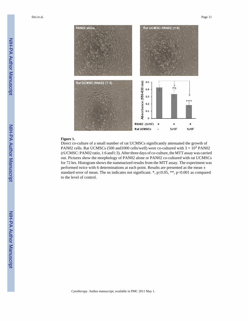
Figure 1.Direct co-culture of a small number of rat UCMSCs significantly attenuated the growth ofPAN02 cells. Rat UCMSCs (500 and1000 cells/well) were co-cultured with 3 × 10³ PAN02(rUCMSC: PAN02 ratio, 1:6 and1:3). After three days of co-culture, the MTT assay was carriedout. Pictures show the morphology of PAN02 alone or PAN02 co-cultured with rat UCMSCsfor 72 hrs. Histogram shows the summarized results from the MTT assay. The experiment wasperformed twice with 6 determinations at each point. Results are presented as the mean ±standard error of mean. The ns indicates not significant. *, p≤0.05, **, p<0.001 as comparedto the level of control.
Doi et al. Page 11
Cytotherapy. Author manuscript; available in PMC 2011 May 1.
NIH
-PA Author Manuscript
NIH
-PA Author Manuscript
NIH
-PA Author Manuscript

Figure 2.[3H] thymidine-uptake into PAN02 cells was significantly attenuated by co-culture with a smallnumber of rat UCMSCs. Rat UCMSCs (1×10³ or 2×10³/well) were co-cultured with PAN02cells (6×10³) in a 24-well culture plate. The [3H] thymidine-uptake was evaluated after 72 hrsof co-culture, as described in the Methods section. The experiment was performed twice withquadruplicate determinations. The error bars represent the standard error of the mean of thesamples. The ns indicates not significant. *, p≤0.05 as compared to the level of control.
Doi et al. Page 12
Cytotherapy. Author manuscript; available in PMC 2011 May 1.
NIH
-PA Author Manuscript
NIH
-PA Author Manuscript
NIH
-PA Author Manuscript
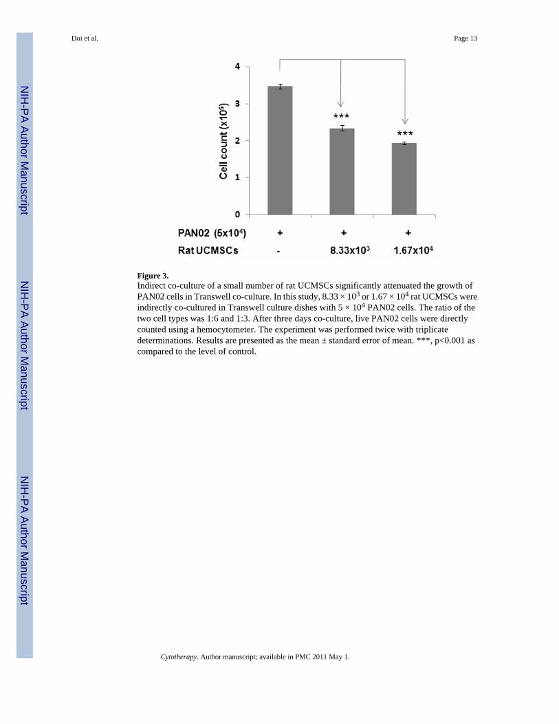
Figure 3.Indirect co-culture of a small number of rat UCMSCs significantly attenuated the growth ofPAN02 cells in Transwell co-culture. In this study, 8.33 × 10³ or 1.67 × 10⁴ rat UCMSCs wereindirectly co-cultured in Transwell culture dishes with 5 × 10⁴ PAN02 cells. The ratio of thetwo cell types was 1:6 and 1:3. After three days co-culture, live PAN02 cells were directlycounted using a hemocytometer. The experiment was performed twice with triplicatedeterminations. Results are presented as the mean ± standard error of mean. ***, p<0.001 ascompared to the level of control.
Doi et al. Page 13
Cytotherapy. Author manuscript; available in PMC 2011 May 1.
NIH
-PA Author Manuscript
NIH
-PA Author Manuscript
NIH
-PA Author Manuscript
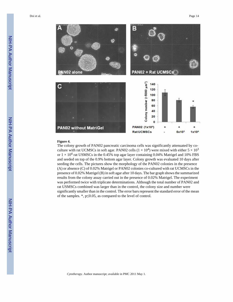
Figure 4.The colony growth of PAN02 pancreatic carcinoma cells was significantly attenuated by co-culture with rat UCMSCs in soft agar. PAN02 cells (1 × 10⁴) were mixed with either 5 × 10³
or 1 × 10⁴ rat USMSCs in the 0.45% top agar layer containing 0.04% Matrigel and 10% FBSand seeded on top of the 0.9% bottom agar layer. Colony growth was evaluated 10 days afterseeding the cells. The pictures show the morphology of the PAN02 colonies in the presence(A) or absence (C) of 0.02% Matrigel or PAN02 colonies co-cultured with rat UCMSCs in thepresence of 0.02% Matrigel (B) in soft agar after 10 days. The bar graph shows the summarizedresults from the colony assay carried out in the presence of 0.02% Matrigel. The experimentwas performed twice with triplicate determinations. Although the total number of PAN02 andrat USMSCs combined was larger than in the control, the colony size and number weresignificantly smaller than in the control. The error bars represent the standard error of the meanof the samples. *, p≤0.05, as compared to the level of control.
Doi et al. Page 14
Cytotherapy. Author manuscript; available in PMC 2011 May 1.
NIH
-PA Author Manuscript
NIH
-PA Author Manuscript
NIH
-PA Author Manuscript
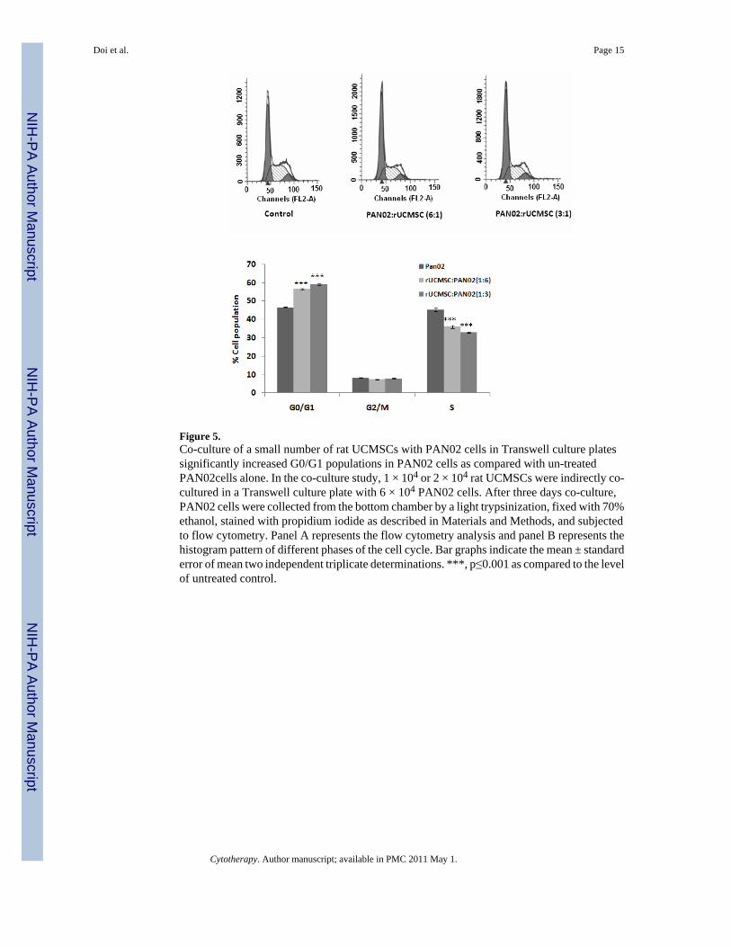
Figure 5.Co-culture of a small number of rat UCMSCs with PAN02 cells in Transwell culture platessignificantly increased G0/G1 populations in PAN02 cells as compared with un-treatedPAN02cells alone. In the co-culture study, 1 × 10⁴ or 2 × 10⁴ rat UCMSCs were indirectly co-cultured in a Transwell culture plate with 6 × 10⁴ PAN02 cells. After three days co-culture,PAN02 cells were collected from the bottom chamber by a light trypsinization, fixed with 70%ethanol, stained with propidium iodide as described in Materials and Methods, and subjectedto flow cytometry. Panel A represents the flow cytometry analysis and panel B represents thehistogram pattern of different phases of the cell cycle. Bar graphs indicate the mean ± standarderror of mean two independent triplicate determinations. ***, p≤0.001 as compared to the levelof untreated control.
Doi et al. Page 15
Cytotherapy. Author manuscript; available in PMC 2011 May 1.
NIH
-PA Author Manuscript
NIH
-PA Author Manuscript
NIH
-PA Author Manuscript
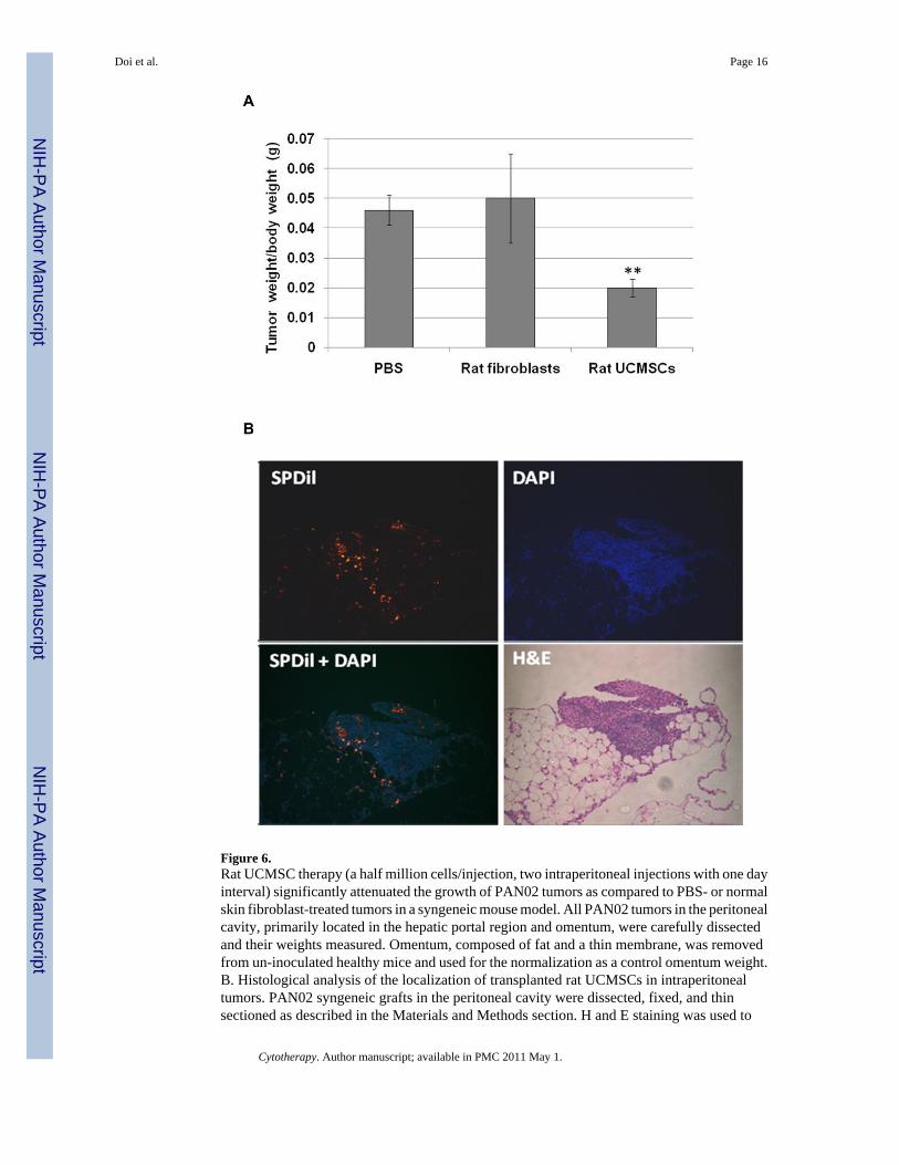
Figure 6.Rat UCMSC therapy (a half million cells/injection, two intraperitoneal injections with one dayinterval) significantly attenuated the growth of PAN02 tumors as compared to PBS- or normalskin fibroblast-treated tumors in a syngeneic mouse model. All PAN02 tumors in the peritonealcavity, primarily located in the hepatic portal region and omentum, were carefully dissectedand their weights measured. Omentum, composed of fat and a thin membrane, was removedfrom un-inoculated healthy mice and used for the normalization as a control omentum weight.B. Histological analysis of the localization of transplanted rat UCMSCs in intraperitonealtumors. PAN02 syngeneic grafts in the peritoneal cavity were dissected, fixed, and thinsectioned as described in the Materials and Methods section. H and E staining was used to
Doi et al. Page 16
Cytotherapy. Author manuscript; available in PMC 2011 May 1.
NIH
-PA Author Manuscript
NIH
-PA Author Manuscript
NIH
-PA Author Manuscript

examine overall tumor morphology. Unstained sections were utilized for the detection of SP-DiI labeled rat UCMSCs after DAPI nuclear counterstaining. In this analysis, SP-DiI labeledrat UCMSCs show bright reddish yellow in a dark background. The error bars represent thestandard error of the mean of the samples. ** p≤0.01 as compared to the level of PBS-treatedcontrols.
Doi et al. Page 17
Cytotherapy. Author manuscript; available in PMC 2011 May 1.
NIH
-PA Author Manuscript
NIH
-PA Author Manuscript
NIH
-PA Author Manuscript
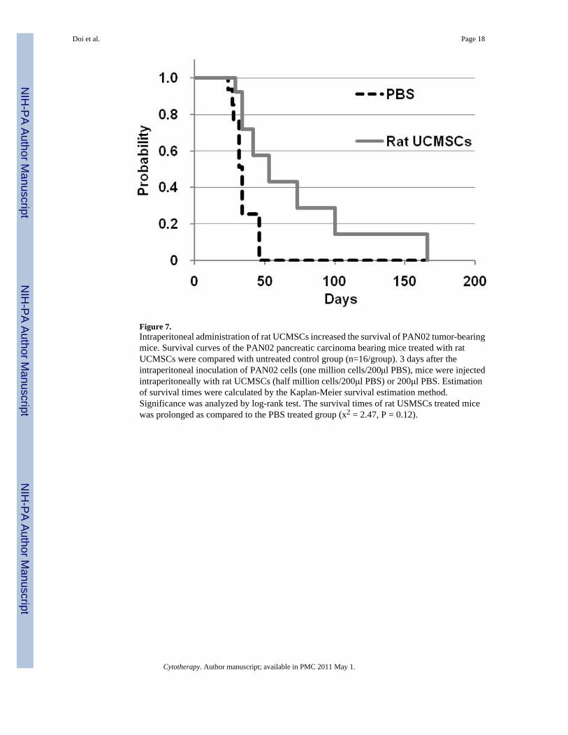
Figure 7.Intraperitoneal administration of rat UCMSCs increased the survival of PAN02 tumor-bearingmice. Survival curves of the PAN02 pancreatic carcinoma bearing mice treated with ratUCMSCs were compared with untreated control group (n=16/group). 3 days after theintraperitoneal inoculation of PAN02 cells (one million cells/200μl PBS), mice were injectedintraperitoneally with rat UCMSCs (half million cells/200μl PBS) or 200μl PBS. Estimationof survival times were calculated by the Kaplan-Meier survival estimation method.Significance was analyzed by log-rank test. The survival times of rat USMSCs treated micewas prolonged as compared to the PBS treated group (x2 = 2.47, P = 0.12).
Doi et al. Page 18
Cytotherapy. Author manuscript; available in PMC 2011 May 1.
NIH
-PA Author Manuscript
NIH
-PA Author Manuscript
NIH
-PA Author Manuscript