Cytokine Storm Plays a Direct Role in the Morbidity and ...
19
Cytokine Storm Plays a Direct Role in the Morbidity and Mortality from Influenza Virus Infection and is Chemically Treatable with a Single Sphingosine-1-Phosphate Agonist Molecule Michael B. A. Oldstone and Hugh Rosen Abstract Cytokine storm defines a dysregulation of and an excessively exaggerated immune response most often accompanying selected viral infections and several autoimmune diseases. Newly emerging and re-emerging infections of the respiratory tract, especially influenza, SARS, and hantavirus post considerable medical prob- lems. Their morbidities and mortalities are often a direct result of cytokine storm. This chapter visits primarily influenza virus infection and resultant cytokine storm. It provides the compelling evidence that illuminates cytokine storm in influenza pathogenesis and the clear findings that cytokine storm is chemically tractable by therapy directed toward sphingosine-1-phosphate receptor (S1PR) modulation, specifically S1P1R agonist therapy. The mechanism(s) of how S1P1R signaling works and the pathways involved are subjects of this review. Contents 1 Introduction........................................................................................................................ 130 2 Influenza Virus Infection .................................................................................................. 131 2.1 Epidemiologic and Experimental Evidence for Cytokine Storm............................ 131 2.2 Tracking and Kinetics of Influenza Virus-Specific CD8 and CD4 T Cells in the Lung and their Modulation by S1P Agonist ................... 132 2.3 Pulmonary Injury and Disease Associated with Influenza and Resultant Cytokine Storm are Treatable with a Single S1P1 Receptor Agonist Molecule...................................................................................................... 135 M. B. A. Oldstone (&) Department of Immunology and Microbial Science, The Scripps Research Institute, 10550 North Torrey Pines Road, La Jolla, CA 92037, USA e-mail: [email protected] H. Rosen Department of Chemical Physiology, The Scripps Research Institute, 10550 North Torrey Pines Road, La Jolla, CA 92037, USA M. B. A. Oldstone and H. Rosen (eds.), Sphingosine-1-Phosphate Signaling in Immunology and Infectious Diseases, Current Topics in Microbiology and Immunology 378, DOI: 10.1007/978-3-319-05879-5_6, ȑ Springer International Publishing Switzerland 2014 129
-
Upload
khangminh22 -
Category
Documents
-
view
0 -
download
0
Transcript of Cytokine Storm Plays a Direct Role in the Morbidity and ...

Cytokine Storm Plays a Direct Rolein the Morbidity and Mortalityfrom Influenza Virus Infectionand is Chemically Treatablewith a Single Sphingosine-1-PhosphateAgonist Molecule
Michael B. A. Oldstone and Hugh Rosen
Abstract Cytokine storm defines a dysregulation of and an excessively exaggeratedimmune response most often accompanying selected viral infections and severalautoimmune diseases. Newly emerging and re-emerging infections of the respiratorytract, especially influenza, SARS, and hantavirus post considerable medical prob-lems. Their morbidities and mortalities are often a direct result of cytokine storm.This chapter visits primarily influenza virus infection and resultant cytokine storm. Itprovides the compelling evidence that illuminates cytokine storm in influenzapathogenesis and the clear findings that cytokine storm is chemically tractable bytherapy directed toward sphingosine-1-phosphate receptor (S1PR) modulation,specifically S1P1R agonist therapy. The mechanism(s) of how S1P1R signalingworks and the pathways involved are subjects of this review.
Contents
1 Introduction........................................................................................................................ 1302 Influenza Virus Infection .................................................................................................. 131
2.1 Epidemiologic and Experimental Evidence for Cytokine Storm............................ 1312.2 Tracking and Kinetics of Influenza Virus-Specific CD8
and CD4 T Cells in the Lung and their Modulation by S1P Agonist ................... 1322.3 Pulmonary Injury and Disease Associated with Influenza and Resultant
Cytokine Storm are Treatable with a Single S1P1 ReceptorAgonist Molecule...................................................................................................... 135
M. B. A. Oldstone (&)Department of Immunology and Microbial Science, The Scripps Research Institute,10550 North Torrey Pines Road, La Jolla, CA 92037, USAe-mail: [email protected]
H. RosenDepartment of Chemical Physiology, The Scripps Research Institute,10550 North Torrey Pines Road, La Jolla, CA 92037, USA
M. B. A. Oldstone and H. Rosen (eds.), Sphingosine-1-Phosphate Signaling in Immunologyand Infectious Diseases, Current Topics in Microbiology and Immunology 378,DOI: 10.1007/978-3-319-05879-5_6, � Springer International Publishing Switzerland 2014
129
2.4 S1P1 Receptors are Located on Pulmonary Endothelial Cells,Which Serve as the Gatekeepers for Cytokine Storm ............................................ 137
2.5 Type I Interferon Signaling is Essential for the Cytokine/ChemokineResponse of Cytokine Storm but is not Involved in Recruitmentof Innate Inflammatory Cells into the Lung ........................................................... 139
2.6 Working Model for the Initial Production of Cytokine Stormand its Chemical Tractability by Single S1P1 Molecules ...................................... 142
3 Conclusions and Future Studies........................................................................................ 1443.1 Conclusions ............................................................................................................... 1443.2 Future Studies ........................................................................................................... 144
References................................................................................................................................ 145
1 Introduction
Newly emerging and re-emerging infections of the respiratory tract pose considerablemedical and public health concerns as well as economic hardships to humans andcountries. The last century witnessed at least five pandemics: 1918/1919, H (viralhemagglutinin) 1 N (viral neuraminidase) 1 Spanish influenza; 1957, H2N2 Asiainfluenza; 1968, H3N2 Hong Kong influenza; 1977, H1N1 Russian influenza; and1997, H5N1 bird influenza (reviewed Wright et al. 2007). In twenty-first century alone,two viral pandemics have already occurred—the first was in 2002 when the new viralpandemic, severe acute respiratory syndrome (SARS), appeared (reviewed Oldstone2010), followed by the first influenza virus pandemic in 2009, H1N1 swine influenza(Dawood et al. 2012). Moreover, Hantaviruses have infected humans in the past andrecently in an outbreak at Yellowstone National Park. These viral infections loom asimportant zoonotic human diseases with the threat of human to human transmissionand excessively high mortality rates. For example, 1918/1919 H1N1 influenzainfections caused the greatest loss of life from any infectious disease or medicalcondition known, visiting roughly 5 % of the world’s population and killing 2 % or40–50 million persons (Ahmed et al. 2007; Johnson and Mueller 2002). The mostrecent influenza pandemic, 2009 H1N1 swine influenza, rapidly infected millionsworldwide with estimates exceeding 290,000 deaths of which more than 201,000resulted from respiratory failure and over 83,000 from cardiovascular complications(Dawood et al. 2012). All of the above diseases in humans (Arankalle et al. 2010;Cheng et al. 2010; Lee et al. 2011) and experimental animals (Baskin et al. 2009;Kobasa et al. 2007; Marcelin et al. 2011; Zhang et al. 2012) are accompanied byearly exacerbation and dysregulation of innate immune responses, a combinationof events called ‘‘cytokine storm.’’ Severe disease and death following infectioncorrelated strongly with the cytokine storm.
Susceptibility or resistance to any viral infection is determined by the balancebetween the virulence of the infecting agent and the resistance of the host including theaggressiveness of the latter’s immune response against the virus infection. When theimmune response is limited due to either host genetics, acquired defects like lymphoiddiseases, immaturity of the immune system in fetuses, newborns, or young children, or
130 M. B. A. Oldstone and H. Rosen

loss of immune vigor in the aged, the advantage is firmly in the virus’s court. However,usually when the infection occurs in individuals with a developed and competentimmune system, the advantage is the host’s, unless the infecting virus overwhelms theindividual’s immune system or the immune response becomes hyperactive resulting inan excessive innate and adoptive immune reaction, the ‘‘cytokine storm’’ phenome-non. Cytokine storm leads to immune-mediated injury (immunopathology).
When available, vaccination is useful in protecting groups of previouslyuninfected (naive) individuals from acute viral respiratory diseases. By this means,the spread of infection is diminished. Additionally antiviral drugs, which weredeveloped as effective therapies to diminish or in some instances prevent ongoinginfections, are reasonably effective, nevertheless come with two marked limita-tions. First, antiviral drugs exert selective pressure on viral progeny, promotingtheir mutation and selection thereby creating a new generation that is more fit andresistant to the drug (Nguyen et al. 2012; Orozovic et al. 2011). Second, the injuryassociated with these acute viral respiratory diseases, including influenza, resultsfrom a combination of the virus’s intrinsic virulence in lysing cells it infects andthe intensity of the immune response which can damage tissues and promote acytokine storm. Antiviral drugs are effective against the virus but not againstcytokine storm or immune-mediated injury.
Recently, while studying human H1N1 2009 influenza virus infection in mice(Walsh et al. 2011; Teijaro et al. 2011) and ferrets (Teijaro et al. 2013), weuncovered the first direct and definitive experimental evidence that cytokine storm,per se, was a major factor in the causation of morbidity and mortality from influenzavirus and some other acute, severe respiratory infections rather than just theaccompanying phenomena. Further, we documented that cytokine storm waschemically treatable using an immunomodulatory small molecule, sphingosine-1-phosphate agonist, which dramatically inhibited the production of cytokines/chemokines and the innate cellular response, thereby blunting both the innate aswell as the adoptive antiviral T cell response (Marsolais et al. 2009; Walsh et al.2011; Teijaro et al. 2011). These events successfully limited immunopathologicinjury. Nevertheless, a sufficient host T cell response remained and coupled with theantiviral antibody response curtailed the acute infection while providing recallimmunologic memory to any renewed insult by the virus. This review focusesprimarily on our experimental work that provided these conclusions.
2 Influenza Virus Infection
2.1 Epidemiologic and Experimental Evidencefor Cytokine Storm
An overly aggressive innate immune response, the early recruitment of inflam-matory leukocytes to the lung and dysregulated immune gene expression were keycontributors to morbidity from the 1918/1919 influenza virus onslaught, as
Cytokine Storm Plays a Direct Role in the Morbidity 131
suggested by experimental infection of macaques with the 1918 H1N1 virus strain(Kobasa et al. 2007; Cilloniz et al. 2009). Clinical studies of humans infected byH5N1 bird influenza virus revealed a significant association between excessiveearly cytokine responses and immune cell recruitment as strongly predictive ofpoor medical outcomes (de Jong et al. 2006). Recently, similar results for influenzavirus infections were reported for experimental animal models (Baskin et al. 2009;Marcelin et al. 2011, Zhang et al. 2012) and for humans (Arankalle et al. 2010;Cheng et al. 2010; Lee et al. 2011). Among reports of H1N1 2009 pandemicinfluenza infections in humans, that of Arankalle et al. (2010) is illuminating.Analyzing viral events and cytokine storms in critically ill-hospitalized patients,the investigators showed that those who died had no difference in influenza viralload from those who recovered. However, the patients who recovered and left thehospital had significantly lower cytokine storm profiles than the population whosuccumbed from the infection. My colleague, Hugh Rosen, and I reasoned thatcalming the host’s aggressive and exaggerated cytokine storm response mightprovide the opportunity to shift the balance from severe morbidity and mortality tosurvival. Our laboratories started jointly about 7 years ago to test this hypothesis(Marsolais et al. 2008). We selected the molecule sphingosine 1-phosphate (S1P)and sought to determine if harmful immunologic processes accompanying H1N12009 influenza infection could be modulated by S1P receptors in the lung. Weselected S1P agonists because of their documented history of modulating lym-phoid trafficking by inducing sequestration of lymphocytes in secondary lymphoidregions. By that means, S1P agonists limit the migration of effector lymphocytes toareas where such cells mediate immunologic injury (Rosen et al. 2007, 2009,2013; see Chaps. 1 (Rosen) and 6 (Cyster) in this volume)). S1P is a signalinglipid present at a concentration of 1–3 nM in plasma and approximately 100 nM inlymph. Physiologically, S1P levels are under tight homeostatic control, and S1Psignals through specific S1P receptors of which there are five (S1P receptors 1–5).These five specific S1P receptors are coupled to different G proteins for the pur-pose of regulating a variety of downstream pathways specific for many cells,tissues, and organs (Rosen et al. 2007, 2009, 2013).
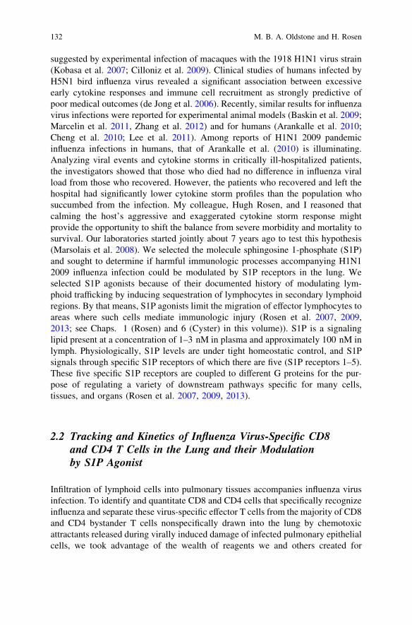
2.2 Tracking and Kinetics of Influenza Virus-Specific CD8and CD4 T Cells in the Lung and their Modulationby S1P Agonist
Infiltration of lymphoid cells into pulmonary tissues accompanies influenza virusinfection. To identify and quantitate CD8 and CD4 cells that specifically recognizeinfluenza and separate these virus-specific effector T cells from the majority of CD8and CD4 bystander T cells nonspecifically drawn into the lung by chemotoxicattractants released during virally induced damage of infected pulmonary epithelialcells, we took advantage of the wealth of reagents we and others created for
132 M. B. A. Oldstone and H. Rosen

lymphocytic choriomeningitis virus (LCMV). Our colleague Yoshi Kawaoka andhis co-workers used reverse genetics (Marsolais et al. 2009) to place the MHC Db-restricted immunodominant LCMV CD8 T cell epitope glycoprotein (Gp) aa31-41and the MHC IAb restricted immunodominant CD4 T cell epitope Gp aa65-77 intothe neuroaminidase stalk of WSN influenza virus. This technology generated arecombinant WSN Flu/LCMV virus that replicated in vivo displaying the samepulmonary geography as wild-type (wt) WSN virus. The experimental plan utilizedGFP- or RFP-labeled, cloned LCMV recognition lymphocytes obtained from T cellreceptor mice in which[98 % of CD8 fluoroprobe-labeled T lymphocytes recog-nized LCMV Gp aa31-41, and[97 % of CD4 fluoroprobe-labeled T lymphocytesrecognized Gp aa65-77. Such GFP/RFP-labeled, virus-specific lymphocytes wereadoptively transferred into naïve H-2b C57Bl/6 mice where they resided in sec-ondary lymphoid tissues as resting lymphocytes. Two days later the recombinantWSN Flu/LCMV was administered intratracheally. Virus replication in pulmonaryepithelial cells (Fig. 1a) was followed by the infiltration of virus-specific CD8 Tcells (red) and virus-specific CD4 T cells (green) (Fig. 1b) at day 6 (Marsolais et al.2009). Kinetic study of infiltrating virus-specific CD8 T cells showed their arrival byday 4, peak amounts at days 6–8, and significant numerical decrease at day 10postinfection (Fig. 1c). There are five S1P receptors, i.e., S1P1, S1P2, S1P3, S1P4,and S1P5. Administration of S1P permissive agonist AAL-R, which signals onS1P1, S1P3, S1P4, and S1P5 receptors but not the S1P2 receptor, significantlyreduced the numbers of virus-specific CD8 T cells entering the lung (Fig. 1d). Theresult was significant protection from pulmonary tissue injury (Fig. 1e) and relatedmortality (Fig. 1f) when compared to the effects of vehicle alone or use of a controlisomer, AAL-S, that is not able to be phosphorylated and cannot signal S1Preceptors. Blunting of innate cytokine and chemokine responses following AAL-Rtreatment was evident and remarkable at day 2 postinfluenza infection (Fig. 1g). Allthese observations were initially made with murine H1N1 WSN virus and laterconfirmed by use of the non-murine adopted human pathogenic H1N1 influenzaviruses A/Wisconsin/WSLH34934/09 and A/California/04/09 (Walsh et al. 2011).In studies with all these influenza viruses, although cytokine/chemokine expressionwas significantly blunted by S1P agonist AAL-R, AAL-R-treated mice terminatedthe virus infection, displayed robust virus-specific CTL responses 7 days afterinfluenza infection, as measured by 51chromium release assay, and also mountedvigorous specific memory T cell responses upon rechallenge with virus 40 days afterthe infection. Further, the kinetics, titers of neutralizing anti-influenza antibodies insera, or immunoglobulin subtypes of either AAL-R or AAL-S or vehicle-treatedmice were equivalent. Together, these results document the validity of our premise.That is, the permissive S1P agonist AAL-R, which signals via S1P1, S1P3, S1P4,and S1P5 receptors, when given locally into the respiratory tract, down-modulatednumbers of virus-specific T cells, decreased innate cytokine/chemokine expressionin the lung parenchyma, and reduced the supply of innate inflammatory cells—NK,PMN, and macrophages (Marsolais et al. 2009; Walsh et al. 2011)—sufficiently toabort cytokine storm. The successful outcome was protection of the host frominfluenza virus infection while still providing an antiviral response that curtailed and
Cytokine Storm Plays a Direct Role in the Morbidity 133

eventually impeded the influenza infection. Our data indicated that 23 of 28 mice(82 %) receiving AAL-R were protected (P = \0.001; only five of 28 died from theinfection) when compared to a dose of virus that killed approximately 80 % of
Fig. 1 Panel a: Distribution of viral antigen (green, left); Panel b: Virus-specific CD4 T cells(green, right) and CD8 T cells (red, left) in the lung 7 days following influenza/LCMV infection;Panel c: Kinetics of virus-specific CD8 T cell infiltration into the lung analyzed by immunohis-tochemistry (upper panels) or FACS (lower panels); Panel d: The S1P permissive agonist AAL-Rsignificantly blunts infiltration of virus-specific CD8 T cells into the lung following influenza/LCMV recombinant virus infection; Panel e: Significant reduction of pulmonary tissue injury andpreservation of alveolar air space in influenza-infected mice treated with AAL-R; Panel f:Significant protection from mortality accompanying influenza virus infection with AAL-R; andPanel g: AAL-R significantly dampens cytokine and chemokine content at day 2 following influenzavirus infection. Figure reprinted from Marsolais et al. (2009), with permission from PNAS
134 M. B. A. Oldstone and H. Rosen

vehicle- or AAL-S-treated mice (22 of 28 mice died) (Walsh et al. 2011).Interestingly, when an optimal dose of the currently used antiviral drug Tamiflu wasadministered by itself, protection was significantly less effective 50 % (14 lived of28 mice treated) compared to survival after AAL-R therapy alone (80 %). Theseresults document a prominent role for cytokine storm as the cause of death frominfluenza infection. Most important is the benefit of S1P agonist therapy for thevictims of multiple influenza virus strains and especially those that are resistant toanti-neuraminidase therapy. Although greater benefit was obtained in blockingcytokine storms with the S1P agonist than with Tamiflu (82 % vs. 50 %) protection,administering both the antiviral drug and the S1P agonist as combined therapywas optimal, yielding a 96 % protection rate from influenza virus challenge (27 of28 mice survived the infection) (Walsh et al. 2011).
2.3 Pulmonary Injury and Disease Associated with Influenzaand Resultant Cytokine Storm are Treatablewith a Single S1P1 Receptor Agonist Molecule
All five S1P receptors couple to different G proteins require many downstreamsignaling pathways (Fig. 2a) (Rosen et al. 2007, 2009, 2013, Chap. 1 in thisvolume). The biological functions of these various S1P receptors are dependent onthe cell/tissue location of the receptors, their expression, and their activation.Knowing that a broadly permissive S1P agonist AAL-R, which signals via S1P1,S1P3, S1P4, and S1P5 but not S1P2 receptors, significantly downregulated thecytokine storm and protected mice from the effects of a pathogenic human H1N1influenza infection (Fig. 1, Panels c–g) (Walsh et al. 2011), we repeated theexperiments shown in Fig. 1, Panels e–g, using two S1P1-specific agonists, CYM-5542 (Walsh et al. 2011), or RP-002 (Teijaro et al. 2011). The results are displayedin Fig. 2 and indicate that either of the two specific S1P1 receptor agonists whosesignal is entirely restricted to S1P1 receptors were as effective as the broadlypermissive AAL-R that signals on S1P1, S1P3, S1P4, and S1P5. The S1P1-specificagonists CYM-5442 were administered intratracheally (2 mg/kg) and RP-002intratracheally (3 mg/kg) or orally (6 mg/kg) (Teijaro et al. 2011). Both S1P1receptor agonist molecules provided protection against a lethal intranasalchallenge with human H1N1 A/Wisconsin/WSLH34934/09 or A/California/04/09(Fig. 2b) and blunted cytokine storm (Fig. 2c and d). The S1P1 receptor agonistssignificantly inhibited secretions of cytokines and chemokines associated withinfluenza virus infection, namely IFN-a, CCL-2, IL-6, TNF-a, CCL-3, CCL-5,CxCl-2, IL-1a, and IFN-c. Observations from several experiments indicated thatamounts of these cytokines/chemokines were inhibited to a degree similar to thatfrom AAL-R treatment. The S1P1 selective agonists also significantly blunted theaccumulation of innate infiltrating inflammatory cells (Fig. 2, Panel d). Notablewere the reductions of macrophages/monocytes (marked by CD11b+, LyG6-,
Cytokine Storm Plays a Direct Role in the Morbidity 135

F480+), neutrophils (CD11b+, LyG6+-, F480-), and natural killer cells (NK1.1+,CD3-). Correspondingly, the quantity of activation marker CD69 was significantlyreduced following S1P1 agonist therapy. Further, pulmonary tissues also reflectedS1P1’s beneficial outcome, since histologic study of mice given this remedymanifested mostly open alveolar air spaces, diminished to negligible inflammatorycell infiltrates and neither edema nor hemorrhage in the lung tissue. Importantly,S1P1 agonist treatment did not enhance viral titers. Influenza infection waseffectively terminated, and both anti-influenza neutralizing antibodies and anti-influenza virus CD8 T cells were generated. Although numbers of T cells werereduced compared to infected mice not receiving S1P1 agonist therapy, they weresufficient to terminate the infection. Further, immune memory was establishedfollowing this S1P1-specific therapy.
Fig. 2 S1P1 specific agonists CYM-5442 and RP-002 are therapeutically equivalent to thepermissive AAL-R agonist that utilizes S1P1, S1P3, S1P4, and S1P5 receptor signaling to bluntcytokine storm and also protect mice from a lethal challenge by human pandemic H1N1 2009A/Wisconsin/WSLH34939/09 (shown) or A/California 04/09 (not shown) viruses. Panel a:Cartoons of the five S1P receptors and their biologic effects. AAL-R signals on receptors S1P1,S1P3, S1P4, and S1P5 but not S1P2. Panel b: S1P1 receptor-specific agonist RP-002 given orallyprotects mice challenged with H1N1 human 2009 influenza virus A/Wisconsin. Treatmentwith S1P1 receptor-specific agonist CYM-5442 after mice are challenged with 2009 influenzaA/Wisconsin inhibits their cytokine/chemokine response (cytokine storm) equivalently totreatment with the permissive AAL-R (Panel c) and also impedes the recruitment of innateimmune cells into their lungs (Panel d). BALF Bronchial lavage fluid, * = p \ 0.01. Figurereprinted from Teijaro et al. (2011), with permission from Elsevier
136 M. B. A. Oldstone and H. Rosen

Thus, severe pulmonary injury and disease associated with influenza infectionand resultant cytokine storm were treatable with a preparation composed of onlyS1P1 receptor agonist molecules, thereby avoiding signaling through S1P2, S1P3,S1P4, and S1P5 receptors. Pharmaceutically this may be of importance if/whenindividually S1P2, S1P3, S1P4, or S1P5 signaling might lead to unwanted harmfulbiologic effects.
2.4 S1P1 Receptors are Located on Pulmonary EndothelialCells, Which Serve as the Gatekeepers for CytokineStorm
Having identified S1P1rec signaling as the primary pathway for the initiation ofcytokine storm, we sought to identify the cell or cells in the lung that expressed theS1P1 receptor. Since epithelial cells are the primary cells infected by influenzaviruses, we suspected that S1P1 receptors might be located on those cells. Todetermine which pulmonary cell types bear the S1P1 receptor, we took advantage ofeGFP-S1P1 receptor knock-in mice made by Stuart Cahalan in the Rosen laboratory(Cahalan et al. 2011, see Cahalan Chap. 4, this volume). In this strain of mice, thenative S1P1 receptor was homologously replaced with a functional fused eGFP-tagged S1P1 receptor (Cahalan et al. 2011). Utilizing this mouse model, we coulddirectly detect eGFP-S1P1 receptor protein expression on pulmonary cells by usingantibodies to specific pulmonary cell markers and flow cytometry (Fig. 3, Panel a).Additional substantiation came from biochemical analysis of these purified pul-monary cells (Fig. 3, Panel b). S1P1-eGFP receptor expression was plentiful on lunglymphatic (CD45-, CD31+, GP38+) and vascular (CD45-, CD31+, GP38-)endothelial cells but, surprisingly, was absent on pulmonary epithelial cells(CD45-, CD31-, EpCAM+) (Fig. 3, Panel a) (Teijaro et al. 2011). These resultswere confirmed by doing Western blots on[98.5 % pure populations of pulmonaryendothelial and epithelial cells (Fig. 3, Panel b). As expected and previouslyreported, CD4 T cells (CD4+, CD3+), CD8 T cells (CD8+, CD3+), and B cells(B200+, CD19+) also expressed the S1P1-eGFP receptor (Fig. 3, Panel a). Incontrast, pulmonary leukocytes, including macrophages/monocytes (CD11c+,CD11b-, F480+), dendritic cells (CD11c+, IA-IE+, CD205+, F480-), neutrophils,NK cells (NK1.1+, CD3-) (Fig. 3, Panel a), and immature lymphoid cells (LIN-,SCA-1+) failed to express significant levels of eGFP-S1P1 receptor protein. S1P1-eGFP receptor expression was similar whether cells were harvested from mice thatwere uninfected or infected with influenza virus. Other experiments in infected miceindicated that S1P1-eGFP receptor expression was not altered during influenza virusinfection. Importantly, S1P1 agonist treatment of infected eGFP-S1P1 receptorknock-in mice did not lessen expression of the S1P1-eGFP receptors indicating thatadministration of specific S1P1 agonist does not degrade the endothelial S1P1receptor. These results signify that the functional agonism of S1P1, not its antag-onist effect of receptor degradation, was the mechanism by which S1P1 receptor
Cytokine Storm Plays a Direct Role in the Morbidity 137

Fig. 3 S1P1 is expressed on endothelial cells and lymphocytes isolated from eGFP-S1P1receptor knock-in mice. Panel a: Cell populations purified using antibodies to specific cellsurface markers and FACS. Purity of all cell populations exceeded 98.5 %. See Teijaro et al.(2011) for details about reagents and experiments. As seen in Panel a, only endothelial cells(lymphoid and vascular) and lymphocytes (CD4+ T cells, CD8+ T cells) expressed the GFP-S1P1receptor marker. Pulmonary epithelial cells, the primary target for influenza virus, do not expressthe S1P1 receptor. Panel b:S1P1 agonism inhibits chemokine expression in endothelial cellsfollowing influenza virus infection. * = p \ 0.01. See Teijaro et al. (2011) for details. Figurereprinted from Teijaro et al. (2011), with permission from Elsevier
138 M. B. A. Oldstone and H. Rosen

blocking molecules CYM-5442 and RP-002 suppressed cytokine storms. In otherstudies, pulmonary endothelial cells were processed to a greater than 98.5 % purityduring the first 48 h following influenza virus infection and treated with S1P1agonist. Assessment of both RNA and protein levels showed that the S1P1 agonistCYM-5442 effectively decreased amounts of cytokines and chemokines made byvascular as well as lymphatic pulmonary endothelial cells (Fig. 3b).
T and B lymphocytes as well as pulmonary endothelial cells were the only cellswithin the lung that expressed measurable amounts of S1P1-eGFP protein (Fig. 3Panel a). We therefore determined whether lymphocytes expressing S1P1 receptorswere involved in S1P1 agonist inhibition of cytokine storm or were merelybystander cells accompanying the innate immune response to influenza virusinfection. Since Rag2-/- mice are deficient in lymphocytes, we reasoned that if suchmice, when infected with influenza virus, generated a cytokine storm that could beblocked by S1P1 agonist, then lymphocytes were ruled out as initiators of cytokinestorms. Our experiments documented that cytokine storm occurred in Rag2-/- miceinfected with influenza virus. Importantly, treatment of infected Rag2-/- mice withthe S1P1 agonist CYM-5442 significantly reduced cytokines and chemokines in thebronchial lavage fluids as well as minimalizing the infiltration of innate cells(macrophages/monocytes and NK cells). Recently, John Teijaro (2013), utilizingcell sorting and a biochemical approach, found S1P1 receptor on plasmacytoiddendritic cells (pDC) whose expression was undetectable in the S1P1-c GFPtransgenic mouse model.
2.5 Type I Interferon Signaling is Essentialfor the Cytokine/Chemokine Response of CytokineStorm but is not Involved in Recruitment of InnateInflammatory Cells into the Lung
As observed in Fig. 4a and b and detailed in Teijaro et al. 2011, amounts of type Iinterferon and almost exclusively the interferon-a species were elevated early afteracute influenza virus infection. The release and action of type I interferon wascrucial for the production of pro-inflammatory cytokines/chemokines, sinceblockage of the type I interferon response by using monoclonal antibody tointerferon-a-b receptor (IFNAR1) significantly reduced the quantity of pulmonarycytokines/chemokines associated with acute influenza infection (Fig. 4b). Further,treatment with S1P1 receptor agonist inhibited the production of interferon-a in thepulmonary bronchial lavage fluid early after initiating influenza virus infection.Proof that this blunting of interferon-a production was a mechanism by whichS1P1 receptor agonist inhibited cytokine storm derived from use of IFNAR1receptor knock-out mice infected with H1N1 virus and treated with S1P1 receptoragonist CYM-5442. Such studies showed a significant reduction of cytokines/chemokines (IFN-a, CCL-2, IL-6 (shown Fig. 4c), IFN-c, CCL-5, CxCl-0, notshown) in the bronchial lavage fluid when compared to results from similar
Cytokine Storm Plays a Direct Role in the Morbidity 139

Fig. 4 Interferon-a is the predominant type 1 interferon produced early following virus infection(Panel a) and is associated with the dysregulation of cytokines and chemokines that causes acytokine storm. Antibody to interferon type 1-a-b receptor significantly blocks release ofcytokines and chemokines (Panel b). Panel c: S1P1 agonist suppression of cytokines is dependenton interferon 1. Panel d: Innate inflammatory cell recruitment is independent of interferon-a-breceptor signaling. Panel e, left: The majority (75–85 %) of interferon-a released followinginfluenza viral infection is from plasmacytoid dendritic cells (pDCs, use of feeble mice—seetext). Panel e, right: Data from S1P1-eGFP knock-in mice indicating that S1P1 receptors are notpresent on surfaces of pulmonary pDCs but are found, as expected, on surfaces of pulmonaryendothelial cells. However, utilizing more sensitive techniques S1P1 receptors are found on pDCs(see text). Figure reprinted from Teijaro et al. (2011), with permission from Elsevier
140 M. B. A. Oldstone and H. Rosen

experimentation in mice with an intact interferon-a-b receptor signaling ability(Fig. 4c). Of interest, blockage of interferon-a-b receptor signaling did not retardpulmonary infiltration by the inflammatory cells—macrophages, monocytes,neutrophils, or NK cells—following S1P1 receptor agonist therapy (Fig. 4d). Thus,the infiltration of innate inflammatory cells was blunted only in interferon-a-bsufficient mice but not in interferon-a-b receptor knock-out mice. This outcomeindicates that regulation of such cell recruitment into the lung was primarilymediated by endothelial cells and was independent of type I interferon signaling(Teijaro et al. 2011). Cytokine/chemokine production in the lung was also mediatedby pulmonary endothelial cells, and S1P1 receptor agonism of such cells inhibitedinterferon-a production leading to the significantly diminished inflammatorycytokine/chemokine responses we observed.
Influenza virus infection induces a robust interferon type I response, despite theearly induction of the viral NS1 protein that suppresses the cellular induction of andresponse to interferon I (Fernandez-Sesma 2007). The predominant type I inter-feron produced early following influenza virus infection is alpha, not beta (Fig. 4a).However, the cellular sources of interferon type I-a produced and amounts made byvarious cell populations have not been clear. The two major pulmonary cell pop-ulations known to make type I interferon in vivo following respiratory viralinfections are pDCs and alveolar macrophages (Kumagai et al. 2007). To judge thecontribution of pDCs to interferon-a production in the lung, we utilized a novelmouse model recently developed at Scripps by Bruce Beutler and termed ‘‘feeble.’’Feeble mice have a specific genetic defect that prevents their pDCs from producingtype I interferon and pro-inflammatory cytokines upon activation of TLR7 andTLR9 ligands by influenza virus stimulation (Blasius et al. 2010). Importantly,there is no disruption of the numbers or vitality of pDCs, and the feeble mousedefect is specifically restricted to pDCs. As displayed in Fig. 4e, when wild type orfeeble mice received H1N1 human 2009 swine influenza with or without S1P1agonist CYM5442 treatment, 75–85 % of total interferon-a was produced by pDCs.Further, interferon-a release was significantly inhibited by the S1P1 agonist CYM-5442. These results were confirmed using a pDC depletion antibody (anti-PDCS-1clone 120.68), which again resulted in significant depletion of pDCs in the lung andcorresponding reductions in interferon-a, CCL2, CCL5, and IL-6. Thus, pDCs arethe essential and major producers of interferon-a (75–85 %) and involved inamplification of cytokine/chemokine volumes following influenza infection. Otherdepletion studies indicate that most of the remaining interferon-a production(*15–25 %) was by alveolar macrophages.
Since S1P1 agonist therapy diminished interferon-a production, and themajority of interferon-a produced was by pDCs, it was important to learn whetheror not pDCs expressed S1P1 receptors on their surfaces. We know that alveolarmacrophages, the other main albeit minority producers of interferon-a do notexpress S1P1 receptors (Fig. 2a). Plasmacytoid dendritic cells were recently foundto express S1P1 receptors (Teijaro 2013). However, using the S1P1 eGFP receptorknock-in mice and pDCs of more than 98.5 % purity failed to show that these cellsexpressed S1P1 receptors (Fig. 4e). Thus, the S1P1-specific receptor is found
Cytokine Storm Plays a Direct Role in the Morbidity 141

primarily on pulmonary endothelial cells with lesser amounts on pulmonary pDCs.S1P1 receptor agonist acts directly on pulmonary endothelial and pDCs and likelyindirectly on alveolar macrophages. We have, as yet, been unable to detect S1P1receptor on alveolar macrophages.
2.6 Working Model for the Initial Productionof Cytokine Storm and its ChemicalTractability by Single S1P1 Molecules
A working model based on the accumulated data for the earliest events of influ-enza infection is provided in Fig. 5. Although there are several possible scenarios,I selected the simplest one in which S1P1 agonist signals a factor(s) that blocks[negative signal(s)] the release of interferon-a from pulmonary pDCs and themigration of innate inflammatory cells from blood vessels into the lung. Thismodel is based on presence of S1P1 receptors on pulmonary endothelial and pDCsbut their absence on lung epithelial cells and the findings that alveolar macro-phages which produce type I interferon do not display S1P1 receptors on theirsurfaces. However, pulmonary endothelial cells and pulmonary pDCs do expressS1P1 receptors on their surfaces. Cytokines/chemokines elicit the initial cytokinestorm reflective of factors (viral or nonviral) produced by the influenza virus-infected lung epithelial cells per se. These factors likely activate pulmonary pDCsand alveolar macrophages to release interferon-a. As chemotoxic factors are thenliberated into the site of action, infiltrating cells of the innate immune system(macrophages/monocytes, NK cells, leukocytes) are drawn into the inflammatorymilieu where they release additional cytokines/chemokines.
After that initial response, a second stage occurs by day 4–8 (see 2.2) via amechanism described in our publication (Marsolais et al. 2009). Here influenzavirus-specific T cells are activated and expand numerically in the mediastinal lymphnodes and pulmonary tissues. These T cells of the adoptive immune system produceadditional inflammatory molecules and lyse influenza virus-infected epithelial cellsthereby augmenting cytokine storm and immune-mediated injury. This secondphase of tissue injury is primarily influenza virus–specific T cell-mediated andsignals through S1P1 but also likely progresses via S1P3 and 4 receptors, as by ourpreliminary results. However, additional data are required to ensure these obser-vations. What is clear is that treatment with S1P permissive-AAL-R agonistssignaling via S1P1,3,4,5 receptors affect adoptive immune T cell-mediatedimmunopathology by downregulating MHC and co-stimulatory molecules of DCslocated in the mediastinal lymph nodes and the lung parenchyma thereby bluntingthe arming, expansion, and migration of virus-specific CD4 and CD8 T cells into thelung (Marsolais et al. 2009).
The cytokine pathways blunted by S1P1 agonist signaling are displayed inFig. 5 by the symbol ‘.
142 M. B. A. Oldstone and H. Rosen

Fig. 5 Schematic of data presented in the text: Proposed pathways and cell–cell crosstalk in thelung following influenza virus infection and S1P1 receptor agonist signaling. Initial events:Viruses infect lung epithelial cells that release one or more (currently unknown) factors thatsignal plasmacytoid cells (primary cell-type involved) and alveolar macrophages to release type 1interferon-a, which dysregulates cytokines/chemokines to elicit cytokine storm. Releasedchemokines attract innate immune inflammatory cells that become activated and releaseadditional chemokines/cytokines to amplify cytokine storm. Therapeutic control of cytokinestorm: Pulmonary endothelial cells and pDCs contain S1P1 receptors on their surfaces, but S1P1receptors are absent from lung epithelial cells and alveolar macrophages. The S1P1 receptoragonist signals pulmonary endothelial cells and pDCs likely to release factors that negativelyregulate the cytokine storm in terms of both its cytokine/chemokine release and infiltration ofinnate inflammatory cells
Cytokine Storm Plays a Direct Role in the Morbidity 143

3 Conclusions and Future Studies
3.1 Conclusions
Cytokine storm plays an essential role in the pathogenesis and clinical outcome ofinfluenza virus infection. Blockade of cytokine storm provides greater protectionthan does antiviral therapy, like that with a neuraminidase inhibitor, and does sowithout compromising the control and clearance of viruses. Moreover, optimaltherapy is achieved by combining S1P agonists with anti-neuraminidase treatment.For the foregoing observations, human pathogenic H1N1 09 influenza virus andmouse adapted H1N1 influenza virus were used.
Sphingosine-1-phosphate (S1P) receptor agonists blunt cytokine storm. Impor-tantly, cytokine storm is chemically disarmed by administering one of the five S1Preceptors: S1P1.
The molecular mechanism of this event involves S1P1 receptor signaling onpulmonary endothelial cells and pulmonary pDCs but not virally infected epithelialcells or alveolar macrophages. Pulmonary endothelial cells are the major gatewaycombined with pulmonary pDCs to precipitate a cytokine storm. S1P1 agonismsuppresses the recruitment of both cytokines, innate and adoptive immune cells.Blunting of innate immune cell function and virus-specific T cell activity lessensmorbidity and prevents mortality associated with experimental models of influenzavirus infection in mice and ferrets. In both species, there is a sufficient antiviralresponse remaining to terminate the virus infection and provide immune memoryupon rechallenge.
Immune cell infiltration and cytokine production are distinct events, but bothare orchestrated by endothelial and pDC cells. Pro-inflammatory cytokineresponses depend on type I interferon signaling; IFN-a is the predominant inter-feron made. The predominant pulmonary cell making type I interferon is the pDC(over 75–85 %); alveolar macrophages make most of the rest.
3.2 Future Studies
1. Investigate interferon I as to the cellular source and signaling pathway(s) in theinfluenza system.
2. Dissect crosstalk and signaling between pulmonary endothelial cells, infectedepithelial cells, and interferon-producing pDCs. Identify the moleculesinvolved. See if these molecules provide potential therapeutic targets.
3. Determine generalities for other acute respiratory infections, e.g., Hantavirus,respiratory syncytial virus, SARS, pneumococcal pneumonia, in which cyto-kine storm plays a prominent role.
4. Study an animal model (subhuman primates) more reflective of influenza inhumans. Results from our studies in ferrets (Teijaro et al. 2013) mirror the
144 M. B. A. Oldstone and H. Rosen

protection supplied by S1P1 agonist therapy in defending against influenzavirus infection in the murine model.
5. Define the S1P pathway and design a genetic screen to identify humans who arethe most susceptible to cytokine storm.
6. Develop specific S1P receptor agonists and antagonists for human therapeutics,focusing initially on S1P1 molecules.
Acknowledgments The experimental work described in this chapter was initiated with andcarried out as a collaboration between my laboratory and that of Hugh Rosen. Graduate student(Stuart Cahalan) and former postdoctoral fellows (David Marsolais, Kevin Walsh, and JohnTeijaro) were instrumental in the findings presented. John Teijaro continues his independent workin this area as a faculty member at Scripps. Yoshi Kawaoka and colleagues in his laboratorieshave been valued collaborators. This work was funded by NIH grants AI009484 (MBAO),AI074564 (MBAO, HR), MH084512 (HR), and was also supported by the NIH/NIAID underAward Number U54 AI057160 to the Midwest Regional Center of Excellence (MRCE) forBiodefense and Emerging Infectious Diseases Research (MBAO).
References
Ahmed R, Oldstone MBA, Palese P (2007) Protective immunity and susceptibility to infectiousdiseases: lessons from the 1918 influenza pandemic. Nat Immunol 8:229–288
Arankalle VA, Lole KS, Arya RP, Tripathy AS, Ramdasi AY, Chadha MS, Sangle SA, KadamDB (2010) Role of host immune response and viral load in the differential outcome ofpandemic H1N1 (2009) influenza virus infection in Indian patients. PLoS ONE 5:e13099
Baskin CR, Bielefeldt-Ohmann H, Tumpey TM, Sabourin PJ, Long JP, García-Sastre A, Tolnay AE,Albrecht R, Pyles JA, Olson PH, Aicher LD, Rosenzweig ER, Murali-Krishna K, Clark EA,Kotur MS, Fornek JL, Proll S, Palermo RE, Sabourin CL, Katze MG (2009) Early and sustainedinnate immune response defines pathology and death in nonhuman primates infected by highlypathogenic influenza virus. Proc Natl Acad Sci USA 106:3455–3460
Blasius AL, Arnold CN, Georgel P, Rutschmann S, Xia Y, Lin P, Ross C, Li X, Smart NG,Beutler B (2010) Slc15a4, AP-3, and Hermansky-Pudlak syndrome proteins are required forToll-like receptor signaling in plasmacytoid dendritic cells. Proc Natl Acad Sci USA107:19973–19978
Cahalan SM, Gonzalez-Cabrera PJ, Sarkisyan G, Nguyen N, Schaeffer MT, Huang L, Yeager A,Clemons B, Scott F, Rosen H (2011) Actions of a picomolar short-acting S1P1 agonist inS1P1-eGFP knock-in mice. Nat Chem Biol 7:254–256
Cheng XW, Lu J, Wu CL, Yi LN, Xie X, Shi XD, Fang SS, Zan H, Kung HF, He ML (2010)Three fatal cases of pandemic 2009 influenza A virus infection in Shenzhen are associatedwith cytokine storm. Respir Physiol Neurobiol 175:185–187
Cillóniz C, Shinya K, Peng X, Korth MJ, Proll SC, Aicher LD, Carter VS, Chang JH, Kobasa D,Feldmann F, Strong JE, Feldmann H, Kawaoka Y, Katze MG (2009) Lethal influenza virusinfection in macaques is associated with early dysregulation of inflammatory related genes.PLoS Pathog 5:e1000604
Dawood FS, Iuliano AD, Reed C, Meltzer MI, Shay DK, Cheng PY, Bandaranayake D, BreimanRF, Brooks WA, Buchy P, Feikin DR, Fowler KB, Gordon A, Hien NT, Horby P, Huang QS,Katz MA, Krishnan A, Lal R, Montgomery JM, Mølbak K, Pebody R, Presanis AM, Razuri H,Steens A, Tinoco YO, Wallinga J, Yu H, Vong S, Bresee J, Widdowson MA (2012) Estimatedglobal mortality associated with the first 12 months of 2009 pandemic influenza A H1N1virus circulation: a modelling study. Lancet Infect Dis 12:687–695
Cytokine Storm Plays a Direct Role in the Morbidity 145
de Jong MD, Simmons CP, Thanh TT, Hien VM, Smith GJ, Chau TN, Hoang DM, Chau NV,Khanh TH, Dong VC, Qui PT, Cam BV, Ha do Q, Guan Y, Peiris JS, Chinh NT, Hien TT,Farrar J (2006) Fatal outcome of human influenza A (H5N1) is associated with high viral loadand hypercytokinemia. Nat Med 12:1203–1207
Fernandez-Sesma A (2007) The influenza virus NS1 protein: inhibitor of innate and adaptiveimmunity. Infect Disord Drug Targets 7:336–343
Johnson NP, Mueller J (2002) Updating the accounts: global mortality of the 1918–1920‘‘Spanish’’ influenza pandemic. Bull Hist Med 76:10–115
Kobasa D, Jones SM, Shinya K, Kash JC, Copps J, Ebihara H, Hatta Y, Kim JH, Halfmann P,Hatta M, Feldmann F, Alimonti JB, Fernando L, Li Y, Katze MG, Feldmann H, Kawaoka Y(2007) Nature 445:319–323
Kumagai Y, Takeuchi O, Kato H, Kumar H, Matsui K, Morii E, Aozasa K, Kawai T, Akira S(2007) Alveolar macrophages are the primary interferon-alpha producer in pulmonaryinfection with RNA viruses. Immunity 27:240–252
Lee N, Wong CK, Chan PK, Chan MC, Wong RY, Lun SW, Ngai KL, Lui GC, Wong BC, Lee SK,Choi KW, Hui DS (2011) Cytokine response patterns in severe pandemic 2009 H1N1 andseasonal influenza among hospitalized adults. PLoS ONE 6:e26050
Marcelin G, Aldridge JR, Duan S, Ghoneim HE, Rehg J, Marjuki H, Boon AC, McCullers JA,Webby RJ (2011) Fatal outcome of pandemic H1N1 2009 influenza virus infection isassociated with immunopathology and impaired lung repair, not enhanced viral burden, inpregnant mice. J Virol 85:11208–11219
Marsolais D, Hahm B, Edelmann KH, Walsh KB, Guerrero M, Hatta Y, Kawaoka Y, Roberts E,Oldstone MB, Rosen H (2008) Local not systemic modulation of dendritic cell S1P receptorsin lung blunts virus-specific immune responses to influenza. Mol Pharmacol 74:896–903
Marsolais D, Hahm B, Walsh KB, Edelmann KH, McGavern D, Hatta Y, Kawaoka Y, Rosen H,Oldstone MB (2009) A critical role for the sphingosine analog AAL-R in dampening thecytokine response during influenza virus infection. Proc Natl Acad Sci USA 106:1560–1565
Nguyen HT, Fry AM, Gubareva LV (2012) Neuraminidase inhibitor resistance in influenzaviruses and laboratory testing methods. Antivir Ther 17:159–173
Oldstone MBA (2010) Severe acute respiratory syndrome (SARS). Viruses, plague and history,Oxford Press, pp 251–283
Orozovic G, Orozovic K, Lennerstrand J, Olsen B (2011) Detection of resistance mutations toantivirals oseltamivir and zanamivir in avian influenza A viruses isolated from wild birds.PLoS ONE 6:e16028
Rosen H, Gonzalez-Cabrera PJ, Sanna MG, Brown S (2009) Sphingosine 1-phosphate receptorsignaling. Annu Rev Biochem 78:743–768
Rosen H, Sanna MG, Cahalan SM, Gonzalez-Cabrera PJ (2007) Tipping the gatekeeper: S1Pregulation of endothelial barrier function. Trends Immunol 28:102–107
Rosen H, Stevens RC, Hanson M, Roberts E, Oldstone MB (2013) Sphingosine-1-phosphate andits receptors: structure, signaling, and influence. Annu Rev Biochem 82:637–662
Teijaro JR, Walsh KB, Cahalan S, Fremgen DM, Roberts E, Scott F, Martinborough E, Peach R,Oldstone MB, Rosen H (2011) Endothelial cells are central orchestrators of cytokineamplification during influenza virus infection. Cell 146:980–991
Teijaro JR, Walsh KB, Long JP, Tordoff KP, Stark GV, Kawaoka Y, Rosen H, Oldstone MB(2013) H1N1 2009 influenza virus infection of ferrets is successfully treated with sphingosine-1-phosphate 1 receptor agonist RP-002. Virology Submitted
Teijaro JR (2013) Unpublished dataWalsh KB, Teijaro JR, Wilker PR, Jatzek A, Fremgen DM, Das SC, Watanabe T, Hatta M,
Shinya K, Suresh M, Kawaoka Y, Rosen H, Oldstone MB (2011) Suppression of cytokinestorm with a sphingosine analog provides protection against pathogenic influenza virus. ProcNatl Acad Sci USA 108:12018–12023
146 M. B. A. Oldstone and H. Rosen

Wright PF, Neumann G, Kawaoka Y (2007) Orthomyxoviruses. In: Knipe DM, Howley PM,Griffin DE, Lamb RA, Martin MA, Roizman B, Straus SE, (eds) Fields virology, Chap 48.Wolters Kluwer, Lippincott Williams & Wilkins, Philadelphia, pp 1691–1740
Zhang Y, Sun H, Fan L, Ma Y, Sun Y, Pu J, Yang J, Qiao J, Ma G, Liu J (2012) Acute respiratorydistress syndrome induced by a swine 2009 H1N1 variant in mice. PLoS ONE 7:e29347
Cytokine Storm Plays a Direct Role in the Morbidity 147