Cyclin-dependent kinase inhibitor 1B (CDKN1B) gene variants in AIP mutation-negative familial...
9
Cyclin-dependent kinase inhibitor 1B ( CDKN1B ) gene variants in AIP mutation-negative familial isolated pituitary adenoma kindreds Maria A Tichomirowa 1 *, Misu Lee 3 *, Anne Barlier 4,5 *, Adrian F Daly 1 , Ilaria Marinoni 3 , Marie-Lise Jaffrain-Rea 6,7 , Luciana A Naves 8 , Patrice Rodien 9 , Vincent Rohmer 9 , Fabio Rueda Faucz 10 , Philippe Caron 11 , Bruno Estour 12 , Pierre Lecomte 13 , Franc ¸oise Borson-Chazot 14 , Alfred Penfornis 15 , Maria Yaneva 16 , Mirtha Guitelman 17 , Emily Castermans 2 , Catherine Verhaege 2 , Jean-Louis We ´ meau 18 , Antoine Tabarin 19 , Carmen Fajardo Montan ˜ana 20 , Brigitte Delemer 21 , Veronique Kerlan 22 , Jean-Louis Sadoul 23 , Christine Cortet Rudelli 18 , Franc ¸oise Archambeaud 24 , Sabine Zacharieva 16 , Marily Theodoropoulou 25 , Thierry Brue 26 , Alain Enjalbert 4,5 , Vincent Bours 2 , Natalia S Pellegata 3 and Albert Beckers 1 Departments of 1 Endocrinology and 2 Molecular Genetics, Centre Hospitalier Universitaire de Lie ` ge, Domaine Universitaire du Sart- Tilman, University of Lie `ge, 4000 Lie `ge, Belgium 3 Institute of Pathology, Helmholtz Zentrum Mu ¨nchen, German Research Center for Environmental Health, Neuherberg, Germany 4 Laboratory of Biochemistry and Molecular Biology, AP-HM Conception 11385 Marseille, France 5 CRN2M, UMR 6231 CNRS, Aix Marseille University, 13344 Marseille, France 6 Department of Experimental Medicine, University of L’Aquila, L’Aquila, Italy 7 Neuromed, IRCCS, 86077 Pozzili, Italy 8 Division of Endocrinology, University of Brasilia, Brasilia, Brazil 9 Department of Endocrinology, Centre Hospitalier Universitaire de Angers, 49033 Angers, France 10 Group for Advanced Molecular Investigation, Graduate Program in Health Science, Laboratory of Molecular Genetics, Pontifı ´cia Universidade Cato ´lica do Parana ´, Parana ´ , Brazil 11 Service d’Endocrinologie, Maladies Me ´taboliques, Nutrition, CHU de Toulouse, Ho ˆpital Larrey, Po ˆle Cardio-Vasculaire et Me ´tabolique, 24 Chemin de Pouvourville, TSA 30030, FR-31059 Toulouse Cedex 9, France 12 Department of Endocrinology, Centre Hospitalier Universitaire de Saint Etienne, 42055 Saint Etienne, France 13 Unit of Endocrinology, Centre Hospitalier Re ´gional Universitaire Tours, 37044 Tours Cedex 9, France 14 Department of Endocrinology, Centre Hospitalier Universitaire de Lyon, 69495 Lyon, France 15 Department of Endocrinology, Centre Hospitalier Universitaire de Besanc ¸on, 25030 Besanc ¸on, France 16 Clinical Center of Endocrinology and Gerontology, 6 bd. “Damian Gruev”, 1303 Sofia, Bulgaria 17 Faculty of Medicine, Neuroscience Institute, University of Buenos Aires, Buenos Aires, Argentina 18 Endocrinology, Diabetology and Metabolism and Ho ˆ pital Claude-Huriez, Centre Hospitalier Re ´ gional Universitaire de Lille, 59037 Lille, France 19 Department of Endocrinology, Ho ˆpital Haut Le ´ve ˆque, CHU de Bordeaux, Avenue de Magellan, 33600 Pessac, France 20 Department of Endocrinology, Hospital Universitario de la Ribera, 46600 Alzira, Valencia, Spain 21 Department of Endocrinology, University Hospital of Reims, Reims, France 22 Department of Endocrinology, Diabetes and Metabolic Diseases, CHU Brest, Ho ˆ pital de la Cavale Blanche, Universite ´ de Bretagne Occidentale, 29609 Brest Cedex, France 23 Department of Endocrinology, Hopital de l’Archet, 06202 Nice, France 24 Department of Internal Medicine and Endocrinology, Hopital du Cluzeau, 87042 Limoges, France 25 Department of Endocrinology, Max Planck Institute of Psychiatry, 80804 Munich, Germany 26 Service d’Endocrinologie, Diabe ` te et Maladies Me ´ taboliques, et Centre de Reference des Maladies Rares d’Origine Hypophysaires DEFHY, Ho ˆpital de la Timone, 13385 Marseille, France (Correspondence should be addressed to A Beckers; Email: [email protected]) *M A Tichomirowa, M Lee and A Barlier contributed equally to this work. Abstract Familial isolated pituitary adenoma (FIPA) occurs in families and is unrelated to multiple endocrine neoplasia type 1 and Carney complex. Mutations in AIP account only for 15–25% of FIPA families. CDKN1B mutations cause MEN4 in which affected patients can suffer from pituitary adenomas. With this study, we wanted to assess whether mutations in CDKN1B occur among a large cohort of Endocrine-Related Cancer (2012) 19 233–241 Endocrine-Related Cancer (2012) 19 233–241 1351–0088/12/019–233 q 2012 Society for Endocrinology Printed in Great Britain DOI: 10.1530/ERC-11-0362 Online version via http://www.endocrinology-journals.org
-
Upload
independent -
Category
Documents
-
view
0 -
download
0
Transcript of Cyclin-dependent kinase inhibitor 1B (CDKN1B) gene variants in AIP mutation-negative familial...
Endocrine-Related Cancer (2012) 19 233–241
Cyclin-dependent kinase inhibitor 1B (CDKN1B)gene variants in AIP mutation-negative familialisolated pituitary adenoma kindreds
Maria A Tichomirowa1*, Misu Lee3*, Anne Barlier4,5*, Adrian F Daly1,Ilaria Marinoni3, Marie-Lise Jaffrain-Rea6,7, Luciana A Naves8, Patrice Rodien9,Vincent Rohmer9, Fabio Rueda Faucz10, Philippe Caron11, Bruno Estour12,Pierre Lecomte13, Francoise Borson-Chazot14, Alfred Penfornis15,Maria Yaneva16, Mirtha Guitelman17, Emily Castermans2, Catherine Verhaege2,Jean-Louis Wemeau18, Antoine Tabarin19, Carmen Fajardo Montanana20,Brigitte Delemer21, Veronique Kerlan22, Jean-Louis Sadoul23, Christine CortetRudelli18, Francoise Archambeaud24, Sabine Zacharieva16,Marily Theodoropoulou25, Thierry Brue26, Alain Enjalbert4,5, Vincent Bours2,Natalia S Pellegata3 and Albert Beckers1
Departments of 1Endocrinology and 2Molecular Genetics, Centre Hospitalier Universitaire de Liege, Domaine Universitaire du Sart-
Tilman, University of Liege, 4000 Liege, Belgium3Institute of Pathology, Helmholtz Zentrum Munchen, German Research Center for Environmental Health, Neuherberg, Germany4Laboratory of Biochemistry and Molecular Biology, AP-HM Conception 11385 Marseille, France5CRN2M, UMR 6231 CNRS, Aix Marseille University, 13344 Marseille, France6Department of Experimental Medicine, University of L’Aquila, L’Aquila, Italy7Neuromed, IRCCS, 86077 Pozzili, Italy8Division of Endocrinology, University of Brasilia, Brasilia, Brazil9Department of Endocrinology, Centre Hospitalier Universitaire de Angers, 49033 Angers, France10Group for Advanced Molecular Investigation, Graduate Program in Health Science, Laboratory of Molecular Genetics, Pontifıcia
Universidade Catolica do Parana, Parana, Brazil11Service d’Endocrinologie, Maladies Metaboliques, Nutrition, CHU de Toulouse, Hopital Larrey, Pole Cardio-Vasculaire et
Metabolique, 24 Chemin de Pouvourville, TSA 30030, FR-31059 Toulouse Cedex 9, France12Department of Endocrinology, Centre Hospitalier Universitaire de Saint Etienne, 42055 Saint Etienne, France13Unit of Endocrinology, Centre Hospitalier Regional Universitaire Tours, 37044 Tours Cedex 9, France14Department of Endocrinology, Centre Hospitalier Universitaire de Lyon, 69495 Lyon, France15Department of Endocrinology, Centre Hospitalier Universitaire de Besancon, 25030 Besancon, France16Clinical Center of Endocrinology and Gerontology, 6 bd. “Damian Gruev”, 1303 Sofia, Bulgaria17Faculty of Medicine, Neuroscience Institute, University of Buenos Aires, Buenos Aires, Argentina18Endocrinology, Diabetology and Metabolism and Hopital Claude-Huriez, Centre Hospitalier Regional Universitaire de Lille, 59037
Lille, France19Department of Endocrinology, Hopital Haut Leveque, CHU de Bordeaux, Avenue de Magellan, 33600 Pessac, France20Department of Endocrinology, Hospital Universitario de la Ribera, 46600 Alzira, Valencia, Spain21Department of Endocrinology, University Hospital of Reims, Reims, France22Department of Endocrinology, Diabetes and Metabolic Diseases, CHU Brest, Hopital de la Cavale Blanche, Universite de Bretagne
Occidentale, 29609 Brest Cedex, France23Department of Endocrinology, Hopital de l’Archet, 06202 Nice, France24Department of Internal Medicine and Endocrinology, Hopital du Cluzeau, 87042 Limoges, France25Department of Endocrinology, Max Planck Institute of Psychiatry, 80804 Munich, Germany26Service d’Endocrinologie, Diabete et Maladies Metaboliques, et Centre de Reference des Maladies Rares d’Origine Hypophysaires
DEFHY, Hopital de la Timone, 13385 Marseille, France
(Correspondence should be addressed to A Beckers; Email: [email protected])
*M A Tichomirowa, M Lee and A Barlier contributed equally to this work.
Abstract
Familial isolated pituitary adenoma (FIPA) occurs in families and is unrelated to multiple endocrineneoplasia type 1 and Carney complex. Mutations in AIP account only for 15–25% of FIPA families.CDKN1B mutations cause MEN4 in which affected patients can suffer from pituitary adenomas.With this study, we wanted to assess whether mutations in CDKN1B occur among a large cohort of
Endocrine-Related Cancer (2012) 19 233–241
1351–0088/12/019–233 q 2012 Society for Endocrinology Printed in Great Britain
DOI: 10.1530/ERC-11-0362
Online version via http://www.endocrinology-journals.org

M A Tichomirowa, M Lee, A Barlier et al.: CDKN1B variants in AIP-negative FIPA kindreds
AIP mutation-negative FIPA kindreds. Eighty-eight AIP mutation-negative FIPA families werestudied and 124 affected subjects underwent sequencing of CDKN1B. Functional analysis ofputative CDKN1B mutations was performed using in silico and in vitro approaches. GermlineCDKN1B analysis revealed two nucleotide changes: c.286AOC (p.K96Q) and c.356TOC(p.I119T). In vitro, the K96Q change decreased p27 affinity for Grb2 but did not segregate withpituitary adenoma in the FIPA kindred. The I119T substitution occurred in a female patient withacromegaly. p27I119T shows an abnormal migration pattern by SDS–PAGE. Three variants(p.S56T, p.T142T, and c.605C36COT) are likely nonpathogenic because In vitro effects were notseen. In conclusion, two patients had germline sequence changes in CDKN1B, which led tofunctional alterations in the encoded p27 proteins in vitro. Such rare CDKN1B variants maycontribute to the development of pituitary adenomas, but their low incidence and lack of clearsegregation with affected patients makeCDKN1Bsequencing unlikely to be of use in routine geneticinvestigation of FIPA kindreds. However, further characterization of the role of CDKN1B in pituitarytumorigenesis in these and other cases could help clarify the clinicopathological profile of MEN4.
Endocrine-Related Cancer (2012) 19 233–241
Introduction
Among primary central nervous system tumors,
pituitary tumors are the second most frequent by site
(14.3%) and the third most frequent (13.1%) general
group by histology (CBTRUS 2011). Cross-sectional
studies reveal that clinically relevant pituitary adeno-
mas are quite prevalent, occurring in approximately
one in 1064–1288 of the general population (Daly et al.
2006b, Fernandez et al. 2010). Although usually
histologically benign, these tumors have a significant
burden in terms of disease effects (hormonal excess/
deficiency and mass effects) and treatment (neurosur-
gery, biological medical therapy, and radiotherapy). In
the case of genetic syndromes with a known pituitary
adenoma predisposition, such as multiple endocrine
neoplasia type 1 (MEN1) and Carney complex (CNC),
mutation screening and clinical surveillance can aid
early diagnosis. Familial isolated pituitary adenoma
(FIPA) is a clinical syndrome unrelated to MEN1 and
CNC (Daly et al. 2006a). Aryl hydrocarbon receptor
interacting protein (AIP) gene mutations were shown
by Vierimaa et al. (2006) to be associated with a low-
penetrance familial form of pituitary tumors. However,
AIP mutations explain only 15–25% of FIPA cases
(Daly et al. 2007) and 12% of macroadenomas in
young adults (Tichomirowa et al. 2011), the remaining
cases have no currently identified genetic cause.
Among other syndromic conditions associated with
pituitary adenomas is MEN4, which was originally
described in a rat model that spontaneously developed a
MEN1-like condition of neuroendocrine tumors (Fritz
et al. 2002, Pellegata et al. 2006). In humans, as in rats,
this is caused by mutation in the cyclin-dependent
kinase inhibitor 1B (CDKN1B) gene that encodes p27
(IFI27), a cyclin-dependent kinase (CDK) inhibitor.
Mutations in this and other CDKs can be associated
234
with very rare cases of multiple endocrine tumorigen-
esis (Georgitsi et al. 2007a, Agarwal et al. 2009,
Molatore et al. 2010). Interest in the role of CDKN1B
mutations in other endocrine-related cancer has risen,
with a recent study showing that 2/86 sporadic
parathyroid adenoma patients had germline CDKN1B
mutations, which, in turn, affected p27 protein levels or
stability (Costa-Guda et al. 2011). Apart from endocrine
neoplasia, CDKN1B mutations may also play a role in
primary ovarian failure (Ojeda et al. 2011).
To date, large studies have not examined whether
CDKN1B genetic variants play a role in the patho-
genesis of FIPA kindreds that are negative for AIP
mutations. We therefore performed a genetic sequen-
cing and in vitro characterization study of CDKN1B
gene variants in a large group of 88 well-characterized
FIPA families with normal AIP sequences.
Materials and methods
Subjects
The study was performed in 88 FIPA families from
France, Belgium, Italy, Brazil, Spain, Argentina,
Germany, and Bulgaria. FIPA kindreds were defined
as families with two or more related persons having
pituitary adenomas without clinical or genetic evi-
dence of MEN1 or CNC. AIP mutations were excluded
from all FIPA kindreds by sequencing and multiplex
ligation-dependent probe amplification.
The FIPA cohort consisted of 1 four-member,
3 three-member, 39 two-member homogeneous, and
45 two-member heterogeneous FIPA families. The
four-member family presented with one corticotropi-
noma, one prolactinoma, and two somatotropinomas
and the three-member family presented with two
somatotropinomas and one nonfunctioning pituitary
www.endocrinology-journals.org
Endocrine-Related Cancer (2012) 19 233–241
adenoma. The 39 two-member homogeneous families
had prolactinomas (nZ23), somatotropinomas
(nZ12), corticotropinoma (nZ2), gonadotropinoma
(nZ1), and nonfunctioning pituitary adenoma (nZ1)
in the affected members. From the total of 181 FIPA
patients, 124 were available for genetic testing.
The study was conducted in accordance with the
guidelines of the Declaration of Helsinki, approved by
Ethics Committee of the University of Liege, and all
subjects provided informed written consent in their
own language for the genetic screening.
CDKN1B genetic analysis and genotyping
Genomic DNA was isolated from blood samples from
at least one affected member of each FIPA family. The
structure of CDKN1B was based on Ensembl
sequences ENSG00000111276. The primers used for
the analysis (two sets of primers were used to amplify
exon 1) were Ex1.1F, GTCTGTGTCTTTTGGCTC-
CG; Ex1.1R, GGTCTGTAGTAGAACTCGGG;
Ex1.2F, GACTTGGAGAAGCACTGCAG; Ex1.2R,
CAAAGCTAAATCAGAATACGC; Ex2F, GGATC-
CAGGATTGTGGGTG; and Ex2R, CCCAGCCTTC-
CCCATTGC. Each 25 ml PCR reaction contained
140 ng genomic DNA, 1.25 ml of each primer,
1.5 mM MgCl2, 10 mM Tris–HCL buffer (pH 8.3),
200 mM dNTPs, and 1.25 U FastStart Taq polymerase
(Roche). PCR conditions were 95 8C for 10 min,
followed by 35 cycles of 30 s at 95 8C, 45 s at 65 8C,
30 s at 72 8C, finishing with 7 min at 72 8C. PCR
products were sequenced using ABI3130XL and
BigDye Terminator v3.1 technology (Applied Biosys-
tems, Foster City, CA, USA).
A group of control samples from normal individuals
(nZ476) were studied to assess CDKN1B allelic
frequencies compared with FIPA patients. These
samples were derived from 326 Italian, 100 Belgian,
and 50 French subjects. To explore the status of a
variant discovered in a Brazilian family, further
genotyping for this specific change was performed in
100 healthy subjects from Brazil.
Reagents
Cell culture materials were purchased from Life
Technologies (Karlsruhe, Germany), Nunc (Wies-
baden, Germany), and Sigma. Inhibitors for protein
kinases A/G/C, staurosporine, H8, and H89 were
purchased from Biaffin (Kassel, Germany). The protein
synthesis inhibitor cycloheximide (CHX) and the
proteasome inhibitor MG132 were purchased from
Sigma.
www.endocrinology-journals.org
Expression vectors, cell lines, and transfections
The p27K96Q and p27I119T mutations were introduced
by site-directed mutagenesis (Quikchange II Site-
Directed Mutagenesis Kit; Stratagene, Waldbronn,
Germany) in the wild-type human CDKN1B cDNA
cloned in a pYFP and pHA backbone as described
previously (Pellegata et al. 2006). MCF7 and HeLa
cells (LGC Standards, Wesel, Germany) were main-
tained in RPMI 1640 and DMEM medium, respect-
ively, supplemented with 10% FCS, 20 mM L-
glutamine, 100 units/ml penicillin G sodium, and
100 mg/ml streptomycin sulfate. GH3 cells (ATCC)
were grown in F12 medium supplemented with 15%
horse serum, 2.5% FCS, 20 mM L-glutamine, 100
units/ml penicillin G sodium, and 100 mg/ml strepto-
mycin. Transient transfection was performed as
described previously (Pellegata et al. 2006).
Drug treatments and pull-down assays
HeLa cells transfected with HA-p27-wt or HA-p27I119T
were treated with 2 nM staurosporine, 2 mM H8, and
2 mM H89 for 24 h. To determine p27 half-life, GH3 cells
that transfected the YFP-p27-wt, YFP-p27K96Q, or
YFP-p27I119T were treated with 25 mg/ml CHX for the
indicated times or with 20 mM of the proteasome
inhibitor MG132 for 5 h. Cell lysates were prepared,
separated, and blotted using standard procedures as
described previously (Pellegata et al. 2006). Primary
antibodies used were anti-p27 monoclonal antibody (BD
Biosciences, Heidelberg, Germany), antiphospho p27
(Thr187; Santa Cruz Biotech, Santa Cruz, CA, USA), and
a-tubulin (Sigma).
HeLa cells transfected with YFP-p27-wt or YFP-
p27K96Q for 24 h were lysed in ice-cold buffer (5 mM
EDTA and 1% Triton-X100). Total protein (500 mg)
was pulled down with 5 mg Grb2-GST recombinant
protein already bound to 2.5 ml glutathione agarose
beads (Upstate, Charlottesville, VA, USA). After
extensive washing, immunoprecipitates were resus-
pended in 25 ml Laemmli buffer. Immunoblotting was
performed using the anti-p27 and subsequently the
anti-Grb2 antibodies (Santa Cruz Biotech).
Immunofluorescence
Immunofluorescence was performed on MCF7 cells
transfected with p27-wt, p27K96Q, or p27I119T on a
coverslip; 24 h later, transfected cells were fixed in
2% paraformaldehyde in PBS for 30 min at room
temperature. Cell nuclei were stained with 1 mg/ml
Hoechst for 5 min at room temperature and mounted
on glass slides. Images were generated using a Zeiss
235
M A Tichomirowa, M Lee, A Barlier et al.: CDKN1B variants in AIP-negative FIPA kindreds
Axiovert 200 epifluorescence microscope including an
Apotome unit (Zeiss, Jena, Germany) using the YFP
and the DAPI channel and processing was carried out
using Zeiss computer software (AIM 3.2).
In silico analysis
To predict splice signals, the following programs were
used: SpliceView (http://bioinfo.itb.cnr.it/oriel/splice-
view.html and http://www.fruitfly.org/seq_tools/
splice.html). The web-based ESEfinder 3.0 program
(available at: http://rulai.cshl.edu/cgi-bin/tools/ESE3/
esefinder.cgi) searches for sequences that act as
binding sites for four members of the serine/arginine-
rich family of splicing enhancer proteins. Input
sequences were screened for consensus-binding
sequences for the SR proteins SF2/ASF (SRSF1),
SC35 (SRSF2), SRP40 (SRSF5), and SRP55 (SRSF6),
developed using the SELEX (systematic evolution of
ligands by exponential enrichment) procedure.
Increased threshold values of 2.5 for SF2/ASF (from
1.956) and 3.0 for SC35 (from 2.383), SRP40 (from
2.670), and SRP55 (from 2.676) were used in order to
minimize false-positive results.
Results
CDKN1B sequencing
Genetic sequencing in the CDKN1B gene revealed two
heterozygous allelic variants, one that did not occur in the
control population, i.e. c.286AOC (p.K96Q), and one
that occurred at a very low frequency, c.356COT
(p.I119T; 1/476, 0.2% of healthy controls). Two other
variants were detected in the matching control popu-
lations: p.S56T (c.167GOC) and p.T142T (c.426GOA).
The c.167GOC (S56T) base substitution was found in
BA
WB: anti-p27
WB: α-tublin
p27-wt-YF
P27
-wt-
YF
P
p27I119T-YF
p27I1
19T-Y
FP
p27K96Q-YF
P27
K96
Q-Y
FP
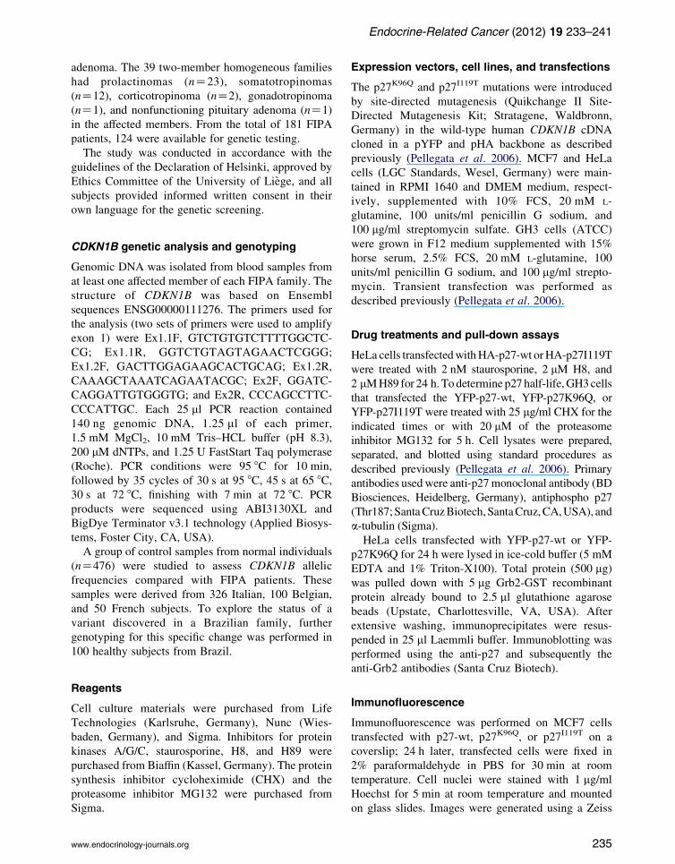
Figure 1 Subcellular localization of wild-type and mutant p27. (A) Hp27-wt, p27K96Q, or p27I119T mutant proteins and examined by westemutant transfected cells. (B) MCF7 cells were transfected as in (Aproteins (wild-type and mutants) were located primarily in the nucle
236
both brothers of a Brazilian two-member heterogeneous
FIPA family with a somatotropinoma and a nonfunction-
ing pituitary adenoma and appeared among 100 Brazilian
controls: 198 chromosomes were G and two were C
(genotype: 196 G/G and two G/C). An intronic change,
c.605C36COT, was seen in one FIPA family member
(male with a giant prolactinoma) and did not occur in
the control subjects; however, in silico modeling
indicated that this variant had no strong effect on splicing
and was deemed to probably represent a nonpathological
change. The previously reported T142T (c.426GOA)
variant was found in three unrelated prolactinoma
patients (one male and two females) across three different
FIPA families. The findings from the genotyping of the
control cohort (nZ476 healthy individuals) were as
follows: c.286AOC (p.K96Q), all 952 chromosomes
were A; c.605C36COT, all 952 chromosomes were C;
c.356TOC (p.I119T), 950 chromosomes were T and one
chromosome was C (genotype: 950 T/T and one T/C);
c.426GOA (p.T142T), 945 chromosomes were G and
seven chromosomes were A (genotype: 945 G/G
and seven G/A).
The I119T change was found in one member of a two-
person homogeneous FIPA family with somatotropino-
mas. The other affected member could not be genetically
tested (Supplementary Figure 1, see section on supple-
mentary data given at the end of this article). The K96Q
variant was found in a homogeneous FIPA family
presenting with prolactinomas, but the variation did not
segregate with prolactinoma-affected patients. The
patient with the K96Q change had hyperprolactinemia
due to a suspected prolactinoma that was treated
chronically with cabergoline when referred, who also
developed breast cancer at the age of 41. The unaffected
sister of this patient was also a carrier of this variant.
DAPI
P
YFP Merge
P
P
eLa cells were transfected with YFP-p27 constructs containingrn blotting. Expression and size of p27 were compared in wt and
) and were determined using fluorescent microscopy. All fusionus.
www.endocrinology-journals.org
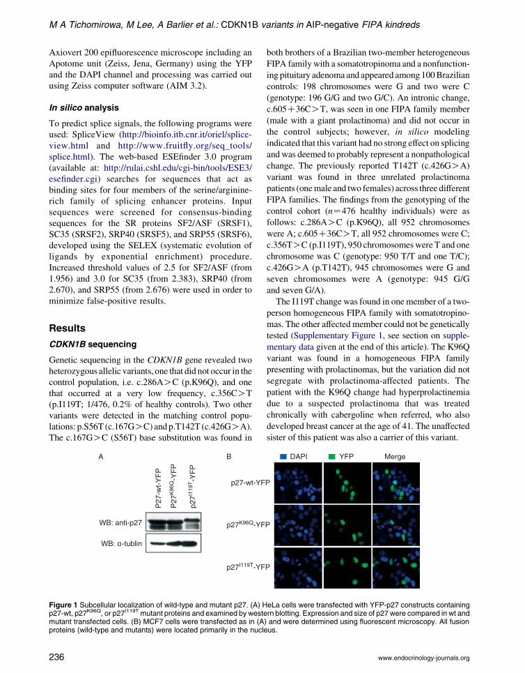
CHX 10 h
MG-132
p27-wt-YFP
– – –
– –– –– –
+ + + + + +
+++
WB: α-tubulin
WB: anti-p27
p27I119T-YFPp27K96Q-YFP
Figure 2 Stability of wild-type and mutant p27. The rate of the turnover of p27-wt, p27K96Q, and p27I119T proteins was measuredin exponentially growing, transiently transfected GH3 cells using cycloheximide (CHX) with and without the proteasome inhibitorMG-132. p27-wt and p27K96Q have a half-life of w10 h in GH3 cells. MG-132 has no effect on p27K96Q degradation. In contrast,p27I119T is more stable than p27-wt.
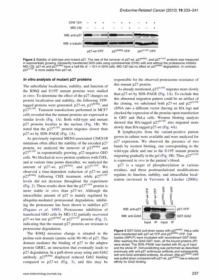
WB: anti-p27
WB: anti-Grb2
p27-
wt-
YF
P
p27-YFP
GST-Grb2
p27-
wt-
YF
P
p27K
96Q
-YF
P
p27K
96Q
-YF
P
Input IP IPInput
Figure 3 GST-Grb2 pull-down assay with p27K96Q. HeLa cellswere transfected with p27-wt-YFP and p27K96Q-YFP. Celllysates (INPUT) were incubated with recombinant GST-Grb2.After washing the Grb2-GST resin, all the bound proteins (IP)were eluted. The SDS–PAGE was loaded with 50 mg of inputand the whole IP. Immunoblotting was performed as describedpreviously with anti-p27 antimouse antibody and subsequentlywith anti-Grb2 antirabbit antibody. As shown, little p27K96Q-YFPwas pulled down compared with p27-wt. p27K96Q has a reducedaffinity for Grb2 binding.
Endocrine-Related Cancer (2012) 19 233–241
In vitro analysis of mutant p27 proteins
The subcellular localization, stability, and function of
the K96Q and I119T mutant proteins were studied
in vitro. To determine the effect of the p27 changes on
protein localization and stability, the following YFP-
tagged proteins were generated: p27-wt, p27K96Q, and
p27I119T. Transient transfections performed in MCF7
cells revealed that the mutant proteins are expressed at
similar levels (Fig. 1A). Both wild-type and mutant
p27 proteins localize to the nucleus (Fig. 1B). We
noted that the p27I119T protein migrates slower than
p27-wt by SDS–PAGE (Fig. 1A).
As previously reported MEN4-associated CDKN1B
mutations often affect the stability of the encoded p27
protein, we analyzed the turnover of p27K96Q and
p27I119T in exponentially growing, p27-negative GH3
cells. We blocked de novo protein synthesis with CHX,
and at various time points thereafter, we analyzed the
amount of p27-wt, p27K96Q, and p27I119T. We
observed a time-dependent reduction of p27-wt and
p27K96Q following CHX treatment, while p27I119T
levels did not decrease throughout the experiment
(Fig. 2). These results show that the p27I119T protein is
more stable in vitro than p27-wt. Although the
intracellular amount of p27 is mainly regulated by
ubiquitin-mediated proteasomal degradation, inhibit-
ing the proteasome has been shown to stabilize p27
(Pagano et al. 1995). Proteasome inhibition of
transfected GH3 cells by MG-132 partially recovered
p27-wt but not p27K96Q or p27I119T proteins (Fig. 2),
indicating that the mutant p27 proteins are resistant to
proteasome degradation.
The K96Q missense change is situated in the
proline-rich domain (amino acids 90–96) of p27. This
domain mediates the binding of p27 to the adaptor
protein GRB2, an interaction that eventually leads to
p27 degradation. In a pull-down assay using anti-Grb2
antibody, p27K96Q displayed reduced Grb2 binding
compared to p27-wt (Fig. 3), and this may be
www.endocrinology-journals.org
responsible for the observed proteasome resistance of
this mutant p27 protein.
As already mentioned, p27I119T migrates more slowly
than p27-wt by SDS–PAGE (Fig. 1A). To exclude that
this abnormal migration pattern could be an artifact of
the cloning, we subcloned both p27-wt and p27I119T
cDNA into a different vector (having an HA tag) and
checked the expression of the proteins upon transfection
in GH3 and HeLa cells. Western blotting analysis
showed that HA-tagged p27I119T also migrated more
slowly than HA-tagged p27-wt (Fig. 4A).
B lymphocytes from the variant-positive patient
grown in culture were available and were analyzed for
p27 expression. We observed the presence of two
bands by western blotting, one corresponding to the
wild-type allele and one to the I119T mutant protein
migrating gradually in the gel (Fig. 4B). Thus, p27I119T
is expressed in vivo in the patient’s blood.
p27 is a target of phosphorylation at various
residues, and these posttranslational modifications
regulate its function, stability, and intracellular local-
ization (reviewed in Vervoorts & Luscher (2008)).
237
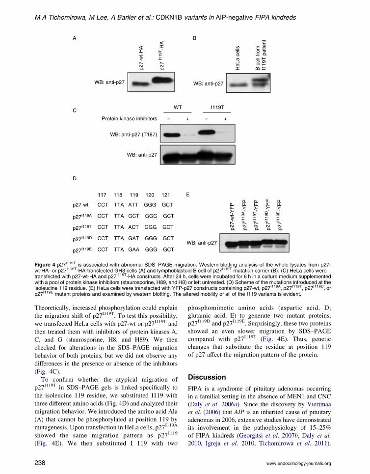
117 118 119 120 121
CCT TTA ATT GGG GCT
CCT TTA GCT GGG GCT
CCT TTA ACT GGG GCT
CCT TTA GAT GGG GCT
CCT TTA GAA GGG GCTWB: anti-p27
WB: anti-p27
WB: anti-p27
C
D
E
A B
WB: anti-p27
HeL
a ce
lls
WB: anti-p27 (T187)
Protein kinase inhibitors
p27-wt
p27-
wt-
YF
P
p27I119A
p27I1
19A-Y
FP
p27I1
19T-Y
FP
p27I1
19D-Y
FP
p27I1
19E-Y
FP
p27I119T
p27I119D
p27I119E
WT I119T
– – ++
p27-
wt-
HA
p27
I119
T-H
A
B c
ell f
rom
I119
T p
atie
ntFigure 4 p27I119T is associated with abnormal SDS–PAGE migration. Western blotting analysis of the whole lysates from p27-wt-HA- or p27I119T-HA-transfected GH3 cells (A) and lymphoblastoid B cell of p27I119T mutation carrier (B). (C) HeLa cells weretransfected with p27-wt-HA and p27I119T-HA constructs. After 24 h, cells were incubated for 6 h in a culture medium supplementedwith a pool of protein kinase inhibitors (staurosporine, H89, and H8) or left untreated. (D) Scheme of the mutations introduced at theisoleucine 119 residue. (E) HeLa cells were transfected with YFP-p27 constructs containing p27-wt, p27I119A, p27I119T, p27I119D, orp27I119E mutant proteins and examined by western blotting. The altered mobility of all of the I119 variants is evident.
M A Tichomirowa, M Lee, A Barlier et al.: CDKN1B variants in AIP-negative FIPA kindreds
Theoretically, increased phosphorylation could explain
the migration shift of p27I119T. To test this possibility,
we transfected HeLa cells with p27-wt or p27I119T and
then treated them with inhibitors of protein kinases A,
C, and G (staurosporine, H8, and H89). We then
checked for alterations in the SDS–PAGE migration
behavior of both proteins, but we did not observe any
differences in the presence or absence of the inhibitors
(Fig. 4C).
To confirm whether the atypical migration of
p27I119T in SDS–PAGE gels is linked specifically to
the isoleucine 119 residue, we substituted I119 with
three different amino acids (Fig. 4D) and analyzed their
migration behavior. We introduced the amino acid Ala
(A) that cannot be phosphorylated at position 119 by
mutagenesis. Upon transfection in HeLa cells, p27I119A
showed the same migration pattern as p27I119
(Fig. 4E). We then substituted I 119 with two
238
phosphomimetic amino acids (aspartic acid, D;
glutamic acid, E) to generate two mutant proteins,
p27I119D and p27I119E. Surprisingly, these two proteins
showed an even slower migration by SDS–PAGE
compared with p27I119T (Fig. 4E). Thus, genetic
changes that substitute the residue at position 119
of p27 affect the migration pattern of the protein.
Discussion
FIPA is a syndrome of pituitary adenomas occurring
in a familial setting in the absence of MEN1 and CNC
(Daly et al. 2006a). Since the discovery by Vierimaa
et al. (2006) that AIP is an inherited cause of pituitary
adenomas in 2006, extensive studies have demonstrated
its involvement in the pathophysiology of 15–25%
of FIPA kindreds (Georgitsi et al. 2007b, Daly et al.
2010, Igreja et al. 2010, Tichomirowa et al. 2011).
www.endocrinology-journals.org
Endocrine-Related Cancer (2012) 19 233–241
In an effort to study other potential genetic causes of
FIPA, we examined CDKN1B sequences in 124
individuals from 88 FIPA AIP mutation-negative
kindreds, as previous studies had concentrated largely
on MEN1-negative MEN1 cohorts (Igreja et al. 2009).
We found two new germline CDKN1B changes in
patients with pituitary adenomas from AIP mutation-
negative FIPA kindreds. Although these sequence
changes were identified in a familial setting and they
altered p27 function or structure in vitro, the K96Q
variant did not segregate with pituitary adenomas in
one kindred. In the case of I119T variant that affected
CDKN1B molecular weight/migration, one of the two
family members affected with a pituitary adenoma was
not available for genetic testing, so it cannot be fully
ruled in or out as a cause of the clinical phenotype.
Based on these findings, CDKN1B changes alone are
not a frequent or likely cause of the FIPA tumor
phenotype but could represent a contributing factor.
Nevertheless, the CDKN1B sequence variants
described here add to growing evidence of a role for
p27-related dysfunction in the development of a subset
of many endocrine tumors within and outside of the
setting of MEN4.
The involvement of p27 in pituitary tumorigenesis
has been demonstrated in animal studies. Indeed, p27-
null mice develop pituitary intermediate lobe adeno-
mas (Fero et al. 1996, Kiyokawa et al. 1996,
Nakayama et al. 1996), and heterozygous p27C/K
mice display pituitary hyperplasia (Fero et al. 1998).
While human pituitary adenomas only rarely showed
somatic CDKN1B mutations, downregulation of p27 is
observed frequently in these tumors, especially in
corticotropinomas (Kawamata et al. 1995, Ikeda et al.
1997, Jin et al. 1997, Takeuchi et al. 1998). Interest
was renewed by the discovery that germline CDKN1B
mutations in both the rat MENX and the human MEN4
syndromes are associated with development of pitu-
itary adenomas (Fritz et al. 2002, Pellegata et al. 2006).
Among the eight MEN4 patients identified to date,
three (37.5%) had pituitary adenomas (a somatotropi-
noma, Cushing disease, and a nonfunctioning ade-
noma), so it appears to be a distinctive disease feature
among these patients, although not as pronounced as
primary hyperparathyroidism (7/8 patients, 87.5%).
The K96Q mutation is situated in the proline-rich
domain (amino acids 90–96) of p27, which mediates
the binding to the adaptor molecule Grb2, which in turn
recruits and leads to activation of Ras (Marinoni &
Pellegata 2011). The interaction between p27 and Grb2
promotes p27 degradation in the cytoplasm (Pagano
et al. 1995, Vervoorts & Luscher 2008). Indeed,
p27K96Q displayed less Grb2 binding during a pull-
www.endocrinology-journals.org
down assay compared with p27-wt. These findings
echo the altered Grb2 interaction reported by Agarwal
et al. (2009) in a patient with a missense mutation at
the previous amino acid residue (P95S) that led to
parathyroid tumors and a metastatic gastrinoma.
The I119T variant affects a residue located in the
so-called ‘scatter domain’ of p27 (amino acids 118–
158), which is responsible for actin cytoskeletal
rearrangement and cell migration, processes involved
in metastatic spread of human tumors (McAllister et al.
2003). This change causes a shift in the migration of
the p27 protein in SDS–PAGE gels. The unique
migration pattern of p27I119T, indicative of posttransla-
tional modifications, was not affected by multiple
kinase inhibitors, suggesting that it is not due to
phosphorylation at this newly created threonine
residue. As glycosylation occurs at serine, threonine,
or aspartic acid residues, the migration shift associated
with the 119T residue could be caused by glycosylation
of the protein (Dennis et al. 1999), thereby conferring
greater stability. In agreement with this finding,
p27I119T is more stable than the p27-wt in vitro.
The I119T sequence change was previously
described as a somatic genetic mutation in a patient
with myeloproliferative disorder (presence of the
change in the patient’s germline was not studied;
Pappa et al. 2005); also the W76X nonsense CDKN1B
mutation found in a MEN4 patient had been previously
identified as a somatic change in hematological
malignancies (Morosetti et al. 1995). Moreover, the
c.356T/C (I119T) change has been reported in a study
of hereditary prostate cancer (Chang et al. 2004), but
the association of the C variant allele with the
predisposition to the disease could not be demon-
strated. The observations that this variant allele is
expressed and translated into protein in our mutation
carrier, in addition to the association of the I119T
change with other tumor types and its potential effect in
the function of p27, make a plausible case that it may
play a role in tumor predisposition.
In conclusion, this study is the first extensive study
of CDKN1B germline variants in a set of 88 FIPA
families that do not have AIP mutations. According to
our data, mutations of CDKN1B are not a cause of
FIPA. However, CDKN1B germline variants associ-
ated with in vitro molecular phenotypes were seen in
nearly 2% of cases studied. Altered p27 function may
infrequently play a role in general pituitary disease
outside of MEN4, although screening for CDKN1B
mutations systematically appears unjustified in the
setting of the O75% of FIPA kindreds not caused by
AIP mutations (Jaffrain-Rea et al. 2011).
239
M A Tichomirowa, M Lee, A Barlier et al.: CDKN1B variants in AIP-negative FIPA kindreds
Supplementary data
This is linked to the online version of the paper at http://dx.
doi.org/10.1530/ERC-11-0362.
Declaration of interest
The authors declare that there is no conflict of interest that
could be perceived as prejudicing the impartiality of the
research reported.
Funding
The study was supported by Fonds d’investissement de
recherche scientifique of the CHU de Liege (FIRS) and by an
unrestricted educational grant from Pfizer (A Beckers) and
Oncogentic Network of the French Ministry of Health,
Centre National de la Recherche Scientifique, University of
Mediterranee and Assistance Publique des Hopitaux de
Marseille (A Barlier).
Acknowledgements
The authors thank Julien Pujol and Morgane Pertuit
(Laboratory of Molecular Biology AP-HM Conception,
Marseille) and Marie-Therese Hagelstein (Department of
Clinical Genetics, CHU de Liege) for their assistance in DNA
sequencing. The authors also thank Dr Lopez, Prof. Tessier
(CHU Limoges), Dr Morange, Dr Valero, (CHU Marseille),
Dr Chevalier, Dr Kunstmann (CHU Nice), Dr Sonnet, Dr
Dupont (CHU Brest), Dr Nunes, Dr Gatta-Cherifi (CHU
Bordeaux), and Dr Julier (CH Ales) for identifying patients.
The authors thank the family members for their collaboration.
References
Agarwal SK, Mateo CM & Marx SJ 2009 Rare germline
mutations in cyclin-dependent kinase inhibitor genes in
multiple endocrine neoplasia type 1 and related states.
Journal of Clinical Endocrinology and Metabolism 94
1826–1834. (doi:10.1210/jc.2008-2083)
CBTRUS (2011). CBTRUS statistical report: primary brain and
central nervous system tumors diagnosed in the United
States in 2004–2007. Source: Central Brain Tumor Registry
of the United States, Hinsdale, IL. Website: www.cbtrus.org.
Chang BL, Zheng SL, Isaacs SD, Wiley KE, Turner A, Li G,
Walsh PC, Meyers DA, Isaacs WB & Xu J 2004 A
polymorphism in the CDKN1B gene is associated with
increased risk of hereditary prostate cancer.CancerResearch
64 1997–1999. (doi:10.1158/0008-5472.CAN-03-2340)
Costa-Guda J, Marinoni I, Molatore S, Pellegata NS &
Arnold A 2011 Somatic mutation and germline sequence
abnormalities in CDKN1B, encoding p27Kip1, in
sporadic parathyroid adenomas. Journal of Clinical
Endocrinology and Metabolism 96 E701–E706. (doi:10.
1210/jc.2010-1338)
Daly AF, Jaffrain-Rea ML, Ciccarelli A, Valdes-Socin H,
Rohmer V, Tamburrano G, Borson-Chazot C, Estour B,
240
Ciccarelli E, Brue T et al. 2006a Clinical characterization
of familial isolated pituitary adenomas. Journal of
Clinical Endocrinology and Metabolism 91 3316–3323.
(doi:10.1210/jc.2005-2671)
Daly AF, Rixhon M, Adam C, Dempegioti A, Tichomirowa
MA & Beckers A 2006b High prevalence of pituitary
adenomas: a cross-sectional study in the province of
Liege, Belgium. Journal of Clinical Endocrinology and
Metabolism 91 4769–4775. (doi:10.1210/jc.2006-1668)
Daly AF, Vanbellinghen JF, Khoo SK, Jaffrain-Rea ML,
Naves LA, Guitelman MA, Murat A, Emy P, Gimenez-
Roqueplo AP, Tamburrano G et al. 2007 Aryl hydro-
carbon receptor-interacting protein gene mutations in
familial isolated pituitary adenomas: analysis in 73
families. Journal of Clinical Endocrinology and Metab-
olism 92 1891–1896. (doi:10.1210/jc.2006-2513)
Daly AF, Tichomirowa MA, Petrossians P, Heliovaara E,
Jaffrain-Rea ML, Barlier A, Naves LA, Ebeling T, Karhu A,
Raappana A et al. 2010 Clinical characteristics and
therapeutic responses in patients with germ-line AIP
mutations and pituitary adenomas: an International colla-
borative study. Journal of Clinical Endocrinology and
Metabolism 95 E373–E383. (doi:10.1210/jc.2009-2556)
Dennis JW, Granovsky M & Warren CE 1999 Glycoprotein
glycosylation and cancer progression. Biochimica et
Biophysica Acta 1473 21–34. (doi:10.1016/S0304-
4165(99)00167-1)
Fernandez A, Karavitaki N & Wass JA 2010 Prevalence of
pituitary adenomas: a community-based, cross-sectional
study in Banbury (Oxfordshire, UK).ClinicalEndocrinology
72 377–382. (doi:10.1111/j.1365-2265.2009.03667.x)
Fero ML, Rivkin M, Tasch M, Porter P, Carow CE, Firpo E,
Polyak K, Tsai LH, Broudy V, Perlmutter RM et al. 1996
A syndrome of multiorgan hyperplasia with features of
gigantism, tumorigenesis, and female sterility in p27-
deficient mice. Cell 85 733–744. (doi:10.1016/S0092-
8674(00)81239-8)
Fero ML, Randel E, Gurley KE, Roberts JM & Kemp CJ 1998
The murine gene p27Kip1 is haplo-insufficient for tumour
suppression. Nature 396 177–180. (doi:10.1038/24179)
Fritz A, Walch A, Piotrowska K, Rosemann M, Schaffer E,
Weber K, Timper A, Wildner G, Graw J, Hofler H et al.2002
Recessive transmission of a multiple endocrine neoplasia
syndrome in the rat. Cancer Research 62 3048–3051.
Georgitsi M, Raitila A, Karhu A, van der Luijt RB, Aalfs CM,
Sane T, Vierimaa O, Makinen MJ, Tuppurainen K,
Paschke R et al. 2007a Germline CDKN1B/p27Kip1
mutation in multiple endocrine neoplasia. Journal of
Clinical Endocrinology and Metabolism 92 3321–3325.
(doi:10.1210/jc.2006-2843)
Georgitsi M, Raitila A, Karhu A, Tuppurainen K, Makinen
MJ, Vierimaa O, Paschke R, Saeger W, van der Luijt RB,
Sane T et al. 2007b Molecular diagnosis of pituitary
adenoma predisposition caused by aryl hydrocarbon
receptor-interacting protein gene mutations. PNAS 104
4101–4105. (doi:10.1073/pnas.0700004104)
www.endocrinology-journals.org
Endocrine-Related Cancer (2012) 19 233–241
Igreja S, Chahal HS, Akker SA, Gueorguiev M, Popovic V,
Damjanovic S, Burman P, Wass JA, Quinton R, Grossman
AB et al. 2009 Assessment of p27 (cyclin-dependent
kinase inhibitor 1B) and aryl hydrocarbon receptor-
interacting protein (AIP) genes in multiple endocrine
neoplasia (MEN1) syndrome patients without any
detectable MEN1 gene mutations. Clinical Endocrinology
70 259–264. (doi:10.1111/j.1365-2265.2008.03379.x)
Igreja S, Chahal HS, King P, Bolger GB, Srirangalingam U,
Guasti L, Chapple JP, Trivellin G, Gueorguiev M, Guegan
K et al. 2010 Characterization of aryl hydrocarbon
receptor interacting protein (AIP) mutations in familial
isolated pituitary adenoma families. Human Mutation 31
950–960. (doi:10.1002/humu.21292)
Ikeda H, Yoshimoto T & Shida N 1997 Molecular analysis
of p21 and p27 genes in human pituitary adenomas.
British Journal of Cancer 76 1119–1123. (doi:10.1038/
bjc.1997.521)
Jaffrain-Rea ML, Daly AF, Angelini M, Petrossians P,
Bours V & Beckers A 2011 Genetic susceptibility in
pituitary adenomas: from pathogenesis to clinical
implications. Expert Review of Endocrinology &
Metabolism 6 195–214 (doi:10.1586/eem.10.87).
(doi:10.1586/eem.10.87)
Jin L, Qian X, Kulig E, Sanno N, Scheithauer BW, Kovacs K,
Young WF Jr & Lloyd RV 1997 Transforming growth
factor-beta, transforming growth factor-beta receptor II, and
p27Kip1 expression in nontumorous and neoplastic human
pituitaries. American Journal of Pathology 151 509–519.
Kawamata N, Morosetti R, Miller CW, Park D, Spirin KS,
Nakamaki T, Takeuchi S, Hatta Y, Simpson J, Wilcyznski
S et al. 1995 Molecular analysis of the cyclin-dependent
kinase inhibitor gene p27/Kip1 in human malignancies.
Cancer Research 55 2266–2269.
Kiyokawa H, Kineman RM, Manova-Todorova KO, Soares
VC, Hoffman ES, Ono M, Khanam D, Hayday AC,
Frohman LA & Koff A 1996 Enhanced growth of mice
lacking the cyclin-dependent kinase inhibitor function of
p27Kip1. Cell 85 721–732. (doi:10.1016/S0092-
8674(00)81238-6)
Marinoni I & Pellegata NS 2011 p27kip1: a new multiple
endocrine neoplasia gene? Neuroendocrinology 93
19–28. (doi:10.1159/000320366)
McAllister SS, Becker-Hapak M, Pintucci G, Pagano M &
Dowdy SF 2003 Novel p27kip1 C-terminal scatter domain
mediates Rac-dependent cell migration independent of cell
cycle arrest functions. Molecular and Cellular Biology 23
216–228. (doi:10.1128/MCB.23.1.216-228.2003)
Molatore S, Marinoni I, Lee M, Pulz E, Ambrosio MR, degli
Uberti EC, Zatelli MC & Pellegata NS 2010 A novel
germline CDKN1B mutation causing multiple endocrine
tumors: clinical, genetic and functional characterization.
Human Mutation 31 E1825–E1835. (doi:10.1002/humu.
21354)
Morosetti R, Kawamata N, Gombart AF, Miller CW, Hatta Y,
Hirama T, Said JW, Tomonaga M & Koeffler HP 1995
www.endocrinology-journals.org
Alterations of the p27KIP1 gene in non-Hodgkin’s
lymphomas and adult T-cell leukemia/lymphoma. Blood
86 1924–1930.
Nakayama K, Ishida N, Shirane M, Inomata A, Inoue T,
Shishido N, Horii I, Loh DY & Nakayama K 1996 Mice
lacking p27Kip1 display increased body size, multiple
organ hyperplasia, retinal dysplasia, and pituitary tumors.
Cell 85 707–720. (doi:10.1016/S0092-8674(00)81237-4)
Ojeda D, Lakhal B, Fonseca DJ, Braham R, Landolsi H,
Mateus HE, Restrepo CM, Elghezal H, Saad A & Laissue
P 2011 Sequence analysis of the CDKN1B gene in
patients with premature ovarian failure reveals a novel
mutation potentially related to the phenotype. Fertility
and Sterility 95 2658–2660. (doi:10.1016/j.fertnstert.
2011.04.045)
Pagano M, Tam SW, Theodoras AM, Beer-Romero P, Del
Sal G, Chau V, Yew PR, Draetta GF & Rolfe M 1995 Role
of the ubiquitin–proteasome pathway in regulating
abundance of the cyclin-dependent kinase inhibitor p27.
Science 269 682–685. (doi:10.1126/science.7624798)
Pappa V, Papageorgiou S, Papageorgiou E, Panani A, Boutou E,
Tsirigotis P, Dervenoulas J, Economopoulos T & Raptis S
2005 A novel p27 gene mutation in a case of unclassified
myeloproliferative disorder. Leukemia Research 29
229–231. (doi:10.1016/j.leukres.2004.06.007)
Pellegata NS, Quintanilla-Martinez L, Siggelkow H, Samson
E, Bink K, Hofler H, Fend F, Graw J & Atkinson MJ 2006
Germ-line mutations in p27 Kip1 cause a multiple
endocrine neoplasia syndrome in rats and humans. PNAS
103 15558–15563. (doi:10.1073/pnas.0603877103)
Takeuchi S, Koeffler HP, Hinton DR, Miyoshi I, Melmed S &
Shimon I 1998 Mutation and expression analysis of the
cyclin-dependent kinase inhibitor gene p27/Kip1 in
pituitary tumors. Journal of Endocrinology 157 337–341.
(doi:10.1677/joe.0.1570337)
Tichomirowa MA, Barlier A, Daly AF, Jaffrain-Rea ML,
Ronchi CL, Yaneva M, Urban JD, Petrossians P,
Elenkova AP, Tabarin A et al. 2011 High prevalence of
AIP gene mutations following focused screening in young
patients with sporadic pituitary macroadenomas.
European Journal of Endocrinology 165 509–515 (DOI:
10.1530/EJE-11-0304). (doi:10.1530/EJE-11-0304)
Vervoorts J & Luscher B 2008 Post-translational regulation
of the tumor suppressor p27(KIP1). Cellular and
Molecular Life Sciences 65 3255–3264. (doi:10.1007/
s00018-008-8296-7)
Vierimaa O, Georgitsi M, Lehtonen R, Vahteristo P, Kokko
A, Raitila A, Tuppurainen K, Ebeling TM, Salmela PI,
Paschke R et al. 2006 Pituitary adenoma predisposition
caused by germline mutations in the AIP gene. Science
312 1228–1230. (doi:10.1126/science.1126100)
Received in final form 30 December 2011Accepted 30 January 2012Made available online as an Accepted Preprint30 January 2012
241