CXCL10 plays a key role as an inflammatory mediator and a non-invasive biomarker of non-alcoholic...
11
CXCL10 plays a key role as an inflammatory mediator and a non-invasive biomarker of non-alcoholic steatohepatitis Xiang Zhang 1,2 , Jiayun Shen 1,2 , Kwan Man 3 , Eagle S.H. Chu 1,2 , Tung On Yau 1,2 , Joanne C.Y. Sung 1 , Minnie Y.Y. Go 1 , Jun Deng 4 , Liwei Lu 4 , Vincent W.S. Wong 1 , Joseph J.Y. Sung 1,2 , Geoffrey Farrell 5 , Jun Yu 1,2,⇑ 1 Institute of Digestive Disease and The Department of Medicine and Therapeutics, State Key Laboratory of Digestive Disease, Li Ka Shing Institute of Health Sciences, The Chinese University of Hong Kong, Hong Kong, China; 2 Gastrointestinal Cancer Biology and Therapeutics Laboratory, Shenzhen Research Institute, The Chinese University of Hong Kong, Shenzhen, China; 3 Department of Surgery, LKS Faculty of Medicine, The University of Hong Kong, Hong Kong, China; 4 Department of Pathology and Center of Infection and Immunology, The University of Hong Kong, Hong Kong, China; 5 Australian National University Medical School at The Canberra Hospital, Canberra, Australia Background & Aims: Perpetuate liver inflammation is crucial in the pathogenesis of non-alcoholic steatohepatitis (NASH). Expression of CXCL10, a pro-inflammatory cytokine, correlates positively with obesity and type 2 diabetes. Whether CXCL10 plays a role in NASH was unknown. We aimed to investigate the functional and clinical impact of CXCL10 in NASH. Methods: Cxcl10 gene-deleted (Cxcl10 / ) and C57BL/6 wild type (WT) mice were fed a methionine- and choline-deficient (MCD) diet for 4 or 8 weeks. In other experiments, we injected neutral- izing anti-CXCL10 mAb into MCD-fed WT mice. Human serum was obtained from 147 patients with biopsy-proven non-alco- holic fatty liver disease and 73 control subjects. Results: WT mice, fed the MCD diet, developed steatohepatitis with higher hepatic CXCL10 expression. Cxcl10 / mice were refractory to MCD-induced steatohepatitis. We further revealed that CXCL10 was associated with the induction of important pro-inflammatory cytokines (TNF-a, IL-1b, and MCP-1) and activation of the NF-jB pathway. CXCL10 was linked to steatosis through upregulation of the lipogenic factors SREBP-1c and LXR, and also to oxidative stress (upregulation of CYP2E1 and C/EBPb). Blockade of CXCL10 protected against hepatocyte injury in vitro and against steatohepatitis development in mice. We further investigated the clinical impact of CXCL10 and found circulating and hepatic CXCL10 levels were significantly higher in human NASH. Importantly, the circulating CXCL10 level was correlated with the degree of lobular inflammation and was an independent risk factor for NASH patients. Conclusions: We demonstrate for the first time that CXCL10 plays a pivotal role in the pathogenesis of experimental steato- hepatitis. CXCL10 maybe a potential non-invasive biomarker for NASH patients. Ó 2014 European Association for the Study of the Liver. Published by Elsevier B.V. All rights reserved. Introduction Non-alcoholic fatty liver disease (NAFLD) has become increas- ingly important worldwide due to changes in lifestyle and resul- tant over-nutrition [1]. Non-alcoholic steatohepatitis (NASH) is a severe form of NAFLD, characterized by necroinflammation and lipid accumulation [2,3]. Little is known about the factors respon- sible for the transition from benign steatosis to steatohepatitis in NAFLD/NASH. As a consequence, apart from addressing lifestyle issues, there are few effective interventions to treat patients with NASH. The present concept about NASH pathogenesis is that increased levels of toxic lipids, such as free fatty acids or free cho- lesterol provide initiating and propagating mechanism for hepa- tocellular injury and resultant inflammation. Inflammation may result from oxidative stress and pro-inflammatory chemokines and cytokines, which perpetuate liver injury and lead to fibrosis [4]. Identification of the pro-inflammatory cytokines, which are associated with lipotoxicity, may improve our understanding of Journal of Hepatology 2014 vol. 61 j 1365–1375 Keywords: Non-alcoholic fatty liver disease; Inflammation; Chemokine; Animal model; Biomarker. Received 16 January 2014; received in revised form 14 June 2014; accepted 6 July 2014; available online 15 July 2014 ⇑ Corresponding author. Address: Institute of Digestive Disease, Department of Medicine and Therapeutics, Prince of Wales Hospital, The Chinese University of Hong Kong, Shatin, NT, Hong Kong, China. Tel.: +852 37636099; fax: +852 21445330. E-mail address: [email protected] (J. Yu). Abbreviations: NAFLD, non-alcoholic fatty liver disease; NASH, non-alcoholic steatohepatitis; CXCL, CXC chemokine ligand; TLR, toll-like receptor; NF-jB, nuclear factor-jB; WT, wild type; MCD, methionine- and choline-deficient; ALT, alanine aminotransferase; TUNEL, terminal deoxynucleotidyl transferase dUTP nick end labelling; FACS, fluorescence activated cell sorting; ROC, receiver operating characteristic; IQR, interquartile range; TBARS, thiobarbituric acid reactive substances; TNF-a, tumor necrosis factor-a; IL, interleukin; MCP-1, monocyte chemoattractant protein 1; C/EBPb, CCAAT/enhancer binding protein beta; COX-2, cyclooxygenase-2; ICAM-1, intercellular adhesion molecule-1; LXR, liver X receptors; SREBP-1c, sterol regulatory element binding protein isoform 1c; ChREBP, carbohydrate response element binding protein; SCD-1, stearoyl-CoA desaturase isoform-1; Cyp, cytochrome P450; mAb, monoclonal antibodies; BMI, body mass index; AUROC, area under the receiver operating characteristic curve; LDL-c, low density lipoprotein-cholesterol; HbA1c, glycated haemoglobin; ACK, ammonium chloride potassium. Research Article
-
Upload
independent -
Category
Documents
-
view
3 -
download
0
Transcript of CXCL10 plays a key role as an inflammatory mediator and a non-invasive biomarker of non-alcoholic...

Research Article
CXCL10 plays a key role as an inflammatory mediator and anon-invasive biomarker of non-alcoholic steatohepatitis
Xiang Zhang1,2, Jiayun Shen1,2, Kwan Man3, Eagle S.H. Chu1,2, Tung On Yau1,2,Joanne C.Y. Sung1, Minnie Y.Y. Go1, Jun Deng4, Liwei Lu4, Vincent W.S. Wong1,
Joseph J.Y. Sung1,2, Geoffrey Farrell5, Jun Yu1,2,⇑
1Institute of Digestive Disease and The Department of Medicine and Therapeutics, State Key Laboratory of Digestive Disease, Li Ka Shing Instituteof Health Sciences, The Chinese University of Hong Kong, Hong Kong, China; 2Gastrointestinal Cancer Biology and Therapeutics Laboratory,Shenzhen Research Institute, The Chinese University of Hong Kong, Shenzhen, China; 3Department of Surgery, LKS Faculty of Medicine, The
University of Hong Kong, Hong Kong, China; 4Department of Pathology and Center of Infection and Immunology, The University of Hong Kong,Hong Kong, China; 5Australian National University Medical School at The Canberra Hospital, Canberra, Australia
Background & Aims: Perpetuate liver inflammation is crucial inthe pathogenesis of non-alcoholic steatohepatitis (NASH).Expression of CXCL10, a pro-inflammatory cytokine, correlatespositively with obesity and type 2 diabetes. Whether CXCL10plays a role in NASH was unknown. We aimed to investigatethe functional and clinical impact of CXCL10 in NASH.Methods: Cxcl10 gene-deleted (Cxcl10�/�) and C57BL/6 wild type(WT) mice were fed a methionine- and choline-deficient (MCD)diet for 4 or 8 weeks. In other experiments, we injected neutral-izing anti-CXCL10 mAb into MCD-fed WT mice. Human serumwas obtained from 147 patients with biopsy-proven non-alco-holic fatty liver disease and 73 control subjects.Results: WT mice, fed the MCD diet, developed steatohepatitiswith higher hepatic CXCL10 expression. Cxcl10�/� mice wererefractory to MCD-induced steatohepatitis. We further revealedthat CXCL10 was associated with the induction of importantpro-inflammatory cytokines (TNF-a, IL-1b, and MCP-1) and
Journal of Hepatology 20
Keywords: Non-alcoholic fatty liver disease; Inflammation; Chemokine; Animalmodel; Biomarker.Received 16 January 2014; received in revised form 14 June 2014; accepted 6 July2014; available online 15 July 2014⇑ Corresponding author. Address: Institute of Digestive Disease, Department ofMedicine and Therapeutics, Prince of Wales Hospital, The Chinese University ofHong Kong, Shatin, NT, Hong Kong, China. Tel.: +852 37636099; fax: +85221445330.E-mail address: [email protected] (J. Yu).Abbreviations: NAFLD, non-alcoholic fatty liver disease; NASH, non-alcoholicsteatohepatitis; CXCL, CXC chemokine ligand; TLR, toll-like receptor; NF-jB,nuclear factor-jB; WT, wild type; MCD, methionine- and choline-deficient; ALT,alanine aminotransferase; TUNEL, terminal deoxynucleotidyl transferase dUTPnick end labelling; FACS, fluorescence activated cell sorting; ROC, receiveroperating characteristic; IQR, interquartile range; TBARS, thiobarbituric acidreactive substances; TNF-a, tumor necrosis factor-a; IL, interleukin; MCP-1,monocyte chemoattractant protein 1; C/EBPb, CCAAT/enhancer binding proteinbeta; COX-2, cyclooxygenase-2; ICAM-1, intercellular adhesion molecule-1; LXR,liver X receptors; SREBP-1c, sterol regulatory element binding protein isoform 1c;ChREBP, carbohydrate response element binding protein; SCD-1, stearoyl-CoAdesaturase isoform-1; Cyp, cytochrome P450; mAb, monoclonal antibodies; BMI,body mass index; AUROC, area under the receiver operating characteristic curve;LDL-c, low density lipoprotein-cholesterol; HbA1c, glycated haemoglobin; ACK,ammonium chloride potassium.
activation of the NF-jB pathway. CXCL10 was linked to steatosisthrough upregulation of the lipogenic factors SREBP-1c and LXR,and also to oxidative stress (upregulation of CYP2E1 and C/EBPb).Blockade of CXCL10 protected against hepatocyte injury in vitroand against steatohepatitis development in mice. We furtherinvestigated the clinical impact of CXCL10 and found circulatingand hepatic CXCL10 levels were significantly higher in humanNASH. Importantly, the circulating CXCL10 level was correlatedwith the degree of lobular inflammation and was an independentrisk factor for NASH patients.Conclusions: We demonstrate for the first time that CXCL10plays a pivotal role in the pathogenesis of experimental steato-hepatitis. CXCL10 maybe a potential non-invasive biomarker forNASH patients.� 2014 European Association for the Study of the Liver. Publishedby Elsevier B.V. All rights reserved.
Introduction
Non-alcoholic fatty liver disease (NAFLD) has become increas-ingly important worldwide due to changes in lifestyle and resul-tant over-nutrition [1]. Non-alcoholic steatohepatitis (NASH) is asevere form of NAFLD, characterized by necroinflammation andlipid accumulation [2,3]. Little is known about the factors respon-sible for the transition from benign steatosis to steatohepatitis inNAFLD/NASH. As a consequence, apart from addressing lifestyleissues, there are few effective interventions to treat patients withNASH. The present concept about NASH pathogenesis is thatincreased levels of toxic lipids, such as free fatty acids or free cho-lesterol provide initiating and propagating mechanism for hepa-tocellular injury and resultant inflammation. Inflammation mayresult from oxidative stress and pro-inflammatory chemokinesand cytokines, which perpetuate liver injury and lead to fibrosis[4]. Identification of the pro-inflammatory cytokines, which areassociated with lipotoxicity, may improve our understanding of
14 vol. 61 j 1365–1375

Research Article
the pathogenesis of NASH, enabling the development of novelpharmacological treatments.One particularly important pro-inflammatory cytokine associ-ated with lipotoxicity is the CXC motif chemokine ligand 10(CXCL10), which recruits inflammatory cells to the site of tissuedamage [5,6]. CXCL10 has been implicated in the pathogenesisof hepatitis C virus infection through interactions with the toll-like receptor (TLR) 2 [7], and in hepatitis B virus-infectionthrough the nuclear factor-jB (NF-jB) pathway [8]. In varioustypes of liver injury, CXCL10 is secreted by hepatocytes in areasof lobular inflammation [9,10] and neutralization of CXCL10accelerates liver regeneration [11]. These data indicate a poten-tial role for CXCL10 in the development of intrahepatic inflamma-tion. Moreover, CXCL10 is upregulated in NASH patients [12] andcorrelates positively with the incidence of obesity and type 2 dia-betes [13,14]. These findings suggest that CXCL10 could be a piv-otal molecule that facilitates transition from benign steatosis toprogressively hepatocellular damage and inflammation insteatohepatitis.
We have recently reported that the anti-oxidant enzymeheme oxygenase-1 protects against development of experimentalsteatohepatitis in association with reduced production of CXCL10[15]. In the present study, we first investigated the functional roleof CXCL10 in the development of steatohepatitis using Cxcl10gene-deleted mice, and further explored the molecular mecha-nisms by which CXCL10 exerts its effects on inflammation, stea-tosis, oxidative stress and apoptosis. We demonstrated by in vitroand in vivo approaches that blockade of CXCL10 (neutralizinganti-CXCL10 mAb) protected against steatohepatitis. In particu-lar, we tested the clinical impact of CXCL10 in 147 patients withbiopsy-proven NAFLD and 73 control subjects and demonstratedthat circulating CXCL10 is an independent risk factor for patientswith NASH.
Materials and methods
Animals and treatments
Age-matched male Cxcl10 knock out (Cxcl10�/�) and C57BL/6 wild type (WT) mice(from Dr. Andrew D. Luster, Harvard Medical School) were fed either a methionine-and choline-deficient (MCD) diet or a control diet for 4 weeks to establish steato-hepatitis, or for 8 weeks to establish fibrosing steatohepatitis [15,16].
For CXCL10 neutralization experiments, male C57BL/6 WT mice were givenCXCL10-specific anti-CXCL10 mAb (R&D System, Minneapolis, MN) by intraperi-toneal injection (50 lg in 200 lL PBS per mouse) at 12 h before MCD diet, andthen the injection was repeated every 2 days for 5 cycles [10,14]. Mice were alsogiven an isotype-matched rat IgG2A mAb (R&D System) at the same time as thecontrols. In a separate experiment, anti-CXCL10 mAb or control mAb were sup-plemented for 10 days under MCD diet after induction of steatohepatitis in micefed the MCD diet for 3 weeks. All animals received humane care and all animalstudies were performed in accordance with guidelines approved by the AnimalExperimentation Ethics Committee of the Chinese University of Hong Kong.
Mice were sacrificed as previously described [17]. Biochemical determinationof serum alanine aminotransferase (ALT) levels, triglycerides and lipid peroxida-tion rates were performed. Liver histology, liver collagen content analysis, cyto-kine profiling assay, cDNA expression array, nuclear DNA binding activity assay,terminal deoxynucleotidyl transferase dUTP nick end labelling (TUNEL) assay,fluorescence activated cell sorting (FACS) analysis, qPCR and western blot wereperformed.
Subjects and human sample collection
Serum samples were collected from 147 patients with biopsy-proven NAFLD and73 healthy subjects as previously described [18,19]. Percutaneous liverbiopsy specimens were collected from 11 patients with NASH, 11 patients with
1366 Journal of Hepatology 2014
simple steatosis and 15 healthy controls in the Prince of Wales Hospital andthe Queen Mary Hospital, Hong Kong. All subjects had given written informedconsent and the study protocol was approved by the Clinical Research EthicsCommittee of the Chinese University of Hong Kong and the University of HongKong.
Statistical analysis
Differences between two groups were compared by the Mann-Whitney U test orStudent’s t test. Multiple group comparisons were made by the Kruskal-Wallistest or one-way ANOVA. Spearman’s correlation coefficient was used to estimatethe association of serum CXCL10 levels and several factors of interest, while mul-tiple linear regression was used to determine the independent factors associatedwith levels of CXCL10. Multiple logistic regression was performed to identify theindependent risk factors of NASH. A receiver operating characteristic (ROC) curveanalysis was conducted to assess the performance of CXCL10 in the prediction ofNAFLD/NASH. All statistical tests were performed using SPSS or GraphPad Soft-ware. Data were expressed as mean ± standard deviation or median (interquartilerange [IQR]) and considered significant at p <0.05.
Additional experimental procedures are provided in the SupplementaryMaterials and methods section.
Results
Hepatic CXCL10 expression is upregulated in experimentalsteatohepatitis and is required for its development
To elucidate the role of CXCL10 in the development of steatohep-atitis, Cxcl10�/� and WT mice were fed control or MCD diets for4 weeks. MCD-fed WT mice developed steatosis, ballooning hepa-tocytes, scattered lobular inflammatory cell infiltration, andinflammatory foci (Fig. 1A), consistent with steatohepatitis. Thiswas associated with increased hepatic CXCL10 mRNA and proteinlevels compared with mice fed a control diet (Fig. 1B), whichshowed normal liver histology (Fig. 1A). Conversely, MCD-fedCxcl10�/� mice showed significant less steatosis (p <0.01) andreduced inflammatory cell infiltration (p <0.01), as indicated bysteatosis and necroinflammatory scores (Fig. 1A). Consistent withthe histologic findings, measurement of serum ALT (p <0.0001),hepatic lipid peroxide by the thiobarbituric acid reactive sub-stances (TBARS) assay (p <0.01) and hepatic triglyceride contents(p <0.01) revealed that loss of CXCL10 protected mice from MCDdiet-induced liver injury (Fig. 1C). The decreased lipid accumula-tion in MCD-fed Cxcl10�/� mice was confirmed by Oil red O stain-ing (Fig. 1A). Taken together, these data suggest that CXCL10contributes to the development of steatohepatitis.
CXCL10 is required for hepatic nutritional fibrosis
To examine whether CXCL10 plays a role in hepatic nutritionalfibrosis, Cxcl10�/� mice and WT mice were fed with control orMCD diet for 8 weeks. Intraparenchymal pericellular fibrosisdeveloped from steatohepatitis in WT mice fed with MCD for8 weeks as shown by Sirius Red staining (Fig. 1D), whilst,MCD-fed Cxcl10�/� mice showed impressively reduced amountsof collagen fibres (Fig. 1D). Morphometric analysis yielded con-cordant results where the Sirius Red-stained collagen areaswere significantly reduced in MCD-fed Cxcl10�/� mice com-pared to MCD-fed WT mice (p <0.05). Moreover, quantitationof collagen by measuring hepatic hydroxyproline content sup-ported the improvement of liver fibrosis by CXCL10 deficiency(Fig. 1D).
vol. 61 j 1365–1375

100
200
300
0
1
2
3
4
5
0
50
100
150
A B
Hep
atic
trig
lyce
ride
(μ
g/m
g pr
otei
n)
Rel
ativ
e C
xcl1
0 m
RN
A
CX
CL1
0 pg
/mg
prot
ein
D
H&
E s
tain
ing
Oil
red
O s
tain
ing
Siri
us re
d st
aini
ng
Hep
atic
col
lage
n ar
ea (μ
m2 /f
ield
)
0 0
5000
10,000
15,000
**
*
Hep
atic
hyd
roxy
prol
ine
(μg/
g liv
er)
*
Control MCD Control MCD
Cxcl10-/-WT
Control MCD Control MCD
Cxcl10-/-WT
Histological scores of liver sections
p <0.05 p <0.0001100
806040200
ControlMCD
0
200
400
600
800
0
2
4
6
C
Ser
um A
LT (U
/L)
Hep
atic
TB
AR
S
(nm
ol/m
g pr
otei
n)
Cxcl10-/-WT Cxcl10-/-WT
Cxcl10-/-WT
ControlMCD
ControlMCD
p <0.0001
***
***
p <0.01
***
****
***
p <0.01
Cxcl10-/-WT Cxcl10-/-WT
p <0.05p <0.05
Histological score WT Cxcl10-/-
Control MCD Control MCDSteatosis 0.00 ± 0.00 2.20 ± 0.27*** 0.00 ± 0.00 1.19 ±
0.96***#
Necro-inflammation 0.45 ± 0.37 2.20 ± 0.27*** 0.00 ± 0.00 1.06 ± 0.86***#
Fig. 1. Deficiency of CXCL10 attenuates experimental steatohepatitis. (A) Representative H&E staining (arrows, inflammatory cells) and Oil red O staining from 4-weekliver sections of Cxcl10�/� and WT mice fed a control or MCD diet. (B) Hepatic CXCL10 mRNA and protein levels in liver tissues of WT mice. (C) Serum ALT, total hepatic lipidperoxide and liver triglyceride content in WT and Cxcl10�/� mice fed control or MCD diet for 4 weeks. (D) Collagen deposition by Sirius Red staining and hydroxyprolinecontent of liver sections in mice fed a control or MCD diet for 8 weeks. Data are mean ± SD, n = 5–8/group. ⁄p <0.05, ⁄⁄p <0.001, ⁄⁄⁄p <0.0001 vs. same genotype mice fedcontrol diet. #p <0.01 vs. WT mice fed MCD diet.
JOURNAL OF HEPATOLOGY
CXCL10 induces hepatic chemokines, cytokines and otherproinflammatory molecules
We next determined the mechanisms of CXCL10 in regulatinghepatic inflammation by analysing chemokines and cytokinesinvolved in inflammation and cell recruitment. In keeping withthe improved liver histology and reduction of liver injury, loss
Journal of Hepatology 2014
of CXCL10 significantly reduced the production of key pro-inflammatory chemokines and cytokines such as tumor necrosisfactor-a (TNF-a), interleukin (IL)-1b, and monocyte chemoattrac-tant protein-1 (MCP-1), as indicated initially by a cytokine profil-ing assay (Fig. 2A) and confirmed by qRT-PCR (SupplementaryFig. 1A–C). We then conducted a cDNA expression assay to iden-tify molecules involved in CXCL10-mediated pathogenesis of
vol. 61 j 1365–1375 1367

WTCxcl10-/-
WTCxcl10-/-
ControlMCD
Cxcl10-/-WT
Cxcl10-/-WT
Cxcl10
-/-WT
Cxcl10
-/-WT
Cxcl10
-/-WT
Cxcl10
-/-WT
Cxcl10
-/-WT
Cxcl10
-/-WT
Cxcl10-/-WT
p <0.05
p <0.0001 p <0.0001p <0.0001
p <0.05
p <0.05
p <0.01
0
1
2
3
0
10
20
30
Cxcl10-/-WTCxcl10-/-WT Cxcl10-/-WT
p <0.05
0
1
2
3
TN
F-α
(pg/
mg
prot
ein)
IL-1
β (p
g/m
g pr
otei
n)
** *
MC
P-1
(pg/
mg
prot
ein)
**
A
***
p-NFκB p65
p-NFκB
p65
GAPDH
Rel
ativ
e pr
otei
n le
vels
B
**
CYP2E1GAPDH
D
Rel
ativ
e C
YP
2E1
mR
NA
SREBP-1c
SCD-1
C
SR
EB
P-1
bin
ding
act
ivity
(OD
450
nm)
C/E
BP
bind
ing
activ
ity (O
D45
0 nm
)
Rel
ativ
e C
/EB
Pβ
prot
ein
Rel
ativ
e C
YP
2E1
prot
ein
0.0
0.5
1.0
1.5
0.0
0.5
1.0
1.5
0.0
0.5
1.0
1.5
2.0
0.0
0.5
1.0
1.5
2.0
Rel
ativ
e pr
otei
n le
vels p <0.05
p <0.05
p <0.05
p <0.05p <0.05p <0.01
p <0.01p <0.01
E F
G
0
20
40
60
TUN
EL
posi
tive
cells
(‰)
Cleavedcaspase3
GAPDH
*
Control MCDWT
Control MCD
Rel
ativ
e cl
eave
dca
spas
e 3
prot
ein
TUN
EL
stai
ning
0.15
0.10
0.05
0.00
15
0
10
5
p-NFκB p50
p-NFκB
p50
IκBα
IκBαN
FκB
bin
ding
act
ivity
(OD
450
nm)
0.6
0.4
0.2
0.0
2.5
2.0
1.5
1.0
0.5
0.0
Cxcl10-/-WT
SREBP-1c
GAPDH
SCD-1
Cxcl10-/-WT
Lamin A
Nuclear C/EBPβ
Cxcl10-/-WT
Cxcl10-/-
Cxcl10-/-
WT
ControlMCD
ControlMCD
1.5
1.0
0.5
0.0
Fig. 2. CXCL10 induces steatohepatitis through hepatic inflammatory molecules, lipogenic factors and oxidative stress. (A) Hepatic TNF-a, IL-1b, and MCP-1 proteinlevels in mice fed control or MCD diet. (B) NF-jB nuclear binding activity and protein levels of phosphorylated NF-jB subunits p65, p50 and NF-jB suppressor IjBa, (C)protein levels of SREBP-1c, SCD1 and nuclear SREBP-1c DNA binding activity, (D) protein levels of C/EBPb and nuclear C/EBP DNA binding activity, (E) hepatic CYP2E1 mRNAand protein expression in MCD-fed mice. (F) TUNEL positive cells per 1000 cells and cleaved caspase 3 expression in liver tissues. (G) Schematic diagram for themechanisms of CXCL10 in the promotion of steatohepatitis. ⁄p <0.05, ⁄⁄p <0.0001 vs. same genotype mice fed control diet.
Research Article
steatohepatitis using an additional panel of inflammatoryresponse factors and comparing their expression in MCD-fedCxcl10�/� and WT mouse livers. Loss of CXCL10 was associatedwith substantially increased expression of CXCL6 (63.2-fold),
1368 Journal of Hepatology 2014
CXCL9 (27.7-fold), E-selectin (SELE), IFN-c, oxidative stress-associ-ated transcription factor CCAAT/enhancer binding protein beta(C/EBPb), and NF-jB signalling components including IL-6 (24.1-fold), CCL22 (13.2-fold), TLR9, CXCL5 and cyclooxygenase-2
vol. 61 j 1365–1375
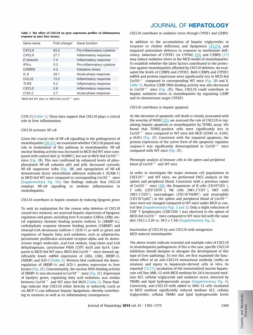
Table 1. The effect of CXCL10 on gene expression profiles of inflammatoryresponse in mice liver tissues.
Gene name Fold change† Gene function
CXCL6 63.2 Pro-inflammatory cytokineCXCL9 27.7 Inflammatory responseE-Selectin 7.4 Inflammatory responseIFN-γ 5.3 Pro-inflammatory cytokineC/EBPβ 4.2 Oxidative stressIL-6 24.1 Acute-phase responseCCL22 13.2 Inflammatory responseTLR9 4.2 Inflammatory responseCXCL5 2.8 Inflammatory responseCOX-2 2.7 Acute-phase response
�MCD-fed WT mice vs. MCD-fed Cxcl10�/� mice.
JOURNAL OF HEPATOLOGY
(COX-2) (Table 1). These data support that CXCL10 plays a criticalrole in liver inflammation.
CXCL10 activates NF-jB
Given the crucial role of NF-jB signalling in the pathogenesis ofsteatohepatitis [20,21], we examined whether CXCL10 played anyrole in modulation of this pathway in steatohepatitis. NF-jBnuclear binding activity was increased in MCD-fed WT mice com-pared with control diet (p <0.0001), but not in MCD-fed Cxcl10�/�
mice (Fig. 2B). This was confirmed by enhanced levels of phos-phorylated NF-jB subunits p65 and p50, decreased cytosolicNF-jB suppressor IjBa (Fig. 2B) and upregulation of NF-jBdownstream factor intercellular adhesion molecule-1 (ICAM-1)in MCD-fed WT mice compared to corresponding Cxcl10�/� mice(Supplementary Fig. 1D). Our findings indicate that CXCL10employs NF-jB signalling to mediate inflammation insteatohepatitis.
CXCL10 contributes to hepatic steatosis by inducing lipogenic genes
To seek an explanation for the reason why deletion of CXCL10caused less steatosis, we assessed hepatic expression of lipogenicregulators and genes, including liver X receptor (LXR)a, LXRb, ste-rol regulatory element binding protein isoform 1c (SREBP-1c),carbohydrate response element binding protein (ChREBP) andstearoyl-CoA desaturase isoform-1 (SCD-1) as well as genes andregulators of hepatic fatty acid oxidation, such as adiponectin,peroxisome proliferator-activated receptor-alpha and its down-stream target molecules, acyl-CoA oxidase, long-chain acyl-CoAdehydrogenase, cytochrome P450 (CYP) 4a10 and 4a14. Com-pared to MCD-fed WT mice, MCD-fed Cxcl10�/� mice showed sig-nificantly lower mRNA expression of LXRa, LXRb, SREBP-1c,ChREBP, and SCD-1 (Table 2). Western blot confirmed the down-regulation of SREBP-1c and SCD-1 protein expression in liverlysates (Fig. 2C). Concomitantly, the nuclear DNA-binding activityof SREBP-1c was decreased in Cxcl10�/� mice (Fig. 2C). Expressionof lipolytic genes, regulating fatty acid oxidation, was similarbetween Cxcl10�/� and WT mice fed MCD (Table 2). These find-ings indicate that CXCL10 either directly or indirectly (such asvia MCP-1) can influence hepatic lipogenesis, thereby contribut-ing to steatosis as well as its inflammatory consequences.
Journal of Hepatology 2014
CXCL10 contributes to oxidative stress through CYP2E1 and C/EBPb
In addition to the accumulation of hepatic triglycerides inresponse to choline deficiency and lipogenesis [22,23], andimpaired antioxidant defences in response to methionine defi-ciency, induction of CYP2E1 (or CYP4A) [24] and C/EBPb [17]may induce oxidative stress in the MCD model of steatohepatitis.To establish whether the latter factors contributed to the protec-tion against steatohepatitis afforded by CXCL10 deletion, we eval-uated the levels of C/EBPb and CYP2E1. Both C/EBPb and CYP2E1mRNA and protein expression were significantly less in MCD-fedCxcl10�/� compared to corresponding WT mice (Fig. 2D and E,Table 1). Nuclear C/EBP DNA-binding activity was also decreasedin Cxcl10�/� mice (Fig. 2D). Thus, CXCL10 could contribute tohepatic oxidative stress in steatohepatitis by regulating C/EBPand its downstream target CYP2E1.
CXCL10 contributes to hepatic apoptosis
As the elevation of apoptotic cell death is closely associated withthe severity of NASH [25], we assessed the role of CXCL10 in reg-ulating hepatic apoptosis in steatohepatitis by TUNEL assay. Wefound that TUNEL-positive cells were significantly less inCxcl10�/� mice compared to WT mice fed MCD (0.94% vs. 4.56%,p <0.01) (Fig. 2F). Consistent with the impaired apoptosis, theprotein expression of the active form of the apoptosis regulatorcaspase-3 was significantly downregulated in Cxcl10�/� micecompared with WT mice (Fig. 2F).
Phenotypic analysis of immune cells in the spleen and peripheralblood of Cxcl10�/� and WT mice
In order to investigate the major immune cell populations inCXCL10�/� and WT mice, we performed FACS analysis in thespleen and peripheral blood. Consistent with a previous reportof Cxcl10�/� mice [26], the frequencies of B cells (CD19+CD3�),T cells (CD3+CD19�), NK cells (NK1.1+CD3�), NKT cells(NK1.1+CD3+), macrophages (CD11b+F4/80+) and neutrophils(CD11b+Ly6G+) in the spleen and peripheral blood of Cxcl10�/�
mice were not changed compared to WT mice under MCD or con-trol diet (Supplementary Figs. 2 and 3). Only a slight reduction ofCD8+ T lymphocytes (CD8+CD4�) was observed in the spleen ofMCD-fed Cxcl10�/� mice compared to WT mice fed with the samediet (34.3 ± 2.26 vs. 38.5 ± 1.34) (Supplementary Fig. 2).
Inactivation of CXCL10 by anti-CXCL10 mAb antagonizesMCD-induced steatohepatitis
The above results indicate essential and multiple roles of CXCL10in steatohepatitis pathogenesis. If this is the case, specific CXCL10inhibition should dampen or abrogate the development of thistype of liver pathology. To test this, we first examined the func-tional effect of an anti-CXCL10 monoclonal antibody (mAb) onsteatosis and injury to hepatocyte-derived cells in vitro. Asreported [15,17], incubation of the immortalized murine hepato-cyte cell line AML-12 with MCD medium for 24 h increased med-ium ALT, cellular triglyceride and oxidative stress, detected byTBARS and lipid hydroperoxide assays (Supplementary Fig. 4).Conversely, anti-CXCL10 mAb added to AML-12 cells incubatedin MCD medium significantly reduced medium ALT, cellulartriglycerides, cellular TBARS and lipid hydroperoxide levels
vol. 61 j 1365–1375 1369

Table 2. Hepatic mRNA expression of genes involved in fatty acid regulation in Cxcl10�/� mice.
Gene WT mice Cxcl10-/- miceControl MCD Control MCD
Lipogenic genesLXRα 1.05 ± 0.33 1.52 ± 0.31** 0.99 ± 0.12 0.97 ± 0.23##
LXRβ 1.05 ± 0.30 1.91 ± 0.68** 1.28 ± 0.31 1.30 ± 0.34#
SREBP-1c 1.01 ± 0.16 0.63 ± 0.30* 0.53 ± 0.23 0.30 ± 0.11#
ChREBP 1.01 ± 0.18 0.66 ± 0.26* 1.25 ± 0.31 0.32 ± 0.08***#
SCD-1 1.035 ± 0.33 0.012 ± 0.006*** 0.570 ± 0.213 0.006 ± 0.004***#
Lipolytic genesAdiponectin 1.44 ± 1.09 0.30 ± 0.29 2.15 ± 0.97 1.08 ± 0.91PPARα 1.01 ± 0.14 0.69 ± 0.23* 1.17 ± 0.14 0.59 ± 0.09***ACO 1.04 ± 0.35 0.28 ± 0.07** 1.27 ± 0.49 0.29 ± 0.06LCAD 1.04 ± 0.31 1.19 ± 0.53 0.74 ± 0.29 0.71 ± 0.22CYP4A10 0.91 ± 0.55 2.79 ± 1.20* 1.30 ± 0.68 1.46 ± 0.61CYP4A14 2.03 ± 2.45 79.5 ± 34.2*** 1.69 ± 1.16 50.2 ± 25.4**
Specific mRNA expression values were normalized to the expression of GAPDH. Data are mean ± SD, n = 5–8/group. ⁄p <0.05, ⁄⁄p <0.01, ⁄⁄⁄p <0.0001 compared withcorresponding mice fed control diet. #p <0.05, ##p <0.0001 compared with WT mice fed the MCD diet.
Research Article
compared to AML-12 cells in MCD medium exposed to controlIgG2A mAb (Supplementary Fig. 4).
We next examined whether administration of the anti-CXCL10mAb by intraperitoneal injection could prevent MCD-inducedsteatohepatitis in vivo. Administration of the anti-CXCL10 mAbto MCD-fed WT mice reduced steatosis and inflammatory cellinfiltration (Fig. 3A), with concordant reduction of serum ALT,hepatic triglyceride and lipid hydroperoxide levels (Fig. 3B) com-pared to MCD-fed mice administered control mAb. Likewise,CXCL10 neutralization suppressed NF-jB binding activity(p <0.01), and reduced the expression of phosphorylated NF-jBsubunits p65 and p50 (p <0.05) and ICAM-1 mRNA (p <0.05)(Fig. 3C). Moreover, blocking CXCL10 significantly decreased thelevels of CYP2E1 (p <0.01) and SREBP-1c (p <0.05) (Fig. 3D).
After confirming a preventive effect on steatohepatitis, wefurther examined whether CXCL10 neutralization could treat ste-atohepatitis after it has been established. After induction of ste-atohepatitis in mice fed the MCD diet for 3 weeks, anti-CXCL10mAb or control mAb was supplemented for 10 days under MCDdiet. Histological analysis of livers by H&E and Oil red O stainingshowed significantly reduced lipid accumulation and inflamma-tory cell infiltration in MCD-fed mice treated with anti-CXCL10mAb (Fig. 3E). Anti-CXCL10 mAb treatment in MCD-fed mice alsosignificantly decreased hepatic triglyceride and lipid peroxidelevels compared to MCD-fed mice administrated with controlmAb (Fig. 3F). Moreover, CXCL10 neutralization suppressed hepa-tic TNF-a (p <0.05) and ICAM-1 (p <0.05) mRNA expression(Fig. 3G). These data added further weight to the effects ofCXCL10 in mediating inflammation, oxidative stress and steatosisin the evolution of steatohepatitis.
CXCL10 is associated with lobular inflammation and acts as anindependent risk factor of human NASH
Since the MCD model reflects pathologically severe steatohepati-tis with choline and amino acid nutritional deficiency and a con-text of ‘‘lipid trapping’’ in the liver with severe oxidative stress, itremains important to establish whether human NASH related to
1370 Journal of Hepatology 2014
over-nutrition is also associated with increased liver expressionand circulating levels of CXCL10. To this end, we assayed CXCL10mRNA in liver biopsy from 15 control subjects and 22 NAFLDpatients (11 simple steatosis patients and 11 human NASHpatients). The results showed that hepatic CXCL10 mRNA levelswere significantly higher in primary NASH tissue compared tosimple steatosis (p <0.05) and normal controls (p <0.001)(Fig. 4A), inferring that hepatic CXCL10 production is prominentin patients with NASH.
We next ascertained the clinical impact of CXCL10 in NASHpatients. We enrolled a well-established prospective cohort of73 control subjects without fatty liver measured by proton-magnetic resonance spectroscopy and 147 age and gendermatched biopsy-proven NAFLD patients, 69 of whom werediagnosed as NASH [18,19]. We found that serum CXCL10 wassignificantly increased in a stepwise fashion from control subjects(111 [IQR: 98–146] pg/ml), patients with simple steatosis (170[133–225] pg/ml) to patients with NASH (248 [154–310] pg/ml)(Fig. 4B, all p <0.0001). In NAFLD patients (simple steatosis andNASH), CXCL10 was significantly and positively correlated withlobular inflammation (rho: 0.26, p = 0.002) and hepatocyte bal-looning degeneration (rho: 0.24, p = 0.004), which are two majorhistological features of NASH (Table 3). Multivariable linearregression analysis also demonstrated that the serum CXCL10level was positively associated with lobular inflammation (Beta:47.9; 95% CI: 15.0–80.8; p = 0.005) and ballooning (Beta: 51.1;95% CI: 20.0–82.1; p = 0.001) independent of metabolic syn-drome, body mass index (BMI), ALT, triglyceride, fasting glucoseand cholesterol. Moreover, we performed a multivariate logisticregression analysis on these subjects and identified that CXCL10was an independent risk factor for NASH in NAFLD patients (OR:1.008, 95% CI: 1.004–1.013, p <0.001) (Table 4, with factorsincluded in the regression model listed).
CXCL10 is a potential biomarker for the clinical diagnosis of NASH
To evaluate the utility of CXCL10 as a biomarker in the diagnosisof NAFLD and NASH, a ROC curve was constructed. CXCL10
vol. 61 j 1365–1375

Control mAb Anti-CXCL10 mAb
PreventionControl mAbAnti-CXCL10 mAb
Control mAbAnti-CXCL10 mAb
Control mAbAnti-CXCL10 mAb
Control mAbAnti-CXCL10 mAb
Control mAbAnti-CXCL10 mAb
A B
C
D
CYP2E1
SREBP-1c
F
H&
E s
tain
ing
Oil
red
O s
tain
ing G
E
H&
E s
tain
ing
Control mAb Anti-CXCL10 mAb
Treatment
Histological score MCDControl mAb Anti-CXCL10 mAb
Steatosis 1.50 ± 0.60 0.60 ± 0.50#
Necro-inflammation 1.20 ± 0.50 0.20 ± 0.50#
Histological score MCDControl mAb Anti-CXCL10 mAb
Steatosis 1.67 ± 0.47 1.13 ± 0.14#
Necro-inflammation 1.33 ± 0.49 0.44 ± 0.50#
p <0.01
Ser
um A
LT (
U/L
) 400
300
200
100
0
Rel
ativ
e IC
AM
-1 m
RN
A
0.0
0.5
1.0
1.5p <0.05
p <0.05
Hep
atic
trig
lyce
ride
(μg/
mg
prot
ein)
80
60
40
20
0
p <0.05
Lipi
d h
ydro
pero
xide
(μM
/mg
prot
ein)
1.5
1.0
0.5
0.0
Control Anti-CXCL10 mAb:
p-NFκB p65
GAPDH
p-NFκB p50
p <0.01
NFκ
B b
indi
ng a
ctiv
ity(O
D45
0nm) 0.6
0.8
0.4
0.2
0.0 p <0.05p <0.05
p-NFκB
p65
p-NFκB
p50R
elat
ive
prot
ein
leve
ls 1.5
1.0
0.5
0.0
CYP2E1
SREBP-1c
p <0.01
p <0.05
Rel
ativ
e pr
otei
n le
vels 1.5
1.0
0.5
0.0
Control Anti-CXCL10 mAb:
Hep
atic
trig
lyce
ride
(μg/
mg
prot
ein)
80
100
60
40
20
0
p <0.05
0
1
2
3
4
5
TBA
RS
(nm
ol/m
g pr
otei
n)
p <0.05
0.0
0.5
1.0
1.5
2.0
Rel
ativ
e IC
AM
-1 m
RN
A
p <0.05
0.0
0.5
1.0
1.5
2.0
2.5
Rel
ativ
e TN
Fα m
RN
A
p <0.05
GAPDH
Fig. 3. CXCL10 neutralization protects against steatohepatitis in vivo. (A) Representative H&E staining, (B) serum ALT, hepatic triglyceride, lipid hydroperoxide, (C) NF-jB binding activity, phospho NF-jB p65, p50, ICAM-1 levels, (D) CYP2E1 and SREBP-1c expression in mice administrated with anti-CXCL10 or control mAb at 12 h beforefeeding MCD. (E) Liver sections with H&E staining and Oil red O staining, respectively, (F) hepatic triglyceride and lipid peroxidation products (TBARS), (G) TNF-a and ICAM-1 mRNA expression from mice injected with anti-CXCL10 or control mAb at 3 weeks after MCD feeding. #p <0.05 vs. mice treated with control mAb. Data are mean ± SD,n = 5/group.
JOURNAL OF HEPATOLOGY
exhibited a high overall accuracy in discriminating NAFLD fromcontrol subjects with the area under the receiver operating char-acteristic curve (AUROC) of 0.81 (95% CI: 0.75–0.87) (Fig. 4C). In
Journal of Hepatology 2014
NAFLD patients, CXCL10 had a moderate accuracy with theAUROC of 0.68 (95% CI: 0.59–0.77) in discriminating NASH fromsimple steatosis (Fig. 4C). If control subjects were also added to
vol. 61 j 1365–1375 1371

A B C Diagnosis of NASH in all subjects
Diagnosis of NAFLD in all subjects
Diagnosis of NASH in all NAFLD patients
0
10
20
30
Rel
ativ
e C
XC
L10
mR
NA p <0.05
p <0.001
0
100
200
300
400400600800
1000
CX
CL1
0 (p
g/m
l)
p <0.0001p <0.0001
SteatosisNASH
ControlSteatosisNASH
Control
AUROC: 0.81 (0.75-0.87)p <0.001
1.0
0.8
0.6
0.4
0.2
0.0
Sen
sitiv
ity
ROC curve
0.0 2.0 4.0 6.0 8.0 0.11-Specificity
1.0
0.8
0.6
0.4
0.2
0.0
Sen
sitiv
ity
AUROC: 0.68 (0.59-0.77) p <0.001
ROC curve
0.0 2.0 4.0 6.0 8.0 0.11-Specificity
1.0
0.8
0.6
0.4
0.2
0.0
Sen
sitiv
ity
AUROC: 0.77 (0.70-0.84) p <0.001
ROC curve
6.0 8.0 0.12.0 4.00.01-Specificity
Fig. 4. CXCL10 in control and NAFLD patients. (A) Hepatic human CXCL10 mRNA levels; (B) Serum CXCL10 protein levels; (C) Receiver-operating characteristics curves ofCXCL10 in diagnosing NAFLD in all subjects, NASH in NAFLD patients and NASH in all subjects.
Table 3. Correlations with CXCL10 in NAFLD patients.
CXCL10rho p value‡
Age 0.16 0.062BMI 0.07 0.408Total cholesterol 0.09 0.304Triglyceride 0.09 0.286Steatosis 0.15 0.070
0.26 0.002BallooningLobular inflammation
0.24 0.004Fibrosis 0.25 0.002
�p value corresponds to Ho: rho = 0.
Table 4. Multivariable analysis for independent risk factors for NASH inNAFLD patients.
OR 95% CI p valueCXCL10 1.008§ 1.004-1.013 <0.001Metabolic syndrome 3.083 1.203-7.903 0.019
Variables entered in the regression model: CXCL10, gender, age, body mass index(BMI), metabolic syndrome, alanine aminotransferase (ALT), fasting glucose, tri-glyceride, low density lipoprotein-cholesterol (LDL-c), glycated haemoglobin(HbA1c).§For every 1 unit increase of CXCL10 level.
Research Article
the analysis, the AUROC of diagnosing NASH increased to 0.77(95% CI: 0.70–0.84) (Fig. 4C). Thus, CXCL10 can be a novel bio-marker for the clinical diagnosis of NAFLD and NASH.
Discussion
The first novel finding in these studies is that Cxcl10�/� miceadministrated with MCD diet showed significantly attenuatedsteatohepatitis compared with WT mice fed the same diet; thesefindings were corroborated by improved liver histology, loweredserum ALT, and hepatic triglyceride content. Moreover, CXCL10deletion was associated with a significant reduction of intrahe-patic oxidative stress, as indicated by decreased lipid peroxidelevels. This change was clearly associated with the attenuation
1372 Journal of Hepatology 2014
of hepatic inflammation. In addition, CXCL10 deficiency confersprotection from hepatic nutritional fibrosis. Our data providethe first evidence that CXCL10 may contribute to lipogenesis,thereby influencing steatosis and possibly lipotoxicity, as wellas hepatocellular injury and perpetuation of liver inflammationin steatohepatitis, at least in the MCD model.
The molecular mechanisms by which CXCL10 exerts its broadrange of functions in steatohepatitis were subsequently studied.As a key pro-inflammatory cytokine, CXCL10 often amplifies theeffects of other cytokines [5]. We therefore evaluated the effectof CXCL10 on other potential cytokines in steatohepatitis andshowed that CXCL10 was associated with induction of TNF-a,IL-1b, and MCP-1. TNF-a is a key inflammatory factor involvedin the development of human NASH [27] and experimental ste-atohepatitis [28]. TNF-a can activate neutrophils, cause insulinresistance and promote NASH development. TNF-a and IL-1bare able to induce MCP-1 in vitro, suggesting that these cytokinesare functionally related [29]. MCP-1 is also an important mole-cule in NASH as it may bridge inflammatory responses with theinduction of insulin resistance [30]. Moreover, MCP-1 can stimu-late lipogenesis to promote steatosis in the liver, allowing inflam-mation to exacerbate steatosis [4]. This suggests that CXCL10induces cytokine expression, leading to the development ofsteatohepatitis.
We further characterized the inflammatory factors, regulatedby CXCL10 in steatohepatitis, by a cDNA array covering 84 well-known inflammatory genes. Our results show that pro-inflamma-tory factors, including IFN-c, TLR9, CXCL9, IL-6, SELE, CXCL6, CCL22,CXCL5, and COX-2, were significantly higher in WT mice than inCxcl10�/� mice fed with MCD. Each of these molecules couldamplify the inflammatory recruitment in steatohepatitis. To bespecific, IFN-c is a major inducer of CXCL10 related to NASHpathogenesis [31]; TLR9 activates IFN regulatory factors thatinduce production of IL-1b, leading to NASH development inmouse model [32]; CXCL9, induced by IFN-c, is increased in thelivers of patients with NASH [33], while IL-6 is a key inflamma-tory factor involved in NASH development [34]. Serum levels ofE-selectin (SELE) are also higher in patients with NASH similarto those of IL-6 [35]. CXCL6 is associated with the severity ofhepatic inflammation in NAFLD patients and it can be used forpredicting NASH progression [36]. Similarly, CCL22 and CXCL5,two small chemokines, are serum markers for NASH and therelated obesity and metabolic syndrome [37]. Finally, COX-2,
vol. 61 j 1365–1375

JOURNAL OF HEPATOLOGY
another pro-inflammatory mediator, plays an important role inmetabolic forms of steatohepatitis as reported earlier by us[28]. In addition to IFN-c and TLR9, a close correlation betweenCXCL10 and TNF-a, MCP-1, IL-6 has been well documented[38,39]. Collectively, these data suggested that induction of pro-inflammatory cytokines and key inflammatory factors by CXCL10is part of a mechanism for the inflammatory recruitment in MCD-induced steatohepatitis.An important observation in the present study is that themajority of the above cytokines and inflammatory factors are reg-ulated by NF-jB signalling (Fig. 2G). TNF-a is a potent activator ofNF-jB, and in turn activated NF-jB induces TNF-a expression[40]. TLR9 and IL-6 can also activate NF-jB [32], while ICAM-1,CCL22, COX-2, IL-1b, MCP-1, and CXCL5 are downstream effectorsof NF-jB activation (Fig. 2G) [20]. These data, when combinedwith the previous finding that NF-jB is a key regulator of earlyhepatic inflammatory recruitment and liver injury in NASH[21], implicate a collaborative interaction of CXCL10 and NF-jBto promote steatohepatitis. Therefore, CXCL10 may act as a lipo-toxic molecule that activates NF-jB and its downstream inflam-matory effectors to induce hepatocyte apoptosis and liverinjury, leading to the progression of steatohepatitis.
The underlying causes of hepatic triglyceride accumulation insteatosis include enhanced uptake and synthesis of fatty acids,and inhibition of fatty acid oxidation. In our experimental steato-hepatitis model, knockout of CXCL10 significantly reduced hepa-tic triglyceride content and steatosis (Fig. 1A and C). Thisreduction was associated with reduced activity of SREBP-1c anddownregulation of SREBP-1c, ChREBP, LXRs, and SCD-1 (Fig. 2and Table 2), which are involved in de novo fatty acid synthesis.In addition to SREBP-1c and ChREBP, LXR is a major transcrip-tional activator for lipogenesis [41]; it modulates the expressionof SREBP-1c through directly binding to the promoter of SREBP-1c. LXR also induces the transcription of the lipogenic genesSCD-1 and ChREBP [41]. Thus, the likely pathways by whichCXCL10 promotes hepatic steatosis include the upregulation ofkey fatty acid synthesis genes that promote fatty acid synthesis(Fig. 2G).
It is of general agreement that oxidative stress facilitates theadvancement of steatosis to steatohepatitis. Among the commonmediators of oxidative stress [42], CYP2E1 is an oxido-reductasethat can promote NASH development by inducing oxidative/nitrosative stress, protein modifications, inflammation and insu-lin resistance [43]. Consistent with this, we confirmed earlierfindings [17,24] that CYP2E1 expression is upregulated in MCD-induced steatohepatitis. Importantly, we showed that deletionof CXCL10 completely abolished the MCD-dependent stimulationof CYP2E1, and significantly reduced the expression of its tran-scriptional activator C/EBPb (Fig. 2D–E). These data were in accor-dance with the lower level of CYP2E1 in MCD-fed Cebpb�/� micecompared with MCD-fed WT mice [41], and demonstrate thatCXCL10 can act upstream of C/EBPb and CYP2E1 to modulateoxidative stress.
If CXCL10 plays a key part in the pathogenesis of steatohepa-titis, it would be important to establish that its functional block-ade ameliorates the severity of steatohepatitis. To test this, weused anti-CXCL10 mAb to neutralize CXCL10 in vitro. Such neu-tralization caused a dose-dependent decrease in triglyceridesecretion and ALT release, together with a concomitant suppres-sion of cellular oxidative stress in AML12 hepatocytes (Supple-mentary Fig. 4). Moreover, anti-CXCL10 mAb ameliorated the
Journal of Hepatology 2014
severity of fatty liver disease in MCD-fed mice. In the presentwork, CXCL10 neutralization using anti-CXCL10 mAb in miceshowed significant improvements in the prevention and regres-sion of steatohepatitis (Fig. 3). These effects were associated withreduced hepatic triglyceride and lipid peroxide levels (Fig. 3).Thus, CXCL10 is a potential target for the prevention and treat-ment of steatohepatitis.
These mechanistic findings of CXCL10 in the evolution ofexperimental steatohepatitis encouraged us to explore the clini-cal impact of CXCL10 in patients with NAFLD and NASH. We firstdemonstrated that CXCL10 was significantly upregulated both inliver and serum samples of NASH patients. Moreover, the circu-lating level of CXCL10 in NASH patients was associated with lob-ular inflammation, which is supported by a previous study thatshowed that increased CXCL10 levels were correlated with thedegree of chronic liver inflammatory damage caused by hepatitisC virus infection [9]. Early identification of patients with NASHmay allow intervention that may alter the course of the disease.Currently, liver biopsy remains the standard method for the diag-nosis of NASH and differentiation from simple steatosis. How-ever, biopsy is an invasive diagnostic procedure that has beenassociated with sampling error and observer variability. Thus,the development of a non-invasive test is paramount to the man-agement of NASH. To date, there are no reliable serologic tests forthe identification of NASH. Identification of such biomarkerwould aid clinicians in the identification of patients with NASH,and allow for non-invasive frequent monitoring of disease pro-gression and response to therapy. Building on the significantlyelevated CXCL10 level in NASH patients, we tested the clinicalutility of CXCL10 as a serologic biomarker for the diagnosis ofNASH. Base on a multivariate Cox regression analyses in a studycohort of 147 NAFLD patients and 73 control subjects, CXCL10was revealed to be a novel risk factor of NASH independent ofmetabolic syndrome, ALT, diabetes and triglycerides (Table 4).Moreover, the AUROC indicated an overall accuracy of 81% todiagnose NAFLD and an accuracy of 77% to diagnose NASH, sug-gesting that circulating CXCL10 production could be regardedas a valuable new diagnostic factor for NAFLD and NASH. How-ever, it should be noted that a few prediction models such asthe NAFLD fibrosis score have also been developed to predictadvanced fibrosis [44,45], These scores are comprised of predict-ing factors of fibrosis such as age, BMI and metabolic factors.While it is interesting that CXCL10 may serve as a marker ofNASH, the finding warrants independent validation. Furthermore,it would also be important to explore its role in conjunction withother predicting factors to improve the diagnosis.
The MCD diet model is a classic and widely adopted dietarymodel for studying NASH. It can induce hepatic steatohepatitiswith inflammation, oxidative stress, mitochondrial DNA damage,apoptosis and fibrosis [46]. Therefore, it is considered as one ofthe best-established models for studying NASH-associatedinflammation, oxidative stress and fibrosis. However, it doesnot fully manifest all human NASH features. Mice fed withMCD diet lose weight instead of being obese and lack insulinresistance [47]. In the future, high-fat and high-fructose model(also termed as American Lifestyle-Induced Obesity Syndrome[ALIOS]), which may result in an obese animal with severe stea-tosis, inflammation, oxidative stress and insulin resistance at16 weeks [46,48], could be used to support our findings.
In conclusion, these observations and interventions demon-strate for the first time that CXCL10 plays an essential role in
vol. 61 j 1365–1375 1373
Research Article
the development of steatohepatitis in the context of fatty liverdisease. Further, the mechanism of this effect is through regula-tion of lipogenesis and oxidative stress either directly or indi-rectly via pathway modulation and pro-inflammatory signalling,altering the expression of other key chemokines, cytokines andpro-inflammatory molecules. Circulating CXCL10 may be a poten-tial biomarker for patients with NAFLD and NASH.Financial support
The project was supported by the Collaborative Research Fund(HKU3/CRF11R, CUHK3/CRF/12R) of the Research Grant CouncilHong Kong; National Basic Research Program of China (973 Pro-gram, 2013CB531401), the Theme-based Research Scheme of theHong Kong Research Grants Council (T12-403-11), the CUHKFocused Investments Scheme B, Shenzhen Municipal Scienceand Technology R & D funding (JCYJ20120619152326450),Shenzhen Virtual University Park Support Scheme to CUHKShenzhen Research Institute and the Australian National Healthand Medical Research Council project grants 585411 and 104288.
Conflict of interest
The authors who have taken part in this study declared that theydo not have anything to disclose regarding funding or conflict ofinterest with respect to this manuscript.
Authors’ contributions
XZ was involved in study design, experiments conduct, and dataanalysis; JS, ESHC, TOY, JCYS, MYYG, and JD performed theresearch; KM, LL, and VWSW provided material support; JJYSand GF commented on the study and revised the paper. JYdesigned, supervised the study and wrote the paper.
Supplementary data
Supplementary data associated with this article can be found, inthe online version, at http://dx.doi.org/10.1016/j.jhep.2014.07.006.
References
[1] Farrell GC, Wong VW, Chitturi S. NAFLD in Asia – As common and importantas in the West. Nat Rev Gastroenterol Hepatol 2013;10:307–318.
[2] Cohen JC, Horton JD, Hobbs HH. Human fatty liver disease: old questions andnew insights. Science 2011;332:1519–1523.
[3] Farrell GC, Larter CZ. Nonalcoholic fatty liver disease: from steatosis tocirrhosis. Hepatology 2006;43:S99–S112.
[4] Farrell GC, van Rooyen D, Gan L, Chitturi S. NASH is an inflammatorydisorder: pathogenic, prognostic, and therapeutic implications. Gut Liver2012;6:149–171.
[5] Neville LF, Mathiak G, Bagasra O. The immunobiology of interferon-gammainducible protein 10 kD (IP-10): a novel, pleiotropic member of the C-X-Cchemokine superfamily. Cytokine Growth Factor Rev 1997;8:207–219.
[6] Luster AD, Unkeless JC, Ravetch JV. Gamma-interferon transcriptionallyregulates an early-response gene containing homology to platelet proteins.Nature 1985;315:672–676.
1374 Journal of Hepatology 2014
[7] Abe T, Fukuhara T, Wen X, Ninomiya A, Moriishi K, Maehara Y, et al. CD44participates in IP-10 induction in cells in which hepatitis C virus RNA isreplicating, through an interaction with Toll-like receptor 2 and hyaluronan.J Virol 2012;86:6159–6170.
[8] Zhou Y, Wang S, Ma JW, Lei Z, Zhu HF, Lei P, et al. Hepatitis B virus protein X-induced expression of the CXC chemokine IP-10 is mediated throughactivation of NF-kappaB and increases migration of leukocytes. J Biol Chem2010;285:12159–12168.
[9] Harvey CE, Post JJ, Palladinetti P, Freeman AJ, Ffrench RA, Kumar RK, et al.Expression of the chemokine IP-10 (CXCL10) by hepatocytes in chronichepatitis C virus infection correlates with histological severity and lobularinflammation. J Leukoc Biol 2003;74:360–369.
[10] Hintermann E, Bayer M, Pfeilschifter JM, Luster AD, Christen U. CXCL10promotes liver fibrosis by prevention of NK cell mediated hepatic stellatecell inactivation. J Autoimmun 2010;35:424–435.
[11] Yoneyama H, Kai Y, Koyama J, Suzuki K, Kawachi H, Narumi S, et al.Neutralization of CXCL10 accelerates liver regeneration in carbon tetrachlo-ride-induced acute liver injury. Med Mol Morphol 2007;40:191–197.
[12] Bertola A, Bonnafous S, Anty R, Patouraux S, Saint-Paul MC, Iannelli A, et al.Hepatic expression patterns of inflammatory and immune response genesassociated with obesity and NASH in morbidly obese patients. PLoS One2010;5:e13577.
[13] Schulthess FT, Paroni F, Sauter NS, Shu L, Ribaux P, Haataja L, et al. CXCL10impairs beta cell function and viability in diabetes through TLR4 signaling.Cell Metab 2009;9:125–139.
[14] Morimoto J, Yoneyama H, Shimada A, Shigihara T, Yamada S, Oikawa Y, et al.CXC chemokine ligand 10 neutralization suppresses the occurrence ofdiabetes in nonobese diabetic mice through enhanced beta cell proliferationwithout affecting insulitis. J Immunol 2004;173:7017–7024.
[15] Yu J, Chu ES, Wang R, Wang S, Wu CW, Wong VW, et al. Heme oxygenase-1protects against steatohepatitis in both cultured hepatocytes and mice.Gastroenterology 2010;138:694–704.
[16] Yu J, Zhang S, Chu ES, Go MY, Lau RH, Zhao J, et al. Peroxisome proliferator-activated receptors gamma reverses hepatic nutritional fibrosis in mice andsuppresses activation of hepatic stellate cells in vitro. Int J Biochem Cell Biol2010;42:948–957.
[17] Shen B, Yu J, Wang S, Chu ES, Wong VW, Zhou X, et al. Phyllanthus urinariaameliorates the severity of nutritional steatohepatitis both in vitro andin vivo. Hepatology 2008;47:473–483.
[18] Shen J, Chan HL, Wong GL, Chan AW, Choi PC, Chan HY, et al. Assessment ofnon-alcoholic fatty liver disease using serum total cell death and apoptosismarkers. Aliment Pharmacol Ther 2012;36:1057–1066.
[19] Shen J, Chan HL, Wong GL, Choi PC, Chan AW, Chan HY, et al. Non-invasivediagnosis of non-alcoholic steatohepatitis by combined serum biomarkers. JHepatol 2012;56:1363–1370.
[20] Baker RG, Hayden MS, Ghosh S. NF-kappaB, inflammation, and metabolicdisease. Cell Metab 2011;13:11–22.
[21] Dela Pena A, Leclercq I, Field J, George J, Jones B, Farrell G. NF-kappaBactivation, rather than TNF, mediates hepatic inflammation in a murinedietary model of steatohepatitis. Gastroenterology 2005;129:1663–1674.
[22] Rizki G, Arnaboldi L, Gabrielli B, Yan J, Lee GS, Ng RK, et al. Mice fed alipogenic methionine-choline-deficient diet develop hypermetabolism coin-cident with hepatic suppression of SCD-1. J Lipid Res 2006;47:2280–2290.
[23] Larter CZ, Yeh MM, Haigh WG, Williams J, Brown S, Bell-Anderson KS, et al.Hepatic free fatty acids accumulate in experimental steatohepatitis: role ofadaptive pathways. J Hepatol 2008;48:638–647.
[24] Leclercq IA, Farrell GC, Field J, Bell DR, Gonzalez FJ, Robertson GR. CYP2E1and CYP4A as microsomal catalysts of lipid peroxides in murine nonalco-holic steatohepatitis. J Clin Invest 2000;105:1067–1075.
[25] Feldstein AE, Canbay A, Angulo P, Taniai M, Burgart LJ, Lindor KD, et al.Hepatocyte apoptosis and fas expression are prominent features of humannonalcoholic steatohepatitis. Gastroenterology 2003;125:437–443.
[26] Dufour JH, Dziejman M, Liu MT, Leung JH, Lane TE, Luster AD. IFN-gamma-inducible protein 10 (IP-10; CXCL10)-deficient mice reveal a role for IP-10 ineffector T cell generation and trafficking. J Immunol 2002;168:3195–3204.
[27] Kugelmas M, Hill DB, Vivian B, Marsano L, McClain CJ. Cytokines and NASH:a pilot study of the effects of lifestyle modification and vitamin E.Hepatology 2003;38:413–419.
[28] Yu J, Ip E, Dela Pena A, Hou JY, Sesha J, Pera N, et al. COX-2 induction in micewith experimental nutritional steatohepatitis: role as pro-inflammatorymediator. Hepatology 2006;43:826–836.
[29] Petrasek J, Bala S, Csak T, Lippai D, Kodys K, Menashy V, et al. IL-1 receptorantagonist ameliorates inflammasome-dependent alcoholic steatohepatitisin mice. J Clin Invest 2012;122:3476–3489.
vol. 61 j 1365–1375

JOURNAL OF HEPATOLOGY
[30] Maher JJ, Leon P, Ryan JC. Beyond insulin resistance: innate immunity innonalcoholic steatohepatitis. Hepatology 2008;48:670–678.[31] Baranova A, Schlauch K, Elariny H, Jarrar M, Bennett C, Nugent C, et al. Gene
expression patterns in hepatic tissue and visceral adipose tissue of patientswith non-alcoholic fatty liver disease. Obes Surg 2007;17:1111–1118.
[32] Miura K, Kodama Y, Inokuchi S, Schnabl B, Aoyama T, Ohnishi H, et al. Toll-like receptor 9 promotes steatohepatitis by induction of interleukin-1beta inmice. Gastroenterology 2010;139:323–334.
[33] Wasmuth HE, Lammert F, Zaldivar MM, Weiskirchen R, Hellerbrand C,Scholten D, et al. Antifibrotic effects of CXCL9 and its receptor CXCR3 inlivers of mice and humans. Gastroenterology 2009;137:309–319.
[34] Wieckowska A, Papouchado BG, Li Z, Lopez R, Zein NN, Feldstein AE.Increased hepatic and circulating interleukin-6 levels in human nonalcoholicsteatohepatitis. Am J Gastroenterol 2008;103:1372–1379.
[35] Musso G, Gambino R, Bo S, Uberti B, Biroli G, Pagano G, et al. Shouldnonalcoholic fatty liver disease be included in the definition of metabolicsyndrome? A cross-sectional comparison with Adult Treatment Panel IIIcriteria in nonobese nondiabetic subjects. Diabetes Care 2008;31:562–568.
[36] Mehta R, Birerdinc A, Neupane A, Shamsaddini A, Afendy A, Elariny H, et al.Expression of inflammation-related genes is altered in gastric tissue ofpatients with advanced stages of NAFLD. Mediators Inflamm2013;2013:684237.
[37] Hammerich L, Heymann F, Tacke F. Role of IL-17 and Th17 cells in liverdiseases. Clin Dev Immunol 2011;2011:345803.
[38] Xu W, Joo H, Clayton S, Dullaers M, Herve MC, Blankenship D, et al.Macrophages induce differentiation of plasma cells through CXCL10/IP-10. JExp Med 2012;209:S1811–S1812.
[39] Tosello-Trampont AC, Landes SG, Nguyen V, Novobrantseva TI, Hahn YS.Kupffer cells trigger nonalcoholic steatohepatitis development in
Journal of Hepatology 2014
diet-induced mouse model through tumor necrosis factor-alpha production.J Biol Chem 2012;287:40161–40172.
[40] Tak PP, Firestein GS. NF-kappaB: a key role in inflammatory diseases. J ClinInvest 2001;107:7–11.
[41] Jump DB, Botolin D, Wang Y, Xu J, Christian B, Demeure O. Fatty acidregulation of hepatic gene transcription. J Nutr 2005;135:2503–2506.
[42] Abdelmegeed MA, Banerjee A, Yoo SH, Jang S, Gonzalez FJ, Song BJ. Criticalrole of cytochrome P450 2E1 (CYP2E1) in the development of high fat-induced non-alcoholic steatohepatitis. J Hepatol 2012;57:860–866.
[43] Rahman SM, Schroeder-Gloeckler JM, Janssen RC, Jiang H, Qadri I, MacleanKN, et al. CCAAT/enhancing binding protein beta deletion in mice attenuatesinflammation, endoplasmic reticulum stress, and lipid accumulation in diet-induced nonalcoholic steatohepatitis. Hepatology 2007;45:1108–1117.
[44] Angulo P, Hui JM, Marchesini G, Bugianesi E, George J, Farrell GC, et al. TheNAFLD fibrosis score: a noninvasive system that identifies liver fibrosis inpatients with NAFLD. Hepatology 2007;45:846–854.
[45] Wong VW, Wong GL, Chim AM, Tse AM, Tsang SW, Hui AY, et al. Validationof the NAFLD fibrosis score in a Chinese population with low prevalence ofadvanced fibrosis. Am J Gastroenterol 2008;103:1682–1688.
[46] Kohli R, Feldstein AE. NASH animal models: are we there yet? J Hepatol2011;55:941–943.
[47] Rinella ME, Green RM. The methionine-choline deficient dietary model ofsteatohepatitis does not exhibit insulin resistance. J Hepatol 2004;40:47–51.
[48] Tetri LH, Basaranoglu M, Brunt EM, Yerian LM, Neuschwander-Tetri BA.Severe NAFLD with hepatic necroinflammatory changes in mice fed transfats and a high-fructose corn syrup equivalent. Am J Physiol GastrointestLiver Physiol 2008;295:G987–G995.
vol. 61 j 1365–1375 1375