Cuticular anatomy of Sphenobaiera huangii (Ginkgoales) from the Lower Jurassic of Hubei, China
13
709 American Journal of Botany 92(4): 709–721. 2005. CUTICULAR ANATOMY OF SPHENOBAIERA HUANGII (GINKGOALES) FROM THE LOWER JURASSIC OF HUBEI,CHINA 1 YONGDONG WANG, 2,5,6 GAE ¨ TAN GUIGNARD, 3 FRE ´ DE ´ RIC THE ´ VENARD, 3 DAVID DILCHER, 4,6 GEORGES BARALE, 3 VOLKER MOSBRUGGER, 5 XIAOJU YANG, 2 AND SHENGWU MEI 2 2 Nanjing Institute of Geology and Palaeontology, Chinese Academy of Sciences, P.R. China; 3 Pale ´obotanique et FRE 2158 Centre National de la Recherche Scientifique (CNRS), Universite ´ Claude-Bernard Lyon 1, Ba ˆtiment Darwin-A, 7 rue Dubois, F-69622 Villeurbanne Cedex, France; 4 Florida Museum of Natural History, University of Florida, Gainesville, Florida 32611-7800 USA; and 5 Institute of Geosciences, University of Tu ¨bingen, Sigwartstrasse10, D-72076 Tu ¨bingen, Germany Sphenobaiera huangii (Sze) Hsu ¨ is typical Early Mesozoic fossil foliage of Ginkgoales in China. It has been recorded from the Upper Triassic to the Lower Jurassic. The cuticular anatomy is investigated based on material from the type locality, Lower Jurassic Hsiangchi Formation, Zigui County, Hubei Province. The specimens are similar to S. huangii, but contain new information about leaf morphology and cuticular anatomy. Lower and upper cuticle is investigated using light and electron microscopy (LM, SEM, and TEM). Many features are described for the first time, including general structures of lower and upper cuticle, stomata, papillae, and cuticular ultrastructure. At the ultrastructural level, two layers have been distinguished in both lower and upper cuticle, including a homogeneous outer layer with granules and a heterogeneous inner layer with fibrils. Based on a literature comparison between S. huangii and other relevant species of Sphenobaiera, S. huangii may represent the best-known taxon in the genus Sphenobaiera in both leaf morphology and cuticular structures. This study provides the first detailed ultrastructural data on the leaf cuticle of Sphenobaiera, one of the oldest foliage taxa of Ginkgoales, and offers further evidence for potential discussion on the taxonomic relationships of S. huangii with other ginkgoalean taxa. Key words: China; fossil; Ginkgoales; Jurassic; leaf cuticle; Sphenobaiera huangii; ultrastructure. The origin and evolution of the ‘‘living fossil’’ Ginkgo have received increased attention and interest from botanists and palaeobotanists. In particular, the discovery and investigation on the reproductive organs (pollen and ovules) of fossil Gink- goales have provided valuable evidence for understanding its long evolutionary history (Zhou and Zhang, 1988, 1989, 1992; Schweitzer and Kirchner, 1995; Poort et al., 1996; Zhou et al., 2002; Zhou and Zheng, 2003; Zheng and Zhou, 2004). How- ever, a number of fossil specimens assigned to Ginkgoales are preserved as compression and/or impression foliage. There- fore, the cuticular structure and anatomy undoubtedly play a significant role for exploring the systematics of foliage taxa of Ginkgoales. 1 Manuscript received 4 November 2003; revision accepted 17 December 2004. The authors thank Prof. Zhiyan Zhou (Nanjing) for helpful suggestion and comments to this study; Prof. Zhaoqi Yao (Nanjing) and Dr. E. V. Bugdaeva (Vladivostok) for kindly translating the Russian references; Mr. Yongqiang Mao (Nanjing) and Mr. Nicolas Labert (Lyon) for technical assistances during the preparation of SEM and TEM samples, and Mr. Terry A. Lott (Gainesville) for help in the preparation of this paper. We thank three anonymous reviewers for their helpful comments and suggestions. This research was supported by the PRA collaborative project between China and France, the National Natural Science Foundation of China Grant (No. 40472004, 40372008, 39900007), the State Key Laboratory of Palaeobiology and Stratigraphy Grant (No. 043113), the Pilot Project of the Knowledge Innovation Program, CAS (No. KZCX2-SW-129), the Talent Training Fund of the State Basic Science Project, NSFC (No. 040203), and the National Science Foundation, U.S.A. (INT 0074295). The first author (Y. D. Wang) acknowledges his sincere thanks for the support of a Research Fellowship from the Alexander von Humboldt Foundation of Germany. This paper is the University of Florida Contribution to Paleobiology publication no. 571. 6 Authors for correspondence (e-mails: [email protected]; dilcher@ flmnh.ufl.edu) Sphenobaiera is one of the oldest fossil foliage types of Ginkgoales and has a global distribution, mainly in the North- ern Hemisphere. It can be traced from the Lower Permian well into the Cretaceous (Taylor and Taylor, 1993). Florin (1936) established this genus for leaves originally included in Baiera Braun that are distinguished from those of the type species Baiera muensteriana (Presl in Sternberg) Heer, mainly by hav- ing wedge-shaped leaves that lack a distinct petiole. Later, Harris and Millington (1974) made an important refinement, transferring the wedge-shaped leaves borne in a bundle of dwarf shoots, to the Czekanowskiales (Sphenarion Harris and Miller, 1974). Since then, Sphenobaiera is generally defined for wedge-shaped leaves that are distinguishable from Ginkgo, Ginkgoites, and Baiera by the absence of a distinct petiole (see also Lydon et al., 2003). So far, over 50 species have been described worldwide for Sphenobaiera, and for some of them cuticle details are known. However, only a few speci- mens have been reported as closely associated with the repro- ductive organs. For example, several Sphenobaiera-type leaves were reported associated with the ovule-organ Karkenia from the Lower Cretaceous of Argentina, the Upper Jurassic of Siberia, the Liassic of Germany, Rhaetic to Jurassic of Iran and Afghanistan, and the Jurassic of Henan, China (Archan- gelsky, 1965; Krassilov, 1972; Schweitzer and Kirchner, 1995; Zhou et al., 2002). According to a recent systematic review by Zhou (1997, 2003), it is quite possible for some species of Sphenobaiera, which are characterized by having less divided wedge-shaped leaves and broad segments, to be referred to the family Karkeniaceae of Ginkgoales. The systematic status of most other species of this form-genus is still unknown at the family level. About 25 species of Sphenobaiera have been described
-
Upload
univ-lyon1 -
Category
Documents
-
view
0 -
download
0
Transcript of Cuticular anatomy of Sphenobaiera huangii (Ginkgoales) from the Lower Jurassic of Hubei, China

709
American Journal of Botany 92(4): 709–721. 2005.
CUTICULAR ANATOMY OF SPHENOBAIERA HUANGII
(GINKGOALES) FROM THE LOWER JURASSIC
OF HUBEI, CHINA1
YONGDONG WANG,2,5,6 GAETAN GUIGNARD,3 FREDERIC THEVENARD,3
DAVID DILCHER,4,6 GEORGES BARALE,3 VOLKER MOSBRUGGER,5
XIAOJU YANG,2 AND SHENGWU MEI2
2Nanjing Institute of Geology and Palaeontology, Chinese Academy of Sciences, P.R. China; 3Paleobotanique et FRE 2158 CentreNational de la Recherche Scientifique (CNRS), Universite Claude-Bernard Lyon 1, Batiment Darwin-A, 7 rue Dubois, F-69622
Villeurbanne Cedex, France; 4Florida Museum of Natural History, University of Florida, Gainesville, Florida 32611-7800 USA; and5Institute of Geosciences, University of Tubingen, Sigwartstrasse10, D-72076 Tubingen, Germany
Sphenobaiera huangii (Sze) Hsu is typical Early Mesozoic fossil foliage of Ginkgoales in China. It has been recorded from theUpper Triassic to the Lower Jurassic. The cuticular anatomy is investigated based on material from the type locality, Lower JurassicHsiangchi Formation, Zigui County, Hubei Province. The specimens are similar to S. huangii, but contain new information about leafmorphology and cuticular anatomy. Lower and upper cuticle is investigated using light and electron microscopy (LM, SEM, and TEM).Many features are described for the first time, including general structures of lower and upper cuticle, stomata, papillae, and cuticularultrastructure. At the ultrastructural level, two layers have been distinguished in both lower and upper cuticle, including a homogeneousouter layer with granules and a heterogeneous inner layer with fibrils. Based on a literature comparison between S. huangii and otherrelevant species of Sphenobaiera, S. huangii may represent the best-known taxon in the genus Sphenobaiera in both leaf morphologyand cuticular structures. This study provides the first detailed ultrastructural data on the leaf cuticle of Sphenobaiera, one of the oldestfoliage taxa of Ginkgoales, and offers further evidence for potential discussion on the taxonomic relationships of S. huangii with otherginkgoalean taxa.
Key words: China; fossil; Ginkgoales; Jurassic; leaf cuticle; Sphenobaiera huangii; ultrastructure.
The origin and evolution of the ‘‘living fossil’’ Ginkgo havereceived increased attention and interest from botanists andpalaeobotanists. In particular, the discovery and investigationon the reproductive organs (pollen and ovules) of fossil Gink-goales have provided valuable evidence for understanding itslong evolutionary history (Zhou and Zhang, 1988, 1989, 1992;Schweitzer and Kirchner, 1995; Poort et al., 1996; Zhou et al.,2002; Zhou and Zheng, 2003; Zheng and Zhou, 2004). How-ever, a number of fossil specimens assigned to Ginkgoales arepreserved as compression and/or impression foliage. There-fore, the cuticular structure and anatomy undoubtedly play asignificant role for exploring the systematics of foliage taxa ofGinkgoales.
1 Manuscript received 4 November 2003; revision accepted 17 December2004.
The authors thank Prof. Zhiyan Zhou (Nanjing) for helpful suggestion andcomments to this study; Prof. Zhaoqi Yao (Nanjing) and Dr. E. V. Bugdaeva(Vladivostok) for kindly translating the Russian references; Mr. YongqiangMao (Nanjing) and Mr. Nicolas Labert (Lyon) for technical assistances duringthe preparation of SEM and TEM samples, and Mr. Terry A. Lott (Gainesville)for help in the preparation of this paper. We thank three anonymous reviewersfor their helpful comments and suggestions. This research was supported bythe PRA collaborative project between China and France, the National NaturalScience Foundation of China Grant (No. 40472004, 40372008, 39900007),the State Key Laboratory of Palaeobiology and Stratigraphy Grant (No.043113), the Pilot Project of the Knowledge Innovation Program, CAS (No.KZCX2-SW-129), the Talent Training Fund of the State Basic Science Project,NSFC (No. 040203), and the National Science Foundation, U.S.A. (INT0074295). The first author (Y. D. Wang) acknowledges his sincere thanks forthe support of a Research Fellowship from the Alexander von HumboldtFoundation of Germany. This paper is the University of Florida Contributionto Paleobiology publication no. 571.
6 Authors for correspondence (e-mails: [email protected]; [email protected])
Sphenobaiera is one of the oldest fossil foliage types ofGinkgoales and has a global distribution, mainly in the North-ern Hemisphere. It can be traced from the Lower Permian wellinto the Cretaceous (Taylor and Taylor, 1993). Florin (1936)established this genus for leaves originally included in BaieraBraun that are distinguished from those of the type speciesBaiera muensteriana (Presl in Sternberg) Heer, mainly by hav-ing wedge-shaped leaves that lack a distinct petiole. Later,Harris and Millington (1974) made an important refinement,transferring the wedge-shaped leaves borne in a bundle ofdwarf shoots, to the Czekanowskiales (Sphenarion Harris andMiller, 1974). Since then, Sphenobaiera is generally definedfor wedge-shaped leaves that are distinguishable from Ginkgo,Ginkgoites, and Baiera by the absence of a distinct petiole(see also Lydon et al., 2003). So far, over 50 species havebeen described worldwide for Sphenobaiera, and for some ofthem cuticle details are known. However, only a few speci-mens have been reported as closely associated with the repro-ductive organs. For example, several Sphenobaiera-typeleaves were reported associated with the ovule-organ Karkeniafrom the Lower Cretaceous of Argentina, the Upper Jurassicof Siberia, the Liassic of Germany, Rhaetic to Jurassic of Iranand Afghanistan, and the Jurassic of Henan, China (Archan-gelsky, 1965; Krassilov, 1972; Schweitzer and Kirchner, 1995;Zhou et al., 2002). According to a recent systematic reviewby Zhou (1997, 2003), it is quite possible for some species ofSphenobaiera, which are characterized by having less dividedwedge-shaped leaves and broad segments, to be referred to thefamily Karkeniaceae of Ginkgoales. The systematic status ofmost other species of this form-genus is still unknown at thefamily level.
About 25 species of Sphenobaiera have been described

710 [Vol. 92AMERICAN JOURNAL OF BOTANY
from Mesozoic deposits in China. Among them, Sphenobaierahuangii (Sze) Hsu is the most typical and widespread leaffound in these deposits. It was initially described as Baierahuangii by Sze (1949), based on impression specimens fromthe Lower Jurassic Hsiangchi flora in western Hubei. Subse-quently, Hsu (in Sze and Hsu, 1954) transferred it to the genusSphenobaiera. Since then, this species has been describedfrom a number of Mesozoic localities in China. Available datashow that specimens of S. huangii are also recorded from theUpper Triassic to Middle Jurassic in Jinmen, Hubei (Feng etal., 1977; Chen, 1984), western and eastern parts of Sichuan(Yang, 1978; Duan and Chen, 1982; Chen et al., 1987), Fe-ngxian and Yan’an of Shaanxi (Liu, 1982; Zhang et al., 1998);Jiangning of Jiangsu (Wang et al., 1982), Yima of Henan(Zeng et al., 1995), Beipiao of western Liaoning (Mi et al.,1996), Datong of Shanxi (Li and Hu, 1984), and eastern Hu-nan (Zhang, 1986) in China. However, most specimens re-ferred to as S. huangii are poorly preserved so yield little in-formation about cuticular anatomy and foliar morphology.
Recently, numerous compressed specimens of Sphenobaierahuangii were collected from the Lower Jurassic HsiangchiFormation in western Hubei, China. Other plants associatedwith S. huangii in this formation include nilssonialeans (Nil-sonnia), bennettitaleans (Pterophyllum, Ptilophyllum, Tyrmia,Otozamites, Anomozamites, Weltrichia, Zamites, and Ctenis),ferns (Todites, Marattia, Phlebopteris, Dictyophyllum, Clath-ropteris, Hausmannia, Coniopteris, and Cladophlebis), otherginkgoaleans (Ginkgoites, Baiera), Czekanowskialeans (Czek-anowskia, Phenicopsis, Ixostrobus, and Stenorachis), conifers(Podozamites, Ferganiella, Swedenborgia, and Elatocladus),and a pteridosperm (Ctenozamites) (see Wang, 1999, 2002).Some of the ferns have been investigated describing fertileorgans, in situ spores, and ultrastructure (Wang, 1999, 2002;Wang and Mei, 1999; Wang et al., 2001). In this paper, wereinvestigate typical ginkgoalean foliage S. huangii, with em-phasis on cuticular anatomy, by using scanning and transmis-sion electron microscopy (SEM, TEM), and light microscopic(LM) observations. This study presents the first informationof cuticle ultrastructure for the genus Sphenobaiera based onwell-preserved Chinese material.
MATERIALS AND METHODS
More than 70 specimens of Sphenobaiera huangii containing cuticle havebeen collected since 1998 from the type locality in Xiangxi Town of ZiguiCounty, Hubei Province (308509200 N, 1108409150 E), where the Jurassic iswell exposed. It consists of an extensive succession of red clastic sedimentsof Middle to Upper Jurassic and a coal-bearing sequence of Lower Jurassic,which is known as the Hsiangchi Formation. This formation is generally re-garded as Early Jurassic based on various palaeontological evidence, such asplant megafossils, spores, pollen, bivalves, and megaspores (Li and Shang,1980; Wu et al., 1980; Meng, 1987; Meng and Zhang, 1987; Yang and Sun,1987; Zhang and Zhang, 1987). The Hsiangchi Formation is up to 205 mthick and composed of grey to greyish black sandstones, siltstones, mud-stones, and carbonaceous shales, intercalated by thin coal seams. This for-mation unconformably overlies the Upper Triassic Shazhenxi Formation andis conformably overlain by the Xietan Formation of Middle Jurassic age (fordetailed information on the stratigraphy, refer to Wang, 1999, 2002). All spec-imens are well preserved as compressions with evident venation and cuticle.In addition, three original specimens from the same locality figured by Sze(1949) as Baiera huangii were reexamined. They are in the palaeobotanicalcollection of the Nanjing Institute of Geology and Palaeontology, ChineseAcademy of Sciences (NIGPAS), Nanjing, with catalog numbers PB 931, PB933, and PB 934.
Selected cuticles were cleaned with hydrofluoric acid and hydrochloric acid,followed by a maceration in Schulze’s solution and a short treatment with 5%ammonia. The lower and upper cuticle were separated and then divided intothree fractions for different microscopic observations. A part of the cuticlewas mounted on permanent slides for observation and photography under LM.For SEM, the samples were prepared on standard stubs and viewed using aJSM 6300 scanning electron microscope (JEOL, Tokyo, Japan) at an accel-eration voltage of 15kV.
For TEM, samples were prepared as for fossil cuticle described in Zhouand Guignard (1998) and Guignard et al. (1998, 2001). The cuticle was pre-stained with osmium tetroxide before embedding in epoxy resin (epon) andstained with uranyl acetate and lead citrate after cutting with a Reichert Ul-tracut S ultramicrotome (Reichert-Leica, Vienna, Austria). Transverse and lon-gitudinal sections were cut in the lower and upper cuticle. Twelve resinblocks, belonging to four specimens, and more than 90 grids were made forobservation under a Philips CM120 transmission electron microscope (Philips,Eidhoven, Netherlands). The maceration and SEM observations were under-taken in NIGPAS, Nanjing, China, while TEM work was done at the ElectronMicroscope Center of Biology and Geology (CMEABG) of University ClaudeBernard Lyon 1, Villeurbanne, France. All specimens examined in this study(including slides and SEM stubs) are deposited in the palaeobotanical collec-tion of NIGPAS, Nanjing, China with registration numbers PB 931 to PB934and PB20041 to PB20075. The TEM blocks and sections are retained in theGuignard collection of the University of Lyon 1, France.
RESULTS
Leaf morphology—More than 70 specimens of Spheno-baiera huangii have been investigated in our collection. Thematerial referred to as S. huangii exhibits a wide range in leafsize, varying from 7.5 to 25 cm long (Figs. 1–4). The leaf isnarrowly wedge-shaped, becoming gradually narrower towardthe base, but without a distinct petiole. In some well-preservedspecimens, a swollen base is attached (Figs. 1, 3). The leavesare divided into two nearly equal lobes. The basal undividedportion is 3–12 cm long or about half the leaf length. Thewidth of the leaf base is 2–5 mm (average 4 mm). The twolobes form a basal acute angle that varies from 108 to 258. Thelobes vary from 6.5 to 12 cm long and 8 to 21 mm wide(average 14 mm wide). The apices of the lobes are blunt orrounded (Figs. 1–4). The venation is parallel in the lobes andconverges slightly towards the apex (Figs. 2–4). The numberof veins are 12–20 per lobe (average 15–16 per lobe), 10–11veins/cm. Fusiform resin bodies occur on the leaf cuticle andare 200–250 3 700–850 mm in size (Fig. 5).
General cuticular structure—The cuticular structure onleaves of different sizes is identical in all essential aspects.Leaves are amphistomatic. The lower cuticle is 3–5 mm thick,and strips of stomatal and non-stomatal zones are well defined(Figs. 7, 8). The stomatal zone is 70–190 mm wide, consistingof 4–6 rows of isodiametric, polygonal to rectangular epider-mal cells (Figs. 7, 8). The non-stomatal zone is marked byvein courses of 100–180 mm wide, consisting of 6–10 rowsof longitudinally elongated, rectangular cells (Figs. 7, 8). Theanticlinal wall is straight and thicker (up to 4 mm thick) thanthose in the upper cuticle. The periclinal wall of the cuticle issmooth and bears distinct papillae in each epidermal cell (Figs.6, 10). Papillae are more developed than those in the uppercuticle, and are rounded or oval in shape, varying in size from18 to 31 mm (average 24 mm based on 18 measurements) (Fig.11). Stomata are regularly distributed between vein coursesand are longitudinally oriented (Figs. 7, 8). Stomata density inthe lower cuticle is higher, up to 20–48 stomata/ mm2 (average

April 2005] 711WANG ET AL.—CUTICULAR ANATOMY OF SPHENOBAIERA HUANGII (GINKGOALES)
Figs. 1–5. Sphenobaiera huangii (Sze) Hsu from the Lower Jurassic Hsiangchi Formation in Hubei, China. 1. The largest specimen found in the type localityin Hubei, China (the specimen is 25 cm long). Specimen no. PB20041. 2. Leaf showing two lobes and the basal undivided part. Specimen no. PB20043. 3. Acomplete leaf showing two lobes and the basal undivided portion. Note the round-shaped apices in lobes and the swollen leaf base. Specimen no. PB20045. 4.One of the impression specimens originally figured as Baiera huangii by Sze (1949) from the Lower Jurassic Hsiangchi Formation in Zigui, Hubei, China. Itis the smallest specimen of S. huangii found so far. Specimen no. PB931. 5. A fusiform resin body from the cuticle of specimen no. PB20044. Scale bars 5 1cm except in Fig. 5 where scale bar 5 300 mm.

712 [Vol. 92AMERICAN JOURNAL OF BOTANY
Figs. 6–11. Sphenobaiera huangii (Sze) Hsu from the Lower Jurassic Hsiangchi Formation in Hubei, China. Light and scanning electron microscopy (LM,SEM) photos of the lower cuticle. 6. Lower cuticle showing well-developed papillae in ordinary epidermal cells and around the stomata (LM). Negative no.35. Specimen no. PB20065. 7–9. Inner view of the lower cuticle from specimen PB20064. 7. Lower magnification showing well-defined vein courses andstomata bands. Negative no. 980671. 8. Detail of Fig. 7 showing four stomata longitudinally orientated and ordinary epidermal cells. Negative no. 980672. 9.A stomata complex showing exposed guard cells, subsidiary cells, and the elliptical to fusiform stomatal pit mouth. Note the fine radial striations in guard cells.Negative no. 980673. Scale bar 5 10 mm. 10. Outer view of the lower cuticle showing developed papillae in ordinary epidermal cells. Negative no. 98048,specimen no. PB20044. 11. Outer view of lower cuticle showing a stomatal complex. Negative no. 98035, specimen no. PB20041. Scale bars 5 100 mm exceptwhere noted.

April 2005] 713WANG ET AL.—CUTICULAR ANATOMY OF SPHENOBAIERA HUANGII (GINKGOALES)
32 stomata/mm2 based on 15 measurements) than those in theupper cuticle.
The upper cuticle is thicker (4–6.5 mm) than the lower oneand is composed of ill-defined, alternating bands of elongatedand isodiametric or polygonal cells, up to 50 mm wide and120 mm long (Figs. 12, 13). The periclinal wall of ordinaryepidermal cells is smooth and bears papillae (Figs. 16, 17).The anticlinal wall is straight to scarcely undulate and up to1.5–2.0 mm thick. Stomata (Figs. 13, 14) are scattered amongepidermal cells and are similar in structure to those of thelower cuticle. The stomata density for the upper cuticle is 13–25 stomata/mm2 (average 18 stomata/mm2 based on 20 mea-surements).
The stomata complex in both lower and upper cuticle isseparated from one another and is oval to rounded in shape(Figs. 8, 9, 14, 15), up to 48–67 mm wide and 48–80 mm long(average 56 3 66 mm based on 20 measurements). Guard cellsare sunken, 28–42 3 33–44 mm in size (average 32 3 38mm). Guard cell poles are usually exposed and beneath thesubsidiary cells, and surface walls are thickened next to thestomatal aperture (Figs. 9, 15). Guard cell inner surfaces usu-ally have fine striations radiating from the aperture region(Figs. 9, 15). Apertures are mostly slitlike and are longer oras long as the length of the pit mouth. There are 5–8 lateralsubsidiary cells per stomata with regular shape and size; twoof them are polar and the others lateral. Most subsidiary cellsare thickened at the proximal margin and many bear distinctand solid papillae overhanging the pit mouth (Figs. 6, 11, 17).The thickened margins of subsidiary cells are connected to oneanother, forming a complete thickened rim surrounding thestomatal pit (Figs. 11, 17).
Cuticular ultrastructure—The lower and upper cuticlewere observed under TEM, both in transverse (Figs. 18–29)and longitudinal sections (Figs. 30–34). These cuticular con-structions are generally similar and are composed of two lay-ers: outer layer A and inner layer B (Figs. 18, 30). Layer Ais homogeneous and composed of granules about 5 nm in di-ameter (Figs. 19, 20, 23, 27, 32). Layer B is heterogeneousand composed mainly of fibrils, variable in density and thick-ness (5–10 nm) (Figs. 21, 24, 25, 29, 33), as well as a mixtureof granules that are similar in diameter as observed in layerA. Fibrils are usually short and wavy (Figs. 21, 25, 29, 33),though sometimes they are straight and longer (Fig. 24). Thethickness of layers A and B is variable depending on the po-sition of the cuticle. In ordinary epidermal cells, cuticle layerA (about 5 mm thick, 76% of the total cuticle thickness) isthicker than layer B (about 1.5 mm thick, 24% of the totalcuticle thickness) (Fig. 18) in transverse sections. No lamellaeor polylamellae have been found in the outermost part of thepresent material.
In the stomatal complex, the subsidiary cell cuticle is thickerthan the guard cell cuticle. The outer part of the subsidiarycell cuticle is composed of layer A near the stomatal aperture(Fig. 23). This layer is mixed with wavy fibrils that are char-acteristic of the middle to inner part of layer B (Figs. 24, 25).The guard cell cuticle is similar to the ordinary cell cuticleand consists of layer A with granules, as well as layer B withfibrils (Figs. 26–29).
In general, the two-layered ultrastructure is consistentamong different parts of lower and upper cuticle, both in trans-verse and longitudinal sections. Minor differences lie in vary-ing thickness of the two layers in different cuticular parts (i.e.,
layer A is thicker in ordinary epidermal cells and anticlinalcell walls, whereas it becomes thinner in subsidiary cells nearstomata pits and also in papillae cuticle cells). To summarizeall anatomical characters and ultrastructural patterns, a three-dimensional reconstruction of S. huangii cuticle is proposed inFig. 35.
DISCUSSION
Wu et al. (1980) first described S. huangii cuticle based ontwo fragmentary specimens from the same locality in Hubei.Some cuticular features of their specimens are generally sim-ilar to those described in the present paper according to thelight microscopic illustrations. However, in Wu et al.’s speci-mens, the papillae are absent in the cuticle; they are well de-veloped in our material. In addition, Huang and Zhou (1980)described several fragmentary specimens as S. huangii fromthe Lower Jurassic Fuxian Formation in Fugu County ofShaanxi Province, northern China. According to their LM mi-crographs, the cuticle of S. huangii from Shaanxi has somesimilarities to our material from Hubei. However, many fea-tures such as papillae, resin bodies, and the stomatal densityare not recorded in the material from Shaanxi. Detailed com-parison is difficult due to poor preservation of specimens fromnot only Shaanxi, but also other localities in China, eventhough a few cuticle fragments were based on LM observa-tions (Zeng et al., 1995; Mi et al., 1996). In some of the lit-erature, such as Zhang (1986) and Zhang et al. (1998), S.huangii was only cited, descriptions and photographs werelacking.
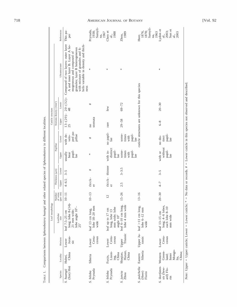
Sphenobaiera huangii is comparable to several other specieswith bilobed leaves or less divided leaves and broader lobesor segments, both in leaf morphology and cuticular structure,e.g., S. biloba Prynada (Prynada, 1938; Samylina, 1967; Chenet al., 1988; Deng, 1995; Deng et al., 1997), S. jugata Zhou(Zhou, 1989), S. ikorfatensis (Seward) Florin (Lydon et al.,2003; Sun et al., 2003), and S. pulchella (Heer) Florin (Heer,1876, 1878; Samylina, 1963). Their main differences and sim-ilarities in terms of leaf morphology and cuticular structure aresummarized in Table 1. It is evident that S. huangii from theLower Jurassic of Hubei, China, may represent the best-knownspecies so far of the genus Sphenobaiera.
The cuticular ultrastructure of Sphenobaiera has not beeninvestigated using electron microscope techniques until now,although other related ginkgoalean or czekanowskialean mem-bers (such as Ginkgo, Ginkgoites, Phoenicopsis, and Arcto-baiera), have been studied using SEM and TEM (Taylor etal., 1989; Villar de Seoane, 1997a; Zhou and Guignard, 1998;Guignard and Zhou, in press). At the ultrastructural level, S.huangii has two distinct layers—an outer granular layer (A)and an inner fibrillar layer (B)—in ordinary epidermal cells,stomata complex (subsidiary and guard cells), anticlinal walls,and in papillate cell cuticle, both in transverse and longitudinalsections. No polylamellate layer was found in the cuticle.
Compared with epidermal cells, the stomatal complex cu-ticle of S. huangii has a thicker layer B; that is, the cuticle inboth guard cells and subsidiary cells is more fibrillous. Thisminor structural difference between stomatal complex cellsand ordinary epidermal cells was also recorded in other taxa,e.g., Hirmeriella muensterii (Schenk) Jung, belonging to Chei-rolepidiaceae of the conifers (Guignard et al., 1998). This cu-ticular anatomy of the stomatal complex is known, in extantplants, to be directly related to the exchange of water and other

714 [Vol. 92AMERICAN JOURNAL OF BOTANY
Figs. 12–17. Sphenobaiera huangii (Sze) Hsu from the Lower Jurassic Hsiangchi Formation in Hubei, China. Light and scanning electron microscopy (LM,SEM) photos of the upper cuticle. 12. Lower magnification showing epidermal cells and the distribution of stomata (LM). Negative no. 980671, specimen no.PB20060. Scale bar 5 100 mm. 13. Four stomata, isodiametric and polygonal ordinary cells. Negative no. 98031, specimen no. PB20061. Scale bar 5 100mm. 14. Two stomata and the outline of epidermal cells. Negative no. 98032, specimen no. PB20041. Scale bar 5 10 mm. 15. Detail of a stoma in Fig. 14showing exposed guard and subsidiary cells and elliptical to fusiform stomatal pit mouth. Note the fine radial striations in guard cells. Negative no. 98032.Scale bar 5 10 mm. 16. Outer view of the upper cuticle showing papillae and stomata. Negative no. 99052. Specimen no. PB20045. Scale bar 5 100 mm. 17.Outer view of a stoma showing thickened subsidiary cells and papillae overhanging the stomata pit. Negative no. 980670, specimen no. PB20064. Scale bar 510 mm.

April 2005] 715WANG ET AL.—CUTICULAR ANATOMY OF SPHENOBAIERA HUANGII (GINKGOALES)
Figs. 18–25. Sphenobaiera huangii (Sze) Hsu from the Lower Jurassic Hsiangchi Formation in Hubei, China. Transmission electron microscopy (TEM)photos of the lower cuticle. All transverse sections. Specimen no. PB20041. 18. General view of a part of the cuticle composed of two anticlinal walls (arrows).The cuticle is composed of a homogeneous outer layer A with granules and a heterogeneous inner layer B mainly with fibrils. Negative no. gtx379. Scale bar5 5 mm. 19. Detail of Fig. 18 showing the outermost part of the cuticle with layer A with a high density of fine granules. Negative no. gtx364. Scale bar 5100 nm. 20. Detail of Fig. 18 showing the middle part of the cuticle with layer A with a low density of fine granules. Negative no. gtx358. Scale bar 5 100nm. 21. Detail of Fig. 18 showing the innermost part of the cuticle of layer B consisting of fine fibrils. Negative no. gtx365. Scale bar 5 200 nm. 22. A wide-opened stoma showing stomata pit (arrow), subsidiary cell (SC), and guard cell (GC) cuticle. Negative no. gtx367, specimen no. PB20045. Scale bar 5 10 mm.23. Detail of outer part of the subsidiary cell cuticle near the stomatal pit, showing layer A. Negative no. gtx371. Scale bar 5 100 nm. 24. Detail of middlepart of the subsidiary cell cuticle near the stomatal pit with layer B showing fibrils (arrow). Negative no. ggw5143. Scale bar 5 100 nm. 25. Detail of innerpart of the subsidiary cell cuticle, showing fibrils (arrows). Negative no. gtx372. Scale bar 5 100 nm.
molecules (Larcher, 1995). In S. huangii, the guard cell cuticleis thinner than subsidiary cell cuticle. From a structure–func-tion view, considering that guard cells are responsible for theopening and closing of stomata, a thinner cuticle is certainlymore efficient than a thicker one.
To date, the leaf cuticular ultrastructure of Ginkgoales hasbeen reported only for a few fossil and living taxa in the fam-ilies Karkeniaceae and Ginkgoaceae, including Ginkgoites ti-grensis Archangelsky, Ginkgo yimaensis Zhou and Zhang andGinkgo biloba L. Ginkgoites tigrensis from the Lower Creta-

716 [Vol. 92AMERICAN JOURNAL OF BOTANY
Figs. 26–34. Sphenobaiera huangii (Sze) Hsu from the Lower Jurassic Hsiangchi Formation in Hubei, China. Transmission electron microscopy (TEM)photos of the lower cuticle. Specimen no. PB20045. 26–29. Transverse sections. Specimen no. PB20045. 26. Detail of Fig. 22, showing guard cell (GC) andthe lower part of the subsidiary cell (SC) cuticle. Negative no. gtx373. Scale bar 5 2 mm. 27. Detail of Fig. 26, showing the outer part of the guard cell cuticlewith layer A. Negative no. gtx376. Scale bar 5 100 nm. 28. Detail of Fig. 26, showing the middle part of the guard cell cuticle with granules. Negative no. gtx377.Scale bar 5 100 nm. 29. Detail of Fig. 26, showing the inner part of the guard cell cuticle with layer B containing fibrils. Negative no. gtx378. Scale bar 5 100nm. 30–34. Longitudinal sections of ordinary cell of the lower cuticle. 30. General view of an ordinary epidermal cell cuticle, showing layers A and B. Negativeno. gtx389. Scale bar 5 2 mm. 31. Detail of Fig. 30, showing the innermost part with layer B and part of layer A. Negative no. gtx383. Scale bar 5 500 nm.

April 2005] 717WANG ET AL.—CUTICULAR ANATOMY OF SPHENOBAIERA HUANGII (GINKGOALES)
Fig. 35. A suggested three-dimensional reconstruction of the cuticular anatomy of Sphenobaiera huangii (Sze) Hsu, based on transverse and longitudinalsections in various parts of the cuticle, including ordinary epidermal cell (OEC), anticlinal wall (AW), papillae (P), stomatal complex region with guard cell(GC), and subsidiary cell (SC).
←
Figs. 26–34. Continued. 32. Detail of Fig. 30, showing the outermost part of the cuticle with layer A. Negative no. gtx381. Scale bar 5 100 nm. 33. Detailof Fig. 30, showing the innermost part of the cuticle with fibrils of layer B. Negative no. gtx385. Scale bar 5 100 nm. 34. Detail of Fig. 30, showing themiddle part of the cuticle with layer A. Negative no. gtx382. Scale bar 5 100 nm.
ceous of Argentina is a representative member of the Karken-iaceae. Available data demonstrate that the cuticular ultrastruc-ture of S. huangii shows a closer affinity with that of G. ti-grensis. According to a recent study by Villar de Seoane(1997a), the cuticular ultrastructure of G. tigrensis from Ar-gentina closely resembles that of S. huangii from China. Theouter part of the G. tigrensis cuticle is compact and homoge-neous, comparable to the outer granular layer of S. huangii.The inner part of the cuticle is slightly reticulate and is struc-turally similar to the heterogeneous inner layer with fibrils ofS. huangii. The polylamellate layer in G. tigrensis is absent(Villar de Seoane, 1997a). So far, the leaf cuticular ultrastruc-ture is known for only one fossil taxon belonging to Gink-goaceae of Ginkgoales. According to Guignard and Zhou (inpress), the leaf cuticle of Ginkgo yimaensis, the oldest Ginkgofrom the Jurassic of China, is obviously distinguished from S.huangii in ultrastructural level by having a distinct outer po-lylamellate layer. The leaf cuticle of living Ginkgo is also dif-ferent from that of S. huangii. Taylor et al. (1989) and Guig-
nard and Zhou (in press) demonstrated that the cuticle of G.biloba is characterized by an outer polylamellate layer andinner granular and fibrillar layers, differing from S. huangii ofChina at the ultrastructural level.
The studies on the fossil leaf cuticular ultrastructure usingtransmission electron microscopy started with the pioneerwork of Archangelsky et al. (1986). Since then, the leaf cutic-ular ultrastructure is known for a number of fossil taxa orgroups, including pteridosperms (Taylor et al., 1989; Baldoniand Barale, 1996; Labe and Barale, 1996; Maheshwari andBajpai, 1996; Bajpai, 1997; Guignard et al., 2001), bennetti-taleans (Barale and Baldoni, 1993; Villar de Seoane, 2001),cycadaleans (Artabe and Archangelsky, 1992; Villar de Seo-ane, 1997b), ginkgoaleans (Taylor et al., 1989; Villar de Seo-ane, 1997a; Guignard and Zhou, in press), czekanowskialeans(Zhou and Guignard, 1998), as well as conifers (Archangelskyand Taylor, 1986; Archangelsky et al., 1986; Del Fueyo et al.,1990; Barale et al., 1992; Guignard et al., 1998; Villar deSeoane, 1998; Zhou et al., 2000). Most of the cuticle is marked
718 [Vol. 92AMERICAN JOURNAL OF BOTANY
TA
BL
E1.
Com
pari
sons
betw
een
Sphe
noba
iera
huan
gii
and
othe
rre
late
dsp
ecie
sof
Sphe
noba
iera
indi
ffer
ent
loca
liti
es.
Spe
cies
Loc
alit
yH
oriz
on
Lea
fm
orph
olog
y
Lea
f/lo
besi
ze
Num
ber
ofve
ins
(per
cm)
Cut
icle
stru
ctur
e
Thi
ckne
ss(m
m)
Upp
erL
ower
Pap
illa
e
Upp
erL
ower
Sto
mat
ade
nsit
y(/
mm
2 )
Upp
erL
ower
Ult
rast
ruct
ure
Ref
eren
ces
S.hu
angi
i(S
ze)
Hsu
Hub
ei,
Chi
naL
ower
Jura
s-si
c
leaf
7.5–
25cm
long
;lo
be8-
(14)
-21
mm
wid
e;lo
bean
gle
108–
258
10–1
14
–6.
53–
5us
uall
ybe
arpa
pil-
lae
wit
hde
-ve
lo-
ped
pa-
pill
ae
13
-(2
0)-
252
0-(
32
)-48
Com
pose
dof
two
laye
rs:
oute
rla
yer
Aan
din
ner
laye
rB
.L
ayer
Aho
-m
ogen
eous
and
com
pose
dof
gran
ules
;la
yer
Bhe
tero
gene
ous
wit
hm
ixtu
reof
gran
ules
and
fi-
bril
sva
riab
lein
dens
ity
and
thic
k-ne
ss
Thi
spa
-pe
r
S.bi
loba
Pry
nada
Sib
eria
Low
erC
reta
-ce
ous
leaf
13cm
long
,lo
be18
–20
mm
wid
e
10–1
3th
ick
-er
#*
#no st
omat
a#
*P
ryna
da,
1938
;S
amyl
i-na
,19
67S.
bilo
baP
ryna
daF
uxin
,L
iaon
-in
g,C
hina
Low
erC
reta
-ce
ous
leaf
upto
17cm
long
,lo
be13
mm
wid
e;lo
bean
gle
108
12th
ick
-er
thin
ner
wit
hin
-di
stin
ctpa
pil-
lae
nopa
pil-
lae
rare
few
*C
hen
etal
.,19
88
S.ju
ncta
Zho
uS
haqi
ao,
Hun
an,
Chi
na
Upp
erT
rias
sic
leaf
6–10
cmlo
ng,
lobe
4m
mw
ide
15–2
62.
53–
3.5
som
e-ti
mes
wit
hpa
pil-
lae
som
e-ti
mes
wit
hpa
pil-
lae
29–5
869
–72
*Z
hou,
1989
S.pu
lche
lla
(Hee
r)F
lori
n
Eas
tern
Sib
eria
Upp
erJu
-ra
ssic
leaf
.11
cmlo
ng,
lobe
6–12
mm
wid
e
13–1
6cu
ticl
est
ruct
ures
are
unkn
own
for
this
spec
ies
Hee
r,18
76,
1878
;S
amyl
i-na
,19
63S.
ikor
fate
n-si
s(S
ew-
ard)
Flo
-ri
n
Wes
tern
Gre
en-
land
;In
ner
Mon
go-
lia,
Chi
na;
Low
erC
reta
-ce
ous
leaf
13–1
5cm
long
;4
–6
lobe
s,ea
chlo
be8–
10m
mw
ide
20–3
04
–73–
5w
ith
orw
itho
utpa
pil-
lae
nodi
s-ti
nct
papi
l-la
e
6–8
20–3
0*
Lyd
onet
al.,
2003
;S
unet
al.,
2003
Not
e:U
pper
5U
pper
cuti
cle;
Low
er5
Low
ercu
ticl
e;*
5N
oda
taor
reco
rds.
#5
Low
ercu
ticl
ein
this
spec
ies
not
obse
rved
and
desc
ribe
d.

April 2005] 719WANG ET AL.—CUTICULAR ANATOMY OF SPHENOBAIERA HUANGII (GINKGOALES)
by an outer polylamellate zone, similar to type 1 of Holloway’s(1982) cuticle classification (six types based on the leaf ultra-structure of living plants). It is noteworthy that Holloway(1982) is very careful to point out the lamellate structure ofthe cuticle proper; where it occurs in the cuticular layers, it isdifficult to illustrate. Some results may depend upon the stain-ing technique, which can alter the appearance of the cuticle.In this study, S. huangii cuticle is stained using a method sim-ilar to that used for G. yimaensis and G. biloba (Guignard andZhou, in press), with osmium tetroxide and a post-stain withuranyl acetate and lead citrate. It is evident that the outer po-lylamellate layers are reported in G. yimaensis and G. biloba(Ginkgoaceae), but were not found in S. huangii, though sim-ilar staining was used in both cases.
The cuticle of S. huangii from the Lower Jurassic of Hubei,China, has some affinities with both Holloway’s type 3 andtype 6. In type 3, the cuticle is partly reticulate without thegranular layer in S. huangii. Type 6 has many different layers,but they are very different from S. huangii. It is known that acuticle with an outer homogeneous layer as described in S.huangii is very distinct and rarely reported in living and fossilrecords. A more or less similar structure was reported in twofossil cycadalean leaves from the Lower Cretaceous of Argen-tina (Villar de Seoane, 1997b) and in Phoenicopsis and Arc-tobaiera leaves of the Czekanowskiales from the Jurassic ofHenan, central China (Zhou and Guignard, 1998), but the cu-ticle of these taxa are otherwise very different in general struc-ture. No known fossil ginkgoalean member bears a cuticlesimilar in ultrastructure to S. huangii; only G. tigrensis (Kar-keniaceae) has a close affinity with S. huangii at the ultrastruc-tural level.
Previous and recent studies show that some species ofSphenobaiera are associated with the ovule Karkenia, belong-ing to the family Karkeniaceae (Archangelsky, 1965; Krassi-lov, 1972; Schweitzer and Kirchner, 1995; Zhou et al., 2002),whereas other species of Sphenobaiera are associated withGinkgo-type ovules. However, in most species of Spheno-baiera, their ovule-bearing organs are still unknown. Obvi-ously, the genus Sphenobaiera is difficult to place into anyknown family within the Ginkgoales, based upon either leafmorphology and/or general cuticular features. According tothis investigation, the ultrastructural data of S. huangii cuticlemay imply a potential affinity to the family Karkeniaceae ofGinkgoales. We have provided useful information for explor-ing the relationship of the leaf cuticle construction among dif-ferent taxa and about the extent of variation in a given taxonand for assessing the significance of cuticular structure in planttaxonomy. Further work is obviously needed in order to pro-vide more reliable data on reproductive organs associated withS. huangii and to confirm its systematic status.
In summary, this investigation of S. huangii from the EarlyJurassic in Hubei, China, provides new insights into the cutic-ular anatomy, as well as the variation in leaf morphology.Many features are described for the first time for this species,based upon light and electron microscopic observations, in-cluding general structure of lower and upper cuticle, stomata,and papillae. At the ultrastructural level, two layers have beendistinguished in both lower and upper cuticle, including a ho-mogeneous outer layer with granules and a heterogeneous in-ner layer with fibrils. Comparison with literature between S.huangii and other relevant species of Sphenobaiera indicatethat this species from the Lower Jurassic of China may rep-resent the best-known taxon in the genus Sphenobaiera in both
leaf morphology and cuticular structures. Particularly, thisstudy provides the first detailed ultrastructural data on the leafcuticle of Sphenobaiera, one of the oldest foliage taxa of gink-goales. This offers further evidence for potential discussion onthe taxonomic relationships with other ginkgoalean taxa.
LITERATURE CITED
ARCHANGELSKY, S. 1965. Fossil Ginkgoales from the Tico Flora, Santa CruzProvince, Argentina. Bulletin of the British Museum (Natural History),Geology 10: 121–137.
ARCHANGELSKY, S., AND T. N. TAYLOR. 1986. Ultrastructural studies of fossilplant cuticles. II. Tarphyderma gen. n., a Cretaceous conifer from Ar-gentina. American Journal of Botany 73: 1577–1587.
ARCHANGELSKY, S., T. N. TAYLOR, AND M. H. KURMANN. 1986. Ultrastruc-tural studies of fossil plant cuticles: Ticoa harrisii from the Early Cre-taceous of Argentina. Botanical Journal of the Linnean Society 92: 101–116.
ARTABE, A. E., AND S. ARCHANGELSKY. 1992. Las cycadales MesodescolaArchangelsky emend. Archangelsky y Petriella 1971 (Cretacico) y Stan-geria Moore (Actual). Ameghiniana 29(2): 115–123.
BAJPAI, U. 1997. Taphonomic constraints on preservation of cuticles in com-pression fossils: fungi induced ultrastructural changes in cuticular mem-branes. Palaeobotanist 46: 31–34.
BALDONI, A. M., AND G. BARALE. 1996. El genero Pachypteris Brongniartemend. Harris en el Jurassico medio de Argentina. Consideracion sobrela distribucion estratigrafica y geografica. Revista Espanola de Paleon-tologia 11(2): 134–142.
BARALE, G., AND A. BALDONI. 1993. L’ultrastructure de la cuticule de qu-elques Bennettitales du Cretace inferieur d’Argentine. Comptes Renduesde l’Academie des Sciences de Paris 316: 1171–1177.
BARALE, G., A. BALDONI, AND E. SAMUEL. 1992. Etude de la cuticule desfeuilles de Podocarpacees du Cretace inferieur de la Formation Baquero(Argentine): Observation en microscopie photonique, electronique a ba-layage et a transmission. Courier Forschungsinstitut Senckenburg 147:215–223.
CHEN, F., X. Y. MENG, S. Q. REN, AND C. L. WU. 1988. The Early Cretaceousflora of Fuxin Basin and Tiefa Basin, Liaoning Province. GeologicalPublishing House, Beijing, China (in Chinese with English summary).
CHEN, G. X. 1984. Pteridophyta, Spermatophyta. In Regional Geological Sur-vey Team of Hubei [ed.], The palaeontological atlas of Hubei Province,560–615. Hubei Science and Technology Press, Wuhan, China (in Chi-nese).
CHEN, Y., S. Y. DUAN, AND Y. C. ZHANG. 1987. Late Triassic Qinghe floraof Sichuan. Botanical Research 2: 83–158 (in Chinese with English sum-mary).
DEL FUEYO, G. M., S. ARCHANGELSKY, AND T. N. TAYLOR. 1990. Una nuevaPodocarpacea fertile (coniferal) del Cretacico inferior de Patagonia Ar-gentina. Ameghiniana 27(1–2): 63–73.
DENG, S. H. 1995. Early Cretaceous flora from Huolinhe of Inner Mongolia.Geological Publishing House, Beijing, China (in Chinese with Englishsummary).
DENG, S. H., S. Q. REN, AND F. CHEN. 1997. Early Cretaceous floras fromHailaer Basin of Inner Mongolia. Geological Publishing House, Beijing,China (in Chinese with English summary).
DUAN, S. Y., AND Y. CHEN. 1982. Mesozoic fossil plants and coal formationof eastern Sichuan Basin. In Compilatory Group of Continental MesozoicStratigraphy and Palaeontology in Sichuan Basin [ed.], Continental Me-sozoic stratigraphy and palaeontology in Sichuan Basin of China, partII, 491–519. People’s Publishing House of Sichuan, Chengdu, China (inChinese).
FENG, S. N., F. S. MENG, G. X. CHEN, Y. H. XI, C. F. ZHANG, AND Y. A.LIU. 1977. Plant kingdom. In Hubei Institute of Geological Science,Henan Bureau of Geology and Mineral Resources [ed.], Palaeontologicalatlas of Central South China, vol. 3, Mesozoic and Cenozoic, 195–262.Geological Publishing House, Beijing, China (in Chinese).
FLORIN, R. 1936. Die fossilen Ginkgophyten von Franz-Joseph-Land nebstErorterungen uber vermeintliche Cordaitales mesozoischen Alters. I. Pa-laeontographica 81B: 71–173.
GUIGNARD, G., K. BOKA, AND M. BARBACKA. 2001. Sun and shade leaves?Cuticle ultrastructure of Jurassic Komlopteris nordenskioeldii (Nathorst)Barbacka. Review of Palaeobotany and Palynology 114: 191–208.

720 [Vol. 92AMERICAN JOURNAL OF BOTANY
GUIGNARD, G., F. THEVENARD, AND J. H. A. VAN KONIJNENBURG-VAN CIT-TERT. 1998. Cuticle ultrastructure of the cheirolepidiaceous conifer Hir-meriella muensteri (Schenk) Jung. Review of Palaeobotany and Paly-nology 104: 115–141.
GUIGNARD, G., AND Z. Y. ZHOU. In press. Comparative studies of leaf cuticleultrastructure between the living and oldest known ginkgos in China.International Journal of Plant Sciences.
HARRIS, T. M., AND J. MILLER. 1974. Czekanowskiales. In T. M. Harris, W.Millington, and J. Miller [eds.], The Yorkshire Jurassic flora, vol. IV,Ginkgoales and Czekanowskiales, 79–150. British Museum (Natural His-tory), London, UK.
HARRIS, T. M., AND W. MILLINGTON. 1974. Ginkgoales. In T. M. Harris, W.Millington, and J. Miller [eds.], The Yorkshire Jurassic flora, vol. IV,Ginkgoales and Czekanowskiales, 2–78. British Museum (Natural His-tory), London, UK.
HEER, O. 1876. Beitrage zur Jura-Flora Ostsibiriens und des Amurlandes.Memoires de L’Academie Imperiale des Sciences de St.-Petersbourg,VIIe Serie 22(12): 1–122.
HEER, O. 1878. Beitrage zur Fossilen Flora Sibriens und des Amurlands.Memoires de L’Academie Imperiale des Sciences de St.-Petersbourg,VIIe Serie 25(6): 1–117.
HOLLOWAY, P. J. 1982. Structure and histochemistry of plant cuticular mem-bers: an overview. In D. F. Cutler, K. L. Alvin, and C. E. Price [eds.],The plant cuticle, 1–32. Academic Press, London, UK.
HUANG, Z. G., AND H. Q. ZHOU. 1980. Fossil plants. In Institute of Geology,Chinese Academy of Geological Sciences [ed.], Mesozoic stratigraphyand palaeontology from the basin of Shaanxi, Gansu and Ningxia, I, 43–104. Geological Publishing House, Beijing, China (in Chinese).
KRASSILOV, V. A. 1972. Mesozoic flora from the Bureja Basin (Ginkgoalesand Czekanowskiales). Akademii Nauka SSSR, Moscow, Russia.
LABE, M., AND G. BARALE. 1996. Etudes ultrastructurales de la cuticule deprespermatophytes fossils du Jurassique. Revue de Paleobiologie 15: 87–103.
LARCHER, W. 1995. Physiological plant ecology. Springer-Verlag, New York,New York, USA.
LI, B. X., AND B. HU. 1984. Fossil plants from the Yongdingzhuang For-mation of the Datong coalfield, northern Shanxi. Acta PalaeontologicaSinica 23(2): 135–147 (in Chinese with English abstract).
LI, W. B., AND Y. K. SHANG. 1980. Spore-pollen assemblages of the MesozoicCoal Series of western Hubei. Acta Palaeontologica Sinica 19: 201–219(in Chinese with English abstract).
LIU, Z. J. 1982. Palaeobotany. In Xi’an Institute of Geology and MineralResources [ed.], Palaeontological atlas of Northwestern China, vol. III,Mesozoic and Cenozoic, 116–139. Geological Publishing House, Beijing,China (in Chinese).
LYDON, S. J., J. WATSON, AND N. A. HARRISON. 2003. The lectotype ofSphenobaiera ikorfatensis (Seward) Florin, a ginkgophyte from the Low-er Cretaceous of western Greenland. Palaeontology 46: 413–421.
MAHESHWARI, H. K., AND U. BAJPAI. 1996. Ultrastructure of the ‘‘cuticularmembrane’’ in two Late Triassic corystospermaceous taxa from India.Palaeobotanist 45: 41–49.
MENG, F. S. 1987. Fossil plants. In Yichang Institute of Geology and MineralResources, Chinese Academy of Geological Sciences [eds.], Biostratig-raphy of the Yangtze Gorge Area, vol. 4, Triassic and Jurassic, 239–257.Geological Publishing House, Beijing, China (in Chinese with Englishsummary).
MENG, F. S., AND Z. L. ZHANG. 1987. The Jurassic system. In Yichang In-stitute of Geology and Mineral Resources, Chinese Academy of Geolog-ical Sciences [eds.], Biostratigraphy of the Yangtze Gorge Area, vol. 4,Triassic and Jurassic, 135–144. Geological Publishing House, Beijing,China (in Chinese with English summary).
MI, J. R., C. L. SUN, Y. W. SUN, S. S. CUI, AND Y. L. AI. 1996. Early-MiddleJurassic phytoecology and coal-accumulating environments in NorthernHebei and Western Liaoning. Geological Publishing House, Beijing, Chi-na (in Chinese with English summary).
POORT, R. J., H. VISSCHER, AND D. L. DILCHER. 1996. Zoidogamy in fossilgymnosperms: the centenary of a concept, with special references toprepollen of late Paleozoic conifers. Proceedings of the National Acad-emy of Sciences, USA 93: 11713–11717.
PRYNADA, V. D. 1938. The materials to knowledge of the Mesozoic flora ofKolyma River basin. Materials on Research of Kolyma-Indigirka RiversRegion, series 2, Geology and Geomorphology 13: 1–74.
SAMYLINA, V. A. 1963. The Mesozoic flora of the lower course of Aldan
River. Transactions of Botanical Institute of Academy of Sciences ofU.S.S.R., series VIII. Palaeobotany 4: 59–139 (in Russian with Englishabstract).
SAMYLINA, V. A. 1967. The Mesozoic flora of the area to the west of theKolyma River (Zyrianka coal-basin). II. Ginkgoales, Coniferales. Trans-actions of Botanical Institute of Academy of Sciences of U.S.S.R., seriesVIII. Palaeobotany 6: 134–175 (in Russian with English abstract).
SCHWEITZER, H.-J., AND M. KIRCHNER. 1995. Die Rhato-Jurassischen Florendes Iran und Afghanistans: 8. Ginkgophyta. Palaeontographica 237B: 1–58.
SUN, G., S. J. LYDON, AND J. WATSON. 2003. Sphenobaiera ikorfatensis(Seward) Florin from the Lower Cretaceous of Huolinhe, eastern InnerMongolia, China. Palaeontology 46: 423–430.
SZE, H. C. 1949. Die Mesozoische Flora aus der Hsiangchi Kohlen Serie inWesthupeh. Palaeontologia Sinica New Series A 2: 1–71.
SZE, H. C., AND J. HSU. 1954. Index fossils of China plants. GeologicalPublishing House, Beijing, China (in Chinese).
TAYLOR, T. N., AND E. L. TAYLOR. 1993. The biology and evolution of fossilplants. Prentice Hall, Englewood Cliffs, New Jersey, USA.
TAYLOR, W. A., T. N. TAYLOR, AND S. ARCHANGELSKY. 1989. Comparativeultrastructure of fossil and living gymnosperm cuticles. Review of Pa-laeobotany and Palynology 59: 145–151.
VILLAR DE SEOANE, L. 1997a. Comparative study between Ginkgoites ti-grensis Archangelsky and Ginkgo biloba Linn. leaves. Palaeobotanist46: 1–12.
VILLAR DE SEOANE, L. 1997b. Estudio cuticular comparado de nuevas Cy-cadales de la Formacion Baquero (Cretacico Inferior), provincia de SantaCruz, Argentina. Revista Espanola de Paleontologia 12(1): 129–140.
VILLAR DE SEOANE, L. 1998. Comparative study of extant and fossil coniferleaves from the Baquero Formation (Lower Cretaceous), Santa CruzProvince, Argentina. Review of Palaeobotany and Palynology 99: 247–263.
VILLAR DE SEOANE, L. 2001. Cuticular study of Bennettitales from theSpringhill Formation, Lower Cretaceous of Patagonia, Argentina. Cre-taceous Research 22: 461–479.
WANG, G. P., Q. S. CHEN, Y. LI, H. LI, S. X. GUO, S. X. LAN, AND K. X.JU. 1982. Plant kingdom. In Nanjing Institute of Geological and MineralResources, Ministry of Geology and Mineral Resources [ed.], Palaeon-tological atlas of East China, III, 236–316. Geological Publishing House,Beijing, China (in Chinese).
WANG, Y. D. 1999. Fertile organs and in situ spores of Marattia asiatica(Kawasaki) Harris (Marattiales) from the Lower Jurassic Hsiangchi For-mation in Hubei, China. Review of Palaeobotany and Palynology 107:125–144.
WANG, Y. D. 2002. Fern ecological implications from the Lower Jurassic inWestern Hubei, China. Review of Palaeobotany and Palynology 119:125–141.
WANG, Y. D., G. GUIGNARD, B. LUGARDON, AND G. BARALE. 2001. Ultra-structure of in situ Marattia asiatica (Marattiaceae) spores from the Low-er Jurassic in Hubei, China. International Journal of Plant Sciences 162:927–936.
WANG, Y. D., AND S. W. MEI. 1999. Fertile organs and in situ spores of amatoniaceous fern from the Lower Jurassic of West Hubei. Chinese Sci-ence Bulletin (English version) 44(14): 1333–1337.
WU, S. Q., M. N. YE, AND B. X. LI. 1980. Upper Triassic and Lower andMiddle Jurassic plants from the Hsiangchi Group, Western Hubei. Mem-oirs of Nanjing Institute of Geology and Palaeontolology, Academia Sin-ica 14: 63–131 (in Chinese with English abstract).
YANG, J. D., AND S. Y. SUN. 1987. Jurassic megaspore assemblages. In In-stitute of Geology, Yichang Institute of Geology and Mineral Resources,Chinese Academy of Geological Sciences [eds.], Biostratigraphy of theYangtze Gorge Area, vol. 4, Triassic and Jurassic, 145–148. GeologicalPublishing House, Beijing, China (in Chinese with English abstract).
YANG, X. H. 1978. Mesozoic plants. In Southwest Institute of Geosciences,Ministry of Geology and Mineral Resources [ed.], Palaeontological atlasof southwestern China. Volume of Sichuan, part 2, 469–536. GeologicalPublishing House, Beijing, China (in Chinese).
ZENG, Y., S. Z. SHEN, AND B. H. FAN. 1995. Flora from the coal-bearingstrata of Yima Formation in western Henan. Jiangxi Science and Tech-nology Publishing House, Nanchang, China (in Chinese).
ZHANG, C. F. 1986. Early Jurassic flora from eastern Hunan. ProfessionalPapers of Stratigraphy and Palaeontology 14: 185–206 (in Chinese withEnglish abstract).

April 2005] 721WANG ET AL.—CUTICULAR ANATOMY OF SPHENOBAIERA HUANGII (GINKGOALES)
ZHANG, H., C. W. XIONG, H. T. LI, Y. D. WANG, AND H. ZHANG. 1998.Jurassic coal-bearing strata and coal accumulation in Northwest China.Geological Publishing House, Beijing, China (in Chinese with Englishsummary).
ZHANG, W. P., AND Z. L. ZHANG. 1987. Jurassic spores and pollen grains. InInstitute of Geology, Yichang Institute of Geology and Mineral Resourc-es, Chinese Academy of Geological Sciences [eds.], Biostratigraphy ofthe Yangtze Gorge Area, vol. 4, Triassic and Jurassic, 135–144. Geolog-ical Publishing House, Beijing, China (in Chinese).
ZHENG, S. L., AND Z. Y. ZHOU. 2004. A new Mesozoic Ginkgo from WesternLiaoning, China and its evolutionary significance. Review of Palaeobo-tany and Palynology 131: 91–103.
ZHOU, Z. Y. 1989. Late Triassic plants from Shaqiao, Hengyang, Hunan Prov-ince. Palaeontologia Cathayana 4: 131–197.
ZHOU, Z. Y. 1997. Mesozoic Ginkgoalean megafossils: a systematic review.In H. Hori, R. W. Ridge, W. Tulecke, P. Del Tredici, J. Tremouillaux-Guiller, and H. Tobe [eds.], Ginkgo biloba—a global treasure from bi-ology to medicine, 183–206. Springer-Verlag, Tokyo, Japan.
ZHOU, Z. Y. 2003. Mesozoic Ginkgoaleans: phylogeny, classification and evo-lutionary trends. Acta Botanica Yunnanica 25(4): 377–396 (in Chinesewith English abstract).
ZHOU, Z. Y., AND G. GUIGNARD. 1998. Leaf cuticle ultrastructure of twoczekanowskialeans from the Middle Jurassic Yima Formation of Henan,China. Review of Palaeobotany and Palynology 102: 179–187.
ZHOU, Z. Y., F. THEVENARD, G. BARALE, AND G. GUIGNARD. 2000. A newxeromorphic conifer from the Lower Cretaceous of Eastern China. Pa-laeontology 43: 561–572.
ZHOU, Z. Y., AND B. L. ZHANG. 1988. Two new ginkgoalean female repro-ductive organs from the Middle Jurassic of Henan Province. ChineseScience Bulletin 33: 1201–1203.
ZHOU, Z. Y., AND B. L. ZHANG. 1989. A Middle Jurassic Ginkgo with ovule-bearing organs from Henan, China. Palaeontographica 211B: 113–133.
ZHOU, Z. Y., AND B. L. ZHANG. 1992. Baiera hallei Sze and associated ovule-bearing organs from the Middle Jurassic of Henan, China. Palaeonto-graphica 224B: 151–169.
ZHOU, Z. Y., B. L. ZHANG, Y. D. WANG, AND G. GUIGNARD. 2002. A newKarkenia (Ginkgoales) from the Jurassic Yima Formation, Henan, Chinaand its megaspore membrane ultrastructure. Review of Palaeobotany andPalynology 120: 91–105.
ZHOU, Z. Y., AND S. L. ZHENG. 2003. The missing link in Ginkgo evolution.Nature 423: 821–822.