Crystal Structure of the VHS and FYVE Tandem Domains of Hrs, a Protein Involved in Membrane...
10
Cell, Vol. 100, 447–456, February 18, 2000, Copyright 2000 by Cell Press Crystal Structure of the VHS and FYVE Tandem Domains of Hrs, a Protein Involved in Membrane Trafficking and Signal Transduction ceptors are primarily trafficked to lysosomes whereas others such as the transferrin receptor are recycled back to the plasma membrane. Regulatory molecules that mediate this trafficking decision might be expected to interact with cargo proteins, membrane phospholipids, Yuxin Mao,* Alexei Nickitenko, ² Xiaoqun Duan, ‡ Thomas E. Lloyd, § Mark N. Wu, § Hugo Bellen, ‡§k and Florante A. Quiocho* ²‡ # * Structural and Computational Biology and Molecular Biophysics Graduate Program and members of the trafficking machinery. One potential candidate that satisfies these criteria is ² Department of Biochemistry and Molecular Biology ‡ Howard Hughes Medical Institute a newly identified protein named Hrs (hepatocyte growth factor regulated tyrosine kinase substrate). As its name § Department of Molecular and Cellular Biology k Department of Molecular and Human Genetics implies, Hrs is tyrosine phosphorylated in response to a variety of growth factors including HGF, EGF, and Baylor College of Medicine Houston, Texas 77030 PDGF (Komada and Kitamura, 1995). Hrs is expressed in the cytoplasm of all cells and is predominantly local- ized to endosomes (Komada et al., 1997). This localiza- tion is consistent with the finding that Vps27p, the yeast Summary homolog of Hrs, is also expressed on endosomes, and null mutants are unable to traffic most proteins to the We have determined the 2 A ˚ X-ray structure of the vacuole (yeast equivalent of the lysosome), leading to 219-residue N-terminal VHS and FYVE tandem domain an exaggerated “class E” compartment (Piper et al., unit of Drosophila Hrs. The unit assumes a pyramidal 1995). Similarly, mice lacking Hrs die early in embryonic structure in which the much larger VHS domain (resi- development and have abnormally enlarged endosomes dues 1–153) forms a rectangular base and the FYVE (Komada and Soriano, 1999). Thus, Hrs has been pro- domain occupies the apical end. The VHS domain is posed to play a role in trafficking cargo from the endo- comprised of an unusual “superhelix” of eight a heli- some to the lysosome. Interestingly, Hrs has also been ces, and the FYVE domain is mainly built of loops, implicated in exocytosis of synaptic vesicles. A splice two double-stranded antiparallel sheets, and a helix variant of rat Hrs named Hrs-2 is a calcium-dependent stabilized by two tetrahedrally coordinated zinc atoms. ATPase, tightly binds the t-SNARE SNAP-25, and inhib- The two-domain structure forms an exact 2-fold- its neurotransmitter release when injected into perme- related homodimer through antiparallel association of abilized PC12 cells (Bean et al., 1997). This suggests mainly FYVE domains. Dimerization creates two iden- that Hrs may regulate synaptic vesicle exocytosis by tical pockets designed for binding ligands with multi- directly interacting with the fusion machinery. ple negative charges such as citrate or phosphatidyl- Hrs contains several conserved domains that are inositol 3-phosphate. present in proteins implicated in signal transduction and/or membrane trafficking. The VHS (Vps27p, Hrs, Introduction STAM) domain is present at the amino terminus of sev- eral proteins believed to play a role in tyrosine kinase Intracellular membrane trafficking events are tightly reg- receptor signaling. STAM (signal-transducing adaptor ulated to ensure proper spatial and temporal delivery of molecule) is tyrosine phosphorylated by Jak3 and Jak2 vesicular cargo. One excellent example of this regulation tyrosine kinases in response to cytokines IL-2 and gran- is the ligand-dependent endocytosis of growth factor ulocyte-macrophage colony-stimulating factor (GM-CSF) receptors. After ligand binding and receptor activation, (Takeshita et al., 1996, 1997). Interestingly, Hrs is also a series of phosphorylation events leads to the recruit- phosphorylated in response to these cytokines and ment of clathrin at the site of the activated receptor binds to STAM via a coiled-coil domain (Asao et al., complex. The plasma membrane containing the recep- 1997). Another VHS-containing protein called EAST (epi- tor pinches off from the membrane to form a clathrin- dermal growth factor receptor-associated protein with coated vesicle, the clathrin cage is actively disassem- SH3 and TAM domains) is tyrosine phosphorylated by bled, and endocytic vesicles fuse with one another to the activated EGF receptor, colocalizes with clathrin, form early endosomes. Next, endosomal cargo is sorted and coimmunoprecipitates eps15, an essential compo- either to recycling endosomes, where it is returned to nent of the ligand-dependent endocytic machinery (Car- the plasma membrane, or to the lysosome, where it is bone et al., 1997; Lohi et al., 1998). Although the function degraded. In this way, signal transduction pathways can of the VHS domain is not yet known, its presence in be regulated by controlling the level of activated recep- STAM, EAST, and Hrs strongly suggests a role for this tor present on the surface of the cell. Little is known domain in tyrosine kinase receptor-mediated endocyto- about how the balance between surface recycling and sis (Lohi and Lehto, 1998). lysosomal degradation of endosomal cargo is achieved. Another interesting domain of Hrs is the FYVE (Fab1p, Some endosomal proteins such as tyrosine kinase re- YOTB, Vac1p, and EEA1) zinc finger domain present in over 40 proteins. This domain has been shown by sev- eral groups to specifically bind phosphatidylinositol # To whom correspondence should be addressed (e-mail: faq@ bcm.tmc.edu). 3-phosphate (PI(3)P) via a conserved (R/K)(R/K)HHCR
-
Upload
independent -
Category
Documents
-
view
0 -
download
0
Transcript of Crystal Structure of the VHS and FYVE Tandem Domains of Hrs, a Protein Involved in Membrane...
Cell, Vol. 100, 447–456, February 18, 2000, Copyright 2000 by Cell Press
Crystal Structure of the VHS and FYVE TandemDomains of Hrs, a Protein Involved in MembraneTrafficking and Signal Transduction
ceptors are primarily trafficked to lysosomes whereasothers such as the transferrin receptor are recycled backto the plasma membrane. Regulatory molecules thatmediate this trafficking decision might be expected tointeract with cargo proteins, membrane phospholipids,
Yuxin Mao,* Alexei Nickitenko,†Xiaoqun Duan,‡ Thomas E. Lloyd,§Mark N. Wu,§ Hugo Bellen,‡§‖and Florante A. Quiocho*†‡#*Structural and Computational Biology
and Molecular Biophysics Graduate Program and members of the trafficking machinery.One potential candidate that satisfies these criteria is†Department of Biochemistry and Molecular Biology
‡Howard Hughes Medical Institute a newly identified protein named Hrs (hepatocyte growthfactor regulated tyrosine kinase substrate). As its name§Department of Molecular and Cellular Biology
‖Department of Molecular and Human Genetics implies, Hrs is tyrosine phosphorylated in response toa variety of growth factors including HGF, EGF, andBaylor College of Medicine
Houston, Texas 77030 PDGF (Komada and Kitamura, 1995). Hrs is expressedin the cytoplasm of all cells and is predominantly local-ized to endosomes (Komada et al., 1997). This localiza-tion is consistent with the finding that Vps27p, the yeastSummaryhomolog of Hrs, is also expressed on endosomes, andnull mutants are unable to traffic most proteins to theWe have determined the 2 A X-ray structure of thevacuole (yeast equivalent of the lysosome), leading to219-residue N-terminal VHS and FYVE tandem domainan exaggerated “class E” compartment (Piper et al.,unit of Drosophila Hrs. The unit assumes a pyramidal1995). Similarly, mice lacking Hrs die early in embryonicstructure in which the much larger VHS domain (resi-development and have abnormally enlarged endosomesdues 1–153) forms a rectangular base and the FYVE(Komada and Soriano, 1999). Thus, Hrs has been pro-domain occupies the apical end. The VHS domain isposed to play a role in trafficking cargo from the endo-comprised of an unusual “superhelix” of eight a heli-some to the lysosome. Interestingly, Hrs has also beences, and the FYVE domain is mainly built of loops,implicated in exocytosis of synaptic vesicles. A splicetwo double-stranded antiparallel sheets, and a helixvariant of rat Hrs named Hrs-2 is a calcium-dependentstabilized by two tetrahedrally coordinated zinc atoms.ATPase, tightly binds the t-SNARE SNAP-25, and inhib-The two-domain structure forms an exact 2-fold-its neurotransmitter release when injected into perme-related homodimer through antiparallel association ofabilized PC12 cells (Bean et al., 1997). This suggestsmainly FYVE domains. Dimerization creates two iden-that Hrs may regulate synaptic vesicle exocytosis bytical pockets designed for binding ligands with multi-directly interacting with the fusion machinery.ple negative charges such as citrate or phosphatidyl-
Hrs contains several conserved domains that areinositol 3-phosphate.present in proteins implicated in signal transductionand/or membrane trafficking. The VHS (Vps27p, Hrs,
Introduction STAM) domain is present at the amino terminus of sev-eral proteins believed to play a role in tyrosine kinase
Intracellular membrane trafficking events are tightly reg- receptor signaling. STAM (signal-transducing adaptorulated to ensure proper spatial and temporal delivery of molecule) is tyrosine phosphorylated by Jak3 and Jak2vesicular cargo. One excellent example of this regulation tyrosine kinases in response to cytokines IL-2 and gran-is the ligand-dependent endocytosis of growth factor ulocyte-macrophage colony-stimulating factor (GM-CSF)receptors. After ligand binding and receptor activation, (Takeshita et al., 1996, 1997). Interestingly, Hrs is alsoa series of phosphorylation events leads to the recruit- phosphorylated in response to these cytokines andment of clathrin at the site of the activated receptor binds to STAM via a coiled-coil domain (Asao et al.,complex. The plasma membrane containing the recep- 1997). Another VHS-containing protein called EAST (epi-tor pinches off from the membrane to form a clathrin- dermal growth factor receptor-associated protein withcoated vesicle, the clathrin cage is actively disassem- SH3 and TAM domains) is tyrosine phosphorylated bybled, and endocytic vesicles fuse with one another to the activated EGF receptor, colocalizes with clathrin,form early endosomes. Next, endosomal cargo is sorted and coimmunoprecipitates eps15, an essential compo-either to recycling endosomes, where it is returned to nent of the ligand-dependent endocytic machinery (Car-the plasma membrane, or to the lysosome, where it is bone et al., 1997; Lohi et al., 1998). Although the functiondegraded. In this way, signal transduction pathways can of the VHS domain is not yet known, its presence inbe regulated by controlling the level of activated recep- STAM, EAST, and Hrs strongly suggests a role for thistor present on the surface of the cell. Little is known domain in tyrosine kinase receptor-mediated endocyto-about how the balance between surface recycling and sis (Lohi and Lehto, 1998).lysosomal degradation of endosomal cargo is achieved. Another interesting domain of Hrs is the FYVE (Fab1p,Some endosomal proteins such as tyrosine kinase re- YOTB, Vac1p, and EEA1) zinc finger domain present in
over 40 proteins. This domain has been shown by sev-eral groups to specifically bind phosphatidylinositol# To whom correspondence should be addressed (e-mail: faq@
bcm.tmc.edu). 3-phosphate (PI(3)P) via a conserved (R/K)(R/K)HHCR
Cell448
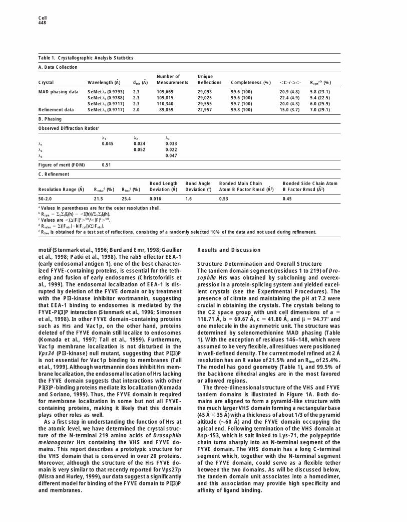
Table 1. Crystallographic Analysis Statistics
A. Data Collection
Number of UniqueCrystal Wavelength (A) dmin (A) Measurements Reflections Completeness (%) ,I./,s. Rsym
a,b (%)
MAD phasing data SeMet l1 (0.9793) 2.3 109,669 29,093 99.6 (100) 20.9 (4.8) 5.8 (23.1)SeMet l2 (0.9788) 2.3 109,815 29,025 99.6 (100) 22.4 (4.9) 5.4 (22.5)SeMet l3 (0.9717) 2.3 110,340 29,555 99.7 (100) 20.0 (4.3) 6.0 (25.9)
Refinement data SeMet l3 (0.9717) 2.0 89,859 22,957 99.8 (100) 15.0 (3.7) 7.0 (29.1)
B. Phasing
Observed Diffraction Ratiosc
l1 l2 l3
l1 0.045 0.024 0.033l2 0.052 0.022l3 0.047
Figure of merit (FOM) 0.51
C. Refinement
Bond Length Bond Angle Bonded Main Chain Bonded Side Chain AtomResolution Range (A) Rvalue
d (%) Rfreee (%) Deviation (A) Deviation (8) Atom B Factor Rmsd (A2) B Factor Rmsd (A2)
50-2.0 21.5 25.4 0.016 1.6 0.53 0.45
a Values in parentheses are for the outer resolution shell.b Rsym 5 ShSi|II(h) 2 ,I(h)|/ShSiII(h).c Values are ,(D|F|)2.1/2/,|F|2.1/2.d Rvalue 5 S(|Fobs|2k|Fcal|)/S|Fobs|.e Rfree is obtained for a test set of reflections, consisting of a randomly selected 10% of the data and not used during refinement.
motif (Stenmark et al., 1996; Burd and Emr, 1998; Gaullier Results and Discussionet al., 1998; Patki et al., 1998). The rab5 effector EEA-1(early endosomal antigen 1), one of the best character- Structure Determination and Overall Structureized FYVE-containing proteins, is essential for the teth- The tandem domain segment (residues 1 to 219) of Dro-ering and fusion of early endosomes (Christoforidis et sophila Hrs was obtained by subcloning and overex-al., 1999). The endosomal localization of EEA-1 is dis- pression in a protein-splicing system and yielded excel-rupted by deletion of the FYVE domain or by treatment lent crystals (see the Experimental Procedures). Thewith the PI3-kinase inhibitor wortmannin, suggesting presence of citrate and maintaining the pH at 7.2 werethat EEA-1 binding to endosomes is mediated by the crucial in obtaining the crystals. The crystals belong toFYVE-PI(3)P interaction (Stenmark et al., 1996; Simonsen the C2 space group with unit cell dimensions of a 5
116.71 A, b 5 69.67 A, c 5 41.80 A, and b 5 94.778 andet al., 1998). In other FYVE domain–containing proteinssuch as Hrs and Vac1p, on the other hand, proteins one molecule in the asymmetric unit. The structure was
determined by selenomethionine MAD phasing (Tabledeleted of the FYVE domain still localize to endosomes(Komada et al., 1997; Tall et al., 1999). Furthermore, 1). With the exception of residues 146–148, which were
assumed to be very flexible, all residues were positionedVac1p membrane localization is not disturbed in theVps34 (PI3-kinase) null mutant, suggesting that PI(3)P in well-defined density. The current model refined at 2 A
resolution has an R value of 21.5% and an Rfree of 25.4%.is not essential for Vac1p binding to membranes (Tallet al., 1999). Although wortmannin does inhibit Hrs mem- The model has good geometry (Table 1), and 99.5% of
the backbone dihedral angles are in the most favoredbrane localization, the endosomal location of Hrs lackingthe FYVE domain suggests that interactions with other or allowed regions.
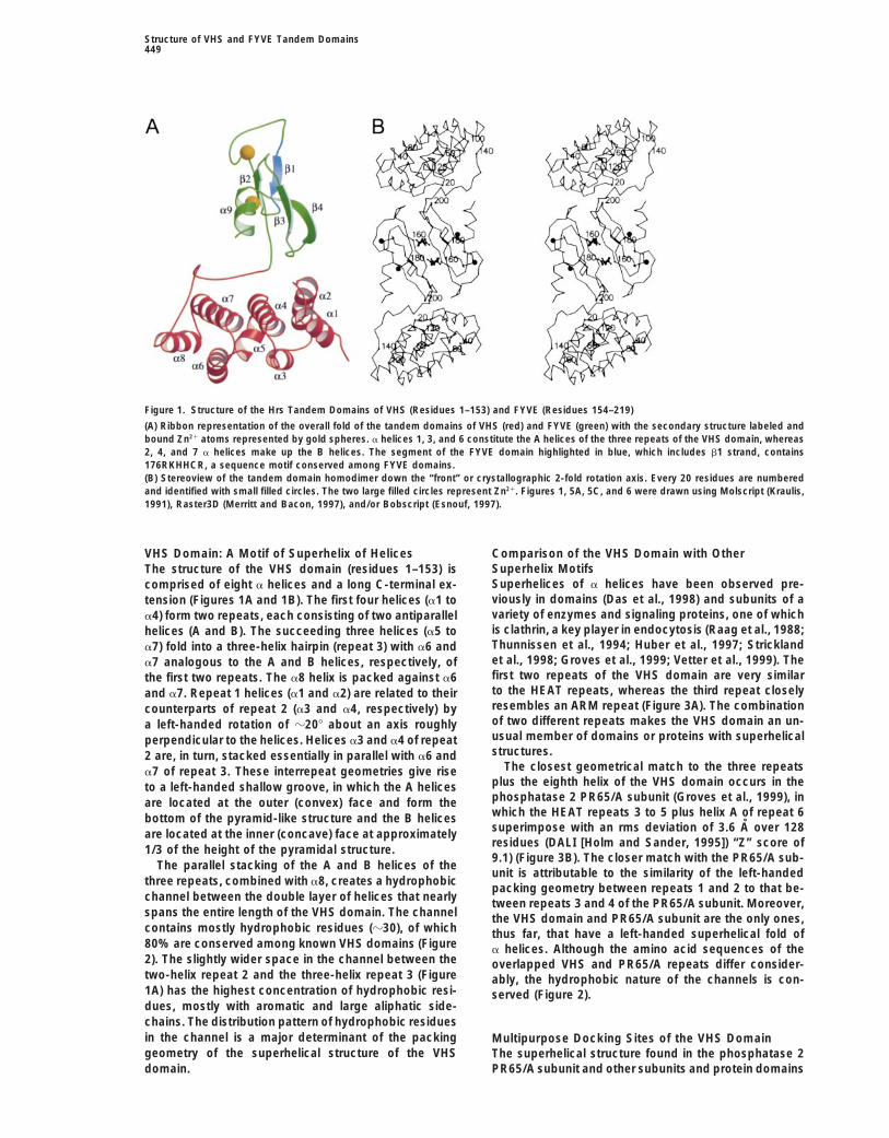
The three-dimensional structure of the VHS and FYVEPI(3)P-binding proteins mediate its localization (Komadaand Soriano, 1999). Thus, the FYVE domain is required tandem domains is illustrated in Figure 1A. Both do-
mains are aligned to form a pyramid-like structure withfor membrane localization in some but not all FYVE-containing proteins, making it likely that this domain the much larger VHS domain forming a rectangular base
(45 A 3 35 A) with a thickness of about 1/3 of the pyramidplays other roles as well.As a first step in understanding the function of Hrs at altitude (z60 A) and the FYVE domain occupying the
apical end. Following termination of the VHS domain atthe atomic level, we have determined the crystal struc-ture of the N-terminal 219 amino acids of Drosophila Asp-153, which is salt linked to Lys-71, the polypeptide
chain turns sharply into an N-terminal segment of themelanogaster Hrs containing the VHS and FYVE do-mains. This report describes a prototypic structure for FYVE domain. The VHS domain has a long C-terminal
segment which, together with the N-terminal segmentthe VHS domain that is conserved in over 20 proteins.Moreover, although the structure of the Hrs FYVE do- of the FYVE domain, could serve as a flexible tether
between the two domains. As will be discussed below,main is very similar to that recently reported for Vps27p(Misra and Hurley, 1999), our data suggest a significantly the tandem domain unit associates into a homodimer,
and this association may provide high specificity anddifferent model for binding of the FYVE domain to PI(3)Pand membranes. affinity of ligand binding.
Structure of VHS and FYVE Tandem Domains449
Figure 1. Structure of the Hrs Tandem Domains of VHS (Residues 1–153) and FYVE (Residues 154–219)
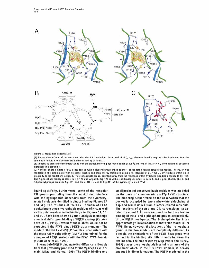
(A) Ribbon representation of the overall fold of the tandem domains of VHS (red) and FYVE (green) with the secondary structure labeled andbound Zn21 atoms represented by gold spheres. a helices 1, 3, and 6 constitute the A helices of the three repeats of the VHS domain, whereas2, 4, and 7 a helices make up the B helices. The segment of the FYVE domain highlighted in blue, which includes b1 strand, contains176RKHHCR, a sequence motif conserved among FYVE domains.(B) Stereoview of the tandem domain homodimer down the “front” or crystallographic 2-fold rotation axis. Every 20 residues are numberedand identified with small filled circles. The two large filled circles represent Zn21. Figures 1, 5A, 5C, and 6 were drawn using Molscript (Kraulis,1991), Raster3D (Merritt and Bacon, 1997), and/or Bobscript (Esnouf, 1997).
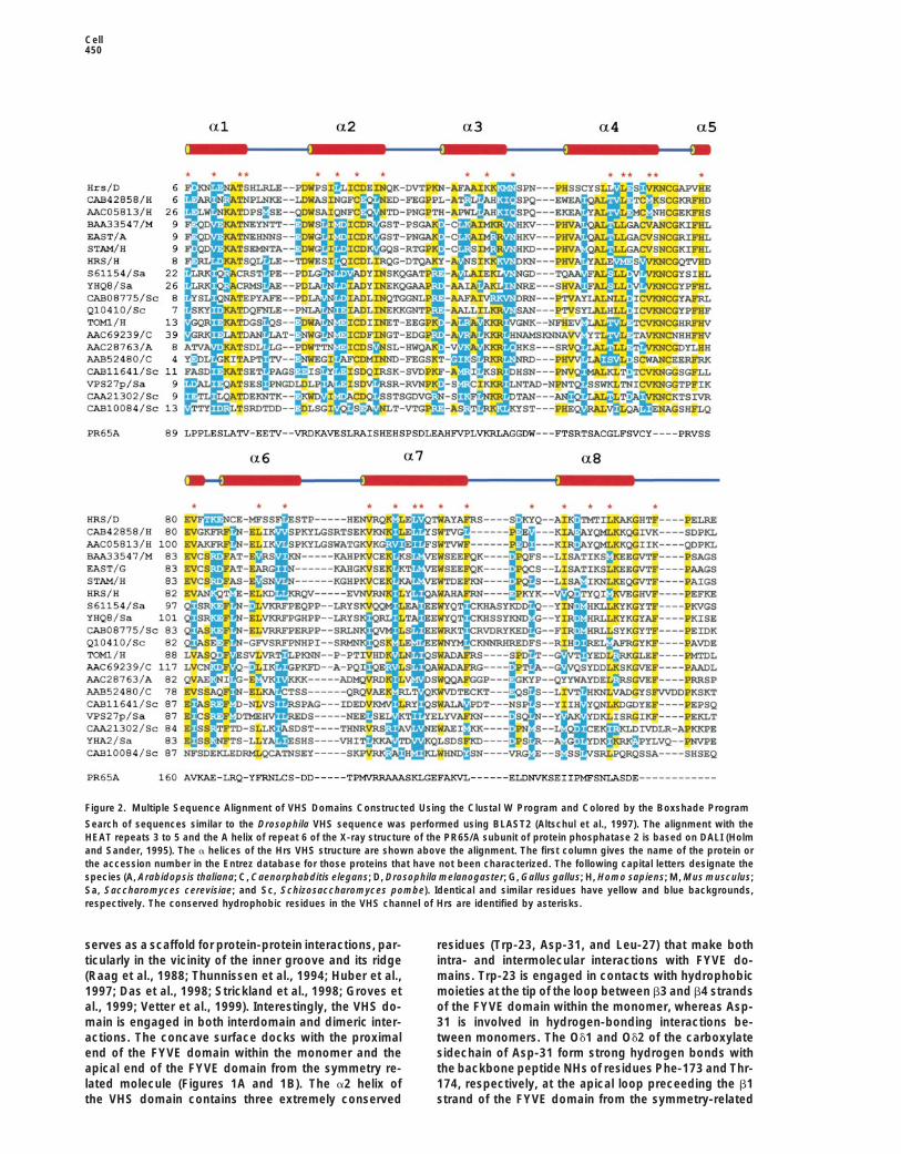
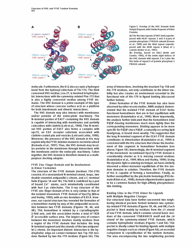
VHS Domain: A Motif of Superhelix of Helices Comparison of the VHS Domain with OtherSuperhelix MotifsThe structure of the VHS domain (residues 1–153) isSuperhelices of a helices have been observed pre-comprised of eight a helices and a long C-terminal ex-viously in domains (Das et al., 1998) and subunits of atension (Figures 1A and 1B). The first four helices (a1 tovariety of enzymes and signaling proteins, one of whicha4) form two repeats, each consisting of two antiparallelis clathrin, a key player in endocytosis (Raag et al., 1988;helices (A and B). The succeeding three helices (a5 toThunnissen et al., 1994; Huber et al., 1997; Stricklanda7) fold into a three-helix hairpin (repeat 3) with a6 andet al., 1998; Groves et al., 1999; Vetter et al., 1999). Thea7 analogous to the A and B helices, respectively, offirst two repeats of the VHS domain are very similarthe first two repeats. The a8 helix is packed against a6to the HEAT repeats, whereas the third repeat closelyand a7. Repeat 1 helices (a1 and a2) are related to theirresembles an ARM repeat (Figure 3A). The combinationcounterparts of repeat 2 (a3 and a4, respectively) byof two different repeats makes the VHS domain an un-a left-handed rotation of z208 about an axis roughlyusual member of domains or proteins with superhelicalperpendicular to the helices. Helices a3 and a4 of repeatstructures.2 are, in turn, stacked essentially in parallel with a6 and
The closest geometrical match to the three repeatsa7 of repeat 3. These interrepeat geometries give riseplus the eighth helix of the VHS domain occurs in theto a left-handed shallow groove, in which the A helicesphosphatase 2 PR65/A subunit (Groves et al., 1999), inare located at the outer (convex) face and form thewhich the HEAT repeats 3 to 5 plus helix A of repeat 6bottom of the pyramid-like structure and the B helicessuperimpose with an rms deviation of 3.6 A over 128are located at the inner (concave) face at approximatelyresidues (DALI [Holm and Sander, 1995]) “Z” score of1/3 of the height of the pyramidal structure.9.1) (Figure 3B). The closer match with the PR65/A sub-
The parallel stacking of the A and B helices of theunit is attributable to the similarity of the left-handed
three repeats, combined with a8, creates a hydrophobicpacking geometry between repeats 1 and 2 to that be-
channel between the double layer of helices that nearly tween repeats 3 and 4 of the PR65/A subunit. Moreover,spans the entire length of the VHS domain. The channel the VHS domain and PR65/A subunit are the only ones,contains mostly hydrophobic residues (z30), of which thus far, that have a left-handed superhelical fold of80% are conserved among known VHS domains (Figure a helices. Although the amino acid sequences of the2). The slightly wider space in the channel between the overlapped VHS and PR65/A repeats differ consider-two-helix repeat 2 and the three-helix repeat 3 (Figure ably, the hydrophobic nature of the channels is con-1A) has the highest concentration of hydrophobic resi- served (Figure 2).dues, mostly with aromatic and large aliphatic side-chains. The distribution pattern of hydrophobic residuesin the channel is a major determinant of the packing Multipurpose Docking Sites of the VHS Domaingeometry of the superhelical structure of the VHS The superhelical structure found in the phosphatase 2
PR65/A subunit and other subunits and protein domainsdomain.
Cell450
Figure 2. Multiple Sequence Alignment of VHS Domains Constructed Using the Clustal W Program and Colored by the Boxshade Program
Search of sequences similar to the Drosophila VHS sequence was performed using BLAST2 (Altschul et al., 1997). The alignment with theHEAT repeats 3 to 5 and the A helix of repeat 6 of the X-ray structure of the PR65/A subunit of protein phosphatase 2 is based on DALI (Holmand Sander, 1995). The a helices of the Hrs VHS structure are shown above the alignment. The first column gives the name of the protein orthe accession number in the Entrez database for those proteins that have not been characterized. The following capital letters designate thespecies (A, Arabidopsis thaliana; C, Caenorphabditis elegans; D, Drosophila melanogaster; G, Gallus gallus; H, Homo sapiens; M, Mus musculus;Sa, Saccharomyces cerevisiae; and Sc, Schizosaccharomyces pombe). Identical and similar residues have yellow and blue backgrounds,respectively. The conserved hydrophobic residues in the VHS channel of Hrs are identified by asterisks.
serves as a scaffold for protein-protein interactions, par- residues (Trp-23, Asp-31, and Leu-27) that make bothintra- and intermolecular interactions with FYVE do-ticularly in the vicinity of the inner groove and its ridge
(Raag et al., 1988; Thunnissen et al., 1994; Huber et al., mains. Trp-23 is engaged in contacts with hydrophobicmoieties at the tip of the loop between b3 and b4 strands1997; Das et al., 1998; Strickland et al., 1998; Groves et
al., 1999; Vetter et al., 1999). Interestingly, the VHS do- of the FYVE domain within the monomer, whereas Asp-31 is involved in hydrogen-bonding interactions be-main is engaged in both interdomain and dimeric inter-
actions. The concave surface docks with the proximal tween monomers. The Od1 and Od2 of the carboxylatesidechain of Asp-31 form strong hydrogen bonds withend of the FYVE domain within the monomer and the
apical end of the FYVE domain from the symmetry re- the backbone peptide NHs of residues Phe-173 and Thr-174, respectively, at the apical loop preceeding the b1lated molecule (Figures 1A and 1B). The a2 helix of
the VHS domain contains three extremely conserved strand of the FYVE domain from the symmetry-related
Structure of VHS and FYVE Tandem Domains451
Figure 3. Overlap of the VHS Domain HelixHairpin Repeats with Similar Repeats of OtherProteins
(A) The first two repeats of VHS (red) superim-posed with HEAT repeats 3 and 4 (cyan) ofprotein phosphate 2 PR65/A subunit (Groveset al., 1999) and the third repeat superim-posed with the ARM repeat 4 (blue) of b
catenin (Huber et al., 1997).(B) Overlap, based on DALI (Holm andSander, 1995), of the entire eight helices ofthe VHS domain with repeats 3 to 5 plus thefirst helix of repeat 6 of protein phosphate 2PR65/A subunit.
molecule. Furthermore, the Od2 also accepts a hydrogen array of interactions, involving the invariant Trp-158 andbond from the hydroxyl sidechain of Thr-174. The third His-179 residues, not only contributes to the dimer sta-conserved VHS residue, Leu-27, is involved in hydropho- bility but also creates an environment essential to thebic interactions with the symmetry-related Phe-173 that functional role of His-179 in ligand binding (discussedis also a highly conserved residue among FYVE do- further below).mains. The VHS domain is a prime example of this type Dimer formation of the FYVE domain has also beenof structure whose concave surface acts as a platform observed by other recent studies. NMR analysis demon-for both interdomain and dimeric interactions. strated that the isolated FYVE domain of EEA1 forms
The VHS domain may also interact with membranes functional homodimers that are in fast equilibrium withand/or proteins of the endocytotic machinery. The monomers (Kutateladze et al., 1999). More importantly,N-terminal portion of EAST containing the VHS domain the analysis further indicated that the homodimers bindis capable of interacting with membranes and partially PI(3)P-bearing membranes much more tightly than thecolocalizes with clathrin (Lohi et al., 1998). This N-termi- corresponding monomers. Binding appears to be morenal VHS portion of EAST also forms a complex with specific for PI(3)P since PI(5)P, a naturally occurring lipideps15, an EGF receptor substrate associated with headgroup, is bound more weakly. The suggestion thatclathrin-coated pits and vesicles (Lohi and Lehto, 1998). the long N-terminal segment of the EEA1 FYVE domainMoreover, the presence of the VHS domain in Hrs may contributes to dimerization (Kutateladze et al., 1999) isexplain why the FYVE deletion mutant is not mislocalized consistent with the Hrs structure that shows the involve-(Komada et al., 1997). Thus, the VHS domain may local- ment of this segment in homodimer formation (seeize proteins to the membrane through interactions with above; Figure 1B). Interestingly, the N-terminal segmentthe membrane and/or the endocytic machinery. Taken of the monomeric recombinant Vps27p FYVE domain istogether, the VHS domain is therefore viewed as a multi- considerably shorter as it is missing several residuespurpose docking adapter. (Kutateladze et al., 1999; Misra and Hurley, 1999). Using
the dynamic light scattering technique, we have similarlyFYVE Zinc Finger Domain and Its Involvement observed a dimer-monomer equilibrium of the Hrs tan-in Dimer Formation dem domains in solution. Therefore, the FYVE domainThe structure of the FYVE domain (residues 154–219)
of Hrs is capable of forming a homodimer. Finally, asconsists of a nonstandard N-terminal strand, loops, two
further exemplified by the pleckstrin homology (PH) do-double-stranded antiparallel b sheets, and a C-terminal
main (Klein et al., 1998), domain oligomerization appearshelix (Figures 1A and 1B). The fold is stabilized by a pairto be a common feature for high-affinity phosphoinosi-of bound Zn21 atoms, each tetrahedrally coordinatedtide binding.with four Cys sidechains. The X-ray structure of the
FYVE zinc finger domain of Hrs is very similar to that ofBinding Sites in the FYVE Dimer for Ligandsthe isolated monomeric FYVE domain of Vps27p (Misrawith Multiple Negative Chargesand Hurley, 1999), with an rms deviation of 0.93 A. How-Our structural data have further uncovered two neigh-ever, our crystal structure has revealed the formation ofboring identical pockets formed between two symme-a homodimer mainly by way of the antiparallel associa-try-related FYVE domains (Figures 1B, 4A, and 4B). Eachtion between two FYVE domains (Figures 1B, 4A, andpocket is lined mainly by residues from the b1 strand4B). This homodimer is related by a crystallographicof one FYVE domain, which contains several basic resi-2-fold axis, and this association buries a total of 1920dues of the conserved 176RKHHCR motif and the b4A2 accessible surface area. The largest area of contactstrand of the symmetry-related FYVE domain, whichbetween the monomers involves mainly residues fromcontains hydrophobic residues (Figures 1 and 5A). Thisa region of the FYVE domain that includes the longpocket is ideally suited for binding ligands with multipleN-terminal segment and the strands of the two antiparal-negative charges such as citrate (Figure 5A), an essentiallel b sheets. An important dimeric interaction is the hy-component in crystallization of the tandem domains.drophobic edge-on contact between two Trp-158 resi-
dues flanked by two His-179 residues (Figure 5A). This The area encompassing the two neighboring pockets
Cell452
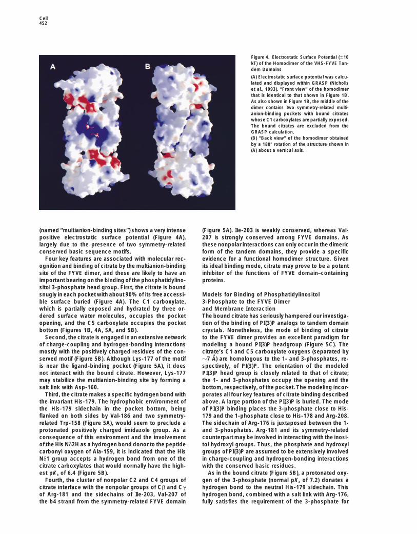
Figure 4. Electrostatic Surface Potential (610kT) of the Homodimer of the VHS-FYVE Tan-dem Domains
(A) Electrostatic surface potential was calcu-lated and displayed within GRASP (Nichollset al., 1993). “Front view” of the homodimerthat is identical to that shown in Figure 1B.As also shown in Figure 1B, the middle of thedimer contains two symmetry-related multi-anion-binding pockets with bound citrateswhose C1 carboxylates are partially exposed.The bound citrates are excluded from theGRASP calculation.(B) “Back view” of the homodimer obtainedby a 1808 rotation of the structure shown in(A) about a vertical axis.
(named “multianion-binding sites”) shows a very intense (Figure 5A). Ile-203 is weakly conserved, whereas Val-207 is strongly conserved among FYVE domains. Aspositive electrostatic surface potential (Figure 4A),
largely due to the presence of two symmetry-related these nonpolar interactions can only occur in the dimericform of the tandem domains, they provide a specificconserved basic sequence motifs.
Four key features are associated with molecular rec- evidence for a functional homodimer structure. Givenits ideal binding mode, citrate may prove to be a potentognition and binding of citrate by the multianion-binding
site of the FYVE dimer, and these are likely to have an inhibitor of the functions of FYVE domain–containingproteins.important bearing on the binding of the phosphatidylino-
sitol 3-phosphate head group. First, the citrate is boundsnugly in each pocket with about 90% of its free accessi- Models for Binding of Phosphatidylinositol
3-Phosphate to the FYVE Dimerble surface buried (Figure 4A). The C1 carboxylate,which is partially exposed and hydrated by three or- and Membrane Interaction
The bound citrate has seriously hampered our investiga-dered surface water molecules, occupies the pocketopening, and the C5 carboxylate occupies the pocket tion of the binding of PI(3)P analogs to tandem domain
crystals. Nonetheless, the mode of binding of citratebottom (Figures 1B, 4A, 5A, and 5B).Second, the citrate is engaged in an extensive network to the FYVE dimer provides an excellent paradigm for
modeling a bound PI(3)P headgroup (Figure 5C). Theof charge-coupling and hydrogen-bonding interactionsmostly with the positively charged residues of the con- citrate’s C1 and C5 carboxylate oxygens (separated by
z7 A) are homologous to the 1- and 3-phosphates, re-served motif (Figure 5B). Although Lys-177 of the motifis near the ligand-binding pocket (Figure 5A), it does spectively, of PI(3)P. The orientation of the modeled
PI(3)P head group is closely related to that of citrate;not interact with the bound citrate. However, Lys-177may stabilize the multianion-binding site by forming a the 1- and 3-phosphates occupy the opening and the
bottom, respectively, of the pocket. The modeling incor-salt link with Asp-160.Third, the citrate makes a specific hydrogen bond with porates all four key features of citrate binding described
above. A large portion of the PI(3)P is buried. The modethe invariant His-179. The hydrophobic environment ofthe His-179 sidechain in the pocket bottom, being of PI(3)P binding places the 3-phosphate close to His-
179 and the 1-phosphate close to His-178 and Arg-208.flanked on both sides by Val-186 and two symmetry-related Trp-158 (Figure 5A), would seem to preclude a The sidechain of Arg-176 is juxtaposed between the 1-
and 3-phosphates. Arg-181 and its symmetry-relatedprotonated positively charged imidazole group. As aconsequence of this environment and the involvement counterpart may be involved in interacting with the inosi-
tol hydroxyl groups. Thus, the phosphate and hydroxylof the His Nd2H as a hydrogen bond donor to the peptidecarbonyl oxygen of Ala-159, it is indicated that the His groups of PI(3)P are assumed to be extensively involved
in charge-coupling and hydrogen-bonding interactionsNd1 group accepts a hydrogen bond from one of thecitrate carboxylates that would normally have the high- with the conserved basic residues.
As in the bound citrate (Figure 5B), a protonated oxy-est pKa of 6.4 (Figure 5B).Fourth, the cluster of nonpolar C2 and C4 groups of gen of the 3-phosphate (normal pKa of 7.2) donates a
hydrogen bond to the neutral His-179 sidechain. Thiscitrate interface with the nonpolar groups of Cb and Cgof Arg-181 and the sidechains of Ile-203, Val-207 of hydrogen bond, combined with a salt link with Arg-176,
fully satisfies the requirement of the 3-phosphate forthe b4 strand from the symmetry-related FYVE domain
Structure of VHS and FYVE Tandem Domains453
Figure 5. Multianion-Binding Site
(A) Stereo view of one of the two sites with the 2 A resolution citrate omit (Fo-Fc), acalc electron density map at 13s. Residues from thesymmetry-related FYVE domain are distinguished by asterisks.(B) Schematic diagram of the interactions with the citrate, involving hydrogen bonds (#3.2 A) and/or salt links (,4 A), along with their observeddistances in angstroms.(C) A model of the binding of PI(3)P headgroup with a glycerol group linked to the 1-phosphate oriented toward the reader. The PI(3)P wasmodeled in the binding site with no steric clashes and then energy minimized using CNS (Brunger et al., 1998). Only residues within closeproximity to the model are included. The 3-phosphate group, oriented away from the reader, is within hydrogen-bonding distance to His-179.The 1-phosphate moiety is close to His-178 and Arg-208. Arg-176 is within salt-linking distance to both 1- and 3 phosphates. The 2- and3-hydroxyl groups are near Arg-181, and the 4-OH is close to Arg-181 of the symmetry-related FYVE.
ligand specificity. Furthermore, some of the nonpolar small pocket of conserved basic residues was modeledon the basis of a monomeric Vps27p FYVE structure.CH groups protruding from the inositol ring interface
with the hydrophobic sidechains from the symmetry- The modeling further relied on the observation that thepocket is occupied by two carboxylate sidechains ofrelated molecule identified in citrate binding (Figures 5A
and 5C). The residues of the FYVE domain of EEA1 Asp and Glu residues from a lattice-related molecule.The locations of the Asp and Glu carboxylates, sepa-equivalent to these hydrophobic residues of Hrs, as well
as the polar residues in the binding site (Figures 5A, 5B, rated by about 9 A, were assumed to be the sites forbinding of the 3- and 1-phosphate groups, respectively,and 5C), have been shown by NMR analysis to undergo
chemical shifts upon binding of PI(3)P analogs (Kutatel- of the PI(3)P headgroup. The 3-phosphate lies in anapproximately similar location as that of the model in Hrsadze et al., 1999). Several of these shifts would not be
expected if the FYVE binds PI(3)P as a monomer. The FYVE dimer. However, the locations of the 1-phosphategroup in the two models are completely different. Asmodel of the Hrs FYVE-PI(3)P complex is consistent with
the reasonably tight affinity (mM Kd) determined for the a result, the orientations of the PI(3)P headgroup withrespect to the binding site differ greatly between thecomplex of PI(3)P analogs with the EEA1 FYVE domain
(Kutateladze et al., 1999). two models. The model with Vps27p (Misra and Hurley,1999) places the phosphatidylinositol in an area of theThe model of PI(3)P binding to Hrs differs considerably
from that previously proposed for the Vps27p FYVE do- monomer which, in the Hrs FYVE domain, is heavilyengaged in dimer formation. The PI(3)P modeled in themain (Misra and Hurley, 1999). The PI(3)P binding to a
Cell454
Figure 6. Model of the Interaction of the VHSand FYVE Tandem Domains with the Mem-brane
The flat “front” surface of the tandem do-mains shown in Figures 1B and 4A interfaceswith the membrane. The region of the VHSdomain protruding into the membrane inter-face contains predominantly basic residues(Arg 3, Lys-8, and Arg-18) and hydrophobicresidues (Phe-2, Leu-17, and Leu-19) thatcould interact nonspecifically with the bilayer.
Vps27p lies sideways or parallel with respect to the b1 to a more solvent exposed ligand [e.g., z50% exposedaccessible surface area of citrate or the modeled PI(3)P)]strand with its 1-phosphate group directed toward the
apical end of the FYVE domain and engaged in an inter- and the elimination of the hydrophobic interactions de-scribed above, resulting in weaker ligand affinities.action with Lys-189 (the equivalent of Lys-177 of Hrs).
In contrast, in our model with the Hrs homodimer, the Therefore, the functional form of the FYVE domain favorsa homodimer.PI(3)P is normal to the b1 strand, its 1-phosphate pro-
truding from the opening of the pocket and far from Lys-177 (Figure 5C). Moreover, of the four key features of Conclusion
In conclusion, the structure reported here reveals theligand recognition and binding to the Hrs dimer de-scribed above, only some of the electrostatic interac- unique superhelical conformation of the VHS domain
and its relationship with the FYVE domain. It furthertions may have semblance with the proposed modelof PI(3)P binding to the Vps27p FYVE monomer. The shows the formation of a homodimer mainly through
the antiparallel association of FYVE domains. The VHSmonomeric binding mode, with the ligand also signifi-cantly exposed to the bulk solvent, may not be sufficient domain’s presence in proteins implicated in membrane
trafficking, superhelical folding character, and involve-for high ligand specificity and affinity.The difference between the two proposed PI(3)P- ment in interdomain and dimeric interactions strongly
suggest that the VHS domain functions as a multipur-binding modes leads to two completely different modelsfor membrane-targeting of the FYVE domain. In our pose docking adapter. Furthermore, the finding of the
dimeric form of the FYVE domain and its complex withmodel shown in Figure 6, the extended tandem domaindimeric structure lies in a horizontal direction with re- citrate provide an excellent paradigm for PI(3)P recogni-
tion. Based on these results, we have proposed a modelspect to the membrane surface. In addition to the asso-ciation between FYVE and the phosphoinositol 3-phos- for PI(3)P binding and membrane interaction of the tan-
dem domains. Finally, the structure of the tandem do-phate headgroup of the membranes (Figures 5C and 6),the model would place the VHS domain in a position to mains reported here serves as a framework for further
studies of Hrs and other related proteins, especially theinteract with membranes (Figure 6), a feature consistentwith the demonstration that the VHS domain interacts distinct role of each domain in membrane trafficking
and signal transduction events.with the membrane (discussed above). In contrast, inthe model based on the “sideways” binding of the PI(3)P
Experimental Proceduresproposed by Misra and Hurley (1999), the FYVE mono-mer lies in a vertical orientation with respect to the mem-
Expression, Purification, and Crystallizationbrane with the tip of the apical end inserted in the mem-The DNA sequences corresponding to the tandem domains (resi-
brane’s interface. This orientation would place the VHS dues 1–219) of Drosophila Hrs were amplified by PCR and cloneddomain of Vps27p, assuming similar pyramidal arrange- into the pTYB1 expression vector of the IMPACT T7 System (New
England BioLabs) as the N-terminal segment fused to the intein andment with the FYVE domain as seen in the Hrs tandemchitin-binding domain unit. To facilitate protein splicing, a glycinedomain structure, very far from the membrane surface.residue was introduced between the Hrs protein and the intein.Finally, in contrast with the Vps27p model, the findingProteins were expressed in Escherichia coli ER2566 cells at roomthat the apical end of the Hrs FYVE domain is buriedtemperature, purified to near homogeneity according to the proce-
and extensively involved in polar and nonpolar interac- dure provided with the IMPACT T7 System, and dialyzed against 50tions with the symmetry-related VHS domain (discussed mM NaCl and 100 mM Tris (pH 8.0), at 48C. To remove minor impuri-
ties, the protein (5 mg/ml) was further purified on an HQ column,above; Figure 1B) would completely preclude the inter-and the stock protein solution was concentrated to 10 mg/ml in 1action of this end with the membrane’s interface.mM DTT and 50 mM citrate (pH 5.5). The protein was crystallizedSince the citrate or the modeled PI(3)P interacts pre-at 48C using the vapor diffusion method with the drop consisting ofdominantly with the basic residues of the conserveda 1:1 mixture of the stock protein solution and the reservoir solution
motif, ligand binding solely to one FYVE domain is not of 15% PEG 10000, 5 mM DTT, and 100 mM HEPES (pH 7.4). Priorprecluded. However, binding to the monomer would, as to data collection, the crystal was flash-frozen in liquid nitrogen with
25% glycerol in the crystallization solution.in the case also of the model with the Vps27p FYVE, lead
Structure of VHS and FYVE Tandem Domains455
Structure Determination H., and Aasland, R. (1998). FYVE fingers bind PtdIns(3)P. Nature394, 432–433.The structure was determined by multiwavelength anomalous dis-
persion (MAD). A 3-wavelength data set was collected from a crystal Groves, M.R., Hanlon, N., Turowski, P., Hemmings, B.A., and Bar-of selenomethionine-substituted protein on beamline X4A at NSLS ford, D. (1999). The structure of the protein phosphatase 2A PR65/Aof the Brookhaven National Laboratory and processed and merged subunit reveals the conformation of its 15 tandemly repeated HEATwith DENZO and SCALEPACK (Otwinowski and Minor, 1997), re- motifs. Cell 96, 99–110.spectively. The positions of four of the six SeMet sites and 2 Zn21
Holm, L., and Sander, C. (1995). Dali: a network tool for proteinwere determined, heavy atom parameters refined, and MAD phases structure comparison. Trends Biochem. Sci. 20, 478–480.calculated at 2.4 A resolution using the suite of programs in SOLVE
Huber, A.H., Nelson, W.J., and Weis, W.I. (1997). Three-dimensional(http://www.hwi.buffalo.edu/SnB). The calculated electron densitystructure of the armadillo repeat region of b-catenin. Cell 90,map, which was solvent flattened in DM (Cowton, 1994), was used871–882.to build an initial model of 180 of the total 220 residues by meansJones, T.A., Zou, J.Y., Cowan, S.W., and Kjeldgaard, M. (1991).of O (Jones et al., 1991). The model was refined in CNS (BrungerImproved methods for building protein models in electron densityet al., 1998) interspersed with model building and fitting of watermaps and the location of errors in these models. Acta Crystallogr.molecules using CHAIN (Sack and Quiocho, 1997). Although theA 47, 110–119.density of the bound citrate was well defined in initial maps, its
model was fitted only in the final rounds of refinement. The final Klein, D.E., Lee, A., Frank, D.W., Marks, M.S., and Lemmon, M.A.model was refined against the 2 A resolution data (92.4% complete (1998). The pleckstrin homology domains of dynamin isoforms re-with 2s cutoff) from the higher-energy remote l3 wavelength (Table 1). quire oligomerization for high affinity phosphoinositide binding. J.
Biol. Chem. 42, 27725–27733.
Komada, M., and Kitamura, N. (1995). Growth factor-induced tyro-Acknowledgmentssine phosphorylation of Hrs, a novel 115-kilodalton protein with astructurally conserved putative zinc finger domain. Mol. Cell Biol.We thank W. Meador and G. Hu for technical and software assis-15, 6213–6221.tance; C. Ogata and R. Abramowitz for assistance with data collec-
tion at the Brookhaven National Laboratory facility; and T. Terwilliger Komada, M., and Soriano, P. (1999). Hrs, a FYVE finger proteinfor SOLVE. H. B. and F. A. Q. are Investigators in the Howard Hughes localized to early endosomes, is implicated in vesicular traffic andMedical Institute. required for ventral folding morphogenesis. Genes Dev. 13, 1475–
1485.
Komada, M., Masaki, R., Yamamoto, A., and Kitamura, N. (1997).Received December 1, 1999; revised January 7, 2000.Hrs, a tyrosine kinase substrate with a conserved double zinc fingerdomain, is localized to the cytoplasmic surface of early endosomes.
References J. Biol. Chem. 272, 20538–20544.
Kraulis, P.J. (1991). MOLSCRIPT: a program to produce both de-Altschul, S.F., Madden, T.L., Schaffer, A.A., Zhang, J., Zhang, Z.,tailed and schematic plots of protein structures. J. Appl. Cryst. 24,Miller, W., and Lipman, D.J. (1997). Gapped BLAST and PSI-BLAST:946–950.a new generation of protein database search programs. NucleicKutateladze, T.G., Ogburn, K.D., Watson, W.T., de Beer, T., Emr,Acids Res. 25, 3389–3402.S.D., Burd, C.G., and Overduin, M. (1999). PhosphatidylinositolAsao, H., Sasaki, Y., Arita, T., Tanaka, N., Endo, K., Kasai, H., Take-3-phosphate recognition by the FYVE domain. Mol. Cell 3, 805–811.shita, T., Endo, Y., Fujita, T., and Sugamura, K. (1997). Hrs is associ-Lohi, O., and Lehto, V.P. (1998). VHS domain marks a group ofated with STAM, a signal-transducing adaptor molecule. Its sup-proteins involved in endocytosis and vesicular trafficking. FEBSpressive effect on cytokine-induced cell growth. J. Biol. Chem. 272,Lett. 440, 255–257.32785–32791.Lohi, O., Poussu, A., Merilainen, J., Kellokumpu, S., Wasenius, V.M.,Bean, A.J., Seifert, R., Chen, Y.A., Sacks, R., and Scheller, R.H.and Lehto, V.P. (1998). EAST, an epidermal growth factor receptor-(1997). Hrs-2 is an ATPase implicated in calcium-regulated secre-and eps15-associated protein with Src homology 3 and tyrosine-tion. Nature 385, 826–829.based activation motif domains. J. Biol. Chem. 273, 21408–21415.Brunger, A.T., Adams, P.D., Clore, G.M., DeLano, W.L., Gros, P.,Merritt, E.A., and Bacon, D.J. (1997). Raster3D version 2.0: a programGrosse-Kunstleve, R.W., Jiang, J.S., Kuszewski, J., Nilges, M.,for photorealistic molecular graphics. Methods Enzymol. 277,Pannu, N.S., et al. (1998). Crystallography and NMR system: a new505–524.software suite for macromolecular structure determination. Acta
Crystallogr. D 54, 905–921. Misra, S., and Hurley, J.H. (1999). Crystal structure of a phosphatidyl-inositol 3-phosphate-specific membrane-targeting motif, the FYVEBurd, C.G., and Emr, S.D. (1998). Phosphatidylinositol(3)-phosphatedomain of Vps27p. Cell 97, 657–666.signaling mediated by specific binding to RING FYVE domains. Mol.
Cell 2, 157–162. Nicholls, A., Bharadwaj, R., and Honig, B. (1993). GRASP: graphicalrepresentation and analysis of surface properties. Biophys. J. 64,Carbone, R., Fre, S., Iannolo, G., Belleudi, F., Mancini, P., Pelicci,166–170.P.G., Torrisi, M.R., and Di Fiore, P.P. (1997). eps15 and eps15R are
essential components of the endocytic pathway. Cancer Res. 57, Otwinowski, Z., and Minor, W. (1997). Processing of X-ray diffractiondata collected in oscillation mode. Methods Enzymol. 276, 307–326.5498–5504.
Christoforidis, S., McBride, H.M., Burgoyne, R.D., and Zerial, M. Patki, V., Lawe, D.C., Corvera, S., Virbasius, J.V., and Chawla, A.(1998). A functional PtdIns(3)P-binding motif. Nature 394, 433–434.(1999). The Rab5 effector EEA1 is a core component of endosome
docking. Nature 397, 621–625. Piper, R.C., Cooper, A.A., Yang, H., and Stevens, T.H. (1995). VPS27controls vacuolar and endocytic traffic through a prevacuolar com-Cowton, K. (1994). “dm”: an automated procedure for phase im-
provements by density modifications. Joint CCP4 and ESF-EACBM partment in Saccharomyces cerevisiae. J. Cell. Biol. 131, 603–617.Newsletter. Protein Cryst. 31, 34–38. Raag, R., Appelt, K., Xuong, N.H., and Banaszak, L. (1988). Structure
of the lamprey yolk lipid-protein complex lipovitellin- phosvitin atDas, A.K., Cohen, P.T.W., and Barford, D. (1998). The structure ofthe tetratricopeptide repeats of protein phosphatase 5: implications 2.8 A resolution. J. Mol. Biol. 200, 553–569.for TPR-mediated protein-protein interactions. EMBO J. 17, 1192– Sack, J.S., and Quiocho, F.A. (1997). CHAIN-A crystallographic mod-1199. eling program. Methods Enzymol. 277, 158–173.Esnouf, R.M. (1997). An extensively modified version of MolScript Simonsen, A., Lippe, R., Christoforidis, S., Gaullier, J.M., Brech, A.,that includes greatly enhanced coloring capabilities. J. Mol. Graph. Callaghan, J., Toh, B.H., Murphy, C., Zerial, M., and Stenmark, H.Model. 15, 132–134. (1998). EEA1 links PI(3)K function to Rab5 regulation of endosome
fusion. Nature 394, 494–498.Gaullier, J.M., Simonsen, A., D’Arrigo, A., Bremnes, B., Stenmark,
Cell456
Stenmark, H., Aasland, R., Toh, B.H., and D’Arrigo, A. (1996). Endo-somal localization of the autoantigen EEA1 is mediated by a zinc-binding FYVE finger. J. Biol. Chem. 271, 24048–24054.
Strickland, C.L., Windsor, W.T., Syto, R., Wang, L., Bond, R., Wu,Z., Schwartz, J., Le, H.V., Beese, L.S., and Weber, P.C. (1998). Crystalstructure of farnesyl protein transferase complexed with a CaaXpeptide and farnesyl diphosphate analogue. Biochemistry 37,16601–16611.
Takeshita, T., Arita, T., Asao, H., Tanaka, N., Higuchi, M., Kuroda, H.,Kaneko, K., Munakata, H., Endo, Y., Fujita, T., et al. (1996). Cloningof a novel signal-transducing adaptor molecule containing an SH3domain and ITAM. Biochem. Biophys. Res. Commun. 225, 1035–1039.
Takeshita, T., Arita, T., Higuchi, M., Asao, H., Endo, K., Kuroda, H.,Tanaka, N., Murata, K., Ishii, N., and Sugamura, K. (1997). STAM,signal transducing adaptor molecule, is associated with Janus ki-nases and involved in signaling for cell growth and c-myc induction.Immunity 6, 449–457.
Tall, G.G., Hama, H., DeWald, D.B., and Horazdovsky, B.F. (1999).The phosphatidylinositol 3-phosphate binding protein Vac1p inter-acts with a Rab GTPase and a Sec1p homologue to facilitate vesicle-mediated vacuolar protein sorting. Mol. Biol. Cell 10, 1873–1889.
Thunnissen, A.M., Dijkstra, A.J., Kalk, K.H., Rozeboom, H.J., Engel,H., Keck, W., and Dijkstra, B.W. (1994). Doughnut-shaped structureof a bacterial muramidase revealed by X-ray crystallography. Nature367, 750–753.
Vetter, I.R., Arndt, A., Kutay, U., Gorlich, D., and Wittinghofer, A.(1999). Structural view of the Ran-Importin b interaction at 2.3 Aresolution. Cell 97, 635–646.
Protein Data Bank ID Code
Atomic coordinates have been deposited with the ID code 1DVP.