Crystal Structure at 1.45 Å Resolution of Alanine Racemase from a Pathogenic Bacterium, Pseudomonas...
10
Crystal Structure at 1.45 Å Resolution of Alanine Racemase from a Pathogenic Bacterium, Pseudomonas aeruginosa, Contains Both Internal and External Aldimine Forms ²,‡ Pierre LeMagueres, § Hookang Im, § Anna Dvorak, § Ulrich Strych, § Michael Benedik, § and Kurt L. Krause* ,§,| Department of Biology and Biochemistry, UniVersity of Houston, Houston, Texas 77204, and Section of Infectious Diseases, Department of Medicine, Baylor College of Medicine, Houston, Texas 77030 ReceiVed July 7, 2003; ReVised Manuscript ReceiVed October 9, 2003 ABSTRACT: The structure of the catabolic alanine racemase, DadX, from the pathogenic bacterium Pseudomonas aeruginosa, reported here at 1.45 Å resolution, is a dimer in which each monomer is comprised of two domains, an eight-stranded R/ barrel containing the PLP cofactor and a second domain primarily composed of -strands. The geometry of each domain is very similar to that of Bacillus stearothermophilus alanine racemase, but the rotation between domains differs by about 15°. This change does not alter the structure of the active site in which almost all residues superimpose well with a low rms difference of 0.86 Å. Unexpectedly, the active site of DadX contains a guest substrate that is located where acetate and propionate have been observed in the Bacillus structures. It is modeled as D-lysine and oriented such that its terminal NZ atom makes a covalent bond with C4′ of PLP. Since the internal aldimine bond between the protein lysine, Lys33, and C4′ of PLP is also unambiguously observed, there appears to be an equilibrium between both internally and externally reacted forms. The PLP cofactor adopts two partially occupied conformational states that resemble previously reported internal and external aldimine complexes. Pseudomonas aeruginosa is an important Gram-negative pathogen that is capable of causing a wide variety of serious infections in humans including pneumonia, septicemia, endocarditis, and osteomyelitis (1). It is one of the top five causes of nosocomial infections, including infections in intensive care units (2). Patients with cystic fibrosis have a high incidence of often life-threatening Pseudomonas infec- tions (3). The tenacious pathogenicity of P. aeruginosa is often coupled with high levels of antibiotic resistance. In fact, hospital isolates of Pseudomonas that are resistant to all commonly used antibiotics have been reported (4, 5). As a result, it would be of significant public health importance to identify new leads for antibiotics directed at this organism. Alanine racemase (EC 5.1.1.1) is an essential enzyme in prokaryotes. It is a pyridoxal 5′-phosphate (PLP)-containing 1 enzyme that catalyzes the interconversion of L-alanine and D-alanine, an essential building block in the biosynthesis of the peptidoglycan layer in cell walls. Pseudomonas contains two different homologous genes for alanine racemase (6). In Salmonella typhimurium, the alr gene is expressed constitutively at low levels to allow the biosynthesis of D-alanine (7) while, in Escherichia coli, the dadX gene is induced when cells are grown on L-alanine (8). Because alanine racemase is absent in virtually all eukaryotes, it has often been proposed as an ideal drug target for the develop- ment of new antibiotics (9, 10). Prior to the current era of structure-aided drug design, alanine racemase was the focus of research showing that many structural analogues of D-alanine inhibit the enzyme (11-14). Unfortunately, these inhibitors all suffered from a lack of specificity, thought to be in part because they acted on other PLP-containing enzymes, including those found in humans (15, 16). One of these compounds, D-cycloserine (DCS), was developed commercially. It acted by inhibiting alanine racemase, as well as the functionally related enzyme, D-ala ligase. DCS is still the only currently marketed antibiotic whose action is based on alanine racemase inhibi- tion. However, its clinical use is limited because of severe side effects, which include central nervous system toxicity (17-19). Since 1997, several studies on the alanine racemase from Bacillus stearothermophilus have elucidated important as- pects of the enzyme’s tertiary structure and essential features of how the enzyme binds substrate and carries out catalysis. Ringe et al. reported the crystal structure of the alanine racemase enzyme Alr from B. stearothermophilus with acetate and propionate (20, 21) and a form with a covalently bound inhibitor, the alanine analogue alanine phosphonate (22). These studies confirmed that this enzyme is a homo- dimer, with each of the monomers containing two domains. The N-terminal domain is an R/ barrel, within which the pyridoxal 5′-phosphate is bound to the protein via an ² This publication was supported, in part, by grants from the National Institutes of Health (AI-46340), the W. M. Keck Foundation, and the Robert A. Welch Foundation. ‡ The coordinates have been deposited in the Protein Data Bank with the entry code 1RCQ. * To whom correspondence should be addressed. Tel: (713) 743- 8370. Fax: (713) 743-8370. E-mail: [email protected]. § University of Houston. | Baylor College of Medicine. 1 Abbreviations: HEPES, N-2-hydroxyethylpiperazine-N′-2-ethane- sulfonic acid; MAD, multiwavelength anomalous dispersion; DCS, D-cycloserine; PLP, pyridoxal 5′-phosphate; rms, root-mean-square. 14752 Biochemistry 2003, 42, 14752-14761 10.1021/bi030165v CCC: $25.00 © 2003 American Chemical Society Published on Web 11/25/2003
Transcript of Crystal Structure at 1.45 Å Resolution of Alanine Racemase from a Pathogenic Bacterium, Pseudomonas...
Crystal Structure at 1.45 Å Resolution of Alanine Racemase from a PathogenicBacterium,Pseudomonas aeruginosa, Contains Both Internal and External Aldimine
Forms†,‡
Pierre LeMagueres,§ Hookang Im,§ Anna Dvorak,§ Ulrich Strych,§ Michael Benedik,§ and Kurt L. Krause*,§,|
Department of Biology and Biochemistry, UniVersity of Houston, Houston, Texas 77204, and Section of Infectious Diseases,Department of Medicine, Baylor College of Medicine, Houston, Texas 77030
ReceiVed July 7, 2003; ReVised Manuscript ReceiVed October 9, 2003
ABSTRACT: The structure of the catabolic alanine racemase, DadX, from the pathogenic bacteriumPseudomonas aeruginosa, reported here at 1.45 Å resolution, is a dimer in which each monomer iscomprised of two domains, an eight-strandedR/â barrel containing the PLP cofactor and a second domainprimarily composed ofâ-strands. The geometry of each domain is very similar to that ofBacillusstearothermophilusalanine racemase, but the rotation between domains differs by about 15°. This changedoes not alter the structure of the active site in which almost all residues superimpose well with a lowrms difference of 0.86 Å. Unexpectedly, the active site of DadX contains a guest substrate that is locatedwhere acetate and propionate have been observed in theBacillusstructures. It is modeled asD-lysine andoriented such that its terminal NZ atom makes a covalent bond with C4′ of PLP. Since the internal aldiminebond between the protein lysine, Lys33, and C4′ of PLP is also unambiguously observed, there appearsto be an equilibrium between both internally and externally reacted forms. The PLP cofactor adopts twopartially occupied conformational states that resemble previously reported internal and external aldiminecomplexes.
Pseudomonas aeruginosais an important Gram-negativepathogen that is capable of causing a wide variety of seriousinfections in humans including pneumonia, septicemia,endocarditis, and osteomyelitis (1). It is one of the top fivecauses of nosocomial infections, including infections inintensive care units (2). Patients with cystic fibrosis have ahigh incidence of often life-threateningPseudomonasinfec-tions (3). The tenacious pathogenicity ofP. aeruginosaisoften coupled with high levels of antibiotic resistance. Infact, hospital isolates ofPseudomonasthat are resistant toall commonly used antibiotics have been reported (4, 5). Asa result, it would be of significant public health importanceto identify new leads for antibiotics directed at this organism.
Alanine racemase (EC 5.1.1.1) is an essential enzyme inprokaryotes. It is a pyridoxal 5′-phosphate (PLP)-containing1
enzyme that catalyzes the interconversion ofL-alanine andD-alanine, an essential building block in the biosynthesis ofthe peptidoglycan layer in cell walls.Pseudomonascontainstwo different homologous genes for alanine racemase (6).In Salmonella typhimurium, the alr gene is expressedconstitutively at low levels to allow the biosynthesis of
D-alanine (7) while, in Escherichia coli, the dadX gene isinduced when cells are grown onL-alanine (8). Becausealanine racemase is absent in virtually all eukaryotes, it hasoften been proposed as an ideal drug target for the develop-ment of new antibiotics (9, 10).
Prior to the current era of structure-aided drug design,alanine racemase was the focus of research showing thatmany structural analogues ofD-alanine inhibit the enzyme(11-14). Unfortunately, these inhibitors all suffered from alack of specificity, thought to be in part because they actedon other PLP-containing enzymes, including those found inhumans (15, 16). One of these compounds,D-cycloserine(DCS), was developed commercially. It acted by inhibitingalanine racemase, as well as the functionally related enzyme,D-ala ligase. DCS is still the only currently marketedantibiotic whose action is based on alanine racemase inhibi-tion. However, its clinical use is limited because of severeside effects, which include central nervous system toxicity(17-19).
Since 1997, several studies on the alanine racemase fromBacillus stearothermophilushave elucidated important as-pects of the enzyme’s tertiary structure and essential featuresof how the enzyme binds substrate and carries out catalysis.Ringe et al. reported the crystal structure of the alanineracemase enzyme Alr fromB. stearothermophiluswithacetate and propionate (20, 21) and a form with a covalentlybound inhibitor, the alanine analogue alanine phosphonate(22). These studies confirmed that this enzyme is a homo-dimer, with each of the monomers containing two domains.The N-terminal domain is anR/â barrel, within which thepyridoxal 5′-phosphate is bound to the protein via an
† This publication was supported, in part, by grants from the NationalInstitutes of Health (AI-46340), the W. M. Keck Foundation, and theRobert A. Welch Foundation.
‡ The coordinates have been deposited in the Protein Data Bank withthe entry code 1RCQ.
* To whom correspondence should be addressed. Tel: (713) 743-8370. Fax: (713) 743-8370. E-mail: [email protected].
§ University of Houston.| Baylor College of Medicine.1 Abbreviations: HEPES,N-2-hydroxyethylpiperazine-N′-2-ethane-
sulfonic acid; MAD, multiwavelength anomalous dispersion; DCS,D-cycloserine; PLP, pyridoxal 5′-phosphate; rms, root-mean-square.
14752 Biochemistry2003,42, 14752-14761
10.1021/bi030165v CCC: $25.00 © 2003 American Chemical SocietyPublished on Web 11/25/2003
aldimine linkage to Lys39. The C-terminal domain interactswith the TIM barrel of the opposite monomer to completethe dimer. According to these structural reports, the archi-tecture of the alanine racemase fromB. stearothermophiluspossesses a unique fold among PLP-containing enzymes inthe structural classification proposed by Jansonius (23).Recent theoretical (24) and experimental (25) studies on thecomplex of Alr with its natural substrate (L-alanine) and PLPcofactor are consistent with the involvement of Lys39,Arg136, Arg219, and Tyr265′ (265′ from the secondmonomer) in the catalytic cycle of Alr. Structural studies ofthe complex formed between alanine racemase and DCS havealso been completed and indicate stable, covalent adductforms between PLP and this antibiotic (26), consistent withearlier reports on other PLP enzymes (27, 28).
Structures of alanine racemase from pathogenic bacteriacould, in principle, serve as a template for the design ofalanine racemase inhibitors of greater specificity and lesstoxicity to humans. There is, however, some doubt withinthe drug-design community concerning the size of the activesite pocket of alanine racemase and whether it containssufficient volume to support an antimicrobial design effort.There is concern that the active site will not allow for thedesign of agents that would not also inhibit eukaryotic PLP-containing enzymes. In addition, it is unknown if there willbe sufficient intraspecies variability to allow for the designof species specific inhibitors. Finally, even the very basicquestion of whether alanine racemase is always dimeric isbeing debated (20, 29).
To help address some of these issues, we report here the1.45 Å resolution structure of the catabolic alanine racemase,DadX, fromP. aeruginosa(6). DadX is a dimeric enzymecomposed of two identical 357 residue monomers. Itssequence possesses 28% identity to theB. stearothermophilusalanine racemase. Its KM for substrate is 1.4 mM, while itsVmax for racemization is about 145 U/mg, all values close tothat of related enzymes from other bacteria (6).
This alanine racemase structure is the first from animportant human pathogen and allows for a detailed com-parison to earlier structures fromBacillus. While thestructures are very similar in both secondary and tertiaryfeatures, we do report significant differences between themincluding altered tertiary packing and some variation insecondary structure. The resolution of this structure allowsus to describe a few structural features of possible mecha-nistic importance. We report some evidence for a nonpro-tonated state of the PLP pyridine nitrogen. We also reportthe unexpected presence of a guest compound within theactive site and describe evidence for its involvement in apartially occupied external aldimine formation with PLP.
MATERIALS AND METHODS
Purification. E. coliBL21 (DE3) containing the plasmidpMB1912 (dadXPA) (6) was grown at 37°C in Luria brothcontaining ampicillin (100µg/mL). At OD600 ) 0.5, IPTG(0.5 mM) was added, and cells were grown overnight at 30°C. Cell pellets were resuspended in 50 mM Tris, pH) 7.6,0.5 mM PLP, and 150µg of purified Serratia marcescensnuclease was added. Cells were lysed using a SpectronicFrench Press at 16 000 psi, and cell debris was removed bycentrifugation. (NH4)2SO4 20 and 60% cuts were done, andfollowing the final cut, the protein pellet was dialyzed against20 mM Tris, pH) 7.6 and filtered through a 0.45µm syringefilter. This material was loaded on a Pharmacia Q-SepharoseHP column and eluted with a 0-0.5 M NaCl gradient. Pooledfractions were concentrated and loaded on a PharmaciaSuperdex 200 column equilibrated with 20 mM Tris, pH)7.6 and 400 mM NaCl. Fractions containing alanine racemasewere pooled and concentrated. To produce selenomethioninemodified protein,E. coli B834 (Novagen) was used as a hostfor pMB1912, and cells were grown in M9 mediumsupplemented with vitamins and selenomethionine. Afterharvest, cells were lysed using B-PER (Pierce) and purifiedas described previously.
Crystallization.The alanine racemase enzyme DadX (15mg/mL) was crystallized at 4°C in sitting drops equilibratedversus 1.5 M (NH4)2SO4, 2% PEG400, and 0.1 M HEPES,pH ) 7.5. Rectangular, deeply yellow crystals were obtainedin space groupC2221 with unit cell parametersa ) 72.68Å, b ) 76.13 Å, andc ) 136.27 Å. There is one DadXmonomer per asymmetric unit.
Data Collection and Processing.Multiple anomalousdispersion (MAD) data were collected at beamline 12-BMDof the BioCARS sector of the Advanced Photon Source atArgonne National Laboratory. Structure determination usingMAD phasing was carried out to avoid any phase bias frommolecular replacement. No molecular replacement solutionwas attempted. All data were collected at-180 °C using aQ4 ADSC CCD detector. Four data sets, each to a resolutionof 2.0 Å, were collected on one crystal of selenomethionineDadX (0.2× 0.2× 0.1 mm). Native data were collected ona wild-type crystal (0.3× 0.3 × 0.15 mm) to 1.45 Åresolution. The data were integrated, scaled, reduced, andmerged using the HKL package (30). The data collectionand data processing statistics for the MAD and native datasets are listed in Table 1.
Structure Determination and Refinement.Using the fourMAD data sets, all nine selenium atomic positions werelocated using the program SOLVE (31), and the data werephased to 2.0 Å resolution with an overall mean figure of
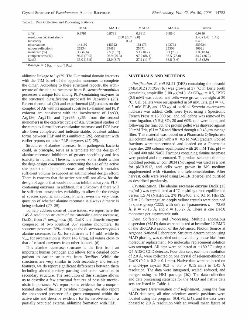
Table 1: Data Collection and Processing Statistics
MAD 1 MAD 2 MAD 3 MAD 4 native
λ (Å) 0.9795 0.9791 0.9611 0.9840 0.9840resolution (Å) (last shell) 2.00 (2.07-2.0) 1.45 (1.48-1.45)mosaicity 0.23 0.40observations 144192 145322 151173 143764 359534unique reflections 25234 25416 25671 25309 56981R-mergea (%) 3.7 (6.6) 7.5 (13.7) 5.1 (10.2) 3.1 (7.9) 3.0 (18.6)completeness (%) 96.5 (79.5) 96.9 (79.3) 97.9 (86.1) 96.5 (78.8) 84.7 (36.2)⟨I/σ⟩ 35.0 (15.9) 22.0 (8.7) 27.2 (11.7) 33.8 (8.6) 51.5 (3.9)
a R-merge) ∑|Iobs - Iavg|/∑|Iavg|.
Crystal Structure ofPseudomonasAlanine Racemase Biochemistry, Vol. 42, No. 50, 200314753
merit of 84%. The protein was traced using ARP/wARP inmodewarpNtrace(32-34). A total of 10 iterations, consist-ing of one cycle of model building and 10 cycles of positionaland Biso refinements using data up to 1.45 Å, yielded theentire trace of the protein, including both N- and C-termini.At the conclusion of the ARP/wARP run, theR-factor andRfree (calculated on 5% of the whole data set) were 17.7 and23.4%, respectively, and the connectivity index measuredby ARP/wARP was excellent at 98%.
Using the side-chains-docking scriptside_dock.shof ARP/wARP, and the amino acid sequence (6), all side chainsexcept Met1 and Ala357 were next fit into electron density.The N- and C-terminal residues and the PLP cofactor werethen manually placed into density, and all solvent moleculesremaining from ARP/wARP were removed. The proteinmodel was submitted to positional and isotropicB-factorrefinement using the program SHELXL (35) with standardrestraints. Next, 306 water molecules were added by com-bining ARP/wARP in modesolVent buildingwith SHELXLrefinements. At this point,R andRfree were 20.1 and 24.1%,respectively, but because of the high resolution of the data(1.45 Å), we carried out anisotropic thermal parameterrefinement for those protein atoms with a real spacecorrelation coefficient higher than 0.90. Real space correla-tion coefficients between the model and the electron densitymap obtained with coefficients 2Fobs- Fcalc (Sigma-A map)(35) were calculated using the program O (36). The averagecorrelation for the entire monomer was 0.96. The 15 atomsof the PLP cofactor were also included in anisotropicrefinement, but all solvent atoms were refined isotropically.Anisotropic refinement resulted inR andRfree parameters of17.0 and 21.9%. Placement of hydrogen atoms and additionof a solvent mask both in SHELXL further decreased theRandRfree values to 14.9 and 20.6%, respectively.
Refinement of Extra Density.At this stage,Fobs - Fcalc
difference Fourier maps (Sigma-D map) (35) revealed largecontiguous electron density of up to 3.5σ adjacent to the PLPcofactor. We also noted a bulge in the 2Fobs - Fcalc densityof C4′ from PLP directed toward this contiguous density.We attempted to fit this extra density with small moleculesfound in our purification and crystallization solutions,including Tris, HEPES, acetate, and propionate, and all 20amino acids. At the conclusion of these efforts, we foundthat the best geometrical fit was obtained with a moleculeof D-lysine modeled into this density such that the terminalnitrogen atom NZ of the side chain forms an externalaldimine linkage with the PLP ring (see also Results andDiscussion and Figure 7). The coordinates of the putativeD-lysine were then added into the protein model withrestraints on the 1,2 and 1,3 distances identical to those usedfor common lysine residues in proteins. To more accuratelymodel the presence of both internal and external aldiminecomplexes, additional refinement was performed as fol-lows: (i) PLP was assumed to exist on two positions, eachwith an occupancy of 0.5; (ii) for each position, planarityrestraints were imposed for the ring and its four substitutingatoms (C2′, O3′, C4′, and C5′) and for the dihedral angledefined by C4′, C4, C3, and O3′; (iii) the distance betweenC4′ (both positions) and NZ (lysine residues) was restrainedto 1.30 Å; (iv) D-lysine and PLP atoms were refined usingisotropic temperature factors. After 40 cycles of refinement,RandRfree were refined to 14.8 and 20.6% for 49 538 reflec-
tions between 30 and 1.45 Å withI > 2σ(I) (Table 2). Thefinal DadX structure contains 357 residues, 2753 non-hydro-gen protein atoms, 306 water molecules, 30 non-hydrogenPLP atoms with an occupancy of 0.5, and 10 non-hydrogenD-lysine atoms. The root-mean-square deviations from ideal-ity for bond lengths and angles are 0.01 Å and 2.18°.
RESULTS AND DISCUSSION
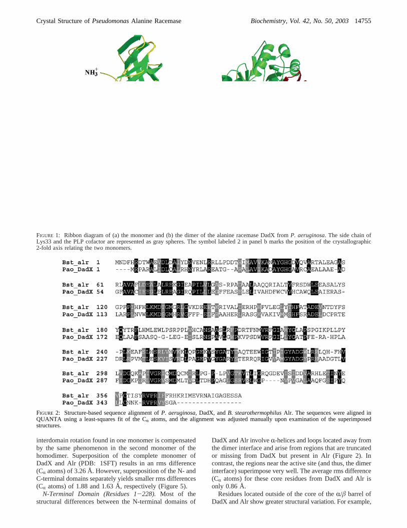
Description of the OVerall Structure.The structure ofDadX is a dimer formed by a head-to-tail association of twomonomers such that the N-terminal domain of one monomerinteracts with the C-terminal domain of the second monomer.The monomers are symmetrically related by a crystallo-graphic 2-fold axis (parallel to the unit cella axis). The DadXmonomer consists of two different domains (Figure 1a). TheN-terminal domain, comprising residues 1-228, is an eight-strandedR/â barrel. The C-terminal domain includes residues229-357 and is primarily composed ofâ-structure. The PLPcofactor is covalently linked to Lys33 and points toward thecenter of theR/â barrel. When viewed from the side, thetwo domains of the monomer are bent relative to each otherat approximately a 130° angle (Figure 1b).
The amino acid sequence identity between DadX fromP.aeruginosaand Alr fromB. stearothermophilus(20) is 28%(Figure 2), and as expected, their tertiary folds are verysimilar (Figure 3). Interestingly, Figure 4 shows that onlyone domain of DadX can be superimposed on an Alrmonomer at a time. This is due to a rotation of the C-terminaldomain in the direction of the N-terminus in Alr that is notpresent in DadX. The rotation is approximately 15° arounda pivot that would be perpendicular to the plane of Figure 4and pass through the CR of Gln342 (Glu355 in Alr).Comparison of the polar interactions at the monomerinterface in DadX and Alr shows that the structural basisfor this rotation is likely to arise from three separate hydrogenbonds that are missing from DadX but present in Alr. Thesethree bonds involve Asp68 and Arg89 of the first Alrmonomer and Phe4, His5, and Asn379 of the second Alrmonomer. Residues corresponding to Phe4 and Asn379 aremissing in DadX because the N- and C-terminal ends ofDadX are shorter than Alr by four residues and 17 residues,respectively (Figure 2).
Despite the difference in the orientation of the monomerdomains in DadX as compared to Alr, the geometry of theactive site in both enzymes is maintained. In both, the
Table 2: Final Refinement Statistics for DadX
R-factora (%) 14.8Rfree (for 2546 reflections, %) 20.6averageB-factors
main chainb (Å2) 19.1side chainc (Å2) 24.0waters 31.0
rms deviationsbond lengths (Å) 0.01bond angles (deg) 2.18
no. of reflections> 2σ 49538no. of residues (% protein) 357 (100%)no. of protein atoms 2753no. of PLP atomsd 30no. of water molecules 306
a R-factor ) ∑|Fobs - Fcalc|/∑|Fobs|. b Anisotropic model.c All but21 side chains refined with anisotropic model.d Two PLPs with 0.5occupancy included in model.
14754 Biochemistry, Vol. 42, No. 50, 2003 LeMagueres et al.
interdomain rotation found in one monomer is compensatedby the same phenomenon in the second monomer of thehomodimer. Superposition of the complete monomer ofDadX and Alr (PDB: 1SFT) results in an rms difference(CR atoms) of 3.26 Å. However, superposition of the N- andC-terminal domains separately yields smaller rms differences(CR atoms) of 1.88 and 1.63 Å, respectively (Figure 5).
N-Terminal Domain (Residues 1-228). Most of thestructural differences between the N-terminal domains of
DadX and Alr involveR-helices and loops located away fromthe dimer interface and arise from regions that are truncatedor missing from DadX but present in Alr (Figure 2). Incontrast, the regions near the active site (and thus, the dimerinterface) superimpose very well. The average rms difference(CR atoms) for these core residues from DadX and Alr isonly 0.86 Å.
Residues located outside of the core of theR/â barrel ofDadX and Alr show greater structural variation. For example,
FIGURE 1: Ribbon diagram of (a) the monomer and (b) the dimer of the alanine racemase DadX fromP. aeruginosa. The side chain ofLys33 and the PLP cofactor are represented as gray spheres. The symbol labeled 2 in panel b marks the position of the crystallographic2-fold axis relating the two monomers.
FIGURE 2: Structure-based sequence alignment ofP. aeruginosa, DadX, andB. stearothermophilusAlr. The sequences were aligned inQUANTA using a least-squares fit of the CR atoms, and the alignment was adjusted manually upon examination of the superimposedstructures.
Crystal Structure ofPseudomonasAlanine Racemase Biochemistry, Vol. 42, No. 50, 200314755

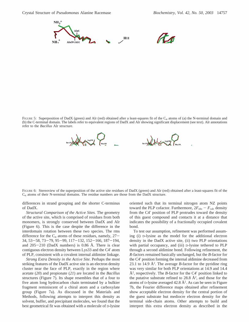
because the N-terminus of DadX is shorter than Alr by fourresidues (Figure 5a), the first residues that show satisfactoryoverlap are Ala6 of DadX and Ala10 of Alr. Because ofinsertions and deletions, most often in loop regions, thereare several regions in Alr that do not superimpose well withDadX. For example, residues 18-27 in DadX superimposepoorly because DadX lacks residues that correspond to Asp30and Asp31. The 219-227 region of DadX superimposespoorly due to a combination of deletion and insertion thatalters the position of theR-helix H11 (Figure 5a). In addition,DadX shows significant structural differences near thefollowing residues of Alr-Gly58, Ser119, Glu143, Leu192,Pro193, and Pro197. As noted, most of these residues donot directly interact with the second monomer or withresidues within the active site, which may explain why thehigh variation in sequence is tolerated.
C-Terminal Domain (Residues 229-357). Ringe et al.have reported that the C-terminal domain of Alr has a uniquetopology (20). The fold of this domain is conserved with
minor differences in the DadX structure, as shown by thesuperposition in Figure 5b. In DadX, the C-terminal domainis composed of 12â-strands and threeR-helices. The firstâ-strand B1, although near the N-terminal domain, belongsto the C-terminal domain as it is inserted between B10 andB20 and participates in a six-strandedâ-sheet (Figure 3).Two other â-sheets made of, respectively, four and twostrands are also observed in this domain. All strands areantiparallel, except B20 and B1, which are parallel. Theoverall dimensions of the C-terminus domain are about 40by 25 by 18 Å. Residues involved in the dimer interfacesuperimpose best between DadX and Alr. For example, therms difference for residues Tyr253, Ala273, and Asp302represented in Figure 5b are 0.56, 0.68, and 0.32 Å,respectively.
Minor structural differences do exist in areas that do notinteract with the second monomer upon dimerization. Forexample, in the turn between B18 and B19 of DadX(identified as loop 1 in Figure 5b), superposition is poorbecause this turn contains an insertion of two residues,Thr309 and Glu313. This loop ends up being wider andextends out of the protein by 4 Å more than in Alr. Further,in the turn between B19 and H13 of DadX, four residues ofAlr are missing (i.e., Gln335, Gly336, Asp337, and Glu338),resulting in the loss of B17 and B18 (Alr labels) from DadX(identified as loop 2 in Figure 5b). This loop in Alr protrudesabout 7 Å farther from the protein than in DadX. Finally,the C-terminus is 17 residues longer in Alr than DadXeliminating B20 of Alr (373-378) from the DadX structure.Note that the five-strandedâ-sheet found in this domain inAlr is composed of six strands in DadX due to slight
FIGURE 3: Topology diagram representing the secondary structureof DadX.
FIGURE 4: Superposition of the monomers of DadX (green) andAlr (red) obtained after a least-squares fit of the CR atoms fromthe N-terminal domains exclusively. It reveals the different inter-domain rotations for DadX and Alr. At the top of the figure, thedistance between structurally equivalent residues is reported.
14756 Biochemistry, Vol. 42, No. 50, 2003 LeMagueres et al.
differences in strand grouping and the shorter C-terminusof DadX.
Structural Comparison of the ActiVe Sites.The geometryof the active site, which is comprised of residues from bothmonomers, is strongly conserved between DadX and Alr(Figure 6). This is the case despite the difference in theinterdomain rotation between these two species. The rmsdifference for the CR atoms of these residues, namely, 27-34, 53-58, 73-79, 95-99, 117-132, 152-166, 187-194,and 205-210 (DadX numbers) is 0.86 Å. There is clearcontiguous electron density between Lys33 and the C4′ atomof PLP, consistent with a covalent internal aldimine linkage.
Strong Extra Density in the ActiVe Site.Perhaps the moststriking features of the DadX active site is an electron densitycluster near the face of PLP, exactly in the region whereacetate (20) and propionate (21) are located in theBacillusstructures (Figure 7). Its shape resembles that of a four tofive atom long hydrocarbon chain terminated by a bulkierfragment reminiscent of a chiral atom and a carboxylategroup (Figure 7a). As discussed in the Materials andMethods, following attempts to interpret this density assolvent, buffer, and precipitant molecules, we found that thebest geometrical fit was obtained with a molecule ofD-lysine
oriented such that its terminal nitrogen atom NZ pointstoward the PLP cofactor. Furthermore, 2Fobs - Fcalc densityfrom the C4′ position of PLP protrudes toward the densityof this guest compound and contacts it at a distance thatindicates the possibility of a fractionally occupied covalentbond.
To test our assumption, refinement was performed assum-ing (i) D-lysine as the model for the additional electrondensity in the DadX active site, (ii) two PLP orientationswith partial occupancy, and (iii)D-lysine tethered to PLPthrough a second aldimine bond. Following refinement, theR-factors remained basically unchanged, but theB-factor forthe C4′ position forming the internal aldimine decreased from23.1 to 14.9 Å2. The averageB-factor for the pyridine ringwas very similar for both PLP orientations at 14.9 and 14.4Å2, respectively. TheB-factor for the C4′ position linked tothe putative substrate refined to 28.8 Å2, and those for theatoms ofD-lysine averaged 42.8 Å2. As can be seen in Figure7b, the Fourier difference maps obtained after refinementshow acceptable electron density for the central portion ofthe guest substrate but mediocre electron density for theterminal side-chain atoms. Other attempts to build andinterpret this extra electron density as described in the
FIGURE 5: Superposition of DadX (green) and Alr (red) obtained after a least-squares fit of the CR atoms of (a) the N-terminal domain and(b) the C-terminal domain. The labels refer to equivalent regions of DadX and Alr showing significant displacement (see text). Alr annotationsrefer to theBacillus Alr structure.
FIGURE 6: Stereoview of the superposition of the active site residues of DadX (green) and Alr (red) obtained after a least-squares fit of theCR atoms of their N-terminal domains. The residue numbers are those from the DadX structure.
Crystal Structure ofPseudomonasAlanine Racemase Biochemistry, Vol. 42, No. 50, 200314757

Materials and Methods were less successful, includingrefining this region as a cluster of three solvent molecules.None of the alternatives we tried was completely satisfactory.While recognizing the speculative nature of our attempt toaccount for this density, we believe the best choice at presentis to interpret this density as a partially ordered guestmolecule, possibly aD-lysine, that may be covalentlyinteracting with PLP.
Orientation and Binding of PLP.In the active site ofDadX, two PLP molecules are located with an angulardisplacement between them of about 12°. One maintains aninternal aldimine linkage with the protein, while the secondmaintains an external aldimine linkage with the guestsubstrate. The two orientations are reminiscent to thoseobserved in the internal (20, 21) and external (22) complexesof Alr from Bacillus (Figure 8). The internal aldiminenitrogen forms a hydrogen bond with O3′ of the internal PLP(2.94 Å), consistent with the protonated nature of the internalSchiff base. Note that, without the imposition of any planarity
restraints, the NZ of Lys33 is almost exactly in the plane ofthe PLP to which it is bound.
In the complex of alanine phosphonate with Alr (22), theimine-PLP-phenol system is planar, and a hydrogen bond isobserved between the external imine nitrogen and the O3′of PLP. This geometry is consistent with mechanisms thatpropose electron delocalization into the PLP ring duringformation of the carbanion intermediate, a process thoughtto occur during catalysis by alanine racemase (20) and byother PLP-dependent enzymes (37-39). However, theelectron density for the external aldimine in our structure isnot clear enough to allow for an unambiguous determinationof its geometry relative to PLP.
Since both the internal and the external aldimine formsappear to be present in the DadX structure, two explanationsare readily apparent. It may be that a statistical mix of internaland external aldimine forms is present in the active sites. Ifthis were the case, we would expect to see evidence of twostates for Lys33, one linked to PLP and one not involved inthe aldimine linkage. These two states have been reportedin the internal and external complexes ofBacillus Alr, forexample. However, in the DadX structure, theB-factor forthe terminal NZ of Lys33 is 19.8 Å2, and there is no sign ofany alternate conformation for Lys33. A second explanationfor our results is that the PLP is involved in a dynamicequilibrium in which both internal and external aldimineforms interchange continuously (Scheme 1). The electrondensity andB-factor values for the active site atoms are moreconsistent with this hypothesis.
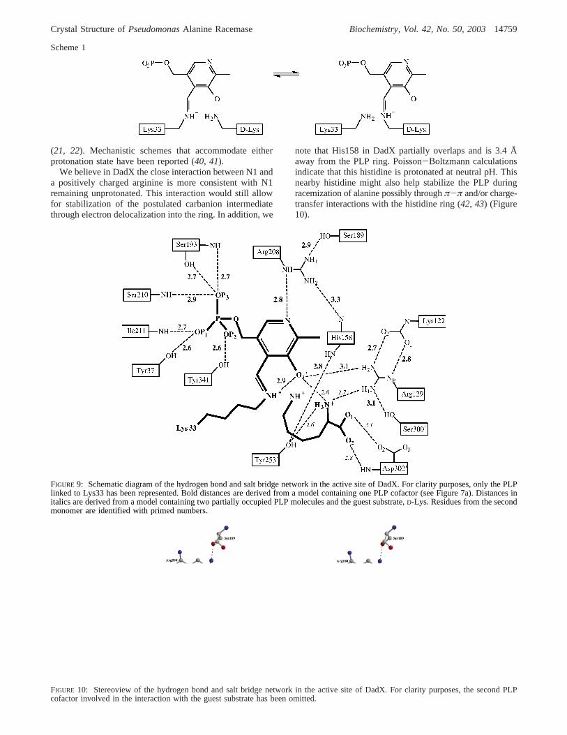
Hydrogen Bonding with PLP. The hydrogen bond networkin the active site of DadX involving PLP, substrate, andnearby residues, as illustrated in Figures 9 and 10, is verysimilar to that of Alr. One important feature found in bothstructures is a strong hydrogen bond between the nitrogenatom N1 of the pyridine ring of PLP and the nitrogen atomNE of the nearby positively charged arginine. In Alr, thedistance ranges from 2.6 to 2.9 Å, while it is 2.8 Å in DadX.In most other PLP enzymes, the N1 atom of PLP interactswith a negatively charged carboxylate, consistent with itsprotonated nature (37). For alanine racemase, some structuralreports have referred to N1 as being protonated (20, 26),while others at times have depicted N1 as being unprotonated
FIGURE 7: 2Fobs - Fcalc (blue, contoured at 1.3σ) andFobs - Fcalc(red, contoured at 1.7σ) electron density maps in the active site ofDadX obtained (a) before modeling the electron density of the guestsubstrate and (b) after modeling the guest substrate withD-lysineand allowing the PLP to adopt two alternate positions with partialoccupancy.
FIGURE 8: Superposition of the PLP cofactors from the DadX(green), Alr/acetate (red), and Alr/alanine phosphonate (gray)structures obtained after a least-squares fit.
14758 Biochemistry, Vol. 42, No. 50, 2003 LeMagueres et al.
(21, 22). Mechanistic schemes that accommodate eitherprotonation state have been reported (40, 41).
We believe in DadX the close interaction between N1 anda positively charged arginine is more consistent with N1remaining unprotonated. This interaction would still allowfor stabilization of the postulated carbanion intermediatethrough electron delocalization into the ring. In addition, we
note that His158 in DadX partially overlaps and is 3.4 Åaway from the PLP ring. Poisson-Boltzmann calculationsindicate that this histidine is protonated at neutral pH. Thisnearby histidine might also help stabilize the PLP duringracemization of alanine possibly throughπ-π and/or charge-transfer interactions with the histidine ring (42, 43) (Figure10).
Scheme 1
FIGURE 9: Schematic diagram of the hydrogen bond and salt bridge network in the active site of DadX. For clarity purposes, only the PLPlinked to Lys33 has been represented. Bold distances are derived from a model containing one PLP cofactor (see Figure 7a). Distances initalics are derived from a model containing two partially occupied PLP molecules and the guest substrate,D-Lys. Residues from the secondmonomer are identified with primed numbers.
FIGURE 10: Stereoview of the hydrogen bond and salt bridge network in the active site of DadX. For clarity purposes, the second PLPcofactor involved in the interaction with the guest substrate has been omitted.
Crystal Structure ofPseudomonasAlanine Racemase Biochemistry, Vol. 42, No. 50, 200314759

Within the DadX active site, Arg208 is stabilized by ahydrogen bond network similar to that found in Alr andincludes Tyr253′, His158, Arg208, and Ser189. Ser189replaces His200 of Alr and makes hydrogen contact withonly one residue (Arg208), while His200 in Alr com-municates with two other histidines (His166 and His127).Consequently, this network in DadX is shortened by tworesidues. The phenolic oxygen O3′ of PLP in DadX is partof a hydrogen bond network that is essentially identical tothat found in Alr, in which O3′ forms a hydrogen bond withArg129 in DadX and Arg136 in Alr.
As was reported for the Alr/proprionate structure, weobserve extra electron density consistent with a carbamylatedlysine at the NZ terminus of Lys122 (Lys129 in Alr). Lys122refines successfully as a carbamylated residue and in its finalposition forms two hydrogen bonds with Arg129. Thissuggests that a carbamylated lysine may be a conservedfeature of alanine racemase and supports the hypothesis ofRinge that this lysine helps to position the nearby argininefor an interaction with both PLP and substrate.
Entryway to the Binding Pocket.The entrance to the activesite of DadX is a corridor defined by OD2 of Asp163, CG2of Ile339, NH2 of Arg278′, and the main chain nitrogen ofGly254′. This entry corridor is about 11.0 Å long and directlyleads to the reactive atom C4′ of PLP. At its end, it isdelimited by two tyrosines, Tyr341 and Tyr253′. Theseresidues define an opening with a width of about 2.7 Å. Thisopening is too narrow for a carbon atom to penetrate (vander Waals radius of 3.4 Å) but is similar to that measuredbetween the corresponding tyrosine residues in the activesite of theBacillusstructures (ca. 3.0 Å). This suggests thatone or both tyrosines must move to allow small substratesto enter and that the entry of larger molecules may beblocked. Studies of alanine racemase in which one of thesetyrosines is mutated to alanine confirm that access to theactive site is significantly improved (44). Teleologically, itmakes sense for alanine racemase to severely restrict activesite access, thereby avoiding indiscriminant and potentiallydangerous amino acid racemization.
Binding Pocket.Substrates that successfully progress pastthe entry corridor enter a binding cavity of about 5.0 by 3.0by 3 Å adjacent to the PLP cofactor. Acetate, propionate,alanine phosphonate, and the putative substrate in DadX areall located in this region in the structures of complexesreported to date (20-22). This cavity is delimited on oneside by PLP, the side chain of Met301′, and the side chainsof Tyr37 and Tyr341 (Figure 10). From this initial activesite cavity, a second cavity on the opposite side of PLP isaccessible. This cavity is of a dimension 6.0 by 4.5 by 7.5Å and contains four water molecules in allBacillusstructures.The boundary of this pocket is formed by residues Lys33,Ala58, Leu78, Glu79, carb-Lys122, Arg129, Val130, Asp302′,and Met303′. One of the four waters (Wat 765 in Alr) isreplaced in DadX by the carboxylate group of theD-lysine,suggesting that this second cavity can participate in substratebinding. The three remaining waters occupying this cavityare located in the same positions in the Alr complexes withacetate, propionate, phosphonate, and as reported here, inDadX, suggesting that they may be a conserved feature ofthe enzyme.
Conclusion and Structural Implications for Drug Design.Alanine racemase has often been proposed as a target for
drug design in antimicrobial development, given its biologicalposition of being essential to prokaryotes but essentiallyabsent from eukaryotes. Inspection of the structure suggeststhat there are at least three ways to design inhibitors to theenzyme. One way would be to place an inhibitor within theactive site. A second way would be to block access to theactive site by placing an inhibitor within the entryway. Athird method would be to design an inhibitor that wouldprevent dimerization. The first method is the most straight-forward, but Alr has the reputation of possessing a smallactive site pocket that would not readily allow for successfuldrug design. Inspection of our structure reveals that the activesite pocket is large enough to accommodate drug candidates.Also, this active site contains several excellent candidatesfor hydrogen bonding as well as hydrophobic interaction.In fact, compounds up to molecular weight 350 have beencomputationally docked and clustered (45). However, basedon analysis of this and earlier alanine racemase structures, aproblem does exist with regard to the size of the entryway.In fact, the static DadX structure would not even admitalanine itself into the binding cavity. However, certainlyalanine and other molecules do enter the active site, albeitwith difficulties commensurate with their size, but this issuehas to be taken into account when designing inhibitors.Adopting the second approach would involve the design ofcompounds to block the entryway. This would avoid theproblem of access to the active site but would be moredifficult in terms of pharmacophore development. Dimer-ization inhibitors of alanine racemase have not yet beendeveloped, but the concept has been demonstrated (46-48).We believe that attempts to design inhibitors using any ofthese approaches is warranted and of potentially significantbiomedical importance. Continued crystallographic study ofthese enzymes and their complexes with inhibitors will alsobe of help in these ongoing drug design efforts.
ACKNOWLEDGMENT
Use of the Advanced Photon Source was supported bythe U.S. Department of Energy, Basic Energy Sciences,Office of Science, under Contract W-31-109-Eng-38. Useof the BioCARS Sector 14 was supported by the NationalInstitutes of Health, National Center for Research Resources,under Grant RR07707. We thank Profs. James Briggs andHarold Kohn and Drs. Chetlen Crossnoe and GabrielaMustata for helpful comments and discussions. We also thankthe Institute of Molecular Design and Molecular Simulations,Inc. for the use of computational resources.
REFERENCES
1. Pollack, M. (1995)Pseudomonas aeruginosa, in Principles andPractice of Infectious Diseases(Mandell, G. L., Bennett, J. E.,and Dolin, R., Eds.) pp 2310-2335, Churchill Livingstone, NewYork.
2. Martone, W. J., and Jarvis, W. R. (1992) Predominant Pathogensin Hospital Infections,J. Antimicrob. Chemother.19-24.
3. FitzSimmons, S. C. (1993) The Changing Epidemiology of CysticFibrosis,J. Pediatr.1-9.
4. Bert, F., Maubec, E., and Bruneau, B. et al. (1998) Multi-resistantPseudomonas aeruginosaOutbreak Associated with ContaminatedTap Water in a Neurosurgery Intensive Care Unit,J. Hops. Infect.53-62.
14760 Biochemistry, Vol. 42, No. 50, 2003 LeMagueres et al.
5. Chen, H. Y., Yuan, M., Ibrahim-Elmagboul, I. B., and Livermore,D. M. (1995) National Survey of Susceptibility to Antimicrobialsamongst Clinical Isolates ofPseudomonas aeruginosa, J. Anti-microb. Chemother.521-534.
6. Strych, U., Huang, H. C., Krause, K. L., and Benedik, M. J. (2000)Characterization of the Alanine Racemases fromPseudomonasaeruginosaPAO1,Curr. Microbiol. 41, 290-294.
7. Wasserman, S. A., Walsh, C. T., and Botstein, D. (1983) TwoAlanine Racemase Genes inSalmonella typhimuriumthat Differin Structure and Function,J. Bacteriol. 153, 1439-1450.
8. Wild, J., Hennig, J., Lobocka, M., Walczak, W., and Klopotowski,T. (1985) Identification of thedadx Gene Coding for thePredominant Isozyme of Alanine Racemase inEscherichia coliK12, Mol. Gen. Genet. 198, 315-322.
9. Lambert, M. P., and Neuhaus, M. P. (1972) Mechanism ofD-Cycloserine Action: Alanine Racemase fromEscherichia coli,J. Bacteriol. 110, 978-987.
10. Silverman, R. B. (1988) The Potential Use of Mechanism-BasedEnzyme Inactivators in Medicine,J. Enzyme Inhib. 2, 73-90.
11. Copie, V., Faraci, W. S., Walsh, C. T., and Griffin, R. G. (1988)Inhibition of Alanine Racemase by Alanine Phosphonate: Detec-tion of an Imine Linkage to Pyridoxal 5′-Phosphate in the EnzymeInhibitor Complex by Solid-State15N Nuclear Magnetic Reso-nance,Biochemistry 27, 4966-4970.
12. Erion, M. D., and Walsh, C. T. (1987) 1-Aminocyclopropane-phosphonate: Time-Dependent Inactivation of 1-Aminocyclopro-panecarboxylate Deaminase andBacillus stearothermophilusAlanine Racemase by Slow dissociation Behavior,Biochemistry26, 3417-3425.
13. Wang, E., and Walsh, C. T. (1978) Suicide Substrates for theAlanine Racemase fromEscherichia coli B, Biochemistry 17,1313-1321.
14. Neuhaus, F. C. (1967) Mechanism of Action, inAntibiotics(Gottleib, D., and Shaw, P. D., Eds.) pp 40, Springer-Verlag, NewYork.
15. Walsh, C. (2003) Antibiotics that Act on Cell Wall Biosynthesis,in Antibioticssactions, origins, resistance, pp 23-49, ASM Press,Washington.
16. Weinstein, L. (1975) Antimicrobial AgentssDrugs Used in theChemotherapy of Tuberculosis and Leprosy, inThe Pharmaco-logical Basis of Therapeutics, 5th ed.(Goodman, L. S. andGilman, A., Eds.) pp 1202-1223, Macmillan Publishing, NewYork.
17. Newton, R. W. (1975) Side Effects of Drugs Used to TreatTuberculosis,Scott. Med. J. 20, 47-49.
18. Yew, W. W., Wong, C. F., Wong, P. C., Lee, J., and Chau, C. H.(1993) Adverse Neurological Reactions in Patients with Multidrug-Resistant Pulmonary Tuberculosis after Coadministration ofCycloserine and Ofloxacin,Clin. Infect. Dis. 17, 288-289.
19. Katz, M. H. (1994) Effect of HIV Treatment on Cognition,Behavior, and Emotion,Psychiatr. Clin. North Am. 17, 227-230.
20. Shaw, J. P., Petsko, G. A., and Ringe, D. (1997) Determinationof the Structure of Alanine Racemase fromBacillus stearo-thermophilusat 1.9 Å Resolution,Biochemistry 36, 1329-1342.
21. Morollo, A. A., Petsko, G. A., and Ringe, D. (1999) Structure ofa Michaelis Complex Analogue: Propionate Binds in the SubstrateCarboxylate Site of Alanine Racemase,Biochemistry 38, 3293-3301.
22. Stamper, C. G. F., Morollo, A. A., and Ringe D. (1998) Reactionof Alanine Racemase with 1-Aminoethylphosphonic Acid Formsa Stable External Aldimine,Biochemistry 37, 10438-10445.
23. Jansonius, J. N. (1998) Structure, Evolution and Action of VitaminB(6) Dependent Enzymes,Curr. Opin. Struct. Biol. 8, 756-759.
24. Ondrechen, M. J., Briggs, J. M., and McCammon, J. A. (2001) AModel for Enzyme-Substrate Interaction in Alanine Racemase,J. Am. Chem. Soc. 123, 2830-2834.
25. Watanabe, A., Yoshimura, T., Mikami, B., and Esaki, N. (1999)Tyrosine 265 of Alanine Racemase Serves as a Base Abstractinga Hydrogen fromL-Alanine: the Counterpart Residue to Lys39Specific toD-Alanine,J. Biochem. 126, 781-786.
26. Fenn, T. D., Stamper, G. F., Morollo, A. A., and Ringe, D. (2003)A side Reaction of Alanine Racemase: Transamination ofCycloserine,Biochemistry 42, 5775-5783.
27. Olson, G. T., Mengmeng, F., Lau, S., Rinehart, K. L., andSilverman, R. B. (1998) An Aromatization Mechanism of
Inactivation of γ-Aminobutyric Acid Aminotransferase for theAntibiotic L-Cycloserine,J. Am. Chem. Soc. 120, 2256-2267.
28. Peisach, D., Chipman, D. M., Van Ophem, P. W., Manning, J.M., and Ringe, D. (1998)D-Cycloserine Inactivation ofD-AminoAcid Aminotransferase Leads to a Stable Noncovalent ProteinComplex with an Aromatic Cycloserine-PLP Derivative,J. Am.Chem. Soc. 120, 2268-2274.
29. Yokoigawa, K., Okubo, Y., and Soda, K. (2003) Subunit Interac-tion of Monomeric Alanine Racemases from FourShigellaSpeciesin Catalytic Reaction,FEMS Microbiol. Lett. 221, 263-267.
30. Otwinowski, Z., and Minor, W. (1997) Processing of X-rayDiffraction Data Collected in Oscillation Mode, inMethods ofEnzymology(C. W. Carter, J., and Sweet, R. M., Eds.) pp 307-326, Academic Press, New York.
31. Terwilliger, T. C., and Berendzen, J. (1999) Automated StructureSolution for MIR and MAD,Acta Crystallogr. D55, 849-861.
32. Lamzin, V. S., and Wilson, K. S. (1997) Automated Refinementfor Protein Crystallography,Methods Enzymol. 277, 269-305.
33. Perrakis, A., Sixma, T. K., Wilson, K. S., and Lamzin, V. S. (1997)wARP: Improvement and Extension of Crystallographic Phasesby Weighted Averaging of Multiple Refined Dummy AtomicModels,Acta Crystallogr. D53, 448-455.
34. Perrakis, A., Morris, R. J., and Lamzin, V. S. (1999) AutomatedProtein Model Building Combined with Iterative Structure Refine-ment,Nat. Struct. Biol. 6, 458-463.
35. Sheldrick, G. M., and Schneider, T. R. (1997) Shelxl: High-Resolution Refinement,Methods Enzymol. 277, 319-343.
36. Jones, T. A., Zou, J. Y., Cowan, S. W., and Kjeldgaard, M. (1991)Improved Methods for Building Protein Models in ElectronDensity Maps and the Location of Errors in these Models,ActaCrystallogr. A47, 110-119.
37. Yano, T., Kuramiysu, S., Tanase, S., Morino, Y., and Kagamiyama,H. (1992) Role of Asp222 in the Catalytic Mechanism ofEscherichia coliAspartate Aminotransferase: The Amino AcidResidue Which Enhances the Function of the Enzyme-BoundCoenzyme Pyridoxal 5′-Phosphate,Biochemistry 31, 5878-5887.
38. Arnone, A., Christen, P., Jansonius, J. N., and Metzler, D. E. (1985)in Transaminases(Christen, P., and Metzler, D. E., Eds.) pp 326-357, John Wiley and Sons, New York.
39. McPhalen, C. A., Vincent, M. G., and Jansonius, J. N. (1992)X-ray Structure Refinement and Comparison of Three Forms ofMitochondrial Aspartate Aminotransferase,J. Mol. Biol. 225,495-517.
40. Ashley Spies, M., and Toney, M. D. (2003) Multiple HydrogenKinetic Isotope for Enzymes Catalyzing Exchange with Solvent:Application to Alanine Racemase,Biochemistry 42, 5099-5107.
41. Watanabe, A., Yoshimura, T., Mikami, B., Kagamiyama, H., andEsaki, N. (2002) Reaction Machanism of Alanine Racemase fromBacillus stearothermophilus, J. Biol. Chem. 277, 19166-19172.
42. Rathore, R., Lindeman, S. V., and Kochi, J. K. (1997) Charge-Transfer Probes for Molecular Recognition via Steric Hindrancein Donor-Acceptor Pairs,J. Am. Chem. Soc. 119, 9393-9404.
43. Le Magueres, P., Lindeman, S. V., and Kochi, J. K. (2000) Novel(Heteromolecular)π-Complexes of Aromatic Cation Radicals.Isolation and Structural Characterization,Org. Lett. 2, 3567-3570.
44. Wayne, M. P., Weisner, J., and Blackburn, J. M. (2002) Site-Directed Mutagenesis of Tyr354 inGeobacillus stearothermo-philusAlanine Racemase Identifies a Role in Controlling SubstrateSpecificity and a Possible Role in the Evolution of AntibioticResistance,ChemBioChem 8, 789-792.
45. Mustata, G. I., and Briggs, J. M. (2002) A Structure-Based DesignApproach for the Identification of Novel Inhibitors: Applicationto an Alanine Racemase,J. Comput.-Aided Mol. Des. 16, 935-953.
46. Boggetto, N., and Reboud-Ravaux, M. (2002) Dimeric Inhibitorsof HIV-1 Protease,Biol. Chem. 383, 1321-1324.
47. Song, M., Rajesh, S., Hayashi, Y., and Kiso, Y. (2001) Designand Synthesis of New Inhibitors of HIV-1 Protease Dimerizationwith Conformationally Constrained Templates,Bioorg. Med.Chem. Lett. 11, 2465-2468.
48. Cochran, A. G. (2001) Protein-Protein Interfaces: Mimics andInhibitors,Curr. Opin. Chem. Biol. 5, 654-659.
BI030165V
Crystal Structure ofPseudomonasAlanine Racemase Biochemistry, Vol. 42, No. 50, 200314761


![â…^Ûnj‰];æ;êŠËßÖ];å^Ÿ÷];l]ˆÓi†Ú - saida.dz](https://static.fdokumen.com/doc/165x107/6313fe3ec72bc2f2dd0437e8/aunjaeesessoeayloiu-saidadz.jpg)