The recognition domain of the methyl-specific endonuclease McrBC flips out 5-methylcytosine
Upload
xn--universitt-stuttgart-jzbCategory
view
0download
0
The EMBO Journal Vol.17 No.22 pp.6757–6766, 1998
Crosslinking the EcoRV restriction endonucleaseacross the DNA-binding site reveals transientintermediates and conformational changes of theenzyme during DNA binding and catalytic turnover
Claudia Schulze, Albert Jeltsch,Ingo Franke, Claus Urbanke1 andAlfred Pingoud2
Institut fur Biochemie (Fachbereich Biologie), Justus-Liebig-Universitat, Heinrich-Buff-Ring 58, D-35392 Giessen, and1Abteilungfur Biophysikalisch-chemische Verfahren, Medizinische HochschuleHannover, Carl-Neuberg-Straße 1, D-30632 Hannover, Germany2Corresponding authore-mail: [email protected]
EcoRV completely encircles bound DNA with twoloops, forming the entry and exit gate for the DNAsubstrate. These loops were crosslinked generatingCL-EcoRV which binds and releases linear DNA onlyslowly, because threading linear DNA into and out ofthe DNA-binding ‘tunnel’ of CL- EcoRV is not veryeffective. If the crosslinking reaction is carried outwith a circular bound DNA, CL- EcoRV is hyperactivetowards the trapped substrate which is cleaved veryquickly but not very accurately. CL-EcoRV also bindsto, but does not cleave, circular DNA when added fromthe outside, because it cannot enter the active site.Based on these results a two-step binding model isproposed for EcoRV: initial DNA binding occurs atthe outer side of the loops before the gate opens andthen the DNA is transferred to the catalytic center.Keywords: protein–DNA association/protein–DNAinteraction/restriction endonuclease/restriction–modification system/specificity
Introduction
Type II restriction enzymes cleave DNA moleculesspecifically at recognition sites comprising 4–8 base pairs.These enzymes have two outstanding properties: (i) theyassociate to their recognition sites at a diffusion-controlledrate, in spite of the fact that there is a large excess ofnon-target DNA; and (ii) they display a very high degree ofsequence specificity (for review see Roberts and Halford,1993; Pingoud and Jeltsch, 1997). Typically, sequencesdiffering by one base pair from their recognition sites arecleaved three to five orders of magnitude more slowlythan the canonical site. On the basis of detailed structuraland biochemical work, the mechanistic basis of both ofthese properties is well understood forEcoRV (recognitionsite: GATATC), which is one of the best studied restrictionendonucleases.EcoRV is a homodimeric enzyme(2329 kDa) whose DNA-binding site is located at thebottom of a deep cleft and flanked by two arms of theenzyme (Figure 1A) (Winkleret al., 1993). The reactionmechanism ofEcoRV comprises several steps: first, theenzyme associates non-specifically to DNA. This occursin a diffusion-controlled reaction (Erskineet al., 1997).
© Oxford University Press 6757
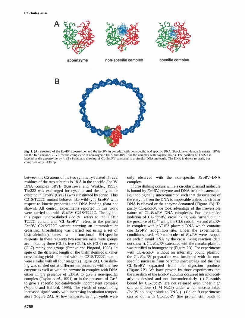
In apparent contrast to the very high association rateconstant ofEcoRV to DNA is the finding that the freeenzyme is in a closed conformation in the crystal in whichthe arms approach each other thereby preventing directentry of the DNA to the binding cleft (Figure 1A).Consequently, during the association reaction conforma-tional changes must take place that open up the DNA-binding site of the enzyme. In the crystal structure, thenon-specific EcoRV–DNA complex has a more openconformation than either freeEcoRV or the specificEcoRV–DNA complex (Winkleret al., 1993). In the non-specific complex, significant parts of the arms appear notto be ordered (Figure 1A). This may be important for themechanism of target site location, becauseEcoRV whilebeing bound non-specifically to DNA, scans several hun-dreds of base pairs by linear diffusion searching for itsrecognition site (Tayloret al., 1991; Jeltschet al., 1996;Jeltsch and Pingoud, 1998). Upon encountering a GATATCsite numerous contacts of the enzyme to the edges of thebases and the DNA backbone are formed which togethertrigger formation of a specific complex (Thielkinget al,1991; Winkler et al., 1993; Stahlet al., 1996). In thespecific complex formed, the arms ofEcoRV are orderedand completely encircle the DNA to form a tightly closedcomplex (Figure 1A) (Winkleret al., 1993; Kostrewa andWinkler, 1995; Perona and Martin, 1997). Upon transitionof the non-specific to the specific complex the catalyticcenter becomes activated leading to DNA cleavage.Finally, the cleavage products dissociate from the enzyme.
In the present study we have investigated how conforma-tional changes of the arms ofEcoRV are involved in themechanism of action of this enzyme. To this end, bothsubunits ofEcoRV were crosslinked across the DNA-binding site at the tips of the arms using a series of divalentchemical crosslinkers. Thereby, a covalently closed ‘ring-shaped’ enzyme is generated, whose DNA-binding site islocated in an artificially created ‘tunnel’ in the center ofthe protein (Figure 1B). These crosslinked proteins werepurified and analyzed with respect to the thermodynamicsand kinetics of DNA binding and cleavage. Our resultsallow us to draw important conclusions regarding themechanism by whichEcoRV acts, the most important ofwhich is that DNA binding occurs in at least two steps.At first, DNA is bound to the outside of the homodimericprotein. Then, the arms open up to allow the DNA tobind at the catalytic site of the enzyme.
Results
Preparation of intramolecularly crosslinked EcoRVTo crosslink the two subunits of anEcoRV dimer specific-ally at the arms of the molecule, we inspected the structurefor suitably located amino acid residues. Thr222 is locatedat the inner side of the arms ofEcoRV. The distance
C.Schulze et al.
Fig. 1. (A) Structure of theEcoRV apoenzyme, and theEcoRV in complex with non-specific and specific DNA (Brookhaven databank entries: 1RVEfor the free enzyme, 2RVE for the complex with non-cognate DNA and 4RVE for the complex with cognate DNA). The position of Thr222 islabeled in the apoenzyme by *. (B) Schematic drawing of CL-EcoRV catenated to a circular DNA molecule. The DNA is drawn to scale, butcomprises only ~130 bp.
between the Cα atoms of the two symmetry-related Thr222residues of the two subunits is 18 Å in the specificEcoRVDNA complex 5RVE (Kostrewa and Winkler, 1995).Thr222 was exchanged for cysteine and the only othercysteine inEcoRV (Cys21) was substituted by serine. ThisC21S/T222C mutant behaves like wild-typeEcoRV withrespect to kinetic properties and DNA binding (data notshown). All control experiments reported in this workwere carried out withEcoRV C21S/T222C. Throughoutthis paper ‘uncrosslinkedEcoRV’ refers to the C21S/T222C variant and ‘CL-EcoRV’ refers to the purifiedEcoRV C21S/T22C variant carrying an intramolecularcrosslink. Crosslinking was carried out using a set ofbis(maleimido)alkanes as bifunctional SH-specificreagents. In these reagents two reactive maleimide groupsare linked by three (CL3), five (CL5), six (CL6) or seven(CL7) methylene groups (Franke and Pingoud, 1998). Inspite of the different length of the bis(maleimido)alkanescrosslinking yields obtained with the C21S/T222C mutantwere similar with all four reagents (Figure 2A). Crosslink-ing was carried out at different temperatures with the freeenzyme as well as with the enzyme in complex with DNAeither in the presence of EDTA to give a non-specificcomplex (Tayloret al., 1991) or in the presence of Ca21
to give a specific but catalytically incompetent complex(Vipond and Halford, 1995). The yields of crosslinkingincreased significantly with increasing incubation temper-ature (Figure 2A). At low temperatures high yields were
6758
only observed with the non-specificEcoRV–DNAcomplex.
If crosslinking occurs while a circular plasmid moleculeis bound byEcoRV, enzyme and DNA become catenated,i.e. topologically interconnected such that dissociation ofthe enzyme from the DNA is impossible unless the circularDNA is cleaved or the enzyme denatured (Figure 1B). Topurify CL-EcoRV, we took advantage of the irreversiblenature of CL-EcoRV–DNA complexes. For preparativeisolation of CL-EcoRV, crosslinking was carried out inthe presence of Ca21 using the CL6 crosslinker andEcoRVin complex with pAT153 plasmid DNA which containsone EcoRV recognition site. Under the experimentalconditions used, ~20 molecules ofEcoRV were trappedon each plasmid DNA by the crosslinking reaction (datanot shown). CL-EcoRV catenated with the circular plasmidwas purified to homogeneity (Figure 2B). For experimentswith CL-EcoRV without an internally bound plasmid,the CL-EcoRV preparation was incubated with the non-specific nuclease fromSerratia marcescensand the freeCL-EcoRV separated from the digestion products(Figure 2B). We have proven by three experiments thatthe crosslink of theEcoRV subunits occurred intramolecul-arly as desired and not intermolecularly. (i) Plasmidsbound by CL-EcoRV are not released even under highsalt conditions (1 M NaCl) under which uncrosslinkedEcoRV no longer binds to DNA. (ii) Gel-shift experimentscarried out with CL-EcoRV (the protein still binds to

Crosslinking EcoRV restriction endonuclease
Fig. 2. (A) Compilation of the yields of the crosslinking reactions withfour different crosslinkers (CL3, CL5, CL6 and CL7, having 3, 5, 6and 7 methylene groups separating the maleimide groups,respectively). Reactions were performed at three different temperaturesusing freeEcoRV as well asEcoRV bound specifically or non-specifically to DNA as described in Materials and methods. Allexperiments were performed at least in triplicate. (B) Purification ofCL-EcoRV catenated to DNA and in a DNA-free form. Coomassie-stained SDS–PAGE gel of theEcoRV preparations obtained [lane 1,uncrosslinkedEcoRV; lane 2, unpurified CL-EcoRV; lane 3, purifiedCL-EcoRV catenated to DNA (the DNA is released from the catenatedcomplex upon incubation with SDS); lane 4, purified CL-EcoRV freeof DNA (after treatment withS.marcescensendonuclease)].
linear DNA, see below) using a DNA fragment thatcontains oneEcoRV site show only one band shift underconditions of specific DNA binding. No band shifts oflow electrophoretic mobility are observed that wouldcorrespond to intermolecularly crosslinkedEcoRV speciesbound to the DNA, which would contain four subunits ofEcoRV under native conditions. (iii) The S-values ofCL-EcoRV and uncrosslinkedEcoRV determined in sedi-mentation velocity runs in the analytical ultracentrifugeare identical within experimental error (CL-EcoRV, 4.3S;uncrosslinked EcoRV, 4.2S) again demonstrating theabsence of tetrameric enzymes, which would be the resultof an intermolecular crosslink.
Kinetic properties of CL-EcoRVIn order to determine if CL-EcoRV retains catalyticactivity, it was purified in complex with circular pAT153plasmid, catenated to the enzyme. Upon incubation withMg21, complete cleavage of the bound plasmid wasobserved within 10 s (Figure 3A). To prove that thecleavage of the internal plasmid is carried out byCL-EcoRV and not by a small contamination ofuncrosslinkedEcoRV, a second plasmid of different lengthwhich contains anEcoRV site was added to the CL-EcoRVpreparation prior to the cleavage reaction. The rationaleof this control experiment is that an external plasmidcannot be cleaved by CL-EcoRV but only by a contamina-
6759
Fig. 3. Cleavage of pAT153 plasmid catenated to CL-EcoRV.(A) Agarose gel electrophoretic analysis showing the cleavage ofpAT153 catenated to CL-EcoRV. Note that the plasmid which containsoneEcoRV site is converted from the superhelical to the linear formwithin 10 s. After 10 min additional bands appear indicating DNAcleavage at non-canonical sites. (B) Temperature dependence of therate of DNA cleavage of CL-EcoRV and uncrosslinkedEcoRVdetermined using a quenched-flow device. In these experiments thetemperature was varied between 4 and 20°C.
tion of uncrosslinkedEcoRV. Our data show that after60 min incubation only 15% of externally added plasmidswere cleaved, whereas after 10 s all internal plasmidswere cleaved, demonstrating that the activity due to acontamination with uncrosslinkedEcoRV is ,1/1000 ofthe activity displayed towards the internal plasmid. Thus,the cleavage of the internal plasmid must have beencarried out by the CL-EcoRV molecules bound to thissubstrate. The rate of DNA cleavage at ambient temper-ature by CL-EcoRV at GATATC sites as determined byquenched-flow experiments (71 min–1) is roughly fourtimes faster than that measured for uncrosslinkedEcoRV(17 min–1) under identical conditions (Figure 3B). Theseresults demonstrate that CL-EcoRV is a very efficientendonuclease towards the internal plasmid DNA. However,we observed three dramatic differences in the catalyticproperties of CL-EcoRV and uncrosslinkedEcoRV, asfollows:
(i) CL-EcoRV cleaves DNA at 2 M NaCl with high rate(ù6 min–1), whereas uncrosslinkedEcoRV is catalyticallyinactive atcNaCl ù500 mM (data not shown). This resultwas expected, because uncrosslinkedEcoRV is unable tobind the substrate in such high salt buffer, whereasCL-EcoRV carries its substrate irreversibly bound and doesnot depend on binding to DNA added from the outside.
(ii) CL-EcoRV shows strong cleavage activity at non-canonical sites as indicated by the rapid occurrence ofadditional bands in the DNA-cleavage reaction of theinternal plasmid (Figure 3A). Analysis of the resultingcleavage pattern reveals thatEcoRV ‘star’ cleavage occurs,i.e. cleavage at sites that differ in one base pair from theGATATC sequence. Such relaxation of specificity hasalready been observed withEcoRV under certain bufferconditions, for example in the presence of Mn21, under
C.Schulze et al.
low ionic strength or in the presence of organic solvents(Halford et al., 1986). However, with CL-EcoRV starcleavage is observed under normal buffer conditions(20 mM Tris–HCl pH 7.5, 50 mM NaCl, 10 mM MgCl2,hereafter termed ‘cleavage buffer’). Under these conditionsstar cleavage is not observed with uncrosslinkedEcoRV,not even with 20-fold higher enzyme concentrations andafter 10-fold longer incubation times (data not shown).This means that star activity of CL-EcoRV must be atleast 2000-fold higher than that of uncrosslinkedEcoRV,a very interesting result suggesting that the crosslink hasfrozenEcoRV in a hyperactive conformation (see below).
(iii) As already mentioned, the rate constant of DNAcleavage by CL-EcoRV at GATATC sites is ~4-fold higherthan that measured for uncrosslinkedEcoRV at ambienttemperature. However, if the temperature is decreased thisdifference increases dramatically, because CL-EcoRV ismuch less sensitive towards a decrease in reaction temper-ature than uncrosslinkedEcoRV (Figure 3B). At 4°C,CL-EcoRV cleaves the internal DNA ~300-fold moreefficiently than uncrosslinkedEcoRV cleaves externalDNA. The temperature dependence of the DNA-cleavagerate [k(T)] was analyzed according to the Arrhenius modelto determine the apparent activation energy of DNAcleavage (EA):
k(T) 5 k0 exp(–EA/RT)
An ln(k) versus 1/T fit of data allowed to calculate anactivation energy of 40 kJ/mol for CL-EcoRV but of220 kJ/mol for uncrosslinkedEcoRV, supporting the con-clusion that CL-EcoRV is frozen by the crosslink in anactivated conformation that is on the way to the transitionstate of DNA cleavage.
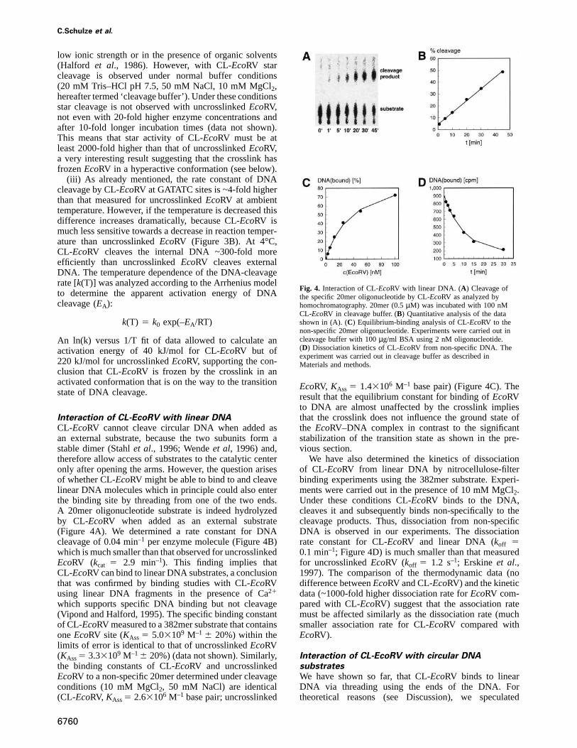
Interaction of CL-EcoRV with linear DNACL-EcoRV cannot cleave circular DNA when added asan external substrate, because the two subunits form astable dimer (Stahlet al., 1996; Wendeet al, 1996) and,therefore allow access of substrates to the catalytic centeronly after opening the arms. However, the question arisesof whether CL-EcoRV might be able to bind to and cleavelinear DNA molecules which in principle could also enterthe binding site by threading from one of the two ends.A 20mer oligonucleotide substrate is indeed hydrolyzedby CL-EcoRV when added as an external substrate(Figure 4A). We determined a rate constant for DNAcleavage of 0.04 min–1 per enzyme molecule (Figure 4B)which is much smaller than that observed for uncrosslinkedEcoRV (kcat 5 2.9 min–1). This finding implies thatCL-EcoRV can bind to linear DNA substrates, a conclusionthat was confirmed by binding studies with CL-EcoRVusing linear DNA fragments in the presence of Ca21
which supports specific DNA binding but not cleavage(Vipond and Halford, 1995). The specific binding constantof CL-EcoRV measured to a 382mer substrate that containsoneEcoRV site (KAss5 5.03109 M–1 6 20%) within thelimits of error is identical to that of uncrosslinkedEcoRV(KAss5 3.33109 M–16 20%) (data not shown). Similarly,the binding constants of CL-EcoRV and uncrosslinkedEcoRV to a non-specific 20mer determined under cleavageconditions (10 mM MgCl2, 50 mM NaCl) are identical(CL-EcoRV, KAss5 2.63106 M–1 base pair; uncrosslinked
6760
Fig. 4. Interaction of CL-EcoRV with linear DNA. (A) Cleavage ofthe specific 20mer oligonucleotide by CL-EcoRV as analyzed byhomochromatography. 20mer (0.5µM) was incubated with 100 nMCL-EcoRV in cleavage buffer. (B) Quantitative analysis of the datashown in (A). (C) Equilibrium-binding analysis of CL-EcoRV to thenon-specific 20mer oligonucleotide. Experiments were carried out incleavage buffer with 100µg/ml BSA using 2 nM oligonucleotide.(D) Dissociation kinetics of CL-EcoRV from non-specific DNA. Theexperiment was carried out in cleavage buffer as described inMaterials and methods.
EcoRV, KAss 5 1.43106 M–1 base pair) (Figure 4C). Theresult that the equilibrium constant for binding ofEcoRVto DNA are almost unaffected by the crosslink impliesthat the crosslink does not influence the ground state ofthe EcoRV–DNA complex in contrast to the significantstabilization of the transition state as shown in the pre-vious section.
We have also determined the kinetics of dissociationof CL-EcoRV from linear DNA by nitrocellulose-filterbinding experiments using the 382mer substrate. Experi-ments were carried out in the presence of 10 mM MgCl2.Under these conditions CL-EcoRV binds to the DNA,cleaves it and subsequently binds non-specifically to thecleavage products. Thus, dissociation from non-specificDNA is observed in our experiments. The dissociationrate constant for CL-EcoRV and linear DNA (koff 50.1 min–1; Figure 4D) is much smaller than that measuredfor uncrosslinkedEcoRV (koff 5 1.2 s–1; Erskine et al.,1997). The comparison of the thermodynamic data (nodifference betweenEcoRV and CL-EcoRV) and the kineticdata (~1000-fold higher dissociation rate forEcoRV com-pared with CL-EcoRV) suggest that the association ratemust be affected similarly as the dissociation rate (muchsmaller association rate for CL-EcoRV compared withEcoRV).
Interaction of CL-EcoRV with circular DNAsubstratesWe have shown so far, that CL-EcoRV binds to linearDNA via threading using the ends of the DNA. Fortheoretical reasons (see Discussion), we speculated

Crosslinking EcoRV restriction endonuclease
Fig. 5. Interaction of CL-EcoRV with circular DNA. (A) Binding of CL-EcoRV and uncrosslinkedEcoRV to circular DNA. Experiments wereperformed in washing buffer containing 0.7 pM radioactively labeled pAT153. (B) Quantitative analysis of the data shown in (A). (C) Reduction ofthe rate of cleavage of the specific 20mer by CL-EcoRV and uncrosslinkedEcoRV by addition of circular plasmid as competitor. Cleavageexperiments were performed in cleavage buffer. The concentrations of pUC8 present as competitor are given in mole base pairs.
whether CL-EcoRV might also be able to bind to circularDNA. Therefore, radioactively labelled circular DNAwas incubated with CL-EcoRV and DNA binding wasmeasured by nitrocellulose-filter binding experiments. Asshown in Figure 5A, significant binding is observed. Wehad confirmed that the substrate preparation did not containa contamination by linear DNA and that it remainedcircular during the binding experiment (data not shown).These data could not be analyzed in terms of microscopicbinding constants, because many enzyme molecules canbind to each substrate molecule. However, binding iso-therms measured with CL-EcoRV and uncrosslinkedEcoRV are virtually identical, with half saturation ofbinding observed in both cases atcEcoRV 5 2–4 nM. Theseresults demonstrate that both enzyme species have a similaraffinity for circular DNA (Figure 5B). This conclusion wascorroborated by competition experiments in which itwas observed that cleavage of the 20mer substrate byCL-EcoRV is strongly inhibited by addition of circularpUC8 plasmid DNA which does not contain anEcoRVsite (Figure 5C)—a result which can only be rationalizedif CL-EcoRV interacts with circular DNA. The inhibitionconstant of pUC8 binding to CL-EcoRV is as measuredby cleavage of the 20mer substrateKI 5 33107 M–1 under
6761
these conditions. Interestingly, linear DNA is not a bettercompetitor than circular DNA (KI 5 33107 M–1) incleavage experiments with the 20mer and CL-EcoRV.Analogous experiments with uncrosslinkedEcoRVresulted in very similar competition curves as measuredfor CL-EcoRV (Figure 5C) with a competition constantfor circular DNA of KI 5 33107 M–1 and for linear DNAof KI 5 13107 M–1, although uncrosslinkedEcoRV canbind to the plasmid at the internal binding site that is notaccessible for circular DNA molecules in CL-EcoRV.Taken together, these experiments demonstrate that circularDNA can bind to CL-EcoRV. Such binding could onlytake place at the surface of the enzyme. The finding thatcircular plasmid DNA can inhibit oligonucleotide cleavageby CL-EcoRV demonstrates that the plasmid can bind tothe outer binding site of CL-EcoRV and thereby preventbinding of the oligonucleotide to the internal site. Thus,binding to both sites is mutually exclusive. The affinityof non-specific DNA toEcoRV is almost identical regard-less of whether crosslinked or uncrosslinked enzymeand circular or linear DNA are employed. These datademonstrate that the binding affinity of the surface bindingsite for non-specific DNA cannot be much lower than thatof the internal binding site which means that under

C.Schulze et al.
Fig. 6. (A) Schematic drawing of the proposed two-step associationmechanism ofEcoRV to DNA. First, the DNA associates to the outerbinding site ofEcoRV. Then, the arms of the enzyme open transientlyand allow entry of the DNA to the internal binding site. (B) Structureof the EcoRV apoenzyme (Winkleret al., 1993), showing a wideshallow groove at the outer surface of the arms. The surface of thisgroove is decorated by 18 basic amino acid residues (shown only forone subunit). The diameter of B-DNA is drawn to scale forcomparison.
equilibrium conditions DNA bound toEcoRV occupiesthe external as well as the internal binding site.
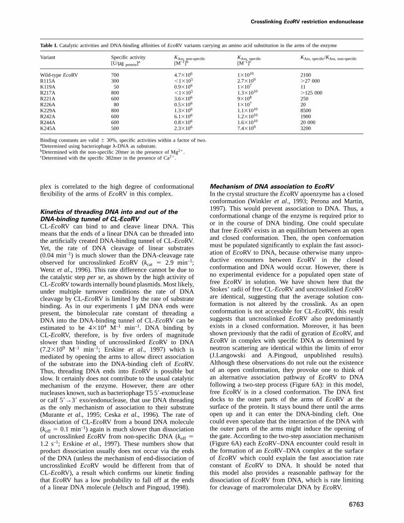
Mutagenesis of amino acid residues assumed tobe involved in initial binding of DNAOur data suggest that DNA initially can bind to the outerside of the arms ofEcoRV. This assumption appearsreasonable, as several basic amino acid residues are locatedon the outer side of the arms ofEcoRV (Figure 6B).Beside Lys119, Arg221 and Arg226, these residues arenot in contact with the DNA in the specific complex(Winkler et al., 1993). All of these amino acid residueswere individually exchanged for alanine by site-directedmutagenesis. The enzyme variants were purified and theirDNA-binding properties characterized (Table I). All ofthe variants retain at least 40% of the activity of wild-typeEcoRV, with the exception of the K119A and R226Avariants, in which amino acid residues are substituted thatcontact the DNA in the specific complex and are veryimportant for indirect readout (Wenzet al., 1996). TheK119A, R221A and R226A variants have a reduced non-
6762
specific affinity for DNA, but their specific DNA-bindingconstants are reduced even more. R242A and K245Abehave like wild-typeEcoRV. However, the non-specificDNA-binding constants of R115A, R217A, K229A andR244A are significantly more reduced than the specificbinding constants, leading to an increased value ofKAss, specific/KAss, non-specific. This result suggests, that theseresidues are important for non-specific DNA binding. Asthey do not interact with DNA bound at the internal bindingsite, it supports the existence of an outer binding site.
Discussion
A common feature of many enzymes which interact withDNA is that the DNA is completely encircled by theprotein. The architecture of a closed complex obviouslyprovides important advantages such as access to bothgrooves of the DNA, high complex stability and theability to locate target sites by linear diffusion. However,association to DNA becomes more complicated than inan open complex: it can occur either via the ends of theDNA by threading them into the protein or it requiresconformational changes of the protein. These conforma-tional changes of the protein can play an important rolein DNA recognition and enzymatic activity (Spolar andRecord, 1994). Here, we have addressed experimentallythese questions using the restriction enzymeEcoRV as amodel system, which is a dimeric protein that encirclesthe DNA with two arms. Both subunits of the enzyme werecrosslinked across the DNA-binding site using bivalentcysteine-specific chemical crosslinkers. If crosslinkingoccurs while a circular plasmid DNA is bound toEcoRV,the enzyme is catenated with the DNA (Figure 1B). Thisinteraction is irreversible as both molecular rings (circularDNA and crosslinked protein) are topologically intercon-nected unless the protein subunits are dissociated (whichrequires protein denaturation) or the DNA is cleaved(which requires Mg21 ions and the presence of anEcoRV-recognition site). The properties of such a chemicallymodified enzyme allow us to draw important conclusionsas to the dynamics ofEcoRV during its interaction withDNA and the mechanism of DNA binding.
Flexibility of the arms of EcoRVThe crosslinking yields ofEcoRV with four crosslinkersof different length are similar, suggesting that the arms ofEcoRV are flexible enough to accommodate differentdistances between the reactive maleimid groups. In agree-ment with this interpretation, crosslinking yields decreaseat lower temperatures, where some of this flexibility islost. Crosslinking yields were significantly larger ifEcoRVis bound non-specifically to DNA than if it were free orin specific complex with DNA. These results are in goodagreement with results of X-ray structure analyses whichshow that parts of the arms ofEcoRV (amino acid residues222–229) are poorly ordered in all complexes indicatingconformational flexibility (Perona and Martin, 1997).Whereas these loops retain a residual structure in theapoenzyme, as well as in specificEcoRV DNA complexes,they could not be modelled ifEcoRV is bound to anon-target DNA (Winkleret al., 1993). Thus, the highcrosslinking yield of the non-specificEcoRV–DNA com-
Crosslinking EcoRV restriction endonuclease
Table I. Catalytic activities and DNA-binding affinities ofEcoRV variants carrying an amino acid substitution in the arms of the enzyme
Variant Specific activity KAss, non-specific KAss, specific KAss, specific/KAss, non-specific[U/µg protein]
a [M–1]b [M–1]c
Wild-type EcoRV 700 4.73106 131010 2100R115A 300 ,13105 2.73109 .27 000K119A 50 0.93106 13107 11R217A 800 ,13105 1.331010 .125 000R221A 600 3.63106 93108 250R226A 80 0.53106 13107 20K229A 800 1.33106 1.131010 8500R242A 600 6.13106 1.231010 1900R244A 600 0.83106 1.631010 20 000K245A 500 2.33106 7.43109 3200
Binding constants are valid6 30%, specific activities within a factor of two.aDetermined using bacteriophageλ-DNA as substrate.bDetermined with the non-specific 20mer in the presence of Mg21.cDetermined with the specific 382mer in the presence of Ca21.
plex is correlated to the high degree of conformationalflexibility of the arms ofEcoRV in this complex.
Kinetics of threading DNA into and out of theDNA-binding tunnel of CL-EcoRVCL-EcoRV can bind to and cleave linear DNA. Thismeans that the ends of a linear DNA can be threaded intothe artificially created DNA-binding tunnel of CL-EcoRV.Yet, the rate of DNA cleavage of linear substrates(0.04 min–1) is much slower than the DNA-cleavage rateobserved for uncrosslinkedEcoRV (kcat 5 2.9 min–1;Wenzet al., 1996). This rate difference cannot be due tothe catalytic stepper se, as shown by the high activity ofCL-EcoRV towards internally bound plasmids. Most likely,under multiple turnover conditions the rate of DNAcleavage by CL-EcoRV is limited by the rate of substratebinding. As in our experiments 1µM DNA ends werepresent, the bimolecular rate constant of threading aDNA into the DNA-binding tunnel of CL-EcoRV can beestimated to be 43104 M–1 min–1. DNA binding byCL-EcoRV, therefore, is by five orders of magnitudeslower than binding of uncrosslinkedEcoRV to DNA(7.23109 M–1 min–1; Erskine et al., 1997) which ismediated by opening the arms to allow direct associationof the substrate into the DNA-binding cleft ofEcoRV.Thus, threading DNA ends intoEcoRV is possible butslow. It certainly does not contribute to the usual catalyticmechanism of the enzyme. However, there are othernucleases known, such as bacteriophage T5 59-exonucleaseor calf 59→39 exo/endonuclease, that use DNA threadingas the only mechanism of association to their substrate(Murante et al., 1995; Ceskaet al., 1996). The rate ofdissociation of CL-EcoRV from a bound DNA molecule(koff 5 0.1 min–1) again is much slower than dissociationof uncrosslinkedEcoRV from non-specific DNA (koff 51.2 s–1; Erskineet al., 1997). These numbers show thatproduct dissociation usually does not occur via the endsof the DNA (unless the mechanism of end-dissociation ofuncrosslinkedEcoRV would be different from that ofCL-EcoRV), a result which confirms our kinetic findingthat EcoRV has a low probability to fall off at the endsof a linear DNA molecule (Jeltsch and Pingoud, 1998).
6763
Mechanism of DNA association to EcoRVIn the crystal structure theEcoRV apoenzyme has a closedconformation (Winkleret al., 1993; Perona and Martin,1997). This would prevent association to DNA. Thus, aconformational change of the enzyme is required prior toor in the course of DNA binding. One could speculatethat freeEcoRV exists in an equilibrium between an openand closed conformation. Then, the open conformationmust be populated significantly to explain the fast associ-ation of EcoRV to DNA, because otherwise many unpro-ductive encounters betweenEcoRV in the closedconformation and DNA would occur. However, there isno experimental evidence for a populated open state offree EcoRV in solution. We have shown here that theStokes’ radii of free CL-EcoRV and uncrosslinkedEcoRVare identical, suggesting that the average solution con-formation is not altered by the crosslink. As an openconformation is not accessible for CL-EcoRV, this resultsuggests that uncrosslinkedEcoRV also predominantlyexists in a closed conformation. Moreover, it has beenshown previously that the radii of gyration ofEcoRV, andEcoRV in complex with specific DNA as determined byneutron scattering are identical within the limits of error(J.Langowski and A.Pingoud, unpublished results).Although these observations do not rule out the existenceof an open conformation, they provoke one to think ofan alternative association pathway ofEcoRV to DNAfollowing a two-step process (Figure 6A): in this model,free EcoRV is in a closed conformation. The DNA firstdocks to the outer parts of the arms ofEcoRV at thesurface of the protein. It stays bound there until the armsopen up and it can enter the DNA-binding cleft. Onecould even speculate that the interaction of the DNA withthe outer parts of the arms might induce the opening ofthe gate. According to the two-step association mechanism(Figure 6A) eachEcoRV–DNA encounter could result inthe formation of anEcoRV–DNA complex at the surfaceof EcoRV which could explain the fast association rateconstant ofEcoRV to DNA. It should be noted thatthis model also provides a reasonable pathway for thedissociation ofEcoRV from DNA, which is rate limitingfor cleavage of macromolecular DNA byEcoRV.
C.Schulze et al.
The two-step binding model is strongly supported byour finding that CL-EcoRV (which can only exist in aclosed conformation) can bind to circular DNA. Thisresult implies that a DNA-binding site at the surface ofEcoRV must exist which is a prerequisite for the two-stepbinding model. Most probably, this binding site is locatedat the outer side of the arms ofEcoRV in the closedconformation. There a wide, shallow groove exits, largeenough to accommodate B-DNA. The surface of thisgroove is decorated with 18 basic amino acid residues(Arg115, Lys119, Arg217, Arg221, Arg226, Lys229,Arg242, Arg244 and Lys245 of each subunit) (Figure 6B).We have shown that in particular Arg115, Arg217 and toa lesser extent Arg244 and Lys229 are required for non-specific binding, but not for specific complex formation.It is not clear at present how exactly the DNA is boundin this groove, if the protein undergoes conformationalchanges upon binding DNA to the outer binding site andwhether the amino acid residues shown to be importantfor non-specific binding make direct contacts to the DNAor contribute to an attractive electrostatic field.
Although EcoRV has been studied intensively over thelast 10 years, both biochemically and structurally, thisextra binding site for DNA had not been found so far[ironically, F.K.Winkler, after solving the structure of theEcoRV apoenzyme, speculated that the DNA-binding siteof EcoRV might be located at the outside of the arms, anidea that was not followed up after solving the cocrystalstructures (personal communication)]. Our data, withoutreasonable doubt, demonstrate the existence of this extrasite which we assume to be an entry site which is occupiedas an intermediate in each catalytic cycle. Non-specificbinding of DNA toEcoRV can occur at the outer bindingsite, as shown by the ability of CL-EcoRV to bind tocircular DNA, and at the internal binding site, as shownby the crystal structure of the non-specificEcoRV–DNAcomplex and the finding that non-specifically bound circu-lar DNA can be catenated with the enzyme by performingthe crosslinking reaction in the presence of DNA. As bothbinding modes are almost isoenergetic, non-specific DNAmay change its position more or less frequently.
Coupling of specific DNA binding to catalysisFollowing non-specific binding ofEcoRV to DNA theenzyme scans the sequence of the DNA by linear diffusionsearching for GATATC sites (Jeltschet al., 1996; Jeltschand Pingoud, 1998). If specific sites are encountered,the catalytic center of the enzyme is activated by aconformational change of theEcoRV–DNA complex.Whereas details of this conformational change areunknown, it is presumably associated with a transition ofthe loose non-specific complex with the DNA fluctuatingbetween the external and the internal binding site to atightly closed form in which the DNA is exclusivelybound at the internal site, as seen in the different specificEcoRV–DNA complexes (Figure 1A) (Winkleret al.,1993; Kostrewa and Winkler, 1995; Perona and Martin,1997). We have shown here that CL-EcoRV in which botharms are permanently joined to each other is more activethan uncrosslinkedEcoRV. The Arrhenius energy of activa-tion of DNA cleavage at canonical sites is reduced by180 kJ/mol in CL-EcoRV compared with nativeEcoRV.Interestingly, ground state DNA binding is barely influ-
6764
enced by the crosslink. In addition, CL-EcoRV is lessaccurate in cleaving DNA than uncrosslinkedEcoRV. Thismeans that the transition state energy for cleavage of astar-site by CL-EcoRV is much lower than for cleavageby uncrosslinkedEcoRV. Taken together, these resultsshow that crosslinkedEcoRV is frozen in a conformationwhich is closer to the conformation of the transition stateof catalysis than the average conformation of uncrosslinkedEcoRV—an unusual outcome of chemical modificationexperiments. It means, that bringing together the arms ofEcoRV and holding the DNA in the internal bindingsite makes the structure more transition state-like. Thesefindings reinforce the conclusion drawn from crystal-structure analyses that large domain movements do occurin EcoRV during DNA cleavage (Perona and Martin,1997). Furthermore, they show that these conformationalchanges are strongly linked to catalysis.
Materials and methods
Site-directed mutagenesis and purification of EcoRV andEcoRV variantsSite-directed PCR mutagenesis was carried out essentially as describedpreviously (Rothet al., 1998). Expression and purification of the enzymevariants were performed by chromatography on Ni-NTA–agarose andphosphocellulose as described previously (Wenzet al., 1994; Jeltsch andPingoud, 1998). Specific activity was determined usingλ-DNA assubstrate as described previously (Wenzet al., 1994).
Crosslinking of EcoRVFor the cysteine-specific chemical crosslinking experiments anEcoRVC21S/T222C variant was prepared by site-directed mutagenesis thatbears only one single cysteine residue per subunit at a position well-suited for intramolecular crosslinking. To obtain anEcoRV preparationin which all cysteine residues are reduced but that does not contain freeSH-reagents, 500µg of the purifiedEcoRV mutant were incubated with10 mM dithioerythritol (DTE) for 2 h at 0°C in 2 ml 20 mM Tris–HClpH 7, 0.25 mM EDTA, 150 mM NaCl, 38% (v/v) glycerol. The solutionwas dialyzed twice against 20 mM Tris–HCl pH 7, 50 mM NaCl toremove DTE. The SH-specific crosslinking reactions were carried outusing a series of bivalent-SH specific crosslinkers consisting of tworeactive maleimide groups linked by three, five, six or seven methylenegroups, synthesized and purified as described (Franke and Pingoud,1998). 1,6-bis(maleimido)hexan containing six methylene groups iscommercially available (Pierce chemicals).
For the analytical crosslinking reactionsEcoRV (1 µM) was incubatedin 20 mM Tris–HCl pH 7.5, 50 mM NaCl containing either 10 mMMgCl2, 10 mM CaCl2 or 2 mM EDTA in the absence or presenceof oligonucleotide (5µM, 59-GAGGAAAGATATCTTTCCTC-39) for30 min. The crosslinkers were added to a final concentration of 2.5 mMand after 3 min incubation the reaction was stopped with 13 mM DTE.For preparative crosslinking reactions 500µg EcoRV were incubatedwith 500 µg pAT153 in 5 ml 20 mM Tris–HCl pH 7, 50 mM NaCl,10 mM CaCl2 for 20 min at room temperature. The crosslinking reactionwas started by the addition of the crosslinker that contains six methylenegroups to a final concentration of 50µM. After 5 min the reaction wasstopped by addition of DTE to a final concentration of 100µM.
Purification of crosslinked EcoRV in complex with DNAThe solution obtained after the preparative crosslinking reaction wasdiluted 1:3 with buffer N (30 mM potassium phosphate pH 7.6, 0.1 mMDTE, 0.01% Lubrol, 0.5 M NaCl, 10 mM imidazole) and applied ontoa small column filled with 2 ml Ni-NTA–Agarose (Qiagen) equilibratedwith buffer N. In contrast to uncrosslinkedEcoRV, CL-EcoRV incomplex with DNA does not bind to Ni-NTA. The flowthrough containingCL-EcoRV was concentrated to a final volume of 500µl using Centriplus10 columns (Amicon) and dialyzed against 20 mM Tris–HCl pH 7.5,50 mM NaCl, 2 mM EDTA (buffer A). The sample was applied onto aMono Q column (5.530.6 cm, Pharmacia) equilibrated with buffer A.The elution was carried out with a gradient of 0–50% buffer B (20 mMTris–HCl pH 7.5, 2 mM EDTA, 2 M NaCl) in 80 min. After aconcentration step using a Centriplus 10 column, theEcoRV preparation
Crosslinking EcoRV restriction endonuclease
was dialyzed against storage buffer [30 mM potassium phosphate pH 7.6,0.5 mM EDTA, 0.1 mM DTE, 0.01% Lubrol, 0.3 M NaCl, 77%(v/v) glycerol].
Purification of crosslinked EcoRV without DNAEcoRV was crosslinked in the presence of plasmid DNA as described.After purification using Ni-NTA–agarose, 1000 U ofS.marcescensnuclease were added to the flowthrough and incubated for 10 h at 4°Cin 20 mM Tris–HCl pH 7.5, 50 mM NaCl, 10 mM MgCl2 to digest theDNA. Then, NaCl was added to a final concentration of 1 M. Underthese conditions,EcoRV does not bind to DNA. CL-EcoRV was separatedfrom the DNA and theS.marcescensnuclease using a Ni-NTA–agarosecolumn as described by Wenzet al. (1994).
Analytical ultracentrifugation experimentsSedimentation velocity runs with CL-EcoRV and uncrosslinkedEcoRVwere carried out at 20°C in 20 mM Tris–HCl pH 7.5, 100 mM NaCl,5 mM MgCl2, at 40 000 r.p.m. in a Beckman Optima SL-A centrifugeequipped with absorption optics and an An-50 8-place rotor. The proteinconcentration was 1µM. Sedimentation velocity data were evaluatedwith the program package AKKUPROG by fitting the time-dependentconcentration profiles calculated with Lamm’s differential equation fora simple sedimenting species to the measured data.
DNA-binding analysesDNA binding was analyzed by nitrocellulose-filter binding experimentsin a dot blot apparatus (Bio-Rad). The nitrocellulose membrane(Schleicher and Schuell) was equilibrated in washing buffer (20 mMTris–HCl pH 7.5, 50 mM NaCl, 10 mM CaCl2) for 30 min. After 30 minincubation at ambient temperature the samples containing labelled DNAand enzyme were sucked through the nitrocellulose membrane andwashed twice. For all binding reactions, CL-EcoRV without DNA wasused. For specific DNA-binding experiments a radioactively labelled382 bp fragment containing a singleEcoRV site was prepared asdescribed (Jeltschet al., 1995). Forty picomolar 382mer was titratedwith increasing amounts of either CL-EcoRV or uncrosslinkedEcoRVin binding buffer [20 mM Tris–HCl pH 7.5, 50 mM NaCl, 10 mMCaCl2, 100 µg/ml bovine serum albumin (BSA)]. Binding to a non-specific oligonucleotide was analyzed using 2 mM of the 20meroligonucleotide 59-GATCGACGAGCTCGTCGATC-39 in cleavage buf-fer (20 mM Tris–HCl pH 7.5, 50 mM NaCl, 10 mM MgCl2) with100 µg/ml BSA. The oligonucleotide was radioactively labelled with[γ-32P]dATP and T4 polynucleotide kinase. To measure binding tocircular plasmid DNA, pAT153 was radioactively labeled by nick-translation with [α-32P]dATP and DNA polymerase I. Binding experi-ments were carried out in washing buffer using 0.7 pM labeled DNA.The detection and quantification of bound DNA was performed usingan InstantImager system (Canberra Packard). To calculateKAss-values,the bound DNA versuscenzymedata obtained in the binding experimentswere fitted to the equation characterizing a bimolecular binding equi-librium.
To determine the rate constant for dissociation ofEcoRV from DNA(koff) 100 nM CL-EcoRV was incubated with 4 nM of radioactive 382 bpDNA fragment in cleavage buffer for 30 min to allow complex formation.Then the sample was diluted 1:50 with cleavage buffer containing 12 nMunlabelled DNA fragment and incubated for a defined period of time.The fraction of DNA still bound byEcoRV was analyzed by nitrocellu-lose-filter binding as described above. Dissociation kinetics were fittedto a mono-exponential decay curve.
DNA-cleavage experiments with plasmid DNACL-EcoRV (100 nM containing 5 nM pAT153 internal plasmid, whichmeans that ~20EcoRV molecules are bound to the plasmid in thispreparation) was incubated in cleavage buffer. After defined time intervalsthe reaction was stopped by addition of agarose gel loading buffercontaining 250 mM EDTA and the amount of DNA cleavage wasanalyzed by agarose gel electrophoresis. For the kinetic experiments aquenched flow apparatus (SFM-3, Bio-logic, Claix, France) was used.The DNA-cleavage reaction was started by mixing a solution of 80 nMCL-EcoRV containing 4 nM pAT153 in 20 mM Tris–HCl pH 7.5, 1 mMEDTA, 10 mM NaCl with an equal volume of 20 mM Tris–HCl pH 7.5,20 mM MgCl2 and stopped by adding the same volume of 50 mMEDTA. Experiments were carried out under the same conditions withuncrosslinkedEcoRV.
Oligonucleotide-cleavage experimentsFor oligonucleotide-cleavage experiments we used 0.5µM radioactivelylabeled specific oligonucleotide (59-GAGGAAAGATATCTTTCCTC-39)
6765
and 100 nM CL-EcoRV (containing 5 nM pAT153 internal plasmid) incleavage buffer. Prior to the addition of the oligonucleotide substrateCL-EcoRV was pre-incubated for 30 min at ambient temperature incleavage buffer to allow for digestion of the internal plasmid. Afterdefined time intervals, aliquots were withdrawn from the reactionmixture, spotted onto a DEAE–cellulose plate (Macherey-Nagel, Du¨ren,Germany) and subjected to homochromatography. Detection and quanti-fication of the separated substrates and products were achieved using anInstantImager system (Canberra Packard). To analyze the interaction ofCL-EcoRV with circular DNA, oligonucleotide cleavage experimentswere carried out using 100 nM CL-EcoRV (containing 5 nM pAT153internal plasmid) in the presence of 0–90 nM circular pUC8 (2665 bp)as a competitor DNA. pUC8 was used, because it does not contain anEcoRV site. Similar experiments were performed with uncrosslinkedEcoRV under identical conditions using 20 nMEcoRV in the presenceof 5 nM linear pAT153.
Acknowledgements
We thank Drs Fritz Winkler and John Perona for unpublished structuralinformation, Dr Stephen Halford for communication of results prior topublication and Drs Wolfgang Wende and Christian Wenz for valuablediscussions. The financial support of the Deutsche Forschungsgemeinsch-aft (Pi-122/12–2) and the Fonds der Chemischen Industrie is gratefullyacknowledged. C.S. is a recipient of a scholarship of the Stipendienfondsder Chemischen Industrie.
References
Ceska,T.A., Sayers,J.R., Stier,G. and Suck,D. (1996) A helical archallowing single-stranded DNA to thread through T5 59-exonuclease.Nature, 382, 90–93.
Erskine,S.G., Baldwin,G.S. and Halford,S.E. (1997) Rapid-reactionanalysis of plasmid DNA cleavage by theEcoRV restrictionendonuclease. Biochemistry, 36, 7567–7576.
Franke,I. and Pingoud,A. (1998) Synthesis and biochemical characteriza-tion of obligatory dimers of the sugar non-specific nuclease fromSerratia marcescensusing specifically designed bismaleimidoalkanesas SH-specific crosslinking reagents.J. Prot. Chem., in press.
Halford,S.E., Lovelady,B.M. and McCallum,S.A. (1986) Relaxedspecificity of theEcoRV restriction endonuclease.Gene, 41, 173–181.
Jeltsch,A. and Pingoud,A. (1998) Kinetic characterization of lineardiffusion of the restriction endonucleaseEcoRV on DNA.Biochemistry, 37, 2160–2169.
Jeltsch,A., Maschke,H., Selent,U., Wenz,C., Ko¨hler,E., Connolly,B.A.,Thorogood,H. and Pingoud,A. (1995) DNA binding specificity of theEcoRV restriction endonuclease is increased by Mg21 binding to ametal ion binding site distinct from the catalytic center of the enzyme.Biochemistry, 34, 6239–6246.
Jeltsch,A., Wenz,C., Stahl,F. and Pingoud,A. (1996) Linear diffusion ofthe restriction endonucleaseEcoRV on DNA is essential for thein vivofunction of the enzyme.EMBO J., 15, 5104–5111.
Kostrewa,D. and Winkler,F.K. (1995) Mg21 binding to the active siteof EcoRV endonuclease: a crystallographic study of complexes withsubstrate and product DNA at 2 Å resolution. Biochemistry, 34,683–696.
Murante,R.S., Rust,L. and Bambara,R.A. (1995) Calf 59 to 39 exo/endonuclease must slide from a 59 end of the substrate to performstructure-specific cleavage.J. Biol. Chem., 270, 30377–30383.
Perona,J.J. and Martin,A.M. (1997) Conformational transition andstructural deformability of EcoRV endonuclease revealed bycrystallographic analysis.J. Mol. Biol., 273, 207–225.
Pingoud,A. and Jeltsch,A. (1997) Recognition and cleavage of DNA bytype-II restriction endonucleases.Eur. J. Biochem., 246, 1–22.
Roberts,R.J. and Halford,S.E. (1993) Type II restriction enzymes. InLinn,S.M., Lloyd,R.S. and Roberts,R.J. (eds),Nucleases.Cold SpringHarbor Laboratory Press, Cold Spring Harbor, NY, pp. 35–88.
Roth,M., Helm-Kruse,S. Friedrich,T. and Jeltsch,A. (1998) Functionalroles of conserved amino acid residues in DNA methyltransferasesinvestigated by site directed mutagenesis ofEcoRV adenin-N6-methyltransferase.J. Biol. Chem., 273, 17333–17342.
Spolar,R.S. and Record,M.T.,Jr (1994) Coupling of local folding to site-specific binding of proteins to DNA. Science, 263, 777–784.
C.Schulze et al.
Stahl,F., Wende,W., Jeltsch,A. and Pingoud,A. (1996) Introduction ofasymmetry in the naturally symmetric restriction endonucleaseEcoRVto investigate intersubunit communication in the homodimeric protein.Proc. Natl Acad. Sci. USA, 93, 6175–6180.
Taylor,J.D., Badcoe,I.G., Clarke,A.R. and Halford,S.E. (1991)EcoRVrestriction endonuclease binds all DNA sequences with equal affinity.Biochemistry, 30, 8743–8753.
Thielking,V., Selent,U., Ko¨hler,E., Wolfes,H., Pieper,U., Geiger,R.,Urbanke,C., Winkler,F.K. and Pingoud,A. (1991) Site-directedmutagenesis studies withEcoRV restriction endonuclease to identifyregions involved in recognition and catalysis. Biochemistry, 30,6416–6422.
Vipond,I.B. and Halford,S.E. (1995) Specific DNA recognition byEcoRVrestriction endonuclease induced by calcium ions. Biochemistry, 34,1113–1119.
Wende,W., Stahl,F. and Pingoud A. (1996) The production andcharacterization of artificial heterodimers of the restrictionendonucleaseEcoRV. Biol. Chem., 377, 625–632.
Wenz,C., Selent,U., Wende,W., Jeltsch,A., Wolfes,H. and Pingoud,A.(1994) Protein engineering of the restriction endonucleaseEcoRV:replacement of an amino acid residue in the DNA binding site leadsto an altered selectivity towards unmodified and modified substrates.Biochim. Biophys. Acta, 1219, 73–80.
Wenz,C., Jeltsch,A. and Pingoud,A. (1996) Probing the indirect readoutof the restriction enzymeEcoRV. J. Biol. Chem., 271, 5565–5573.
Winkler,F.K. et al. (1993) The crystal structure ofEcoRV endonucleaseand of its complexes with cognate and non-cognate DNA fragments.EMBO J., 12, 1781–1795.
Received August 20, 1998; revised and accepted September 28, 1998
6766
Copyright © 2022 FDOKUMEN