Critical role of PBEF expression in pulmonary cell inflammation and permeability
12
Critical role of PBEF expression in pulmonary cell inflammation and permeability Peng Liu a,b,1 , Hailong Li a,1 , Javier Cepeda a , Li Qin Zhang a , Xiuyun Cui b , Joe G.N. Garcia c , Shui Qing Ye a, * a Department of Surgery and Department of Molecular Microbiology and Immunology, University of Missouri, Columbia, MO, USA b Department of Biochemistry, Dalian Medical University, Dalian, China c Department of Medicine, University of Chicago, Chicago, IL, USA Received 13 April 2008; revised 5 September 2008; accepted 13 October 2008 Abstract Previous studies in our lab have identified pre-B-cell colony enhancing factor (PBEF) as a novel biomarker in acute lung injury. This study continues to elucidate the underlying molecular mechanism of PBEF in the pathogenesis of acute lung injury in pulmonary cell culture models. Our results revealed that IL-1b induced PBEF expression in pulmonary vascular endothelial cells at the transcriptional level and a 1535 T- variant in the human PBEF gene promoter significantly attenuated its binding to an IL-1b-induced unknown transcription factor. This may underlie the reduced expression of PBEF and thus the lower susceptibility to acute lung injury in 1535T carriers. Furthermore, overexpression of PBEF significantly augmented IL-8 secretion and mRNA expression by more than 6-fold and 2-fold in A549 cells and HPAEC, respectively. It also significantly augmented IL-1b-mediated cell permeability by 44% in A549 cells and 65% in endothelial cells. The knockdown of PBEF expression significantly inhibited IL-1b-stimulated IL-8 secretion and mRNA level by 60% and 70%, respectively, and the knockdown of PBEF expression also significantly attenuated IL-1b-induced cell permeability by 29% in epithelial cells and 24% in endothelial cells. PBEF expression also affected the expression of two other inflammatory cytokines (IL-16 and CCR3 genes). These results suggest that PBEF is critically involved in pulmonary vascular and epithelial inflammation and permeability, which are hallmark features in the pathogenesis of acute lung injury. This study lends further support to our finding that PBEF is a potential new target in acute lung injury. Ó 2008 International Federation for Cell Biology. Published by Elsevier Ltd. All rights reserved. Keywords: Pulmonary inflammation; Permeability; PBEF; Acute lung injury; IL-1b; IL-8; Epithelial cells; Endothelial cells; Inflammatory cytokines 1. Introduction Acute lung injury (ALI) and its more severe form, acute respiratory distress syndrome (ARDS), are characterized by alveolar inflammation, injury and increased pulmonary capil- lary permeability. The clinical consequence is an impaired gas exchange and ultimate respiratory failure (Leaver and Evans, 2007; Levitt and Matthay, 2006). Despite considerable prog- ress in basic and clinical research, the mortality and morbidity of ALI/ARDS remain high because the etiology and molecular pathogenesis are still incompletely understood. In our previous study on animal models of ALI, we identified pre-B-cell colony enhancing factor (PBEF) as a significantly upregulated gene in ALI (Ye et al., 2005a). We discovered single nucleotide polymorphisms in the human PBEF gene promoter. Moreover, we found that carriers of the haplotype GC from SNPs T-1001G and C-1543T had a 7.7-fold higher risk of ALI. The T-variant from the SNP C-1535T resulted in a significant decrease in the transcription rate (1.8-fold) by the * Corresponding author. Department of Surgery and Department of Molec- ular Microbiology and Immunology, University of Missouri School of Medi- cine, Medical Science Bldg. #M701, DC097.00, One Hospital Drive, Columbia, MO 65212, USA. Fax: þ1 573 884 3330. E-mail address: [email protected] (S.Q. Ye). 1 These authors contributed equally. 1065-6995/$ - see front matter Ó 2008 International Federation for Cell Biology. Published by Elsevier Ltd. All rights reserved. doi:10.1016/j.cellbi.2008.10.015 Cell Biology International 33 (2009) 19e30 www.elsevier.com/locate/cellbi
-
Upload
independent -
Category
Documents
-
view
1 -
download
0
Transcript of Critical role of PBEF expression in pulmonary cell inflammation and permeability
Cell Biology International 33 (2009) 19e30www.elsevier.com/locate/cellbi
Critical role of PBEF expression in pulmonary cellinflammation and permeability
Peng Liu a,b,1, Hailong Li a,1, Javier Cepeda a, Li Qin Zhang a, Xiuyun Cui b,Joe G.N. Garcia c, Shui Qing Ye a,*
a Department of Surgery and Department of Molecular Microbiology and Immunology, University of Missouri, Columbia, MO, USAb Department of Biochemistry, Dalian Medical University, Dalian, China
c Department of Medicine, University of Chicago, Chicago, IL, USA
Received 13 April 2008; revised 5 September 2008; accepted 13 October 2008
Abstract
Previous studies in our lab have identified pre-B-cell colony enhancing factor (PBEF) as a novel biomarker in acute lung injury. This studycontinues to elucidate the underlying molecular mechanism of PBEF in the pathogenesis of acute lung injury in pulmonary cell culture models.Our results revealed that IL-1b induced PBEF expression in pulmonary vascular endothelial cells at the transcriptional level and a �1535 T-variant in the human PBEF gene promoter significantly attenuated its binding to an IL-1b-induced unknown transcription factor. This mayunderlie the reduced expression of PBEF and thus the lower susceptibility to acute lung injury in �1535T carriers. Furthermore, overexpressionof PBEF significantly augmented IL-8 secretion and mRNA expression by more than 6-fold and 2-fold in A549 cells and HPAEC, respectively. Italso significantly augmented IL-1b-mediated cell permeability by 44% in A549 cells and 65% in endothelial cells. The knockdown of PBEFexpression significantly inhibited IL-1b-stimulated IL-8 secretion and mRNA level by 60% and 70%, respectively, and the knockdown of PBEFexpression also significantly attenuated IL-1b-induced cell permeability by 29% in epithelial cells and 24% in endothelial cells. PBEFexpression also affected the expression of two other inflammatory cytokines (IL-16 and CCR3 genes). These results suggest that PBEF iscritically involved in pulmonary vascular and epithelial inflammation and permeability, which are hallmark features in the pathogenesis of acutelung injury. This study lends further support to our finding that PBEF is a potential new target in acute lung injury.� 2008 International Federation for Cell Biology. Published by Elsevier Ltd. All rights reserved.
Keywords: Pulmonary inflammation; Permeability; PBEF; Acute lung injury; IL-1b; IL-8; Epithelial cells; Endothelial cells; Inflammatory cytokines
1. Introduction
Acute lung injury (ALI) and its more severe form, acuterespiratory distress syndrome (ARDS), are characterized byalveolar inflammation, injury and increased pulmonary capil-lary permeability. The clinical consequence is an impaired gas
* Corresponding author. Department of Surgery and Department of Molec-
ular Microbiology and Immunology, University of Missouri School of Medi-
cine, Medical Science Bldg. #M701, DC097.00, One Hospital Drive,
Columbia, MO 65212, USA. Fax: þ1 573 884 3330.
E-mail address: [email protected] (S.Q. Ye).1 These authors contributed equally.
1065-6995/$ - see front matter � 2008 International Federation for Cell Biology.
doi:10.1016/j.cellbi.2008.10.015
exchange and ultimate respiratory failure (Leaver and Evans,2007; Levitt and Matthay, 2006). Despite considerable prog-ress in basic and clinical research, the mortality and morbidityof ALI/ARDS remain high because the etiology and molecularpathogenesis are still incompletely understood.
In our previous study on animal models of ALI, we identifiedpre-B-cell colony enhancing factor (PBEF) as a significantlyupregulated gene in ALI (Ye et al., 2005a). We discoveredsingle nucleotide polymorphisms in the human PBEF genepromoter. Moreover, we found that carriers of the haplotype GCfrom SNPs T-1001G and C-1543T had a 7.7-fold higher risk ofALI. The T-variant from the SNP C-1535T resulted ina significant decrease in the transcription rate (1.8-fold) by the
Published by Elsevier Ltd. All rights reserved.
20 P. Liu et al. / Cell Biology International 33 (2009) 19e30
reporter gene assay (Ye et al., 2005a). Our findings wereconfirmed and extended in a separate and larger population(>1000 patients) by Bajwa et al. (2006). We further found thata reduction in PBEF protein expression by siRNA, significantlyattenuated pulmonary artery endothelial cell barrier dysfunc-tion induced by the potent edemagenic agent, thrombin, asreflected by reductions in transendothelial electric resistance(Ye et al., 2005b). Taken together, these results strongly indi-cate that PBEF may be a potential novel candidate gene andbiomarker in ALI.
The objective of this study was to elucidate the molecularmechanisms underlying PBEF in the pathogenesis of acutelung injury using human pulmonary cells as in vitro cellmodels. Since increased vascular and epithelial cell inflam-mation and permeability processes are important features ofALI, we assessed the effect of PBEF knockdown with PBEFsiRNA and PBEF overexpression on basal and IL-1b-mediatedhuman pulmonary epithelial cell (A549) and human pulmo-nary artery endothelial cell (HPAEC) IL-8 production andpermeability by in vitro cell permeability assay. We alsoexamined the role of PBEF expression on the expression ofother inflammatory cytokines, such as IL-16 and CCR3.
2. Materials and methods
2.1. Materials
A rabbit anti-human IL-8 polyclonal antibody (Cat. No. sc-7922) and a mouse anti-human b-actin monoclonal antibody(Cat. No. A1978) were obtained from SigmaeAldrich (St. Louis,MO, USA). A rabbit anti-human PBEF polyclonal antibody wasfrom the Bethyl Laboratories, Inc. (Cat. No. A300-372A,Montgomery, TX, USA). A recombinant human IL-1b (Cat. No.201-LB) was obtained from R&D Systems Inc. (Minneapolis,MN, USA). Superscript III Reverse Transcriptase (Cat. No.18080044) and Platinum Taq DNA polymerase (Cat. No.10966018) were obtained from Invitrogen (Carlsbad, CA, USA).Tricine was purchased from SigmaeAldrich (Cat. No. T0377,St. Louis, MO, USA). Sources of other key reagents are specifiedin relevant texts.
2.2. Cell culture
Human A549 cell, a lung carcinomatous type II alveolarepithelial cell line, was obtained from ATCC (Cat. No. CCL-185�, Manassas, VA, USA) and maintained in DMEM sup-plemented with 10% fetal bovine serum, 2 mM glutamine,penicillin/streptomycin. Primary human pulmonary arteryendothelial cells (HPAEC, Cat. No. CC-2530) were obtainedfrom Cambrex Bio Science Inc. (Walkersville, MD, USA) andmaintained in EGM�-2 Endothelial Cell Medium-2 (Cat. No.CC-4176). All cells were cultured at 37 �C in a humidifiedatmosphere of 5% CO2, 95% air. Cells from each primary flaskwere detached with 0.25% trypsin, resuspended in fresh culturemedia, and seeded into 6-well plates for Western blotting or RT-PCR analysis or seeded into the culture inserts for in vitro cellpermeability assays.
2.3. Inhibition of newly synthesized PBEF mRNA
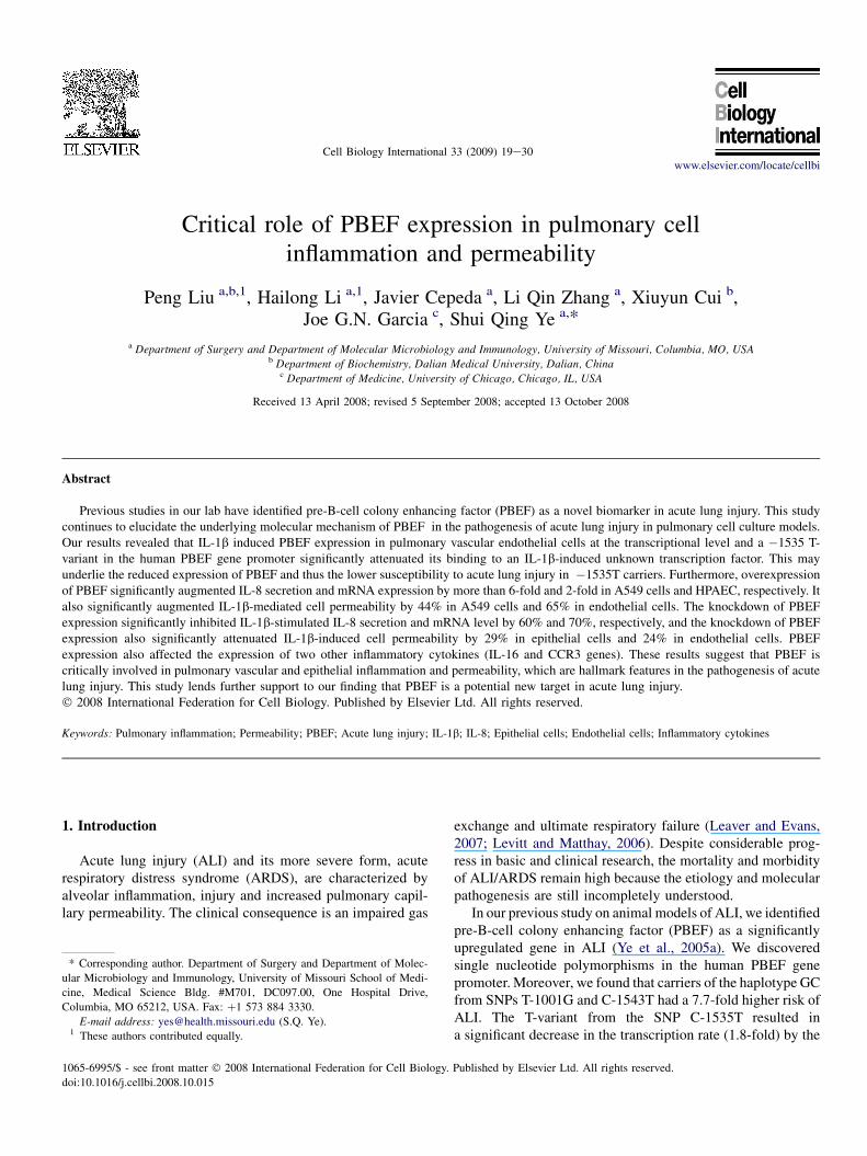
HPAEC were grown to confluence and incubated in thepresence or absence of IL-1b (10 ng/ml) plus or minus 5 mg/mlactinomycin D, a transcription inhibitor, for 4 h. After treat-ment, total cellular RNA was analyzed by a semi-quantitativeRT-PCR for the expression of PBEF mRNA. Briefly, total RNA(0.5 mg) from each sample was used as a starting material in thereverse transcriptase reaction. The primers for human PBEFcDNA and human ribosomal protein S18 (RPS18) gene, usedas a house-keeping gene control, were designed based onpublished GenBank mRNA sequences: NM_005746.2 andK03432, respectively. The primer sequences for the humanPBEF were as follows: PBEF-5, 50-AAG CTT TTT AGG GCCCTTTG-30; PBEF-3, 50-AGG CCA TGT TTT ATT TGC TGA-30.The PCR product size was 319 bp. The primer sequences forthe human 18S rRNAwere: M18R5, ATG GCC GTT CTTAGTTGGTG; M18R3, GGG ACT TAA TCA ACG CAAGC; PCRproduct size was 154 bp. The primers were synthesized byIntegrated DNATechnologies (Coralville, IA). After the reversetranscriptase reaction, one-tenth of the RT product was used ina typical PCR run of 25 cycles. The PCR products were sepa-rated on agarose gel electrophoresis and visualized by ethidiumbromide staining. The band image was acquired using theAlpha Imager and analyzed by the AlphaEase� Stand AloneSoftware (Alpha Innotech Corp., San leandro, CA). Statisticalcomparative analysis of integrated density value (mean -� SEM) between control and ALI groups was performed usingthe unpaired t-test.
2.4. Electrophoretic mobility shift assay (EMSA)
EMSA was performed essentially as described by Wanget al. (1998), except that the probe was labeled with Biotininstead of a radioisotope and most reagents were provided byPierce (Rockford, IL). Two 30-mer probes: �1535T, 50-tac attggt gag tgc Ttg gtg ata cct tcc-30 and �1535C, 50-tac att ggtgag tgc Ctg gtg ata cct tcc-30, were synthesized and 50
biotinylated by Integrated DNA Technologies (Coralville, IA).They differ in one nucleotide as indicated by the capital fontat the �1535 position of the PBEF gene promoter. Nuclearextracts from HPAEC, with or without IL1-b (10 ng/ml)treatment for 4 h, were prepared using NE-PER� nuclear andcytoplasmic extraction reagents (Pierce, Rockford, IL). Halt�protease inhibitor cocktail (Pierce, Rockford, IL) was addedinto the nuclear extract to inhibit potential protein degradationby endogenous proteases. Frozen aliquots of the nuclearproteins were stored at �70 �C for future use. The proteinconcentration of nuclear extracts was determined by themethods of Bradford (1976). DNA-binding reactions andchemiluminescent nucleic acid detection were carried outaccording to the supplier’s instruction in LightShift� chemi-luminescent EMSA kit (Pierce, Rockford, IL). The imageswere acquired and recorded using The FluroChem� imagingsystem (Alpha Innotech Corporation, San Leandro, CA).
21P. Liu et al. / Cell Biology International 33 (2009) 19e30
2.5. Transfection of PBEF siRNA into human A549 cells
PBEF stealth siRNAwas designed based on the human PBEFcDNA reference sequence (NM_005746.1) using the BLOCK-iT� RNAi Designer (Invitrogen, Carlsbad, CA, USA). UsingGFP-labeled non-specific siRNA, we first optimized theconditions for human A549 cells transfection and achieved>90% transfection efficiency using the Lipofectamine 2000reagent (Cat. No. 11668-019, Invitrogen, Carlsbad, CA, USA).To transfect PBEF stealth siRNA into human A549 cells, cellswere seeded for 24 h in the regular growth medium (withoutantibiotics) so that they would be 80e90% confluent at the timeof transfection. For each transfection in 24-well plates, 50 pmolPBEF stealth siRNA was diluted in 50 ml Opti-MEM I withoutserum and gently mixed with 1 ml Lipofectamine 2000 diluted inthe 50 ml Opti-MEM I (Cat. No. 31985-062, Invitrogen, Carls-bad, CA, USA). After incubation for 15 min at room tempera-ture, PBEF stealth siRNA and Lipofectamine 2000 complexeswere added to each well. Cell culture plates were gently mixedby rocking back and forth. The amounts of PBEF stealth siRNAand Lipofectamine 2000 were adjusted according to thedifferent sizes of cell culture plates. Transfected cells werefurther incubated at 37 �C for 48 h until the treatment with IL-1bwas performed. Transfection of PBEF siRNA into HPAEC wascarried out as described previously by Ye et al. (2005b).
2.6. Preparation and expression of the PBEF-overexpressing construct pCAGGS-mPBEF
A mouse PBEF (mPBEF) coding cDNA sequence wasamplified from a mouse lung total RNA (Cat. No. 736511,Stratagene, La Jolla, CA, USA) by RT-PCR using the followingprimer pair: tta gaa ttc gcc acc atg aat gct gcg gca gaagc; tag aattcc taA TGG TGA TGG TGA TGATG caa atg agg tgc cac gtcctgct. The bold letters indicate the optimized Kozac sequence.The capitalized letter section is the His tag sequence. The italicsequences are EcoRI adaptors. The amplified mouse PBEFcDNA was digested with EcoRI, subcloned into the uniqueEcoRI site of pCAGGS vector, i.e., pCAGGS-mPBEF, and thensequence-verified. In this construct, mouse PBEF expressionwas driven by a chicken beta-actin/rabbit beta-globin hybridpromoter (AG) with an enhancer from the human cytomegalo-virus immediate early promoter (CMV-IE). Overexpression ofPBEF in A549 cells was carried out by a transient transfection ofpCAGGS-mPBEF into A549 cells. Briefly, one day beforetransfection, A549 cells were plated in 6-well plate at 5 � 105
cells/well in 2 ml of growth medium without antibiotics. On the
Table 1
Primers and product sizes
Product 50 Primers 30 Primers
PBEF AAGCTTTTTAGGGCCCTTTG AGGCCA
IL-8 ATGACTTCCAAGCTGGCCGT CCTCTTC
b-Actin CAAACATGATCTGGGTCATCTTCTC GCTCGT
IL-16 TAGTGCCAAGGTACAAACAGGTG GGGTCT
CCR3 AGCCCCTAAAGCAGCACTAA TGATAGC
day of transfection, cells were at 95% confluence. For each well,4 mg plasmid DNA was transfected using Lipofectamine 2000(Cat. No. 11668-019, Invitrogen, Carlsbad, CA, USA) accordingto the supplier’s instructions. Cell medium and cell lysateproteins were harvested at different time points of interest afterthe transfection, respectively.
2.7. Isolation of RNA and RT-PCR analysis
Total RNA from in vitro cultured cells was isolated using theTRIzol solution (Cat. No. 15596-018, Invitrogen, Carlsbad,CA, USA) according to the supplier’s instruction. RT-PCR wasperformed using the following procedures. One microgramtotal RNA was reverse transcribed using an Invitrogen reversetranscriptase (Superscript III, Cat. No. 18080-044) with randomhexamers at 50 �C for 1 h followed by 70 �C for 15 min and4 �C for 5 min in a 20-ml reaction volume. Each PCR reactionfrom the cDNA template (2 ml RT product) was performedusing gene specific primers (Table 1) with 94 �C for 3 min, then32 cycles at 94 �C for 1 min, 55 �C for 1 min and 72 �C for1 min, followed by 72 �C for 7 min for the final extension.b-Actin was used as a house-keeping gene control. PCRproducts were separated on a 1.5% agarose gel and stained byEthidium Bromide (0.5 mg/ml). The band image was acquiredusing an Alpha Imager and analyzed by the AlphaEase�Stand Alone Software (Alpha Innotech Corp., San leandro,CA, USA).
2.8. Western blotting
Western blotting analysis was performed as describedpreviously (McGlothlin et al., 2005; Ye et al., 2005a,b) witha minor modification. Briefly, after washing with PBS, cells-were lysed with 500 ml of cell lysis buffer containing10 mM Tris (pH 7.4), 1% Triton X-100, 0.5% Nonidet P-40,150 mM NaCl, 1 mM EDTA, 0.2 mM EGTA, 0.2 mM vana-date, 0.2 mM PMSF, and 0.5% protease inhibitor cocktail. Totalcell lysates were cleared by centrifugation, boiled with the sameamount of 4� SDS sample buffer for 5 min. Total proteins ofcell lysates were quantified using the BCA Protein Assay Kit(Pierce Biotechnology, Inc., Rockford, IL, USA). An equalamount of total protein from each sample was then subjected to16.5% Tris/tricine polyacrylamide gel electrophoresis. Theseparated proteins were transferred to PVDF membranes byelectrotransfer. The blots were subsequently blocked with 5%bovine serum albumin in phosphate-buffered saline containing0.1% Tween 20 (TBS-T) at room temperature for 1 h and then
Size (bp) Accession no.
TGTTTTATTTGCTGACAAA 319 NM_005746
AAAAACTTCTCCACACC 297 NM_000584
CGTCGACAACGGCTC 487 NM_001101
CAAACTCAGATGCCTAT 280 NM_172217
TTAGGCGTCACCA 228 NM_001837

15
20
25
30
PBEF
β-actin
0 5 10 15 20 25 IL-1β (ng/ml)
Time (24 hours)
Celllysate
** ** ** ****
en
sity valu
e (x10
5)
A
B
22 P. Liu et al. / Cell Biology International 33 (2009) 19e30
incubated at 4 �C overnight with a primary antibody of interest.After washing three times for 10 min each with TBS-T, themembrane was incubated with a horseradish-peroxidase-linkedsecondary antibody of interest at room temperature for 1 h. Theblots were then visualized with the ECL Western blot detectionsystem (Cat. No. RPN2106, Amersham Bosciences, Buck-inghamshire, UK). The same membrane was re-probed with ananti-human b-actin antibody. b-actin was used as an internalcontrol. The band image was acquired using an Alpha Imagerand analyzed by the AlphaEase� Stand Alone Software (AlphaInnotech Corp., San Leandro, CA, USA). Band density onWestern blot images was used as a measure of assayed proteinlevel.
5
10
ated
d
2.9. In vitro cell permeability assay
00 5 10 15 20 25
Dose (ng/ml)
In
teg
r
Fig. 1. Doseeresponse of IL-1b-induced PBEF protein expression in A549
cells. (A) A representative Western blotting image of PBEF and b-actin protein
detections. After starving for serum overnight, A549 cells were stimulated
with different doses of IL-1b, as indicated, for 24 h�. Equal amount of total
cell lysate protein from each sample was separated by 16.5% SDS-PAGE and
immunodetected by Western blotting using anti-human PBEF or b-actin
antibodies. Protein band images on the Western blotting were acquired using
the Alpha Imager (Alpha Innotech Corp., San Leandro, CA). The displayed
image is one of three replicate samples and is typical of those obtained. (B)
Quantitation of cell lysate PBEF protein level by densitometry analysis. The
protein band intensity on the Western blotting was analyzed by the AlphaEase
Stand Alone software (Alpha Innotech Corp., San Leandro, CA). The inte-
grated density value of each band was used as a measure of the protein level.
Results from each group are presented as mean � SD of 3 samples. Statistical
comparative analyses of PBEF levels between the control (IL-1b, 0 ng/ml) and
different treatment groups (IL-1b, 5e25 ng/ml) were performed using the
unpaired t-test. **p < 0.01.
The in vitro cell permeability assay was carried out accordingto the manufacturer’s instructions (Cat. No. ECM640, Millipore,Billerica, MA, USA). Briefly, cells were seeded to the cultureinserts of permeability chamber (1.0 � 106 cells/ml), whichwere coated by collagen. Then, cells were incubated at 37 �C,5% CO2, until a monolayer formed. After IL-1b (Cat. No. 636-R1, R&D systems, Minneapolis, MN, USA) was added, cellswere incubated for another 18 h in a 37 �C, 5% CO2 tissueculture incubator. Monolayers cultured with either basalmedium or growth medium, and blank inserts were included ascontrols. Finally, 150 ml of FITC-Dextran was added to eachinsert for 5 min at room temperature, and then 100 ml of thesolution in the bottom chamber was transferred to a 96-wellplate. The plate was read in a TriStar Multimode Reader (LB941, Berthold Technologies GnbH & Co. KG, Bad Wildbad,Germany) with a 485 nm excitation and 535 nm emissionwavelength. The data were expressed in relative fluorescentunits.
2.10. Statistical analysis
Statistical analyses were performed using the SigmaStat(ver 3.5, Systat Software, Inc., San Jose, CA, USA). Resultsare expressed as means � SD of 4 samples from at least twoindependent experiments. Stimulated samples were comparedwith controls by unpaired Student’s t-test; p < 0.05 wasconsidered statistically significant.
3. Results
3.1. Doseeresponse and time course of IL-1b-inducedPBEF protein expression in A549 cells
Since there was no previous publication on whether IL-1b caninduce PBEF expression in lung epithelial cells, we first per-formed a pilot experiment showing the induction of PBEFexpression by IL-1b in A549 cells (data not shown) before wedetermined the doseeresponse and time course to optimize theexperimental conditions of IL-1b-induced PBEF expression inA549 cells. Results in Fig. 1 show that with different dosetreatments of IL-1b (5e25 ng/ml), cell lysate PBEF expression
was significantly increased compared to the control group at eachtested dosage. In the time course experiment (Fig. 2), PBEFprotein is up to a higher level at 24 h after IL-1b treatment(3.4� 106 � 1.4 � 105 vs. 2.17 � 106 � 0.3 � 105 (control),n ¼ 3, **p < 0.01 for 24 h). These data indicate that IL-1bsignificantly increased PBEF protein expression in a dose-dependent and time-dependent manner in A549 cells under ourexperimental conditions.
3.2. IL-1b induction of PBEF expression at mRNA levelin A549 cells
To investigate whether IL-1b also augments PBEF expressionat the mRNA level in A549 cells, we performed a semi-quantitativeRT-PCR analysis of PBEF mRNA levels in IL-1b-induced A549cells. As presented in Fig. 3, PBEF mRNA level in IL-1b treatedA549 cells were significantly increased compared to the controlgroups (3.17 � 106 � 1.44 � 105 vs. 1.73� 106 � 1.20 � 105
(control), n ¼ 3, **p < 0.01).
3.3. IL-1b induces the transcription of the PBEF gene
IL-1b similarly increased PBEF mRNA level in HPAEC.We further determined whether this effect was due to the

0
105
152025303540
PBEF
β-actin
Celllysate
0 2 4 6 18 24Time (h)
IL-1 ββ (15ng/ml)
0 6 18 24Time (h)
In
teg
rated
d
en
sity valu
e (x10
5)
* ** ****
**
A
B
2 4
Fig. 2. Time course of IL-1b-induced PBEF protein expression in A549 cells.
(A) A representative Western blotting image of PBEF and b-actin protein
detections. After starving for serum overnight, A549 cells were stimulated
with IL-1b (15 ng/ml) at different time points (h) as indicated. Equal amount
of total cell lysate protein from each sample was separated by 16.5% SDS-
PAGE and immunodetected by the Western blotting using anti-human PBEF or
b-actin antibodies. b-Actin was used as a house-keeping gene and loading
control. Protein band images on the Western blotting were acquired using the
Alpha Imager (Alpha Innotech Corp., San Leandro, CA). The displayed image
is one of three replicate samples and is typical of those obtained. (B) Quan-
titation of cell lysate PBEF protein level by densitometry analysis. Cell lysate
PBEF protein levels were quantified by densitometry analyses in the same way
as described in Fig.1. *p < 0.05; **p < 0.01.
0
5
10
15
20
25
30
35
Control
15ng/ml
PBEF
β -actin
Control IL-1ββ
In
teg
rated
d
en
sity valu
e (x10
5)
PBEF
**
A
B
Fig. 3. Effects of IL-1b treatment on the mRNA expression of PBEF in A549
cells. (A) A representative gel image of PBEF and b-actin mRNA detections.
After starving for serum overnight, A549 cells were stimulated with IL-1b
(15 ng/ml) for 4 h. Total cell RNA was reverse-transcribed, amplified by PCR
using the gene specific primers (Table 1), separated by 1.5% agarose elec-
trophoresis and visualized with ethidium bromide. The gel image was captured
using the Alpha Imager (Alpha Innotech Corp., San Leandro, CA). b-Actin
was used as a house-keeping gene control. The displayed image is one of three
replicate samples and is typical of those obtained. (B) Quantitation of PBEF
mRNA levels by densitometry analysis. The mRNA band intensity on the gel
image was analyzed by the AlphaEase Stand Alone software (Alpha Innotech
Corp., San Leandro, CA). The integrative density value of each band was used
as a measure of the mRNA level. Results from each group are presented as
mean � SD of 3 samples. Statistical comparative analyses of PBEF mRNA
levels between the control and IL-1b-treated group were performed using the
unpaired t-test.; **p < 0.01.
23P. Liu et al. / Cell Biology International 33 (2009) 19e30
increased transcription or mRNA stability of PBEF expres-sion. HPAEC were treated for 4 h with IL-1b (10 ng/ml) in thepresence or absence of actinomycin D (5 mg/ml), a transcrip-tion inhibitor. Cellular mRNA levels were assayed by a semi-quantitative RT-PCR. A representative gel pattern of HPAECPBEF mRNA by a semi-quantitative RT-PCR is displayed inFig. 4a. Integrated density values of each group are presentedin bar graphs in Fig. 4b; control: 4.6 � 106 � 12.7 � 105; IL1-b: 9.5 � 106 � 6.0 � 105; IL-1b þ act D: 6.2 � 106
� 14.6 � 105). IL1-b alone increased PBEF mRNA level2.07-fold (n ¼ 4, p < 0.01) vs. control. Simultaneous treat-ment of HPAEC with IL-1b plus act D prevented the IL1-b induction of PBEF mRNA, supporting that IL-1b inducesthe transcription of the PBEF gene.
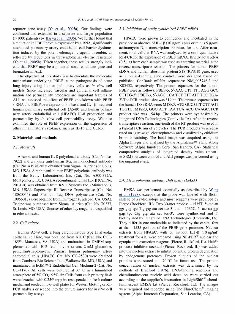
3.4. EMSA analysis of the human PBEF promoter�1535T and �1535C 30-mer oligonucleotides
We reported previously that the T-variant in the humanPBEF promoter SNP C-1535T resulted in a significantdecrease of the luciferase reporter gene expression (Ye et al.,2005a). We examined whether the T-variant altered thebinding affinity to any transcription factor, which may underlieits effect on the decrease in the transcription of PBEF geneexpression. As presented in Fig. 5, EMSA analysis revealedthat �1535 T-variant in the human PBEF gene promoter has
less binding to an unknown transcription factor than thecommon �1535 C-allele, this difference became morepronounced after the IL-1b or TNFa treatment. The bindingswere specific since their unlabeled ‘‘cold’’ oligos could totallycompete out the binding.
3.5. IL-1b induction of IL-8 and PBEF at the mRNA levelin HPAEC
IL-8 is a known IL-1b inducible biomarker in acute lunginjury. PBEF may directly or indirectly affect IL-1 b-inducedIL-8 expression during the inflammatory process. To this end, toexamined whether IL-1b could also affect IL-8 and PBEFexpression at the mRNA level in HPAEC, we quantified IL-8and PBEF mRNA levels in IL-1b-treated primary HPAEC. Asshown in Fig. 6, IL-8 and PBEF mRNA levels in IL-1b-treatedprimary HPAEC significantly increased compared to the controlgroup (4.03 � 104 � 1.4 � 103 vs. 2.07 � 104 � 0.7 � 103
(control), n ¼ 4, **p < 0.01; 1.10 � 104 � 0.4 � 103 vs.0.67 � 104 � 0.2 � 103 (control), n ¼ 4, **p < 0.01, respec-tively). These results indicate that IL-1b also induces IL-8 andPBEF expression at the mRNA level in HPAEC.
In
teg
rated
d
en
sity valu
e (X
10
6
)
0
2
4
6
8
10
12
**
Control IL1-β IL1-β+ ActD
18s rRNA PBEF
310 bp355 bp
1 3A
B
2 3 1 2
Fig. 4. RT-PCR semi-quantification of PBEF mRNA in HPAEC treated
without or with either IL-1b or IL-1b þ Act D. HPAEC were treated without
or with either IL-1b (10 ng/ml) or IL-1b þ Act D (5 mg/ml). Total RNA was
reverse-transcribed, amplified by PCR using gene specific primers, separated
on 2% agarose electrophoresis, analyzed by an Alpha Innotech densitometer.
(a) A representative image. 1-control; 2-IL1-b; 3-IL1-b þ Act D. (b) Densi-
tometry analyses. **p < 0.01.
0
0.5
1
1.5
2
2.5
3
3.5
4
4.5
Control
15ng/ml
IL-8
**
**
IL-8
β-actin
PBEF
Control IL-1β
In
teg
rated
d
en
sity valu
e (x10
4
)
PBEF
A
B
Fig. 6. Effects of the IL-1b stimulation on IL-8 and PBEF mRNA expressions
in HPAEC. (A) A representative gel image of IL-8, PBEF and b-actin mRNA
detections. After starving for serum overnight, HPAEC were stimulated with
IL-1b (15 ng/ml) for 4 h. Total cell RNA was reverse-transcribed, amplified by
PCR using the gene specific primers (Table 1), separated by 1.5% agarose
electrophoresis and visualized with ethidium bromide. The gel image was
captured using the Alpha Imager (Alpha Innotech Corp., San Leandro, CA). b-
Actin was used as a house-keeping gene control. The displayed image is one of
four replicate samples and is typical of those obtained. (B) Quantitation of IL-
8 and PBEF mRNA levels by the densitometry analysis. The mRNA band
intensity on the gel image was analyzed by the AlphaEase Stand Alone
software (Alpha Innotech Corp., San Leandro, CA). The integrative density
24 P. Liu et al. / Cell Biology International 33 (2009) 19e30
3.6. PBEF overexpression augmented IL-8 secretionfrom A549 cells and HPAEC
value of each band was used as a measure of the mRNA level. Results from
each group are presented as mean � SD of 4 samples from two separate
experiments. Statistical comparative analyses of IL-8 and PBEF mRNA levels
between the control and IL-1b-treated group were performed using the
unpaired t-test. **p < 0.01.
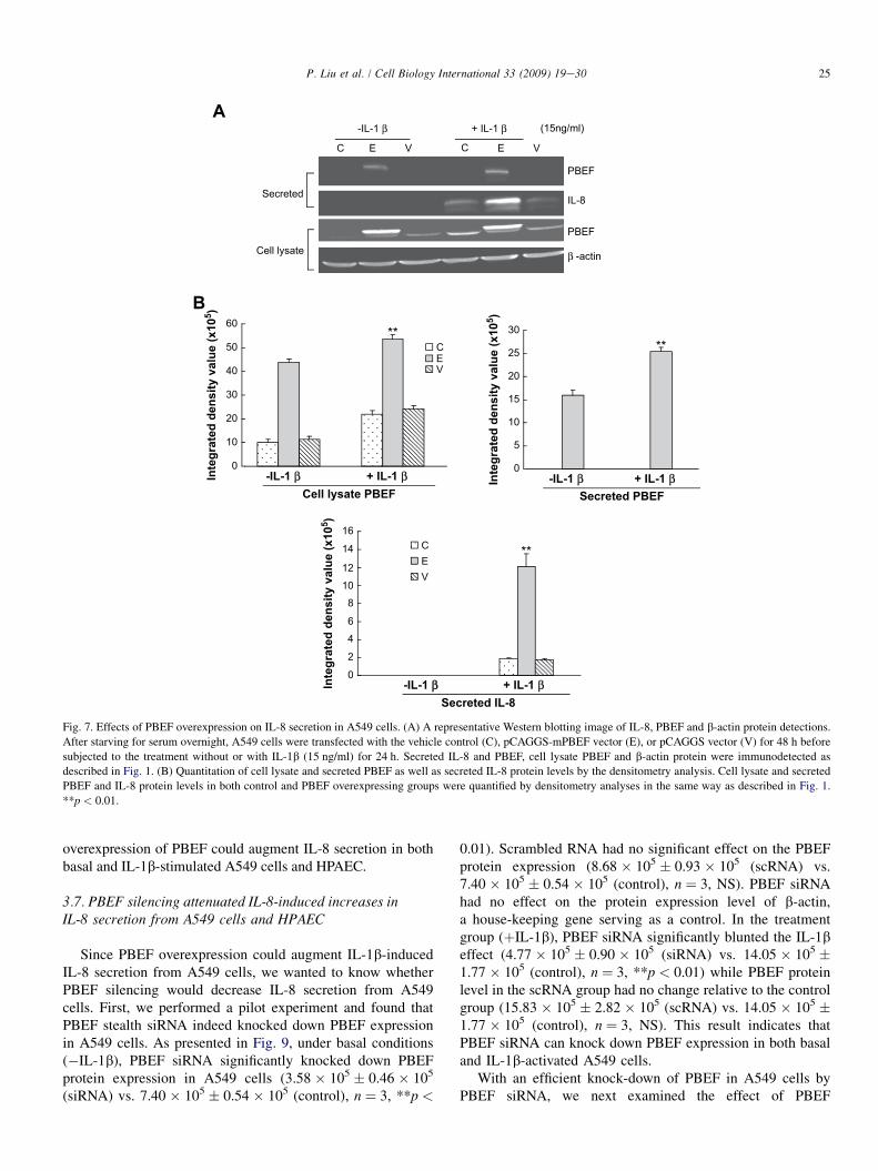
We first tested whether the pCAGGS-mPBEF constructcould indeed increase PBEF expression in A549 cells. Aspresented in Fig. 7, the overexpression of PBEF in A549 cellstransiently transfected with pCAGGS-mPBEF was achievedunder basal conditions (�IL-1b), as evidenced in the medium
UnknownTF
T-1535 + + +
BioT-1535 + + + +
C-1535 + + +
BioC-1535 + + + + +
TNFα IL-1β Base c
+ ++
Fig. 5. Electrophoretic mobility shift assay on �1535C and �1535 T binding
to an unknown transcription factor (TF). HPAEC were treated without (base)
or with either IL-1b (10 ng/ml) or TNFa (10 ng/ml) for 4 h. Ten micrograms
of nuclear extract protein in each sample were incubated with either biotin
labeled c-1535 (Bio C-1535) or BioT-1535 oligo in the absence or presence of
their unlabeled 200-fold excess counterparts. The bindings were visualized by
a chemiluminecence. c, no oligo control.
(15.98 � 105 � 1.1 � 104 vs. 0.0 � 0.0 (vector and control),n ¼ 3, **p < 0.01). In IL-1b-stimulated conditions, the PBEFlevel in the medium was further increased compared to thebasal conditions (25.43 � 105 � 0.9 � 104 vs. 15.98 � 105
� 1.1 � 104, n ¼ 3, **p < 0.01). Similar observations wereobtained with PBEF protein expression in the cell lysate ofA549 cells transiently transfected with pCAGGS-mPBEFwhile the pCAGGS vector control group did not show anychange in PBEF protein expression level. These results clearlyshow that pCAGGS-mPBEF vector increased PBEF expres-sion in both basal and IL-1b-activated A549 cells.
With a significant increase of PBEF expression in A549 cellsby pCAGGS-mPBEF vector, we next examined the effect ofPBEF overexpression on the IL-1b-induced increases in IL-8secretion from A549 cells. Fig. 7 demonstrated that PBEFoverexpression significantly increased IL-8 secretion from A549cells compared to the control group (12.09 � 105 � 1.43 � 105
vs. 1.85 � 105 � 1.0 � 104 (control), n ¼ 3, **p < 0.01). Asimilar phenomenon was also observed in HPAEC as presentedin Fig. 8. PBEF overexpression significantly increased IL-8secretion from HPAEC compared to the control group(25.18 � 105 � 1.03 � 105 vs. 10.59 � 105 � 0.31 � 105
(control), n ¼ 3, **p < 0.01). These results indicate that
PBEF
β -actin
PBEF
Secreted
Cell lysate
-IL-1 β + IL-1 β
C E V E V
(15ng/ml)
IL-8
A
B
2
0
46
81012
14
16CEV
-IL-1 ββ + IL-1 β
**
Secreted IL-8
0
5
10
15
20
25
30
0
10
20
30
40
50
60
CEV
-IL-1 β + IL-1 βIn
teg
rated
d
en
sity valu
e (x10
5)
**
Cell lysate PBEFIn
teg
rated
d
en
sity valu
e (x10
5)
-IL-1 β + IL-1 βSecreted PBEF
**In
teg
rated
d
en
sity valu
e (x10
5)
C
Fig. 7. Effects of PBEF overexpression on IL-8 secretion in A549 cells. (A) A representative Western blotting image of IL-8, PBEF and b-actin protein detections.
After starving for serum overnight, A549 cells were transfected with the vehicle control (C), pCAGGS-mPBEF vector (E), or pCAGGS vector (V) for 48 h before
subjected to the treatment without or with IL-1b (15 ng/ml) for 24 h. Secreted IL-8 and PBEF, cell lysate PBEF and b-actin protein were immunodetected as
described in Fig. 1. (B) Quantitation of cell lysate and secreted PBEF as well as secreted IL-8 protein levels by the densitometry analysis. Cell lysate and secreted
PBEF and IL-8 protein levels in both control and PBEF overexpressing groups were quantified by densitometry analyses in the same way as described in Fig. 1.
**p < 0.01.
25P. Liu et al. / Cell Biology International 33 (2009) 19e30
overexpression of PBEF could augment IL-8 secretion in bothbasal and IL-1b-stimulated A549 cells and HPAEC.
3.7. PBEF silencing attenuated IL-8-induced increases inIL-8 secretion from A549 cells and HPAEC
Since PBEF overexpression could augment IL-1b-inducedIL-8 secretion from A549 cells, we wanted to know whetherPBEF silencing would decrease IL-8 secretion from A549cells. First, we performed a pilot experiment and found thatPBEF stealth siRNA indeed knocked down PBEF expressionin A549 cells. As presented in Fig. 9, under basal conditions(�IL-1b), PBEF siRNA significantly knocked down PBEFprotein expression in A549 cells (3.58 � 105 � 0.46 � 105
(siRNA) vs. 7.40 � 105 � 0.54 � 105 (control), n ¼ 3, **p <
0.01). Scrambled RNA had no significant effect on the PBEFprotein expression (8.68 � 105 � 0.93 � 105 (scRNA) vs.7.40 � 105 � 0.54 � 105 (control), n ¼ 3, NS). PBEF siRNAhad no effect on the protein expression level of b-actin,a house-keeping gene serving as a control. In the treatmentgroup (þIL-1b), PBEF siRNA significantly blunted the IL-1beffect (4.77 � 105 � 0.90 � 105 (siRNA) vs. 14.05 � 105 �1.77 � 105 (control), n ¼ 3, **p < 0.01) while PBEF proteinlevel in the scRNA group had no change relative to the controlgroup (15.83 � 105 � 2.82 � 105 (scRNA) vs. 14.05 � 105 �1.77 � 105 (control), n ¼ 3, NS). This result indicates thatPBEF siRNA can knock down PBEF expression in both basaland IL-1b-activated A549 cells.
With an efficient knock-down of PBEF in A549 cells byPBEF siRNA, we next examined the effect of PBEF

0
5
10
15
20
25
30
CEV
-IL-1β + IL-1βIn
teg
rated
d
en
sity valu
e (x10
5)
**
Secreted IL-8
C
02468
101214161820 C
EV
PBEF
β-actin
-IL-1β + IL-1β
C E V E V
In
teg
rated
d
en
sity valu
e (x10
5)
****
Cell lysate
IL-8Secreted
-IL-1β + IL-1β
Cell lysate PBEF
A
B
C
Fig. 8. Effects of PBEF overexpression on IL-8 secretion in HPAEC. (A) A
representative Western blotting image of IL-8, PBEF and b-actin protein
detections. HPAEC were treated n the same way as those in A549 cells as
described in Fig. 5. Secreted IL-8, cell lysate PBEF and b-actin protein were
immunodetected in the same way as described in Fig. 1. (B) Quantitation of
cell lysate PBEF protein levels by densitometry analysis. (C) Quantitation of
secreted IL-8 levels by densitometry analysis. Secreted IL-8 and cell lysate
PBEF protein levels were quantified by densitometry analyses in the same way
as those in A549 cells as described in Fig. 5. **p < 0.01.
-IL-1 ββ + IL-1 β
In
te
gra
te
d d
en
sity
v
alu
e (x
10
5)
**
Secreted IL-8
C
ControlsiRNAscRNA
ControlsiRNAscRNA
C Si Sc C Si Sc
-IL-1 β + IL-1 β
PBEF
β-actin
In
te
gra
te
d d
en
sity
v
alu
e (x
10
5)
****
-IL-1 β + IL-1 β
(15ng/ml)A
B
Secreted
Cell lysate
IL-8
Cell lysate PBEF
20181614121086420
20
1618
1412108
46
20
Fig. 9. Effects of PBEF knock down on IL-8 secretion in A549 cells. (A) A
representative Western blotting image of IL-8, PBEF and b-actin protein
detections. After starving for serum overnight, A549 cells were transfected
with the vehicle control (C), scrambled siRNA (Sc), or PBEF stealth siRNA
(Si) for 48 h before subjected to treatment without or with IL-1b (15 ng/ml)
for 24 h. Secreted IL-8, cell lysate PBEF and b-actin protein were immuno-
detected as described in Fig. 1. (B) Quantitation of cell lysate PBEF protein
levels by densitometry analysis. (C) Quantitation of secreted IL-8 levels by
densitometry analysis. Cell lysate PBEF and secreted IL-8 protein levels in
various treatments of A549 cells were quantified by densitometry analyses in
the same way as described in Fig. 1.**p < 0.01.
26 P. Liu et al. / Cell Biology International 33 (2009) 19e30
knockdown on the IL-1b-induced increase of IL-8 secretionfrom A549 cells. Fig. 9 demonstrates that PBEF silencingsignificantly decreased IL-8 secretion from A549 cells inIL-1b-stimulated conditions compared to the control group(10.1 � 105 � 0.82 � 105 (siRNA) vs. 17.14 � 105 � 0.35� 105 (control), n ¼ 3,**p < 0.01), while secreted IL-8 levels
in the scRNA group did not change relative to the controlgroup. (16.38 � 105 � 0.24 � 105 (scRNA) vs. 17.14 � 105
� 0.35 � 105 (control) n ¼ 3, NS). A similar phenomenonwas also observed in HPAEC, as presented in Fig. 10. PBEFknock-down significantly decreased IL-8 secretion fromHPAEC compared to the control group (15.23 � 105 �1.47 � 105 vs. 26.23 � 105 � 0.23 � 105 (control), n ¼ 3,**p < 0.01). These results indicate that PBEF is involved inIL-8 expression and secretion under both basal and IL-1b-induced conditions in A549 cells and HPAEC.
3.8. Effect of PBEF expression on IL-16 and CCR3 geneexpression
To further investigate the relationship between PBEF andother inflammatory cytokines and the function of PBEF in theinflammation pathway, we examined the effect of PBEFexpression on IL-16 and CCR3 gene expression. We selectedIL-16 and CCR3 because based on our unpublished observa-tion, knockdown of PBEF expression significantly inhibitedthe expression of genes involved in inflammation including IL-16 and CCR3 (Ye et al., unpublished observation). Results inFig. 11 show that knockdown of PBEF in A549 cells couldattenuate IL-1b-stimulated increase of IL-16 and CCR3 geneexpression, while the overexpression of PBEF in A549 cellscould promote IL-1b-stimulated increase of IL-16 and CCR3gene expression at the mRNA level. Taken together, these dataindicate that PBEF might be an important signal transducer orinitiator in the inflammation pathway.
**
****
In
te
gra
te
d d
en
sity
v
alu
e (x
10
4)
-IL-1 β + IL-1 β
Secreted IL-8
ControlsiRNAscRNA
ControlsiRNAscRNA
PBEF
β-actin
In
te
gra
te
d d
en
sity
v
alu
e (x
10
5)
-IL-1 β + IL-1 β
C Si Sc C Si Sc
-IL-1 β + IL-1 β
C
A
B
Secreted
Cell lysate
IL-8
Cell lysate PBEF
181614121086420
1009080706050403020100
Fig. 10. Effects of PBEF knock down on IL-8 secretion in HPAEC. (A) A
representative Western blotting image of IL-8, PBEF and b-actin protein
detections. HPAEC were treated in the same way as those in A549 cells as
described in Fig. 7. Secreted IL-8, cell lysate PBEF and b-actin protein were
immunodetected in the same way as described in Fig. 1. (B) Quantitation of
cell lysate PBEF protein levels by densitometry analysis. (C) Quantitation of
secreted IL-8 levels by densitometry analysis. Secreted IL-8 and cell lysate
PBEF protein levels were quantified by densitometry analyses in the same way
as those in A549 cells as described in Fig. 7. **p < 0.01.
C E V C E V
IL-16
CCR3
PBEF
ββ-actin
B
IL-16
- I L-1 β + IL-1 β
C Si Sc C Si Sc
CCR3
PBEF
β-actin
IL-16
PBEF
A
-IL-1 β + IL-1 β
Fig. 11. RT-PCR analyses of PBEF, IL-16, CCR3 and b-actin mRNA levels in
A549 cells. A549 cells were grown and transfected with the vehicle control
(C), PBEF stealth siRNA (Si), scrambled siRNA (Sc), pCAGGS-mPBEF
vector (E), or pCAGGS vector (V) for 48 h before being subjected to the
treatment without or with IL-1b (15 ng/ml) for 4 h. Total cell RNA was
reverse-transcribed, amplified by PCR using the gene specific primers
(Table 1), separated by 1.5% agarose electrophoresis and visualized with
ethidium bromide. (A) A representative RT-PCR gel image of PBEF, IL-16,
CCR3 and b-actin mRNA detections in PBEF-knockdown A549 cells. (B) A
representative RT-PC R gel image of PBEF, IL-16, CCR3 and b-actin mRNA
detections in PBEF-overexpression A549 cells.
27P. Liu et al. / Cell Biology International 33 (2009) 19e30
3.9. PBEF expression affected cell permeability in A549cells and HPAEC
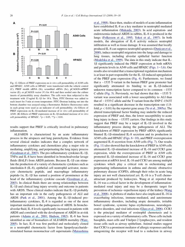
To assess whether PBEF expression affected epithelial celland HPAEC permeability, we performed an in vitro monolayercell permeability assay in A549 cells and HPAEC transfectedwith PBEF stealth siRNA, pCAGGS-mPBEF vector and othercontrols with IL-1b treatment. As presented in Fig. 12, PBEFsiRNA significantly decreased the IL-1b-induced A549 cellpermeability level in the PBEF siRNA treated group comparedto the IL-1b-alone treatment (17333.33 � 1030.24 vs.24333.33 � 820.56 relative fluorescence units, n ¼ 4,**p < 0.01). Similar observations were obtained in HPAEC,whose cell permeability level in the PBEF siRNA groupsignificantly decreased compared to the IL-1b-alone treatment(17833.33 � 1120.2 vs. 23505 � 1690.5 relative fluorescenceunits, n ¼ 4, *p < 0.05). Scrambled RNA had no effect on theIL-1b-induced cell permeability in both A549 cells and
HPAEC. These results indicate that PBEF siRNA significantlyattenuated the IL-1b-induced barrier-disruption and higherpermeability in epithelial cells and endothelial cells. Fig. 12also demonstrates that PBEF overexpression significantlypromoted the IL-1b-induced A549 cell permeability levelin the pCAGGS-mPBEF transfected group compared tothe IL-1b-alone treatment (34950.09 � 2093.69 vs.24333.33 � 820.56 relative fluorescence units, n ¼ 4,**p < 0.01). Similar observations were obtained in HPAEC,whose cell permeability level in the PBEF overexpressiongroup significantly increased compared to the IL-1b-alonetreatment (38833.33 � 2644.86 vs. 23505 � 1690.5 relativefluorescence units, n ¼ 4, **p < 0.01). These results indicatethat overexpression of PBEF in A549 cells and HPAECaugmented IL-1b-induced lung epithelial cell barrierdysfunction and thus increased cell permeability in vitro.
4. Discussion
The data from this study indicate that IL-1b significantlyinduced the PBEF expression at a transcriptional level inHPAEC. PBEF overexpression augmented IL-8 secretion fromA549 cells and HPAEC. The knockdown of PBEF expressionby PBEF siRNA significantly blunted IL-1b-induced IL-8secretion and its production in A549 cells and HPAEC. These
Relative F
lu
orescen
ce U
nits (x10
3)
A
B
A549 Cells
HPAEC
P< 0.01
P< 0.01
C Si Sc E V
C Si Sc E V
P< 0.01
P< 0.05
05
10152025303540
05
1015202530354045
Fig. 12. Effects of PBEF expression on in vitro cell permeability of A549 cells
and HPAEC. A549 cells or HPAEC were transfected with the vehicle control
(C), PBEF stealth siRNA (Si), scrambled siRNA (Sc), pCAGGS-mPBEF
vector (E), or pCAGGS vector (V) for 48 h and then seeded onto the culture
inserts of permeability assay chambers. The cells were then subjected to the
treatment with 15 ng/ml IL-1b for 18 h. Then, FITC-Dextran was added to
each insert for 5 min at room temperature. FITC-Dextran leaking out into the
bottom chamber was assayed using a fluorometer. Relative fluorescence units
in each group were used as an indicator of cell permeability. (A) Effects of
PBEF expression on IL-1b-mediated increase of in vitro cell permeability of
A549. (B) Effects of PBEF expression on IL-1b-mediated increase of in vitro
cell permeability of HPAEC. *p < 0.05, **p < 0.01.
28 P. Liu et al. / Cell Biology International 33 (2009) 19e30
results support that PBEF is critically involved in pulmonaryinflammation.
ALI/ARDS is characterized by an acute inflammatoryprocess in the airspaces and lung parenchyma. Evidence fromseveral clinical studies indicates that a complex network ofinflammatory cytokines and chemokines play a major role inmediating, amplifying, and perpetuating the lung injury process(Goodman et al., 2003). The pro-inflammatory cytokines IL-1b,TNFa and IL-8 have been identified in bronchoalveolar lavagefluids (BALF) from ARDS patients. Because IL-1b can stimu-late the production of a variety of chemotactic cytokines such asinterleukin-8 (IL-8), epithelial cell neutrophil activator, mono-cyte chemotactic peptide, and macrophage inflammatorypeptide-1a, IL-1b has earned a position of prominence at thehead of the inflammatory cytokine cascade (Goodman et al.,2003). In clinical fluids there are strong relationships betweenIL-1b and clinical lung injury severity and outcome in patientswith ARDS. These clinical studies indicate that IL-1b probablyhas an important role in the early pathogenesis of ALI andARDS (Park et al., 2001). Among the IL-1b-induced pro-inflammatory cytokines, IL-8 is regarded as one of the mostimportant mediators in the pathogenesis of ARDS. In broncho-alveolar fluid, IL-8 levels significantly increased in patients withARDS and correlated with the development of ARDS in at-riskpatients (Adams et al., 2001; Hudson, 1982). IL-8 has beenidentified as one of biomarkers of ALI/ARDS mortality (Ware,2005). In fact, IL-8 was first purified and molecularly clonedas a neutrophil chemotactic factor from lipopolysaccharide-stimulated human mononuclear cell supernatants (Matsushima
et al., 1988). Since then, studies of models of acute inflammationhave established IL-8 as a key mediator in neutrophil mediatedacute inflammation (Mukaida, 2003). In acid aspiration- andendotoxemia-induced ARDS in rabbits, IL-8 is produced in thelungs (Folkesson et al., 1995; Yokoi et al., 1997). In bothmodels, the abrogation of IL-8 activity reduces neutrophilinfiltration as well as tissue damage. It was assumed that locallyproduced IL-8 can suppress neutrophil apoptosis (Dunican et al.,2000), induce neutrophil migration into the lungs and damage tolung tissues, including alveolar epithelial barrier function(Modelska et al., 1999). The data in this study indicate that IL-1b significantly induced the PBEF expression at both mRNAand protein levels in A549 cells and HPAEC (Figs. 1e3,6). Ourresults also revealed that a transcriptional regulation mechanismis at least in part responsible for the IL-1b-induced upregulationof the PBEF gene expression (Fig. 4). Furthermore, we foundthat a �1535 T-variant in the human PBEF gene promoter hadsignificantly attenuated its binding to an IL-1b-inducedunknown transcription factor compared to its common �1535C-allele (Fig. 5). Previously, we had shown that this �1535 T-variant was associated with a lower susceptibility to ALI thanthat of�1535 C-allele and the T-variant from the SNP C-1543Tresulted in a significant decrease in the transcription rate (1.8-fold; p < 0.01) by the reporter gene assay (Ye et al., 2005a). Thealtered transcriptional factor binding may underlie the reducedexpression of PBEF and, thus, the lower susceptibility to acutelung injury in those �1535T carriers. Our findings in this studysuggest that PBEF may be a target of IL-1b involved in theinflammatory process during the pathogenesis of ALI. Theknockdown of PBEF expression by PBEF siRNA significantlyblunted IL-1b-stimulated IL-8 secretion and its production inA549 cells and HPAEC (Figs. 6e10), and PBEF overexpressionaugmented IL-8 secretion from A549 cells (Fig. 7). Our results(Fig. 11) also showed that the knockdown of PBEF in A549 cellsattenuated IL-1b-stimulated increase of IL-16 and CCR3 geneexpression, while the overexpression of PBEF in A549 cellspromoted IL-1b-stimulated increase of IL-16 and CCR3 geneexpression at mRNA level. IL-16 and CCR3 are among multiplecytokines which play a critical role in orchestrating andperpetuating inflammation in asthma and chronic obstructivepulmonary disease (COPD), although their roles in acute lunginjury are not well characterized yet. IL-16 is a T-cell chemo-attractant produced by leukocytes. Wang et al. reported thatIL-16 was a critical factor in the development of inflammation-mediated renal injury and may be a therapeutic target forprevention of ischemiaereperfusion injury of the kidney (Wanget al., 2008). A plethora of studies have also implicated IL-16 inexacerbating infectious, immune-mediated, and autoimmuneinflammatory disorders, including atopic dermatitis, irritablebowel syndrome, systemic lupus erythematosus, neurodegen-erative disorders, and viral infections (Glass et al., 2006). CCR3is the principal mediator of eosinophil chemotaxis and isexpressed on a variety of inflammatory cells. These cells includebasophils, mast cells and T-helper-2 lymphocytes, and residenttissue cells such as airway epithelium. Animal studies suggestthat CCR3 is a prominent mediator of allergic responses and thatantagonizing the receptor will lead to a reduction in airway

29P. Liu et al. / Cell Biology International 33 (2009) 19e30
inflammation (De Lucca, 2006). IL-16 and CCR3 may beamong the repertoire of bioactive molecules regulated by thePBEF gene and be implicated in the pathogenesis of acute lunginjury or other inflammatory diseases. These data implicatePBEF in being an important ‘‘master’’ signal transducer orinitiator in the inflammation pathway to regulate the synthesis ofIL-8 or other inflammatory cytokines, and PBEF could playa critical role as an inflammatory cytokine during the patho-genesis of ALI.
Both endothelial and epithelial injuries were observed byearlier seminal ultrastructural studies of the lung in patientsdying with ALI secondary to sepsis (Bachofen and Weibel,1977, 1982). The vascular endothelial cell monolayer serves asa dynamic, semiselective barrier that regulates transport of fluidand macromolecules between blood and the interstitium. Avariety of physical, inflammatory, and bioactive stimuli alter theendothelial barrier, leading to formation of paracellular gapsthereby increasing vessel permeability and compromisingorgan function (Csortos et al., 2007; Dudek and Garcia, 2001;Mehta and Malik, 2006). Increased vascular permeabilitycontributes to the profound pathophysiological derangementsin ALI (De Lucca, 2006). Epithelial injury in the lungs is one ofthe hallmarks of ALI in humans (Liener et al., 2002). During theprocess of acute inflammation, endothelial permeability wasincreased rapidly. If the alveolar epithelium is also damaged,the change in both endothelial and epithelial permeability couldlead to major alveolar flooding with high-molecular-weightproteins, with prolonged changes in gas exchange and a muchhigher likelihood of disordered repair (Parsons et al., 2005).Normally, the lung alveolar epithelium is an extremely tightbarrier that restricts the movement of proteins and liquid fromthe interstitium into the alveolar spaces. In ALI, impairedalveolar epithelial function in the lungs is a marker of pooroutcome of ALI (Thomas et al., 2005). A number of inflam-matory cytokines including IL-1b and IL-8 can induce oraggravate the inflammation of endothelial and epithelial cells,leading to their barrier dysfunctions (Leone et al., 2007).Results in Fig. 12 of this study have demonstrated that over-expression of PBEF augmented both basal and IL-1b-inducedincrease in the cell permeability of both A549 cells andHPAEC, while the knockdown of PBEF expression by PBEFsiRNA significantly attenuated IL-1b-induced cell permeabilityof A549 cells and HPAEC, suggesting that PBEF may have animportant role in both endothelial and epithelial cell barrierregulation. A549 cells are a type II tumor epithelial cell line.While the type II epithelial cell only covers 7% of the alveolarsurface area it constitutes 67% of the epithelial cell numberswithin the alveoli (Crapo et al., 1982) pointing to itsbiochemical importance. The permeability increase of A549cells in the IL-1b-stimulated condition involved both para-cellular permeability, with gap formation visualized by actincytoskeleton staining, and basement membrane permeability(Lacherade et al., 2001). This IL-1b-mediated effect may bepartly due to its induction of IL-8 production. IL-8 wasdemonstrated to induce the actin fiber formation and intercel-lular gap formation of endothelial cells (Schraufstatter et al.,2001). IL-8 could have the same effect on epithelial barrier
function. Our previous study found that reductions in PBEFprotein expression significantly attenuated endothelial barrierdysfunction induced by the potent edemagenic agent, thrombin,reflected by reductions in transendothelial electric resistance(Ye et al., 2005b). Furthermore, we found that reductions inPBEF protein expression blunted thrombin-mediated increasesin Ca2þ entry, polymerized actin formation, and myosin lightchain phosphorylation, events critical to the thrombin-mediatedpermeability response (Ye et al., 2005b). It remains to beelucidated whether similar mechanisms operate to account forthe attenuating effects of PBEF knockdown on IL-1b-inducedepithelial and endothelial cell barrier dysfunction. It should bementioned that a similar observations as those found in A549cells were also obtained in primary human lung small airwayepithelial cells (Cat. No. CC-2547, Lonza, Walkersville, MD,USA) (data not shown).
In summary, our results demonstrated that a transcriptionalregulation mechanism is at least in part responsible for theIL-1b-induced upregulation of the PBEF gene expression andaltered transcriptional factor binding may underlie the reducedexpression of PBEF and thus lower susceptibility to acute lunginjury in those �1535T carriers. We found that the over-expression of PBEF significantly augmented IL-8 secretionand mRNA expression by more than 6-fold and 2-fold in A549and HPAEC, respectively. It also significantly augmentedIL-1b-mediated cell permeability by 44% in A549 cells and65% in endothelial cells. The knockdown of PBEF expressionsignificantly inhibited IL-1b-stimulated IL-8 secretion andmRNA levels by 60% and 70%, respectively, and the knock-down of PBEF expression also significantly attenuated IL-1b-induced cell permeability by 29% in epithelial cells and 24%in endothelial cells. Furthermore, we found that PBEFexpression could also affect the expression of IL-16 and CCR3genes, two inflammatory cytokines. These results suggest thatPBEF may play a critical role as an inflammatory cytokine inthe development of pulmonary inflammation and dysregula-tion of pulmonary vascular endothelial and alveolar epithelialcell barriers, which may be an important mechanism under-lying PBEF in the pathogenesis of ALI. These results lendfurther support that PBEF may represent a new diagnostic andtherapeutic target in ALI.
Acknowledgements
This work was in part supported by National Heart, Lung,and Blood Institute Grants HL 080042 (S.Q. Ye),2P01HL058064-070001 (J.G.N. Garcia) and the University ofMissouri-Columbia Start-up fund (S.Q. Ye).
References
Adams JM, Hauser CJ, Livingston DH, Lavery RF, Fekete Z, Deitch EA. Early
trauma polymorphonuclear neutrophil responses to chemokines are asso-
ciated with development of sepsis, pneumonia, and organ failure. J Trauma
2001;51:452e6.
Bachofen M, Weibel ER. Alterations of the gas exchange apparatus in adult
respiratory insufficiency associated with septicemia. Am Rev Respir Dis
1977;116:589e615.

30 P. Liu et al. / Cell Biology International 33 (2009) 19e30
Bachofen M, Weibel ER. Structural alterations of lung parenchyma in the
adult respiratory distress syndrome. Clin Chest Med 1982;3:35e56.
Bajwa EK, Yu C, Gong MN, Thompson BT, Christiani DC. PBEF gene
polymorphisms influence the risk of developing ARDS. Am J Respir Crit
Care Med 2006;3. A272.
Bradford MM. A rapid and sensitive method for the quantitation of microgram
quantities of protein utilizing the principle of proteinedye binding. Anal
Biochem 1976;72(7):248e54.
Crapo JD, Barry BE, Gehr P, Bachofen M, Weibel ER. Cell number and cell
characteristics of the normal human lung. Am Rev Respir Dis 1982;126:
332e7.
Csortos C, Kolosova I, Verin AD. Regulation of vascular endothelial cell
barrier function and cytoskeleton structure by protein phosphatases of the
PPP family. Am J Physiol Lung Cell Mol Physiol 2007;293(4):L843e54.
De Lucca GV. Recent developments in CCR3 antagonists. Curr Opin Drug
Discov Devel 2006;9(4):516e24.
Dudek SM, Garcia JG. Cytoskeletal regulation of pulmonary vascular
permeability. J Appl Physiol 2001;91(4):1487e500.
Dunican AL, Leuenroth SJ, Grutkoski P, Ayala A, Simms HH. TNFalpha-
induced suppression of PMN apoptosis is mediated through interleukin-8
production. Shock 2000;14(3):284e8.
Folkesson HG, Matthay MA, Hebert CA, Broaddus VC. Acid aspiration-
induced lung injury in rabbits is mediated by interleukin-8-dependent
mechanisms. J Clin Invest 1995;96:107e16.
Glass WG, Sarisky RT, Vecchio AM. Not-so-sweet sixteen: the role of IL-16 in
infectious and immune-mediated inflammatory diseases. J Interferon
Cytokine Res 2006;26(8):511e20.
Goodman RB, Pugin J, Lee JS, Matthay MA. Cytokine-mediated inflammation
in acute lung injury. Cytokine Growth Factor Rev 2003;14(6):523e35.
Hudson LD. Causes of the adult respiratory distress syndromeeclinical
recognition. Clin Chest Med 1982;3:195e212.
Lacherade JC, Van LA, Planus E, Escudier E, D’Ortho MP, Lafuma C, et al.
Evaluation of basement membrane degradation during TNF-alpha-induced
increase in epithelial permeability. Am J Physiol Lung Cell Mol Physiol
2001;281(1):134e43.
Leaver SK, Evans TW. Acute respiratory distress syndrome. BMJ 2007;
335(7616):389e94.
Leone AK, Chun JA, Koehler CL, Caranto J, King JM. Effect of proin-
flammatory cytokines, tumor necrosis factor-alpha and interferon-gamma
on epithelial barrier function and matrix metalloproteinase-9 in Madin
Darby canine kidney cells. Cell Physiol Biochem 2007;19(1-4):99e112.
Levitt JE, Matthay MA. Treatment of acute lung injury: historical perspective
and potential future therapies. Semin Respir Crit Care Med 2006;27(4):
426e37.
Liener UC, Bruckner UB, Knoferl MW, Steinbach G, Kinzl L, Gebhard F.
Chemokine activation within 24 �after blunt accident trauma. Shock 2002;
17:169e72.
Matsushima K, Morishita K, Yoshimura T, Lavu S, Kobayashi Y, Lew W, et al.
Molecular cloning of a human monocyte-derived neutrophil chemotactic
factor (MDNCF) and the induction of MDNCF mRNA by interleukin 1
and tumor necrosis factor. J Exp Med 1988;167(6):1883e93.
McGlothlin JR, Gao L, Lavoie T, Simon BA, Easley RB, Ma SF, et al.
Molecular cloning and characterization of canine pre-B-cell colony-
enhancing factor. Biochem Genet 2005;43(3-4):127e41.
Mehta D, Malik AB. Signaling mechanisms regulating endothelial perme-
ability. Physiol Rev 2006;86:279e367.
Modelska K, Pittet JF, Folkesson HG, Courtney V, Matthay MA. Acid-induced
lung injury. Protective effect of anti-interleukin-8 pretreatment on alveolar
epithelial barrier function in rabbits. Am J Respir Crit Care Med 1999;160:
1450e6.
Mukaida N. Pathophysiological roles of interleukin-8/CXCL8 in pulmonary
diseases. Am J Physiol Lung Cell Mol Physiol 2003;284(4):L566e77.
Park WY, Goodman RB, Steinberg KP, Ruzinski JT, Radella 2nd F, Park DR,
et al. Cytokine balance in the lungs of patients with acute respiratory
distress syndrome. Am J Respir Crit Care Med 2001;164:1896e903.
Parsons PE, Eisner MD, Thompson BT, Matthay MA, Ancukiewicz M,
Bernard GR. Lower tidal volume ventilation and plasma cytokine markers of
inflammation in patients with acute lung injury. Crit Care Med 2005;33:1e6.
Schraufstatter IU, Chung J, Burger M. IL-8 activates endothelial cell CXCR1
and CXCR2 through Rho and Rac signaling pathways. Am J Physiol Lung
Cell Mol Physiol 2001;280(6):L1094e103.
Thomas R, Martin NH, Morio N, Gustavo MB. Apoptosis and epithelial injury
in the lungs. Proc Am Thorac Soc 2005;2:214e20.
Wang S, Diao H, Guan Q, Cruikshank WW, Delovitch TL, Jevnikar AM, et al.
Decreased renal ischemiaereperfusion injury by IL-16 inactivation.
Kidney Int 2008;73(3):318e26.
Wang Y, Xiao L, Thiagalingam A, Nelkin BD, Casero Jr RA. The identifi-
cation of a cis-element and a trans-acting factor involved in the response to
polyamines and polyamine analogues in the regulation of the human
spermidine/spermine N1-acetyltransferase gene transcription. J Biol Chem
1998;273(51):34623e30.
Ware LB. Prognostic determinants of acute respiratory distress syndrome in
adults: impact on clinical trial design. Crit Care Med 2005;33(Suppl 3):
217e22.
Ye SQ, Simon B, Maloney JP, Zambelli-Weiner A, Gao L, Grant A, et al. Pre-
B-cell colony-enhancing factor as a potential novel biomarker in acute
lung injury. Am J Respir Crit Care Med 2005a;171(2):361e70.
Ye SQ, Zhang LQ, Adyshev D, Usatyuk PV, Garcia A, Lavoie TL, et al. Pre-B-
cell-colony-enhancing factor is critically involved in thrombin-induced
lung endothelial cell barrier dysregulation. Microvasc Res 2005b;70(3):
142e51.
Yokoi K, Mukaida N, Harada A, Watanabe Y, Matsushima K. Prevention of
endotoxemia-induced acute respiratory distress syndrome-like lung injury
in rabbits by a monoclonal antibody to IL-8. Lab Invest 1997;76:375e84.


![Pulmonary Inflammation Induced by a Recombinant Brugia malayi [gamma]-glutamyl transpeptidase Homolog: Involvement of Humoral Autoimmune Responses](https://static.fdokumen.com/doc/165x107/631e10e40ff042c6110c2b14/pulmonary-inflammation-induced-by-a-recombinant-brugia-malayi-gamma-glutamyl-transpeptidase.jpg)