Bronchial Inflammation and Airway Responses to Deep Inspiration in Asthma and Chronic Obstructive...
52
Title: Bronchial inflammation and airway responses to deep inspiration in asthma and COPD Annelies M. Slats 1 , Kirsten Janssen 1 , Annemarie van Schadewijk 1 , Dirk T. van der Plas 1 , Robert Schot 1 , Joost G. van den Aardweg 2 , Johan C. de Jongste 3 , Pieter S. Hiemstra 1 , Thais Mauad 4 , Klaus F. Rabe 1 , Peter J. Sterk 1,5 1 Department of Pulmonology, Leiden University Medical Center, The Netherlands, 2 Department of Pulmonology, Medical Center Alkmaar, The Netherlands, 3 Department of Pediatrics, Erasmus University Medical Center - Sophia Children’s hospital, Rotterdam, The Netherlands, 4 Department of Pathology, Sao Paulo University Medical School, Brazil, 5 Department of Respiratory Medicine, Academic Medical Center, University of Amsterdam, The Netherlands. Corresponding author: Annelies M. Slats Email: [email protected] Phone: +31-71-5263261 Fax: +31-71-5154691 Grants: This study was supported by the Netherlands Asthma Foundation (3.2.02.34) Running title: Airway inflammation and effects of deep inspiration Descriptor number: 60 (asthma: pathophysiology) Word count body: 3552 This article has an online data supplement, which is accessible from this issue’s table of content online at www.atsjournals.org This study adds to the understanding of the pathophysiological mechanism that impairs the bronchodilatory effects of deep breaths in asthma and COPD. It shows that inflammation is involved in this mechanism in asthma, but not in COPD. AJRCCM Articles in Press. Published on March 22, 2007 as doi:10.1164/rccm.200612-1814OC Copyright (C) 2007 by the American Thoracic Society.
Transcript of Bronchial Inflammation and Airway Responses to Deep Inspiration in Asthma and Chronic Obstructive...

Title: Bronchial inflammation and airway responses to deep inspiration in
asthma and COPD
Annelies M. Slats1, Kirsten Janssen1, Annemarie van Schadewijk1, Dirk T. van der
Plas1, Robert Schot1, Joost G. van den Aardweg2, Johan C. de Jongste3, Pieter S.
Hiemstra1, Thais Mauad4, Klaus F. Rabe1, Peter J. Sterk1,5
1Department of Pulmonology, Leiden University Medical Center, The Netherlands,
2Department of Pulmonology, Medical Center Alkmaar, The Netherlands,
3Department of Pediatrics, Erasmus University Medical Center - Sophia Children’s
hospital, Rotterdam, The Netherlands, 4Department of Pathology, Sao Paulo
University Medical School, Brazil, 5Department of Respiratory Medicine, Academic
Medical Center, University of Amsterdam, The Netherlands.
Corresponding author: Annelies M. Slats
Email: [email protected] Phone: +31-71-5263261 Fax: +31-71-5154691
Grants: This study was supported by the Netherlands Asthma Foundation (3.2.02.34)
Running title: Airway inflammation and effects of deep inspiration
Descriptor number: 60 (asthma: pathophysiology)
Word count body: 3552
This article has an online data supplement, which is accessible from this issue’s table
of content online at www.atsjournals.org
This study adds to the understanding of the pathophysiological mechanism that
impairs the bronchodilatory effects of deep breaths in asthma and COPD. It shows
that inflammation is involved in this mechanism in asthma, but not in COPD.
AJRCCM Articles in Press. Published on March 22, 2007 as doi:10.1164/rccm.200612-1814OC
Copyright (C) 2007 by the American Thoracic Society.

1
Abstract
Rationale; Deep inspirations provide physiological protection against airway
narrowing in healthy subjects, which is impaired in asthma and COPD. Airway
inflammation has been suggested to alter airway mechanics during deep inspiration.
Objective; We tested the hypothesis that the number of bronchial inflammatory cells
is related to deep inspiration-induced bronchodilation in asthma and COPD.
Methods and measurements; In a cross-sectional study three modified
methacholine challenges were performed in 13 mild persistent asthmatics, 12 mild to
moderate COPD patients and 12 healthy control subjects. After a 20-minute period of
deep inspiration avoidance, inhalation of methacholine was followed by either one or
five deep inspirations, or preceded by five deep inspirations. The response to deep
inspiration was measured by forced oscillation technique. Inflammatory cells were
counted within the lamina propria and airway smooth muscle area in bronchial
biopsies of patients with asthma and COPD.
Main Results; The reduction in expiratory resistance by one and five deep
inspirations was significantly less in asthma (mean change ± SD -0.5±0.8 cmH2O/L/s
and -0.9±1.0 cmH2O/L/s) and COPD (+0.2±1.1 cmH2O/L/s and +0.4±1.0 cmH2O/L/s),
as compared to healthy subjects (-1.5±1.3 cmH2O/L/s and -2.0±1.2 cmH2O/L/s,
p=0.05 and p=0.001 respectively). In asthma this was related to an increase in mast
cell numbers within the airway smooth muscle area (r=0.72, p=0.03), and in CD4+
lymphocytes in the lamina propria (r=0.61, p=0.04).
Conclusions; Inflammation in the airway smooth muscle bundles and submucosa of
bronchial biopsies is positively associated with impaired airway mechanics during
deep inspiration in asthma, but not in COPD.
Word count: 248
ikontalipos
www.clinicaltrials.gov id: NCT00279136
2
Key words: Mast cells, airway smooth muscle, resistance of the respiratory system,
forced oscillation technique, deep breath.
This study is registered at www.clinicaltrials.gov: NCTOO279136
3
Text
INTRODUCTION
Airway hyperresponsiveness is a key feature of asthma (1) and is also frequently
present in Chronic Obstructive Pulmonary Disease (COPD) patients (2). Deep
breaths play a major role in modulating airway responsiveness. In healthy subjects,
deep breaths reduce the level of pharmacologically-induced airways obstruction
(bronchodilation) (3), whereas prohibition of taking deep breaths enhances the
reaction to a bronchoconstrictor agent (4). Furthermore, deep breaths taken before
bronchial challenge reduce the consequent airways obstruction (bronchoprotection)
(5,6). Thus, deep inspirations provide physiological protection against airway
narrowing.
In asthma it has been shown that these beneficial effects of deep inspiration
are impaired (5,7,8), and that deep inspirations may even enhance obstruction during
exacerbations (9). Several studies have demonstrated that the bronchodilatory effect
of a deep inspiration is also reduced in COPD (10,11), which may be related to
parenchymal damage (12,13). Understanding of the pathological processes that lead
to impairment of this protective mechanism against airway narrowing is required for
attempts to restore it, and thereby advancing treatment in asthma and COPD.
Both asthma and COPD are characterized by airway inflammation, although
the predominant inflammatory cell profiles are different (14,15). Indeed, inflammation
of the airways has been suggested to influence airway mechanics by inducing airway
remodeling and thereby increasing airway wall thickness (16). This could result in
reduced strain transmission from the parenchyma to the airways during deep
inspiration or altered responses of the airway wall to the stretch imposed on it (17).
Anti-inflammatory treatment improves deep inspiration-induced bronchodilation in
4
asthma, suggesting a role of airway inflammation as contributive to this mechanism
(18−20).
Although, a relationship between airway responses to deep inspiration, without
pharmacologically induced airway narrowing, and inflammatory cell counts in sputum
has been shown twice (21,22), this has not yet been shown for inflammatory cells
within bronchial biopsies. We hypothesized that the number of inflammatory cells in
the airway smooth muscle bundles and lamina propria of bronchial biopsies of
patients with asthma and COPD is related to impaired airway responses to deep
inspiration.
The aim of the present study was to examine airway responses to deep
inspiration in relation to the number of inflammatory cells in the airway smooth
muscle bundles and bronchial submucosa, in patients with asthma and COPD. Since
a difference has been found in the response of the airways to either one or five deep
inspirations (6,23), we aimed to examine this relationship under both circumstances.
We used the forced oscillation technique to examine the resistance of the respiratory
system (respiratory resistance), since this technique enables to record deep
inspiration-induced changes continuously and for a longer period of time following
deep inspiration. Some of the results of this study have been previously reported in
the form of an abstract (24,25).
METHODS The full methods section is given in the online supplement
Subjects
The full methods section is given in the online supplement. For this study we enrolled
13 non-smoking atopic patients with intermittent and mild persistent asthma (GINA
5
steps 1 and 2; PC20 methacholine < 8 mg/ml) (1), 12 patients with mild to moderate
COPD (GOLD I and II (26); > 10 pack years; FEV1 reversibility to salbutamol < 12%
of predicted) and 12 non-smoking healthy subjects (< 2 pack years;
PC20methacholine > 16 mg/ml). All patients were clinically stable, and had not used
inhaled or oral corticosteroids within 3 months prior to the study. The institutional
review board for human studies approved the protocol, and the subjects gave their
written informed consent before entering the study.
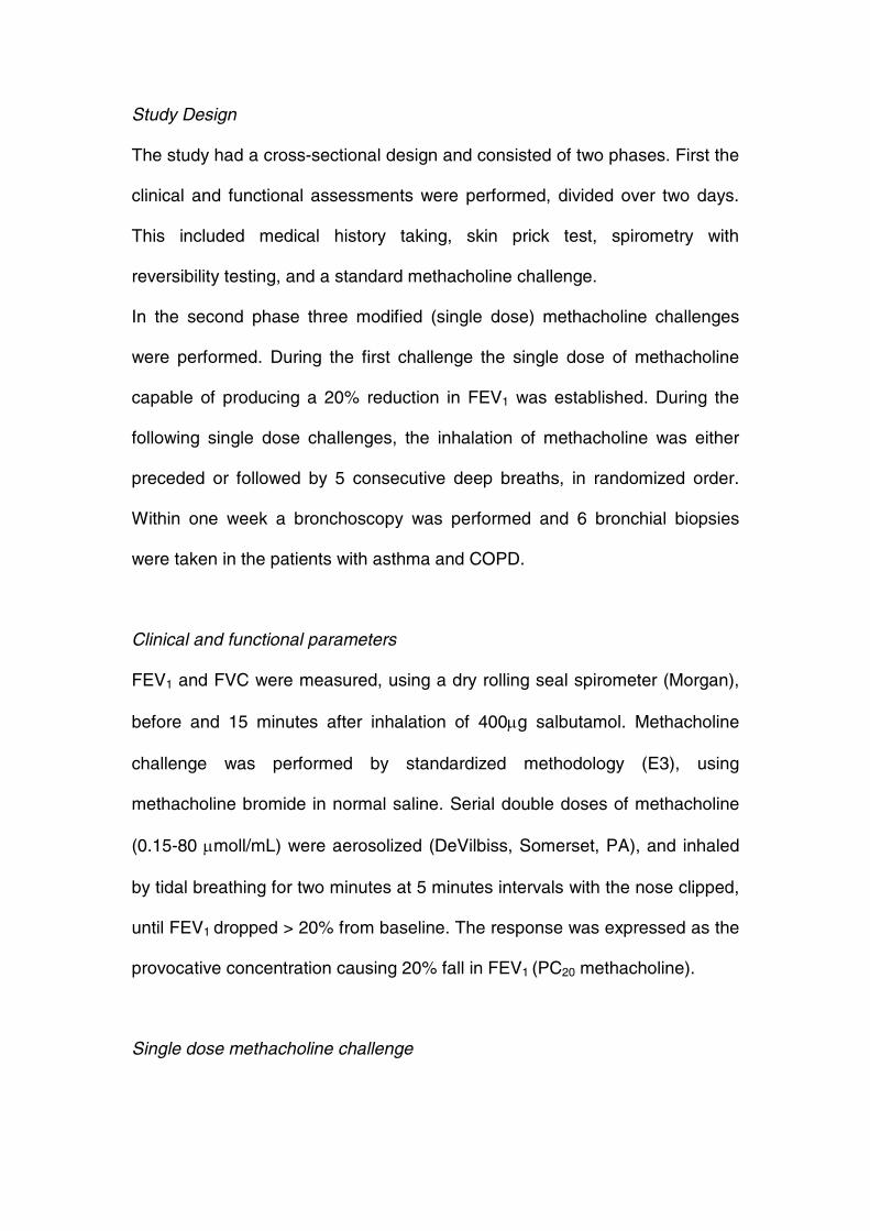
Study Design
The study had a cross-sectional design. Baseline clinical and functional assessments
were performed, divided over two days, including medical history taking, skin prick
test, spirometry with reversibility testing, and a standard methacholine challenge.
In the second phase three modified (single dose) methacholine challenges were
performed (5) (see online supplement). During the first challenge the single dose of
methacholine capable of producing a 20% reduction in FEV1 was established, whilst
the bronchodilator response to one deep inspiration (slow inspiration to TLC, followed
by a passive exhalation) was measured (figure 1a). During the following single dose
challenges, the inhalation of this dose of methacholine was either preceded
(bronchoprotection: figure 1b) or followed by five consequent deep breaths
(bronchodilation: figure 1c), in randomized order. The resistance of the respiratory
system (respiratory resistance) was measured continuously during the breathing
maneuvers using a forced oscillation device (Woolcock institute, Australia) (8) with an
applied oscillation frequency of 8 Hz and an amplitude of ± 1 cmH2O (see online
supplement). Within one week a bronchoscopy was performed and six bronchial
biopsies were taken in the patients with asthma and COPD. The healthy subjects
were not included in the biopsy study, since we aimed to examine the relationship

6
between inflammation and the impaired effect of a deep inspiration within these
disease groups.
Bronchoscopy and immunohistochemistry and image analysis
Bronchoscopy was performed according to a standardized and validated protocol in
our laboratory (27). Disposable forceps’ (radial edge, Boston Scientific) were used to
take six biopsy specimens at (sub)segmental level.
Four biopsies were fixed for 24 hrs in 4% neutral buffered formaldehyde, processed,
and embedded in paraffin. From paraffin-embedded tissues 4µm thick sections were
cut and Haematoxylin-Eosin (H-E) staining was used to evaluate overall bronchial
architecture. Sections of two biopsies, selected on morphological quality criteria
(intact reticular basal membrane and submucosa without crushing artifacts, large
blood clots, or only epithelial scrapings were rejected), per subject were stained and
analyzed. Sections were incubated at room temperature with monoclonal antibodies
directed against CD3, CD4, CD8 (T lymphocytes), EG2 (eosinophils), AA1 (tryptase-
positive mast cells), CD68 (macrophages), and NE (neutrophils). Digital images from
the stained sections were obtained, and fully automated cell counts (KS400, Zeiss)
were performed in the lamina propria (at least 0.125mm2) by a validated method (28).
The number of tryptase-positive mast cells in the airway smooth muscle bundles
were automatically counted in a manually selected airway smooth muscle area (at
least 0.1mm2 (29)), using serial sections stained for alpha smooth muscle-actin and
myosin to identify the airway smooth muscle area. Positively stained cells were
expressed as the number of cells/0.1mm2.
Analysis
Respiratory resistance was measured during 60 seconds of tidal breathing, followed
by one or five slow deep breaths to TLC, and another minute of tidal breathing. Deep

7
inspiration-induced bronchodilation was expressed as the difference between the
mean resistance of all data points of 3 tidal breaths after and of 3 tidal breaths before
the deep inspiration (30,31), which was calculated separately for inspiratory
resistance and expiratory resistance. The latter was done since respiratory
resistance fluctuates during tidal breathing, due to volume and flow differences
between inspiration and expiration, and may be affected differently by deep
inspiration maneuvers (32). Bronchoprotection by deep inspirations was expressed
as the difference in the increase in resistance by methacholine when either five or no
deep inspirations were taken before methacholine inhalation.
The outcome parameters were (log)transformed if necessary. The differences
between the three groups were analyzed using analysis of variance (ANOVA), with
Tukey's honestly significant difference test as post hoc analysis or Kruskall Wallis
Test. Within-group differences were analyzed by 2-tailed paired t-tests or Wilcoxon
ranks test. Spearman’s rank correlation coefficient was used to explore associations
between inflammatory cell counts and deep inspiration-induced changes in
respiratory resistance. We used SPSS version 12.01 for all analyses (SPSS Inc,
Chicago). P-values < 0.05 were considered statistically significant. Sample-size
estimation and details on the analysis are given in the online supplement.
RESULTS
Functional parameters
The patient characteristics are given in table 1. PC20 methacholine (geometric mean
+ SD in doubling dose) was significantly lower in asthma (1.0 ± 1.5 mg/ml) and
COPD (2.15 ± 1.8 mg/ml), as compared to healthy controls (50.1 ± 1.3mg/ml,
8
p<0.001). COPD patients were significantly older, had smoked, and their lung
function was significantly more impaired than the asthmatic patients and healthy
control subjects (table 1).
Single dose methacholine challenges
FEV1 dropped > 20% from baseline in all subjects by the single dose of methacholine
(mean % fall in FEV1 ± SD: 29.7 ± 8.0%, 23.5 ± 2.8%, and 26.8 ± 7.6% for asthma,
COPD and healthy controls respectively), which was not significantly different
between the groups (p=0.08). Tidal volume before and after methacholine inhalation
was not significantly different between the groups (p > 0.7), nor was the inspiratory
volume of either 1 (mean ± SD: asthma 1.6L ± 0.5, COPD 1.6L ± 0.5, healthy
controls 1.5L ± 0.5; p = 0.9) or the mean of 5 deep inspirations (asthma 2.2L ± 0.7,
COPD 2.0L ± 0.4, healthy controls 2.1L ± 0.6; p = 0.6)
In the three groups inspiratory resistance was significantly increased by the single
dose of methacholine (figure 2a; mean change ± SD was +1.4 ± 1.5 cmH2O/L/s in
asthma, +0.6 ± 0.7 cmH2O/L/s in COPD, and +2.1 ± 1.0 cmH2O/L/s in healthy
controls). Expiratory resistance was also significantly increased in asthma and
healthy controls (figure 2b; +1.4 ± 1.6 cmH2O/L/s and +1.9 ± 1.1 cmH2O/L/s), but not
in COPD patients (-0.05 ± 0.9 cmH2O/L/s). The increase in resistance by a single
dose of methacholine was not significantly reduced when five deep inspirations were
taken before methacholine (bronchoprotection) in any of the three groups (figure 2).
In asthma and healthy subjects, after inhalation of methacholine, both one and five
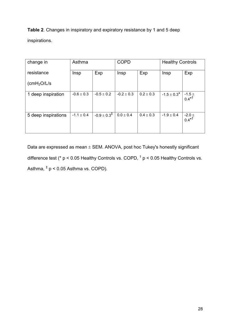
deep inspirations significantly reduced inspiratory and expiratory resistance (table 2;
figure 3a and b for 1 and 5 deep inspirations respectively). In COPD, no significant

9
reduction in expiratory resistance was observed by either one or five deep
inspirations, and only inspiratory resistance was significantly reduced by five deep
inspirations. The reduction in expiratory resistance induced by both one and five
deep breaths was significantly larger in healthy controls than in asthma and COPD
(table 2; p < 0.05 and p < 0.01). Further, the reduction in expiratory resistance during
tidal breathing by 5 deep breaths was significantly larger in asthma than in COPD
(table 2; p < 0.05). The change induced in expiratory resistance by 5 deep
inspirations resembles a mean ± SD % reduction in airway obstruction of 67 ± 4.4 %
in healthy controls, 22 ± 2.3 % in asthmatic patients, and an increase of 13 ± 3.3 % in
COPD patients.
Bronchial inflammation
The numbers of inflammatory cells in the lamina propria of bronchial biopsies
per cell type are shown in table 3. Asthmatic patients had significantly more
eosinophils (EG2+ cells) in the lamina propria as compared to COPD patients. Also
the number of CD4+ lymphocytes and the CD4+/CD8+ lymphocyte ratio tended to be
higher in asthma than in COPD, but this did not reach significance (p=0.09 and
p=0.06 respectively). Among the inflammatory cell types analyzed, predominantly
mast cells were observed in the airway smooth muscle bundles (figure 4). In asthma
74%, and in COPD 76% of the biopsies contained sufficient (> 0.1mm2) airway
smooth muscle area. The mean area analyzed in asthma was 0.24 ± 0.11 mm2 and
in COPD 0.36 ± 0.20 mm2 (p = 0.11).
In asthma the reduction in resistance by one deep breath was positively
associated with the number of CD4+ cells/0.1mm2 (r = 0.61, p = 0.04; figure 5a). In

10
addition, the number of mast cells in the airway smooth muscle bundles correlated
positively with the reduction in resistance by five deep breaths (r = 0.72, p = 0.03,
figure 5b). In COPD there were no significant correlations between the changes in
resistance by deep inspirations and inflammatory cell counts within the lamina
propria, or the number of mast cells in the airway smooth muscle bundles.
DISCUSSION
The results of this study demonstrate that the bronchodilatory effect of deep
inspiration is impaired in intermittent and mild persistent asthma as compared to
healthy subjects, and even more markedly impaired in mild to moderate COPD
patients. Interestingly, in asthma the reduced bronchodilatory effect of a deep
inspiration was associated with increased numbers of mast cells within the airway
smooth muscle bundles and increased CD4+ lymphocyte counts in the bronchial
lamina propria. These findings suggest that the impairment of deep inspiration-
induced bronchodilation in asthma is a result of inflammatory mechanisms within the
airway smooth muscle area and bronchial wall, possibly resulting in altered airway
mechanics by influencing airway smooth muscle characteristics or increasing airway
wall thickness.
To our knowledge this is the first study showing a relationship of inflammatory
cell counts in the airway smooth muscle area and lamina propria of bronchial
biopsies with airway responses to deep inspiration in asthma. In general, our
physiological results are in line with previous studies showing reduced
bronchodilation following deep inspiration in asthma and COPD as compared to
healthy control subjects (23,33). Although, we did find a significant reduction in
respiratory resistance by both one and five deep inspirations in the asthmatic

11
patients, this was significantly less than in the healthy subjects. This partly preserved
deep inspiration-induced bronchodilation in asthma differs from other studies
showing almost no reduction in airways obstruction by deep inspiration in asthmatic
patients (34). This might be explained by differences in disease severity and the level
of airway hyperresponsiveness of the participating subjects. The asthmatic patients
in our study had intermittent or mild persistent asthma, needing no other medication
than bronchodilators on demand. Furthermore, the method of measuring airway
responses to deep inspiration differs amongst studies and may influence the
outcome parameters as well (35).
Notably, we did not find a bronchoprotective effect of deep inspirations in the
healthy control group, whereas this has been shown by several studies in the past
(3,5). This seems to be explained by the methods used to assess airways
obstruction. Bronchoprotection by deep inspiration has predominantly been observed
by using measurements implicitly including a deep breath, such as FEV1, whereas it
could not be established by parameters without a deep breath during the
measurement (36). We purposely chose the latter in order to examine the unaffected
protective effect of deep inspirations against the dynamics of airway narrowing and
therefore may have missed bronchoprotection as reported when using FEV1. Taken
together these findings suggest that deep inspirations taken prior to methacholine
inhalations improve subsequent bronchodilatory effects of deep breaths in healthy
subjects, and thus prevent a fall in FEV1, but may not necessarily prevent the
obstruction itself.
We aimed to look at relationships between bronchial inflammation and deep
breath effects within a group of asthmatic and COPD patients, and therefore selected
the patients that matched the key features of these two distinct disease groups. As

12
expected, this resulted in significant differences between the groups with regard to
age and lung function. However, neither in COPD nor in healthy controls was a
relationship found between deep breath-induced reduction in respiratory resistance
and age or lung function (r < 0.4, p > 0.2). Therefore, the differences between COPD
and healthy controls are most likely a result of pathophysiological changes in COPD.
We used a modified single-dose methacholine challenge to induce a given
level of airways obstruction in all subjects in order to measure both the
bronchoprotective and the bronchodilatory effect of deep inspirations. During the first
challenge we established the dose that induced a reduction in FEV1 of at least 20%,
and used that dose for the other two challenges. We could not determine whether the
subsequent single-dose challenges induced the same fall in FEV1 in absence of
performing spirometry. However, since there was no significant difference within the
groups between the 3 challenges with regard to respiratory resistance following
methacholine inhalation, we presume that the level of obstruction was approximately
the same as in the dose-finding challenge.
Interestingly, in the patients with COPD the fall in FEV1 induced by
methacholine was not accompanied by a significant increase in respiratory
resistance, a finding that we cannot fully explain. This may have limited the possibility
of reducing respiratory resistance by deep inspiration in this group. However, there
was no direct relationship between the increase in respiratory resistance by
methacholine and the reduction in respiratory resistance by deep inspirations,
suggesting that the absence of the bronchodilatory effect of deep inspirations in
COPD was not necessarily dependent on the absence of an increase in respiratory
resistance. Hence, this finding may provide new information on the functionally

13
relevant pathophysiology of the airways in mild to moderate COPD patients, which
requires further investigation.
In this study protocol we have used the forced oscillation technique to
measure airway responses to deep inspiration. The limitation of this method is that
the results represent resistance of the complete respiratory system, including the
upper airways, and thus the site of the obstruction or deep inspiration-induced
bronchodilation is difficult to determine. However, this technique enabled us to
monitor respiratory resistance continuously, and therefore we were able to measure
the effect of deep breaths on the dynamics of airway obstruction both during the
deep breaths as well as during tidal breathing.
How can we interpret these results? During a deep inspiration the airways are
dilated, as shown on CT scan (37), both in healthy and asthmatic adults, presumably
as a result of the airway-parenchymal coupling. However, it appeared that in asthma
deep breaths could not reduce respiratory resistance to the same extent as in healthy
subjects. We found that increased numbers of CD4+ lymphocytes in the lamina
propria of bronchial biopsies were associated with impaired bronchodilation following
a deep breath in asthma. It is likely that these cells indirectly reflect the inflammatory
changes within the bronchial wall that prevent adequate stretch of the airways and
airway smooth muscle layer. CD4+ lymphocytes are involved in eosinophilic
inflammation, and are associated with vasodilation and microvascular leakage (38).
These inflammatory changes may narrow the internal airway diameter, and at the
same time increase the outer wall perimeter, thereby decreasing the force applied to
the airways by the parenchyma during deep inspiration (17,39). In addition, a similar
relationship with CD4+ cells was not found at slightly larger changes in resistance
induced by 5 deep inspirations. This may indicate that inflammation as reflected by

14
CD4+ lymphocytes within the lamina propria indeed decreases deep inspiration-
induced stretch of the airways, but does not fully prevent it, which may be overcome
by multiple stretching maneuvers. Another hypothesis regarding the role of
inflammation in the impairment of the bronchodilatory effect of deep inspiration is the
reduction in the stretch-induced release of inhibiting factors, such as nitric oxide. The
CD4+ lymphocytes within the bronchial wall may counteract these active
bronchodilating mechanisms.
Most strikingly, we found a correlation between the number of mast cells
within the smooth muscle bundles and deep inspiration-induced bronchodilation in
asthma. Mast cells can promote airway smooth muscle contraction, by releasing
histamine, prostaglandin D2 and TNF-alpha (29,40). We speculate that the
localization of the mast cells within the smooth muscle cells could result in a
physiologically altered intrinsic contractile function, leading to an increased formation
of actin and myosin cross bridges, more difficult to disrupt by deep inspiration-
induced stretch of the airways, which has been referred to as the latch state (41).
These data further extend the results found by Brightling et al (29) showing increased
numbers of mast cells in the airway smooth muscle bundles in bronchial biopsies of
patients with asthma as compared to healthy controls or patients with eosinophilic
bronchitis, which was related to airway hyperresponsiveness.
Interestingly, in COPD there was no significant reduction in respiratory
resistance by deep breaths. An absolute loss of alveolar attachments might explain
this observation, since this would result in uncoupling of the airway-parenchyma
interdependence, leading to less strain imposed on the airways by the parenchyma
during deep inspiration (42). Indeed, it has been shown that the loss of alveolar
attachments was related to less bronchodilation by deep breaths in mild to moderate

15
COPD patients (13). Since we did not find a direct relationship between inflammatory
cells within the bronchial wall and deep breath-induced bronchodilation in COPD, we
speculate that the marked loss of the ability to reduce respiratory resistance by deep
inspiration is predominantly due to structural damage of the airways or lung
parenchyma in this disease.
What could be the clinical implication of our study? The correlation of
inflammatory cells within the submucosa and airway smooth muscle bundles with the
bronchodilatory effect of a deep inspiration in asthma indicates that the impaired
airway mechanics may, at least partially, be restored by treatment. Indeed, it has
been shown that airway responses to deep inspirations can be improved by
treatment with (inhaled) corticosteroids (18−20). Furthermore, since deep inspirations
are likely to play a role in airway hyperresponsiveness (3), perceived symptoms (43)
and excaberations (9) in asthma, measurement of airway responses to deep
inspiration may give additional information on current disease status. In COPD, our
findings indicate that airway inflammation plays a less prominent role in the
pathophysiological mechanism of deep breath-induced bronchodilation in COPD,
which limits the options for intervention. However, deep breath responses may be a
sensitive parameter to find early lung damage by smoking.
We conclude that deep inspiration-induced bronchodilation is reduced in
intermittent and mild persistent asthma as compared to healthy subjects, and absent
in mild to moderate COPD patients. In asthma the bronchodilatory effect of deep
inspirations is related to inflammatory cell counts within airway smooth muscle
bundles and bronchial wall, whereas in moderate COPD this relation could not be
found. These results indicate that the physiological protection against airway

16
narrowing by deep inspiration is impaired in both asthma and COPD, but this may be
due to different pathophysiological mechanisms.

17
References
1. NHLBI/WHO workshop report.Publication No.95-3659. Bethesda,MD,National
Institutes of Healths 1991 Global initiative for Asthma Management and
Prevention. www.ginasthma.org (update november 2006).
2. Grootendorst, DC and Rabe, KF. Mechanisms of bronchial hyperreactivity in
asthma and chronic obstructive pulmonary disease. Proc Am Thorac Soc
2004;1:77-87
3. Scichilone, N, Permutt, S, Togias, A. The lack of the bronchoprotective and not
the bronchodilatory ability of deep inspiration is associated with airway
hyperresponsiveness. Am J Respir Crit Care Med 2001;163:413-419
4. Skloot, G, Permutt, S, Togias, A. Airway hyperresponsiveness in asthma: a
problem of limited smooth muscle relaxation with inspiration. J Clin Invest
1995;96:2393-2403
5. Kapsali, T, Permutt, S, Laube, B, Scichilone, N, Togias, A. Potent
bronchoprotective effect of deep inspiration and its absence in asthma. J Appl
Physiol 2000;89:711-720
6. Scichilone, N, Kapsali, T, Permutt, S, Togias, A. Deep inspiration-induced
bronchoprotection is stronger than bronchodilation. Am J Respir Crit Care Med
2000;162:910-916
7. Jensen, A, Atileh, H, Suki, B, Ingenito, EP, Lutchen, KR. Selected contribution:
airway caliber in healthy and asthmatic subjects: effects of bronchial challenge
and deep inspirations. J Appl Physiol 2001;91:506-515

18
8. Salome, CM, Thorpe, CW, Dipa, C, Brown, NJ, Berend, N, King, GG. Airway re-
narrowing following deep inspiration in asthmatic and nonasthmatic subjects.
Eur Respir J 2003;22:62-68
9. Lim, TK, Ang, SM, Rossing, TH, Ingenito, EP, Ingram, RH, Jr. The effects of
deep inhalation on maximal expiratory flow during intensive treatment of
spontaneous asthmatic episodes. Am Rev Respir Dis 1989;140:340-343
10. Fairshter, RD. Airway hysteresis in normal subjects and individuals with chronic
airflow obstruction. J Appl Physiol 1985;58:1505-1510
11. Fairshter, RD. Effect of a deep inspiration on expiratory flow in normals and
patients with chronic obstructive pulmonary disease. Bull Eur Physiopathol
Respir 1986;22:119-125
12. Corsico, A, Milanese, M, Baraldo, S, Casoni, GL, Papi, A, Riccio, AM, Cerveri, I,
Saetta, M, Brusasco, V. Small airway morphology and lung function in the
transition from normality to chronic airway obstruction. J Appl Physiol
2003;95:441-447
13. Scichilone, N, Bruno, A, Marchese, R, Vignola, AM, Togias, A, Bellia, V.
Association between reduced bronchodilatory effect of deep inspiration and loss
of alveolar attachments. Respir Res 2005;6:55
14. Jeffery, PK. Remodeling and inflammation of bronchi in asthma and chronic
obstructive pulmonary disease. Proc Am Thorac Soc 2004;1:176-183
15. Fabbri, LM, Romagnoli, M, Corbetta, L, Casoni, G, Busljetic, K, Turato, G,
Ligabue, G, Ciaccia, A, Saetta, M, Papi, A. Differences in airway inflammation in
19
patients with fixed airflow obstruction due to asthma or chronic obstructive
pulmonary disease. Am J Respir Crit Care Med 2003;167:418-424
16. Elias, JA, Zhu, Z, Chupp, G, Homer, RJ. Airway remodeling in asthma. J Clin
Invest 1999;104:1001-1006
17. Macklem, PT. A theoretical analysis of the effect of airway smooth muscle load
on airway narrowing. Am J Respir Crit Care Med 1996;153:83-89
18. Corsico, A, Pellegrino, R, Zoia, MC, Barbano, L, Brusasco, V, Cerveri, I. Effects
of inhaled steroids on methacholine-induced bronchoconstriction and gas
trapping in mild asthma. Eur Respir J 2000;15:687-692
19. Scichilone, N, Permutt, S, Bellia, V, Togias, A. Inhaled corticosteroids and the
beneficial effect of deep inspiration in asthma. Am J Respir Crit Care Med
2005;172:693-699
20. Slats, AM, Sont, JK, van Klink, RH, Bel, EH, Sterk, PJ. Improvement in
bronchodilation following deep inspiration after a course of high-dose oral
prednisone in asthma. Chest 2006;130:58-65
21. Pliss, LB, Ingenito, EP, Ingram, RH, Jr. Responsiveness, inflammation, and
effects of deep breaths on obstruction in mild asthma. J Appl Physiol
1989;66:2298-2304
22. Pacini, F, Filippelli, M, Duranti, R, Rosi, E, Romagnoli, I, Grazzini, M, Stendardi,
L, Misuri, G, Scano, G. Reduction in bronchodilation following a deep inhalation
is poorly related to airway inflammation in asthma. Eur Respir J 1999;14:1055-
1060

20
23. King, GG, Moore, BJ, Seow, CY, Pare, PD. Time course of increased airway
narrowing caused by inhibition of deep inspiration during methacholine
challenge. Am J Respir Crit Care Med 1999;160:454-457
24. Slats, AM, Aardweg, JG, De Jongste, J, Schot, R, Rabe, KF, Sterk, PJ. Deep
Inspiration-Induced Bronchodilation and Bronchoprotection in COPD: A
Comparison with Asthma. Proceedings of the American Thoracic Society
2006;3:A452
25. Slats, AM, Janssen, K, van Schadewijk, A, van den Aardweg, JG, Schot, R, van
der Plas, DT, Mauad, T, Rabe, KF, Sterk, PJ. Airway inflammation and airway
dynamics during deep inspiration in asthma and COPD. Eur Respir J
2006;28:832s
26. Global strategy for the diagnosis, management, and prevention of chronic
obstructive pulmonary disease. Global Initiative for Chronic Obstructive Lung
Disease. NHLBI/WHO workshop report 2003. www.goldcopd.org Update
november 2006.
27. de Kluijver, J, Schrumpf, JA, Evertse, CE, Sont, JK, Roughley, PJ, Rabe, KF,
Hiemstra, PS, Mauad, T, Sterk, PJ. Bronchial matrix and inflammation respond
to inhaled steroids despite ongoing allergen exposure in asthma. Clin Exp
Allergy 2005;35:1361-1369
28. Sont, JK, De Boer, WI, van Schadewijk, WA, Grunberg, K, van Krieken, JH,
Hiemstra, PS, Sterk, PJ. Fully automated assessment of inflammatory cell
counts and cytokine expression in bronchial tissue. Am J Respir Crit Care Med
2003;167:1496-1503

21
29. Brightling, CE, Bradding, P, Symon, FA, Holgate, ST, Wardlaw, AJ, Pavord, ID.
Mast-cell infiltration of airway smooth muscle in asthma. N Engl J Med
2002;346:1699-1705
30. Schweitzer, C, Moreau-Colson, C, Marchal, F. Respiratory impedance response
to a deep inhalation in asthmatic children with spontaneous airway obstruction.
Eur Respir J 2002;19:1020-1025
31. Marchal, F, Schweitzer, C, Moreau-Colson, C. Respiratory impedance response
to a deep inhalation in children with history of cough or asthma. Pediatr
Pulmonol 2002;33:411-418
32. Thorpe, CW, Salome, CM, Berend, N, King, GG. Modeling airway resistance
dynamics after tidal and deep inspirations. J Appl Physiol 2004;97:1643-1653
33. Scichilone, N, Marchese, R, Catalano, F, Vignola, AM, Togias, A, Bellia, V.
Bronchodilatory effect of deep inspiration is absent in subjects with mild COPD.
Chest 2004;125:2029-2035
34. Scichilone, N, Marchese, R, Soresi, S, Interrante, A, Togias, A, Bellia, V. Deep
inspiration-induced changes in lung volume decrease with severity of asthma.
Respir Med 2006
35. Burns, GP and Gibson, GJ. The apparent response of airway function to deep
inspiration depends on the method of assessment. Respir Med 2001;95:251-
257
22
36. Crimi, E, Pellegrino, R, Milanese, M, Brusasco, V. Deep breaths, methacholine,
and airway narrowing in healthy and mild asthmatic subjects. J Appl Physiol
2002;93:1384-1390
37. Brown, RH, Scichilone, N, Mudge, B, Diemer, FB, Permutt, S, Togias, A. High-
resolution computed tomographic evaluation of airway distensibility and the
effects of lung inflation on airway caliber in healthy subjects and individuals with
asthma. Am J Respir Crit Care Med 2001;163:994-1001
38. Bousquet, J, Jeffery, PK, Busse, WW, Johnson, M, Vignola, AM. Asthma. From
bronchoconstriction to airways inflammation and remodeling. Am J Respir Crit
Care Med 2000;161:1720-1745
39. Macklem, PT. A hypothesis linking bronchial hyperreactivity and airway
inflammation: implications for therapy. Ann Allergy 1990;64:113-116
40. Bradding, P, Walls, AF, Holgate, ST. The role of the mast cell in the
pathophysiology of asthma. J Allergy Clin Immunol 2006;117:1277-1284
41. Fredberg, JJ. Frozen objects: small airways, big breaths, and asthma. J Allergy
Clin Immunol 2000;106:615-624
42. Lambert, RK and Pare, PD. Lung parenchymal shear modulus, airway wall
remodeling, and bronchial hyperresponsiveness. J Appl Physiol 1997;83:140-
147
43. Sont, JK, Booms, P, Bel, EH, Vandenbroucke, JP, Sterk, PJ. The severity of
breathlessness during challenges with inhaled methacholine and hypertonic

23
saline in atopic asthmatic subjects. The relationship with deep breath-induced
bronchodilation. Am J Respir Crit Care Med 1995;152:38-44

24
Figure legends
Figure 1. Single dose challenge measurements
This figure describes the three different single dose methacholine challenges. The
line shows the time in minutes, and the arrows show the number of deep breaths
taken. Baseline measurements of respiratory resistance (Rrs) and FEV1 were
followed by a period of 20 minutes with deep breath avoidance. A) methacholine
inhalation was followed by Rrs measurement with one deep breath and FEV1
measurement to determine whether this dose could reduce FEV1 by at least 20%. B)
methacholine inhalation preceded by 5 deep inspirations, followed by Rrs
measurement with 1 deep breath. C) methacholine inhalation followed by Rrs
measurement with 5 deep breaths.
Figure 2. Changes in respiratory resistance by methacholine
This figure shows the individual data points per group before (pre) and after (post)
methacholine inhalation, for inspiratory (panel A) and expiratory (panel B) resistance.
Data are expressed in cmH2O/L/s, and the horizontal lines represent the mean. The
squares connected by solid lines ( ) represent the data with no deep inspirations
taken before methacholine inhalation (figure 1A), the triangles connected by dashed
lines ( ) represent the challenge when 5 deep inspirations were taken before
methacholine inhalation (figure 1B). Methacholine significantly increased the
inspiratory resistance in all three groups and expiratory resistance in only asthma
and healthy controls, and not in COPD. 5 Deep inspirations did not protect against
the increase in inspiratory and expiratory resistance in all three groups.
Figure 3. Changes in expiratory resistance by deep breath.
In this figure the paired data (mean ± SEM) for asthma ( ), COPD ( ), and
healthy controls ( ) are depicted. The data are expressed as the mean expiratory

25
resistance during 3 tidal expirations before methacholine inhalation (premch), 3 tidal
expirations after methacholine inhalation (postmch), the passive expiration of the
deep inspiration (DI), and 3 tidal expirations following deep inspiration (postDI). Panel
A shows the data of the measurement when 1 deep inspiration was taken following
methacholine inhalation (figure 1A). Panel B shows the data of the measurement
when 5 deep inspirations were taken (figure 1C), where data point ‘DI 5’ represents
the mean of the resistance during the 5 passive expirations of the 5 deep breaths.
The reduction in expiratory resistance during tidal breathing by 1 and by 5 deep
breaths was significantly larger in healthy subjects as compared to asthmatic patients
(† p < 0.05) and to COPD patients (* p < 0.05). Further, the reduction in expiratory
resistance during tidal breathing by 5 deep breaths was significantly larger in asthma
than in COPD (‡ p < 0.05).
Figure 4. Photomicrographs of mast cell and myosin staining.
Example of a bronchial biopsy section immunohistochemically stained for (A)
tryptase+ mast cells (original magnification: x200; EPI = epithelium, SUBM =
submucosa, ASM = airway smooth muscle), and (B) a serial section of the same
biopsy stained for myosin (original magnification: x200). The smooth muscle area
was manually selected (C) in the mast cell staining by using the myosin staining
(original magnification: x200). In the selected area (D) mast cells (arrows) were
automatically counted (original magnification: x200).
Figure 5. Relationship between inflammatory cell counts and deep inspiration-
induced bronchodilation in asthma.
This figure shows the relationship between the change in inspiratory resistance by A)
1 deep inspiration and the number of CD4+ lymphocytes/0.1mm2 in the lamina

26
propria (r=0.61, p=0.04), and by B) 5 deep inspirations and the number of AA1+ mast
cells/0.1mm2 in the airway smooth muscle bundles (r=0.73, p=0.03).

27
Table 1. Patient characteristics
Asthma COPD Healthy Controls
Sex (male/female) 5/8 8/4 2/10
Age (years) 23.8 ± 5.7 57.9 ± 7.5*‡ 32.8 ± 13.8
BMI (kg/m2) 22.9 ± 2.1 26.3 ± 3.3*‡ 22.2 ± 3.4
Pack Years 0.04 ± 0.1 38.9 ± 15.6*‡ 0.33 ± 0.8
post salb FEV1 % pred 103.9 ± 11.1 78.6 ± 13.9*‡ 107.4 ± 12.6
post salb FEV1/FVC (%) 87.0 ± 6.4 60.9 ± 7.6*‡ 85.6 ± 8.3
PC20 methacholine
(mg/ml)
1.0 ± 1.5 2.2 ± 1.8 50.7 ± 1.3*†
Single dose methacholine
(mg/ml)
3.3 ± 1.4 10.0 ± 1.9‡ 72.5 ± 1.5*†
% fall in FEV1 (single-
dose methacholine
challenge)
29.7 ± 8.0 23.5 ± 2.8 26.8 ± 7.6
Data are expressed as mean ± SD, except for sex (number), PC20 methacholine
(geometric mean ± SD in doubling doses). ANOVA, post hoc Tukey's honestly
significant difference test (* p < 0.05 Healthy Controls vs. COPD, † p < 0.05 Healthy
Controls vs. Asthma, ‡ p < 0.05 Asthma vs. COPD).
28
Table 2. Changes in inspiratory and expiratory resistance by 1 and 5 deep
inspirations.
Asthma COPD Healthy Controls change in
resistance
(cmH2O/L/s
Insp Exp Insp Exp Insp Exp
1 deep inspiration -0.6 ± 0.3 -0.5 ± 0.2
-0.2 ± 0.3
0.2 ± 0.3
-1.5 ± 0.3* -1.5 ±0.4*†
5 deep inspirations -1.1 ± 0.4
-0.9 ± 0.3‡ 0.0 ± 0.4
0.4 ± 0.3
-1.9 ± 0.4
-2.0 ±0.4*†
Data are expressed as mean ± SEM. ANOVA, post hoc Tukey's honestly significant
difference test (* p < 0.05 Healthy Controls vs. COPD, † p < 0.05 Healthy Controls vs.
Asthma, ‡ p < 0.05 Asthma vs. COPD).

29
Table 3. Inflammatory cell counts in bronchial biopsies.
asthma COPD
CD3+ cells 53.3 [14.0-134.0] 25.5 [3.0-160.5]
CD4+ cells 24.8 [9.5-86.0] 12.0 [1.5-83.0]
CD8+ cells 25.8 [7.0-62.0] 15.0 [5.0-93.0]
CD4+/CD8+ cells 1.7 [0.6-4.4] 0.6 [0.1-4.4]
EG2+ cells 1.5 [0.0-8.0]* 0.3 [0.0-3.0 ]
AA1+ cells 10.0 [1.0-24.0] 16.0 [2.0-56.0]
AA1+ cells in airway
smooth muscle bundles
2 [0.0-7.0] 1.5 [1.0-3.0]
CD68+ cells 19.0 [8.0-53.0] 9.3 [3.0-100.0]
NE+ cells 1.8 [0.0-14.0] 2.0 [0.0-41.0]
The numbers of cells are expressed as median [range] per 0.1 mm2. * p < 0.05
between groups.

Figure 1. Single dose challenge measurements
time (min)20 22 250-5
RrsRrsDIFEV1 FEV1Mch
avoidance of deep breath
time (min)20 220-5
RrsRrsDIFEV1 Mch
avoidance of deep breath
5 DIs
19
time (min)20 220-5
RrsRrs
5 DIsFEV1 Mch
avoidance of deep breath
A
B
C

Figure 2. Changes in respiratory resistance by a single-dose methacholine
pre post pre post pre post pre post pre post pre post0123456789
1011
A p < 0.05 p < 0.01
p < 0.05p < 0.01p < 0.01p < 0.05
Insp
irat
ory
resi
stan
ce(c
mH
2O/L
/s)
pre post pre post pre post pre post pre post pre post0123456789
1011121314
Asthma COPD Healthy controls
Bp < 0.05
p < 0.01
p < 0.01
p < 0.01
Expi
rato
ryre
sist
ance
(cm
H2O
/L/s
)

Figure 3. Changes in expiratory resistance by A) 1 deep breath, B) 5 deep breaths
pre mch post mch DI post DI
2
3
4
5
6*
A COPDasthmaHealthy controls
Expi
rato
ryre
sist
ance
(cm
H2O
/L/s
)
pre mch post mch DI 5 post DI
2
3
4
5
6 *B
Expi
rato
ryre
sist
ance
(cm
H2O
/L/s
)

Figure 4

Figure 5. Relationship between inflammatory cell counts and deep inspiration-
induced bronchodilation in asthma.
10 100
-4
-3
-2
-1
0
1
2A
CD4+ cells/0.1mm2
chan
gein
Rrs
byD
I(c
mH
2O/L
/s)
1 10
-5
-4
-3
-2
-1
0
1B
AA1+ cells/0.1mm2 ASM area
chan
gein
Rrs
by5
DI
(cm
H2O
/L/s
)

Bronchial inflammation and airway responses to deep inspiration in
asthma and COPD
Annelies M. Slats, Kirsten Janssen, Annemarie van Schadewijk, Dirk T. van
der Plas, Robert Schot, Joost G. van den Aardweg, Johan C. de Jongste,
Pieter S. Hiemstra, Thais Mauad, Klaus F. Rabe, Peter J. Sterk
ONLINE SUPPLEMENT

METHODS
Subjects
For this study we enrolled 13 patients with intermittent and mild persistent
asthma (GINA step 1 and 2) (E1), 12 patients with mild to moderate COPD
(GOLD I and II) (E2) and 12 healthy subjects. The patients with asthma were
all non-smokers or ex-smokers (< 5 pack years), had a history of episodic
wheezing or chest tightness, a baseline forced expiratory volume in 1s (FEV1)
of more than 70% of predicted, the provocative concentration of methacholine
causing a 20% fall in FEV1 (PC20 methacholine) was less than 8 mg/mL, and
they were all atopic as determined by one or more positive skin prick tests to
10 common aeroallergen extracts. The patients with COPD were all smokers
or ex-smokers (> 10 pack years), had a history of chronic symptoms of cough
and/or dyspnea, had an FEV1/FVC ratio of less than 70% of predicted post
bronchodilator, and the reversibility of FEV1 by salbutamol was less than 12%
of predicted. All the patients were clinically stable, used β2-agonists on
demand only, and had no history of a recent (< 2 weeks) respiratory tract
infection or other relevant diseases. None of the asthmatic or COPD patients
had used inhaled or oral corticosteroid within 3 months prior to the study. The
healthy subjects that participated in the study had no history of respiratory
symptoms, were non-smokers or ex-smokers (< 5 pack years), had a baseline
FEV1 of more than 80% of predicted, the PC20 methacholine was more than
16 mg/mL, and they had no positive reaction to the skin prick test.
The institutional review board for human studies approved the protocol, and
the subjects gave their written informed consent before entering the study.
Study Design
The study had a cross-sectional design and consisted of two phases. First the
clinical and functional assessments were performed, divided over two days.
This included medical history taking, skin prick test, spirometry with
reversibility testing, and a standard methacholine challenge.
In the second phase three modified (single dose) methacholine challenges
were performed. During the first challenge the single dose of methacholine
capable of producing a 20% reduction in FEV1 was established. During the
following single dose challenges, the inhalation of methacholine was either
preceded or followed by 5 consecutive deep breaths, in randomized order.
Within one week a bronchoscopy was performed and 6 bronchial biopsies
were taken in the patients with asthma and COPD.
Clinical and functional parameters
FEV1 and FVC were measured, using a dry rolling seal spirometer (Morgan),
before and 15 minutes after inhalation of 400µg salbutamol. Methacholine
challenge was performed by standardized methodology (E3), using
methacholine bromide in normal saline. Serial double doses of methacholine
(0.15-80 µmoll/mL) were aerosolized (DeVilbiss, Somerset, PA), and inhaled
by tidal breathing for two minutes at 5 minutes intervals with the nose clipped,
until FEV1 dropped > 20% from baseline. The response was expressed as the
provocative concentration causing 20% fall in FEV1 (PC20 methacholine).
Single dose methacholine challenge

Three different modified methacholine challenges were performed during this
study (E4). The first challenge (manuscript figure 1A) was performed to
determine the dose of methacholine capable of inducing a fall in FEV1 of at
least 20%. At baseline FEV1 and respiratory resistance were measured three
times. Respiratory resistance was measured continuously during one minute
of tidal breathing, a slow deep inhalation to TLC, a passive exhalation, and a
subsequent minute of tidal breathing. Then the subject was asked to withhold
from taking deep breaths for a period of 20 minutes. The avoidance of deep
breath was monitored using a thoracic respiratory transducer (Jaeger),
showing the volume signal on a monitor. When tidal volume was doubled or
more, then the time of deep breath avoidance was restarted. Subsequently, a
single dose of methacholine (approximately the cumulative dose of the PC20
methacholine) was inhaled for two minutes by tidal breathing, and two
minutes later respiratory resistance was measured again during 1 minute of
tidal breathing, a deep inspiration, passive expiration and a subsequent
minute of tidal breathing. This was followed by a forced expiratory maneuver
to measure FEV1. When the FEV1 dropped by more than 20%, this dose was
used for the two following challenges. If the fall in FEV1 did not reach 20%,
another challenge was scheduled at least 24 hours apart using the doubling
concentration of the former single-dose challenge with a maximum of three
attempts.
The two following challenges were performed in randomized order. After
measuring baseline values and a 20 minute period of deep breath avoidance
the subject would take 5 deep breaths before (bronchoprotection, manuscript
figure 1B) or after (bronchodilation, manuscript figure 1C) the inhalation of

methacholine. The 5 deep breaths taken after methacholine inhalation were
performed while measuring respiratory resistance.
Forced Oscillation Technique
The resistance of the respiratory system was measured continuously during
breathing maneuvers using a forced oscillation device (Woolcock institute,
Australia) (E5) with an applied oscillation frequency of 8 Hz and an amplitude
of ± 1 cmH2O. The device was calibrated using calibration tubes of known
resistance. The subjects breathed through an antibacterial filter, with
resistance of 0.2 cmH2O/L/s. Flow was measured using a 50-mm diameter
Fleisch Pneumotachograph (Vitalograph Ltd, Maids Moreton, UK), and
differential pressure was measured using a ± 2.5 cm H2O solid-state
transducer (Sursense DCAL4; Honeywell Sensing and Control, Milpitas, CA,
USA). Mouth pressure was measured using a similar transducer with a higher
range (±12.5 cmH2O). Analog pressure and flow signals were digitized at 400
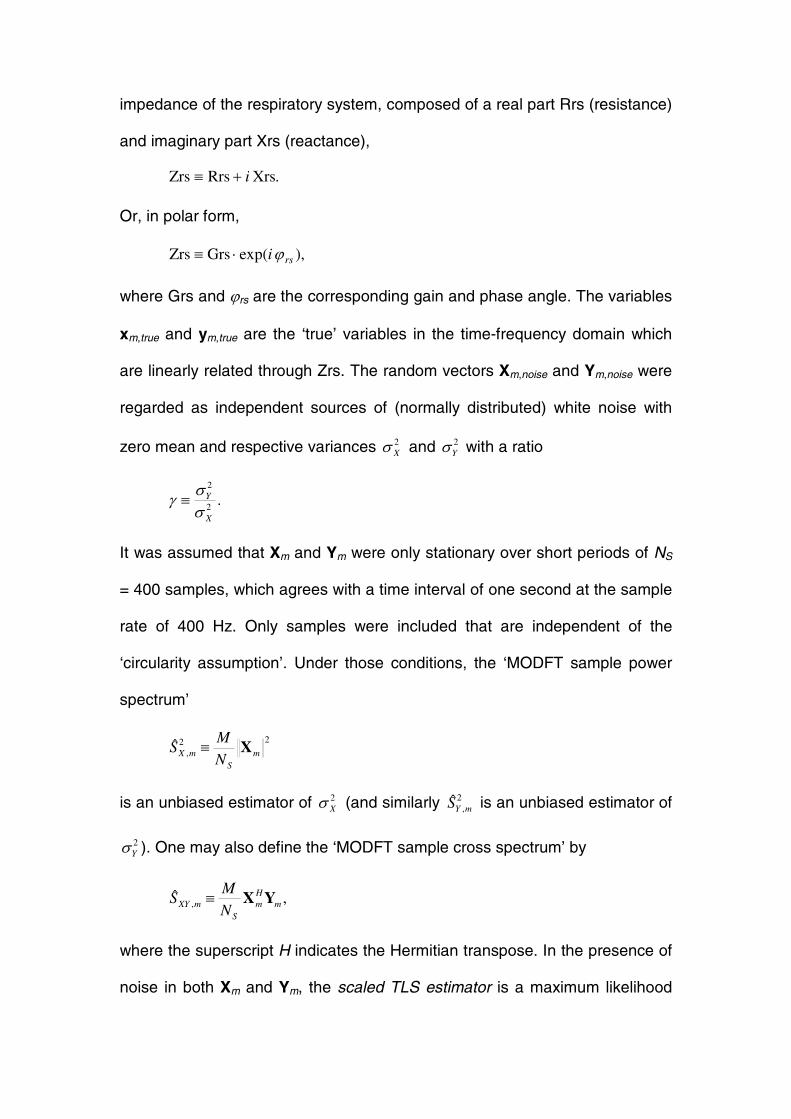
Hz. The time- and frequency-dependent respiratory impedance Zrs was
estimated based on the hypothesis that random errors occur in both pressure
and flow. This yields a Total Least Squares (TLS) estimate of respiratory
impedance as a function of time and frequency and allows an estimation of
confidence intervals in the course of time. The latter is important since it can
be expected that respiratory impedance changes from inspiration to expiration
and on the longer term due to the interventions in the experiment. It can
therefore be expected that the confidence intervals of Zrs also change in time.
This model is further explained in the appendix of this online supplement.

Bronchoscopy
The bronchoscopies were performed according to a standardized and
validated protocol (E6) and were performed by experienced pulmonologists.
The procedure involved detailed explanation to the patient, premedication (0.5
mg atropine s.c., 20 mg codeine p.o., 400 µg salbutamol p.i.) and local
anesthesia of the larynx and lower airways (10% lidocaine spray in the
oropharynx, 2% lidocaine in lower airways). During the procedure 100%
oxygen was delivered by nasal canula (2 l/min), whilst transcutaneous oxygen
saturation was monitored continuously by oximeter (Nellcor N-180) with a
finger probe. Disposable forceps (radial edge, Boston Scientific) were used to
take six biopsy specimens at (sub)segmental level.
Immunohistochemistry and image analysis
Four biopsies were fixed for 24 hrs in 4% neutral buffered formaldehyde,
processed, and embedded in paraffin. From paraffin-embedded tissues 4µm
thick sections were cut and Haematoxylin-Eosin (H-E) staining was used to
evaluate overall bronchial architecture, epithelial integrity, and extent of the
inflammatory infiltrate. Sections of two biopsies, selected on morphological
quality criteria (intact reticular basal membrane and submucosal without
crushing artifacts, large blood clots, or only epithelial scrapings), per subject
were stained and analyzed. Immunohistochemistry staining on paraffin
embedded tissue was performed on 4 µm thick sections. If required,
immunohistochemical staining was preceded by antigen retrieval. Sections
were incubated at room temperature with monoclonal antibodies directed
against CD3 (1:50, clone PS1, Novocastra), CD4 (1:50, clone 4B12,

Novocastra), CD8 (1:100, clone 1A5, Novocastra) (T-lymphocytes), EG2
(eosinophils, 1:600, clone EG-2; Pharmacia), AA1 (mast cell tryptase,
1:16000, clone AA1; DAKO), CD68 (macrophages, 1:400, clone PG-M1;
DAKO), and NE (neutrophils, 1:200, clone NP57, DAKO). Digital images from
the stained sections were obtained using a three-chip color camera (Zeiss
Vision Ks400 system, Kontron/Zeiss). Fully automated cell counts were
performed in the lamina propria by a validated method (E7). Positive stained
cells were expressed as the number of cells/0.1mm2. The number of mast
cells in the airway smooth muscle bundles were automatically counted in
manually selected airway smooth muscle area using the sections stained for
alpha-smooth muscle-actin and myosin (manuscript figure 4).
Analysis
Respiratory resistance was measured during 60 seconds of tidal breathing, 1
or 5 slow deep breaths to TLC and another minute of tidal breathing (figure
E1). Deep inspiration-induced bronchodilation was expressed as the
difference between the mean resistance of all data points during 3 tidal
breaths after and during 3 tidal breaths before the deep inspiration (E8;E9),
which was calculated separately for inspiratory resistance and expiratory
resistance. Bronchoprotection by deep inspirations was expressed as the
difference in the increase in resistance by methacholine when either 5 or no
deep inspirations were taken before methacholine inhalation.
The sample-size of 12 subjects per group was based on our own repeatability
data with regard to biopsy immunohistochemistry, allowing the detection of 2
fold differences in bronchial biopsy cell numbers between groups, if α=0.05

(two-sided) and β=0.80 (one-sided) (E10). This sample-size was also
sufficient to detect a deep inspiration induced change in respiratory resistance
of 1 cmH2O/L/s within and between groups, if α=0.05 (two-sided) and β=0.80
(one-sided) (E11).
The outcome parameters were (log)transformed if necessary. The differences
between the three groups were analyzed using analysis of variance (ANOVA),
with Tukey's honestly significant difference test as post hoc analysis. Within-
group differences were analyzed by 2-tailed paired t-tests. When the data
were not normally distributed non-parametric equivalents were used.
Spearman’s rank correlation coefficient was used to explore associations
between inflammatory cell counts and deep inspiration-induced changes in
respiratory resistance. P-values < 0.05 were considered statistically
significant.
Appendix.
Analysis of pressure and flow data obtained by forced oscillation technique.
The measurement consists of a sequence of N successive samples of flow xt
and pressure yt, where the discrete time index t indicates the order in the
sequence. The complete pressure and flow recordings can be represented by
column vectors,
.and 2
1
2
1
≡
≡
NN y
y
y
x
x
x
MMyx
These vectors were transformed to the discrete time-frequency domain using
a version of the short-time Fourier transform with maximal overlap between

successive spectral values (the ‘maximal overlap discrete Fourier transform’
or MODFT). This amounts to circular convolution with a time-invariant
complex-valued filter. When the width of the filter is M (expressed in number
of samples), the MODFT acts as a band-pass filter around frequency fm ≡ m/M
(the gain is unity at this frequency and smaller at all other frequencies). The
circular convolution can be expressed by the N × N circulant matrix
),(circ mm h≡M
where the N dimensional column vector hm represents the impulse response
sequence of the filter. The lth component of this vector is (for l = 1, …, N)
−≤≤
≡,otherwise,0
;10),/π2exp()/1(,
MlMmliMh lm
where the imaginary number i2 ≡ − 1. The band-pass filtered signals are the N
dimensional column vectors
.and yyxx mmmm MM ≡≡
As a result, the components xm,t and ym,t of xm and ym are associated with
frequency fm = m/M and time t. The MODFT was applied with a filter width of
M = 100. We considered m = 2, associated with frequency fm = 2/100. At the
sample rate of 400 Hz, this corresponds to 8 Hz.
The hypothesis was that pressure and flow are linearly related in the
time-frequency domain, but with independent sources of white noise in both
variables,
+⋅=+⋅= −
.Zrs
;Zrs
,,
,,1
noisemtruemm
noisemtruemm
YxY
XyX(1)
In this model, Xm and Ym are random vectors (in uppercase) corresponding to
pressure and flow, respectively. The complex number Zrs is the ‘true’
impedance of the respiratory system, composed of a real part Rrs (resistance)
and imaginary part Xrs (reactance),
.XrsRrsZrs i+≡
Or, in polar form,
),exp(GrsZrs rsiϕ⋅≡
where Grs and ϕrs are the corresponding gain and phase angle. The variables
xm,true and ym,true are the ‘true’ variables in the time-frequency domain which
are linearly related through Zrs. The random vectors Xm,noise and Ym,noise were
regarded as independent sources of (normally distributed) white noise with
zero mean and respective variances 2Xσ and 2
Yσ with a ratio
.2
2
X
Y
σσγ ≡
It was assumed that Xm and Ym were only stationary over short periods of NS
= 400 samples, which agrees with a time interval of one second at the sample
rate of 400 Hz. Only samples were included that are independent of the
‘circularity assumption’. Under those conditions, the ‘MODFT sample power
spectrum’
22,
ˆm
SmX N
MS X≡
is an unbiased estimator of 2Xσ (and similarly 2
,ˆ
mYS is an unbiased estimator of
2Yσ ). One may also define the ‘MODFT sample cross spectrum’ by
,ˆ, m
Hm
SmXY N
MS YX≡
where the superscript H indicates the Hermitian transpose. In the presence of
noise in both Xm and Ym, the scaled TLS estimator is a maximum likelihood

estimator of Zrs (1). This estimator, which will be denoted by rsZ , can be
derived from the smallest singular value of the matrix
[ ].)( mmm YXγ≡A
Since the variance ratio γ was not known, it was estimated by the ratio
,/ˆ/ˆ 222,
2, mmmXmY SS XY=
which seems plausible in view of the model of Equation (E12). This amounts
to a ‘reduced major axis’ approach in the time-frequency domain. The
resulting TLS estimator is relatively simple. It turns out that the squared gain
estimator equals the abovementioned ratio,
,ˆ/ˆrsG 2,
2,
2mXmY SS=
while the estimator of the phase angle is
).(argˆ ,mXYrs S=ϕ
The estimator of the real part is rsϕcosrsGrsR ⋅= . Assuming that the noise is
normally distributed, the ratio of the MODFT sample power versus the ‘true’
power follows an approximate chi-square distribution. The equivalent degrees
of freedom η of this distribution can be derived along the same lines as in
Welch’s overlapping segment averaging technique (E13). With the used M
and NS, this results in η ~ 10. Although the total variance 2Xσ and 2
Yσ was
unknown, the ratio of the residual versus the explained power is independent
of the total variance and follows an F distribution (as in ordinary least squares
estimation). This yields confidence intervals of the real and imaginary part of
Zrs, as described in Reference (E14) (Section 10.3.4). Estimated rsR values
were only included in the calculation of mean inspiratory and expiratory

resistance if they were significantly different from zero at the 5% significance
level.
Reference List
E1. NHLBI/WHO workshop report. Publication No.95-
659.Bethesda,MD,National Institutes of Healths 1991 Global initiative for
Asthma Management and Prevention. www.ginasthma.org (update
2006).
E2. Global strategy for the diagnosis, management, and prevention of
chronic obstructive pulmonary disease. Global Initiative for Chronic
Obstructive Lung Disease. NHLBI/WHO workshop report.
www.goldcopd.org. Update 2003.
E3. Sterk, P. J., L. M. Fabbri, P. H. Quanjer, D. W. Cockcroft, P. M. O'Byrne,
S. D. Anderson, E. F. Juniper, and J. L. Malo. Airway responsiveness.
Standardized challenge testing with pharmacological, physical and
sensitizing stimuli in adults. Report Working Party Standardization of
Lung Function Tests, European Community for Steel and Coal. Official
Statement of the European Respiratory Society. Eur.Respir.J.Suppl
1993;16:53-83.
E4. Kapsali, T., S. Permutt, B. Laube, N. Scichilone, and A. Togias. Potent
bronchoprotective effect of deep inspiration and its absence in asthma.
J.Appl.Physiol 2000;89:711-720.
E5. Salome, C. M., C. W. Thorpe, C. Dipa, N. J. Brown, N. Berend, and G.
G. King. Airway re-narrowing following deep inspiration in asthmatic and
nonasthmatic subjects. Eur.Respir.J. 2003;22:62-68.
E6. de Kluijver, J., J. A. Schrumpf, C. E. Evertse, J. K. Sont, P. J. Roughley,
K. F. Rabe, P. S. Hiemstra, T. Mauad, and P. J. Sterk. Bronchial matrix
and inflammation respond to inhaled steroids despite ongoing allergen
exposure in asthma. Clin.Exp.Allergy 2005;35:1361-1369.
E7. Sont, J. K., W. I. De Boer, W. A. van Schadewijk, K. Grunberg, J. H. van
Krieken, P. S. Hiemstra, and P. J. Sterk. Fully automated assessment of
inflammatory cell counts and cytokine expression in bronchial tissue.
Am.J.Respir.Crit Care Med. 2003;167:1496-1503.
E8. Schweitzer, C., C. Moreau-Colson, and F. Marchal. Respiratory
impedance response to a deep inhalation in asthmatic children with
spontaneous airway obstruction. Eur.Respir.J. 2002;19:1020-1025.
E9. Marchal, F., C. Schweitzer, and C. Moreau-Colson. Respiratory
impedance response to a deep inhalation in children with history of
cough or asthma. Pediatr.Pulmonol. 2002;33:411-418.
E10. Sont, J. K., L. N. Willems, C. E. Evertse, R. Hooijer, P. J. Sterk, and J. H.
van Krieken. Repeatability of measures of inflammatory cell number in
bronchial biopsies in atopic asthma. Eur.Respir.J. 1997;10:2602-2608.
E11. Slats, A. M., H. P. A. A. van Veen, S. A. Gauw, R. Schot, J. G. van den
Aardweg, E. H. Bel, and P. J. Sterk. Prolonged bronchodilation following
deep inspiration predicts reversibility to salbutamol in severe asthma.
Eur.Respir.J.Suppl 2005;26:373s.
E12. van Huffel, S. and J. Vandewalle. The Total Least Squares Problem:
Computational Aspects and Analysis. Philadelphia: Society for Industrial
and Applied Mathematics. 1991
E13. Welch, P. D. The use of fast fourier transform for the estimation of power
spectra: a method based on time averaging over short, modified
periodograms. IEEE Transactions on Audio and Electroacoustics
1967;15:70-73.
E14.Jenkins, G. M. and Watts, D. G. Spectral analysis and its applications.
San Francisco: Holden-Day. 1968.

Figure E1. Example of resistance and reactance registration in the time
domain.
In this figure an example is shown of registration of both respiratory resistance
(blue) and reactance (red) in the time domain, as measured by the forced
oscillation technique. The grey lines show the 95% confidence interval.
Registration of the flow signal of the same measurement is demonstrated as
well.

Figure E1. Example of resistance and reactance registration in the time
domain.


![bronchial-hygiene-therapy.ppt [Read-Only] - Semantic Scholar](https://static.fdokumen.com/doc/165x107/6317b9679076d1dcf80beb6a/bronchial-hygiene-therapyppt-read-only-semantic-scholar.jpg)