ultrasonic thickness measurement of ships legislation reason ...
Ci
JGa
b
c
d
e
f
g
h
a
ARAA
KCAMAMM
1
atsAftiac2rt
M0
0h
Biological Psychology 92 (2013) 275– 281
Contents lists available at SciVerse ScienceDirect
Biological Psychology
journa l h o me page: www.elsev ier .com/ locate /b iopsycho
ortical thickness, mental absorption and meditative practice: Possiblemplications for disorders of attention
oshua A. Granta,b,c,d,∗, Emma G. Duerdena,b,c,d, Jérôme Courtemanchec,d,e, Mariya Cherkasovaf,ary H. Duncanb,g,h, Pierre Rainvilleb,c,d,g
Département de Physiologie, Université de Montréal, Montréal, QC, CanadaLe Groupe de Recherche sur le Système Nerveux Central (GRSNC), Montréal, QC, CanadaCentre de Recherche en Neuropsychologie et Cognition (CERNEC), Montréal, QC, CanadaCentre de Recherche de l’Institut Universitaire de Gériatrie de Montréal (CRIUGM), Montréal, QC, CanadaDépartement de Psychologie, Université de Montréal, Montréal, QC, CanadaResearch Institute of the McGill University Health Centre, Montréal, QC, CanadaDépartement de Stomatologie, Université de Montréal, Montréal, QC, CanadaDepartment of Neurology and Neurosurgery, McGill University, Montréal, QC, Canada
r t i c l e i n f o
rticle history:eceived 11 May 2012ccepted 12 September 2012vailable online 6 October 2012
eywords:
a b s t r a c t
Mental training techniques rooted in meditation are associated with attention improvement, increasedactivation and cortical thickening of attention/executive-related brain areas. Interestingly, attention-deficit/hyperactivity disorder (ADHD) is associated with behavioural deficits, hypo-activation and corticalthinning of similar networks. This study assessed the relationship between prior meditative training,attentional absorption, and cortical thickness. Grey matter thickness was measured in 18 meditators
ortical thicknessttentional absorptioneditation
ttention deficit hyperactivity disorderindfulnessental training
and 18 controls. Subjective reports of attentional absorption were modestly higher in meditators andacross the entire sample correlated positively with cortical thickness in several regions correspondingto cingulo-fronto-parietal attention networks. Within these regions the meditation group had greatercortical thickness which was positively related to the extent of prior training. Evidence suggesting thatmeditative practice activates these cortical areas, improves attention and may ameliorate symptoms ofADHD by targeting vulnerable brain regions is discussed.
. Introduction
The term meditation refers to a family of mental exercisesimed at enhancing the practitioner’s ability to attain and main-ain a target state, often attentional or affective in nature (e.g.ustained attention or a state of compassion) (Lutz et al., 2008).lthough often viewed as spiritual, many such techniques are,
or the most part, completely secular and are gaining recogni-ion as clinically relevant (Chiesa and Serretti, 2010). Functionalmaging studies have reported that meditating in an MRI scannerctivates attention-related cortices such as the anterior cingulateortex (ACC) and frontoparietal networks (Brefczynski-Lewis et al.,
007; Manna et al., 2010). A number of studies have also reportedegional grey matter differences between individuals who medi-ate and those who do not (Pagnoni and Cekic, 2007; Grant et al.,∗ Corresponding author. Present address: Department of Social Neuroscience,ax Planck Institute for Human Cognitive and Brain Sciences, Stephanstrasse 1a,
4103 Leipzig, Germany. Tel.: +49 341 9940 2653; fax: +49 341 9940 2356.E-mail address: [email protected] (J.A. Grant).
301-0511/$ – see front matter © 2012 Elsevier B.V. All rights reserved.ttp://dx.doi.org/10.1016/j.biopsycho.2012.09.007
© 2012 Elsevier B.V. All rights reserved.
2010; Holzel et al., 2008, 2010; Lazar et al., 2005; Luders et al.,2009; Vestergaard-Poulsen et al., 2009). In all cases meditators havebeen found to have more grey matter, in specific regions, than non-meditators. Further, several of these effects have involved regionsimplicated in attention/executive processing (e.g. ACC, superiorand middle frontal gyri and orbitofrontal regions) (Grant et al.,2010; Holzel et al., 2008; Lazar et al., 2005; Luders et al., 2009;Vestergaard-Poulsen et al., 2009). Behaviourally, practitioners ofmeditation have been shown to perform significantly better onattention and executive function tasks such as the Attention Net-work Task (ANT), the Stroop Task, attentional blink, Symbol DigitModalities Test, verbal fluency, and the n-back task (Prakash et al.,2010; Zeidan et al., 2010; Tang et al., 2007; van Leeuwen et al.,2009). While it is certainly possible that there are pre-existingdifferences in meditators, there is now evidence from longitu-dinal studies that improvement in attention performance (Lutzet al., 2009; Tang et al., 2007; Zeidan et al., 2010) and increases
in grey matter (Holzel et al., 2010, 2011) occur over the courseof meditative training. Importantly, preliminary evidence suggeststhat meditative training may be an effective adjunct treatmentfor patients suffering from attention-deficit/hyperactivity disorder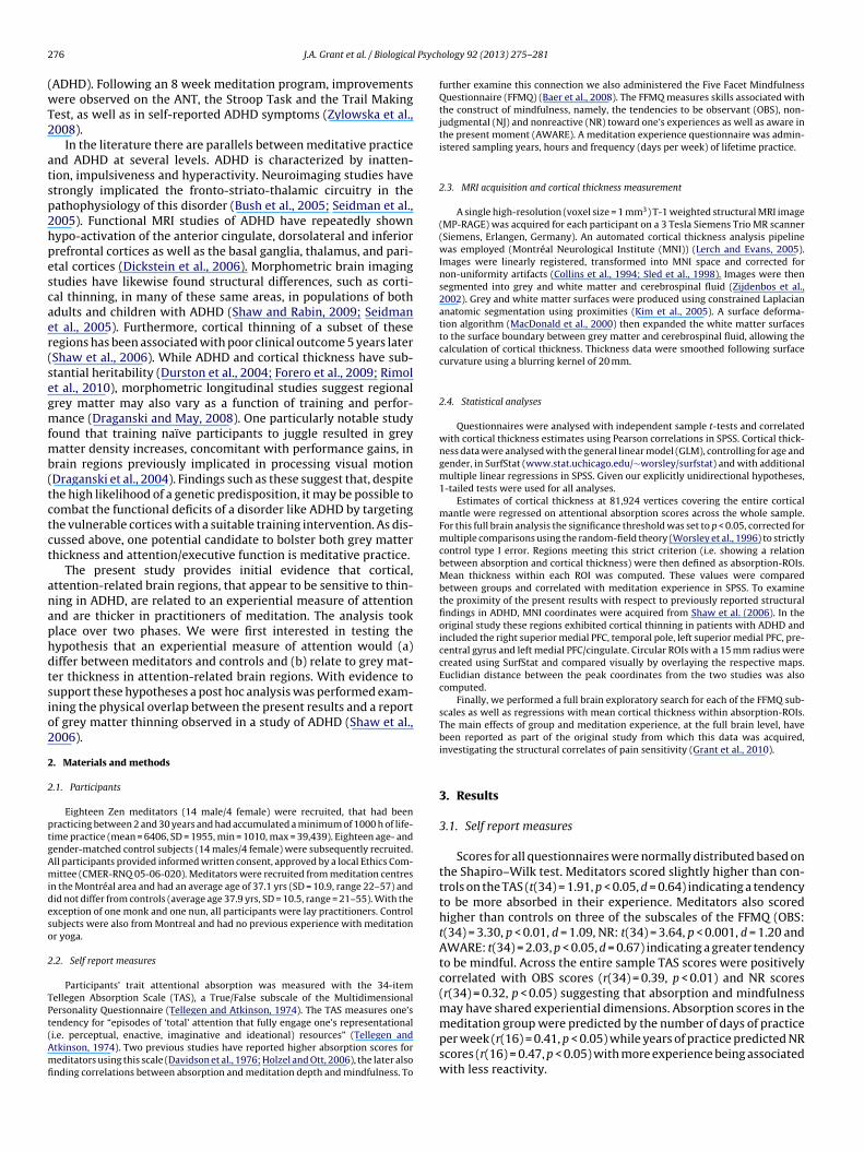
2 l Psych
(wT2
atsp2hpescaer(segmfmb(tctct
anaphdtsio2
2
2
ptgAmideso
2
TPt(Amfi
76 J.A. Grant et al. / Biologica
ADHD). Following an 8 week meditation program, improvementsere observed on the ANT, the Stroop Task and the Trail Making
est, as well as in self-reported ADHD symptoms (Zylowska et al.,008).
In the literature there are parallels between meditative practicend ADHD at several levels. ADHD is characterized by inatten-ion, impulsiveness and hyperactivity. Neuroimaging studies havetrongly implicated the fronto-striato-thalamic circuitry in theathophysiology of this disorder (Bush et al., 2005; Seidman et al.,005). Functional MRI studies of ADHD have repeatedly shownypo-activation of the anterior cingulate, dorsolateral and inferiorrefrontal cortices as well as the basal ganglia, thalamus, and pari-tal cortices (Dickstein et al., 2006). Morphometric brain imagingtudies have likewise found structural differences, such as corti-al thinning, in many of these same areas, in populations of bothdults and children with ADHD (Shaw and Rabin, 2009; Seidmant al., 2005). Furthermore, cortical thinning of a subset of theseegions has been associated with poor clinical outcome 5 years laterShaw et al., 2006). While ADHD and cortical thickness have sub-tantial heritability (Durston et al., 2004; Forero et al., 2009; Rimolt al., 2010), morphometric longitudinal studies suggest regionalrey matter may also vary as a function of training and perfor-ance (Draganski and May, 2008). One particularly notable study
ound that training naïve participants to juggle resulted in greyatter density increases, concomitant with performance gains, in
rain regions previously implicated in processing visual motionDraganski et al., 2004). Findings such as these suggest that, despitehe high likelihood of a genetic predisposition, it may be possible toombat the functional deficits of a disorder like ADHD by targetinghe vulnerable cortices with a suitable training intervention. As dis-ussed above, one potential candidate to bolster both grey matterhickness and attention/executive function is meditative practice.
The present study provides initial evidence that cortical,ttention-related brain regions, that appear to be sensitive to thin-ing in ADHD, are related to an experiential measure of attentionnd are thicker in practitioners of meditation. The analysis tooklace over two phases. We were first interested in testing theypothesis that an experiential measure of attention would (a)iffer between meditators and controls and (b) relate to grey mat-er thickness in attention-related brain regions. With evidence toupport these hypotheses a post hoc analysis was performed exam-ning the physical overlap between the present results and a reportf grey matter thinning observed in a study of ADHD (Shaw et al.,006).
. Materials and methods
.1. Participants
Eighteen Zen meditators (14 male/4 female) were recruited, that had beenracticing between 2 and 30 years and had accumulated a minimum of 1000 h of life-ime practice (mean = 6406, SD = 1955, min = 1010, max = 39,439). Eighteen age- andender-matched control subjects (14 males/4 female) were subsequently recruited.ll participants provided informed written consent, approved by a local Ethics Com-ittee (CMER-RNQ 05-06-020). Meditators were recruited from meditation centres
n the Montréal area and had an average age of 37.1 yrs (SD = 10.9, range 22–57) andid not differ from controls (average age 37.9 yrs, SD = 10.5, range = 21–55). With thexception of one monk and one nun, all participants were lay practitioners. Controlubjects were also from Montreal and had no previous experience with meditationr yoga.
.2. Self report measures
Participants’ trait attentional absorption was measured with the 34-itemellegen Absorption Scale (TAS), a True/False subscale of the Multidimensionalersonality Questionnaire (Tellegen and Atkinson, 1974). The TAS measures one’s
endency for “episodes of ‘total’ attention that fully engage one’s representationali.e. perceptual, enactive, imaginative and ideational) resources” (Tellegen andtkinson, 1974). Two previous studies have reported higher absorption scores foreditators using this scale (Davidson et al., 1976; Holzel and Ott, 2006), the later alsonding correlations between absorption and meditation depth and mindfulness. To
ology 92 (2013) 275– 281
further examine this connection we also administered the Five Facet MindfulnessQuestionnaire (FFMQ) (Baer et al., 2008). The FFMQ measures skills associated withthe construct of mindfulness, namely, the tendencies to be observant (OBS), non-judgmental (NJ) and nonreactive (NR) toward one’s experiences as well as aware inthe present moment (AWARE). A meditation experience questionnaire was admin-istered sampling years, hours and frequency (days per week) of lifetime practice.
2.3. MRI acquisition and cortical thickness measurement
A single high-resolution (voxel size = 1 mm3) T-1 weighted structural MRI image(MP-RAGE) was acquired for each participant on a 3 Tesla Siemens Trio MR scanner(Siemens, Erlangen, Germany). An automated cortical thickness analysis pipelinewas employed (Montréal Neurological Institute (MNI)) (Lerch and Evans, 2005).Images were linearly registered, transformed into MNI space and corrected fornon-uniformity artifacts (Collins et al., 1994; Sled et al., 1998). Images were thensegmented into grey and white matter and cerebrospinal fluid (Zijdenbos et al.,2002). Grey and white matter surfaces were produced using constrained Laplaciananatomic segmentation using proximities (Kim et al., 2005). A surface deforma-tion algorithm (MacDonald et al., 2000) then expanded the white matter surfacesto the surface boundary between grey matter and cerebrospinal fluid, allowing thecalculation of cortical thickness. Thickness data were smoothed following surfacecurvature using a blurring kernel of 20 mm.
2.4. Statistical analyses
Questionnaires were analysed with independent sample t-tests and correlatedwith cortical thickness estimates using Pearson correlations in SPSS. Cortical thick-ness data were analysed with the general linear model (GLM), controlling for age andgender, in SurfStat (www.stat.uchicago.edu/∼worsley/surfstat) and with additionalmultiple linear regressions in SPSS. Given our explicitly unidirectional hypotheses,1-tailed tests were used for all analyses.
Estimates of cortical thickness at 81,924 vertices covering the entire corticalmantle were regressed on attentional absorption scores across the whole sample.For this full brain analysis the significance threshold was set to p < 0.05, corrected formultiple comparisons using the random-field theory (Worsley et al., 1996) to strictlycontrol type I error. Regions meeting this strict criterion (i.e. showing a relationbetween absorption and cortical thickness) were then defined as absorption-ROIs.Mean thickness within each ROI was computed. These values were comparedbetween groups and correlated with meditation experience in SPSS. To examinethe proximity of the present results with respect to previously reported structuralfindings in ADHD, MNI coordinates were acquired from Shaw et al. (2006). In theoriginal study these regions exhibited cortical thinning in patients with ADHD andincluded the right superior medial PFC, temporal pole, left superior medial PFC, pre-central gyrus and left medial PFC/cingulate. Circular ROIs with a 15 mm radius werecreated using SurfStat and compared visually by overlaying the respective maps.Euclidian distance between the peak coordinates from the two studies was alsocomputed.
Finally, we performed a full brain exploratory search for each of the FFMQ sub-scales as well as regressions with mean cortical thickness within absorption-ROIs.The main effects of group and meditation experience, at the full brain level, havebeen reported as part of the original study from which this data was acquired,investigating the structural correlates of pain sensitivity (Grant et al., 2010).
3. Results
3.1. Self report measures
Scores for all questionnaires were normally distributed based onthe Shapiro–Wilk test. Meditators scored slightly higher than con-trols on the TAS (t(34) = 1.91, p < 0.05, d = 0.64) indicating a tendencyto be more absorbed in their experience. Meditators also scoredhigher than controls on three of the subscales of the FFMQ (OBS:t(34) = 3.30, p < 0.01, d = 1.09, NR: t(34) = 3.64, p < 0.001, d = 1.20 andAWARE: t(34) = 2.03, p < 0.05, d = 0.67) indicating a greater tendencyto be mindful. Across the entire sample TAS scores were positivelycorrelated with OBS scores (r(34) = 0.39, p < 0.01) and NR scores(r(34) = 0.32, p < 0.05) suggesting that absorption and mindfulnessmay have shared experiential dimensions. Absorption scores in the
meditation group were predicted by the number of days of practiceper week (r(16) = 0.41, p < 0.05) while years of practice predicted NRscores (r(16) = 0.47, p < 0.05) with more experience being associatedwith less reactivity.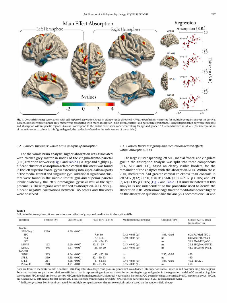
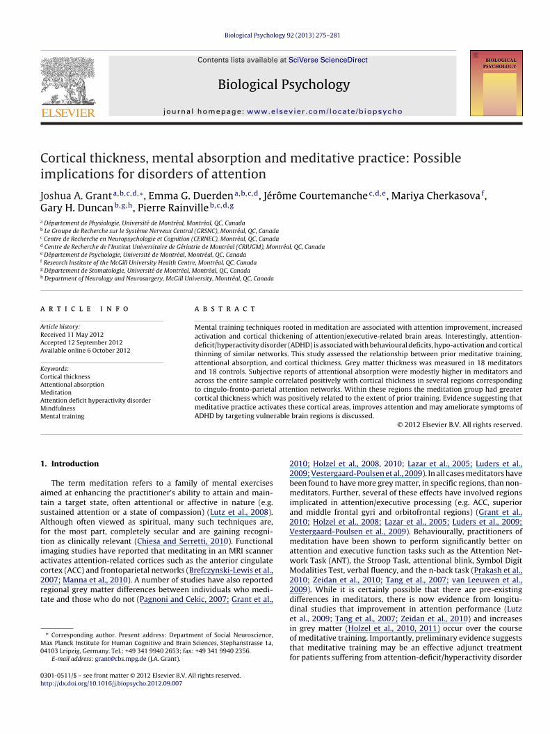
J.A. Grant et al. / Biological Psychology 92 (2013) 275– 281 277
Fig. 1. Cortical thickness correlations with self-reported absorption. Areas in orange-red (t-threshold = 3.6) are Bonferonni-corrected for multiple comparison over the corticalsurface. Regions where thinner grey matter was associated with more absorption (blue-green clusters) did not reach significance. (Right) Relationship between thicknessand absorption within specific regions. R-values correspond to the partial correlations after controlling for age and gender. S.R. = standardized residuals. (For interpretationo rsion
3
w(niotlpnw
TF
DRcp
f the references to colour in this figure legend, the reader is referred to the web ve
.2. Cortical thickness: whole brain analysis of absorption
For the whole brain analysis, higher absorption was associatedith thicker grey matter in nodes of the cingulo-fronto-parietal
CFP) attention networks (Fig. 1 and Table 1). A large and highly sig-ificant cluster of absorption-related cortical thickness was found
n the left superior frontal gyrus extending into supra-callosal partsf the medial frontal and cingulate gyri. Additional significant clus-ers were found in the middle frontal gyri and superior parietal
obule bilaterally, the left supramarginal gyrus as well as the rightrecuneus. These regions were defined as absorption-ROIs. No sig-ificant negative correlations between TAS scores and thicknessere observed.able 1ull brain thickness/absorption correlations and effects of group and meditation in absorp
Location Vertices (#) Cluster (t, p) Peak-MNI (x, y, z)
FrontalSFG-Cing L 1220 4.60, <0.001*
SFG −7, 9, 69
ACC −7, 16, 40
PCC −12, −24, 43
MFG R 132 4.60, <0.05* 35, 31, 38
MFG L 446 4.51, =0.01* −25, 40, 39
ParietalSMG L 533 4.64, <0.001* −45, −35, 26
SPL R 369 4.55, <0.001* 32, −50, 53
SPL L 211 4.26, <0.05* −4, −53, 55
PrCun R 240 4.21, =0.01* 18, −83, 45
ata are from 18 meditators and 18 controls. SFG-Cing refers to a large contiguous regioneported r-values are partial correlation coefficients, that is, representing unique varianceortex; med-PFC, medial prefrontal cortex; MFG, middle frontal gyrus; MNI, Montreal Neurecuneus; MFG, left medial frontal gyrus; SFG-Cing, superior frontal gyrus-cingulate; SP
* Indicates p-values Bonferonni-corrected for multiple comparison over the entire cort
of the article.)
3.3. Cortical thickness: group and meditation-related effectswithin absorption-ROIs
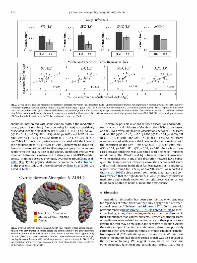
The large cluster spanning left SFG, medial frontal and cingulategyri in the absorption analysis was split into three components(SFG, ACC and PCC), based on clearly visible borders, for theremainder of the analyses with the absorption-ROIs. Within theseROIs, meditators had greater cortical thickness than controls inleft SFG (t(32) = 1.90, p < 0.05), SMG (t(32) = 2.37, p < 0.05) and SPL
(t(32) = 1.85, p < 0.05) (Fig. 2 and Table 1). It must be noted that thisanalysis is not independent of the procedure used to derive theabsorption ROIs. With knowledge that the meditators scored higheron the absorption questionnaire the analysis becomes circular andtion-ROIs.
Meditation training (r/p) Group dif (t/p) Closest ADHD-peak(mm-structure)
0.42, <0.05 (yr) 1.95, <0.05 6.2 SFG/Med-PFC L0.49, <0.05 (yr) ns 8.8 Med-PFC/ACC Lns ns 38.2 Med-PFC/ACC L0.43, <0.05 (yr) ns 24.1 SFG/Med-PFC R0.53, <0.05 (yr) ns 41.0 SFG/Med-PFC L
ns 2.35, <0.05 >50ns ns >500.44, <0.05 (yr) 1.95, <0.05 49.3 PreCG L0.55, <0.05 (h) ns >50
which was divided into superior frontal, anterior and posterior cingulate regions. after accounting for age and gender in the regression model. ACC, anterior cingulaterological Institute; PCC, posterior cingulate cortex; PreCG, precentral gyrus; PrCun,L, superior parietal lobule; SMG, supramarginal gyrus.ical surface based on the random-field theory.
278 J.A. Grant et al. / Biological Psychology 92 (2013) 275– 281
Fig. 2. Group differences and meditation experience correlations within the absorption-ROIs. (Upper panels) Meditators had significantly thicker grey matter in the superiorfrontal gyrus (SFG), superior parietal lobule (SPL) and supramarginal gyrus (SMG) all of the left side. M = meditators, C = controls. (Lower panels) Partial regression plots showt ountinb perien(
sga(aatfroc2il
FrYdoit
he standardized residuals (S.R.) of cortical thickness and years of practice after accest fit line maintains the true relationship between the variables. More years of exACC) and middle frontal gyrus (MFG). For additional regions see Table 1.
hould be interpreted with some caution. Within the meditationroup, years of training (after accounting for age) was positivelyssociated with thickness of the left SFG (r(13) = 0.42, p < 0.05), ACCr(13) = 0.49, p < 0.05), SPL (r(13) = 0.44, p < 0.05) and MFG bilater-lly (left: r(13) = 0.53, p < 0.05; right: r(13) = 0.43, p < 0.05) (Fig. 2nd Table 1). Hours of experience was associated with thickness ofhe right precuneus (r(13) = 0.50, p < 0.05). There were no group dif-erences or correlations with total hemispheric grey matter volumeeinforcing the focal nature of the effects. Significant overlap wasbserved between the main effect of absorption and ADHD-related
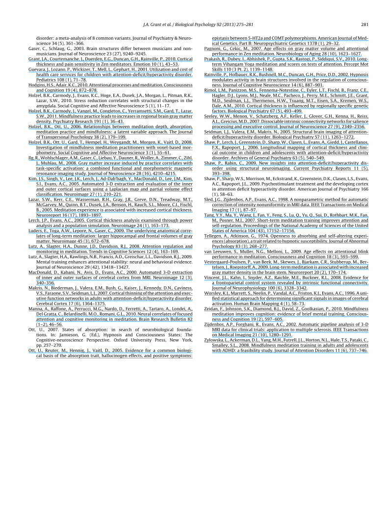
ortical thinning observed previously by another group (Shaw et al.,006) (Fig. 3). The physical distance between the peaks observedn the present study and those observed by Shaw et al. (2006) areisted in Table 1.
ig. 3. Overlap between absorption and ADHD. Red: regions where absorption cor-elated with grey matter thickness across the entire sample of the present report.ellow: ROIs derived from Shaw et al. (2006) where attention-deficit hyperactivityisorder (ADHD) was associated with thinner cortex and worse outcome. Orange:verlap between the main effect of absorption and cortical thinning in ADHD. (Fornterpretation of the references to colour in this figure legend, the reader is referredo the web version of the article.)
g for age, separately for each variable. The R-value is the partial coefficient and thece was associated with greater thickness of left SFG, SPL, anterior cingulate cortex
To examine possible relations between absorption and mindful-ness, mean cortical thickness of the absorption-ROIs was regressedon the FFMQ revealing positive associations between OBS scoresand left SFG (r(33) = 0.46, p < 0.01), MFG (r(33) = 0.34, p < 0.05), SPL(r(33) = 0.48, p < 0.01) and SMG (r(33) = 0.37, p < 0.05). NR scoreswere associated with mean thickness in the same regions withthe exception of the SMG (left SFG: r(33) = 0.37, p < 0.05; MFG:r(33) = 0.31, p < 0.05; SPL: r(33) = 0.34, p < 0.05). In each of thesecases, greater thickness was associated with higher self-reportedmindfulness. The AWARE and NJ subscales were not associatedwith mean thickness in any of the absorption network ROIs. Subse-quent full brain searches revealed a correlation between NR scoresand cortical thickness in the right fusiform gyrus but no additionalregions were found for OBS, NJ or AWARE scores. As reported inGrant et al. (2010) a global search contrasting meditators and con-trols revealed that the right dorsal ACC was significantly thicker inmeditators and a single region on the right precentral gyrus wasfound to be related to hours of meditation experience.
4. Discussion
Attentional absorption has been described as one’s tendencyfor “episodes of ‘total’ attention that fully engage one’s represen-tational resources” (Tellegen and Atkinson, 1974). Consistent withprevious reports (Davidson et al., 1976; Holzel and Ott, 2006), medi-tators had a greater, albeit modest, tendency to become absorbed intheir experiences than control subjects. Further, absorption scoresof meditators were related to the frequency of their practice sug-gesting the trait may be malleable and sensitive to training. Acrossthe entire sample of meditators and controls, absorption positivelycorrelated with grey matter thickness, at multiple nodes of cingulo-
fronto-parietal (CFP) attention/executive networks. Within theseregions meditators had thicker cortex which was also related tothe extent of training. We suggest below, based on these andother structural, functional and behavioural results, that there is
Psych
sicair
strk(2ebtctulolr(tsc2eomia
Stmpgbtsfisientittc
f(tajoirmipt
J.A. Grant et al. / Biological
ubstantial evidence indicating that meditative training has a pos-tive impact on attention. We further describe the remarkableorrespondence, albeit in opposing directions, between meditationnd ADHD and speculate about the use of this form of mental train-ng to bolstering grey matter thickness in highly susceptible corticalegions implicated in the disorder.
Structurally, the present report shows that individuals’ propen-ity for attentional absorption is related to the grey matterhickness of left ACC, SFG, SMG and bilateral MFG and SPL. Theseegions are all parts of attention and executive control networksnown under various formulations as the dorsal attention networkBush et al., 2000; Cabeza and Nyberg, 2000; Corbetta and Shulman,002), the frontoparietal control system (Vincent et al., 2008) or thexecutive control network (Seeley et al., 2007). These systems haveeen postulated to coordinate attention under competing condi-ions. In absolute terms, meditators had thicker grey matter thanontrols in each of the above-mentioned regions, significantly so inhe left SFG, SMG and SPL. Further, the years of practice of individ-al meditators, after controlling for age, predicted the thickness of
eft SFG, ACC, SPL and MFG bilaterally. One possible interpretationf these results is that repeatedly engaging in meditative practiceeads, overtime, to increases in grey matter thickness in brainegions important for the control and maintenance of attentionan alternative is discussed below). This interpretation is consis-ent with a growing body of work, including a longitudinal trainingtudy, reporting thicker or more dense grey matter for meditatorsompared to controls (Grant et al., 2010; Holzel et al., 2008, 2010,011; Lazar et al., 2005; Luders et al., 2009; Vestergaard-Poulsent al., 2009). Indeed, several of these studies have reported effectsf meditation in attention/executive regions such as ACC, superior,iddle and orbito-frontal regions. Importantly, functional imag-
ng studies have also shown that engaging in meditative practicectivates these same networks.
The main effect of absorption and cortical thickness in left ACC,FG, SMG and bilateral MFG and SPL corresponds well with atten-ion and executive control networks. Functional MRI studies of
editation have reported engagement of these networks duringractice (Lutz et al., 2008). Brefczynski-Lewis et al. (2007) foundreater activation of left MFG, DLPFC, SMG, right SFG and SPLilaterally during sustained attention in advanced Tibetan medi-ators compared to novices. Similarly, Manna et al. (2010) reportedtronger cingulate and medial PFC activation during meditationor Theravadin monks compared to controls. Interestingly, a brain-maging study from our group examining attentional absorptiontemming from hypnotic induction reported absorption-specificncreases in regional cerebral blood flow in the ACC as well as bilat-rally in the SFG, MFG, IFG and IPL (Rainville et al., 2002). Thus,etworks functionally activated during both absorption and medi-ative practice, appear to overlap substantially with those observedn the present study to relate, structurally, to attentional absorp-ion. This lends further support to the idea that meditation is arainable skill that is reflected in anatomical and functional brainhanges.
The idea that repeated practice or skill learning can result inunctional and structural changes in the brain is certainly not newBuonomano and Merzenich, 1998). However it has only been inhe past decade that MRI technology and analyses have becomedvanced enough to show, in living humans, that skills such asuggling (Draganski et al., 2004), mirror reading (Ilg et al., 2008)r playing a musical instrument (Gaser and Schlaug, 2003) cannfluence the amount of grey matter in functionally relevant brainegions. In a similar manner, learning the skills associated with
editation, such as being mindful or sustaining one’s focus, mayncrease grey matter in regions implicated in attention/executiverocessing. Fittingly, we also observed correlations between cor-ical thickness and facets of mindfulness, which were stronger
ology 92 (2013) 275– 281 279
in meditators, within regions where absorption correlated withgrey matter thickness. This suggests that absorption and aspectsof mindfulness, such as the tendency to be observant and non-reactive, may share common neural substrates. Similar to otherskills, changes in brain function and structure resulting from train-ing in meditation should have a corresponding behavioural effect.Evidence also suggests meditative practice is associated with per-formance improvements on attention and executive control tasks.
Behaviourally, substantial evidence now exists, from both lon-gitudinal and cross-sectional studies, that meditative training canenhance performance on attention/executive tasks such the ANT,Stroop, attentional blink, a cued-response task, the Symbol DigitModalities Test, verbal fluency, and the n-back task (Prakash et al.,2010; Zeidan et al., 2010; Hodgins and Adair, 2010; Tang et al.,2007; van Leeuwen et al., 2009). Taken as a whole, the existent lit-erature suggests meditative practice functionally and structurallyalters attention and executive control networks and improves per-formance on tasks which engage these regions. Interestingly, in allthree domains the story is the reverse for ADHD.
In the present report, the thickness of cortical regions asso-ciated with absorption and mindfulness overlapped with regionsreported to undergo cortical thinning in ADHD populations, specif-ically the cingulate, prefrontal and parietal cortices (Shaw et al.,2006) (Fig. 3). Of particular interest, cortical thinning of the cingu-late cortex, measured longitudinally, has been found to be related toworse outcome in patients (Shaw et al., 2006). In the present reportwe created a region of interest centred on the peak from that studyand found direct overlap with the main effect of absorption. In thesame ACC cluster, a positive relationship was observed betweenthickness and the extent of meditators’ training. Additional reportsof cortical thinning in adults with ADHD (Makris et al., 2007) haveimplicated the ACC, DLFPC and parietal lobules where again, in thepresent report, absorption was associated with thicker grey matterand meditation experience.
It is within these same networks that the majority of functionalabnormalities have been observed in children, adolescents, andadults with ADHD while performing cognitive tasks tapping atten-tion and executive function (Bush et al., 2010). A meta-analysis of 16functional imaging studies of ADHD reported significant patternsof hypo-activity of anterior cingulate, dorsolateral and inferior pre-frontal cortices as well as basal ganglia, thalamus, and the parietalcortex in sufferers of the disorder (Dickstein et al., 2006). As dis-cussed above, activation of many of these regions is enhanced inmeditators (Brefczynski-Lewis et al., 2007; Manna et al., 2010).Finally, preliminary behavioural results suggest that an 8 week, sec-ular, meditation-based program can improve symptoms of ADHDand scores on measures of attention and executive control such asthe ANT, Stroop and the Trail Making Test in adults and adolescents(Zylowska et al., 2008).
Taken as a whole, existing behavioural, functional and structuralimaging studies provide a link between meditation and ADHD; theformer being associated with performance enhancement, increasedactivation and thicker cortex while the later is associated with per-formance decrement, hypoactivation and cortical thinning. Thiscould potentially indicate that the cognitive processes engagedand strengthened by meditation, possibly reflected by corticalthickening, are similar to those impaired in ADHD and associatedwith functional and structural abnormalities. The contribution ofthe present results is that of a potential mechanism by whichmeditative practice may act; via changes in cortical thickness,itself potentially induced from repeated functional activation ofthe relevant circuits. While this interpretation admittedly exceeds
what is reasonable for the results presented here, it highlights theremarkable consistency across studies and measures. Further sup-port for this link can be found in other domains as well. Seeleyet al. (2007) used resting-state MRI under task-free conditions
2 l Psych
twistFpseb
5
tamdmittAtaHtcpdc2e
aftStnttdmsmsioiem
Dmiewntamact
80 J.A. Grant et al. / Biologica
o link the executive-control network (considerably overlappingith the present results) to prior performance of the Trail Mak-
ng Test (TMT). The TMT is an executive task requiring one towitch between competing attentional sets and has been showno improve in ADHD populations following meditation training.inally, there is evidence that stimulant treatment for ADHD actsartly by staving off cortical thinning (Shaw et al., 2009). Thisuggests that it may be the engagement of specific brain regions,nabled via the treatment and verified by positive changes inehaviour, which prevents grey matter loss.
. Limitations
Thus far we have argued that our results combined withhe existent literature point to meditation as a plausible causalgent, positively influencing cortical thickness and potentiallyodulating behaviour in turn. However, such causality cannot be
etermined from a cross sectional study and indeed a case can beade for alternative interpretations. For example, it may be, and
s perhaps even likely, that there is a substantial genetic contribu-ion to one’s tendency for absorption (Ott et al., 2005; Ott, 2007), ashere is for both regional cortical thickness (Rimol et al., 2010) andDHD (Durston et al., 2004; Forero et al., 2009). It may be that, due
o such factors, a phenotype characterized by high absorption isttracted to, and perseveres in, the practice of meditation. Indeed,olzel and Ott (2006) suggest it may be one’s propensity for absorp-
ion that influences the depth of the meditative state. Nonetheless,ontributions to complex mental phenomenon will likely never beurely genetic or environmental and the existing longitudinal evi-ence suggests a significant contribution of meditative training tohanges in attention performance (Prakash et al., 2010; Zeidan et al.,010; Hodgins and Adair, 2010; Tang et al., 2007; van Leeuwent al., 2009) and grey matter (Holzel et al., 2010) over time.
An unfortunate limitation of the present study is the lack of performance-based measure of attention. This stems from theact that these data were collected for a study designed to assesshe structural correlates of pain perception (Grant et al., 2010).uch a measure would have certainly been helpful. Nonetheless,he strong correlation between absorption and cortical thickness,early exclusively in attention/executive networks, suggests thathe scale may indeed tap these processes. It is interesting to notehat the measure of absorption employed in the present reportepends on participants’ access to the quality or stability of theirental experience. This suggests that there is a definite relation-
hip between brain structure and the experience one has of theirental state, and further, that this is consistent across a modest
ample size. While an experiential-based construct like absorptions not likely to be useful diagnostically for attention-related dis-rders, it does seem relevant. After all, whether highly focused ornattentive, our mental experience encompasses all that we canver consciously know, making the quality of that experience aotivating factor for treatment.It should also be noted that other disorders such as Alzheimer’s
isease involve similar networks and effects observed here mayore generally reflect a maintenance of cortical thickness in med-
tation as suggested previously (Pagnoni and Cekic, 2007; Lazart al., 2005) and resulting from any number of factors associatedith the practice or lifestyle. Nonetheless, given the prelimi-ary evidence for the effectiveness of meditation as an adjunctreatment for ADHD (Zylowska et al., 2008), there seems to bedequate justification for larger longitudinal studies combining
ental training via meditation with performance measures ofttention/executive control and MRI morphometry. Importantly,linical studies could also directly test the hypothesis that medita-ive training will increase cortical thickness in sufferers of ADHD
ology 92 (2013) 275– 281
and ultimately, link those changes with behavioural improvement.The substantial financial burden that this and related disordersplace on health care systems (Guevara et al., 2001), raises the incen-tive, both socially and economically, for such a complementarytreatment option.
To conclude, the current study provides evidence that grey mat-ter thickness, overlapping with regions of the brain susceptible tothinning in disorders of attention, is associated with an individual’sexperience of mental absorption or engagement in their experi-ence. Individuals who practice meditation had thicker cortex inseveral of these regions which was also related to the extent of theirtraining. We suggest that these results and the existent literatureprovide ample justification for further inquiry into the potentialuse of meditative practice as an adjunct treatment for ADHD.
Financial disclosures
None of the authors have any financial or other relationship thatmight lead to a conflict of interest.
Acknowledgments
This research was supported jointly by a Canadian Institutesof Health Research (CIHR) operating grant (P.R.) and a Mind andLife Institute Varela Grant (J.A.G.). J.A.G has also been supportedby a fellowship from CIHR. We would like to thank L’Unité deNeuroimagerie Fonctionnelle du Centre de recherche de l’Institutuniversitaire de gériatrie de Montréal for technical help with scan-ning and members of the Duncan-Rainville labs.
References
Baer, R.A., Smith, G.T., Lykins, E., Button, D., Krietemeyer, J., Sauer, S., Walsh, E.,Duggan, D., Williams, J.M., 2008. Construct validity of the five facet mindfulnessquestionnaire in meditating and nonmeditating samples. Assessment 35 (2),207–216.
Brefczynski-Lewis, J.A., Lutz, A., Schaefer, H.S., Levinson, D.B., Davidson, R.J., 2007.Neural correlates of attentional expertise in long-term meditation practitioners.Proceedings of the National Academy of Sciences of the United States of America104 (27), 11483–11488.
Buonomano, D.V., Merzenich, M.M., 1998. Cortical plasticity: from synapses to maps.Annual Review of Neuroscience 21, 149–186.
Bush, G., 2010. Attention-deficit/hyperactivity disorder and attention networks.Neuropsychopharmacology 35 (1), 278–300.
Bush, G., Luu, P., Posner, M.I., 2000. Cognitive and emotional influences in anteriorcingulate cortex. Trends in Cognitive Sciences 4 (6), 215–222.
Bush, G., Valera, E.M., Seidman, L.J., 2005. Functional neuroimaging of attention-deficit/hyperactivity disorder: a review and suggested future directions.Biological Psychiatry 57 (11), 1273–1284.
Cabeza, R., Nyberg, L., 2000. Imaging cognition II: an empirical review of 275 PETand fMRI studies. Journal of Cognitive Neuroscience 12 (1), 1–47.
Chiesa, A., Serretti, A., 2010. A systematic review of neurobiological and clinicalfeatures of mindfulness meditations. Psychological Medicine 40 (8), 1239–1252.
Collins, D.L., Neelin, P., Peters, T.M., Evans, A.C., 1994. Automatic 3D intersubjectregistration of MR volumetric data in standardized Talairach space. Journal ofComputer Assisted Tomography 18 (2), 192–205.
Corbetta, M., Shulman, G.L., 2002. Control of goal-directed and stimulus-drivenattention in the brain. Nature Reviews Neuroscience 3 (3), 201–215.
Davidson, R.J., Goleman, D.J., Schwartz, G.E., 1976. Attentional and affective concomi-tants of meditation: a cross-sectional study. Journal of Abnormal Psychology 85(2), 235–238.
Dickstein, S.G., Bannon, K., Castellanos, F.X., Milham, M.P., 2006. The neural corre-lates of attention deficit hyperactivity disorder: an ALE meta-analysis. Journalof Child Psychology and Psychiatry and Allied Disciplines 47 (10), 1051–1062.
Draganski, B., Gaser, C., Busch, V., Schuierer, G., Bogdahn, U., May, A., 2004. Neu-roplasticity: changes in grey matter induced by training. Nature 427 (6972),311–312.
Draganski, B., May, A., 2008. Training-induced structural changes in the adult humanbrain. Behavioural Brain Research 192 (1), 137–142.
Durston, S., Hulshoff Pol, H.E., Schnack, H.G., Buitelaar, J.K., Steenhuis, M.P., Minderaa,
R.B., Kahn, R.S., van Engeland, H., 2004. Magnetic resonance imaging of boys withattention-deficit/hyperactivity disorder and their unaffected siblings. Journal ofthe American Academy of Child and Adolescent Psychiatry 43 (3), 332–340.Forero, D.A., Arboleda, G.H., Vasquez, R., Arboleda, H., 2009. Candidate genesinvolved in neural plasticity and the risk for attention-deficit hyperactivity
Psych
G
G
G
H
H
H
H
H
I
K
L
L
L
L
L
M
M
M
O
O
MRI data for clinical trials: application to multiple sclerosis. IEEE Transactions
J.A. Grant et al. / Biological
disorder: a meta-analysis of 8 common variants. Journal of Psychiatry & Neuro-science 34 (5), 361–366.
aser, C., Schlaug, G., 2003. Brain structures differ between musicians and non-musicians. Journal of Neuroscience 23 (27), 9240–9245.
rant, J.A., Courtemanche, J., Duerden, E.G., Duncan, G.H., Rainville, P., 2010. Corticalthickness and pain sensitivity in Zen meditators. Emotion 10 (1), 43–53.
uevara, J., Lozano, P., Wickizer, T., Mell, L., Gephart, H., 2001. Utilization and cost ofhealth care services for children with attention-deficit/hyperactivity disorder.Pediatrics 108 (1), 71–78.
odgins, H.S., Adair, K.C., 2010. Attentional processes and meditation. Consciousnessand Cognition 19 (4), 872–878.
olzel, B.K., Carmody, J., Evans, K.C., Hoge, E.A., Dusek, J.A., Morgan, L., Pitman, R.K.,Lazar, S.W., 2010. Stress reduction correlates with structural changes in theamygdala. Social Cognitive and Affective Neuroscience 5 (1), 11–17.
olzel, B.K., Carmody, J., Vangel, M., Congleton, C., Yerramsetti, S.M., Gard, T., Lazar,S.W., 2011. Mindfulness practice leads to increases in regional brain gray matterdensity. Psychiatry Research 191 (1), 36–43.
olzel, B.K., Ott, U., 2006. Relationships between meditation depth, absorption,meditation practice and mindfulness: a latent variable approach. The Journalof Transpersonal Psychology 38 (2), 179–199.
olzel, B.K., Ott, U., Gard, T., Hempel, H., Weygandt, M., Morgen, K., Vaitl, D., 2008.Investigation of mindfulness meditation practitioners with voxel-based mor-phometry. Social Cognitive and Affective Neuroscience 3 (1), 55–61.
lg, R., Wohlschlager, A.M., Gaser, C., Liebau, Y., Dauner, R., Woller, A., Zimmer, C., Zihl,J., Muhlau, M., 2008. Gray matter increase induced by practice correlates withtask-specific activation: a combined functional and morphometric magneticresonance imaging study. Journal of Neuroscience 28 (16), 4210–4215.
im, J.S., Singh, V., Lee, J.K., Lerch, J., Ad-Dab’bagh, Y., MacDonald, D., Lee, J.M., Kim,S.I., Evans, A.C., 2005. Automated 3-D extraction and evaluation of the innerand outer cortical surfaces using a Laplacian map and partial volume effectclassification. Neuroimage 27 (1), 210–221.
azar, S.W., Kerr, C.E., Wasserman, R.H., Gray, J.R., Greve, D.N., Treadway, M.T.,McGarvey, M., Quinn, B.T., Dusek, J.A., Benson, H., Rauch, S.L., Moore, C.I., Fischl,B., 2005. Meditation experience is associated with increased cortical thickness.Neuroreport 16 (17), 1893–1897.
erch, J.P., Evans, A.C., 2005. Cortical thickness analysis examined through poweranalysis and a population simulation. Neuroimage 24 (1), 163–173.
uders, E., Toga, A.W., Lepore, N., Gaser, C., 2009. The underlying anatomical corre-lates of long-term meditation: larger hippocampal and frontal volumes of graymatter. Neuroimage 45 (3), 672–678.
utz, A., Slagter, H.A., Dunne, J.D., Davidson, R.J., 2008. Attention regulation andmonitoring in meditation. Trends in Cognitive Sciences 12 (4), 163–169.
utz, A., Slagter, H.A., Rawlings, N.B., Francis, A.D., Greischar, L.L., Davidson, R.J., 2009.Mental training enhances attentional stability: neural and behavioral evidence.Journal of Neuroscience 29 (42), 13418–13427.
acDonald, D., Kabani, N., Avis, D., Evans, A.C., 2000. Automated 3-D extractionof inner and outer surfaces of cerebral cortex from MRI. Neuroimage 12 (3),340–356.
akris, N., Biederman, J., Valera, E.M., Bush, G., Kaiser, J., Kennedy, D.N., Caviness,V.S., Faraone, S.V., Seidman, L.J., 2007. Cortical thinning of the attention and exec-utive function networks in adults with attention-deficit/hyperactivity disorder.Cerebral Cortex 17 (6), 1364–1375.
anna, A., Raffone, A., Perrucci, M.G., Nardo, D., Ferretti, A., Tartaro, A., Londei, A.,Del Gratta, C., Belardinelli, M.O., Romani, G.L., 2010. Neural correlates of focusedattention and cognitive monitoring in meditation. Brain Research Bulletin 82(1–2), 46–56.
tt, U., 2007. States of absorption: in search of neurobiological founda-
tions. In: Jamieson, G. (Ed.), Hypnosis and Consciousness States: TheCognitive-neuroscience Perspective. Oxford University Press, New York,pp. 257–270.tt, U., Reuter, M., Hennig, J., Vaitl, D., 2005. Evidence for a common biologi-cal basis of the absorption trait, hallucinogen effects, and positive symptoms:
ology 92 (2013) 275– 281 281
epistasis between 5-HT2a and COMT polymorphisms. American Journal of Med-ical Genetics. Part B: Neuropsychiatric Genetics 137B (1), 29–32.
Pagnoni, G., Cekic, M., 2007. Age effects on gray matter volume and attentionalperformance in Zen meditation. Neurobiology of Aging 28 (10), 1623–1627.
Prakash, R., Dubey, I., Abhishek, P., Gupta, S.K., Rastogi, P., Siddiqui, S.V., 2010. Long-term Vihangam Yoga meditation and scores on tests of attention. Percept MotSkills 110 (3 Pt. 2), 1139–1148.
Rainville, P., Hofbauer, R.K., Bushnell, M.C., Duncan, G.H., Price, D.D., 2002. Hypnosismodulates activity in brain structures involved in the regulation of conscious-ness. Journal of Cognitive Neuroscience 14 (6), 887–901.
Rimol, L.M., Panizzon, M.S., Fennema-Notestine, C., Eyler, L.T., Fischl, B., Franz, C.E.,Hagler, D.J., Lyons, M.J., Neale, M.C., Pacheco, J., Perry, M.E., Schmitt, J.E., Grant,M.D., Seidman, L.J., Thermenos, H.W., Tsuang, M.T., Eisen, S.A., Kremen, W.S.,Dale, A.M., 2010. Cortical thickness is influenced by regionally specific geneticfactors. Biological Psychiatry 67 (5), 493–499.
Seeley, W.W., Menon, V., Schatzberg, A.F., Keller, J., Glover, G.H., Kenna, H., Reiss,A.L., Greicius, M.D., 2007. Dissociable intrinsic connectivity networks for salienceprocessing and executive control. Journal of Neuroscience 27 (9), 2349–2356.
Seidman, L.J., Valera, E.M., Makris, N., 2005. Structural brain imaging of attention-deficit/hyperactivity disorder. Biological Psychiatry 57 (11), 1263–1272.
Shaw, P., Lerch, J., Greenstein, D., Sharp, W., Clasen, L., Evans, A., Giedd, J., Castellanos,F.X., Rapoport, J., 2006. Longitudinal mapping of cortical thickness and clini-cal outcome in children and adolescents with attention-deficit/hyperactivitydisorder. Archives of General Psychiatry 63 (5), 540–549.
Shaw, P., Rabin, C., 2009. New insights into attention-deficit/hyperactivity dis-order using structural neuroimaging. Current Psychiatry Reports 11 (5),393–398.
Shaw, P., Sharp, W.S., Morrison, M., Eckstrand, K., Greenstein, D.K., Clasen, L.S., Evans,A.C., Rapoport, J.L., 2009. Psychostimulant treatment and the developing cortexin attention deficit hyperactivity disorder. American Journal of Psychiatry 166(1), 58–63.
Sled, J.G., Zijdenbos, A.P., Evans, A.C., 1998. A nonparametric method for automaticcorrection of intensity nonuniformity in MRI data. IEEE Transactions on MedicalImaging 17 (1), 87–97.
Tang, Y.Y., Ma, Y., Wang, J., Fan, Y., Feng, S., Lu, Q., Yu, Q., Sui, D., Rothbart, M.K., Fan,M., Posner, M.I., 2007. Short-term meditation training improves attention andself-regulation. Proceedings of the National Academy of Sciences of the UnitedStates of America 104 (43), 17152–17156.
Tellegen, A., Atkinson, G., 1974. Openness to absorbing and self-altering experi-ences (absorption), a trait related to hypnotic susceptibility. Journal of AbnormalPsychology 83 (3), 268–277.
van Leeuwen, S., Muller, N.G., Melloni, L., 2009. Age effects on attentional blinkperformance in meditation. Consciousness and Cognition 18 (3), 593–599.
Vestergaard-Poulsen, P., van Beek, M., Skewes, J., Bjarkam, C.R., Stubberup, M., Ber-telsen, J., Roepstorff, A., 2009. Long-term meditation is associated with increasedgray matter density in the brain stem. Neuroreport 20 (2), 170–174.
Vincent, J.L., Kahn, I., Snyder, A.Z., Raichle, M.E., Buckner, R.L., 2008. Evidence fora frontoparietal control system revealed by intrinsic functional connectivity.Journal of Neurophysiology 100 (6), 3328–3342.
Worsley, K.J., Marrett, S., Neelin, P., Vandal, A.C., Friston, K.J., Evans, A.C., 1996. A uni-fied statistical approach for determining significant signals in images of cerebralactivation. Human Brain Mapping 4 (1), 58–73.
Zeidan, F., Johnson, S.K., Diamond, B.J., David, Z., Goolkasian, P., 2010. Mindfulnessmeditation improves cognition: evidence of brief mental training. Conscious-ness and Cognition 19 (2), 597–605.
Zijdenbos, A.P., Forghani, R., Evans, A.C., 2002. Automatic pipeline analysis of 3-D
on Medical Imaging 21 (10), 1280–1291.Zylowska, L., Ackerman, D.L., Yang, M.H., Futrell, J.L., Horton, N.L., Hale, T.S., Pataki, C.,
Smalley, S.L., 2008. Mindfulness meditation training in adults and adolescentswith ADHD: a feasibility study. Journal of Attention Disorders 11 (6), 737–746.
Copyright © 2022 FDOKUMEN