Cortical Thickness, Surface Area, and Volume of the Brain Reward System in Alcohol Dependence:...
14
Cortical Thickness, Surface Area, and Volume of the Brain Reward System in Alcohol Dependence: Relationships to Relapse and Extended Abstinence Timothy C. Durazzo, Duygu Tosun, Shannon Buckley, Stefan Gazdzinski, Anderson Mon, Susanna L. Fryer, and Dieter J. Meyerhoff Background: At least 60% of those treated for an alcohol use disorder will relapse. Empirical study of the integrity of the brain reward system (BRS) is critical to understanding the mecha- nisms of relapse as this collection of circuits is implicated in the development and maintenance of all forms of addictive disorders. This study compared thickness, surface area, and volume in neo- cortical components of the BRS among nonsmoking light-drinking controls (controls), individuals who remained abstinent and those who relapsed after treatment. Methods: Seventy-five treatment-seeking alcohol-dependent individuals (abstinent for 7 ± 3 days) and 43 controls completed 1.5T proton magnetic resonance imaging studies. Parcel- lated morphological data were obtained for following bilateral components of the BRS: rostral and caudal anterior cingulate cortex, insula, medial and lateral orbitofrontal cortex (OFC), rostral and caudal middle and superior frontal gyri, amygdala and hippocampus as well as for 26 other bilateral neocortical regions. Alcohol-dependent participants were followed over 12-months after baseline study and were classified as abstainers (no alcohol consumption; n = 24) and relapsers (any alcohol consumption; n = 51) at follow-up. Results: Relapsers and abstainers demonstrated lower cortical thickness in the vast majority of BRS regions as well as lower global thickness compared to controls. Relapsers had lower total BRS surface area than both controls and abstainers, but abstainers were not significantly different from controls on any surface area measure. Relapsers demonstrated lower volumes than controls in the majority of regions, while abstainers showed lower volumes than controls in the superior frontal gyrus, insula, amygdala, and hippocampus, bilaterally. Relapsers exhibited smaller vol- umes than abstainers in the right rostral middle and caudal middle frontal gyri and the lateral OFC, bilaterally. In relapsers, lower baseline volumes and surface areas in multiple regions were associated with a greater magnitude of post-treatment alcohol consumption. Conclusions: Results suggest relapsers demonstrated morphological abnormalities in regions involved in the ‘‘top down’’ regulation ⁄ modulation of internal drive states, emotions, reward pro- cessing, and behavior, which may impart increased risk for the relapse ⁄ remit cycle that afflicts many with an alcohol use disorder. Results also highlight the importance of examining both corti- cal thickness and surface area to better understand the nature of regional volume loss frequently observed in alcohol use disorders. Results from this report are consistent with previous research implicating plastic neurobiological changes in the BRS in the maintenance of addictive disorders. Key words: Alcohol Dependence, Neuroimaging, Brain Volume, Cortical Thickness, Surface Area, Relapse. I T IS ESTIMATED that at least 60% of individuals who seek treatment for an alcohol use disorder (i.e., alcohol dependence or abuse) will resume hazardous levels of alcohol consumption (Krampe et al., 2007; McKay et al., 2006; Miller et al., 2001), typically within 6 months following treat- ment (Maisto et al., 2006, 2007; Udo et al., 2009). However, a significant portion of those with alcohol use disorders do not return to a chronically relapsing ⁄ remitting course after treatment (Delucchi and Weisner, 2010; Miller et al., 2001; Moos and Moos, 2006; Moos et al., 2006). Sustained absti- nence and the chronic relapsing ⁄ remitting cycle in alcohol use disorders appear to result from a complex interplay among multiple biopsychosocial factors (Baler and Volkow, 2006; Bradizza et al., 2006; Donovan, 1996; Kalivas and Volkow, 2005; Moos and Moos, 2006; Walter et al., 2006; Witkiewitz and Marlatt, 2007). A considerable amount of research has addressed the potential neuropsychological, psychiatric, soci- odemographic, and behavioral factors associated with relapse in alcohol use disorders (e.g., Bottlender and Soyka, 2005; From the Department of Radiology and Biomedical Imaging (TCD, AM, SLF, DJM), University of California, San Francisco, California; Center for Imaging of Neurodegenerative Diseases (TCD, DT, SB, SG, AM, SLF, DJM), San Francisco VA Medical Center, San Francisco, California. Received for publication July 16, 2010; accepted November 28, 2010. Reprint requests: Timothy C. Durazzo, PhD, Center for Imaging of Neurodegenerative Disease (114M), San Francisco Veterans Administration Medical Center, 4150 Clement St. (114M), San Fran- cisco, CA 94121; Tel.: (415) 221 4810 x4157; Fax: (415) 750 2152; E-mail: [email protected] Copyright Ó 2011 by the Research Society on Alcoholism. DOI: 10.1111/j.1530-0277.2011.01452.x Alcoholism: Clinical and Experimental Research Vol. 35, No. 6 June 2011 Alcohol Clin Exp Res, Vol 35, No 6, 2011: pp 1187–1200 1187
Transcript of Cortical Thickness, Surface Area, and Volume of the Brain Reward System in Alcohol Dependence:...

Cortical Thickness, Surface Area, and Volume of the
Brain Reward System in Alcohol Dependence:
Relationships to Relapse and Extended Abstinence
Timothy C. Durazzo, Duygu Tosun, Shannon Buckley, Stefan Gazdzinski, Anderson Mon,Susanna L. Fryer, and Dieter J. Meyerhoff
Background: At least 60% of those treated for an alcohol use disorder will relapse. Empiricalstudy of the integrity of the brain reward system (BRS) is critical to understanding the mecha-nisms of relapse as this collection of circuits is implicated in the development and maintenance ofall forms of addictive disorders. This study compared thickness, surface area, and volume in neo-cortical components of the BRS among nonsmoking light-drinking controls (controls), individualswho remained abstinent and those who relapsed after treatment.
Methods: Seventy-five treatment-seeking alcohol-dependent individuals (abstinent for7 ± 3 days) and 43 controls completed 1.5T proton magnetic resonance imaging studies. Parcel-lated morphological data were obtained for following bilateral components of the BRS: rostraland caudal anterior cingulate cortex, insula, medial and lateral orbitofrontal cortex (OFC), rostraland caudal middle and superior frontal gyri, amygdala and hippocampus as well as for 26 otherbilateral neocortical regions. Alcohol-dependent participants were followed over 12-months afterbaseline study and were classified as abstainers (no alcohol consumption; n = 24) and relapsers(any alcohol consumption; n = 51) at follow-up.
Results: Relapsers and abstainers demonstrated lower cortical thickness in the vast majority ofBRS regions as well as lower global thickness compared to controls. Relapsers had lower totalBRS surface area than both controls and abstainers, but abstainers were not significantly differentfrom controls on any surface area measure. Relapsers demonstrated lower volumes than controlsin the majority of regions, while abstainers showed lower volumes than controls in the superiorfrontal gyrus, insula, amygdala, and hippocampus, bilaterally. Relapsers exhibited smaller vol-umes than abstainers in the right rostral middle and caudal middle frontal gyri and the lateralOFC, bilaterally. In relapsers, lower baseline volumes and surface areas in multiple regions wereassociated with a greater magnitude of post-treatment alcohol consumption.
Conclusions: Results suggest relapsers demonstrated morphological abnormalities in regionsinvolved in the ‘‘top down’’ regulation ⁄modulation of internal drive states, emotions, reward pro-cessing, and behavior, which may impart increased risk for the relapse ⁄ remit cycle that afflictsmany with an alcohol use disorder. Results also highlight the importance of examining both corti-cal thickness and surface area to better understand the nature of regional volume loss frequentlyobserved in alcohol use disorders. Results from this report are consistent with previous researchimplicating plastic neurobiological changes in the BRS in the maintenance of addictive disorders.
Key words: Alcohol Dependence, Neuroimaging, Brain Volume, Cortical Thickness, SurfaceArea, Relapse.
I T IS ESTIMATED that at least 60% of individuals whoseek treatment for an alcohol use disorder (i.e., alcohol
dependence or abuse) will resume hazardous levels of alcohol
consumption (Krampe et al., 2007; McKay et al., 2006;Miller et al., 2001), typically within 6 months following treat-ment (Maisto et al., 2006, 2007; Udo et al., 2009). However,a significant portion of those with alcohol use disorders donot return to a chronically relapsing ⁄ remitting course aftertreatment (Delucchi and Weisner, 2010; Miller et al., 2001;Moos and Moos, 2006; Moos et al., 2006). Sustained absti-nence and the chronic relapsing ⁄ remitting cycle in alcohol usedisorders appear to result from a complex interplay amongmultiple biopsychosocial factors (Baler and Volkow, 2006;Bradizza et al., 2006; Donovan, 1996; Kalivas and Volkow,2005; Moos and Moos, 2006; Walter et al., 2006; Witkiewitzand Marlatt, 2007). A considerable amount of research hasaddressed the potential neuropsychological, psychiatric, soci-odemographic, and behavioral factors associated with relapsein alcohol use disorders (e.g., Bottlender and Soyka, 2005;
From the Department of Radiology and Biomedical Imaging(TCD, AM, SLF, DJM), University of California, San Francisco,California; Center for Imaging of Neurodegenerative Diseases (TCD,DT, SB, SG, AM, SLF, DJM), San Francisco VA Medical Center,San Francisco, California.
Received for publication July 16, 2010; accepted November 28, 2010.Reprint requests: Timothy C. Durazzo, PhD, Center for Imaging
of Neurodegenerative Disease (114M), San Francisco VeteransAdministration Medical Center, 4150 Clement St. (114M), San Fran-cisco, CA 94121; Tel.: (415) 221 4810 x4157; Fax: (415) 750 2152;E-mail: [email protected]
Copyright � 2011 by the Research Society on Alcoholism.
DOI: 10.1111/j.1530-0277.2011.01452.x
Alcoholism: Clinical and Experimental Research Vol. 35, No. 6June 2011
Alcohol Clin Exp Res, Vol 35, No 6, 2011: pp 1187–1200 1187

Bradizza et al., 2006; Driessen et al., 2001; Glenn and Par-sons, 1991; Kodl et al., 2008; McKay, 1999; Moos and Moos,2006; Ritvo and Park, 2007; Rosenbloom et al., 2004; Vengel-iene et al., 2008; Zywiak et al., 2006). However, the latentneurobiological factors that contribute to sustained absti-nence and ⁄or increased risk for relapse after treatment foralcohol use disorders are not well understood. A greaterunderstanding of these neurobiological factors is necessary toidentify the mechanisms associated with both sustained long-term abstinence and the relapse ⁄ remit cycle that afflicts somany with an alcohol use disorders.Human in vivo neuroimaging methods have facilitated
study of the neurobiological correlates of relapse in alcoholuse disorders (Fowler et al., 2007; Volkow et al., 2003, 2004,2008) and encompass studies of brain blood flow, morphol-ogy, and metabolites. In individuals studied at approximately18 days of abstinence from alcohol, lower frontal cerebralblood flow was observed in those who relapsed relative tothose who remained abstinent for approximately 2 monthsfollowing treatment (Noel et al., 2002). Higher brain activa-tion in the putamen, anterior cingulate, and medial prefrontalcortex was related to the level of alcohol consumption inthose who relapsed 2 months after treatment (Grusser et al.,2004). Higher BOLD response in the thalamus and striatumin response to affectively positive cues were inversely relatedto drinking days and overall alcohol intake in those whorelapsed after treatment (Heinz et al., 2007). In our studies oftreatment-seeking individuals with alcohol use disorders,assessed after 1 week and again after 5 weeks of abstinence(Durazzo et al., 2010a), we observed that those who relapsedwithin 12 months of treatment demonstrated lower frontalgray matter (GM) perfusion at both 1 and 5 weeks of absti-nence compared to controls and participants that remainedabstinent for at least 12 months. Controls and abstainers wereequivalent on frontal GM perfusion at both assessmentpoints. In treatment-seeking alcoholics initially studiedbetween 1 and 12 weeks of abstinence (Wrase et al., 2008),those who relapsed within 6 months following treatmentdemonstrated significantly lower amygdala volume comparedto individuals who maintained sobriety over the same inter-val. In addition, relapsers demonstrated smaller hippocampaland ventral striatal volumes than controls, but were equiva-lent to abstainers on volumes in these regions. Abstainersexhibited a smaller ventral striatum than controls. In thetreatment group, as a whole, smaller amygdala volume wascorrelated with greater alcohol craving, which appeared to bedriven by the relapsers. In treatment-seeking alcoholics absti-nent for 3 to 5 days, those who relapsed within 3 weeks ofstudy demonstrated lower concentrations of cerebellar N-acetylaspartate [NAA; a surrogate indicator of neuronalintegrity (Moffett et al., 2007)] and choline-containing com-pounds [Cho; marker of cell membrane turnover and ⁄or syn-thesis (Ross and Bluml, 2001)] at 3 to 5 days of abstinencerelative to controls. No differences in cerebellar metabolitelevels at 3 to 5 days of abstinence were observed betweenindividuals who relapsed between 3 weeks and 3 months and
controls. No group differences between were found for frontalwhite matter (WM) metabolite levels (Parks et al., 2002).We previously combined measures from multimodality
proton magnetic resonance (MR) studies, neurocognitive,psychiatric, and sociodemographic assessment to predict out-come following treatment for alcohol use disorders. Unipolarmood disorders and neurocognitive measures of processingspeed, decreased levels of NAA in temporal GM and frontalWM as well as lower levels of frontal GM Cho were indepen-dent predictors of resumption of hazardous levels of alcoholconsumption within 12 months following treatment (Durazzoet al., 2008). In a follow-up study (Durazzo et al., 2010b), wespecifically assessed brain metabolite levels in multiple regionsof the brain reward system (BRS), including the dorsolateralprefrontal cortex (DLPFC), anterior cingulate cortex (ACC),insula, superior corona radiata (SCR), and cerebellar vermis,in treatment-seeking alcohol-dependent individuals at approx-imately 1-week-of-abstinence (baseline) and in nonsmokingcontrols. Participants who resumed hazardous levels of alco-hol consumption within 12 months of treatment demon-strated significantly lower baseline NAA concentrations thancontrols and abstainers in all regions. Relapsers also exhibitedlower concentrations of creatine-containing compounds thanabstainers in the DLPFC, SCR, and cerebellar vermis.Abstainers did not differ from controls on metabolite concen-trations in any region.Taken together, the available neuroimaging literature sug-
gests biochemical, metabolic, and ⁄or morphologic abnormali-ties in multiple components of the BRS in early recovery areassociated with relapse after treatment for alcohol use dis-orders. Neurobiological abnormalities in the BRS areimplicated as major contributors to the development andmaintenance of all forms of substance use disorders (Bowirratand Oscar-Berman, 2005; Kalivas and O’Brien, 2008; Kalivasand Volkow, 2005; Koob, 2003; Lubman et al., 2004; Makriset al., 2008b; Pierce and Kumaresan, 2006; Volkow et al.,2004, 2008; Wrase et al., 2008). Major components of theBRS include, but are not limited to, the DLPFC, orbitofron-tal cortex (OFC), insula, ACC, amygdala, hippocampus, thal-amus, nucleus accumbens, ventral tegmental area, and otherregions ⁄nuclei in the ventral pallidum and basal forebrain(Kalivas and Volkow, 2005; Makris et al., 2008b; Volkowet al., 2008).To our knowledge, there are no published reports concur-
rently examining neocortical surface area, thickness, and vol-ume in treatment-seeking alcohol-dependent individuals. It iswell established that the layered neocortical cellular architec-ture demonstrates a modular ⁄columnar organization that isoriented perpendicular to the cortical surface (Innocenti andVercelli, 2010). Neocortical surface area is suggested to reflectthe number and ⁄or width of columns, while cortical thicknessis related to the number or density of cells in a column (Rakic,1988). Cortical thickness is associated with neurocognitivefunction in healthy controls (Choi et al., 2008; Dickersonet al., 2008; Walhovd et al., 2006) and cocaine users (Makriset al., 2008a). As cortical volume is the product of cortical
1188 DURAZZO ET AL.

surface area and thickness, examination of both metrics mayprovide more specific information on the consequences ofalcohol use disorders on the cellular architecture of regionalneocortical tissue (Hutton et al., 2009; Makris et al., 2008a;Panizzon et al., 2009).The primary purpose of this study was to examine brain
morphology in multiple components of the BRS in alcohol-dependent individuals near the inception of outpatient treat-ment for alcohol use disorders (i.e., baseline) to determinewhether these measures distinguish those who relapsed aftertreatment from those who remained abstinent over a 12-month period. Morphological assessment focused on mea-surements of regional neocortical surface area, thickness, andvolume. We predicted the alcohol-dependent cohort (ALC),as a whole, would demonstrate lower baseline cortical thick-ness, smaller surface areas, and volumes than nonsmoking,light-drinking controls in the following neocortical compo-nents of the brain BRS: rostral and caudal ACC, insula, med-ial and lateral OFC, rostral and caudal middle frontal gyriand superior frontal gyri (the rostral and caudal middle fron-tal and superior frontal gyri comprise the bulk of theDLPFC). Based on our previous spectroscopic imaging find-ings in this cohort (Durazzo et al., 2010b), we predicted thatthose who resumed hazardous alcohol consumption followingtreatment demonstrate lower baseline cortical thickness, smal-ler surface areas, and volumes than individuals who remainedabstinent and controls in the above-listed components of theBRS. We also predicted that lower cortical thickness, surfaceareas, and volumes in these BRS components are related togreater levels of alcohol consumption in those who relapsedafter outpatient treatment.
METHODS
Participants
Seventy-five outpatient participants (4 women) were recruited fromthe VA Medical Center Substance Abuse Day Hospital and the Kai-ser Permanente Chemical Dependence Recovery Program in SanFrancisco. Primary inclusion criteria for the alcohol-dependent par-ticipants were fluency in English, DSM-IV diagnosis of alcoholdependence or alcohol abuse at the time of enrollment, consumptionof greater than 150 standard alcohol-containing drinks (i.e., 13.6 g ofethanol) per month for at least 8 years prior to enrollment for men,or consumption of greater than 80 drinks per month for at least6 years prior to enrollment for women. Controls (n = 43; 4 females)were recruited from the local community. Participants were between28 and 66 years of age. See Table 1 for group demographic data. Allparticipants provided written informed consent prior to studyaccording to the Declaration of Helsinki, and the informed consentdocument and procedures were approved by the University of Cali-fornia San Francisco and the San Francisco VA Medical Center.Approximately 70% of participants in the current report wereincluded in our earlier work (Durazzo et al., 2010a,b).Medical exclusion criteria for all participants were history of any
of the following: dependence on any substance other than alcohol ornicotine in the 5 years immediately prior to enrollment, intravenousdrug use in the 5 years immediately prior to enrollment in thestudy, current opioid agonist therapy, intrinsic cerebral masses,HIV ⁄AIDS, cerebrovascular accident, brain aneurysm, arteriovenousmalformations, peripheral vascular disease, myocardial infarction,
uncontrolled chronic hypertension (systolic >180 mm Hg and ⁄ordiastolic >120 mm Hg), type I diabetes, moderate or severe chronicobstructive pulmonary disease, nonalcohol-related seizures, signifi-cant exposure to known neurotoxins (e.g., toluene, carbon tetra-chloride), demyelinating and neurodegenerative diseases, clinicallydocumented Wernicke–Korsakoff syndrome, alcohol-induced per-sisting dementia, penetrating head trauma, and closed head injuryresulting in loss of consciousness for more than 10 minutes. Psychia-tric exclusion criteria were history of schizophrenia-spectrum dis-orders, bipolar disorder, dissociative disorders, post-traumatic stressdisorder, obsessive-compulsive disorder, panic disorder, and majordepression with mood-incongruent psychotic symptoms. Hepatitis C,type-2 diabetes, hypertension, unipolar mood disorder (major depres-sion and ⁄or substance-induced mood disorder) were permitted in theALC given their high prevalence in alcohol use disorders (Hasinet al., 2007; Mertens et al., 2003, 2005; Parekh and Klag, 2001;Stinson et al., 2005). Controls had no history of any DSM-IV axis Idisorder. Participants were urine-tested for illicit substances immedi-ately before all assessments (i.e., cannabinoids, opiates, phencycli-dine, cocaine, and amphetamines) and did not test positive for thesesubstances at any assessment.
Baseline Assessment
Baseline clinical and MR procedures for the alcohol-dependentparticipants were conducted 7 ± 3 days after last drink. All alcohol-dependent individuals were actively involved in stabilization ⁄ earlyrecovery outpatient treatment at the time of the baseline assessment,and duration of programs typically ranged from 14 to 28 days.
Table 1. Baseline Group Demographic, Alcohol and CigaretteConsumption, Mood and Anxiety Self-Report Measures
VariableControls(n = 43)
Abstainers(n = 24)
Relapsers(n = 51)
Age 47.3 (7.9) 51.0 (12.1) 49.6 (7.7)Education 16.3 (2.5) 14.6 (2.3) 13.7 (2.0)Caucasian (%) 79 71 77AMNART 118 (7) 115 (9) 114 (9)1-year avedrinks ⁄ month
13 (14) 333 (162) 399 (167)
Lifetime avedrinks ⁄ month
17 (15) 198 (120) 247 (152)
Months of heavydrinking
NA 236 (110) 270 (114)
Lifetime years ofregular drinking
29 (8) 36 (10) 35 (9)
Smokers (%) 0 50 59FTND total NA 5.6 (1.2) 5.0 (2.3)Smoking duration NA 23 (13) 23 (11)Cigarette pack years NA 25 (18) 26 (20)Beck depressioninventory
4 (4) 12 (9) 16 (10)
STAI trait 38 (7) 45 (11) 49 (12)Comorbid psychiatricdisorder (%)
NA 24 50
Comorbid medicalcondition (%)
NA 56 54
Comorbid substanceabuse disorder (%)
NA 16 21
Body mass index 25.9 (3.1) 27.5 (5.5) 27.1 (6.5)History of previoustreatment (%)
NA 63 55
Number of previoustreatment attempts
NA 1.4 (1.7) 1.8 (1.8)
AMNART, American National Adult Reading Test; FTND, Fager-strom Test for Nicotine Dependence; NA, not applicable; STAI, State-Trait Anxiety Inventory. Mean (standard deviation).
BRAIN REWARD SYSTEM IN RELAPSE AND ABSTINENCE 1189
Clinical Measures. At the baseline assessment, participants com-pleted the clinical interview for DSM-IV Axis I Disorders, Version2.0 (SCID-I ⁄P (First et al., 1998) and semi-structured interviews forlifetime alcohol consumption (Lifetime Drinking History; Sobell andSobell, 1992; Sobell et al., 1988) and substance use (in-house ques-tionnaire assessing substance type, and quantity and frequency ofuse). From the Lifetime Drinking History, average number of alco-holic drinks per month over 1 year prior to enrollment, average num-ber of drinks per month over lifetime, lifetime years of regulardrinking (i.e., years in which the participant consumed at least onealcoholic drink per month), age at onset and duration of heavydrinking (defined as drinking more than 100 drinks per month inmales and 80 drinks per month in females) were calculated. Premor-bid verbal intelligence was estimated with the American NationalAdult Reading Test (Grober and Sliwinski, 1991). Participants alsocompleted standardized questionnaires assessing depressive [BeckDepression Inventory (BDI) (Beck, 1978)], and anxiety symptomatol-ogy [(State-Trait Anxiety Inventory, Form Y-2 (STAI) (Spielbergeret al., 1977)], and nicotine dependence via the Fagerstrom ToleranceTest for Nicotine Dependence (FTND) (Fagerstrom et al., 1991).These measures were typically completed within 1 day of the MRstudy described below.
Magnetic Resonance Acquisition and Analyses. Image Acquisi-tion:At baseline, a volumetric magnetization-prepared rapid gradientecho (MPRAGE) was acquired with TR ⁄TE ⁄TI = 9.7 ⁄4 ⁄300 ms,15� flip angle, 1 · 1 mm2 in-plane resolution, and 1.5-mm-thickcoronal partitions oriented perpendicular to the main long axes ofbilateral hippocampi as seen on sagittal scout MRI. See Gazdzinskiand colleagues (2005) for detailed information on MR acquisitionmethods.Image Processing: The publically available Freesurfer (v4.5) volu-
metric segmentation and cortical surface reconstruction methods(Dale et al., 1999; Fischl and Dale, 2000; Fischl et al., 1999, 2004)were used to obtain regional measures of neocortical volumes(mm3), surface area (mm2), and thickness (mm). A normalizedintensity image was created after correction for field inhomogenei-ties and the skull and other extrinsic, nonparenchymal tissue hadbeen removed. The intensity-normalized, skull-stripped image wasthen further processed by a segmentation procedure based on thegeometric structure of the gray ⁄white interface. The resulting vol-ume was covered with a triangular tessellation and deformed toproduce an accurate and smooth representation of the gray ⁄whiteinterface as well as the pial surface. Vertex-based cortical thicknessmeasurements (see Fig. 1) were obtained as the distance betweenthe reconstructed surface representations of the gray–white interfaceand pial surfaces. The reconstructed cortical surface models for eachparticipant were manually inspected to ensure segmentation accu-racy. Each cortical surface was spatially normalized to a templatecortical surface using a nonrigid high-dimensional spherical averag-ing method to align cortical folding patterns. Spatial normalizationto the template cortical surface allowed to automatically parcellatethe neocortical surfaces into 34 anatomical regions of interest (ROI;
see Fig. 2) per cortical hemisphere (Fischl et al., 2004). Average cor-tical thickness, surface area, and volume were obtained for all 34bilateral neocortical ROIs. Volumes were also obtained for theamygdala and hippocampus. Total BRS [i.e., rostral and caudalACC, insula, medial and lateral OFC, rostral and caudal middlefrontal gyri and superior frontal gyri cortical thickness, surface area,and volume (plus amygdala and hippocampus for volumes)] werecalculated by summing the values for the respective morphologicalmeasures across the individual BRS ROIs for both hemispheres.Global cortical thickness, surface area, and volume regions werecalculated by summing the values for the respective morphologicalmeasures across all 34 parcellated ROIs for both hemispheres.Average values for thickness, surface area, and volume were alsocalculated for the total BRS and global neocortex.
Follow-Up Assessment for Alcohol Dependent Cohort
Primary follow-up for the alcohol-dependent participantsoccurred between 1 and 12 months after baseline studies. Forty-seven of 75 alcohol-dependent participants were revaluated237 ± 84 days after baseline assessment with all MR, psychiatricand behavioral measures administered at the baseline assessment.The Timeline Follow-Back Interview (Sobell and Sobell, 1992) wasused to assess post-treatment alcohol consumption, and the quan-tity ⁄ frequency of any other substance use was recorded. For theremaining 28 participants, follow-up assessment involved face-to-face and ⁄or telephone contact with participants (n = 14), reviewof available medical records (confined to entries from mentalhealth professionals providing outpatient substance abuse treat-ment for the participant; n = 11), and ⁄or telephone interview ofcollateral sources (i.e., family or friends; n = 3).Participants were designated as abstainers (n = 24) if they met
all the following criteria: (i) self-reported no alcohol consumptionbetween the baseline assessment and follow-up; (ii) there was noreport of alcohol consumption between the baseline and follow-upin available medical records; and (iii) available laboratory indicatorsof alcohol consumption (e.g., gamma glutamyltransferase) werewithin normal limits at follow-up. Participants were designated asrelapsers (n = 51) if they met any of the following criteria: (i) anyself-reported alcohol consumption between the baseline assessmentand follow-up via telephone or in-person interview; (ii) alcohol con-sumption was indicated in medical records; (iii) report of alcoholuse by a relative or close friend of the participant via telephone orin-person interview. To assist in characterizing the severity of thedrinking episode(s) for relapsers, we identified the number of partic-ipants who met Project MATCH criteria for an alcohol relapse(i.e., males: ‡3 consecutive days of consumption of ‡6 drinks perday; females: ‡3 consecutive days of consumption of ‡4 drinks perday). These criteria were applied only to those relapsers who pro-vided specific quantity ⁄ frequency information regarding their drink-ing episodes after the baseline assessment (see Table 2).The 24 abstainers were initially reassessed 223 ± 78 days and the
51 relapsers 251 ± 97 days after the baseline assessment; the assess-ment interval was not statistically significant between groups. All 24abstainers were again successfully re-contacted in person or via tele-phone after the initial follow-up assessment, at different intervals, toobtain self-reports on their drinking status. At the longest follow-upinterval, abstainers self-reported 1028 ± 679 days (min. = 365,max. = 2508) of continuous sobriety following their baseline assess-ment. This information was verified by medical records and ⁄or col-lateral sources when possible.
Data Analyses
Alcohol-Dependent Cohort and Controls. In this analysis, wecompared controls and the combined ALC cohort (i.e., abstainers +relapsers) to test our prediction that alcohol use disorders areFig. 1. Freesurfer parcellation of neocortical regions of interest.
1190 DURAZZO ET AL.

associated with abnormalities of thickness, surface area, and volumein neocortical components of the BRS. Group comparisons amongcontrols and ALC were conducted with multivariate analysis of
covariance (MANCOVA), with intracranial volume and age as cova-riates. There was a trend (p = 0.10) for younger age in controls thanALC, and age shows robust associations with regional neocorticalvolume (Pfefferbaum et al., 1998; Sullivan et al., 2004), surface area,and thickness (Hutton et al., 2009; Im et al., 2008; Kochunov et al.,2007, 2010). Significant univariate tests (p < 0.05) for each ROI werefollowed up with pairwise t-tests. For regional BRS thickness, surfacearea, and volumes, alpha levels for pairwise t-tests were corrected formultiple comparisons according to the number of BRS ROIs (16ROIs for regional neocortical thickness and surface area, and 20ROIs for regional volumes [i.e., 16 neocortical regions plus bilateralhippocampus and amygdala]) and the average intercorrelationsamong the BRS ROIs for all groups combined for each morphologi-cal measure (see Sankoh et al., 1997). Average intercorrelationsamong individual BRS regions and the corresponding adjusted alphalevels for pairwise t-tests were as follows: for volumes (r = 0.49,p £ 0.011), for surface area (r = 0.45, p £ 0.011), and for corticalthickness (r = 0.40, p £ 0.009). Total BRS and global corticalthickness, surface area, and volume for controls and ALC werecompared with a MANCOVA, with intracranial volume and age ascovariates. BRS and global measures alpha levels for pairwise t-testswere corrected for multiplicity according to the number of totalmeasures (i.e., 6) and the average intercorrelations among these mea-sures (r = 0.52), yielding an adjusted p £ 0.022. Effect sizes for pair-wise comparisons were calculated via Cohen’s d (Cohen, 1988).
Fig. 2. Cross-sectional representation of neocortical thickness from a Freesurfer anatomical parcel. Pial surface represent outer boundary of neocorticalgray matter.
Table 2. Post-Treatment Alcohol Consumption Characteristics ofRelapsers
Variable Mean (SD) Minimum Maximum
Duration ofabstinence (days)
126 (77) 2 337
Duration ofdrinking episode(s)(days)
87 (104) 3 226
Drinks per dayduring drinkingepisode(s)
13 (8) 3 24
Total drinksduring drinkingepisode(s)
860 (939) 9 3504
Percent meetingProject MATCHrelapse criteria
96
Duration of abstinence, number of consecutive abstinent days frombaseline assessment to first drink; Duration of drinking episode(s), totalnumber of days where at least one alcoholic beverage was consumed.
BRAIN REWARD SYSTEM IN RELAPSE AND ABSTINENCE 1191

Abstainers, Relapsers, and Controls. Group comparisons bet-ween abstainers, relapsers, and controls on BRS neocortical thickness,surface area, and volume were made with multivariate analysisof covariance (MANCOVA), with intracranial volume and age ascovariates to test our hypothesis that the alcohol-dependent partici-pants differed in baseline BRS morphological measures as a functionof relapse status. For each neocortical morphological measure, signi-ficant univariate tests (p < 0.05) forROIs were followed upwith pair-wise t-tests; alpha levels for these t-tests used the same correctedp-values for pairwise t-tests as described above in controls versusALC. Alpha levels for pairwise t-tests were corrected for multiplicityas described for controls versus ALC (p £ 0.011 for BRS volumes,p £ 0.011 for BRS surface area and p £ 0.009, BRS cortical thickness,p £ 0.022 for total BRS and global neocortical measures). Effect sizesfor pairwise comparisonswere calculated viaCohen’s d (Cohen, 1988).
Associations of Baseline Morphology With Pre-TreatmentAlcohol and Cigarette Consumption in ALC and Post-TreatmentAlcohol Consumption in Relapsers. Relationships between BRSROI volume, surface area, cortical thickness, and pre-treatment alco-hol and cigarette consumption were examined in the ALC group withSpearman’s rho. Post-treatment relapse severity variables for relaps-ers (e.g., duration of relapse, number of drinks consumed duringrelapse) were examined with Spearman’s rho. To identify any consis-tent patterns in these analyses, alpha levels (p £ 0.05) for these corre-lations were not adjusted for multiplicity of tests. Analyses relatingbrain morphology to post-treatment alcohol consumption in relaps-ers were confined to only those participants who provided detailedinformation regarding their post-treatment alcohol consumption(n = 24). All analyses were conducted with SPSS v17.
RESULTS
Demographic, Alcohol, and Cigarette ConsumptionVariables
Seventy-nine percent of the controls and 74% of the ALCwere Caucasian. Of the 75 alcohol-dependent participants, 24(32%) were abstainers and 51 (68%) were relapsers. All treat-ment-seeking participants met DSM-IV criteria for alcoholdependence (with physiological dependence) at study enroll-ment. Ninety-six percent of relapsers met Project MATCHcriteria for an alcohol relapse. Abstainers and relapsers werenot different on age, education, predicted premorbid verbalintelligence and the frequency of previous treatment for alco-hol use disorders (see Table 1). Abstainers and relapsers werealso equivalent on number of months of heavy drinking andyears of regular drinking. Relapsers showed weak trends toconsume more drinks per month over 1 year prior to enroll-ment (p = 0.11) and over lifetime than abstainers (p = 0.17).The frequency of smokers was equivalent between relapsersand abstainers, and they were not different cigarette con-sumption variables (see Table 1). Table 2 provides alcoholuse characteristics for relapsers between the baseline assess-ment and follow-up.
Comorbid Psychiatric, Medical, and Substance UseDisorders in ALC
Relapsers and abstainers were equivalent on BDI andSTAI scores and on the frequency of medical conditions
(primarily hypertension and hepatitis C) and substance usedisorders (see Table 1). Relapsers demonstrated a signifi-cantly higher frequency (p < 0.001) of comorbid psychiatricconditions (primarily major depression and substance-induced mood disorder with depressive features). Approxi-mately 30% of participants diagnosed with a unipolarmood disorder were on antidepressant medication, andapproximately 60% percent of hypertensive participants tookantihypertensive medications; there were no differencesbetween relapsers and abstainers in frequency of use of thesemedications.
Baseline Morphology in the BRS
Neocortical Thickness. ALC and Controls: TheMANCOVA indicated ALC and controls were significantlydifferent across individual BRS ROIs (F16, 99 = 3.04,p < 0.001). MANCOVA for total BRS and global thickness,surface area, and volumes indicated significant differencesbetween ALC and controls (F6, 109 = 3.04, p < 0.001). Uni-variate tests were significant (all p £ 0.01) for the followingROIs: left rostral and right caudal ACC, left and right rostralmiddle frontal gyri, left caudal middle frontal gyrus, left andright superior frontal gyri, left and right insula, left and rightmedial OFC, left lateral OFC and global neocortical thick-ness. Age was a significant predictor (p < 0.01) for all regionsexcept for the right and left caudal ACC, right insula andtotal BRS neocortical thickness. Intracranial volume (ICV)was a significant predictor (p < 0.05) for the bilateral rostralACC, right superior frontal gyrus, bilateral lateral OFC, andfor total BRS and global neocortical thickness. Pairwise com-parisons indicated that the ALC demonstrated significantlylower volumes than controls in all of the above regions and intotal BRS and global neocortical thickness (see Table 3).Abstainers, Relapsers, and Controls: The MANCOVA indi-
cated groups were significantly different across individualBRS ROIs (F28, 202 = 2.32, p < 0.001). MANOVA for totalBRS and global thickness, surface area, and volumes indi-cated significant differences among abstainers, relapsers, andcontrols (F6, 109 = 3.04, p < 0.001). Univariate tests indi-cated significant group differences (all p £ 0.02) for the fol-lowing ROIs: Right rostral ACC, left and right rostral middlefrontal gyri, left and right caudal middle frontal gyri, left andright superior frontal gyri, left and right insula, right medialOFC, left and right lateral OFC as well as for total BRS andglobal neocortical thickness. Results from pairwise groupcomparisons are given in Table 3. In summary, compared tocontrols, relapsers demonstrated lower cortical thickness in 12of 16 BRS ROIs, and abstainers showed lower thickness thancontrols in 11 of 16 regions. Both abstainers and relapsershad significantly lower total BRS and global neocorticalthickness than controls. No significant differences in neocorti-cal thickness were observed between abstainers and relapsersacross individual BRS ROIs, but relapsers showed trends(p = 0.02) for lower thickness in the left and right superior
1192 DURAZZO ET AL.

frontal gyrus and right lateral OFC than abstainers. Theabove findings for comparisons between abstainers andrelapsers remained unchanged after including alcohol con-sumption variables, smoking status, psychiatric, substanceabuse, and medical comorbidities as covariates. Additionally,the above results for all group comparisons for total BRS andglobal neocortical thickness were virtually identical whenthe average thickness values were used as the dependentmeasures.
Neocortical SurfaceArea. ALC and Controls:MANCOVAindicated no significant group difference across individualBRS ROIs (F16, 99 = 1.27, p = 0.23), and the univariatetest for total BRS (p = 0.11) and global (p = 0.07) surfacearea were not significant. Age was a significant predictorfor the left caudal ACC (p = 0.008). ICV was a significantpredictor of all individual BRS regions, total BRS, andglobal neocortical surface area (p < 0.001).Abstainers, Relapsers, and Controls:MANCOVA for group
was significant for neocortical BRS surface area (F32, 198 =1.91, p = 0.019); however, univariate tests were only signifi-cant (p < 0.05) for the right caudal ACC, right lateral OFCcortex, and total BRS surface area. The univariate test forglobal surface area was not significant (p = 0.08). Resultsfrom pairwise comparisons indicated relapsers demonstratedsignificantly lower surface area than controls in the right cau-dal ACC and lower total BRS surface area than controls andabstainers (see Table 4). There were no significant differences
between abstainers and controls in regional and total BRSsurface area, and, in several regions, abstainers had numeri-cally higher values than controls. The lower global neocorticalsurface area in relapsers compared to abstainers remainedsignificant after including alcohol consumption variables,smoking status, psychiatric, substance abuse, and medicalcomorbidities as covariates. No alcohol consumption variableor comorbid condition was a significant predictor of surfacearea in the BRS ROIs assessed. Additionally, the aboveresults for all group comparisons for total BRS and globalneocortical surface area were virtually identical whenthe average surface area values were used as the dependentmeasures.
Neocortical, Amygdala, and Hippocampal Volumes. ALCand Controls: The omnibus MANCOVA for group wassignificant (F16, 99 = 3.04, p < 0.001). Univariate tests weresignificant (p < 0.05) in the following ROIs: left rostral andright caudal ACC, left and right rostral middle frontal gyri,left caudal middle frontal gyrus, left and right superior frontalgyri, left and right insula, left and right medial OFC, left andright lateral OFC, left and right amygdala, left and right hip-pocampus, and total BRS and global neocortical volume.Age was a significant predictor (p < 0.01) for total BRS vol-ume, global volume and all individual BRS regions exceptACC subregions, the insula, amygdala, and hippocampus,bilaterally. ICV was a significant predictor (p < 0.001) of allindividual BRS regions and total BRS and global volume.
Table 3. Baseline Regional Cortical Thicknessa
Region SubregionControls(n = 43)
ALC(n = 75)
Abstainers(n = 24)
Relapsers(n = 51)
Effect size
ALC vs.controls
Abstainers vs.controls
Relapsers vs.controls
Relapsers vs.abstainers
ACC L rostral 2.97 (0.21) 2.89 (0.20) 2.87 (0.20) 2.90 (0.22) 0.36 0.48 0.34 0.19R rostral 2.96 (0.22) 2.81 (0.20)* 2.81 (0.22)** 2.82 (0.21)*** 0.71 0.68 0.68 0.05L caudal 2.67 (0.26) 2.61 (0.25) 2.61 (0.28) 2.62 (0.26) 0.24 0.22 0.19 0.03R caudal 2.50 (0.20) 2.48 (0.20) 2.45 (0.20) 2.49 (0.21) 0.10 0.25 0.05 0.19
Frontal Lrostral middle 2.39 (0.11) 2.29 (0.11)* 2.27 (0.10)** 2.29 (0.12)*** 0.91 1.14 0.87 0.18R rostralmiddle 2.43 (0.11) 2.30 (0.11)* 2.30 (0.12)** 2.30 (0.11)*** 1.18 1.13 1.18 0.00L caudal middle 2.46 (0.13) 2.38 (0.12)* 2.37 (0.13)** 2.40 (0.12)*** 0.64 0.80 0.48 0.24R caudal middle 2.47 (0.13) 2.36 (0.12)* 2.39 (0.12) 2.35 (0.12)*** 0.88 0.64 0.96 0.08L superior 2.71 (0.12) 2.59 (0.11)* 2.62 (0.12)** 2.56 (0.12)*** 1.04 0.75 1.25 0.50R superior 2.69 (0.13) 2.56 (0.12)* 2.60 (0.12)** 2.54 (0.12)*** 1.04 0.72 1.20 0.50
Insula L 3.04 (0.16) 2.91 (0.15)* 2.89 (0.17)** 2.92 (0.16)*** 0.82 0.90 0.75 0.18R 3.11 (0.16) 2.96 (0.15)* 2.95 (0.17)** 2.96 (0.16)*** 0.98 0.97 0.93 0.06
OFC L medial 2.52 (0.18) 2.48 (0.16) 2.49 (0.17) 2.48 (0.15) 0.23 0.18 0.24 0.06R medial 2.51 (0.15) 2.40 (0.16)* 2.40 (0.16)** 2.41 (0.17)*** 0.71 0.71 0.63 0.06L lateral 2.72 (0.13) 2.56 (0.12)* 2.56 (0.13)** 2.55 (0.13)*** 1.28 1.23 1.23 0.00R lateral 2.69 (0.13) 2.59 (0.12)* 2.61 (0.13)** 2.54 (0.12)*** 0.80 0.62 1.20 0.56
TotalBRScorticalthickness
37.43 (1.28) 35.93 (1.23)* 35.74 (1.27)** 36.03 (1.26)*** 1.19 1.32 1.10 0.22
Globalcorticalthickness
171.00 (5.68) 163.92 (5.66)* 164.15 (5.68)** 163.80 (5.66)*** 1.25 1.01 1.40 0.16
aMean (standard deviation) adjusted for age and intracranial volume in mm.*ALC < Controls, p £ 0.009; **Abstainers < Controls, p £ 0.009; ***Relapsers < Controls, p £ 0.009ACC, anterior cingulate cortex; ALC, alcohol-dependent cohort (abstainers + relapsers); L, left; OFC, orbitofrontal cortex; R, right; BRS, brain
reward system.
BRAIN REWARD SYSTEM IN RELAPSE AND ABSTINENCE 1193

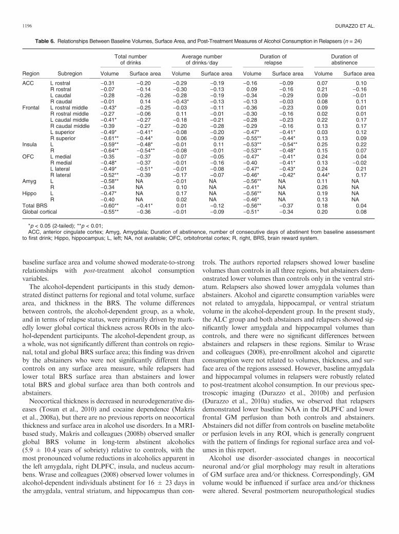
Pairwise comparisons indicated ALC showed lower volumesthan controls in all of the foregoing BRS components aswell as for total BRS and global volume (see Table 5).Abstainers, Relapsers, and Controls: The MANCOVA for
group was significant (F28, 202 = 2.37, p < 0.001). Univariatetests indicated significant group differences (p < 0.05) in thefollowing ROIs: left rostral and right caudal ACC, left andright rostral middle frontal gyri, left and right caudal middlefrontal gyri, left and right superior frontal gyri, left and rightinsula, left and right medial OFC, left and right lateral OFC,left and right amygdala, left and right hippocampus, and totalBRS and global volume. No significant group differenceswere observed for total intracranial volume. Results frompairwise comparisons are given in Table 5. Compared to con-trols, relapsers demonstrated lower volumes in 17 of 20reward system regions, while abstainers showed lower vol-umes than controls in the bilateral superior frontal cortex,insula, amygdalae, and hippocampi. Both relapsers andabstainers showed lower total BRS and global volume thancontrols. Relapsers exhibited significantly smaller volumesthan abstainers in the right rostral middle and right caudalmiddle frontal gyri, the left and right lateral OFC and relaps-ers showed trends (p < 0.05) for lower volumes than abstain-ers in the left and right superior frontal gyri. relapsers hadlower total BRS volume than abstainers, but relapsers
and abstainers were not significantly different on globalneocortical volumes. The observed regional volume differ-ences between relapsers and abstainers remained significantafter including alcohol consumption variables, smoking sta-tus, psychiatric, substance abuse, and medical comorbiditiesas covariates. No alcohol consumption variable or comorbidcondition was a significant predictor of volume in any region.The above results for BRS thickness, surface area, and vol-
ume were virtually identical if the neocortical measures werescaled to the individual’s intracranial volume (ICV) ratherthan entering ICV as a covariate in the models. Additionally,the above results for all group comparisons for total BRS andglobal neocortical volume were virtually identical when theaverage values were used as the dependent measures.
Associations of Baseline Morphology, Pre-TreatmentAlcohol, and Cigarette Use in Alcohol-Dependent Partici-pants. There were no significant bivariate associationsbetween regional and global measures for volumes, surfacearea, thickness, and pre-treatment alcohol and cigarette useduration and consumption levels after controlling for age.
Associations of Baseline Morphology With Post-Treat-ment Alcohol Consumption in Relapsers. The most consis-tent patterns observed were associations between lower
Table 4. Baseline Regional Surface Areaa
Region SubregionControls(n = 43)
ALC(n = 75)
Abstainers(n = 24)
Relapsers(n = 51)
Effect size
ALC vs.controls
Abstainers vs.controls
Relapsers vs.controls
Relapsers vs.abstainers
ACC L rostral 724 (118) 697 (118) 715 (117) 688 (121) 0.22 0.08 0.30 0.23R rostral 552 (116) 552 (114) 553 (116) 551 (115) 0.00 0.01 0.02 0.03L caudal 662 (130) 674 (128) 645 (129) 683 (129) 0.10 0.16 0.19 0.29R caudal 808 (131) 749 (129) 774 (132) 737 (130)* 0.45 0.25 0.54 0.28
Frontal L rostralmiddle
5836 (701) 5669 (700) 5864 (700) 5577 (696) 0.24 0.26 0.37 0.41
R rostralmiddle
6032 (826) 5917 (821) 6195 (823) 5788 (814) 0.12 0.19 0.30 0.50
L caudalmiddle
2507 (459) 2428 (450) 2476 (457) 2405 (456) 0.17 0.07 0.22 0.16
R caudalmiddle
2216 (380) 2289 (372) 2369 (377) 2251 (371) 0.19 0.40 0.09 0.32
L superior 7509 (643) 7257 (675) 7328 (676) 7225 (671) 0.38 0.27 0.43 0.15R superior 7371 (643) 7156 (640) 7279 (636) 7098 (635) 0.34 0.14 0.43 0.28
Insula L 2279 (390) 2140 (389) 2270 (394) 2213 (392) 0.35 0.02 0.17 0.15R 2221 (388) 2186 (388) 2202 (391) 2171 (390) 0.09 0.05 0.13 0.08
OFC L medial 1610 (177) 1577 (175) 1612 (176) 1546 (176) 0.19 0.02 0.36 0.36R medial 1778 (196) 1758 (193) 1776 (195) 1749 (192) 0.11 0.01 0.15 0.14L lateral 2602 (230) 2623 (233) 2704 (230) 2585 (229) 0.09 0.44 0.07 0.52R lateral 2507 (210) 2509 (208) 2646 (205) 2444 (207) 0.01 0.66 0.30 0.98
TotalBRSsurfacearea
46907 (2793) 46018 (2875) 47156 (2816) 45487 (2699)*,** 0.31 0.09 0.52 0.61
Globalcorticalsurfacearea
1.54 · 105 (6.233) 1.52 · 105 (6.213) 1.53 · 105 (6.693) 1.51 · 105 (6.663)* 0.36 0.14 0.46 0.32
aMean (standard deviation) adjusted for age and intracranial volume in mm2.*Relapsers < Controls, p < 0.01; **Relapsers < Abstainers, p < 0.01.ACC, anterior cingulate cortex; ALC, alcohol-dependent cohort (abstainers + relapsers); L, left; OFC, orbitofrontal cortex; R, right; BRS, brain
reward system.
1194 DURAZZO ET AL.
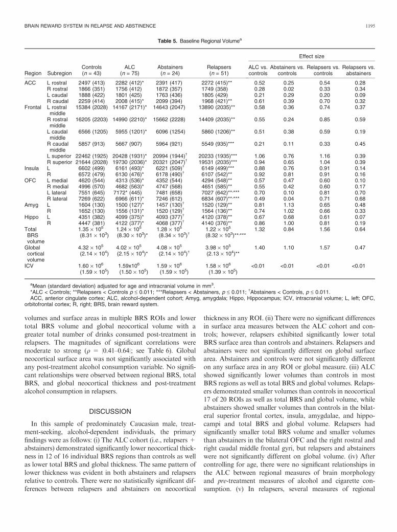
volumes and surface areas in multiple BRS ROIs and lowertotal BRS volume and global neocortical volume with agreater total number of drinks consumed post-treatment inrelapsers. The magnitudes of significant correlations weremoderate to strong (q = |0.41–0.64|; see Table 6). Globalneocortical surface area was not significantly associated withany post-treatment alcohol consumption variable. No signifi-cant relationships were observed between regional BRS, totalBRS, and global neocortical thickness and post-treatmentalcohol consumption in relapsers.
DISCUSSION
In this sample of predominately Caucasian male, treat-ment-seeking, alcohol-dependent individuals, the primaryfindings were as follows: (i) The ALC cohort (i.e., relapsers +abstainers) demonstrated significantly lower neocortical thick-ness in 12 of 16 individual BRS regions than controls as wellas lower total BRS and global thickness. The same pattern oflower thickness was evident in both abstainers and relapsersrelative to controls. There were no statistically significant dif-ferences between relapsers and abstainers on neocortical
thickness in any ROI. (ii) There were no significant differencesin surface area measures between the ALC cohort and con-trols; however, relapsers exhibited significantly lower totalBRS surface area than controls and abstainers. Relapsers andabstainers were not significantly different on global surfacearea. Abstainers and controls were not significantly differenton any surface area in any ROI or global measure. (iii) ALCshowed significantly lower volumes than controls in mostBRS regions as well as total BRS and global volumes. Relaps-ers demonstrated smaller volumes than controls in neocortical17 of 20 ROIs as well as total BRS and global volume, whileabstainers showed smaller volumes than controls in the bilat-eral superior frontal cortex, insula, amygdalae, and hippo-campi and total BRS and global volume. Relapsers hadsignificantly smaller total BRS volume and smaller volumesthan abstainers in the bilateral OFC and the right rostral andright caudal middle frontal gyri, but relapsers and abstainerswere not significantly different on global volume. (iv) Aftercontrolling for age, there were no significant relationships inthe ALC between regional measures of brain morphologyand pre-treatment measures of alcohol and cigarette con-sumption. (v) In relapsers, several measures of regional
Table 5. Baseline Regional Volumea
Region SubregionControls(n = 43)
ALC(n = 75)
Abstainers(n = 24)
Relapsers(n = 51)
Effect size
ALC vs.controls
Abstainers vs.controls
Relapsers vs.controls
Relapsers vs.abstainers
ACC L rostral 2497 (413) 2282 (412)* 2391 (417) 2272 (415)** 0.52 0.25 0.54 0.28R rostral 1866 (351) 1756 (412) 1872 (357) 1749 (358) 0.28 0.02 0.33 0.34L caudal 1888 (422) 1801 (425) 1763 (436) 1805 (429) 0.21 0.29 0.20 0.09R caudal 2259 (414) 2008 (415)* 2099 (394) 1968 (421)** 0.61 0.39 0.70 0.32
Frontal L rostralmiddle
15384 (2028) 14167 (2171)* 14643 (2047) 13890 (2035)** 0.58 0.36 0.74 0.37
R rostralmiddle
16205 (2203) 14990 (2210)* 15662 (2228) 14409 (2035)** 0.55 0.24 0.85 0.59
L caudalmiddle
6566 (1205) 5955 (1201)* 6096 (1254) 5860 (1206)** 0.51 0.38 0.59 0.19
R caudalmiddle
5857 (913) 5667 (907) 5964 (921) 5549 (935)*** 0.21 0.11 0.33 0.45
L superior 22462 (1925) 20428 (1931)* 20994 (1944)� 20233 (1935)*** 1.06 0.76 1.16 0.39R superior 21644 (2028) 19730 (2036)* 20321 (2047)� 19531 (2035)*** 0.94 0.65 1.04 0.39
Insula L 6602 (499) 6161 (493)* 6221 (509)� 6149 (499)*** 0.88 0.76 0.91 0.14R 6572 (479) 6130 (476)* 6178 (490)� 6107 (542)** 0.92 0.81 0.91 0.16
OFC L medial 4620 (544) 4313 (536)* 4352 (544) 4294 (548)** 0.57 0.47 0.60 0.10R medial 4996 (570) 4682 (563)* 4747 (568) 4651 (585)** 0.55 0.42 0.60 0.17L lateral 7551 (645) 7172* (445) 7481 (658) 7027 (642)**,*** 0.70 0.10 0.81 0.70R lateral 7269 (622) 6966 (611)* 7246 (612) 6834 (607)**,*** 0.49 0.04 0.71 0.68
Amyg L 1604 (130) 1500 (127)* 1457 (130)� 1520 (129)** 0.81 1.13 0.65 0.48R 1652 (130) 1556 (131)* 1520 (129)� 1564 (136)** 0.74 1.02 0.66 0.33
Hippo L 4351 (382) 4099 (375)* 4093 (377)� 4120 (378)** 0.67 0.68 0.61 0.07R 4447 (381) 4122 (377)* 4068 (377)� 4140 (376)** 0.86 1.00 0.81 0.19
TotalBRSvolume
1.35 · 105
(8.31 · 103)1.24 · 105
(8.30 · 103)*1.28 · 105
(8.34 · 103)�1.22 · 105
(8.32 · 103)**,***1.32 0.84 1.56 0.64
Globalcorticalvolume
4.32 · 105
(2.14 · 104)4.02 · 105
(2.15 · 104)*4.08 · 105
(2.14 · 104)�3.98 · 105
(2.13 · 104)**1.40 1.10 1.57 0.47
ICV 1.60 · 106
(1.59 · 105)1.59x10⁶
(1.50 · 105)1.59 · 106
(1.59 · 105)1.58 · 106
(1.39 · 105)<0.01 <0.01 <0.01 <0.01
aMean (standard deviation) adjusted for age and intracranial volume in mm3.*ALC < Controls; **Relapsers < Controls p £ 0.011; ***Relapsers < Abstainers, p £ 0.011; �Abstainers < Controls, p £ 0.011.ACC, anterior cingulate cortex; ALC, alcohol-dependent cohort; Amyg, amygdala; Hippo, Hippocampus; ICV, intracranial volume; L, left; OFC,
orbitofrontal cortex; R, right; BRS, brain reward system.
BRAIN REWARD SYSTEM IN RELAPSE AND ABSTINENCE 1195
baseline surface area and volume showed moderate-to-strongrelationships with post-treatment alcohol consumptionvariables.The alcohol-dependent participants in this study demon-
strated distinct patterns for regional and total volume, surfacearea, and thickness in the BRS. The volume differencesbetween controls, the alcohol-dependent group, as a whole,and in terms of relapse status, were primarily driven by mark-edly lower global cortical thickness across ROIs in the alco-hol-dependent participants. The alcohol-dependent group, asa whole, was not significantly different than controls on regio-nal, total and global BRS surface area; this finding was drivenby the abstainers who were not significantly different thancontrols on any surface area measure, while relapsers hadlower total BRS surface area than abstainers and lowertotal BRS and global surface area than both controls andabstainers.Neocortical thickness is decreased in neurodegenerative dis-
eases (Tosun et al., 2010) and cocaine dependence (Makriset al., 2008a), but there are no previous reports on neocorticalthickness and surface area in alcohol use disorders. In a MRI-based study, Makris and colleagues (2008b) observed smallerglobal BRS volume in long-term abstinent alcoholics(5.9 ± 10.4 years of sobriety) relative to controls, with themost pronounced volume reductions in alcoholics apparent inthe left amygdala, right DLPFC, insula, and nucleus accum-bens. Wrase and colleagues (2008) observed lower volumes inalcohol-dependent individuals abstinent for 16 ± 23 days inthe amygdala, ventral striatum, and hippocampus than con-
trols. The authors reported relapsers showed lower baselinevolumes than controls in all three regions, but abstainers dem-onstrated lower volumes than controls only in the ventral stri-atum. Relapsers also showed lower amygdala volumes thanabstainers. Alcohol and cigarette consumption variables werenot related to amygdala, hippocampal, or ventral striatumvolume in the alcohol-dependent group. In the present study,the ALC group and both abstainers and relapsers showed sig-nificantly lower amygdala and hippocampal volumes thancontrols, and there were no significant differences betweenabstainers and relapsers in these regions. Similar to Wraseand colleagues (2008), pre-enrollment alcohol and cigaretteconsumption were not related to volumes, thickness, and sur-face area of the regions assessed. However, baseline amygdalaand hippocampal volumes in relapsers were robustly relatedto post-treatment alcohol consumption. In our previous spec-troscopic imaging (Durazzo et al., 2010b) and perfusion(Durazzo et al., 2010a) studies, we observed that relapsersdemonstrated lower baseline NAA in the DLPFC and lowerfrontal GM perfusion than both controls and abstainers.Abstainers did not differ from controls on baseline metaboliteor perfusion levels in any ROI, which is generally congruentwith the pattern of findings for regional surface area and vol-umes in this report.Alcohol use disorder–associated changes in neocortical
neuronal and ⁄or glial morphology may result in alterationsof GM surface area and ⁄or thickness. Correspondingly, GMvolume would be influenced if surface area and ⁄or thicknesswere altered. Several postmortem neuropathological studies
Table 6. Relationships Between Baseline Volumes, Surface Area, and Post-Treatment Measures of Alcohol Consumption in Relapsers (n = 24)
Region Subregion
Total numberof drinks
Average numberof drinks ⁄ day
Duration ofrelapse
Duration ofabstinence
Volume Surface area Volume Surface area Volume Surface area Volume Surface area
ACC L rostral )0.31 )0.20 )0.29 )0.19 )0.16 )0.09 0.07 0.10R rostral )0.07 )0.14 )0.30 )0.13 0.09 )0.16 0.21 )0.16L caudal )0.28 )0.26 )0.28 )0.19 )0.34 )0.29 0.09 )0.01R caudal )0.01 0.14 )0.43* )0.13 )0.13 )0.03 0.08 0.11
Frontal L rostral middle )0.43* )0.25 )0.03 )0.11 )0.36 )0.23 0.09 0.01R rostral middle )0.27 )0.06 0.11 )0.01 )0.30 )0.16 0.02 0.01L caudal middle )0.41* )0.27 )0.18 )0.21 )0.28 )0.23 0.22 0.17R caudal middle )0.39 )0.27 )0.20 )0.28 )0.29 )0.16 0.13 0.17L superior )0.49* )0.41* )0.08 )0.20 )0.47* )0.41* 0.03 0.12R superior )0.61** )0.44* 0.06 )0.09 )0.55** )0.44* 0.13 0.09
Insula L )0.59** )0.48* )0.01 0.11 )0.53** )0.54** 0.25 0.22R )0.64** )0.54** )0.08 )0.01 )0.53** )0.48* 0.15 0.07
OFC L medial )0.35 )0.37 )0.07 )0.05 )0.47* )0.41* 0.24 0.04R medial )0.48* )0.37 )0.01 )0.16 )0.40 )0.41* 0.13 )0.02L lateral )0.49* )0.51* )0.01 )0.08 )0.47* )0.43* 0.24 0.21R lateral )0.52** )0.39 )0.17 )0.07 )0.46* )0.42* 0.44* 0.17
Amyg L )0.58** NA )0.01 NA )0.56** NA 0.11 NAR )0.34 NA 0.10 NA )0.41* NA 0.26 NA
Hippo L )0.47* NA 0.17 NA )0.56** NA 0.19 NAR )0.40 NA 0.02 NA )0.46* NA 0.13 NA
Total BRS )0.60** )0.41* 0.01 )0.12 )0.56** )0.37 0.18 0.04Global cortical )0.55** )0.36 )0.01 )0.09 )0.51* )0.34 0.20 0.08
*p < 0.05 (2-tailed); **p < 0.01;ACC, anterior cingulate cortex; Amyg, Amygdala; Duration of abstinence, number of consecutive days of abstinent from baseline assessment
to first drink; Hippo, hippocampus; L, left; NA, not available; OFC, orbitofrontal cortex; R, right, BRS, brain reward system.
1196 DURAZZO ET AL.

in alcohol use disorders report neuronal loss in superiorfrontal neocortical regions (see Harper, 2009, for review),reduced glial cell density and size in the DLPFC (Miguel-Hidalgo et al., 2002), and lower neuronal and glial celldensity in the OFC (Miguel-Hidalgo et al., 2006). Otherneuropathological studies of alcohol use disorders, however,found no abnormalities in neocortical neuronal cell volumes,neuronal and glial cell numbers or lobar and global neocorti-cal surface area, thickness, and volume (Fabricius et al.,2007; Jensen and Pakkenberg, 1993). In this ALC, pre-treatment medical and psychiatric comorbidities, alcoholand cigarette consumption were not associated with any ofthe MR-based morphologic measures in BRS componentsor global measures. Therefore, the potential mechanismscontributing to the variability in regional surface areas,thickness, and corresponding volumes observed in the alco-hol-dependent participants are unclear and likely involvegenetic, comorbid, and ⁄or environmental factors not evalu-ated in this research.Although relapsers demonstrated significantly lower total
BRS volumes and surface area than abstainers, these groupswere not significantly different on global neocortical volumeor surface area. This suggests measures of volume and sur-face area in the BRS may better distinguish abstainers andrelapsers compared to global neocortical measures. Withinthe BRS, the greatest morphological differences betweenabstainers and relapsers were apparent in left and right lateralOFC, where relapsers demonstrated significantly smaller sur-face area and volume. Intact OFC functions are critical foradaptive and flexible inhibitory decision-making processes.Neurobiological abnormalities in the OFC have been linkedto emotional and behavior disturbances that may confer riskfor the relapse ⁄ remit cycle commonly observed in all sub-stance use disorders (Baler and Volkow, 2006; Kalivas andO’Brien, 2008; Kalivas and Volkow, 2005). Specifically, theOFC is involved in emotion-related learning and regulationof internal affective and drive states (Dom et al., 2005; Rolls,2004). The OFC is proposed to be principally involved in therepresentation of the reinforcing, affective, and goal values ofa stimulus (Rolls and Grabenhorst, 2008), which is criticalfor self-modification of behavior in accordance with changesin reinforcement contingencies (Dom et al., 2005; Rolls andGrabenhorst, 2008; Spinella, 2002). Behavioral manifesta-tions of OFC injury ⁄dysfunction include impulsivity ⁄disinhibition, inaccurate interpretation of social and emo-tional cues from others, and inappropriate expression ofemotional and internal drive states in complex social con-texts. Some distinctions have been made between the func-tions subserved by the medial and lateral regions of the OFC(Rolls and Grabenhorst, 2008), but it is unclear whether thereis any regional functional specificity within the OFC in alco-hol use disorders.While the mechanisms contributing to the regional and glo-
bal morphology exhibited by abstainers and relapsers areunclear, there are distinct functional implications for baselinesurface area, thickness, and volume measured in the BRS for
these ALCs. Overall, the morphological findings in abstainersand relapsers suggest that the clinical syndrome of alcoholdependence in this cohort is primarily associated with signifi-cantly thinner neocortex in components of the BRS as well asfor the global neocortex; this may represent a premorbid con-dition and serve as a proxy measure for increased risk forthe development of alcohol use disorders. These assertions aresupported by the significantly lower neocortical thicknessacross the majority of BRS ROIs, the lower total BRS, andglobal thickness in both abstainers and relapsers relative tocontrols and the lack of associations of regional and globalmorphologic measures with comorbid psychiatric, substanceuse, medical conditions, and pre-treatment alcohol and ciga-rette consumption in the ALC. Additionally, abstainersreported an average of 3 years (1028 ± 679 days) of continu-ous sobriety following outpatient treatment at long-term fol-low-up despite demonstrating significantly lower baselineneocortical thickness in 11 of 16 ROIs and lower total BRSand global thickness than controls. With respect to surfacearea measures, results suggest that the surface areas of com-ponents of the BRS in this cohort may not be exclusivelymediated by the clinical syndrome of alcohol dependence.Specifically, relapsers exhibited lower total BRS surfaces areathan abstainers and controls, whereas abstainers and controlswere not significantly different on any surface area measure.With respect to volumes, the differences between abstainersand controls were driven by lower cortical thickness inabstainers. Additionally, BRS volume and surface area mea-sures in relapsers demonstrated moderate-to-strong relation-ships with the magnitude of their post-treatment alcoholconsumption, while no associations between baseline corticalthickness measures and severity of relapse were observed.Taken together, this suggests that both neocortical surfacearea and volume in the BRS ROIs investigated may serve asproxy markers for risk of relapse and ⁄or predict the level ofseverity of an episode of relapse in this cohort. However, itmust be noted that approximately 60% of the alcohol-depen-dent participants had at least one previous treatment, and it isunknown whether the abstainers and relapsers evidenced thesame regional morphological pattern observed in this reportat the times of their previous treatments.Limitations of this study include the reliance on self-report
and ⁄or medical records for the determination of drinkingstatus at follow-up for some participants, the inability toexamine for sex effects because of the small number offemale participants, and the modest number of participantsin the abstainer group. We did not examine the influence ofcoping skills, stress response, self-esteem ⁄ self-efficacy, socialsupport, neurocognition and personality disorders, neurocog-nitive variables, or gene polymorphisms reported to predictrelapse after treatment for AUD (e.g., Bradizza et al., 2006;Krampe et al., 2006; Miller et al., 1996; Sinha and Li, 2007;Teichner et al., 2001; Walter et al., 2006; Wojnar et al.,2009). It is highly likely that the magnitude and chronicity ofalcohol consumption before and after treatment in our ALCwas influenced not only by the integrity of their brain
BRAIN REWARD SYSTEM IN RELAPSE AND ABSTINENCE 1197
morphology, but also by genetic or other premorbid andenvironmental factors not assessed in this phase of ourresearch.The results from this morphological study, combined with
our previous neuroimaging findings in this cohort, suggestrelapsers demonstrate significant adverse neurobiologicalchanges in multiple nodes of the BRS. Taken together, ourMR studies with this cohort suggest relapsers experiencedysfunction in regions involved in the ‘‘top down’’regulation ⁄modulation of internal drive states, emotions,reward processing, and reward-related behavior (Baler andVolkow, 2006; Kalivas and Volkow, 2005; Paulus, 2007;Redish et al., 2008; Rolls and Grabenhorst, 2008; Sinha andLi, 2007), which may impart increased risk for the relapse ⁄remit cycle that afflicts many with alcohol use disorders. Theclinical relevance of the morphological abnormalities issuggested by the associations of baseline surface areas andvolumes in multiple components of the BRS with measures ofpost-treatment alcohol consumption in relapsers. Results alsohighlight the importance of examining both cortical thicknessand surface area to better understand the nature of regionalvolume loss frequently observed in alcohol use disorders. Itis well documented that sustained abstinence from alcoholis associated with neocortical volume increases in thosewith alcohol use disorders. Longitudinal studies examiningboth surface area and cortical thickness may clarify the natureof abstinence-related volume changes. Additionally, longi-tudinal assessment over periods of sustained abstinence,combined with potential genetic markers of vulnerability(e.g., Wojnar et al., 2009) will further assist in identifyingpremorbid factors that may influence the risk of relapsein alcohol use disorders.
ACKNOWLEDGMENT
This material is the result of work supported by NationalInstitutes of Health (AA10788 to D.J.M. and DA24136 toT.C.D.) and with resources and the use of facilities at the SanFrancisco Veterans Administration Medical Center, SanFrancisco CA. We thank Mary Rebecca Young, Bill Clift,Jeanne Eichenbaum and Drs. Peter Banys and Ellen Herbstof the Veterans Administration Substance Abuse Day Hospi-tal and Dr. David Pating, Karen Moise and their colleaguesat the Kaiser Permanente Chemical Dependency RecoveryProgram in San Francisco for their valuable assistance inrecruiting participants. We also extend our gratitude to thestudy participants, who made this research possible.
REFERENCES
Baler RD, Volkow ND (2006) Drug addiction: the neurobiology of disrupted
self-control. Trends Mol Med 12:559–566.
Beck AT (1978) Depression Inventory. Center for Cognitive Therapy,
Philadelphia.
Bottlender M, Soyka M (2005) Efficacy of an intensive outpatient rehabilita-
tion program in alcoholism: predictors of outcome 6 months after treat-
ment. Eur Addict Res 11:132–137.
Bowirrat A, Oscar-BermanM (2005) Relationship between dopaminergic neu-
rotransmission, alcoholism, and Reward Deficiency syndrome. Am J Med
Genet B Neuropsychiatr Genet 132:29–37.
Bradizza CM, Stasiewicz PR, Paas ND (2006) Relapse to alcohol and drug
use among individuals diagnosed with co-occurring mental health and sub-
stance use disorders: a review. Clin Psychol Rev 26:162–178.
Choi YY, Shamosh NA, Cho SH, DeYoung CG, Lee MJ, Lee JM, Kim SI,
Cho ZH, Kim K, Gray JR, Lee KH (2008) Multiple bases of human intelli-
gence revealed by cortical thickness and neural activation. J Neurosci
28:10323–10329.
Cohen J (1988) Statistical Power Analysis for the Behavioral Sciences.
Lawrence Erlbaum Associates, Hillsdale, NJ.
Dale AM, Fischl B, Sereno MI (1999) Cortical surface-based analysis. I.
Segmentation and surface reconstruction. Neuroimage 9:179–194.
Delucchi KL, Weisner C (2010) Transitioning into and out of problem
drinking across seven years. J Stud Alcohol Drugs 71:210–218.
Dickerson BC, Fenstermacher E, Salat DH, Wolk DA, Maguire RP, Desikan
R, Pacheco J, Quinn BT, Van der Kouwe A, Greve DN, Blacker D, Albert
MS, Killiany RJ, Fischl B (2008) Detection of cortical thickness correlates
of cognitive performance: reliability across MRI scan sessions, scanners,
and field strengths. Neuroimage 39:10–18.
Dom G, Sabbe B, Hulstijn W, van den Brink W (2005) Substance use disor-
ders and the orbitofrontal cortex: systematic review of behavioral decision-
making and neuroimaging studies. Br J Psychiatry 187:209–220.
Donovan DM (1996) Assessment issues and domains in the prediction of
relapse. Addiction 91(Suppl.):S29–S36.
Driessen M, Meier S, Hill A, Wetterling T, Lange W, Junghanns K (2001)
The course of anxiety, depression and drinking behaviours after completed
detoxification in alcoholics with and without comorbid anxiety and depres-
sive disorders. Alcohol Alcohol 36:249–255.
Durazzo T, Gazdzinski S, Mon A, Meyerhoff D (2010a) Cortical perfu-
sion in alcohol-dependent individuals during short-term abstinence: rela-
tionships to resumption of hazardous drinking after treatment. Alcohol
44:201–210.
Durazzo TC, Gazdzinski S, Yeh PH, Meyerhoff DJ (2008) Combined
neuroimaging, neurocognitive and psychiatric factors to predict alcohol
consumption following treatment for alcohol dependence. Alcohol Alcohol
43:683–691.
Durazzo TC, Pathak V, Gazdzinski S, Mon A, Meyerhoff DJ (2010b) Metab-
olite levels in the brain reward pathway discriminate those who remain
abstinent from those who resume hazardous alcohol consumption after
treatment for alcohol dependence. J Stud Alcohol Drugs 71:278–289.
Fabricius K, Pakkenberg H, Pakkenberg B (2007) No changes in neocortical
cell volumes or glial cell numbers in chronic alcoholic subjects compared to
control subjects. Alcohol Alcohol 42:400–406.
Fagerstrom KO, Heatherton TF, Kozlowski LT (1991) Nicotine addiction
and its assessment. Ear Nose Throat J 69:763–765.
First MB, Spitzer RL, Gibbon M, Williams JBW (1998) Structured Clinical
Interview for DSM-IV Axis I Disorders – Patient Edition (SCID-I ⁄P,Version 2.0, 8 ⁄ 98 revision). Biometrics Research Department, New York,
NY.
Fischl B, Dale AM (2000) Measuring the thickness of the human cerebral cor-
tex from magnetic resonance images. Proc Natl Acad Sci USA 97:11050–
11055.
Fischl B, Destrieux C, Halgren E, Segonne F, Salat DH, Busa E, Seidman LJ,
Goldstein J, Kennedy D, Caviness V, Makris N, Rosen B, Dale AM (2004)
Automatic parcellation of the human cerebral cortex. CerebCortex 14:11–22.
Fischl B, SerenoMI, Dale AM (1999) Cortical surface-based analysis. II: infla-
tion, flattening, and a surface-based coordinate system. Neuroimage 9:195–
207.
Fowler JS, Volkow ND, Kassed CA, Chang L (2007) Imaging the addicted
human brain. Sci Pract Perspect 3:4–16.
Gazdzinski S, Durazzo TC, Studholme C, Song E, Banys P, Meyerhoff DJ
(2005) Quantitative brain MRI in alcohol dependence: preliminary evidence
for effects of concurrent chronic cigarette smoking on regional brain vol-
umes. Alcohol Clin Exp Res 29:1484–1495.
1198 DURAZZO ET AL.

Glenn SW, Parsons OA (1991) Prediction of resumption of drinking in post-
treatment alcoholics. Int J Addict 26:237–254.
Grober E, Sliwinski M (1991) Development and validation of a model for esti-
mating premorbid verbal intelligence in the elderly. J Clin Exp Neuropsy-
chol 13:933–949.
Grusser SM, Wrase J, Klein S, Hermann D, Smolka MN, Ruf M,
Weber-Fahr W, Flor H, Mann K, Braus DF, Heinz A (2004) Cue-induced
activation of the striatum and medial prefrontal cortex is associated with
subsequent relapse in abstinent alcoholics. Psychopharmacology (Berl)
175:296–302.
Harper C (2009) The neuropathology of alcohol-related brain damage. Alco-
hol Alcohol 44:136–140.
Hasin DS, Stinson FS, Ogburn E, Grant BF (2007) Prevalence, correlates, dis-
ability, and comorbidity of DSM-IV alcohol abuse and dependence in the
United States: results from the National Epidemiologic Survey on Alcohol
and Related Conditions. Arch Gen Psychiatry 64:830–842.
Heinz A, Wrase J, Kahnt T, Beck A, Bromand Z, Grusser SM, Kienast T,
Smolka MN, Flor H, Mann K (2007) Brain activation elicited by affectively
positive stimuli is associated with a lower risk of relapse in detoxified alco-
holic subjects. Alcohol Clin Exp Res 31:1138–1147.
Hutton C, Draganski B, Ashburner J, Weiskopf N (2009) A comparison
between voxel-based cortical thickness and voxel-based morphometry in
normal aging. Neuroimage 48:371–380.
Im K, Lee JM, Lyttelton O, Kim SH, Evans AC, Kim SI (2008) Brain size
and cortical structure in the adult human brain. Cereb Cortex 18:2181–
2191.
Innocenti GM, Vercelli A (2010) Dendritic bundles, minicolumns, columns,
and cortical output units. Front Neuroanat 4:11.
Jensen GB, Pakkenberg B (1993) Do alcoholics drink their neurons away?
Lancet 342:1201–1204.
Kalivas PW, O’Brien C (2008) Drug addiction as a pathology of staged neuro-
plasticity. Neuropsychopharmacology 33:166–180.
Kalivas PW, Volkow ND (2005) The neural basis of addiction: a pathology of
motivation and choice. Am J Psychiatry 162:1403–1413.
Kochunov P, Coyle T, Lancaster J, Robin DA, Hardies J, Kochunov V, Bart-
zokis G, Stanley J, Royall D, Schlosser AE, Null M, Fox PT (2010) Process-
ing speed is correlated with cerebral health markers in the frontal lobes as
quantified by neuroimaging. Neuroimage 49:1190–1199.
Kochunov P, Thompson PM, Lancaster JL, Bartzokis G, Smith S, Coyle
T, Royall DR, Laird A, Fox PT (2007) Relationship between white mat-
ter fractional anisotropy and other indices of cerebral health in normal
aging: tract-based spatial statistics study of aging. Neuroimage 35:478–
487.
Kodl MM, Fu SS, Willenbring ML, Gravely A, Nelson DB, Joseph AM
(2008) The impact of depressive symptoms on alcohol and cigarette con-
sumption following treatment for alcohol and nicotine dependence. Alcohol
Clin Exp Res 32:92–99.
Koob GF (2003) Alcoholism: allostasis and beyond. Alcohol Clin Exp Res
27:232–243.
Krampe H, Stawicki S, Hoehe MR, Ehrenreich H (2007) Outpatient Long-
term Intensive Therapy for Alcoholics (OLITA): a successful biopsychoso-
cial approach to the treatment of alcoholism. Dialogues Clin Neurosci
9:399–412.
Krampe H, Wagner T, Stawicki S, Bartels C, Aust C, Kroener-Herwig B,
Kuefner H, Ehrenreich H (2006) Personality disorder and chronicity of
addiction as independent outcome predictors in alcoholism treatment.
Psychiatr Serv 57:708–712.
Lubman DI, Yucel M, Pantelis C (2004) Addiction, a condition of compulsive
behaviour? Neuroimaging and neuropsychological evidence of inhibitory
dysregulation. Addiction 99:1491–1502.
Maisto SA, Clifford PR, Stout RL, Davis CM (2007) Moderate drinking in
the first year after treatment as a predictor of three-year outcomes. J Stud
Alcohol Drugs 68:419–427.
Maisto SA, ZywiakWH, Connors GJ (2006) Course of functioning 1 year fol-
lowing admission for treatment of alcohol use disorders. Addict Behav
31:69–79.
Makris N, Gasic GP, Kennedy DN, Hodge SM, Kaiser JR, Lee MJ, Kim
BW, Blood AJ, Evins AE, Seidman LJ, Iosifescu DV, Lee S, Baxter C,
Perlis RH, Smoller JW, Fava M, Breiter HC (2008a) Cortical thickness
abnormalities in cocaine addiction – a reflection of both drug use and a
pre-existing disposition to drug abuse? Neuron 60:174–188.
Makris N, Oscar-Berman M, Jaffin SK, Hodge SM, Kennedy DN, Caviness
VS,Marinkovic K, Breiter HC, Gasic GP, Harris GJ (2008b) Decreased vol-
ume of the brain reward system in alcoholism. Biol Psychiatry 64:192–202.
McKay JR (1999) Studies of factors in relapse to alcohol, drug and nicotine use:
a critical review ofmethodologies and findings. J StudAlcohol 60:566–576.
McKay JR, Franklin TR, Patapis N, Lynch KG (2006) Conceptual, methodo-
logical, and analytical issues in the study of relapse. Clin Psychol Rev
26:109–127.
Mertens JR, Lu YW, Parthasarathy S, Moore C, Weisner CM (2003) Medical
and psychiatric conditions of alcohol and drug treatment patients in an
HMO: comparisonwithmatched controls. Arch InternMed 163:2511–2517.
Mertens JR, Weisner C, Ray GT, Fireman B, Walsh K (2005) Hazardous
drinkers and drug users in HMO primary care: prevalence, medical condi-
tions, and costs. Alcohol Clin Exp Res 29:989–998.
Miguel-Hidalgo JJ, Overholser JC, Meltzer HY, Stockmeier CA, Rajkowska
G (2006) Reduced glial and neuronal packing density in the orbitofrontal
cortex in alcohol dependence and its relationship with suicide and duration
of alcohol dependence. Alcohol Clin Exp Res 30:1845–1855.
Miguel-Hidalgo JJ, Wei J, Andrew M, Overholser JC, Jurjus G, Stockmeier
CA, Rajkowska G (2002) Glia pathology in the prefrontal cortex in alcohol
dependence with and without depressive symptoms. Biol Psychiatry
52:1121–1133.
Miller WR, Walters ST, Bennett ME (2001) How effective is alcoholism treat-
ment in the United States? J Stud Alcohol 62:211–220.
Miller WR, Westerberg VS, Harris RJ, Tonigan JS (1996) What predicts
relapse? Prospective testing of antecedent models. Addiction 91(Suppl.):
S155–S172.
Moffett JR, Ross B, Arun P, Madhavarao CN, Namboodiri AM (2007) N-
Acetylaspartate in the CNS: from neurodiagnostics to neurobiology. Prog
Neurobiol 81:89–131.
Moos RH, Moos BS (2006) Rates and predictors of relapse after natural and
treated remission from alcohol use disorders. Addiction 101:212–222.
Moos RH, Moos BS, Timko C (2006) Gender, treatment and self-help in
remission from alcohol use disorders. Clin Med Res 4:163–174.
Noel X, Sferrazza R, Van Der Linden M, Paternot J, Verhas M, Hanak C,
Pelc I, Verbanck P (2002) Contribution of frontal cerebral blood flow mea-
sured by (99m)Tc-Bicisate spect and executive function deficits to predicting
treatment outcome in alcohol-dependent patients. Alcohol Alcohol 37:347–
354.
Panizzon MS, Fennema-Notestine C, Eyler LT, Jernigan TL, Prom-Wormley
E, Neale M, Jacobson K, Lyons MJ, Grant MD, Franz CE, Xian H, Tsu-
ang M, Fischl B, Seidman L, Dale A, Kremen WS (2009) Distinct genetic
influences on cortical surface area and cortical thickness. Cereb Cortex
19:2728–2735.
Parekh RS, Klag MJ (2001) Alcohol: role in the development of hyper-
tension and end-stage renal disease. Curr Opin Nephrol Hypertens 10:
385–390.
Parks MH, Dawant BM, Riddle WR, Hartmann SL, Dietrich MS, Nickel
MK, Price RR, Martin PR (2002) Longitudinal brain metabolic character-
ization of chronic alcoholics with proton magnetic resonance spectroscopy.
Alcohol Clin Exp Res 26:1368–1380.
Paulus MP (2007) Neural basis of reward and craving – a homeostatic point
of view. Dialogues Clin Neurosci 9:379–387.
Pfefferbaum A, Sullivan EV, Rosenbloom MJ, Mathalon DH, Lim KO
(1998) A controlled study of cortical gray matter and ventricular
changes in alcoholic men over a 5-year interval. Arch Gen Psychiatry
55:905–912.
Pierce RC, Kumaresan V (2006) The mesolimbic dopamine system: the final
common pathway for the reinforcing effect of drugs of abuse? Neurosci Bio-
behav Rev 30:215–238.
Rakic P (1988) Specification of cerebral cortical areas. Science 241:170–176.
BRAIN REWARD SYSTEM IN RELAPSE AND ABSTINENCE 1199

Redish AD, Jensen S, Johnson A (2008) A unified framework for addiction:
vulnerabilities in the decision process. Behav Brain Sci 31:415–437. discus-
sion 437–87.
Ritvo JI, Park C (2007) The psychiatric management of patients with alcohol
dependence. Curr Treat Options Neurol 9:381–392.
Rolls ET (2004) The functions of the orbitofrontal cortex. Brain Cogn 55:11–
29.
Rolls ET, Grabenhorst F (2008) The orbitofrontal cortex and beyond: from
affect to decision-making. Prog Neurobiol 86:216–244.
Rosenbloom MJ, Pfefferbaum A, Sullivan EV (2004) Recovery of short-term
memory and psychomotor speed but not postural stability with long-term
sobriety in alcoholic women. Neuropsychology 18:589–597.
Ross B, Bluml S (2001) Magnetic resonance spectroscopy of the human brain.
Anat Rec 265:54–84.
Sankoh AJ, Huque MF, Dubey SD (1997) Some comments on frequently
used multiple endpoint adjustment methods in clinical trials. Stat Med
16:2529–2542.
Sinha R, Li CS (2007) Imaging stress- and cue-induced drug and alcohol crav-
ing: association with relapse and clinical implications. Drug Alcohol Rev
26:25–31.
Sobell LC, Sobell MB (1992) Timeline follow-back: a technique forasses-
sing self-reported alcohol consumption, in Measuring Alcohol Consump-
tion (Litten R, Allen J eds), pp. 41–72. The Humana Press Inc.,
Totowa, NJ.
Sobell LC, Sobell MB, Riley DM, Schuller R, Pavan DS, Cancilla A,
Klajner F, Leo GI (1988) The reliability of alcohol abusers’ self-reports
of drinking and life events that occurred in the distant past. J Stud
Alcohol 49:225–232.
Spielberger CD, Gorsuch RL, Lushene R, Vagg PR, Jacobs GA (1977) Self-
Evaluation Questionaire.
Spinella M (2002) Correlations among behavioral measures of orbitofrontal
function. Int J Neurosci 112:1359–1369.
Stinson FS, Grant BF, Dawson DA, Ruan WJ, Huang B, Saha T (2005)
Comorbidity between DSM-IV alcohol and specific drug use disorders in
the United States: results from the National Epidemiologic Survey on Alco-
hol and Related Conditions. Drug Alcohol Depend 80:105–116.
Sullivan EV, Rosenbloom M, Serventi KL, Pfefferbaum A (2004) Effects of
age and sex on volumes of the thalamus, pons, and cortex. Neurobiol Aging
25:185–192.
Teichner G, Horner MD, Harvey RT (2001) Neuropsychological predictors of
the attainment of treatment objectives in substance abuse patients. Int J
Neurosci 106:253–263.
Tosun D, Mojabi P, Weiner MW, Schuff N (2010) Joint analysis of structural
and perfusion MRI for cognitive assessment and classification of Alzhei-
mer’s disease and normal aging. Neuroimage 52:186–197.
Udo T, Clifford PR, Davis CM, Maisto SA (2009) Alcohol use post AUD
treatment initiation as a predictor of later functioning. Am J Drug Alcohol
Abuse 35:128–132.
Vengeliene V, Bilbao A, Molander A, Spanagel R (2008) Neuropharmacology
of alcohol addiction. Br J Pharmacol 154:299–315.
Volkow ND, Fowler JS, Wang GJ (2003) The addicted human brain: insights
from imaging studies. J Clin Invest 111:1444–1451.
Volkow ND, Fowler JS, Wang GJ (2004) The addicted human brain viewed
in the light of imaging studies: brain circuits and treatment strategies. Neu-
ropharmacology 47(Suppl. 1):3–13.
Volkow ND, Wang GJ, Fowler JS, Telang F (2008) Overlapping neuronal cir-
cuits in addiction and obesity: evidence of systems pathology. Philos Trans
R Soc Lond B Biol Sci 363:3191–3200.
Walhovd KB, Fjell AM, Dale AM, Fischl B, Quinn BT, Makris N, Salat D,
Reinvang I (2006) Regional cortical thickness matters in recall after months
more than minutes. Neuroimage 31:1343–1351.
Walter M, Gerhard U, Duersteler-MacFarland KM, Weijers HG, Boening J,
Wiesbeck GA (2006) Social factors but not stress-coping styles predict
relapse in detoxified alcoholics. Neuropsychobiology 54:100–106.
Witkiewitz K, Marlatt GA (2007) Modeling the complexity of post-treatment
drinking: it’s a rocky road to relapse. Clin Psychol Rev 27:724–738.
Wojnar M, Brower KJ, Strobbe S, Ilgen M, Matsumoto H, Nowosad I,
Sliwerska E, Burmeister M (2009) Association between Val66Met
brain-derived neurotrophic factor (BDNF) gene polymorphism and
post-treatment relapse in alcohol dependence. Alcohol Clin Exp Res
33:693–702.
Wrase J, Makris N, Braus DF, Mann K, Smolka MN, Kennedy DN, Cavi-
ness VS, Hodge SM, Tang L, Albaugh M, Ziegler DA, Davis OC, Kissling
C, Schumann G, Breiter HC, Heinz A (2008) Amygdala volume associated
with alcohol abuse relapse and craving. Am J Psychiatry 165:1179–1184.
Zywiak WH, Stout RL, Trefry WB, Glasser I, Connors GJ, Maisto SA, West-
erberg VS (2006) Alcohol relapse repetition, gender, and predictive validity.
J Subst Abuse Treat 30:349–353.
1200 DURAZZO ET AL.