
Correspondence and Brief Communications
29
Correspondence and Brief Communications Correspondence and brief communications are wel- comed and need not concern only what has been published in this journal. We shall print items of interest to our readers, such as experimental, clinical, and philosophical observations; reports of work in progress; educational notes; and travel accounts relevant to plastic surgery. We reserve the right to edit communications to meet requirements of space and format. Any financial interest relevant to the content of the correspondence must be disclosed. Submission of a letter constitutes permission for the American Society of Plastic Surgeons and its licensees and assignees to publish it in the journal and in any other form or medium. The views, opinions, and conclusions expressed in the Letters to the Editor represent the personal opinions of the individual writers and not those of the publisher, the Editorial Board, or the sponsors of the journal. Any stated views, opin- ions, and conclusions do not reflect the policy of any of the sponsoring organizations or of the institutions with which the writer is affiliated, and the publisher, the Editorial Board, and the sponsoring organizations assume no responsibility for the content of such letters. JUST STOP OR JUST COMPLAIN Sir: Dr. Sean Boutros makes a well-intentioned plea for a pos- itive outlook on plastic surgery in “Just Stop” (Plast. Reconstr. Surg. 111: 1355, 2003), but we offer slightly different advice to our colleagues. To those who are currently quiet, please just complain. To those who are already speaking out, please be more vociferous! What is the line that demarcates complaint and advocacy? Are we complaining when we argue for a patient to receive a breast reduction to relieve pain? Is it complaining when a breast cancer survivor needs our support to undergo a pro- cedure to restore her sense of femininity? Should we be silent when many of us waste billions of dollars on defensive med- icine lest we be sued for leaving some pebble unturned? Why should we not speak out when we are forced to dictate notes to fill a relatively capricious Evaluation and Management template checklist as opposed to providing real care? Is it being bitter when we write second and third letters of appeal to managed care companies as our patients’ ombudsman? These difficulties are part of medicine’s complex land- scape. We have chosen to love this profession despite all its difficulties, but with that choice, we have also undertaken a responsibility to change the status quo. It is our duty to com- plain or, at the very least, to support those who advocate on our behalf. As members of several plastic surgery and medical societies, we joined organizations of colleagues and compa- triots who, precisely because they do complain, protect the integrity of our field. Our dues pay to organize these “com- plaints” so that they may champion our rights and those of our patients as effectively as possible, protecting them from charlatans and upholding the highest standards of care for our profession. As academicians steeped in the difficulties and joys of practice and as wide-eyed medical students about to embark on a surgical career, we do not chastise those who complain, and in fact, we encourage others to join in with our chorus. After all, if we do not complain, who will? DOI: 10.1097/01.PRS.0000089262.60250.B5 Navin Singh, M.D. Plastic and Reconstructive Surgery Johns Hopkins School of Medicine, and R. Adams Cowley Shock Trauma Center University of Maryland School of Medicine Baltimore, Md. Anand Mehra Columbia College of Physicians and Surgeons New York, N.Y. Correspondence to Dr. Singh Plastic and Reconstructive Surgery Johns Hopkins School of Medicine 601 N. Caroline Street McElderry 8130-C Baltimore, Md. 21287-0981 [email protected] REPLY Sir: There is no doubt that we have not only the right but also the duty to voice our concerns to bring about a positive change for both the patients and ourselves. However, our duty extends with the responsibility to choose a venue and tone appropriate for our profession. Our profession is more than an occupation. It is a sacred relationship to our fellow man. From the child with a cleft lip to the woman with an aging face, we are charged to do whatever we can to make their lives better. It is a gift and a privilege, and we should not forget it. As educators and leaders of the field, the efforts to bring about a positive change should be strong but carefully cho- sen. The labor should be targeted to those settings that will help accomplish our goals and should not serve to embitter residents and medical students. The point of my original letter is best illustrated by the fact that a medical student coauthored the response. Someone who has embarked on a career in medicine with lofty goals should be wet with anticipation for the times ahead. The fact that medical students around the country who have yet to feel the joy of helping or healing are complaining only attests that we as a group have not fulfilled our duty to the next gener- ation of doctors. As mentors for the neophytes, we should fuel the excitement and passion inherent to helping and healing and not the difficulties associated with our mission. Again, improving our ability to fulfill our charge is ex- 1947
-
Upload
khangminh22 -
Category
Documents
-
view
5 -
download
0
Transcript of Correspondence and Brief Communications
Correspondence andBrief Communications
Correspondence and brief communications are wel-comed and need not concern only what has been publishedin this journal. We shall print items of interest to ourreaders, such as experimental, clinical, and philosophicalobservations; reports of work in progress; educational notes;and travel accounts relevant to plastic surgery. We reserve theright to edit communications to meet requirements of spaceand format. Any financial interest relevant to the content ofthe correspondence must be disclosed. Submission of a letterconstitutes permission for the American Society of PlasticSurgeons and its licensees and assignees to publish it in thejournal and in any other form or medium.
The views, opinions, and conclusions expressed in theLetters to the Editor represent the personal opinions of theindividual writers and not those of the publisher, the EditorialBoard, or the sponsors of the journal. Any stated views, opin-ions, and conclusions do not reflect the policy of any of thesponsoring organizations or of the institutions with which thewriter is affiliated, and the publisher, the Editorial Board, andthe sponsoring organizations assume no responsibility for thecontent of such letters.
JUST STOP OR JUST COMPLAIN
Sir:Dr. Sean Boutros makes a well-intentioned plea for a pos-
itive outlook on plastic surgery in “Just Stop” (Plast. Reconstr.Surg. 111: 1355, 2003), but we offer slightly different adviceto our colleagues. To those who are currently quiet, please justcomplain. To those who are already speaking out, please bemore vociferous!
What is the line that demarcates complaint and advocacy?Are we complaining when we argue for a patient to receivea breast reduction to relieve pain? Is it complaining when abreast cancer survivor needs our support to undergo a pro-cedure to restore her sense of femininity? Should we be silentwhen many of us waste billions of dollars on defensive med-icine lest we be sued for leaving some pebble unturned? Whyshould we not speak out when we are forced to dictate notesto fill a relatively capricious Evaluation and Managementtemplate checklist as opposed to providing real care? Is itbeing bitter when we write second and third letters of appealto managed care companies as our patients’ ombudsman?
These difficulties are part of medicine’s complex land-scape. We have chosen to love this profession despite all itsdifficulties, but with that choice, we have also undertaken aresponsibility to change the status quo. It is our duty to com-plain or, at the very least, to support those who advocate onour behalf. As members of several plastic surgery and medicalsocieties, we joined organizations of colleagues and compa-triots who, precisely because they do complain, protect theintegrity of our field. Our dues pay to organize these “com-plaints” so that they may champion our rights and those ofour patients as effectively as possible, protecting them fromcharlatans and upholding the highest standards of care forour profession.
As academicians steeped in the difficulties and joys ofpractice and as wide-eyed medical students about to embarkon a surgical career, we do not chastise those who complain,and in fact, we encourage others to join in with our chorus.After all, if we do not complain, who will?DOI: 10.1097/01.PRS.0000089262.60250.B5
Navin Singh, M.D.Plastic and Reconstructive SurgeryJohns Hopkins School of Medicine, andR. Adams Cowley Shock Trauma CenterUniversity of Maryland School of MedicineBaltimore, Md.Anand MehraColumbia College of Physicians and SurgeonsNew York, N.Y.
Correspondence to Dr. SinghPlastic and Reconstructive SurgeryJohns Hopkins School of Medicine601 N. Caroline StreetMcElderry 8130-CBaltimore, Md. [email protected]
REPLY
Sir:There is no doubt that we have not only the right but also
the duty to voice our concerns to bring about a positivechange for both the patients and ourselves. However, ourduty extends with the responsibility to choose a venue andtone appropriate for our profession. Our profession is morethan an occupation. It is a sacred relationship to our fellowman. From the child with a cleft lip to the woman with anaging face, we are charged to do whatever we can to maketheir lives better. It is a gift and a privilege, and we should notforget it.
As educators and leaders of the field, the efforts to bringabout a positive change should be strong but carefully cho-sen. The labor should be targeted to those settings that willhelp accomplish our goals and should not serve to embitterresidents and medical students.
The point of my original letter is best illustrated by the factthat a medical student coauthored the response. Someonewho has embarked on a career in medicine with lofty goalsshould be wet with anticipation for the times ahead. The factthat medical students around the country who have yet to feelthe joy of helping or healing are complaining only attests thatwe as a group have not fulfilled our duty to the next gener-ation of doctors. As mentors for the neophytes, we should fuelthe excitement and passion inherent to helping and healingand not the difficulties associated with our mission.
Again, improving our ability to fulfill our charge is ex-1947

tremely important, as is improving our own lives. Choosinghow and when we express our complaints requires thoughtand judgment.
Sean Boutros, M.D.230 West 55th St., #5DNew York, N.Y. [email protected]
RECONSTRUCTION OF THE COLUMELLA IN APEDIATRIC PATIENT
Sir:Reconstruction of columellar defects continues to be a
surgical challenge. Total loss of the columella and membra-nous septum poses even more difficulty than partial loss. Inthis letter, we report on a pediatric patient with a defectinvolving the entire columella along with the membranousseptum and the caudal cartilaginous septum. Reconstructionof this defect was accomplished with the combination of acostal cartilage graft to support the nasal dorsum and an alarrim skin flap combined with a skin-cartilage composite graftto replace the absent columella.
A 3-year-old girl presented with a history of soft-tissuetumor involving the columella that had been present sincebirth. This tumor had been trimmed repeatedly by a familyphysician in another country. No further information wasavailable regarding the nature of the original lesion. Thepatient’s initial examination revealed total absence of thecolumella, the membranous septum, and the caudal carti-laginous septum. The nasal tip had lost projection and ap-peared collapsed. Heavy crusting interfered with air passagethrough the nose.
The surgical reconstruction was performed when the pa-tient was 4.5 years old. A skin flap was elevated from the rightalar rim based on the nasal tip (Fig. 1). Through the sameincision, the dorsum was undermined to create a pocket forthe costal cartilage graft. A 4-cm-long graft was harvested fromthe eighth rib with its perichondrium. A thin Kirschner wire
was threaded through the graft to prevent future warping ofthe cartilage. The graft was carved around the Kirschner wireuntil the desired shape and length were achieved. After thegraft was positioned in its pocket (Fig. 2), another Kirschnerwire was introduced through the skin to fix the costal cartilagegraft to the nasal bones. This maneuver reestablished theprojection of the tip and relieved collapsing forces from theneocolumella.
The alar rim skin flap bridged the single nostril and wasinset into the midline of the sill (Fig. 3). The blood supply tothis flap was noticed to be especially rich, probably due to theloss of the columellar artery early in life. A skin-cartilagecomposite graft from the concha was shaped and inset tocomplete the reconstruction (Fig. 3). The alar rim skin flapserved as the bed for the composite graft, whose “take” reliedentirely on its vascularity.
A dorsal splint was applied for additional fixation of the
FIG. 1. Total columellar defect extending to the mem-branous septum and the caudal cartilaginous septum. Designof the medially based alar rim skin flap.
FIG. 2. Costal cartilage graft curved around Kirschnerwire to support the nasal dorsum and the tip.
FIG. 3. Inset of the alar rim flap and the composite skin-cartilage graft.
1948 PLASTIC AND RECONSTRUCTIVE SURGERY, December 2003

costal cartilage graft. The percutaneous Kirschner wire wasremoved 3 weeks after the operation. The skin flap and thecomposite graft healed well, with a pleasing postoperativecontour and color match (Fig. 4).
Among the nasal aesthetic subunits, the columella is themost difficult to reconstruct. Loss of the columella can becaused by malignancy, trauma, or infection, with resultantfunctional and aesthetic deformities that may be devastat-ing for the patient.1 Nasolabial flaps (unilateral or bilat-eral),2–5 forehead flaps,5–7 dorsal flaps based on the angularartery,1,8 bilateral alar flaps rotated from the tip,9,10 a forkedflap from the upper lip,11 and microvascular free-tissuetransfer have been described as possible surgical options.Most of these techniques require staged procedures forfurther contour adjustments and tip support. Compositegrafts for columellar reconstruction have been describedin partial columellar and alar rim defects,12 where suffi-cient vascular bed was available.
The technique described in this letter relies on severalimportant principles in nasal reconstruction. The neocolu-mella is not to be expected to support the collapsed tip. Thissupport should come from a different source, such as a stabledorsal cartilage graft, that will relieve pressure from the re-constructed columella and prevent its distortion.
The blood supply to the superiorly based alar rim skin flapis improved in the case of earlier loss of columellar artery,according to the principle of surgical delay. This flap provedto be reliable not only in terms of surviving but also in termsof supporting a composite graft.
The cartilage within the composite graft serves one pur-pose only: to shape the columella and improve its contour.This thin conchal cartilage probably will not provide sub-stantial mechanical support for the tip, contrary to columellarstruts used in rhinoplasty. It has been our practice to harvestthe composite graft from the junction of the floor and the wallof the concha, as a relatively large graft can be recruited safelywithout creating a donor-site deformity.
The total columellar defect presents a unique problemthat taxes the ingenuity of the plastic surgeon. Reconstruc-tion of this defect with a combination of dorsal costal cartilagegraft, alar rim skin flap, and composite auricular skin-carti-lage graft is presented. We recommend adding this techniqueto the surgical armamentarium for reconstruction of thesecomplex lesions.DOI: 10.1097/01.PRS.0000089264.31445.E3
Alexander Margulis, M.D.Bruce S. Bauer, M.D.Hongshik Han, M.D.Pravin K. Patel, M.D.Division of Plastic SurgeryShriners Hospital for Children, andChildren’s Memorial HospitalChicago, Ill.
Correspondence to Dr. Margulis2300 Children’s Plaza, No. 93Chicago, Ill. [email protected]
REFERENCES
1. Smith, V., and Papay, F. A. Surgical options in columel-lar reconstruction. Otolaryngol. Head Neck Surg. 120:947, 1999.
2. Ozkus, I., Cek, D. I., and Ozcus, K. The use of bifidnasolabial flaps in reconstruction of the columella.Ann. Plast. Surg. 29: 461, 1992.
3. Yanai, A., Nagata, S., and Tanaka, H. Reconstruction ofthe columella with bilateral nasolabial flaps. Plast. Re-constr. Surg. 77: 129, 1986.
4. Dolan, R., and Arena, S. Reconstruction of the totalcolumellar defect. Laryngoscope 105: 1141, 1995.
5. Burget, G. C., and Menick, F. J. Subtotal and total nasalreconstruction. In Aesthetic Reconstruction of the Nose. St.Louis: Mosby, 1994. Pp. 313-343.
6. Menick, F. J. Anatomic reconstruction of the nasal tipcartilages in secondary and reconstructive rhinoplasty.Plast. Reconstr. Surg. 104: 2187, 1999.
7. Menick, F. J. A 10-year experience in nasal reconstruc-tion with the three-stage forehead flap. Plast. Reconstr.Surg. 109: 1839, 2002.
8. Conley, J., Sachs, M. E., and Donovan, D. Mini alarmyocutaneous flaps for nasolabial-columella recon-struction. Otolaryngol. Head Neck Surg. 91: 380, 1983.
9. Jackson, I. T. Local Flaps in Head and Neck Reconstruction.St. Louis: Mosby, 1985. Pp. 1338-1340.
10. Millard, D. R. A Rhinoplasty Tetralogy. Boston: Little,Brown, 1996. Pp. 334-423.
11. Pincus, R. L., and Bukachevsky, R. P. Medially basedhorizontal nasolabial flaps for reconstruction of col-umellar defects. Arch. Otolaryngol. Head Neck Surg. 116:973, 1990.
12. Cho, B. C., Park, J. W., and Baik, B. S. Correction ofsevere secondary cleft lip nasal deformity using a com-posite graft: Current approach and review. Ann. Plast.Surg. 48: 131, 2002.
CHIN AUGMENTATION WITH CONCHALCARTILAGE
Sir:We have read the article “Chin Augmentation with Con-
chal Cartilage” by F. Viterbo (Plast. Reconstr. Surg. 111: 899,2003), and in our view and given our clinical and experi-mental experience, there are some points that should beilluminated.
The author performed conchal grafts to the chin of pa-tients who requested mentoplasty by itself or combined withrhinoplasty, rhytidoplasty, or submental lipoplasty. The con-
FIG. 4. Postoperative view.
Vol. 112, No. 7 / CORRESPONDENCE 1949

chal cartilage was harvested subperichondrally through a pos-terior 3-cm incision. The specimen measured 3 � 1 cm, whichwas sufficient to project the chin 2 to 3 mm. For 4 to 5 mmof projection, both cartilages were used. The graft was posi-tioned under the periosteum and was held with two 5-0 nylonsutures. In these cases, the conchal cartilage graft was a suit-able option for chin augmentations up to 5 mm.
Mild to moderate microgenic patients would not needosteotomies, and augmentation mentoplasty could beenough to balance the facial profile.1,2 The use of autologousgrafts might be more appropriate in many circumstances, butresorption is a potential problem with all biologic grafts.Adams3 claimed that Silastic (either rubber-filled or gel-filled) prostheses (Dow Corning, Midland, Mich.) producedthe most pleasing and long-lasting results for the chin andwere superior to biological materials. The biological graftspossess the same risk of infection in the early postoperativeperiod as the alloplastic implants, but in long-term follow-up,the integration and the increased vascularity of the biologicalgrafts prevent infection.4–6 This integration process couldculminate in increased resorption.7 The surgeon shouldchoose the best alternative for chin augmentation accordingto the patient’s demands. We prefer to use autologous graftsfor augmentation according to the severity of the microgenia.The bone grafts harvested from the iliac crest or rib revealpermanent results, especially in severe microgenia. The re-placement of the bone graft subperiosteally helps in the in-tegration of the graft to the recipient area. All other grafts,including cartilage, dermofat, or adipose, should be placedsubdermally, over the mental muscles, instead of subperios-teally. According to our experience, the pressure caused bythe inelastic property of the periosteum increases the resorp-tion of tissues except in the case of bone. The author indi-cated good results at 4 years of follow-up and explained thefinal outcome as the ossification of the cartilage. Could thisossification be new bone formation under the periosteumduring cartilage resorption?
In 1958, Aufricht1 described the use of the nasal hump thatwas removed during rhinoplasty and placed supraperioste-ally. In his series of 700 cases, resorption was not encoun-tered, as supported by plain roentgenograms. Mottura8 alsoused the osteocartilaginous hump harvested from the rhino-plasty for augmentation of the chin, and the late results wereindicated by tridimensional computed tomography, whichshowed osteointegration with the mandibular bone and noresorption of the grafts or alteration of the structure of thebone. The main deficient tissue in microgenia is the bone; soin our view, autologous tissues, including bone, should beused in augmentation. Conchal cartilage grafts have beenused in nasal augmentation in rhinoplasty operations.9,10 Thetwo dimensions of the conchal cartilage grafts enable goodsources in augmentation, but the thin structure could not besufficient in prominence. The author presents chin augmen-tations up to 5 mm with a follow-up of 4 years. Could the thincartilage grafts supply a prominence of 5 mm, and could theyresist resorption under the periosteum for a long time?Would a maximum of 5 mm of prominence be acceptable toboth the patient and the surgeon?
Conchal cartilage grafts are complementary sources ofgrafts not only in aesthetic surgery but also in reconstructivesurgery.11,12 Use of these cartilage grafts is a convenientmethod for chin augmentation but only in selected and suit-able patients. The preoperative planning should be meticu-lous, and the resorption of these grafts should not beforgotten.DOI: 10.1097/01.PRS.0000089265.01498.2E
A. Çagrı Uysal, M.D.M. Sahin Alagöz, M.D.R. Erkin Ünlü, M.D.Ömer Sensöz, M.D.Department of Plastic and Reconstructive SurgeryAnkara Numune Training and Research HospitalAnkara, Turkey
Correspondence to Dr. UysalDoruk Sitesi No. 1306530 ÇayyoluAnkara, [email protected]
REFERENCES
1. Aufricht, G. Combined plastic surgery of the nose andchin. Am. J. Surg. 95: 231, 1958.
2. Gibson, F. B., and Calhoun, K. H. Chin position inprofile analysis: Comparison of techniques and intro-duction of the lower facial triangle. Arch. Otolaryngol.Head Neck Surg. 118: 273, 1992.
3. Adams, J. S. Grafts and implants in nasal and chin aug-mentation: A rational approach to material selection.Otolaryngol. Clin. North Am. 20: 913, 1987.
4. Joos, U., and Kleinheinz, J. Reconstruction of the se-verely resorbed (class VI) jaws: Routine or exception?J. Craniomaxillofac. Surg. 28: 1, 2000.
5. Newman, J., Dolsky, R. L., and Mai, S. T. Submentalliposuction extraction with hard chin augmentation.Arch. Otolaryngol. 110: 454, 1984.
6. Goldemberg, B. Dermofat graft for profileplasty. Aes-thetic Plast. Surg. 10: 41, 1986.
7. Breitbart, A. S., and Ablaza, V. J. Implant materials. InS. J. Aston, R. W. Beasley, and C. H. Thorne (Eds.),Grabb and Smith’s Plastic Surgery, 5th Ed. Philadelphia:Lippincott-Raven, 1997. P. 39.
8. Mottura, A. A. Chin augmentation with nasal osteocar-tilaginous graft. Plast. Reconstr. Surg. 109: 783, 2002.
9. Muenker, R. The bilateral conchal cartilage graft: Anew technique in augmentation rhinoplasty. AestheticPlast. Surg. 8: 37, 1984.
10. Regnault, P. Nasal augmentation in the problem nose.Aesthetic Plast. Surg. 11: 1, 1987.
11. Siemian, W. R., and Samiian, M. R. Malar augmentationusing autogenous composite conchal cartilage andtemporalis fascia. Plast. Reconstr. Surg. 82: 395, 1988.
12. Stark, R. B., and Frileck, S. P. Conchal cartilage graftsin augmentation rhinoplasty and orbital floor frac-ture. Plast. Reconstr. Surg. 43: 591, 1969.
REPLY
Sir:I read attentively the interesting observations made by Drs.
Uysal et al. concerning my article, “Chin Augmentation withConchal Cartilage” (Plast. Reconstr. Surg. 111: 899, 2003). Iwould like to take this opportunity to clarify this method inmore detail.
Although the cartilage is not very thick, its undulationsallow a 2-mm to 3-mm projection and even a 4-mm to 5-mmprojection when two cartilage pieces are used.
When suturing both cartilages, it is important to placethem so that the undulations are not superposed, because this
1950 PLASTIC AND RECONSTRUCTIVE SURGERY, December 2003

leads to a smaller projection. On the contrary, one should tryto add the undulations of one piece of cartilage to the un-dulations of the other.
In cases where projections greater than 5 mm are needed,we have opted for a basilar osteotomy of the mandible withadvancement or implants of alloplastic materials such as po-rous polyethylene.
The ossification seen on a radiograph in one of our casescan perfectly explain the lack of absorption of these grafts inall our series. We believe that the subperiosteal placing ofsuch grafts should be done precisely to allow this ossification.We believe that if the implants were placed subcutaneously,they would show an undesired mobility. We do not believethat this cartilage undergoes absorption to subsequently giverise to this ossification process.
In all cases of this series, both the patient and the surgeonwere satisfied with the results. The meticulous selection ofcases for this method should obviously be emphasized.
Fausto Viterbo, M.D., Ph.D.Discipline of Plastic SurgeryFaculdade de MedicinaUniversidade Estadual PaulistaRua Magnólia 265Botucatu SP 18607-670, [email protected]
BUTTOCK IMPLANTS FOR MALE CHESTENHANCEMENT
Sir:Little attention has been paid among plastic surgeons to
male chest enhancement. Even though this procedure is rarecompared with other body implant operations, more men areasking for and even undertaking this operation.
Aiache1 was one of the first surgeons to describe the tech-nique, and recently Horn2 reported his results in 12 patients.The surgical technique is well explained in these articles.
Not many manufacturers produce and distribute pectoralimplants. Silimed Corporation (Dallas, Texas) provides theseimplants in the United States, but in Spain (and, I presume,in all of Europe), anatomical pectoral implants as describedare not found.
We are using buttock implants (Polytech-Silimed EuropeGmbH, Dieburg, Germany) for pectoral enhancement. TheOtero and Vergara implants are almond-shaped, properlyoriented (Fig. 1), and fit very well in the pocket. Otero im-plants are smooth; Vergara implants are textured. I have usedboth types, and the selection depends on the patient’s wishes
and body frame. The most common volume is 180 cc, and itis only available in the Otero implants. In Vergara implants,volume starts at 240 cc. The advantage of textured oversmooth implants is that they have 1 cm more of projection.If the chest is narrow or if lateral herniation or upward dis-placement of the implant is a concern, Vergara implants aremore suitable than Otero implants for giving more projectionwith the same width and length.DOI: 10.1097/01.PRS.0000089267.80808.B3
Jesús Benito-Ruiz, M.D.Plastic and Aesthetic Surgeryc/ Muntaner 260 2° 3a
08021 Barcelona, [email protected]
REFERENCES
1. Aiache, A. E. Male chest correction: Pectoral implantsand gynecomastia. Clin. Plast. Surg. 18: 823, 1991.
2. Horn, G. A new concept in male chest reshaping: An-atomical pectoral implants and liposculpture. AestheticPlast. Surg. 26: 23, 2002.
ABOUT IMAGES WITH BREAST IMPLANTS ANDGALACTOCELE
Sir:I read the communication by Dr. Abenavoli et al. pub-
lished in the January of 2003 issue of the Journal entitled“Breast Implant Evaluation: Pitfall of Magnetic ResonanceImaging” (Plast. Reconstr. Surg. 111: 507, 2003), and I wouldlike to share my experience with image diagnosis of fluidcollections related to breast implants, focusing on seroma.
Late appearance of seroma is a frequent complication withbreast implants, and it is very common for it to be misdiag-nosed or to appear without clinical symptoms or signs. Sur-geons often find some amount of fluid when changing pros-theses in the space between the implant and the capsule.Seroma, a collection of fluid or protein-like fluid, is a con-dition that may appear in an early or late postoperative courseof breast implantation. Inflammatory cells release chemicalmediators such as histamine and several prostaglandins,which results in an increase of interstitial fluid.1 Immediateand late seroma are different. Immediate seroma is an in-flammatory condition that involves almost all the implant’sinsertion for the first 2 weeks. The amount of fluid may bescarce, but in all the sonograms performed at that time, aregular seroma can be observed. Late seroma has anotheretiology. The rubber friction of a textured implant causes thecapsule to split, leaving two envelopes: the inner capsuleadheres to the implant and the outer capsule is smooth-walled, as usually found with slick silicone implants attachedto the muscle or mammary gland. The seroma is found in thespace between the two capsules.2 Excessive upper limb mo-tion, usually in submuscular implantation, causes this fric-tion. The first and the best diagnostic method is breast ul-trasonography. What are the pitfalls of this procedure? Thisis an operator-dependent procedure. This is a study that mustbe performed by a radiologist experienced in breast imaging.Ultrasonography is most accurate when image interpretationtakes place at the time of the study.3 Real-time ultrasono-graphic images are more useful than hard-copy images. Abreast cyst can be misdiagnosed as a cross-section of a wrin-kled implant. The sonogram is reliable and less expensive3FIG. 1. Otero’s implant for male chest augmentation.
Vol. 112, No. 7 / CORRESPONDENCE 1951

with an experienced observer. The breast sonogram is my firstchoice in diagnosis of breast implant rupture, too. The levelof confidence is 87 percent.4
In seroma treatment, I first attempt to control the situationwithout an operation by focusing on eliminating the possibleorigin of the complication. If I have any doubt concerning thecollection, I may do a needle aspiration under ultrasoundassistance. The treatment I prefer is to make a compressionbandage of the breast and an immobilization cast of theupper limb1 to prevent any motion (10 to 15 days). As far asmedication is concerned, a dual-acting (fast and slow release)corticosteroid and antibiotic therapy (ciprofloxacin, 1 g perday for 1 week) are indicated. Avoiding motion in the area isthe way to ensure that both capsules may fuse again. I use thesurgical approach for cases of relapsing seroma but not as afirst-choice treatment.
In their brief communication, Abenavoli et al. report a veryinteresting case. Galactocele is a rare condition inside themammary gland that is associated with sudden breast-feedingsuppression within the few months preceding the appearanceof a clinical mass. It may be detected by ultrasonography,5 butit is difficult to differentiate from a seroma outside the glandwith magnetic resonance imaging or any radiological tool.Both give the image of a collection but not the characteristicsof the effusion (very similar amount of protein content),which must be confirmed by needle aspiration under echo-graphic control. It is not well established in the medicalliterature that a milky collection outside the mammary glandcan be considered a galactocele. It is usually situated in thecentral breast area with its own cystic wall. To confirm thediagnosis, a careful pathological study6 is necessary to deter-mine whether the growth is really a cyst or a dilated duct andto identify the nature of the epithelial lining. There arereports of galactocele in infants.7 This case and other casesthat have been described8 are outside the mammary gland,and the cystic wall is the capsule of the implant. AlthoughAbenavoli et al. give no information about the type of im-plant, the subpectoral placement and the magnetic reso-nance image of a bilateral collection raise the possibility of alate seroma (more frequent with textured implants and withprotein content) tinged with milk.
With regard to magnetic resonance in breast implant im-aging, it is difficult to diagnose any characteristics of fluidcollection. The only possible diagnosis is just fluid collection,and the surgeon should decide what to do, matching theimages and the clinical case. Magnetic resonance imaging isa diagnostic method that has high sensitivity but low speci-ficity.9 This difference between sensitivity and specificitymeans that although this is a photographic image (whichdoes not rely on the observer to get the image, as with ul-trasound), it requires expertise in reading images. Ikeda etal.9 report that magnetic resonance imaging sensitivity was100 percent and specificity was 63 percent in the diagnosis ofbreast implant rupture. How many cases have been misdiag-nosed due to magnetic resonance imaging as breast implantrupture because of overinterpreting findings within the im-plant instead of interpreting contour abnormalities?
Regarding the relationship between galactocele and highprolactin levels, it is noticeable that many patients who un-dergo breast augmentation have high prolactin levels, andgalactocele is a rare condition.8 Also, it is true that galactor-rhea is a very common finding and is associated with highlevels of prolactin. Vincent10 studied preoperative prolactin inbreast augmentation and found high prolactin levels in hisseries and no galactocele in more than 2 years of postoper-
ative control. The relationship between high prolactin levelsand galactocele needs further study to be confirmed.
Diagnostic imaging methods (ultrasonography, mammog-raphy, or magnetic resonance imaging) require careful read-ing by an expert in breast implants, who will match the find-ings with the patient’s clinical record to obtain an accuratediagnosis.DOI: 10.1097/01.PRS.0000089268.74391.51
Oscar A. Zimman, M.D., Ph.D.Department of SurgeryDivision of Plastic SurgeryHospital de Clínicas “José de San Martín”School of MedicineUniversity of Buenos AiresAv. Córdoba 2351Buenos Aires C1120AAF, [email protected]
REFERENCES
1. Knight, C. D., Jr., Griffen, D., and Knight, C. D. Pre-vention of seroma in mastectomy wounds: The effectof shoulder immobilization. Arch. Surg. 130: 99, 1995.
2. Robinson, H. N. Double capsule formation with tex-tured silicone implants. In Proceedings of the 25th An-nual Meeting of the Aesthetic Plastic Surgery Society, LosAngeles, Calif., May 3-8, 1992.
3. Chung, K. C., Wilkins, E. G., Beil, R. J., Jr., et al. Diag-nosis of silicone gel breast implant rupture by ultra-sonography. Plast. Reconstr. Surg. 97: 104, 1996.
4. Liston, J. C., Malata, C. M., Varma, S., Scott, M., andSharpe, D. T. The role of ultrasound imaging in thediagnosis of breast implant rupture: A prospectivestudy. Br. J. Plast. Surg. 47: 477, 1994.
5. Sawhney, S., Petkovska, L., Ramadan, S., Al-Muhtaseb, S.,Jain, R., and Sheikh, M. Sonographic appearance ofgalactocele. J. Clin. Ultrasound 30: 18, 2002.
6. Azzopardi, J. Problems in Breast Pathology. Philadelphia:Saunders, 1979. Pp. 68-70.
7. Saray, A., Aydin, O., Ozer, C., and Tamer, L. Galacto-cele: A rare cause of breast enlargement in an infant.Plast. Reconstr. Surg. 108: 972, 2001.
8. Deloach, E. D., Lord, S. A., and Ruf, L. E. Unilateralgalactocele following augmentation mammaplasty.Ann. Plast. Surg. 33: 68, 1994.
9. Ikeda, D. M., Borofsky, H. B., Herfkens, R. J., Sawyer-Glover, A. M., Birdwell, R. L., and Glover, G. H. Sil-icone breast implant rupture: Pitfalls of magnetic res-onance imaging and relative efficacies of magneticresonance, mammography and ultrasound. Plast. Re-constr. Surg. 104: 2054, 1999.
10. Vincent, N. Prolactin levels related to the fibrous en-velope around mammary implants (in Spanish). Pre-sented at the Sociedad de Cirugía Plástica de BuenosAires, Buenos Aires, Argentina, December 4, 2000.
REPLY
Sir:First, I would like to thank Dr. Zimman for the attention
he gave to our letter1; however, I wish to make some elementsclear that I think are useful to point out. Utilization of theultrasound method in studying the breast even after an aug-
1952 PLASTIC AND RECONSTRUCTIVE SURGERY, December 2003

mentation mastoplasty is well established, as this technique isnot invasive, is rapidly performed with a good rate of reli-ability, and is not so expensive. Yet, when an alteration of themammary prothesis or another abnormal condition of thebreast containing the prothesis is suspected, the degree ofreliability of the ultrasound method is no longer satisfactory;thus, it is necessary to resort to a nuclear magnetic resonanceexamination, which guarantees greater reliability. A nuclearmagnetic resonance examination is, in fact, a useful methodthat, though limited, provides greater reliability when com-pared with other methods. Moreover, a further guarantee isthe fact that the operator’s skill is not directly proportionalto the method’s reliability, which is not the case in an ultra-sound examination.
The purpose of our letter, therefore, was exactly that ofhighlighting a further possible limit of this methodology. Thecase proposed by us was that of a patient with submuscularlyplaced, textured mammary protheses who, a few months afterthe operation, suddenly had an effusion around one of theprotheses that was diagnosed at the nuclear magnetic reso-nance examination as a seroma but in actuality was discoveredto be a milk effusion after surgical exploration. This shouldbe remembered and suspected in cases demonstrating similarcharacteristics.
Regarding the method proposed by Dr. Zimman of usingneedle aspiration under ultrasound control for the diagnosis,due to its invasiveness and danger, I firmly believe it shouldbe reserved for special cases and only applied by expert med-ical personnel.
Fabio Massimo Abenavoli, M.D.Via Savoia 72Rome 00198, Italy
REFERENCE
1. Abenavoli, F. M., Corelli, R., and Giordano, L. Breastimplant evaluation: Pitfall of magnetic resonance im-aging. Plast. Reconstr. Surg. 111: 507, 2003.
THE ELECTROCARDIOGRAPHY DOT AS APREOPERATIVE MARKER FOR NIPPLE-AREOLA
COMPLEX RECONSTRUCTION
Sir:We read with interest the letter by Mahajan et al. entitled
“The Electrocardiography Dot as a Preoperative Marker forNipple-Areola Complex Reconstruction” (Plast. Reconstr.Surg. 111: 955, 2003). We congratulate the authors on draw-ing attention to this use of the electrocardiography dot, whichwe, too, have found extremely useful in determining the idealposition for the future nipple-areola complex. We would liketo point out, however, that despite the authors stating that thistechnique has not been previously published, we reportedthis use along with our technique for nipple-areola recon-struction in 1997.1
According to our experience, the main advantages of theuse of the electrocardiography dot are as follows. First, thefuture position of the nipple-areola complex can be visualizedin three dimensions, meaning that symmetry can be checkedfrom the sides as well as from the front. Second, the patientis actively involved in the decision-making process, ensuring
long-term patient satisfaction.DOI: 10.1097/01.PRS.0000089270.22537.F1
G. J. Zambacos, M.D.A. D. Mandrekas, M.D.Artion Plastic Surgery CenterAthens, Greece
Correspondence to Dr. ZambacosArtion Plastic Surgery Center11 D. Vassiliou StreetN. PsyhikoAthens 15451, [email protected]
REFERENCE
1. Mandrekas, A. D., and Zambacos, G. J. Modified quad-rapod flap. Ann. Plast. Surg. 38: 195, 1997.
MEASURING OUTCOMES IN AESTHETICSURGERY
Sir:“Measuring Outcomes in Aesthetic Surgery: A Compre-
hensive Review of the Literature” by Ching et al. (Plast. Re-constr. Surg. 111: 469, 2003) is a provocative article exploringa topic important to all plastic surgeons. The question is notwhether to evaluate our results but how.1,2 I offer the followingviews with the hope of expanding our perspective on thesubject.
The authors have identified body image and quality-of-lifemeasures to be of greatest use in determining aesthetic sur-gery outcomes. These measurements are subjective, based onthe objective observation of the cosmetic surgical result. Anobjective evaluation is essential, because the quality of theresult will profoundly influence the subjective impression thepatient derives.
The authors state, “the creation of beauty is subjective andeludes clear definition.” True, but understanding beauty isnot beyond our capacity. When toddlers are shown photo-graphs of an attractive face and a less attractive face, theyspend more time looking at the attractive face. Young stu-dents rate better-looking teachers as better teachers. Good-looking people are perceived as healthier than their homelycounterparts. We have an innate appreciation for beauty inthe human form.
It is true that neoclassical Greek and present-day ideals donot show a correlation. It shows that our perceptions ofbeauty are colored by environmental influences, culturaltransmission, and social biases. Consider how the media in-fluence styles, trends, and promote fads.
Beauty is the subjective impression of an objective object.In the human form, beauty requires symmetry (an objectiveobservation) of average parts (that which we consider normal,affected by environmental and cultural differences and bi-ases) and harmony (that the parts fit together in an expectedway). This “normal” framework is necessary, as it defines ourexpectations of what is beautiful in the human form.
The authors contend, “There is likely to be little consensusbetween surgeons in the types of measurements that areconsidered important in grading cosmetic surgery results.”But plastic surgeons agree on the principles that form thefoundation of our specialty. This provides agreements upon
Vol. 112, No. 7 / CORRESPONDENCE 1953
which further consensus can be built. We can all agree thatscar is not a desirable attribute of cosmetic surgery. We alwaysaim to diminish scar. We also agree that symmetry of pairedbody parts and of the two halves of single parts is essential forbeauty. Has a patient ever requested asymmetric breasts? Is amalpositioned umbilicus or startled brow desirable? Do wenot aim to avoid tension lines or distorted earlobes whenperforming a face lift? Do we not strive for smooth curvilinearlines to avoid contour deformities such as submental puck-ering after a face lift or unsightly bulges and depressions afterliposuction? We all strive to avoid malposition, distortion,asymmetry, contour deformity, and scar. These are objectivelyverifiable deviations from an ideal result I previously de-scribed.1,3 “Furthermore, these methods are likely to be ex-pensive and laborious, requiring special equipment to im-plement,” the authors contend. On the contrary, the resultsare obtainable at no cost, by simple observation requiring, atmost, a ruler or tape measure.
I take issue with the authors who classified my gradingsystem as subjective. The dictionary defines subjective as “ex-isting in the mind” and “characteristic as perceived as op-posed to reality as it is in itself.”4,5 It is “peculiar to a particularindividual modified by individual biases and limitations.”5
Objective is “something real and observable.”4 As all flawcategories in my grading system are real and observable, theyare objective.
I found, using my grading system, that a flawless resultalways created happy patients as long as I respected theirdesires in shape and size. I suggest that patient dissatisfactionis due to a compromised result rather than “unfavorableinterpersonal relationships between the patient and doctor.”Patients see flaws and they do not like them; this leads tounhappiness, created by the flaws. The doctor’s refusal to“see” the apparent flaw aggravates the problem and may leadto “unfavorable interpersonal relationships.” Open, honestcommunication leads to honest answers, and I do not believethere is a “high likelihood of bias from patients reportingtheir satisfaction to their surgeons.” I find patients to becompetent in identifying “flaws,” and the surgeon’s objectiveverification and correction leads to satisfied patients.
Taking the above into consideration, I expect that theauthors’ concepts will provide us with a better understandingof our patients. I hope they take my comments in a positivevein and use them to carry their ideas to fruition.DOI: 10.1097/01.PRS.0000089271.96407.D5
Eugene J. Strasser, M.D.1505 University DriveCoral Springs, Fla. 33071
REFERENCES
1. Strasser, E. An objective grading system for the evalu-ation of cosmetic surgical results. Plast. Reconstr. Surg.104: 2282, 1999.
2. Losken, A., and Bostwick, J. Discussion on evaluation ofresults in aesthetic plastic surgery: Preliminary obser-vations on mammaplasty. Plast. Reconstr. Surg. 106:1636, 2000.
3. Strasser, E. Application of an objective grading systemfor the evaluation of cosmetic surgical results. Plast.Reconstr. Surg. 109: 1733, 2002.
4. Barnhart, C. L., and Barnhart, R. K. (Eds.). The WorldBook Dictionary. Chicago: World Book-Childcraft In-ternational, 1980.
5. Grove, P. B. (Ed.). Webster’s Third New International Dic-
tionary of the English Language Unabridged: With SevenLanguage Dictionaries. Chicago: Encyclopedia Britan-nica, 1986. Pp. 155-156, 2275-2276.
REPLY
Sir:We thank Dr. Strasser for his concerns regarding our re-
view on the assessment of cosmetic surgical outcomes (Mea-suring Outcomes in Cosmetic Surgery: A Comprehensive Re-view of the Literature. Plast. Reconstr. Surg. 111: 469, 2003). Dr.Strasser is clearly an experienced surgeon who has developedan elegant grading system for evaluating surgical results.1
However, we maintain our view that his grading system, in itspresent form, is not sufficient for measuring outcomes incosmetic surgery.
In our study, we conducted a comprehensive review of theentire cosmetic surgery literature to identify the methods thathave been used to measure aesthetic surgical outcomes.2 Wesubjected these instruments to a systematic evaluation by anestablished method to assess their value,3 including theirreliability and validity. In brief, validity refers to the question:“Does this instrument measure the health state in question?”Reliability refers to whether an instrument consistently mea-sures the same result upon repeated measurements made bya single assessor or multiple individuals.
Dr. Strasser’s malposition, distortion, asymmetry, contourdeformity, and scar, or MDACS, scale measures the severityof flaws arising from cosmetic surgical interventions. Specif-ically, the qualities of malposition, distortion, asymmetry,contour deformity, and scarring are graded on an ordinalscale of 0 to 15. Although these qualities are clinically im-portant, we question their exclusive use in the assessment ofpatient outcomes.
Outcomes research refers to a branch of medical sciencethat aims to study the effects of medical interventions, takinginto account patients’ experiences, preferences, and values.4
This is in contrast to traditional methods, such as the MDACSscale, that center on the perspective of the surgeon in eval-uating surgical results.
After our review, we concluded that the assessment ofoutcome from a patient’s perspective is the most pertinentperspective in aesthetic surgery, as the satisfaction of thepatient is ultimately the true measure of success.
In terms of validity, we question whether the MDACS scalecan accurately measure the outcome of interest. Although weagree with Dr. Strasser that “the concept of beauty is subjec-tive and eludes a clear definition,”1 we do not believe that themeasurement of flaws alone should be the basis on which weassess relevant outcomes. In our experience, malposition,distortion, asymmetry, contour deformity, and poor scarringdo not occur in most patients. How, then, can we measure andcompare outcomes in patients when these negative qualitiesare largely absent?
Another difference in our opinions arises from the clas-sification of the MDACS scale as subjective. As Dr. Strasser haspreviously stated, “One surgeon’s ‘good’ result may be per-ceived by another surgeon in a totally different light.”1 Con-sequently, it remains entirely possible to us that one surgeon’ssymmetry could be another’s asymmetry. Moreover, a pa-tient’s perception of these varied flaws could easily be entirelydifferent from a surgeon’s perception. All of these qualitiesare in accordance with our definition of subjective, which is
1954 PLASTIC AND RECONSTRUCTIVE SURGERY, December 2003
“modified or affected by personal views, experience, orbackground.”5
Although Dr. Strasser claims that these results may be“obtainable at no cost, by simple observation requiring, atmost, a ruler or tape measure,” it is unclear what exactly isbeing measured. While these flaw qualities could be “real andobservable,” we conclude that the grading of these flaws isclearly subjective as opposed to objective, which we define as:“of or having to do with a material object as distinguishedfrom a mental concept, idea or belief”6 and “expressing ordealing with facts or conditions as perceived without distor-tion by personal feelings, prejudices, or interpretations.”7
Some of these problems could be addressed by refine-ments of the MDACS scale to define the grading of each ofthe characteristics so that easily reproducible measurementscould be made (e.g., malposition and asymmetry). However,this may prove to be difficult in categories of contour defor-mity, distortion, and scar. In conjunction with an assessmentof validity as previously mentioned, an analysis of the reli-ability of the instrument would be crucial. Scores need to bereproducible both by individual surgeons examining thesame result on multiple occasions (intrarater reliability) andmultiple surgeons examining the same patient (interraterreliability). Furthermore, the scale would benefit from a for-mal study to examine the correlation of scores to patientsatisfaction.
We have no doubt that the scale devised by Dr. Strasser maybe a clinically useful tool to document surgical results. Thequalities measured by the scale are relevant and, with mod-ification, could become an important component of a mul-timodal outcome assessment, which would include patientsatisfaction and quality-of-life measures. This may allow forthe assessment of true outcomes in cosmetic surgery that willallow our specialty to compare results of specific patientgroups and quantify the improvement in quality of life weoffer our patients.
The achievement of these goals would be a notable ad-vance in the field of cosmetic surgery. Finally, we could beginto compare surgical interventions to determine which are themost beneficial. The quantification of the improvement inquality of life would provide the ability to demonstrate to ourother surgical colleagues and to health organizations theremarkable benefits we can offer through cosmetic surgery.
We believe that only through the assessment of outcomesusing valid and reliable instruments can our field of aestheticsurgery progress. We encourage Dr. Strasser to refine his grad-ing system so that we can work together to achieve this goal.
Shim Ching, M.D., M.Sc.Achilleas Thoma, M.D., M.Sc.Randi E. McCabe, Ph.D., C. Psych.Martin M. Antony, Ph.D., C. Psych.Division of Plastic SurgeryDepartment of Surgery, andAnxiety Treatment and Research CentreSt. Joseph’s Healthcare Hamilton, andDepartment of Psychiatry and Behavioural NeurosciencesMcMaster UniversityHamilton, Ontario, Canada
Correspondence to Dr. Thoma206 James Street South, Suite 101Hamilton, Ontario, L8P [email protected]
REFERENCES
1. Strasser, E. An objective grading system for the evalu-ation of cosmetic surgical results. Plast. Reconstr. Surg.104: 2282, 1999.
2. Ching, S., Thoma, A., McCabe, R. E., and Antony, M.Measuring outcomes in cosmetic surgery: A compre-hensive review of the literature. Plast. Reconstr. Surg.111: 469, 2003.
3. McDowell, I., and Newell, C. Measuring Health: A Guideto Rating Scales and Questionnaires. New York: OxfordUniversity Press, 1996.
4. Clancy, C. M., and Eisenberg, J. M. Outcomes research:Measuring the end results of health care. Science 282:245, 1998.
5. Strasser, E. Application of an objective grading systemfor the evaluation of cosmetic surgical results. Plast.Reconstr. Surg. 109: 1733, 2002.
6. Boyer, M., Ellis, K., Harris, D. R., and Soukhanov, A. H.(Eds.). The American Heritage Dictionary. Boston:Houghton Mifflin, 1986. P. 677.
7. Merriam-Webster, Incorporated. The Merriam-Web-ster Online Dictionary. Available at: http://www.m-w.com/home.htm. Accessed April 2003.
A SIMPLE METHOD TO STABILIZE THE PENISDURING OPERATION
Sir:One of the reasons for technical difficulty in performing
operations on the penis is that it can be too small, too mobile,or too flaccid to be manipulated and fixed appropriatelyduring the operation. Our method to fix the penis duringphalloplasty is presented.
Several sutures are placed around the coronary sulcus.These sutures are tied together, and the knot is connectedwith rubber bands. The other end of the rubber bands isfixed onto the sterilized handle of the surgical light withadhesive tape. By moving the surgical light back and forthor left and right, the penis can be moved and fixed ap-propriately, presenting the necessary aspect of the penis tothe surgeon. Appropriate tension given by the rubberbands also enables the surgeon to determine whether cur-vature of the penis has been adequately corrected. Suturesmay be left in place after the operation and used as thefixation method to maintain the penis in a straight andupright position.DOI: 10.1097/01.PRS.0000089273.63724.F8
Takao Harashina, M.D.Yukako Ohtsuki, M.D.Department of Plastic and Reconstructive SurgerySaitama Medical CenterSaitama Medical School
Correspondence to Dr. HarashinaDepartment of Plastic and Reconstructive SurgerySaitama Medical CenterSaitama Medical SchoolKamoda, KawagoeSaitama 350-8550, [email protected]
Vol. 112, No. 7 / CORRESPONDENCE 1955

COMPARTMENT SYNDROME AFTERSUBSTITUTION VAGINOPLASTY: AN ONEROUS
MEDICAL COMPLICATION
Sir:A 16-year-old female patient with a diagnosis of 5-�-reduc-
tase deficiency was seen in the pediatric surgery ward duringconsultations for complaints of erythema, tenderness, andedema in her left leg. She had been seen and diagnosed bypediatric surgeons as having male pseudohermaphroditism,and previous serial operations had been carried out. At themost recent operation, substitution vaginoplasty using a co-lonic segment was performed. She declared that her com-plaints emerged immediately on the same day as the opera-tion. The physical examination revealed increased diameterof the left leg and a hard, warm, and tender posterior com-partment. The arterial pulses of the lower extremities werepatent. She was then intimately followed with the likely di-agnosis of deep venous thrombosis or compartment syn-drome. A Doppler ultrasonogram of the lower extremity wasunremarkable. Elevation and cold application did help a littlethat night, but with the persistent clinical findings, the patientwas taken to the operating room in the morning with thediagnosis of compartment syndrome. Fasciotomies of the legwere performed using one medial incision and one lateralincision, without fibulectomy. After an uneventful 1-weekhospital stay, the fasciotomies were closed in a stepwise fash-ion (first the lateral fasciotomy followed 1 week later by themedial one). Because she tended to keep her left ankle inplantar flexion, electromyography was performed before shewas discharged to rule out any concomitant nerve injuries.The electromyogram was consistent with completely normalperoneal and tibial nerves. She was prescribed an ankle-footorthosis and was discharged to complete a regimen of homeexercises.
Compartment syndrome in the lower extremities afterprolonged periods of stay in the lithotomy position ensues asa consequence of increased pressure within a limited tissuespace that impedes arterial perfusion. It is associated withdiverse circumstances and risk factors and may display a sur-plus of permanent and disabling sequelae. Despite the factthat its exact pathophysiologic mechanisms remain specula-tive, there are three phases during which the compartmentpressure appears to increase: an immediate rise in pressureis seen in the first phase as soon as the legs are placed in thelithotomy position; a second phase of ischemia is due to thedisruption of the microarterial circulation of the limb; andeventually, there is a third phase when adequate tissue per-fusion is restored.1 This self-perpetuating cycle usually endswith decompression by a timely fasciotomy.
A high index of suspicion and prompt diagnosis of com-partment syndrome should definitely precede the fasciotomy.Any of the following “six Ps” can alert the physician to itsclinical diagnosis: progressive pain out of proportion to theclinical situation, paresthesia, paresis, pain on passive stretch,pink skin, and presence of pulse until compartment pressureexceeds arterial inflow pressures and compartment pressureof greater than 30 mmHg. The risk factors (i.e., obesity,peripheral vascular disease, hypotension, hypothermia, hy-povolemia, and the type and duration of the lithotomy po-sition) can also be clues for its likely diagnosis.
Unless appropriately handled, untoward neuromuscularsequelae of compartment syndrome may be quite challengingand are generally reported to be in close proportion to thetime the patient remains in the lithotomy position (e.g., it isunexpected when the patient is in the position for less than
5 hours)2; however, each hour is estimated to increase the riskof motor neuropathy 100 times, and if the patient is in thelithotomy position for more than 6 hours, the scenario mayturn out to be irreversible muscle damage.3 In our patient, theoperation lasted 3.5 hours, and no compressive dressing wasapplied. Also, the patient had not experienced any hypoten-sion during the operation. Thus, we believe that our incidentstemmed from the inappropriate stirrups used during theoperation, and fortunately, a possible neurological injury wasruled out by an electromyogram.
Overall, by presenting our patient, we intended to orientclinicians toward being vigilant against such an obtrusivecomplication as compartment syndrome, especially in pa-tients with the aforementioned risk factors. We also under-score the necessity of its prevention and prompt evaluationof compartment syndrome rather than treating it and hopinga perturbing disability does not ensue.DOI: 10.1097/01.PRS.0000089274.09090.D5
Levent Özçakar, M.D.Ibrahim Ötgün, M.D.Arbay O. Ciftci, M.D.M. Cemalettin Aksoy, M.D.Departments of Physical Medicine and Rehabilitation, Pediatric
Surgery, and Orthopedic SurgeryHacettepe University Medical SchoolAnkara, Turkey
Correspondence to Dr. ÖzçakarYeni Ankara sokak 27/1Cebeci, Ankara, [email protected]
REFERENCES
1. Mumtaz, F. H., Chew, H., and Gelister, J. S. Lower limbcompartment syndrome associated with the lithotomyposition: Concepts and perspectives for the urologist.B.J.U. Int. 90: 792, 2002.
2. Martin, J. T. Compartment syndromes: Concepts andperspectives for the anesthesiologist. Anesth. Analg. 75:275, 1992.
3. Warner, M. A., Martin, J. T., Schroeder, D. R., Offord,K. P., and Chute, C. G. Lower-extremity motor neu-ropathy associated with surgery performed on patientsin the lithotomy position. Anesthesiology 81: 6, 1994.
FLAP COVERAGE REQUIREMENT FOLLOWINGMUSCLE-SLIDING OPERATION FOR MODERATE-TYPE VOLKMANN CONTRACTURE ASSOCIATEDWITH POOR SKIN QUALITY ON THE FOREARM
Sir:A 14-year-old girl was admitted to our hospital with con-
tracture of her right hand. From her history, it was under-stood that she had sustained a crush injury to her right fore-arm 3 months earlier. On the day of the injury, she had beenexamined in an emergency clinic, and her radiographs hadshown no bone fractures. Because the patient’s forearm hadstarted to swell, she had been observed for 2 days. Her fore-arm skin had become necrotic, and following surgicaldébridement of the necrotic tissues, the area had been cov-ered with a skin graft. By the time she returned for a follow-upvisit, contracture had developed on the forearm. The patient
1956 PLASTIC AND RECONSTRUCTIVE SURGERY, December 2003
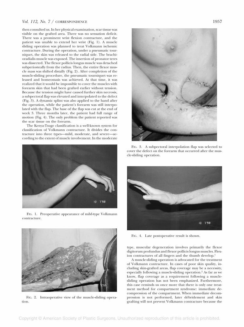
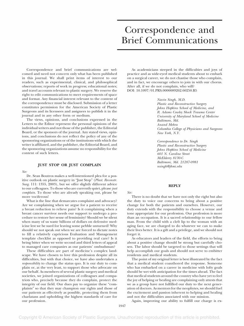
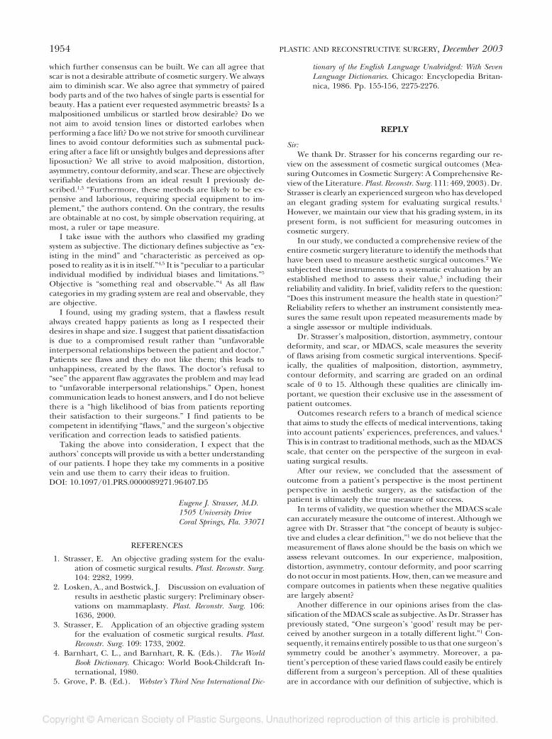
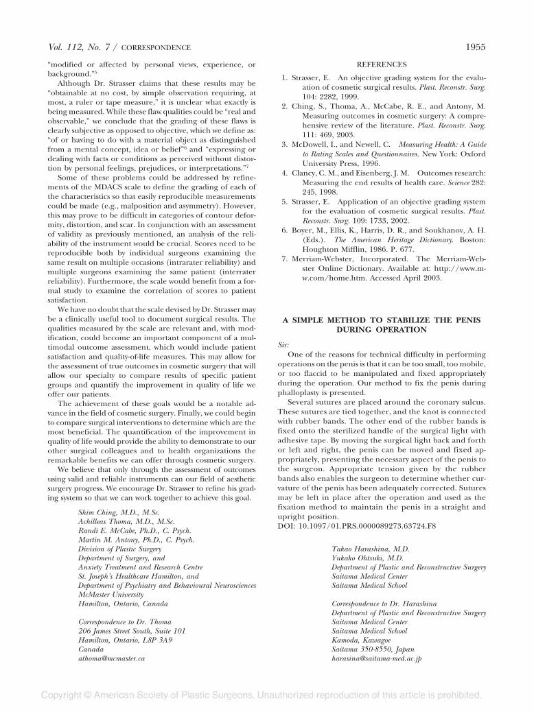
then consulted us. In her physical examination, scar tissue wasvisible on the grafted area. There was no sensation deficit.There was a prominent wrist flexion contracture, and thepatient was unable to extend her wrist (Fig. 1). A musclesliding operation was planned to treat Volkmann ischemiccontracture. During the operation, under a pneumatic tour-niquet, the skin was released to the radial side. The brachi-oradialis muscle was exposed. The insertion of pronator tereswas dissected. The flexor pollicis longus muscle was detachedsubperiosteally from the radius. Then, the entire flexor mus-cle mass was shifted distally (Fig. 2). After completion of themuscle-sliding procedure, the pneumatic tourniquet was re-leased and homeostasis was achieved. At that time, it wasrealized that it would be impossible to cover the muscles withforearm skin that had been grafted earlier without tension.Because the tension might have caused further skin necrosis,a subpectoral flap was elevated and interpolated to the defect(Fig. 3). A dynamic splint was also applied to the hand afterthe operation, while the patient’s forearm was still interpo-lated with the flap. The base of the flap was cut at the end ofweek 3. Three months later, the patient had full range ofmotion (Fig. 4). The only problem the patient reported wasthe scar tissue on the forearm.
The Kenya-Tsuge classification is a well-known system forclassification of Volkmann contracture. It divides the con-tracture into three types—mild, moderate, and severe—ac-cording to the extent of muscle involvement. In the moderate
type, muscular degeneration involves primarily the flexordigitorum profundus and flexor pollicis longus muscles. Flex-ion contractures of all fingers and the thumb develop.1
A muscle-sliding operation is advocated for the treatmentof Volkmann contracture. In cases of poor skin quality, in-cluding skin-grafted areas, flap coverage may be a necessity,especially following a muscle-sliding operation.2 As far as weknow, flap coverage as a requirement following a muscle-sliding operation has not been emphasized. Furthermore,this case reminds us once more that there is only one treat-ment method for compartment syndrome: immediate de-compression of the compartment. When immediate decom-pression is not performed, later débridement and skingrafting will not prevent Volkmann contracture because the
FIG. 1. Preoperative appearance of mild-type Volkmanncontracture.
FIG. 2. Intraoperative view of the muscle-sliding opera-tion.
FIG. 3. A subpectoral interpolation flap was selected tocover the defect on the forearm that occurred after the mus-cle-sliding operation.
FIG. 4. Late postoperative result is shown.
Vol. 112, No. 7 / CORRESPONDENCE 1957

skeletal muscles cannot tolerate ischemia after 8 hours.2
DOI: 10.1097/01.PRS.0000089275.72452.DE
Mehmet Oguz Yenidünya, M.D.Department of Plastic, Reconstructive, and Aesthetic SurgeryFatih University HospitalAnkara, TurkeyBülent Adil Tasbas, M.D.Department of Orthopedics and TraumatologyAnkara Numune Training and Research HospitalAnkara, Turkey
Correspondence to Dr. YenidünyaFatih University HospitalAlparslan Turkes Caddesi, No. 57Emek, Ankara, Turkey
REFERENCES
1. Tsuge, K. Management of established Volkmann’s con-tracture. In D. P. Green (Ed.), Operative Hand Surgery,Vol. 1, 2nd Ed. New York: Churchill Livingstone, 1988.
2. Rowland, S. A. Fasciotomy: The treatment of compart-ment syndrome. In D. P. Green, R. N. Hotchkiss, andW. C. Pederson (Eds.), Green’s Operative Hand Surgery,Vol. 1, 4th Ed. New York: Churchill Livingstone, 1999.
AVOIDING INJURY TO THE INFERIOR OBLIQUEMUSCLE DURING LOWER LID
BLEPHAROPLASTY
Sir:Mowlavi et al.1 recently published results of the dissections
that they performed with the intent of giving us useful ana-tomical bony landmarks to avoid injury to the inferior obliquemuscle during blepharoplasty surgery.
Although gross anatomical origins and insertions are im-portant, the functional surround is even more importantwhen dealing with a muscle as unusual as the inferior obliquemuscle. The inferior oblique muscle is approximately 36 mmfrom its origin in an area of the anterior orbital floor that islateral and inferior to the lacrimal fossa.2 From there, it travelslaterally and posteriorly in its initial third, penetrates thelower eyelid retractors in its middle third, and then envelopsthe globe in its final third to insert in a very short (1-mm)tendon on the lateral sclera. This insertion is broad and fromabout 18 mm to about 28 mm posterior to the limbus at thelevel of the lateral rectus muscle. Transection or injury in thisinitial first segment frequently does little permanent damageto the function of the inferior oblique muscle. This is becauseits middle segment, which traverses the capsular palpebralfascial area and the Lockwood ligament, will act as a second-ary origin if the first segment has been transected or injured.In the past, when overaction of the inferior oblique musclewas to be surgically eliminated, such transections of the initialfirst segment were carried out because of their relative easeof performance.3 The inferior oblique muscle regained someor all of its functions, and the surgical procedure did not havethe desired weakening effect. The second segment, namelythe 9 or 10 mm that underlie the inferior rectus muscle, thuscan act as a functional origin. The muscle emerges fromunderneath the inferior rectus and receives its innervation, abranch of the oculomotor nerve and its blood supply. Themost dangerous area in which to transect or injure the infe-
rior oblique muscle is in the 2 mm lateral to the inferior rectusmuscle, because injury in this area frequently will injure itsnerve supply and blood supply. Other methods of operatingon the inferior oblique muscle involve a myomectomy of themuscle itself with or without secondary insertion inferiorly oran inferior oblique recession. Today, of the procedures thatare used, only lateral recession or lateral myomectomy isperformed to reliably reduce the muscle’s function.
With this functional information, the problem of inferioroblique injury after lower lid blepharoplasty can be under-stood. In the transcutaneous method of lower lid blepharo-plasty, the nature of the incision exposes the lateral andcentral fat areas better than it does the medial fat area. Thisis because the usual incision is hinged medially and the in-cision continues outward along the margin of the lower lidand is then redirected downward at the lateral canthus, givinga larger skin flap laterally than medially. The tissue dissectionthen proceeds from anterior to posterior in the orbit, directlytraversing the inferior orbital septum to expose the fat pads.Because of the larger exposure laterally and the fact that theinferior oblique muscle is well above and deep to the septum,fat can be excised with relative impunity laterally. Medially,because of the poorer exposure, fat removal may occur, whichrisks inferior oblique injury in the first third of the muscle.However, permanent injury here is less common than in thetransconjunctival approaches because, as I mentioned above,even transection of the first third of the inferior obliquemuscle still allows the middle section of the inferior rectusmuscle to act as a secondary origin for the inferior oblique.
However, the transconjunctival approach has a differentset of complications. Because of its relatively superior ap-proach from the lower lid conjunctiva and its tunneling be-neath the inferior fornix, the conjunctiva is thus separatedfrom the more external capsular palpebral fascia and lid. Thesurgeon finds himself in no man’s land. He is too high toapproach the infero-orbital fat directly. He is behind theinferior orbital septum, and he must tunnel anteriorly toapproach the fat. He is already dangerously close to theinferior oblique muscle in both its central third and its lateralthird path. Especially at the juncture of the central thirdunderlying the inferior rectus and the Lockwood ligament,there is great danger of transecting the inferior oblique mus-cle and of injuring the nerve supply as mentioned previously.The best approach to avoid this is to incise the conjunctivaand immediately tunnel anterior and then inferior to theinferior tarsal muscle so that one is below the level of theinferior oblique in a similar position as in the transcutaneousapproach. In the medial area, it is possible to injure theinferior oblique muscle through this method, but again, in-jury in that portion is not as catastrophic as injury in themiddle and lateral thirds. I believe that this functional un-derstanding of the anatomy of the inferior oblique musclerather than a purely anatomic understanding will serve theprospective surgeon well through either the transcutaneousor transconjunctival approach.
The following are a few comments on the diagrams in thearticle. In Figure 1, there is a diagrammatic representation ofthe coronal view of the orbit (it is not an anteroposterior view,as labeled) that shows the medial and central fat pads tra-versed by an inferior oblique muscle and no lateral fat pad.Furthermore, it gives the impression that the anatomic originof the inferior oblique is a splayed-out group of muscle fibersthat bunch together and are only in intimate association withthe inferior rectus in the lateral one-quarter of its insertion.
The second diagram, Figure 2, introduces confusion be-cause it refers to some landmark as being in centimeters,
1958 PLASTIC AND RECONSTRUCTIVE SURGERY, December 2003
whereas most of the discussion in the text is given in milli-meters. Furthermore, the authors state that, “This incisionshould be placed 10 mm inferior to the corneal scleral limbuson the eyelid side.” This is extremely confusing because theprospective surgeon is thus urged to measure 10 mm from thelimbus on the globe and then to reflect it onto the lid surfaceto make the incision. Far better would have been to state thatthe incision should be made approximately 5 mm below theinferior border of the tarsal plate, namely that the measure-ment should be given on the actual structure to be incised.In the last paragraph on page 1321, I believe there is both amisprint and an important conceptual problem. The sen-tence states, “Specifically, the origin of the inferior obliquemuscle is found to be 9.4 mm lateral to the medial canthusand its insertion 21.7 mm lateral to the medial canthus.” Ibelieve that what the author meant was “21.7 mm medial to thelateral canthus.” Furthermore, trying to find the insertion ofthe inferior oblique muscle by using a bony identificationpoint, namely the lateral canthus, is extremely misleadingbecause the insertion of the inferior oblique is a broad ten-don, and it is on the globe and is not one point or a small area.DOI: 10.1097/01.PRS.0000089276.49030.F4
Joshua Frankel, M.D.Eye Care Specialists of Northeastern Pennsylvania601 Wyoming AvenueKingston, Pa. 18704
REFERENCES
1. Mowlavi, A., Neumeister, M. W., and Wilhelmi, B. J.Lower blepharoplasty using bony anatomical land-marks to identify and avoid injury to the inferioroblique muscle. Plast. Reconstr. Surg. 110: 1318, 2002.
2. Helveston, E. M. Atlas of Strabismus Surgery. St. Louis:Mosby, 1973. Pp. 16-18.
3. Helveston, E. M., et al. (Eds.). Symposium on Strabismus:Transactions of the New Orleans Academy of Ophthalmology.St. Louis: Mosby, 1978. Pp. 183-184.
REPLY
Sir:We thank Dr. Frankel for his interest in our article, “Lower
Blepharoplasty Using Bony Anatomical Landmarks to Iden-tify and Avoid Injury to the Inferior Oblique Muscle” (Plast.Reconstr. Surg. 110: 1318, 2002). Dr. Frankel’s clinical exper-tise in oculoplastics is made apparent by his in-depth discus-sion regarding variable degree of functional muscle deficitdepending on the injured segment of the inferior obliquemuscle (medial, central, or lateral segment). This clinicalinsight, as apparent in the initial body of his text, isappreciated.
Unfortunately, Dr. Frankel distracts the reader from thisinsight when he chooses to barrage the authors with hisinsignificant and inaccurate criticism. Insignificant com-ments include fussing over the numerical units used (10 mmversus 1 cm) or the “view label” of a figure (coronal versusanteroposterior). These criticisms over semantic differencesare really unnecessary. Of even more concern are his inac-curate criticisms of the body of the text. He criticizes theauthors for presenting anatomic guidelines that have beenpreviously published by other reputed clinicians. If Dr.Frankel feels obliged to comment on previously publishedmaterial, then he is urged to use the provided references to
check his claims. Indeed, Yousif et al.1 write, “the origin of theinferior oblique muscle is found 9.4 mm lateral to the medialcanthus and its insertion 21.7 mm lateral to the medial can-thus,” as stated in our article and not as incorrectly altered byDr. Frankel’s editorial.
We believe that our published anatomical landmarks uti-lizing the orbital rim, infraorbital foramen, and supraorbitalnotch provide dependable landmarks for localization of theinferior oblique muscle course. This knowledge may facilitateorbital fat resection and avoid injury to the inferior obliquemuscle during lower lid blepharoplasty.
Arian Mowlavi, M.D.Bradon J. Wilhelmi, M.D.The Plastic Surgery InstituteSouthern Illinois University School of Medicine747 N. RutledgeP.O. Box 19653Springfield, Ill. [email protected]
REFERENCE
1. Yousif, N. J., Sonderman, P., Dzwierzynski, W. W., andLarson, D. L. Anatomic considerations in transcon-junctival blepharoplasty. Plast. Reconstr. Surg. 96: 1271,1995.
BILATERAL ELASTOFIBROMA AS AN UNUSUALCAUSE OF SHOULDER PAIN
Sir:Elastofibroma is a rare benign entity that was first de-
scribed by Järvi and Saxen in 1961.1 The lesion is eight timesmore common in women than in men.2 It is located at theinferior angle of the scapula.3 Bilateral involvement is re-ported in 10 to 50 percent of cases.4–8 Usually it is a slow-growing tumor, but short mass-doubling times of 4 monthshave also been described.3 The majority of patients withelastofibroma are asymptomatic. Many complain of stiffshoulders or local pain, and some suffer from severe chestpain.7 We present a patient with bilateral elastofibroma thathad caused persisting shoulder pain for several months aftera fall down the stairs.
The patient referred to us was a 46-year-old Italian femalepizza baker who had fallen down the stairs months earlierwhile carrying a sack of flour from the cellar to prepare thepizza dough. She sustained a bilateral contusion of the shoul-der girdle. Conventional radiographs ruled out a fracture.Magnetic resonance imaging of the glenohumeral jointshowed a superficial rupture of the left supraspinatus tendonand subacromial impingement. At this time, the subscapularregion was not examined by magnetic resonance imaging.After several months of conservative treatment without painrelief, an orthopedic surgeon performed arthroscopy-assistedsubacromial decompression.
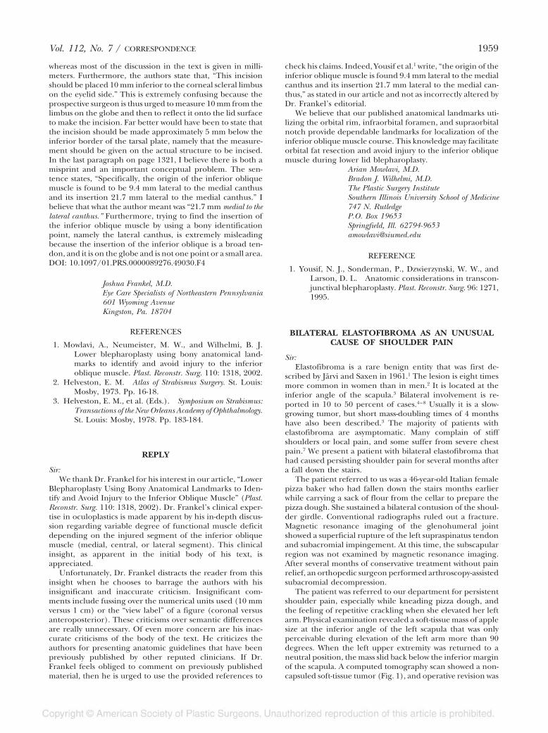
The patient was referred to our department for persistentshoulder pain, especially while kneading pizza dough, andthe feeling of repetitive crackling when she elevated her leftarm. Physical examination revealed a soft-tissue mass of applesize at the inferior angle of the left scapula that was onlyperceivable during elevation of the left arm more than 90degrees. When the left upper extremity was returned to aneutral position, the mass slid back below the inferior marginof the scapula. A computed tomography scan showed a non-capsuled soft-tissue tumor (Fig. 1), and operative revision was
Vol. 112, No. 7 / CORRESPONDENCE 1959

carried out due to suspicion of an old hematoma. A dense,fibroelastic, noncapsuled mass was found intraoperatively(Fig. 2). The tumor adhered both to the inferior angle of thescapula and to the periosteum of the ribs and was excised intoto. Histologically, the tumor consisted of intermingled eo-sinophilic collagen fibers and wormlike elastic fibers, fibro-blasts, and mature fat cells, thus confirming the diagnosis ofelastofibroma (Fig. 3).
The postoperative course was uneventful. The patient wasdischarged on the twelfth postoperative day. During the re-habilitation period, the patient complained of right-sidedshoulder pain with a palpable infrascapular mass (Fig. 4).Total resection again revealed an elastofibroma.
Since Järvi and Saxen1 first described elastofibroma in1961, a few more than 300 cases have been documented inthe literature. A Japanese group studied 170 cases. Fifty-fivecases occurred in the same family line. Some of the patientshad multiple elastofibroma, and one patient had sevenmasses simultaneously. Of the patients, 158 patients werefemale and only 12 were male. The average age was 70 years(range, 35 to 94 years).7 The youngest patient ever reportedwas a 6-year-old girl.8 One patient was observed over a periodof 67 years before the bilateral tumors were removed surgi-cally due to increasing pain.7
Elastofibroma is characterized histologically by the pres-ence of abnormal elastic fibers within a stroma of fibroadi-pose tissue.9 Uncommon locations have been reported, in-cluding the deltoid muscle, olecranon, thoracic wall,vertebral canal, ischial tuberosity, greater trochanter, foot,inguinal areas, orbita, stomach, greater omentum,2 and eventhe tricuspid valve.2,10 When the patients hold their armsslightly elevated forward and adducted, the scapula is dis-placed laterally and the lesion protrudes underneath thescapula.
FIG. 1. The computed tomography scan of the left scap-ular region shows a tumorous mass deep to the serratusanterior muscle. The tissue predominantly has muscle atten-uation interspersed with strands of fat.
FIG. 2. The gross appearance of the bisected tumor.
FIG. 3. Elastic staining of the tumor demonstrates nu-merous globular, wormlike elastic materials. Central cores arevisible within beadlike elastic fibers (elastin stain, �400).
FIG. 4. Clinical photograph of the dorsal aspect of thepatient 10 days after operation of the left-sided lesion. Thetumor protrudes from beneath the scapula during eleva-tion of the right arm. The right side was operated on 10days later.
1960 PLASTIC AND RECONSTRUCTIVE SURGERY, December 2003

Whether elastofibroma is a true neoplasm or a reactivefibrous lesion that produces not only collagen but also ab-normal elastic fibers is controversial and remains undeter-mined.8 The lesions have a characteristic location and a spe-cific magnetic resonance imaging appearance, which allowsaccurate prospective diagnosis. The magnetic resonance fea-tures are different from those of most other soft-tissue tu-mors, reflecting entrapped fat within a predominantly densefibrous mass. On T1- and T2-weighted images, it appears asa soft-tissue mass with signal intensity of skeletal muscle in-terlaced with streaks with the signal intensity of fat. On com-puted tomography scans, it is a poorly defined, inhomoge-neous mass with attenuation of skeletal muscle, againcontaining linear streaks of fat.11,12
The importance of recognizing the benign nature of thislesion to avoid an unnecessary operation is apparent.DOI: 10.1097/01.PRS.0000089278.52641.6A
Steffen Heck, M.D.Gausepohl Thomas, M.D.Konrad Mader, M.D.Andreas P. Wulke, M.D.Dietmar Pennig, M.D.Department of Trauma Surgery and Hand and Reconstructive
SurgerySt. Vinzenz HospitalCologne, GermanyWalter Mellin, M.D.Institute for Pathology, Cytology, and CytogeneticsCologne, Germany
Correspondence to Dr. HeckDepartment of Trauma Surgery and Hand and Reconstructive
SurgerySt. Vinzenz HospitalMerheimer St. 221-223D-50733 Cologne, [email protected]
REFERENCES
1. Järvi, O., and Saxen, E. Elastofibroma dorsi. Acta Pathol.Microbiol. Scand. Suppl. 144: 83, 1961.
2. Bennett, K. G., Organ, C. H., Cook, S., and Pitha, J.Bilateral elastofibroma dorsi: A case report. Surgery103: 605, 1988.
3. Turna, A., Yilmaz, M. A., Ürer, N., Bedirhan, M. A., andGürses, A. Bilateral elastofibroma dorsi. Ann. Thorac.Surg. 73: 630, 2002.
4. Buchholz, A., and Wellner, K. Das Elastofibrom dorsi.Zentralbl. Chir. 115: 1327, 1990.
5. Enzinger, F. M., and Weiss, S. W. Soft Tissue Tumors. St.Louis: Mosby, 1983. P. 33.
6. Hoffman, J. J., Klein, M. H., and McLenerney, V. K.Bilateral elastofibroma: A case report and review ofthe literature. Clin. Orthop. 325: 245, 1996.
7. Nagamine, N., Nohara, Y., and Ito, E. Elastofibroma inOkinawa: A clinopathologic study of 170 cases. Cancer50: 1794, 1982.
8. Marin, M. L., Perzin, K. H., and Markowitz, A. M. Elasto-fibroma dorsi: Benign chest wall tumor. J. Thorac. Car-diovasc. Surg. 98: 234, 1989.
9. Kobayashi, H., Hirokawa, M., Shimizu, M., et al. Scrape
cytology of elastofibroma: Report of a case with diag-nostic cytologic features. Acta Cytol. 43: 485, 1999.
10. Mohan, J. C., Goel, P. K., Gambhir, D. S., Khanna, S. K.,and Arora, R. Calcified mobile papillary fibroelas-toma of the tricuspid valve: A case report. IndianHeart J. 39: 237, 1987.
11. Kransdorf, M. J., Meis, J. M., and Montgomery, E.Elastofibroma: MR and CT appearance with radiolog-ic-pathologic correlation. A.J.R. Am. J. Roentgenol. 159:575, 1992.
12. Nakano, T., Tsutsumi, Z., Hada, T., and Higashino, K.Radiological manifestation of elastofibroma: A casereport and review of the literature. Br. J. Radiol. 64:1069, 1990.
TREATMENT OF MANDIBLE FRACTURES USINGBIOABSORBABLE PLATES
Sir:As surgeons at an experienced clinic in which approxi-
mately 200 patients per year are operated on, we wish tocommend and criticize Drs. Kim and Kim’s article entitled“Treatment of Mandibular Fractures Using BioabsorbablePlates” (Plast. Reconstr. Surg. 110: 25, 2002).
The classic tenet for closed reduction and intermaxillaryfixation is to maintain the fixation 3 to 4 weeks for the sub-condylar and 4 to 6 weeks for the other areas of the mandible.We have shortened the fixation period up to 3 to 4 weeks forall areas of the mandible since 1987.1 During the rigid fixationprocedures, intermaxillary fixation is used in restoring andsecuring normal occlusion and can be removed by the end ofthe operation or within the first postoperative week. In thisregard, it seems unlikely to use both bioabsorbable plates andintermaxillary fixation for 2 to 3 weeks in eight cases, asreported in the study. Keeping the intermaxillary fixation foran additional 1 or 2 weeks would have sufficed for the bonehealing without the extra disadvantages of the bioabsorbablescrews and plates.2,3 The authors also reported that all thepatients operated on were given a liquid or soft diet for 1month after the operation, regardless of the intermaxillaryfixation period or osteosynthesis material. This is anothercontradictory statement on behalf of the advantage of rigidfixation, which leads to early jaw movements and a normaldiet.4
There is a statement that rigid metallic fixation deprivesbone of normal stress and prevents the rapid formation ofprimary callus. Is this an acceptable view per the literature onfacial bone healing, which has an intramembranous devel-opmental origin, and on any rigid fixation modality, as in thestudy?5–7 Application of bioabsorbable materials on humanbeings could make the study more valuable after basic sciencestudies, including stress testing, were performed, especiallyon the corpus and angulus regions. With respect to obtainingsatisfactory results, indications should be clarified accordingto mandibular regions.
Complications such as infection are often associated withthe method of treatment. The rate of complications would belowered to 3 percent after experience was gained with theplating system.8–10 With an 8.1 percent infection rate, bioab-sorbable plates seem to cause a higher complication rate. Thesubcondylar area, angle, and cuspid region and the mentalforamen are the naturally weak areas of the mandible. The
Vol. 112, No. 7 / CORRESPONDENCE 1961
holes drilled for the screws also involve the risk of refracture,which is why heat necrosis of bone should be avoided at alltimes. From this point of view, the osteolytic changes seenaround the self-reinforced absorbable polylactide screws in 27percent of cases may cause a high rate of additional weakareas in the mandible.
In conclusion, because of the above-mentioned reasons,we do not have confidence in the ability to anatomicallyreduce and stabilize the fractured fragments with the self-reinforced poly-L-lactide multilayer plates.DOI: 10.1097/01.PRS.0000089332.70602.13
Turgut Ortak, M.D.R. Erkin Ünlü, M.D.Asu Deniz Yılmaz, M.D.Ömer Sensöz, M.D.Department of Plastic and Reconstructive SurgeryAnkara Numune Training and Research HospitalAnkara, Turkey
Correspondence to Dr. OrtakResat Nuri Sok.82/12 Doruk Apt., Y. AyrancıAnkara, [email protected]
REFERENCES
1. Sensöz, Ö., Erdogan, B., Kanmaz, B., and Koçer, U.Analysis of the mandibular fractures and shorteningthe fixation period in treatment. Turk. J. Res. Med. Sci.5: 83, 1987.
2. Schmidt, B. L., Kearns, G., Gordon, N., and Kaban,L. B. A. Financial analysis of maxillomandibular fix-ation versus rigid internal fixation for treatment ofmandibular fractures. J. Oral Maxillofac. Surg. 58: 1206,discussion 1210, 2000.
3. Haers, P. E., and Sailer, H. F. Biodegradable self-rein-forced poly-L/DL-lactide plates and screws in bimax-illary orthognathic surgery: Short term skeletal stabil-ity and material related failures. J. Craniomaxillofac.Surg. 26: 363, 1998.
4. Jaques, B., Richter, M., and Arza, A. Treatment of man-dibular fractures with rigid osteosynthesis: Using theAO system. J. Oral Maxillofac. Surg. 55: 1402, discussion1406, 1997.
5. Haug, R. H., and Schwimmer, A. Fibrous union of themandible: A review of 27 patients. J. Oral Maxillofac.Surg. 52: 832, 1994.
6. Hoffman, W. Y., Barton, R. M., Price, M., and Mathes, S. J.Rigid internal fixation vs. traditional techniques forthe treatment of mandible fractures. J. Trauma 30:1032, 1990.
7. Bagby, G. W., and Janes, J. M. The effect of compressionon the rate of fracture healing using a special plate.Am. J. Surg. 95: 761, 1958.
8. Iızuka, T., Lindqvist, C., Hallikainen, D., and Paukku, P.Infection after rigid internal fixation of mandibularfractures: A clinical and radiologic study. J. Oral Max-illofac. Surg. 49: 585, 1991.
9. Aksoy, E., Ünlü, R. E., and Sensöz, Ö. A retrospectivestudy on epidemiology and treatment of maxillofa-cial fractures. J. Craniofac. Surg. 13: 772, 2002.
10. Ezsias, A., and Sugar, A. W. Pathological fractures of themandible: A diagnostic and treatment dilemma. Br. J.Oral Maxillofac. Surg. 32: 303, 1994.
REANIMATION OPTIONS IN PATIENTS WITHHEMIFACIAL MICROSOMIA AND MARGINAL
MANDIBULAR NERVE PALSY
Sir:Hemifacial microsomia is one of the most commonly pre-
senting craniofacial anomalies, with an incidence of approx-imately one in 5000 live births.1,2 Although in 10 to 30 percentof cases it manifests bilaterally, when almost universally asym-metric, more commonly it affects the right side of the face inboys.3,4 The principal defects seen, as classically described byGorlin et al.,5 are hypoplasia of the mandible, macrostomia,and microtia. The condition has a varied phenotype and canbe associated with a variety of other congenital anomalies;some advocate solitary microtia as the simplest manifestationof the condition.6 Soft-tissue deficiency and hypoplasia of themuscles of mastication are intimately associated with the de-gree of mandibular deformity,7 and cranial nerve involve-ment is seen in approximately 20 to 45 percent of cases.8,9 Thedegree of external ear deformity, rather than other bony orsoft-tissue defects, is a predictor of seventh nerve involve-ment.8,10 Etiology of the condition remains unclear,2,11–14 butfacial palsy associated with hemifacial microsomia is thoughtto be secondary to abnormal middle ear seventh nerve path-ways and fallopian canal atresia.8,15 Most often, marginal man-dibular paralysis is seen, although paralysis of the completefacial musculature may be present.10,16
Surgical options for the paralyzed marginal mandibularbranch in the normal face have been reported using a varietyof muscle transpositions to the lower lip, including the or-bicularis oris, platysma, and anterior belly of digastric mus-cles, all with pleasing results.17–19 Free-muscle transfer hasbeen used to recreate the lower lip depressor function inisolated deficits and as a double muscle transfer in cases ofcomplete unilateral palsy.19,20 Reinnervation using hypoglos-sal nerve transfer to the lower lip musculature has had lesssatisfactory results.18 Previous studies on marginal mandibu-lar branch reanimation, assessing all the above techniques,have concluded that local muscle transfers offer the bestresults in the otherwise normal face.18,19
In our unit, we have encountered three patients with hemi-facial microsomia and an isolated paralysis of the marginalmandibular branch of the facial nerve. As stated above, it is,under normal circumstances, preferable to use a local muscletransposition flap for reanimation; our flap of choice is theanterior belly of digastric muscle. In each of these patients,there was no platysma muscle or anterior belly of digastricmuscle; the procedure was therefore abandoned in two ofthese patients. In the third case, a mylohyoid flap, previouslydescribed following cadaveric anatomical dissection,21 wasused for reanimation. These cases illustrate the difficultiesfaced when attempting to restore lower lip function in thosewith hemifacial microsomia. Direct neurotization using cross-facial nerve grafts22,23 is generally inappropriate due to thelate presentation of these cases. Beyond 1 year, the successachieved using this technique or reinnervation with the hy-poglossal nerve is less than optimal23 because of atrophy of themusculature.24 Alongside the hypoplastic masticatory mus-cles, it would appear that other muscles can also be hypo-plastic or absent, perhaps by virtue of shared innervation withthe masticatory muscles (in the case of the anterior belly of
1962 PLASTIC AND RECONSTRUCTIVE SURGERY, December 2003

digastric muscle) or as a result of involvement of the cervicalbranch of the facial nerve (with platysma), thus ruling outthese common muscle transfers. Previous authors have com-mented on the value of microneurovascular muscle transferin cases of complex facial hypoplastic syndromes, and a recentseries reported good results using this technique applied tomarginal mandibular branch paralysis in hemifacial micro-somia.19,25 Despite concerns about potentially hypoplastic ves-sels, the senior author has found, as others have also report-ed,25 that the vessels are of adequate caliber but locatedaberrantly.
We would like to draw readers’ attention to this clinicalproblem and advocate caution when planning reanimationprocedures in patients with hemifacial microsomia. In suchcases, it is our view that free-tissue transfer is the procedureof choice because the anterior belly of digastric muscle can-not be relied upon.DOI: 10.1097/01.PRS.0000089280.57545.96
Anthony MacQuillan, M.R.C.S.Martin Vesely, F.R.C.S.Douglas Harrison, F.R.C.S.Adriaan Grobbelaar, M.Med.(Plast.), F.C.S.(S.A.), F.R.C.S.(Plast.)The RAFT Institute of Plastic SurgeryMount Vernon HospitalNorthwood, Middlesex, United Kingdom
Correspondence to Dr. MacQuillanThe RAFT Institute of Plastic SurgeryMount Vernon HospitalRickmansworth RoadNorthwood, Middlesex HA6 2RNUnited [email protected]
REFERENCES
1. Grabb, W. C. The first and second branchial arch syn-drome. Plast. Reconstr. Surg. 36: 485, 1965.
2. Poswillow, D. The pathogenesis of first and secondbranchial arch syndrome. Plast. Reconstr. Surg. 35: 302,1973.
3. Ross, R. B. Lateral facial dysplasia (first and secondbranchial arch syndrome, hemifacial microsomia).Birth Defects Orig. Artic. Ser. 11: 51, 1975.
4. Cousley, R., and Calvert, M. Current concepts in theunderstanding and management of hemifacial micro-somia. Br. J. Plast. Surg. 50: 536, 1997.
5. Gorlin, R. J., Jue, K. L., Jacobsen, V., and Goldschmidt,E. Oculoauriculovertebral dysplasia. J. Pediatr. 63:991, 1963.
6. Bennun, R. D., Mulliken, J. B., Kaban, L. B., and Murray,J. E. Microtia: A microform of hemifacial microso-mia. Plast. Reconstr. Surg. 76: 859, 1985.
7. Kane, A. A., Lo, L. J., Christensen, G. E., Vannier, M. W.,and Marsh, J. L. Relationship between bone andmuscles of mastication in hemifacial microsomia.Plast. Reconstr. Surg. 99: 990, 1997.
8. Bassila, M. K., and Goldberg, R. The association of facialpalsy and/or sensorineural hearing loss in patientswith hemifacial microsomia. Cleft Palate J. 26: 287,1989.
9. Vento, A., LaBrie, R., and Mulliken, J. The OMENSclassification of hemifacial microsomia. Cleft PalateCraniofac. J. 28: 68, 1991.
10. Murray, J. E., Kaban, L. B., and Mulliken, J. B. Analysisand treatment of hemifacial microsomia. Plast. Recon-str. Surg. 74: 186, 1984.
11. Cousley, R. R. J., and Wilson, D. J. Hemifacial micro-somia: Developmental consequence of perturbationof the auriculofacial cartilage model. Am. J. Med. Genet.42: 461, 1992.
12. Akyuz, S., Mumcu, G., Atasu, M., et al. Hemifacial mi-crosomia: Oral, clinical, genetic and dermatoglyphicfindings. J. Clin. Pediatr. Dent. 23: 63, 1998.
13. de Ravel, T. J., Legius, E., Brems, H., Van Hoestenberghe,R., Gillis, P. H., and Fryns, J. P. Hemifacial micro-somia in two patients further supporting chromo-somal mosaicism as a causative factor. Clin. Dysmorphol.10: 263, 2001.
14. Jacobsson, C., and Granstrom, G. Clinical appearanceof spontaneous and induced first and secondbranchial arch syndromes. Scand. J. Plast. Reconstr.Surg. Hand Surg. 31: 125, 1997.
15. Converse, J. M., Coccaro, P. J., Becker, M., and Wood-Smith, D. On hemifacial microsomia: The first andsecond branchial arch syndrome. Plast. Reconstr. Surg.51: 268, 1973.
16. Mulliken, J. B., and Kaban, J. B. Analysis and treatmentof hemifacial microsomia in childhood. Clin. Plast.Surg. 14: 91, 1987.
17. Pluckett, C. L., Neale, H. W., and Pickrell, K. L. Dy-namic correction of unilateral paralysis of the lowerlip. Plast. Reconstr. Surg. 55: 397, 1975.
18. Terzis, J. K., and Kalantarian, B. Microsurgical strate-gies in 74 patients for restoration of dynamic depres-sor muscle mechanism: A neglected target in facialreanimation. Plast. Reconstr. Surg. 105: 1917, discussion1932, 2000.
19. Tulley, P., Webb, A., Chana, J. S., et al. Paralysis of themarginal mandibular branch of the facial nerve:Treatment options. Br. J. Plast. Surg. 53: 378, 2000.
20. Ueda, K., Harii, K., and Yamada, A. Free vascularizeddouble muscle transplantation for the treatment offacial paralysis. Plast. Reconstr. Surg. 95: 1288, 1995.
21. Aszmann, O., Ebmer, J., and Dellon, A. The anatomicbasis for the innervated mylohyoid/digastric flap infacial reanimation. Plast. Reconstr. Surg. 102: 369, 1998.
22. Inigo, F., Ysunza, A., Ortiz-Monasterio, F., and Drucker-Colin, R. Early postnatal treatment of congenital fa-cial palsy in patients with hemifacial microsomia. Int.J. Pediatr. Otorhinolaryngol. 23: 57, 1993.
23. Ysunza, A., Inigo, F., Rojo, P., Drucker-Colin, R., andOrtiz-Monasterio, F. O. Congenital facial palsy andcrossed facial nerve grafts: Age and outcome. Int. J. Pe-diatr. Otorhinolaryngol. 36: 125, 1996.
24. Ferreira, M. C. Cross facial nerve grafting. Clin. Plast.Surg. 11: 211, 1984.
25. Takushima, A., Harii, K., Asato, H., and Yamada, A.Neurovascular free-muscle transfer to treat facial pa-ralysis associated with hemifacial microsomia. Plast.Reconstr. Surg. 109: 1219, 2002.
AUTOSTATIC SYNDROME IN ASSOCIATIONWITH FACIAL NERVE TRANSPLANTATION
Sir:Figure 15 in Dr. Wei Wang et al.’s article on facial rean-
imation (Neurovascular Musculus Obliquus Internus Abdo-minis Flap Free Transfer for Facial Reanimation in a Single
Vol. 112, No. 7 / CORRESPONDENCE 1963

Stage. Plast. Reconstr. Surg. 110: 1430, 2002) confirms theoccurrence of autostatic syndrome.1
The syndrome almost invariably occurs after cross-facialnerve grafting, because axons destined for the orbicularis orisalso innervate the muscle flap. This makes the angle of themouth move outward when the lips are pursed, instead ofmedially.
However original, Dr. Wang’s technique cannot preventthis. We must continue seeking other solutions in the rean-imation of facial paralysis.DOI: 10.1097/01.PRS.0000089333.22025.DC
J.-P. A. Nicolai, M.D.Department of SurgeryGroningen University HospitalP.O. Box 30 0019700 RB Groningen, The Netherlands
REFERENCE
1. Nicolai, J.-P. A., and Robinson, P.H. Autostatic syn-drome after cross-facial nerve transplantation. Ann.Plast. Surg. 19: 337, 1987.
MEDIAL PEDICLED ORBICULARIS OCULI FLAP
Sir:We read with great interest the article by Jelks et al., en-
titled “Medial Canthal Reconstruction Using a MediallyBased Upper Eyelid Myocutaneous Flap” (Plast. Reconstr. Surg.110: 1636, 2002). Indeed, reconstruction of the media can-thal area is a frequently encountered problem after tumorablations, and we have been using a flap from the upper eyelidfor this purpose. Our technique was published in 2001 withthe title “Medial Pedicled Orbicularis Oculi Flap for MedialCanthal Resurfacing.”1 Furthermore, we have transferred thisflap not only to the medial canthal region but also to thenearby defects. These extended applications of the flap werepublished in December of 2002,2 coinciding with the above-mentioned article by Jelks et al. We would like to commenton some points since we have used the same flap for severalyears.
1. We agree with the authors that the eyelid skin is a betteralternative in the reconstruction of the medial canthalarea, the lateral nasal area, and the bony nasal dorsumthan the classic methods, such as the frontal or the gla-bellar flaps, which are thicker.
2. For the defects confined to the median canthal region, weprefer to transfer the flap from the contralateral eyelid.1
This has two advantages. First, as the flap extends to theopposite site, the deformity caused by the pedicle at thepivot point is less pronounced. Second, we are skepticalabout the ipsilateral flap vascularity, which can be harmedat the medial canthal level. Although the flap has a richblood supply, as described by the authors, we still thinkthat the untouched contralateral site is a better option asthe donor site.
3. The medial canthal region is rich in vascular arcades. Thisarea receives its blood supply from both the externalcarotid system (i.e., the infratrochlear and angular arter-ies) and the internal carotid system (i.e., the supraorbital,supratrochlear, dorsal nasal, and medial palpebral arter-ies).3 The medial palpebral artery is the major contribu-tor to the medial pedicled orbicularis oculi flap. However,due to the rich vascular connections, the flap can be
nourished from the external carotid system (angular ar-tery) as well. Therefore, there is no need to pay anyattention to the isolation of the medial palpebral perfo-rators while dissecting the flap, contrary to what the au-thors suggest. The dissection of the flap ends at themedial canthal region, ensuring the preservation of theangular system, which is located medial to the medialcanthus.
4. In our article, we have described the creation of a subcu-taneous tunnel that allows us to transfer this flap as aninterpolation flap. Thus, the skin between the flap andthe defect is untouched, extra scar is avoided, and betteraesthetic results are achieved (Fig. 1).
5. The flap size is directly proportionate to the skin excess ofthe upper lid. Therefore, careful patient selection is im-portant. Even the smaller defects may be difficult to coverin a patient with minimum skin excess of the upper lid.
FIG. 1. The skin island is marked according to the defectarea. (Above) The dotted area is deepithelialized. (Below) Theflap is transferred through the subcutaneous tunnel.
1964 PLASTIC AND RECONSTRUCTIVE SURGERY, December 2003

We believe this flap will be used frequently in thefuture.DOI: 10.1097/01.PRS.0000089282.29835.4C
Erdem Tezel, M.D.Ahmet Sönmez, M.D.Ayhan Numanoglu, M.D.Department of Plastic and Reconstructive SurgeryMarmara University School of MedicineIstanbul, Turkey
Correspondence to Dr. TezelMarmara Üniversitesi HastanesiPlastik ve Reconstrüktif Cerrahi A.D.Tophaneliolu Cad. No. 13/1581190 Altunizade, Istanbul, [email protected]
REFERENCES
1. Tezel, E., Sönmez, A., and Numanoglu, A. Medial pedi-cled orbicularis oculi flap for medial canthal resur-facing. Ann. Plast. Surg. 47: 213, 2001.
2. Tezel, E., Sönmez, A., and Numanoglu, A. Medial pedi-cled orbicularis oculi flap. Ann. Plast. Surg. 49: 599,2002.
3. Clemente, C. D. (Ed.). Gray’s Anatomy of the Human Body,30th Ed. Philadelphia: Lea & Febiger, 1985. Pp. 685-688.
EFFECT OF ULTRASOUND-ASSISTEDLIPOSUCTION ON SKIN PERFUSION
Sir:We read with great interest the article by Gupta et al.,
entitled “Effect of Liposuction on Skin Perfusion” (Plast. Re-constr. Surg. 110: 1748, 2002). We congratulate the authors ontheir well-delineated study and for their efforts to elucidatethe effects of ultrasound-assisted liposuction on tissues, com-paring it to conventional wet liposuction. The authors usedthe laser Doppler flowmeter to measure skin perfusion, andthe results showed a decrease in skin perfusion in the con-ventional wet liposuction group soon after infiltration with awetting solution (containing vasoconstrictor), which per-sisted during the first week of follow-up. In the ultrasound-assisted group, after the same decrease related to the wettingsolution, the skin perfusion increased (above the preopera-tive level) from the first hour until the first week postoper-atively. Such results are undoubtedly interesting.
However, we cannot agree with some inferences that theauthors elaborate on in the Discussion. They argue that themechanism of action of ultrasound-assisted liposuction ismore selective to adipocytes and infer that this represents lessdamage to adjacent tissues, which would explain the en-hanced skin perfusion in this group. Having such reasoningas a basis, one could consider this study as clinical evidenceof the selectivity of ultrasound-assisted technique in vivo. Itsounds logical, and the authors’ Discussion indeed drives usto such a conclusion.
Yet when we analyze this logical sequence, there are pointson which we do not agree. First, most of the evidence of theso-called selectivity to adipocytes in ultrasound-assisted lipo-suction is found in in vitro studies. Second, less damage toadjacent tissues has not been demonstrated in vivo, and clin-ical evidence (such as the reports of surgical complications,
namely skin necrosis on areas subjected to ultrasound-assistedliposuction) does not favor such a hypothesis. Third, en-hanced skin perfusion can be secondary to an inflammatoryresponse. Local vasodilation and increased blood flow arepresent in the early phases of inflammation.1 Thus, the en-hanced skin perfusion could indicate tissue damage, not theopposite.
To clarify the subject, further studies would be necessary,for example, with histopathologic or biochemical analysis oftissues subjected in vivo to different techniques of liposuc-tion. One such study was concluded by Cárdenas-Camarenaet al. and entitled “Laboratory and Histopathologic Compar-ative Study of Internal Ultrasound-Assisted Lipoplasty andTumescent Lipoplasty,” published in this Journal in Septem-ber of 2002 (Plast. Reconstr. Surg. 110: 1158, 2002). Theirconclusions are indeed somewhat different from those ofGupta et al. In this study, the indicators of tissue damageshowed statistically significant higher levels in the group sub-mitted to ultrasound-assisted liposuction.
There is certainly much more to be elucidated on thissubject. Even though we do not agree with the authors’ in-ferences, they must be congratulated on a well-conductedstudy, which certainly adds information to the theme andincites further discussion.DOI: 10.1097/01.PRS.0000089283.41138.3F
Victor Vieira Orsi, M.D.Antônio Carlos Pinto Oliveira, M.D.Porto Alegre, Brazil
Correspondence to Dr. OrsiRua Vieira de Castro 275/401CEP 90.040-320Porto Alegre, [email protected]
REFERENCE
1. Cotran, R. S., Kumar, V., and Robbins, S. L. (Eds.). In-flammation and repair. In Robbins Pathologic Basis ofDisease, 4th Ed. Philadelphia: Saunders, 1989. Pp. 39-86.
SAFER LIPOSUCTION FOR HOSPITAL STAFF
Sir:We report a simple and cost-effective technique designed
to reduce the risk to the operating surgeon and operatingroom staff during liposuction procedures.
Since Teimourian and Fisher1 first described liposuc-tion in 1981, a considerable volume of research has beenpublished on the risks to the patient and potential com-plications.2,3 Relatively few authors have addressed the po-tential risks to the surgeon and associated operating roomstaff. The advent of wet, superwet, and tumescent tech-niques has led to the infusion of increasing amounts offluid before suction lipectomy.4,5 Ultrasound is often usedto facilitate tissue removal. Many surgeons now prefer theuse of syringe-generated suction to machine-driven sys-tems. This generates large volumes of fluid-fat mix thatmust be transferred from syringes to a suitable containerand subsequently disposed of.
The surgical field contains potentially infectious organ-isms that may be released into the operating room environ-
Vol. 112, No. 7 / CORRESPONDENCE 1965

ment as a liposanguinous aerosol. Although no cases of clin-ical infection have been reported in the literature, Higashi etal.6 have demonstrated that hepatitis C virus is found in tissuefluid produced by ultrasonic dissection. Cukier et al.7 havedemonstrated that viable Pseudomonas aeruginosa can be aero-solized by suction lipoplasty and remains present in the en-vironment for a number of hours. This represents a signifi-cant risk to operating room personnel. Almost any pathogenis capable of breaching host defenses when aerosolized andinhaled deep into lung parenchyma. Case reports in theliterature document aerosol-based infection with many or-ganisms, including Influenza virus, Lassa virus, Gram-nega-tive bacilli, Mycobacterium intracellulare, Mycobacterium scrofula-
ceum, Brucella, Chlamydia, and Rickettsia. These potential riskshave been reduced by the use of filters on machine-drivensuction systems. No such safeguards exist for syringe-basedsystems.
The transfer of fat-fluid mix from syringe to measuring jugor other such container carries such a risk of aerosolizingpotentially infectious agents. We describe a simple and cost-effective measure to reduce the risk to the operating roomstaff and to prevent spillage of fat-fluid mix in subsequenttransfer.
We use a measuring jug to receive the discarded fat-fluidmix. Before use, the container is prepared by stretching alarge surgical glove over the top of the jug (Fig. 1). A singlestab incision is made in the tensioned membrane. Whenevacuating the contents of a syringe-based liposuction system,the tip of the cannula is passed through this hiatus into the“closed” jug (Fig. 2). Fat-fluid mix can then be rapidly ex-pelled with minimal risk to staff. In the event that the jug isknocked over, this system also prevents significant spillage.This technique does not interfere with surgical technique orinhibit the ability to measure the volume aspirated. It does notsignificantly reduce the rate at which the procedure can beperformed.
We present a simple and cost-effective technique to reducethe risk of aerosolization of pathogenic agents during syringeliposuction. This reduces the potential risk to operating roomstaff without adversely affecting the operative time.DOI: 10.1097/01.PRS.0000089284.65246.52
James A. Haeney, M.R.C.S.Alastair J. Platt, F.R.C.S.(Plast.)Department of Plastic and Reconstructive SurgeryCastle Hill HospitalEast Yorkshire, United Kingdom
Correspondence to Dr. Haeney7 Melton GroveWest Bridgford, Nottingham NG2 7NXUnited [email protected]
REFERENCES
1. Teimourian, B., and Fisher, J. B. Suction curettage toremove excess fat for body contouring. Plast. Reconstr.Surg. 68: 50, 1981.
2. Teimourian, B., and Rogers, W. B. A national surveyof complications associated with suction lipectomy:A comparative study. Plast. Reconstr. Surg. 84: 628,1989.
3. Teimourian, B., and Adham, M. N. A national survey ofcomplications associated with suction lipectomy: Whatwe did then and what we do now. Plast. Reconstr. Surg.105: 1881, 2000.
4. Fodor, P. B. Wetting solutions in aspirative lipoplasty: Aplea for safety in liposuction (Editorial). Aesthetic Plast.Surg. 19: 379, 1995.
5. Klein, J. A. Tumescent technique for local anesthesiaimproves safety in large-volume liposuction. Plast. Re-constr. Surg. 92: 1085, 1993.
6. Higashi, H., Matsumata, T., and Hayashi, J. Detection ofhepatitis C virus RNA in the ultrasonic dissector irri-gating solution used in liver surgery. Br. J. Surg. 81:1346, 1994.
7. Cukier, J., Price, M. F., and Gentry, L. O. Suction
FIG. 1. A large surgical glove is stretched over the top ofthe jug to seal it.
FIG. 2. To evacuate the contents of the syringe into the“closed” jug, the tip of the cannula is passed through anincision made in the glove.
1966 PLASTIC AND RECONSTRUCTIVE SURGERY, December 2003

lipoplasty: Biohazardous aerosols and exhaustmist—the clouded issue. Plast. Reconstr. Surg. 83:494, 1989.
GESTATIONAL GIGANTISM AFTER REDUCTIONMAMMAPLASTY
Sir:Drs. Ah�an, Solinc, and Megli� offer a dramatic case of
“Gestational Gigantism after Reduction Mammaplasty.”1
They present a case report of a patient who developed bilat-eral generalized enlargement during pregnancy, 4 years aftera reduction mammaplasty. They declare that, to their knowl-edge, “no such case has been previously reported.”
Dr. Shulman of Israel and I reported in the British Journalof Plastic Surgery in 1971 two cases of massive breast hyper-trophy following reduction mammaplasty.2 One was a virginal16-year-old female patient who developed spontaneous pro-gressive asymmetric enlargement of her breasts, and theother was a 22-year-old mother of one who developed unre-mitting massive breast hypertrophy during pregnancy.
Both patients had previously undergone breast reduction forunexplained massive hypertrophy. Both women required ampu-tation, with one ultimately accepting prosthetic reconstruction.
While I call this to the attention of the authors for the sakeof accuracy in reporting, I must take this occasion to appealto all authors to do a thorough literature review before de-claring originality.DOI: 10.1097/01.PRS.0000089285.40725.64
Arthur G. Ship, M.D.1049 Fifth AvenueNew York, N.Y. 10028
REFERENCES
1. Ah�an, U., Solinc, M., and Megli�, L. Gestational gi-gantomastia after reduction mammaplasty: Complica-tion or coincidence? Plast. Reconstr. Surg. 111: 956,2003.
2. Ship, A. G., and Shulman, J. Virginal and gravid mam-mary gigantism: Recurrence after reduction mamma-plasty. Br. J. Plast. Surg. 24: 396, 1971.
REPLY
Sir:Dr. Ship kindly commented on our article, “Gestational
Gigantism after Reduction Mammaplasty” (Plast. Reconstr.Surg. 111: 956, 2003), and appealed to all authors to do athorough literature review before declaring originality.
In reviewing the literature to prepare our case report, we reada very interesting article entitled “Virginal and Gravid MammaryGigantism: Recurrence after Reduction Mammaplasty.”1 Theauthors, Dr. Ship and Dr. Shulman, described virginal gigantismin a 16-year-old white female patient with an 8-month history ofspontaneously occurring, progressive, asymmetric enlargementof both breasts and the case of a 22-year-old white woman,mother of one, with a 6-month history of unremitting breastenlargement with marked discomfort and tense and tender skinwho responded only briefly to a course of testosterone andNorluten (Parke-Davis, Morris Plains, N.J.).
In both cases, physical examination revealed typical signsand symptoms of mammary gigantism. In both patients, a
reduction mammaplasty was followed by early massive recur-rence. In the first case, 6 months postoperatively, the breasthad enlarged to equal the preoperative size; and in the sec-ond case, approximately 3 months after the operation, pro-gressive breast enlargement became apparent, with alarmingenlargement during pregnancy 9 months later. Completesubcutaneous amputation was performed in both cases 6 and10 months after the original reduction mammaplasty.
In our article, we presented a case report of a 23-year-oldhealthy white woman who came to our department becauseof marked ptosis and medium-sized hypertrophy of herbreasts. There were no signs or symptoms of sudden unre-mitting massive hypertrophy, tense and tender skin, ormarked discomfort. No facts of medical significance in eitherher obstetrical/gynecological or family history have beenfound, and she was on no medication.
The ptosis and mammary hypertrophy were noted, and astandard bilateral breast reduction was performed using theMcKissock technique. The postoperative period was unre-markable. However, 4 years later, she presented 10 weekspregnant to the emergency room unit of the obstetrics/gy-necology department with a history of sudden and painfulenlargement of her breasts beginning days earlier (typicalsigns and symptoms of mammary gigantism/gravid macro-mastia). In the next 48 hours, she came to our outpatientclinic with the question, “Is this a late complication of re-duction mammaplasty or just a coincidence?” After discus-sions with her gynecologist and endocrinologist, urgent cur-ative surgical treatment (total mastectomy) was performed.The following explanation was given: Juvenile (adolescent orvirginal) hypertrophy or adolescent macromastia is an un-common event wherein a young girl, after a normal puberty,has continued breast growth and enlargement even into ma-ture years. The breasts do not decrease in size, and a reduc-tion mammaplasty is required. Even more rare is gravid mac-romastia, which develops rapidly after the onset of pregnancy.As for adolescent macromastia, the exact cause is unknown,but once established, it occurs with each subsequent preg-nancy and all breast tissue should be removed.
This case report is different from that of Dr. Ship and Dr.Shulman in that the initial presentation was for mammary hyper-trophy requiring a standard reduction mammaplasty. The onset ofmammary gigantism was 4 years subsequent to this operation, dur-ingtheinitial trimesterof thepatient’s firstpregnancy. If thepatienthad come to the first visit with sudden, progressive, massive hyper-trophy and tense and tender skin (as was the case in both of Dr.Ship’s patients), we would have suggested to her the appropriatetreatment, totalmastectomyandimmediatereconstruction insteadof a reduction mammaplasty.
Uroš Ahcan, M.D., Ph.D.Zaloška 7Ljubljana, Slovenia
REFERENCE
1. Ship, A. G., and Shulman, J. Virginal and gravid mam-mary gigantism: Recurrence after reduction mamma-plasty. Br. J. Plast. Surg. 24: 396, 1971.
ULTRASOUND ASPIRATION FORGYNECOMASTIA
Sir:In their article “Classification and Management of Gy-
necomastia: Defining the Role of Ultrasound-Assisted Aspi-
Vol. 112, No. 7 / CORRESPONDENCE 1967

ration” (Plast. Reconstr. Surg. 111: 909, 2003), Rohrich et al.present a series of 61 patients seen over a period of 14 years.“We offer . . . the role of ultrasound-assisted liposuction as asuperior method for safe and effective removal of male breasttissue, based on our experience with 61 patients evaluatedand treated for gynecomastia from 1987 to 2000.” However,only the last 4 years, or about 17 cases, assuming even dis-tribution through the years, are applicable to the main thrustof the article—the role of ultrasound-assisted liposuction.“Ultrasound-assisted liposuction . . . has recently emerged asthe preferred technique for management of gynecomastia.”In support of their contention, the authors use another oftheir own publications as the only applicable supporting ref-erence. “Several studies have confirmed the selective emul-sification of fat leaving higher density structures . . . relativelyundamaged using ultrasound-assisted liposuction. At higherenergy settings, ultrasound-assisted liposuction is effective inremoval of the dense, fibrotic parenchymal tissue,” they state,without supportive evidence and in clear contradiction oftheir previous statement.
“More recently, ultrasound-assisted liposuction hasemerged as the preferred surgical technique for most casesof gynecomastia, providing minimal scarring and efficientremoval of both glandular and fibrotic breast tissues,” theauthors state, without supporting evidence. They presentCourtiss’s study claiming a high complication rate with ex-cisional techniques. These complications include overresec-tion, underresection, hematoma, seroma, and unattractivescarring, which are complications that ultrasound-assistedliposuction allegedly avoids. But are complications not ad-verse events that occur despite a properly executed proce-dure? Certainly overresection and underresection are tech-nical errors, not complications. All the authors’ cases showunderresection of the gynecomastia. Seromas and hemato-mas respond to meticulous technique and proper drainage inmost cases but may occur despite proper technique. And theunattractive scarring associated with skin reduction is thesame with excisional technique and ultrasound-assisted lipo-suction, as demonstrated by the authors’ Figure 6, where eventhey accept skin excision 6 to 9 months after the originalprocedure.
The authors also present a classification system for gy-necomastia. For all grades of gynecomastia, ultrasound-as-sisted liposuction is their treatment of choice. I suggest thata more useful classification takes into consideration threefactors: fat versus glandular tissue and skin elasticity. Fat canbe excised or suctioned (whatever technique), glandular tis-sue requires excision (as the authors’ pictures show), and skinelasticity is either adequate, borderline, or poor. If skin elas-ticity is adequate or borderline, the inferior circumareolarincision works well. In borderline cases, skin resection mayoccasionally be necessary after about 6 months. With poorelasticity (as in their Fig. 6), immediate skin resection willprevent the need for a certain second procedure. I see nocompelling evidence presented by the authors that ultra-sound-assisted liposuction removes glandular tissue or that itoffers any benefit over traditional liposuction for fatcontouring.
The preoperative and postoperative photographs provideconclusive evidence that ultrasound-assisted liposuction isineffective for glandular tissue removal. The preoperativephotographs all show gynecomastia. The postoperative pho-tographs all show gynecomastia, though to a lesser degreedue to fat removal. The postoperative photograph in Figure3 shows more gynecomastia than the preoperative photo-graph in Figure 2. The inframammary scars visible on the
photographs are more apparent than the usual infra-areolarincisions with the open technique. The patient shown inFigure 6 needed extended skin resection and still does. Sincegynecomastia is an all-or-none process, like pregnancy, anddiffers only by the degree apparent, the authors’ photographspresent conclusive evidence that ultrasound-assisted liposuc-tion alone provides unsatisfactory results for glandulargynecomastia.
In their questionable exuberance over the use of ultra-sound-assisted liposuction, the authors forgot that it is not thetechnique but the result that counts. Their results fall shortof desirable and obtainable.DOI: 10.1097/01.PRS.0000089287.94099.B7
Eugene J. Strasser, M.D.1505 University DriveCoral Springs, Fla. 33071
REPLY
Sir:We appreciate Dr. Strasser’s interest in our article, “Clas-
sification and Management of Gynecomastia: Defining theRole of Ultrasound-Assisted Liposuction (Plast. Reconstr. Surg.111: 909, 2003). His comments are duly noted, but his con-clusions are somewhat misguided and perplexing. Our articlefocused primarily on ultrasound-assisted liposuction as an-other modality available for plastic surgeons to use in themanagement of gynecomastia. The excellent discussion byDr. Mladick (Plast. Reconstr. Surg. 111: 924, 2003) serves as animportant statement of variations in aesthetic goals and tech-niques. There are numerous appropriate methods to obtaincorrection of gynecomastia. To this consideration, we chal-lenged our previous notion that mere correction was ade-quate. We presented a technique based on our experiencethat has evolved to be our preferred method.
As with any board-certified plastic surgeon who gains ex-perience in cosmetic surgery procedures, improvements orchanges in particular techniques evolve simultaneously withan appreciation for a more mature aesthetic ideal. Our initialgoals for elimination of all male breast tissue were supplantedby an ideal for optimal contouring and restoration of a normalmale breast contour. We are attentive to our gynecomastiapatients who have told us that they do not like diminution orloss of their masculine silhouette. They still want some centralbreast and areola fullness and desire no indentation or con-tour depression in their central breast area. Our satisfactionrate has been exceedingly high using ultrasound-assistedliposuction.
Dr. Strasser is quick to condemn the authors’ long-termexperience with ultrasound-assisted liposuction, both the mo-dality of treatment and the results. First, he suggests that theauthors state “without supportive evidence and in clear con-tradiction of their previous statement” regarding the selectiveemulsification of fat at lower ultrasound-assisted liposuctionsettings and improved efficacy in removal of dense glandulartissue at higher settings. This claim largely ignores the pub-lished works by Zocchi,1 Gingrass,2–4 Maxwell,3 Kenkel,4,5 andRohrich.4,5 The only “contradiction” is Dr. Strasser’s dismissaland the overwhelming evidence to support the contrary.
Second, he claims that presented photographs “presentconclusive evidence that ultrasound-assisted liposuctionalone provides unsatisfactory results for glandular gynecom-astia.” On what is he basing his assertions, a displeasing,
1968 PLASTIC AND RECONSTRUCTIVE SURGERY, December 2003

overresected chest contour? Indeed, our emphasis was a res-toration of a normal male chest contour, not its elimination. Ifstudies show that up to 65 percent of all males have some formof gynecomastia, are we as plastic surgeons operating on 65percent of male chests, or even half that number? More likelyis that we are seeing severe cases and that “normal” constitutesa wide range of appearances, most with some glandular and fatexcess. Dr. Strasser’s claim that “gynecomastia is an all-or-none process, like pregnancy, and differing only by the de-gree apparent” demonstrates a clear confusion of his ownterminology or a lack of understanding of the disease. Theimportance of aesthetic balance and the appreciation of pro-portional improvement have been welcome advancements inthe field of rhinoplasty. Should we not embrace these prin-ciples when approaching the treatment of gynecomastia? Byshifting our surgical goal to be optimal contouring and shapeand not merely the “all-or-none” correction as suggested byDr. Strasser, we leave behind the antiquated notion that nor-mal males have no breast tissue.
Dr. Strasser stated, “It’s not the technique but the resultthat counts.” Taking this one step further, the result is notdependent upon the technique that is applied. The result isdirectly proportional to the talents, skill, and artistry of thesurgeon, no matter what technique is used. Many plasticsurgeons can obtain excellent results using many types ofmodalities, including traditional liposuction and/or exci-sion. Our technique using ultrasound-assisted liposuctionprovides the surgeon with another excellent option to use formanagement of this common problem in males.
We are aware of Dr. Strasser’s enthusiasm for “objectivegrading systems” for the evaluation of cosmetic surgery.6 Heassigns a differential point system based upon his own sub-jective hierarchy of good and bad features of a postoperativeresult. We have attempted to precisely demonstrate our ex-perience and the evolution of our technique. However, thissubjective standard imposed by Dr. Strasser prevents his con-currence. Evidently, from his evaluation through a perplex-ing point system, the “results fall short of desirable and ob-tainable.” We acknowledge him for his personal standards,but we invite him to share his experience of gynecomastiasurgery with the plastic surgery community. We strongly urgehim to show his technique and long-term results, with orwithout liposuction. By publishing a scientific, clinical articlein a peer-reviewed journal, other plastic surgeons could com-pare his results to those in the literature.
We eagerly look forward to Dr. Strasser’s publication in theJournal. In life, as in plastic surgery, it is always better topractice what you preach.
Rod J. Rohrich, M.D.Department of Plastic SurgeryUniversity of Texas Southwestern Medical Center5323 Harry Hines Boulevard, E7.212Dallas, Texas 75390–[email protected]
REFERENCES
1. Zocchi, M. L. Ultrasonic-assisted lipoplasty: Technicalrefinements and clinical evaluations. Clin. Plast. Surg.23: 575, 1996.
2. Gingrass, M. K., and Shermak, M. A. The treatment ofgynecomastia with ultrasound-assisted lipoplasty. Per-spect. Plast. Surg. 12: 101, 1999.
3. Maxwell, G. P., and Gingrass, M. K. Ultrasound-assistedlipoplasty: A clinical study of 250 consecutive patients.Plast. Reconstr. Surg. 101: 189, 1998.
4. Kenkel, J. M., Gingrass, M. K., and Rohrich, R. J. Ul-trasound-assisted lipoplasty: Basic science and clinicalresearch. Clin. Plast. Surg. 26: 221, 1999.
5. Kenkel, J. M., Robinson, J. B., Beran, S. J., et al. Thetissue effects of ultrasound-assisted lipoplasty. Plast.Reconstr. Surg. 102: 213, 1998.
6. Strasser, E. J. Application of an objective grading systemfor the evaluation of cosmetic surgical results. Plast.Reconstr. Surg. 109: 1733, 2002.
EFFECTS OF REDUCTION MAMMAPLASTY ONPULMONARY FUNCTION AND SYMPTOMS OF
MACROMASTIA
Sir:I read with interest the recent article by Sood et al.1 and
acknowledge their effort in producing a good article on acontentious subject. Indeed, there is a lack of evidence basein the selection criteria for breast reduction surgery that hasled to the rationing of health in this aspect of breast surgeryacross the world. The difficulties with third-party insurance inthis respect are also well documented. It is, therefore, a timelycontribution and adds to the body of evidence another in-stance of quantitative benefit of reduction mammaplasty.
A significant finding in this study is the fact that patientsbenefit from this procedure regardless of body mass index,which unfortunately is still being used to select patients forthis procedure. This indeed has also been confirmed by otherstudies.2
The number of patients in this study is a significant im-provement on previous work done in this area, and the useof a well-validated respiratory function questionnaire givesfurther validity to their work.
From a critical viewpoint, however, this work has a demeritthat also affects almost all research done in breast reduction,namely, the absence of level 1 or 2a evidence (lack of anyrandomized work). This is illustrated by the absence of anadequate control group (i.e., those who have macromastiabut do not have an early operation). An addition of a suitablecontrol group will eliminate the confounding factor posed bylearning effect, because it could be argued that the instruc-tions for respiratory function test can be learned, thus ex-plaining improvement in some of the voluntary parameters.
Also, although efforts were made to standardize the test forall patients, no mention was made of the time of day whenthese tests were carried out. It is well known that there is adiurnal variation in respiratory function of up to 10 percentin healthy subjects.3
DOI: 10.1097/01.PRS.0000089289.06874.AB
O. C. Iwuagwu, M.B., B.S., F.R.C.S.Academic Surgical UnitUniversity of HullCastle Hill HospitalCastle Road, CottinghamEast Riding HU16 5JQ, United Kingdom
REFERENCES
1. Sood, R., Mount, D. L., Coleman, J. J., III, et al. Effectsof reduction mammaplasty on pulmonary functionand symptoms of macromastia. Plast. Reconstr. Surg.111: 688, 2003.
2. Zubowski, R., Zins, J. E., Foray-Kaplon, A., et al. Rela-tionship of obesity and specimen weight to complica-
Vol. 112, No. 7 / CORRESPONDENCE 1969

tions in reduction mammaplasty. Plast. Reconstr. Surg.106: 998, 2000.
3. Brewis, R. A. L., Gibson, G. J., and Geddes, D. M. (Eds.).Respiratory Medicine. London: Baillière Tindall, 1990.
REPLY
Sir:The article by Sood et al.1 was interesting to read. Dr.
Iwuagwu raises three reasonable objections. The first oneconcerns the lack of a control group. This is both a legitimatecomment and one that is difficult for surgeons to address dueto practice and ethical issues. It is one thing to randomlyassign patients to receive one of two pills, each of which couldbenefit them. It is another thing to randomly operate onpatients or not. The proportion of articles that I read thathave random assignment to surgery for patients is quitesmall—animals, yes, but patients, no. You certainly cannotconduct a double-blind study where neither the surgeon northe patient knows what happened. Here, I think one simplyhas to do the best with what one has.
The second comment concerns the possible learning ef-fect. That is always a possibility. However, I note that theforced expiratory volume in 1 second did not increase, andit would seem that it would be subject to a learning effect ifa sizable one exists.
Unfortunately, I am not familiar with the tests that weresignificant (maximal voluntary ventilation, inspiratory capac-ity, and peak expiratory flow rate). In the past, I have workedon studies of the effect of smog and cigarette smoking in LosAngeles. The physicians I worked with there mainly examinedforced expiratory volume in 1 second, forced vital capacity,and forced expiratory flow 25/75 and preferred forced ex-piratory volume in 1 second for most of their work. They (Dr.Tashkin and Dr. Gong) were knowledgeable about what mea-surements should be used depending on the purpose of thestudy.
We did not correct for time of day in those studies, but theywere conducted on large groups of people. Again, if this is aproblem, it did not show up in forced vital capacity, forcedexpiratory volume in 1 second, or forced expiratory flow. Aplus in this study, as far as I can see, is that they did takemultiple measurements both before and after the operationso they could assess change in several pulmonary measures.
I apologize for not being knowledgeable about the teststhat were significant. The authors do explain on pages 692and 693 of the article what these three tests measure. If youwish to go further with this, I would suggest consulting witha pulmonary physician concerning the problem. I think themajor issue is whether or not it makes physical sense thatthree particular measurements changed following the breastreduction operation.
Virginia Clark, Ph.D.852 Sporseen RoadSequim, Wash. [email protected]
REFERENCE
1. Sood, R., Mount, D. L., Coleman, J. J., III, et al. Effectsof reduction mammaplasty on pulmonary functionand symptoms of macromastia. Plast. Reconstr. Surg.111: 688, 2003.
SOFT-TISSUE SARCOMA ARISING FROM ATISSUE NECROSIS CAUSED BY AN
INTRAMUSCULAR INJECTION OF DICLOFENAC
Sir:In our city, we have been observing occasional cases of
tissue necrosis associated with intramuscular administrationof diclofenac. In the literature, also, there are some reportsshowing the same fact.1–4 The manufacturers discourage itsuse in the arm but advocate it in the gluteal region, where themuscle bulk is thicker.
Here we report on a unique case conducted in our de-partment. In December of 2000, a 45-year-old dark-skinnedwoman received a diclofenac injection in the right glutealregion to treat cold symptoms. Shortly after that, a chronicinflammation developed at the injection site, and then anodule appeared in the same area. It is important to say that,according to the patient, there was nothing abnormal in thisregion before the injection. Initially, the primary care sur-geon thought it could be an abscess, so some unsuccessful
FIG. 1. Preoperative view of the ulcerated mass in theright gluteal region.
FIG. 2. Perioperative view of the patient in the ventraldecubitus position. There is a tie-over dressing over the ul-ceration. The dotted line corresponds to the mass on palpation.The outer line depicts the 3-cm margin around tumor (line ofresection).
1970 PLASTIC AND RECONSTRUCTIVE SURGERY, December 2003
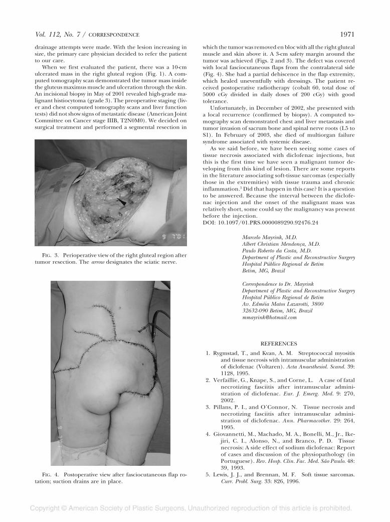
drainage attempts were made. With the lesion increasing insize, the primary care physician decided to refer the patientto our care.
When we first evaluated the patient, there was a 10-cmulcerated mass in the right gluteal region (Fig. 1). A com-puted tomography scan demonstrated the tumor mass insidethe gluteus maximus muscle and ulceration through the skin.An incisional biopsy in May of 2001 revealed high-grade ma-lignant histiocytoma (grade 3). The preoperative staging (liv-er and chest computed tomography scans and liver functiontests) did not show signs of metastatic disease (American JointCommittee on Cancer stage IIIB, T2N0M0). We decided onsurgical treatment and performed a segmental resection in
which the tumor was removed en bloc with all the right glutealmuscle and skin above it. A 3-cm safety margin around thetumor was achieved (Figs. 2 and 3). The defect was coveredwith local fasciocutaneous flaps from the contralateral side(Fig. 4). She had a partial dehiscence in the flap extremity,which healed uneventfully with dressings. The patient re-ceived postoperative radiotherapy (cobalt 60, total dose of5000 cGy divided in daily doses of 200 cGy) with goodtolerance.
Unfortunately, in December of 2002, she presented witha local recurrence (confirmed by biopsy). A computed to-mography scan demonstrated chest and liver metastasis andtumor invasion of sacrum bone and spinal nerve roots (L5 toS1). In February of 2003, she died of multiorgan failuresyndrome associated with systemic disease.
As we said before, we have been seeing some cases oftissue necrosis associated with diclofenac injections, butthis is the first time we have seen a malignant tumor de-veloping from this kind of lesion. There are some reportsin the literature associating soft-tissue sarcomas (especiallythose in the extremities) with tissue trauma and chronicinflammation.5 Did that happen in this case? It is a questionto be answered. Because the interval between the diclofe-nac injection and the onset of the malignant mass wasrelatively short, some could say the malignancy was presentbefore the injection.DOI: 10.1097/01.PRS.0000089290.92476.24
Marcelo Mayrink, M.D.Albert Christian Mendonça, M.D.Paulo Roberto da Costa, M.D.Department of Plastic and Reconstructive SurgeryHospital Público Regional de BetimBetim, MG, Brazil
Correspondence to Dr. MayrinkDepartment of Plastic and Reconstructive SurgeryHospital Público Regional de BetimAv. Edméia Matos Lazarotti, 380032632-090 Betim, MG, [email protected]
REFERENCES
1. Rygmstad, T., and Kvan, A. M. Streptococcal myositisand tissue necrosis with intramuscular administrationof diclofenac (Voltaren). Acta Anaesthesiol. Scand. 39:1128, 1995.
2. Verfaillie, G., Knape, S., and Corne, L. A case of fatalnecrotizing fasciitis after intramuscular admini-stration of diclofenac. Eur. J. Emerg. Med. 9: 270,2002.
3. Pillans, P. I., and O’Connor, N. Tissue necrosis andnecrotizing fasciitis after intramuscular admini-stration of diclofenac. Ann. Pharmacother. 29: 264,1995.
4. Giovannetti, M., Machado, M. A., Bonelli, M., Jr., Ike-jiri, C. I., Alonso, N., and Branco, P. D. Tissuenecrosis: A side effect of sodium diclofenac: Reportof cases and discussion of the physiopathology (inPortuguese). Rev. Hosp. Clin. Fac. Med. São Paulo. 48:39, 1993.
5. Lewis, J. J., and Brennan, M. F. Soft tissue sarcomas.Curr. Probl. Surg. 33: 826, 1996.
FIG. 3. Perioperative view of the right gluteal region aftertumor resection. The arrow designates the sciatic nerve.
FIG. 4. Postoperative view after fasciocutaneous flap ro-tation; suction drains are in place.
Vol. 112, No. 7 / CORRESPONDENCE 1971

A RARE CASE OF DERMOID CYST BEHINDTHE EAR
Sir:A 39-year-old Turkish man presented with a postauricular
cyst that had been present since childhood (Fig. 1). Thecomputed tomography scan revealed an encapsulated tumorwith no intracranial extension.
A surgical excision of the mass was carried out with thepatient under general anesthesia. The cyst contained mucoidfluid and keratin. The histologic examination confirmed thatthe lesion was a dermoid cyst. A good cosmetic result wasobtained with direct skin closure.
The most accepted theory about dermoid cysts was pos-tulated by New and Erich1 and states there is a congenital“inclusion” of germ layers in the deeper tissues along theline of embryonic fusion. The disorganized growth anddifferentiation of these sequestered cells give rise to der-moid cysts. Dermoid cysts can be divided into three distincthistologic types, depending on their histological appear-ance: epidermoid, dermoid, and teratoid. Epidermoidcysts contain laminated keratinous material and have nosebaceous glandular structures in their lining. Dermoidcysts are subcutaneous tumors lined by stratified squamousepithelium and contain cutaneous appendages such as hairfollicles, sebaceous glands, and sweat glands in variableamounts. Teratomas are true neoplasms arising from toti-potent cells and contain representative cells from all threeembryonic germ layers.
The differential diagnosis of a postauricular cyst includesan epidermal inclusion cyst, a trichilemmal cyst, lipoma, anda hemangioma. Trichilemmal or sebaceous cysts clinicallyresemble epidermoid cysts. The diagnosis is confirmed his-tologically with amorphous keratinous material contained inthe cystic cavity. Lipomas are common benign soft-tissue ad-ipose tumors and may have the same appearance as dermoid
cysts. Hemangiomas are usually present at birth and are be-nign tumors of vascular endothelium and can spontaneouslyinvolute.
Dermoid cysts are usually present at birth and are pre-dominantly found in males. They are asymptomatic, slowlyexpanding, unilocular, cystic masses. Most occur in the ova-ries,2 with less than 7 percent occurring in the head and neckregion.3
When they occur in the head and neck region, their mostcommon location is the fronto-orbital upper outer quadrantof the orbit. Other locations are the midline of the nose orneck, the sublingual region, and the sternal, perineal, scrotal,and sacral areas.4
Dermoid cysts of the auricle are extremely rare. There areonly three reported cases of postauricular dermoid cysts,5–7
although five cases of auricular dermoid cysts have beenreported in the Japanese literature, and they all occurred onthe superior pole of the ear.2
DOI: 10.1097/01.PRS.0000089334.04867.D8
B. A. De SouzaC. DeyN. CarverDepartment of Plastic and Reconstructive SurgeryThe Royal London HospitalSt. Bart’s and The Royal London NHS TrustWhitechapel, London, United Kingdom
Correspondence to Dr. De Souza88 The ChineGrange ParkLondon N21 2EH, United Kingdom
REFERENCES
1. New, G. B., and Erich, J. B. Dermoid cysts of the headand neck. Surg. Gynecol. Obstet. 65: 48, 1937.
2. Ikeda, M., Muto, J., and Omachi, S. Dermoid cyst of theauricle: Report of two cases. Auris Nasus Larynx 16:193, 1990.
3. Reddy, V. S. Radhakrishna, K., and Rao, P. L. Lingualdermoid. J. Pediatr. Surg. 26: 1389, 1991.
4. Batsakis, J. G. Teratomas of the head and neck. In Tu-mors of the Head and Neck: Clinical and Pathological Con-siderations, 2nd Ed. Baltimore: Williams & Wilkins,1979. Pp. 226-232.
5. Samper, A. Dermoid cyst on the auriculotemporal area.Plast. Reconstr. Surg. 106: 947, 2000.
6. Bauer, D. J., Diwan, R., Honig, B. K., and Yokel, B. Largeasymptomatic mass on the ear: Dermoid cyst of theauricle. Arch. Dermatol. 130: 913, 1994.
7. Meagher, P. J., and Morrison, W. A. An unusual pre-sentation of bilateral prominent ears. Br. J. Plast. Surg.54: 366, 2001.
A NEW DEVICE: A PORTABLE AND EFFECTIVEIRRIGATION SYSTEM FOR PRESSURE ULCERS
Sir:In the current aging society, pressure ulcers are an important
issue in medicine and nursing. Daily prevention and daily treat-ment are essential to cure a pressure ulcer. Recognizing theprevalence of pressure ulcers and the need for preventive tools,the Agency for Healthcare Research and Quality assembled aFIG. 1. Lump in postauricular region of the right ear.
1972 PLASTIC AND RECONSTRUCTIVE SURGERY, December 2003

national interdisciplinary panel to develop a set of best practiceguidelines for pressure ulcer prevention.1,2 The theory for ournew device is described in Figure 1.
The prototype consisted of a funnel, a drainpipe, a por-table sprinkler, a hose, and a bucket. In this report, we in-troduce the new type, which has a hand-made plastic ulcercup composed of two half-balls and a drainpipe (Fig. 2).
The big half-ball effectively prevents drainage splash. Thesmall half-ball prevents reflux of drainage by allowing thedrainage to pool. The portable sprinkler was bought at a lowprice in a country store. It is better if the portable sprinklerhas a mechanism that continues to give pressure to fluid bymanual pumping and gripping for continuous spurt.
In the ulcer cup, the drainage point is at the low positionof the cup at profile. The hose connects the cup and thebucket. All of the contaminated water is drained into thebucket through the hose (Fig. 3).
We used this system in a hospital on 10 patients withpressure ulcers at the sacral portion. The grade of pressureulcer according to the National Pressure Ulcer Advisory Panelwas stage III or IV. Five months later, one case at stage IV
became stage II, and two cases at stage IV became stage III.The other cases remained at the same stage (Fig. 4).
Pressure ulcer prevention is a key element in nursing.For patients, development of a pressure ulcer is a majorsource of morbidity, which is associated with an increasedrisk of death. Once pressure ulcers have occurred, cleans-ing and débridement are the usual prerequisites to furthertreatment or ultimate healing, because cleaning the woundof inflammatory stimuli such as devitalized tissue, chemi-cals, and bacteria involves débridement and cleansing pro-cedures. Generally, it is difficult to treat the pressure ulcersbecause they are deeply infected. Débridement can be
FIG. 1. Diagram of the new irrigation system, showing thecourse of the water and the placement of the equipment.
FIG. 2. The ulcer cup is a handmade plastic cup com-posed of two half-balls and a drainpipe.
FIG. 3. The irrigation system (hose and bucket are notshown).
FIG. 4. Clinical application. After the hose is released,contaminated water is drained through the pipe. As shown inthe photograph, the new system can be used with one hand.
Vol. 112, No. 7 / CORRESPONDENCE 1973

accomplished surgically, mechanically (wet-to-dry gauze),or chemically (autolysis or topical enzymes). Several recentstudies document the benefits of each of these techniques.Cleansing is the process of using a large volume of fluid toremove loosely adherent foreign material. Numerouswound-cleansing solutions and novel irrigation systemshave been introduced in recent years.3 Systems and facil-ities must use additional resources (staff time and treat-ments) if a patient develops a pressure ulcer. If possible,cleaning the wound by irrigation using a large volume ofwater with appropriate pressure is not only best but alsosaves additional resources. Our method addresses thisproblem. In the literature, there are many reports on ir-rigation systems, including closed continuous irrigation,4 adental water irrigation device,5 and a portable shower.Irrigation with a shower is one of the best approaches interms of pressure, but a large volume of water is needed toclean the wound, and naturally, drainage for contaminatedwater is needed. No reports about irrigation systems con-sider drainage. Because there are many patients who can-not be easily moved to another place (e.g., treatment roomor bathroom) in a hospital or a nursing home, it is veryimportant to use a portable irrigation system for everydaytreatment and continuation of care.
We have discovered a new device with which to treat pres-sure ulcers. The advantages of this system are low cost, port-ability, reduced time for treatment, ease of use, and controlof contaminated drainage.DOI: 10.1097/01.PRS.0000089334.04867.D8
Takafumi Chin, M.D.Hiko Hyakusoku, M.D.Department of Plastic and Reconstructive SurgeryNippon Medical School HospitalTokyo, Japan
Correspondence to Dr. ChinDepartment of Plastic and Reconstructive SurgeryNippon Medical School Hospital1-1-5 Sendagi, Bunkyo-kuTokyo 113-8603, [email protected]
REFERENCES
1. Bergstorm, N., Bennet, M., and Carlson, C. ClinicalPractice Guideline Number 3: Pressure Ulcers in Adults:Prediction and Prevention (AHCPR PublicationNo. 95-0653). Rockville, Md.: U.S. Departmentof Health and Human Services, Public Health Ser-vice, Agency for Health Care Policy and Research,1994.
2. Debra, S. Adherence to pressure ulcer preventionguidelines. J. Am. Geriatr. Soc. 51: 56, 2003.
3. Rodeheaver, G. T. Pressure ulcer débridement andcleansing: A review of current literature Ostomy WoundManage. 45: 80, 1999.
4. Takahiro, I. Local continuous irrigation for pressureulcers of spinal cord injured patients. Jpn. J. P. U. 2:264, 2000.
5. Nourse, A. M., and Myers, W. Dental water irrigationdevice used for cleaning decubitus ulcers. Phys. Ther.58: 1219, 1978.
A DOUBLE POCKET FOR TISSUE EXPANDERIMPLANTATION IN BREAST RECONSTRUCTION
Sir:We present a new technique we have used recently in
postmastectomy breast reconstruction by a tissue expanderimplantation. Tissue expansion utilizes the principle of grad-ual stretching of the chest wall tissue and aims to accommo-date a larger implant after a complete mastectomy with largeskin loss.1 In this kind of reconstruction, the tissue expanderis always placed in a submusculofascial position.2
To create the usual pocket for the implant, the lateralmastectomy scar is excised, and the dissection is carried downto the pectoralis major muscle; the lateral fibers of the muscleare separated, and the dissection is extended beneath themusculofascial layer.3,4 With this technique, we obtain a singlesubmusculofascial pocket ready to accommodate the expand-able device.
In August of 2002, a 58-year-old woman presented to ourdepartment asking for a breast reconstruction. In 1998, thepatient had undergone a complete right mastectomy withtotal excision of axillary nodes for an intraductal breast can-cer; moreover, in 2001, the patient had undergone an aor-tocoronary bypass to treat an acute heart attack.
At the first visit, our patient presented not only with theusual axillary and transpectoral scars (Fig. 1, A) but also witha long longitudinal scar in the sternal region due to herprevious heart surgery (Fig. 1, B). Because of the lack of softtissues, we decided to place a tissue expander implant in theusual submuscular pocket.
In the operating room, after excision of the lateral mas-tectomy scar, the submusculofascial layer was found and el-evated to create the submuscular pocket for the tissue ex-pander (Fig. 2, B). During the dissection, however, probablyas a consequence of the combination of previous mastectomyand heart surgery, severe fibrosis and fibrotic adherenceswere found not only under the pectoralis major but alsobetween the muscle and the overlying soft tissue. Thinkingabout the subsequent difficulty in device expansion due tothe poor elasticity of this particular patient’s chest wall, wedecided on a new procedure; we created a second pocket(Fig. 2, A), placed over the pectoralis major, using a blunt anddifficult dissection of the muscle fascia from the overlyingfibrotic subdermal layer.
This second pocket was not meant to contain any devicebut was created to form a mobile layer in the chest wall to help
FIG. 1. Preoperative view. A, Axillary and transpectoralscars from previous mastectomy. B, Long longitudinal scar inthe sternal region due to previous heart surgery.
1974 PLASTIC AND RECONSTRUCTIVE SURGERY, December 2003

soft tissues stretch during the expander filling. In this way, wecould easily introduce a 500-cc Becker mammary expander,with the fill valve placed laterally in a subcutaneous pocket.The patient was discharged 5 days after the operation, and westarted filling the mammary expander about 3 weeks later.
Currently, the expander device is inflated by 290 cc ofsaline solution, and we have had no trouble during the weeklyinstillation.
In conclusion, we think that this double-pocket techniquefor expander implantation can be very helpful for the sur-geon, particularly in cases like ours, with a minimal increase
in surgical time.DOI: 10.1097/01.PRS.0000089334.04867.D8
Luigi Valdatta, M.D.Christian Mortarino, M.D.Alessandro Thione, M.D.Mara Buoro, M.D.Carlo FidanzaPlastic Surgery UnitUniversity of InsubriaOspedale di CircoloFondazione MacchiVarese, Italy
Correspondence to Dr. ValdattaChirurgia PlasticaOspedale di CircoloViale Borri 5721100 Varese, [email protected]
REFERENCES
1. Argenta, L. C. Reconstruction of the breast by tissueexpansion. Clin. Plast. Surg. 11: 257, 1984.
2. Becker, H. Breast reconstruction using an inflatablebreast implant with detachable reservoir. Plast. Recon-str. Surg. 73: 678, 1984.
3. Bostwick, J. Breast reconstruction following mastec-tomy. Contemp. Surg. 27: 19, 1985.
4. Dick, G. O., and Brown, S. A. Breast reconstructionusing modified tissue expansion. Plast. Reconstr. Surg.76: 613, 1986.
FIG. 2. Intraoperative view. A, Supramuscular pocket toincrease chest tissue’s mobility. B, Submusculofascial pocketto accommodate the expandable device (green abdominalgauze fills the pocket).
Vol. 112, No. 7 / CORRESPONDENCE 1975