Control of the replicative life span of human fibroblasts by p16 and the polycomb protein Bmi1
14
10.1128/MCB.23.1.389-401.2003. 2003, 23(1):389. DOI: Mol. Cell. Biol. Dimri Lohuizen, Vimla Band, Judith Campisi and Goberdhan P. Christian Beausejour, Jacqueline J. L. Jacobs, Maarten van Koji Itahana, Ying Zou, Yoko Itahana, Jose-Luis Martinez, Polycomb Protein Bmi-1 Human Fibroblasts by p16 and the Control of the Replicative Life Span of http://mcb.asm.org/content/23/1/389 Updated information and services can be found at: These include: REFERENCES http://mcb.asm.org/content/23/1/389#ref-list-1 at: This article cites 67 articles, 31 of which can be accessed free CONTENT ALERTS more» articles cite this article), Receive: RSS Feeds, eTOCs, free email alerts (when new http://journals.asm.org/site/misc/reprints.xhtml Information about commercial reprint orders: http://journals.asm.org/site/subscriptions/ To subscribe to to another ASM Journal go to: on August 22, 2014 by guest http://mcb.asm.org/ Downloaded from on August 22, 2014 by guest http://mcb.asm.org/ Downloaded from
-
Upload
independent -
Category
Documents
-
view
1 -
download
0
Transcript of Control of the replicative life span of human fibroblasts by p16 and the polycomb protein Bmi1

10.1128/MCB.23.1.389-401.2003.
2003, 23(1):389. DOI:Mol. Cell. Biol. DimriLohuizen, Vimla Band, Judith Campisi and Goberdhan P.Christian Beausejour, Jacqueline J. L. Jacobs, Maarten van Koji Itahana, Ying Zou, Yoko Itahana, Jose-Luis Martinez, Polycomb Protein Bmi-1Human Fibroblasts by p16 and the Control of the Replicative Life Span of
http://mcb.asm.org/content/23/1/389Updated information and services can be found at:
These include:
REFERENCEShttp://mcb.asm.org/content/23/1/389#ref-list-1at:
This article cites 67 articles, 31 of which can be accessed free
CONTENT ALERTS more»articles cite this article),
Receive: RSS Feeds, eTOCs, free email alerts (when new
http://journals.asm.org/site/misc/reprints.xhtmlInformation about commercial reprint orders: http://journals.asm.org/site/subscriptions/To subscribe to to another ASM Journal go to:
on August 22, 2014 by guest
http://mcb.asm
.org/D
ownloaded from
on A
ugust 22, 2014 by guesthttp://m
cb.asm.org/
Dow
nloaded from

MOLECULAR AND CELLULAR BIOLOGY, Jan. 2003, p. 389–401 Vol. 23, No. 10270-7306/03/$08.00�0 DOI: 10.1128/MCB.23.1.389–401.2003Copyright © 2003, American Society for Microbiology. All Rights Reserved.
Control of the Replicative Life Span of Human Fibroblastsby p16 and the Polycomb Protein Bmi-1
Koji Itahana,1† Ying Zou,1 Yoko Itahana,2 Jose-Luis Martinez,3 Christian Beausejour,1Jacqueline J. L. Jacobs,4 Maarten van Lohuizen,4 Vimla Band,3 Judith Campisi,1
and Goberdhan P. Dimri3*Life Sciences Division, Lawrence Berkeley National Laboratory, Berkeley, California 947201; California Pacific Medical Center,
San Francisco, California 941152; Division of Radiation and Cancer Biology, Department of Radiation Oncology,New England Medical Center, Boston, Massachusetts 021113; and Division of Molecular Genetics,
The Netherlands Cancer Institute, 1066 CX Amsterdam, The Netherlands4
Received 20 August 2002/Returned for modification 20 September 2002/Accepted 30 September 2002
The polycomb protein Bmi-1 represses the INK4a locus, which encodes the tumor suppressors p16 andp14ARF. Here we report that Bmi-1 is downregulated when WI-38 human fibroblasts undergo replicativesenescence, but not quiescence, and extends replicative life span when overexpressed. Life span extension byBmi-1 required the pRb, but not p53, tumor suppressor protein. Deletion analysis showed that the RING fingerand helix-turn-helix domains of Bmi-1 were required for life span extension and suppression of p16. Further-more, a RING finger deletion mutant exhibited dominant negative activity, inducing p16 and prematuresenescence. Interestingly, presenescent cultures of some, but not all, human fibroblasts contained growth-arrested cells expressing high levels of p16 and apparently arrested by a p53- and telomere-independentmechanism. Bmi-1 selectively extended the life span of these cultures. Low O2 concentrations had no effect onp16 levels or life span extension by Bmi-1 but reduced expression of the p53 target, p21. We propose that somehuman fibroblast strains are more sensitive to stress-induced senescence and have both p16-dependent andp53/telomere-dependent pathways of senescence. Our data suggest that Bmi-1 extends the replicative life spanof human fibroblasts by suppressing the p16-dependent senescence pathway.
Normal human fibroblasts irreversibly arrest growth after afinite number of divisions owing to a process termed replicativesenescence (8–10, 17). It is generally accepted that humanfibroblasts senesce because they eventually acquire one ormore short, dysfunctional telomeres (14, 30, 34, 38). Telomereshortening is an inevitable consequence of the biochemistry ofDNA replication and lack of telomerase expression by mostsomatic cells. Indeed, ectopic expression of telomerase confersan indefinite replicative life span (replicative immortality) on avariety of normal human cells without inducing significantgenomic instability (5, 40, 63). However, it is now clear thatmany stimuli, including DNA damage and oxidative stress,cause cells to arrest growth with a senescent phenotype, indepen-dent of telomere length or structure (16, 31, 34, 35, 53, 59, 67).
Bmi-1 was identified as a c-myc-cooperating oncogene inmurine B- and T-cell lymphomagenesis (25, 62). Bmi-1 is atranscriptional repressor belonging to the Polycomb groupgene family (61). Polycomb group proteins, and the counter-acting Trithorax group proteins, are crucial for maintainingproper gene expression patterns during development (44). Acritical target of Bmi-1 is the INK4a locus, which encodes thep16 and p19ARF (p14ARF in humans) tumor suppressor pro-teins (28). Overexpression of Bmi-1 extends the replicative life
spans of mouse and human fibroblasts (28), possibly by re-pressing p16 and p19/p14ARF. Studies using mouse embryofibroblasts (MEFs) harboring specific deletions in the INK4alocus indicate that p19ARF, but not p16, is required for repli-cative senescence of mouse cells (36, 56). However, in contrastto human cells, mouse cells have long telomeres and expresstelomerase (59, 67). Thus, there are important differences be-tween mouse and human cells. We therefore sought to deter-mine the role of Bmi-1 in the replicative senescence of humanfibroblasts.
Human cells require the p53 and pRb tumor suppressorproteins in order to senesce. Hence, the replicative life span ofhuman cells is significantly extended when p53 and/or pRbfunction is suppressed by simian virus 40 (SV40) T antigen,human papilloma virus (HPV) E6 or E7 proteins, dominantnegative p53 mutants, or antisense oligonucleotides (7, 17, 21,57). Dysfunctional telomeres are thought to resemble damagedDNA and hence elicit a p53-dependent damage response (12,49). Consistent with this idea, as human fibroblasts senesce,p53 is posttranslationally modified (3, 26, 64), and the p21gene, a key p53 target and growth inhibitor, is highly expressed(26, 41). Some senescent cells also express high levels of p16,which inhibits the inactivating phosphorylation of pRb (1, 22,35, 42, 52, 53, 68).
p16 is induced by a variety of stimuli (45), including DNAdamage (47, 55), reactive oxidants (11), and some chemother-apeutic drugs (52). In these cases, p16 mediates a telome-re-independent senescence response, which has been termedpremature or accelerated senescence. Although telome-rase appears to be sufficient to immortalize human fibroblasts,
* Corresponding author. Mailing address: Department of RadiationOncology, New England Medical Center, 750 Washington St., NEMC#824, Boston, MA 02111. Phone: (617) 636-9002. Fax: (617) 636-6205.E-mail: [email protected].
† Present address: Department of Molecular and Cellular Oncology,The University of Texas M. D. Anderson Cancer Center, Houston, TX77030.
389
on August 22, 2014 by guest
http://mcb.asm
.org/D
ownloaded from

telomerase cannot immortalize human mammary epithelialcells and keratinocytes unless p16 has been inactivated (typi-cally by methylation) (35, 46). It is generally thought that p21establishes the senescence arrest, while p16 maintains the ar-rest (1, 60). However, the precise relationship between repli-cative life span, status of the p53 and pRb pathways, and p16is not yet clear, especially for human fibroblasts.
It was recently shown that Bmi-1 activates telomerase inhuman mammary epithelial cells but not fibroblasts (18). Thus,the mechanism by which Bmi-1 extends the replicative life spanof human fibroblasts remains obscure. Downregulation of p16is an attractive candidate, but there may be other targets ofBmi-1, as important as or more important than p16, that me-diate its effect on life span. Moreover, it is not known whetherendogenous Bmi-1 expression changes when human cells se-nesce, as is the case for several growth-regulatory genes (10,15, 23, 54, 58), nor whether Bmi-1 is responsible for upregu-lating p16 in senescent cells. To understand the roles of Bmi-1and p16 in human fibroblast senescence, we examined theirexpression in presenescent and senescent cells and the effectsof wild-type and mutant Bmi-1 proteins on the replicative lifespan of several human fibroblast lines. We show that the Bmi-1is downregulated in a senescence-specific manner and thatpRb, but not p53, is required for its effects on life span. Wealso show that highly p16-expressing cells are detectable inpresenescent cultures of some, but not all, human fibroblaststrains. The ability of Bmi-1 to extend replicative life span isrobust only in strains with relatively few high p16-expressingcells (when presenescent) and is not altered by culturing cellsin a low concentration (3%) of oxygen.
MATERIALS AND METHODS
Cells and cell culture. WI-38 human fetal lung fibroblasts were obtained asdescribed elsewhere (19). 82-6 (skin) and BJ (foreskin, also called HCA2) fibro-blasts were from J. Oshima (University of Washington, Seattle) and J. Smith(University of Texas, San Antonio), respectively. Cells were cultured and madesenescent by serial passage, as described elsewhere (19). Cells were given[3H]thymidine for 3 days, processed for autoradiography to determine the per-cent radiolabeled nuclei (%LN), and stained for senescence-associated �-galac-tosidase (SA–�-Gal), as described elsewhere (19). Cultures with �70% LN wereconsidered presenescent, and those with �5% LN and �80% SA–�-Gal-positivecells were considered senescent. Three percent oxygen (O2) was obtained byincreasing N2 while maintaining 10% CO2, using an O2 regulator (RemingBioinstruments, Redfield, N.Y.). Cell death was measured by staining adherentcells with Mitocapture (BioVision Inc., Mountain View, Calif.) to assess mito-chondrial membrane permeability (29).
Vectors and retroviruses. Retroviruses carrying E6 or E7 were provided by D.Galloway (Fred Hutchinson Cancer Center, Seattle, Wash.). Wild-type and mu-tant Bmi-1 proteins were expressed by using pBabe and pMSCV-hygro (pM0)retroviral vectors, as described elsewhere (16). Infection efficiencies were always�70%.
Northern analysis. Total cellular RNA (15 to 20 �g), purified with a commer-cial kit (Promega, Madison, Wis.), was analyzed by Northern blotting as de-scribed elsewhere (15). The blot was hybridized to 32P-labeled cDNA probes,stripped, and rehybridized to a control probe corresponding to Gi2� mRNA,which does not change during senescence (54).
Western analysis. Cells were washed with phosphate-buffered saline (PBS),lysed in sample buffer (37) lacking �-mercaptoethanol, and analyzed immedi-ately or frozen at �80°C. Prior to analysis, �-mercaptoethanol was added andsamples were heated at 95°C for 5 min. Proteins were separated in 10% or 4 to15% denaturing polyacrylamide gels and transferred to polyvinylidene difluoridemembranes, and the membranes were blocked, incubated with primary andsecondary antibodies, and washed, as described elsewhere (16, 18, 27). Secondaryantibodies were detected by chemiluminescence (ECL Plus; Amersham-Phar-macia). Antibodies recognizing p53 (Ab6; DO-1) and �-tubulin (Ab1) were from
Calbiochem. p21 (6B6), p16 (Ab-1; DCS-50.1), and Ki67 (NCL-Ki67p) antibod-ies were from Pharmingen, NeoMarkers, and Novocastra Laboratories, respec-tively. Anti-Bmi-1 (F6) was described elsewhere (2). Signals were quantified bydensitometry.
Immunofluorescence. Cells were cultured in four-well slide chambers, fixedwith 3.7% formaldehyde in PBS, washed with PBS, and permeabilized with 0.5%Triton X-100 in PBS. Slides were blocked with 10% nonfat milk in PBS, incu-bated with primary and secondary antibodies in blocking solution for 1 h each,mounted in VectaShield containing DAPI (4�,6�-diamidino-2-phenylindole; Vec-tor Laboratories), and viewed by epifluorescence. For p16 staining, H1299 (32)and HeLa cells served as negative and positive controls, respectively. For SA–�-Gal/p16 costaining, cells on six-well plates were stained for SA–�-Gal (19) atpH 5.5 to increase sensitivity, washed, permeabilized, and stained as describedabove, except that incubation with primary antibody was done for 16 h at 4°C.
Telomerase and telomere length assays. Telomerase activity was assessed byusing a telomere repeat amplification protocol (33) kit (Intergen Co., Purchase,N.Y.) as instructed by the supplier. Telomere lengths were analyzed as describedelsewhere (5, 24). Briefly, 2 �g of genomic DNA was digested with HinfI andRsaI and analyzed by Southern blotting with a 32P-labeled (TTAGGG)3 probe.Signals were analyzed with a PhosphorImager (Molecular Dynamics). Meantelomere lengths were determined from the signals, as described elsewhere (24).
RESULTS
Bmi-1 levels decline in senescent. but not quiescent, cells.We examined Bmi-1 expression during the replicative senes-cence of WI-38 human fibroblasts. We analyzed by Northernblotting RNA from presenescent cells, proliferating in 10%serum or made quiescent by low-level (0.2%) serum, and se-nescent cells similarly cultured in 10% or low-level serum. Inthese experiments, the %LN of senescent cultures was �1%.Compared to a control (Gi2�) mRNA (54), Bmi-1 mRNA washighly expressed by presenescent, but not senescent, cells (Fig.1A). Western analysis showed that Bmi-1 protein was likewiseabundant in presenescent, but not senescent, cells (Fig. 1B).Importantly, the decline in Bmi-1 levels was independent ofgrowth state per se. Bmi-1 levels did not decline in quiescentcells, suggesting specific downregulation upon senescence (Fig.1). Similar results were obtained with TIG-3 fibroblasts (notshown). The senescence-associated decline in Bmi-1 levels wasmost obvious when cultures were maintained for �2 weeksafter the %LN reached 5% (not shown). These data suggestthat downregulation of endogenous Bmi-1 expression contrib-utes to the senescence of human fibroblasts.
Senescence of Bmi-1-overexpressing fibroblasts. Bmi-1overexpression extends the replicative life span of near-senes-cent human fibroblasts, accompanied by downregulation ofp16 (28). p16 downregulation suppresses pRb function, whichin turn can cause a crisis-like state characterized by massivecell death, as seen in human fibroblasts that express the pRb-inactivating protein E7 (6). We therefore cultured Bmi-1-over-expressing WI-38 cells to senescence and characterized theirphenotype. Bmi-1 was overexpressed by using a retrovirus(pBabe-Bmi-1). Cells infected with an insertless virus (B0)served as a control.
We infected near-senescent cells with pBabe-Bmi-1. Repli-cative life span increased by 4 population doublings (PD),relative to control cells (Fig. 2A). In addition, the %LN in-creased transiently after infection but eventually declined asthe cells senesced (Fig. 2B). Consistent with these results,Bmi-1 transiently lowered the fraction of cells expressing SA–�-Gal, a marker of the senescent phenotype (19) (Fig. 2C).Eventually, however, SA–�-Gal-positive cells reached controllevels as the culture senesced. The morphology of senescent
390 ITAHANA ET AL. MOL. CELL. BIOL.
on August 22, 2014 by guest
http://mcb.asm
.org/D
ownloaded from

Bmi-1-overexpressing cells closely resembled that of controlsenescent cells (Fig. 2D). Nonetheless, senescent Bmi-1-over-expressing cells continued to overexpress Bmi-1 (data notshown), as expected from studies using retroviruses to expressproteins throughout the human fibroblast life span (16, 28).Thus, Bmi-1 drives cell division beyond the normal senescencecheckpoint but does not alter the senescence morphology,growth arrest, absence of cell death, or SA–�-Gal expression.
Bmi-1 domains important for replicative life span exten-sion. Bmi-1 has an N-terminal RING finger (RF), a centralhelix-turn-helix-turn-helix-turn (HT), and a C-terminal PEST-like domain (62). The C terminus also contains an essentialnuclear localization signal (NLS2) (13, 18). To identify thedomain(s) important for replicative life span extension, weoverexpressed Bmi-1 deletion mutants lacking either the RF(RF), HT (HT), or HT plus NLS2 (HTNLS2) domains inpresenescent WI-38 cells and measured the replicative lifespan. Both the RING and HT domains were required for lifespan extension (Fig. 3A). Interestingly, although p16 levels arereported to be low in presenescent cells, wild-type Bmi-1 over-
expression significantly promoted growth, even at an early pas-sage (Fig. 3A). Overall, Bmi-1 overexpression extended repli-cative life span by 5 PD, similar to the extension seen in cellsinfected near senescence (Fig. 2A). Western analysis con-firmed that wild-type and mutant Bmi-1 were expressed asexpected (Fig. 3B).
Compared to controls, cells expressing Bmi-1 lacking the RFdomain (RF) senesced more rapidly (Fig. 3A). MEFs lackingBmi-1 senesce more rapidly due to accelerated accumulationof p16 and p19ARF (28). We therefore asked whether RFBmi-1 accelerated p16 or p14ARF accumulation. p16 wasstrongly induced (5-fold) by RF Bmi-1 and to a lesserextent (2-fold) by HT Bmi-1 (Fig. 3B). The HTNLS2mutant did not alter p16 expression, consistent with failure tolocalize to the nucleus (18). Due to the extremely low abun-dance of p14ARF in human fibroblasts, its levels were not mea-sured (not shown). Taken together, the results suggest that theRF deletion mutant accelerates senescence by upregulatingp16 in presenescent human fibroblasts and thus acts as a dom-inant negative mutant.
Role of p53 and pRb in life span extension by Bmi-1. Be-cause elimination of either the p53 or pRb tumor suppressorpathway extends replicative life span, downregulation of theINK4a locus by Bmi-1 can potentially regulate both pathways.We therefore asked which pathway is critical for control ofreplicative life span by Bmi-1. We overexpressed wild-typeBmi-1 in WI-38 fibroblasts that also express HPV E6 (p53eliminated), HPV E7 (pRb eliminated), HPV E6 and E7, orSV40 large T antigen (both p53 and pRb eliminated). Bmi-1overexpression extended the replicative life span of E6-ex-pressing cells by 5.7 (standard error [SE], 0.7; n � 4) PD (Fig.4A), same extent to which it did in control cells 5.7 (SE, 1.0; n� 4) PD (Fig. 2A and 3A). By contrast, Bmi-1 had no signif-icant effect on the replicative life span of cells expressing E7,E6 and E7, or SV40 large T antigen (Fig. 4B to D). These datasuggest that abolition of the pRb, but not p53, pathway isrequired for Bmi-1 to extend the replicative life span of humanfibroblasts.
Bmi-1 overexpression causes a crisis-like state in p53-defi-cient fibroblasts. Loss of p53 function extends the replicativelife span of human fibroblasts, but E6-expressing cells eventu-ally senesce (20, 60). By contrast, loss of both p53 and pRbfunction causes crisis at the end of the life span. Cultures incrisis have heterogeneous morphologies and growth states(growing, apoptotic, and senescent cells), and a high %LN withlittle or no net increase in cell number (6, 57, 66). Culturesexpressing both E6 and Bmi-1 had a mixture of senescent,growing, and dying cells at the end of their life span (Fig. 5A),with an elevated %LN compared to that of cells expressing noprotein, Bmi-1, or E6 alone (Fig. 2B and 5B). These culturesalso had more apoptotic cells (Fig. 5C). The Bmi-1 RF andHT mutants had no effect on cell death in E6-expressing cells(Fig. 5C). Together, these data suggest that overexpression ofBmi-1 induces a crisis-like state as p53-deficient cells reach theend of their replicative life span, consistent with Bmi-1 actingprimarily by abolishing the pRb, but not p53, pathway.
Effect of Bmi-1 on telomerase and telomere length. It wasrecently reported that Bmi-1 induces telomerase in humanmammary epithelial cells but not fibroblasts (18). Similarly,Bmi-1 failed to induce telomerase in E6-expressing fibroblasts,
FIG. 1. Bmi-1 is downregulated in senescent, but not quiescent,cells. Presenescent (Presen) or senescent (Sen) WI-38 cells in 10%serum (�), or cultured in 0.2% serum (�) for 3 days to render prese-nescent cells quiescent, were harvested for isolation of RNA or proteinand analyzed as described in Materials and Methods. Senescent cellswere cultured for 2 months after the labeling index reached 5% andhad a labeling index of �1% when harvested. (A) Northern analysisfor Bmi-1 and Gi2� (control) mRNA. Three independent experimentsyielded similar results. (B) Western analysis for Bmi-1 and �-tubulin(control). Presenescent Bmi-1-overexpressing cells (B-Bmi-1) servedas a positive control. Four independent experiments showed similarresults.
VOL. 23, 2003 SENESCENCE IN HUMAN FIBROBLASTS 391
on August 22, 2014 by guest
http://mcb.asm
.org/D
ownloaded from

even at the end of their replicative life span, when the culturesentered crisis (data not shown).
It was recently suggested that stressful culture conditionscan induce p16, and subsequent premature senescence, inmammary epithelial cells, keratinocytes, and some fibroblasts(45, 46, 59). Such cultures senesce with longer telomeres thanthose arrested by the p53-mediated telomere-dependent path-way. By downregulating p16, Bmi-1 might inhibit senescencedue to stress, but not telomere dysfunction, and Bmi-1-over-expressing cells should senesce with average telomere lengthsshorter than those of controls. Alternatively, Bmi-1 may slowtelomere erosion, causing senescence with telomere lengthssimilar to those of controls. To distinguish between these pos-sibilities, we measured the length of terminal restriction frag-ments (TRFs) of presenescent and senescent Bmi-1-overex-
pressing cells. Control presenescent cells had an average TRFof 8.4 kb, which shortened to 6.2 kb at senescence (Fig. 5D).Cells overexpressing Bmi-1 senesced with yet shorter TRFs of5.3 kb (Fig. 5D). Cells expressing the RF mutant, by contrast,senesced with TRFs that were slightly longer than those ofcontrol and wild-type Bmi-1-expressing cells. Similar resultswere obtained in E6-expressing cells. These cells senesced withshorter TRFs (5.3 kb) than control cells, but Bmi-1 causedfurther erosion to 4.5 kb. These results suggest that Bmi-1 canrescue cells from stress-induced senescence, driving additionalcell division and telomere attrition, until cells eventually se-nesce by the p53/telomere-dependent mechanism.
p16 expression in presenescent cultures. Early-passage fi-broblasts have been shown to express low levels of p16, evenwhen telomeres are relatively long (1, 20, 22). This could be
FIG. 2. Bmi-1 overexpression extends replicative life span but does not prevent senescence. Midpassage WI-38 cells (PD 34) were infected withretroviruses carrying no insert (B0) or Bmi-1 (B-Bmi-1), selected in puromycin, and passaged until senescence. (A) Cell number was determinedon the indicated days after selection, and the number of PD completed was calculated. (B) DNA synthesis (%LN) during a 3-day interval wasmeasured as described in Materials and Methods. (C) Cells were stained for SA–�-Gal, as described elsewhere (19). (D) Cells were photographedat the end of their replicative life span, 40 days after selection for control cells and 70 days after selection for Bmi-1-overexpressing cells(magnification, �200).
392 ITAHANA ET AL. MOL. CELL. BIOL.
on August 22, 2014 by guest
http://mcb.asm
.org/D
ownloaded from

due to a small fraction of high-p16-expressing cells in prese-nescent cultures, which senesce prematurely due to stress. Im-munostaining showed that early-passage WI-38 (PD 24) cul-tures contained a significant fraction (20.5% 0.92% [SE];n � 4) of cells that express a high level of p16, with p16 beingessentially undetectable in the remaining cells (Fig. 6A; Ta-ble 1). Most cells in Bmi-1-overexpressing cultures (94.4% 0.75%; n � 4) did not express p16 (Fig. 6B). Moreover, mostp16-positive cells in early-passage cultures failed to express theproliferation marker Ki67 (90%) (51) but expressed the se-nescence marker SA–�-Gal (�80%) (Fig. 6C and D; Table 1).Thus, even early-passage WI-38 cultures contained some cellswith high p16, which Bmi-1 overexpression decreased. It ispossible that these high-p16-expressing cells have one or more
dysfunctional telomeres. However, phenotypic and telomerelength analyses of senescent Bmi-1-overexpressing cultures areconsistent with these cells having senesced prematurely, likelydue to stress.
Western analysis confirmed that Bmi-1 overexpression rap-idly decreased p16 levels in presenescent WI-38 cultures;moreover, p16 levels increased modestly when these culturessenesced (Fig. 6E). However, p16 was highest in normal se-nescent cultures after they were maintained for �2 months(%LN � 1%) (Fig. 6E). p16 also increased when Bmi-1-over-expressing cells were maintained for 2 months after senescence(Fig. 6E), but at this time retrovirally expressed Bmi-1 haddeclined, probably due to silencing of the retroviral promoter.Importantly, p16 and Bmi-1 expression levels were always in-versely correlated, supporting the idea that downregulation ofBmi-1 during replicative senescence contributes to the upregu-lation of p16.
Bmi-1 extends replicative life span in low concentrations ofO2. One candidate for inducing p16 and the stress-inducedsenescence of cultured cells is O2 concentration, which typi-cally is 29% (atmospheric) and significantly higher than thelevel in most tissues. Indeed, low O2 concentrations (2 to 5%)are known to promote the growth and extend the replicativelife span of human cells (43, 48). Thus, reduced O2 mightsuppress p16 in presenescent WI-38 cultures and abolish theability of Bmi-1 to extend their replicative life span. To test thispossibility, we grew presenescent cells (PD 24) in 3% O2 for 2weeks, infected them with control (B0) or Bmi-1-expressing(B-Bmi-1) retroviruses in 3% O2, and continued growth in 3%O2 until senescence. Half the B0 culture was also grown in 20%O2. As expected, 3% O2 extended life span by 7.6 PD (Fig. 7A,compare B0 in 3% versus 20% O2). Unexpectedly, Bmi-1 fur-ther extended the life span of cells in 3% O2 (Fig. 7A) by �4PD, similar to its effect in 20% O2 (Fig. 3A). To confirm thisfinding, we cultured B0 cells in 20% O2 until near-senescenceand then superinfected them with murine stem cell virus-basedretroviruses carrying no insert (control, M0) or Bmi-1 (M-Bmi-1). After selection, cells growth was monitored in 3 and 20%O2. Bmi-1 promoted growth in 3% O2 (Fig. 7B), similar to itseffect in 20% O2 (Fig. 2). Thus, low O2 concentration did notabolish the ability of Bmi-1 to extend replicative life span inWI-38 cells.
Low O2 concentration affects p21, but not p16, expression.To determine whether reduced O2 concentration alters p16expression and understand why it extends replicative life span,we measured p16 and p21 levels in WI-38 cells grown in 20 or3% O2. p21 levels were greatly reduced when cells were grownin 3% O2 (Fig. 7C). By contrast, p16 levels were unchanged(Fig. 7C). In addition, Bmi-1 overexpression reduced p16 lev-els, regardless of the O2 concentration, but had no effect onp21 (Fig. 7C). These data suggest that the p53- and telomere-dependent pathway, and not the pRb pathway, contributes tolife span extension by low-O2 culture conditions.
Effects of p16 and Bmi-1 in other fibroblast strains. If Bmi-1extends the replicative life span of WI-38 cells by downregu-lating p16 in presenescent cultures, it may not affect fibroblaststhat do not have stress-induced senescent cells at an earlypassage. To examine this possibility, we overexpressed Bmi-1in two additional human fibroblast strains. BJ and 82-6 cellssenesce after 80 to 90 and 40 to 50 PD, respectively. These
FIG. 3. Bmi-1 domains required for life span extension and p16repression. (A) Presenescent WI-38 cells (PD 24) were infected withcontrol (B0), wild-type (B-WT), or mutant (RF, HT, or HTNLS2)Bmi-1-expressing retroviruses, as described in the text. After selection,cells were serially passaged, and cell number and number of PD com-pleted were determined. (B) Proteins prepared from cells infected withthe indicated retroviruses were analyzed by Western blotting forBmi-1, p16, and �-tubulin (control). mt, mutant Bmi-1 proteins. Signalintensities were quantified by densitometry using multiple exposures toobtain signals in the linear range. The number beneath each band inthe p16 blot is the signal intensity normalized to intensity of the�-tubulin signal, with the value for control cells (B0) set at 1.
VOL. 23, 2003 SENESCENCE IN HUMAN FIBROBLASTS 393
on August 22, 2014 by guest
http://mcb.asm
.org/D
ownloaded from

strains were passaged until the cultures reached 60% LN andthen were infected with B0 or B-Bmi-1 retroviruses. The %LN,determined 4 days after selection, showed no increase in thecase of BJ (B0%LN � 57%, versus 58% for B-Bmi-1) and aslight increase in the case of 82-6 (B0%LN � 61%, versus 66%for B-Bmi-1). Consistent with these data, Bmi-1 overexpres-sion did not significantly extend the replicative life span of BJcells (Fig. 8A) but slightly extended the life span of 82-6 (2PD) (Fig. 8B). There was no significant difference in the levelsof retrovirally expressed Bmi-1 in BJ, 82-6, and WI-38 cells(Fig. 8C). Thus, in contrast to WI-38, Bmi-1 had little (82-6) orno (BJ) effect on the replicative life span of other fibroblaststrains.
Because Bmi-1 appeared to act primarily through p16 inWI-38 cells, we examined p16 levels in BJ and 82-6 cells by
Western analysis. Early-passage (70% LN) WI-38 cells ex-pressed p16 at a relatively low level, compared to senescentWI-38, but mid-passage BJ and 82-6 cells (60% LN) ex-pressed even less p16 (Fig. 8C). Interestingly, compared toWI-38, BJ and 82-6 cells expressed somewhat higher levels ofp21, which were not affected by Bmi-1. 82-6 cells expressedslightly more p16 than BJ cells, and Bmi-1 decreased p16 levelsin these cells. By contrast, p16 was almost undetectable in BJcells and was unaffected by Bmi-1. Even fully senescent BJcultures expressed barely detectable levels of p16 (Fig. 8D).Consistent with the Western data, we could not detect p16 byimmunofluorescence in presenescent or senescent BJ (notshown).
Taken together, our data suggest that life span extensionmediated by Bmi-1 depends on the ability to repress p16 and
FIG. 4. Bmi-1 overexpression extends the replicative life span of E6-expressing but not E7-, E6�E7-, or T-antigen-expressing cells. (A) Prese-nescent WI-38 cells expressing E6 were infected with control (B0) or Bmi-1 (B-Bmi-1)-expressing retroviruses, selected, and passaged until the endof the replicative life span, and cell number and number of PD completed were determined. (B) Presenescent WI-38 cells expressing E7 weresimilarly infected with B0 and B-Bmi-1 retroviruses and analyzed. (C) Early-passage E6-expressing cells were superinfected with an E7-expressingretrovirus and then further infected with B0 and B-Bmi-1 retroviruses and analyzed. (D) Presenescent WI-38 cells expressing SV40 large T antigenwere infected with B0 and B-Bmi-1 retroviruses and analyzed.
394 ITAHANA ET AL. MOL. CELL. BIOL.
on August 22, 2014 by guest
http://mcb.asm
.org/D
ownloaded from
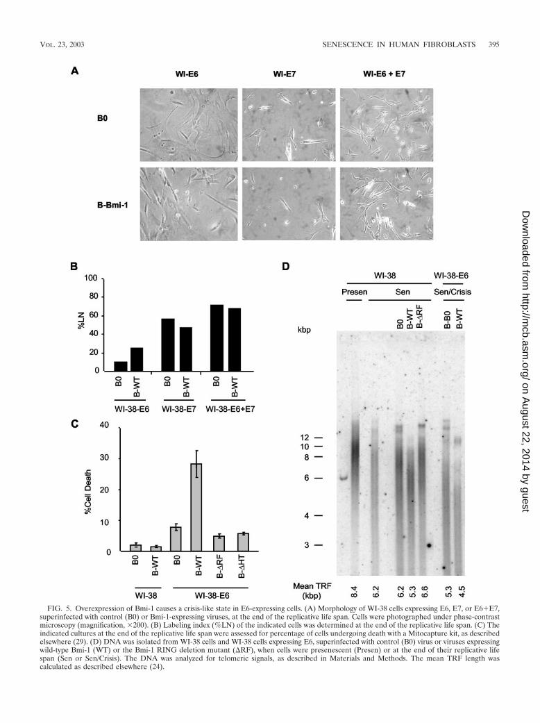
FIG. 5. Overexpression of Bmi-1 causes a crisis-like state in E6-expressing cells. (A) Morphology of WI-38 cells expressing E6, E7, or E6�E7,superinfected with control (B0) or Bmi-1-expressing viruses, at the end of the replicative life span. Cells were photographed under phase-contrastmicroscopy (magnification, �200). (B) Labeling index (%LN) of the indicated cells was determined at the end of the replicative life span. (C) Theindicated cultures at the end of the replicative life span were assessed for percentage of cells undergoing death with a Mitocapture kit, as describedelsewhere (29). (D) DNA was isolated from WI-38 cells and WI-38 cells expressing E6, superinfected with control (B0) virus or viruses expressingwild-type Bmi-1 (WT) or the Bmi-1 RING deletion mutant (RF), when cells were presenescent (Presen) or at the end of their replicative lifespan (Sen or Sen/Crisis). The DNA was analyzed for telomeric signals, as described in Materials and Methods. The mean TRF length wascalculated as described elsewhere (24).
VOL. 23, 2003 SENESCENCE IN HUMAN FIBROBLASTS 395
on August 22, 2014 by guest
http://mcb.asm
.org/D
ownloaded from

396 ITAHANA ET AL. MOL. CELL. BIOL.
on August 22, 2014 by guest
http://mcb.asm
.org/D
ownloaded from

correlates with the level of p16 expressed by presenescentcultures. Moreover, human fibroblasts differ in their propensityto induce p16 at an early passage and thus in the malleabilityof their replicative life span.
DISCUSSION
The p53 and pRb tumor suppressors are critical for thesenescence growth arrest of human cells, which constitutes animportant barrier to cellular immortalization and hence neo-
plastic transformation. p53 activates target genes that, in turn,induce apoptosis or arrested growth (26, 27). Because senes-cent fibroblasts arrest growth without signs of apoptosis (17,26), p53 very likely acts primarily to establish and/or maintainthe senescence growth arrest (17, 26). p21 is thought to be amajor effector of p53 activity in senescent cells (20, 60), which,in turn, is thought to be triggered by critical telomere loss (12,26, 34, 49, 64, 67).
p16, acting through the pRb pathway, is also thought to beimportant for the senescence response (1, 22, 28, 42, 52, 53,
FIG. 6. High-p16-expressing cells in presenescent WI-38 cultures and effect of Bmi-1 overexpression. (A) Presenescent cells at PD 24 (Presen)and senescent (Sen) cells were immunostained for p16 as described in Materials and Methods and photographed (magnification, �100) at the sameexposure for each set. Nuclei were visualized by DAPI staining. HeLa and H1299 cells served as positive and negative controls for p16 expression,respectively. (B) Presenescent WI-38 cells at PD 24 were infected with control (B0) or Bmi-1-expressing retroviruses, stained for p16 and DAPIafter selection and photographed (magnification, �200). (C) Presenescent WI-38 cells at PD 24 were immunostained for p16 and Ki67 as describedin Materials and Methods and photographed (magnifications, �300 [top] and �400 [bottom]). The arrows show examples of p16-positive cells thatare Ki67 negative. (D) Presenescent cells at PD 29 were stained for SA–�-Gal activity and subsequently immunostained for p16 as described inMaterials and Methods. SA–�-Gal staining was photographed under phase-contrast and bright field (SA–�-Gal) microscopy. HeLa cells served asa positive control for p16 staining. Magnification, �200. (E) Presenescent cells (PD 24, Presen) were infected with retroviruses carrying no insert(B0) or Bmi-1 (B-Bmi-1), selected, and passaged until senescence (Sen; %LN � 10%). Cells were further cultured for 2 months (B0-L andB-Bmi-1-L; %LN � 1%). Cell lysates were analyzed by Western blotting for Bmi-1, p16, and �-tubulin (control) proteins.
VOL. 23, 2003 SENESCENCE IN HUMAN FIBROBLASTS 397
on August 22, 2014 by guest
http://mcb.asm
.org/D
ownloaded from

60). p16 is induced by certain oncogenes (53, 68) and otherdamage or stress signals (11, 45, 47, 52) and is required for thetelomere-independent senescence of some human epithelialcells (35, 46). However, p16 has been reported to rise andinhibit CDK activity only after human fibroblasts have com-pletely arrested due to replicative senescence (60). Indeed, p16levels were slightly elevated in near-senescent, compared topresenescent, WI-38 cells and rose substantially only after se-nescent cells were maintained for several weeks. Despite rel-atively low p16 levels prior to senescence, Bmi-1 extended thereplicative life span of WI-38. However, we found that early-passage cultures had a significant number of high-p16-express-ing cells, which appeared to be senescent. Bmi-1 selectivelyreduced the proportion of these cells. We used retroviruses,which do not infect nondividing cells, to overexpress Bmi-1.Thus, Bmi-1 could not act by repressing p16 directly in theprematurely senescent cells. Rather, it is likely that Bmi-1prevents new induction of p16 and thus inhibits new cells fromsenescing prematurely, which we propose is caused by extrinsicstress.
Deletion analysis of Bmi-1 suggested that the RF and HTdomains of Bmi-1 were required for life span extension ofWI-38 fibroblasts. Interestingly, the RF Bmi-1 mutant accel-erated replicative senescence and p16 accumulation. Since RFmutants of Bmi-1 can dimerize with wild-type Bmi-1 (50) andwild-type Bmi-1 downregulates p16 expression, we suggest thatthe RF mutant upregulates p16 by inhibiting wild-type Bmi-1function, thus acting as a dominant negative mutant. The pre-cise mechanism by which p16 and senescence are induced bythe RF mutant remains to be investigated.
We suggest that cells prematurely undergo senescence incultures such as WI-38 independent of telomere length andfunction. Indeed, senescent Bmi-1-overexpressing WI-38 cellshad shorter telomeres than senescent control cells, consistentwith Bmi-1 driving additional cell division and telomere ero-sion. In addition, Bmi-1 did not extend the life span of cellsexpressing E7, E6�E7, or T antigen, which is expected ifBmi-1 does not act by retarding telomere erosion. These stud-ies also show that Bmi-1 extends life span by abolishing thepRb, not the p53, pathway. p53 is thought to mediate thesenescence response to telomere dysfunction (3, 6, 12, 26, 49,67), although a p53-dependent but telomere-independent se-nescence pathway was recently identified in human keratino-
cytes (46). Finally, early-passage BJ cells were devoid of pre-maturely senescent (high-p16-expressing) cells, and Bmi-1 hadno effect on their replicative life span.
We propose two means by which human fibroblasts senesce(Fig. 9). In cultures such as WI-38 (Fig. 9, left), prematurelysenescent cells may continually arise because the cells are
FIG. 7. Bmi-1 overexpression extends replicative life span in lowO2 concentrations. (A) Presenescent WI-38 cells (PD 24) were cul-tured for 2 weeks in 3% O2, infected with control (B0) or Bmi-1(B-Bmi1)-expressing retrovirus, and selected in 3% O2. Half the con-trol culture (B0) was moved to 20% O2 on the day indicated by thedashed arrow. All cells were passaged until senescence. The solidarrow indicates the day of superinfection (B). (B) A control culture(B0) was superinfected with murine stem cell retroviruses carrying noinsert (M0) or Bmi-1 (M-Bmi-1) in either in 3 or 20% O2. Cells wereselected in hygromycin and passaged until senescence. (C) Presenes-cent cells (PD 24) were cultured in 3 or 20% O2 for 2 weeks andinfected with retroviruses carrying no insert (B0) or Bmi-1 (B-Bmi-1)in either 3 or 20% O2. Cells were selected and passaged for 1 week.p16, p21, and �-tubulin (control) expression was determined by West-ern analysis. Three independent experiments showed similar results.
TABLE 1. Expression of p16 and either Ki67 or SA–�-Gal inpresenescent WI-38 fibroblastsa
Protein % Positive cellsb
p16 .......................................................................................... 19.8 0.55Ki67 ........................................................................................ 61.8 1.81p16 � Ki67 ............................................................................ 2.2 0.26
p16 .......................................................................................... 20.2 0.60SA–�-Gal ............................................................................... 20.6 0.46p16 � SA–�-Gal................................................................... 16.4 0.48
a Exponentially growing presenescent WI-38 cells were stained for p16, Ki67,and/or SA–�-Gal as described in Materials and Methods. The total number ofcells was determined by counting DAPI-stained nuclei or intact cells by usingfluorescence or phase-contrast microscopy, respectively. SA–�-Gal-stained cellswere counted under bright-field microscopy.
b Values are means and SE of four independent experiments. For each exper-iment, at least 300 cells were scored.
398 ITAHANA ET AL. MOL. CELL. BIOL.
on August 22, 2014 by guest
http://mcb.asm
.org/D
ownloaded from

sensitive to environmental stress. This senescence, which weterm “extrinsic senescence,” is likely caused by stochastic, as-yet-unidentified, cellular or extracellular events and is medi-ated by p16. Extrinsic senescence is independent of telomeres,unlike replicative senescence, which is intrinsically determinedby telomere function. The remaining cells in such cultureseventually undergo p53 telomere-dependent replicative senes-cence. p16 levels continue to rise in these cultures becausereplicatively senescent cells remain sensitive to stress caused byextrinsic factors. Bmi-1 suppresses extrinsic senescence by re-pressing p16 and thus extends the replicative capacity of theculture, but telomere erosion causes Bmi-1-overexpressingcells to eventually senesce. Consistent with this model, cellsexpressing a dominant negative Bmi-1 mutant senesced morerapidly, with high p16 expression, than control cultures.
Cultures such as BJ (Fig. 9, right) may be resistant to stressinduced by extrinsic factors or incapable of expressing highlevels of p16 in response to such stress. Hence, p16 levelsremain low and the cells undergo intrinsic replicative senes-cence owing to telomere erosion. Consistent with this idea, BJcells senesce with shorter telomeres (4.7 kb) than those ofsenescent IMR-90 (30) or WI-38 cells (6 to 7 kb). Moreover,Bmi-1-overexpressing WI-38 cells senesced with telomerelengths similar to those of senescent BJ cells. Finally, we findthat BJ cells maintain a high labeling index (%LN � 90%)after ectopic expression of telomerase, where telomerase-ex-pressing WI-38 cells continually produce unlabeled senescent(SA–�-Gal-positive) cells (%LN � 50 to 70%) (not shown),suggesting that extrinsic senescence depends on the fibroblaststrain and is independent of telomeres. Based on these dataand published reports on the replicative life span of fibroblastsof different origins (39), we speculate that lung-derived fibro-blasts senesce by extrinsic and intrinsic mechanisms, whichdepend on p16 and telomeres, respectively (Fig. 9, left), whilemost skin-derived fibroblasts undergo primarily intrinsic repli-cative senescence by the telomere-dependent mechanism (Fig.9, right).
What type of stress induces p16 in fibroblasts? AtmosphericO2 (20%), in which most cells are cultured, is much higher thanO2 concentrations in vivo, which can vary from 2 to 10% (4).Reduced O2 has long been known to promote the growth andextend the replicative life span of cultured human cells (43,48). Therefore, we tested the idea that reduced (3%) O2 mightprevent extrinsic senescence and p16 upregulation in culturessuch as WI-38. However, levels of p21, not p16, declined in 3%O2. Consistent with Bmi-1 acting through p16, not p21, Bmi-1extended the life span of WI-38 cells regardless of the O2 level.Because p21 is a p53 target gene and p21 expression declinedin 3% O2, low O2 concentrations may extend replicative lifespan exclusively through the p53- and telomere-dependent se-nescence mechanism. Indeed, 40% O2 shortens replicative lifespan and accelerates telomere shortening (65). Although O2
levels did not affect p16 expression, BJ fibroblasts were re-cently shown to have relatively high antioxidant activity andlow levels of protein carbonyls, peroxides, and lipofuscin, com-pared to WI-38 and MRC-5 fibroblasts (39). It is possible thatthese secondary metabolites can regulate p16 and hence in-duce extrinsic senescence. Whatever the case, our findingssuggest that not only MEFs but also some strains of humanfibroblasts are sensitive to stress in culture and presumably in
FIG. 8. p16 inducibility determines life span extension potential byBmi-1 overexpression. (A) BJ cells (%LN � 60%) were infected withretroviruses carrying no insert (B0) or Bmi-1 (B-Bmi-1) and passageduntil senescence, and life span was determined as described in Mate-rials and Methods. (B) 82-6 fibroblasts (%LN � 60%) were similarlyinfected and analyzed. (C) Protein extracts were prepared from BJ and82-6 (%LN � 60%) and WI-38 (%LN � 70%) cells prior to infection(Preselection) and after infection with B0 or B-Bmi-1 retroviruses andselection (Postselection). Extracts were analyzed by Western blottingfor Bmi-1, p16, p21, and �-tubulin (control) proteins. The p16 blot wasexposed for a longer interval to detect the signal in BJ cells. (D) Prese-nescent (%LN � 70%) and senescent (%LN � 1%) WI-38 and BJcultures were analyzed by Western blotting for p16 and actin (control).Extracts from senescent BJ cells cultured for an additional 2 monthsafter senescence (BJ-L, %LN � 1%) were also analyzed, with similarresults.
VOL. 23, 2003 SENESCENCE IN HUMAN FIBROBLASTS 399
on August 22, 2014 by guest
http://mcb.asm
.org/D
ownloaded from

vivo. Bmi-1 overcomes this p16-dependent, telomere-indepen-dent extrinsic senescence. It has been speculated that cellularsenescence (whether extrinsic or intrinsic) can compromise theregenerative capacity of tissues (8, 9, 19). Bmi-1 may helpmaintain the regenerative capacity of these tissues.
ACKNOWLEDGMENTS
This work was supported by grants from the National Institutes ofHealth (AG09909 to J.C. and AG16851 to G.P.D.) and startup fundsfrom the Cancer Center, New England Medical Center, Boston, Mass.(G.P.D.).
We thank Miguel Rubio for providing near-senescent 82-6 cells.
REFERENCES
1. Alcorta, D. A., Y. Xiong, D. Phelps, G. Hannon, D. Beach, and J. C. Barrett.1996. Involvement of the cyclin-dependent kinase inhibitor p16 (INK4a) inreplicative senescence of normal human fibroblasts. Proc. Natl. Acad. Sci.USA 93:13742–13747.
2. Alkema, M. J., M. Bronk, E. Verhoeven, A. P. Otte, L. J. van’t Veer, A. Berns,and M. van Lohuizen. 1997. Identification of Bmi1-interacting proteins asconstituents of a multimeric mammalian Polycomb complex. Genes Dev.11:226–240.
3. Atadja, P., H. Wong, I. Garkavtsev, C. Veillette, and K. Riabowol. 1995.Increased activity of p53 in senescing fibroblasts. Proc. Natl. Acad. Sci. USA92:8348–8352.
4. Balin, A. K., A. J. Fisher, and D. M. Carter. 1984. Oxygen modulates growthof human cells at physiological partial pressures. J. Exp. Med. 160:152–166.
5. Bodnar, A. G., M. Ouellette, M. Frolkis, S. E. Holt, C. P. Chiu, G. B. Morin,C. B. Harley, J. W. Shay, S. Lichtsteiner, and W. E. Wright. 1998. Extensionof life span by introduction of telomerase into normal human cells. Science279:349–352.
6. Bond, J. A., M. F. Haughton, J. M. Rowson, P. J. Smith, V. Gire, D. Wynford-Thomas, and F. S. Wyllie. 1999. Control of replicative life span in humancells: barriers to clonal expansion intermediate between M1 senescence andM2 crisis. Mol. Cell. Biol. 19:3103–3114.
7. Bond, J. A., F. S. Wyllie, and D. Wynford-Thomas. 1994. Escape fromsenescence in human diploid fibroblasts induced directly by mutant p53.Oncogene 9:1885–1889.
8. Campisi, J. 1996. Replicative senescence: an old lives’ tale? Cell 84:497–500.9. Campisi, J. 1997. The biology of replicative senescence. Eur. J. Cancer
33:703–709.10. Campisi, J. 2001. Cellular senescence as a tumor-suppressor mechanism.
Trends Cell Biol. 11:27–31.
11. Chen, Q. M. 2000. Replicative senescence and oxidant-induced prematuresenescence. Beyond the control of cell cycle checkpoints. Ann. N. Y. Acad.Sci. 908:111–125.
12. Chin, L., S. E. Artandi, Q. Shen, A. Tam, S. L. Lee, G. J. Gottlieb, C. W.Greider, and R. A. DePinho. 1999. p53 deficiency rescues the adverse effectsof telomere loss and cooperates with telomere dysfunction to acceleratecarcinogenesis. Cell 97:527–538.
13. Cohen, K. J., J. S. Hanna, J. E. Prescott, and C. V. Dang. 1996. Transfor-mation by the Bmi-1 oncoprotein correlates with its subnuclear localizationbut not its transcriptional suppression activity. Mol. Cell. Biol. 16:5527–5535.
14. de Lange, T. 2001. Telomere capping—one strand fits all. Science 292:1075–1076.
15. Dimri, G. P., E. Hara, and J. Campisi. 1994. Regulation of two E2F-relatedgenes in presenescent and senescent human fibroblasts. J. Biol. Chem. 269:16180–16186.
16. Dimri, G. P., K. Itahana, M. Acosta, and J. Campisi. 2000. Regulation of asenescence checkpoint response by the E2F1 transcription factor andp14(ARF) tumor suppressor. Mol. Cell. Biol. 20:273–285.
17. Dimri, G. P., and J. Campisi. 1994. Molecular and cell biology of replicativesenescence. Cold Spring Harbor Symp. Quant. Biol. 59:67–73.
18. Dimri, G. P., J. L. Martinez, J. J. Jacobs, P. Keblusek, K. Itahana, M. vonLohuizen, J. Campisi, D. E. Wazer, and V. Band. 2002. The Bmi-1 oncogeneinduces telomerase activity and immortalizes human mammary epithelialcells. Cancer Res. 62:4736–4745.
19. Dimri, G. P., X. Lee, G. Basile, M. Acosta, G. Scott, C. Roskelley, E. E.Medrano, M. Linskens, I. Rubelj, O. M. Pereira-Smith, M. Peacocke, and J.Campisi. 1995. A novel biomarker identifies senescent human cells in cultureand in aging skin in vivo. Proc. Natl. Acad. Sci. USA 92:9363–9367.
20. Dulic, V., G. E. Beney, G. Frebourg, L. F. Drullinger, and G. H. Stein. 2000.Uncoupling between phenotypic senescence and cell cycle arrest in agingp21-deficient fibroblasts. Mol. Cell. Biol. 20:6741–6754.
21. Hara, E., H. Tsurui, A. Shinozaki, S. Nakada, and K. Oda. 1991. Coopera-tive effect of antisense-Rb and antisense-p53 oligomers on the extension oflife span in human diploid fibroblasts, TIG-1. Biochem. Biophys. Res. Com-mun. 179:528–534.
22. Hara, E., R. Smith, D. Parry, H. Tahara, S. Stone, and G. Peters. 1996.Regulation of p16CDKN2 expression and its implications for cell immortal-ization and senescence. Mol. Cell. Biol. 16:859–867.
23. Hara, E., T. Yamaguchi, H. Nojima, T. Ide, J. Campisi, H. Okayama, and K.Oda. 1994. Id-related genes encoding helix-loop-helix proteins are requiredfor G1 progression and are repressed in senescent human fibroblasts. J. Biol.Chem. 269:2139–2145.
24. Harley, C. B., A. B. Futcher, and C. W. Greider. 1990. Telomeres shortenduring ageing of human fibroblasts. Nature 345:458–460.
25. Haupt, Y., W. S. Alexander, G. Barri, S. P. Klinken, and J. M. Adams. 1991.
FIG. 9. Model of senescence mechanisms in human fibroblasts. Presenescent fibroblast cultures that resemble WI-38 constantly producesenescent cells by the extrinsic (p16-dependent, telomere-independent) mechanism (left). Presenescent cultures that resemble BJ fibroblastsproduce few senescent cells via this extrinsic mechanism (right). As the cultures proliferate and near the end of their replicative life span, cellsundergoing senescence by the p53- and telomere-dependent mechanism (intrinsic or replicative senescence) increase in both types of cultures.However, senescent cells produced by the extrinsic senescence mechanism are present only in WI-38-type cultures.
400 ITAHANA ET AL. MOL. CELL. BIOL.
on August 22, 2014 by guest
http://mcb.asm
.org/D
ownloaded from

Novel zinc finger gene implicated as myc collaborator by retrovirally accel-erated lymphomagenesis in E mu-myc transgenic mice. Cell 65:753–763.
26. Itahana, K., G. Dimri, and J. Campisi. 2001. Regulation of cellular senes-cence by p53. Eur. J. Biochem. 268:2784–2791.
27. Itahana, K., G. P. Dimri, E. Hara, Y. Itahana, Y. Zou, P. Y. Desprez, and J.Campisi. 2002. A role for p53 in maintaining and establishing the quiescencegrowth arrest in human cells. J. Biol. Chem. 277:18206–18214.
28. Jacobs, J. J., K. Kieboom, S. Marino, R. A. DePinho, and M. van Lohuizen.1999. The oncogene and Polycomb-group gene bmi-1 regulates cell prolif-eration and senescence through the INK4a locus. Nature 397:164–168.
29. Kaminker, P. G., S. H. Kim, R. D. Taylor, Y. Zebarjadian, W. D. Funk, G. B.Morin, P. Yaswen, and J. Campisi. 2001. TANK2, a new TRF1-associatedpoly(ADP-ribose) polymerase, causes rapid induction of cell death uponoverexpression. J. Biol. Chem. 276:35891–35899.
30. Karlseder, J., A. Smogorzewska, and T. de Lange. 2002. Senescence inducedby altered telomere state, not telomere loss. Science 295:2446–2449.
31. Kato, D., K. Miyazawa, M. Ruas, M. Starborg, I. Wada, T. Oka, T. Sakai, G.Peters, and E. Hara. 1998. Features of replicative senescence induced byaddition of antennapedia-p16INK4A fusion protein to human diploid fibro-blasts. FEBS Lett. 427:203–208.
32. Kawabe, S., J. A. Roth, D. R. Wilson, and R. E. Meyn. 2000. Adenovirus-mediated p16INK4a gene expression radiosensitizes non-small cell lung can-cer cells in a p53-dependent manner. Oncogene 19:5359–5366.
33. Kim, N. W., M. A. Piatyszek, K. R. Prowse, C. B. Harley, M. D. West, P. L.Ho, G. M. Coviello, W. E. Wright, S. L. Weinrich, and J. W. Shay. 1994.Specific association of human telomerase activity with immortal cells andcancer. Science 266:2011–2015.
34. Kim, S. H., P. Kaminker, and J. Campisi. 2002. Telomeres, aging andcancer: in search of a happy ending. Oncogene 21:503–511.
35. Kiyono, T., S. A. Foster, J. I. Koop, J. K. McDougall, D. A. Galloway, andA. J. Klingelhutz. 1998. Both Rb/p16INK4a inactivation and telomeraseactivity are required to immortalize human epithelial cells. Nature 396:84–88.
36. Krimpenfort, P., K. C. Quon, W. J. Mooi, A. Loonstra, and A. Berns. 2001.Loss of p16INK4a confers susceptibility to metastatic melanoma in mice.Nature 413:83–86.
37. Laemmli, U. K. 1970. Cleavage of structural proteins during the assembly ofthe head of bacteriophage T4. Nature 227:680–685.
38. Levy, M. Z., R. C. Allsopp, A. B. Futcher, C. W. Greider, and C. B. Harley.1992. Telomere end-replication problem and cell aging. J. Mol. Biol. 225:951–960.
39. Lorenz, M., G. Saretzki, N. Sitte, S. Metzkow, and T. von Zglinicki. 2001. BJfibroblasts display high antioxidant capacity and slow telomere shorteningindependent of hTERT transfection. Free Radic. Biol. Med. 31:824–831.
40. Morales, C. P., S. E. Holt, M. Ouellette, K. J. Kaur, Y. Yan, K. S. Wilson,M. A. White, W. E. Wright, and J. W. Shay. 1999. Absence of cancer-associated changes in human fibroblasts immortalized with telomerase. Nat.Genet. 21:115–118.
41. Noda, A., Y. Ning, S. F. Venable, O. M. Pereira-Smith, and J. R. Smith. 1994.Cloning of senescent cell-derived inhibitors of DNA synthesis using an ex-pression screen. Exp. Cell Res. 211:90–98.
42. Ohtani, N., Z. Zebedee, T. J. Huot, J. A. Stinson, M. Sugimoto, Y. Ohashi,A. D. Sharrocks, G. Peters, and E. Hara. 2001. Opposing effects of Ets andId proteins on p16INK4a expression during cellular senescence. Nature409:1067–1070.
43. Packer, L., and K. Fuehr. 1977. Low O2 concentration extends the life spanof cultured human diploid cells. Nature 267:423–425.
44. Pirrotta, V. 1998. Polycombing the genome: PcG, trxG, and chromatin si-lencing. Cell 93:333–336.
45. Ramirez, R. D., C. P. Morales, B. S. Herbert, J. M. Rohde, C. Passons, J. W.Shay, and W. E. Wright. 2001. Putative telomere-independent mechanismsof replicative aging reflect inadequate growth conditions. Genes Dev. 15:398–403.
46. Rheinwald, J. G., W. C. Hahn, M. R. Ramsey, J. Y. Yu, Z. Guo, H. Tsao, M.De Luca, C. Catricala, and K. M. O’Toole. 2002. A two-stage, p16(INK4A)-and p53-dependent keratinocyte senescence mechanism that limits replica-tive potential independent of telomere status. Mol. Cell. Biol. 22:5157–5172.
47. Robles, S. J., and G. R. Adami. 1998. Agents that cause DNA double strandbreaks lead to p16INK4a enrichment and the premature senescence ofnormal fibroblasts. Oncogene 16:1113–1123.
48. Saito, H., A. T. Hammond, and R. E. Moses. 1995. The effect of low O2tension on the in vitro-replicative life span of human diploid fibroblast cellsand their transformed derivatives. Exp. Cell Res. 217:272–279.
49. Saretzki, G., N. Sitte, U. Merkel, R. E. Wurm, and T. von Zglinicki. 1999.Telomere shortening triggers a p53-dependent cell cycle arrest via accumu-lation of G-rich single stranded DNA fragments. Oncogene 18:5148–5158.
50. Satijn, D. P., and A. P. Otte. 1999. RING1 interacts with multiple Polycomb-group proteins and displays tumorigenic activity. Mol. Cell. Biol. 19:57–68.
51. Schluter, C., M. Duchrow, C. Wohlenberg, M. H. Becker, G. Key, H. D. Flad,and J. Gerdes. 1993. The cell proliferation-associated antigen of antibodyKi-67: a very large, ubiquitous nuclear protein with numerous repeatedelements, representing a new kind of cell cycle-maintaining proteins. J. CellBiol. 123:513–522.
52. Schmitt, C. A., J. S. Fridman, M. Yang, S. Lee, E. Baranov, R. M. Hoffman,and S. W. Lowe. 2002. A senescence program controlled by p53 andp16INK4a contributes to the outcome of cancer therapy. Cell 109:335–346.
53. Serrano, M., A. W. Lin, M. E. McCurrach, D. Beach, and S. W. Lowe. 1997.Oncogenic ras provokes premature cell senescence associated with accumu-lation of p53 and p16INK4a. Cell 88:593–602.
54. Seshadri, T., J. A. Uzman, J. Oshima, and J. Campisi. 1993. Identification ofa transcript that is down-regulated in senescent human fibroblasts. Cloning,sequence analysis, and regulation of the human L7 ribosomal protein gene.J. Biol. Chem. 268:18474–18480.
55. Shapiro, G. I., C. D. Edwards, M. E. Ewen, and B. J. Rollins. 1998.p16INK4A participates in a G1 arrest checkpoint in response to DNA dam-age. Mol. Cell. Biol. 18:378–387.
56. Sharpless, N. E., N. Bardeesy, K. H. Lee, D. Carrasco, D. H. Castrillon, A. J.Aguirre, E. A. Wu, J. W. Horner, and R. A. DePinho. 2001. Loss of p16INK4awith retention of p19Arf predisposes mice to tumorigenesis. Nature 413:86–91.
57. Shay, J. W., O. M. Pereira-Smith, and W. E. Wright. 1991. A role for bothRB and p53 in the regulation of human cellular senescence. Exp. Cell Res.196:33–39.
58. Shelton, D. N., E. Chang, P. S. Whittier, D. Choi, and W. D. Funk. 1999.Microarray analysis of replicative senescence. Curr. Biol. 9:939–945.
59. Sherr, C. J., and R. A. DePinho. 2000. Cellular senescence: mitotic clock orculture shock? Cell 102:407–410.
60. Stein, G. H., L. F. Drullinger, A. Soulard, and V. Dulic. 1999. Differentialroles for cyclin-dependent kinase inhibitors p21 and p16 in the mechanismsof senescence and differentiation in human fibroblasts. Mol. Cell. Biol. 19:2109–2117.
61. van der Lugt, N. M., J. Domen, K. Linders, M. van Roon, E. Robanus-Maandag, H. te Riele, M. van der Valk, J. Deschamps, M. Sofroniew, M. vanLohuizen, and A. Berns. 1994. Posterior transformation, neurological abnor-malities, and severe hematopoietic defects in mice with a targeted deletionof the bmi-1 proto-oncogene. Genes Dev. 8:757–769.
62. van Lohuizen, M., S. Verbeek, B. Scheijen, E. Wientjens, H. van der Gulden,and A. Berns. 1991. Identification of cooperating oncogenes in E mu-myctransgenic mice by provirus tagging. Cell 65:737–752.
63. Vaziri, H., J. A. Squire, T. K. Pandita, G. Bradley, R. M. Kuba, H. Zhang, S.Gulyas, R. P. Hill, G. P. Nolan, and S. Benchimol. 1999. Analysis of genomicintegrity and p53-dependent G1 checkpoint in telomerase-induced extended-life-span human fibroblasts. Mol. Cell. Biol. 19:2373–2379.
64. Vaziri, H., M. D. West, R. C. Allsopp, T. S. Davison, Y. S. Wu, C. H.Arrowsmith, G. G. Poirier, and S. Benchimol. 1997. ATM-dependent telo-mere loss in aging human diploid fibroblasts and DNA damage lead to thepost-translational activation of p53 protein involving poly(ADP-ribose) poly-merase. EMBO J. 16:6018–6033.
65. von Zglinicki, T., G. Saretzki, W. Docke, and C. Lotze. 1995. Mild hyperoxiashortens telomeres and inhibits proliferation of fibroblasts: a model forsenescence? Exp. Cell Res. 220:186–193.
66. Wright, W. E., O. M. Pereira-Smith, and J. W. Shay. 1989. Reversiblecellular senescence: implications for immortalization of normal human dip-loid fibroblasts. Mol. Cell. Biol. 9:3088–3092.
67. Wright, W. E., and J. W. Shay. 2000. Telomere dynamics in cancer progres-sion and prevention: fundamental differences in human and mouse telomerebiology. Nat. Med. 6:849–851.
68. Zhu, J., D. Woods, M. McMahon, and J. M. Bishop. 1998. Senescence ofhuman fibroblast induced by oncogenic Raf. Genes Dev. 12:2997–3007.
VOL. 23, 2003 SENESCENCE IN HUMAN FIBROBLASTS 401
on August 22, 2014 by guest
http://mcb.asm
.org/D
ownloaded from