Inactivation of Myocardin and p16 during Malignant Transformation Contributes to a Differentiation...
14
Cancer Cell Article Inactivation of Myocardin and p16 during Malignant Transformation Contributes to a Differentiation Defect Michael Milyavsky, 1,5 Igor Shats, 1,5 Alina Cholostoy, 1 Ran Brosh, 1 Yosef Buganim, 1 Lilach Weisz, 1 Ira Kogan, 1 Merav Cohen, 1 Maria Shatz, 2 Shalom Madar, 1 Eyal Kalo, 1 Naomi Goldfinger, 1 Jun Yuan, 3 Shulamit Ron, 4 Karen MacKenzie, 3 Amir Eden, 4 and Varda Rotter 1, * 1 Department of Molecular Cell Biology 2 Department of Biological Regulation Weizmann Institute of Science, Rehovot 76100, Israel 3 Children’s Cancer Institute for Medical Research, Randwick, New South Wales 2031, Australia 4 Institute of Life Sciences, The Hebrew University, Jerusalem 91904, Israel 5 These authors contributed equally to this work. *Correspondence: [email protected] DOI 10.1016/j.ccr.2006.11.022 SUMMARY Myocardin is known as an important transcriptional regulator in smooth and cardiac muscle devel- opment. Here we found that myocardin is frequently repressed during human malignant transforma- tion, contributing to a differentiation defect. We demonstrate that myocardin is a transcriptional target of TGFb required for TGFb-mediated differentiation of human fibroblasts. Serum deprivation, intact contact inhibition response, and the p16 ink4a /Rb pathway contribute to myocardin induction and differentiation. Restoration of myocardin expression in sarcoma cells results in differentiation and inhibition of malignant growth, whereas inactivation of myocardin in normal fibroblasts increases their proliferative potential. Myocardin expression is reduced in multiple types of human tumors. Collectively, our results demonstrate that myocardin is an important suppressive modifier of the malignant transformation process. INTRODUCTION Escape from the tissue/lineage-specific differentiation program is one of the fundamental aspects of tumorigen- esis in general and sarcomagenesis in particular. Human primary fibroblasts, which could be induced to undergo differentiation toward myofibroblasts and to some extent to smooth muscle cells (SMCs), might serve as a faithful model to study molecular mechanisms under- lying differentiation defects in sarcomas and in the tumor stroma compartment, in which fibroblasts play an impor- tant role. The highly organized actin cytoskeleton of these cells is characterized by varying levels of smooth muscle a-actin (aSMA), smooth muscle myosin heavy chain (SM MHC), calponin (CNN1), and transgelin (SM22). The genes encoding these proteins are coordinately regulated at the transcriptional level in myofibroblasts and SMCs and therefore are informative indicators of the differentiation in these cell types (Kumar and Owens, 2003; Pipes et al., 2006). On the other hand, actin cytoskeleton undergoes transformation-associated disruption in fibroblasts. The molecular basis responsible for this differentiation disrup- tion remains unclear (Button et al., 1995; Leavitt et al., 1985). The most common oncogenic changes described in sarcomas include inactivation of p16 INK4A (p16), Rb1, and p53 tumor-suppressor genes (Helman and Meltzer, SIGNIFICANCE The malignant transformation process is associated with defects in cell-cycle regulation and disruption of the nor- mal differentiation programs in both neoplastic and adjacent stroma cells. However, deregulation of the complex interplay between differentiation stimuli, mesenchymal tissue-specific transcription factors, and cell-cycle-regula- tory machinery is not completely understood. In this study we identified a role in human carcinogenesis for the im- portant regulator of cardiac and smooth muscle differentiation myocardin. We show that myocardin mRNA levels and activity are positively regulated by the p16/Rb pathway, providing a molecular link between cell-cycle and dif- ferentiation defects during tumor development. The antiproliferative activity of myocardin shown here suggests that its reactivation may represent an effective therapeutic approach in cancer treatment. Cancer Cell 11, 133–146, February 2007 ª2007 Elsevier Inc. 133
-
Upload
independent -
Category
Documents
-
view
2 -
download
0
Transcript of Inactivation of Myocardin and p16 during Malignant Transformation Contributes to a Differentiation...
Cancer Cell
Article
Inactivation of Myocardin and p16during Malignant TransformationContributes to a Differentiation DefectMichael Milyavsky,1,5 Igor Shats,1,5 Alina Cholostoy,1 Ran Brosh,1 Yosef Buganim,1 Lilach Weisz,1 Ira Kogan,1
Merav Cohen,1 Maria Shatz,2 Shalom Madar,1 Eyal Kalo,1 Naomi Goldfinger,1 Jun Yuan,3 Shulamit Ron,4
Karen MacKenzie,3 Amir Eden,4 and Varda Rotter1,*1 Department of Molecular Cell Biology2 Department of Biological Regulation
Weizmann Institute of Science, Rehovot 76100, Israel3 Children’s Cancer Institute for Medical Research, Randwick, New South Wales 2031, Australia4 Institute of Life Sciences, The Hebrew University, Jerusalem 91904, Israel5 These authors contributed equally to this work.
*Correspondence: [email protected]
DOI 10.1016/j.ccr.2006.11.022
SUMMARY
Myocardin is known as an important transcriptional regulator in smooth and cardiac muscle devel-opment. Here we found that myocardin is frequently repressed during human malignant transforma-tion, contributing to a differentiation defect. We demonstrate that myocardin is a transcriptionaltarget of TGFb required for TGFb-mediated differentiation of human fibroblasts. Serum deprivation,intact contact inhibition response, and the p16ink4a/Rb pathway contribute to myocardin inductionand differentiation. Restoration of myocardin expression in sarcoma cells results in differentiationand inhibition of malignant growth, whereas inactivation of myocardin in normal fibroblasts increasestheir proliferative potential. Myocardin expression is reduced in multiple types of human tumors.Collectively, our results demonstrate that myocardin is an important suppressive modifier of themalignant transformation process.
INTRODUCTION
Escape from the tissue/lineage-specific differentiation
program is one of the fundamental aspects of tumorigen-
esis in general and sarcomagenesis in particular.
Human primary fibroblasts, which could be induced to
undergo differentiation toward myofibroblasts and to
some extent to smooth muscle cells (SMCs), might serve
as a faithful model to study molecular mechanisms under-
lying differentiation defects in sarcomas and in the tumor
stroma compartment, in which fibroblasts play an impor-
tant role. The highly organized actin cytoskeleton of these
cells is characterized by varying levels of smooth muscle
Ca
a-actin (aSMA), smooth muscle myosin heavy chain (SM
MHC), calponin (CNN1), and transgelin (SM22). The genes
encoding these proteins are coordinately regulated at the
transcriptional level in myofibroblasts and SMCs and
therefore are informative indicators of the differentiation
in these cell types (Kumar and Owens, 2003; Pipes et al.,
2006). On the other hand, actin cytoskeleton undergoes
transformation-associated disruption in fibroblasts. The
molecular basis responsible for this differentiation disrup-
tion remains unclear (Buttonetal., 1995;Leavitt et al., 1985).
The most common oncogenic changes described in
sarcomas include inactivation of p16INK4A (p16), Rb1,
and p53 tumor-suppressor genes (Helman and Meltzer,
SIGNIFICANCE
The malignant transformation process is associated with defects in cell-cycle regulation and disruption of the nor-mal differentiation programs in both neoplastic and adjacent stroma cells. However, deregulation of the complexinterplay between differentiation stimuli, mesenchymal tissue-specific transcription factors, and cell-cycle-regula-tory machinery is not completely understood. In this study we identified a role in human carcinogenesis for the im-portant regulator of cardiac and smooth muscle differentiation myocardin. We show that myocardin mRNA levelsand activity are positively regulated by the p16/Rb pathway, providing a molecular link between cell-cycle and dif-ferentiation defects during tumor development. The antiproliferative activity of myocardin shown here suggeststhat its reactivation may represent an effective therapeutic approach in cancer treatment.
ncer Cell 11, 133–146, February 2007 ª2007 Elsevier Inc. 133
Cancer Cell
Myocardin in Transformation and Differentiation
2003), which control progression through the cell cycle as
a function of mitogen availability, contact inhibition, and
oncogene activation. For some in vitro differentiation
models, interference with intact cell-cycle regulation im-
pairs subsequent differentiation. In some experimental
settings where exit from the cell cycle is not required, max-
imal expression of differentiation markers is still depen-
dent on the function of an intact Rb tumor-suppressor
pathway (Halevy et al., 1995; Novitch et al., 1996).
Transforming growth factor b (TGFb) is a potent regula-
tor of differentiation in many tissues, including mesen-
chyme (Massague et al., 2000). Furthermore, TGFb is
involved in inflammation, fibrosis, and malignant transfor-
mation processes, in which fibroblasts, myofibroblasts,
and SMCs play a pivotal role. Treatment of primary human
fibroblasts with TGFb results in the induction of the fibro-
blast-myofibroblast differentiation program (Chambers
et al., 2003; Serini and Gabbiani, 1999).
Recently, it was found that myocardin (Myocd), a
smooth and cardiac muscle-specific transcriptional regu-
lator, is an essential transcriptional cofactor for a variety of
SMC-specific genes (Wang et al., 2001). Myocardin drives
transcription through interaction with the ubiquitous tran-
scription factor serum response factor (SRF), which acts
on a responsive element (CArG box) that is commonly
found in many smooth muscle and myofibroblast gene
promoters (Pipes et al., 2006). However, despite the
established executive function of myocardin in SMC differ-
entiation, its role in transformation and TGFb-mediated
myofibroblast differentiation is largely unknown.
The regulation of cytoskeletal/differentiation genes
clearly involves a complex interplay between differentia-
tion stimuli, mesenchymal tissue-specific transcription
factors, and cell-cycle-regulatory machinery (Kumar and
Owens, 2003). Oncogenic mutations may impinge on sev-
eral levels of regulation along the differentiation process.
Therefore, a detailed examination of the loss of cytoskele-
tal/differentiation markers in the context of controlled
stepwise malignant transformation will provide insight to
the molecular interactions between these two processes.
In our previous study, we found that hTERT-induced im-
mortalization of WI-38 human fibroblasts (WI-38T) resulted
in the spontaneous emergence of rapidly proliferating var-
iants (Tfast). These clones had a defective response to con-
tact inhibition and did not express the p16 tumor-suppres-
sor gene (Milyavsky et al., 2003). Utilizing genome-wide
expression profiling, we found that Tfast cells and their de-
rivatives were characterized by a reduced expression of
genes involved in various aspects of mesenchymal cell
development and differentiation compared with primary
WI-38 or early passage WI-38/hTERT cells (Tslow). Numer-
ous genes that were downregulated encode well-charac-
terized, functional markers of the myofibroblast phenotype
aSMA, CALD1, SM-MHC, SM22, and CNN1 (Milyavsky
et al., 2005).
In the present study, we found that human mesenchy-
mal transformation is associated with a differentiation
block accompanied by downregulation of myocardin
in vitro and in vivo. We demonstrate that myocardin is
134 Cancer Cell 11, 133–146, February 2007 ª2007 Elsevier Inc.
positively regulated by the p16/Rb pathway. Moreover,
myocardin is induced by TGFb and is essential for TGFb-
induced myofibroblast differentiation. Ectopic expression
of myocardin in tumor cells restores expression of differen-
tiation markers and inhibits the transformed phenotype.
Our results provide considerable insight into the regulation
of myocardin and suggest that its downregulation is
important for cancer development.
RESULTS
Human Mesenchymal Cells with Inactivated p16
Exhibit Disrupted Basal and TGFb-Induced
Myofibroblast Differentiation
Coordinated reduction in the expression of numerous
differentiation markers observed in Tfast cells indicated
that the cells acquired a defect in the fibroblast-myofibro-
blast differentiation program during immortalization (Mi-
lyavsky et al., 2005). To characterize this defect in detail,
Tslow and Tfast WI-38 cells were treated with an inducer of
myofibroblast differentiation, TGFb, and analyzed for the
induction of several differentiation markers by quantitative
real-time PCR (QRT-PCR). These experiments were per-
formed with confluent cultures in serum-free conditions,
as is common practice in differentiation studies. In agree-
ment with our previous findings, Tfast cells expressed
barely detectable basal levels of p16 and reduced levels
of several well-known myofibroblast markers, such as
CNN1, aSMA, and SM-MHC compared with Tslow cells.
Upon TGFb treatment, a robust induction of these markers
was evident in Tslow cells, but not in Tfast cells (Figure 1A),
demonstrating a complete block in the TGFb-induced my-
ofibroblast differentiation program in Tfast cells. p16 levels
were not affected by the TGFb treatment in both Tslow and
Tfast cells. Induction of immediate-early targets of TGFb,
such as JunB and Id1, was comparable in Tslow and Tfast
cells, while the induction of p15INK4B growth inhibitor was
blunted in Tfast cells (Figure 1B).
To substantiate the correlation between loss of p16 and
deregulation of myofibroblast markers, additional strains
of human mesenchymal cells were analyzed. To that
end, early and late passages (psg 17 and psg 66, respec-
tively) of the hTERT-immortalized human prostate-derived
SMCs PM151T (Kogan et al., 2006) and precrisis and post-
crisis premalignant MRC5T human fibroblasts (Taylor
et al., 2004) were used. Both cell types exhibited down-
regulation of p16 during hTERT-mediated immortalization.
In agreement with our observations in WI-38T cells, both
immortalized mesenchymal strains had significantly lower
basal levels of aSMA, SM22, and CNN1. In addition, post-
crisis MRC5T cells exhibited reduced TGFb-mediated
myofibroblast differentiation (Figures 1C and 1D).
In an attempt to understand the molecular details under-
lying the differential TGFb response in WI-38 Tslow and Tfast
cells, we monitored the activation and levels of some clas-
sic components of the TGFb pathway, such as SMAD2/3
and ERK1/2, under conditions identical to those used
for the differentiation induction. Beyond subtle kinetic
differences, p16-deficient WI-38 Tfast fibroblasts initiated
Cancer Cell
Myocardin in Transformation and Differentiation
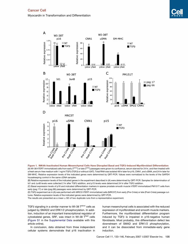
Figure 1. INK4A-Inactivated Human Mesenchymal Cells Have Disrupted Basal and TGFb-Induced Myofibroblast Differentiation
(A) WI-38 hTERT-immortalized cells from early (Tslow) or late (Tfast) passages were grown to confluence, serum starved for 24 hr, and then treated with
a fresh serum-free medium with 1 ng/ml TGFb (TGFb) or without it (NT). Total RNA was isolated 48 hr later for p16, CNN1, and aSMA, and 24 hr later for
SM-MHC. Relative expression levels of the indicated genes were determined by QRT-PCR. Values were normalized to the levels of the GAPDH
housekeeping control in the same cDNA samples.
(B) Relative expression levels of the indicated genes in the experiment described in (A) were determined by QRT-PCR. Samples for determination of
Id1 and JunB levels were collected 2 hr after TGFb addition, and p15 levels were determined 24 hr after TGFb addition.
(C) Basal expression levels of p16 and indicated differentiation markers in sparse prostate smooth muscle hTERT-immortalized PM151T cells from
early (psg 17) or late (psg 66) passages were determined by QRT-PCR.
(D) TGFb experiment as in (A) was performed with MRC5 hTERT-immortalized cells (MRC5T) from early (Pre-Crisis) or late (Post-Crisis) passage cul-
tures. Relative expression levels of the indicated genes were determined by QRT-PCR.
The results are presented as a mean ± SD of two duplicate runs from a representative experiment.
TGFb signaling in a similar manner to WI-38 Tslow cells as
judged by SMAD2 and ERK1/2 phosphorylation. In addi-
tion, induction of an important transcriptional regulator of
cytoskeletal genes, SRF, was intact in WI-38 Tfast cells
(Figure S1 in the Supplemental Data available with this
article online).
In conclusion, data obtained from three independent
cellular systems demonstrate that p16 inactivation in
Ca
human mesenchymal cells is associated with the reduced
expression of myofibroblast and smooth muscle markers.
Furthermore, the myofibroblast differentiation program
induced by TGFb is impaired in p16-negative human
fibroblasts. Most probably, this differentiation defect lies
downstream of SMAD and ERK1/2 phosphorylation,
and it can be dissociated from immediate-early gene
induction.
ncer Cell 11, 133–146, February 2007 ª2007 Elsevier Inc. 135
Cancer Cell
Myocardin in Transformation and Differentiation
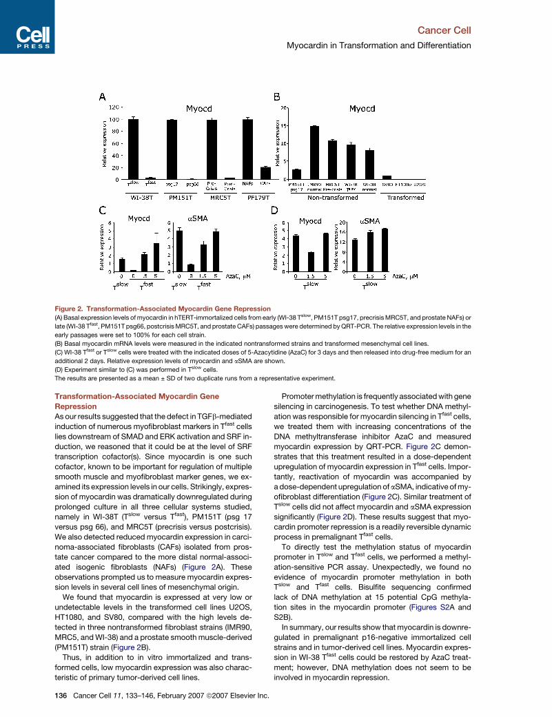
Figure 2. Transformation-Associated Myocardin Gene Repression
(A) Basal expression levels of myocardin in hTERT-immortalized cells from early (WI-38 Tslow, PM151T psg17, precrisis MRC5T, and prostate NAFs) or
late (WI-38 Tfast, PM151T psg66, postcrisis MRC5T, and prostate CAFs) passages were determined by QRT-PCR. The relative expression levels in the
early passages were set to 100% for each cell strain.
(B) Basal myocardin mRNA levels were measured in the indicated nontransformed strains and transformed mesenchymal cell lines.
(C) WI-38 Tfast or Tslow cells were treated with the indicated doses of 5-Azacytidine (AzaC) for 3 days and then released into drug-free medium for an
additional 2 days. Relative expression levels of myocardin and aSMA are shown.
(D) Experiment similar to (C) was performed in Tslow cells.
The results are presented as a mean ± SD of two duplicate runs from a representative experiment.
Transformation-Associated Myocardin Gene
Repression
As our results suggested that the defect in TGFb-mediated
induction of numerous myofibroblast markers in Tfast cells
lies downstream of SMAD and ERK activation and SRF in-
duction, we reasoned that it could be at the level of SRF
transcription cofactor(s). Since myocardin is one such
cofactor, known to be important for regulation of multiple
smooth muscle and myofibroblast marker genes, we ex-
amined its expression levels in our cells. Strikingly, expres-
sion of myocardin was dramatically downregulated during
prolonged culture in all three cellular systems studied,
namely in WI-38T (Tslow versus Tfast), PM151T (psg 17
versus psg 66), and MRC5T (precrisis versus postcrisis).
We also detected reduced myocardin expression in carci-
noma-associated fibroblasts (CAFs) isolated from pros-
tate cancer compared to the more distal normal-associ-
ated isogenic fibroblasts (NAFs) (Figure 2A). These
observations prompted us to measure myocardin expres-
sion levels in several cell lines of mesenchymal origin.
We found that myocardin is expressed at very low or
undetectable levels in the transformed cell lines U2OS,
HT1080, and SV80, compared with the high levels de-
tected in three nontransformed fibroblast strains (IMR90,
MRC5, and WI-38) and a prostate smooth muscle-derived
(PM151T) strain (Figure 2B).
Thus, in addition to in vitro immortalized and trans-
formed cells, low myocardin expression was also charac-
teristic of primary tumor-derived cell lines.
136 Cancer Cell 11, 133–146, February 2007 ª2007 Elsevier Inc.
Promoter methylation is frequently associated with gene
silencing in carcinogenesis. To test whether DNA methyl-
ation was responsible for myocardin silencing in Tfast cells,
we treated them with increasing concentrations of the
DNA methyltransferase inhibitor AzaC and measured
myocardin expression by QRT-PCR. Figure 2C demon-
strates that this treatment resulted in a dose-dependent
upregulation of myocardin expression in Tfast cells. Impor-
tantly, reactivation of myocardin was accompanied by
a dose-dependent upregulation of aSMA, indicative of my-
ofibroblast differentiation (Figure 2C). Similar treatment of
Tslow cells did not affect myocardin and aSMA expression
significantly (Figure 2D). These results suggest that myo-
cardin promoter repression is a readily reversible dynamic
process in premalignant Tfast cells.
To directly test the methylation status of myocardin
promoter in Tslow and Tfast cells, we performed a methyl-
ation-sensitive PCR assay. Unexpectedly, we found no
evidence of myocardin promoter methylation in both
Tslow and Tfast cells. Bisulfite sequencing confirmed
lack of DNA methylation at 15 potential CpG methyla-
tion sites in the myocardin promoter (Figures S2A and
S2B).
In summary, our results show that myocardin is downre-
gulated in premalignant p16-negative immortalized cell
strains and in tumor-derived cell lines. Myocardin expres-
sion in WI-38 Tfast cells could be restored by AzaC treat-
ment; however, DNA methylation does not seem to be
involved in myocardin repression.
Cancer Cell
Myocardin in Transformation and Differentiation
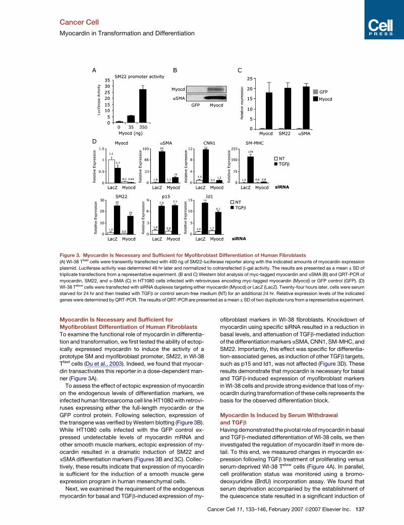
Figure 3. Myocardin Is Necessary and Sufficient for Myofibroblast Differentiation of Human Fibroblasts
(A) WI-38 Tfast cells were transiently transfected with 400 ng of SM22-luciferase reporter along with the indicated amounts of myocardin expression
plasmid. Luciferase activity was determined 48 hr later and normalized to cotransfected b-gal activity. The results are presented as a mean ± SD of
triplicate transfections from a representative experiment. (B and C) Western blot analysis of myc-tagged myocardin and aSMA (B) and QRT-PCR of
myocardin, SM22, and a-SMA (C) in HT1080 cells infected with retroviruses encoding myc-tagged myocardin (Myocd) or GFP control (GFP). (D)
WI-38 Tslow cells were transfected with siRNA duplexes targeting either myocardin (Myocd) or LacZ (LacZ). Twenty-four hours later, cells were serum
starved for 24 hr and then treated with TGFb or control serum-free medium (NT) for an additional 24 hr. Relative expression levels of the indicated
genes were determined by QRT-PCR. The results of QRT-PCR are presented as a mean ± SD of two duplicate runs from a representative experiment.
Myocardin Is Necessary and Sufficient for
Myofibroblast Differentiation of Human Fibroblasts
To examine the functional role of myocardin in differentia-
tion and transformation, we first tested the ability of ectop-
ically expressed myocardin to induce the activity of a
prototype SM and myofibroblast promoter, SM22, in WI-38
Tfast cells (Du et al., 2003). Indeed, we found that myocar-
din transactivates this reporter in a dose-dependent man-
ner (Figure 3A).
To assess the effect of ectopic expression of myocardin
on the endogenous levels of differentiation markers, we
infected human fibrosarcoma cell line HT1080 with retrovi-
ruses expressing either the full-length myocardin or the
GFP control protein. Following selection, expression of
the transgene was verified by Western blotting (Figure 3B).
While HT1080 cells infected with the GFP control ex-
pressed undetectable levels of myocardin mRNA and
other smooth muscle markers, ectopic expression of my-
ocardin resulted in a dramatic induction of SM22 and
aSMA differentiation markers (Figures 3B and 3C). Collec-
tively, these results indicate that expression of myocardin
is sufficient for the induction of a smooth muscle gene
expression program in human mesenchymal cells.
Next, we examined the requirement of the endogenous
myocardin for basal and TGFb-induced expression of my-
Ca
ofibroblast markers in WI-38 fibroblasts. Knockdown of
myocardin using specific siRNA resulted in a reduction in
basal levels, and attenuation of TGFb-mediated induction
of the differentiation markers aSMA, CNN1, SM-MHC, and
SM22. Importantly, this effect was specific for differentia-
tion-associated genes, as induction of other TGFb targets,
such as p15 and Id1, was not affected (Figure 3D). These
results demonstrate that myocardin is necessary for basal
and TGFb-induced expression of myofibroblast markers
in WI-38 cells and provide strong evidence that loss of my-
ocardin during transformation of these cells represents the
basis for the observed differentiation block.
Myocardin Is Induced by Serum Withdrawal
and TGFb
Having demonstrated the pivotal role of myocardin in basal
and TGFb-mediated differentiation of WI-38 cells, we then
investigated the regulation of myocardin itself in more de-
tail. To this end, we measured changes in myocardin ex-
pression following TGFb treatment of proliferating versus
serum-deprived WI-38 Tslow cells (Figure 4A). In parallel,
cell proliferation status was monitored using a bromo-
deoxyuridine (BrdU) incorporation assay. We found that
serum deprivation accompanied by the establishment of
the quiescence state resulted in a significant induction of
ncer Cell 11, 133–146, February 2007 ª2007 Elsevier Inc. 137

Cancer Cell
Myocardin in Transformation and Differentiation
Figure 4. Myocardin Is Induced by
Serum Withdrawal and TGFb
(A) WI-38 Tslow cells were serum starved for
24 hr (�) or grown in a full serum-containing
medium (+). Then TGFb or the corresponding
control medium was added for 6 hr. Myocardin
mRNA levels were determined by QRT-PCR,
and the proportion of cells in S phase of the
cell cycle was determined by the BrdU incor-
poration assay.
(B and C) WI-38 Tslow cells (B) and PM151T
psg 22 cells (C) were serum starved for 24 hr
and then treated with TGFb or mock treated
for the indicated times. Relative expression
levels of myocardin and aSMA were deter-
mined by QRT-PCR.
The results of QRT-PCR are presented as
a mean ± SD of two duplicate runs from a
representative experiment.
myocardin mRNA. Subsequent treatment of quiescent
cells with TGFb induced an additional robust increase in
myocardin expression. Notably, myocardin was induced
to significantly lower levels when TGFb treatment was
applied to the cells proliferating in the presence of serum.
Next, we investigated the kinetics of myocardin induc-
tion during the course of the TGFb treatment. As shown
in Figure 4B, myocardin was significantly upregulated
following serum starvation of WI-38 Tslow cells, and TGFb
treatment resulted in an additional robust induction of
myocardin peaking at 6 hr, followed by a gradual decline
to the basal levels by 24 hr. This transient pattern of myo-
cardin induction explains the fact that we did not detect
elevation of myocardin mRNA levels in the experiment
shown in Figure 3D, where the samples were collected at
24 hr time points. Similarly, myocardin was gradually in-
duced by serum starvation and TGFb treatment in the early
passage of prostate SMCs (PM151T psg 24), further sub-
stantiating our findings (Figure 4C). Notably, in both cell
types myocardin mRNA induction preceded that of aSMA,
suggesting that transcriptional induction of myocardin is
an important step in the induction of myofibroblast differ-
entiation induced by TGFb. In summary, we have shown
that myocardin mRNA levels are positively regulated by
serum withdrawal and TGFb.
138 Cancer Cell 11, 133–146, February 2007 ª2007 Elsevier In
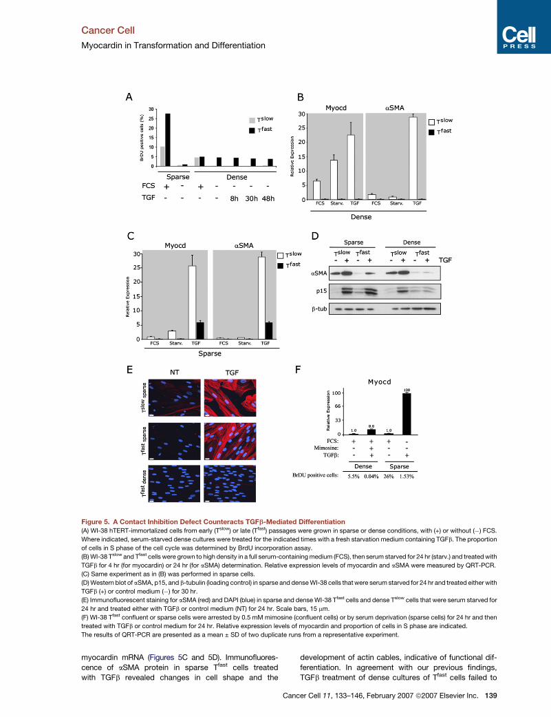
A Contact Inhibition Defect of Tfast Cells Counteracts
TGFb-Mediated Differentiation
We have previously described that WI-38 Tfast cells are
characterized by a contact inhibition defect that enables
them to reach a saturation density up to 7-fold higher
than that of Tslow cells (Milyavsky et al., 2003). To test the
effect of cell density on the TGFb-mediated differentiation,
we performed an experiment in sparse and dense cultures
of WI-38 Tslow and Tfast cells. We found that serum depriva-
tion of sparse cultures resulted in an almost complete inhi-
bition of DNA synthesis in both Tslow and Tfast cells
(Figure 5A).
However, a different situation was observed in the con-
fluent conditions. While Tslow cultures completely ceased
proliferation, dense WI-38 Tfast cells lost their capacity to
exit the cell cycle upon serum withdrawal and TGFb
treatment (Figure 5A).
Addition of TGFb resulted in myocardin induction and
robust aSMA upregulation in dense cultures of Tslow
cells, but not of Tfast cells (Figure 5B). Strikingly, we
found that under sparse conditions TGFb upregulated
myocardin and aSMA in Tfast cells. However, the induced
level of aSMA was much lower in Tfast compared with
Tslow cells as measured by both QRT-PCR and Western
blotting consistently with the partial upregulation of
c.
Cancer Cell
Myocardin in Transformation and Differentiation
Figure 5. A Contact Inhibition Defect Counteracts TGFb-Mediated Differentiation
(A) WI-38 hTERT-immortalized cells from early (Tslow) or late (Tfast) passages were grown in sparse or dense conditions, with (+) or without (�) FCS.
Where indicated, serum-starved dense cultures were treated for the indicated times with a fresh starvation medium containing TGFb. The proportion
of cells in S phase of the cell cycle was determined by BrdU incorporation assay.
(B) WI-38 Tslow and Tfast cells were grown to high density in a full serum-containing medium (FCS), then serum starved for 24 hr (starv.) and treated with
TGFb for 4 hr (for myocardin) or 24 hr (for aSMA) determination. Relative expression levels of myocardin and aSMA were measured by QRT-PCR.
(C) Same experiment as in (B) was performed in sparse cells.
(D) Western blot of aSMA, p15, and b-tubulin (loading control) in sparse and dense WI-38 cells that were serum starved for 24 hr and treated either with
TGFb (+) or control medium (�) for 30 hr.
(E) Immunofluorescent staining for aSMA (red) and DAPI (blue) in sparse and dense WI-38 Tfast cells and dense Tslow cells that were serum starved for
24 hr and treated either with TGFb or control medium (NT) for 24 hr. Scale bars, 15 mm.
(F) WI-38 Tfast confluent or sparse cells were arrested by 0.5 mM mimosine (confluent cells) or by serum deprivation (sparse cells) for 24 hr and then
treated with TGFb or control medium for 24 hr. Relative expression levels of myocardin and proportion of cells in S phase are indicated.
The results of QRT-PCR are presented as a mean ± SD of two duplicate runs from a representative experiment.
myocardin mRNA (Figures 5C and 5D). Immunofluores-
cence of aSMA protein in sparse Tfast cells treated
with TGFb revealed changes in cell shape and the
C
development of actin cables, indicative of functional dif-
ferentiation. In agreement with our previous findings,
TGFb treatment of dense cultures of Tfast cells failed to
ancer Cell 11, 133–146, February 2007 ª2007 Elsevier Inc. 139
Cancer Cell
Myocardin in Transformation and Differentiation
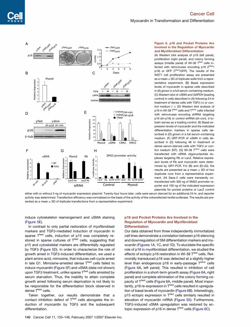
Figure 6. p16 and Pocket Proteins Are
Involved in the Regulation of Myocardin
and Myofibroblast Differentiation
(A) Western blot analysis of p16 (left panel),
proliferation (right panel), and colony forming
assays (middle panel) of WI-38 Tfast cells in-
fected with retroviruses encoding p16 (Tfast/
p16) or GFP (Tfast/GFP). The results of the
WST1 cell proliferation assay are presented
as a mean ± SD of triplicate wells from a repre-
sentative experiment. (B) Basal expression
levels of myocardin in sparse cells described
in (A) grown in a full serum-containing medium.
(C) Western blot of aSMA and GAPDH (loading
control) in cells described in (A) following 24 hr
treatment of dense cells with TGFb (+) or con-
trol medium (�). (D) Western blot analysis of
p16 in WI-38 Tfast cells and Tslow cells infected
with retroviruses encoding shRNA targeting
p16 (sh-p16) or control shRNA (sh-con). b-tu-
bulin serves as a loading control. (E) Basal ex-
pression levels of myocardin and the indicated
differentiation markers in sparse cells de-
scribed in (D) grown in a full serum-containing
medium. (F) QRT-PCR of aSMA in cells de-
scribed in (D) following 48 hr treatment of
dense serum-starved cells with TGFb or con-
trol medium (NT). (G) WI-38 Tslow cells were
transfected with siRNA oligonucleotide du-
plexes targeting Rb or LacZ. Relative expres-
sion levels of Rb and myocardin were deter-
mined by QRT-PCR. For (B) and (E)–(G), the
results are presented as a mean ± SD of two
duplicate runs from a representative experi-
ment. (H) Saos-2 cells were transiently co-
transfected with 300 ng of SM22 promoter re-
porter and 100 ng of the indicated expression
plasmids for pocket proteins or LacZ control
either with or without 5 ng of myocardin expression plasmid. Twenty-four hours later, cells were serum starved for an additional 24 hr, and reporter
activity was determined. Transfection efficiency was normalized on the basis of the activity of the cotransfected renilla luciferase. The results are pre-
sented as a mean ± SD of triplicate transfections from a representative experiment.
induce cytoskeleton rearrangement and aSMA staining
(Figure 5E).
In contrast to only partial restoration of myofibroblast
markers and TGFb-mediated induction of myocardin in
sparse Tfast cells, induction of p15 was completely re-
stored in sparse cultures of Tfast cells, suggesting that
p15 and cytoskeletal markers are differentially regulated
by TGFb (Figure 5D). In order to characterize the role of
growth arrest in TGFb-induced differentiation, we used a
plant amino acid, mimosine, that induces cell-cycle arrest
in late G1. Mimosine-arrested dense Tfast cells failed to
induce myocardin (Figure 5F) and aSMA (data not shown)
upon TGFb treatment, unlike sparse Tfast cells arrested by
serum starvation. Thus, the inability to attain complete
growth arrest following serum deprivation is not likely to
be responsible for the differentiation block observed in
dense Tfast cells.
Taken together, our results demonstrate that a
contact inhibition defect of Tfast cells abrogates the in-
duction of myocardin by TGFb and the subsequent
differentiation.
140 Cancer Cell 11, 133–146, February 2007 ª2007 Elsevier Inc.
p16 and Pocket Proteins Are Involved in the
Regulation of Myocardin and Myofibroblast
Differentiation
Our data obtained from three independently immortalized
cell lines demonstrate a correlation between p16 silencing
and downregulation of SM differentiation markers and my-
ocardin (Figures 1A, 1C, and 1D). To elucidate the specific
role of p16 in myofibroblast differentiation, we studied the
effects of ectopic p16 restoration in WI-38 Tfast cells. Ret-
rovirally transduced p16 was detected at a slightly higher
level than endogenous p16 in early-passage Tslow cells
(Figure 6A, left panel). This resulted in inhibition of cell
proliferation in a short-term growth assay (Figure 6A, right
panel) and complete elimination of the colony forming ca-
pacity of Tfast cells (Figure 6A, middle panel). Most impor-
tantly, p16 re-expression in Tfast cells resulted in upregula-
tion of basal levels of myocardin (Figure 6B). Interestingly,
p15 ectopic expression in Tfast cells similarly resulted in
elevation of myocardin mRNA (Figure S5). Furthermore,
TGFb-induced aSMA upregulation was restored by ec-
topic expression of p16 in dense Tfast cells (Figure 6C).
Cancer Cell
Myocardin in Transformation and Differentiation
As a complementary approach to evaluate the role of
p16 in the process of myofibroblast differentiation, we
knocked down the expression of endogenous p16 using
shRNA. Transduction of Tslow cells with the retrovirus
encoding p16-directed shRNA (Tslow/sh-p16) resulted in
approximately 90% reduction in the p16 protein level
(Figure 6D) and in decreased basal myocardin expression
(Figure 6E). This was accompanied by a reduction of basal
levels of aSMA, CNN1, and SM22 mRNAs (Figure 6E).
Next, we examined the effect of p16 knockdown on
TGFb-induced differentiation. Figure 6F demonstrates
that treatment of confluent and serum-deprived cultures
of Tslow/sh-p16 cells resulted in impaired induction of
differentiation, as evidenced by the attenuated aSMA up-
regulation compared with the Tslow/sh-con cells. Collec-
tively, these results demonstrate that p16 is an upstream
regulator of myocardin expression and that p16 loss may
contribute to differentiation defects through myocardin
downregulation.
Since both p16 and serum deprivation ultimately lead
to growth arrest through activation of Rb tumor suppres-
sor, we examined the possible involvement of Rb in myo-
cardin regulation by knocking down Rb expression in
WI-38 Tslow cells. As shown in Figure 6G, transfection of
these cells with siRNA duplexes directed against Rb
resulted in a significant downregulation of myocardin
mRNA levels.
To determine whether the AzaC effect on myocardin
reactivation in Tfast cells (Figure 2C) is downstream of
p16 re-expression and subsequent activation of the pRb
pathway, we knocked down the expression of Rb or p16
in Tfast cells. Then we treated these cells together with their
counterparts infected with the control shRNA with AzaC
and monitored myocardin expression. Our results demon-
strate that knockdown of either Rb or p16 abrogated
myocardin induction following AzaC treatment (Figure S3).
Next, we tested whether downstream targets of p16,
namely pRb, p107, or p130 may cooperate with myocardin
during the induction of the differentiation transcriptional
program. To test this possibility, we cotransfected SM22
promoter reporter construct together with the above-
mentioned pocket proteins into Rb-negative Saos-2 cells.
All three pocket proteins were found to upregulate the ac-
tivity of this prototype SM promoter, confirming their role in
the regulation of smooth muscle genes (Figure 6H). Most
importantly, all three pocket proteins cooperated with
the exogenous myocardin in activation of this promoter.
These results suggest that, in addition to the regulation
of myocardin mRNA, pocket proteins also cooperate
with myocardin protein during transcriptional activation
of SM promoters.
Collectively, these results indicate that p16 and pocket
proteins positively regulate myocardin expression and
activity, which potentially explains the contribution of
p16 loss to differentiation defects.
Antiproliferative Activity of Myocardin
Differentiation defects are one of the most common fea-
tures of cancer cells. Thus, our findings regarding the
C
key role of myocardin in myofibroblast differentiation
and its frequent downregulation during immortalization
and transformation processes prompted us to assess
the effect of ectopic expression of myocardin on growth
characteristics of HT1080 fibrosarcoma cells (Figures 3B
and 3C). Interestingly, we found that the growth rate of
myocardin-expressing cells was similar to that of GFP-
expressing control cells when measured under standard
culture conditions at relatively high cell density (data not
shown). However, when the cells were seeded at a lower
density of 1000 cell/cm2, myocardin-expressing cells
underwent a complete growth inhibition (Figure 7A). Sim-
ilarly, myocardin expression completely abrogated the
colony forming ability of HT1080 cells (Figure 7B). Anchor-
age-independent growth is an established assay to as-
sess the malignant potential of cells. As demonstrated
in Figure 7C, ectopic expression of myocardin entirely
abolished the ability of HT1080 cells to form colonies in
soft agar.
In agreement with the results obtained in HT1080 cells,
myocardin restoration in a tumor cell line created from on-
cogenically transformed WI-38 Tfast cells was able to block
tumor cell growth in vitro and restore expression of SM22
and CNN1 (Figure S4).
To determine the effects of endogenous myocardin on
growth characteristics of WI-38 Tslow cells, we inactivated
it by two independent techniques. Figure 7D demonstrates
that cells infected with a dominant-negative myocardin
form acquired enhanced proliferative potential as mani-
fested by a colony forming assay. Similar results were
obtained when myocardin was knocked down using
RNA interference (Figure 7E). Notably, the colony forming
ability was significantly higher in prostate-derived CAFs
with reduced myocardin levels (Figure 2A) compared
with the more distal fibroblasts from the same patient
(Figure 7F). In summary, these results demonstrate anti-
proliferative activity of myocardin in human mesenchymal
cells.
Characterization of Myocardin Expression in Human
Tumors
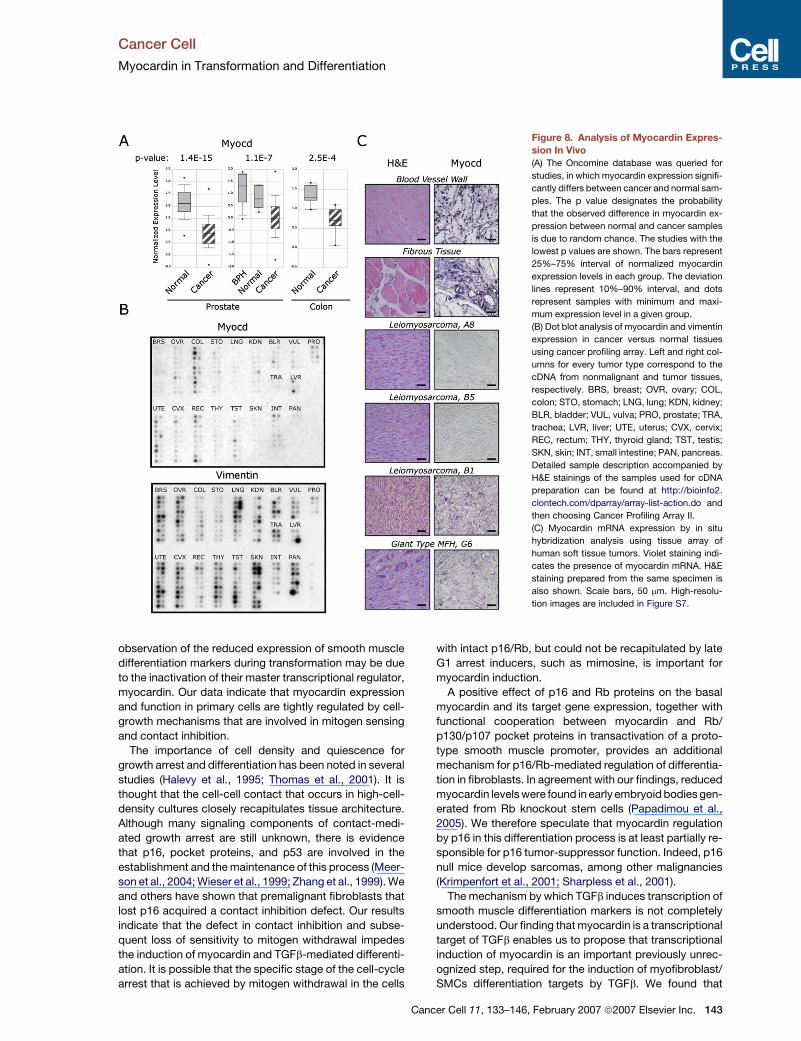
To further examine the hypothesis that myocardin repres-
sion is a frequent event during malignant transformation,
we searched the Oncomine database (Rhodes et al.,
2004) for clinical tumor samples that have significant
downregulation of myocardin expression compared with
the corresponding normal tissues. As demonstrated in
Figure 8A, we found that myocardin was significantly
downregulated in prostate and colon tumor samples
compared with normal tissue controls (Dhanasekaran et al.,
2001; Lapointe et al., 2004; Zou et al., 2002).
Next, we examined myocardin expression in matched
pairs of cDNA prepared from tumor and adjacent normal
tissue derived from individual patients by dot blot analysis
using the Cancer Profiling Array. One hundred and fifty-
four matched pairs representing different cancer tissues
were hybridized with myocardin-specific probe (Fig-
ure 8B). Specificity of myocardin probe was validated by
northern blot analysis in Tslow and Tfast cells. As expected,
ancer Cell 11, 133–146, February 2007 ª2007 Elsevier Inc. 141

Cancer Cell
Myocardin in Transformation and Differentiation
Tfast cells exhibited diminished expression of myocardin as
compared with Tslow cells (Figure S6). Array analysis re-
vealed clear reduction in myocardin expression in colon
(9/10), cervix (9/10), uterus (8/10), and rectum (5/10) can-
cer samples (Figure 8B).
Figure 7. Antiproliferative Activity of Myocardin
(A–C) WST1 proliferation (A), colony forming (B), and soft agar (C) as-
says of HT1080 cells infected with retroviruses encoding myc-tagged
myocardin (Myocd) or GFP control described in Figure 3B. Scale bar,
1 mm. The results of the WST1 cell proliferation assay are presented as
a mean ± SD of triplicate wells from a representative experiment.
(D) WI-38/Tslow cells were infected with retroviruses encoding either
wild-type (wt-Myocd) or dominant-negative (DN-MyocD) myocardin
forms or GFP and plated for colony forming assay.
(E) WI-38/Tslow cells were transfected with siRNA duplexes targeting
either myocardin (Myocd) or LacZ (LacZ) control and 2 days later
plated for colony forming assay.
(F) Comparable passages of prostate fibroblasts isolated from non-
malignant (NAFs) or carcinoma-adjacent areas (CAFs) were plated
for colony forming assay.
142 Cancer Cell 11, 133–146, February 2007 ª2007 Elsevier Inc
Although myocardin reduction in colon cancer was de-
tected by both methods, its reduced expression in pros-
tate tissue was evident only from a bioinformatics survey.
Importantly, Oncomine results were extracted from two
independent studies that compared 62 prostate cancer
samples with 41 normal prostate controls (Lapointe
et al., 2004) and 59 prostate cancer samples with 16 be-
nign prostate hyperplasias (BPH) and 6 normal prostate
tissues (Dhanasekaran et al., 2001). Therefore, one plausi-
ble explanation for this apparent discrepancy might stem
from the small number of prostate cancer cDNAs pre-
sented on the Cancer Profiling Array. It is equally possible
that differences existed between the methodologies used
for tissue harvesting, processing, and normalization in
the studies referenced in Oncomine and in the Cancer
Profiling Array.
To determine whether the reduced myocardin levels are
attributable to the overrepresentation of epithelial com-
partment in tumor samples or reflect changes in the
malignant stroma, we reprobed the membrane with a pan-
mesenchymal marker, vimentin. As is evident from Fig-
ure 8B, vimentin expression was not significantly changed
in those tumor samples, in which significant myocardin re-
duction was detected. Thus, we conclude that reduced
myocardin expression in some epithelial tumors reflects
changes in the tumor stroma.
Finally, we characterized myocardin expression at the
cellular level in vivo using mRNA in situ hybridization
in a panel of soft tissue tumors and adjacent normal tis-
sues. Myocardin-specific signal was abundantly detected
in normal fibrous and aorta-derived tissues, most probably
corresponding to the myofibroblasts and SMCs (Fig-
ure 8C). On the other hand, myocardin expression was
undetectable or low in 43% (3/7) leiomyosarcomas, 33%
(1/3) malignant blood vessel tumors, and 22% (3/14) of fi-
brous tissue tumors. Representative samples of negative
leiomyosarcomas (A8 and B5) are shown. Notably, in
some leiomyosarcomas (sample B1) and in giant cell
type malignant fibrous histiocytoma (sample G6), we de-
tected intratumor heterogeneity in myocardin expression.
In those cases, regions composed of poorly differentiated
malignant cells contained less myocardin signal than more
highly differentiated malignant counterparts (Figure 8C).
Based on the combined evidence from all three inde-
pendent in vivo approaches, together with the ability of
myocardin to inhibit the malignant phenotype of fibro-
sarcoma cell lines and to restrict proliferation of primary
fibroblasts, we conclude that myocardin is an important
constituent of the growth-regulatory circuit aimed to
protect cells from malignant transformation.
DISCUSSION
Although the key role of myocardin in the processes of
cardiac and smooth muscle development is firmly estab-
lished, its involvement in the neoplastic process was not
previously addressed. Our study provides experimental
evidence to support the involvement of myocardin in
carcinogenesis. Our results indicate that the long-known
.
Cancer Cell
Myocardin in Transformation and Differentiation
Figure 8. Analysis of Myocardin Expres-
sion In Vivo
(A) The Oncomine database was queried for
studies, in which myocardin expression signifi-
cantly differs between cancer and normal sam-
ples. The p value designates the probability
that the observed difference in myocardin ex-
pression between normal and cancer samples
is due to random chance. The studies with the
lowest p values are shown. The bars represent
25%–75% interval of normalized myocardin
expression levels in each group. The deviation
lines represent 10%–90% interval, and dots
represent samples with minimum and maxi-
mum expression level in a given group.
(B) Dot blot analysis of myocardin and vimentin
expression in cancer versus normal tissues
using cancer profiling array. Left and right col-
umns for every tumor type correspond to the
cDNA from nonmalignant and tumor tissues,
respectively. BRS, breast; OVR, ovary; COL,
colon; STO, stomach; LNG, lung; KDN, kidney;
BLR, bladder; VUL, vulva; PRO, prostate; TRA,
trachea; LVR, liver; UTE, uterus; CVX, cervix;
REC, rectum; THY, thyroid gland; TST, testis;
SKN, skin; INT, small intestine; PAN, pancreas.
Detailed sample description accompanied by
H&E stainings of the samples used for cDNA
preparation can be found at http://bioinfo2.
clontech.com/dparray/array-list-action.do and
then choosing Cancer Profiling Array II.
(C) Myocardin mRNA expression by in situ
hybridization analysis using tissue array of
human soft tissue tumors. Violet staining indi-
cates the presence of myocardin mRNA. H&E
staining prepared from the same specimen is
also shown. Scale bars, 50 mm. High-resolu-
tion images are included in Figure S7.
observation of the reduced expression of smooth muscle
differentiation markers during transformation may be due
to the inactivation of their master transcriptional regulator,
myocardin. Our data indicate that myocardin expression
and function in primary cells are tightly regulated by cell-
growth mechanisms that are involved in mitogen sensing
and contact inhibition.
The importance of cell density and quiescence for
growth arrest and differentiation has been noted in several
studies (Halevy et al., 1995; Thomas et al., 2001). It is
thought that the cell-cell contact that occurs in high-cell-
density cultures closely recapitulates tissue architecture.
Although many signaling components of contact-medi-
ated growth arrest are still unknown, there is evidence
that p16, pocket proteins, and p53 are involved in the
establishment and the maintenance of this process (Meer-
son et al., 2004; Wieser et al., 1999; Zhang et al., 1999). We
and others have shown that premalignant fibroblasts that
lost p16 acquired a contact inhibition defect. Our results
indicate that the defect in contact inhibition and subse-
quent loss of sensitivity to mitogen withdrawal impedes
the induction of myocardin and TGFb-mediated differenti-
ation. It is possible that the specific stage of the cell-cycle
arrest that is achieved by mitogen withdrawal in the cells
Ca
with intact p16/Rb, but could not be recapitulated by late
G1 arrest inducers, such as mimosine, is important for
myocardin induction.
A positive effect of p16 and Rb proteins on the basal
myocardin and its target gene expression, together with
functional cooperation between myocardin and Rb/
p130/p107 pocket proteins in transactivation of a proto-
type smooth muscle promoter, provides an additional
mechanism for p16/Rb-mediated regulation of differentia-
tion in fibroblasts. In agreement with our findings, reduced
myocardin levels were found in early embryoid bodies gen-
erated from Rb knockout stem cells (Papadimou et al.,
2005). We therefore speculate that myocardin regulation
by p16 in this differentiation process is at least partially re-
sponsible for p16 tumor-suppressor function. Indeed, p16
null mice develop sarcomas, among other malignancies
(Krimpenfort et al., 2001; Sharpless et al., 2001).
The mechanism by which TGFb induces transcription of
smooth muscle differentiation markers is not completely
understood. Our finding that myocardin is a transcriptional
target of TGFb enables us to propose that transcriptional
induction of myocardin is an important previously unrec-
ognized step, required for the induction of myofibroblast/
SMCs differentiation targets by TGFb. We found that
ncer Cell 11, 133–146, February 2007 ª2007 Elsevier Inc. 143
Cancer Cell
Myocardin in Transformation and Differentiation
SRF protein levels are downregulated by serum starvation
and increase upon TGFb treatment (Figure S1), correlating
with aSMA induction. These observations, combined with
the published data in the field and our myocardin siRNA
data (Figure 3D), suggest that upregulation of both myo-
cardin and SRF are necessary for aSMA induction.
Robust antiproliferative activity of myocardin in vitro as
well as its reduced expression in some primary sarcomas
and in the tumor stroma set the ground for further studies
on myocardin function in the carcinogenic process. The
fact that the close myocardin homolog MKL1 is frequently
involved in oncogenic fusions in megakaryoblastic leuke-
mia provides an additional piece of circumstantial evi-
dence for the role of myocardin in cancer (Ma et al.,
2001). Our results suggest that myocardin might act as
a downstream mediator of tumor-suppressive functions
of p16/Rb and/or TGFb in some tumor types. It is clear,
however, that additional transformation-related pathways
regulate myocardin levels and activity. The possibility that
in a subset of cancers myocardin acts as an independent
tumor suppressor targeted by mutations or epigenetically
silenced also requires further investigation. Since myocar-
din knockout is embryonically lethal due to severe defects
in vascular development (Li et al., 2003), conditional or
tissue-specific knockouts are needed for further studies
of the myocardin role in transformation.
The role of myocardin in human carcinogenesis demon-
strated in this study has potentially important basic and
clinical implications. First, myocardin loss may be a fre-
quent event and a powerful oncogenic trigger not only in
mesenchymal, but also in epithelial, tumorigenesis. Sec-
ond, pharmacological reactivation of myocardin may rep-
resent a promising therapeutic strategy, consistent with
the proven transformation-suppressive role of its direct
target, calponin (Horiuchi et al., 1999). Finally, myocardin
targets such as aSMA and calponin are widely used as
markers in sarcoma diagnosis and classification, where
low calponin expression correlates with aggressive clinical
behavior of human angiomyolipoma and osteosarcoma
(Islam et al., 2004; Yamamura et al., 1998). Thus, evalua-
tion of myocardin status might represent a valuable diag-
nostic and prognostic factor in sarcoma.
In summary, our results provide mechanistic insights to
myocardin regulation and demonstrate that, in addition to
its well-recognized function in development and differenti-
ation, myocardin plays an important role in the process of
malignant transformation.
EXPERIMENTAL PROCEDURES
Cell Culture and Treatments
The primary human embryonic lung fibroblasts WI-38 and MRC-5 as
well as their hTERT-immortalized derivatives (MacKenzie et al., 2000;
Milyavsky et al., 2003, 2005; Taylor et al., 2004), hTERT-expressing
normal fibroblasts (NAFs) and carcinoma-associated prostate fibro-
blasts (CAFs) (strain PF179T), and IMR90 human primary fibroblasts
were cultured in MEM supplemented with 10% FCS, 1 mM sodium
pyruvate, 2 mM L-glutamine, and antibiotics. HT1080 and SV80 fibro-
sarcoma and Saos-2 and U2OS osteosarcoma cell lines were main-
tained in DMEM supplemented with 10% FCS and antibiotics.
144 Cancer Cell 11, 133–146, February 2007 ª2007 Elsevier Inc
hTERT-immortalized (PM151T) human prostate SMCs were grown
as described in Kogan et al. (2006).
All cells were maintained in a humidified incubator at 37�C and
5% CO2.
5-Azacytidine (AzaC) and mimosine were purchased from Sigma.
Retroviral Infections
Retrovirus infection procedures using Phoenix producing cells have
been described in detail in Milyavsky et al. (2003).
Plasmids and siRNA
pcDNA3.1-myocardin-myc encoding the full-length myc-tagged
version of mouse myocardin, pcDNA3.1-DN-myocardin-myc encod-
ing dominant-negative form of mouse myocardin and SM22-luciferase
reporter were provided by Dr. Olson (The University of Texas South-
western Medical Center, Dallas, TX).
The pRetroSuper-Hygro (pRSH) construct for stable shRNA expres-
sion and pRSH-sh-p16INK4A was provided by Dr. Agami (The Nether-
lands Cancer Institute, Amsterdam, The Netherlands). pRS-sh-con
that is directed specifically against murine p63 protein was used as
a control shRNA.
pWZL-blasticidin and pWZL-GFP-blasticidin vectors were provided
by Dr. Hahn (Dana-Farber Cancer Institute Boston, MA). pcDNA3-pRb,
pCMV-p107, and pCMV-p130 were provided by Dr. Ginsberg (Bar-Ilan
University, Israel), pCMV5-p15 was provided by J. Massague (Memo-
rial Sloan-Kettering Cancer Center, New York, NY).
pWZL-p16INK4A-blasticidin, pWZL-MyocD-myc-blasticidin, pWZL-
DN-MyocD-myc-blasticidin, and pBabe-p15-Hygro constructs are
described in the Supplemental Data.
The targeted sequences for shRNAs and synthetic RNAi duplexes
are provided in the Supplemental Data.
Reporter Gene Assays
For reporter gene assays, cells were transfected with Fugene 6 Trans-
fection Reagent (Boehringer Mannheim). Cell extracts were prepared
48 hr after transfection, and firefly and Renilla luciferase activities
were determined using Promega materials and procedures. The re-
sults are presented as a mean ± SD of normalized promoter activity
of triplicate wells from a representative experiment.
Quantitative Real-Time PCR
Total RNA was isolated using the RNAeasy kit (Qiagen) according
to the manufacturer’s protocol. A 2 mg aliquot of the total RNA was
reverse transcribed using MMLV RT (Promega) and random hexamer
primers. QRT-PCR was performed using SYBR Green PCR Master Mix
(Applied Biosystems) on ABI 7000 instrument (Applied Biosystems).
The values for the specific genes were normalized to the GAPDH
housekeeping control. Primer sequences used in this study are
provided in the Supplemental Data. The results are presented as a
mean ± SD of two duplicate runs from a representative experiment.
TGFb-Mediated Induction of Myofibroblast Differentiation
For experiments in dense conditions, cells were grown to visual conflu-
ence in 6 cm plates. For experiments in sparse conditions, 105 cells
were seeded in 6 cm plates 2 days before starvation initiation. When
starvation is indicated, cells were washed twice with PBS and changed
to serum-free medium for 24 hr. Cells were exposed to 1 ng/ml TGFb-1
(R&D Systems, Abingdon, UK) for the time periods indicated in the
figure legends.
Western Blotting Analysis
The following primary antibodies were used: smooth muscle a-actin
(Clone 1A4, Sigma); b-tubulin (Sigma); GAPDH (Chemicon MAB374);
p16, p15, and SRF (Santa Cruz Biotechnology); and phospho-Smad2
(Ser465/467) and anti-total SMAD2/3 (Cell Signaling Technology).
a-myc antibody was a gift from Dr. Peles (Weizmann Institute of
Science, Israel). Anti-phospho-ERK1/2 antibody was a gift from
.
Cancer Cell
Myocardin in Transformation and Differentiation
Dr. Seger (Weizmann Institute of Science, Israel). Detailed description
of antibodies is provided in the Supplemental Data.
BrdU Incorporation Assay
Cells were labeled for 30 min with 10 mm of BrdU (Sigma) and stained
with FITC-conjugated anti-BrdU (Becton Dickinson) as described else-
where (Milyavsky et al., 2003) and detailed in the Supplemental Data.
Immunofluorescence
Immunofluorescence of aSMA was performed essentially as described
by Chambers et al. (2003).
Cell Proliferation Assay
The commercial WST1 cell proliferation assay was done according
to the manufacturer’s procedure (Roche Diagnostics, Mannheim,
Germany). The values represent mean ± SD of triplicate wells from
a representative experiment.
Colony Forming Assay
Four thousand HT1080 cells per well of a six-well plate or 2000 WI-38 or
500 PF179T cells per 6 cm plate were seeded. Medium was replaced
every 3–4 days. Two weeks after plating, cells were fixed and stained
by crystal violet.
Anchorage-Independent Growth
Anchorage-independent growth was determined by assaying colony
formation in soft agar. Cells (104) were suspended in 0.5 ml of growth
medium containing blasticidin and 0.3% Seaplaque low-melting-
temperature agarose (BMA, Rockland, ME) and plated in triplicates in
a 6-well plate over a 1.5 ml layer of solidified 0.5% agarose/medium
mixture. The cells were fed every 3 days by adding 100 ml of blastici-
din-containing growth medium. Colonies were photographed at 350
magnification after 2 weeks using a binocular microscope.
Northern Blot, In Situ Hybridization, and Cancer Profiling Arrays
Protocols for northern blot, in situ hybridization using the Soft Tissue
Cancer tissue array (SO801, US Biomax), and detection of myocardin
and vimentin using cancer profiling array II (Clontech) are provided in
the Supplemental Data.
Analysis of Myocardin Promoter Methylation
Details of methylation-specific PCR and bisulfite sequencing are
provided in the Supplemental Data.
Supplemental Data
The Supplemental Data include Supplemental Experimental Proce-
dures and seven supplemental figures and can be found with this
article online at http://www.cancercell.org/cgi/content/full/11/2/133/
DC1/.
ACKNOWLEDGMENTS
The authors would like to thank Dr. Dina Ron for her helpful advice on
in situ hybridization analysis. This research was supported by a Center
of Excellence Grant from the Flight Attendant Medical Research Insti-
tute (FAMRI) and the Yad Abraham Center for Cancer Diagnosis and
Therapy. V.R. is the incumbent of the Norman and Helen Asher Profes-
sorial Chair Cancer Research at the Weizmann Institute of Science.
Received: June 4, 2006
Revised: September 29, 2006
Accepted: November 28, 2006
Published: February 12, 2007
REFERENCES
Button, E., Shapland, C., and Lawson, D. (1995). Actin, its associated
proteins and metastasis. Cell Motil. Cytoskeleton 30, 247–251.
Ca
Chambers, R.C., Leoni, P., Kaminski, N., Laurent, G.J., and Heller, R.A.
(2003). Global expression profiling of fibroblast responses to trans-
forming growth factor-b1 reveals the induction of inhibitor of differen-
tiation-1 and provides evidence of smooth muscle cell phenotypic
switching. Am. J. Pathol. 162, 533–546.
Dhanasekaran, S.M., Barrette, T.R., Ghosh, D., Shah, R., Varambally,
S., Kurachi, K., Pienta, K.J., Rubin, M.A., and Chinnaiyan, A.M. (2001).
Delineation of prognostic biomarkers in prostate cancer. Nature 412,
822–826.
Du, K.L., Ip, H.S., Li, J., Chen, M., Dandre, F., Yu, W., Lu, M.M., Owens,
G.K., and Parmacek, M.S. (2003). Myocardin is a critical serum
response factor cofactor in the transcriptional program regulating
smooth muscle cell differentiation. Mol. Cell. Biol. 23, 2425–2437.
Halevy, O., Novitch, B.G., Spicer, D.B., Skapek, S.X., Rhee, J., Han-
non, G.J., Beach, D., and Lassar, A.B. (1995). Correlation of terminal
cell cycle arrest of skeletal muscle with induction of p21 by MyoD.
Science 267, 1018–1021.
Helman, L.J., and Meltzer, P. (2003). Mechanisms of sarcoma devel-
opment. Nat. Rev. Cancer 3, 685–694.
Horiuchi, A., Nikaido, T., Taniguchi, S., and Fujii, S. (1999). Possible
role of calponin h1 as a tumor suppressor in human uterine leiomyo-
sarcoma. J. Natl. Cancer Inst. 91, 790–796.
Islam, A.H., Ehara, T., Kato, H., Hayama, M., and Nishizawa, O. (2004).
Loss of calponin h1 in renal angiomyolipoma correlates with aggres-
sive clinical behavior. Urology 64, 468–473.
Kogan, I., Goldfinger, N., Milyavsky, M., Cohen, M., Shats, I., Dobler,
G., Klocker, H., Wasylyk, B., Voller, M., Aalders, T., et al. (2006).
hTERT-immortalized prostate epithelial and stromal-derived cells: An
authentic in vitro model for differentiation and carcinogenesis. Cancer
Res. 66, 3531–3540.
Krimpenfort, P., Quon, K.C., Mooi, W.J., Loonstra, A., and Berns, A.
(2001). Loss of p16Ink4a confers susceptibility to metastatic mela-
noma in mice. Nature 413, 83–86.
Kumar, M.S., and Owens, G.K. (2003). Combinatorial control of
smooth muscle-specific gene expression. Arterioscler. Thromb.
Vasc. Biol. 23, 737–747.
Lapointe, J., Li, C., Higgins, J.P., van de Rijn, M., Bair, E., Montgomery,
K., Ferrari, M., Egevad, L., Rayford, W., Bergerheim, U., et al. (2004).
Gene expression profiling identifies clinically relevant subtypes of
prostate cancer. PNAS 101, 811–816. Published online January 7,
2004. 10.1073/pnas.0304146101.
Leavitt, J., Gunning, P., Kedes, L., and Jariwalla, R. (1985). Smooth
muscle alpha-action is a transformation-sensitive marker for mouse
NIH 3T3 and Rat-2 cells. Nature 316, 840–842.
Li, S., Wang, D.Z., Wang, Z., Richardson, J.A., and Olson, E.N. (2003).
The serum response factor coactivator myocardin is required for
vascular smooth muscle development. Proc. Natl. Acad. Sci. USA
100, 9366–9370.
Ma, Z., Morris, S.W., Valentine, V., Li, M., Herbrick, J.A., Cui, X., Bou-
man, D., Li, Y., Mehta, P.K., Nizetic, D., et al. (2001). Fusion of two
novel genes, RBM15 and MKL1, in the t(1;22)(p13;q13) of acute mega-
karyoblastic leukemia. Nat. Genet. 28, 220–221.
MacKenzie, K.L., Franco, S., May, C., Sadelain, M., and Moore, M.A.
(2000). Mass cultured human fibroblasts overexpressing hTERT en-
counter a growth crisis following an extended period of proliferation.
Exp. Cell Res. 259, 336–350.
Massague, J., Blain, S.W., and Lo, R.S. (2000). TGFb signaling in
growth control, cancer, and heritable disorders. Cell 103, 295–309.
Meerson, A., Milyavsky, M., and Rotter, V. (2004). p53 mediates den-
sity-dependent growth arrest. FEBS Lett. 559, 152–158.
Milyavsky, M., Shats, I., Erez, N., Tang, X., Senderovich, S., Meerson,
A., Tabach, Y., Goldfinger, N., Ginsberg, D., Harris, C.C., et al. (2003).
Prolonged culture of telomerase-immortalized human fibroblasts
leads to a premalignant phenotype. Cancer Res. 63, 7147–7157.
ncer Cell 11, 133–146, February 2007 ª2007 Elsevier Inc. 145
Cancer Cell
Myocardin in Transformation and Differentiation
Milyavsky, M., Tabach, Y., Shats, I., Erez, N., Cohen, Y., Tang, X.,
Kalis, M., Kogan, I., Buganim, Y., Goldfinger, N., et al. (2005). Transcrip-
tional programs following genetic alterations in p53, INK4A, and H-Ras
genes along defined stages of malignant transformation. Cancer Res.
65, 4530–4543.
Novitch, B.G., Mulligan, G.J., Jacks, T., and Lassar, A.B. (1996). Skel-
etal muscle cells lacking the retinoblastoma protein display defects in
muscle gene expression and accumulate in S and G2 phases of the cell
cycle. J. Cell Biol. 135, 441–456.
Papadimou, E., Menard, C., Grey, C., and Puceat, M. (2005). Interplay
between the retinoblastoma protein and LEK1 specifies stem cells
toward the cardiac lineage. EMBO J. 24, 1750–1761.
Pipes, G.C., Creemers, E.E., and Olson, E.N. (2006). The myocardin
family of transcriptional coactivators: Versatile regulators of cell
growth, migration, and myogenesis. Genes Dev. 20, 1545–1556.
Rhodes, D.R., Yu, J., Shanker, K., Deshpande, N., Varambally, R.,
Ghosh, D., Barrette, T., Pandey, A., and Chinnaiyan, A.M. (2004).
Large-scale meta-analysis of cancer microarray data identifies com-
mon transcriptional profiles of neoplastic transformation and progres-
sion. Proc. Natl. Acad. Sci. USA 101, 9309–9314.
Serini, G., and Gabbiani, G. (1999). Mechanisms of myofibroblast
activity and phenotypic modulation. Exp. Cell Res. 250, 273–283.
Sharpless, N.E., Bardeesy, N., Lee, K.H., Carrasco, D., Castrillon,
D.H., Aguirre, A.J., Wu, E.A., Horner, J.W., and DePinho, R.A. (2001).
Loss of p16Ink4a with retention of p19Arf predisposes mice to tumor-
igenesis. Nature 413, 86–91.
146 Cancer Cell 11, 133–146, February 2007 ª2007 Elsevier In
Taylor, L.M., James, A., Schuller, C.E., Brce, J., Lock, R.B., and Mack-
enzie, K.L. (2004). Inactivation of p16INK4a, with retention of pRB and
p53/p21cip1 function, in human MRC5 fibroblasts that overcome
a telomere-independent crisis during immortalization. J. Biol. Chem.
279, 43634–43645.
Thomas, D.M., Carty, S.A., Piscopo, D.M., Lee, J.S., Wang, W.F., For-
rester, W.C., and Hinds, P.W. (2001). The retinoblastoma protein acts
as a transcriptional coactivator required for osteogenic differentiation.
Mol. Cell 8, 303–316.
Wang, D., Chang, P.S., Wang, Z., Sutherland, L., Richardson, J.A.,
Small, E., Krieg, P.A., and Olson, E.N. (2001). Activation of cardiac
gene expression by myocardin, a transcriptional cofactor for serum
response factor. Cell 105, 851–862.
Wieser, R.J., Faust, D., Dietrich, C., and Oesch, F. (1999). p16INK4
mediates contact-inhibition of growth. Oncogene 18, 277–281.
Yamamura, H., Yoshikawa, H., Tatsuta, M., Akedo, H., and Takahashi,
K. (1998). Expression of the smooth muscle calponin gene in human
osteosarcoma and its possible association with prognosis. Int. J.
Cancer 79, 245–250.
Zhang, H.S., Postigo, A.A., and Dean, D.C. (1999). Active tran-
scriptional repression by the Rb-E2F complex mediates G1 arrest
triggered by p16INK4a, TGFb, and contact inhibition. Cell 97,
53–61.
Zou, T.T., Selaru, F.M., Xu, Y., Shustova, V., Yin, J., Mori, Y., Shibata,
D., Sato, F., Wang, S., Olaru, A., et al. (2002). Application of cDNA
microarrays to generate a molecular taxonomy capable of distinguish-
ing between colon cancer and normal colon. Oncogene 21, 4855–4862.
c.