Control of epithelial cell migration and invasion by the IKKβ- and CK1α-mediated degradation of...
12
Developmental Cell Article Control of Epithelial Cell Migration and Invasion by the IKKb- and CK1a-Mediated Degradation of RAPGEF2 Roberto Magliozzi, 1 Teck Yew Low, 2,3 Bart G.M.W. Weijts, 4 Tianhong Cheng, 1 Emma Spanjaard, 1 Shabaz Mohammed, 2,3 Anouk van Veen, 6 Huib Ovaa, 5 Johan de Rooij, 1 Fried J.T. Zwartkruis, 6 Johannes L. Bos, 6 Alain de Bruin, 4 Albert J.R. Heck, 2,3 and Daniele Guardavaccaro 1, * 1 Hubrecht Institute-KNAW and University Medical Center Utrecht, Uppsalalaan 8, 3584 CT Utrecht, The Netherlands 2 Biomolecular Mass Spectrometry and Proteomics, Bijvoet Center for Biomolecular Research and Utrecht Institute for Pharmaceutical Sciences, Utrecht University, Padualaan 8, 3584 CH Utrecht, The Netherlands 3 The Netherlands Proteomics Center, Padualaan 8, 3584 CH Utrecht, The Netherlands 4 Department of Pathobiology, Faculty of Veterinary Medicine, Utrecht University, Yalelaan 1, 3584 CL Utrecht, The Netherlands 5 Division of Cell Biology, The Netherlands Cancer Institute, Plesmanlaan 121, 1066 CX Amsterdam, The Netherlands 6 Department of Physiological Chemistry and Center for Biomedical Genetics, University Medical Center Utrecht, Universiteitsweg 100, 3584 CG Utrecht, The Netherlands *Correspondence: [email protected] http://dx.doi.org/10.1016/j.devcel.2013.10.023 SUMMARY Epithelial cell migration is crucial for the develop- ment and regeneration of epithelial tissues. Aberrant regulation of epithelial cell migration has a major role in pathological processes such as the develop- ment of cancer metastasis and tissue fibrosis. Here, we report that in response to factors that promote cell motility, the Rap guanine exchange factor RAPGEF2 is rapidly phosphorylated by I-kappa-B- kinase-b and casein kinase-1a and consequently degraded by the proteasome via the SCF bTrCP ubiq- uitin ligase. Failure to degrade RAPGEF2 in epithelial cells results in sustained activity of Rap1 and inhibi- tion of cell migration induced by HGF, a potent metastatic factor. Furthermore, expression of a degradation-resistant RAPGEF2 mutant greatly suppresses dissemination and metastasis of human breast cancer cells. These findings reveal a molecu- lar mechanism regulating migration and invasion of epithelial cells and establish a key direct link be- tween IKKb and cell motility controlled by Rap- integrin signaling. INTRODUCTION Epithelial cell migration and invasiveness are crucial for morpho- genesis during embryonic development and for tissue regenera- tion. In these processes, epithelial cells lose cell-cell adhesion, develop a mesenchymal cell polarity and, eventually, acquire a highly motile phenotype that enables the invasion of surrounding tissues (Thiery, 2002; Yang and Weinberg, 2008). This biological process, known as epithelial-mesenchymal transition (EMT), has been implicated in diseases such as fibrosis and carcinoma development. Understanding the molecular mechanisms con- trolling epithelial cell migration is key to develop strategies that may have clinical potential. Rap, a small guanosine triphosphatase (GTPase) of the Ras family, is a major regulator of cell polarity, adhesion, and migra- tion (Boettner and Van Aelst, 2009; Bos, 2005). It was originally identified as a protein able to revert the transformed phenotype of K-Ras (Kitayama et al., 1989). Biochemical and genetic studies in various model systems have revealed that Rap is a potent activator of integrins (Duchniewicz et al., 2006; Katagiri et al., 2000; Reedquist et al., 2000; Sebzda et al., 2002). Indeed, a number of growth factors and cytokines stimulate integrin- mediated cell adhesion through the activation of Rap. In addition, Rap is required for the formation and maintenance of E-cadherin-mediated cell-cell adhesion independently of its effects on integrins (Hogan et al., 2004; Knox and Brown, 2002; Kooistra et al., 2007; Price et al., 2004). As other small GTPases, Rap acts as a molecular switch by cycling between inactive GDP-bound and active GTP-bound forms. The transition between these two conformations is tightly controlled by specific guanine nucleotide exchange factors (GEFs), which promote the conversion from the inactive GDP- bound conformation into the active GTP-bound conformation and GTPase-activating proteins (GAPs), which stimulate the intrinsic hydrolytic GTPase activity accelerating the conversion into the inactive GDP-bound form. Rap activity is regulated by a multitude of extracellular signals, which control distinct Rap GEFs and GAPs (Pannekoek et al., 2009). Among them, RAPGEF2 (also known as PDZGEF1, CNRASGEF, NRAPGEP, RA-GEF-1) specifically activates Rap1 and its close relative Rap2 in vitro and in vivo by stimulating GDP-GTP exchange (de Rooij et al., 1999; Kuiperij et al., 2003; Liao et al., 1999; Oht- suka et al., 1999; Rebhun et al., 2000). Genetic approaches have shown that the Caenorhabditis elegans homolog of RAPGEF2, pxf-1, is required for Rap-mediated maintenance of epithelial integrity (Pellis-van Berkel et al., 2005). In Drosophila, loss of function mutants of dPDZGEF/Dizzy display defective develop- ment of various organs including eye, wing, and ovary (Lee et al., 2002). In particular, dPDZGEF controls the formation of 574 Developmental Cell 27, 574–585, December 9, 2013 ª2013 Elsevier Inc.
Transcript of Control of epithelial cell migration and invasion by the IKKβ- and CK1α-mediated degradation of...

Developmental Cell
Article
Control of Epithelial Cell Migrationand Invasion by the IKKb- and CK1a-MediatedDegradation of RAPGEF2Roberto Magliozzi,1 Teck Yew Low,2,3 Bart G.M.W. Weijts,4 Tianhong Cheng,1 Emma Spanjaard,1 Shabaz Mohammed,2,3
Anouk van Veen,6 Huib Ovaa,5 Johan de Rooij,1 Fried J.T. Zwartkruis,6 Johannes L. Bos,6 Alain de Bruin,4
Albert J.R. Heck,2,3 and Daniele Guardavaccaro1,*1Hubrecht Institute-KNAW and University Medical Center Utrecht, Uppsalalaan 8, 3584 CT Utrecht, The Netherlands2Biomolecular Mass Spectrometry and Proteomics, Bijvoet Center for Biomolecular Research and Utrecht Institute for Pharmaceutical
Sciences, Utrecht University, Padualaan 8, 3584 CH Utrecht, The Netherlands3The Netherlands Proteomics Center, Padualaan 8, 3584 CH Utrecht, The Netherlands4Department of Pathobiology, Faculty of Veterinary Medicine, Utrecht University, Yalelaan 1, 3584 CL Utrecht, The Netherlands5Division of Cell Biology, The Netherlands Cancer Institute, Plesmanlaan 121, 1066 CX Amsterdam, The Netherlands6Department of Physiological Chemistry and Center for Biomedical Genetics, University Medical Center Utrecht, Universiteitsweg 100,
3584 CG Utrecht, The Netherlands
*Correspondence: [email protected]://dx.doi.org/10.1016/j.devcel.2013.10.023
SUMMARY
Epithelial cell migration is crucial for the develop-ment and regeneration of epithelial tissues. Aberrantregulation of epithelial cell migration has a majorrole in pathological processes such as the develop-ment of cancer metastasis and tissue fibrosis. Here,we report that in response to factors that promotecell motility, the Rap guanine exchange factorRAPGEF2 is rapidly phosphorylated by I-kappa-B-kinase-b and casein kinase-1a and consequentlydegraded by the proteasome via the SCFbTrCP ubiq-uitin ligase. Failure to degrade RAPGEF2 in epithelialcells results in sustained activity of Rap1 and inhibi-tion of cell migration induced by HGF, a potentmetastatic factor. Furthermore, expression of adegradation-resistant RAPGEF2 mutant greatlysuppresses dissemination and metastasis of humanbreast cancer cells. These findings reveal a molecu-lar mechanism regulating migration and invasion ofepithelial cells and establish a key direct link be-tween IKKb and cell motility controlled by Rap-integrin signaling.
INTRODUCTION
Epithelial cell migration and invasiveness are crucial for morpho-
genesis during embryonic development and for tissue regenera-
tion. In these processes, epithelial cells lose cell-cell adhesion,
develop a mesenchymal cell polarity and, eventually, acquire a
highly motile phenotype that enables the invasion of surrounding
tissues (Thiery, 2002; Yang and Weinberg, 2008). This biological
process, known as epithelial-mesenchymal transition (EMT), has
been implicated in diseases such as fibrosis and carcinoma
development. Understanding the molecular mechanisms con-
574 Developmental Cell 27, 574–585, December 9, 2013 ª2013 Elsev
trolling epithelial cell migration is key to develop strategies that
may have clinical potential.
Rap, a small guanosine triphosphatase (GTPase) of the Ras
family, is a major regulator of cell polarity, adhesion, and migra-
tion (Boettner and Van Aelst, 2009; Bos, 2005). It was originally
identified as a protein able to revert the transformed phenotype
of K-Ras (Kitayama et al., 1989). Biochemical and genetic
studies in various model systems have revealed that Rap is a
potent activator of integrins (Duchniewicz et al., 2006; Katagiri
et al., 2000; Reedquist et al., 2000; Sebzda et al., 2002). Indeed,
a number of growth factors and cytokines stimulate integrin-
mediated cell adhesion through the activation of Rap. In
addition, Rap is required for the formation and maintenance of
E-cadherin-mediated cell-cell adhesion independently of its
effects on integrins (Hogan et al., 2004; Knox and Brown,
2002; Kooistra et al., 2007; Price et al., 2004).
As other small GTPases, Rap acts as a molecular switch by
cycling between inactive GDP-bound and active GTP-bound
forms. The transition between these two conformations is tightly
controlled by specific guanine nucleotide exchange factors
(GEFs), which promote the conversion from the inactive GDP-
bound conformation into the active GTP-bound conformation
and GTPase-activating proteins (GAPs), which stimulate the
intrinsic hydrolytic GTPase activity accelerating the conversion
into the inactive GDP-bound form. Rap activity is regulated by
a multitude of extracellular signals, which control distinct Rap
GEFs and GAPs (Pannekoek et al., 2009). Among them,
RAPGEF2 (also known as PDZGEF1, CNRASGEF, NRAPGEP,
RA-GEF-1) specifically activates Rap1 and its close relative
Rap2 in vitro and in vivo by stimulating GDP-GTP exchange
(de Rooij et al., 1999; Kuiperij et al., 2003; Liao et al., 1999; Oht-
suka et al., 1999; Rebhun et al., 2000). Genetic approaches have
shown that the Caenorhabditis elegans homolog of RAPGEF2,
pxf-1, is required for Rap-mediated maintenance of epithelial
integrity (Pellis-van Berkel et al., 2005). In Drosophila, loss of
function mutants of dPDZGEF/Dizzy display defective develop-
ment of various organs including eye, wing, and ovary (Lee
et al., 2002). In particular, dPDZGEF controls the formation of
ier Inc.
Developmental Cell
RAPGEF2 Degradation Controls Cell Migration
adherens junctions during furrow formation in the ventral epithe-
lium (Spahn et al., 2012). Moreover, deletion of dPDZGEF results
in loss of both germline and somatic stem cells due to an impair-
ment of adherens junctions at the hub-stem cell interface (Wang
et al., 2006). RAPGEF2�/� mouse embryos die between E11.5
and E12.5 with severe organogenesis defects, indicating that
RAPGEF2 is essential for embryonic development inmice (Bilasy
et al., 2009; Satyanarayana et al., 2010; Wei et al., 2007). Alto-
gether, these genetic studies indicate that RAPGEF2 plays a
fundamental role in the development and maintenance of
epithelia, however, the molecular mechanisms that regulate
RAPGEF2 levels and functions remain largely unknown.
SCF ubiquitin ligases target key cellular regulatory proteins for
ubiquitin-mediated proteolysis (Cardozo and Pagano, 2004; Jin
et al., 2004). They are composed of the core subunits Skp1,
Cul1, Rbx1, and one of many F-box proteins that serve as
specific substrate-receptor subunits. SCFbTrCP has been impli-
cated in the degradation of proteins controlling cell cycle pro-
gression, apoptosis, circadian rhythms, and differentiation
(Frescas and Pagano, 2008). All substrates of SCFbTrCP contain
a conserved destruction motif with the consensus DSGXX(X)S,
which, once phosphorylated, mediates the binding to the
WD40 b-propeller structure of bTrCP. Mammals express two
paralogous bTrCP proteins (bTrCP1, also known as FBXW1,
and bTrCP2, also called FBXW11), yet their biochemical proper-
ties are indistinguishable. Wewill therefore use the term bTrCP to
refer to both, unless specified otherwise.
Here, we show that, in response to metastatic factors,
RAPGEF2 is rapidly phosphorylated by CK1a on a conserved
degron and ubiquitylated by SCFbTrCP. CK1a-mediated phos-
phorylation of RAPGEF2 is stimulated by IKKb, which phosphor-
ylates RAPGEF2 on Ser1254. RAPGEF2 ubiquitylation triggers
its proteasome-dependent degradation, enabling inactivation
of Rap1 and induction of cell motility. Remarkably, inhibition of
RAPGEF2 proteolysis blocks migration of epithelial cells and
suppresses metastasis of breast cancer cells. Thus, CK1a-
and IKKb-dependent degradation of RAPGEF2 represents a crit-
ical event required for epithelial cell migration and invasion.
RESULTS
Rapid bTrCP-Dependent Degradation of RAPGEF2 inResponse to Stimuli that Induce Cell MigrationTo identify substrates of the SCFbTrCP ubiquitin ligase, we used
an immunoaffinity assay followed by mass spectrometry anal-
ysis (Kruiswijk et al., 2012; Low et al., 2013). HEK293T cells
were transfected with FLAG-HA epitope-tagged bTrCP2 and
treated with the proteasome inhibitor MG132. Proteins that
coimmunoprecipitated with FLAG-HA-bTrCP2 were analyzed
by liquid chromatography-tandem mass spectrometry. We
recovered 14 peptides corresponding to the Rap guanine nucle-
otide exchange factor RAPGEF2 (Figure S1A available online).
We then followed the reciprocal approach and immunopurified
FLAG-HA epitope-tagged RAPGEF2 from HEK293T cells. We
identified 7, 14, 3, 2, and 1 peptides derived from the SCF
subunits bTrCP1, bTrCP2, Skp1, Cul1, and Rbx1, respectively
(Figure S1B). In addition, peptides corresponding to the small
GTPases Rap1 (isoforms A and B) and Rap2 (isoforms B and
C) were detected in the RAPGEF2 immunopurification (Fig-
Developm
ure S1C). Of note, we never observed other members of the
Ras family of small G-proteins when we used RAPGEF2 as bait.
To verify the specificity of the bTrCP-RAPGEF2 binding, we
immunoprecipitated a number of FLAG epitope-tagged F-box
proteins as well as the related proteins Cdh1 and Cdc20 from
HEK293T cells and examined their ability to pull-down endoge-
nous RAPGEF2. bTrCP1 and its paralog bTrCP2 coimmunopre-
cipitated with endogenous RAPGEF2 (Figure 1A), whereas other
members of the FBXW family of F-box proteins, FBXW2, FBXW4,
FBXW5, FBXW7, FBXW8, or the APC/C activators Cdh1 and
Cdc20 (also containing WD40 repeats) did not. A complex with
the endogenous bTrCP and RAPGEF2 proteins was also
observed (Figure 1B).
It has been shown that the WD40 b-propeller structure of
bTrCP is required for the interaction with its substrate proteins
and thatmutation of a specific arginine residue (Arg447 of human
bTrCP2, isoformC) in theWD40 repeats abolishes both the bind-
ing and ubiquitin conjugation of the substrate (Kruiswijk et al.,
2012; Wu et al., 2003). To determine if the WD40 b-propeller
structure of bTrCP is responsible for the binding to RAPGEF2,
we expressed in HEK293T cells wild-type bTrCP2 and the
bTrCP2(R447A) mutant, which were then immunoprecipitated.
Whereas wild-type bTrCP2 immunoprecipitated endogenous
RAPGEF2 and the established substrate b-catenin, the
bTrCP2(R447A) mutant did not (Figure 1C).
To test whether RAPGEF2 is a substrate of the SCFbTrCP ubiq-
uitin ligase, we reconstituted the ubiquitylation of RAPGEF2
in vitro. bTrCP1, but not an inactive bTrCP1(DF box) mutant,
was able to efficiently ubiquitylate RAPGEF2 (Figure 1D).
Before examining a putative function of the SCFbTrCP ubiquitin
ligase in targeting RAPGEF2 for degradation, we sought to find
under which condition RAPGEF2 is degraded in the cell. As
Rap1 is a key mediator of cell adhesion, we hypothesized that
RAPGEF2 may be downregulated in response to stimuli that
disrupt cell adhesion and induce cell migration. To test this
hypothesis, we analyzed RAPGEF2 protein levels in epithelial
Madin-Darby canine kidney (MDCK) cells treated with hepato-
cyte growth factor/scatter factor (HGF/SF). This is a well-estab-
lished in vitro model system that has been extensively used to
study the mechanisms by which epithelial cells become migra-
tory, mesenchymal-like cells. HGF is known to induce centrifugal
spreading of MDCK cell colonies, loss of cell-cell adhesion, and
increase in cell motility without stimulating cell growth (Gherardi
et al., 1989; Stoker et al., 1987; Stoker and Perryman, 1985;
Tanimura et al., 1998). Figure 1E shows that RAPGEF2 levels
rapidly decreased in response to HGF. The proteasome inhibitor
MG132 prevented the decrease of RAPGEF2, indicating that
RAPGEF2 degradation is mediated by the proteasome.
RAPGEF2 destruction was also triggered following treatment
with phorbol-12-myristate-13-acetate (PMA) (Figure 1F), which
is known to induce a marked scattering of MDCK cells without
affecting significantly cell proliferation (Rosen et al., 1991; Tani-
mura et al., 1998), but not in response to a number of other
growth factors, such as epidermal growth factor (EGF) or insu-
lin-like growth factor (IGF), which do not induce scattering of
MDCK cells (Tanimura et al., 1998) (Figures S1D and S1E). Pro-
teasome-dependent degradation of RAPGEF2 was observed
upon HGF treatment of human epithelial kidney HEK293 cells
(Figure S1F), which form epithelial layers similar to MDCK cells
ental Cell 27, 574–585, December 9, 2013 ª2013 Elsevier Inc. 575

A
B
C
D
GH I
FE
Figure 1. RAPGEF2 Is Targeted for Degradation by SCFbTrCP in Response to Stimuli that Induce Cell Migration
(A) The indicated FLAG-tagged F-box proteins (FBPs), the APC/C activators Cdh1 and Cdc20 or an empty vector (EV) were expressed in HEK293T cells. Forty-
eight hours after transfection, cells were treated for 5 hr with the proteasome inhibitor MG132, then harvested and lysed. Whole cell extracts were immuno-
precipitated (IP) with anti-FLAG resin and immunoblotted with the indicated antibodies. AB1 and AB2 are two different anti-RAPGEF2 antibodies.
(B) HEK293T cells were treated for 5 hr with the proteasome inhibitor MG132, then harvested and lysed. Whole cell extracts were immunoprecipitated (IP) with
anti-RAPGEF2 antibody and immunoblotted with the indicated antibodies.
(C) Arg447 in theWD40 repeat of bTrCP2 is required for the interaction with RAPGEF2. HEK293T cells were transfected as indicated and analyzed as in (A). WCE,
whole cell extract; WT, wild-type.
(D) RAPGEF2, Skp1, Cul1, and Rbx1 were expressed in HEK293T cells in the absence or presence of FLAG-tagged bTrCP1 or a FLAG-tagged bTrCP1(DF-box)
mutant. After immunopurification with anti-FLAG resin, in vitro ubiquitylation of RAPGEF2 was performed as described in the Supplemental Experimental
Procedures. Samples were analyzed by immunoblotting with an anti-RAPGEF2 antibody. The bracket indicates a ladder of bands corresponding to poly-
ubiquitylated RAPGEF2.
(E and F) MDCK cells were treated with HGF (E) or PMA (F) with or without the proteasome inhibitor MG132. Cells were collected at the indicated times and lysed.
Whole cell extracts were subjected to immunoblotting with the indicated antibodies. Actin is shown as a loading control.
(G) HEK293 cells were transfected with the indicated siRNA oligonucleotides and treated with PMA. Cells were then collected and analyzed as in (E).
(H and I) HEK293 cells were transfected with an empty vector or FLAG-tagged bTrCP1. Forty-eight hours after transfection, cells were treated, when indicated,
with MG132 and with either HGF (H) or PMA (I) for 4 hr, then harvested and lysed. Whole cell extracts were immunoprecipitated (IP) with anti-FLAG resin, and
immunoblotted with anti-RAPGEF2 and anti-FLAG antibodies.
See also Figure S1.
Developmental Cell
RAPGEF2 Degradation Controls Cell Migration
and have an intact HGF signaling (Sakkab et al., 2000). The
degradation of RAPGEF2 in response to the motogenic stimulus
is a rapid event that starts much earlier than the downregulation
of E-cadherin, suggesting that the degradation of RAPGEF2 is
not an indirect consequence of cell junction disassembly (data
not shown).
To test whether the degradation of RAPGEF2 observed in
response to factors that induce cell migration is mediated by
bTrCP, we reduced the levels of both bTrCP1 and bTrCP2 in
HEK293 cells using a previously validated siRNA (Guardavac-
576 Developmental Cell 27, 574–585, December 9, 2013 ª2013 Elsev
caro et al., 2008; Kruiswijk et al., 2012). We found that bTrCP
knockdown blocked the PMA-induced degradation of RAPGEF2
(Figure 1G). Accordingly, the binding of bTrCP to endogenous
RAPGEF2 was stimulated by both HGF and PMA (Figures 1H
and 1I).
HGF-Induced Phosphorylation of RAPGEF2 by CK1aTriggers RAPGEF2 DegradationThe WD40 b-propeller structure of bTrCP interacts with its
substrate proteins via a diphosphorylated degradation motif
ier Inc.
Developmental Cell
RAPGEF2 Degradation Controls Cell Migration
(phosphodegron) with the consensus DpSGXX(X)pS (Cardozo
and Pagano, 2004; Frescas and Pagano, 2008; Wu et al.,
2003). We identified one canonical DpSGXX(X)pS motif in
human RAPGEF2 that might potentially be the phosphodegron
(Figure S2A). We mutated the serine residues in this motif to
alanine and determined the ability of the RAPGEF2 mutant to
interact with bTrCP. Whereas wild-type RAPGEF2 immunopre-
cipitated bTrCP1, the RAPGEF2(S1244A/S1248A) mutant did
not (Figure 2A). The motif surrounding S1244 and S1248
is highly conserved in vertebrate orthologs of RAPGEF2
(Figure 2B).
As a further method to examine whether phosphorylation is
required for the interaction of RAPGEF2 with bTrCP, we used
immobilized synthetic peptides comprising the bTrCP-binding
domain of RAPGEF2 (aa 1240–1252 in human RAPGEF2). As
shown in Figure 2C, a RAPGEF2-derived peptide containing
phosphoserine residues at positions Ser1244 and Ser1248
associated with in vitro translated bTrCP1, but not with a
different F-box protein, whereas the unphosphorylated peptide
did not associate at all, suggesting that phosphorylation of
Ser1244 and Ser1248 directly mediates the association with
bTrCP.
To investigate whether Ser1244 and Ser1248 are phos-
phorylated in vivo in response to factors that induce cell motility,
we generated a phosphospecific antibody against the1240DAADpSGRGpSWTSC1252 peptide with phosphoserine
residues at positions 1244 and 1248. This antibody detected
wild-type RAPGEF2, but not the RAPGEF2(S1244A/S1248A)
mutant (Figure S2B). Moreover, l-phosphatase treatment of
immunoprecipitated wild-type RAPGEF2 inhibited RAPGEF2
detection by the phosphospecific antibody (Figure S2C). We
then used this antibody to test whether RAPGEF2 is phosphory-
lated in vivo. Figure 2D shows that RAPGEF2 was phosphory-
lated on Ser1244 and Ser1248 in HEK293 cells that were treated
with HGF.
In the RAPGEF2 immunopurification described above, we also
recovered three peptides corresponding to casein kinase 1
(CK1, isoform a; Figure S2D).We first confirmed that CK1a coim-
munoprecipitated with RAPGEF2 in vivo (Figure S2E). To test
whether CK1a is involved in the phosphorylation of RAPGEF2,
we used pharmacological inhibitors and found that the CK1
inhibitors D4476 and IC261 prevented both the HGF-induced
binding of bTrCP1 to RAPGEF2 and the phosphorylation of
RAPGEF2 on Ser1244/Ser1248 (Figure 2E). Accordingly,
D4476 blocked the HGF-induced degradation of RAPGEF2 (Fig-
ure 2F). To rule out nonspecific effects of these inhibitors, we
silenced CK1a by RNAi (Gao et al., 2011; Tapia et al., 2006).
The knockdown of CK1a inhibited the proteasome-dependent
degradation of RAPGEF2 in response to HGF (Figure 2G) as
well as RAPGEF2 interaction with bTrCP (Figure 2H).
In order to determine if CK1a directly phosphorylates the
RAPGEF2 degron, we carried out an in vitro kinase assay, using
purified recombinant CK1a. CK1a, but not CK2, GSK3b, or
CDK1 (kinases involved in the phosphorylation of other sub-
strates of bTrCP), phosphorylated the degron of RAPGEF2
in vitro, as shown by the recognition by our phosphospecific anti-
body (Figure 2I). Altogether these results indicate that
CK1a-mediated phosphorylation of RAPGEF2 on Ser1244/
Ser1248 is required for RAPGEF2 degradation induced by HGF.
Developm
IKKb-Mediated Phosphorylation of RAPGEF2 IsRequired for RAPGEF2 DegradationSubstrates of bTrCP are phosphorylated on their degrons
following an initial phosphorylation event that either generates
a binding site for the kinase phosphorylating the degron or
exposes an otherwise masked degron. We noticed a consensus
sequence for phosphorylation by I-kappa-B kinase (IKK) in close
proximity to the RAPGEF2 phosphodegron (Figure S3A). Phorbol
esters (PMA) and HGF have been shown to stimulate the activity
of IKKb in epithelial cells (Fan et al., 2005, 2007, 2009; Hah and
Lee, 2003; Huang et al., 2003; Muller et al., 2002). First, we
confirmed that treatment of epithelial cells with HGF or PMA
results in the activation of IKKb (Figure S3B). We then tested
whether IKKb is able to phosphorylate RAPGEF2 by performing
an in vitro kinase assay using recombinant kinases and immuno-
purified RAPGEF2, which had been previously dephosphory-
lated. IKKb, and to a lesser extent IKKa, were able to phosphor-
ylate RAPGEF2 (as shown by incorporation of radiolabeled
phosphate), however, no phosphorylation was detected by our
phosphospecific antibody on the RAPGEF2 degron (Figures 3A
and S3C), indicating that IKKa/b phosphorylates residues of
RAPGEF2 different from Ser1244 and Ser1248.
Next, we tested whether RAPGEF2 phosphorylation by IKKb
affects the CK1a-dependent phosphorylation of the RAPGEF2
degron in vitro. When purified recombinant IKKb was used to
phosphorylate RAPGEF2 (and washed away before CK1a addi-
tion), stimulation of the CK1a-mediated phosphorylation of the
RAPGEF2 degron was observed (Figures 3B and S3C). IKKa
did not have any effect on the CK1a-mediated phosphorylation
of the RAPGEF2 degron (Figure S3C). Accordingly, in cultured
cells, CK1a-mediated phosphorylation of the RAPGEF2 degron
(Figures 3C and 3D), RAPGEF2 binding to bTrCP (Figure 3D),
and RAPGEF2 ubiquitylation (Figure 3E) were stimulated by the
overexpression of wild-type IKKb and, more extensively, the
constitutively active IKKb(S177E/S181E) mutant.
To assess whether IKKb is involved in the degradation of
RAPGEF2, we overexpressed IKKb and analyzed the abundance
of RAPGEF2 in the absence or presence of the proteasome
inhibitor MG132. Overexpression of the constitutively active
IKKb(S177E/S181E) mutant, but not the constitutively inactive
IKKb(S177A/S181A) mutant, resulted in RAPGEF2 downregula-
tion, which was prevented by proteasomal inhibition (Figure 3F).
Conversely, knockdown of IKKb prevented both the HGF- and
the PMA-induced degradation of RAPGEF2 (Figures 3G, 3H,
and S3D). Accordingly, pharmacological inhibition of IKK
blocked both the CK1a-dependent phosphorylation of the
RAPGEF2 degron and RAPGEF2 binding to bTrCP (Figure 3I).
Further, we found that IKKb was coimmunoprecipitated with
RAPGEF2 in vivo (Figure S3E). Taken together, these results indi-
cate that IKKb-dependent phosphorylation of RAPGEF2 is
required for RAPGEF2 degradation induced by HGF and medi-
ated by bTrCP.
We then employed mass spectrometry to pinpoint the specific
RAPGEF2 sites targeted by IKKb. Immunopurified, dephos-
phorylated RAPGEF2 was subjected to an in vitro kinase assay
in the presence or absence of purified kinases, prior to mass
spectrometry analysis. We identified phosphopeptides contain-
ing phospho-Ser1254 in IKKb-treated RAPGEF2 samples (Fig-
ures S3F and S3G). These phosphopeptides were not found in
ental Cell 27, 574–585, December 9, 2013 ª2013 Elsevier Inc. 577
A
B
C D
EF G
H
I
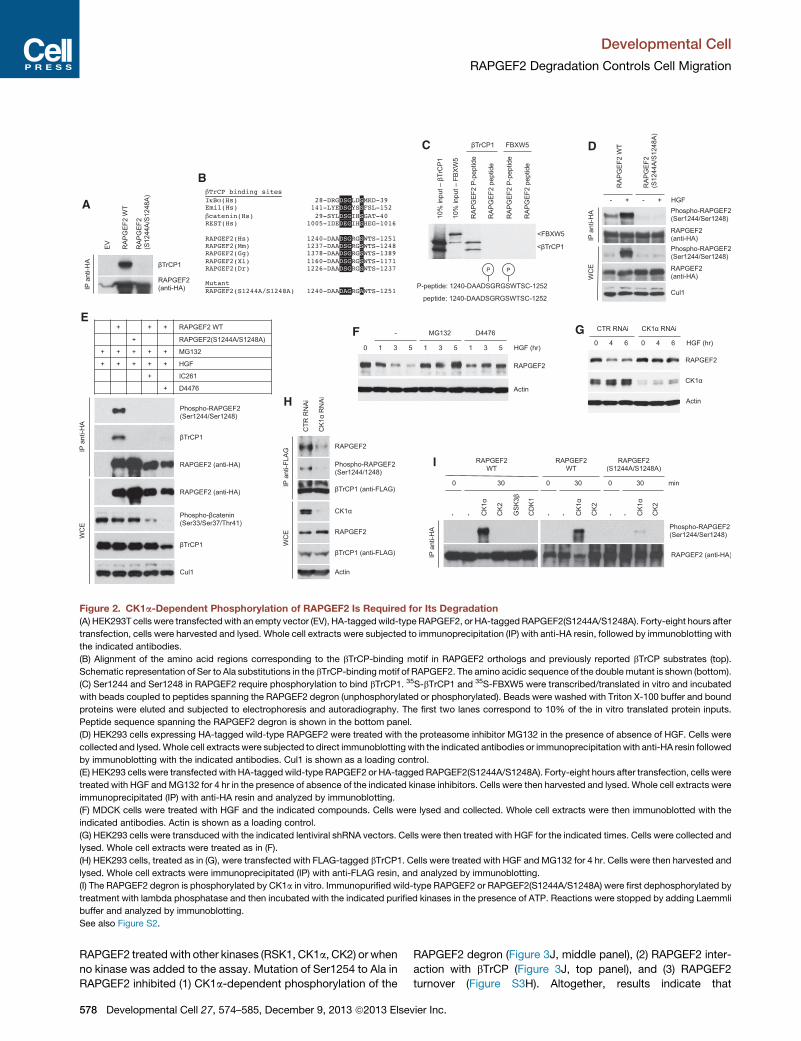
Figure 2. CK1a-Dependent Phosphorylation of RAPGEF2 Is Required for Its Degradation(A) HEK293T cells were transfectedwith an empty vector (EV), HA-taggedwild-type RAPGEF2, or HA-taggedRAPGEF2(S1244A/S1248A). Forty-eight hours after
transfection, cells were harvested and lysed. Whole cell extracts were subjected to immunoprecipitation (IP) with anti-HA resin, followed by immunoblotting with
the indicated antibodies.
(B) Alignment of the amino acid regions corresponding to the bTrCP-binding motif in RAPGEF2 orthologs and previously reported bTrCP substrates (top).
Schematic representation of Ser to Ala substitutions in the bTrCP-binding motif of RAPGEF2. The amino acidic sequence of the double mutant is shown (bottom).
(C) Ser1244 and Ser1248 in RAPGEF2 require phosphorylation to bind bTrCP1. 35S-bTrCP1 and 35S-FBXW5 were transcribed/translated in vitro and incubated
with beads coupled to peptides spanning the RAPGEF2 degron (unphosphorylated or phosphorylated). Beads were washed with Triton X-100 buffer and bound
proteins were eluted and subjected to electrophoresis and autoradiography. The first two lanes correspond to 10% of the in vitro translated protein inputs.
Peptide sequence spanning the RAPGEF2 degron is shown in the bottom panel.
(D) HEK293 cells expressing HA-tagged wild-type RAPGEF2 were treated with the proteasome inhibitor MG132 in the presence of absence of HGF. Cells were
collected and lysed.Whole cell extracts were subjected to direct immunoblottingwith the indicated antibodies or immunoprecipitation with anti-HA resin followed
by immunoblotting with the indicated antibodies. Cul1 is shown as a loading control.
(E) HEK293 cells were transfected with HA-taggedwild-type RAPGEF2 or HA-tagged RAPGEF2(S1244A/S1248A). Forty-eight hours after transfection, cells were
treated with HGF andMG132 for 4 hr in the presence of absence of the indicated kinase inhibitors. Cells were then harvested and lysed. Whole cell extracts were
immunoprecipitated (IP) with anti-HA resin and analyzed by immunoblotting.
(F) MDCK cells were treated with HGF and the indicated compounds. Cells were lysed and collected. Whole cell extracts were then immunoblotted with the
indicated antibodies. Actin is shown as a loading control.
(G) HEK293 cells were transduced with the indicated lentiviral shRNA vectors. Cells were then treated with HGF for the indicated times. Cells were collected and
lysed. Whole cell extracts were treated as in (F).
(H) HEK293 cells, treated as in (G), were transfected with FLAG-tagged bTrCP1. Cells were treated with HGF and MG132 for 4 hr. Cells were then harvested and
lysed. Whole cell extracts were immunoprecipitated (IP) with anti-FLAG resin, and analyzed by immunoblotting.
(I) The RAPGEF2 degron is phosphorylated by CK1a in vitro. Immunopurified wild-type RAPGEF2 or RAPGEF2(S1244A/S1248A) were first dephosphorylated by
treatment with lambda phosphatase and then incubated with the indicated purified kinases in the presence of ATP. Reactions were stopped by adding Laemmli
buffer and analyzed by immunoblotting.
See also Figure S2.
Developmental Cell
RAPGEF2 Degradation Controls Cell Migration
RAPGEF2 treatedwith other kinases (RSK1, CK1a, CK2) or when
no kinase was added to the assay. Mutation of Ser1254 to Ala in
RAPGEF2 inhibited (1) CK1a-dependent phosphorylation of the
578 Developmental Cell 27, 574–585, December 9, 2013 ª2013 Elsev
RAPGEF2 degron (Figure 3J, middle panel), (2) RAPGEF2 inter-
action with bTrCP (Figure 3J, top panel), and (3) RAPGEF2
turnover (Figure S3H). Altogether, results indicate that
ier Inc.

A
E
B
FH
I
C D
JG
Figure 3. IKKb Stimulates the CK1a-Mediated Degradation of RAPGEF2
(A) Immunopurifiedwild-type RAPGEF2 andRAPGEF2(S1244A/S1248A) were first dephosphorylated by treatment with lambda phosphatase and then incubated
with the indicated purified kinases in the presence of g32P ATP. Reactions were stopped by adding Laemmli buffer, run on SDS-PAGE and analyzed by auto-
radiography (top panels) and immunoblotting (bottom panels).
(B) Immunopurified wild-type RAPGEF2 was first dephosphorylated by treatment with lambda phosphatase and then incubated with the indicated kinases.
Kinases were then washed away prior to addition of CK1a. Reactions were stopped at the indicated times and analyzed by immunoblotting.
(C and D) HEK293 cells were transfected with the indicated constructs. Cells were treated with HGF and MG132 for 4 hr. Cells were then harvested and lysed.
Whole cell extracts were immunoprecipitated (IP) with anti-HA resin, and immunoblotted with antibodies specific for the indicated proteins. Immunocomplexes
were treated with lambda phosphatase when indicated. CA, constitutively active.
(E) HEK293 cells, transfected with the indicated constructs, were treated as in (C), then harvested and lysed in 0.1% Triton X-100 lysis buffer. Whole cell extracts
were denatured by adding 1% SDS and boiling for 10 min. SDS was quenched and diluted. Whole cell extracts were then immunoprecipitated (IP) with anti-HA
resin, and immunoblotted with antibodies specific for the indicated proteins. The bracket indicates a ladder of bands corresponding to poly-ubiquitylated
RAPGEF2.
(F) HEK293were transfected with HA-tagged RAPGEF2 and the constitutively active IKKb(S177E/S181E) or inactive IKKb(S177A/S181A) mutant and treated with
MG132 in presence of HGF. After 48 hr, cells were collected, lysed and subjected to immunoblotting. CA, constitutively active; CI, constitutively inactive.
(G andH) Cells were transfectedwith the indicated siRNA oligonucleotides and treatedwith HGF only (G) or HGF and cycloheximide (H) to block protein synthesis.
Cells were then collected at the indicated times. Whole cell extracts were analyzed by immunoblotting. Cul1 is shown as a loading control.
(I) HEK293 cells were transfected with HA-tagged wild-type RAPGEF2. Forty-eight hours after transfection, cells were treated with HGF andMG132 for 4 hr in the
presence or absence of the IKK inhibitor SC-514. Cells were then harvested and lysed. Whole cell extracts were immunoprecipitated (IP) with anti-HA resin, and
immunoblotted with the indicated antibodies.
(J) HEK293 cells were transfected with an empty vector (EV), HA-tagged wild-type RAPGEF2, HA-tagged RAPGEF2(S1244A/S1248A), or HA-tagged
RAPGEF2(S1254A). Forty-eight hours after transfection, cells were treated with the proteasome inhibitor MG132 in the presence of HGF, then harvested and
lysed. Whole cell extracts were analyzed as in (I).
See also Figure S3.
Developmental Cell
RAPGEF2 Degradation Controls Cell Migration
IKKb-mediated phosphorylation of RAPGEF2 on Ser1254
promotes the phosphorylation of the RAPGEF2 degron by
CK1a, RAPGEF2 binding to bTrCP, and RAPGEF2 degradation.
RAPGEF2 Degradation Controls the HGF-InducedMigration of Epithelial CellsTo examine the biological function of RAPGEF2 degradation, we
transduced MDCK cells with lentiviruses expressing physiolog-
ical levels of wild-type RAPGEF2 or RAPGEF2(S1244A/
S1248A) (Figure S4A). We first confirmed that both the steady
state levels and the half-life of RAPGEF2(S1244A/S1248A)
Developm
were increased when compared with wild-type RAPGEF2 in
MDCK cells treated with HGF (Figures 4A and 4B) or PMA (Fig-
ures S4B and S4C). As expected, growth factors that do not
induce scattering of MDCK cells, such as EGF and PDGF, did
not lead to degradation of either wild-type RAPGEF2 or
RAPGEF2(S1244A/S1248A) (Figures S4D and S4E). Notably,
the RAPGEF2(S1244A/S1248A) mutant that escaped degrada-
tion upon HGF stimulation localized to the plasma membrane
(Figure S4F).
Next, we examined whether RAPGEF2 degradation affected
the activity of Rap1 in response to HGF. Whereas in cells
ental Cell 27, 574–585, December 9, 2013 ª2013 Elsevier Inc. 579

A
B
C
D
F I
E
J
GH
Figure 4. RAPGEF2 Degradation Is Required for Rap1 Inactivation and Stimulation of Cell Migration in Response to HGF
(A and B) MDCK cells, transducedwith lentiviruses expressing HA-tagged wild-type RAPGEF2 or HA-tagged RAPGEF2(S1244A/S1248A), were treated with HGF
only (A) or HGF and the inhibitor of protein synthesis cycloheximide (B) for the indicated times. Cells were then collected and analyzed by immunoblotting. Actin
and Cul1 are shown as a loading control.
(C) MDCK cells, transduced as in (A), were treated with HGF for the indicated times. Cells were collected and lysed. Whole cell extracts were analyzed by
immunoblotting with the indicated antibodies and in a pull-down assay using a GST fusion of the activated Rap1-binding domain of RalGDS. The levels of
precipitated Rap1 were determined by immunoblotting using an anti-Rap1 antibody. The asterisk indicates a nonspecific band. Actin is shown as a loading
control. To facilitate comparison, a dotted line separates samples from cells expressing wild-type RAPGEF2 and samples from cells expressing
RAPGEF2(S1244A/S1248A). The graph shows the abundance of Rap1-GTP normalized to total Rap1 and relative to Rap1-GTP in cells expressing wild-type
RAPGEF2 at time 0. Values are averaged with the ones from three additional independent experiments (n = 4 ± SD).
(D) MDCK cells, transduced with lentiviruses expressing wild-type RAPGEF2 or RAPGEF2(S1244A/S1248A), were treated with HGF and imaged by time-lapse
phase-contrast microscopy for 16 hr. Representative phase-contrast images from the time-lapse series are shown. Scale bars represent 100 mm.
(E) Quantification of scattering from time-lapse experiments. The graph shows the number of islands scattered (islands in which cells have disrupted cell-cell
contacts) after HGF treatment. p < 0.001 (Pearson’s c2 test). Only islands including 5–15 cells were scored.
(F) An automated cell tracking software was employed to measure the average migration velocity of MDCK cells expressing wild-type RAPGEF2 or the
RAPGEF2(S1244A/S1248A) mutant in the presence of HGF. For each cell line/condition, three independent time-lapse image series (at least 300 individual cells)
were analyzed.
(G) Representative individual migratory tracks of MDCK cells expressing wild-type RAPGEF2 or RAPGEF2(S1244A/S1248A) in the presence of HGF.
(H) Track distance of individual cells shown in (G). Horizontal lines represent the mean. p < 0.001 (Student’s t test).
(I) MDCK cells expressing wild-type RAPGEF2 or the RAPGEF2(S1244A/S1248A) mutant were grown to confluence. Cell monolayers were wounded and then
treated with HGF. Cells were photographed immediately after wounding (0 hr) and after 4 (4 hr) and 8 hr (8 hr).
(J) The graph represents the relative wound closure at 0, 4, and 8 hr (n = 5 ± SD).
*p = 0.005 (Student’s t test). See also Figure S4.
Developmental Cell
RAPGEF2 Degradation Controls Cell Migration
expressing wild-type RAPGEF2, Rap1 was first rapidly activated
and then inactivated following HGF treatment (as shown by the
amount of the GTP-bound Rap1), cells expressing the stable
RAPGEF2 mutant displayed sustained Rap1 activity (Figure 4C).
On the contrary, Rap2 activity was neither regulated by HGF
treatment nor affected by RAPGEF2 degradation in HGF-treated
cells (data not shown).
580 Developmental Cell 27, 574–585, December 9, 2013 ª2013 Elsev
To assess the effect of defective degradation of RAPGEF2 on
HGF-induced cell scattering, we employed a live-cell micro-
scopy assay (de Rooij et al., 2005; Loerke et al., 2012). MDCK
cells expressing physiological levels of wild-type RAPGEF2 or
RAPGEF2(S1244A/S1248A) were treated with HGF and followed
by time-lapse imaging (Figure 4D). We quantified cell scattering
by scoring the percentage of cell islands (groups of 5–16 cells) in
ier Inc.

Developmental Cell
RAPGEF2 Degradation Controls Cell Migration
which three or more cells had disrupted contacts with neigh-
boring cells (Figure 4E). MDCK cells expressing the stable
RAPGEF2(S1244A/S1248A) mutant displayed decreased scat-
tering when compared with cells expressing wild-type
RAPGEF2. Next, we employed an automated cell tracking soft-
ware that tracks individual cell velocity and trajectories from
consecutive time-lapse images (de Rooij et al., 2005; Loerke
et al., 2012). As shown in Figure 4F, cells expressing the
RAPGEF2 stable mutant displayed defective induction of
average cell speed following HGF treatment. Accordingly,
individual migratory tracks of MDCK cells expressing
RAPGEF2(S1244AS1248A) treated with HGF were shorter
when compared with the ones of MDCK cells expressing wild-
type RAPGEF2 (Figures 4G and 4H).
Scattering of epithelial cells in response to HGF is character-
ized by two major steps, i.e., loss of cell adhesion, followed by
an increase in cell motility (Loerke et al., 2012). To assess in
which of these two processes the degradation of RAPGEF2 is
involved, we analyzed the motility of noncontacted cells (not
starting from cell islands) expressing wild-type RAPGEF2 or
the nondegradable RAPGEF2(S1244A/S1248A) mutant in
response to HGF. As shown in Figures S4G–S4I, HGF-induced
motility of noncontacted cells expressing RAPGEF2(S1244A/
S1248A) is reduced when compared with the one of noncon-
tacted cells expressing wild-type RAPGEF2 indicating that the
degradation of RAPGEF2 is required for the HGF-induced in-
crease in cell migration even in the absence of adherens
junctions.
To confirm that the nondegradable RAPGEF2 mutant inhibits
cell motility independently of cell-cell adhesion, we analyzed
the HGF-induced scattering in cells in which cell-cell junctions
were previously inhibited by low calcium conditions (Figures
S4J–S4M). Cells expressing the stable RAPGEF2 mutant
displayed a remarkable decrease in motility when compared
with cells expressing wild-type RAPGEF2 even in low calcium
conditions.
Taken together, these results indicate that RAPGEF2 degra-
dation is required for HGF-induced cell migration. As expected,
HGF-induced motility of MDCK cells was inhibited if RAPGEF2
degradation was bypassed by ectopic expression of the consti-
tutively active Rap1V12 mutant (Figures S4N–S4O).
As an additional method to analyze the role of RAPGEF2
degradation in cell migration, we employed the wound-healing
assay, which monitors the HGF-stimulated migration of cells
into a scratch made in a confluent monolayer of MDCK cells.
Following treatment with HGF, MDCK cells expressing the
nondegradable RAPGEF2(S1244A/S1248A) mutant were unable
to close the wound gap (Figures 4I and 4J).
Many studies have demonstrated that Rap1 controls inside-
out signaling regulating integrin activity (Arai et al., 2001; Bos
et al., 2001; Caron et al., 2000; Katagiri et al., 2000, 2003; Kinbara
et al., 2003; Reedquist et al., 2000; Sebzda et al., 2002). To test
whether the defective migration of MDCK cells expressing the
degradation-resistant RAPGEF2 mutant is linked to misregula-
tion of integrin activity, we analyzed the activity of b1-integrins
in HGF-treated MDCK cells using an antibody (9EG7) that
detects the active conformation of b1-integrins. As shown in Fig-
ures S4P and S4Q, untreatedMDCK cells expressing either wild-
type RAPGEF2 or RAPGEF2(S1244A/S1248A) displayed 9EG7
Developm
staining mostly at the cell periphery. Whereas HGF treatment
of cells expressing wild-type RAPGEF2 resulted in a general
reduction of 9EG7 staining, it did not cause any detectable
change in the intensity of 9EG7 staining in MDCK cells ex-
pressing the degradation-resistant RAPGEF2 mutant. Of note,
the expression of b1-integrins did not change in response
to HGF either in control cells or in cells expressing the
RAPGEF2(S1244A/S1248A) mutant (data not shown). These
results indicate that in cells expressing the degradation-resistant
RAPGEF2mutant, defective stimulation of cell motility correlates
with misregulation of b1-integrins activity.
Failure to Degrade RAPGEF2 Inhibits Invasion andMetastasis of Human Breast Cancer CellsNext, we investigated the role of the CK1a-IKKb-bTrCP-
mediated degradation of RAPGEF2 in mediating tumor cell
invasion, dissemination, and metastasis. Highly metastatic
MDA-MB-231 breast cancer cells expressing physiological
levels of wild-type RAPGEF2 or RAPGEF2(S1244A/S1248A)
were assayed for their invasion potential in vitro using a standard
transwell assay. As shown in Figures 5A–5C, expression of
the degradation-resistant RAPGEF2(S1244A/S1248A) mutant
greatly inhibited the invasive migration of MDA-MB-231 cells
stimulated by HGF.
We then tested the metastatic potential of MDA-MB-231
breast cancer cells expressing the degradation-resistant
RAPGEF2 mutant using a zebrafish xenograft model for cancer
invasion-metastasis (Lee et al., 2009; Zhang et al.,
2012). We injected red-fluorescent-labeled MDA-MB-231 cells
expressing physiological levels of wild-type RAPGEF2 or
RAPGEF2(S1244A/S1248A) into the perivitelline space (ventrally
from the sub intestinal vein and anterior from the Duct of Cuvier)
of 48-hpf zebrafish embryos bearing green-fluorescent-labeled
endothelial cells [Tg(fli1a:eGFP)] (Figure 5D). Tumor cell dissem-
ination was examined in the trunk region 48 hr postinjection.
Strikingly, cells expressing wild-type RAPGEF2 (as well as
parental MDA-MB-231 cells) disseminated to the trunk region,
whereas cells expressing the nondegradable RAPGEF2 mutant
displayed a remarkably decreased ability to disseminate and
metastasize (Figures 5E–5G). Of note, neither the rate and
amount of neovascularization nor the tumor size of the xeno-
grafts showed apparent difference between the conditions.
DISCUSSION
In this study, we demonstrate that when cells are stimulated with
factors that induce cell motility, such as the metastatic factor
HGF, the Rap guanine nucleotide exchange factor RAPGEF2
is rapidly targeted for proteasome-dependent degradation by
the SCFbTrCP ubiquitin ligase in cooperation with IKKb and
CK1a. By phosphorylating RAPGEF2 on Ser1254, IKKb primes
RAPGEF2 for phosphorylation by CK1a on a conserved degron
(Ser1244/Ser1248) triggering RAPGEF2 ubiquitylation and
proteasomal degradation.
These findings reveal a molecular mechanism by which HGF-
MET signaling, which can be induced through paracrine and
autocrine production of HGF, stimulates epithelial cell motility.
By triggering the destruction of RAPGEF2, HGF induces the
inactivation of Rap1, a crucial regulator of the integrin function.
ental Cell 27, 574–585, December 9, 2013 ª2013 Elsevier Inc. 581

G
E
B
A
D
C
F
Figure 5. Expression of a Degradation-Resistant RAPGEF2 Mutant Blocks Invasion and Metastasis of Human Breast Cancer Cells
(A) MDA-MB-231 breast cancer cells, transduced with lentiviruses expressing HA-tagged wild-type RAPGEF2 or HA-tagged RAPGEF2(S1244A/S1248A), were
treated with HGF for the indicated times. Cells were then collected and analyzed by immunoblotting with the indicated antibodies. Cul1 is shown as a loading
control.
(B and C) MDA-MB-231 cells, transduced as in (A), were subjected to an in vitro invasion assay using HGF as chemoattractant as described in Supplemental
Experimental Procedures. Invading cells were stained with crystal violet. Representative photographs of three experiments are shown in (B). Quantification of
cells that invaded through the matrix is shown in (C). Data are mean ± SD of three experiments.
(D) Scheme of the zebrafish embryo and injections performed.
(E) MDA-MB-231 breast cancer cells expressing wild-type RAPGEF2 or RAPGEF2(S1244A/S1248A) were labeled with DiI and injected into the perivitelline space
of 48-hpf Tg(fli1a:eGFP) zebrafish embryos. Parental MDA-MB-321 cells were used as additional control. Dissemination of cancer cells were scored in the trunk
region 2 days postinjection using confocal microscopy. Representative micrographs of tumor and neovascularization (upper panel) and trunk region with
metastatic cells (lower panels) 48 hr postinjection are shown.
(F and G) The graphs show the quantification of the number of cells metastasized to the trunk region (at least n = 50 injections for each condition). Data are
presented as the average (±SEM) compared to the control condition from two independent experiments. For statistical analysis Kruskal-Wallis test was usedwith
Dunn’s post hoc test (***p < 0.001).
Developmental Cell
RAPGEF2 Degradation Controls Cell Migration
Failure to degrade RAPGEF2 in response to HGF results in
sustained Rap1 and integrin activity and prevents the HGF-
induced stimulation of epithelial cell migration.
A number of studies have reported seemingly contradictory
results on the role of Rap1 in the regulation of cell migration.
Indeed, it has been shown that either increased or decreased
activity of Rap1 promotes cell motility (Ahmed et al., 2012;
Freeman et al., 2010; Kim et al., 2012; Lyle et al., 2008; McSherry
et al., 2011; Ohba et al., 2001; Yajnik et al., 2003; Zheng et al.,
2009). Moreover, in cancer cells, both overactivation and inacti-
vation of Rap1 have been associated with increased metastasis.
These conflicting findings can be explained at least in part by cell
type-specific and tumor type-specific effects of Rap1 on cell
582 Developmental Cell 27, 574–585, December 9, 2013 ª2013 Elsev
migration and invasiveness. Indeed, it has been reported that
activated Rap1 promotes the metastatic invasion of prostate
and pancreatic carcinoma cells but inhibits invasion of osteosar-
coma and squamous cell carcinoma cells.
Interestingly, preventing Rap1 activation (by ectopic expres-
sion of Rap1GAP) or cycling (by expressing a constitutively
active Rap1 mutant) inhibits the ability of melanoma cells to
extravasate from the microvasculature and form metastatic
lesions in the lungs, indicating that dynamic regulation of Rap1
activity is required for metastatic dissemination of melanoma
cells (Freeman et al., 2010). It is also important to mention that
various means, e.g., overexpression/activation of different
Rap1 GEFs or GAPs, overexpression of constitutively active, or
ier Inc.

Developmental Cell
RAPGEF2 Degradation Controls Cell Migration
inactive Rap1 mutants, have been employed to manipulate
the activity of Rap1. These different strategies can affect distinct
cellular pools of Rap1, which, via different Rap1 effectors,
can lead to different outcomes. Furthermore, it is well estab-
lished that Rap1 controls multiple steps in the metastatic
cascade, from the initial movement through the stroma and
the intravasation into the blood and lymphatic vessels, to the
extravasation and invasion of the stroma of a second tumor
site. As a result, whereas a specific step might require activation
of the Rap1-integrin signaling and increased cell adhesion to
the extracellular matrix, a different step might benefit from
decreased Rap1/integrin activity and decreased adhesion. In
this regard, during HGF-induced cell scattering, we observe
a biphasic regulation of Rap1 with an initial rapid increase
of Rap1 activity, followed by its decrease. It is likely that
RAPGEF2 accounts for the initial rise in Rap1 activity, although
the role of other GEFs, such as C3G, cannot be ruled out at
this stage. It has been shown that RAPGEF2 acts not only as
an upstream activator of Rap1, but is in turn activated by
Rap1-GTP, via direct association of its RA domain with Rap1-
GTP (Hisata et al., 2007; Liao et al., 1999, 2001). This ensures
that once activated, RAPGEF2 triggers a positive activation
loop leading to the amplification of Rap1-mediated signaling.
We propose that IKKb- and CK1a-mediated degradation of
RAPGEF2 represents a mechanism to stop the RAPGEF2-
Rap1-GTP auto-amplification loop and inactivate Rap1-medi-
ated signaling, enabling cell migration.
The direct involvement of IKKb in the degradation of RAPGEF2
is intriguing. Indeed, the IKK complex is the major signaling node
of the NF-kB pathway, which regulates immune and inflamma-
tory responses. It is well established that inflammatory cells,
and in particular, tumor-associated macrophages, are present
at the invasive front of carcinomas where they stimulate motility
of tumor cells. Tumor-associated macrophages secrete proin-
flammatory cytokines, such as TNFa, which activate the IKK
complex and the downstream NF-kB signaling. By inducing
RAPGEF2 degradation and consequent inhibition of Rap, a
potent and ubiquitous activator of integrins, IKK would directly
control integrin-mediated epithelial cell adhesion, migration,
and polarity.
Although a number of molecular mechanisms underlying the
prometastatic function of the NF-kB signaling pathway have
been proposed, these are all based on the ability of NF-kB tran-
scription factors to translocate into the nucleus and control the
expression of genes involved in EMT (Snail), invasion, (matrix
metalloproteinase-9), and survival (BCL-XL, XIAP). Our study
suggests that the IKK complex can mediate motility and inva-
siveness of cancer cells in both transcription-dependent (via
the activation of NF-kB transcription factors) and -independent
(via degradation of the Rap1 activator RAPGEF2) manners.
Of note, we observe proteasome-dependent degradation of
RAPGEF2 in response to phorbol esters or HGF, regarded as
weaker activators of IKK if compared with proinflammatory cyto-
kines such as TNFa (Fan et al., 2005, 2007, 2009; Hah and Lee,
2003; Huang et al., 2003; Muller et al., 2002). Interestingly, we
detect an accelerated degradation of RAPGEF2 when cells are
treated with both HGF and TNFa, suggesting a synergistic action
of these two growth factors on RAPGEF2 proteolysis
(Figure S4R).
Developm
In conclusion, we have shown that HGF induces rapid protea-
somal degradation of the Rap activator RAPGEF2 and that
expression of a nondegradable mutant of RAPGEF2 inhibits
epithelial cell migration. Moreover, we have demonstrated that
inhibition of RAPGEF2 degradation dramatically suppresses
invasion and dissemination of breast cancer cells. A plethora
of genetic and biochemical data have demonstrated that HGF,
produced by stromal cells, and its tyrosine kinase receptor
MET, present in tumor cells, play a causal role in metastasis
formation during cancer progression. Notably, somatic and
germline mutations, as well as amplification of the MET locus,
are frequently found in human tumors. We suggest that, by inhib-
iting epithelial cell motility and invasion, degradation-resistant
forms of RAPGEF2 might provide beneficial effects against the
metastatic spread of cancer cells.
EXPERIMENTAL PROCEDURES
Gene Silencing by Small Interfering RNA
The sequence and validation of the oligonucleotides corresponding to bTrCP1
and bTrCP2 were previously published (Guardavaccaro et al., 2008). Cells
were transfected with the oligonucleotides twice (24 and 48 hr after plating)
using Oligofectamine (Invitrogen) according to manufacturer’s recommenda-
tions. Forty-eight hours after the last transfection, lysates were prepared and
analyzed by immunoblotting.
Zebrafish
All zebrafish strains were maintained at the Hubrecht Institute under standard
husbandry conditions. The transgenic line used was Tg(fli1a:egfp)y1 (Lawson
and Weinstein, 2002).
Invasion Assay in Zebrafish
Zebrafish were grown in 75 mM 1-phenyl 2-thiourea (PTU) (Sigma-Aldrich)
dissolved in E3 medium (5 mM NaCl, 0.17 mM KCl, 0.33 mM CaCl2,
0.33 mM MgSO4). Cells for injections were labeled with the lipophilic tracer
DiI (Invitrogen) 12 hr before injection. Cells were trypsinized and dissolved in
PBS at the concentration of 400 cells/nl. Approximately 800 cells (2 nl) were
injected into the perivitelline space of 48 hpf zebrafish embryos. Neovascula-
rization and metastasis were monitored and quantified.
Imaging of Zebrafish Embryos
Embryos were mounted in 0.5%–1% low melting point agarose (Invitrogen)
dissolved in E3 medium (5 mM NaCl, 0.17 mM KCl, 0.33 mM CaCl2,
0.33 mMMgSO4) on a culture dish with a glass coverslip replacing the bottom.
Imaging was performed with a Leica SP2 confocal microscope (Leica Micro-
systems) using a 103 or 203 objective with digital zoom.
SUPPLEMENTAL INFORMATION
Supplemental Information includes Supplemental Experimental Procedures
and four figures and can be found with this article online at http://dx.doi.org/
10.1016/j.devcel.2013.10.023.
ACKNOWLEDGMENTS
We thank R. Bolder, R. Lim, and the Hubrecht Imaging Center (HIC) for their
contributions, A. Ciechanover, M. Karin, I. Larik, J. Beaudouin, J. den Hertog,
N. Geijsen, H. Clevers, F. Galvagni, and W. Wei for reagents, and S. Schulte-
Merker for helpful comments and discussion. Work in D.G.’s laboratory was
supported by the Royal Dutch Academy of Arts and Sciences (KNAW), the
Dutch Cancer Society (KWF), the Cancer Genomics Centre, and the European
Union under Marie Curie Actions (FP7). A.d.B. was supported by Netherlands
Organization for Scientific Research (NWO). T.Y.L., S.M., and A.J.R.H. were
supported by the Netherlands Proteomics Center (NPC).
ental Cell 27, 574–585, December 9, 2013 ª2013 Elsevier Inc. 583

Developmental Cell
RAPGEF2 Degradation Controls Cell Migration
Received: June 25, 2013
Revised: October 4, 2013
Accepted: October 29, 2013
Published: November 27, 2013
REFERENCES
Ahmed, S.M., Theriault, B.L., Uppalapati, M., Chiu, C.W., Gallie, B.L., Sidhu,
S.S., and Angers, S. (2012). KIF14 negatively regulates Rap1a-Radil signaling
during breast cancer progression. J. Cell Biol. 199, 951–967.
Arai, A., Nosaka, Y., Kanda, E., Yamamoto, K., Miyasaka, N., and Miura, O.
(2001). Rap1 is activated by erythropoietin or interleukin-3 and is involved in
regulation of beta1 integrin-mediated hematopoietic cell adhesion. J. Biol.
Chem. 276, 10453–10462.
Bilasy, S.E., Satoh, T., Ueda, S.,Wei, P., Kanemura, H., Aiba, A., Terashima, T.,
and Kataoka, T. (2009). Dorsal telencephalon-specific RA-GEF-1 knockout
mice develop heterotopic cortical mass and commissural fiber defect. Eur.
J. Neurosci. 29, 1994–2008.
Boettner, B., and Van Aelst, L. (2009). Control of cell adhesion dynamics by
Rap1 signaling. Curr. Opin. Cell Biol. 21, 684–693.
Bos, J.L. (2005). Linking Rap to cell adhesion. Curr. Opin. Cell Biol. 17,
123–128.
Bos, J.L., de Rooij, J., and Reedquist, K.A. (2001). Rap1 signalling: adhering to
new models. Nat. Rev. Mol. Cell Biol. 2, 369–377.
Cardozo, T., and Pagano, M. (2004). The SCF ubiquitin ligase: insights into a
molecular machine. Nat. Rev. Mol. Cell Biol. 5, 739–751.
Caron, E., Self, A.J., and Hall, A. (2000). The GTPase Rap1 controls functional
activation of macrophage integrin alphaMbeta2 by LPS and other inflamma-
tory mediators. Curr. Biol. 10, 974–978.
de Rooij, J., Boenink, N.M., van Triest, M., Cool, R.H., Wittinghofer, A., and
Bos, J.L. (1999). PDZ-GEF1, a guanine nucleotide exchange factor specific
for Rap1 and Rap2. J. Biol. Chem. 274, 38125–38130.
de Rooij, J., Kerstens, A., Danuser, G., Schwartz, M.A., and Waterman-Storer,
C.M. (2005). Integrin-dependent actomyosin contraction regulates epithelial
cell scattering. J. Cell Biol. 171, 153–164.
Duchniewicz, M., Zemojtel, T., Kolanczyk, M., Grossmann, S., Scheele, J.S.,
and Zwartkruis, F.J. (2006). Rap1A-deficient T and B cells show impaired
integrin-mediated cell adhesion. Mol. Cell. Biol. 26, 643–653.
Fan, S., Gao, M., Meng, Q., Laterra, J.J., Symons, M.H., Coniglio, S., Pestell,
R.G., Goldberg, I.D., and Rosen, E.M. (2005). Role of NF-kappaB signaling in
hepatocyte growth factor/scatter factor-mediated cell protection. Oncogene
24, 1749–1766.
Fan, S., Meng, Q., Laterra, J.J., and Rosen, E.M. (2007). Ras effector pathways
modulate scatter factor-stimulated NF-kappaB signaling and protection
against DNA damage. Oncogene 26, 4774–4796.
Fan, S., Meng, Q., Laterra, J.J., and Rosen, E.M. (2009). Role of Src signal
transduction pathways in scatter factor-mediated cellular protection. J. Biol.
Chem. 284, 7561–7577.
Freeman, S.A., McLeod, S.J., Dukowski, J., Austin, P., Lee, C.C., Millen-
Martin, B., Kubes, P., McCafferty, D.M., Gold, M.R., and Roskelley, C.D.
(2010). Preventing the activation or cycling of the Rap1 GTPase alters adhe-
sion and cytoskeletal dynamics and blocks metastatic melanoma cell extrav-
asation into the lungs. Cancer Res. 70, 4590–4601.
Frescas, D., and Pagano, M. (2008). Deregulated proteolysis by the F-box
proteins SKP2 and beta-TrCP: tipping the scales of cancer. Nat. Rev.
Cancer 8, 438–449.
Gao, D., Inuzuka, H., Tan, M.K., Fukushima, H., Locasale, J.W., Liu, P., Wan,
L., Zhai, B., Chin, Y.R., Shaik, S., et al. (2011). mTOR drives its own activation
via SCF(bTrCP)-dependent degradation of the mTOR inhibitor DEPTOR. Mol.
Cell 44, 290–303.
Gherardi, E., Gray, J., Stoker, M., Perryman, M., and Furlong, R. (1989).
Purification of scatter factor, a fibroblast-derived basic protein that modulates
epithelial interactions and movement. Proc. Natl. Acad. Sci. USA 86, 5844–
5848.
584 Developmental Cell 27, 574–585, December 9, 2013 ª2013 Elsev
Guardavaccaro, D., Frescas, D., Dorrello, N.V., Peschiaroli, A., Multani, A.S.,
Cardozo, T., Lasorella, A., Iavarone, A., Chang, S., Hernando, E., and
Pagano, M. (2008). Control of chromosome stability by the beta-TrCP-
REST-Mad2 axis. Nature 452, 365–369.
Hah, N., and Lee, S.T. (2003). An absolute role of the PKC-dependent NF-
kappaB activation for induction of MMP-9 in hepatocellular carcinoma cells.
Biochem. Biophys. Res. Commun. 305, 428–433.
Hisata, S., Sakisaka, T., Baba, T., Yamada, T., Aoki, K., Matsuda, M., and
Takai, Y. (2007). Rap1-PDZ-GEF1 interacts with a neurotrophin receptor at
late endosomes, leading to sustained activation of Rap1 and ERK and neurite
outgrowth. J. Cell Biol. 178, 843–860.
Hogan, C., Serpente, N., Cogram, P., Hosking, C.R., Bialucha, C.U., Feller,
S.M., Braga, V.M., Birchmeier, W., and Fujita, Y. (2004). Rap1 regulates the
formation of E-cadherin-based cell-cell contacts. Mol. Cell. Biol. 24, 6690–
6700.
Huang, W.C., Chen, J.J., Inoue, H., and Chen, C.C. (2003). Tyrosine phosphor-
ylation of I-kappa B kinase alpha/beta by protein kinase C-dependent c-Src
activation is involved in TNF-alpha-induced cyclooxygenase-2 expression.
J. Immunol. 170, 4767–4775.
Jin, J., Cardozo, T., Lovering, R.C., Elledge, S.J., Pagano,M., andHarper, J.W.
(2004). Systematic analysis and nomenclature of mammalian F-box proteins.
Genes Dev. 18, 2573–2580.
Katagiri, K., Hattori, M., Minato, N., Irie, Sk., Takatsu, K., and Kinashi, T. (2000).
Rap1 is a potent activation signal for leukocyte function-associated antigen 1
distinct from protein kinase C and phosphatidylinositol-3-OH kinase. Mol. Cell.
Biol. 20, 1956–1969.
Katagiri, K., Maeda, A., Shimonaka, M., and Kinashi, T. (2003). RAPL, a Rap1-
binding molecule that mediates Rap1-induced adhesion through spatial regu-
lation of LFA-1. Nat. Immunol. 4, 741–748.
Kim, W.J., Gersey, Z., and Daaka, Y. (2012). Rap1GAP regulates renal cell
carcinoma invasion. Cancer Lett. 320, 65–71.
Kinbara, K., Goldfinger, L.E., Hansen, M., Chou, F.L., and Ginsberg, M.H.
(2003). Ras GTPases: integrins’ friends or foes? Nat. Rev. Mol. Cell Biol. 4,
767–776.
Kitayama, H., Sugimoto, Y., Matsuzaki, T., Ikawa, Y., and Noda, M. (1989). A
ras-related gene with transformation suppressor activity. Cell 56, 77–84.
Knox, A.L., and Brown, N.H. (2002). Rap1GTPase regulation of adherens junc-
tion positioning and cell adhesion. Science 295, 1285–1288.
Kooistra, M.R., Dube, N., and Bos, J.L. (2007). Rap1: a key regulator in cell-cell
junction formation. J. Cell Sci. 120, 17–22.
Kruiswijk, F., Yuniati, L., Magliozzi, R., Low, T.Y., Lim, R., Bolder, R.,
Mohammed, S., Proud, C.G., Heck, A.J., Pagano, M., and Guardavaccaro,
D. (2012). Coupled activation and degradation of eEF2K regulates protein
synthesis in response to genotoxic stress. Sci. Signal. 5, ra40.
Kuiperij, H.B., de Rooij, J., Rehmann, H., van Triest, M., Wittinghofer, A., Bos,
J.L., and Zwartkruis, F.J. (2003). Characterisation of PDZ-GEFs, a family of
guanine nucleotide exchange factors specific for Rap1 and Rap2. Biochim.
Biophys. Acta 1593, 141–149.
Lawson, N.D., and Weinstein, B.M. (2002). In vivo imaging of embryonic
vascular development using transgenic zebrafish. Dev. Biol. 248, 307–318.
Lee, J.H., Cho, K.S., Lee, J., Kim, D., Lee, S.B., Yoo, J., Cha, G.H., and Chung,
J. (2002). Drosophila PDZ-GEF, a guanine nucleotide exchange factor for
Rap1 GTPase, reveals a novel upstream regulatory mechanism in the
mitogen-activated protein kinase signaling pathway. Mol. Cell. Biol. 22,
7658–7666.
Lee, S.L., Rouhi, P., Dahl Jensen, L., Zhang, D., Ji, H., Hauptmann, G., Ingham,
P., and Cao, Y. (2009). Hypoxia-induced pathological angiogenesis mediates
tumor cell dissemination, invasion, and metastasis in a zebrafish tumor model.
Proc. Natl. Acad. Sci. USA 106, 19485–19490.
Liao, Y., Kariya, K., Hu, C.D., Shibatohge, M., Goshima, M., Okada, T., Watari,
Y., Gao, X., Jin, T.G., Yamawaki-Kataoka, Y., and Kataoka, T. (1999). RA-GEF,
a novel Rap1A guanine nucleotide exchange factor containing a Ras/Rap1A-
associating domain, is conserved between nematode and humans. J. Biol.
Chem. 274, 37815–37820.
ier Inc.

Developmental Cell
RAPGEF2 Degradation Controls Cell Migration
Liao, Y., Satoh, T., Gao, X., Jin, T.G., Hu, C.D., and Kataoka, T. (2001).
RA-GEF-1, a guanine nucleotide exchange factor for Rap1, is activated by
translocation induced by association with Rap1*GTP and enhances Rap1-
dependent B-Raf activation. J. Biol. Chem. 276, 28478–28483.
Loerke, D., le Duc, Q., Blonk, I., Kerstens, A., Spanjaard, E., Machacek, M.,
Danuser, G., and deRooij, J. (2012). Quantitative imaging of epithelial cell scat-
tering identifies specific inhibitors of cell motility and cell-cell dissociation. Sci.
Signal. 5, rs5.
Low, T.Y., Magliozzi, R., Guardavaccaro, D., and Heck, A.J. (2013). Unraveling
the ubiquitin-regulated signaling networks by mass spectrometry-based
proteomics. Proteomics 13, 526–537.
Lyle, K.S., Raaijmakers, J.H., Bruinsma, W., Bos, J.L., and de Rooij, J. (2008).
cAMP-induced Epac-Rap activation inhibits epithelial cell migration by modu-
lating focal adhesion and leading edge dynamics. Cell. Signal. 20, 1104–1116.
McSherry, E.A., Brennan, K., Hudson, L., Hill, A.D., and Hopkins, A.M. (2011).
Breast cancer cell migration is regulated through junctional adhesion mole-
cule-A-mediated activation of Rap1 GTPase. Breast Cancer Res. 13, R31.
Muller, M., Morotti, A., and Ponzetto, C. (2002). Activation of NF-kappaB is
essential for hepatocyte growth factor-mediated proliferation and tubulogen-
esis. Mol. Cell. Biol. 22, 1060–1072.
Ohba, Y., Ikuta, K., Ogura, A., Matsuda, J., Mochizuki, N., Nagashima, K.,
Kurokawa, K., Mayer, B.J., Maki, K., Miyazaki, J., and Matsuda, M. (2001).
Requirement for C3G-dependent Rap1 activation for cell adhesion and
embryogenesis. EMBO J. 20, 3333–3341.
Ohtsuka, T., Hata, Y., Ide, N., Yasuda, T., Inoue, E., Inoue, T., Mizoguchi, A.,
and Takai, Y. (1999). nRap GEP: a novel neural GDP/GTP exchange protein
for rap1 small G protein that interacts with synaptic scaffolding molecule
(S-SCAM). Biochem. Biophys. Res. Commun. 265, 38–44.
Pannekoek, W.J., Kooistra, M.R., Zwartkruis, F.J., and Bos, J.L. (2009). Cell-
cell junction formation: the role of Rap1 and Rap1 guanine nucleotide
exchange factors. Biochim. Biophys. Acta 1788, 790–796.
Pellis-van Berkel, W., Verheijen, M.H., Cuppen, E., Asahina, M., de Rooij, J.,
Jansen, G., Plasterk, R.H., Bos, J.L., and Zwartkruis, F.J. (2005).
Requirement of the Caenorhabditis elegans RapGEF pxf-1 and rap-1 for
epithelial integrity. Mol. Biol. Cell 16, 106–116.
Price, L.S., Hajdo-Milasinovic, A., Zhao, J., Zwartkruis, F.J., Collard, J.G., and
Bos, J.L. (2004). Rap1 regulates E-cadherin-mediated cell-cell adhesion.
J. Biol. Chem. 279, 35127–35132.
Rebhun, J.F., Castro, A.F., and Quilliam, L.A. (2000). Identification of guanine
nucleotide exchange factors (GEFs) for the Rap1 GTPase. Regulation of MR-
GEF by M-Ras-GTP interaction. J. Biol. Chem. 275, 34901–34908.
Reedquist, K.A., Ross, E., Koop, E.A., Wolthuis, R.M., Zwartkruis, F.J., van
Kooyk, Y., Salmon, M., Buckley, C.D., and Bos, J.L. (2000). The small
GTPase, Rap1, mediates CD31-induced integrin adhesion. J. Cell Biol. 148,
1151–1158.
Rosen, E.M., Goldberg, I.D., Liu, D., Setter, E., Donovan, M.A., Bhargava, M.,
Reiss, M., and Kacinski, B.M. (1991). Tumor necrosis factor stimulates epithe-
lial tumor cell motility. Cancer Res. 51, 5315–5321.
Sakkab, D., Lewitzky,M., Posern, G., Schaeper, U., Sachs, M., Birchmeier,W.,
and Feller, S.M. (2000). Signaling of hepatocyte growth factor/scatter factor
Developm
(HGF) to the small GTPase Rap1 via the large docking protein Gab1 and the
adapter protein CRKL. J. Biol. Chem. 275, 10772–10778.
Satyanarayana, A., Gudmundsson, K.O., Chen, X., Coppola, V., Tessarollo, L.,
Keller, J.R., and Hou, S.X. (2010). RapGEF2 is essential for embryonic hema-
topoiesis but dispensable for adult hematopoiesis. Blood 116, 2921–2931.
Sebzda, E., Bracke, M., Tugal, T., Hogg, N., and Cantrell, D.A. (2002). Rap1A
positively regulates T cells via integrin activation rather than inhibiting lympho-
cyte signaling. Nat. Immunol. 3, 251–258.
Spahn, P., Ott, A., and Reuter, R. (2012). The PDZ-GEF protein Dizzy regulates
the establishment of adherens junctions required for ventral furrow formation
in Drosophila. J. Cell Sci. 125, 3801–3812.
Stoker, M., and Perryman, M. (1985). An epithelial scatter factor released by
embryo fibroblasts. J. Cell Sci. 77, 209–223.
Stoker, M., Gherardi, E., Perryman, M., and Gray, J. (1987). Scatter factor is a
fibroblast-derived modulator of epithelial cell mobility. Nature 327, 239–242.
Tanimura, S., Chatani, Y., Hoshino, R., Sato, M., Watanabe, S., Kataoka, T.,
Nakamura, T., and Kohno, M. (1998). Activation of the 41/43 kDamitogen-acti-
vated protein kinase signaling pathway is required for hepatocyte growth
factor-induced cell scattering. Oncogene 17, 57–65.
Tapia, J.C., Torres, V.A., Rodriguez, D.A., Leyton, L., and Quest, A.F. (2006).
Casein kinase 2 (CK2) increases survivin expression via enhanced beta-cate-
nin-T cell factor/lymphoid enhancer binding factor-dependent transcription.
Proc. Natl. Acad. Sci. USA 103, 15079–15084.
Thiery, J.P. (2002). Epithelial-mesenchymal transitions in tumour progression.
Nat. Rev. Cancer 2, 442–454.
Wang, H., Singh, S.R., Zheng, Z., Oh, S.W., Chen, X., Edwards, K., and Hou,
S.X. (2006). Rap-GEF signaling controls stem cell anchoring to their niche
through regulating DE-cadherin-mediated cell adhesion in the Drosophila
testis. Dev. Cell 10, 117–126.
Wei, P., Satoh, T., Edamatsu, H., Aiba, A., Setsu, T., Terashima, T., Kitazawa,
S., Nakao, K., Yoshikawa, Y., Tamada, M., and Kataoka, T. (2007). Defective
vascular morphogenesis and mid-gestation embryonic death in mice lacking
RA-GEF-1. Biochem. Biophys. Res. Commun. 363, 106–112.
Wu, G., Xu, G., Schulman, B.A., Jeffrey, P.D., Harper, J.W., and Pavletich, N.P.
(2003). Structure of a beta-TrCP1-Skp1-beta-catenin complex: destruction
motif binding and lysine specificity of the SCF(beta-TrCP1) ubiquitin ligase.
Mol. Cell 11, 1445–1456.
Yajnik, V., Paulding, C., Sordella, R., McClatchey, A.I., Saito, M., Wahrer, D.C.,
Reynolds, P., Bell, D.W., Lake, R., van den Heuvel, S., et al. (2003). DOCK4, a
GTPase activator, is disrupted during tumorigenesis. Cell 112, 673–684.
Yang, J., and Weinberg, R.A. (2008). Epithelial-mesenchymal transition: at the
crossroads of development and tumor metastasis. Dev. Cell 14, 818–829.
Zhang, L., Zhou, F., Drabsch, Y., Gao, R., Snaar-Jagalska, B.E., Mickanin, C.,
Huang, H., Sheppard, K.A., Porter, J.A., Lu, C.X., and ten Dijke, P. (2012).
USP4 is regulated by AKT phosphorylation and directly deubiquitylates
TGF-b type I receptor. Nat. Cell Biol. 14, 717–726.
Zheng, H., Gao, L., Feng, Y., Yuan, L., Zhao, H., and Cornelius, L.A. (2009).
Down-regulation of Rap1GAP via promoter hypermethylation promotes mela-
noma cell proliferation, survival, and migration. Cancer Res. 69, 449–457.
ental Cell 27, 574–585, December 9, 2013 ª2013 Elsevier Inc. 585