Contribution of Ara h 2 to peanut-specific, immunoglobulin E-mediated, cell activation
12
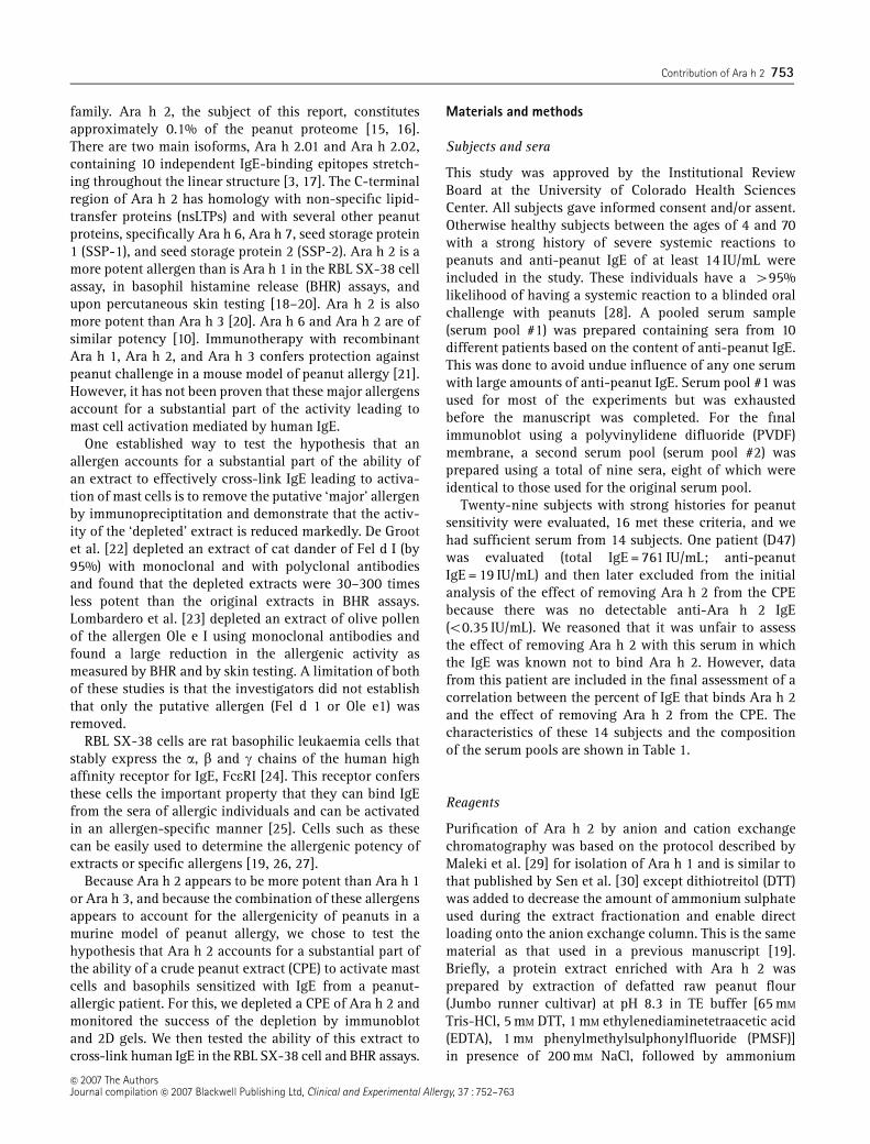
ORIGINAL PAPER Contribution of Ara h 2 to peanut-specific, immunoglobulin E-mediated, cell activation R. A. McDermott w , H. S. Porterfield , R. El Mezayen , A. W. Burks z , L. Pons z , D. G. Schlichting , B. Solomon ‰1 , J. S. Redzic ‰ , R. J. Harbeck w , M. W. Duncan ‰z , K. C. Hansen ‰z and S. C. Dreskin Division of Allergy and Clinical Immunology, University of Colorado at Denver and Health Sciences Center, Denver, CO, USA, w National Jewish Medical and Research Center, Denver, CO, USA, z Duke University, Durham, NC, USA, ‰ Proteomics Shared Resource, University of Colorado Cancer Center, University of Colorado at Denver and Health Sciences Center, Denver, CO, USA and z Department of Pediatrics, School of Medicine, University of Colorado at Denver and Health Science Center, Denver, CO, USA Clinical and Experimental Allergy Correspondence: Stephen C. Dreskin, Division of Allergy and Clinical Immunology, University of Colorado Health Sciences Center, Campus Box B164, 4200 E. Ninth Ave. Denver, CO 80262, USA. E-mail: [email protected] Summary Background Ara h 2 is a potent peanut allergen but its contribution to the ability of a crude peanut extract (CPE) to cross-link IgE and activate mast cells has not been rigorously evaluated. Objective To measure the contribution that Ara h 2 makes to the effector function of a CPE. Methods Ara h 2 was specifically removed from a CPE as demonstrated by immunoblots, 2D gels, and an inhibitory ELISA. Functional assays of sham-treated and Ara h 2-depleted CPEs were performed with RBL SX-38 cells sensitized with IgE from highly peanut-allergic subjects and with naturally sensitized basophils. Results Depletion of 99% of the Ara h 2 from the CPE led to an increase in the concentration of the CPE necessary to give 50% of maximal degranulation (EC50) of the SX-38 cells following sensitization with sera that contain anti-Ara h 2 IgE. Assays with a pool of 10 sera showed a small but significant increase in the EC50 following depletion of Ara h 2 (1.65 0.15-fold; P o 0.05) and assays of seven individual sera showed a similar increase in the average EC50 (1.7 0.2-fold; P o 0.02). The percent of the anti-peanut IgE that binds Ara h 2 correlated with an increase in the EC50 of the CPE following depletion of Ara h 2 (r = 0.83; P o 0.02). On the other hand, data from three of these patients studied with a basophil histamine release assay did not show a significant effect of depletion of Ara h 2. Conclusion Based on its ability to cross-link IgE effectively, Ara h 2 is clearly an important peanut allergen. Its ability to cross-link IgE effectively from a specific serum is related to the proportion of anti-Ara h 2 in that serum but Ara h 2 does not account for a majority of the effector activity of the CPE for any of the sera studied. Keywords allergens, Ara h 2, IgE, mast cells, peanuts, RBL SX-38 cells Submitted 18 August 2006; revised 15 January 2007; accepted 1 February 2007 Introduction Immediate hypersensitivity reactions to peanuts, Arachis hypogaea, are unusual in that they are much more prevalent and severe than for other legumes and tend to persist into adulthood [1]. Eight principal allergens of peanut (Ara h 1–Ara h 8) are accepted by the International Union of Immunological Societies, Allergen Nomencla- ture Sub-Committee [2–7]. Ara h 1 (63 kD), Ara h 2 (17–19 kD doublet) and Ara h 3 (60 kD) have been desig- nated major peanut allergens based on the frequency of patients whose IgE binds to these proteins [8, 9]. Ara h 6, another important peanut allergen, is 59% homologous with Ara h 2 [10]. Lehmann et al. [11] have recently defined the protease-stable core of Ara h 2 and Ara h 6 and demonstrated resistance to proteolytic treatment. Significant efforts are underway to generate hypoaller- genic peanuts and to design allergen-specific immuno- therapy with genetically modified allergens [12–14]. Ara h 2 and Ara h 6 belong to the conglutin family of seed storage proteins, and are part of the 2S albumin 1 Current address: Peter MacCallum Cancer Centre St Andrews Place East, Melbourne, Vic. 3002, Australia Clinical and Experimental Allergy, 37, 752–763 c 2007 The Authors Journal compilation c 2007 Blackwell Publishing Ltd
Transcript of Contribution of Ara h 2 to peanut-specific, immunoglobulin E-mediated, cell activation

ORIGINAL PAPER
Contribution of Ara h 2 to peanut-specific, immunoglobulin E-mediated,cell activationR. A. McDermott�w, H. S. Porterfield�, R. El Mezayen�, A. W. Burksz, L. Ponsz, D. G. Schlichting�, B. Solomon‰1, J. S. Redzic‰,R. J. Harbeckw, M. W. Duncan‰z, K. C. Hansen‰z and S. C. Dreskin��Division of Allergy and Clinical Immunology, University of Colorado at Denver and Health Sciences Center, Denver, CO, USA, wNational Jewish Medical and Research
Center, Denver, CO, USA, zDuke University, Durham, NC, USA, ‰Proteomics Shared Resource, University of Colorado Cancer Center, University of Colorado at Denver
and Health Sciences Center, Denver, CO, USA and zDepartment of Pediatrics, School of Medicine, University of Colorado at Denver and Health Science Center, Denver,
CO, USA
Clinical andExperimental
Allergy
Correspondence:Stephen C. Dreskin, Division of Allergyand Clinical Immunology, University ofColorado Health Sciences Center,Campus Box B164, 4200 E. Ninth Ave.Denver, CO 80262, USA.E-mail: [email protected]
Summary
Background Ara h 2 is a potent peanut allergen but its contribution to the ability of a crudepeanut extract (CPE) to cross-link IgE and activate mast cells has not been rigorouslyevaluated.Objective To measure the contribution that Ara h 2 makes to the effector function of a CPE.Methods Ara h 2 was specifically removed from a CPE as demonstrated by immunoblots, 2Dgels, and an inhibitory ELISA. Functional assays of sham-treated and Ara h 2-depleted CPEswere performed with RBL SX-38 cells sensitized with IgE from highly peanut-allergic subjectsand with naturally sensitized basophils.Results Depletion of�99% of the Ara h 2 from the CPE led to an increase in the concentrationof the CPE necessary to give 50% of maximal degranulation (EC50) of the SX-38 cellsfollowing sensitization with sera that contain anti-Ara h 2 IgE. Assays with a pool of 10 serashowed a small but significant increase in the EC50 following depletion of Ara h 2(1.65� 0.15-fold; Po 0.05) and assays of seven individual sera showed a similar increase inthe average EC50 (1.7� 0.2-fold; Po 0.02). The percent of the anti-peanut IgE that binds Arah 2 correlated with an increase in the EC50 of the CPE following depletion of Ara h 2 (r = 0.83;Po 0.02). On the other hand, data from three of these patients studied with a basophilhistamine release assay did not show a significant effect of depletion of Ara h 2.Conclusion Based on its ability to cross-link IgE effectively, Ara h 2 is clearly an importantpeanut allergen. Its ability to cross-link IgE effectively from a specific serum is related to theproportion of anti-Ara h 2 in that serum but Ara h 2 does not account for a majority of theeffector activity of the CPE for any of the sera studied.
Keywords allergens, Ara h 2, IgE, mast cells, peanuts, RBL SX-38 cellsSubmitted 18 August 2006; revised 15 January 2007; accepted 1 February 2007
Introduction
Immediate hypersensitivity reactions to peanuts, Arachishypogaea, are unusual in that they are much moreprevalent and severe than for other legumes and tend topersist into adulthood [1]. Eight principal allergens ofpeanut (Ara h 1–Ara h 8) are accepted by the InternationalUnion of Immunological Societies, Allergen Nomencla-ture Sub-Committee [2–7]. Ara h 1 (63 kD), Ara h 2
(17–19 kD doublet) and Ara h 3 (60 kD) have been desig-nated major peanut allergens based on the frequency ofpatients whose IgE binds to these proteins [8, 9]. Ara h 6,another important peanut allergen, is 59% homologouswith Ara h 2 [10]. Lehmann et al. [11] have recentlydefined the protease-stable core of Ara h 2 and Ara h 6and demonstrated resistance to proteolytic treatment.Significant efforts are underway to generate hypoaller-genic peanuts and to design allergen-specific immuno-therapy with genetically modified allergens [12–14].
Ara h 2 and Ara h 6 belong to the conglutin family ofseed storage proteins, and are part of the 2S albumin
1Current address: Peter MacCallum Cancer Centre St AndrewsPlace East, Melbourne, Vic. 3002, Australia
Clinical and Experimental Allergy, 37, 752–763
�c 2007 The Authors
Journal compilation �c 2007 Blackwell Publishing Ltd
family. Ara h 2, the subject of this report, constitutesapproximately 0.1% of the peanut proteome [15, 16].There are two main isoforms, Ara h 2.01 and Ara h 2.02,containing 10 independent IgE-binding epitopes stretch-ing throughout the linear structure [3, 17]. The C-terminalregion of Ara h 2 has homology with non-specific lipid-transfer proteins (nsLTPs) and with several other peanutproteins, specifically Ara h 6, Ara h 7, seed storage protein1 (SSP-1), and seed storage protein 2 (SSP-2). Ara h 2 is amore potent allergen than is Ara h 1 in the RBL SX-38 cellassay, in basophil histamine release (BHR) assays, andupon percutaneous skin testing [18–20]. Ara h 2 is alsomore potent than Ara h 3 [20]. Ara h 6 and Ara h 2 are ofsimilar potency [10]. Immunotherapy with recombinantAra h 1, Ara h 2, and Ara h 3 confers protection againstpeanut challenge in a mouse model of peanut allergy [21].However, it has not been proven that these major allergensaccount for a substantial part of the activity leading tomast cell activation mediated by human IgE.
One established way to test the hypothesis that anallergen accounts for a substantial part of the ability ofan extract to effectively cross-link IgE leading to activa-tion of mast cells is to remove the putative ‘major’ allergenby immunopreciptitation and demonstrate that the activ-ity of the ‘depleted’ extract is reduced markedly. De Grootet al. [22] depleted an extract of cat dander of Fel d I (by95%) with monoclonal and with polyclonal antibodiesand found that the depleted extracts were 30–300 timesless potent than the original extracts in BHR assays.Lombardero et al. [23] depleted an extract of olive pollenof the allergen Ole e I using monoclonal antibodies andfound a large reduction in the allergenic activity asmeasured by BHR and by skin testing. A limitation of bothof these studies is that the investigators did not establishthat only the putative allergen (Fel d 1 or Ole e1) wasremoved.
RBL SX-38 cells are rat basophilic leukaemia cells thatstably express the a, b and g chains of the human highaffinity receptor for IgE, FceRI [24]. This receptor confersthese cells the important property that they can bind IgEfrom the sera of allergic individuals and can be activatedin an allergen-specific manner [25]. Cells such as thesecan be easily used to determine the allergenic potency ofextracts or specific allergens [19, 26, 27].
Because Ara h 2 appears to be more potent than Ara h 1or Ara h 3, and because the combination of these allergensappears to account for the allergenicity of peanuts in amurine model of peanut allergy, we chose to test thehypothesis that Ara h 2 accounts for a substantial part ofthe ability of a crude peanut extract (CPE) to activate mastcells and basophils sensitized with IgE from a peanut-allergic patient. For this, we depleted a CPE of Ara h 2 andmonitored the success of the depletion by immunoblotand 2D gels. We then tested the ability of this extract tocross-link human IgE in the RBL SX-38 cell and BHR assays.
Materials and methods
Subjects and sera
This study was approved by the Institutional ReviewBoard at the University of Colorado Health SciencesCenter. All subjects gave informed consent and/or assent.Otherwise healthy subjects between the ages of 4 and 70with a strong history of severe systemic reactions topeanuts and anti-peanut IgE of at least 14 IU/mL wereincluded in the study. These individuals have a 495%likelihood of having a systemic reaction to a blinded oralchallenge with peanuts [28]. A pooled serum sample(serum pool #1) was prepared containing sera from 10different patients based on the content of anti-peanut IgE.This was done to avoid undue influence of any one serumwith large amounts of anti-peanut IgE. Serum pool #1 wasused for most of the experiments but was exhaustedbefore the manuscript was completed. For the finalimmunoblot using a polyvinylidene difluoride (PVDF)membrane, a second serum pool (serum pool #2) wasprepared using a total of nine sera, eight of which wereidentical to those used for the original serum pool.
Twenty-nine subjects with strong histories for peanutsensitivity were evaluated, 16 met these criteria, and wehad sufficient serum from 14 subjects. One patient (D47)was evaluated (total IgE = 761 IU/mL; anti-peanutIgE = 19 IU/mL) and then later excluded from the initialanalysis of the effect of removing Ara h 2 from the CPEbecause there was no detectable anti-Ara h 2 IgE(o0.35 IU/mL). We reasoned that it was unfair to assessthe effect of removing Ara h 2 with this serum in whichthe IgE was known not to bind Ara h 2. However, datafrom this patient are included in the final assessment of acorrelation between the percent of IgE that binds Ara h 2and the effect of removing Ara h 2 from the CPE. Thecharacteristics of these 14 subjects and the compositionof the serum pools are shown in Table 1.
Reagents
Purification of Ara h 2 by anion and cation exchangechromatography was based on the protocol described byMaleki et al. [29] for isolation of Ara h 1 and is similar tothat published by Sen et al. [30] except dithiotreitol (DTT)was added to decrease the amount of ammonium sulphateused during the extract fractionation and enable directloading onto the anion exchange column. This is the samematerial as that used in a previous manuscript [19].Briefly, a protein extract enriched with Ara h 2 wasprepared by extraction of defatted raw peanut flour(Jumbo runner cultivar) at pH 8.3 in TE buffer [65 mM
Tris-HCl, 5 mM DTT, 1 mM ethylenediaminetetraacetic acid(EDTA), 1 mM phenylmethylsulphonylfluoride (PMSF)]in presence of 200 mM NaCl, followed by ammonium
�c 2007 The AuthorsJournal compilation �c 2007 Blackwell Publishing Ltd, Clinical and Experimental Allergy, 37 : 752–763
Contribution of Ara h 2 753
sulphate precipitation at 14% saturation. After high-speedcentrifugation, Ara h 2 in the supernatant was precipitatedby increasing the ammonium sulphate from 14% to 22%saturation. The 22% precipitate was resuspended inTE buffer by sonication, cleared by centrifugation, andthen loaded onto a DEAE weak anion exchange column(Bio-Rad, Hercules, CA, USA) and eluted with a lineargradient of NaCl (100–300 mM). Fractions were analysedby SDS-PAGE with a Coomassie blue staining. Earlyfractions containing Ara h 2 (doublet) alone (no Ara h 1contamination) were pooled, concentrated, buffer ex-changed to distilled water (pH 7.0 with drops of 6 N NaOH)containing 1 mM DTT, and then lyophilized.
Crude peanut extract
Twenty grams of fresh raw Georgia green peanuts, a kindgift from the Golden Peanut Company, Ashburn, GA, USAwere frozen in liquid nitrogen and finely ground with amortar and pestle. Following extensive defatting withdiethyl ether, the dried peanut flour was resuspended in100 mL of ice-cold buffer [150 mM NaCl, 50 mM Tris, pH7.4 with protease inhibitors (Roche, Nutley, NJ, USAcomplete, EDTA-free)] and mixed overnight at 4 1C. Theextract [25 mg/mL (BCA, Pierce, Rockford, IL, USA)] wasclarified by centrifugation (10 000 g, 30 min), sterile fil-tered, and frozen in aliquots at �70 1C.
Anti-peptide antibodies
Two peptide epitopes (13–14 amino acids each) of Ara h 2[GenBank sequence AAN77576 (gi:26245477)] were cho-sen in consultation with Dr Shing-Erh Yen (Invitrogen,Carlesbad, CA, USA) based on uniqueness, antigenicity,surface probability, flexibility, and an antigenic index [31,32]. The antibody that was most useful was directedagainst the C-terminal sequence, CDLEVESGGRDRY,which consists of amino acids 160–172 of Ara h 2.02(GenBank sequence AAN77576 and SwissProt accessionnumber, Q8GV20) and is also the C-terminus of Ara h 2.01[3, 33]. The second peptide was DRRDPYSPSPYDR, whichconsists of amino acids 79–91 of Ara h 2.02 (above), andincludes the immunodominant IgE-binding epitopeDPYSPS that is also present in Ara h 2.01 [3, 33]. Tworabbits were immunized with peptide-KLH conjugates ofboth peptides [regular antibody service with a combina-tion immunization strategy (Invitrogen)]. Antibodies wereaffinity purified on peptide columns and eluted with 3 M
KSCN. The yields were 5–12 mg. Both antibodies fromeach rabbit identified a 17–19 kDa doublet on immuno-blots (data not shown). The affinity-purified anti-Ara h 2peptide antibodies were individually conjugated to acti-vated agarose beads as per the manufacturer’s instructions(Pierce ProFoundTM Co-Immunoprecipitation Kit,Rockford, IL, USA). Only the antibodies against the Ara h2 amino acids #160–172 bound strongly enough to
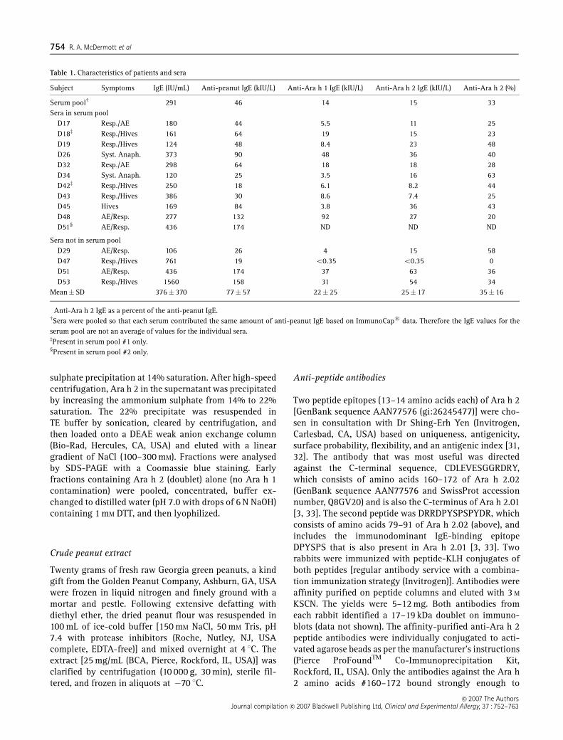
Table 1. Characteristics of patients and sera
Subject Symptoms IgE (IU/mL) Anti-peanut IgE (kIU/L) Anti-Ara h 1 IgE (kIU/L) Anti-Ara h 2 IgE (kIU/L) Anti-Ara h 2 (%)�
Serum poolw 291 46 14 15 33Sera in serum pool
D17 Resp./AE 180 44 5.5 11 25D18z Resp./Hives 161 64 19 15 23D19 Resp./Hives 124 48 8.4 23 48D26 Syst. Anaph. 373 90 48 36 40D32 Resp./AE 298 64 18 18 28D34 Syst. Anaph. 120 25 3.5 16 63D42z Resp./Hives 250 18 6.1 8.2 44D43 Resp./Hives 386 30 8.6 7.4 25D45 Hives 169 84 3.8 36 43D48 AE/Resp. 277 132 92 27 20D51‰ AE/Resp. 436 174 ND ND ND
Sera not in serum poolD29 AE/Resp. 106 26 4 15 58D47 Resp./Hives 761 19 o0.35 o0.35 0D51 AE/Resp. 436 174 37 63 36D53 Resp./Hives 1560 158 31 54 34
Mean�SD 376� 370 77� 57 22� 25 25� 17 35� 16
�Anti-Ara h 2 IgE as a percent of the anti-peanut IgE.wSera were pooled so that each serum contributed the same amount of anti-peanut IgE based on ImmunoCaps data. Therefore the IgE values for theserum pool are not an average of values for the individual sera.zPresent in serum pool #1 only.‰Present in serum pool #2 only.
�c 2007 The AuthorsJournal compilation �c 2007 Blackwell Publishing Ltd, Clinical and Experimental Allergy, 37 : 752–763
754 R. A. McDermott et al

Ara h 2 in solution to deplete CPE of Ara h 2. Thisantibody was used throughout the experiments to follow.Preimmune IgG was isolated on protein G and covalentlylinked to sepharose as described above. Anti-peptideantibodies against Ara h 1 (amino acid #599–612; KE-SPEKEDQEEE NQ; Swiss-Prot, Q547W5), Ara h 3 (aminoacid # 90–103; EEPHTQGRRSQSQR; Swiss-Prot, O82580),and Ara h 6 (amino acid # 42–59; EQEQYD-SYNFGSTRSSDQ; Swiss-Prot, Q9SQG5) were raised andused for immunoblots.
Depletion of Ara h 2 from a crude peanut extract
CPE was diluted to 1 mg/mL in coupling buffer (Pierce)and 800 mL was incubated with the anti-Ara h 2-conju-gated agarose column containing �5 mg of antibody(overnight, 4 1C). The eluate was then collected and storedat �80 1C. A control column was constructed as outlinedabove, except that pre-immune rabbit IgG (purified onprotein G) was used to generate sham-treated CPE. Thecolumns were regenerated by treatment with 0.1 M gly-cine, pH 2.5, a total of three times before binding activitywas lost.
Two dimensional gel electrophoresis
All gel equipment and supplies were obtained fromAmersham Biosciences/GE Healthcare (Piscataway, NJ,USA) unless otherwise noted. Fifty micrograms of proteinwas minimally labelled with Cy3 (Ara h 2-depleted CPE)and Cy5 (sham-treated CPE) CyDyes (4 1C, 30 min).Cy3- and Cy5-labelled samples were mixed together andbrought up to a volume of 450 mL in standard rehydrationbuffer. The sample was passively rehydrated overnightonto a 24 cm immobilized pH gradient strip (IPG 3–10 NL)and isoelectric focusing (IPGphor apparatus, AmershamBiosciences/GE Healthcare) was then performed accordingto the manufacturer’s recommendations. Reduction andalkylation were carried out on-strip using DTT and iodoa-cetamide, respectively. Strips were then applied to aprecast 8–16% SDS-PAGE gel (Jule Inc., Milford, CT,USA) for electrophoresis (Ettan DALT 12 electrophoresisunit, 20 W/gel, 25 1C). Labelled proteins were visualizedby scanning the gels at 100 mm resolution (Typhoon 9400scanner, Amersham Biosciences/GE Healthcare).
MALDI-TOF MS protein identification
Protein spots of interest were excised from the gel usingthe Ettan robotic picker and placed into a micro-well platefor in-gel digestion using the Ettan digester. Plugs werewashed twice with 100mL of 50mM NH4HCO3/50% methanolfor 5 min, once with 100mL of 75% acetonitrile for 10minand twice with 100mL of 100% acetonitrile for 10min. Thegel plugs were dried at room temperature for 50min and
then trypsin (Promega, San Luis Obispo, CA, USA) was addedto each well (10mL, 10mg/mL in 20 mM NH4HCO3) anddigestion proceeded at room temperature for 16h.
All digests were analysed by MALDI-TOF MS (VoyagerDE-PRO, Applied Biosystems, Foster City, CA, USA) usingpeptide mass fingerprinting as described previously [34,35]. Spectra were collected over the range m/z 500–5000.No cleanup of digests was performed. Peptide massfingerprints were internally calibrated to monoisotopictrypsin autolysis peaks (m/z = 515.33, 842.51, 1045.56,2211.11). Spectra were processed using ProTS Data(Steamboat Springs, CO, USA) to generate centroided peaklists that were submitted to MS-Fit program of the ProteinProspector Suite (v. 4.21.3, UCSF Mass SpectrometryFacility, San Francisco, CA, USA) for database searching.Spectral processing included defining baseline, noise andsignal-to-noise ratio as well as monoisotopic peak selec-tion. A signal-to-noise ratio in ProTS data of 44 wasrequired for inclusion in the peak list. Database searcheswere conducted using the non-redundant protein data-base (NCBI, database version 05/07/2006) and the Swis-sprot database (version 2007.05.02). Other settings inProTS included peak amplitude set at 100, peak width250, and chemical noise factor 1.5. Key parametersincluded the following: peptide error tolerance was set at� 60 p.p.m., fixed modification of carbamidomethylationof cysteine side chains, and variable modification ofhydroxylation of P, oxidation of M, and protein N-terminus acetylated. Searches were not constrained by pIor molecular weight.
Immunoblots
1D gel immunoblots: 30 mg of sham treated or Ara h 2-depleted CPE in standard (reducing) sample buffer wererun on 4–20% polyacrylamide gels and transferred ontonitrocellulose with 20 v overnight at 4 1C in transfer buffer(25 mM Tris/192 mM glycine/20% methanol). The nitrocel-lulose was blocked for 2 h in 3% milk powder in tris-buffered saline (50 mM Tris-HCl, 150 mM NaCl, pH 7.4) andprobed with rabbit anti-peptide antibodies to Ara h 1, Arah 2 (#78–92), Ara h 3, and Ara h 6 at dilutions of1 : 100–400. The nitrocellulose was developed with horse-radish peroxidase (HRP)-conjugated goat anti-rabbit IgG(1 : 20 000), followed by an enhanced chemiluminescencereagent (Amersham) for 10–60 s.
2D gel immunoblots: 450 mg of untreated CPE was runon a 2D gel, transferred to nitrocellulose as describedabove, probed with human serum, and developed withHRP-murine anti-human IgE (1 : 3000) (Bio-Rad). A repli-cate gel was transferred onto a PVDF membrane assuggested by Boldt et al. [36]. For the blot onto the PVDFmembrane, all reagents and settings were the same as fornitrocellulose, except the HRP-murine anti-human IgEwas diluted to 1 : 30 000 to avoid a high background.
�c 2007 The AuthorsJournal compilation �c 2007 Blackwell Publishing Ltd, Clinical and Experimental Allergy, 37 : 752–763
Contribution of Ara h 2 755

ImmunoCaps assay with Ara h 2
ImmunoCaps assays for Ara h 1 and Ara h 2 wereperformed as described previously [19].
Enzyme linked immunoglobin assay inhibition assay
Fifty microlitres of purified Ara h 2 (5 mg/mL in 0.1 M Nacarbonate buffer, pH 9.6) was adhered to microtitre wellsat 4 1C overnight. The plate was then washed with 1�phosphate-buffered saline (PBS)/0.05% Tween 20 (PBST)and then blocked for 2 h at RT with PBS containing 3%non-fat milk powder (Bio-Rad). During the blockingtime, affinity-purified rabbit anti-Ara h 2 (amino acids:145–157), diluted 1 : 100 000 (final concentration = 10 ng/mL) in PBST with 3% non-fat milk, was incubated withvarying concentrations (0–1.0 mg/mL) of purified Ara h 2,sham-treated CPE or Ara h 2-depleted CPE. The anti-Ara h2 with or without inhibitors was added to the wellscontaining adhered Ara h 2 for 1 h at room temperature.Following extensive washing the plate was developedwith horseradish peroxidase labelled mouse anti-rabbitIgG and tetra methyl benzidine (TMB) (Sigma, St Louis,MO, USA). A standard curve was generated based oninhibition with purified Ara h 2 (0.001–1.0 mg/mL). Wefound that low levels of Ara h 2 (o0.01 mg/mL) weredifficult to measure in the depleted extract, presumablydue to interference by cross-reactive proteins (see theimmunoblot in Fig. 1). For this reason, a secondary curvewas generated based on the sham-depleted extract andthis was used to calculate the Ara h 2 content in thedepleted extract (data not shown).
RBL SX-38 cell assay
The sensitivity and specificity of the RBL SX-38 cell assayhave been previously described [19, 25]. RBL SX-38 cellssensitized with IgE from patients with undetectable anti-peanut IgE are not activated when exposed to a CPE [25].RBL SX-38 cells were grown and the functional assaysperformed with 3H-5HT (tritium-labelled, 5-hydroxytryp-tamine or serotonin) as described previously [19]. Labelledand sensitized RBL SX-38 cells were triggered withanti-IgE (3 mg/mL), CPE (250 ng/mL), depleted CPE orsham-treated CPE (0.25–250 ng/mL). Percent release ofserotonin was calculated as described previously [24, 25].
Basophil histamine release
BHR was performed with unwashed basophils frompeanut-allergic subjects using the Histamine Elisa Kit(Beckman Coulter, Miami, FL, USA). Depleted CPE andsham-treated CPE were diluted (0.025–250 ng/mL) in thehistamine release buffer supplied by the manufacturer.
Spontaneous degranulation was subtracted from totaldegranulation with CPE to give the ‘net degranulation’and data are presented as percent histamine release.Basophils were also stimulated after washing in thehistamine release buffer. This caused a marginal increasein the background but with a very similar dose responsefor both depleted and sham-treated CPE (data not shown).
Determination of EC50 and calculation of the allergenicpotency of the Ara h 2-depleted crude peanut extract
The EC50 of an extract or allergen is the dose giving 50%of the degranulation seen with an optimal amount of CPE.To determine EC50 accurately, the effect of five half-logdilutions was studied. The values for the concentrations ofthe extracts were log-transformed and non-linear regres-sion was performed using a second order polynomialequation. The intercept on the abscissa was determinedfor a value of 50% on the ordinate. Computer-generatedbest-fit lines are shown in each figure. An increase in theEC50 represents a decrease in potency [26].
Fig. 1. Depletion of Ara h 2 but not Ara h 1 from crude peanut extract(CPE) as demonstrated on immunoblot. Five hundred nanograms of sham-treated CPE or Ara h 2 depleted-CPE were run on a 4–20% polyacrylamidegel and transferred onto nitrocellulose. The peanut proteins were thenprobed with either affinity-purified anti-Ara h 1, amino acids #599-612(a); affinity-purified anti-Ara h 2, amino acids #79-91 (b); affinity-purified anti-Ara h 3, amino acids #90-103 (c); or with affinity-purifiedanti-Ara h 6, amino acids #42-59 (d). Development with enhancedchemiluminescence was 10–30 s. This was repeated with similar results.
�c 2007 The AuthorsJournal compilation �c 2007 Blackwell Publishing Ltd, Clinical and Experimental Allergy, 37 : 752–763
756 R. A. McDermott et al
Reproducibility
The RBL SX-38 cell assay was performed with a singleserum sample (D19) on three separate 96-well plates onthree separate days over a period of several months todetermine the inter- and intra-assay coefficient of variation(CV) (SD/mean� 100) [37]. The intra-assay CV for determi-nation of the EC 50 of a CPE was 29� 6% (n= 3) and theinter-assay CV (SD/mean� 100) was 105% (data notshown). Thus, within an assay, we can detect differences ofEC50 in the range of 30%, and values for EC50 acrossseparate experiments therefore vary, on the average, by afactor of 2. For each serum, the comparisons of Ara h 2-depleted and shan-treated CPE were done in the same assay.
Statistical analysis
Statistical comparisons were all two tailed. P-valueso0.05 were considered to be statistically significant. Tocarry out comparisons among experiments, release ofserotonin was expressed as a percentage of that seen withoptimal doses of the original CPE in the same assay. Datafrom Ara h 2-depleted and sham-depleted extracts werecompared with paired t-tests. Deviations of ratios from thepredicted value of 1.0 were analysed with one-samplet-tests. Spearman’s rank correlation test was used to assesscorrelations. All figures, best-fit lines, EC50 values andstatistical data were generated with GraphPad Prismversion 4.0b (GraphPad Software, San Diego CA, USA).
Results
Successful depletion of Ara h 2
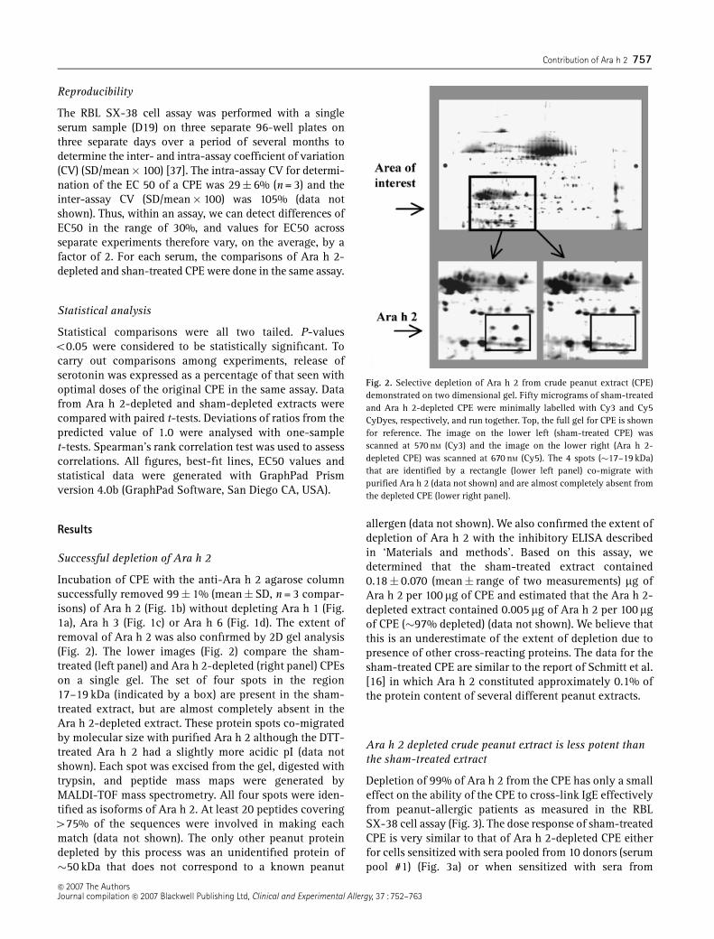
Incubation of CPE with the anti-Ara h 2 agarose columnsuccessfully removed 99� 1% (mean� SD, n = 3 compar-isons) of Ara h 2 (Fig. 1b) without depleting Ara h 1 (Fig.1a), Ara h 3 (Fig. 1c) or Ara h 6 (Fig. 1d). The extent ofremoval of Ara h 2 was also confirmed by 2D gel analysis(Fig. 2). The lower images (Fig. 2) compare the sham-treated (left panel) and Ara h 2-depleted (right panel) CPEson a single gel. The set of four spots in the region17–19 kDa (indicated by a box) are present in the sham-treated extract, but are almost completely absent in theAra h 2-depleted extract. These protein spots co-migratedby molecular size with purified Ara h 2 although the DTT-treated Ara h 2 had a slightly more acidic pI (data notshown). Each spot was excised from the gel, digested withtrypsin, and peptide mass maps were generated byMALDI-TOF mass spectrometry. All four spots were iden-tified as isoforms of Ara h 2. At least 20 peptides covering475% of the sequences were involved in making eachmatch (data not shown). The only other peanut proteindepleted by this process was an unidentified protein of�50 kDa that does not correspond to a known peanut
allergen (data not shown). We also confirmed the extent ofdepletion of Ara h 2 with the inhibitory ELISA describedin ‘Materials and methods’. Based on this assay, wedetermined that the sham-treated extract contained0.18� 0.070 (mean� range of two measurements) mg ofAra h 2 per 100 mg of CPE and estimated that the Ara h 2-depleted extract contained 0.005 mg of Ara h 2 per 100 mgof CPE (�97% depleted) (data not shown). We believe thatthis is an underestimate of the extent of depletion due topresence of other cross-reacting proteins. The data for thesham-treated CPE are similar to the report of Schmitt et al.[16] in which Ara h 2 constituted approximately 0.1% ofthe protein content of several different peanut extracts.
Ara h 2 depleted crude peanut extract is less potent thanthe sham-treated extract
Depletion of 99% of Ara h 2 from the CPE has only a smalleffect on the ability of the CPE to cross-link IgE effectivelyfrom peanut-allergic patients as measured in the RBLSX-38 cell assay (Fig. 3). The dose response of sham-treatedCPE is very similar to that of Ara h 2-depleted CPE eitherfor cells sensitized with sera pooled from 10 donors (serumpool #1) (Fig. 3a) or when sensitized with sera from
Fig. 2. Selective depletion of Ara h 2 from crude peanut extract (CPE)demonstrated on two dimensional gel. Fifty micrograms of sham-treatedand Ara h 2-depleted CPE were minimally labelled with Cy3 and Cy5CyDyes, respectively, and run together. Top, the full gel for CPE is shownfor reference. The image on the lower left (sham-treated CPE) wasscanned at 570 nM (Cy3) and the image on the lower right (Ara h 2-depleted CPE) was scanned at 670 nM (Cy5). The 4 spots (�17–19 kDa)that are identified by a rectangle (lower left panel) co-migrate withpurified Ara h 2 (data not shown) and are almost completely absent fromthe depleted CPE (lower right panel).
�c 2007 The AuthorsJournal compilation �c 2007 Blackwell Publishing Ltd, Clinical and Experimental Allergy, 37 : 752–763
Contribution of Ara h 2 757

individual donors (Figs 3b–h). Three of the sera shown inFig. 3 were part of serum pool #1 and 4 were not. EC50values with the sham-treated and Ara h 2-depleted CPEswere determined for serum pool #1 and for seven indivi-dual sera, four of which were part of the serum pool(Table 2). All the sera in the serum pool and all the sevenindividual sera used to calculate the effect of removingAra h 2 from the CPE contained anti-Ara h 2 IgE
(7–63 kIU/L) (Table 1). For the serum pool, the EC50 withthe Ara h 2-depleted CPE is 1.6� 0.15-fold more than thatseen with the sham-treated CPE (n = 3; Po 0.05). Simi-larly, looking at seven individual sera, the EC50 with theAra h 2-depleted CPE is 1.7� 0.21-fold more than thatseen with the sham-treated CPE (n = 7; Po 0.02). Anassay was also performed with the serum (D47) that hadno detectable anti-Ara h 2 IgE. The EC50 with the sham-treated extract was 54 ng/mL and for the depleted extractwas 47 ng/mL. This change of � 13% is within the error ofthe assay and is compatible with no change due todepletion of Ara h 2 as should be the case as there is noIgE that binds to Ara h 2. Similar experiments withbasophils (naturally sensitized with IgE) from three of thepeanut-allergic donors showed no consistent effect ofremoval of 99% of the Ara h 2 from the CPE (Figs 4a–cand Table 2; P = NS).
Correlation between the RBL SX-38 cell assay and theImmunoCaps assay
It is reasonable to ask whether the amount of IgE thatbinds to Ara h 2 in the ImmunoCap assay predicts the
Fig. 3. Effect of depletion of Ara h 2 in the RBL SX-38 cell assay of IgEcross-linking. RBL SX-38 cells were sensitized with sera from peanut-allergic subjects and stimulated with untreated crude peanut extract(CPE) (not shown), sham-treated CPE (&, solid lines) or Ara h 2-depletedCPE (m, dashed lines). Spontaneous degranulation (buffer) was sub-tracted from total degranulation to give the ‘net degranulation’ asdescribed in ‘Materials and methods’. In these experiments, the back-ground degranulation was 18� 3% (n = 6; mean�SEM) and followingthe maximal dose of untreated CPE, the total degranulation was 64� 5%.After subtracting the background for each experiment, this gives a netrelease was 47� 4%. With sham-treated CPE and with the Ara h 2-depleted CPE, the net releases were 44� 4 and 42� 7% respectively. Inthis figure, net degranulation for all data points is expressed aspercentage of maximal degranulation within each assay. All assays wereperformed in triplicate and data points are mean� SD. If standard errorbars are not visible, the error was smaller than the symbol. Data using thefollowing sera are shown: (a) the serum pool, (b) D19, (c) D26, (d) D29, (e)D32, (f) D48, (g) D51, (h) D53. Two additional replicates with the serumpool and D47 are not shown.
Fig. 4. Functional assay of IgE cross-linking on basophils from peanut-allergic subjects. Basophils in heparanized blood were stimulated withbuffer, sham-treated crude peanut extract (CPE) (&, solid lines), or Ara h2-depleted-CPE (m, dashed lines). All assays were performed in triplicateand data points are mean�SD. If standard error bars are not visible, theerror was smaller than the symbol. In these experiments, the backgrounddegranulation was 9� 4% (n = 3; mean� SEM). Data using basophilsfrom the following subjects are shown: (a) D19, (b) D51, (c) D53.
�c 2007 The AuthorsJournal compilation �c 2007 Blackwell Publishing Ltd, Clinical and Experimental Allergy, 37 : 752–763
758 R. A. McDermott et al

importance of Ara h 2 in the CPE for individual serastudied in the RBL SX-38 cell assay. As can be seen in Fig. 5,there is a strong correlation (r = 0.83; Po 0.02; n = 8)between the percent of anti-peanut IgE that binds to Ara h 2in the ImmunoCap assay (abscissa) and the fold-increasein the CPE for that serum following depletion of Arah 2 (ordinate). The data with the pooled serum (anti-Ara h 2 IgE = 33%) of the anti-peanut IgE and shift inEC50 due to depletion of Ara h 2 of 1.65-fold are notplotted but fall near the best-fit line.
Many potential allergens are seen on a two dimensionalimmunoblot
Because depletion of Ara h 2 from the CPE has only aminor impact on the allergenicity of the extract, it isreasonable to ask how large is the repertoire of potentialpeanut allergens? For this reason, a 2D immunoblot wasperformed with the serum pools (Figs 6a and d) and withserum from one individual patient (Fig. 6b) to identifyother candidate allergens. A control blot (Fig. 6c) wasprobed with serum from a peanut-tolerant patient with
allergic rhinitis (total IgE = 960 IU/mL; anti-peanutIgE = o0.35 IU/mL). Ara h 2 is clearly visible in Figs. 6a,b and d, although the density of binding of IgE to Ara h 2is dwarfed by that of other proteins especially in the blotsusing nitrocellulose (Figs 6a and b). Previous work hasshown that purified Ara h 2 binds IgE less well onimmunoblots than does Ara h 1 [19] and this is recapitu-lated here. The 2D blot probed with the control serumshowed no specific spots after a 5-min exposure (Fig. 6c).The 2D gel transferred onto a PVDF membrane instead ofnitrocellulose showed a somewhat different pattern withdecreased detection of higher molecular weight allergensand enhanced detection of lower molecular weight aller-gens (Fig. 6d). Thus, many peanut proteins specificallybind IgE (Figs 6a, b and d) and, based on these findings,could account for the majority of the effector activity ofpeanuts.
Discussion
Ara h 1, Ara h 2, and Ara h 3 have been identified as themajor peanut allergens based on the large numbers of
Table 2. Effective concentration 50 (EC50)
Sera
RBL SX-38 cells Basophils
EC50 (ng/mL) Fold change in EC50(Ara h 2-depletedCPE/sham-treated CPE)
EC50 (ng/mL) Fold change in EC50(Ara h 2-depletedCPE/sham-treated CPE)Sham-treated CPE Ara h 2-depleted CPE Sham-treated CPE Ara h 2-depleted CPE
Serum pool�
Assay #1 14 19 1.4 ND NDAssay #2 28 49 1.8 ND NDAssay #3 6.0 11 1.8 ND ND
95% CI 1.0–2.3Mean�SEM 16� 6 26� 12 1.65� 0.15P-value� o 0.05P-valuew o 0.04Individual sera with anti-Ara h 2w
D19 6.2 8.9 1.4 0.025 0.011 0.44D26 18 31 1.7 ND NDD29 5.7 16 2.8 ND NDD32 34 44 1.3 ND NDD48 2.4 2.9 1.2 ND NDD51 2.2 4.0 1.8 11 13 1.18D53 15 24 1.6 0.23 0.035 0.15
95% CI 1.2–2.2Mean�SEM 12� 4 19� 5 1.7� 0.21 3.8� 3.7 7.5� 4.3 0.59� 0.53P-value� o0.02 NSP-valuew o 0.004 NSIndividual serum with no anti-Ara h 2z
D47 54 47 0.87 ND ND
Paired t-test comparing log values of sham treated and Ara h 2 depleted.�Serum pool includes D19, D26, D32, D48 and 6 other sera (not shown individually).wMean is different from 1.0; One sample t-test.zThis serum did not contain any anti-Ara h 2 specific IgE and is not included in the analysis of the effect of depleting Ara h 2.CPE, crude peanut extract; CI, confidence interval.
�c 2007 The AuthorsJournal compilation �c 2007 Blackwell Publishing Ltd, Clinical and Experimental Allergy, 37 : 752–763
Contribution of Ara h 2 759

patients who have IgE that bind to proteins migrating at63, 17–19 kD, and 60 kD, respectively [1, 9, 15, 38]. Ara h 6has been shown to be an important peanut allergen witha potency similar to that of Ara h 2 [10]. The definition ofwhat constitutes a ‘major’ peanut allergen is still evolving[39, 40].
Peanut-allergic patients have a wide range of total andpeanut-specific IgE and the data obtained with thesepatients who have high anti-peanut IgE (414 kU/L) maynot pertain to all peanut-sensitive subjects. However,there is no current reason to believe that this will not bethe case.
Although the ability of an allergen to generate an IgEresponse in susceptible patients and to bind IgE in in vitroassays is important, we, and others, have focused on theability of allergens to cross-link IgE and lead to activationof mast cells and basophils as an important measure ofallergenicity [19, 20, 25, 26]. In addition to binding IgE,critical determinants of this function of allergens are thenumber of IgE-binding epitopes on the allergen and theallergen’s affinity for IgE [41–43]. Based on functionalassays, Ara h 2 is more active at cross-linking IgE than areAra h 1 or Ara h 3 and is similar to Ara h 6 [10, 19, 20].
The ability of allergens to cross-link IgE bound to FceRIcan be studied with cell-based assays such as the RBL SX-38 cells or human basophils sensitized in vivo or ex vivowith anti-peanut IgE [25, 44]. The RBL SX-38 cell assayhas advantages over the BHR assay in that it does notrequire fresh cells from a patient for each experiment. TheBHR assay has advantages over the RBL SX-38 cell assayin that the entire system is from the affected patient.Koppleman et al. were able to test Ara h 1, 2, and 3 withskin test titrations in allergic subjects [20]. Unfortunately,in the United States, skin testing such as this requires aninvestigational new drug (IND) application for each ex-tract or purified protein to be tested. This makes skintesting impractical in the United States.
Ara h 2 is clearly a potent and important allergen as itconstitutes 0.1–0.2% of the peanut proteome and it bindsa substantial amount of IgE from peanut-allergic patients(Table 1). These experiments were designed to test thehypothesis that Ara h 2 is also responsible for a substantialportion of the ability of a CPE to cross-link IgE from apeanut-allergic subject. We did this by quantitatively andspecifically removing Ara h 2 from CPEs by immunode-pletion and then assessing the ability of the depletedextract to cross-link IgE/IgE receptor complexes as mea-sured by cell activation. Sham-treated and Ara h 2-depleted extracts were tested for their ability to cross-linkhuman anti-peanut IgE on RBL SX-38 cells sensitizedwith IgE from peanut allergic subjects or on naturallysensitized basophils from peanut-allergic subjects. Re-moving �99% of the Ara h 2 (Figs 2 and 3) has a modesteffect in the RBL SX-38 cell assay that is variable amongsera (Fig. 4) and no consistent effect in the BHR assay
(Fig. 5). Although the coefficient of variation of the BHRassay is reported to be 30%, the lack of an effect on theBHR assay probably reflects its lack of precision in ourhands [45].
These data are very different from those of de Grootet al. [22], who found that depletion of 95% of Fel d 1 bymonoclonal antibodies reduced the allergenic potency bya factor of 30–40. Our data are also very different fromthose of Lombardero et al. [23], who found that depletionof 99.5% of Ole e 1 from an extract of Olea europaearesulted in a shift in the BHR dose–response of 125 times.
The quantitative contribution of Ara h 2 to the potencyof the CPE is related to the quantitative change in theEC50, but determining the specific contribution from theavailable data is not straightforward. It is reasonable toargue that, if depletion of 99% of Ara h 2 resulted in acomplete diminution of allergenicity for a given serum,the EC50 would shift from its initial value to infinity andwe would say that the entire potency of the CPE is due toAra h 2 for that serum. On the other hand, if depletion ofAra h 2 from the CPE led to a 10-fold increase in the EC50(the depleted extract was 10-fold less potent), we proposethat, in this situation, Ara h 2 would account for 90% ofthe activity of the extract for that specific serum. Simi-larly, if the change in EC50 were threefold (�1/2 log), Arah 2 could be said to account for �45% (1/2 of 90%) of theactivity. Our data showing only a 1.7� 0.2-fold increasein the EC50 due to removing Ara h 2 suggest that thecontribution of Ara h 2 to the allergenic potency of theCPE is, on the average, in the 20% range, althoughvariable among our subjects. Using our approach todetermine the contribution of an allergen to the potencyof an extract, we argue that the de Groot data demonstrate
Fig. 5. Correlation between the percent of anti-peanut IgE that binds toAra h 2 and the effect of depletion of Ara h 2 on the RBL SX-38 cell assay.For eight individual sera, the percent of anti-peanut IgE that binds to Arah 2 from Table 1 is plotted on the abscissa and fold increase in EC50 ofthe extract for that serum when Ara h 2 is removed from the crude peanutextract is plotted on the ordinate (Table 2). Data from patient D47(no binding of Ara h 2) is included. Spearman’s correlation coefficient= 0.83; Po 0.02. A best-fit line was generated using a polynomialequation Y ¼ Aþ BX þ CX2
and is shown.
�c 2007 The AuthorsJournal compilation �c 2007 Blackwell Publishing Ltd, Clinical and Experimental Allergy, 37 : 752–763
760 R. A. McDermott et al

that Fel d 1 accounted for �95% of the allergenic potencyof their cat dander extract and the Lombardero datademonstrate that Ole e 1 accounts for �99% of theallergenic potency of the olive pollen extract.
One potential limitation of this study is the variabilityof these biologic assays. However, in our hands, the intra-assay coefficient of variation of the RBL SX-38 cell assayfor determination of the EC50 is 29� 6% (data notshown), makes this a very sensitive assay of cell activa-tion. We found that depletion of 99% of Ara h 2 from theCPE leads to a 1.65� 0.15-fold increase (Po 0.05) in theEC50 in the RBL SX-38 cell assay using a serum pool and a1.7� 0.2-fold increase (Po 0.02) using seven individualsera, four of which were in the serum pool. In contrast tothe findings with the SX-38 cells, depletion of Ara h 2 hadno detectable effect on the BHR assay. Even though BHRhas been used to assess allergenic extracts that have beendepleted of specific allergens and can easily detect a logchange in activity, the BHR assay is not optimal formaking this type of comparison [22, 23].
Another potential limitation of this study is the use ofraw and not roasted peanuts. We decided to use extracts ofraw peanuts for two reasons. First, peanut extracts usedclinically for skin testing and peanut flour used for foodchallenges are made with raw peanuts. Second, roastedpeanuts have been reported to bind more IgE and the Arah 2 trypsin inhibition activity is increased [46–48]. Thistype of experiment would be much more complicated withroasted peanuts where adducts of Ara h 2 are formed withother peanut proteins, making immunodepletion techni-cally much more challenging.
Because depletion of Ara h 2 from a CPE has a small butsignificant effect on the activity of the CPE in these cellularassays, it is reasonable to conclude that Ara h 2 is animportant allergen and, because it is more potent than Ara h
1 and Ara h 3, it is likely more important. However, Ara h 2,by itself does not account for a substantial part of the abilityof our CPE to cross-link IgE from our cohort of peanut-allergic patients. Not surprisingly, the importance of Ara h 2appears to be greater in those patients with IgE that bindsthe most Ara h 2 (Fig. 5). It is possible that, if we were ableto identify patients whose IgE binds large amounts of Ara h2 and conversely small amounts of Ara h 1, 3, and 6, thecontribution of Ara h 2 would be more significant.
However, based on our finding on 2D immunoblot (Fig. 6),there are a large number of potential peanut allergens. Bythis logic, some other allergens may synergize with Arah 2, may act independently, or may act in concert withother allergens to account for the activity in CPEs. Anexcellent candidate that may account for a substantialpart for this remaining activity is the related peanutallergen, Ara h 6, a protein of 15 kD. It is possible that thereactivity to peanut allergens may be due to a group ofallergens working in concert and the nature of thesepeanut allergens may vary from patient to patient. Analternative to this idea is that Ara h 2 is a major allergenresponsible for a great deal of the observed cross-linking,but when it is removed, other allergens with homologouslinear and/or 3D epitopes become more active.
Acknowledgements
The authors thank Mr Mike Simpson for technical assis-tance, Dr Pamela Wolfe for statistical advice and MsAndrea T. Dreskin and Ms June K. Inuzuka for help inpreparing the figures.
This work was supported by an NIH grant, AI052164-01, to Dr Dreskin and institutional funds, UCHSC andNJMRC.
Fig. 6. Two-dimensional (2D) immunoblot of crude peanut extract. Four hundred and fifty micrograms of a crude peanut extract was separated on a 2Dgel, transferred onto nitrocellulose (a–c) or a polyvinylidene difluoride (PVDF) membrane (d), washed, and developed with horseradish peroxidase-murine anti-human IgE as described. All images shown were after a 5-min exposure with enhanced chemiluminescence. (a) Probed overnight witha 1 : 10 dilution of serum pool #1 (final IgE = 23 IU/mL; final anti-peanut IgE = 4.3 IU/mL). (b) Probed overnight with a 1 : 20 dilution of serumfrom a peanut-allergic subject (D26), (final IgE = 19 IU/mL; final anti-peanut IgE = 4.5 IU/mL). (c) A control blot was probed overnight with a 1 : 30dilution of serum from a peanut-tolerant, highly grass pollen-sensitive patient with anti-peanut IgE o 0.35 kU/L (final IgE = 32 IU/mL; final anti-grassIgE = 3.1 IU/mL). (d) 2D gel transferred onto a PVDF membrane and probed with a 1 : 10 dilution of a second batch of pooled serum (pool #2;see ‘Materials and methods’) (final IgE = 24 IU/mL; final anti-peanut IgE = 3.9 IU/mL).
�c 2007 The AuthorsJournal compilation �c 2007 Blackwell Publishing Ltd, Clinical and Experimental Allergy, 37 : 752–763
Contribution of Ara h 2 761
References
1 Sampson HA. Update on food allergy. J Allergy Clin Immunol2004; 113:805–19; quiz 20.
2 Burks AW, Cockrell G, Stanley JS, Helm RM, Bannon GA.Recombinant peanut allergen Ara h I expression and IgE bindingin patients with peanut hypersensitivity. J Clin Invest 1995;96:1715–21.
3 Stanley JS, King N, Burks AW et al. Identification and mutationalanalysis of the immunodominant IgE binding epitopes of themajor peanut allergen Ara h 2. Arch Biochem Biophys 1997;342:244–53.
4 Kleber-Janke T, Crameri R, Appenzeller U, Schlaak M, BeckerWM. Selective cloning of peanut allergens, including profilinand 2S albumins, by phage display technology. Int Arch AllergyImmunol 1999; 119:265–74.
5 Rabjohn P, Helm EM, Stanley JS et al. Molecular cloning andepitope analysis of the peanut allergen Ara h 3. J Clin Invest1999; 103:535–42.
6 Mittag D, Akkerdaas J, Ballmer-Weber BK et al. Ara h 8, a Bet v1-homologous allergen from peanut, is a major allergen inpatients with combined birch pollen and peanut allergy. J AllergyClin Immunol 2004; 114:1410–7.
7 Chapman MD Allergen nomenclature in allergens and allergenimmunothergy, 3rd edition. Ed by Lockey RF, Bukantz SC,Bousquet J. New York: Marcel Dekker, 2004, pp. 51–64.
8 Burks W, Sampson HA, Bannon GA. Peanut allergens. Allergy1998; 53:725–30.
9 Shreffler WG, Beyer K, Chu TH, Burks AW, Sampson HA.Microarray immunoassay: association of clinical history, in vitroIgE function, and heterogeneity of allergenic peanut epitopes.J Allergy Clin Immunol 2004; 113:776–82.
10 Koppelman SJ, de Jong GA, Laaper-Ertmann M et al. Purificationand immunoglobulin E-binding properties of peanut allergenAra h 6: evidence for cross-reactivity with Ara h 2. Clin ExpAllergy 2005; 35:490–7.
11 Lehmann K, Schweimer K, Reese G et al. Structure and stability of2S albumin-type peanut allergens: implications for the severityof peanut allergic reactions. Biochem J 2006; 395:463–72.
12 Dodo H, Konan K, Viquez O. A genetic engineering strategy toeliminate peanut allergy. Curr Allergy Asthma Rep 2005; 5:67–73.
13 Scholl I, Boltz-Nitulescu G, Jensen-Jarolim E. Review of novelparticulate antigen delivery systems with special focus on treat-ment of type I allergy. J Control Release 2005; 104:1–27.
14 Li XM. Beyond allergen avoidance: update on developingtherapies for peanut allergy. Curr Opin Allergy Clin Immunol2005; 5:287–92.
15 Breiteneder H, Radauer C. A classification of plant food aller-gens. J Allergy Clin Immunol 2004; 113:821–30; quiz 31.
16 Schmitt DA, Cheng H, Maleki SJ, Burks AW. Competitive inhibi-tion ELISA for quantification of Ara h 1 and Ara h 2, the majorallergens of peanuts. J AOAC Int 2004; 87:1492–7.
17 Chatel JM, Bernard H, Orson FM. Isolation and characterizationof two complete Ara h 2 isoforms cDNA. Int Arch AllergyImmunol 2003; 131:14–8.
18 Palmer GW, Dibbern DA, Burks AW et al. Ara h 2 is greater than50 times more potent than Ara h 1 in a functional assay ofpeanut protein allergenicity, a difference not appreciated withimmunoblots. J Allergy Clin Immunol 2002; 109:S300.
19 Palmer GW, Dibbern DA, Burks AW et al. Comparative potency ofAra h 1 and Ara h 2 in immunochemical and functional assaysof allergenicity. Clin Immunol 2005; 115:301–12.
20 Koppelman SJ, Wensing M, Ertmann M, Knulst AC, Knol EF.Relevance of Ara h1, Ara h2 and Ara h3 in peanut-allergicpatients, as determined by immunoglobulin E Western blotting,basophil–histamine release and intracutaneous testing: Ara h2 isthe most important peanut allergen. Clin Exp Allergy 2004;34:583–90.
21 Li XM, Srivastava K, Huleatt JW, Bottomly K, Burks AW,Sampson HA. Engineered recombinant peanut protein and heat-killed Listeria monocytogenes coadministration protects againstpeanut-induced anaphylaxis in a murine model. J Immunol2003; 170:3289–95.
22 de Groot H, van Swieten P, van Leeuwen J, Lind P, Aalberse RC.Monoclonal antibodies to the major feline allergen Fel d I. I.Serologic and biologic activity of affinity-purified Fel d I andof Fel d I-depleted extract. J Allergy Clin Immunol 1988;82:778–86.
23 Lombardero M, Quirce S, Duffort O et al. Monoclonal antibodiesagainst Olea europaea major allergen: allergenic activity ofaffinity-purified allergen and depleted extract and developmentof a radioimmunoassay for the quantitation of the allergen.J Allergy Clin Immunol 1992; 89:884–94.
24 Wiegand TW, Williams PB, Dreskin SC, Jouvin MH, Kinet JP,Tasset D. High-affinity oligonucleotide ligands to human IgEinhibit binding to Fc epsilon receptor I. J Immunol 1996;157:221–30.
25 Dibbern DJ, Palmer G, Williams P, Bock S, Dreskin S. RBL cellsexpressing Human FceRI are a sensitive tool for exploringfunctional IgE-allergen interactions. J Immunol Methods 2003;274:37–45.
26 Reese G, Viebranz J, Leong-Kee SM et al. Reduced allergenicpotency of VR9-1, a mutant of the major shrimp allergen Pen a 1(tropomyosin). J Immunol 2005; 175:8354–64.
27 Vogel L, Luttkopf D, Hatahet L, Haustein D, Vieths S. Develop-ment of a functional in vitro assay as a novel tool for thestandardization of allergen extracts in the human system.Allergy 2005; 60:1021–8.
28 Sampson HA. Utility of food-specific IgE concentrations inpredicting symptomatic food allergy. J Allergy Clin Immunol2001; 107:891–6.
29 Maleki SJ, Kopper RA, Shin DS et al. Structure of the majorpeanut allergen Ara h 1 may protect IgE-binding epitopes fromdegradation. J Immunol 2000; 164:5844–9.
30 Sen M, Kopper R, Pons L, Abraham EC, Burks AW, Bannon GA.Protein structure plays a critical role in peanut allergen stabilityand may determine immunodominant IgE-binding epitopes.J Immunol 2002; 169:882–7.
31 Coligan JE, Kruisbeek AM, Margulies DH, Shevach EM, StroberWE, eds. Current protocols in immunology, Chapter 9: Peptides.New York: John Wiley and Sons Inc., 1991.
32 Coligan JE, Dunn BM, Ploegh HL, Speicher DW, Wingfield PT,eds. Current protocols in protein science, Chapter 2,computational analysis. New York: John Wiley and SonsInc., 1995.
33 Hales BJ, Bosco A, Mills KL et al. Isoforms of the major peanutallergen Ara h: IgE binding in children with peanut allergy 2.Int Arch Allergy Immunol 2004; 135:101–7.
�c 2007 The AuthorsJournal compilation �c 2007 Blackwell Publishing Ltd, Clinical and Experimental Allergy, 37 : 752–763
762 R. A. McDermott et al

34 Nicolls MR, D’Antonio JM, Hutton JC, Gill RG, Czwornog JL,Duncan MW. Proteomics as a tool for discovery: proteinsimplicated in Alzheimer’s disease are highly expressed in normalpancreatic islets. J Proteome Res 2003; 2:199–205.
35 Thiede B, Hohenwarter W, Krah A et al. Peptide mass fingerprint-ing. Methods 2005; 35:237–47.
36 Boldt A, Fortunato D, Conti A et al. Analysis of the compositionof an immunoglobulin E reactive high molecular weight proteincomplex of peanut extract containing Ara h 1 and Ara h 3/4.Proteomics 2005; 5:675–86.
37 Motulsky H. Intuitive biostatistics, 1st Edn. New York: OxfordUniversity Press, 1995.
38 Burks AW, Williams LW, Connaughton C, Cockrell G, O’Brien TJ,Helm RM. Identification and characterization of a second majorpeanut allergen, Ara h II, with use of the sera of patients withatopic dermatitis and positive peanut challenge. J Allergy ClinImmunol 1992; 90:962–9.
39 Aalberse RC. Structural biology of allergens. J Allergy ClinImmunol 2000; 106:228–38.
40 Chapman MD. Allergen nomenclature. In: Lockey RF, BukantzSC, Bousquet J, eds, Allergens and allergen immunotherapy. NewYork: Marcel Dekker, Inc, 2004; 51–64.
41 Maeyama K, Hohman RJ, Metzger H, Beaven MA. Quantitativerelationships between aggregation of IgE receptors, generationof intracellular signals, and histamine secretion in rat basophilic
leukemia (2H3) cells. Enhanced responses with heavy water.Enhanced responses with heavy water. J Biol Chem 1986;261:2583–92.
42 Torigoe C, Inman JK, Metzger H. An unusual mechanism forligand antagonism. Science 1998; 281:568–72.
43 Mita H, Yasueda H, Akiyama K. Affinity of IgE antibody toantigen influences allergen-induced histamine release. Clin ExpAllergy 2000; 30:1583–9.
44 Kleine Budde I, de Heer PG, van der Zee JS, Aalberse RC. Thestripped basophil histamine release bioassay as a tool for thedetection of allergen-specific IgE in serum. Int Arch AllergyImmunol 2001; 126:277–85.
45 van der Zee JS, van Swieten P, Jansen HM, Aalberse RC. Skintests and histamine release with P1-depleted Dermatophagoidespteronyssinus body extracts and purified P1. J Allergy ClinImmunol 1988; 81:884–96.
46 Maleki SJ, Chung SY, Champagne ET, Raufman JP. The effects ofroasting on the allergenic properties of peanut proteins. J AllergyClin Immunol 2000; 106:763–8.
47 Maleki SJ, Viquez O, Jacks T et al. The major peanut allergen, Arah 2, functions as a trypsin inhibitor, and roasting enhances thisfunction. J Allergy Clin Immunol 2003; 112:190–5.
48 Maleki SJ, Hurlburt BK. Structural and functional alterations inmajor peanut allergens caused by thermal processing. J AOACInt 2004; 87:1475–9.
�c 2007 The AuthorsJournal compilation �c 2007 Blackwell Publishing Ltd, Clinical and Experimental Allergy, 37 : 752–763
Contribution of Ara h 2 763