Regeneration of transgenic peanut plants from stably transformed embryogenic callus
10
I I S[{VI|IR S('IENTII l(" PUBLINHERS IR]I AND Plant Science 93 (1993) 185-194 plan cienc e Regeneration of transgenic peanut plants from stably transformed embryogenic callus Peggy Ozias-Akins *a, Jennifer A. Schnall b, William F. Anderson c, Chong ?ingsit a, Thomas E. Clemente d, Michael J. Adang e, Arthur K. Weissingert aDepartment of Horticulture and cUSDA-ARS, University of Georgia Coastal Plain Experiment Station, Tifton, GA 31793, USA bDepartment of Crop Science and dDepartment of Plant Pathology, North Carolina State University, Raleigh, NC 27695, USA eDepartment of Entomology, University of Georgia. Athens, GA 30602, USA (Received 30 March 1993; revision received 14 June 1993; accepted 18 June 1993) Abstract Embryogenic tissue cultures of Arachis hypogaea L. (peanut or groundnut), have been transformed via microprojec- tile bombardment. We introduced a gene (hph) conferring resistance to the antibiotic hygromycin under the control of the CaMV 35S promoter. Selection for resistant callus was initiated 4-5 weeks post-bombardment on medium con- taining 10-20 mg/1 hygromycin. Twelve percent of the bombardments resulted in recovery of a transgenic cell line. An average of two transgenic embryogenic cell lines was isolated per bombardment experiment over 4-6 months of continuous selection. Each bombardment experiment consisted of 11-19 plates, and each plate contained approximate- ly fourteen 25 mm 2 embryogenic callus pieces. Thus, nearly 1% of the bombarded callus pieces produced a stably transformed cell line. Over 100 plants have been regenerated collectively from all of the transformed cell lines. The presence and integration of foreign DNA in hygromycin-resistant callus lines and regenerated plants has been confirm- ed by polymerase chain reaction amplification of a defined portion of the chimeric gene and by Southern hybridization analysis. Hygromycin resistance was expressed in leaflets from transformed plants which remained green when cultured on basal medium containing hygromycin. Leaflets from control, non-transformed plants turned brown within 3 weeks on the hygromycin-containing medium. Key words: Arachis hypogaea; Groundnut; Hygromycin resistance; Plant transformation 1. Introduction Arachis hypogaea L. is a legume crop that is grown primarily for its seed which are used for *Correspondence regarding tissue culture. tCorrespondence regarding microprojectile bombardment. human consumption and oil extraction. Commer- cial production of this sub-tropical crop is confin- ed to the southern regions of the United States; however, on a global scale, peanut production has a significant impact in tropical and sub-tropical regions of North and South America, Africa, and Asia. Disease pressures vary among continents 0168-9452/93/$06.00 © 1993 Elsevier Scientific Publishers Ireland Ltd. All rights reserved. SSDI 0168-9452(93)03688-R
Transcript of Regeneration of transgenic peanut plants from stably transformed embryogenic callus
I I S[{VI|IR S( ' IENTI I l (" PUBLINHERS I R ] I A N D Plant Science 93 (1993) 185-194
plan cienc e
Regeneration of transgenic peanut plants from stably transformed embryogenic callus
Peggy Ozias-Akins *a, Jennifer A. Schnall b, William F. Anderson c, Chong ?ingsit a, Thomas E. Clemente d, Michael J. Adang e, Arthur K. Weissingert
aDepartment of Horticulture and cUSDA-ARS, University of Georgia Coastal Plain Experiment Station, Tifton, GA 31793, USA bDepartment of Crop Science and dDepartment of Plant Pathology, North Carolina State University, Raleigh, NC 27695, USA
eDepartment of Entomology, University of Georgia. Athens, GA 30602, USA
(Received 30 March 1993; revision received 14 June 1993; accepted 18 June 1993)
Abstract
Embryogenic tissue cultures of Arachis hypogaea L. (peanut or groundnut), have been transformed via microprojec- tile bombardment. We introduced a gene (hph) conferring resistance to the antibiotic hygromycin under the control of the CaMV 35S promoter. Selection for resistant callus was initiated 4-5 weeks post-bombardment on medium con- taining 10-20 mg/1 hygromycin. Twelve percent of the bombardments resulted in recovery of a transgenic cell line. An average of two transgenic embryogenic cell lines was isolated per bombardment experiment over 4-6 months of continuous selection. Each bombardment experiment consisted of 11-19 plates, and each plate contained approximate- ly fourteen 25 mm 2 embryogenic callus pieces. Thus, nearly 1% of the bombarded callus pieces produced a stably transformed cell line. Over 100 plants have been regenerated collectively from all of the transformed cell lines. The presence and integration of foreign DNA in hygromycin-resistant callus lines and regenerated plants has been confirm- ed by polymerase chain reaction amplification of a defined portion of the chimeric gene and by Southern hybridization analysis. Hygromycin resistance was expressed in leaflets from transformed plants which remained green when cultured on basal medium containing hygromycin. Leaflets from control, non-transformed plants turned brown within 3 weeks on the hygromycin-containing medium.
Key words: Arachis hypogaea; Groundnut; Hygromycin resistance; Plant transformation
1. Introduction
Arachis hypogaea L. is a legume crop that is grown primarily for its seed which are used for
*Correspondence regarding tissue culture. tCorrespondence regarding microprojectile bombardment.
human consumption and oil extraction. Commer- cial product ion o f this sub-tropical crop is confin- ed to the southern regions of the United States; however, on a global scale, peanut product ion has a significant impact in tropical and sub-tropical regions o f Nor th and South America, Africa, and Asia. Disease pressures vary among continents
0168-9452/93/$06.00 © 1993 Elsevier Scientific Publishers Ireland Ltd. All rights reserved. SSDI 0168-9452(93)03688-R

186 P.Ozias-Akins et al . / Plant Sci. 93 (1993) 185-194
and adapted germplasm, but production and qual- ity can be severely limited under less than ideal growing conditions. Genetic engineering is one means of transferring individual genes for pest resistance or other traits into elite germplasm of a cultivated species [1,2]. It is a more rapid alterna- tive to, but not a complete substitute for, incor- poration of desirable traits from the readily accessible gene pool of a species.
Transgenic peanut callus has been produced via Agrobacterium- and biolistic mediated transfor- mation [3,4]. Regeneration of transgenic peanut plants has not yet been reported. As a recipient tissue for foreign DNA, we have chosen to use a callus culture system, i.e., repetitive embryogene- sis, that has the advantages of long-term mainten- ance, and thus constant availability, as well as application to multiple genotypes [5]. Stable trans- formation effected by microprojectile bombard- ment has been successful in a number of plants, but mOSt commonly with cell suspension cultures or explanted tissues such as leaf pieces, rather than callus cultures. Microprojectile bombardment uses high velocity particles to penetrate cell walls and to deliver DNA into intact plant cells [6]. This cir- cumvents the host-range limitations of Agrobac- terium, and the difficulties associated with plant regeneration from protoplasts. Microprojectile bombardment has been used successfully to trans- form tobacco [7], soybean [8], cotton [9], maize [10], papaya [11], cranberry [12], and wheat [13]. In several of these studies, fertile plants have been regenerated [14]. The biolistic method was con- sidered to be the most effective approach to the transformation of peanut given the potential for problems associated with genotype- or tissue- Agrobacterium strain specificity reported for many legumes [15]. The present study was designed to develop a transformation system for peanut that potentially would be applicable to a wide range of genotypes. Introduction of specific genes that may impact crop production can now be initiated.
2. Materials and methods
2.1. Plant material, culture &itiation and main- tenance
Peanut plants (cultivars Toalson and Florunner)
were grown in the greenhouse in 15-inch diameter pots containing a 1:1 (v/v) mixture of potting mix and sand or field soil. Immature pods from self pollinations were harvested 3-4 weeks after the pegs had penetrated the soil. Pods were scrubbed in a mild detergent solution and rinsed for 1 min in 70% (v/v) ethanol followed by deionized water. Immature seeds were removed from the surface- cleaned pods and placed in water until all had been collected. The seeds were surface sterilized by se- quential treatment for 1 min in 70% ethanol, 20 rain in 1% (w/v) NaOCI, and 4 rinses with sterile deionized water. The immature embryo was removed from the seed and the 2 cotyledons were separated from the shoot-root axis. All 3 embryo parts were cultured on one half of a 10 cm plastic culture dish with the abaxial surface of the cotyledons in contact with the medium. The initia- tion medium was composed of Murashige and Skoog (MS) [16] salts and vitamins, 3% sucrose, 0.5 mg/1 picloram, and 0.8% agar. The pH was ad- justed to 5.8 prior to autoclaving. After initiation of somatic embryos and embryogenic callus, long- term maintenance of embryogenic callus took place by subculture at 4-week intervals on the same medium as above except with 3 mg/1 picloram and 1 g/1 filter-sterilized glutamine (hereafter referred to as maintenance medium). All embryogenic cultures were grown in the dark at 28°C.
2.2. Plasmid constructs and particle preparation Plasmid pH602 was developed as a 'micro Ti'
binary vector for use in Agrobacterium tume- faciens-mediated gene transfer. The pH602 vector is based on pH575 [17], but permits selection of transformed plant cells on medium containing hygromycin. This was accomplished by replacing the kanamycin resistance cassette of pH575 with the hph [18] coding region under the control of the cauliflower mosaic virus 35S promoter and the TDNA ORF25/26 polyadenylation sequence [19]. Plasmid pRT99gus [20], which contains the /3- glucuronidase (GUS) gene behind the CaMV 35S promoter, was used to optimize bombardment conditions. Plasmid DNA was isolated from recombinant E. coli by the alkaline lysis method [21] and was purified on a cesium chloride gra-

P. Ozias-Akins et aL /Plant Sci. 93 (1993) 185-194 187
dient. Supercoiled DNA was adsorbed onto gold particles according to the manufacturer's protocol (Bio-Rad, Richmond, CA).
2.3. Microprojectile bombardment Embryogenic cultures were bombarded approx-
imately 2 weeks after each transfer when it was assumed that the cultures would be between the lag and stationary phases of growth. Microprojec- tile bombardment was performed using a pro- totype model of the PDS 1000/He apparatus (Bio-Rad). Each bombardment delivered approx- imately 1 txg DNA (1.25/~g DNA for experiment 219) and 600 #g of microcarriers. Gold particles 1.0 txm in diameter were accelerated using 1800 psi of helium (1550 psi for experiment 219) under a vacuum of 71 cm Hg. The sample platform was placed 5 cm below the launch assembly. Each experiment included one plate bombarded with pRT99gus and the remaining plates with pH602, which allowed us to determine instrument perfor- mance during each experiment.
2.4. Transient expression and selection of stably transformed callus
Transient GUS expression was assayed 24 h after bombardment by staining embryogenic callus pieces in a solution containing 5 mM potassium ferricyanide, 5 mM potassium ferrocyanide, 0.3% Triton X-100, and 1 mg/ml 5-bromo-4-chloro-3- indolyl glucuronide (X-gluc) for 5-8 h at 37°C [22,23]. GUS-positive foci (sites of GUS activity whether single cell or group of cells) from 3-4 callus pieces were counted in each of 6 experiments with cultivar Toalson (experiments began in November 1991 and labelled 265,266,270,272, 277,285). One experiment (219) had been initiated several months earlier. Callus bombarded with a selectable marker gene was maintained for one subculture period under non-selective conditions. Tissues then were placed under selection for one or two subcultures on agar maintenance medium containing either 5 or 10 mg/l hygromycin. Surviv- ing embryogenic callus from each experiment (except 219) was split into two sets. One set con- tinued to be transferred every 4 weeks onto agar maintenance medium with 10 mg/l hygromycin. The other set was transferred to 125 ml Erlen-
meyer flasks containing 25 ml liquid maintenance medium plus 20 mg/1 hygromycin. Liquid cultures were shaken continuously at 130 rev./min, and selection medium was replaced every 2 weeks. Em- bryogenic cultures under selection were monitored periodically. Actively proliferating callus was transferred to fresh medium in a separate flask and labelled at that time as a cell line. Cell lines were increased on both liquid and solid media.
2.5. DNA analysis DNA was isolated from flesh or frozen (-80°C)
tissue following the method of Doyle and Doyle [24]. Large quantities of tissue (0.5-10 g) were ground in liquid nitrogen with a mortar and pestle. Small quantities of tissue (< 300 rag) were homogenized in a microcentrifuge tube with a pel- let pestle. Vacuum-dried precipitated DNA was redissolved in TE (containing 10 mM Tris and 1 mM ethylenediaminetetraacetic acid (EDTA), pH 8) and treated with 10 t~g/ml RNase.
Polymerase chain reaction (PCR) amplification of a portion of the chimeric hygromycin phospho- transferase (hph) gene was carried out with published primer sequences [25]: one 27- nucleotide primer was specific for the CaMV 35S promoter (5 ' -ATATCTCCACTGACGTAAGG- GATGACG-3') and the second 26-nucleotide primer was homologous to a portion of the hph coding region (5 ' -GAATTCCCCAATGTCAA- GCACTTCCG-3'). The amplification reactions were carried out using a Perkin-Elmer Cetus DNA thermal cycler under the following conditions: 94°C for 30 s (denaturation), 45°C for 30 s (anneal- ing), 72°C for 60 s (extension), for 30 cycles, followed by a 4°C soak cycle until recovery. The amplified products were assayed by electro- phoresis in 1.4% agarose (Seakem HGT) gels in I x TBE (0.089 M Tris, 0.089 M boric acid, 0.002 M EDTA, pH 8.2). A few of the cell lines and one of the plants that showed a positive PCR reaction for presence of foreign DNA were subjected to South- ern hybridization analysis. DNA (10 /~g) was digested with HindIII, which recognizes three sites in the pH602 plasmid (Fig. 1). Digested (10 ~g) and undigested (5 t~g) DNAs were subjected to electrophoresis overnight at 25 V on a 0.8°/,, aga- rose (Seakem HGT) gel in l x TBE buffer, and
188 P. Ozias-Akins et al. / Plant Sci. 93 (1993) 185-194
Itln am 3,750 bp
4,030 bp ~ / I pH60: ~_~ Hin dill
5,700 bp
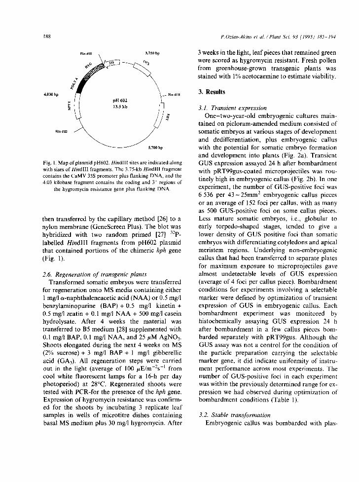
Fig. 1. Map of plasmid pH602. Hindlll sites are indicated along with sizes of HindlIl fragments. The 3.75-kb Hindlll fragment contains the CaMV 35S promoter plus flanking DNA, and the 4.03 kilobase fragment contains the coding and 3' regions of
the hygromycin resistance gene plus flanking DNA.
then transferred by the capillary method [26] to a nylon membrane (GeneScreen Plus). The blot was hybridized with two random primed [27] 32p_ labelled HindlII fragments from pH602 plasmid that contained portions of the chimeric hph gene (Fig. 1).
2.6. Regeneration of transgenic plants Transformed somatic embryos were transferred
for regeneration onto MS media containing either 1 mg/1 ot-naphthaleneacetic acid (NAA) or 0.5 mg/1 benzylaminopurine (BAP) + 0.5 mg/1 kinetin + 0.5 mg/l zeatin + 0.1 mg/1 NAA + 500 mg/1 casein hydrolysate. After 4 weeks the material was transferred to B5 medium [28] supplemented with 0.1 mg/l BAP, 0.1 mg/1 NAA, and 25/~M AgNO3. Shoots elongated during the next 4 weeks on MS (2% sucrose)+ 3 mg/1 BAP + 1 mg/l gibberellic acid (GA3). All regeneration steps were carried out in the light (average of 100 t~E/m-2s -l from cool white fluorescent lamps for a 16-h per day photoperiod) at 28°C. Regenerated shoots were tested with PCR-for the presence of the hph gene. Expression of hygromycin resistance was confirm- ed for the shoots by incubating 3 replicate leaf samples in wells of microtitre dishes containing basal MS medium plus 30 mg/1 hygromycin. After
3 weeks in the light, leaf pieces that remained green were scored as hygromycin resistant. Fresh pollen from greenhouse-grown transgenic plants was stained with 1% acetocarmine to estimate viability.
3. Results
3.1. Transient expression One-two-year-old embryogenic cultures main-
tained on picloram-amended medium consisted of somatic embryos at various stages of development and dedifferentiation, plus embryogenic callus with the potential for somatic embryo formation and development into plants (Fig. 2a). Transient GUS expression assayed 24 h after bombardment with pRT99gus-coated microprojectiles was rou- tinely high in embryogenic callus (Fig. 2b). In one experiment, the number of GUS-positive foci was 6 536 per 4 3 - 2 5 mm: embryogenic callus pieces or an average of 152 foci per callus, with as many as 500 GUS-positive foci on some callus pieces. Less mature somatic embryos, i.e., globular to early torpedo-shaped stages, tended to give a lower density of GUS positive foci than somatic embryos with differentiating cotyledons and apical meristem regions. Underlying non-embryogenic callus that had been transferred to separate plates for maximum exposure to microprojectiles gave almost undetectable levels of GUS expression (average of 4 foci per callus piece). Bombardment conditions for experiments involving a selectable marker were defined by optimization of transient expression of GUS in embryogenic callus. Each bombardment experiment was monitored by histochemically assaying GUS expression 24 h after bombardment in a few callus pieces bom- barded separately with pRT99gus. Although the GUS assay was not a control for the condition of the particle preparation carrying the selectabte marker gene, it did indicate uniformity of instru- ment performance across most experiments. The number of GUS-positive foci in each experiment was within the previously determined range for ex- pression we had observed during optimization of bombardment conditions (Table 1).
3.2. Stable transformation Embryogenic callus was bombarded with plas-

P.Ozias-Akins et al . / Plant Sci. 93 (1993) 185-194 189
Fig. 2. (a) Embryogenic callus of A. hypogaea 'Toalson' that shows somatic embryos at various stages of development. (b) Transient GUS expression in embryogenic callus 24 h post-bombardment. (c) Selection of a hygromycin-resistant cell line on agar medium
(experiment 219). (d) Hygromycin-resistant plantlet regenerated from stably transformed embryogenic cell line 270-T1.
mid pH602 containing a hygromycin resistance gene under the control of the CaMV 35S prom- oter. Bombarded tissues were not immediately placed under selection, but were grown for one subculture period (4 weeks) on maintenance medi- um. Tissues were first exposed to low levels of
hygromycin (5-10 mg/1) approximately 5 weeks post-bombardment. Hygromycin levels were in- creased to 20 rag/1 in subsequent subcultures. Se- lection for hygromycin-resistant cell lines was successful using both solid and liquid selection media (Fig. 2c). However, selection in liquid medi-

190
Table 1 Transgenic cell lines recovered
P.Ozias-Akins et al. / Plant Sci. 93 (1993) 185-194
from microprojectile bombarded embryogenic callus of peanut
Experiment Number of Number of Numbers of Nomenclature Number of Amplification number bombard- callus pieces GUS-positive of Hyg r cell weeks under of 35S:hph b
ments a tested for loci lines recovered selection transient GUS
Southern hybridiza- tion c
219 11 0 nt 219-T1 32 + + 219-T5 26 + +
265 18 3 3-1000 265-T1 30 + + 266 15 4 28-237 266-T1 16 + nt 270 19 3 43-66 270-T1 12 + +
270-T2 14 + + 270-T3 32 + nt
272 18 2 258-> 1000 272-TI 28 + nt 272-T2 28 + nt
277 19 3 50-163 277-T 1 14 + + 277-T2 24 + nt
285 14 1 10 285-T1 16 + + 285-T2 16 + nt 285-T3 16 + nt
aEach bombardment consisted of a single culture dish. bpCR amplification of a 380 bp region of the chimeric hygromycin resistance gene from genomic DNA of a putatively transformed cell line. Clntegration of plasmid DNA into the genome of the recipient tissue was confirmed by hybridization of genomic DNA with a por- tion of the plasmid (+; nt = not tested).
um was more efficient and faster for e l iminat ing non- t ransformed tissues. In experiments where selection in solid and liquid media was carried out
in parallel, only selection in liquid resulted in recovery of transgenic cell lines. Transformed callus usually appeared as a rapidly-growing mass of tissue 3 - 4 months after ini t iat ion of the liquid
cultures. Each putative independent t ransformant was
numbered at the time of isolation and separately mainta ined for subsequent D N A analysis and regeneration. Four teen hygromycin-resistant cell lines were selected from a total of seven bombard- ment experiments. All but 2 pairs of the cell lines were derived from separate bombardmen t s and were undoubted ly independent t ransformat ion events. The two pairs of cell lines were sufficiently separated in integrity or time of origin to have a high probabil i ty of being independent events. Oli- gonucleotide primers specific to sequences in the CaMV 35S5 promoter and hph coding region
amplified the expected 380 nucleotide fragment from each hygromycin-resistant cell line but no
amplification was observed from control, non- transformed tissue (Table 1, Fig. 3). Genomic D N A from all of the cell lines tested showed inte-
gration of the foreign gene, as determined by
Southern hybridizat ion analysis (Table 1, Fig. 4). Hybridizat ion of high molecular weight, un- digested genomic D N A with the combined 3.75 and 4.03 kb H i n d l I I fragments from pH602 show-
ed the absence of free plasmid DNA. The hybrid- ization pat tern of H i n d l I I - d i g e s t e d genomic D N A from some of the t ransformed cell lines indicated that the H i n d l I I sites f lanking the selectable mark-
er gene probably were distanced from the H i n d l l I
site internal to the hygromycin resistance gene or deleted dur ing integrat ion of the plasmid DNA. Further molecular analysis would be required to
determine the extent of rearrangement of integrat- ed plasmid DNA.
Two experiments were conducted without selec-

P.Ozias-Akins et al. / Plant Sci. 93 (1993) 185-194 191
t
"7 ~,
0 v- v-. ~D (0 ~ 1~ r~ O0 O0
380bp
Fig. 3. An ethidium bromide-stained gel showing amplification of the expected 380 bp fragment of the chimeric hygromycin re- sistance gene from 8 independent transgenic cell lines (numbers correspond with those listed in Table 1) and 2 transgenic plants (219-T5-1; 270-T1-6). No amplification was observed from the control callus DNA (Toalson). Strong amplification was observed from the positive control (plasmid only) DNA. Details of reaction and electrophoresis conditions are given in
Materials and methods.
tion in order to determine if selection, and thus use of a selectable marker gene, could be avoided with our transformation system. Approximately 5 weeks post-bombardment, the embryogenic calli were transferred to regeneration media. In the first experiment, calli (cultivar Toalson) were bom- barded with pRT99gus so that regenerated shoots could be rapidly screened for GUS activity. Leaflets from plants developing in each of 120 plates were pooled for the GUS assay. Out of ap- proximately 600 plants tested, none gave the blue GUS reaction product expected for a stably trans- formed plant. In the second experiment, cultivar Florunner calli were bombarded with pH602 and leaflets from regenerated plants were tested for hygromycin resistance. No leaflets remained green on hygromycin-containing medium (30 mg/l) after 3 weeks. No PCR amplification of the hph gene was obtained from DNA of plantlets whose leaflets browned more slowly on hygromycin.
3.3. Plant regeneration from stable transformants Transgenic cell lines proliferated prior to initia-
tion of plant regeneration so that the cell lines could be maintained as well as used for regenera- tion. In this manner, plants could be regenerated indefinitely from a competent cell line. The sequence of media optimal for plant regeneration had previously been determined for cultivar Toalson (Ozias-Akins, unpublished results). Ap- proximately 3 months were needed for plant regeneration from an embryogenic cell line. Using this regeneration method (outlined in Materials and methods), all of the transformed cell lines have produced plants (Fig. 2d). At least 100 plants have been regenerated and over 80 of these plants have been transferred to soil in the greenhouse. Flowers with a high proportion of stainable pollen have been produced. Of 8 plants from cell line 270-T1 tested for amplification of the foreign gene, all 8 were positive (Fig. 3). Likewise, two plants from 219-T5, tested by the same method, also were posi- tive. Southern hybridization analysis of one plant (Fig. 4) confirmed the positive PCR results for transformation. Leaves from the transgenic plants remained green and formed a small amount of callus after 3 weeks on medium containing 30 mg/1 hygromycin, whereas all leaves from non-trans- formed control plants turned brown and died.
4. Discussion
While many resources have been devoted to the development of a transformation system for soy- bean [8] and important cereals [10, 13], much less work has been focused on the transformation of peanut. Transformation of forage legumes generally has been more straightforward than transformation of grain legumes [15]. Approaches that rely on tissue culture for the regeneration of transgenic plants must take into account the type of culture (callus, suspension or protoplast), the mode of plant regeneration (embryogenesis vs. organogenesis), the maintenance of regeneration potential over time, and the ability to select stably transformed tissue. We have developed an em- bryogenic culture system for multiple genotypes [5,29,30] that satisfies two important criteria: long- term preservation of regeneration capacity and

192 P. Ozias-Akins et al. / Plant Sci. 93 (1993) 185-194
Et L1-LL~
"0 C
i m
"r'.
L£-LL~
"0
~)
"0
~I-OL~
L1-£9~
L1-6L~
L-£1-6L~
J~JI~ULI
UOSleOl
L1-£8~ A
~I-OL~
L1-£9~:
L.L-6 L~
L-£1-6 L~
~09 qd
P3LO O P ~ 4 H
~ < ~ - Z i l ~
= ° _ , < z . ~ = Z ~ . ~
a E ~ g
=m N ~
o ~
~ ~ ' - ~ . _
< = ~ ~.-
o ,..-, . _ - ~
~o~
,- "?' o ~
N e e ~


194
4 T.E. Clemente, D. Robertson, T.G. Isleib, M.K. Beute and A.K. Weissinger, Evaluation of leaflets from mature zygotic embryos as recipient tissue for biolistic gene trans- fer to peanut (Arachis hypogaea). Transgen. Res., 1 (1992) 275-284.
5 P. Ozias-Akins, W.F. Anderson and C.C. Holbrook, So- matic embryogenesis in Arachis hypogaea L.: genotype comparison. Plant Sci., 83 (1992) 103-111.
6 J.C. Sanford, Biolistic plant transformation. Physiol. Plant., 79 (1990) 206-209.
7 D.T. Tomes, A.K. Weissinger, M. Ross, R. Higgins, B. Drummond, S. Schaaf, J. Malone-Schoneberg, M. Staebell, P. Flynn, J. Anderson and J. Howard, Transgen- ic tobacco plants and their progeny derived by micropro- jectile bombardment of tobacco leaves. Plant Mol. Biol., 14 (1990) 261-268.
8 P. Christou, W.F. Swain, N.S. Yang and D.E. McCabe, Inheritance and expression of foreign genes in transgenic soybean plants. Proc. Natl. Acad. Sci. USA, 86 (1989) 7500-7504.
9 J.J. Finer and M.D. McMullen, Transformation of cotton (Gossypium hirsutum L.) via particle bombardment. Plant Cell Rep., 8 (1990) 586-589.
10 W.J. Gordon-Kamm, T.M. Spencer, M.L. Mangano, T.R. Adams, R.J. Daines, W.G. Start, J.V. O'Brien, S.A. Chambers, W.R. Adams Jr., N.G. Willetts, T.B. Rice, C.J. Mackey, R.W. Krueger, A.P. Kausch and P.G. Lemaux, Transformation of maize cells and regeneration of fertile transgenic plants. Plant Cell, 2 (1990) 603-618.
11 M.M.M. Fitch, R.M. Manshardt, D. Gonsalves, J.L. Slightom and J.C. Sanford, Stable transformation of papaya via microprojectile bombardment. Plant Cell Rep., 9 (1990) 189-194.
12 R. Serres, E. Stang, D. McCabe, D. Russel, D Mahr and B. McCown, Gene transfer using electric discharge parti- cle bombardment and recovery of transformed cranberry plants. J. Am. Soc. Hort. Sci., 117 (1992) 174-180.
13 V. Vasil, A.M. Castillo, M.E. Fromm and I.K. Vasil, Her- bicide resistant fertile transgenic wheat plants obtained by microprojectile bombardment of regenerable embryogenic callus. Bio/Technology, 10 (1992) 667-674.
14 R.G. Birch and T. Franks, Development and optimization of microprojectile systems for plant genetic transforma- tion. Aust. J. Plant Physiol., 18 (1991) 453-469.
15 V. Kumar and M.R. Davey, Genetic improvement of legumes using somatic cell and molecular techniques. Euphytica, 55 (1991) 157-169.
16 T. Murashige and F. Skoog, A revised medium for rapid growth and bioassays with tobacco tissue cultures. Physiol. Plant., 15 (1962) 473-497.
17 E. Firoozabady, D.L. DeBoer, D.J. Merlo, E.L. Halk, L.N. Amerson, K.E. Rashka and E.E. Murray, Transfor- mation of cotton (Gossypium hirsutum L.) by Agrobac- terium tumefaciens and regeneration of transgenic plants. Plant Mol. Biol., 10 (1987) 105-116.
18 K.R. Kaster, S.G. Burgett, R.N. Nagaraja and T.D. ln- golia, Analysis of a bacterial hygromycin B resistance gene by transcriptional and translational fusions and by DNA sequencing. Nucl. Acids Res., 1 l (1983) 6895-6911.
P.Ozias-Akins et al. / Plant Sci. 93 (1993) 185-194
19 E.E. Murray, T.R. Rocheleau, M. Eberle, C. Stock, V. Sekar and M.J. Adang, Analysis of unstable transcripts of insecticidal crystal protein genes of Bacillus thuringiensis in transgenic plants and electroporated protoplasts. Plant Mol. Biol., 16 (1991) 1035-1060.
20 R. T6pfer, J. Schell and H.H. Steinbiss, Versatile cloning vectors for transient gene expression and direct gene transfer in plant cells. Nucl. Acids Res., 16 (1988) 8725.
21 F.M. Ausubel, R. Brent, R.E. Kingston, D.D. Moore, J.A. Smith, J.G. Seidman and K. Struhl, Current Pro- tocols in Molecular Biology. Greene Publishing, New York 1987.
22 R.A. Jefferson, Assaying chimeric genes in plants: the GUS gene fusion system. Plant Mol. Biol. Rep., 5 (1987) 387-405.
23 T.M. Klein, E.C. Harper, Z. Svab, J.C. Sanford, M.E. Fromm and P. Maliga, Stable genetic transformation of intact Nicotiana cells by the particle bombardment pro- cess. Proc. Natl. Acad. Sci. USA, 85 (1988) 8502-8505.
24 J.J. Doyle and J.L. Doyle, A rapid DNA isolation proce- dure for small quantities of fresh leaf tissue. Phytochem. Bull., 19 (1987) 11-15.
25 M.A. Haring, M.J. Teeuwen de Vroomen, H.J.J. Nijkamp and J. Hille, trans-activation of an artificial dTam3 transposable element in transgenic tobacco plants. Plant Mol. Biol., 16 (1991) 39-47.
26 E.M. Southern, Detection of specific sequences among DNA fragments separated by gel electrophoresis. J. Mol. Biol., 98 (1975) 503-517.
27 A.P. Feinberg and B. Vogelstein, A technique for radiolabelling DNA restriction endonuclease fragments to high specific activity. Anal. Biochem., 132 (1983) 6-13.
28 O.L. Gamborg, R.A. Miller and K. Ojima, Nutrient re- quirements of suspension cultures of soybean root cells. Exp. Cell Res., 50 (1968) 151-158.
29 P. Ozias-Akins, Plant regeneration from immature em- bryos of peanut. Plant Cell Rep., 8 (1989) 217-218.
30 P. Ozias-Akins, C. Singsit and W.D. Branch, Interspecific hybrid inviability in crosses of Arachis hypogaea x A. stenosperma can be overcome by in vitro embryo matura- tion or somatic embryogenesis. J. Plant Physiol., 140 (1992) 207-212.
31 M.C.P. Timmermans, P. Maliga, J. Vieira and J. Messing, The pFF plasmids: cassettes utilising CaMV sequences for expression of foreign genes in plants. J. Biotech., 14 (1990) 333-344.
32 J.J. Finer and M.D. McMullen, Transformation of soy- bean via particle bombardment of embryogenic suspen- sion culture tissue. In Vitro 27P (1991) 175-182.
33 P. Vain, M.D. McMullen and J.J. Finer, Osmotic treat- ment enhances particle bombardment-mediated transient and stable transformation of maize. Plant Cell Rep., 12 (1993) 84-88.
34 K. Arumuganathan and E.D. Earle, Nuclear DNA con- tent of some important plant species. Plant Mol. Biol. Rep., 9 (1991) 208-218.