Complications in Laser Cutaneous Surgery - Taylor & Francis ...

Appraisal of a Leishmania major Strain Stably ExpressingmCherry Fluorescent Protein for Both In Vitro and In VivoStudies of Potential Drugs and Vaccine againstCutaneous LeishmaniasisEstefania Calvo-Alvarez1., Nestor Adrian Guerrero2., Raquel Alvarez-Velilla1, Christopher
Fernandez Prada1, Jose Marıa Requena2, Carmen Punzon3, Miguel Angel Llamas3, Francisco J. Arevalo3,
Luis Rivas4, Manuel Fresno2,3, Yolanda Perez-Pertejo1, Rafael Balana-Fouce1*, Rosa M. Reguera1
1 Departamento de Ciencias Biomedicas, Universidad de Leon, Leon, Spain, 2 Centro de Biologıa Molecular ‘‘Severo Ochoa’’, Universidad Autonoma de Madrid, Madrid,
Spain, 3 Diomune, Parque Cientifico de Madrid, Madrid, Spain, 4 Centro de Investigaciones Biologicas, Madrid, Spain
Abstract
Background: Leishmania major cutaneous leishmaniasis is an infectious zoonotic disease. It is produced by a digeneticparasite, which resides in the phagolysosomal compartment of different mammalian macrophage populations. There is anurgent need to develop new therapies (drugs) against this neglected disease that hits developing countries. The main goalof this work is to establish an easier and cheaper tool of choice for real-time monitoring of the establishment andprogression of this pathology either in BALB/c mice or in vitro assays. To validate this new technique we vaccinated micewith an attenuated Dhsp70-II strain of Leishmania to assess protection against this disease.
Methodology: We engineered a transgenic L. major strain expressing the mCherry red-fluorescent protein for real-timemonitoring of the parasitic load. This is achieved via measurement of fluorescence emission, allowing a weekly record of thefootpads over eight weeks after the inoculation of BALB/c mice.
Results: In vitro results show a linear correlation between the number of parasites and fluorescence emission over a range offour logs. The minimum number of parasites (amastigote isolated from lesion) detected by their fluorescent phenotype was10,000. The effect of antileishmanial drugs against mCherry+L. major infecting peritoneal macrophages were evaluated bydirect assay of fluorescence emission, with IC50 values of 0.12, 0.56 and 9.20 mM for amphotericin B, miltefosine andparomomycin, respectively. An experimental vaccination trial based on the protection conferred by an attenuated Dhsp70-IImutant of Leishmania was used to validate the suitability of this technique in vivo.
Conclusions: A Leishmania major strain expressing mCherry red-fluorescent protein enables the monitoring of parasitic loadvia measurement of fluorescence emission. This approach allows a simpler, faster, non-invasive and cost-effective techniqueto assess the clinical progression of the infection after drug or vaccine therapy.
Citation: Calvo-Alvarez E, Guerrero NA, Alvarez-Velilla R, Prada CF, Requena JM, et al. (2012) Appraisal of a Leishmania major Strain Stably Expressing mCherryFluorescent Protein for Both In Vitro and In Vivo Studies of Potential Drugs and Vaccine against Cutaneous Leishmaniasis. PLoS Negl Trop Dis 6(11): e1927.doi:10.1371/journal.pntd.0001927
Editor: Genevieve Milon, Institut Pasteur, France
Received April 30, 2012; Accepted October 16, 2012; Published November 29, 2012
Copyright: � 2012 Calvo-Alvarez et al. This is an open-access article distributed under the terms of the Creative Commons Attribution License, which permitsunrestricted use, distribution, and reproduction in any medium, provided the original author and source are credited.
Funding: This research was supported by Ministerio de Ciencia y Tecnologıa (grants AGL2010 16078/GAN), Instituto de Salud Carlos III (grant PI09/0448 and theNetwork of Tropical Diseases RICET RD06/0021/1004). RAV, CFP and ECA are pre-doctoral fellows granted by RICET (ISCIII), Junta de Castilla y Leon (ESF; EuropeanSocial Founding) and University of Leon, respectively to RMR. Instituto de Salud Carlos III (Network of Tropical Disasese RICET RD06/0021/0016), Ministerio deCiencia e Innovacion (SAF2010-17833), ChagasEpiNet 223034 European Union Seventh Framework Programme and Fundacion Ramon Areces to MF. Funding byISCIII-RETIC RD06/0021/0008-FEDER to JMR and ISCIII-RETIC RD06/0021/0006-FEDER to LR is also acknowledged. The funders had no role in study design, datacollection and analysis, decision to publish, or preparation of the manuscript.
Competing Interests: The authors have declared that no competing interests exist.
* E-mail: [email protected]
. These authors contributed equally to this work.
Introduction
Leishmania major is the main cause of cutaneous leishmaniasis
(CL) in the Old World. Parasites are transmitted by Phlebotominae
sandflies whilst blood feeding on infected mammalian hosts. CL is
widely spread in the developing world, affecting people in 88
countries with 1.5 million new cases reported each year. CL
usually produces ulcers on the exposed parts of the body that often
leave disfiguring scars, which in turn, can cause serious social
prejudice [1].
Conventional in vivo animal models for the study of parasite-host
relationships involve large number of animals. These animals are
required to be slaughtered at different time points in order to
identify both anatomical distribution and parasite numbers in
PLOS Neglected Tropical Diseases | www.plosntds.org 1 November 2012 | Volume 6 | Issue 11 | e1927
organs and tissues. Furthermore, this approach has some
important limitations that must be overcome: i) post-mortem
analysis of animals makes it impossible to track the space/time
progression of the pathogen within the hosts; ii) spread of the
pathogen to unexpected anatomic sites can remain undetected; iii)
in order to achieve precise and relevant data, it is necessary to kill
large numbers of animals. Recent real-time in-vivo imaging
techniques with genetically modified pathogens represent a
valuable complementary tool. They can be used for conventional
studies of pathogenesis and therapy as long as the modified
pathogen retains the virulence of the parental strain. Moreover,
this has led to an increased number of reports concerning
genetically modified parasites that express bioluminescent and/or
fluorescent reporters. This was principally developed for in-vitro
infection studies and to monitor diseases in living animals [2,3].
Bioluminescent pathogens expressing the sea pansy Renilla
reniformis luciferase have been used in experimental murine
infections of Toxoplasma gondii [4] as well as in the rodent malaria
parasite Plasmodium berghei [5]. A recent study has allowed scientists
to identify the liver stages of firefly luciferase-expressing parasites
in living animals [6].
This approach has also been successfully implemented in
trypanosomatids. Lang and co-workers showed that a luciferase
expressing L. amazonensis strain was useful for rapid screening of
drugs in infected macrophages [7]. Further studies used the same
techniques with L. major [8], L. infantum [9] and in in-vivo murine
experimental infections [7,10]. Besides, the use of Trypanosoma
brucei expressing R. reniformis luc gene has permitted scientists to find
unusual colonizing niches during the progression of African
trypanosomiasis [11].
Fluorescent imaging offers several benefits: i) unlike light-
emitting proteins, fluorescent reporters do not require specific
substrates: ii) the fluorescence emitted is very stable over time and
iii) this approach is useful when studying tissue harvested from
infected animals since parasites can be individually identified [12].
The first transgenic Leishmania species expressing the green
fluorescent protein (GFP) was reported by Beverley’s group [13].
Episomally transfected Leishmania spp. with GFP or enhanced GFP
(EGFP) have enhanced High Throughput Screening (HTS)
methods in free-living promastigotes [14–17] and amastigotes
[18–22]. However, only recently, the stable transfection of the
EGFP reporter has been found suitable for both in vitro and in vivo
infection studies [21–24]. Although native GFP produces signif-
icant fluorescence and is extremely stable, the excitation maximum
is close to the ultraviolet range, which can result in damaging
living cells.
Red-fluorescence labelled parasites have been used to determine
the early stages of CL pathogeny at the infection site (revised by
Millington and co-workers [25]). By combining a L. major Red
Fluorescent Protein 1 (RFP)-expressing strain and dynamic
intravital microscopy, the site of sandfly bites has been identified
in vivo in a mouse model. The study reveals an essential role for
both neutrophils and dendritic cells that converge at localized sites
of acute inflammation in the skin following pathogen deposition
[26,27]. Using mCherry-L. infantum chagasi – responsible of visceral
leishmaniasis in the New World – researchers have been able to
report the recruitment of neutrophils and their role in non-
ulcerative forms of leishmaniasis [28]. In addition, to study the
mechanism regulating dentritic cell recruitment and activation in
susceptible BALB/c [29] and resistant C57BL/6 mice [30] DsRed
labelled parasites were used
Fluorescent parasites have been used to explain some aspects of
Leishmania biology. L. donovani lines stably expressing either EGFP
or RFP have been used to identify hybrid parasites produced
during the early development of the sandfly [31]. In addition, a L.
major strain, which episomally expressed the DsRed protein, was
used for quantifying the infectious dosage transmitted by a sandfly
bite [32].
Based on the improved photostability as well as suitability for
intravital imaging, mCherry was considered the best choice for our
studies in comparison to other red fluorescent proteins [33].
mCherry is a protein derived from the coral Discosoma striata RFP.
It has a maximum emission peak at 610 nm with a 587 nm
excitation wavelength. Despite the fact that it is 50% less bright
than EGFP, it is more photostable and it has higher tissue
penetration [12]. Because of this, it is the best-suited choice in
applications of single-molecule fluorescence or multicolour fluo-
rescent imaging [34]. In this report we describe the use of a stably
mCherry-transfected L. major strain as a valuable tool to both in
vitro assays for drug screening and in vivo pre-clinical vaccine studies
in real-time.
Materials and Methods
Mice and parasitesThe animal research described in this manuscript complied with
Spanish (Ley 32/2007) and European Union Legislation (2010/
63/UE). The used protocols were approved by the Animal Care
Committee of the Centro de Biologıa Molecular and Universidad
Autonoma de Madrid (Spain).
Female BALB/c mice (6–8 week old) were purchased from
Harlan Interfauna Iberica S.A. (Barcelona, Spain) and maintained
in specific-pathogen-free facilities for this study.
L. major LV39c5 (RHO/SU/59/P) strain was used for
generating mCherry transgenic promastigotes. Parasites were
cultured at 26uC in M199 supplemented with 25 mM HEPES
pH 7.2, 0.1 mM adenine, 0.0005% (w/v) hemin, 2 mg/ml
biopterin, 0.0001% (w/v) biotin, 10% (v/v) heat-inactivated foetal
calf serum (FCS) and antibiotic cocktail (50 U/ml penicillin,
50 mg/ml streptomycin). Attenuated Dhsp70-II (Dhsp70-II::NEO/
Dhsp70-II::HYG), used as candidate vaccine [35], is a null mutant
for the hsp70-type-II gene, generated by targeted deletion in L.
infantum (MCAN/ES/96/BCN150) strain [36]. Dhsp70-II pro-
mastigotes were grown in RPMI 1640 (Sigma-Aldrich) culture
medium supplemented with 10% (v/v) FCS, 50 U/ml penicillin
and 50 mg/ml streptomycin.
Author Summary
Leishmaniasis is a parasitic disease that is far fromeradication. The lack of an efficacious vaccine andtreatment failures are major factors in its intractableworldwide prevalence. A non-invasive imaging techniqueusing genetically engineered parasites that expressedfluorescent proteins could give to researchers a quantita-tive and visual tool to characterize the parasite burden inexperimental infections. In addition, it can be useful fordetermining the efficacy of candidate vaccines or drugsusing High Throughput Screening methods that allow thetesting of libraries of compounds in an automated 96-wellplate format. Herein, we demonstrate that there is a goodcorrelation between fluorescence emission and the para-site load, thus permitting the use of this output to monitorthe progression of the disease. In order to validate this toolwe have immunized mice prior the parasite challenge witha red-emitting parasite strain, confirming the scientificsuitability of this approach as a valuable alternative model.
mCherry-Fluorescent L. major Strain
PLOS Neglected Tropical Diseases | www.plosntds.org 2 November 2012 | Volume 6 | Issue 11 | e1927

Generation of a mCherry+L. major strainThe 711-bp mCherry coding region was amplified by PCR from
pRSETb-mCherry vector, a kindly gift from Dr Roger Y. Tsien –
Departments of Pharmacology and Chemistry & Biochemistry,
UCSD (USA) – [37] with the primers RBF634 and RBF600
(Table 1). PCR product was cut with appropriate restriction
enzymes and ligated into the BglII and NotI sites of the pLEXSY-
hyg2 expression vector (Jena Bioscience GmbH, Germany).
Parasites expressing mCherry Open Reading Frame (ORF) were
obtained by transfection of L. major with the large SwaI targeting
fragment derived from pLEXSY-mCherry by electroporation and
subsequent plating on semisolid media containing 200 mg/ml
hygromycin B (Sigma-Aldrich) as previously described [38].
Correct integration of mCherry ORF into the 18S rRNA locus of
the resulting transgenic clones (mCherry+L. major) was confirmed
by Southern blot and PCR amplification analyses, using the
primers of Table 1. The fluorescent of stable-transfected mCherry
clones was confirmed by both flow cytometry (BD FACSCantoII)
and confocal microscopy (Nikon Eclipse TE2000E).
In vitro infections and drug screeningStarch-elicited peritoneal macrophages were recovered from
BALB/c mice and then 56104 cells were plated on black 96 wells
plates with clear bottom. Macrophages were infected at a ratio of
five metacyclic promastigotes per macrophage. Metacyclic
mCherry+L. major promastigotes were isolated from stationary
cultures (4–5 days old) by Ficoll gradient centrifugation [39].
Briefly, 2 ml of parasite suspension in M199 containing approx-
imately 76107 stationary-phase promastigotes were layered onto a
discontinuous density gradient in a 15 ml conical tube consisting
of 2 ml of 20% (w/v) Ficoll stock solution made in distilled water
and 2 ml of 10% (w/v) Ficoll diluted in M199 medium.
Metacyclic parasites were opsonized with 4% (v/v) C52 mouse
deficient serum (The Jackson Laboratory, USA) at 37uC for
30 min and resuspended in RPMI containing 10% (v/v) FCS [40].
The infection was synchronized by centrifugation (3306g, 3 min
at 4uC) and infected macrophages were incubated at 37uC in a
humidified 5% CO2 atmosphere [41]. Cells were washed
extensively with phosphate buffer saline (PBS) to remove the free
parasites and overlaid with fresh medium, which was replaced
daily thereafter. After one day incubation, to allow differentiation
into amastigotes, drugs (miltefosine, amphotericin B and paromo-
mycin) were added to the appropriate wells in a threefold dilution
series in RPMI (Sigma-Aldrich) with 10% (v/v) FCS and cells were
further incubated at 37uC for a further incubation of 72 h. Plates
were read in a fluorescence microplate reader (Synergy HT;
BioTek) (lex = 587 nm; lem = 610 nm).
In vivo infectionsMetacyclic promastigotes were isolated from stationary cultures
(4–5 days old) by negative selection with peanut agglutinin for
mouse infections. Briefly, promastigotes were resuspended in PBS
at 108 cells/ml, and peanut agglutinin (Vector laboratories) was
added at 50 mg/ml; the sample was incubated for 25 min at room
temperature. After centrifugation at 2006g for 10 min, the
supernatant contained the non-agglutinated metacyclic promasti-
gotes [42].
The virulence of L. major parasites was maintained by passage in
BALB/c mice by injecting hind footpads with 106 stationary-phase
parasites. After 6–8 weeks, animals were euthanized and popliteal
lymph nodes were dissected, mechanically dissociated, homoge-
nized and filtered. L. major amastigotes were isolated from murine
lymph nodes by passing the tissue through a wire mesh followed by
disrupting the cells sequentially through 25G1/2 and 27G1/2
needles, and polycarbonate membrane filters with pore size of 8, 5
and 3 mm (Isopore, Millipore) [43]. Isolated amastigotes were
transformed to promastigote forms by culturing at 26uC in
Schneider’s medium (Gibco, BRL, Grand Island, NY, USA)
supplemented with 20% (v/v) FCS, 100 U/ml penicillin and
100 mg/ml streptomycin. For infections, amastigote-derived pro-
mastigotes with less than five passages in vitro were used.
BALB/c mice were injected with several inocula (104, 105 and
106 promastigotes/mouse) during the setting up of the model. For
protection studies mice were vaccinated intravenously (tail-vein
injection) with the Dhsp70-II mutant strain (26107 promastigotes/
mouse) [35], or injection of PBS (control group), and four weeks
post-vaccination, were infected with 26105 mCherry+L. major
metacyclic promastigotes. The infections were performed by
injection of parasites in 50 ml PBS in the right hind footpads.
The growth of the lesion was monitored by fluorescence emission
detection (see below). The contralateral footpad of each animal
represented the negative control value. Footpad swelling was
measured using a Vernier calliper and data were represented as
the increment of the lesion size respect to the not infected footpad.
Table 1. Oligonucleotides used in this work.
Oligo No. Sequencea,b Purposedc
RBF 634 (7) ccgCTCGAGgaAGATCTCCACCATGGTGAGCAAGGGCG mCherry F
RBF 600 (8) ataagaatGCGGCCGCTTACTTGTACAGCTCGTCCATGC mCherry R
RBF 630 (1) CTTGTTTCAAGGACTTAGCCATG 59integration F
RBF 637 (2) TATTCGTTGTCAGATGGCGCAC 59integration R
RBF 644 (3) CATGTGCAGCTCCTCCCTTTC 39integration F
RBF 645 (4) CCTTGTTACGACTTTTGCTTC 39integration R
RBF 646 (5) ATGAAAAAGCCTGAACTCACC HYG F
RBF 647 (6) CTATTCCTTTGCCCTCGGAC HYG R
RBF 630 (9) CTTGTTTCAAGGACTTAGCCATG Southern probe F
RBF 631 (10) GCGGAAACCGCAAGATTTTTGC Southern probe R
aUnderlined sequence indicates restriction site.bBold sequence indicates optimized translation initiation sequence.cOrientation of primers: F, forward; R, reverse.doi:10.1371/journal.pntd.0001927.t001
mCherry-Fluorescent L. major Strain
PLOS Neglected Tropical Diseases | www.plosntds.org 3 November 2012 | Volume 6 | Issue 11 | e1927

In vivo fluorescence imaging and calibrationFluorescence emission was measured using an intensified
charged coupled device camera of the In Vivo Imaging System
(IVIS 100, Xenogen). Wild type- and mCherry+L. major-infected
animals were lightly anesthetized with 2.5–3.5% isoflurane and
then reduced to 1.5–2.0%. Anesthetized animals were placed in
the camera chamber, and the fluorescence signal was acquired for
3 s. Fluorescence determinations, recorded by the IVIS 100
system, were expressed as a pseudocolour on a gray background,
with red colour denoting the highest intensity and blue the lowest.
To quantify fluorescence, a region of interest was outlined and
analyzed by using the Living Image Software Package (version
2.11, Xenogen).
Quantification of parasite loadThe total number of living parasites invading the target organs
(popliteal lymph node draining the injected site) was calculated
from single-cell suspensions that were obtained by homogenization
of the tissue through a wire mesh. The cells were washed and
cultured in Schneider’s medium containing 20% (v/v) heat-
inactivated FCS, 100 U/ml penicillin and 100 mg/ml streptomy-
cin. The cell suspensions were serially diluted and dispensed into
96-well plates. The plates were incubated for 10 days and then
each well was examined and classified as positive or negative
according to whether or not viable promastigotes were present.
The number of parasites was calculated as follows: Limit Dilution
Assay Units (LDAU) = (geometric mean of titer from quadrupli-
cate cultures)6(reciprocal fraction of the homogenized organ
added to the first well). The titer was the reciprocal of the last
dilution in which parasites were observed [44].
Results
Generation of a functionally fluorescent L. major strainAimed to create a L. major fluorescent strain we electroporated
wild-type promastigotes with the lineal 5874 bp SwaI-SwaI
fragment containing the ORF encoding mCherry as well as the
hyg selection marker of the pLEXSY-mCherry plasmid. After
selection on semisolid plates containing 100 mg/ml hygromycin B,
individual colonies were seeded in M199 liquid medium supple-
mented with 10% FCS and hygromycin B. Genomic DNA
isolated from these cultures was used to confirm the correct
integration of the target sequence into the 18S rRNA locus of L.
major genome. Figure 1A shows a schematic representation of the
planned integration. Genomic DNA from wild-type strain and two
hygromycin B resistant clones were digested with NdeI and
hybridized with a labelled external probe (EP). As shown in the
Southern analysis of Fig. 1B, wild-type DNA digested with NdeI
yielded an 8.4-kb hybridization band, whereas in the two-
hygromycin B resistant clones an additional 3.8-kb hybridization
band was observed; this band is generated by the integration event
(Fig. 1A) corresponding to the expected size. Further confirmation
of the correct planned replacements was confirmed by PCR
(Fig. 1C) using the set of primers depicted in Fig. 1A.
The mCherry expression in stable-transfected L. major promas-
tigotes (mCherry+L. major) was monitored by flow cytometry. Cell
populations of mCherry+L. major strain and a parasite line
containing the pLEXSY-mCherry episome emitted strong red
fluorescence when they were excited at wavelength of 587 nm
(Fig. 2A). Clones with integrated mCherry gene had an average 10-
fold higher fluorescence than the ones expressing the gene
episomally. This is an expected result given that the mCherry gene
was integrated under the control of rRNA promoter, which is
known to present high-level transcription rates. As shown in
Fig. 2B, both strains (episomal and integrative) were strongly more
fluorescent than untransfected parasites.
In order to establish the correlation between parasite number
and fluorescence intensity, different number of procyclic and
metacyclic promastigotes as well as freshly isolated amastigotes
from infected animals were placed in 96-well plates and their
fluorescence intensity was measured spectrofluorometrically. A
clear correlation between fluorescence intensity and the number of
the three parasitic forms was observed (Fig. 2C).
The stability of mCherry expression was monitored over a
period of 6 months after transfection and no change was observed
in fluorescence intensity during this period, even in the absence of
hygromycin B.
In vitro infections with mCherry+L. major parasitesOnce the infectivity of the mCherry+L. major parasites was
recovered through mouse infections, the amastigotes obtained
from cutaneous lesions were differentiated back into promasti-
gotes. These cells were grown up to stationary phase and used to
infect freshly isolated BALB/c peritoneal macrophages at a 5:1
multiplicity in 24-well plates. Figure 3 shows fluorescence images
of either promastigotes or amastigotes internalized in macrophag-
es. A strong red fluorescence emission from free-living mCher-
ry+L. major promastigotes was observed by confocal microscopy
(Fig. 3B). Similarly, round-shaped red fluorescent emitting
amastigotes were observed inside parasitophorous vacuoles in
the cytoplasm of the infected macrophages (Fig. 3F). The course of
the in vitro infection was followed over a period of 48 h by
measuring the absolute fluorescence of the infection and the
percentage of infected macrophages. These experiments were
carried out in parallel with others using the classical Giemsa
staining to determine parasite load in vitro (data not shown). No
differences between both methods were accounted thus pointing to
the suitability of fluorescence analyses to assess the infectivity of
mCherry+L. major strain on mouse macrophages. A major
application of a fluorescent Leishmania model would be its
usefulness to perform HTS of potential leishmanicidal compounds
in vitro. To assess the suitability of our mCherry+L. major for this
goal, current drugs used in the treatment of human leishmaniasis
(miltefosine, paromomycin and amphotericin B) were assayed in
Leishmania-macrophage infections at different concentrations over
a 72 h-span. Absolute fluorescence emitted by mCherry+L. major
infected macrophages was plotted vs. drug concentrations,
obtaining the dose-response curves of Fig. 4. Nonlinear regression
analysis of the curves, fitted by the SigmaPlot statistic package,
reached IC50 values of 0.1260.03 mM for amphotericin B,
0.5760.12 mM for miltefosine and 9.2063.59 mM for paromo-
mycin. In all the cases the difference in fluorescence emission
corresponded to a difference in the percentage of infected cells,
also observed microscopically. These findings clearly showed that
the mCherry+L. major strain is a useful tool for in vitro drug
screening.
Validation of a CL murine model with mCherry+L. majorIn order to determine whether mCherry+L. major parasites
could be detected in vivo using whole-body imaging, 104, 105 and
106 metacyclic forms of the fluorescent-transgenic strain were
injected subcutaneously into the hind footpads of six BALB/c mice
per group. Lesion progression monitored both by direct measuring
of fluorescence emission by mCherry+L. major amastigotes
(recorded in an IVIS 100) and by the development of hind-limb
lesions assessed by measuring the thickness of the footpads with a
Vernier calliper. Animals were examined every seven days for a
total of eight weeks (except the group infected with 106 parasites
mCherry-Fluorescent L. major Strain
PLOS Neglected Tropical Diseases | www.plosntds.org 4 November 2012 | Volume 6 | Issue 11 | e1927

that were sacrificed at 6th week post-infection because the
appearance of ulcerations in the footpads).
Figure 5 shows the fluorescence intensity recorded weekly from
the footpads of representative mice of each inoculum group (104,
105 and 106 metacyclic promastigotes per mouse). The fluorescent
signal (estimated as average radiance: p/s/cm2/sr) was plotted
against the infection time of each inoculum (Fig. 6A). Fluorescent
signal was detected after the first week post-infection in mice
infected with 106 metacyclic parasites (radiance = 0.266108 p/s/
cm2/sr), reaching a radiance of 5.06108 p/s/cm2/sr five weeks
later. In the group of mice injected with 105 metacyclic parasites,
the fluorescence was detected the third week after inoculation
(radiance = 0.86108 p/s/cm2/sr), reaching similar intensity than
the mice group injected with 106 parasites at the 8th week of
inoculation. Finally, the fluorescence signal of mice injected with
104 metacyclic mCherry+L. major parasites was not detectable until
the 5th week (radiance = 0.356108 p/s/cm2/sr), reaching the
maximum intensity (radiance = 1.146108 p/s/cm2/sr) at the end
of the experiment.
The success of infection defined as the lesion onset and its
development in the inoculated footpads over the time, was
observed in every mice. Although there was a good correlation to
lesion size, fluorescence was more sensitive to evaluate the
progression of infection. Figure 6B shows that lesion emergence
was dependent on the size of pathogen inoculum and it was hardly
measurable during the first weeks after infection. Lesion size in
mm was 0.34, 0.94 and 0.57 measured at the third, fifth and
seventh week, respectively corresponding to 106, 105 and 104
metacyclic promastigotes per inoculum. It is remarkable that a
weak but measurable fluorescence signal from infected hind limbs
was detectable two weeks prior to visible and measurable injury
took place in all dose groups.
As expected and since BALB/c mice have a predisposition to
develop an anti-inflammatory Th2 response, the lesions appearing
Figure 1. Strategy of integration of mCherry encoding gene into genomic rRNA locus of L. major. A) Scheme of the structure of the 18SrRNA locus on wild type and planned integration of mCherry gene. Key: utr1: 59non-translated region of aprt gene; utr2: 1.4 kb intergenic region fromcam operon; and utr3: 59UTR of dhfr-ts gene; hyg; hygromycin B resistance cassette. The narrow white box on the left side corresponds to the externalprobe (EP) used for southern blot analyses. B) Southern blot analysis of two L. major clones after selection with hygromycin B. DNA was digested withNdeI and hybridized with the labelled EP. The similar intensities of 8.4 and 3.8 kbp bands could be due to inefficiency transfer of the large band to themembrane. C) PCR confirmation of successful integration of the reporter cassette. Primers 1/2 and 3/4 together confirm the correct integration of thereporter cassette from the 59 and 39 sides, respectively. Primers 5/6 confirm the presence of the HYG marker, and primers 7/8, 9/10, confirm thepresence of mCherry ORF, respectively. Primers: 1(RBF630); 2(RBF637); 3(RBF644); 4(RBF645); 5(RBF646); 6(RBF647); 7(RBF634) and 8(RBF600) (seeTable 1 for sequences).doi:10.1371/journal.pntd.0001927.g001
mCherry-Fluorescent L. major Strain
PLOS Neglected Tropical Diseases | www.plosntds.org 5 November 2012 | Volume 6 | Issue 11 | e1927

after infection setup were non-healing without treatment. A
correlation analysis of the fluorescence emitted by the lesions
toward the end of the 8-week period from mice infected with 104
and 105 metacyclic mCherry+L. major parasites (Fig. 6C) shows
significant differences between both groups (P,0.001) using
unpaired t-Student test. These differences were also found when
the lesion thickness of the footpads was compared using the
traditional calliper-based method (Fig. 6D). There was a clear
correlation between both parameters in both dosing groups with
an estimated Pearson coefficient of 0.94. At the end of the
experiments, animals were sacrificed and the popliteal lymph
nodes draining the lesions were dissected under sterile conditions.
The parasite load of these organs was determined by the limit
dilution method. Figure 6E shows the number of promastigote-
transforming amastigotes estimated in the animals of both dosing
groups, showing significant differences (P,0.001) and correlating
highly with both size lesion and fluorescence (Pearson coeffi-
cient = 0.79).
Application of mCherry+L. major to a model of murinevaccination
The suitability of this in vivo approach was assessed for the
evaluation of an experimental vaccination protocol against CL
that had been previously shown to be effective on a L. major–
BALB/c infection model [35]. In previous studies, it was
established that intravenous inoculation with Leishmania promas-
tigotes, lacking both alleles of the hsp70-II gene (Dhsp70-II line),
confers a partial protection against L. major infection in mice. For
this study, we inoculated a group of six mice with 26107
promastigotes of Dhsp70-II mutant and four weeks later, mice
were challenged with 26105 metacyclic forms of the mCherry+L.
major strain into mouse footpads. In parallel to the vaccinated
group, a control group was injected with the same inoculum of
mCherry-expressing transgenic parasites.
Red fluorescence emission in the footpad of mice infected with
mCherry+L. major parasites was followed over the time in both
groups (Fig. 7A). Fluorescence signal was detected in both groups
four weeks after challenge; however, fluorescence signal was higher
in control group mice than in vaccinated animals. By the end of
the 8th week animals were euthanized, the poplyteal lymph nodes
dissected, homogenized and the parasite load determined as
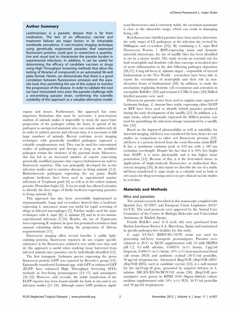
above. Figure 7B shows an 80% reduction (P,0.001) in the
parasite load of popliteal lymph nodes of vaccinated group related
to the control group. Therefore, since reproducible results were
obtained with both parasite quantification and fluorescence
emission methods, we conclude that the murine model of CL
established with the mCherry+L. major fluorescent strain might be
a suitable system for testing antileishmanial therapies both in vitro
and in vivo.
Discussion
Transgenic parasites expressing reporter proteins are valuable
tools to perform robust HTS platforms [45] and to understand the
underlying mechanisms of pathogenesis [3]. GFP is one of the
most commonly used reporters among fluorescent proteins.
Several mutants derived from native GFP have been developed
to cover longer wavelengths of the spectrum. Reporter molecules,
whose emission peak is in the red spectral range, the same as
mCherry, are excellent candidates for these kinds of studies.
Furthermore, light absorption by tissues in the red and far-red
spectra is reduced and consequently, the penetration is higher
[46]. Moreover, mCherry is the best general-purpose red
monomer due to its superior photostability compared to
mStrawberry and DsRed, which is inadequately folded at 37uC[13].
The integration of the reporter gene into the 18S rRNA locus of
L. major represents an efficient and effective strategy to guarantee a
stable expression when the parasites need to be grown in the
absence of selection drugs for both in vitro screenings and in mice
infections [7,22–25,47,48].
mCherry fluorescence was detected in the different stages of the
L. major life cycle. Lesion-derived amastigotes showed two times
less activity than metacyclic promastigotes. In turn, these were
three times less fluorescent than logarithmic promastigotes. Similar
Figure 2. mCherry gene is functionally expressed in L. majorparasites. Wild type (WT), pLEXSY episomal- (EPI) and integrated- (IN)mCherry expressing promastigotes were grown in the presence ofhygromycin B and fluorescence levels were measured by flowcytometry. A) Histogram plot representative of the distribution ofmCherry fluorescence levels in different populations of cells. B) Meanfluorescence intensity emitted by WT and the engineered parasitestrains. The fluorescence is expressed as arbitrary units (AU). C)Correlation between fluorescence signal and the number of logarithmicpromastigotes (N), metacyclic promastigotes (#) and freshly isolatedamastigotes (m) of mCherry+L. major. Two-fold serial dilutions wereapplied and parasites were counted using a Coulter counter.doi:10.1371/journal.pntd.0001927.g002
mCherry-Fluorescent L. major Strain
PLOS Neglected Tropical Diseases | www.plosntds.org 6 November 2012 | Volume 6 | Issue 11 | e1927

results were reported in promastigotes of different Leishmania
species, in which luciferase expression was much higher than that
of amastigotes from animal lesions and experimentally infected
macrophages, respectively [7,47]. However, Mißlitz and co-
workers [23] showed that EGFP expression levels were 2–10
times higher in amastigotes than in promastigotes of both L.
mexicana and L. major. Although these species were stably
transfected by the integration into the 18S rRNA locus; they
differed in the downstream region of the reporter gene. Whilst no
specific 39 untranslated region implicated in the stage-specific
regulation was included downstream on the luc gene [47]; the
intergenic calmodulin A region was configured into the pLEXSY
plasmid ([7] and the present work). In a similar way, the cysteine
proteinase intergenic region (cpb2.8) was included in the studies
conducted by Mißlitz [23]. Intergenic sequences responsible for a
high transcription rate in amastigotes should be included in future
vectors for regulating the reporter’s expression. In this sense,
technologies such as RNA sequencing can provide a complete
transcriptome that could be used to improve the expression
technology in both promastigote and amastigote forms [49,50].
Assays designed to simplify rapid and large-scale drug
screenings are not performed on the clinically relevant parasite
stage, but on promastigotes instead. Axenic amastigotes have also
been screened by means of HTS platforms [9,51,52]. However,
expression arrays comparing both axenic amastigotes and those
isolated from infected macrophages have shown metabolic
differences, impaired intracellular transport and altered response
to oxidative stress [53].
The suitability of mCherry+L. major transgenic strain is an
important tool for bulk testing of drugs in the intracellular
amastigote stage. This was demonstrated further by using three
drugs in clinical use against leishmaniasis: amphotericin B,
paromomycin and miltefosine.
Most of the drug screening assays attempted to analyze
intracellular parasites using GFP-tranfected Leishmania spp. Theses
methodologies clearly showed that there was not enough sensitivity
to enable a precise and reliable microplate screening. Conse-
quently an in-depth flow cytometric analysis is required [54].
Recently, a novel method for assessing the activity of potential
leishmanicidal compounds on intracellular amastigotes through
the use of resazurin (a fluorescent dye with emission wavelength in
the red spectrum) has advanced to microplate analysis [55]. Unlike
the GFP-expressing parasites, mCherry emission is also found in
the same spectral range as resazurin. This level of sensitivity was
sufficient to detect 104 amastigotes isolated from lesions. This
means that mCherry reporter provides several benefits over
fluorescent proteins for performing HTS into microplate format.
Other advantages of fluorescent proteins are that they allow a
dynamic follow up (kinetic monitoring) of the drug efficiency using
a single plate. Drugs must be maintained in the culture medium
Figure 3. Fluorescent detection of mCherry+L. major transfected parasites by confocal microscopy. Top panel (A, B, C, D) L. majorpromastigotes. Bottom panel (E, F, G, H) BALB/c mouse peritoneal macrophages experimentally infected with mCherry+L. major metacyclicpromastigotes. (A, E) Differential Interference Contrast (DIC); (B, F) mCherry+L. major emitting red fluorescence; (C, G) DAPI staining of nucleic acids;(D, H) merged images. The microscopy images were acquired with a Nikon Eclipse TE2000E confocal microscope.doi:10.1371/journal.pntd.0001927.g003
Figure 4. IC50 calculation after a 72-h period of exposure to currently used leishmanicidal drugs in infected peritoneal mousemacrophages with mCherry+L. major. Drugs were added in a threefold dilution series, being the highest concentration 100, 2 and 500 mM formiltefosine, amphotericine B and paromomycin, respectively. IC50 values were calculated from dose-response curves performed in triplicate andrepeated twice after nonlinear fitting with the SigmaPlot program.doi:10.1371/journal.pntd.0001927.g004
mCherry-Fluorescent L. major Strain
PLOS Neglected Tropical Diseases | www.plosntds.org 7 November 2012 | Volume 6 | Issue 11 | e1927

mCherry-Fluorescent L. major Strain
PLOS Neglected Tropical Diseases | www.plosntds.org 8 November 2012 | Volume 6 | Issue 11 | e1927

for a time long enough for them to take effect. On the contrary,
multiple plates are required if a specific substrate is added,
requiring one for each recorded time interval.
Through our research we want to raise the importance of the
source of host cells used for experimental infections when drug-
screening assays are carried out. Several differences in the host-
parasite interactions have been pinpointed when comparing
primary macrophages with immortalized human macrophage-like
cell lines [56]. Most of the current multiwell-screening methods
involve established-macrophage cell lines since it is quite difficult
to scale-up a procedure based on primary macrophages
[57,18,58,20,22,59]. Accordingly, a well-planned combination of
different approaches (promastigote/intracellular; cell line/primary
cultures) would help us to identify lead compounds through large-
scale drug screening [60,61].
The manipulation of large numbers of potential drugs not only
requires easy-to-use, repeatable and readily quantifiable tests, but
also it needs to mimic natural conditions within the host cell.
Because of the profound influence of the host’s immune response
on the treatment of leishmaniasis, new approaches should include
the whole immunopathological environment found at the host-
parasite interaction site. However, only one alternative approach
has been used in order to transfer this immunological concept to
HTS systems [43,61].
The main advantages of mCherry-transfected parasites are
automation and miniaturization. As experiments are performed in
96-well plates, reducing costs of reagents, and time of analysis is of
great importance. Besides, we can also eliminate tedious steps such
as staining or cell lysis. In addition this allows a dynamic follow up
as cells remain viable after each reading time interval.
As the stable integration of the gene encoding reporter proteins
represents a valuable tool for assessing whole-body imaging in
laboratory mice [47,7,10,62–64], we decided to use the same
mCherry-transfected strain for in vivo applications. Experimental
infections with L. major in BALB/c mice footpads resulted in a non-
healing and destructive chronic lesion at the site of injection. The
mCherry in vivo model developed in this study clearly allowed the
fluorescence signal in the first week post-inoculation with 16106
Figure 5. Progression of an experimental infection with mCherry+L. major in BALB/c mice. Photographs of mouse footpads over the timeafter inoculation with 104; 105 and 106 mCherry+L. major metacyclic promastigotes. The images were taken weekly using an In Vivo Imaging System(IVIS 100; Xenogen) device. Six mice per dose were used in this experiment, and one representative mouse was chosen for all of the photographs.Examples of Regions of Interests (ROIs) used for quantification are marked in yellow.doi:10.1371/journal.pntd.0001927.g005
Figure 6. Follow up of the in vivo Leishmania major infection. A) Plot comparing the progression of fluorescence signal (pixel/second/cm2/sr)mean 6 SEM over the time. (B) Plot comparing the progression of lesion size (mm) mean 6 SEM over the time. Key: infective inocula: 106 (N), 105
(#), 104 (m) metacyclic promastigotes per mouse. Effect of infective inocula on: C) lesion size (mm), D) fluorescence signal (pixel/second/cm2/sr), E)parasitic load (LDAU) in the poplyteal lymph node draining the lesion of infected animals. Key: infective inocula: 104 (#), 105 (N). Data wereindividually collected at the end of the experiment.doi:10.1371/journal.pntd.0001927.g006
mCherry-Fluorescent L. major Strain
PLOS Neglected Tropical Diseases | www.plosntds.org 9 November 2012 | Volume 6 | Issue 11 | e1927
stationary parasites, a dose used in leishmanial research to induce
the rapid development of CL. Similar models in BALB/c mice
with EGFP, used 10- and 200-fold parasite doses and the
fluorescence signal was visualized afterwards [21,24]. The lymph
node draining the lesion was not detected in this study, probably
because the lower inoculum used or because of the shorter time of
testing when the animals were killed. Previously, reports detected
the fluorescence or luminescence signal emitted by the lymph node
a long time after post-infection (2.5–10 months) [7,21,23].
In order to evaluate the eligibility of our fluorescent tool for the
monitoring of in vivo treatments, we applied this approach by
evaluating an experimental vaccine against leishmaniasis that had
been previously shown to be effective. Our previous studies
showed that a L. infantum strain lacking the hsp70-II gene (Dhsp70-
II line) conferred resistance to a subsequent infection with L. major
[35,36]. We found that the progression of the infection was
efficiently and effectively observed by recording the mCherry
signal through real-time imaging. Vaccination of infected mice for
a period of 8 weeks with the vaccine reduced the infection when
compared with the control group. Further to this, Mehta and co-
workers successfully used a similar vaccination approach to assess
the efficiency of a real-time imaging platform using an engineered
strain of L. amazonensis expressing the egfp gene [24].
In conclusion, we have developed a valuable fluorescence-
emitting L. major transfected strain. This strain allows us: i) actual
imaging, which is important when studying tissue harvested from
an infected animal because parasites can be individually identified;
ii) to easily develop new, fast and efficient platforms for the
screening of potential leishmanicide drugs testing thousands of
compounds in Leishmania amastigote-infected macrophages; iii) to
reproduce the infection in real-time due to the virulence of L.
major-transfected strain, which in turn increases the sensitivity of
detection especially at the earlier phases of the process. Further-
more, this avoids the unnecessary slaughter of large amounts of
animals at different time-points owing to direct imaging and
fluorescence testing, which can be performed without traumatic
handling to the animals.
Acknowledgments
We appreciate SM Beverley (Washington University at Saint Louis MO,
USA) for all the assistance in macrophage infections. We also thank to M
Soto and V Franco (Centro de Biologıa Molecular Severo Ochoa, Madrid,
Spain) for his assistance in recovery the infective parasites from animals.
Author Contributions
Conceived and designed the experiments: RMR MF LR JMR. Performed
the experiments: ECA NAG RAV CFP CP MAL FJA. Analyzed the data:
ECA NAG YPP RMR JMR. Contributed reagents/materials/analysis
tools: LR RBF RMR MF. Wrote the paper: RMR RBF JMR.
References
1. Desjeux P (2004) Leishmaniasis: current situation and new perspectives. Comp
Immunol Microbiol Infect Dis 27: 305–318.
2. Hutchens M, Luker GD (2007) Applications of bioluminescence imaging to the
study of infectious diseases. Cell Microbiol 9: 2315–2322.
3. Lang T, Lecoeur H, Prina E (2009) Imaging Leishmania development in their host
cells. Trends Parasitol 25: 464–473.
4. Saeij JP, Boyle JP, Grigg ME, Arrizabalaga G, Boothroyd JC (2005)
Bioluminescence imaging of Toxoplasma gondii infection in living mice reveals
dramatic differences between strains. Infect Immun 73: 695–702.
5. Franke-Fayard B, Waters AP, Janse CJ (2006) Real-time in vivo imaging oftransgenic bioluminescent blood stages of rodent malaria parasites in mice. Nat
Protoc 1: 476–485.
6. Ploemen IH, Prudencio M, Douradinha BG, Ramesar J, Fonager J, et al. (2009)
Visualisation and quantitative analysis of the rodent malaria liver stage by realtime imaging. PLoS One 4: e7881.
7. Lang T, Goyard S, Lebastard M, Milon G (2005) Bioluminescent Leishmania
expressing luciferase for rapid and high throughput screening of drugs acting on
amastigote-harbouring macrophages and for quantitative real-time monitoring
of parasitism features in living mice. Cell Microbiol 7: 383–392.
8. Buckner FS, Wilson AJ (2005) Colorimetric assay for screening compounds
against Leishmania amastigotes grown in macrophages. Am J Trop Med Hyg 72:
600–605.
9. Sereno D, Roy G, Lemesre JL, Papadopoulou B, Ouellette M (2001) DNAtransformation of Leishmania infantum axenic amastigotes and their use in drug
screening. Antimicrob Agents Chemother 45: 1168–1173.
10. Lecoeur H, Buffet P, Morizot G, Goyard S, Guigon G, et al. (2007)
Optimization of topical therapy for Leishmania major localized cutaneousleishmaniasis using a reliable C57BL/6 model. PLoS Negl Trop Dis 1: e34.
11. Claes F, Vodnala SK, van Reet N, Boucher N, Lunden-Miguel H, et al. (2009)
Bioluminescent imaging of Trypanosoma brucei shows preferential testis dissemi-
nation, which may hamper drug efficacy in sleeping sickness. PLoS Negl Trop
Dis 3: e486.
12. Shaner NC, Steinbach PA, Tsien RY (2005) A guide to choosing fluorescent
proteins. Nat Methods 2: 905–909.
13. Ha DS, Schwarz JK, Turco SJ, Beverley SM (1996) Use of the green fluorescent
protein as a marker in transfected Leishmania. Mol Biochem Parasitol 77: 57–64.
14. Kamau SW, Grimm F, Hehl AB (2001) Expression of green fluorescent protein
as a marker for effects of antileishmanial compounds in vitro. Antimicrob AgentsChemother 45: 3654–3656.
15. Singh N, Dube A (2004) Short report: fluorescent Leishmania: application to anti-
leishmanial drug testing. Am J Trop Med Hyg 71: 400–402.
16. Chan MM, Bulinski JC, Chang KP, Fong D (2003) A microplate assay forLeishmania amazonensis promastigotes expressing multimeric green fluorescent
protein. Parasitol Res 89: 266–271.
17. Okuno T, Goto Y, Matsumoto Y, Otsuka H, Matsumoto Y (2003) Applications
of recombinant Leishmania amazonensis expressing egfp or the b-galactosidase genefor drug screening and histopathological analysis. Exp Anim 52: 109–118.
18. Dube A, Singh N, Sundar S, Singh N (2005) Refractoriness to the treatment of
sodium stibogluconate in Indian kala-azar field isolates persist in in vitro and in
vivo experimental models. Parasitol Res 96: 216–223.
19. Kram D, Thale C, Kolodziej H, Kiderlen AF (2008) Intracellular parasite kill:
flow cytometry and NO detection for rapid discrimination between anti-
leishmanial activity and macrophage activation. J Immunol Methods 333: 79–
88.
20. Singh N, Gupta R, Jaiswal AK, Sundar S, Dube A (2009) Transgenic Leishmania
donovani clinical isolates expressing green fluorescent protein constitutively for
rapid and reliable ex vivo drug screening. J Antimicrob Chemother 64: 370–374.
21. Bolhassani A, Taheri T, Taslimi Y, Zamanilui S, Zahedifard F, et al. (2011)
Fluorescent Leishmania species: development of stable GFP expression and itsapplication for in vitro and in vivo studies. Exp Parasitol 127: 637–645.
22. Pulido SA, Munoz DL, Restrepo AM, Mesa CV, Alzate JF, et al. (2012)
Improvement of the green fluorescent protein reporter system in Leishmania spp.
for the in vitro and in vivo screening of antileishmanial drugs. Acta Trop 122:
36–45
Figure 7. Effect of vaccination with Dhsp70-II Leishmaniamutant on development of a BALB/c CL model produced bymCherry+L. major strain. A) Top panel shows a non-infected animal(negative control) and the fluorescent signal emitted by hind footpadsof animals infected with 26105 metacyclic mCherry+L. major promas-tigotes at different time points. Bottom panel shows the footpads ofanimals immunized by intravenous administration of 107 Dhsp70-IImetacyclic promastigotes per mouse four weeks before the challenge.Six mice in each group were imaged weekly. B) Parasite burdens inpopliteal lymph nodes mean (LDAU/lymph node6105) 6 SEM of sixanimals. Statistical differences were observed between groups; *,P,0.01 using de paired Student t test.doi:10.1371/journal.pntd.0001927.g007
mCherry-Fluorescent L. major Strain
PLOS Neglected Tropical Diseases | www.plosntds.org 10 November 2012 | Volume 6 | Issue 11 | e1927

23. Miblitz A, Mottram JC, Overath P, Aebischer T (2000) Targeted integration
into a rRNA locus results in uniform and high level expression of transgenes inLeishmania amastigotes. Mol Biochem Parasitol 107: 251–261.
24. Mehta SR, Huang R, Yang M, Zhang XQ, Kolli B, et al. (2008) Real-time in vivo
green fluorescent protein imaging of a murine leishmaniasis model as a new toolfor Leishmania vaccine and drug discovery. Clin Vaccine Immunol 15: 1764–
1770.25. Millington OR, Myburgh E, Mottram JC, Alexander J (2010) Imaging of the
host/parasite interplay in cutaneous leishmaniasis. Exp Parasitol 126: 310–317.
26. Peters N, Egen J, Secundino N, Debrabant A, Kimblin N, et al. (2008) In vivo
imaging reveals an essential role for neutrophils in leishmaniasis transmitted by
sandflies. Science 321: 970–974.27. Ribeiro-Gomes F, Peters N, Debrabant A, Sacks DL (2012) Efficient capture of
infected neutrophils by dendritic cells in the skin inhibits the early anti-Leishmania
response. PLoS Pathog 8: e1002536.
28. Thalhofer CJ, Chen Y, Sudan B, Love-Homan L, Wilson ME (2011) Leukocytes
infiltrate the skin and draining lymph nodes in response to the protozoanLeishmania infantum chagasi. Infect Immun 79: 108–117.
29. Lecoeur H, de La Llave E, Osorio Y, Fortea J, Goyard S, et al. (2010) Sorting ofLeishmania-bearing dendritic cells reveals subtle parasite-induced modulation of
host-cell gene expression. Microbes Infect 12: 46–54.
30. De Trez C, Magez S, Akira S, Ryffel B, Carlier Y, et al. (2009) iNOS-Producinginflammatory dendritic cells constitute the major infected cell type during the
chronic Leishmania major infection phase of C57BL/6 resistant mice. PLoS Pathog5: e1000494.
31. Sadlova J, Yeo M, Seblova V, Lewis MD, Mauricio I, et al. (2011) Visualisationof Leishmania donovani fluorescent hybrids during early stage development in the
sandfly vector. PLoS One 6: e19851.
32. Kimblin N, Peters N, Debrabant A, Secundino N, Egen J, et al. (2008)Quantification of the infectious dose of Leishmania major transmitted to the skin by
single sandflies. Proc Nat Acad Sci USA 105: 10125–10130.33. Graewe S, Retzlaff S, Struck N, Janse CJ, Heussler VT (2009) Going live: a
comparative analysis of the suitability of the RFP derivatives RedStar, mCherry
and tdTomato for intravital and in vitro live imaging of Plasmodium parasites.Biotechnol J 4: 895–902.
34. Seefeldt B, Kasper R, Seidel T, Tinnefeld P, Dietz KJ et al. (2008) Fluorescentproteins for single-molecule fluorescence applications. J Biophotonics 1: 74–82.
35. Carrion J, Folgueira C, Soto M, Fresno M, Requena JM (2011) Leishmania
infantum HSP70-II null mutant as candidate vaccine against leishmaniasis: a
preliminary evaluation. Parasit Vectors 4: 150.
36. Folgueira C, Quijada L, Soto M, Abanades DR, Alonso C, et al. (2005) Thetranslational efficiencies of the two Leishmania infantum HSP70 mRNAs, differing
in their 39-untranslated regions, are affected by shifts in the temperature ofgrowth through different mechanisms. J Biol Chem 280: 35172–35183.
37. Shaner NC, Campbell RE, Steinbach PA, Giepmans BN, Palmer AE, et al.
(2004) Improved monomeric red, orange and yellow fluorescent proteins derivedfrom Discosoma sp. red fluorescent protein. Nat Biotechnol 22: 1567–1572.
38. Kapler GM, Coburn CM, Beverley SM (1990) Stable transfection of the humanparasite Leishmania major delineates a 30-kilobase region sufficient for extrachro-
mosomal replication and expression. Mol Cell Biol 10: 1084–1094.39. Spath GF, Beverley SM (2001) A lipophosphoglycan-independent method for
isolation of infective Leishmania metacyclic promastigotes by density gradient
centrifugation. Exp Parasitol 99: 97–103.40. Racoosin EL, Beverley SM (1997) Leishmania major: promastigotes induce
expression of a subset of chemokine genes in murine macrophages. Exp Parasitol85: 283–295.
41. Gaur U, Showalter M, Hickerson S, Dalvi R, Turco SJ, et al. (2009) Leishmania
donovani lacking the Golgi GDP-Man transporter LPG2 exhibit attenuatedvirulence in mammalian hosts. Exp Parasitol 122: 182–191.
42. Sacks DL, Perkins PV (1984) Identification of an infective stage of Leishmania
promastigotes. Science 223: 1417–1419.
43. Osorio Y, Travi BL, Renslo AR, Peniche AG, Melby PC (2011) Identification of
small molecule lead compounds for visceral leishmaniasis using a novel ex vivo
splenic explant model system. PLoS Negl Trop Dis 5: e962.
44. Lima HC, Bleyenberg JA, Titus RG (1997) A simple method for quantifyingLeishmania in tissues of infected animals. Parasitol Today 13: 80–82.
45. Dube A, Gupta R, Singh N (2009) Reporter genes facilitating discovery of drugs
targeting protozoan parasites. Trends Parasitol 25: 432–439.
46. Lin MZ, McKeown MR, Ng HL, Aguilera TA, Shaner NC, et al. (2009)
Autofluorescent proteins with excitation in the optical window for intravital
imaging in mammals. Chem Biol 16: 1169–1179.
47. Roy G, Dumas C, Sereno D, Wu Y, Singh AK, et al. (2000) Episomal and stable
expression of the luciferase reporter gene for quantifying Leishmania spp.
infections in macrophages and in animal models. Mol Biochem Parasitol 110:
195–206.
48. Singh N, Gupta R, Jaiswal AK, Sundar S, Dube A (2009) Transgenic Leishmania
donovani isolates expressing green fluorescent protein constitutively for rapid and
reliable ex vivo drug screening. J Antimicrob Chemother 64: 370–374.
49. Rastrojo A, Aguado B, Carrasco F, Martın D, Crespillo A, et al. (2012) The
transcriptome of Leishmania major by deep RNA sequencing: transcript
annotation and relative expression levels in the axenic promastigote stage. Nucl
Acids Res (submmited).
50. Holzer TR, McMaster WR, Forney JD (2006) Expression profiling by whole-
genome interspecies microarray hybridization reveals differential gene expres-
sion in procyclic promastigotes, lesion-derived amastigotes, and axenic
amastigotes in Leishmania mexicana. Mol Biochem Parasitol 146: 198–218.
51. Callahan HL, Portal AC, Devereaux R, Grogl M (1997) An axenic amastigote
system for drug screening. Antimicrob Agents Chemother 41: 818–822.
52. Ravinder, Bhaskar, Gangwar S, Goyal N (2012) Development of luciferase
expressing Leishmania donovani axenic amastigotes as primary model for in vitro
screening of antileishmanial compounds. Curr Microbiol. (in press).
53. Rochette A, Raymond F, Corbeil J, Ouellette M, Papadopoulou B (2009)
Whole-genome comparative RNA expression profiling of axenic and intracel-
lular amastigote forms of Leishmania infantum. Mol Biochem Parasitol 165: 32–47.
54. Sereno D, Cordeiro Da Silva A, Mathieu-Daude F, Ouaissi A (2007) Advances
and perspectives in Leishmania cell based drug-screening procedures. Parasitol Int
56: 3–7.
55. Bilbao-Ramos P, Sifontes-Rodrıguez S, Dea-Ayuela MA, Bolas-Fernandez F
(2012) A fluorometric method for evaluation of pharmacological activity against
intracellular Leishmania amastigotes. J Microbiol Methods 89: 8–11
56. Hsiao CH, Ueno N, Shao JQ, Schroeder KR, Moore KC, et al. (2011) The
effects of macrophage source on the mechanism of phagocytosis and intracellular
survival of Leishmania. Microbes Infect 13: 1033–1044
57. Plock A, Sokolowska-Kohler W, Presber W (2001) Application of flow cytometry
and microscopical methods to characterize the effect of herbal drugs on
Leishmania spp. Exp Parasitol 97: 141–153.
58. Ashutosh, Gupta S, Ramesh, Sundar S, Goyal N (2005) Use of Leishmania
donovani field isolates expressing the luciferase reporter gene in in vitro drug
screening. Antimicrob Agents Chemother 49: 3776–3783
59. Siqueira-Neto JL, Moon S, Jang J, Yang G, Lee C, et al. (2012) An image-based
high-content screening assay for compounds targeting intracellular Leishmania
donovani amastigotes in human macrophages. PLoS Negl Trop Dis 6: e1671.
60. De Muylder G, Ang KK, Chen S, Arkin MR, Engel JC, et al. (2011) A screen
against Leishmania intracellular amastigotes: comparison to a promastigote screen
and identification of a host cell-specific hit. PLoS Negl Trop Dis 5:e1253.
61. Balana-Fouce R, Prada CF, Requena JM, Cushman M, Pommier Y, et al.
(2012) Indotecan (LMP400) and AM13-55: Two novel indenoisoquinolines show
potential for treating visceral leishmaniasis. Antimicrob Agents Chemother 56:
5264–5270.
62. Thalhofer CJ, Graff JW, Love-Homan L, Hickerson SM, Craft N, et al. (2010) In
vivo imaging of transgenic Leishmania parasites in a live host. J Vis Exp 41: pii
1980.
63. Lecoeur H, Buffet PA, Milon G, Lang T (2010) Early curative applications of the
aminoglycoside WR279396 on an experimental Leishmania major-loaded
cutaneous site do not impair the acquisition of immunity. Antimicrob Agents
Chemother 54: 984–990.
64. Latorre-Esteves E, Akilov OE, Rai P, Beverley SM, Hasan T (2010) Monitoring
the efficacy of antimicrobial photodynamic therapy in a murine model of
cutaneous leishmaniasis using L. major expressing GFP. J Biophotonics 3: 328–
335.
mCherry-Fluorescent L. major Strain
PLOS Neglected Tropical Diseases | www.plosntds.org 11 November 2012 | Volume 6 | Issue 11 | e1927
Copyright © 2022 FDOKUMEN