Connecting the study of wild influenza with the potential for pandemic disease
25
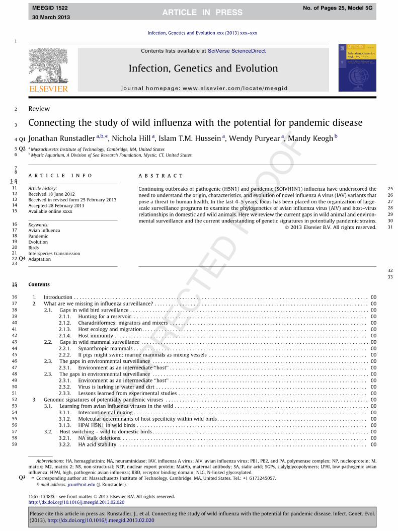
1 2 Review 3 Connecting the study of wild influenza with the potential for pandemic disease 4 Jonathan Runstadler a,b,⇑ Q1 , Nichola Hill a , Islam T.M. Hussein a , Wendy Puryear a , Mandy Keogh b 5 a Massachusetts Q2 Institute of Technology, Cambridge, MA, United States 6 b Mystic Aquarium, A Division of Sea Research Foundation, Mystic, CT, United States 7 8 10 article info 11 Article history: 12 Received 18 June 2012 13 Received in revised form 25 February 2013 14 Accepted 28 February 2013 15 Available online xxxx 16 Keywords: 17 Avian influenza 18 Pandemic 19 Evolution 20 Birds 21 Interspecies transmission 22 Q4 Adaptation 23 24 abstract 25 Continuing outbreaks of pathogenic (H5N1) and pandemic (SOIVH1N1) influenza have underscored the 26 need to understand the origin, characteristics, and evolution of novel influenza A virus (IAV) variants that 27 pose a threat to human health. In the last 4–5 years, focus has been placed on the organization of large- 28 scale surveillance programs to examine the phylogenetics of avian influenza virus (AIV) and host–virus 29 relationships in domestic and wild animals. Here we review the current gaps in wild animal and environ- 30 mental surveillance and the current understanding of genetic signatures in potentially pandemic strains. 31 Ó 2013 Elsevier B.V. All rights reserved. 32 33 34 35 Contents 36 1. Introduction .......................................................................................................... 00 37 2. What are we missing in influenza surveillance? ............................................................................. 00 38 2.1. Gaps in wild bird surveillance ...................................................................................... 00 39 2.1.1. Hunting for a reservoir ..................................................................................... 00 40 2.1.2. Charadriiformes: migrators and mixers ....................................................................... 00 41 2.1.3. Host ecology and migration ................................................................................. 00 42 2.1.4. Host immunity ........................................................................................... 00 43 2.2. Gaps in wild mammal surveillance .................................................................................. 00 44 2.2.1. Synanthropic mammals .................................................................................... 00 45 2.2.2. If pigs might swim: marine mammals as mixing vessels ......................................................... 00 46 2.3. The gaps in environmental surveillance .............................................................................. 00 47 2.3.1. Environment as an intermediate ‘‘host’’ ....................................................................... 00 48 2.3. The gaps in environmental surveillance .............................................................................. 00 49 2.3.1. Environment as an intermediate ‘‘host’’ ....................................................................... 00 50 2.3.2. Virus is lurking in water and dirt ............................................................................ 00 51 2.3.3. Lessons learned from experimental studies .................................................................... 00 52 3. Genomic signatures of potentially pandemic viruses ......................................................................... 00 53 3.1. Learning from avian influenza viruses in the wild ...................................................................... 00 54 3.1.1. Intercontinental mixing .................................................................................... 00 55 3.1.2. Molecular determinants of host specificity within wild birds ...................................................... 00 56 3.1.3. HPAI H5N1 in wild birds ................................................................................... 00 57 3.2. Host switching – wild to domestic birds .............................................................................. 00 58 3.2.1. NA stalk deletions......................................................................................... 00 59 3.2.2. HA acid stability .......................................................................................... 00 1567-1348/$ - see front matter Ó 2013 Elsevier B.V. All rights reserved. http://dx.doi.org/10.1016/j.meegid.2013.02.020 Abbreviations: HA, hemagglutinin; NA, neuraminidase; IAV, influenza A virus; AIV, avian influenza virus; PB1, PB2, and PA, polymerase complex; NP, nucleoprotein; M, matrix; M2, matrix 2; NS, non-structural; NEP, nuclear export protein; MatAb, maternal antibody; SA, sialic acid; SGPs, sialylglycopolymers; LPAI, low pathogenic avian influenza; HPAI, high, pathogenic avian influenza; RBD, receptor binding domain; NLG, N-linked glycosylated. ⇑ Corresponding author at: Massachusetts Institute of Technology, Cambridge, MA, United States. Tel.: +1 6173245057. Q3 E-mail address: [email protected] (J. Runstadler). Infection, Genetics and Evolution xxx (2013) xxx–xxx Contents lists available at SciVerse ScienceDirect Infection, Genetics and Evolution journal homepage: www.elsevier.com/locate/meegid MEEGID 1522 No. of Pages 25, Model 5G 30 March 2013 Please cite this article in press as: Runstadler, J., et al. Connecting the study of wild influenza with the potential for pandemic disease. Infect. Genet. Evol. (2013), http://dx.doi.org/10.1016/j.meegid.2013.02.020
Transcript of Connecting the study of wild influenza with the potential for pandemic disease
1
2
3
4 Q1
5 Q26
78
1 0
1112131415
16171819202122 Q423
2 4
3435
363738394041424344454647484950515253545556575859
Q3
Infection, Genetics and Evolution xxx (2013) xxx–xxx
MEEGID 1522 No. of Pages 25, Model 5G
30 March 2013
Contents lists available at SciVerse ScienceDirect
Infection, Genetics and Evolution
journal homepage: www.elsevier .com/locate /meegid
Review
Connecting the study of wild influenza with the potential for pandemic disease
Jonathan Runstadler a,b,⇑, Nichola Hill a, Islam T.M. Hussein a, Wendy Puryear a, Mandy Keogh b
a Massachusetts Institute of Technology, Cambridge, MA, United Statesb Mystic Aquarium, A Division of Sea Research Foundation, Mystic, CT, United States
25262728293031
a r t i c l e i n f o
Article history:Received 18 June 2012Received in revised form 25 February 2013Accepted 28 February 2013Available online xxxx
Keywords:Avian influenzaPandemicEvolutionBirdsInterspecies transmissionAdaptation
1567-1348/$ - see front matter � 2013 Elsevier B.V. Ahttp://dx.doi.org/10.1016/j.meegid.2013.02.020
Abbreviations: HA, hemagglutinin; NA, neuraminidmatrix; M2, matrix 2; NS, non-structural; NEP, nucleinfluenza; HPAI, high, pathogenic avian influenza; RB⇑ Corresponding author at: Massachusetts Institute
E-mail address: [email protected] (J. Runstadler).
Please cite this article in press as: Runstadler, J.,(2013), http://dx.doi.org/10.1016/j.meegid.2013
a b s t r a c t
Continuing outbreaks of pathogenic (H5N1) and pandemic (SOIVH1N1) influenza have underscored theneed to understand the origin, characteristics, and evolution of novel influenza A virus (IAV) variants thatpose a threat to human health. In the last 4–5 years, focus has been placed on the organization of large-scale surveillance programs to examine the phylogenetics of avian influenza virus (AIV) and host–virusrelationships in domestic and wild animals. Here we review the current gaps in wild animal and environ-mental surveillance and the current understanding of genetic signatures in potentially pandemic strains.
� 2013 Elsevier B.V. All rights reserved.
32
33
Contents
1. Introduction . . . . . . . . . . . . . . . . . . . . . . . . . . . . . . . . . . . . . . . . . . . . . . . . . . . . . . . . . . . . . . . . . . . . . . . . . . . . . . . . . . . . . . . . . . . . . . . . . . . . . . . . . . 002. What are we missing in influenza surveillance? . . . . . . . . . . . . . . . . . . . . . . . . . . . . . . . . . . . . . . . . . . . . . . . . . . . . . . . . . . . . . . . . . . . . . . . . . . . . . 00
2.1. Gaps in wild bird surveillance . . . . . . . . . . . . . . . . . . . . . . . . . . . . . . . . . . . . . . . . . . . . . . . . . . . . . . . . . . . . . . . . . . . . . . . . . . . . . . . . . . . . . . 00
2.1.1. Hunting for a reservoir. . . . . . . . . . . . . . . . . . . . . . . . . . . . . . . . . . . . . . . . . . . . . . . . . . . . . . . . . . . . . . . . . . . . . . . . . . . . . . . . . . . . . 002.1.2. Charadriiformes: migrators and mixers . . . . . . . . . . . . . . . . . . . . . . . . . . . . . . . . . . . . . . . . . . . . . . . . . . . . . . . . . . . . . . . . . . . . . . . 002.1.3. Host ecology and migration . . . . . . . . . . . . . . . . . . . . . . . . . . . . . . . . . . . . . . . . . . . . . . . . . . . . . . . . . . . . . . . . . . . . . . . . . . . . . . . . . 002.1.4. Host immunity . . . . . . . . . . . . . . . . . . . . . . . . . . . . . . . . . . . . . . . . . . . . . . . . . . . . . . . . . . . . . . . . . . . . . . . . . . . . . . . . . . . . . . . . . . . 002.2. Gaps in wild mammal surveillance . . . . . . . . . . . . . . . . . . . . . . . . . . . . . . . . . . . . . . . . . . . . . . . . . . . . . . . . . . . . . . . . . . . . . . . . . . . . . . . . . . 00
2.2.1. Synanthropic mammals . . . . . . . . . . . . . . . . . . . . . . . . . . . . . . . . . . . . . . . . . . . . . . . . . . . . . . . . . . . . . . . . . . . . . . . . . . . . . . . . . . . . 002.2.2. If pigs might swim: marine mammals as mixing vessels . . . . . . . . . . . . . . . . . . . . . . . . . . . . . . . . . . . . . . . . . . . . . . . . . . . . . . . . . 002.3. The gaps in environmental surveillance . . . . . . . . . . . . . . . . . . . . . . . . . . . . . . . . . . . . . . . . . . . . . . . . . . . . . . . . . . . . . . . . . . . . . . . . . . . . . . 00
2.3.1. Environment as an intermediate ‘‘host’’ . . . . . . . . . . . . . . . . . . . . . . . . . . . . . . . . . . . . . . . . . . . . . . . . . . . . . . . . . . . . . . . . . . . . . . . 002.3. The gaps in environmental surveillance . . . . . . . . . . . . . . . . . . . . . . . . . . . . . . . . . . . . . . . . . . . . . . . . . . . . . . . . . . . . . . . . . . . . . . . . . . . . . . 00
2.3.1. Environment as an intermediate ‘‘host’’ . . . . . . . . . . . . . . . . . . . . . . . . . . . . . . . . . . . . . . . . . . . . . . . . . . . . . . . . . . . . . . . . . . . . . . . 002.3.2. Virus is lurking in water and dirt . . . . . . . . . . . . . . . . . . . . . . . . . . . . . . . . . . . . . . . . . . . . . . . . . . . . . . . . . . . . . . . . . . . . . . . . . . . . 002.3.3. Lessons learned from experimental studies . . . . . . . . . . . . . . . . . . . . . . . . . . . . . . . . . . . . . . . . . . . . . . . . . . . . . . . . . . . . . . . . . . . . 003. Genomic signatures of potentially pandemic viruses . . . . . . . . . . . . . . . . . . . . . . . . . . . . . . . . . . . . . . . . . . . . . . . . . . . . . . . . . . . . . . . . . . . . . . . . . 00
3.1. Learning from avian influenza viruses in the wild . . . . . . . . . . . . . . . . . . . . . . . . . . . . . . . . . . . . . . . . . . . . . . . . . . . . . . . . . . . . . . . . . . . . . . 003.1.1. Intercontinental mixing . . . . . . . . . . . . . . . . . . . . . . . . . . . . . . . . . . . . . . . . . . . . . . . . . . . . . . . . . . . . . . . . . . . . . . . . . . . . . . . . . . . . 003.1.2. Molecular determinants of host specificity within wild birds . . . . . . . . . . . . . . . . . . . . . . . . . . . . . . . . . . . . . . . . . . . . . . . . . . . . . . 003.1.3. HPAI H5N1 in wild birds . . . . . . . . . . . . . . . . . . . . . . . . . . . . . . . . . . . . . . . . . . . . . . . . . . . . . . . . . . . . . . . . . . . . . . . . . . . . . . . . . . . 00
3.2. Host switching – wild to domestic birds . . . . . . . . . . . . . . . . . . . . . . . . . . . . . . . . . . . . . . . . . . . . . . . . . . . . . . . . . . . . . . . . . . . . . . . . . . . . . . 00
3.2.1. NA stalk deletions. . . . . . . . . . . . . . . . . . . . . . . . . . . . . . . . . . . . . . . . . . . . . . . . . . . . . . . . . . . . . . . . . . . . . . . . . . . . . . . . . . . . . . . . . 003.2.2. HA acid stability . . . . . . . . . . . . . . . . . . . . . . . . . . . . . . . . . . . . . . . . . . . . . . . . . . . . . . . . . . . . . . . . . . . . . . . . . . . . . . . . . . . . . . . . . . 00ll rights reserved.
ase; IAV, influenza A virus; AIV, avian influenza virus; PB1, PB2, and PA, polymerase complex; NP, nucleoprotein; M,ar export protein; MatAb, maternal antibody; SA, sialic acid; SGPs, sialylglycopolymers; LPAI, low pathogenic avianD, receptor binding domain; NLG, N-linked glycosylated.of Technology, Cambridge, MA, United States. Tel.: +1 6173245057.
et al. Connecting the study of wild influenza with the potential for pandemic disease. Infect. Genet. Evol..02.020

606162636465666768697071
72
73
74
75
76
77
78
79
80
81
82
83
84
85
86
87
88
89
90
91
92
93
94
95
96
97
98
99
100
101
102
103
104
105
106
107
108
109
110
111
112
113
114
115
116
117
118
119
120
121
122
123
2 J. Runstadler et al. / Infection, Genetics and Evolution xxx (2013) xxx–xxx
MEEGID 1522 No. of Pages 25, Model 5G
30 March 2013
3.2.3. HA and NS substitutions . . . . . . . . . . . . . . . . . . . . . . . . . . . . . . . . . . . . . . . . . . . . . . . . . . . . . . . . . . . . . . . . . . . . . . . . . . . . . . . . . . . 00
Please(2013
3.3. Host switching – Mammalian jumps . . . . . . . . . . . . . . . . . . . . . . . . . . . . . . . . . . . . . . . . . . . . . . . . . . . . . . . . . . . . . . . . . . . . . . . . . . . . . . . . . 00
3.3.1. HA receptor binding domain . . . . . . . . . . . . . . . . . . . . . . . . . . . . . . . . . . . . . . . . . . . . . . . . . . . . . . . . . . . . . . . . . . . . . . . . . . . . . . . . 003.3.2. Polymerase . . . . . . . . . . . . . . . . . . . . . . . . . . . . . . . . . . . . . . . . . . . . . . . . . . . . . . . . . . . . . . . . . . . . . . . . . . . . . . . . . . . . . . . . . . . . . . 003.3.3. Genetic reassortments . . . . . . . . . . . . . . . . . . . . . . . . . . . . . . . . . . . . . . . . . . . . . . . . . . . . . . . . . . . . . . . . . . . . . . . . . . . . . . . . . . . . . 003.3.4. HA–NA balance . . . . . . . . . . . . . . . . . . . . . . . . . . . . . . . . . . . . . . . . . . . . . . . . . . . . . . . . . . . . . . . . . . . . . . . . . . . . . . . . . . . . . . . . . . . 003.3.5. Codon usage bias . . . . . . . . . . . . . . . . . . . . . . . . . . . . . . . . . . . . . . . . . . . . . . . . . . . . . . . . . . . . . . . . . . . . . . . . . . . . . . . . . . . . . . . . . 003.3.6. Glycosylation . . . . . . . . . . . . . . . . . . . . . . . . . . . . . . . . . . . . . . . . . . . . . . . . . . . . . . . . . . . . . . . . . . . . . . . . . . . . . . . . . . . . . . . . . . . . 003.4. Inter-mammalian airborne transmission . . . . . . . . . . . . . . . . . . . . . . . . . . . . . . . . . . . . . . . . . . . . . . . . . . . . . . . . . . . . . . . . . . . . . . . . . . . . . . 00
4. Concluding remarks . . . . . . . . . . . . . . . . . . . . . . . . . . . . . . . . . . . . . . . . . . . . . . . . . . . . . . . . . . . . . . . . . . . . . . . . . . . . . . . . . . . . . . . . . . . . . . . . . . . . 00Acknowledgements . . . . . . . . . . . . . . . . . . . . . . . . . . . . . . . . . . . . . . . . . . . . . . . . . . . . . . . . . . . . . . . . . . . . . . . . . . . . . . . . . . . . . . . . . . . . . . . . . . . . 00References . . . . . . . . . . . . . . . . . . . . . . . . . . . . . . . . . . . . . . . . . . . . . . . . . . . . . . . . . . . . . . . . . . . . . . . . . . . . . . . . . . . . . . . . . . . . . . . . . . . . . . . . . . . 00
124
125
126
127
128
129
130
131
132
133
134
135
136
137
138
139
140
141
142
143
144
145
146
147
148
149
150
151
152
153
154
155
156
157
158
159
160
161
162
163
164
165
166
167
168
169
170
171
172
173
174
1. Introduction
Nearly 20 years ago, in his landmark review of influenza, RobWebster pointed out the probability that birds may serve as asource of all influenza A viruses (IAV) that become endemic inother species (1992). The emergence and maintenance of H5N1 lin-eages in wild and domestic birds and the 2009 novel pandemicstrain of H1N1 virus with avian origins in humans have reinforcedthis view, yet shown the origin of epidemic virus to be complicated(Neumann et al., 2009; Shortridge et al., 1998). In many respects,recent influenza events emphasize the importance of understand-ing the ecology and evolution of IAV in wild animal vectors and vir-al reservoir species (Fouchier and Munster, 2009; Melville andShortridge, 2006; Munster et al., 2007; Normile, 2006). Here, wereview the recent literature in influenza with an emphasis onunderstanding (i) how surveillance research in wild animals andthe environment can benefit public health and (ii) on how knowl-edge of the molecular determinants important in influenza evolu-tion in wild species can inform pandemic preparedness.
Influenza viruses are normally classified by the antigenic prop-erties of their highly variable major surface proteins, hemaggluti-nin (HA) and neuraminidase (NA). These two proteins are theprimary targets of protective immunity in the host. Seventeen sub-types of hemagglutinin (HA: H1–H17) and 9 subtypes of neuram-indase (NA: N1–N9) are described and all but one (H17 in bats(Tong et al., 2012)) and nearly all combinations have been isolatedfrom wild birds (Olsen et al., 2006; Webster et al., 1992) althoughsome more frequently than others. The influenza HA mediates viralbinding to host cells and delivery of the viral genome into the cellcytoplasm while the NA assists in viral exit by cutting sialic acidties to the host cell membrane. The viral genome of eight single-stranded negative sense RNA segments encodes 10+ proteinsdepending on the strain. In addition to the HA and NA, three pro-teins form the polymerase complex (PB1, PB2, and PA) and bindthe RNA segments with nucleoprotein (NP); matrix (M) and matrix2 (M2) comprise the protein coat of the virus; and the non-structural (NS) and nuclear export protein (NEP) interact with cel-lular proteins and processes to assist viral replication and exit andavoid the host immune response. Several additional proteins havebeen identified in the PB1 and PA segments that are variably pres-ent through alternative transcriptional open reading frames, splic-ing, or secondary start codons. These include PB1-F2 and a suite ofrecently discovered PA forms (Jagger et al., 2012; Muramoto et al.,2012), all of which seem to impact virulence of infection and whichdemand further study.
Since the emergence of a highly pathogenic form of H5N1 avianinfluenza from a domestic goose in 1997, and its subsequent trans-mission to humans (de Jong et al., 1997), birds have receivedincreased attention as the source of all natural IAV variants. Onrare occasions, the highly pathogenic forms of IAV have beenreported in wild birds – the first outbreak with mortality in wild
cite this article in press as: Runstadler, J., et al. Connecting the study of), http://dx.doi.org/10.1016/j.meegid.2013.02.020
birds being identified as an H5N3 influenza strain in common ternsof South Africa in 1961 (Becker, 1966). However, retrospectiveanalysis has identified avian origins for all segments of human pan-demic viruses. This includes the ‘‘Spanish flu’’ of 1918, an H1N1strain that was perhaps one of the greatest natural disasters inhuman history and is estimated to have contributed to the deathof over 50 million people worldwide. Subsequent pandemic virusesthough less severe have had enormous impact on human healthand include an H2N2 virus in 1957, an H3N2 virus in 1968, andthe pH1N1 virus, now endemic, in 2009. Each of these strainsresulted from the reassortment of contemporary human strainswith viruses derived from birds, but probably delivered throughinfection of an intermediate host such as the pig. Whether the1918 virus moved into humans directly from an avian host is con-troversial. Regardless, the avian origin of all these viruses hasspurred research into the avian host in hopes of understandingthe characteristics and predictability of pandemic strains at theirroot.
Domestic and wild birds have been implicated as key agents forinterspecies transmission to mammalian hosts of diverse taxaincluding whales, seals, pigs, horses, and also humans (Claaset al., 1998; Mandler et al., 1990; Reperant et al., 2009; Zhouet al., 2009). Phylogenetic analysis has even revealed that somegene segments belonging to previous human pandemic strainsare still circulating in wild bird reservoirs. The NA genes of someH9N2 viruses isolated from migratory ducks in Hokkaido, Japan,clustered with those of H3N2 viruses responsible for causing thehuman pandemic of 1968 (Liu et al., 2003). Moreover, it has beenspeculated that the 3 parents of the triple reassortant virus thatcaused the 2009 H1N1 pandemic may have been assembled inone place by migratory birds (Gibbs et al., 2009). As such, increas-ing emphasis is now placed on understanding the evolution andmolecular determinants of novel and pathogenic forms of influenzathat originate from the IAV in birds.
Surveillance research in wild birds holds the promise of inform-ing public health preparedness for pandemic and seasonal influ-enza. Field surveillance studies to detect avian influenza viruses(AIV) in animal vectors was organized in the early 1970s, culminat-ing with detection of influenza virus from the cloacal swabs of wildducks (Slemons et al., 1974). Into the 1990s, research in the birdhost centred on describing the viral natural history and its mainte-nance in waterfowl hosts. In response to the threat of Asian originH5N1, sampling efforts have increased by an order of magnitude ormore in the last 5 years, particularly in the US and mainland China(Butler, 2012). These efforts have begun to tie the viral natural his-tory and studies on viral evolution to the potential for generatingnovel pandemic viral strains. What is clear from past work is thatthe evolution and natural history of the virus is highly dependenton the epizootiology of infection in the avian host. It is hoped thatunderstanding the virus in reservoir species such as gulls andducks may help refine viral surveillance and identify unique virus
wild influenza with the potential for pandemic disease. Infect. Genet. Evol.

175
176
177
178
179
180
181
182
183
184
185
186
187
188
189
190
191
192
193
194
195
196
197
198
199
200
201
202
203
204
205
206
207
208
209
210
211
212
213
214
215
216
217
218
219
220
221
222
223
224
225
226
227
228
229
230
231
232
233
234
235
236
237
238
239
240
241
242
243
244
245
246
247
248
249
250
251
252
253
254
255
256
257
258
259
260
261
262
263
264
265
266
267
268
269
270
271
272
273
274
275
276
277
278
279
280
281
282
283
284
285
286
287
288
289
290
291
292
293
294
295
296
297
298
299
300
301
J. Runstadler et al. / Infection, Genetics and Evolution xxx (2013) xxx–xxx 3
MEEGID 1522 No. of Pages 25, Model 5G
30 March 2013
for further study. However, large biases exist in the geographic dis-tribution of sampling sites and most countries still have little or noorganized surveillance. Countries where H5N1 is endemic, includ-ing Egypt, India, Bangladesh, Viet Nam and Indonesia often sufferfrom a lack of capacity to diagnose and characterize virusesin-country. These gaps in viral surveillance in the wild, the focusof the first half of this review, will need to be addressed to makethe most of current surveillance research efforts.
Viruses that come out of surveillance work in wild animals isalso enabling laboratory based studies to clarify the moleculardeterminants of interspecies transmission, virulence and pathoge-nicity, the focus of the second half of our review. Experimentalwork with potentially troublesome virus or viral segments beforethey become a problem, should enable the development of broadlyor specific protective vaccines and therapeutics for interventionbefore a pandemic is started. Influenza is unique in some respectsin that one or a combination of three mechanisms – point muta-tion, segment reassortment, or, less commonly, recombinationmay generate genomic diversity. While recombination events donot seem to occur frequently, (Boni et al., 2008; Hirst et al.,2004; Pasick et al., 2005, 2009) their impact could be large anddeserves further study. High rates of mutation are produced bythe viral RNA polymerase, which lacks proofreading ability duringtranscription of the genome. In essence, mutation renders the hostinfected with a population of similar viruses with varying levels offitness in the given host. Selection of novel variants that possessenhanced fitness is responsible for drift in viral strains. Unfortu-nately, only consensus strain sequence is commonly reported sothe importance and dynamics of variants in the host are poorlyunderstood. This is one major gap that needs to be addressed.However, what seems to prove the biggest challenge for humanhealth and a result of interspecies transmission is the ability forco-infecting viruses to swap segments (reassortment), producingnovel strains that are antigenically distant from the original (i.e.– novel combinations of HA and NA as well as internal segments).This process allows the virus to ‘sidestep’ the immune system ofthe host and spread through populations (Webby and Webster,2001; Webster et al., 1992). While some subtypes contain strainsthat are partially cross seroreactive, the HA sequence of influenzamay differ by over 30% at the amino acid level and show limitedcross reactivity in serological assays (Alexander, 2000; Duganet al., 2008). As reassortment goes, viruses in pigs may be a majorreservoir of human emergent strains because of the potential tomix with human subtypes (Hass et al., 2011; Shu et al., 1994).However, pigs may not be the only animals for which this mixingis likely to take place (see Section 2.2.2). Studies in many other spe-cies indicate that interspecies transmission is relatively frequent(Capua and Alexander, 2002), but that epidemics are thought toalmost always be self-limiting because viruses are not maintainedor do not become endemic in alternative host species. The specificgenetics governing host range are undoubtedly polygenic, but maydepend on the co-evolution of viral gene products with host cellu-lar machinery to produce a competitive virus capable of establish-ing infection through transmission. The steady frequency withwhich this occurs in humans (Morens et al., 2010) highlights thatthis is a difficult but achievable and possibly even a predictableevent in nature. Understanding this dynamic in avian and otherreservoir and spillover hosts holds promise to help define the cri-teria to look for in potentially pandemic virus. It is also possiblethat pandemics are the result of rare events that facilitate genesisof rare viruses that are challenging to predict. Several programs areunderway to study whether IAV shows a ‘pandemic signature’, totest these competing theories, and to understand if study of wildIAV can inform public health risk for potential pandemic influenza.In the following sections, we explore the gaps needing work in wildanimal surveillance and highlight advances in our molecular
Please cite this article in press as: Runstadler, J., et al. Connecting the study of(2013), http://dx.doi.org/10.1016/j.meegid.2013.02.020
understanding that promises to improve public health prepared-ness for influenza.
2. What are we missing in influenza surveillance?
2.1. Gaps in wild bird surveillance
Field surveillance studies to detect IAV in animal vectors havebeen conducted for over 40 years, beginning with detection in wildducks (Slemons et al., 1974). In response to the threat of Asian ori-gin H5N1, sampling efforts increased by an order of magnitudesince 2005 particularly in the US and mainland China (Butler,2012). In-depth wild bird surveillance has helped distil key con-cepts in IAV ecology including the role of (i) aquatic wild birds asreservoirs, (ii) migratory flyways as barriers to viral evolution,(iii) young immuno-naïve birds as the hub of the wheel in IAV cir-culation, (iv) fecal–oral transmission as the primary transmissionroute in ducks and (v) warm temperatures and physico-chemicalproperties of aquatic habitat in limiting IAV infectivity. Now atthe conclusion of continent-scale surveillance projects targetingwild birds (Deliberto et al., 2009; Ip et al., 2008), the questionsarise: how has understanding of IAV advanced? And are thereany critical gaps in understanding that remain?
2.1.1. Hunting for a reservoirMany of the dogmas of IAV continue to be the guiding principles
that shape the way surveillance is conducted. Waterbirds belong-ing to the two orders Anseriformes (ducks, swans, geese) and Char-adriiformes (shorebirds, gulls, auks) have long been recognized asthe natural reservoirs of IAV (Webster et al., 1992) Within this eco-logically diverse group the dabbling ducks (family: Anatidae), par-ticularly mallards (Anas platyrhynchos) are believed to be a primaryhost. This notion stems from decades of field studies in NorthAmerica (Alfonso et al., 1995; Hinshaw et al., 1980; Ip et al.,2008) and Europe (Munster et al., 2007; Wallensten et al., 2007)that report highest prevalence in mallards compared to other sym-patric bird species. The role of mallards as a robust host for IAV issupported by experimental studies that demonstrate high titres ofvirus shed over extended periods by hatch year birds thatremained asymptomatic (12 days: Jourdain et al., 2010; >7 days:Keawcharoen et al., 2008). However, ecological context is impor-tant when considering findings from wildlife surveillance.
Mallards are the most ubiquitous waterbird species across theirHolarctic distribution and are intensively managed in NorthAmerica and Europe to ensure sizeable populations for hunting(Sedinger and Herzog, 2012). Large sample sizes are easy to obtain,especially from hunter-killed mallards, making the logistics ofsampling this species easier than any other wild bird. Withinthe US, hunter-killed birds accounted for >30% of all 78,300Anatidae samples (positive and negative) deposited in the Influ-enza Research Database (IRD: accessed September 2012). Over70% of hunter-harvested samples were collected from only 5 spe-cies of 51 Anatidae – mallard, green-winged teal (Anas carolinensis)northern shoveler (Anas clypeata), northern pintail (Anas acuta) andAmerican wigeon (Anas americana), in descending order. Thisestimate of hunter-harvested Anatidae samples is conservativebecause it does not include the large-scale surveillance effort bythe US Department of Agriculture that sourced the majority ofsamples (68%) from hunter-shot birds (Deliberto et al., 2009), buthighlights the dependency of surveillance on hunting as a sourceof inexpensive and readily-available samples, despite biases inspecies as well as seasonal timing, sex and age of hunted birds(Heitmeyer et al., 1993; Pace and Afton, 1999).
Species that are not viewed as ‘table birds’ and less sought afterfor consumption (i.e. northern shoveler, gadwall, Anas strepera) or
wild influenza with the potential for pandemic disease. Infect. Genet. Evol.
302
303
304
305
306
307
308
309
310
311
312
313
314
315
316
317
318
319
320
321
322
323
324
325
326
327
328
329
330
331
332
333
334
335
336
337
338
339
340
341
342
343
344
345
346
347
348
349
350
351
352
353
354
355
356
4 J. Runstadler et al. / Infection, Genetics and Evolution xxx (2013) xxx–xxx
MEEGID 1522 No. of Pages 25, Model 5G
30 March 2013
non-game species (i.e. gulls, shorebirds, passerines) may present achallenge to obtain large sample sizes adequate for detection ofIAV. Consideration of sample size is especially critical at winteringand stop-over sites where IAV prevalence is lower compared tobreeding and fall staging grounds where prevalence reaches a peakin many aquatic birds (Guberti et al., 2007). Careful assessment ofwhich species are high priority for surveillance demands a shiftfrom opportunistic to sustained, long-term sampling plans thatconsider the diversity of wild bird species and spatio-temporal var-iation in infection patterns along the migratory flyway. Drawing onthe expertise of ornithologists to capture non-game species mayensure sufficient sample sizes for species that may play an impor-tant yet undetermined role in hosting IAV strains with panzooticpotential (Winker et al., 2008).
2.1.2. Charadriiformes: migrators and mixersThe current yardstick for measuring success of a surveillance
study is detection of a large number of positive samples (Hoyeet al., 2010b). This approach has perpetuated the focus on dabblingducks, while the role of other water bird species in IAV ecology isoverlooked. The product is a global bias towards sampling Anseri-formes that account for the majority of wild bird samples collectedin North America (78%), Europe (76%), Asia (35–58%) and Russia(47%) (Fig. 1). In contrast, sampling of Charadriiformes – anotherrecognized reservoir for AIV, accounts for only 3–31% of globalsurveillance efforts (Fig. 1). This sampling bias has led to over-representation of viral subtypes associated with ducks in surveil-lance and genomic data, including the ubiquitous H3N8 and
Fig. 1. Surveillance effort targeted towards wild birds. Anseriformes (ducks, geese, swansamples from wild bird orders collected in North America and Europe. Data are based onDatabase at 20 September, 2012 (n = 152,312). Other sampled wild bird orders include: C(kingfishers, bee-eaters, rollers, hornbills), Cuculiformes (cuckoos, roadrunners, GruifoPelicaniformes (pelicans), Piciformes (woodpeckers), Procellariiformes (albatrosses, shearcolour in this figure legend, the reader is referred to the web version of this article.)
Please cite this article in press as: Runstadler, J., et al. Connecting the study of(2013), http://dx.doi.org/10.1016/j.meegid.2013.02.020
H4N6. Virus pools of Anseriformes and Charadriiformes have longbeen recognised as distinct (Kawaoka et al., 1988; Sharp et al.,1993). Overlap occurs with promiscuous subtypes (H3, H6, H7),however the circulation of H9, H13 and H16 is generally limitedto Charadriiformes (Krauss et al., 2004; Krauss and Webster,2010). Uncovering the full viral diversity hosted by the wild birdreservoir, including Charadriiformes represents a more effectivestrategy for detecting influenza precursors with capacity to switchhosts and seed a human pandemic.
Charadriiformes possess host traits that favour transmission,dispersal and hemispheric mixing of IAV suggesting they play auniquely different role in the ecology of IAV compared to ducks(Gaidet et al., 2012). Shorebirds are highly gregarious along theirmigratory cycle and are true long-distance migrants connectingthe northern and southern hemispheres (Gill et al., 2009). Thiscombination of host factors provides a mechanism for hemisphericreassortment of IAV and the movement of novel lineages that dis-place locally circulating strains. This is consistent with the higherfrequency of hemispheric reassortment observed in IAV fromshorebirds and gulls compared to other water bird hosts (Bahlet al., 2009; Dugan et al., 2008; Ramey et al., 2010; Widjaja et al.,2004). A growing number of studies have shown that the internalgenes of virus isolated from Charadriformes in North America areof Eurasian origin, a pattern observed along the Pacific (Rameyet al., 2010; Wille et al., 2011a) and Atlantic coasts (Wille et al.,2011b) suggesting that wild birds belonging to this order are pri-mary candidates for introduction of hemispheric reassortant virus.Enhanced surveillance of shorebirds and gulls may facilitate early
s: green) and Charadriiformes (shorebirds, gulls, auks: blue) account for almost allnumber of samples (positive, negative & untested) deposited in Influenza Research
iconiiformes (storks, herons, egrets), Columbiiformes (doves, pigeons), Coraciiformesrmes (cranes, rails), Sphenisciformes (penguins), Passeriformes (perching birds),waters, petrels), Sphenisciformes (penguins). (For interpretation of the references to
wild influenza with the potential for pandemic disease. Infect. Genet. Evol.

357
358
359
360
361
362
363
364
365
366
367
368
369
370
371
372
373
374
375
376
377
378
379
380
381
382
383
384
385
386
387
388
389
390
391
392
393
394
395
396
397
398
399
400
401
402
403
404
405
406
407
408
409
410
411
412
413
414
415
416
417
418
419
420
421
422
423
424
425
426
427
428
429
430
431
432
433
434
435
436
437
438
439
440
441
442
443
444
445
446
447
448
449
450
451
452
453
454
455
456
457
458
459
460
461
462
463
464
465
466
467
468
469
470
471
472
473
474
475
476
477
478
479
480
481
482
483
484
485
486
J. Runstadler et al. / Infection, Genetics and Evolution xxx (2013) xxx–xxx 5
MEEGID 1522 No. of Pages 25, Model 5G
30 March 2013
detection of IAV strains imported from regions where highly path-ogenic IAV is endemic or the incursion of novel segments into theendemic viral population.
A complete understanding of the global movements of IAVrequires that Charadriiformes be incorporated into surveillanceprograms. Specifically, longer-term studies need to be establishedto compliment the site at Delaware Bay, US that has yielded thebulk of virus samples from shorebirds, primarily from the narrowwindow of spring migration (Krauss et al., 2004; Krauss and Web-ster, 2010). Comprehensive sampling of Charadriiformes willnecessitate greater international collaboration to target siteswhere migratory flyways overlap, allowing hemispheric reassor-tants to be more readily detected. Surveillance has thus far beenNorth America- and Europe-centric (Butler, 2012) and rarely incor-porates sampling sites in Eurasia despite the fact that breedingpopulations of migratory birds often span both hemispheres atnorthern latitudes (i.e. Beringian region, Arctic Russia). Long-standing political divides, the remoteness of sampling sites andlack of in-country diagnostic laboratories presents a challenge forconducting surveillance in Africa, the Middle East, Russia, SouthAmerica and Asia, however researchers are increasingly making in-roads (Fereidouni et al., 2010; Gaidet et al., 2012; Shestopalovet al., 2006). A commitment to capacity building and a mutual re-solve to understand avian influenza dynamics across internationalboundaries may help to address this geographic bias.
2.1.3. Host ecology and migrationThe influence of host ecology, behaviour and migration on
transmission represents a large knowledge gap in our understand-ing of IAV in part because virology and ecology remain two dispa-rate fields that rarely overlap during the investigation of IAV inwild birds. Phylogeographic studies have distilled key conceptsincluding the generalized pattern of gene flow from north to southalong migratory flyways (Lam et al., 2012; Pearce et al., 2009) andrelative separation of North American and Eurasian virus pools(Pearce et al., 2010; Ramey et al., 2011) despite evidence of migra-tory connectivity of wild birds between hemispheres (Flint et al.,2009; Winker and Gibson, 2010). Studies of virus evolution thatrely on analysis of publically-available sequences can advanceour broad understanding of IAV dynamics (Bahl et al., 2009; Lamet al., 2012) but mechanisms that drive virus gene flow in wildbirds remain elusive without consideration of host ecological‘metadata’. However, tracking migratory animals can present alogistical and financial roadblock for studies that seek to investi-gate how host behaviour can promote or prevent transmission ofpathogens (Altizer et al., 2011). Use of markers including ring-bands or more recent technology including satellite transmitters,geolocaters and stable isotope analysis of body tissues offers pros-pects for identifying the migratory behaviour and geographic ori-gin of hosts if integrated with traditional surveillance.
Migration is common among wild birds from seasonal habitats;however there is variation in the propensity to migrate. Among thesame species a continuum of migration strategies can exist withresidency and long-distance migration on either ends of the spec-trum (Alerstam et al., 2003). The effect of migration strategy on IAVdynamics has only recently been explored in detail, facilitated bythe use of stable isotopes (Gunnarsson et al., 2012; Hill et al.,2012a) and trace element profiles (Fries et al., In press) in flightfeathers. Using stable isotopes to identify the breeding origin ofmallards, virus detected in resident mallards during winter in Cal-ifornia became the predominant IAV circulating in locally-breedingmallards in summer, supporting the view that residents act as res-ervoirs (Hill et al., 2012a). In contrast, migrants introduced virusfrom northern breeding grounds including Alaska, but circulationof imported virus appeared to be limited. Virological studies haveidentified wild bird-mediated dispersal as the mechanism for the
Please cite this article in press as: Runstadler, J., et al. Connecting the study of(2013), http://dx.doi.org/10.1016/j.meegid.2013.02.020
spread of Eurasian lineage HA subtypes along the Pacific Flywayresulting in an outbreak of H6N2 in poultry in California (Bahlet al., 2009; zu Dohna et al., 2009). A more nuanced understandingof the migration strategies of host species is key for predicting geneflow patterns and the introduction of Eurasian origin virus intoagricultural regions that support farming of pigs or poultry.
The ability of migratory birds to spread IAV, particularly highlypathogenic subtypes, has been a divisive topic among researcherswith evidence both for (Gaidet et al., 2008; Saad et al., 2007) andagainst (Gauthier-Clerc et al., 2007; van Gils et al., 2007). Centralto this question is an understanding of how far birds can migratebefore symptoms impact flight performance. Using satellite telem-etry, the potential for 19 species of wild birds from Asia, Africa andEurope to spread HPAI was recently assessed by Gaidet et al(2010). Comparison of dispersal rates showed that the commonteal (Anas crecca) had greatest potential to carry HPAI over500 km during the asymptomatic period of infection, yet the like-lihood of this event was restricted to 5–15 days during spring orfall migration. Furthermore, co-mingling of satellite-tracked wildbirds with domestic ducks – that can act as a reservoir for HPAI,days prior to migration was a predictor of wild bird outbreaksalong the Central Asian Flyway between 2005 and 2010 (Newmanet al., 2012). These studies highlight that dispersive potential isspecies-dependent, governed by flight performance, host pathobi-ology, virulence of IAV strains and spatio-temporal overlap withreservoir hosts. A shift to residency in some animal populationstriggered by mild temperatures or dependency on agriculture orhuman resources may promote local circulation of more virulentstrains (Altizer et al., 2011). The correlation between migrationstrategy of the host and virulence of transported pathogens isunderstudied in wild birds and warrants investigation in view ofimplications for IAV under climate change scenarios.
2.1.4. Host immunitySurveillance programs place an emphasis on collection of
virus, while the host response to infection is often overlooked. Pro-duction of antibodies to limit and overcome infection of IAV is cen-tral to the adaptive immune response in birds and should beincorporated into surveillance efforts to identify which speciesare involved in IAV circulation. Patterns of higher sero-prevalencerelative to virus prevalence have been observed across many wildbird taxa, including geese (Hoye et al., 2010a), gulls (Toennessenet al., 2011) and ducks (De Marco et al., 2003, 2005). Investmentin antibodies is a common strategy in the protection against IAVand partly explains the seasonal pattern of infection in waterbirds.Virus prevalence peaks after the breeding season in ducks, reflect-ing a build-up of young, immuno-naïve ducks in summer (Gubertiet al., 2007; Hinshaw et al., 1985). Juvenile ducks are also the pri-mary hosts at wintering sites (Ferro et al., 2010; Hill et al., 2012b)suggesting that exposure to novel strains occurs at both ends of themigratory flyway, a burden on the developing immune system ofyoung birds. Despite collective evidence that immunity in juvenilesdrives the epidemic curve of IAV in nature, surveillance programsrarely place value on collection of paired swabs and serum fromwild birds that may both inform and predict viral dynamics.
A neglected aspect that is likely to impact the scale and timingof infection among young birds on the breeding grounds is mater-nal antibody (MatAb) transfer. Ducklings or chicks may gain pro-tection from influenza by MatAb, primarily class IgY, passedthrough the egg yolk (Liu and Higgins, 1990). Persistence of MatAbvaries markedly from 3 to 40 days depending on life-history strat-egy and may directly correlate with length of incubation in birds(Lee et al., 2008; Tella et al., 2002). In common quail (Coturnixcoturnix) MatAb wane at 15 days, however in the longer-livedCory’s shearwater (Calonectris diomedea) – a seabird that lays a sin-gle egg with a long incubation time, MatAb were still detectable in
wild influenza with the potential for pandemic disease. Infect. Genet. Evol.

487
488
489
490
491
492
493
494
495
496
497
498
499
500
501
502
503
504
505
506
507
508
509
510
511
512
513
514
515
516
517
518
519
520
521
522
523
524
525
526
527
528
529
530
531
532
533
534
535
536
537
538
539
540
541
542
543
544
545
546
547
548
549
550
551
552
553
554
555
556
557
558
559
560
561
562
563
564
565
566567
568
569
570
571
572
573
574
575
576
577
578
579
580
581
582
583
584
585
586
587
588
589
590
591
592
593
594
595
596
597
598
599
600
601
602
603
604
605
606
607
608
609
610
611
612
613
614
615
6 J. Runstadler et al. / Infection, Genetics and Evolution xxx (2013) xxx–xxx
MEEGID 1522 No. of Pages 25, Model 5G
30 March 2013
chicks at 30–40 days of age (Garnier et al., 2012). Under thishypothesis, long-lived birds such as seabirds are expected to pro-duce longer lasting MatAb affording greater protection to juveniles.Maternal antibodies that provide immunity against IAV have beenidentified in gull eggs (Hammouda et al., 2011; Pearce-Duvet et al.,2009) but have not been investigated in other reservoir species.Waning immunity due to catabolism of MatAb may be responsiblefor the peak in IAV prevalence observed in ducklings weeks afterthey have left the nest and become flight capable. Further fieldand laboratory studies should be directed at understanding howcharacteristics of MatAb including temporal persistence andcross-protection may play a critical but unrecognized role in gov-erning infection dynamics in juveniles and viral evolution acrossa range of reservoir species. Better understanding of MatAbdynamics may also help researchers to characterize the epidemiccurve in wild birds and identify where and when to target surveil-lance efforts.
Antibody-mediated immunity is thought to drive antigenic evo-lution of avian influenza in humans (Ferguson et al., 2003) and wildbirds (Dugan et al., 2008). However evidence for a causal relation-ship between antibody production and virus evolution in wildbirds has never been conclusively drawn. The 26-year study byKrauss et al. (2004) has contributed towards understanding thetemporal pattern of subtype turnover in wild birds. Yet our knowl-edge of virus evolution remains incomplete without understandinghow the immune system acts as a selection pressure constantlymodifying the virus pool in wild birds. MatAb confer protectionagainst strains infecting the mother from prior seasons, whileacquired immunity acts against currently circulating strains. Nostudy has investigated the relationship between antibodies thatdevelop in juveniles and the fate of targeted subtypes in subse-quent seasons. Widely distributed subtypes in ducks; H3N8 andH4N6, predominate from year-to-year and are expected to evadethe immune response of the host owing to antigenic drift, muchlike H1, H2 and H3 in human populations. Assessing rates of anti-genic drift among H3N8 and H4N6 in wild birds may reveal theimmunologic and genetic hallmarks of virus that have heightenedfitness in the wild bird reservoir. Implications of evading hostimmunity include widespread and persistent circulation in wildbirds and ultimately a heightened chance of spillover to non-reser-voir species.
2.2. Gaps in wild mammal surveillance
2.2.1. Synanthropic mammalsMammals closely associated with human settlement may rep-
resent a pathway for interspecies virus transmission and adapta-tion. More broadly, the increasing interface between animals andhumans has led to emergence of infectious disease on a globalscale (Daszak et al., 2000; Lebarbenchon et al., 2008). Emergenceof highly pathogenic IAV is no exception, with the evolution ofH5N1 facilitated by co-mingling of wild and domestic ducks onrice paddies (Gilbert et al., 2008; Hulse-Post et al., 2005) followedby spillover to humans involved in the market chain (Martinet al., 2011). Agricultural practices in Asia have been identifiedas critical to the spread of influenza; however other human activ-ities that diminish barriers between host species are often over-looked by surveillance programs. Study of synanthropic wildmammals is a critical gap in our knowledge of how IAV evolvesto exploit novel hosts brought into close contact with avian res-ervoir species. Isolation of a novel lineage of IAV from little yel-low-shouldered bats (Sturnira lilium) resulting in designation ofan H17 subtype highlights that wild mammals can act as a reser-voir with the potential for spillover to sympatric domestic andagricultural animals (Tong et al., 2012). Investigation is neededto clarify which genetic markers and host combinations allow
Please cite this article in press as: Runstadler, J., et al. Connecting the study of(2013), http://dx.doi.org/10.1016/j.meegid.2013.02.020
influenza to jump the species barrier, triggered by close contactbetween birds and mammals.
Recent interest in free-ranging mammalian hosts has demon-strated that a larger than expected number of wild species arecompetent hosts for IAV. Wild house mice (Mus musculus) sampledafter an outbreak of low pathogenic H5N8 at a game bird breedingfacility tested sero-positive to IAV (Shriner et al., 2012). The possi-bility of spillover from migratory ducks sighted at the facilityprompted the authors to experimentally infect house mice withmallard-origin IAV and demonstrate that replication occurs effi-ciently without adaptation of the virus to a mammalian host(Shriner et al., 2012). The ability of IAV to cross the species barrierand replicate in mammals without adaptation has also been dem-onstrated in ferrets experimentally infected with H1N9 and H6N1(Driskell et al., 2012). Neither virus subtype showed an affinity fora-2,6 linked sialic acid (SA) receptors suggesting limited selectionfor mammalian adaptation in the laboratory setting. These caseshighlight the relative ease with which IAV circulating in wild birdsmay spread among sympatric mammalian populations, howevernatural infections have rarely been documented. Pikas (Ochotonacurzoniae) represent one of the few free-ranging mammals natu-rally-infected by IAV (highly pathogenic H5N1) circulating amongmigratory waterfowl at Qinghai Lake, China (Zhou et al., 2009).Lack of diseased or dead mammals at outbreak sites (pikas: Zhouet al., 2009) or limited clinical symptoms in experimentally in-fected mammals, depending on subtype (mice: Driskel et al.,2010; ferrets: Driskell et al., 2012; Hinshaw et al., 1981) may maskinfection and contribute to a low rate of detection in wildmammals.
Co-ordinated sampling of wild birds and mammals may shedlight on mechanisms that allow influenza to overcome the hostbarrier in nature, including which subtypes and host combinationsare conducive to spillover. Species with abundant urban popula-tions such as raccoons (Procyon lotor), European rabbits (Oryctola-gus cuniculus) or bats that have the potential to interact with wildbirds are a prime candidate. Raccoons sero-surveyed for IAV haveshown exposure to subtypes commonly circulating in wild waterbirds (H1, H3, H4 and H10: Hall et al., 2008) and poultry (H5N1:Horimoto et al., 2011) providing evidence of interspecies transmis-sion. Synanthropic rodents and bats are targets for surveillance bypublic health agencies because of the need to curb zoonotic trans-mission of mammal-borne pathogens, most notably hantavirus inrodents (Phan et al., 2011) and lyssaviruses in bats (Kuzminet al., 2012). Trapping and abatement programs led by governmentagencies may be a source of a large number of samples from areaswith concentrated human populations (i.e. urban and recreationalparks). Expanding sampling of free-ranging mammals in conjunc-tion with existing public health surveillance is imperative to mon-itor spread of AIV in view of how readily interspecies transmissioncan occur when avian and mammalian populations overlap. Synan-thropic mammals may be at highest risk of co-infection from avianand mammalian strains of influenza providing conditions suitablefor reassortment in nature.
2.2.2. If pigs might swim: marine mammals as mixing vesselsMarine mammals are a particularly interesting and phylogenet-
ically diverse group whose members comprise multiple lineageswhich underwent numerous independent re-invasions of the seasand rely on the marine environment for food. Marine mammalsare globally distributed and found in nearly all coastal waterwaysand shorelines. The coastal environment provides an interfacebetween marine and terrestrial habitats where avian reservoirsof influenza collide (sea ducks, gulls, and shorebirds) and overlapspatio-temporally with marine mammals, providing an opportu-nity for interspecies transmission of IAV. In general pinnipeds(seals, sea lions, and fur seals) are aggregate, seasonal breeders
wild influenza with the potential for pandemic disease. Infect. Genet. Evol.
616
617
618
619
620
621
622
623
624
625
626
627
628
629
630
631
632
633
634
635
636
637
638
639
640
641
642
643
644
645
646
647
648
649
650
651
652
653
654
655
656
657
658
659
660
661
662
663
664
665
666
667
668
669
670
671
672
673
674
675
676
677
678
679
680
681
682
683
684
685
686
687
688
689
690
691
692
693
694
695
696
697
698
699
700
701
702
703
704
705
706
707
708
709
710
711
712
713
714
715
716
717
718
719
720
721
722
723
724
725
726
727
728
729
730
731
732
733
734
735
736
737
738
739
740
741
742
743
744
745
746
J. Runstadler et al. / Infection, Genetics and Evolution xxx (2013) xxx–xxx 7
MEEGID 1522 No. of Pages 25, Model 5G
30 March 2013
resulting in highly synchronized terrestrial parturition, which maylead to heightened interactions between birds, pinnipeds, domesticanimals (dogs) and humans. Our understanding of IAV in marinemammals is predominantly from pinnipeds and has stemmed fromsampling stranded animals that have washed ashore in populatedareas, bio-monitoring of wild populations deemed to be of conser-vation concern, mortalities resulting from entanglement in fishinggear, and sampling associated with subsistance hunted animals.While previous reviews did not consider there to be strong evi-dence for a transmission pathway between marine mammals andhumans (Alexander and Brown, 2000), there is increasing supportof the transmission of zoonotics between marine mammals andhumans (Hunt et al., 2008; Siembieda et al., 2008; Webster,1981; Webster et al., 1981). Several IAV isolated from marinemammals have demonstrated a preference for infection and repli-cation in mammalian hosts (Hinshaw et al., 1981; Lang et al., 1981;Webster et al., 1981) including documented infection in a techni-cian (Webster, 1981). Further, the recently isolated H3N8 fromharbor seals (Phoca vitulina) demonstrated for the first time natu-rally acquired mutations that indicate mammalian adaptations(Anthony et al., 2012). These findings highlight the importance ofIAV surveillance in wild marine mammals in which evidence of fre-quent transmission is accumulating, but for which many gaps inunderstanding remain and may pose a public health risk.
Since the first isolation of IAV in swine (H1N1) (Shope, 1931), ithas become evident that multiple subtypes of IAV of either avian orhuman descent can infect pigs (Guan et al., 1996; Karasin et al.,2000; Peiris et al., 2001). Similar to pigs, multiple IAV have beenisolated from marine mammals (Greig, 2011; Hinshaw et al.,1986). Additionally, avian-like (H3N8, H3N3, H4N5, H4N6, H7N7)and human influenza A (H3N2) and B viruses have been isolatedfrom several species of marine mammals found within the coastalenvironment (Anthony et al., 2012; Blanc et al., 2009; Mandleret al., 1990; Ohishi et al., 2002, 2004, 2006; Osterhaus et al.,2000). The susceptibility of marine mammals to infection of bothavian and human influenza viruses may, in part, be due to the typeand distribution of SA receptors in tissues including the respiratorytract. Anthony et al. (2012) reported the presence of a-2,6 linkedSA and to a lesser degree a-2,3 linked SA receptors within therespiratory track of harbor seals. These findings are in contrast toan earlier study that found only a-2,3 linked SA receptors presentin respiratory tracks of ‘‘seals and whales’’ (Ito et al. 1999). Attach-ment of avian-origin H7N7 was predominately found in the upperrespiratory tracts of harbor and grey (Halichoerus grypus) seals(Ramis et al., 2012) which corresponds to the distribution of a-2,3 linked SA receptors in seals (Anthony et al., 2012; Ito et al.,1999) whereas attachment of human H3N2 influenza wasobserved in the bronchiolar and alveolar epithelium of harbor por-poise and to a lesser degree in harbor seals (Ramis et al., 2012).These findings suggest that the location of infection within therespiratory track may differ for human and avian influenza virusesin marine mammals. Further, the observed anatomical differencesbetween marine mammal groups (cetacean, pinnipeds) may leadto differences in susceptibility to IAV in these mammals.
In addition to genetic reassortment between avian and humaninfluenza viruses, adaptation of an AIV leading to efficient infectionin a mammalian host may also lead to influenza pandemics(Webster et al., 1992). There is evidence of such adaptation inthe IAV isolated from marine mammals. In September 2011 anunusually high number of seals were observed stranded alongthe coast of New England. The mortalities were associated withan infection of avian-origin H3N8 possessing recent mutations sug-gesting adaptations to the mammalian host (D701N in PB2 andbinding to a-2,6 linked SA receptors). A total of 37 amino acid sub-stitutions distinguished this seal H3N8 virus from other avianH3N8 viruses (Anthony et al., 2012); the exact role of the rest of
Please cite this article in press as: Runstadler, J., et al. Connecting the study of(2013), http://dx.doi.org/10.1016/j.meegid.2013.02.020
the mutations in adaptation is not yet understood. However, thiswas not the first occurrence of an H3N8 virus in marine mammals.An H3N8 subtype was isolated from one harp seal caught in fishinggear off Cape Cod between December 2005 and August 2007, withno significant pathology reported (Bogomolni et al., 2008). TheH3N8 has drawn attention to the role of marine mammals in theecology and evolution of IAV; however, the H3N8 virus was notthe first case of IAV isolated from a marine mammal.
2.2.2.1. The long history of IAV in marine mammals. The first IAV(H1N3) isolated from a marine mammal was from a baleen whale(family Balaenopteridae) (Lvov et al., 1978). Shortly thereafter, amass die off of harbor seals, impacting an estimated 20% of thepopulation near Cape Cod was attributed to severe pneumoniaand H7N7 infection (Webster et al., 1981). Most seals affected bythe die off were young of the year, suggesting that the state ormaturity of the immune system plays a role in susceptibility toIAV. The seal H7N7 was antigenically and genetically similar toavian H7N7 but showed greater ability to infect, replicate and pro-duce pneumonia in a broad range of experimentally infected mam-mals compared to domestic birds (Kida et al., 1982; Lang et al.,1981; Murphy et al., 1983; Webster et al., 1981). Infection and dis-ease observed in seals following experimental exposure to H7N7differed between species with harbor seals exhibiting disease sim-ilar to naturally infected seals (Webster et al., 1981). Grey seals hadno indication of infection; whereas harp seals showed no clinicalsigns of disease but had pathological changes and virus recoveredfrom some seals with surviving seals becoming sero-positive(Geraci et al., 1984). Following the H7N7 mass mortality, harbor,grey, hooded (Cystophora cristata) and harp (Pagophilus groenlandi-cus) seals and pups were culled from eastern Canadian waters toassess the presence of IAV and antibodies (Geraci et al., 1984).While no viruses were isolated from any seals, antibodies toH7N7 were found in 3 adult grey seals, while the other specieswere sero-negative (Geraci et al., 1984). These findings raise anumber of questions about the susceptibility to IAV infectionbetween marine mammal species and age groups. From a publichealth perspective, the observation of conjunctivitis in a personworking with the experimentally infected seals may be the mostprofound (Webster, 1981). Following the experimentally infectedseal sneezing on a technician, seal H7N7 was recovered from theconjunctiva, suggesting that H7N7 may be transmitted betweenmarine mammals and humans (Webster, 1981; Webster et al.,1981).
A second epizootic of seal pneumonia occurred from 1982 to1983 in New England and was associated with an H4N5 influenzavirus, the first time this virus had been isolated outside of birds(Hinshaw et al., 1984). Other subtypes including H4N6 and H3N3have also been isolated from seals that died of pneumonia alongthe Cape Cod peninsula in 1991 and 1992 (Callan et al., 1995). Anti-genic and genetic analyses showed that all genes were of avian ori-gin (Callan et al., 1995; Hinshaw et al., 1984; Webster et al., 1981).The repeated outbreak of pneumonia associated with IAV of avianorigin suggests transmission between avian host and seals, high-lighting the importance of surveillance studies in these popula-tions to gain a better understanding of interspecies transmissionof IAV.
It is not surprising that much of what is known about IAV inmarine mammals is based on samples from amphibious pinnipeds.However, influenza has been isolated from stranded cetaceans(dolphins and whales). Two influenza viruses (H13N2 andH13N9), demonstrating dual infection, were isolated from an obvi-ously emaciated and ill pilot whale (Globicephala melaena)stranded along the New England Coast. Phylogenetic analyses sug-gest the viruses originated from gulls (Hinshaw et al., 1986) andsimilar to other IAV isolated from seals (Lang et al., 1981; Webster
wild influenza with the potential for pandemic disease. Infect. Genet. Evol.

747
748
749
750
751
752
753
754
755
756
757
758
759
760
761
762
763
764
765
766
767
768
769
770
771
772
773
774
775
776
777
778
779
780
781
782
783
784
785
786
787
788
789
790
791
792
793
794
795
796
797
798
799
800
801
Table 1Caption1Q7
HA Subtype Species Location Year Detection method Ref.
NDa Dall’s porpoiseMinke whaleBelugaRinged sealHarbor sealHarbor sealNorthern elephant sealCalifornia sea lion
western North Pacificwestern North PacificCanadian ArcticCanadian ArcticCaliforniaBritish ColombiaCaliforniaCalifornia
2000–20012000–20011984–19941984–19942007200720072007
ELISA; WesternELISA; WesternELISAELISA
Ohishi (2006)Ohishi (2006)Nielson (2001)Nielson (2001)IRD (December 2012)IRD (December 2012)IRD (December 2012)IRD (December 2012)
H1 H1H1N1H1N3
‘‘Seals’’South American fur sealFamily Balaenopteridae
Bering SeaUruguaySouth Pacific
1978–1988September 20041974–1976
NP-ELISA; HIHIMDCK cells
de Boer (1990)Blanco (2009)Lvov (1978)
H3 H3H3H3H3N2H3N2H3N2H3N3H3N8H3N8
‘‘Seals’’Kuril harbor Sealringed sealCaspian sealsBaikal sealRinged sealHarbor sealHarbor sealHarbor seal
Bering SeaHokkaido, JapanAlaskaCaspian SeaBaikal SeaCentral Russian ArcticCape CodCape CodCape Cod
1978–19881998–20051978–19951993, 1997, 1998, 2000199820021991–19922005–2007; 2011
NP-ELISA; HIELISA; HIHIELISA; HIELISA; HIELISA; HIPCR;HIRT-PCRPCR
de Boer (1990)Fujii (2007)Danner (1998)Ohishi (2002)Ohishi (2004)Ohishi (2004)Callan (1995)Bogomolni (2008)Anthony (2012)
H4 H4H4N5H4N6
‘‘Seals’’Harbor sealHarbor seal
North and Bering SeasCape CodCape Cod
1988; 1978–1988June 1982–March 1983January 1991
ELISAHI, NIPCR;HI
de Boer (1990)Hinshaw (1984)Callan (1995)
H6 H6 Kuril harbor seal Hokkaido, Japan 1998–2005 ELISA;HI Fujii (2007)
H7 H7H7H7N7H7N7
‘‘Seals’’ringed Sealharbor SealRinged seal
Bering SeaAlaskaCape CodCentral Russian Arctic
1978–19881978–1995December 19792002
NP-ELISA; HIELISA; HI
de Boer (1990)Danner (1998)Geraci (1981)Ohishi (2004)
H10 H10N7N2N3
Walrus St. Lawrence Island and Round Island, Alaska 1994–1996 AGID Calle (2002)
H12 H12 ‘‘Seals’’ Bering Sea 1978–1988 NP-ELISA; HI de Boer (1990)H13 H13N2
H13N9Pilot whalePilot whale
New EnglandNew England
19841984
Chicken egg Hinshwa (1986)Hinshwa (1986)
HI, hemagglutination inhibition assay; NI, neuraminidase inhibition; AGID, Agar gel immunodiffusion; NP-ELISA, nucleoprotein (NP) ELISA.a Not determined.
8 J. Runstadler et al. / Infection, Genetics and Evolution xxx (2013) xxx–xxx
MEEGID 1522 No. of Pages 25, Model 5G
30 March 2013
et al., 1981), viral replication was observed in ferrets followingintranasal inoculation (Hinshaw et al., 1986). The repeated infec-tions in marine mammals during the last 30–40 years in New Eng-land and the associated sampling and laboratory studies have beenthe foundation of our understanding of IAV in marine mammals.However, the role of marine mammals in the ecology and evolutionof influenza, outside of these mass mortalities and in other parts ofthe world remain limited.
2.2.2.2. Looking back to understand the present. These outbreaks inmarine mammals and their implications for public health haveprompted several retrospective studies assessing the seropreva-lance of IAV antibodies. Serological evidence of exposure to avian-and human-origin IAV have been reported in several species ofmarine mammals from across the globe (Blanc et al., 2009; Ohishiet al., 2002, 2004, 2006); however, sampling effort has variedgreatly. When subtype has been determined, H3 has been most fre-quently reported (Table 1). Serological evidence of exposure to IAVhas also been reported in cetaceans (Dall’s porpoise, Phocoenoidesdalli and Minke whale) hunted in the Western Pacific Ocean(Ohishi et al., 2006). The presence of antibodies in the Minke whalemay be of importance as this species performs large annual migra-tions (Kasamatu et al., 1995) that may provide a mechanism for theintroduction of IAV to new regions, species or individuals (Altizeret al., 2011).
Based on serological evidence, Caspian (Pusa caspica), ringedand Baikal (Pusa sibirica) seals were exposed to human-originH3N2 (Ohishi et al., 2002, 2004) further supporting a possible
Please cite this article in press as: Runstadler, J., et al. Connecting the study of(2013), http://dx.doi.org/10.1016/j.meegid.2013.02.020
transmission route between marine mammals and humans. Sealsfrom Hokkaido, Japan were sero-positive for H3 and H6 subtypes(neuraminidase was not determined) between 1998 and 2005. Inall years, sero-positive seals included juveniles which the authorssuggest is evidence of sporadic infections in this population (Fujiiet al., 2007). Based on this evidence, the authors propose that sealsmay be a reservoir for IAV of human origin with implications forpublic health.
The seroprevalence of IAV in marine mammals of the arctic hasbeen of particular interest, in part because this region is sensitiveto climate change and also due to the reliance of subsistence hunt-ers on these populations. Interspecies transmission of IAV has beendocumented through routes of ingestion and inhalation of aerosol-ized viruses in other species. Therefore, the presence of IAV in mar-ine mammals hunted for human consumption has a directimplication for public health. Antibodies to influenza A have beenfound in many arctic species of marine mammals including beluga(Delphinapterus leucas), ringed seal, harp seals, hooded seals, andwalrus (Odobenus rosmarus) (Calle et al., 2002; Danner and McGr-egor, 1998; Nielsen et al., 2001; Stuen et al., 1994). While otherspecies including narwhals (Monodon monoceros), bowhead whale(Balaena mysticetus), bearded seals (Erignathus barbatus) and onepopulation of walruses (Calle et al., 2002, 2008; Nielsen et al.,2001) have not shown antibodies. These studies were based onretrospective serological samples and viral isolates were notcollected. Therefore, genetic analysis could not be performed, leav-ing open the question of the role of IAV in the arctic environmentand its potential impact on public health.
wild influenza with the potential for pandemic disease. Infect. Genet. Evol.

802
803
804
805
806
807
808
809
810
811
812
813
814
815
816
817
818
819
820
821
822
823
824
825
826
827
828
829
830
J. Runstadler et al. / Infection, Genetics and Evolution xxx (2013) xxx–xxx 9
MEEGID 1522 No. of Pages 25, Model 5G
30 March 2013
Surprisingly antibodies to influenza B, normally a human onlyvirus and the isolation of influenza B virus in harbor seals has beenreported (Blanc et al., 2009; Osterhaus et al., 2000). These observa-tions further highlight the need for systematic and prospective sur-veillance of influenza in wild marine mammals. The currentknowledge of influenza virus in marine mammals has been builtupon opportunistic and relatively low sampling, in part due tologistical and permit constrains of handling and sampling marinemammals required under the US Marine Mammal Protection Act(MMPA, 1972). Therefore, prospective surveillance of influenza inmarine mammals will require the collaboration with governmentagencies and non-profits to facilitate adequate sampling in orderto better understand the influence of these mammals on the ecol-ogy and evolution of influenza viruses and the potential impacts onpublic health.
Table 2Caption2
HA Subtype Source of virus Location Season
H1 H1H1N1H1N2
Lake waterRiver waterEnvironmentc
RussiaNetherlandsIndiana
NRa
NRNR
H2 H2N2H2N3
EnvironmentEnvironment
New JerseyNew York
NRNR
H3 H3H3N2H3N2H3N3H3N6H3N6H3N8H3N8H3N8
Pond sedimentPond waterPond waterPond waterRiver waterSewageLake waterLake waterPond water
AlaskaMinnesotaHong KongHong KongNetherlandsNetherlandsMinnesotaAlaskaMinnesota
Fall–SpringWater <12 �November–November–NRNRSeptemberSummer/FaWater <12 �
H4 H4N1H4N2H4N6H4N6H4N8
Lake waterLake waterLake waterLake waterPond water
AlbertaAlbertaMinnesotaAlaskaMinnesota
AugustAugustSeptemberSummer/FaWater <12 �
H5 H5N1H5N1H5N1H5N2H5N3H5N4
Pond water, mud, soilMudWater, plants, soil, mudEnvironmentEnvironmentEnvironment
CambodiaCambodiaCambodiaIndiana, NYCaliforniaNew York
2007–2010NRNRNRNRNR
H6 H6N2H6N8H6N8H6N8
Lake waterRiver waterFreshwater clamsNew York
AlbertaNetherlandsNRNR
AugustNRNRNR
H7 H7N2H7N3H7N3
Lake waterSewageLake water
AlbertaNetherlandsAlaska
AugustNRSummer/Fa
H8 H8 Pond sediment Alaska Fall–Spring
H9 H9H9N2H9N2
WaterLake waterPond water
BangledashDongting LakeHong Kong
NRSpringNovember–
H10 H10N8 Lake water Dongting Lake NR
H11 H11H11N6H11N8H11N9H11N9
Pond sedimentEnvironmentEnvironmentPond waterEnvironment
AlaskaDelawareMaryland, CAMinnesotaMaryland
Fall–SpringNRNRWater <12 �
H12 H12H12N3
Pond sedimentRiver water
AlaskaNetherlands
Fall–SpringNR
H13 H13N2H13N6
Lake waterPond water
MinnesotaMinnesota
NRNR
a Not reported.b Influenza Research Database.c Reported as ‘‘environment’’ in the Influenza Research Database.
Please cite this article in press as: Runstadler, J., et al. Connecting the study of(2013), http://dx.doi.org/10.1016/j.meegid.2013.02.020
2.3. The gaps in environmental surveillance
2.3.1. Environment as an intermediate ‘‘host’’The role of environmental persistence of virus in the overall
ecology of influenza is undoubtedly a critical one and likely playsa significant role in species specificity, periodicity of infection,reassortment, and epidemic initiations and persistence. Severalmodelling efforts have concluded that AIV requires an environ-mental component of indirect transmission (Breban et al., 2009;Lebarbenchon et al., 2010; Rohani et al., 2009), and mounting cir-cumstantial evidence supports this hypothesis. In one particularlyinteresting case, turkeys in Minnesota and their water sourceswere monitored for AIV. The H13N2 virus, typically associated withgulls, was detected in a turkey for the first time only after it wasdetected in the pond water 2 weeks prior (Sivanandan et al.,
Isolation method Detection method Ref.
NRUltrafiltrationNR
NRRTPCRNR
IRDb
Heijnen 2009IRD
NRNR
NRNR
IRDIRD
CAprilApril
llC
UnconcentratedUnconcentratedConcentratedConcentratedUltrafiltrationUltrafiltrationUnconcentratedErythrocyte assayUnconcentrated
RTPCREgg inoculationEgg inoculationEgg inoculationRTPCRRTPCREgg inoculationEgg inoculationEgg inoculation
Lang (2006)Halvorson (1983)Markwell (1982)Markwell (1982)Heijnen (2009)Heijnen (2009)Stallknecht (2010)Ito (1995)Halvorson (1983)
llC
UnconcentratedUnconcentratedUnconcentratedErythrocyte assayUnconcentrated
Egg inoculationEgg inoculationEgg inoculationEgg inoculationEgg inoculation
Hinshaw (1979, 1980)Hinshaw (1980)Stallknecht (1995)Ito (1995)Halvorson (1983)
Erythrocyte assayElution and concUnconcentratedNRNRNR
RTPCRRTPCR, Egg inocRTPCRNRNRNR
Horm (2012)Horm (2011)Vong (2008)IRDIRDIRD
UnconcentratedUltrafiltrationNRNR
Egg inoculationRTPCRNRNR
Hinshaw (1980)Heijnen (2009)Huyvaert (2012)IRD
ll
UnconcentratedUltrafiltrationErythrocyte assay
Egg inoculationRTPCREgg inoculation
Hinshaw (1979, 1980)Heijnen (2009)Ito (1995)
Unconcentrated RTPCR Lang (2006)
April
NRErythrocyte assayConcentrated
NREgg inoculationEgg inoculation
IRDZhang (2011)Markwell (1982)
Erythrocyte assay Egg inoculation Zhang (2011)
CNR
UnconcentratedNRNRUnconcentratedNR
RTPCRNRNREgg inoculationNR
Lang (2006)IRDIRDHalvorson (1983)IRD
UnconcentratedUltrafiltration
RTPCRRTPCR
Lang (2006)Heijnen (2009)
Erythrocyte assayErythrocyte assay
Egg inoculationEgg inoculation
Sivandan (1991)Sivandan (1991)
wild influenza with the potential for pandemic disease. Infect. Genet. Evol.

831
832
833
834
835
836
837
838
839
840
841
842
843
844
845
846
847
848
849
850
851
852
853
854
855
856
857
858
859
860
861
862
863
864
865
866
867
868
869
870
871
872
873
874
875
876
877
878
879
880
881
882
883
884
885
886
887
888
889
890
891
892
893
894
895
896
897
898
899
900
901
902
903
904
905
906
907
908
909
910
911
912
913
914
915
916
917
918
919
920
921
922
923
924
925
926
927
928
929
930
931
932
933
934
935
936
937
938
939
940
941
Fig. 2. Detection of IAV from environmental sources. Data represent counts of thethirteen hemagglutinin (HA) subtypes detected. Data are sourced from reports ofenvironmental detection published in the literature and sequences available onGenbank.
10 J. Runstadler et al. / Infection, Genetics and Evolution xxx (2013) xxx–xxx
MEEGID 1522 No. of Pages 25, Model 5G
30 March 2013
1991). This led the authors to conclude that gulls likely shed virusinto the pond where it persisted and ultimately infected turkeys.Another study monitored ducks and turkeys that shared a waterhabitat and found in nearly all cases, viruses detected in turkeyswere first detected in the ducks (Halvorson et al., 1983). A recentreport describes an H9N2 isolated from an egret in Dongting Lake(Wang et al., 2012a), the same subtype typically found in ducksand chickens and isolated several years prior from the water of thatsame lake (Zhang et al., 2011b). On the heels of the 2004 H5N1 epi-demic, an extensive survey of H5N1 seropositive individuals inCambodia found that swimming or bathing in pond water werestrong risk factors for seropositive status (Vong et al., 2009). Sup-porting this correlation, a Vietnam woman and a Cambodian childeach contracted H5N1 with no identifiable risk factors other thanswimming in contaminated water (WHO, 2007). Further H5N1 epi-demiological analyses showed a strong correlation between mini-mal distance to the nearest lake or wetland and the likelihood ofan outbreak, as well as an inverse relationship between outbreaks,precipitation and bird density (Fang et al., 2008).
Given the logistics of collecting and screening large volumes ofwater for virus, sampling methodologies have not yet been refined.When large enough quantities of virus are present, unconcentratedwater can be tested with some success (Halvorson et al., 1983;Hinshaw et al., 1979, 1980; Leung et al., 2007; Stallknecht et al.,2010). For a much more sensitive screening, large volumes of waterneed to be concentrated through a number of possible mechanisms(Heijnen and Medema, 2009; Roepke et al., 1989; Sivanandan et al.,1991) and there is concern that the handling may destroy the influ-enza virus. Although the concentration of natural water samplesneeds further optimization, the erythrocyte binding assaydescribed by Roepke et al. (1989) has become a broadly acceptedmeans for detecting virus from water. This assay capitalizes onHA binding to SA and uses SA expressing chicken erythrocytes toprecipitate virus out of solution. The recovered fraction can thenbe screened for viral RNA using RTPCR or live virus via egg or tissueinoculation. Unfortunately, several PCR inhibitors reside in envi-ronmental samples and therefore limit the efficiency of RTPCR,and concentration methods can damage virus particles and there-fore limit the efficiency of egg inoculations. Coupled with the diffi-culties of obtaining, filtering and concentrating large volumes ofwater, environmental surveillance is understandably in its infancy.
2.3.2. Virus is lurking in water and dirtWhile screening has been limited, 13 of the 16 HA subtypes
known to circulate in wild birds have been detected in a widerange of freshwater, including river water, lakes and ponds, stand-ing puddles near farms, and drinking water from poultry cages (seeTable 2 and Fig. 2). While only a small number of viruses have beenrecovered in total, the geographic distribution is surprisingly largegiven these low numbers. Environmental recovery of virus hasspanned multiple countries, flyways, and biomes, even includingwarm, temperate regions considered to be inhospitable to viruspersistence (Henaux et al., 2012) (Fig. 3). In many cases, only viralRNA was detected (Heijnen and Medema, 2009; Henaux et al.,2012; Lang et al., 2008), either because the virus failed to growin eggs or the studies were not designed to test for live virus. None-theless, a wide representation of live virus has been recovered fromegg inoculations using both concentrated and unconcentrated nat-ural water sources (Halvorson et al., 1983; Hinshaw et al., 1979,1980; Ito et al., 1995; Leung et al., 2007; Markwell and Shortridge,1982; Sivanandan et al., 1991; Stallknecht et al., 2010; Zhang et al.,2011a). H2 and H8 are the only subtypes present in ducks thathave not yet been recovered from water, however both of thesesubtypes are rare even within the duck population (0.9% and0.3%, respectively) (Krauss et al., 2004), and each has beenrecovered from soil. Interestingly, despite the nearly exclusive
Please cite this article in press as: Runstadler, J., et al. Connecting the study of(2013), http://dx.doi.org/10.1016/j.meegid.2013.02.020
prevalence of H9–H13 in shorebirds with only rare occurrencesin ducks (Krauss et al., 2004), all of these subtypes have beenrecovered from freshwater sources. The only subtypes that havenot yet been isolated from any environmental source are H14–H16, typically associated with marine birds and detected at lowprevalence even in the natural reservoir. There are currently no re-ports attempting AIV detection from a marine environment.
Even fewer studies have looked at persistence and/or detectionof virus in environmental reservoirs beyond water, such as lakesediment, soil, flora and fauna. The analyses that have been doneyielded a remarkably high prevalence of virus (Fig. 2). AlthoughHorm et al. (2012) only looked for H5N1, a high prevalence of viralRNA was still found in environmental samples spanning straw,dust, mud, and aquatic plants from five households in Cambodia.An analysis of lake sediment from small Alaskan ponds heavily uti-lized by migratory birds found AIV RNA in 55.6% of the samplestested (Lang et al., 2008), with a large diversity in the subtypesidentified. There has been one report of H5N1 recovered from asmall fish in Cambodia (Horm et al., 2012) and another of H6N8recovered from freshwater clams (Huyvaert et al., 2012).
Only three studies to date have compared concurrent data fromwaterfowl and either water (Halvorson et al., 1983; Hinshaw et al.,1980) or lake sediment (Lang et al., 2008). As might be expected,one of these studies (Hinshaw et al., 1980) found only a subset ofthe high diversity of viruses detected in ducks were recovered fromthe water and in 2 subsequent years, reflected the most prevalentduck subtypes. The question remains as to whether the subtypeswere the most prevalent in ducks because they persist the bestin water, or if their detection was purely stochastic and reflectedhigher viral shedding. In contrast to a strictly stochastic model, asimilar study design found 21 subtypes in ducks over a 2 yearperiod, with only four that could be recovered from pond water(Halvorson et al., 1983). In this instance, the water associatedAIV did not reflect the most common subtypes found in ducks.Likewise, a study of lake sediment recovered numerous AIV whichlargely reflected those present in ducks the prior season, howeverthere were subtypes prevalent in ducks not found in water and asubtype found in water that was not seen in the ducks (Langet al., 2008). These discrepancies highlight the current inabilityto predict what might be found in the environment purely basedon what is found in the bird population and underscores our needto better understand environmental dynamics.
Viral pathogenicity may also differ between viruses persistingin water and those found by surveillance of birds. One study recov-ered several H9N2 isolates from both surface water in the DongtingLake wetland, and feces deposited along the shore. An assessment
wild influenza with the potential for pandemic disease. Infect. Genet. Evol.

942
943
944
945
946
947
948
949
950
951
952
953
954
955
956
957
958
959
960
961
962
963
964
965
966
967
968
969
970
971
972
973
974
975
976
977
978
979
980
981
982
Fig. 3. Geographic distribution of IAV detected from the environment spans biomes and migratory flyways of wild birds. Sampling sites are indicated by a white circle.
J. Runstadler et al. / Infection, Genetics and Evolution xxx (2013) xxx–xxx 11
MEEGID 1522 No. of Pages 25, Model 5G
30 March 2013
of key amino acid changes known to impact pathogenicity revealedno apparent differences amongst the isolates, yet those derivedfrom the water were more pathogenic in mice than those derivedfrom feces (Zhang et al., 2011b). Similarly, H13N2 recovered con-currently from pond water and turkey also showed differences inpathogenicity, with the water derived virus being more pathogenicin mice (Laudert et al., 1993). It is unclear if viruses are underselective pressure for pathogenicity in the environment, orwhether this selection would impact the severity of disease in sub-sequently infected wild birds. Alternatively, adaptation towardsstability in the environment may inadvertently result in unrecog-nized amino acid changes associated with increased pathogenicityin mammals. Both explanations highlight the need to identify themolecular basis for environmental stability of AIV and conse-quences for transmission in the natural reservoir. (See Fig. 4)
2.3.3. Lessons learned from experimental studiesA handful of experimental studies have begun to define the
dynamics of AIV stability outside of the host. The majority of thesestudies have inoculated distilled water with a known quantity ofvirus isolated from amniotic allantoic fluid and manipulated the
Fig. 4. Genomic segments involved in enhanced virulence and/or host switching. Segmephenotypic changes are highlighted in solid black and discussed within the text. Most ofevents of HPAI H5N1 from domestic birds. The fact that 6 segments are involved in mstudying IAV that switches to mammals.
Please cite this article in press as: Runstadler, J., et al. Connecting the study of(2013), http://dx.doi.org/10.1016/j.meegid.2013.02.020
model for temperature, pH, and salinity. Inoculated virus isfollowed over time and typically assayed by either egg inoculationor titration in a tissue culture system to quantify the presence oflive virus. Under these carefully controlled laboratory conditions,general trends have emerged. As a broad statement, clean filteredwater maintained at a low temperature (below 17 �C), low salinity(freshwater, <0.5 ppt), and neutral pH (6.8–7.4) provides the viruswith the greatest longevity. Under these ideal conditions, it is notuncommon to see virus maintaining infectivity for nearly a year(Lebarbenchon et al., 2011, 2012; Nazir et al., 2011), with onereport recovering live virus after 667 days (Brown et al., 2007).These analyses clearly provide the proof-of-principle that influenzacan readily persist across wild bird migratory seasons. Unfortu-nately, while it is convenient to look for general trends in viral per-sistence under a defined panel of parameters, mounting evidenceunderscores that the picture is much more complex and onlybeginning to be understood.
Experimental studies have convincingly demonstrated that astemperature increases, viral persistence decreases at an expo-nential rate (Stallknecht et al., 2010). As a general approxima-tion, most studies have found that virus persists for nearly
nts carrying mutations that have been shown experimentally to be involved in suchthe mutations recorded in viruses isolated from wild birds are related to spill over
ammalian adaptation reflects the complexity of this process and the bias towards
wild influenza with the potential for pandemic disease. Infect. Genet. Evol.
983
984
985
986
987
988
989
990
991
992
993
994
995
996
997
998
999
1000
1001
1002
1003
1004
1005
1006
1007
1008
1009
1010
1011
1012
1013
1014
1015
1016
1017
1018
1019
1020
1021
1022
1023
1024
1025
1026
1027
1028
1029
1030
1031
1032
1033
1034
1035
1036
1037
1038
1039
1040
1041
1042
1043
1044
1045
1046
1047
1048
1049
1050
1051
1052
1053
1054
1055
1056
1057
1058
1059
1060
1061
1062
1063
1064
1065
1066
1067
1068
1069
1070
1071
1072
1073
1074
1075
1076
1077
1078
1079
1080
1081
1082
1083
1084
1085
1086
1087
1088
1089
1090
1091
1092
1093
1094
1095
1096
1097
1098
1099
1100
1101
1102
1103
1104
1105
1106
1107
1108
1109
1110
1111
1112
1113
1114
12 J. Runstadler et al. / Infection, Genetics and Evolution xxx (2013) xxx–xxx
MEEGID 1522 No. of Pages 25, Model 5G
30 March 2013
1 year at 0–4 �C (Lebarbenchon et al., 2011, 2012; Nazir et al.,2010), approximately 6 months at 10–17 �C (Brown et al.,2007; Lebarbenchon et al., 2011, 2012; Nazir et al., 2011; Stall-knecht et al., 1990a, 1990b), 1 month at 20–23 �C (Lebarbenchonet al., 2011, 2012), and a week or less at 28 �C or higher(Lebarbenchon et al., 2012; Nazir et al., 2010, 2011; Stallknechtet al., 1990a, 1990b). Despite these temperature generalities, agreat deal is still unknown about virus circulation in the naturalsetting. Natural water sources are not maintained at constanttemperatures, and while modest fluctuations (17 �C/23 �C) havelittle effect on virus stability (Lebarbenchon et al., 2012), moreextreme fluctuations could be more damaging. One studyshowed that cycling between �20 �C and 4 �C dramaticallyreduced virus longevity (Lebarbenchon et al., 2011), while an-other (Shoham et al., 2012) found only a 25% decline in infectiv-ity after multiple freeze/thaw cycles or a year of frozen storage.Virus isolates further differ in thermostability, particularly atcolder temperatures (Brown et al., 2009; Nazir et al., 2011)and irrespective of HA subtype (Scholtissek, 1985). Interestingly,even within the same virus strain, differences in stability arefound. An H7N3 recovered from a mallard remained infectiousfor 6 months, while an H7N3 recovered from a laughing gull re-tained infectiousness for 7.5 months (Stallknecht et al., 2010). Inan analysis of 5 closely related reassortant of low pathogenicavian influenza (LPAI), all viruses showed decreased persistencewith higher temperatures, but the rate of that decline differedbetween viruses and the rank order for virus stability differedacross temperatures. For example, H3N8 was the shortest livedof the 5 viruses at 4 �C, but was one of the longest lived at17 �C (Lebarbenchon et al., 2012). Likewise H6N1 declined rap-idly with increased temperature, while H4N8 was relatively ro-bust at all temperatures tested. These five viruses werereassortants isolated from wild ducks in Minnesota from thesame region at the same time, and since they showed no differ-ence in viral shedding, these modest differences in viral persis-tence could conceivably impact which strain is propagated inthe host population. These sorts of studies further illuminatethe incomplete picture that is obtained when surveillance effortsfocus exclusively on the viruses present in the bird populationwithout consideration to the environmental component.
Since influenza is an enveloped virus, the inverse correlationbetween viral persistence and temperature may in part be ex-plained by the effect of temperature on lipid fluidity and mem-brane stability. At 4 �C the lipids that comprise the influenzaouter envelope are ordered, rigid, and therefore environmentallystable. This state changes as temperature increases, with the lipidsbecoming completely disordered and therefore fluid at 41 �C (Pol-ozov et al., 2008). The disordered fraction facilitates the ability ofthe virus to fuse, uncoat, and establish infection. Temperaturehas been demonstrated as a key determinant for aerosolization(Lowen et al., 2007, 2008), and lipid composition of influenza hasbeen shown to impact fusability, infectivity (Sun and Whittaker,2003; Takeda et al., 2003) and the potential for aerosolization (Pol-ozov et al., 2008). The balance between ordered (cold tolerant) anddisordered (fusion competent) states of the virus likely impactstolerance for indirect transmission and pathogenicity and is prob-ably determined by lipid composition. As lipid composition is ac-quired from the host cell and at least partially directed by viralcomponents (Rossman and Lamb, 2011; Veit and Thaa, 2011), lipidcomposition would be expected to vary between different virusesand host species. These differences may therefore help to explainthe variation in temperature stability across influenza isolatesand merits further study.
While most viruses are labile in high salinity associated withocean water, there is considerable strain variability in the contin-uum between fresh and brackish water (Brown et al., 2009;
Please cite this article in press as: Runstadler, J., et al. Connecting the study of(2013), http://dx.doi.org/10.1016/j.meegid.2013.02.020
Stallknecht et al., 2010). A close analysis of several LPAI H5 andH7 documented that H7 decrease less rapidly at a high salinitythan H5 (Brown et al., 2007). Even between two different isolatesof high pathogenic avian influenza (HPAI) H5N1, one isolate per-sisted longer with low salinity and the other at high salinity(Brown et al., 2007). AIV is typically the most stable at a neutralto slightly basic pH (7.4–8.2) (Brown et al., 2009), with extremehighs and lows detrimental to the virus and resulting in a nearly7-fold drop in persistence (Irwin et al., 2011). There are howeverexceptions, since H6N4 shows the greatest stability at pH 8.6(Brown et al., 2009), and there is considerable variability in thepH threshold isolates can tolerate before being rendered non-infec-tious (Scholtissek, 1985; Stallknecht et al., 2010). H2 and H11maintain viability down to 4.8 and 4.6 respectively, while H8 andseveral H1, H5, and H7s are rendered non-infectious at a pH ofapproximately 6.0 (Scholtissek, 1985). Finally, there is a generaltrend for AIV to be more robust than human influenza at low pH(Webster et al., 1978).
Several studies have also found interactions in how tempera-ture, salinity, and pH impact viral persistence. Viruses in brackishconditions (0.5–20 ppt) persist the longest at pH 6.2, while thesame virus in freshwater (>0.5 ppt) persist the longest at a pH8.2 (Stallknecht et al., 1990a). This same trend for higher salinityto overcome low pH was also seen for at least some virus isolatesby Keeler et al. (2012). For a given virus, temperature can influenceboth ideal salinity (Brown et al., 2007) and pH tolerance (Stegmannet al., 1987). The HA fusion mechanism is irreversibly triggered atpH 5 and in physiological conditions, triggering outside of theendosomal compartment renders the virus nonviable. Howeverthe HA conformational change at low pH does not occur whenthe virus is at 0 �C (Stegmann et al., 1987) and therefore lower tem-peratures are protective against low pH.
Differences in aquatic compositions might influence which sub-types can persist in different habitats, thereby impacting speciesspecificity of AIV. Several environmental parameters of interesthave previously been proposed, including presence of or adherenceto metals and organic compounds, sunlight, bacteria, biofilms andbivalves (Stallknecht et al., 2010). The handful of studies that havebegun to address these additional features of the biotope and itsrole in viral stability have already begun to uncover intriguingresults. Survival of virus is favoured in filtered water comparedto unfiltered water (Domanska-Blicharz et al., 2010; Irwin et al.,2011). This was nicely demonstrated by inoculating virus into Bal-tic Sea water. Untreated water resulted in a rapid loss of infectious-ness, while pre-filtering the sample of water prior to inoculationresulted in prolonged virus survival (Domanska-Blicharz et al.,2010). This suggests that at least some microorganisms may bedetrimental to environmental persistence of virus. Likewise, clamsmay also limit virus persistence. When placed into H3N8 inocu-lated water, freshwater clams reduced the virus concentration tojust above the detection limit within 24 h and the virus ladenclams did not infect ducks upon consumption (Faust et al., 2009).Differences in susceptibility across subtypes and how that mayrelate to host specificity have not yet been examined. Microorgan-isms or freshwater clams may limit indirect virus transmission inspecific water habitats and thereby influence which hosts becomeinfected. While some aquatic life forms may hinder the persistenceof virus in water, a small number of studies have suggested suchindividuals may act as a potential reservoir for virus. Followingexperimental inoculation, live virus (H5N1) can be recovered fromtadpoles and fighting fish 1 day after the animals are submergedinto infected water (Horm et al., 2012). High concentrations of viralRNA were detected in water fleas placed into H4N6 or H5N1 inoc-ulated water (Abbas et al., 2012), and virus was recovered frommussels for up to 6 days after placement into contaminatedwater (Horm et al., 2012). Further work is needed to know if these
wild influenza with the potential for pandemic disease. Infect. Genet. Evol.

1115
1116
1117
1118
1119
1120
1121
1122
1123
1124
1125
1126
1127
1128
1129
1130
1131
1132
1133
1134
1135
1136
1137
1138
1139
1140
1141
1142
1143
1144
1145
1146
1147
1148
1149
1150
1151
1152
1153
1154
1155
1156
1157
1158
1159
1160
1161
1162
1163
1164
1165
1166
1167
1168
1169
1170
1171
1172
1173
1174
1175
87,970
40,667
20.000
30.000
40.000
50.000
60.000
70.000
80.000
90.000
100.000
No.
sam
ples
col
lect
ed
Anatidae (n=142,480)
J. Runstadler et al. / Infection, Genetics and Evolution xxx (2013) xxx–xxx 13
MEEGID 1522 No. of Pages 25, Model 5G
30 March 2013
potential reservoirs either release infectious virus back into theenvironment, or serve as a viral delivery vehicle to birds that in-clude them in their diet.
Environmental contributions to the indirect transmission of AIVare complex, multifaceted, and barely beginning to be understood.A more nuanced understanding of how environmental parametersimpact virus persistence can help to identify habitats where trans-mission, and especially interspecies transmission, is most likely tooccur. While the current methodological limitations render envi-ronmental surveillance a particularly challenging task, the criticalnature of the results in understanding AIV ecology and potentialhuman epidemics necessitates that such surveillance efforts arepursued.
1176
1177
1178
1179
1180
1181
1182
1183
1184
1185
1186
1187
4,829
1,029
8,140
5,318
0
1.000
2.000
3.000
4.000
5.000
6.000
7.000
8.000
9.000
Cloacal Oral-pharyngeal Pooled Fecal
No.
sam
ples
col
lect
ed
Laridae (n=19,316)
7,081 6,762
0
10,000
Cloacal Oral-pharyngeal Pooled Fecal
Fig. 5. Sample types from Anatidae (ducks, swans, geese) and Laridae (gulls, terns,kittiwakes) collected during global surveillance. Data are based on number ofsamples (positive, negative & untested) deposited in the Influenza ResearchDatabase at 20 September, 2012.
3. Genomic signatures of potentially pandemic viruses
Transmission of IAV to humans from wild animals or environ-mental sources caused four major pandemics in the last two centu-ries: H1N1 (Spanish flu) in 1918, H2N2 in 1957, H3N2 in 1968 andH1N1 in 2009 (Cheng et al., 2012; Taubenberger and Morens, 2006;Wright et al., 2007). The past pandemics and the threat of H5 andH9 emergence in humans highlight the public health concern.Unfortunately, frequent genetic drift and shifts are driving influ-enza virus evolution at a very high pace (Chen and Holmes2006;Taubenberger and Morens 2009), making predicting featuresof the next pandemic virus difficult. Pandemic preparednessinvolves three main approaches: analysing viruses that causedthe previous pandemics, understanding virus/host ecology, andmonitoring viral evolution by continuous surveillance. Analysingthe sequences of previous pandemic viruses is instrumental inunderstanding how they evolved and identifying the molecularsignatures associated with host switching and enhanced virulenceto humans. Studying the virus ecology, which is tightly bound tothe host ecology, is essential for understating the patterns of AIVglobal spread. Early detection of pandemic virus precursors viacontinuous surveillance of wild animals is a pre-emptive strikeagainst the aftermath of a pandemic because it provides us a gold-en opportunity to prepare vaccine seed strains before the pan-demic even begins (Monto et al., 2006). The numerous studies ofprevious pandemic viruses revealed that they all carried at leastone or more avian genomic segments and arose by one of twomechanisms; either via gradual adaptation of a purely avian virusor modification of a human-adapted virus by genetic reassortment(Smith et al., 2009; Taubenberger and Kash, 2010). In this sectionof our review, we will highlight the known molecular changes(see Fig. 5) associated with the complex multi-step process of hostswitching from wild to domestic birds and then to mammalianspecies and eventually inter-mammalian transmissibility (the pan-demic prototype).
1188
1189
1190
1191
1192
1193
1194
1195
1196
1197
1198
1199
1200
1201
1202
3.1. Learning from avian influenza viruses in the wild
Surveillance in wild birds may serve as an early warning systemfor highly pathogenic or potentially pandemic influenza strains(Hoye et al., 2010b), but isolation of strongly pathogenic strains,including H5N1 has been infrequent. Infection of birds is mostlyasymptomatic, where the virus infects epithelial cells lining theintestine and is shed in the feces. Transmission of influenza virusto a non-infected bird is thought to occur mainly via the fecal–oralroute (Munster et al., 2007) but only on rare occasions is the highlypathogenic form of IAV reported in wild birds. The first reportedcase of high mortality in wild birds was due to H5N3 infection incommon terns (Sterna hirundo) of South Africa in 1961 (Becker,1966). More recently, in late 2002, an outbreak of HPAI H5N1was first reported in migratory waterfowl in Hong Kong, and a
Please cite this article in press as: Runstadler, J., et al. Connecting the study of(2013), http://dx.doi.org/10.1016/j.meegid.2013.02.020
few months later, an avian H5N1 virus closely related to one ofthese viruses was isolated from two human cases (Sturm-Ramirezet al., 2004).
Direct transmission from wild birds to humans, however, isextremely rare and usually has involved an intermediate speciessuch as domestic poultry (Reperant et al., 2012). Serologic evidencesuggests that some bird hunters and banders are exposed to IAVstrains that are found mainly in wild ducks such as H11N9 (Gillet al., 2006; Gray et al., 2011), but clinical infection is not welldocumented. For H5N1, the only report of suspected direct trans-mission of H5N1 from wild birds to humans was in Azerbaijan asa result of de-feathering of infected swans (Gilsdorf et al., 2006).
Phylogenetic analysis has revealed that some gene segmentsbelonging to previous human pandemic strains are still circulatingin wild bird reservoirs. For instance, the NA genes of some H9N2viruses isolated from migratory ducks in Hokkaido, Japan, clus-tered with those of H3N2 viruses that caused the human pandemicof 1968 (Liu et al., 2003). Moreover, it has been speculated that the3 parents of the triple reassortant virus that caused the 2009 H1N1pandemic may have been assembled in one place by migratorybirds (Gibbs et al., 2009). With the exponential rise in sequenceinformation in public databases, a critical need is to phenotypicallycharacterize individual strains which appear to be unique or to car-ry characteristic mutations of highly pathogenic virus.
3.1.1. Intercontinental mixingMixed infection and reassortment have been shown to be extre-
mely common in wild birds Wang et al., 2008). After analyzing 167
wild influenza with the potential for pandemic disease. Infect. Genet. Evol.

1203
1204
1205
1206
1207
1208
1209
1210
1211
1212
1213
1214
1215
1216
1217
1218
1219
1220
1221
1222
1223
1224
1225
1226
1227
1228
1229
1230
1231
1232
1233
1234
1235
1236
1237
1238
1239
1240
1241
1242
1243
1244
1245
1246
1247
1248
1249
1250
1251
1252
1253
1254
1255
1256
1257
1258
1259
1260
1261
1262
1263
1264
1265
1266
1267
1268
1269
1270
1271
1272
1273
1274
1275
1276
1277
1278
1279
1280
1281
1282
1283
1284
1285
1286
1287
1288
1289
1290
1291
1292
1293
1294
1295
1296
1297
1298
1299
1300
1301
1302
1303
1304
1305
1306
1307
1308
1309
1310
1311
1312
1313
1314
1315
1316
1317
1318
1319
1320
1321
1322
1323
1324
1325
1326
1327
1328
1329
1330
1331
1332
14 J. Runstadler et al. / Infection, Genetics and Evolution xxx (2013) xxx–xxx
MEEGID 1522 No. of Pages 25, Model 5G
30 March 2013
complete genomes recovered from cloacal swabs of 14 bird speciessampled across North America, (Dugan et al., 2008) proposed that,in the absence of strong mammalian selective pressure thatfavours the spread of only a limited number of stable subtypes,IAV exist in wild birds as a large pool of transient genome constel-lations that are continuously reshuffled by reassortment. Theimpact of these reassortment events on the evolution of LPAIviruses is not clearly understood. A similar assessment of oral–pharyngeal samples has yet to be attempted, possibly owing tothe poor recovery of virus from the trachea of dabbling ducks(Munster et al., 2009; Webster et al., 1978). Gulls and passerinesfit the description of an intermediate host more closely than mal-lards, but oral–pharyngeal samples are lacking because of difficul-ties implicit with capture of these non-game species. Collection ofdroppings or fecal samples is a more popular strategy for samplingLaridae – fecal samples comprised 28% of total samples collectedglobally, compared to the 5% collected from Anatidae (Fig. 2). Col-lection of fresh droppings from the ground is easier and less in-volved than live capture and allows a larger number of samplesto be collected (Hoye et al., 2010b). However, this strategy is likelyto be biased when used to assess prevalence for Laridae that shedIAV primarily from the respiratory tract based on experimentalstudies (Brown et al., 2006; Costa et al., 2011). Moreover,###Sturm-Ramirez et al. (2005) observed that viruses replicatedto higher levels in the trachea than in the cloaca of both inoculatedand contact birds, suggesting that the digestive tract might not bethe main site of H5N1 influenza virus replication in ducks and thatthe fecal–oral route might not be the only transmission route.Thorough assessment of potential intermediate hosts in nature willrequire increased collection of oral–pharyngeal swabs from avianspecies including those that have traditionally not been a focusof surveillance.
Global phylogenetic analysis splits IAV into two main lineages:Eurasian and American, which reflect the ecological and geo-graphic separation of avian hosts. However, they are not com-pletely separated because of the intercontinental movements ofsome long-distance migrant ducks, gulls, and shorebirds (Liuet al., 2004; Makarova et al., 1999). A majority of these interconti-nental gene-mixing events in the US are found in viruses isolatedfrom Alaska, which is located at the crossroads of Eurasian andNorth American migratory flyways and receives about 1.5–2.9 mil-lion birds from Asia every year (Winker and Gibson, 2010).Although still a rare event overall, viruses carrying a mix of Eur-asian and American genes have been isolated at a high frequencyfrom gulls (Dugan et al., 2008; Widjaja et al., 2004). A thoroughsequence analysis of gull influenza isolates from the US revealedsegments with a mosaic phylogeographic pattern; with at leastone segment in the majority of those viruses originating fromEurasian lineages (Wille et al., 2011a). No study to date hasdetected movement of a complete virus derived from either cladeinto the other continent (Krauss et al., 2007).
3.1.2. Molecular determinants of host specificity within wild birdsWild birds play an important role in the ecology of IAV, how-
ever, the factors governing interspecies transmission or host-subtype associations are largely unknown. Studying the patternsof attachment of a human (H3N2) and an avian (H6N1) virus tothe colon and trachea sections from 12 wild bird species using his-tochemistry techniques revealed significant variations betweenclosely related avian species, suggesting that the ability of wildbirds to serve as hosts for AIV strongly varies among species(Jourdain et al., 2011). Some gull species, in particular, may beimportant to IAV reassortment due to their frequent intercontinen-tal movements. Although H13 and H16 subtypes are believed to begull-specific, gulls as a group have been shown to host many otherviral subtypes (Munster et al., 2007; Olsen et al., 2006; Wille et al.,
Please cite this article in press as: Runstadler, J., et al. Connecting the study of(2013), http://dx.doi.org/10.1016/j.meegid.2013.02.020
2011a). Phylogenetic analysis of the HA and other internal genes ofH7N3 viruses isolated from gulls and shorebirds in the DelawareBay area showed that they are closely related to HPAI H7N3 virusesthat caused the 2004 outbreak in chickens in British Columbia(Hirst et al., 2004; Krauss et al., 2007). The Delaware Bay H7N3viruses replicated well in chickens and killed chicken embryos,suggesting that they might have high potential to evolve into HPAIif transmitted to chickens.
The majority of viruses isolated from gulls though are unable toinfect experimentally inoculated ducks, suggesting there are hostbarriers between wild bird species (Kawaoka et al., 1988). Theexact mechanisms controlling these observed species preferencesare not clearly understood, but will most likely involve differencesin receptor specificities between viruses isolated from ducks andgulls as a result of host adaptation (Matrosovich et al., 2008,2009). Some receptor-binding site substitutions that are uniquefor gull-specific subtypes (such as Y98F, A138S and E190T inH16, G228S and R229W in both H13 and H16) could be playing arole in fine-tuning the interaction with non-identical receptors inthese hosts. The substitution of G to S at position 228 is of partic-ular importance because it has been shown to affect receptor-binding preference of human H2 and H3 viruses (see further dis-cussion in Section 3.3.1). A switch from P to L at position 215 iscapable of changing the configuration of the receptor bindingdomain (RBD). Furthermore, at position 222, all viruses isolatedfrom ducks carry a bulky amino acid (K, P, R, L, Q or W), which issubstituted by a small one (G) in the HA of H13 viruses.
It is often overlooked that avian viruses may not be uniform intheir binding ability to a-2,3 linked SA receptors. However, bycomparing the patterns of viral binding to a panel of synthetic sial-ylglycopolymers (SGPs) having the same terminal a-2,3 linked SAfragment and differing only in the structure of the inner parts ofthe carbohydrate chain, Yamnikova et al. (2003) and Gambaryanet al. (2005) were able to demonstrate significant differencesbetween chicken, duck, and gull viruses. These findings raisedquestions about the impact of these differences on the transmissi-bility of IAV.
A better understanding of the exact chemical nature of glycanreceptors and their distribution on tissues from different speciesof wild birds would greatly enhance our knowledge of the virus/host interactions in the wild. Although several investigators havestudied the receptor expression patterns in chickens, ducksand other species of domestic and wild birds (Costa et al., 2012;Kuchipudi et al., 2009; Pillai and Lee, 2010; Yu et al., 2011), thesestudies have probed only for the two main glycosidic linkage typesusing lectin histochemistry staining. Therefore, a comprehensiveunderstanding of glycan receptor distribution on tissues of differ-ent species of birds is still lacking. Mass spectrometry is an impor-tant tool that provides a systematic analysis of the total glycancontent of tissues because of its ability to detect glycans in com-plex mixtures with high sensitivity. It provides an insight intothe fine structural details of glycans such as length and branching,beyond the simple description of the glycosidic linkage (Nichollset al., 2012; Viswanathan et al., 2010).
3.1.3. HPAI H5N1 in wild birdsH5N1 viruses are a particularly high priority due to their fre-
quent emergence in poultry as HPAI and to documented humandisease. Surveillance studies of wild birds in northern Europe pro-vided evidence that they harbor the LPAI ancestral viruses of HPAIH5 and H7 strains found in poultry. For each of the HPAI outbreaksthat occurred in Europe since 1997, closely related LPAI relativeswere found in mallards (Munster et al., 2005). In countries whereH5N1 infections of wild birds have been documented, such as Chi-na, there was little evidence that HPAI H5N1 strains were perpet-uated early on (Ellis et al., 2004). Therefore, the role of wild birds in
wild influenza with the potential for pandemic disease. Infect. Genet. Evol.

1333
1334
1335
1336
1337
1338
1339
1340
1341
1342
1343
1344
1345
1346
1347
1348
1349
1350
1351
1352
1353
1354
1355
1356
1357
1358
1359
1360
1361
1362
1363
1364
1365
1366
1367
1368
1369
1370
1371
1372
1373
1374
1375
1376
1377
1378
1379
1380
1381
1382
1383
1384
1385
1386
1387
1388
1389
1390
1391
1392
1393
1394
1395
1396
1397
1398
1399
1400
1401
1402
1403
1404
1405
1406
1407
1408
1409
1410
1411
1412
1413
1414
1415
1416
1417
1418
1419
1420
1421
1422
1423
1424
1425
1426
1427
1428
1429
1430
1431
1432
1433
1434
1435
1436
1437
1438
1439
1440
1441
1442
1443
1444
1445
1446
1447
1448
1449
1450
1451
1452
1453
1454
1455
1456
1457
1458
1459
J. Runstadler et al. / Infection, Genetics and Evolution xxx (2013) xxx–xxx 15
MEEGID 1522 No. of Pages 25, Model 5G
30 March 2013
the geographic spread of HPAI H5N1, particularly to the US, isstrongly debated (Flint, 2007). It has been speculated that infectedAsian wild birds can’t transport H5N1 for long distances becauseinfection would negatively affect their health and hinder or signif-icantly delay migration (Normile, 2005). Although surveys con-ducted in the US during the period 2006–2008 showed that wildbirds were free of HPAI H5N1 (Deliberto et al., 2009), globally,H5N1 disease clusters along several flyways were found to be asso-ciated with the seasonal migration of wild birds, spreading fromendemic poultry sources in southern China to other regions (Siet al., 2009). Wild birds have also been implicated in the spreadof H5N1 to countries in the Middle East such as Egypt (Saadet al., 2007). Since H5N1 viruses were present in apparentlyhealthy migratory birds just before their migration in this region,it was proposed that wild birds in synergism with poultry tradeplay an important role in the spread of H5N1 over long distances(Chen et al., 2006c; Kilpatrick et al., 2006). The question of whetherwild birds can be silent carriers may rely on thedocumentation ofinfection in birds that are healthy enough to migrate on both endsof the flyways. Indeed, some experimental infection studies,showed that HPAV H5N1 infection is not fatal for certain speciesof waterfowl and shorebirds (Brown et al., 2006; Kalthoff et al.,2008; Keawcharoen et al., 2008; Perkins and Swayne, 2002), sug-gesting that wild bird species, particularly mallards, can potentiallybe long-distance vectors of highly pathogenic avian influenza virus(H5N1).
3.1.3.1. Molecular markers of H5N1 pathogenicity in wild birds. Asmentioned, several H5N1 outbreaks in Asia have resulted in mor-talities in waterfowl since 2002. The molecular determinants ofpathogenicity in ducks are poorly understood. A few studies havepointed to the PA and PB1 subunits of the polymerase complexas major contributors to virulence in ducks. Introducing two muta-tions into the PB1 (Y436H) and PA (T515A) genes reduced the vir-ulence of a small-plaque phenotype of A/Vietnam/1203/04 (H5N1),which is known to be highly virulent for ferrets, mice and mallards(Hulse-Post et al., 2007). Two amino acid substitutions in the PA(S224P and N383D) of the A/duck/Hubei/49/05 virus were associ-ated with a highly virulent phenotype (Song et al., 2011). Addi-tional work on H5N1 pathogenicity in wild birds is needed toclarify the constraints on H5N1 evolution and transmission in res-ervoir species.
3.1.3.2. Spillover from domestic to wild birds. Many of the wild birdmortalities due to H5N1 have coincided with outbreaks in poultry(Kwon et al., 2005; Lee et al., 2005). Molecular analysis of theseviruses indicated that the majority of them are spillover eventsfrom domestic poultry outbreaks. H5N1 viruses that caused an out-break in Qinghai Lake in western China in 2005, which resulted inhigh mortalities in bar-headed geese (Anser indicus) and gulls hadmulti-basic insertions at the cleavage site of HA and a deletion of20 amino acids in the NA, which indicate previous adaptation todomestic chickens (see Section 3.2.1) (Chen et al., 2005, 2006b).In addition, surveys on IAV in wild black-billed magpies (Pica hud-sonia) in Guangxi, China, have identified some interesting H9N2reassortants carrying H5N1-like PB1 genes, which presumablyderived from the co-circulating H9N2 and H5N1 viruses. Thesereassortants had similar motifs at the HA cleavage site to LPAIH9 chicken isolates and also NA stalk deletions similar to currentprevailing chicken isolates, suggesting that these viruses weretransmitted from domestic chickens (Dong et al. 2011b). Therepeated transmission from poultry to wild birds has raised someconcerns about the possibility of H5N1 adapting to and becomingendemic in wild bird populations. As of 2011 the United NationsFood and Agriculture Organization considers H5N1 virus to be en-demic in China, Bangladesh, Vietnam, Indonesia, India, and Egypt
Please cite this article in press as: Runstadler, J., et al. Connecting the study of(2013), http://dx.doi.org/10.1016/j.meegid.2013.02.020
(http://www.cdc.gov/flu/avianflu/h5n1-animals.htm). The estab-lishment of silent H5N1 infections in wild birds would pose a seri-ous threat, especially if they retain pathogenicity to other species(Boyce et al., 2009).
3.2. Host switching – wild to domestic birds
IAV are usually introduced to domestic poultry either directlyvia shared aquatic habitats and drinking water sources or indi-rectly via contaminated farming equipment (Alexander, 2007;Reperant et al., 2012). However, adaptive changes are commonlyseen to establish infection in domestic poultry with wild bird de-rived IAV. In fact, since human isolates from domestic poultry out-breaks frequently resemble them, it was proposed that adaptationto land-based poultry facilitates transmission of novel IAV to hu-mans (Wright et al., 2007). The additional finding that a-2,6 linkedSA receptors were found with great abundance in chicken trachealsections strongly suggests that chickens can be important interme-diate hosts for generating zoonotic IAVs (Kuchipudi et al., 2009).
The HA protein is synthesized as a single polypeptide precursor(HA0), which is matured by proteolytic cleavage via trypsin-likecellular enzymes, producing the HA1 and HA2 proteins (Skeheland Wiley, 2000). Many H5 and H7 viruses evolve into HPAI inthe chicken, usually through the acquisition of polybasic aminoacid insertions (R and K residues) at the HA0 cleavage site. Thischange facilitates systemic virus spread by rendering the HA0cleavable by ubiquitous proteases available in many body tissues.However, it was shown that acquisition of a polybasic cleavage siteby itself was not sufficient for converting virus into HPAI for chick-ens, and other changes involving additional viral proteins arerequired (Stech et al., 2009).
Surveillance of IAV circulating in domestic poultry is considereda high priority for eliminating potential human epidemics resultingfrom zoonotic transmission from poultry. The H5N1 viruses thatspread in Hong Kong in late 1997 crossed the species barrier tohumans, causing respiratory infection in 18 patients and death in6 after close contact with poultry. Fortunately the virus didn’tspread from person to person and a culling of over 1.5 millionchickens is largely credited with averting further human infectionand possibly pandemic spread (Shortridge et al., 2000). A compre-hensive map of the various determinants involved in adaptation todomestic birds is still lacking. The currently known molecular fea-tures that reflect viral adaptation to poultry include: in-frame dele-tion in the NA stalk region and substitutions in HA and NS.
3.2.1. NA stalk deletionsHA binds to SA linked to cellular membrane glycoproteins,
whereas the sialidase activity of the NA facilitates the release ofprogeny virions as a receptor-destroying enzyme, essential forrelease of viral particles from the host cell after budding. The NAstalk is a structure that separates the globular, enzymatically activehead of the tetrameric NA from the hydrophobic transmembranedomain (Russell et al., 2006). Stalk deletions associated with adap-tation to poultry have been reported in many subtypes and usuallyrange from 20 to 30 amino acid residues (Campitelli et al., 2004;Giannecchini et al., 2010; Li et al., 2010; Mundt et al., 2009; Sorrellet al., 2010). IAV with a short stalk have not been isolated fromwaterfowl except in cases where HPAI H5N1 spilled over to wildbirds (Chen et al., 2006b; Liu et al., 2005), strongly suggesting thatthis variant does not have a selective advantage in wild birds. Acomprehensive analysis of thousands of NA sequences by Li et al.(2011) revealed that these deletions were often accompanied bychanges in the HA such as addition of glycosylation sites, presum-ably to maintain functional balance between HA and NA, which isnecessary for viral infectivity (Lu et al., 2005; Matrosovich et al.,1999; Mitnaul et al., 2000; Wagner et al., 2000).
wild influenza with the potential for pandemic disease. Infect. Genet. Evol.

1460
1461
1462
1463
1464
1465
1466
1467
1468
1469
1470
1471
1472
1473
1474
1475
1476
1477
1478
1479
1480
1481
1482
1483
1484
1485
1486
1487
1488
1489
1490
1491
1492
1493
1494
1495
1496
1497
1498
1499
1500
1501
1502
1503
1504
1505
1506
1507
1508
1509
1510
1511
1512
1513
1514
1515
1516
1517
1518
1519
1520
1521
1522
1523
1524
1525
1526
1527
1528
1529
1530
1531
1532
1533
1534
1535
1536
1537
1538
1539
1540
1541
1542
1543
1544
1545
1546
1547
1548
1549
1550
1551
1552
1553
1554
1555
1556
1557
1558
1559
1560
1561
1562
1563
1564
1565
1566
1567
1568
1569
15701571
1572
1573
1574
1575
1576
1577
1578
1579
1580
1581
1582
1583
1584
1585
1586
16 J. Runstadler et al. / Infection, Genetics and Evolution xxx (2013) xxx–xxx
MEEGID 1522 No. of Pages 25, Model 5G
30 March 2013
Experimentally, it was shown that NA stalk deletions enhancedIAV replication in chickens; however, the molecular mechanismbehind this growth advantage is still unclear. The sialidase activityof NA orchestrates the release of progeny virions. Although thesedeletions are expected to negatively affect the function of NA,the release of a recombinant LPAI H1N1 virus carrying an engi-neered NA stalk deletion was not affected (Munier et al., 2010).
Previous studies have shown that Japanese quails (Coturnixjaponica) can play an important role as an intermediate host inthe adaptation of IAV to land-based birds. Japanese quail are highlysusceptible to wild-bird derived IAV and have been implicated inthe transmission of IAV subtypes that have crossed the species bar-rier to humans, including H5N1 and H9N2 (Guan et al., 1999;Makarova et al., 2003; Wan and Perez, 2006). Sequence analysisof a quail-adapted mallard strain of H2N2 (A/Mallard/Potsdam/178-4/83) identified 6 mutations in 4 genes, PB2 (A588V), PB1(Q268R, D398E, S654I), NP (A234T) and HA (N155D), suggestingthat the internal genes also play a role in host adaptation (Sorrelland Perez, 2007). However, adaption of the quail-adapted virusto chickens was accompanied by an additional mutation in theHA (K303Q) and a deletion in the NA stalk region. These adaptivechanges altered viral behaviour from intestinal shedding to shed-ding and transmission via the respiratory tract, indicating thatthe NA stalk deletion is a major determinant of respiratory tropismof IAV (Sorrell et al., 2010).
3.2.2. HA acid stabilityAfter virus uptake by receptor-mediated endocytosis, the virus
is exposed to the acidic pH of the endosome, which triggers fusionbetween the viral and endosomal membrane and release of the vir-al nucleocapsids into the cytoplasm (Palese and Shaw, 2007). Theacid stability of HA is another factor affecting IAV pathogenicityand ecology. Mutations that modulate HA acid stability have beenassociated with changes in viral pathogenicity and environmentalpersistence. An increase in H5N1 pathogenicity in chickens wascorrelated with an increase in the pH of HA activation, whichwas linked to variations at residues 104 and 115 located in theN- and C-termini of helix-110 of HA1 (DuBois et al., 2011). Onthe other hand, an H5N1 virus carrying an H24Q mutation, whichdecreased the pH of HA activation, was shed more extensively frominfected mallards into drinking water and persisted for a longerperiod in the environment (Reed et al., 2010). The molecular deter-minants of viral persistence in the environment deserve furtherinvestigation as described above. Although a few studies haverevealed some intriguing differences in the environmental persis-tence of viral isolates belonging to the same HA subtype in partic-ular, decoding these determinants has been hampered by the lackof HA sequences (Brown et al., 2007; Stallknecht et al., 2010).
3.2.3. HA and NS substitutionsOther HA amino acid substitutions associated with IAV adapta-
tion to chickens are A198V and S274F (Li and Cardona, 2010). TheNS genes of H5N1 viruses, which re-emerged from geese in HongKong’s chickens in 2001, carried a unique 5-aa deletion (position80–84) in the middle of the NS1 protein (Guan et al., 2002).Whether these substitutions are associated with the interspeciestransmission from aquatic birds to domestic birds is still unknown.More experiments are needed to delineate the biological role ofthese mutations.
3.3. Host switching – Mammalian jumps
IAV circulating in birds may also acquire certain changes thatrender them transmissible to mammals, including humans, pigs,horses, dogs and seals. Despite the debate in the literature aboutthe origin of the 1918 H1N1 pandemic virus and whether it
Please cite this article in press as: Runstadler, J., et al. Connecting the study of(2013), http://dx.doi.org/10.1016/j.meegid.2013.02.020
originated from a purely adapted avian virus or as a result ofreassortment (Smith et al., 2009; Taubenberger and Kash, 2010),H5N1 and H9N2 viruses represent elegant examples of mamma-lian adaptation. H9N2 are low-pathogenic IAVs that were firstlyisolated in 1966 in the US from turkeys (Homme and Easterday,1970). Since their isolation in North America, H9N2 IAV on theNorth American continent have been found mainly in shorebirdsand wild ducks, with no evidence of permanent lineages of theseviruses in land-based poultry. In 1988, the isolation of an H9N2virus from Japanese quail in Southern China was the first re-corded land-based poultry case of H9N2 in Asia (Perez et al.,2003). H9N2 viruses continued to disseminate and became ende-mic in domestic poultry outside of North America (Bi et al., 2010;Dong et al., 2011a; Fusaro et al., 2011; Hossain et al., 2008; Xuet al., 2007). Since 1997, there have been several reports of trans-mission of H9N2 IAV from land-based poultry to mammals,including humans and pigs (Cong et al., 2007; Lin et al., 2000).Experimentally, it was shown that H9N2 IAV acquired affinityto bind efficiently to a-2,6 linked SA receptors (Matrosovichet al., 2001), considered one of the key elements for human infec-tivity. Given the potential of H9N2 to transmit to humans, thisgroup of viruses is currently on the list of the WHO as a poten-tially pandemic virus (Alexander et al., 2009; Li et al., 2003).
In Section 3.3, we will highlight the main genetic markers ofsuch IAV species jumps to mammalian hosts, particularly humans,where research efforts have focused.
3.3.1. HA receptor binding domainThe RBD of HA is a critical determinant of IAV host tropism and
transmissibility because it mediates the initial interaction betweenthe virus and the SA receptor (Chandrasekaran et al., 2008). Struc-turally, it is composed of 3 main elements: helix-190 (residues188–194), loop-220 (residues 221–228) and loop-130 (residues134–138). Other highly conserved residues, such as Tyr98,Trp153, His183 and Tyr195, form the base of the receptor-bindingpocket (Skehel and Wiley, 2000). Amino acid substitutions affect-ing the conformation of the RBD usually result in changes in thereceptor-binding affinity of HA and a consequent switch in hostspecies specificity (Medina and Garcia-Sastre, 2011). HA recog-nizes host glycans with terminal SA residues, which represent adiverse family of sugars with a 9-carbon backbone that vary instructure among different species. SA are the outermost unit onglycan chains with two main types of linkage to the underlyinggalactose (Gal) arising from carbon-2. SA can either be linked tocarbon-3 of Gal to form an a-2, 3 glycosidic linkage or to carbon-6 of Gal to form an a-2,6 glycosidic linkage (Nicholls et al., 2008;Wilks et al., 2012). It is generally believed that avian viruses pref-erentially bind to SA receptors with an a-2,3 linkage, whereas hu-man viruses prefer an a-2,6 linked SA; and a switch from a-2,3 toa-2,6 is a prerequisite for the adaptation of avian viruses to thehuman host (Wright et al., 2007). Therefore, identifying RBDmutations that would enable this switch might be of great valueto preparing for the emergence of pandemic strains. Additionalstudies also suggest that HA-receptor interactions are more com-plex than the simple a-2,3 versus a-2,6 dichotomy. In addition tothe type of the linkage, the terminal SA itself and the overall glycansize and topology are also important determinants of bindingaffinity (Chandrasekaran et al., 2008; Gambaryan et al., 2003; Imaiand Kawaoka, 2012; Ito et al., 2000; Suzuki et al, 2000).
The effects of point mutations and topology of the RBD on thereceptor binding affinity of IAV have been extensively studied inviruses that caused pandemics in humans (H1, H2 and H3) andviruses considered to be potentially pandemic (H5 and H9). In par-ticular, the recent advances in glycan microarray technologies haverevolutionized our understanding of the interaction between influ-enza viruses and their host cell receptors. This technology enables
wild influenza with the potential for pandemic disease. Infect. Genet. Evol.

1587
1588
1589
1590
1591
1592
1593
15941595
1596
1597
1598
1599
1600
1601
1602
1603
1604
1605
1606
1607
1608
1609
1610
1611
1612
1613
1614
1615
1616
1617
1618
1619
1620
1621
1622
1623
1624
1625
1626
1627
1628
1629
1630
1631
1632
1633
1634
1635
1636
1637
1638
1639
1640
1641
1642
1643
1644
1645
1646
1647
1648
1649
1650
1651
1652
1653
1654
1655
1656
1657
1658
1659
1660
1661
1662
1663
1664
1665
1666
1667
1668
1669
1670
1671
1672
1673
1674
1675
1676
1677
1678
1679
1680
1681
1682
1683
1684
1685
1686
1687
1688
1689
1690
1691
1692
1693
1694
1695
1696
1697
1698
1699
1700
1701
1702
1703
1704
1705
1706
1707
1708
1709
1710
1711
1712
1713
J. Runstadler et al. / Infection, Genetics and Evolution xxx (2013) xxx–xxx 17
MEEGID 1522 No. of Pages 25, Model 5G
30 March 2013
investigators to pinpoint, with a high degree of accuracy, the differ-ences between HA binding to hundreds of different glycans at-tached to a single chip (Stevens et al., 2006c). Comparativesequence analysis revealed that human adapted H2N2 and H3N2viruses, which caused the 1957 and 1968 pandemics, required asfew as two amino acid substitutions near the RBD (Q226L andG228S) to switch their receptor binding affinity from the aviana-2,3 to the human a-2,6 type (Connor et al., 1994). On the otherhand, two different amino acid substitutions (E190D and G225D)within the RBD of H1N1 viruses, which caused the 1918Spanish flu pandemic, mediated the direct avian-to-human switch(Matrosovich et al., 2000; Stevens et al., 2006a). Despite the struc-tural similarities between the HA proteins of H5N1 and 1918H1N1, introducing the E190D/G225D mutations did not enhancethe binding of the HA of a HPAI H5N1 virus (A/Vietnam/1203/2004) to a-2-6 linked SA on a glycan array chip. Surprisingly, how-ever, introducing the G226L/G228S double mutation (typical of H2and H3), did not fully convert the H5N1’s HA to a-2,6 linked SAspecificity; although it reduced its binding affinity to a-2,3 linkedSA (Stevens et al., 2006b). Other HA mutations, such as N182Kand Q192R, have been reported to enhance the binding of H5 tothe human-type receptor (Yamada et al., 2006). In H9N2 viruses,a frequently detected mutation, Q226L, was shown to increasethe affinity of virus to bind to human-type a-2,6 linked SA recep-tors, replicate better in human airway epithelial cells, and transmitmore efficiently to direct contacts in a ferret model (Matrosovichet al., 2001; Wan and Perez, 2007; Wan et al., 2008). However, ami-no acid substitutions within the RBD do not always correlate withenhanced virus transmissibility. An example is the D222G muta-tion in 2009 pandemic H1N1 virus (Belser et al., 2011). Therefore,changes in the RBD that are associated with IAV adaptation tohumans seem to be very complex and subtype-dependent. Morework will be needed to determine if patterns are apparent in therepertoire of potential sequence changes.
3.3.2. PolymeraseReceptor binding is only one part of a successful viral life cycle
and a productive infection in the host. The polymerase complex(PB2, PB1 and PA) is essential for transcribing and replicating thenegative-sense viral genomic RNA. Polymerase genes appear tobe critical for adaptation of AIV to the human host (Boivin et al.,2010). Replacing the polymerase gene complex of A/Vietnam/1203/04, a fatal human case H5N1 isolate, with that of a non-lethalstrain completely attenuated it, highlighting the importance of thepolymerase complex for viral virulence (Salomon et al., 2006).Using the wealth of sequences available for thousands of IAV iso-lates, several investigators used a suite of computational tools toidentify markers that discriminate human from avian viruses, inan attempt to understand how avian viruses adapt to humansand cause pandemics. Only a subset of these markers was con-served in all human pandemic influenza virus sequences, such asthe A199S, E627K and K702R substitutions in the PB2 protein.Although these markers were distributed among all genes, themajority of them were found in the three proteins of the viral poly-merase complex, particularly at the domains where these proteinsinteract (Allen et al., 2009; Chen et al., 2006a; Finkelstein et al.,2007; Tamuri et al., 2009). Moreover, several adaptive evolutionexperiments in mouse models have linked lethal mutations tothe viral polymerase genes (Gabriel et al., 2007, 2005; Ping et al.,2011). Together, these studies suggest that the polymerase com-plex is highly influenced by the host environment.
3.3.2.1. PB2. Among the 3 proteins (PB2, PB1 and PA) of the viralpolymerase complex, PB2 appears to play the most profound rolein viral adaptation to mammalian hosts, particularly humans. AnE627K mutation is one of the most important determinants that
Please cite this article in press as: Runstadler, J., et al. Connecting the study of(2013), http://dx.doi.org/10.1016/j.meegid.2013.02.020
confer the ability to infect humans because it allows the virus,which normally grows at 40 �C in the avian intestinal tract, to growat the lower temperature of the human upper respiratory tract(33 �C) (Gabriel et al., 2005; Subbarao et al., 1993). The E627Kchange has been correlated with the enhanced virulence of manyHPAI H5N1 strains and was shown to be essential for optimalinteraction of PB2 with NP and other cellular proteins involved intranscription and replication (Labadie et al., 2007; Ng et al.,2012). D701N is another PB2 mutation that has been shown tobe implicated in adaptation to growth in human cells (Li et al.,2005; Steel et al., 2009). These mutations were not found in theH1N1 2009 pandemic virus (pdmH1N1) and introducing them byreverse genetics did not increase polymerase activity or have animpact on virus replication in vitro or in vivo (Herfst et al., 2010;Jagger et al., 2010). These findings sparked interest in finding otherPB2 residues that might contribute to enhanced pdmH1N1 IAVreplication in mammalian cells. 590S and 591R mutations havebeen identified as important residues for polymerase activity andfor efficient virus replication (Mehle and Doudna, 2009). Basedon a crystal structure of the C-terminal regions of H5N1 andH1N1 PB2, residues 590 and 591 were found to lie very close toresidue 627 (Yamada et al., 2010). Therefore, it was concluded thatthese 2 residues may compensate for the lack of lysine at position627 and confer efficient replication on pdmH1N1 in mammals.Other PB2 markers of suggested pathogenicity have also been iden-tified in H9N2 viruses, which are another group of high concern forpotential pandemic strains. A combination of either D253N/Q591Kor M147L/E627K mutations resulted in a polymerase with higherin vitro activity and increased viral replication efficiency in humanbronchial epithelial cells and mice (Mok et al., 2011; Wang et al.,2012b).
3.3.2.2. PB1. PB1 in the polymerase complex has also been reportedto contribute to viral adaptation to the mammalian host. PB1-F2 isa small proapoptotic viral protein (90 amino acids) that is encodedwithin the PB1 gene by an alternative reading frame (Chen et al.,2001). A single amino acid substitution (N66S), which was foundin both Hong Kong 1997 H5N1 and the 1918 pandemic H1N1 virus,was shown to increase virulence in mice (Conenello et al., 2007).
3.3.2.3. PA. Serial passage of the LPAI wild-bird H5N2 in a mousemodel identified a T97I in the PA protein to be a key determinantof enhanced virus replication in mice (Song et al., 2009). Moreover,a recent study showed that the exchange of entire PA segmentsbetween avian and human viruses (akin to a reassortment event)facilitates viral adaptation to humans. An avian polymerase fromA/green-winged teal/Ohio/175/1986(H2N1) carrying a PA subunitfrom the 2009 pdmH1N1 virus exhibited increased polymeraseactivity in vitro and helped the virus to overcome growth restric-tion in human cells. Reassortant viruses showed enhanced replica-tion kinetics and pathogenicity to mice. This enhancement inreplication efficiency was mapped to a single amino acid substitu-tion in the PA (T552S) (Mehle et al., 2012). These and other muta-tions in the genes encoding the viral polymerase havedemonstrated that genetic diversity encoded in these segmentsmay play a very important role in viral adaptation and pathogenic-ity in a new host. Many more such changes in wild viruses awaitanalysis.
3.3.3. Genetic reassortmentsIn addition to adaptational mutation, reassortment is an impor-
tant mechanism for the generation of potentially pandemic influ-enza strains. Sequence analysis revealed that the pandemics of1957 and 1968 were caused by avian-human reassortants thatacquired human receptor binding properties (Taubenberger andKash, 2010). Phylogenetic analysis has suggested that the H5N1
wild influenza with the potential for pandemic disease. Infect. Genet. Evol.

1714
1715
1716
1717
1718
1719
1720
1721
1722
1723
1724
1725
1726
1727
1728
1729
1730
1731
1732
1733
1734
1735
1736
1737
1738
1739
1740
1741
1742
1743
1744
1745
1746
1747
1748
1749
1750
1751
1752
1753
1754
1755
1756
1757
1758
1759
1760
1761
1762
1763
1764
1765
1766
1767
1768
1769
1770
1771
1772
1773
1774
1775
1776
1777
1778
1779
1780
1781
1782
1783
1784
1785
1786
1787
1788
1789
1790
1791
1792
1793
1794
1795
1796
1797
1798
1799
1800
1801
1802
1803
1804
1805
1806
1807
1808
1809
1810
1811
1812
1813
1814
1815
1816
1817
1818
1819
1820
1821
1822
1823
1824
1825
1826
18271828
1829
18301831
1832
1833
1834
1835
1836
1837
1838
1839
1840
18 J. Runstadler et al. / Infection, Genetics and Evolution xxx (2013) xxx–xxx
MEEGID 1522 No. of Pages 25, Model 5G
30 March 2013
influenza viruses that caused 1997s outbreak in Hong Kong werereassortants that obtained their internal gene segments from aquail H9N2 virus (Qa/HK/G1/97) (Guan et al., 1999). Several stud-ies have been conducted to investigate the possibility of pandemicstrain emergence via reassortment between human and avian orswine viruses. A human H3N2 reassortant virus carrying the inter-nal genes of avian H5N1 exhibited reduced replication and trans-mission in a ferret model, suggesting that the genetic basis ofmammalian transmissibility is complex (Maines et al., 2006). Coin-fection with H9N2 and 2009 pdmH1N1 influenza viruses in thesame host (e.g., pigs and humans) could provide the opportunityfor reassortment between these viruses. Experimentally, 127hybrid viruses derived from these two subtypes by reversegenetics showed high genetic compatibility and more than halfreplicated to a high titre in vitro. In vivo studies of 73 of 127 reas-sortants revealed that all viruses were able to infect mice withoutprior adaptation and 8 reassortants exhibited higher pathogenicitythan both parental viruses (Sun et al., 2011). Moreover, it was alsoshown that H9N2/H3N2 (seasonal influenza) and H9N2/pdmH1N1(swine flu) reassortant viruses have been shown to infect andtransmit by respiratory droplet transmission in ferrets after adap-tation by serial passage and incorporation of amino acid changeson the surface and internal genes (Kimble et al., 2011; Sorrellet al., 2009). In addition, H9N2/pdmH1N1 reassortants replicatedand transmitted more efficiently in pigs than the parental H9N2virus (Qiao et al., 2012). The sum of this work demonstrates thepossibility of novel pandemic strains being generated from reas-sortment between avian H9N2 and other IAV subtypes.
3.3.4. HA–NA balanceDuring viral budding, progeny virions remain attached to the
cell surface via HA until the enzymatic activity of NA destroysthe receptors and releases those cell-bound viruses (Palese andShaw, 2007). A functional balance between the activities of HAand NA has been suggested to play a role in establishing and sus-taining efficient human transmissibility (Xu et al., 2012; Yenet al., 2011). Therefore, mutations that alter this delicate balancemight be used as indicators for pandemic potential. This conceptwas best demonstrated with pdm2009 H1N1, which resulted fromreassortment of several swine lineage viruses. The NA and M seg-ments of this virus came from Eurasian avian-like swine H1N1,and the other 6 segments came from North American swineH1N2, which itself was a triple reassortant of classical swineH1N1 virus (providing the HA, NP, and NS segments), a NorthAmerican avian H1N1 virus (providing the PB2 and PA segments),and a human H3N2 virus (providing the PB1 segment) (Gartenet al., 2009). Survey of the swine progenitors of pdmH1N1 hasidentified a swine H1N2 virus from Hong Kong, which differedfrom the pdmH1N1 by only the NA segment. This H1N2 virushad similar receptor specificity to the pdmH1N1, but grew at lowertitres. Introducing the NA segment from pdmH1N1 did notimprove viral replication efficiency; however, it did increase therespiratory droplet transmissibility in a ferret model, suggestingthat a functional match among the 8 gene segments is requiredfor efficient mammalian/human transmission (Yen et al., 2011).
3.3.5. Codon usage biasCodon-usage bias is another consideration that may be involved
in IAV adaptation to the mammalian host. The degenerate geneticcode means that synonymous codons can code for the same aminoacid. Although synonymous mutations are not expected to causeany change in the amino acid sequence, it is observed that thesecodons are not used in equal measure during translation. Further-more, this bias differs between different species and is generallyattributed to differences in the availability of tRNA during proteintranslation. Accumulating evidence suggests this bias is subject to
Please cite this article in press as: Runstadler, J., et al. Connecting the study of(2013), http://dx.doi.org/10.1016/j.meegid.2013.02.020
selective pressure that varies according to the host species and isnot due to random mutations (Plotkin and Kudla, 2011). Sinceviruses are completely dependent on host cellular machinery fortheir protein synthesis, it is logical to assume that the codon usagefeatures of the host will most likely influence viral evolution andadaptation. Some studies have proposed that IAV have inheritedan evolutionary advantage that allowed them to transmit from anon-human to a human host due to their low codon usage bias(Ahn et al., 2006; Liu et al., 2010). However, it was also shown thatthe codon usage patterns of human IAV were distinct from those ofavian viruses. Generally, human viral genes had a lower GC contentand the nucleotide G was used frequently as the 3rd codon positionin the viral genome (Wong et al., 2010), suggesting a certain degreeof mammalian adaptation is required before sustained transmis-sion in humans.
3.3.6. GlycosylationMany HA molecules are N-linked glycosylated (NLG) at several
sites in the globular head and stem through attachment of a glycanmoiety to asparagine at the consensus sequence Asn-X-Ser/Thr;where X is any amino acid except proline (Kim and Park, 2012).Glycosylation of the HA’s globular head helps modulate the biolog-ical activity and receptor binding specificity of HA, and hence theoverall viral virulence in several ways. Glycosylation near antigenicepitopes also shields HA from antibody-mediated neutralization,leading to escape from immune recognition (Das et al., 2010).Therefore, any amino acid changes that would lead to acquisitionor loss of glycosylation sites should be carefully scrutinized. Anincreased number of N-linked glycans attached to the head of HAwas shown to attenuate H3N2 in mice (Vigerust et al., 2007). Onthe other hand, loss of a glycosylation site via a single amino acidsubstitution (Asn-246-Ser) in the HA of another H3N2 virus wasaccompanied by increased virulence to mice (Reading et al.,2009). Glycosilation is also a potent regulator of receptor bindingaffinity. Acquisition of NLG at the globular head can reduce theaffinity of HA to its receptors, possibly through simple steric hin-drance by the bulky side chain of the oligosaccharide, which blocksaccess of HA to the SA receptor (Ohuchi et al., 1997; Wagner et al.,2002; Wagner et al., 2000). The presence of NLG on residue N158 ofH5N1 was shown to decrease the affinity of HA to bind to a-2,6linked SA (Chen et al., 2012). On the other hand, loss of a glycosyl-ation site in the lab-adapted strain A/Puerto Rico/8/34 at residue131, adjacent to the RBD of HA, increased the binding affinity tothe a-2,6 linked SA receptor (Das et al., 2011). Introducing aT160A mutation in the HA of A/Vietnam/1203/2004(H5N1)removed a glycosylation site at residue 158, but had no effect onthe receptor preference of this virus. However, when compensatedwith an additional Q226L mutation, which is known to help IAVadapt to the a-2,6 linked SA human receptors, the T160A/Q226Ldouble mutant exhibited altered receptor-binding specificity froma-2,3 linked SA to a-2,6 linked SA Wang et al., 2010).
Changes in NLG have also been shown to modulate transmissi-bility. In a guinea pig model, an H5N1 virus that could bind to botha-2,3 linked SA and a-2,6 linked SA lost its affinity for a-2,6 linkedSA after introduction of the A160T mutation, which resulted in lossof a glycosylation site at residues 158–160 and a consequent loss oftransmissibility of the parental virus (Gao et al., 2009).
3.4. Inter-mammalian airborne transmission
The H5N1 outbreak that occurred in Hong Kong in 1997 pro-vided the first concrete evidence that purely avian viruses canacquire the necessary adaptive changes to be transmitted directlyto humans without prior reassortment in a mammalian host andcan be fatal (Bender et al., 1999; Shortridge et al., 1998). A funda-mental question that influenza virologists are trying to answer is
wild influenza with the potential for pandemic disease. Infect. Genet. Evol.
1841
1842
1843
1844
1845
1846
1847
1848
1849
1850
1851
1852
1853
1854
1855
1856
1857
1858
1859
1860
1861
1862
1863
1864
1865
1866
1867
1868
1869
1870
1871
1872
1873
1874
1875
1876
1877
1878
1879
1880
1881
1882
1883
1884
1885
1886
1887
1888
1889
1890
1891
1892
1893
1894
1895
1896
1897
1898
1899
1900
1901
1902
1903
1904
1905
1906
1907
1908
1909
1910
1911
1912191319141915191619171918191919201921192219231924192519261927192819291930193119321933193419351936Q519371938193919401941194219431944194519461947
J. Runstadler et al. / Infection, Genetics and Evolution xxx (2013) xxx–xxx 19
MEEGID 1522 No. of Pages 25, Model 5G
30 March 2013
what are the minimal genetic requirements that would renderH5N1 potentially pandemic, i.e., airborne transmissible betweenhumans? Two recent experiments were conducted to address thisquestion. These experiments relied on serial passage of H5N1 orH5N1/H1N1 reassortant viruses in ferrets and studied the changesthat would allow these viruses to spread via droplet transmissionbetween separately caged animals. Both experiments have shownthat only a handful of changes could convert a virus into one thatis efficiently transmissible in mammals (Herfst et al., 2012; Imaiet al., 2012). Ferrets are the best mammalian models for avianinfluenza. Similar to humans, they have a predominance of a-2,6linked SA in the upper airway and a lesser amount of a-2,3 linkedSA on the respiratory epithelia (Matsuoka et al., 2009). Imai et al.(2012) used a reassortant virus containing an H5 HA within thebackground of pdmH1N1 virus. This virus was capable of droplettransmission in the ferret model after acquiring 4 HA mutations:N158D, N224K, Q226L and T318I. In the sister experiment, Herfstet al. (2012) modified an Indonesian strain of H5N1 by introducing2 HA mutations (Q222L and G224S) to a priori switch receptoraffinity from a-2,3 linked SA to a-2,6 linked SA and the E627Kmutation in the PB2 to help the virus grow at the lower tempera-ture of the mammalian upper respiratory tract. This geneticallymodified virus became airborne transmissible after acquiring 2more HA mutations (H103Y and T156A) during serial passage inferrets, suggesting that H5N1 can potentially become airbornetransmissible between mammals without reassortment in anintermediate host. Although the starting points in these 2 experi-ments were different, they selected for viruses with similar pheno-types characterized by altered HA receptor specificity, loss of aglycosylation site and increased pH stability of the HA. A compre-hensive analysis of thousands of sequences from surveillance dataaccumulated over the last 15 years revealed that 2 of these muta-tions are commonly found in circulating H5N1 strains in someendemic countries, such as Egypt. Therefore, it may be concludedthat some viruses might require only 3 additional mutations tobecome airborne transmissible between mammals in the wild(Russell et al., 2012). These elegant experiments clearly showedthat H5N1 pose a serious pandemic threat to humans, and there-fore those mutations have to be carefully monitored in all currentand future surveillance efforts.
1948194919501951195219531954195519561957195819591960196119621963196419651966196719681969197019711972197319741975197619771978197919801981
4. Concluding remarks
The future of influenza research seems to depend on an abilityto marry the persistent efforts of researchers to understand themechanisms of viral infection and protection, (driven by the needfor effective vaccine) with the growing interest in understandingthe origin and evolution of new and continuing viral threats in nat-ure. The advent of next generation and third generation sequencingtechnology and other tools to examine viral and host dynamics ingreater detail and in higher throughput is already paying dividendsin many areas of research in this field as demonstrated by severalexperiments referred to in this review. However, even as a newpandemic virus may erupt this season and dictate a need forintense study and vaccine development, we should be mindful ofthe continuing need for investment in longer-term projects tounderstand the basic immunology, evolution, and ecology ofviruses in the wild. These efforts will aid predictive models andhave a major impact on the public health preparedness for futurepandemic threats. To push forward, influenza research must con-tinue towards becoming an interdisciplinary effort combining thework of ecologists, ornithologists, epidemiologists, modelers,geneticists and molecular and cellular virologists as well as others.The insights highlighted in this review along with new efforts to fillthe gaps in the study of wild virus and to unlock the molecular,
Please cite this article in press as: Runstadler, J., et al. Connecting the study of(2013), http://dx.doi.org/10.1016/j.meegid.2013.02.020
ecological, and evolutionary criteria that govern the formation ofpotential pandemic strains will help to clarify and mitigate thelooming threat of influenza to human and animal populations.
Acknowledgements
This work was supported by the National Institute of Allergyand Infectious Diseases (contract number HHSN266200700010C),and the Massachusetts Institute of Technology.
References
Abbas, M.D., Nazir, J., Stumpf, P., Marschang, R.E., 2012. Role of water fleas (Daphniamagna) in the accumulation of avian influenza viruses from the surroundingwater. Intervirol. 55, 365–371.
Ahn, I., Jeong, B.J., Bae, S.E., Jung, J., Son, H.S., 2006. Genomic analysis of influenza Aviruses, including avian flu (H5N1) strains. Eur. J. Epidemiol. 21, 511–519.
Alerstam, T., Hedenstrom, A., Akesson, S., 2003. Long-distance migration: evolutionand determinants. Oikos 103, 247–260.
Alexander, D.J., 2000. A review of avian influenza in different bird species. Vet.Microbiol. 74, 3–13.
Alexander, D.J., 2007. An overview of the epidemiology of avian influenza. Vaccine25, 5637–5644.
Alexander, D.J., Brown, I.H., 2000. Recent zoonoses caused by influenza A viruses.Rev. Sci. Tech. Off. Int. Epiz. 19, 197–225.
Alexander, P.E., De, P., Rave, S., 2009. Is H9N2 avian influenza virus a pandemicpotential? Can. J. Infect. Dis. Med. Microbiol. 20, e35–36.
Alfonso, C.P., Cowen, B.S., van Campen, H., 1995. Influenza A viruses isolated fromwaterfowl in two wildlife management areas of Pennsylvania. J. Wildl. Dis. 31,179–185.
Allen, J.E., Gardner, S.N., Vitalis, E.A., Slezak, T.R., 2009. Conserved amino acidmarkers from past influenza pandemic strains. BMC Microbiol. 9, 77.
Altizer, S., Bartel, R., Han, B.A., 2011. Animal migration and infectious disease risk.Science 331, 296–302296.
Anthony, S.J., Leger, J.A.S., Pugllares, K., Ip, H.S., Chan, J.M., Carpenter, Z.W.,Navarrete-Maclas, I., Sanchez-Leon, M., Sallki, J.T., Pedersen, J., Karesh, W.,Daszak, P., Rabadan, R., Rowles, T., Lipkin, W.I., 2012. Emergence of fatal avianinfluenza in New England harbor seals. MBio 3, e00112–e00166.
Bahl, J., Vijaykrishna, D., Holmes, E.C., Smith, G.J., Guan, Y., 2009. Gene flow andcompetitive exclusion of avian influenza A virus in natural reservoir hosts.Virology 390, 289–297.
Becker, W.B., 1966. The isolation and classification of Tern virus: influenza A-TernSouth Africa – 1961. J. Hyg. 64, 309–320.
Belser, J.A., Jayaraman, A., Raman, R., Pappas, C., Zeng, H., Cox, N.J., Katz, J.M.,Sasisekharan, R., Tumpey, T.M., 2011. Effect of D222G mutation in thehemagglutinin protein on receptor binding, pathogenesis and transmissibilityof the 2009 pandemic H1N1 influenza virus. PLoS One 6, e25091.
Bender, C., Hall, H., Huang, J., Klimov, A., Cox, N., Hay, A., Gregory, V., Cameron, K.,Lim, W., Subbarao, K., 1999. Characterization of the surface proteins of influenzaA (H5N1) viruses isolated from humans in 1997–1998. Virology 254, 115–123.
Bi, J., Deng, G., Dong, J., Kong, F., Li, X., Xu, Q., Zhang, M., Zhao, L., Qiao, J., 2010.Phylogenetic and molecular characterization of H9N2 influenza isolates fromchickens in Northern China from 2007–2009. PLoS One 5, e13063.
Blanc, A., Ruchansky, D., Clara, M., Achaval, F., Baas, A.L., Arbiza, J., 2009. Serologicevidence of influenza A and B viruses in South American Fur Seals(Arctocephalus australis). J. Wildl. Dis. 45, 519–521.
Bogomolni, A.L., Gast, R.J., Ellis, J.C., Dennett, M., Pugliares, K.R., Lentell, B.J., Moore,M.J., 2008. Victims or vectors: a survey of marine vertebrate zoonoses fromcoastal waters of the Northwest Atlantic. Dis. Aquat. Org. 81, 13–38.
Boivin, S., Cusack, S., Ruigrok, R.W., Hart, D.J., 2010. Influenza A virus polymerase:structural insights into replication and host adaptation mechanisms. J. Biol.Chem. 285, 28411–28417.
Boni, M.F., Zhou, Y., Taubenberger, J.K., Holmes, E.C., 2008. Homologousrecombination is very rare or absent in human influenza A virus. J. Virol. 82,4807–4811.
Boyce, W.M., Sandrock, C., Kreuder-Johnson, C., Kelly, T., Cardona, C., 2009. Avianinfluenza viruses in wild birds: a moving target. Comp. Immunol. Microbiol.Infect. Dis. 32, 275–286.
Breban, R., Drake, J.M., Stallknecht, D.E., Rohani, P., 2009. The role of environmentaltransmission in recurrent avian influenza epidemics. PLoS Comput. Biol. 5,e1000346.
Brown, J.D., Goekjian, G., Poulson, R., Valeika, S., Stallknecht, D.E., 2009. Avianinfluenza virus in water: infectivity is dependent on pH, salinity andtemperature. Vet. Microbiol. 136, 20–26.
Brown, J.D., Stallknecht, D.E., Beck, J.R., Suarez, D.L., Swayne, D.E., 2006.Susceptibility of North American ducks and gulls to H5N1 highly pathogenicavian influenza viruses. Emerg. Infect. Dis. 12, 1663–1670.
Brown, J.D., Swayne, D.E., Cooper, R.J., Burns, R.E., Stallknecht, D.E., 2007. Persistenceof H5 and H7 avian influenza viruses in water. Avian Dis. 51, 285–289.
Butler, D., 2012. Flu surveillance lacking. Nature 483, 520–522.Callan, R.J., Early, G., Kida, H., Hinshaw, V.S., 1995. The appearance of H3 influenza
viruses in seals. J. Gen. Virol. 76, 199–203.
wild influenza with the potential for pandemic disease. Infect. Genet. Evol.
1982198319841985198619871988198919901991199219931994199519961997199819992000200120022003200420052006200720082009201020112012201320142015201620172018201920202021202220232024202520262027202820292030203120322033203420352036203720382039204020412042204320442045204620472048204920502051205220532054205520562057205820592060206120622063206420652066
20672068206920702071207220732074207520762077207820792080208120822083208420852086208720882089209020912092209320942095209620972098209921002101210221032104210521062107210821092110211121122113211421152116211721182119212021212122212321242125212621272128Q621292130213121322133213421352136213721382139214021412142214321442145214621472148214921502151
20 J. Runstadler et al. / Infection, Genetics and Evolution xxx (2013) xxx–xxx
MEEGID 1522 No. of Pages 25, Model 5G
30 March 2013
Calle, P.P., Seagars, D.J., McClave, C., Senne, D., House, C., House, J.A., 2002. Viral andbacterial serology of free-ranging Pacific Walrus. J. Wildl. Dis. 38, 93–100.
Calle, P.P., Seagars, D.J., McClave, C., Senne, D., House, C., House, J.A., 2008. Viral andBacterail Serology of Six Free-Ranging Bearded Seals Erignathus barbatus. Dis.Aquat. Org. 81, 77–80.
Campitelli, L., Mogavero, E., De Marco, M.A., Delogu, M., Puzelli, S., Frezza, F.,Facchini, M., Chiapponi, C., Foni, E., Cordioli, P., Webby, R., Barigazzi, G.,Webster, R.G., Donatelli, I., 2004. Interspecies transmission of an H7N3influenza virus from wild birds to intensively reared domestic poultry inItaly. Virology 323, 24–36.
Capua, I., Alexander, D.J., 2002. Avian influenza and human health. Acta Trop. 83, 1–6.
Chandrasekaran, A., Srinivasan, A., Raman, R., Viswanathan, K., Raguram, S.,Tumpey, T.M., Sasisekharan, V., Sasisekharan, R., 2008. Glycan topologydetermines human adaptation of avian H5N1 virus hemagglutinin. Nat.Biotechnol. 26, 107–113.
Chen, G.W., Chang, S.C., Mok, C.K., Lo, Y.L., Kung, Y.N., Huang, J.H., Shih, Y.H., Wang,J.Y., Chiang, C., Chen, C.J., Shih, S.R., 2006a. Genomic signatures of human versusavian influenza A viruses. Emerg. Infect. Dis. 12, 1353–1360.
Chen, H., Li, Y., Li, Z., Shi, J., Shinya, K., Deng, G., Qi, Q., Tian, G., Fan, S., Zhao, H., Sun,Y., Kawaoka, Y., 2006b. Properties and dissemination of H5N1 viruses isolatedduring an influenza outbreak in migratory waterfowl in western China. J. Virol.80, 5976–5983.
Chen, H., Smith, G.J., Li, K.S., Wang, J., Fan, X.H., Rayner, J.M., Vijaykrishna, D., Zhang,J.X., Zhang, L.J., Guo, C.T., Cheung, C.L., Xu, K.M., Duan, L., Huang, K., Qin, K.,Leung, Y.H., Wu, W.L., Lu, H.R., Chen, Y., Xia, N.S., Naipospos, T.S., Yuen, K.Y.,Hassan, S.S., Bahri, S., Nguyen, T.D., Webster, R.G., Peiris, J.S., Guan, Y., 2006c.Establishment of multiple sublineages of H5N1 influenza virus in Asia:implications for pandemic control. Proc. Natl. Acad. Sci. USA 103, 2845–2850.
Chen, H., Smith, G.J., Zhang, S.Y., Qin, K., Wang, J., Li, K.S., Webster, R.G., Peiris, J.S.,Guan, Y., 2005. Avian flu: H5N1 virus outbreak in migratory waterfowl. Nature436, 191–192.
Chen, R., Holmes, E.C., 2006. Avian influenza virus exhibits rapid evolutionarydynamics. Mol. Biol. Evol. 23, 2336–2341.
Chen, W., Calvo, P.A., Malide, D., Gibbs, J., Schubert, U., Bacik, I., Basta, S., O’Neill, R.,Schickli, J., Palese, P., Henklein, P., Bennink, J.R., Yewdell, J.W., 2001. A novelinfluenza A virus mitochondrial protein that induces cell death. Nat. Med. 7,1306–1312.
Chen, W., Sun, S., Li, Z., 2012. Two glycosylation sites in H5N1 influenza virushemagglutinin that affect binding preference by computer-based analysis. PLoSOne 7, e38794.
Cheng, V.C., To, K.K., Tse, H., Hung, I.F., Yuen, K.Y., 2012. Two years after pandemicinfluenza A/2009/H1N1: what have we learned? Clin. Microbiol. Rev. 25, 223–263.
Claas, E.C.J., de Jong, J.C., van Beek, R., Rimmelzwaan, G.F., Osterhaus, A.D.M.E., 1998.Human influenza virus A/HongKong/156/97 (H5N1) infection. Vaccine 16, 977–978.
Conenello, G.M., Zamarin, D., Perrone, L.A., Tumpey, T., Palese, P., 2007. A singlemutation in the PB1-F2 of H5N1 (HK/97) and 1918 influenza A virusescontributes to increased virulence. PLoS Pathog. 3, 1414–1421.
Cong, Y.L., Pu, J., Liu, Q.F., Wang, S., Zhang, G.Z., Zhang, X.L., Fan, W.X., Brown, E.G.,Liu, J.H., 2007. Antigenic and genetic characterization of H9N2 swine influenzaviruses in China. J. Gen. Virol. 88, 2035–2041.
Connor, R.J., Kawaoka, Y., Webster, R.G., Paulson, J.C., 1994. Receptor specificity inhuman, avian, and equine H2 and H3 influenza virus isolates. Virology 205, 17–23.
Costa, T., Chaves, A.J., Valle, R., Darji, A., van Riel, D., Kuiken, T., Majo, N., Ramis, A.,2012. Distribution patterns of influenza virus receptors and viral attachmentpatterns in the respiratory and intestinal tracts of seven avian species. Vet. Res.43, 28.
Costa, T.P., Brown, J.D., Howerth, E.W., Stallknecht, D.E., 2011. Variation in viralshedding patterns between different wild bird species infected experimentallywith low-pathogenicity avian influenza viruses that originated from wild birds.Avian Pathol. 40, 119–124.
Danner, G.R., McGregor, M.W., 1998. Serologic Evidence of Influenza Virus Infectionin a Ringed Seal (Phoca hispida) from Alaska. Mar. Mamm. Sci. 14, 380–384.
Das, S.R., Hensley, S.E., David, A., Schmidt, L., Gibbs, J.S., Puigbo, P., Ince, W.L.,Bennink, J.R., Yewdell, J.W., 2011. Fitness costs limit influenza A virushemagglutinin glycosylation as an immune evasion strategy. Proc. Natl. Acad.Sci. USA 108, E1417–E1422.
Das, S.R., Puigbo, P., Hensley, S.E., Hurt, D.E., Bennink, J.R., Yewdell, J.W., 2010.Glycosylation focuses sequence variation in the influenza A virus H1hemagglutinin globular domain. PLoS Pathog. 6, e1001211.
Daszak, P., Cunningham, A.A., Hyatt, A.D., 2000. Emerging infectious diseases ofwildlife – threats to biodiversity and human health. Science 287, 443–449.
de Jong, J.C., Claas, E.C., Osterhaus, A.D., Webster, R.G., Lim, W.L., 1997. A pandemicwarning? Nature 389, 554.
De Marco, M.A., Foni, E., Campitelli, L., Delogu, M., Raffini, E., Chiapponi, C.,Barigazzi, G., Cordioli, P., Di Trani, L., Donatelli, I., 2005. Influenza viruscirculation in wild aquatic birds in Italy during H5N2 and H7N1 poultryepidemic periods (1998 to 2000). Avian Pathol. 34, 480–485.
De Marco, M.A., Foni, G.E., Campitelli, L., Raffini, E., Di Trani, L., Delogu, M., Guberti,V., Barigazzi, G., Donatelli, I., 2003. Circulation of influenza viruses in wildwaterfowl wintering in Italy during the 1993–99 period: evidence of virusshedding and seroconversion in wild ducks. Avian Dis. 47, 861–866.
Please cite this article in press as: Runstadler, J., et al. Connecting the study of(2013), http://dx.doi.org/10.1016/j.meegid.2013.02.020
Deliberto, T.J., Swafford, S.R., Nolte, D.L., Pedersen, K., Lutman, M.W., Schmit, B.B.,Baroch, J.A., Kohler, D.J., Franklin, A., 2009. Surveillance for highly pathogenicavian influenza in wild birds in the USA. Integr. Zool. 4, 426–439.
Domanska-Blicharz, K., Minta, Z., Smietanka, K., Marche, S., van den Berg, T., 2010.H5N1 high pathogenicity avian influenza virus survival in different types ofwater. Avian Dis. 54, 734–737.
Dong, G., Luo, J., Zhang, H., Wang, C., Duan, M., Deliberto, T.J., Nolte, D.L., Ji, G., He, H.,2011a. Phylogenetic diversity and genotypical complexity of H9N2 influenza Aviruses revealed by genomic sequence analysis. PLoS One 6, e17212.
Dong, G., Xu, C., Wang, C., Wu, B., Luo, J., Zhang, H., Nolte, D.L., Deliberto, T.J., Duan,M., Ji, G., He, H., 2011b. Reassortant H9N2 influenza viruses containing H5N1-like PB1 genes isolated from black-billed magpies in Southern China. PloS One 6,e25808.
Driskel, E.A., Jones, C.A., Stallknecht, D.E., Howerth, E.W., Tompkins, S.M., 2010.Avian influenza virus isolates from wild birds replicate and cause disease in amouse model of infection. Virology 399, 280–289.
Driskell, E.A., Pickens, J.A., Humberd-Smith, J., Gordy, J.T., Bradley, K.C., Steinhauer,D.A., Berghaus, R.D., Stallknecht, D.E., Howerth, E.W., Tompkins, S.M., 2012. Lowpathogenic avian influenza isolates from wild birds replicate and transmit viacontact in ferrets without prior adaptation. PLoS One 7, e38067.
DuBois, R.M., Zaraket, H., Reddivari, M., Heath, R.J., White, S.W., Russell, C.J., 2011.Acid stability of the hemagglutinin protein regulates H5N1 influenza viruspathogenicity. PLoS Pathog. 7, e1002398.
Dugan, V.G., Chen, R., Spiro, D.J., Sengamalay, N., Zaborsky, J., Ghedin, E., Nolting, J.,Swayne, D.E., Runstadler, J.A., Happ, G.M., Senne, D., Wang, R., Slemons, R.D.,Holmes, E.C., Taubenberger, J.K., 2008. The evolutionary genetics andemergence of avian influenza viruses in wild birds. PLoS Pathog. 4, e1000076.
Ellis, T.M., Bousfield, R.B., Bissett, L.A., Dyrting, K.C., Luk, G.S., Tsim, S.T., Sturm-Ramirez, K., Webster, R.G., Guan, Y., Malik Peiris, J.S., 2004. Investigation ofoutbreaks of highly pathogenic H5N1 avian influenza in waterfowl and wildbirds in Hong Kong in late 2002. Avian Pathol. 33, 492–505.
Fang, L.Q., de Vlas, S.J., Liang, S., Looman, C.W., Gong, P., Xu, B., Yan, L., Yang, H.,Richardus, J.H., Cao, W.C., 2008. Environmental factors contributing to thespread of H5N1 avian influenza in mainland China. PloS One 3, e2268.
Faust, C., Stallknecht, D., Swayne, D., Brown, J., 2009. Filter-feeding bivalves canremove avian influenza viruses from water and reduce infectivity. Proc. Biol. Sci.Roy. Soc. 276, 3727–3735.
Fereidouni, S.R., Werner, O., Starick, E., Beer, M., Harder, T.C., Aghakhan, M.,Modirrousta, H., Amini, H., Moghaddam, M.K., Bozorghmehrifard, M.H.,Akhavizadegan, M.A., Gaidet, N., Newman, S.H., Hammoumi, S., Cattoli, G.,Globig, A., Hoffmann, B., Sehati, M.E., Masoodi, S., Dodman, T., Hagemeijer, W.,Mousakhani, S., Mettenleiter, T.C., 2010. Avian influenza virus monitoring inwintering waterbirds in Iran, 2003–2007. Virol. J. 7, 43.
Ferguson, N.M., Galvani, A.P., Bush, R.M., 2003. Ecological and immunologicaldeterminants of influenza evolution. Nature 422, 428–433.
Ferro, P.J., Budke, C.M., Peterson, M.J., Cox, D., Roltsch, E., Merendino, T., Nelson, M.,Lupiani, B., 2010. Multiyear surveillance for avian influenza virus in waterfowlfrom wintering grounds, Texas coast, USA. Emerg. Infect. Dis. 16, 1224–1230.
Finkelstein, D.B., Mukatira, S., Mehta, P.K., Obenauer, J.C., Su, X., Webster, R.G.,Naeve, C.W., 2007. Persistent host markers in pandemic and H5N1 influenzaviruses. J. Virol. 81, 10292–10299.
Flint, P.L., 2007. Applying the scientific method when assessing the influence ofmigratory birds on the dispersal of H5N1. Virol. J. 4, 132.
Flint, P.L., Ozaki, K., Pearce, J.M., Guzzetti, B., Higuchi, H., Fleskes, J.P., Shimada, T.,Derksen, D.V., 2009. Breeding season sympatry facilitates genetic exchangeamong allopatric wintering populations of northern pintails in Japan andCalifornia. Condor 111, 591–598.
Fouchier, R.A., Munster, V.J., 2009. Epidemiology of low pathogenic avian influenzaviruses in wild birds. Rev. Sci. Tech. 28, 49–58.
Fries, A.C., Slemons, R.D., Gibbs, H.L., in press. Use of trace element profiles infeathers to establish residency status of mallards (Anas platyrhynchos). J. Wildl.Dis.
Fujii, K., Kakumoto, C., Kobayashi, M., Saito, S., Kariya, T., Watanabe, Y., Sakoda, Y.,Kida, H., Suzuki, M., 2007. Serological Evidence of Influenza A virus infection inKuril harbor seals (Phoca vitulina stejnegeri) of Hokkaido, Japan. Virology 69,259–263.
Fusaro, A., Monne, I., Salviato, A., Valastro, V., Schivo, A., Amarin, N.M., Gonzalez, C.,Ismail, M.M., Al-Ankari, A.R., Al-Blowi, M.H., Khan, O.A., Maken Ali, A.S.,Hedayati, A., Garcia Garcia, J., Ziay, G.M., Shoushtari, A., Al Qahtani, K.N., Capua,I., Holmes, E.C., Cattoli, G., 2011. Phylogeography and evolutionary history ofreassortant H9N2 viruses with potential human health implications. J. Virol. 85,8413–8421.
Gabriel, G., Abram, M., Keiner, B., Wagner, R., Klenk, H.D., Stech, J., 2007. Differentialpolymerase activity in avian and mammalian cells determines host range ofinfluenza virus. J. Virol. 81, 9601–9604.
Gabriel, G., Dauber, B., Wolff, T., Planz, O., Klenk, H.D., Stech, J., 2005. The viralpolymerase mediates adaptation of an avian influenza virus to a mammalianhost. Proc. Natl. Acad. Sci. USA 102, 18590–18595.
Gaidet, N., Cattoli, G., Hammoumi, S., Newman, S.H., Hagemeijer, W., Takekawa, J.Y.,Cappelle, J., Dodman, T., Joannis, T., Gil, P., Monne, I., Fusaro, A., Capua, I., Manu,S., Micheloni, P., Ottosson, U., Mshelbwala, J.H., Lubroth, J., Domenech, J.,Monicat, F., 2008. Evidence of infection by H5N2 highly pathogenic avianinfluenza viruses in healthy wild waterfowl. PLoS Pathog. 4, e1000127.
Gaidet, N., Gappelle, J., Takekawa, J.Y., Prosser, D.J., Iverson, S.A., Douglas, D.C., Perry,W.M., Mundkur, T., Newman, S.H., 2010. Potential spread of highly pathogenic
wild influenza with the potential for pandemic disease. Infect. Genet. Evol.

21522153215421552156215721582159216021612162216321642165216621672168216921702171217221732174217521762177217821792180218121822183218421852186218721882189219021912192219321942195219621972198219922002201220222032204220522062207220822092210221122122213221422152216221722182219222022212222222322242225222622272228222922302231223222332234223522362237
22382239224022412242224322442245224622472248224922502251225222532254225522562257225822592260226122622263226422652266226722682269227022712272227322742275227622772278227922802281228222832284228522862287228822892290229122922293229422952296229722982299230023012302230323042305230623072308230923102311231223132314231523162317231823192320232123222323
J. Runstadler et al. / Infection, Genetics and Evolution xxx (2013) xxx–xxx 21
MEEGID 1522 No. of Pages 25, Model 5G
30 March 2013
avian influenza H5N1 by wildfowl: dispersal ranges and rates determined fromlarge-scale satellite telemetry. J. Appl. Ecol. 47, 1147–1157.
Gaidet, N., Ould El Mamy, A.B., Cappelle, J., Caron, A., Cumming, G.S., Grosbois, V.,Gil, P., Hammoumi, S., de Almeida, R.S., Fereidouni, S.R., Cattoli, G., Abolnik, C.,Mundava, J., Fofana, B., Ndlovu, M., Diawara, Y., Hurtado, R., Newman, S.H.,Dodman, T., Balanca, G., 2012. Investigating avian influenza infection hotspotsin Old-World shorebirds. PLoS One 7, e46049.
Gambaryan, A.S., Tuzikov, A.B., Bovin, N.V., Yamnikova, S.S., Lvv, D.K., Webster, R.G.,Matrosovich, M.N., 2003. Differences between influenza virus receptors ontarget cells of duck and chicken and receptor specificity of the 1997 H5N1chicken and human influenza viruses from Hong Kong. Avian Dis. 47, 1154–1160.
Gambaryan, A., Yamnikova, S., Lvov, D., Tuzikov, A., Chinarev, A., Pazynina, G.,Webster, R., Matrosovich, M., Bovin, N., 2005. Receptor specificity of influenzaviruses from birds and mammals: new data on involvment of the innerfragments of the carbohydrate chain. Virology 334, 276–283.
Gao, Y., Zhang, Y., Shinya, K., Deng, G., Jiang, Y., Li, Z., Guan, Y., Tian, G., Li, Y., Shi, J.,Liu, L., Zeng, X., Bu, Z., Xia, X., Kawaoka, Y., Chen, H., 2009. Identification ofamino acids in HA and PB2 critical for the transmission of H5N1 avian influenzaviruses in a mammalian host. PLoS Pathog. 5, e1000709.
Garnier, R., Ramos, R., Staszewski, V., Militao, T., Lobato, E., Gonzalez-Solis, J.,Boulinier, T., 2012. Maternal antibody persistence: a neglected life-history traitwith implications from albatross conservation to comparative immunology.Proc. Biol. Sci. Roy. Soc. 279, 2033–2041.
Garten, R.J., Davis, C.T., Russell, C.A., Shu, B., Lindstrom, S., Balish, A., Sessions, W.M.,Xu, X., Skepner, E., Deyde, V., Okomo-Adhiambo, M., Gubareva, L., Barnes, J.,Smith, C.B., Emery, S.L., Hillman, M.J., Rivailler, P., Smagala, J., de Graaf, M.,Burke, D.F., Fouchier, R.A., Pappas, C., Alpuche-Aranda, C.M., Lopez-Gatell, H.,Olivera, H., Lopez, I., Myers, C.A., Faix, D., Blair, P.J., Yu, C., Keene, K.M., Dotson Jr.,P.D., Boxrud, D., Sambol, A.R., Abid, S.H., St George, K., Bannerman, T., Moore,A.L., Stringer, D.J., Blevins, P., Demmler-Harrison, G.J., Ginsberg, M., Kriner, P.,Waterman, S., Smole, S., Guevara, H.F., Belongia, E.A., Clark, P.A., Beatrice, S.T.,Donis, R., Katz, J., Finelli, L., Bridges, C.B., Shaw, M., Jernigan, D.B., Uyeki, T.M.,Smith, D.J., Klimov, A.I., Cox, N.J., 2009. Antigenic and genetic characteristics ofswine-origin 2009 A(H1N1) influenza viruses circulating in humans. Science325, 197–201.
Gauthier-Clerc, M., Lebarbenchon, C., Thomas, F., 2007. Recent expansion of highlypathogenic avian influenza H5N1: a critical review. Ibis 149, 202–214.
Geraci, J.R., Aubin, D.J.S., Barker, I.K., 1984. Susceptibility of Grey (Halichoerusgrypus) and Harp (Phoca groenlandica) seals to the influenza virus andmycoplasma of epizootic pneumonia of harbor seals (Phoca vitulina). Can. J.Fish. Aquat. Sci. 41, 151–156.
Giannecchini, S., Clausi, V., Di Trani, L., Falcone, E., Terregino, C., Toffan, A., Cilloni, F.,Matrosovich, M., Gambaryan, A.S., Bovin, N.V., Delogu, M., Capua, I., Donatelli, I.,Azzi, A., 2010. Molecular adaptation of an H7N3 wild duck influenza virusfollowing experimental multiple passages in quail and turkey. Virology 408,167–173.
Gibbs, A.J., Armstrong, J.S., Downie, J.C., 2009. From where did the 2009 ’swine-origin’ influenza A virus (H1N1) emerge? Virol. J. 6, 207.
Gilbert, M., Xiao, X., Pfeiffer, D.U., Epprecht, M., Boles, S., Czarnecki, C.,Chaitaweesub, P., Kalpravidh, W., Minh, P.Q., Otte, M.J., Martin, V.,Slingenbergh, J., 2008. Mapping H5N1 highly pathogenic avian influenza riskin Southeast Asia. Proc. Natl. Acad. Sci. USA 105, 4769–4774.
Gill Jr., R.E., Tibbitts, T.L., Douglas, D.C., Handel, C.M., Mulcahy, D.M., Gottschalck,J.C., Warnock, N., McCaffery, B.J., Battley, P.F., Piersma, T., 2009. Extremeendurance flights by landbirds crossing the Pacific Ocean: ecological corridorrather than barrier? Proc. Roy. Soc. B 279, 447–457.
Gill, J.S., Webby, R., Gilchrist, M.J., Gray, G.C., 2006. Avian influenza amongwaterfowl hunters and wildlife professionals. Emerg. Infect. Dis. 12, 1284–1286.
Gilsdorf, A., Boxall, N., Gasimov, V., Agayev, I., Mammadzade, F., Ursu, P.,Gasimov, E., Brown, C., Mardel, S., Jankovic, D., Pimentel, G., Ayoub, I.A.,Elassal, E.M., Salvi, C., Legros, D., Pessoa da Silva, C., Hay, A., Andraghetti, R.,Rodier, G., Ganter, B., 2006. Two clusters of human infection with influenzaA/H5N1 virus in the Republic of Azerbaijan, February–March 2006. Euro.Surveill. 11, 122–126.
Gray, G.C., Ferguson, D.D., Lowther, P.E., Heil, G.L., Friary, J.A., 2011. A national studyof US bird banders for evidence of avian influenza virus infections. J. Clin. Virol.51, 132–135.
Greig, D.J., 2011. Health, Disease, Mortality and Survival in Wild and RehabilitatedHarbor Seals (Phoca vitulina) in San Francisco Bay and Along the CentralCalifornia Coast, School of Biology. University of St. Andrews, p. 197.
Guan, Y., Peiris, J.S., Lipatov, A.S., Ellis, T.M., Dyrting, K.C., Krauss, S., Zhang, L.J.,Webster, R.G., Shortridge, K.F., 2002. Emergence of multiple genotypes of H5N1avian influenza viruses in Hong Kong SAR. Proc. Natl. Acad. Sci. USA 99, 8950–8955.
Guan, Y., Shortridge, K.F., Krauss, S., Li, P.H., Kawaoka, Y., Webster, R.G., 1996.Emergence of avain H1N1 influenza viruses in pigs in China. J. Virol. 70, 8041–8046.
Guan, Y., Shortridge, K.F., Krauss, S., Webster, R.G., 1999. Molecular characterizationof H9N2 influenza viruses: were they the donors of the ‘‘internal’’ genes ofH5N1 viruses in Hong Kong? Proc. Natl. Acad. Sci. USA 96, 9363–9367.
Guberti, V., Scremin, M., Busani, L., Bonfanti, L., Terregino, C., 2007. A simulationmodel for low-pathogenicity avian influenza viruses in dabbling ducks inEurope. Avian Dis. 51, 275–278.
Gunnarsson, G., Latorre-Margalef, N., Hobson, K.A., Van Wilgenburg, S.L., Elmberg, J.,Olsen, B., Fouchier, R.A., Waldenstrom, J., 2012. Disease dynamics and bird
Please cite this article in press as: Runstadler, J., et al. Connecting the study of(2013), http://dx.doi.org/10.1016/j.meegid.2013.02.020
migration–linking mallards Anas platyrhynchos and subtype diversity of theinfluenza A virus in time and space. PLoS One 7, e35679.
Hall, J.S., Bentler, K.T., Landolt, G., Elmore, S.A., Minnis, R.B., Campbell, T.A., Barras,S.C., Root, J.J., Pilon, J., Pabilonia, K., Driscoll, C., Slate, D., Sullivan, H., McLean,R.G., 2008. Influenza infection in wild raccoons. Emerg. Infect. Dis. 14, 1842–1848.
Halvorson, D., Karunakaran, D., Senne, D., Kelleher, C., Bailey, C., Abraham, A.,Hinshaw, V., Newman, J., 1983. Epizootiology of avian influenza – simultaneousmonitoring of sentinel ducks and turkeys in Minnesota. Avian Dis. 27, 77–85.
Hammouda, A., Pearce-Duvet, J., Chokri, M.A., Arnal, A., Gauthier-Clerc, M.,Boulinier, T., Selmi, S., 2011. Prevalence of Influenza A antibodies in yellow-legged gull (Larus michahellis) eggs and adults in Southern Tunisia. Vector BorneZoon. Dis. 11, 1583–1590.
Hass, J., Matuszewski, S., Cieslik, D., Haase, M., 2011. The role of swine as ‘‘mixingvessel’’ for interspecies transmission of the influenza A subtype H1N1: asimultaneous Bayesian inference of phylogeny and ancestral hosts. Infect.Genet. Evol. 11, 437–441.
Heijnen, L., Medema, G., 2009. Surveillance of influenza A and the pandemicinfluenza A (H1N1) 2009 in sewage and surface water in the Netherlands. J.Water Health 9, 434–442.
Heitmeyer, M.E., Fredrickson, L.H., Humburg, D.D., 1993. Further evidence of biasesassociated with hunter-killed mallards. J. Wildl. Manag. 57, 733–740.
Henaux, V., Samuel, M.D., Dusek, R.J., Fleskes, J.P., Ip, H.S., 2012. Presence of avianinfluenza viruses in waterfowl and wetlands during summer 2010 in California:are resident birds a potential reservoir? PloS One 7, e31471.
Herfst, S., Chutinimitkul, S., Ye, J., de Wit, E., Munster, V.J., Schrauwen, E.J.,Bestebroer, T.M., Jonges, M., Meijer, A., Koopmans, M., Rimmelzwaan, G.F.,Osterhaus, A.D., Perez, D.R., Fouchier, R.A., 2010. Introduction of virulencemarkers in PB2 of pandemic swine-origin influenza virus does not result inenhanced virulence or transmission. J. Virol. 84, 3752–3758.
Herfst, S., Schrauwen, E.J., Linster, M., Chutinimitkul, S., de Wit, E., Munster, V.J.,Sorrell, E.M., Bestebroer, T.M., Burke, D.F., Smith, D.J., Rimmelzwaan, G.F.,Osterhaus, A.D., Fouchier, R.A., 2012. Airborne transmission of influenza A/H5N1 virus between ferrets. Science 336, 1534–1541.
Hill, N.J., Takekawa, J.Y., Ackerman, J.T., Hobson, K.A., Herring, G., Cardona, C.J.,Runstadler, J.A., Boyce, W.M., 2012a. Migration strategy affects avian influenzadynamics in mallards (Anas platyrhynchos). Mol. Ecol. 21, 5986–5999.
Hill, N.J., Takekawa, J.Y., Cardona, C.J., Meixell, B.W., Ackerman, J.T., Runstadler, J.A.,Boyce, W.M., 2012b. Cross-seasonal patterns of avian influenza virus inbreeding and wintering migratory birds: a flyway perspective. Vector BorneZoon. Dis. 12, 243–253.
Hinshaw, V.S., Bean, W.J., Geraci, J., Fiorelli, P., Early, G., Webster, R.G., 1986.Characterization of Two Influenza A Viruses from a Pilot Whale. J. Virol. 58,655–656.
Hinshaw, V.S., Bean, W.J., Webster, R.G., Rehg, J.E., Fiorelli, P., Early, G., Geraci, J.R.,Aubin, D.J.S., 1984. Are seals frequently infected with avian influenza viruses? J.Virol. 51, 863–865.
Hinshaw, V.S., Webster, R.G., Easterday, B.C., Bean, William.J., 1981. Replication ofavian invluenza A viruses in mammals. Infect. Immun. 34, 354–361.
Hinshaw, V.S., Webster, R.G., Turner, B., 1979. Water-bone transmission of influenzaA viruses? Intervirology 11, 66–68.
Hinshaw, V.S., Webster, R.G., Turner, B., 1980. The perpetuation oforthomyxoviruses and paramyxoviruses in Canadian waterfowl. Can. J.Microbiol. 26, 622–629.
Hinshaw, V.S., Wood, J.M., Webster, R.G., Deibel, R., Turner, B., 1985. Circulation ofinfluenza viruses and paramyxoviruses in waterfowl originating from twodifferent areas of North America. B. World Health Organ. 63, 711–719.
Hirst, M., Astell, C.R., Griffith, M., Coughlin, S.M., Moksa, M., Zeng, T., Smailus, D.E.,Holt, R.A., Jones, S., Marra, M.A., Petric, M., Krajden, M., Lawrence, D., Mak, A.,Chow, R., Skowronski, D.M., Tweed, S.A., Goh, S., Brunham, R.C., Robinson, J.,Bowes, V., Sojonky, K., Byrne, S.K., Li, Y., Kobasa, D., Booth, T., Paetzel, M., 2004.Novel avian influenza H7N3 strain outbreak, British Columbia. Emerg. Infect.Dis. 10, 2192–2195.
Homme, P.J., Easterday, B.C., 1970. Avian influenza virus infections. I. Characteristicsof influenza A-turkey-Wisconsin-1966 virus. Avian Dis. 14, 66–74.
Horimoto, T., Maeda, K., Murakami, S., Kiso, M., Iwatsuki-Horimoto, K., Sashika, M.,Ito, T., Suzuki, K., Yokoyama, M., Kawaoka, Y., 2011. Highly pathogenic avianinfluenza virus infection in feral raccoons, Japan.. Emerg. Infect. Dis. 17, 714–717.
Horm, S.V., Gutierrez, R.A., Sorn, S., Buchy, P., 2012. Environment: a potential sourceof animal and human infection with influenza A (H5N1) virus. Influenza OtherRespir. Viruses 6, 442–448.
Hossain, M.J., Hickman, D., Perez, D.R., 2008. Evidence of expanded host range andmammalian-associated genetic changes in a duck H9N2 influenza virusfollowing adaptation in quail and chickens. PLoS One 3, e3170.
Hoye, B.J., Munster, V.J., Nishiura, H., Fouchier, R.A.M., Madsen, J., Klaassen, M.,2010a. Reconstructing an annual cycle of interaction: natural infection andantibody dynamics to avian influenza along a migratory flyway. Oikos 120,748–755.
Hoye, B.J., Munster, V.J., Nishiura, H., Klaassen, M., Fouchier, R.A., 2010b.Surveillance of wild birds for avian influenza virus. Emerg. Infect. Dis. 16,1827–1834.
Hulse-Post, D.J., Franks, J., Boyd, K., Salomon, R., Hoffmann, E., Yen, H.L., Webby, R.J.,Walker, D., Nguyen, T.D., Webster, R.G., 2007. Molecular changes in thepolymerase genes (PA and PB1) associated with high pathogenicity of H5N1influenza virus in mallard ducks. J. Virol. 81, 8515–8524.
wild influenza with the potential for pandemic disease. Infect. Genet. Evol.

2324232523262327232823292330233123322333233423352336233723382339234023412342234323442345234623472348234923502351235223532354235523562357235823592360236123622363236423652366236723682369237023712372237323742375237623772378237923802381238223832384238523862387238823892390239123922393239423952396239723982399240024012402240324042405240624072408
2409241024112412241324142415241624172418241924202421242224232424242524262427242824292430243124322433243424352436243724382439244024412442244324442445244624472448244924502451245224532454245524562457245824592460246124622463246424652466246724682469247024712472247324742475247624772478247924802481248224832484248524862487248824892490249124922493
22 J. Runstadler et al. / Infection, Genetics and Evolution xxx (2013) xxx–xxx
MEEGID 1522 No. of Pages 25, Model 5G
30 March 2013
Hulse-Post, D.J., Sturm-Ramirez, K.M., Humberd, J., Seiler, P., Govorkova, E.A.,Krauss, S., Scholtissek, C., Puthavathana, P., Buranathai, C., Nguyen, T.D., Long,H.T., Naipospos, T.S., Chen, H., Ellis, T.M., Guan, Y., Peiris, J.S., Webster, R.G.,2005. Role of domestic ducks in the propagation and biological evolution ofhighly pathogenic H5N1 influenza viruses in Asia. Proc. Natl. Acad. Sci. USA 102,10682–10687.
Hunt, T.D., Ziccardi, M.H., Gulland, F.M.D., Yochem, P.K., Hird, D.W., Rowles, T.,Mazet, J.A.K., 2008. Health risks for marine mammal workers. Dis. Aquat. Org.81, 81–92.
Huyvaert, K.P., Carlson, J.S., Bentler, K.T., Cobble, K.R., Nolte, D.L., Franklin, A.B.,2012. Freshwater clams as bioconcentrators of avian influenza virus in water.Vector Borne Zoon. Dis. 12, 904–906.
Imai, M., Kawaoka, Y., 2012. The role of receptor binding specificity in interspeciestransmission of influenza viruses. Curr. Opin. Virol. 2, 160–167.
Imai, M., Watanabe, T., Hatta, M., Das, S.C., Ozawa, M., Shinya, K., Zhong, G., Hanson,A., Katsura, H., Watanabe, S., Li, C., Kawakami, E., Yamada, S., Kiso, M., Suzuki, Y.,Maher, E.A., Neumann, G., Kawaoka, Y., 2012. Experimental adaptation of aninfluenza H5 HA confers respiratory droplet transmission to a reassortant H5HA/H1N1 virus in ferrets. Nature 486, 420–428.
Ip, H.S., Flint, P.L., Franson, J.C., Dusek, R.J., Derksen, D.V., Gill Jr., R.E., Ely, C.R.,Pearce, J.M., Lanctot, R.B., Matsuoka, S.M., Irons, D.B., Fischer, J.B., Oates, R.M.,Petersen, M.R., Fondell, T.F., Rocque, D.A., Pedersen, J.C., Rothe, T.C., 2008.Prevalence of Influenza A viruses in wild migratory birds in Alaska: patterns ofvariation in detection at a crossroads of intercontinental flyways. Virol. J. 5, 71.
Irwin, C.K., Yoon, K.J., Wang, C., Hoff, S.J., Zimmerman, J.J., Denagamage, T.,O’Connor, A.M., 2011. Using the systematic review methodology to evaluatefactors that influence the persistence of influenza virus in environmentalmatrices. Appl. Environ. Microbiol. 77, 1049–1060.
Ito, T., Kawaoka, Y., Nomura, A., Otsuki, K., 1999. Receptor specificity of influenza aviruses from sea mammals correlates with lung sialyloligosaccharides in theseanimals. J. Vet. Med. Sci. 61, 955–958.
Ito, T., Okazaki, K., Kawaoka, Y., Takada, A., Webster, R.G., Kida, H., 1995.Perpetuation of influenza A viruses in Alaskan waterfowl reservoirs. Arch.Virol. 140, 1163–1172.
Ito, T., Suzuki, Y., Suzuki, T., Takada, A., Horimoto, T., Wells, K., Kida, H., Otsuki, K.,Kiso, M., Ishida, H., Kawaoka, Y., 2000. Recognition of N-glycolylneuraminic acidlinked to galactose by the alpha2,3 linkage is associated with intestinalreplication of influenza A virus in ducks. J. Virol. 74, 9300–9305.
Jagger, B.W., Memoli, M.J., Sheng, Z.M., Qi, L., Hrabal, R.J., Allen, G.L., Dugan, V.G.,Wang, R., Digard, P., Kash, J.C., Taubenberger, J.K., 2010. The PB2-E627Kmutation attenuates viruses containing the 2009 H1N1 influenza pandemicpolymerase. MBio. 1, e00067–10.
Jagger, B.W., Wise, H.M., Kash, J.C., Walters, K.-A., Wills, N.M., Xiao, Y.-L., Dunfee,R.L., Schwartzman, L.M., Ozinsky, A., Bell, G.L., Dalton, R.M., Lo, A., Efstathiou, S.,Atkins, J.F., Firth, A.E., Taubenberger, J.K., Digard, P., 2012. An overlappingprotein-coding region in influenza a virus segment 3 modulates the hostresponse. Science 337, 199–204.
Jourdain, E., Gunnarsson, G., Wahlgren, J., Latorre-Margalef, N., Brojer, C., Sahlin, S.,Svensson, L., Waldenstrom, J., Lundkvist, A., Olsen, B., 2010. Influenza virus in anatural host, the mallard: experimental infection data. PLoS One 5, e8935.
Jourdain, E., van Riel, D., Munster, V.J., Kuiken, T., Waldenstrom, J., Olsen, B.,Ellstrom, P., 2011. The pattern of influenza virus attachment varies among wildbird species. PLoS One 6, e24155.
Kalthoff, D., Breithaupt, A., Teifke, J.P., Globig, A., Harder, T., Mettenleiter, T.C., Beer,M., 2008. Highly pathogenic avian influenza virus (H5N1) in experimentallyinfected adult mute swans. Emerg. Infect. Dis. 14, 1267–1270.
Karasin, A.I., Brown, I.H., Carman, S., Olsen, C.W., 2000. Isolation andcharacterization of H4N6 avian influenza viruses from pigs with pneumoniain Canada. J. Virol. 74, 9322–9327.
Kasamatu, F., Nishiwaki, S., Ishikawa, H., 1995. Breeding areas and southboundmirgrations of southern minke whales Balaenoptera acutorostrata. Mar. Ecol.Prog. Ser. 119, 1–10.
Kawaoka, Y., Chambers, T.M., Sladen, W.L., Webster, R.G., 1988. Is the gene pool ofinfluenza viruses in shorebirds and gulls different from that in wild ducks?Virology 163, 247–250.
Keawcharoen, J., van Riel, D., van Amerongen, G., Bestebroer, T., Beyer, W.E., vanLavieren, R., Osterhaus, A.D., Fouchier, R.A., Kuiken, T., 2008. Wild ducks as long-distance vectors of highly pathogenic avian influenza virus (H5N1). Emerg.Infect. Dis. 14, 600–607.
Keeler, S.P., Berghaus, R.D., Stallknecht, D.E., 2012. Persistence of low pathogenicavian influenza viruses in filtered surface water from waterfowl habitats inGeorgia, USA. J. Wildl. Dis. 48, 999–1009.
Kida, H., Brown, L.E., Webster, R.G., 1982. Biological activity of monoclonalantibodies to operationally defined antigenic regions on the hemagglutininmolecule of A/Seal/Massachusetts/1/80 (H7N7) influenza virus. Virology 122,38–47.
Kilpatrick, A.M., Chmura, A.A., Gibbons, D.W., Fleischer, R.C., Marra, P.P., Daszak, P.,2006. Predicting the global spread of H5N1 avian influenza. Proc. Natl. Acad. Sci.USA 103, 19368–19373.
Kim, J.I., Park, M.S., 2012. N-linked glycosylation in the hemagglutinin of influenza aviruses. Yonsei Med. J. 53, 886–893.
Kimble, J.B., Sorrell, E., Shao, H., Martin, P.L., Perez, D.R., 2011. Compatibility ofH9N2 avian influenza surface genes and 2009 pandemic H1N1 internalgenes for transmission in the ferret model. Proc. Natl. Acad. Sci. USA 108,12084–12088.
Please cite this article in press as: Runstadler, J., et al. Connecting the study of(2013), http://dx.doi.org/10.1016/j.meegid.2013.02.020
Krauss, S., Obert, C.A., Franks, J., Walker, D., Jones, K., Seiler, P., Niles, L., Pryor, S.P.,Obenauer, J.C., Naeve, C.W., Widjaja, L., Webby, R.J., Webster, R.G., 2007.Influenza in migratory birds and evidence of limited intercontinental virusexchange. PLoS Pathog. 3, e167.
Krauss, S., Walker, D., Pryor, S.P., Niles, L., Chenghong, L., Hinshaw, V.S., Webster,R.G., 2004. Influenza A viruses of migrating wild aquatic birds in North America.Vector Borne Zoon. Dis. 4, 177–189.
Krauss, S., Webster, R.G., 2010. Avian influenza virus surveillance and wild birds:past and present. Avian Dis. 54, 394–398.
Kuchipudi, S.V., Nelli, R., White, G.A., Bain, M., Chang, K.C., Dunham, S., 2009.Differences in influenza virus receptors in chickens and ducks: implications forinterspecies transmission. J. Mol. Genet. Med. 3, 143–151.
Kuzmin, I.V., Shi, M., Orciari, L.A., Yager, P.A., Velasco-Villa, A., Kuzmina, N.A.,Streicker, D.G., Bergman, D.L., Rupprecht, C.E., 2012. Molecular inferencessuggest multiple host shifts of rabies viruses from bats to mesocarnivores inArizona during 2001–2009. PLoS Pathog. 8, e1002786.
Kwon, Y.K., Joh, S.J., Kim, M.C., Lee, Y.J., Choi, J.G., Lee, E.K., Wee, S.H., Sung, H.W.,Kwon, J.H., Kang, M.I., Kim, J.H., 2005. Highly pathogenic avian influenza inmagpies (Pica pica sericea) in South Korea. J. Wildl. Dis. 41, 618–623.
Labadie, K., Dos Santos Afonso, E., Rameix-Welti, M.A., van der Werf, S., Naffakh, N.,2007. Host-range determinants on the PB2 protein of influenza A virusescontrol the interaction between the viral polymerase and nucleoprotein inhuman cells. Virology 362, 271–282.
Lam, T.T., Ip, H.S., Ghedin, E., Wentworth, D.E., Halpin, R.A., Stockwell, T.B., Spiro,D.J., Dusek, R.J., Bortner, J.B., Hoskins, J., Bales, B.D., Yparraguirre, D.R., Holmes,E.C., 2012. Migratory flyway and geographical distance are barriers to the geneflow of influenza virus among North American birds. Ecol. Lett. 15, 24–33.
Lang, A.S., Kelly, A., Runstadler, J.A., 2008. Prevalence and diversity of avianinfluenza viruses in environmental reservoirs. J. Gen. Virol. 89, 509–519.
Lang, G., Gagnon, A., Geraci, J.R., 1981. Isolation of an influenza A virus from seals.Arch. Virol. 68, 189–195.
Laudert, E., Sivanandan, V., Halvorson, D., Shaw, D., Webster, R.G., 1993. Biologicaland molecular characterization of H13N2 influenza type A viruses isolated fromturkeys and surface water. Avian Dis. 37, 793–799.
Lebarbenchon, C., Brown, S.P., Poulin, R., Gauthier-Clerc, M., Thomas, F., 2008.Evolution of pathogens in a man-made world. Mol. Ecol. 17, 475–484.
Lebarbenchon, C., Feare, C.J., Renaud, F., Thomas, F., Gauthier-Clerc, M., 2010.Persistence of highly pathogenic avian influenza viruses in natural ecosystems.Emerg. Infect. Dis. 16, 1057–1062.
Lebarbenchon, C., Sreevatsan, S., Lefevre, T., Yang, M., Ramakrishnan, M.A., Brown,J.D., Stallknecht, D.E., 2012. Reassortant influenza A viruses in wild duckpopulations: effects on viral shedding and persistence in water. Proc. Biol. Sci.Roy. Soc. 279, 3967–3975.
Lebarbenchon, C., Yang, M., Keeler, S.P., Ramakrishnan, M.A., Brown, J.D.,Stallknecht, D.E., Sreevatsan, S., 2011. Viral replication, persistence in waterand genetic characterization of two influenza A viruses isolated from surfacelake water. PloS One 6, e26566.
Lee, C.W., Suarez, D.L., Tumpey, T.M., Sung, H.W., Kwon, Y.K., Lee, Y.J., Choi, J.G., Joh,S.J., Kim, M.C., Lee, E.K., Park, J.M., Lu, X., Katz, J.M., Spackman, E., Swayne, D.E.,Kim, J.H., 2005. Characterization of highly pathogenic H5N1 avian influenza Aviruses isolated from South Korea. J. Virol. 79, 3692–3702.
Lee, K.A., Wikelski, M., Robinson, W.D., Robinson, T.R., Klasing, K.C., 2008.Constitutive immune defences correlate with life-history variables in tropicalbirds. J. Anim. Ecol. 77, 356–363.
Leung, Y.H., Zhang, L.J., Chow, C.K., Tsang, C.L., Ng, C.F., Wong, C.K., Guan, Y., Peiris,J.S., 2007. Poultry drinking water used for avian influenza surveillance. Emerg.Infect. Dis. 13, 1380–1382.
Li, J., Cardona, C.J., 2010. Adaptation and transmission of a wild duck avian influenzaisolate in chickens. Avian Dis. 54, 586–590.
Li, J., zu Dohna, H., Anchell, N.L., Adams, S.C., Dao, N.T., Xing, Z., Cardona, C.J., 2010.Adaptation and transmission of a duck-origin avian influenza virus in poultryspecies. Virus Res. 147, 40–46.
Li, J., Zu Dohna, H., Cardona, C.J., Miller, J., Carpenter, T.E., 2011. Emergence andgenetic variation of neuraminidase stalk deletions in avian influenza viruses.PLoS One 6, e14722.
Li, K.S., Xu, K.M., Peiris, J.S., Poon, L.L., Yu, K.Z., Yuen, K.Y., Shortridge, K.F., Webster,R.G., Guan, Y., 2003. Characterization of H9 subtype influenza viruses from theducks of southern China: a candidate for the next influenza pandemic inhumans? J. Virol. 77, 6988–6994.
Li, Z., Chen, H., Jiao, P., Deng, G., Tian, G., Li, Y., Hoffmann, E., Webster, R.G.,Matsuoka, Y., Yu, K., 2005. Molecular basis of replication of duck H5N1 influenzaviruses in a mammalian mouse model. J. Virol. 79, 12058–12064.
Lin, Y.P., Shaw, M., Gregory, V., Cameron, K., Lim, W., Klimov, A., Subbarao, K., Guan,Y., Krauss, S., Shortridge, K., Webster, R., Cox, N., Hay, A., 2000. Avian-to-humantransmission of H9N2 subtype influenza A viruses: relationship between H9N2and H5N1 human isolates. Proc. Natl. Acad. Sci. USA 97, 9654–9658.
Liu, J., Xiao, H., Lei, F., Zhu, Q., Qin, K., Zhang, X.W., Zhang, X.L., Zhao, D., Wang, G.,Feng, Y., Ma, J., Liu, W., Wang, J., Gao, G.F., 2005. Highly pathogenic H5N1influenza virus infection in migratory birds. Science 309, 1206.
Liu, J.H., Okazaki, K., Bai, G.R., Shi, W.M., Mweene, A., Kida, H., 2004. Interregionaltransmission of the internal protein genes of H2 influenza virus in migratoryducks from North America to Eurasia. Virus Genes 29, 81–86.
Liu, J.H., Okazaki, K., Shi, W.M., Kida, H., 2003. Phylogenetic analysis ofhemagglutinin and neuraminidase genes of H9N2 viruses isolated frommigratory ducks. Virus Genes 27, 291–296.
wild influenza with the potential for pandemic disease. Infect. Genet. Evol.
2494249524962497249824992500250125022503250425052506250725082509251025112512251325142515251625172518251925202521252225232524252525262527252825292530253125322533253425352536253725382539254025412542254325442545254625472548254925502551255225532554255525562557255825592560256125622563256425652566256725682569257025712572257325742575257625772578
2579258025812582258325842585258625872588258925902591259225932594259525962597259825992600260126022603260426052606260726082609261026112612261326142615261626172618261926202621262226232624262526262627262826292630263126322633263426352636263726382639264026412642264326442645264626472648264926502651265226532654265526562657265826592660266126622663
J. Runstadler et al. / Infection, Genetics and Evolution xxx (2013) xxx–xxx 23
MEEGID 1522 No. of Pages 25, Model 5G
30 March 2013
Liu, S.S., Higgins, D.A., 1990. Yolk sac transmission and post-hatching ontogeny ofserum immunoglobulins in the duck (Anas platyrhynchos). Comp. Biochem.Physiol. B Biochem. Mol. Biol. 97, 637–644.
Liu, X., Wu, C., Chen, A.Y., 2010. Codon usage bias and recombination events forneuraminidase and hemagglutinin genes in Chinese isolates of influenza A virussubtype H9N2. Arch. Virol. 155, 685–693.
Lowen, A.C., Mubareka, S., Steel, J., Palese, P., 2007. Influenza virus transmission isdependent on relative humidity and temperature. PLoS Pathog. 3, 1470–1476.
Lowen, A.C., Steel, J., Mubareka, S., Palese, P., 2008. High temperature (30 �C) blocksaerosol but not contact transmission of influenza virus. J. Virol. 82, 5650–5652.
Lu, B., Zhou, H., Ye, D., Kemble, G., Jin, H., 2005. Improvement of influenza A/Fujian/411/02 (H3N2) virus growth in embryonated chicken eggs by balancing thehemagglutinin and neuraminidase activities, using reverse genetics. J. Virol. 79,6763–6771.
Lvov, D.K., Zdanov, V.M., Sazonov, A.A., Braude, N.A., Vladimirtceva, E.A., Agafonova,L.V., Skijanskaja, E.I., Kaverin, N.V., Reznik, V.I., Pysina, T.V., Oserovic, A.M.,Berzin, A.A., Mjasnikova, I.A., Podcernjaeva, R.Y., Klimenko, S.M., Andrejev, V.P.,Yakhno, M.A., 1978. Comparison of influenza viruses isolated from man andfrom whales. Bull. World Health Org. 56, 923–930.
Maines, T.R., Chen, L.M., Matsuoka, Y., Chen, H., Rowe, T., Ortin, J., Falcon, A., Nguyen,T.H., Mai le, Q., Sedyaningsih, E.R., Harun, S., Tumpey, T.M., Donis, R.O., Cox, N.J.,Subbarao, K., Katz, J.M., 2006. Lack of transmission of H5N1 avian–humanreassortant influenza viruses in a ferret model. Proc. Natl. Acad. Sci. USA 103,12121–12126.
Makarova, N.V., Kaverin, N.V., Krauss, S., Senne, D., Webster, R.G., 1999.Transmission of Eurasian avian H2 influenza virus to shorebirds in NorthAmerica. J. Gen. Virol. 80, 3167–3171.
Makarova, N.V., Ozaki, H., Kida, H., Webster, R.G., Perez, D.R., 2003. Replication andtransmission of influenza viruses in Japanese quail. Virology 310, 8–15.
Mandler, J., Gorman, O.T., Ludwig, S., Schroeder, E., Fitch, W.M., Webster, R.G.,Scholtissek, C., 1990. Derivation of the nucleoproteins (NP) of influenza Aviruses isolated from marine mammals. Virology 176, 255–261.
Markwell, D.D., Shortridge, K.F., 1982. Possible waterborne transmission andmaintenance of influenza viruses in domestic ducks. Appl. Environ. Microbiol.43, 110–115.
Martin, V., Pfeiffer, D.U., Zhou, X., Xiao, X., Prosser, D.J., Guo, F., Gilbert, M., 2011.Spatial distribution and risk factors of highly pathogenic avian influenza (HPAI)H5N1 in China. PLoS Pathog. 7, e1001308.
Matrosovich, M., Stech, J., Klenk, H.D., 2009. Influenza receptors, polymerase andhost range. Rev. Sci. Tech. 28, 203–217.
Matrosovich, M., Tuzikov, A., Bovin, N., Gambaryan, A., Klimov, A., Castrucci, M.R.,Donatelli, I., Kawaoka, Y., 2000. Early alterations of the receptor-bindingproperties of H1, H2, and H3 avian influenza virus hemagglutinins after theirintroduction into mammals. J. Virol. 74, 8502–8512.
Matrosovich, M., Zhou, N., Kawaoka, Y., Webster, R., 1999. The surface glycoproteinsof H5 influenza viruses isolated from humans, chickens, and wild aquatic birdshave distinguishable properties. J. Virol. 73, 1146–1155.
Matrosovich, M.N., Gambaryan, A.S., Klenk, H.-D., 2008. Receptor specificity ofinfluenza viruses and its alteration during interspecies transmission. In: Klenk,H.-D., Matrosovich, M.N., Stech, J. (Eds.), Avian Influenza (Monographs inVirology). Karger, Switzerland, Basel, pp. 134–155.
Matrosovich, M.N., Krauss, S., Webster, R.G., 2001. H9N2 influenza A viruses frompoultry in Asia have human virus-like receptor specificity. Virology 281, 156–162.
Matsuoka, Y., Lamirande, E.W., Subbarao, K., 2009. The ferret model for influenza.Curr. Protoc. Microbiol. (Chapter 15, Unit 15G 12).
Medina, R.A., Garcia-Sastre, A., 2011. Influenza A viruses: new researchdevelopments. Nat. Rev. Microbiol. 9, 590–603.
Mehle, A., Doudna, J.A., 2009. Adaptive strategies of the influenza virus polymerasefor replication in humans. Proc. Natl. Acad. Sci. USA 106, 21312–21316.
Mehle, A., Dugan, V.G., Taubenberger, J.K., Doudna, J.A., 2012. Reassortment andmutation of the avian influenza virus polymerase PA subunit overcome speciesbarriers. J. Virol. 86, 1750–1757.
Melville, D.S., Shortridge, K.F., 2006. Spread of H5N1 avian influenza virus: anecological conundrum. Lett. Appl. Microbiol. 42, 435–437.
Mitnaul, L.J., Matrosovich, M.N., Castrucci, M.R., Tuzikov, A.B., Bovin, N.V., Kobasa,D., Kawaoka, Y., 2000. Balanced hemagglutinin and neuraminidase activities arecritical for efficient replication of influenza A virus. J. Virol. 74, 6015–6020.
Mok, C.K., Yen, H.L., Yu, M.Y., Yuen, K.M., Sia, S.F., Chan, M.C., Qin, G., Tu, W.W.,Peiris, J.S., 2011. Amino acid residues 253 and 591 of the PB2 protein of avianinfluenza virus A H9N2 contribute to mammalian pathogenesis. J. Virol. 85,9641–9645.
Monto, A.S., Comanor, L., Shay, D.K., Thompson, W.W., 2006. Epidemiology ofpandemic influenza: use of surveillance and modeling for pandemicpreparedness. J. Infect. Dis. 194, S92–S97.
Morens, D.M., Taubenberger, J.K., Folkers, G.K., Fauci, A.S., 2010. Pandemicinfluenza’s 500th anniversary. Clin. Infect. Dis. 51, 1442–1444.
Mundt, E., Gay, L., Jones, L., Saavedra, G., Tompkins, S.M., Tripp, R.A., 2009.Replication and pathogenesis associated with H5N1, H5N2, and H5N3 low-pathogenic avian influenza virus infection in chickens and ducks. Arch. Virol.154, 1241–1248.
Munier, S., Larcher, T., Cormier-Aline, F., Soubieux, D., Su, B., Guigand, L., Labrosse,B., Cherel, Y., Quere, P., Marc, D., Naffakh, N., 2010. A genetically engineeredwaterfowl influenza virus with a deletion in the stalk of the neuraminidase hasincreased virulence for chickens. J. Virol. 84, 940–952.
Please cite this article in press as: Runstadler, J., et al. Connecting the study of(2013), http://dx.doi.org/10.1016/j.meegid.2013.02.020
Munster, V.J., Baas, C., Lexmond, P., Bestebroer, T.M., Guldemeester, J., Beyer, W.E.,de Wit, E., Schutten, M., Rimmelzwaan, G.F., Osterhaus, A.D., Fouchier, R.A.,2009. Practical considerations for high-throughput influenza A virussurveillance studies of wild birds by use of molecular diagnostic tests. J. Clin.Microbiol. 47, 666–673.
Munster, V.J., Baas, C., Lexmond, P., Waldenstrom, J., Wallensten, A., Fransson, T.,Rimmelzwaan, G.F., Beyer, W.E., Schutten, M., Olsen, B., Osterhaus, A.D.,Fouchier, R.A., 2007. Spatial, temporal, and species variation in prevalence ofinfluenza A viruses in wild migratory birds. PLoS Pathog. 3, e61.
Munster, V.J., Wallensten, A., Baas, C., Rimmelzwaan, G.F., Schutten, M., Olsen, B.,Osterhaus, A.D., Fouchier, R.A., 2005. Mallards and highly pathogenic avianinfluenza ancestral viruses, northern Europe. Emerg. Infect. Dis. 11, 1545–1551.
Muramoto, Y., Noda, T., Kawakami, E., Akkina, R., Kawaoka, Y., 2012. Identification ofnovel influenza A virus proteins translated from PA mRNA. J. Virol..
Murphy, B.R., Harper, J., Sly, D.L., London, W.T., Miller, N.T., Webster, R.G., 1983.Evaluation of the A/Seal/Mass/1/80 virus in squirrel monkeys. Infect. Immun.42, 424–426.
Nazir, J., Haumacher, R., Ike, A., Stumpf, P., Bohm, R., Marschang, R.E., 2010. Long-term study on tenacity of avian influenza viruses in water (distilled water,normal saline, and surface water) at different temperatures. Avian Dis. 54, 720–724.
Nazir, J., Haumacher, R., Ike, A.C., Marschang, R.E., 2011. Persistence of avianinfluenza viruses in lake sediment, duck feces, and duck meat. Appl. Environ.Microbiol. 77, 4981–4985.
Neumann, G., Noda, T., Kawaoka, Y., 2009. Emergence and pandemic potential ofswine-origin H1N1 influenza virus. Nature 459, 931–939.
Newman, S.H., Hill, N.J., Spragens, K.A., Janies, D., Voronkin, I.O., Prosser, D.J., Yan, B.,Lei, F., Batbayar, N., Natsagdorj, T., Bishop, C.M., Butler, P.J., Wikelski, M.,Balachandran, S., Mundkur, T., Douglas, D.C., Takekawa, J.Y., 2012. Eco-virological approach for assessing the role of wild birds in the spread of avianinfluenza H5N1 along the Central Asian Flyway. PLoS One 7, e30636.
Ng, A.K., Chan, W.H., Choi, S.T., Lam, M.K., Lau, K.F., Chan, P.K., Au, S.W., Fodor, E.,Shaw, P.C., 2012. Influenza polymerase activity correlates with the strength ofinteraction between nucleoprotein and PB2 through the host-specific residue K/E627. PLoS One 7, e36415.
Nicholls, J.M., Chan, R.W., Russell, R.J., Air, G.M., Peiris, J.S., 2008. Evolvingcomplexities of influenza virus and its receptors. Trends Microbiol. 16, 149–157.
Nicholls, J.M., Lai, J., Garcia, J.-M., 2012. Investigating the interaction betweeninfluenza and sialic acid: making and breaking the link. In: von Itzstein, M. (Ed.),Influenza Virus Sialidase – A Drug Discovery Target, Milestones in DrugTherapy. Springer, Basel, pp. 31–45.
Nielsen, O., Clavijo, A., Boughen, J.A., 2001. Serologic evidence of influencza Ainfection in marine mammals of arctic Canada. J. Wildl. Dis. 37, 820–827.
Normile, D., 2005. Avian influenza. Are wild birds to blame? Science 310, 426–428.Normile, D., 2006. Avian influenza. Evidence points to migratory birds in H5N1
spread. Science 311, 1225.Ohishi, K., Kishida, N., Ninomiya, A., Kida, H., Takada, Y., Miyazaki, N., Boltunov, A.N.,
Maruyama, T., 2004. Antibodies to human related H3 influenza A virus in Baikalseals (Phoca sibirica) and ringed seals (Phoca hispida) in Russia. Microbiol.Immunol. 48, 905–909.
Ohishi, K., Maruyama, T., Ninomiya, A., Kida, H., Zenitani, P., Bando, T., Fujise, Y.,Nakamatsu, K., Miyazaki, N., Boltunov, A.N., 2006. Serologic investigation ofinfluenza A virus infection in cetaceans from the Western North Pacific and theSouthern Oceans. Mar. Mamm. Sci. 22, 214–221.
Ohishi, K., Ninomiya, A., Kida, H., Park, C.-H., Maruyama, T., Khuraskin, L.S.,Miyazaki, N., 2002. Serological evidence of transmission of human influenza Aand B viruses to Caspian seals (Phoca caspica). Microbiol. Immunol. 46, 639–644.
Ohuchi, M., Ohuchi, R., Feldmann, A., Klenk, H.D., 1997. Regulation of receptorbinding affinity of influenza virus hemagglutinin by its carbohydrate moiety. J.Virol. 71, 8377–8384.
Olsen, B., Munster, V.J., Wallensten, A., Waldenstrom, J., Osterhaus, A.D., Fouchier,R.A., 2006. Global patterns of influenza a virus in wild birds. Science 312, 384–388.
Osterhaus, A.D.M.E., Rimmelzwaan, G.F., Martina, B.E.E., Bestebroer, T.M., Fouchier,R.A.M., 2000. Influenza B virus in seals. Science 288, 1051–1053.
Pace, R.M.I., Afton, A.D., 1999. Direct recovery rates of lesser scaup banded innorthwestern Minnesota: sources of heterogeneity. J. Wildl. Manag. 63, 389–395.
Palese, P., Shaw, M.L., 2007. Orthomyxoviridae: the viruses and their replication. In:Knipe, D.M., Howley, P.M. (Eds.), Fields Virology, 5th ed. Lippincott Wiliams &Wilkins, Philadelphia, PA, pp. 1647–1689.
Pasick, J., Berhane, Y., Hooper-McGrevy, K., 2009. Avian influenza: the Canadianexperience. Revue Scient. Tech. 28, 349–358.
Pasick, J., Handel, K., Robinson, J., Copps, J., Ridd, D., Hills, K., Kehler, H., Cottam-Birt,C., Neufeld, J., Berhane, Y., Czub, S., 2005. Intersegmental recombinationbetween the haemagglutinin and matrix genes was responsible for theemergence of a highly pathogenic H7N3 avian influenza virus in BritishColumbia. J. Gen. Virol. 86, 727–731.
Pearce-Duvet, J.M.C., Gauthier-Clerc, M., Jourdain, E., Boulinier, T., 2009. Maternalantibody transfer in yellow-legged gulls. Emerg. Infect. Dis. 15, 1147–1149.
Pearce, J.M., Ramey, A.M., Flint, P.L., Koehler, A.V., Fleskes, J.P., Franson, J.C., Hall, J.S.,Derkson, D.V., Ip, H.S., 2009. Avian influenza at both ends of a migratory flyway:characterizing viral genomic diversity to optimize surveillance plans for NorthAmerica. Evol. Appl. 2, 457–468.
wild influenza with the potential for pandemic disease. Infect. Genet. Evol.

26642665266626672668266926702671267226732674267526762677267826792680268126822683268426852686268726882689269026912692269326942695269626972698269927002701270227032704270527062707270827092710271127122713271427152716271727182719272027212722272327242725272627272728272927302731273227332734273527362737273827392740274127422743274427452746274727482749
27502751275227532754275527562757275827592760276127622763276427652766276727682769277027712772277327742775277627772778277927802781278227832784278527862787278827892790279127922793279427952796279727982799280028012802280328042805280628072808280928102811281228132814281528162817281828192820282128222823282428252826282728282829283028312832283328342835
24 J. Runstadler et al. / Infection, Genetics and Evolution xxx (2013) xxx–xxx
MEEGID 1522 No. of Pages 25, Model 5G
30 March 2013
Pearce, J.M., Ramey, A.M., Ip, H.S., Gill Jr., R.E., 2010. Limited evidence of trans-hemispheric movement of avian influenza viruses among contemporary NorthAmerican shorebird isolates. Virus Res. 148, 44–50.
Peiris, J.S.M., Guan, Y., Markwell, D., Ghose, P., Webster, R.G., Shortridge, K.F., 2001.Cocirculation of avain H9N2 and contemporary ‘‘human’’ H3N2 influenza Aviruses in pigs in Southeastern China: potential for genetic reassortment? J.Virol. 75, 9679–9686.
Perez, D.R., Lim, W., Seiler, J.P., Yi, G., Peiris, M., Shortridge, K.F., Webster, R.G., 2003.Role of quail in the interspecies transmission of H9 influenza A viruses:molecular changes on HA that correspond to adaptation from ducks to chickens.J. Virol. 77, 3148–3156.
Perkins, L.E., Swayne, D.E., 2002. Susceptibility of laughing gulls (Larus atricilla) toH5N1 and H5N3 highly pathogenic avian influenza viruses. Avian Dis. 46, 877–885.
Phan, T.G., Kapusinszky, B., Wang, C.L., Rose, R.K., Lipton, H.L., Delwart, E.L., 2011.The fecal viral flora of wild rodents. PLoS Pathog. 7, e1002218.
Pillai, S.P., Lee, C.W., 2010. Species and age related differences in the type anddistribution of influenza virus receptors in different tissues of chickens, ducksand turkeys. Virol. J. 7, 5.
Ping, J., Keleta, L., Forbes, N.E., Dankar, S., Stecho, W., Tyler, S., Zhou, Y., Babiuk, L.,Weingartl, H., Halpin, R.A., Boyne, A., Bera, J., Hostetler, J., Fedorova, N.B.,Proudfoot, K., Katzel, D.A., Stockwell, T.B., Ghedin, E., Spiro, D.J., Brown, E.G.,2011. Genomic and protein structural maps of adaptive evolution of humaninfluenza A virus to increased virulence in the mouse. PLoS One 6, e21740.
Plotkin, J.B., Kudla, G., 2011. Synonymous but not the same: the causes andconsequences of codon bias. Nat. Rev. Genet. 12, 32–42.
Polozov, I.V., Bezrukov, L., Gawrisch, K., Zimmerberg, J., 2008. Progressive orderingwith decreasing temperature of the phospholipids of influenza virus. Nat. Chem.Biol. 4, 248–255.
Qiao, C., Liu, Q., Bawa, B., Shen, H., Qi, W., Chen, Y., Mok, C., Garcia-Sastre, A., Richt, J.,Ma, W., 2012. Pathogenicity and transmissibility of reassortant H9 influenzaviruses with genes from pandemic H1N1 virus. J. Gen. Virol. 93, 2337–2345.
Ramey, A.M., Pearce, J.M., Ely, C.R., Sheffield Guy, L.M., Irons, D.B., Derksen, D.V., Ip,H.S., 2010. Transmission and reassortment of avian influenza viruses at theAsian–North American interface. Virology 406, 352–359.
Ramey, A.M., Pearce, J.M., Reeves, A.B., Franson, J.C., Petersen, M.R., Ip, H.S., 2011.Evidence for limited exchange of avian influenza viruses between seaducks anddabbling ducks at Alaska Peninsula coastal lagoons. Arch. Virol. 156, 1813–1821.
Ramis, A.J., van Riel, D., van de Bildt, M.W., Osterhaus, A., Kuiken, T., 2012. InfluenzaA and B virus attachment to respiratory tract in marine mammals. Emerg.Infect. Dis. 18, 817–820.
Reading, P.C., Pickett, D.L., Tate, M.D., Whitney, P.G., Job, E.R., Brooks, A.G., 2009.Loss of a single N-linked glycan from the hemagglutinin of influenza virus isassociated with resistance to collectins and increased virulence in mice. Respir.Res. 10, 117.
Reed, M.L., Bridges, O.A., Seiler, P., Kim, J.K., Yen, H.L., Salomon, R., Govorkova, E.A.,Webster, R.G., Russell, C.J., 2010. The pH of activation of the hemagglutininprotein regulates H5N1 influenza virus pathogenicity and transmissibility inducks. J. Virol. 84, 1527–1535.
Reperant, L.A., Kuiken, T., Osterhaus, A.D., 2012. Adaptive pathways of zoonoticinfluenza viruses: from exposure to establishment in humans. Vaccine 30,4419–4434.
Reperant, L.A., Rimmelzwaan, G.F., Kuiken, T., 2009. Avian influenza viruses inmammals. Rev. Sci. Tech. Oie. 28, 137–159.
Roepke, D.C., Halvorson, D.A., Goyal, S.M., Kelleher, C.J., 1989. An adsorption–elutiontechnique for the recovery of influenza virus from water. Avian Dis. 33, 649–653.
Rohani, P., Breban, R., Stallknecht, D.E., Drake, J.M., 2009. Environmentaltransmission of low pathogenicity avian influenza viruses and its implicationsfor pathogen invasion. Proc. Nat. Acad. Sci. USA 106, 10365–10369.
Rossman, J.S., Lamb, R.A., 2011. Influenza virus assembly and budding. Virology 411,229–236.
Russell, C.A., Fonville, J.M., Brown, A.E., Burke, D.F., Smith, D.L., James, S.L., Herfst, S.,van Boheemen, S., Linster, M., Schrauwen, E.J., Katzelnick, L., Mosterin, A.,Kuiken, T., Maher, E., Neumann, G., Osterhaus, A.D., Kawaoka, Y., Fouchier, R.A.,Smith, D.J., 2012. The potential for respiratory droplet-transmissible A/H5N1influenza virus to evolve in a mammalian host. Science 336, 1541–1547.
Russell, R.J., Haire, L.F., Stevens, D.J., Collins, P.J., Lin, Y.P., Blackburn, G.M., Hay,A.J., Gamblin, S.J., Skehel, J.J., 2006. The structure of H5N1 avian influenzaneuraminidase suggests new opportunities for drug design. Nature 443,45–49.
Saad, M.D., Ahmed, L.S., Gamal-Eldein, M.A., Fouda, M.K., Khalil, F., Yingst, S.L.,Parker, M.A., Montevillel, M.R., 2007. Possible avian influenza (H5N1) frommigratory bird, Egypt. Emerg. Infect. Dis. 13, 1120–1121.
Salomon, R., Franks, J., Govorkova, E.A., Ilyushina, N.A., Yen, H.L., Hulse-Post, D.J.,Humberd, J., Trichet, M., Rehg, J.E., Webby, R.J., Webster, R.G., Hoffmann, E., 2006.The polymerase complex genes contribute to the high virulence of the humanH5N1 influenza virus isolate A/Vietnam/1203/04. J. Exp. Med. 203, 689–697.
Scholtissek, C., 1985. Stability of infectious influenza A viruses to treatment at lowpH and heating. Arch. Virol. 85, 1–11.
Sedinger, J.S., Herzog, M.P., 2012. Harvest and dynamics of duck populations. J.Wildl. Manag. 76, 1108–1116.
Sharp, G.B., Kawaoka, Y., Wright, S.M., Turner, B., Hinshaw, V., Webster, R.G., 1993.Wild ducks are the reservoir for only a limited number of influenza A subtypes.Epidemiol. Infect. 110, 161–176.
Please cite this article in press as: Runstadler, J., et al. Connecting the study of(2013), http://dx.doi.org/10.1016/j.meegid.2013.02.020
Shestopalov, A.M., Durimanov, A.G., Evseenko, V.A., Ternovoi, V.A., Rassadkin, Y.N.,Razumova, Y.V., Zaykovskaya, A.V., Zolotykh, S.I., Netesov, S.V., 2006. H5N1influenza virus, domestic birds, Western Siberia, Russia. Emerg. Infect. Dis. 12,1167–1169.
Shoham, D., Jahangir, A., Ruenphet, S., Takehara, K., 2012. Persistence of avianinfluenza viruses in various artificially frozen environmental water types.Influenza Res. Treat. 2012, 912326.
Shope, R.E., 1931. Swine influenza. III. Filtration experiments and etiology. J. Exp.Med. 54, 1151–1153.
Shortridge, K.F., Gao, P., Guan, Y., Ito, T., Kawaoka, Y., Markwell, D., Takada, A.,Webster, R.G., 2000. Interspecies transmission of influenza viruses: H5N1 virusand a Hong Kong SAR perspective. Vet. Microbiol. 74, 141–147.
Shortridge, K.F., Zhou, N.N., Guan, Y., Gao, P., Ito, T., Kawaoka, Y., Kodihalli, S., Krauss,S., Markwell, D., Murti, K.G., Norwood, M., Senne, D., Sims, L., Takada, A.,Webster, R.G., 1998. Characterization of avian H5N1 influenza viruses frompoultry in Hong Kong. Virology 252, 331–342.
Shriner, S.A., VanDalen, K.K., Mooers, N.L., Ellis, J.W., Sullivan, H.J., Root, J.J., Pelzel,A.M., Franklin, A.B., 2012. Low-pathogenic avian influenza viruses in wild housemice. PLoS One 7, e39206.
Shu, L.L., Lin, Y.P., Wright, S.M., Shortridge, K.F., Webster, R.G., 1994. Evidence forinterspecies transmission and reassortment of influenza A viruses in pigs insouthern China. Virology 202, 825–833.
Si, Y., Skidmore, A.K., Wang, T., de Boer, W.F., Debba, P., Toxopeus, A.G., Li, L., Prins,H.H., 2009. Spatio-temporal dynamics of global H5N1 outbreaks match birdmigration patterns. Geospat. Health 4, 65–78.
Siembieda, J., Johnson, C.K., Boyce, W., Sandrock, C., Cardona, C., 2008. Risk for avianinfluenza virus exposure at human–wildlife interface. Emerg. Infect. Dis. 14,1151–1153.
Sivanandan, V., Halvorson, D.A., Laudert, E., Senne, D.A., Kumar, M.C., 1991. Isolation ofH13N2 influenza A virus from turkeys and surface water. Avian Dis. 35, 974–977.
Skehel, J.J., Wiley, D.C., 2000. Receptor binding and membrane fusion in virus entry:the influenza hemagglutinin. Annu. Rev. Biochem. 69, 531–569.
Slemons, R.D., Johnson, D.C., Osborn, J.S., Hayes, F., 1974. Type-A influenzaviruses isolated from wild free-flying ducks in California. Avian Dis. 18,119–124.
Smith, G.J., Bahl, J., Vijaykrishna, D., Zhang, J., Poon, L.L., Chen, H., Webster, R.G.,Peiris, J.S., Guan, Y., 2009. Dating the emergence of pandemic influenza viruses.Proc. Natl. Acad. Sci. USA 106, 11709–11712.
Song, J., Feng, H., Xu, J., Zhao, D., Shi, J., Li, Y., Deng, G., Jiang, Y., Li, X., Zhu, P., Guan, Y., Bu,Z., Kawaoka, Y., Chen, H., 2011. The PA protein directly contributes to the virulenceof H5N1 avian influenza viruses in domestic ducks. J. Virol. 85, 2180–2188.
Song, M.S., Pascua, P.N., Lee, J.H., Baek, Y.H., Lee, O.J., Kim, C.J., Kim, H., Webby, R.J.,Webster, R.G., Choi, Y.K., 2009. The polymerase acidic protein gene of influenzaa virus contributes to pathogenicity in a mouse model. J. Virol. 83, 12325–12335.
Sorrell, E.M., Perez, D.R., 2007. Adaptation of influenza A/Mallard/Potsdam/178-4/83 H2N2 virus in Japanese quail leads to infection and transmission in chickens.Avian Dis. 51, 264–268.
Sorrell, E.M., Song, H., Pena, L., Perez, D.R., 2010. A 27-amino-acid deletion in theneuraminidase stalk supports replication of an avian H2N2 influenza A virus inthe respiratory tract of chickens. J. Virol. 84, 11831–11840.
Sorrell, E.M., Wan, H., Araya, Y., Song, H., Perez, D.R., 2009. Minimal molecularconstraints for respiratory droplet transmission of an avian–human H9N2influenza A virus. Proc. Natl. Acad. Sci. USA 106, 7565–7570.
Stallknecht, D.E., Goekjian, V.H., Wilcox, B.R., Poulson, R.L., Brown, J.D., 2010. Avianinfluenza virus in aquatic habitats: what do we need to learn? Avian Dis. 54,461–465.
Stallknecht, D.E., Kearney, M.T., Shane, S.M., Zwank, P.J., 1990a. Effects of pH,temperature, and salinity on persistence of avian influenza viruses in water.Avian Dis. 34, 412–418.
Stallknecht, D.E., Shane, S.M., Kearney, M.T., Zwank, P.J., 1990b. Persistence of avianinfluenza viruses in water. Avian Dis. 34, 406–411.
Stech, O., Veits, J., Weber, S., Deckers, D., Schroer, D., Vahlenkamp, T.W., Breithaupt,A., Teifke, J., Mettenleiter, T.C., Stech, J., 2009. Acquisition of a polybasichemagglutinin cleavage site by a low-pathogenic avian influenza virus is notsufficient for immediate transformation into a highly pathogenic strain. J. Virol.83, 5864–5868.
Steel, J., Lowen, A.C., Mubareka, S., Palese, P., 2009. Transmission of influenza virusin a mammalian host is increased by PB2 amino acids 627K or 627E/701N. PLoSPathog. 5, e1000252.
Stegmann, T., Booy, F.P., Wilschut, J., 1987. Effects of low pH on influenza virus.Activation and inactivation of the membrane fusion capacity of thehemagglutinin. J. Biol. Chem. 262, 17744–17749.
Stevens, J., Blixt, O., Glaser, L., Taubenberger, J.K., Palese, P., Paulson, J.C., Wilson, I.A.,2006a. Glycan microarray analysis of the hemagglutinins from modern andpandemic influenza viruses reveals different receptor specificities. J. Mol. Biol.355, 1143–1155.
Stevens, J., Blixt, O., Paulson, J.C., Wilson, I.A., 2006b. Glycan microarraytechnologies: tools to survey host specificity of influenza viruses. Nat. Rev.Microbiol. 4, 857–864.
Stevens, J., Blixt, O., Tumpey, T.M., Taubenberger, J.K., Paulson, J.C., Wilson, I.A.,2006c. Structure and receptor specificity of the hemagglutinin from an H5N1influenza virus. Science 312, 404–410.
Stuen, S., Hsve, P., Osterhaus, M.E., Arnemo, J.M., Mousgaard, A., 1994. Serologicalinvestigation of virus imfection in harp seals (Phoca groenlandica) and hoodedseals (Cystophora cristata). Vet. Rec. 134, 502–503.
wild influenza with the potential for pandemic disease. Infect. Genet. Evol.

28362837283828392840284128422843284428452846284728482849285028512852285328542855285628572858285928602861286228632864286528662867286828692870287128722873287428752876287728782879288028812882288328842885288628872888288928902891289228932894289528962897289828992900290129022903290429052906290729082909291029112912291329142915291629172918291929202921
2922292329242925292629272928292929302931293229332934293529362937293829392940294129422943294429452946294729482949295029512952295329542955295629572958295929602961296229632964296529662967296829692970297129722973297429752976297729782979298029812982298329842985298629872988298929902991299229932994299529962997299829993000300130023003300430053006
3007
J. Runstadler et al. / Infection, Genetics and Evolution xxx (2013) xxx–xxx 25
MEEGID 1522 No. of Pages 25, Model 5G
30 March 2013
Sturm-Ramirez, K.M., Ellis, T., Bousfield, B., Bissett, L., Dyrting, K., Rehg, J.E., Poon, L.,Guan, Y., Peiris, M., Webster, R.G., 2004. Reemerging H5N1 influenza viruses inHong Kong in 2002 are highly pathogenic to ducks. J. Virol. 78, 4892–4901.
Sturm-Ramirez, K.M., Hulse-Post, D.J., Govorkova, E.A., Humberd, J., Seiler, P.,Puthavathana, P., Buranathai, C., Nguyen, T.D., Chaisingh, A., Long, H.T.,Naipospos, T.S., Chen, H., Ellis, T.M., Guan, Y., Peiris, J.S., Webster, R.G., 2005.Are ducks contributing to the endemicity of highly pathogenic H5N1 influenzavirus in Asia? J. Virol. 79, 11269–11279.
Subbarao, E.K., London, W., Murphy, B.R., 1993. A single amino acid in the PB2 geneof influenza A virus is a determinant of host range. J. Virol. 67, 1761–1764.
Sun, X., Whittaker, G.R., 2003. Role for influenza virus envelope cholesterol in virusentry and infection. J. Virol. 77, 12543–12551.
Sun, Y., Qin, K., Wang, J., Pu, J., Tang, Q., Hu, Y., Bi, Y., Zhao, X., Yang, H., Shu, Y., Liu, J.,2011. High genetic compatibility and increased pathogenicity of reassortantsderived from avian H9N2 and pandemic H1N1/2009 influenza viruses. Proc.Natl. Acad. Sci. USA 108, 4164–4169.
Suzuki, Y., Ito, T., Suzuki, T., Holland Jr., R.E., Chambers, T.M., Kiso, M., Ishida, H.,Kawaoka, Y., 2000. Sialic acid species as a determinant of the host range ofinfluenza A viruses. J. Virol. 74, 11825–11831.
Takeda, M., Leser, G.P., Russell, C.J., Lamb, R.A., 2003. Influenza virus hemagglutininconcentrates in lipid raft microdomains for efficient viral fusion. Proc. Natl.Acad. Sci. USA 100, 14610–14617.
Tamuri, A.U., Dos Reis, M., Hay, A.J., Goldstein, R.A., 2009. Identifying changes inselective constraints: host shifts in influenza. PLoS Comput. Biol. 5, e1000564.
Taubenberger, J.K., Kash, J.C., 2010. Influenza virus evolution, host adaptation, andpandemic formation. Cell Host Microbe 7, 440–451.
Taubenberger, J.K., Morens, D.M., 2006. 1918 Influenza: the mother of allpandemics. Emerg. Infect. Dis. 12, 15–22.
Taubenberger, J.K., Morens, D.M., 2009. Pandemic influenza – including a riskassessment of H5N1. Rev. Sci. Tech. 28, 187–202.
Tella, J.L., Scheuerlein, A., Ricklefs, R.E., 2002. Is cell-mediated immunity related tothe evolution of life-history strategies in birds? Proc. Biol. Sci. Roy. Soc. 269,1059–1066.
Toennessen, R., Germundsson, A., Jonassen, C.M., Haugen, I., Berg, K., Barrett, R.T.,Rimstad, E., 2011. Virological and serological surveillance for type A influenza inthe black-legged kittiwake (Rissa tridactyla). Virol. J. 8, 21.
Tong, S., Li, Y., Rivailler, P., Conrardy, C., Castillo, D.A., Chen, L.M., Recuenco, S.,Ellison, J.A., Davis, C.T., York, I.A., Turmelle, A.S., Moran, D., Rogers, S., Shi, M.,Tao, Y., Weil, M.R., Tang, K., Rowe, L.A., Sammons, S., Xu, X., Frace, M., Lindblade,K.A., Cox, N.J., Anderson, L.J., Rupprecht, C.E., Donis, R.O., 2012. A distinct lineageof influenza A virus from bats. Proc. Natl. Acad. Sci. USA 109, 4269–4274.
van Gils, J.A., Munster, V.J., Radersma, R., Liefhebber, D., Fouchier, R.A., Klaassen, M.,2007. Hampered foraging and migratory performance in swans infected withlow-pathogenic avian influenza A virus. PLoS One 2, e184.
Veit, M., Thaa, B., 2011. Association of influenza virus proteins with membrane rafts.Adv. Virol. 2011, 370606.
Vigerust, D.J., Ulett, K.B., Boyd, K.L., Madsen, J., Hawgood, S., McCullers, J.A., 2007. N-linked glycosylation attenuates H3N2 influenza viruses. J. Virol. 81, 8593–8600.
Viswanathan, K., Chandrasekaran, A., Srinivasan, A., Raman, R., Sasisekharan, V.,Sasisekharan, R., 2010. Glycans as receptors for influenza pathogenesis.Glycoconj. J. 27, 561–570.
Vong, S., Ly, S., Van Kerkhove, M.D., Achenbach, J., Holl, D., Buchy, P., Sorn, S., Seng,H., Uyeki, T.M., Sok, T., Katz, J.M., 2009. Risk factors associated with subclinicalhuman infection with avian influenza A (H5N1) virus – Cambodia, 2006. J.Infect. Dis. 199, 1744–1752.
Wagner, R., Heuer, D., Wolff, T., Herwig, A., Klenk, H.D., 2002. N-Glycans attached tothe stem domain of haemagglutinin efficiently regulate influenza A virusreplication. J. Gen. Virol. 83, 601–609.
Wagner, R., Wolff, T., Herwig, A., Pleschka, S., Klenk, H.D., 2000. Interdependence ofhemagglutinin glycosylation and neuraminidase as regulators of influenza virusgrowth: a study by reverse genetics. J. Virol. 74, 6316–6323.
Wallensten, A., Munster, V.J., Latorre-Margalef, N., Brytting, M., Elmberg, J.,Fouchier, R.A.M., Fransson, T., Haemig, P.D., Karlsson, M., Lundkvist, A.,Osterhaus, A.D.M.E., Stervander, M., Waldenstrom, J., Olsen, B., 2007.Surveillance of influenza A virus in migratory waterfowl in Northern Europe.Emerg. Infect. Dis. 13, 404–411.
Wan, H., Perez, D.R., 2006. Quail carry sialic acid receptors compatible with bindingof avian and human influenza viruses. Virology 346, 278–286.
Wan, H., Perez, D.R., 2007. Amino acid 226 in the hemagglutinin of H9N2 influenzaviruses determines cell tropism and replication in human airway epithelialcells. J. Virol. 81, 5181–5191.
Wan, H., Sorrell, E.M., Song, H., Hossain, M.J., Ramirez-Nieto, G., Monne, I., Stevens,J., Cattoli, G., Capua, I., Chen, L.M., Donis, R.O., Busch, J., Paulson, J.C., Brockwell,C., Webby, R., Blanco, J., Al-Natour, M.Q., Perez, D.R., 2008. Replication andtransmission of H9N2 influenza viruses in ferrets: evaluation of pandemicpotential. PLoS One 3, e2923.
Wang, B., Chen, Q., Chen, Z., 2012a. Complete genome sequence of an H9N2 avianinfluenza virus isolated from egret in Lake Dongting Wetland. J. Virol. 86,11939.
Wang, J., Sun, Y., Xu, Q., Tan, Y., Pu, J., Yang, H., Brown, E.G., Liu, J., 2012b. Mouse-adapted H9N2 influenza A virus PB2 protein M147L and E627K mutations arecritical for high virulence. PLoS One 7, e40752.
Wang, R., Soll, L., Dugan, V., Runstadler, J., Happ, G., Slemons, R.D., Taubenberger,J.K., 2008. Examining the hemagglutinin subtype diversity among wild duck-origin influenza A viruses using ethanol-fixed cloacal swabs and a novel RT-PCRmethod. Virology 375, 182–189.
Please cite this article in press as: Runstadler, J., et al. Connecting the study of(2013), http://dx.doi.org/10.1016/j.meegid.2013.02.020
Wang, W., Lu, B., Zhou, H., Suguitan Jr., A.L., Cheng, X., Subbarao, K., Kemble, G., Jin,H., 2010. Glycosylation at 158N of the hemagglutinin protein and receptorbinding specificity synergistically affect the antigenicity and immunogenicity ofa live attenuated H5N1 A/Vietnam/1203/2004 vaccine virus in ferrets. J. Virol.84, 6570–6577.
Webby, R.J., Webster, R.G., 2001. Emergence of influenza A viruses. Philos. Trans.Roy. Soc. Lond. B Biol. Sci. 356, 1817–1828.
Webster, R.G., 1981. Conjunctivitis in human beings caused by influenza A virus ofseals. N. Engl. J. Med. 304, 911.
Webster, R.G., Bean, W.J., Gorman, O.T., Chambers, T.M., Kawaoka, Y., 1992.Evolution and ecology of influenza A viruses. Microbiol. Rev. 56, 152–179.
Webster, R.G., Hinshaw, V.S., Bean, W.J., Wyke, K.L.V., Geraci, J.R., Aubin, D.J.S.,Petursson, G., 1981. Characteriszation of an influenza A virus from seals.Virology 113, 712–724.
Webster, R.G., Yakhno, M., Hinshaw, V.S., Bean, W.J., Murti, K.G., 1978. Intestinalinfluenza: replication and characterization of influenza viruses in ducks.Virology 84, 268–278.
WHO, 2007. Review of latest available evidence on potential transmission of avianinfluenza (H5N1) through water and sewage and ways to reduce the risks tohuman health.
Widjaja, L., Krauss, S.L., Webby, R.J., Xie, T., Webster, R.G., 2004. Matrix gene ofinfluenza a viruses isolated from wild aquatic birds: ecology and emergence ofinfluenza a viruses. J. Virol. 78, 8771–8779.
Wilks, S., de Graaf, M., Smith, D.J., Burke, D.F., 2012. A review of influenzahaemagglutinin receptor binding as it relates to pandemic properties. Vaccine30, 4369–4376.
Wille, M., Robertson, G.J., Whitney, H., Bishop, M.A., Runstadler, J.A., Lang, A.S.,2011a. Extensive geographic mosaicism in avian influenza viruses from gulls inthe northern hemisphere. PLoS One 6, e20664.
Wille, M., Robertson, G.J., Whitney, H., Ojkic, D., Lang, A.S., 2011b. Reassortment ofAmerican and Eurasian genes in an influenza A virus isolated from a greatblack-backed gull (Larus marinus), a species demonstrated to move betweenthese regions. Arch. Virol. 156, 107–115.
Winker, K., Gibson, D.D., 2010. The Asia-to-America influx of avian influenza wildbird hosts is large. Avian Dis. 54, 477–482.
Winker, K., Spackman, E., Swayne, D.E., 2008. Rarity of influenza A virus in springshorebirds, southern Alaska. Emerg. Infect. Dis. 14, 1314–1316.
Wong, E.H., Smith, D.K., Rabadan, R., Peiris, M., Poon, L.L., 2010. Codon usage biasand the evolution of influenza A viruses. Codon Usage Biases of Influenza Virus.BMC Evol. Biol. 10, 253.
Wright, P.F., Neumann, G., Kawaoka, Y., 2007. Orthomyxoviruses. In: Knipe, D.M.,Howley, P.M. (Eds.), Fields Virology, 5th ed. Lippincott Williams & Wilkins,Philadelphia, PA, pp. 1690–1740.
Xu, K.M., Smith, G.J., Bahl, J., Duan, L., Tai, H., Vijaykrishna, D., Wang, J., Zhang, J.X., Li,K.S., Fan, X.H., Webster, R.G., Chen, H., Peiris, J.S., Guan, Y., 2007. The genesis andevolution of H9N2 influenza viruses in poultry from southern China, 2000 to2005. J. Virol. 81, 10389–10401.
Xu, R., Zhu, X., McBride, R., Nycholat, C.M., Yu, W., Paulson, J.C., Wilson, I.A., 2012.Functional balance of the hemagglutinin and neuraminidase activitiesaccompanies the emergence of the 2009 H1N1 influenza pandemic. J. Virol.86, 9221–9232.
Yamada, S., Hatta, M., Staker, B.L., Watanabe, S., Imai, M., Shinya, K., Sakai-Tagawa,Y., Ito, M., Ozawa, M., Watanabe, T., Sakabe, S., Li, C., Kim, J.H., Myler, P.J., Phan,I., Raymond, A., Smith, E., Stacy, R., Nidom, C.A., Lank, S.M., Wiseman, R.W.,Bimber, B.N., O’Connor, D.H., Neumann, G., Stewart, L.J., Kawaoka, Y., 2010.Biological and structural characterization of a host-adapting amino acid ininfluenza virus. PLoS Pathog. 6, e1001034.
Yamada, S., Suzuki, Y., Suzuki, T., Le, M.Q., Nidom, C.A., Sakai-Tagawa, Y., Muramoto,Y., Ito, M., Kiso, M., Horimoto, T., Shinya, K., Sawada, T., Kiso, M., Usui, T., Murata,T., Lin, Y., Hay, A., Haire, L.F., Stevens, D.J., Russell, R.J., Gamblin, S.J., Skehel, J.J.,Kawaoka, Y., 2006. Haemagglutinin mutations responsible for the binding ofH5N1 influenza A viruses to human-type receptors. Nature 444, 378–382.
Yamnikova, S.S., Gambaryan, A.S., Tuzikov, A.B., Bovin, N.V., Matrosovich, M.N.,Fedyakina, I.T., Grinev, A.A., Blinov, V.M., Lvov, D.K., Suarez, D.L., Swayne, D.E.,2003. Differences between HA receptor-binding sites of avian influenza virusesisolated from Laridae and Anatidae. Avian Dis. 47, 1164–1168.
Yen, H.L., Liang, C.H., Wu, C.Y., Forrest, H.L., Ferguson, A., Choy, K.T., Jones, J., Wong,D.D., Cheung, P.P., Hsu, C.H., Li, O.T., Yuen, K.M., Chan, R.W., Poon, L.L., Chan,M.C., Nicholls, J.M., Krauss, S., Wong, C.H., Guan, Y., Webster, R.G., Webby, R.J.,Peiris, M., 2011. Hemagglutinin-neuraminidase balance confers respiratory-droplet transmissibility of the pandemic H1N1 influenza virus in ferrets. Proc.Natl. Acad. Sci. USA 108, 14264–14269.
Yu, J.E., Yoon, H., Lee, H.J., Lee, J.H., Chang, B.J., Song, C.S., Nahm, S.S., 2011.Expression patterns of influenza virus receptors in the respiratory tracts of fourspecies of poultry. J. Vet. Sci. 12, 7–13.
Zhang, H., Xu, B., Chen, Q., Chen, J., Chen, Z., 2011a. Characterization of an H10N8influenza virus isolated from Dongting lake wetland. Virol. J. 8, 42.
Zhang, H., Xu, B., Chen, Q., Chen, Z., 2011b. Characterization of H9N2 influenzaviruses isolated from Dongting Lake wetland in 2007. Arch. Virol. 156, 95–105.
Zhou, J., Sun, W., Wang, J., Guo, J., Yin, W., Wu, N., Li, L., Yan, Y., Liao, M., Huang, Y.,Luo, K., Jiang, X., Chen, H., 2009. Characterization of the H5N1 highly pathogenicavian influenza virus derived from wild pikas in China. J. Virol. 83, 8957–8964.
zu Dohna, H., Li, J., Cardona, C.J., Miller, J., Carpenter, T.E., 2009. Invasions byEurasian avian influenza virus H6 genes and replacement of the virus’ NorthAmerican clade. Emerg. Infect. Dis. 15, 1040–1045.
wild influenza with the potential for pandemic disease. Infect. Genet. Evol.