
Characterization In Vitro and In Vivo of a Pandemic H1N1 Influenza Virus from a Fatal Case
12
Characterization In Vitro and In Vivo of a Pandemic H1N1 Influenza Virus from a Fatal Case Ariel Rodriguez 1,2. , Ana Falcon 1,2. , Maria Teresa Cuevas 3 , Francisco Pozo 3 , Susana Guerra 4 , Blanca Garcı´a-Barreno 2,3 , Pamela Martinez-Orellana 5 , Pilar Pe ´ rez-Bren ˜a 3 , Maria Montoya 5,6 , Jose Antonio Melero 2,3 , Manuel Pizarro 7 , Juan Ortin 1,2 , Inmaculada Casas 3 , Amelia Nieto 1,2 * 1 Centro Nacional de Biotecnologı ´a, C.S.I.C. Darwin 3, Cantoblanco, Madrid, Spain, 2 Ciber de Enfermedades Respiratorias, Mallorca, Illes Balears, Spain, 3 Centro Nacional de Microbiologı ´a, Instituto de Salud Carlos III, Majadahonda, Madrid, Spain, 4 Dpto. de Medicina Preventiva, Salud Pu ´ blica y Microbiologı ´a, Universidad Auto ´ noma de Madrid, Madrid, Spain, 5 Centre de Recerca en Sanitat Animal (CReSA), UAB-IRTA, Campus de la Universitat Auto ` noma de Barcelona, Bellaterra, Barcelona, Spain, 6 Institut de Recerca i Tecnologia Agroalimentarias (IRTA), Barcelona, Spain, 7 Servicio de Anatomia Patologica, Hospital Clı ´nico Veterinario, Facultad de Veterinaria, Universidad Complutense, Madrid, Spain Abstract Pandemic 2009 H1N1 (pH1N1) influenza viruses caused mild symptoms in most infected patients. However, a greater rate of severe disease was observed in healthy young adults and children without co-morbid conditions. Here we tested whether influenza strains displaying differential virulence could be present among circulating pH1N1 viruses. The biological properties and the genotype of viruses isolated from a patient showing mild disease (M) or from a fatal case (F), both without known co-morbid conditions were compared in vitro and in vivo. The F virus presented faster growth kinetics and stronger induction of cytokines than M virus in human alveolar lung epithelial cells. In the murine model in vivo, the F virus showed a stronger morbidity and mortality than M virus. Remarkably, a higher proportion of mice presenting infectious virus in the hearts, was found in F virus-infected animals. Altogether, the data indicate that strains of pH1N1 virus with enhanced pathogenicity circulated during the 2009 pandemic. In addition, examination of chemokine receptor 5 (CCR5) genotype, recently reported as involved in severe influenza virus disease, revealed that the F virus-infected patient was homozygous for the deleted form of CCR5 receptor (CCR5D32). Citation: Rodriguez A, Falcon A, Cuevas MT, Pozo F, Guerra S, et al. (2013) Characterization In Vitro and In Vivo of a Pandemic H1N1 Influenza Virus from a Fatal Case. PLoS ONE 8(1): e53515. doi:10.1371/journal.pone.0053515 Editor: Andreas Zirlik, University of Freiburg, Germany Received July 16, 2012; Accepted November 30, 2012; Published January 10, 2013 Copyright: ß 2013 Rodriguez et al. This is an open-access article distributed under the terms of the Creative Commons Attribution License, which permits unrestricted use, distribution, and reproduction in any medium, provided the original author and source are credited. Funding: This work was supported by Instituto de Salud Carlos III (Programa especial de investigacio ´n sobre la gripe pa ´ndemica GR09/0023, GR09/0040, GR09/ 0039) and Ciber de Enfermedades Respiratorias. The funders had no role in study design, data collection and analysis, decision to publish, or preparation of the manuscript. Competing Interests: The authors have declared that no competing interests exist. * E-mail: [email protected] . These authors contributed equally to this work. Introduction Influenza A viruses are endemic in many wild avian species and can produce infections in mammals with varying morbidity and mortality rates. In humans they cause annual epidemics and occasional pandemics of respiratory disease with potentially fatal outcome. Since 1997, highly pathogenic avian H5N1 viruses have sporadically produced infections in humans but although this emerging virus presented a high mortality rate it showed poor transmissibility between humans and did not spread over human population [1]. By contrast, a new influenza A virus from H1N1 subtype, possessing high transmissibility but relatively low viru- lence, emerged in 2009 (pH1N1) rapidly spreading across the entire globe and causing the first pandemic of the 21st century [2,3]. Infection with the new pandemic viruses produced mild symptoms in the majority of infected people but compared with the previous seasonal H1N1, they caused a greater rate of severe or complicated illness in healthy young adults and children [4,5]. The co-morbid conditions were similar for both the pH1N1 and the previous seasonal influenza viruses and include chronic metabolic disease, primarily diabetes mellitus and renal disease, chronic lung and cardiac disease, immunosuppressive conditions and neoplasms [6]. In addition, obesity and pregnancy were associated to pH1N1 severe infections [7,8,9]. The pre-existing immune status and the existence of underlying chronic conditions definitely contributed to the patient outcome. However, the reasons why some pandemic H1N1 infected patients developed severe disease and even died, while others did not are still unknown, as some of the infected patients developing severe symptoms did not present obvious impairments in health condition. To analyze possible virulence differences, we have compared two contemporary human pH1N1 viruses from two patients without known co-morbid conditions, one that became fatal while the other showed only mild respiratory disease. The results indicate that the virus isolated from the fatal case replicates faster, induces higher levels of cytokines in human alveolar lung epithelial cells and is more pathogenic in a murine model in vivo, compared with the virus obtained from the patient with mild disease. In addition, the deceased patient was homozygous for the CCR5D32 allele, a rare genetic background found in less than 1% of the PLOS ONE | www.plosone.org 1 January 2013 | Volume 8 | Issue 1 | e53515
-
Upload
independent -
Category
Documents
-
view
0 -
download
0
Transcript of Characterization In Vitro and In Vivo of a Pandemic H1N1 Influenza Virus from a Fatal Case

Characterization In Vitro and In Vivo of a Pandemic H1N1Influenza Virus from a Fatal CaseAriel Rodriguez1,2., Ana Falcon1,2., Maria Teresa Cuevas3, Francisco Pozo3, Susana Guerra4,
Blanca Garcıa-Barreno2,3, Pamela Martinez-Orellana5, Pilar Perez-Brena3, Maria Montoya5,6, Jose
Antonio Melero2,3, Manuel Pizarro7, Juan Ortin1,2, Inmaculada Casas3, Amelia Nieto1,2*
1Centro Nacional de Biotecnologıa, C.S.I.C. Darwin 3, Cantoblanco, Madrid, Spain, 2Ciber de Enfermedades Respiratorias, Mallorca, Illes Balears, Spain, 3Centro Nacional
de Microbiologıa, Instituto de Salud Carlos III, Majadahonda, Madrid, Spain, 4Dpto. de Medicina Preventiva, Salud Publica y Microbiologıa, Universidad Autonoma de
Madrid, Madrid, Spain, 5Centre de Recerca en Sanitat Animal (CReSA), UAB-IRTA, Campus de la Universitat Autonoma de Barcelona, Bellaterra, Barcelona, Spain, 6 Institut
de Recerca i Tecnologia Agroalimentarias (IRTA), Barcelona, Spain, 7 Servicio de Anatomia Patologica, Hospital Clınico Veterinario, Facultad de Veterinaria, Universidad
Complutense, Madrid, Spain
Abstract
Pandemic 2009 H1N1 (pH1N1) influenza viruses caused mild symptoms in most infected patients. However, a greater rate ofsevere disease was observed in healthy young adults and children without co-morbid conditions. Here we tested whetherinfluenza strains displaying differential virulence could be present among circulating pH1N1 viruses. The biologicalproperties and the genotype of viruses isolated from a patient showing mild disease (M) or from a fatal case (F), bothwithout known co-morbid conditions were compared in vitro and in vivo. The F virus presented faster growth kinetics andstronger induction of cytokines than M virus in human alveolar lung epithelial cells. In the murine model in vivo, the F virusshowed a stronger morbidity and mortality than M virus. Remarkably, a higher proportion of mice presenting infectiousvirus in the hearts, was found in F virus-infected animals. Altogether, the data indicate that strains of pH1N1 virus withenhanced pathogenicity circulated during the 2009 pandemic. In addition, examination of chemokine receptor 5 (CCR5)genotype, recently reported as involved in severe influenza virus disease, revealed that the F virus-infected patient washomozygous for the deleted form of CCR5 receptor (CCR5D32).
Citation: Rodriguez A, Falcon A, Cuevas MT, Pozo F, Guerra S, et al. (2013) Characterization In Vitro and In Vivo of a Pandemic H1N1 Influenza Virus from a FatalCase. PLoS ONE 8(1): e53515. doi:10.1371/journal.pone.0053515
Editor: Andreas Zirlik, University of Freiburg, Germany
Received July 16, 2012; Accepted November 30, 2012; Published January 10, 2013
Copyright: � 2013 Rodriguez et al. This is an open-access article distributed under the terms of the Creative Commons Attribution License, which permitsunrestricted use, distribution, and reproduction in any medium, provided the original author and source are credited.
Funding: This work was supported by Instituto de Salud Carlos III (Programa especial de investigacion sobre la gripe pandemica GR09/0023, GR09/0040, GR09/0039) and Ciber de Enfermedades Respiratorias. The funders had no role in study design, data collection and analysis, decision to publish, or preparation of themanuscript.
Competing Interests: The authors have declared that no competing interests exist.
* E-mail: [email protected]
. These authors contributed equally to this work.
Introduction
Influenza A viruses are endemic in many wild avian species and
can produce infections in mammals with varying morbidity and
mortality rates. In humans they cause annual epidemics and
occasional pandemics of respiratory disease with potentially fatal
outcome. Since 1997, highly pathogenic avian H5N1 viruses have
sporadically produced infections in humans but although this
emerging virus presented a high mortality rate it showed poor
transmissibility between humans and did not spread over human
population [1]. By contrast, a new influenza A virus from H1N1
subtype, possessing high transmissibility but relatively low viru-
lence, emerged in 2009 (pH1N1) rapidly spreading across the
entire globe and causing the first pandemic of the 21st century
[2,3]. Infection with the new pandemic viruses produced mild
symptoms in the majority of infected people but compared with
the previous seasonal H1N1, they caused a greater rate of severe
or complicated illness in healthy young adults and children [4,5].
The co-morbid conditions were similar for both the pH1N1 and
the previous seasonal influenza viruses and include chronic
metabolic disease, primarily diabetes mellitus and renal disease,
chronic lung and cardiac disease, immunosuppressive conditions
and neoplasms [6]. In addition, obesity and pregnancy were
associated to pH1N1 severe infections [7,8,9]. The pre-existing
immune status and the existence of underlying chronic conditions
definitely contributed to the patient outcome. However, the
reasons why some pandemic H1N1 infected patients developed
severe disease and even died, while others did not are still
unknown, as some of the infected patients developing severe
symptoms did not present obvious impairments in health
condition.
To analyze possible virulence differences, we have compared
two contemporary human pH1N1 viruses from two patients
without known co-morbid conditions, one that became fatal while
the other showed only mild respiratory disease. The results
indicate that the virus isolated from the fatal case replicates faster,
induces higher levels of cytokines in human alveolar lung epithelial
cells and is more pathogenic in a murine model in vivo, compared
with the virus obtained from the patient with mild disease. In
addition, the deceased patient was homozygous for the CCR5D32allele, a rare genetic background found in less than 1% of the
PLOS ONE | www.plosone.org 1 January 2013 | Volume 8 | Issue 1 | e53515
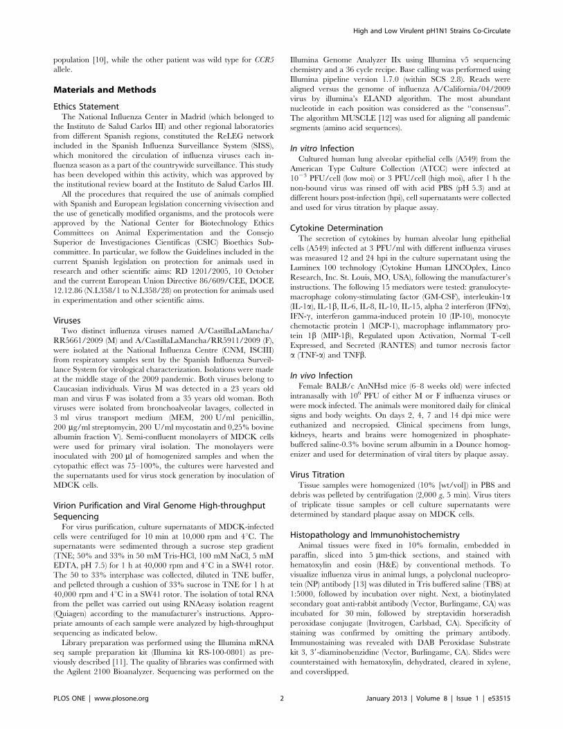
population [10], while the other patient was wild type for CCR5
allele.
Materials and Methods
Ethics StatementThe National Influenza Center in Madrid (which belonged to
the Instituto de Salud Carlos III) and other regional laboratories
from different Spanish regions, constituted the ReLEG network
included in the Spanish Influenza Surveillance System (SISS),
which monitored the circulation of influenza viruses each in-
fluenza season as a part of the countrywide surveillance. This study
has been developed within this activity, which was approved by
the institutional review board at the Instituto de Salud Carlos III.
All the procedures that required the use of animals complied
with Spanish and European legislation concerning vivisection and
the use of genetically modified organisms, and the protocols were
approved by the National Center for Biotechnology Ethics
Committees on Animal Experimentation and the Consejo
Superior de Investigaciones Cientıficas (CSIC) Bioethics Sub-
committee. In particular, we follow the Guidelines included in the
current Spanish legislation on protection for animals used in
research and other scientific aims: RD 1201/2005, 10 October
and the current European Union Directive 86/609/CEE, DOCE
12.12.86 (N.L358/1 to N.L358/28) on protection for animals used
in experimentation and other scientific aims.
VirusesTwo distinct influenza viruses named A/CastillaLaMancha/
RR5661/2009 (M) and A/CastillaLaMancha/RR5911/2009 (F),
were isolated at the National Influenza Centre (CNM, ISCIII)
from respiratory samples sent by the Spanish Influenza Surveil-
lance System for virological characterization. Isolations were made
at the middle stage of the 2009 pandemic. Both viruses belong to
Caucasian individuals. Virus M was detected in a 23 years old
man and virus F was isolated from a 35 years old woman. Both
viruses were isolated from bronchoalveolar lavages, collected in
3 ml virus transport medium (MEM, 200 U/ml penicillin,
200 mg/ml streptomycin, 200 U/ml mycostatin and 0,25% bovine
albumin fraction V). Semi-confluent monolayers of MDCK cells
were used for primary viral isolation. The monolayers were
inoculated with 200 ml of homogenized samples and when the
cytopathic effect was 75–100%, the cultures were harvested and
the supernatants used for virus stock generation by inoculation of
MDCK cells.
Virion Purification and Viral Genome High-throughputSequencingFor virus purification, culture supernatants of MDCK-infected
cells were centrifuged for 10 min at 10,000 rpm and 4uC. Thesupernatants were sedimented through a sucrose step gradient
(TNE; 50% and 33% in 50 mM Tris-HCl, 100 mM NaCl, 5 mM
EDTA, pH 7.5) for 1 h at 40,000 rpm and 4uC in a SW41 rotor.
The 50 to 33% interphase was collected, diluted in TNE buffer,
and pelleted through a cushion of 33% sucrose in TNE for 1 h at
40,000 rpm and 4uC in a SW41 rotor. The isolation of total RNA
from the pellet was carried out using RNAeasy isolation reagent
(Quiagen) according to the manufacturer’s instructions. Appro-
priate amounts of each sample were analyzed by high-throughput
sequencing as indicated below.
Library preparation was performed using the Illumina mRNA
seq sample preparation kit (Illumina kit RS-100-0801) as pre-
viously described [11]. The quality of libraries was confirmed with
the Agilent 2100 Bioanalyzer. Sequencing was performed on the
Illumina Genome Analyzer IIx using Illumina v5 sequencing
chemistry and a 36 cycle recipe. Base calling was performed using
Illumina pipeline version 1.7.0 (within SCS 2.8). Reads were
aligned versus the genome of influenza A/California/04/2009
virus by illumina’s ELAND algorithm. The most abundant
nucleotide in each position was considered as the ‘‘consensus’’.
The algorithm MUSCLE [12] was used for aligning all pandemic
segments (amino acid sequences).
In vitro InfectionCultured human lung alveolar epithelial cells (A549) from the
American Type Culture Collection (ATCC) were infected at
1023 PFU/cell (low moi) or 3 PFU/cell (high moi), after 1 h the
non-bound virus was rinsed off with acid PBS (pH 5.3) and at
different hours post-infection (hpi), cell supernatants were collected
and used for virus titration by plaque assay.
Cytokine DeterminationThe secretion of cytokines by human alveolar lung epithelial
cells (A549) infected at 3 PFU/ml with different influenza viruses
was measured 12 and 24 hpi in the culture supernatant using the
Luminex 100 technology (Cytokine Human LINCOplex, Linco
Research, Inc. St. Louis, MO, USA), following the manufacturer’s
instructions. The following 15 mediators were tested: granulocyte-
macrophage colony-stimulating factor (GM-CSF), interleukin-1a(IL-1a), IL-1b, IL-6, IL-8, IL-10, IL-15, alpha 2 interferon (IFNa),IFN-c, interferon gamma-induced protein 10 (IP-10), monocyte
chemotactic protein 1 (MCP-1), macrophage inflammatory pro-
tein 1b (MIP-1b), Regulated upon Activation, Normal T-cell
Expressed, and Secreted (RANTES) and tumor necrosis factor
a (TNF-a) and TNFb.
In vivo InfectionFemale BALB/c AnNHsd mice (6–8 weeks old) were infected
intranasally with 106 PFU of either M or F influenza viruses or
were mock infected. The animals were monitored daily for clinical
signs and body weights. On days 2, 4, 7 and 14 dpi mice were
euthanized and necropsied. Clinical specimens from lungs,
kidneys, hearts and brains were homogenized in phosphate-
buffered saline-0.3% bovine serum albumin in a Dounce homog-
enizer and used for determination of viral titers by plaque assay.
Virus TitrationTissue samples were homogenized (10% [wt/vol]) in PBS and
debris was pelleted by centrifugation (2,000 g, 5 min). Virus titers
of triplicate tissue samples or cell culture supernatants were
determined by standard plaque assay on MDCK cells.
Histopathology and ImmunohistochemistryAnimal tissues were fixed in 10% formalin, embedded in
paraffin, sliced into 5 mm-thick sections, and stained with
hematoxylin and eosin (H&E) by conventional methods. To
visualize influenza virus in animal lungs, a polyclonal nucleopro-
tein (NP) antibody [13] was diluted in Tris buffered saline (TBS) at
1:5000, followed by incubation over night. Next, a biotinylated
secondary goat anti-rabbit antibody (Vector, Burlingame, CA) was
incubated for 30 min, followed by streptavidin horseradish
peroxidase conjugate (Invitrogen, Carlsbad, CA). Specificity of
staining was confirmed by omitting the primary antibody.
Immunostaining was revealed with DAB Peroxidase Substrate
kit 3, 39-diaminobenzidine (Vector, Burlingame, CA). Slides were
counterstained with hematoxylin, dehydrated, cleared in xylene,
and coverslipped.
High and Low Virulent pH1N1 Strains Co-Circulate
PLOS ONE | www.plosone.org 2 January 2013 | Volume 8 | Issue 1 | e53515
CCR5 Allele DeterminationGenotyping was performed by standard methods described
elsewhere [14]. Briefly, the RNA from bronchoalveolar lavages of
patients, mice kidneys or cultured A549 cells, was amplified using
previously reported primers surrounding the 32-bp deletion in the
CCR5 gene [15] for patient and A549 cells and 59-CATTATA-
CATGCAGTCCTC-39 and 59-GATGGCAAAGATGAGCC-
TAC-39 primers for mice DNA.
Results
To determine possible differences in virulence, we compared the
biological properties of two distinct influenza viruses named mild
(M) and fatal (F), both isolated from young patients without known
health risk conditions. Virus M was detected in a patient that
presented mild respiratory symptoms, whereas virus F was isolated
from a patient that developed severe pneumonia, was hospitalized
in the intensive care unit and died several days after admission.
Genetic Characterization of M and F VirusesGenetic characterization of M and F viruses was performed by
ultrasequencing of purified virion RNAs obtained after two
passages in MDCK cells. The consensus sequences obtained for
the viral genomes represent more than 95% of the total sequence
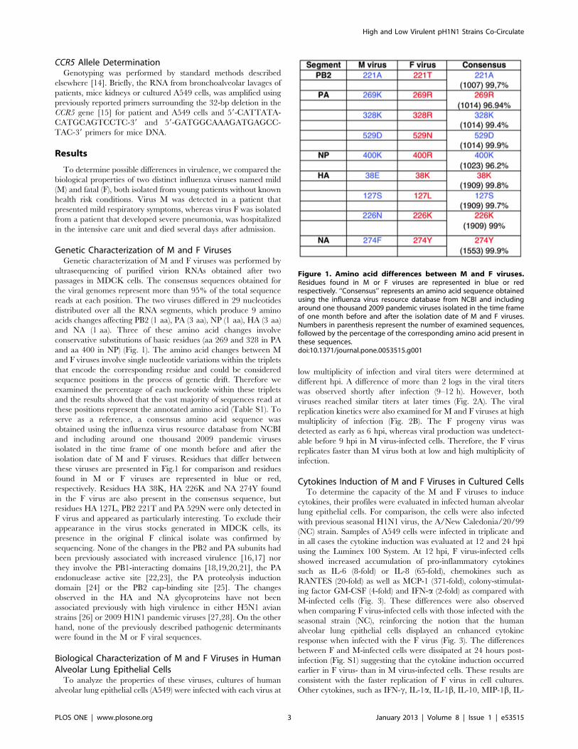
reads at each position. The two viruses differed in 29 nucleotides
distributed over all the RNA segments, which produce 9 amino
acids changes affecting PB2 (1 aa), PA (3 aa), NP (1 aa), HA (3 aa)
and NA (1 aa). Three of these amino acid changes involve
conservative substitutions of basic residues (aa 269 and 328 in PA
and aa 400 in NP) (Fig. 1). The amino acid changes between M
and F viruses involve single nucleotide variations within the triplets
that encode the corresponding residue and could be considered
sequence positions in the process of genetic drift. Therefore we
examined the percentage of each nucleotide within these triplets
and the results showed that the vast majority of sequences read at
these positions represent the annotated amino acid (Table S1). To
serve as a reference, a consensus amino acid sequence was
obtained using the influenza virus resource database from NCBI
and including around one thousand 2009 pandemic viruses
isolated in the time frame of one month before and after the
isolation date of M and F viruses. Residues that differ between
these viruses are presented in Fig.1 for comparison and residues
found in M or F viruses are represented in blue or red,
respectively. Residues HA 38K, HA 226K and NA 274Y found
in the F virus are also present in the consensus sequence, but
residues HA 127L, PB2 221T and PA 529N were only detected in
F virus and appeared as particularly interesting. To exclude their
appearance in the virus stocks generated in MDCK cells, its
presence in the original F clinical isolate was confirmed by
sequencing. None of the changes in the PB2 and PA subunits had
been previously associated with increased virulence [16,17] nor
they involve the PB1-interacting domains [18,19,20,21], the PA
endonuclease active site [22,23], the PA proteolysis induction
domain [24] or the PB2 cap-binding site [25]. The changes
observed in the HA and NA glycoproteins have not been
associated previously with high virulence in either H5N1 avian
strains [26] or 2009 H1N1 pandemic viruses [27,28]. On the other
hand, none of the previously described pathogenic determinants
were found in the M or F viral sequences.
Biological Characterization of M and F Viruses in HumanAlveolar Lung Epithelial CellsTo analyze the properties of these viruses, cultures of human
alveolar lung epithelial cells (A549) were infected with each virus at
low multiplicity of infection and viral titers were determined at
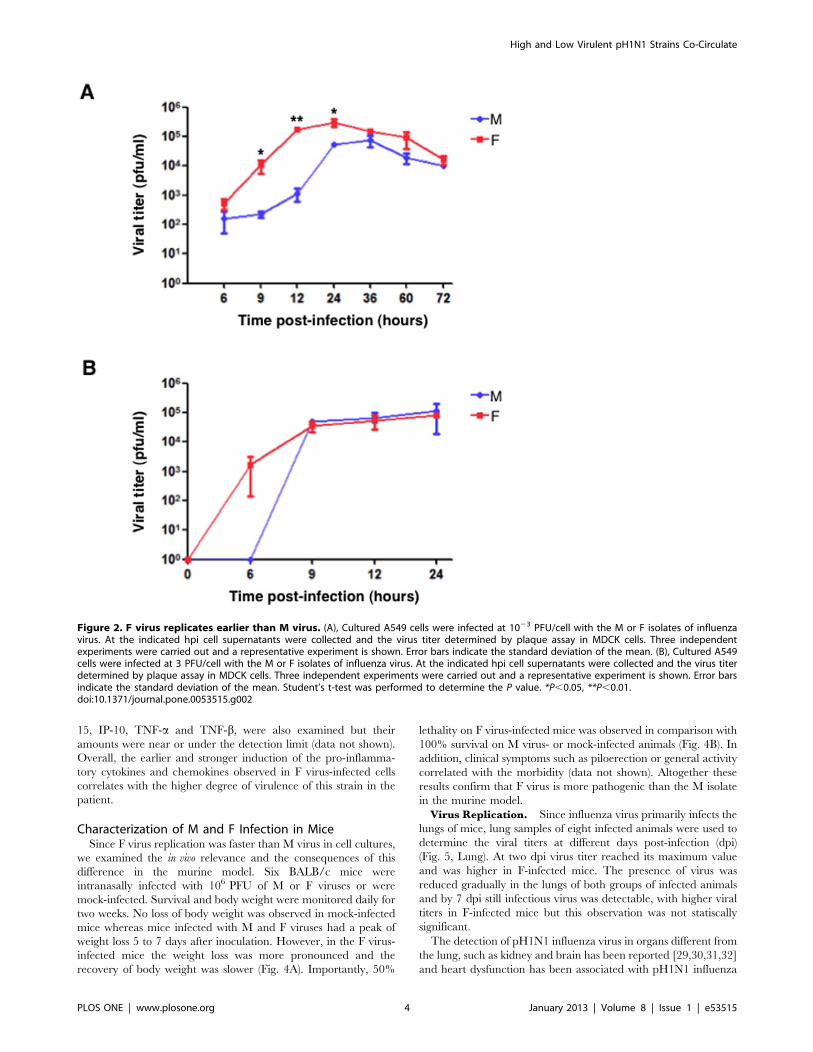
different hpi. A difference of more than 2 logs in the viral titers
was observed shortly after infection (9–12 h). However, both
viruses reached similar titers at later times (Fig. 2A). The viral
replication kinetics were also examined for M and F viruses at high
multiplicity of infection (Fig. 2B). The F progeny virus was
detected as early as 6 hpi, whereas viral production was undetect-
able before 9 hpi in M virus-infected cells. Therefore, the F virus
replicates faster than M virus both at low and high multiplicity of
infection.
Cytokines Induction of M and F Viruses in Cultured CellsTo determine the capacity of the M and F viruses to induce
cytokines, their profiles were evaluated in infected human alveolar
lung epithelial cells. For comparison, the cells were also infected
with previous seasonal H1N1 virus, the A/New Caledonia/20/99
(NC) strain. Samples of A549 cells were infected in triplicate and
in all cases the cytokine induction was evaluated at 12 and 24 hpi
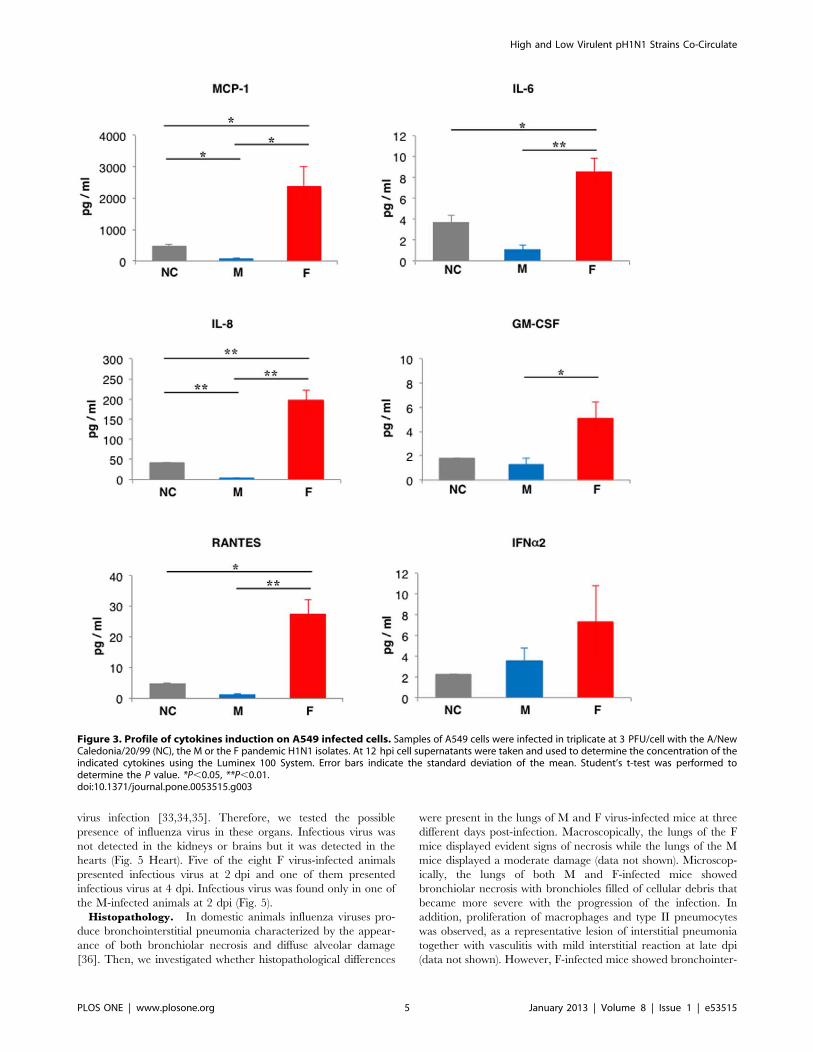
using the Luminex 100 System. At 12 hpi, F virus-infected cells
showed increased accumulation of pro-inflammatory cytokines
such as IL-6 (8-fold) or IL-8 (65-fold), chemokines such as
RANTES (20-fold) as well as MCP-1 (371-fold), colony-stimulat-
ing factor GM-CSF (4-fold) and IFN-a (2-fold) as compared with
M-infected cells (Fig. 3). These differences were also observed
when comparing F virus-infected cells with those infected with the
seasonal strain (NC), reinforcing the notion that the human
alveolar lung epithelial cells displayed an enhanced cytokine
response when infected with the F virus (Fig. 3). The differences
between F and M-infected cells were dissipated at 24 hours post-
infection (Fig. S1) suggesting that the cytokine induction occurred
earlier in F virus- than in M virus-infected cells. These results are
consistent with the faster replication of F virus in cell cultures.
Other cytokines, such as IFN-c, IL-1a, IL-1b, IL-10, MIP-1b, IL-
Figure 1. Amino acid differences between M and F viruses.Residues found in M or F viruses are represented in blue or redrespectively. ‘‘Consensus’’ represents an amino acid sequence obtainedusing the influenza virus resource database from NCBI and includingaround one thousand 2009 pandemic viruses isolated in the time frameof one month before and after the isolation date of M and F viruses.Numbers in parenthesis represent the number of examined sequences,followed by the percentage of the corresponding amino acid present inthese sequences.doi:10.1371/journal.pone.0053515.g001
High and Low Virulent pH1N1 Strains Co-Circulate
PLOS ONE | www.plosone.org 3 January 2013 | Volume 8 | Issue 1 | e53515
15, IP-10, TNF-a and TNF-b, were also examined but their
amounts were near or under the detection limit (data not shown).
Overall, the earlier and stronger induction of the pro-inflamma-
tory cytokines and chemokines observed in F virus-infected cells
correlates with the higher degree of virulence of this strain in the
patient.
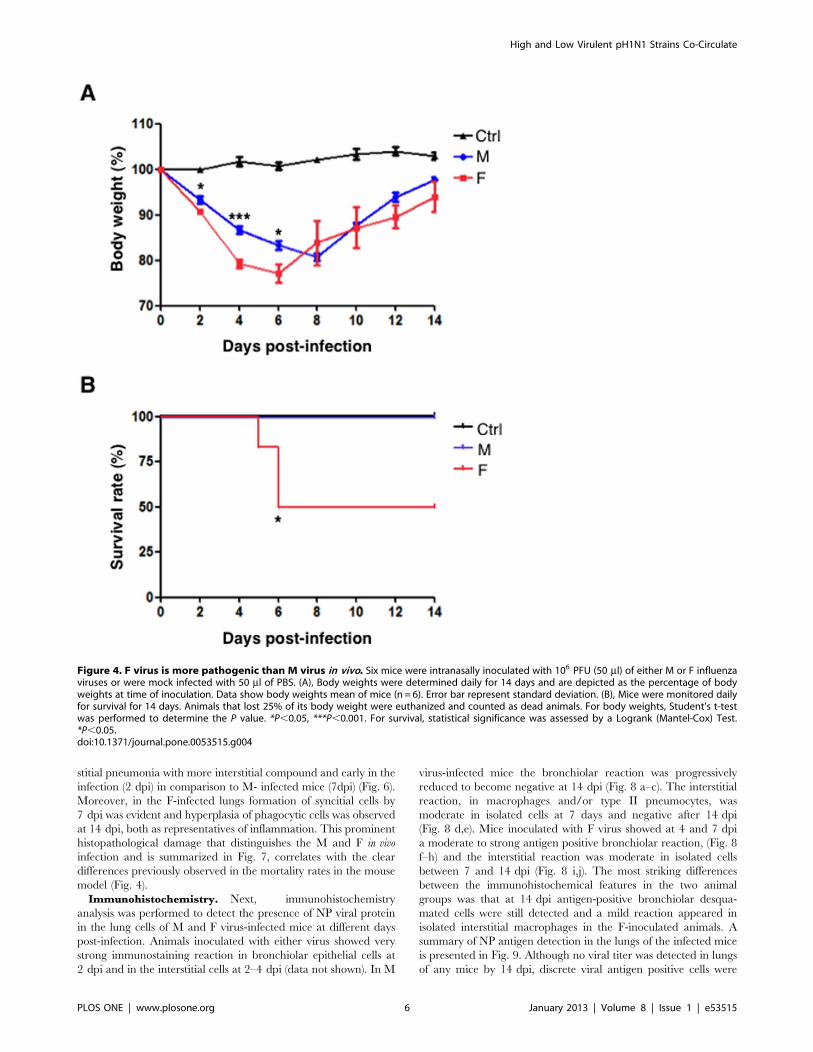
Characterization of M and F Infection in MiceSince F virus replication was faster than M virus in cell cultures,
we examined the in vivo relevance and the consequences of this
difference in the murine model. Six BALB/c mice were
intranasally infected with 106 PFU of M or F viruses or were
mock-infected. Survival and body weight were monitored daily for
two weeks. No loss of body weight was observed in mock-infected
mice whereas mice infected with M and F viruses had a peak of
weight loss 5 to 7 days after inoculation. However, in the F virus-
infected mice the weight loss was more pronounced and the
recovery of body weight was slower (Fig. 4A). Importantly, 50%
lethality on F virus-infected mice was observed in comparison with
100% survival on M virus- or mock-infected animals (Fig. 4B). In
addition, clinical symptoms such as piloerection or general activity
correlated with the morbidity (data not shown). Altogether these
results confirm that F virus is more pathogenic than the M isolate
in the murine model.
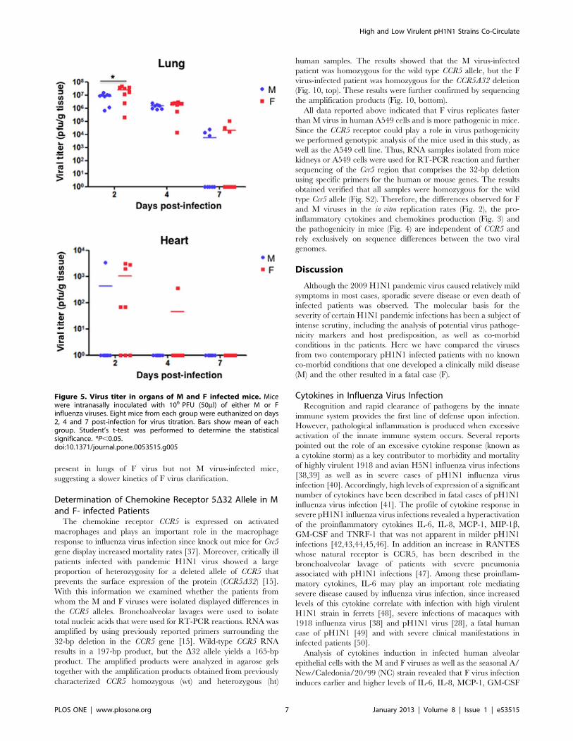
Virus Replication. Since influenza virus primarily infects the
lungs of mice, lung samples of eight infected animals were used to
determine the viral titers at different days post-infection (dpi)
(Fig. 5, Lung). At two dpi virus titer reached its maximum value
and was higher in F-infected mice. The presence of virus was
reduced gradually in the lungs of both groups of infected animals
and by 7 dpi still infectious virus was detectable, with higher viral
titers in F-infected mice but this observation was not statiscally
significant.
The detection of pH1N1 influenza virus in organs different from
the lung, such as kidney and brain has been reported [29,30,31,32]
and heart dysfunction has been associated with pH1N1 influenza
Figure 2. F virus replicates earlier than M virus. (A), Cultured A549 cells were infected at 1023 PFU/cell with the M or F isolates of influenzavirus. At the indicated hpi cell supernatants were collected and the virus titer determined by plaque assay in MDCK cells. Three independentexperiments were carried out and a representative experiment is shown. Error bars indicate the standard deviation of the mean. (B), Cultured A549cells were infected at 3 PFU/cell with the M or F isolates of influenza virus. At the indicated hpi cell supernatants were collected and the virus titerdetermined by plaque assay in MDCK cells. Three independent experiments were carried out and a representative experiment is shown. Error barsindicate the standard deviation of the mean. Student’s t-test was performed to determine the P value. *P,0.05, **P,0.01.doi:10.1371/journal.pone.0053515.g002
High and Low Virulent pH1N1 Strains Co-Circulate
PLOS ONE | www.plosone.org 4 January 2013 | Volume 8 | Issue 1 | e53515
virus infection [33,34,35]. Therefore, we tested the possible
presence of influenza virus in these organs. Infectious virus was
not detected in the kidneys or brains but it was detected in the
hearts (Fig. 5 Heart). Five of the eight F virus-infected animals
presented infectious virus at 2 dpi and one of them presented
infectious virus at 4 dpi. Infectious virus was found only in one of
the M-infected animals at 2 dpi (Fig. 5).
Histopathology. In domestic animals influenza viruses pro-
duce bronchointerstitial pneumonia characterized by the appear-
ance of both bronchiolar necrosis and diffuse alveolar damage
[36]. Then, we investigated whether histopathological differences
were present in the lungs of M and F virus-infected mice at three
different days post-infection. Macroscopically, the lungs of the F
mice displayed evident signs of necrosis while the lungs of the M
mice displayed a moderate damage (data not shown). Microscop-
ically, the lungs of both M and F-infected mice showed
bronchiolar necrosis with bronchioles filled of cellular debris that
became more severe with the progression of the infection. In
addition, proliferation of macrophages and type II pneumocytes
was observed, as a representative lesion of interstitial pneumonia
together with vasculitis with mild interstitial reaction at late dpi
(data not shown). However, F-infected mice showed bronchointer-
Figure 3. Profile of cytokines induction on A549 infected cells. Samples of A549 cells were infected in triplicate at 3 PFU/cell with the A/NewCaledonia/20/99 (NC), the M or the F pandemic H1N1 isolates. At 12 hpi cell supernatants were taken and used to determine the concentration of theindicated cytokines using the Luminex 100 System. Error bars indicate the standard deviation of the mean. Student’s t-test was performed todetermine the P value. *P,0.05, **P,0.01.doi:10.1371/journal.pone.0053515.g003
High and Low Virulent pH1N1 Strains Co-Circulate
PLOS ONE | www.plosone.org 5 January 2013 | Volume 8 | Issue 1 | e53515
stitial pneumonia with more interstitial compound and early in the
infection (2 dpi) in comparison to M- infected mice (7dpi) (Fig. 6).
Moreover, in the F-infected lungs formation of syncitial cells by
7 dpi was evident and hyperplasia of phagocytic cells was observed
at 14 dpi, both as representatives of inflammation. This prominent
histopathological damage that distinguishes the M and F in vivo
infection and is summarized in Fig. 7, correlates with the clear
differences previously observed in the mortality rates in the mouse
model (Fig. 4).
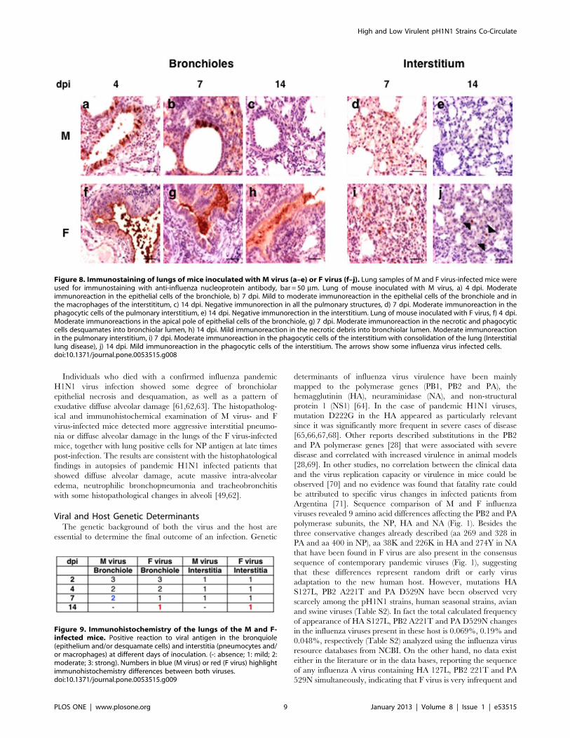
Immunohistochemistry. Next, immunohistochemistry
analysis was performed to detect the presence of NP viral protein
in the lung cells of M and F virus-infected mice at different days
post-infection. Animals inoculated with either virus showed very
strong immunostaining reaction in bronchiolar epithelial cells at
2 dpi and in the interstitial cells at 2–4 dpi (data not shown). In M
virus-infected mice the bronchiolar reaction was progressively
reduced to become negative at 14 dpi (Fig. 8 a–c). The interstitial
reaction, in macrophages and/or type II pneumocytes, was
moderate in isolated cells at 7 days and negative after 14 dpi
(Fig. 8 d,e). Mice inoculated with F virus showed at 4 and 7 dpi
a moderate to strong antigen positive bronchiolar reaction, (Fig. 8
f–h) and the interstitial reaction was moderate in isolated cells
between 7 and 14 dpi (Fig. 8 i,j). The most striking differences
between the immunohistochemical features in the two animal
groups was that at 14 dpi antigen-positive bronchiolar desqua-
mated cells were still detected and a mild reaction appeared in
isolated interstitial macrophages in the F-inoculated animals. A
summary of NP antigen detection in the lungs of the infected mice
is presented in Fig. 9. Although no viral titer was detected in lungs
of any mice by 14 dpi, discrete viral antigen positive cells were
Figure 4. F virus is more pathogenic than M virus in vivo. Six mice were intranasally inoculated with 106 PFU (50 ml) of either M or F influenzaviruses or were mock infected with 50 ml of PBS. (A), Body weights were determined daily for 14 days and are depicted as the percentage of bodyweights at time of inoculation. Data show body weights mean of mice (n = 6). Error bar represent standard deviation. (B), Mice were monitored dailyfor survival for 14 days. Animals that lost 25% of its body weight were euthanized and counted as dead animals. For body weights, Student’s t-testwas performed to determine the P value. *P,0.05, ***P,0.001. For survival, statistical significance was assessed by a Logrank (Mantel-Cox) Test.*P,0.05.doi:10.1371/journal.pone.0053515.g004
High and Low Virulent pH1N1 Strains Co-Circulate
PLOS ONE | www.plosone.org 6 January 2013 | Volume 8 | Issue 1 | e53515
present in lungs of F virus but not M virus-infected mice,
suggesting a slower kinetics of F virus clarification.
Determination of Chemokine Receptor 5D32 Allele in Mand F- infected PatientsThe chemokine receptor CCR5 is expressed on activated
macrophages and plays an important role in the macrophage
response to influenza virus infection since knock out mice for Crc5
gene display increased mortality rates [37]. Moreover, critically ill
patients infected with pandemic H1N1 virus showed a large
proportion of heterozygosity for a deleted allele of CCR5 that
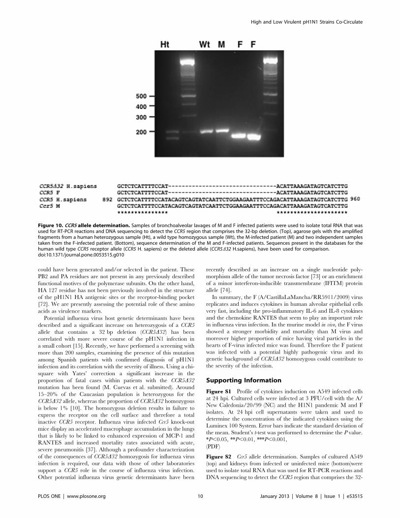
prevents the surface expression of the protein (CCR5D32) [15].With this information we examined whether the patients from
whom the M and F viruses were isolated displayed differences in
the CCR5 alleles. Bronchoalveolar lavages were used to isolate
total nucleic acids that were used for RT-PCR reactions. RNA was
amplified by using previously reported primers surrounding the
32-bp deletion in the CCR5 gene [15]. Wild-type CCR5 RNA
results in a 197-bp product, but the D32 allele yields a 165-bp
product. The amplified products were analyzed in agarose gels
together with the amplification products obtained from previously
characterized CCR5 homozygous (wt) and heterozygous (ht)
human samples. The results showed that the M virus-infected
patient was homozygous for the wild type CCR5 allele, but the F
virus-infected patient was homozygous for the CCR5D32 deletion
(Fig. 10, top). These results were further confirmed by sequencing
the amplification products (Fig. 10, bottom).
All data reported above indicated that F virus replicates faster
than M virus in human A549 cells and is more pathogenic in mice.
Since the CCR5 receptor could play a role in virus pathogenicity
we performed genotypic analysis of the mice used in this study, as
well as the A549 cell line. Thus, RNA samples isolated from mice
kidneys or A549 cells were used for RT-PCR reaction and further
sequencing of the Ccr5 region that comprises the 32-bp deletion
using specific primers for the human or mouse genes. The results
obtained verified that all samples were homozygous for the wild
type Ccr5 allele (Fig. S2). Therefore, the differences observed for F
and M viruses in the in vitro replication rates (Fig. 2), the pro-
inflammatory cytokines and chemokines production (Fig. 3) and
the pathogenicity in mice (Fig. 4) are independent of CCR5 and
rely exclusively on sequence differences between the two viral
genomes.
Discussion
Although the 2009 H1N1 pandemic virus caused relatively mild
symptoms in most cases, sporadic severe disease or even death of
infected patients was observed. The molecular basis for the
severity of certain H1N1 pandemic infections has been a subject of
intense scrutiny, including the analysis of potential virus pathoge-
nicity markers and host predisposition, as well as co-morbid
conditions in the patients. Here we have compared the viruses
from two contemporary pH1N1 infected patients with no known
co-morbid conditions that one developed a clinically mild disease
(M) and the other resulted in a fatal case (F).
Cytokines in Influenza Virus InfectionRecognition and rapid clearance of pathogens by the innate
immune system provides the first line of defense upon infection.
However, pathological inflammation is produced when excessive
activation of the innate immune system occurs. Several reports
pointed out the role of an excessive cytokine response (known as
a cytokine storm) as a key contributor to morbidity and mortality
of highly virulent 1918 and avian H5N1 influenza virus infections
[38,39] as well as in severe cases of pH1N1 influenza virus
infection [40]. Accordingly, high levels of expression of a significant
number of cytokines have been described in fatal cases of pH1N1
influenza virus infection [41]. The profile of cytokine response in
severe pH1N1 influenza virus infections revealed a hyperactivation
of the proinflammatory cytokines IL-6, IL-8, MCP-1, MIP-1b,GM-CSF and TNRF-1 that was not apparent in milder pH1N1
infections [42,43,44,45,46]. In addition an increase in RANTES
whose natural receptor is CCR5, has been described in the
bronchoalveolar lavage of patients with severe pneumonia
associated with pH1N1 infections [47]. Among these proinflam-
matory cytokines, IL-6 may play an important role mediating
severe disease caused by influenza virus infection, since increased
levels of this cytokine correlate with infection with high virulent
H1N1 strain in ferrets [48], severe infections of macaques with
1918 influenza virus [38] and pH1N1 virus [28], a fatal human
case of pH1N1 [49] and with severe clinical manifestations in
infected patients [50].
Analysis of cytokines induction in infected human alveolar
epithelial cells with the M and F viruses as well as the seasonal A/
New/Caledonia/20/99 (NC) strain revealed that F virus infection
induces earlier and higher levels of IL-6, IL-8, MCP-1, GM-CSF
Figure 5. Virus titer in organs of M and F infected mice. Micewere intranasally inoculated with 106 PFU (50ml) of either M or Finfluenza viruses. Eight mice from each group were euthanized on days2, 4 and 7 post-infection for virus titration. Bars show mean of eachgroup. Student’s t-test was performed to determine the statisticalsignificance. *P,0.05.doi:10.1371/journal.pone.0053515.g005
High and Low Virulent pH1N1 Strains Co-Circulate
PLOS ONE | www.plosone.org 7 January 2013 | Volume 8 | Issue 1 | e53515

and RANTES than M or NC infections (Fig. 3). Besides the role
described for IL-6, IL-8, MCP-1 and GM-CSF in patients with
pH1N1 severe infections, the chemokine RANTES, a natural
agonist of CCR5 seems to control the production of IL-6, IL-8,
MCP-1 and IFNs in influenza virus-infected cultured alveolar
epithelial cells [51]. Therefore, it is conceivable that high levels of
these chemokines and pro-inflammatory cytokines would in-
crement cellular recruitment to the lungs at earlier times, thus
contributing to inflammation and tissue consolidation. Indeed,
there was a moderate increase in the histopathological damage in
F virus- versus M virus-infected mice (Fig. 6–7). Also, this rapid
induction of pro-inflammatory cytokines and chemokines supports
the notion that a deregulation of the host immune response in the
early stages of the infection, might contribute to increase the
severity associated with more virulent pH1N1 strains.
Pandemic H1N1 Influenza Virus Infection in vivoThe comparison of the F and M viruses in the murine model
showed that F virus was more pathogenic, as indicated by the
morbidity and mortality rates observed in the F-infected mice. The
pathogenicity of several pH1N1 viruses in mice has been studied
previously. Mice infected with pH1N1 virus showed weight loss
and high titers in the lung but in most studies more than 106 PFU
were required to reach a 50% lethal dose or even the mice
survived at the highest dose used [32,52,53]. In a particular study,
the characterization of several strains of pH1N1 viruses in mice
indicated efficient replication in the lungs but not extrapulmonary
virus spread and no lethality, suggesting that pandemic viruses
display mild to moderate virulence when compared with highly
pathogenic viruses such as the 1918 virus [54]. In contrast 50% of
the mice infected at 106 PFU with the F virus but not with the M
virus died, indicating that F virus is more pathogenic than other
pH1N1 viruses. Moreover, infectious virus was recovered in higher
proportion of animals and remained at later days post-infection in
the hearts of F virus-infected mice (Fig. 5). Efficient dissemination
of F virus might have contributed also to increase the severity of
the infection. Accordingly, viral particles have been found in the
hearts of both domestic ducks infected with H5N1 highly
pathogenic avian virus [55] and fatal cases of 2009 pandemic
infected patients [56]. Extrapulmonary complications of influenza
virus infection have been previously reported. Around 50% of
patients without previous cardiac history have abnormal electro-
cardiogram findings [57] and a link has been recently reported
between influenza infection and acute myocardial infarction [58]
and fulminant myocarditis in children [59] and adults [60].
Figure 6. Histopathology of lungs of mice inoculated with M virus (a–d) or F virus (e–h). Lung samples of M and F virus-infected mice wereused for histopathological H&E examination at the indicated days post-infection. ba r = 50 mm. Lung of mouse inoculated with M virus, a) 2 dpi. Mildcongestion and diffuse lymphoid and phagocytic cells infiltrates in the interstitium. b) 4 dpi. Moderate thickened of interalveolar walls c) 7 dpi.Interstitial pneumonia with moderate proliferation of pneumocytes type II. d) 14 dpi. Interstitial pneumonia with thickened interalveolar walls. Lungof mouse inoculated with F virus, e) 2 dpi. Thickened interalveolar walls with consolidation of the pulmonary parenchima (interstitial pneumonia). f)4 dpi. Moderate increase of interstitial macrophages. g) 7 dpi. Increase of macrophages and syncitial cells formation. h) 14 dpi. Severe hyperplasia ofphagocytic cells in the interstitium. The arrows show some examples of the lesion indicated in each case.doi:10.1371/journal.pone.0053515.g006
Figure 7. Histopathological signs of the lungs of the M and F-infected mice. Histopathological lesions (1: mild; 2: moderate; 3:severe). Numbers in blue (M virus) or red (F virus) highlighthistopathological differences between both viruses.doi:10.1371/journal.pone.0053515.g007
High and Low Virulent pH1N1 Strains Co-Circulate
PLOS ONE | www.plosone.org 8 January 2013 | Volume 8 | Issue 1 | e53515
Individuals who died with a confirmed influenza pandemic
H1N1 virus infection showed some degree of bronchiolar
epithelial necrosis and desquamation, as well as a pattern of
exudative diffuse alveolar damage [61,62,63]. The histopatholog-
ical and immunohistochemical examination of M virus- and F
virus-infected mice detected more aggressive interstitial pneumo-
nia or diffuse alveolar damage in the lungs of the F virus-infected
mice, together with lung positive cells for NP antigen at late times
post-infection. The results are consistent with the histophatological
findings in autopsies of pandemic H1N1 infected patients that
showed diffuse alveolar damage, acute massive intra-alveolar
edema, neutrophilic bronchopneumonia and tracheobronchitis
with some histopathological changes in alveoli [49,62].
Viral and Host Genetic DeterminantsThe genetic background of both the virus and the host are
essential to determine the final outcome of an infection. Genetic
determinants of influenza virus virulence have been mainly
mapped to the polymerase genes (PB1, PB2 and PA), the
hemagglutinin (HA), neuraminidase (NA), and non-structural
protein 1 (NS1) [64]. In the case of pandemic H1N1 viruses,
mutation D222G in the HA appeared as particularly relevant
since it was significantly more frequent in severe cases of disease
[65,66,67,68]. Other reports described substitutions in the PB2
and PA polymerase genes [28] that were associated with severe
disease and correlated with increased virulence in animal models
[28,69]. In other studies, no correlation between the clinical data
and the virus replication capacity or virulence in mice could be
observed [70] and no evidence was found that fatality rate could
be attributed to specific virus changes in infected patients from
Argentina [71]. Sequence comparison of M and F influenza
viruses revealed 9 amino acid differences affecting the PB2 and PA
polymerase subunits, the NP, HA and NA (Fig. 1). Besides the
three conservative changes already described (aa 269 and 328 in
PA and aa 400 in NP), aa 38K and 226K in HA and 274Y in NA
that have been found in F virus are also present in the consensus
sequence of contemporary pandemic viruses (Fig. 1), suggesting
that these differences represent random drift or early virus
adaptation to the new human host. However, mutations HA
S127L, PB2 A221T and PA D529N have been observed very
scarcely among the pH1N1 strains, human seasonal strains, avian
and swine viruses (Table S2). In fact the total calculated frequency
of appearance of HA S127L, PB2 A221T and PA D529N changes
in the influenza viruses present in these host is 0.069%, 0.19% and
0.048%, respectively (Table S2) analyzed using the influenza virus
resource databases from NCBI. On the other hand, no data exist
either in the literature or in the data bases, reporting the sequence
of any influenza A virus containing HA 127L, PB2 221T and PA
529N simultaneously, indicating that F virus is very infrequent and
Figure 8. Immunostaining of lungs of mice inoculated with M virus (a–e) or F virus (f–j). Lung samples of M and F virus-infected mice wereused for immunostaining with anti-influenza nucleoprotein antibody, bar = 50 mm. Lung of mouse inoculated with M virus, a) 4 dpi. Moderateimmunoreaction in the epithelial cells of the bronchiole, b) 7 dpi. Mild to moderate immunoreaction in the epithelial cells of the bronchiole and inthe macrophages of the interstititum, c) 14 dpi. Negative immunorection in all the pulmonary structures, d) 7 dpi. Moderate immunoreaction in thephagocytic cells of the pulmonary interstitium, e) 14 dpi. Negative immunorection in the interstitium. Lung of mouse inoculated with F virus, f) 4 dpi.Moderate immunoreactions in the apical pole of epithelial cells of the bronchiole, g) 7 dpi. Moderate immunoreaction in the necrotic and phagocyticcells desquamates into bronchiolar lumen, h) 14 dpi. Mild immunoreaction in the necrotic debris into bronchiolar lumen. Moderate immunoreactionin the pulmonary interstitium, i) 7 dpi. Moderate immunoreaction in the phagocytic cells of the interstitium with consolidation of the lung (Interstitiallung disease), j) 14 dpi. Mild immunoreaction in the phagocytic cells of the interstitium. The arrows show some influenza virus infected cells.doi:10.1371/journal.pone.0053515.g008
Figure 9. Immunohistochemistry of the lungs of the M and F-infected mice. Positive reaction to viral antigen in the bronquiole(epithelium and/or desquamate cells) and interstitia (pneumocytes and/or macrophages) at different days of inoculation. (-: absence; 1: mild; 2:moderate; 3: strong). Numbers in blue (M virus) or red (F virus) highlightimmunohistochemistry differences between both viruses.doi:10.1371/journal.pone.0053515.g009
High and Low Virulent pH1N1 Strains Co-Circulate
PLOS ONE | www.plosone.org 9 January 2013 | Volume 8 | Issue 1 | e53515
could have been generated and/or selected in the patient. These
PB2 and PA residues are not present in any previously described
functional motives of the polymerase subunits. On the other hand,
HA 127 residue has not been previously involved in the structure
of the pH1N1 HA antigenic sites or the receptor-binding pocket
[72]. We are presently assessing the potential role of these amino
acids as virulence markers.
Potential influenza virus host genetic determinants have been
described and a significant increase on heterozygosis of a CCR5
allele that contains a 32 bp deletion (CCR5D32) has been
correlated with more severe course of the pH1N1 infection in
a small cohort [15]. Recently, we have performed a screening with
more than 200 samples, examining the presence of this mutation
among Spanish patients with confirmed diagnosis of pH1N1
infection and its correlation with the severity of illness. Using a chi-
square with Yates’ correction a significant increase in the
proportion of fatal cases within patients with the CCR5D32mutation has been found (M. Cuevas et al. submitted). Around
15–20% of the Caucasian population is heterozygous for the
CCR5D32 allele, whereas the proportion of CCR5D32 homozygous
is below 1% [10]. The homozygous deletion results in failure to
express the receptor on the cell surface and therefore a total
inactive CCR5 receptor. Influenza virus infected Ccr5 knock-out
mice display an accelerated macrophage accumulation in the lungs
that is likely to be linked to enhanced expression of MCP-1 and
RANTES and increased mortality rates associated with acute,
severe pneumonitis [37]. Although a profounder characterization
of the consequences of CCR5D32 homozygosis for influenza virus
infection is required, our data with those of other laboratories
support a CCR5 role in the course of influenza virus infection.
Other potential influenza virus genetic determinants have been
recently described as an increase on a single nucleotide poly-
morphism allele of the tumor necrosis factor [73] or an enrichment
of a minor interferon-inducible transmembrane (IFITM) protein
allele [74].
In summary, the F (A/CastillaLaMancha/RR5911/2009) virus
replicates and induces cytokines in human alveolar epithelial cells
very fast, including the pro-inflammatory IL-6 and IL-8 cytokines
and the chemokine RANTES that seem to play an important role
in influenza virus infection. In the murine model in vivo, the F virus
showed a stronger morbidity and mortality than M virus and
moreover higher proportion of mice having viral particles in the
hearts of F-virus infected mice was found. Therefore the F patient
was infected with a potential highly pathogenic virus and its
genetic background of CCR5D32 homozygous could contribute to
the severity of the infection.
Supporting Information
Figure S1 Profile of cytokines induction on A549 infected cells
at 24 hpi. Cultured cells were infected at 3 PFU/cell with the A/
New Caledonia/20/99 (NC) and the H1N1 pandemic M and F
isolates. At 24 hpi cell supernatants were taken and used to
determine the concentration of the indicated cytokines using the
Luminex 100 System. Error bars indicate the standard deviation of
the mean. Student’s t-test was performed to determine the P value.
*P,0.05, **P,0.01, ***P,0.001,
(PDF)
Figure S2 Ccr5 allele determination. Samples of cultured A549
(top) and kidneys from infected or uninfected mice (bottom)were
used to isolate total RNA that was used for RT-PCR reactions and
DNA sequencing to detect the CCR5 region that comprises the 32-
Figure 10. CCR5 allele determination. Samples of bronchoalveolar lavages of M and F infected patients were used to isolate total RNA that wasused for RT-PCR reactions and DNA sequencing to detect the CCR5 region that comprises the 32-bp deletion. (Top), agarose gels with the amplifiedfragments from a human heterozygous sample (Ht), a wild type homozygous sample (Wt), the M-infected patient (M) and two independent samplestaken from the F-infected patient. (Bottom), sequence determination of the M and F-infected patients. Sequences present in the databases for thehuman wild type CCR5 receptor allele (CCR5 H. sapiens) or the deleted allele (CCR5D32 H.sapiens), have been used for comparison.doi:10.1371/journal.pone.0053515.g010
High and Low Virulent pH1N1 Strains Co-Circulate
PLOS ONE | www.plosone.org 10 January 2013 | Volume 8 | Issue 1 | e53515
bp deletion. The sequence of the CCR5 allele from human (CCR5
H. sapiens) or mice (Ccr5 M. musculus) have been used for
comparison. The red line corresponds to the 32 bp deletion found
in the CCR5D32 samples.
(PDF)
Table S1 Nucleotides at the indicated positions of M and F
RNAs.
(PDF)
Table S2 Frequency of different residues at PB2 221, PA 529
and HA 127 positions in human, swine and avian viruses.
(PDF)
Acknowledgments
We would like to acknowledge members of the Spanish Influenza
Surveillance System working in the identification and declaration of
patients during the 2009 pandemia. The authors also acknowledge
technical assistance from Manuela Lopez-Valero, Ana Calderon, Monica
Gonzalez-Esguevillas, Mar Molinero, Nieves Cruz, Noelia Reyes and
Silvia Moreno at the National Influenza Center in Madrid (CNM) and Y.
Fernandez and N. Zamarreno at the Centro Nacional de Biotecnologıa.
J.C. Oliveros provided helpful analysis of RNA sequences. We are indebted
to P. Gastaminza and S. F. Gonzalez for critical review of the manuscript.
Author Contributions
Conceived and designed the experiments: AF AR MTC MP IC AN.
Performed the experiments: AF AR MTC FP BGB PMO MM MP SG.
Analyzed the data: AF AR MTC PPB JAM JO IC AN. Contributed
reagents/materials/analysis tools: MTC FP PPB IC. Wrote the paper: AN.
References
1. Sambhara S, Poland GA (2010) H5N1 Avian influenza: preventive and
therapeutic strategies against a pandemic. Annual review of medicine 61: 187–
198.
2. Neumann G, Noda T, Kawaoka Y (2009) Emergence and pandemic potential of
swine-origin H1N1 influenza virus. Nature 459: 931–939.
3. Garten RJ, Davis CT, Russell CA, Shu B, Lindstrom S, et al. (2009) Antigenic
and genetic characteristics of swine-origin 2009 A(H1N1) influenza viruses
circulating in humans. Science 325: 197–201.
4. Louie JK, Acosta M, Winter K, Jean C, Gavali S, et al. (2009) Factors associated
with death or hospitalization due to pandemic 2009 influenza A(H1N1) infection
in California. JAMA 302: 1896–1902.
5. Perez-Padilla R, de la Rosa-Zamboni D, Ponce de Leon S, Hernandez M,
Quinones-Falconi F, et al. (2009) Pneumonia and respiratory failure from swine-
origin influenza A (H1N1) in Mexico. N Engl J Med 361: 680–689.
6. Falagas ME, Koletsi PK, Baskouta E, Rafailidis PI, Dimopoulos G, et al. (2011)
Pandemic A(H1N1) 2009 influenza: review of the Southern Hemisphere
experience. Epidemiology and infection 139: 27–40.
7. Falagas ME, Cholevas NV, Kapaskelis AM, Vouloumanou EK, Michalopoulos
A, et al. (2010) Epidemiological aspects of 2009 H1N1 influenza: the
accumulating experience from the Northern Hemisphere. European journal of
clinical microbiology & infectious diseases: official publication of the European
Society of Clinical Microbiology 29: 1327–1347.
8. Singanayagam A, Wood V, Chalmers JD (2011) Factors associated with severe
illness in pandemic 2009 influenza a (H1N1) infection: implications for triage in
primary and secondary care. The Journal of infection 63: 243–251.
9. Louie JK, Acosta M, Samuel MC, Schechter R, Vugia DJ, et al. (2011) A novel
risk factor for a novel virus: obesity and 2009 pandemic influenza A (H1N1).
Clin Infect Dis 52: 301–312.
10. Martinson JJ, Chapman NH, Rees DC, Liu YT, Clegg JB (1997) Global
distribution of the CCR5 gene 32-basepair deletion. Nature genetics 16: 100–
103.
11. Minoche AE, Dohm JC, Himmelbauer H (2011) Evaluation of genomic high-
throughput sequencing data generated on Illumina HiSeq and Genome
Analyzer systems. Genome biology 12: R112.
12. Edgar RC (2004) MUSCLE: multiple sequence alignment with high accuracy
and high throughput. Nucleic acids research 32: 1792–1797.
13. Jorba N, Coloma R, Ortin J (2009) Genetic trans-complementation establishes
a new model for influenza virus RNA transcription and replication. PLoS
Pathog 5: e1000462.
14. Abdi R, Smith RN, Makhlouf L, Najafian N, Luster AD, et al. (2002) The role of
CC chemokine receptor 5 (CCR5) in islet allograft rejection. Diabetes 51: 2489–
2495.
15. Keynan Y, Juno J, Meyers A, Ball TB, Kumar A, et al. (2010) Chemokine
receptor 5 D32 allele in patients with severe pandemic (H1N1) 2009. Emerg
Infect Dis 16: 1621–1622.
16. Zhou B, Li Y, Halpin R, Hine E, Spiro DJ, et al. (2011) PB2 residue 158 is
a pathogenic determinant of pandemic H1N1 and H5 influenza a viruses in
mice. Journal of virology 85: 357–365.
17. Mok CK, Yen HL, Yu MY, Yuen KM, Sia SF, et al. (2011) Amino acid residues
253 and 591 of the PB2 protein of avian influenza virus A H9N2 contribute to
mammalian pathogenesis. Journal of virology 85: 9641–9645.
18. Ohtsu Y, Honda Y, Sakata Y, Kato H, Toyoda T (2002) Fine mapping of the
subunit binding sites of influenza virus RNA polymerase. Microbiol Immunol
46: 167–175.
19. Sugiyama K, Obayashi E, Kawaguchi A, Suzuki Y, Tame JR, et al. (2009)
Structural insight into the essential PB1-PB2 subunit contact of the influenza
virus RNA polymerase. EMBO J 28: 1803–1811.
20. Perales B, de la Luna S, Palacios I, Ortın J (1996) Mutational analysis identifies
functional domains in the influenza A virus PB2 polymerase subunit. J Virol 70:
1678–1686.
21. Zurcher T, de lLS, Sanz EJ, Nieto A, Ortin J (1996) Mutational analysis of the
influenza virus A/Victoria/3/75 PA protein: studies of interaction with PB1
protein and identification of a dominant negative mutant. J Gen Virol 77: 1745–
1749.
22. Dias A, Bouvier D, Crepin T, McCarthy AA, Hart DJ, et al. (2009) The cap-
snatching endonuclease of influenza virus polymerase resides in the PA subunit.
Nature 458: 914–918.
23. Yuan P, Bartlam M, Lou Z, Chen S, Zhou J, et al. (2009) Crystal structure of an
avian influenza polymerase PA(N) reveals an endonuclease active site. Nature
458: 909–913.
24. Sanz-Ezquerro JJ, Zurcher T, de la Luna S, Ortin J, Nieto A (1996) The amino-
terminal one-third of the influenza virus PA protein is responsible for the
induction of proteolysis. J Virol 70: 1905–1911.
25. Guilligay D, Tarendeau F, Resa-Infante P, Coloma R, Crepin T, et al. (2008)
The structural basis for cap binding by influenza virus polymerase subunit PB2.
Nat Struct Mol Biol 15: 500–506.
26. Hulse DJ, Webster RG, Russell RJ, Perez DR (2004) Molecular determinants
within the surface proteins involved in the pathogenicity of H5N1 influenza
viruses in chickens. J Virol 78: 9954–9964.
27. Ye J, Sorrell EM, Cai Y, Shao H, Xu K, et al. (2010) Variations in the
hemagglutinin of the 2009 H1N1 pandemic virus: potential for strains with
altered virulence phenotype? PLoS Pathog 6: e1001145.
28. Safronetz D, Rockx B, Feldmann F, Belisle SE, Palermo RE, et al. (2011)
Pandemic swine-origin H1N1 influenza A virus isolates show heterogeneous
virulence in macaques. Journal of virology 85: 1214–1223.
29. Nin N, Lorente JA, Sanchez-Rodriguez C, Granados R, Ver LS, et al. (2011)
Kidney histopathological findings in fatal pandemic 2009 influenza A (H1N1).
Intensive care medicine 37: 880–881.
30. Carmona F, Carlotti AP, Ramalho LN, Costa RS, Ramalho FS (2011) Evidence
of Renal Infection in Fatal Cases of 2009 Pandemic Influenza A (H1N1).
American journal of clinical pathology 136: 416–423.
31. Sun R, Luo J, Gao Y, He H (2009) Different infection routes of avian influenza
A (H5N1) virus in mice. Integrative zoology 4: 402–408.
32. Kwon D, Shin K, Kim S, Ha Y, Choi JH, et al. (2010) Replication and
pathogenesis of the pandemic (H1N1) 2009 influenza virus in mammalian
models. Journal of microbiology 48: 657–662.
33. Gokhroo RK, Barjaty HD, Bhawna K (2011) Cardiac conduction system
affection in a case of swine flu. The Journal of the Association of Physicians of
India 59: 51–52.
34. Wiegand JA, Torgersen C, Bloechlinger S, Takala J, Dunser MW (2011)
Influenza A(H1N1) infection and severe cardiac dysfunction in adults: A case
series. Wiener klinische Wochenschrift 123: 120–123.
35. Brown SM, Pittman J, Miller Iii RR, Horton KD, Markewitz B, et al. (2011)
Right and left heart failure in severe H1N1 influenza A infection. The European
respiratory journal: official journal of the European Society for Clinical
Respiratory Physiology 37: 112–118.
36. Zachary JF, McGavin MD (2012) Pathological Basis of Veterinary Disease:
Elsevier, St.Louis, Missouri.
37. Dawson TC, Beck MA, Kuziel WA, Henderson F, Maeda N (2000) Contrasting
effects of CCR5 and CCR2 deficiency in the pulmonary inflammatory response
to influenza A virus. Am J Pathol 156: 1951–1959.
38. Kobasa D, Jones SM, Shinya K, Kash JC, Copps J, et al. (2007) Aberrant innate
immune response in lethal infection of macaques with the 1918 influenza virus.
Nature 445: 319–323.
High and Low Virulent pH1N1 Strains Co-Circulate
PLOS ONE | www.plosone.org 11 January 2013 | Volume 8 | Issue 1 | e53515
39. de Jong MD, Simmons CP, Thanh TT, Hien VM, Smith GJ, et al. (2006) Fatal
outcome of human influenza A (H5N1) is associated with high viral load andhypercytokinemia. Nature medicine 12: 1203–1207.
40. Cheng XW, Lu J, Wu CL, Yi LN, Xie X, et al. (2011) Three fatal cases of
pandemic 2009 influenza A virus infection in Shenzhen are associated withcytokine storm. Respiratory physiology & neurobiology 175: 185–187.
41. To KK, Hung IF, Li IW, Lee KL, Koo CK, et al. (2010) Delayed clearance ofviral load and marked cytokine activation in severe cases of pandemic H1N1
2009 influenza virus infection. Clinical infectious diseases: an official publication
of the Infectious Diseases Society of America 50: 850–859.42. Lee N, Wong CK, Chan PK, Chan MC, Wong RY, et al. (2011) Cytokine
Response Patterns in Severe Pandemic 2009 H1N1 and Seasonal Influenzaamong Hospitalized Adults. PLoS One 6: e26050.
43. Estella A (2011) Cytokine levels in bronchoalveolar lavage and serum in 3patients with 2009 Influenza A(H1N1)v severe pneumonia. Journal of infection
in developing countries 5: 540–543.
44. Lee N, Chan PK, Wong CK, Wong KT, Choi KW, et al. (2011) Viral clearanceand inflammatory response patterns in adults hospitalized for pandemic 2009
influenza A(H1N1) virus pneumonia. Antiviral therapy 16: 237–247.45. Almansa R, Anton A, Ramirez P, Martin-Loeches I, Banner D, et al. (2011)
Direct association between pharyngeal viral secretion and host cytokine response
in severe pandemic influenza. BMC infectious diseases 11: 232.46. Yu X, Zhang X, Zhao B, Wang J, Zhu Z, et al. (2011) Intensive cytokine
induction in pandemic H1N1 influenza virus infection accompanied by robustproduction of IL-10 and IL-6. PLoS One 6: e28680.
47. Zuniga J, Torres M, Romo J, Torres D, Jimenez L, et al. (2011) Inflammatoryprofiles in severe pneumonia associated with the pandemic influenza A/H1N1
virus isolated in Mexico City. Autoimmunity 44: 562–570.
48. Svitek N, Rudd PA, Obojes K, Pillet S, von Messling V (2008) Severe seasonalinfluenza in ferrets correlates with reduced interferon and increased IL-6
induction. Virology 376: 53–59.49. Nakajima N, Hata S, Sato Y, Tobiume M, Katano H, et al. (2010) The first
autopsy case of pandemic influenza (A/H1N1pdm) virus infection in Japan:
detection of a high copy number of the virus in type II alveolar epithelial cells bypathological and virological examination. Japanese journal of infectious diseases
63: 67–71.50. Hayden FG, Fritz R, Lobo MC, Alvord W, Strober W, et al. (1998) Local and
systemic cytokine responses during experimental human influenza A virusinfection. Relation to symptom formation and host defense. The Journal of
clinical investigation 101: 643–649.
51. Phung TT, Sugamata R, Uno K, Aratani Y, Ozato K, et al. (2011) Key role ofRANTES (regulated upon activation normal T-cell expressed and secreted),
nonstructural protein1 and myeloperoxidase in cytokine storm induced byinfluenza virus PR-8(A/H1N1) infection in A549 bronchial epithelial cells.
Microbiology and immunology.
52. Maines TR, Jayaraman A, Belser JA, Wadford DA, Pappas C, et al. (2009)Transmission and pathogenesis of swine-origin 2009 A(H1N1) influenza viruses
in ferrets and mice. Science 325: 484–487.53. Itoh Y, Shinya K, Kiso M, Watanabe T, Sakoda Y, et al. (2009) In vitro and
in vivo characterization of new swine-origin H1N1 influenza viruses. Nature460: 1021–1025.
54. Belser JA, Wadford DA, Pappas C, Gustin KM, Maines TR, et al. (2010)
Pathogenesis of pandemic influenza A (H1N1) and triple-reassortant swineinfluenza A (H1) viruses in mice. Journal of virology 84: 4194–4203.
55. Wasilenko JL, Arafa AM, Selim AA, Hassan MK, Aly MM, et al. (2011)Pathogenicity of two Egyptian H5N1 highly pathogenic avian influenza viruses
in domestic ducks. Archives of virology 156: 37–51.
56. Ru YX, Li YC, Zhao Y, Zhao SX, Yang JP, et al. (2011) Multiple organ invasionby viruses: pathological characteristics in three fatal cases of the 2009 pandemic
influenza A/H1N1. Ultrastructural pathology 35: 155–161.57. Ison MG, Campbell V, Rembold C, Dent J, Hayden FG (2005) Cardiac findings
during uncomplicated acute influenza in ambulatory adults. Clinical infectious
diseases: an official publication of the Infectious Diseases Society of America 40:
415–422.
58. Warren-Gash C, Smeeth L, Hayward AC (2009) Influenza as a trigger for acute
myocardial infarction or death from cardiovascular disease: a systematic review.
The Lancet infectious diseases 9: 601–610.
59. Bratincsak A, El-Said HG, Bradley JS, Shayan K, Grossfeld PD, et al. (2010)
Fulminant myocarditis associated with pandemic H1N1 influenza A virus in
children. Journal of the American College of Cardiology 55: 928–929.
60. Liao YC, Hsieh YC, Chang WC, Huang JL, Ting CT, et al. (2011) Fulminant
myocarditis in an adult with 2009 pandemic influenza A (H1N1 influenza)
infection. Journal of the Chinese Medical Association: JCMA 74: 130–133.
61. Prasad HB, Puranik SC, Kadam DB, Sangle SA, Borse RT, et al. (2011)
Retrospective analysis of necropsy findings in patients of H1N1 and their
correlation to clinical features. The Journal of the Association of Physicians of
India 59: 498–500.
62. Nakajima N, Sato Y, Katano H, Hasegawa H, Kumasaka T, et al. (2011)
Histopathological and immunohistochemical findings of 20 autopsy cases with
2009 H1N1 virus infection. Modern pathology: an official journal of the United
States and Canadian Academy of Pathology, Inc.
63. Bautista E, Chotpitayasunondh T, Gao Z, Harper SA, Shaw M, et al. (2010)
Clinical aspects of pandemic 2009 influenza A (H1N1) virus infection. The New
England journal of medicine 362: 1708–1719.
64. de Wit E, Fouchier RA (2008) Emerging influenza. Journal of clinical virology:
the official publication of the Pan American Society for Clinical Virology 41: 1–
6.
65. Chen H, Wen X, To KK, Wang P, Tse H, et al. (2010) Quasispecies of the
D225G substitution in the hemagglutinin of pandemic influenza A(H1N1) 2009virus from patients with severe disease in Hong Kong, China. The Journal of
infectious diseases 201: 1517–1521.
66. Mak GC, Au KW, Tai LS, Chuang KC, Cheng KC, et al. (2010) Association of
D222G substitution in haemagglutinin of 2009 pandemic influenza A (H1N1)
with severe disease. Euro surveillance: bulletin europeen sur les maladies
transmissibles = European communicable disease bulletin 15.
67. Kilander A, Rykkvin R, Dudman SG, Hungnes O (2010) Observed association
between the HA1 mutation D222G in the 2009 pandemic influenza A(H1N1)
virus and severe clinical outcome, Norway 2009–2010. Euro surveillance:
bulletin europeen sur les maladies transmissibles = European communicable
disease bulletin 15.
68. L’Vov D K, Burtseva EI, Prilipov AG, Bogdanova VS, Shchelkanov M, et al.
(2010) [A possible association of fatal pneumonia with mutations of pandemic
influenza A/H1N1 sw1 virus in the receptor-binding site of the HA1 subunit].
Voprosy virusologii 55: 4–9.
69. Meunier I, Embury-Hyatt C, Stebner S, Gray M, Bastien N, et al. (2012)
Virulence differences of closely related pandemic 2009 H1N1 isolates correlate
with increased inflammatory responses in ferrets. Virology 422: 125–131.
70. Xu L, Bao L, Zhou J, Wang D, Deng W, et al. (2011) Genomic polymorphism of
the pandemic A (H1N1) influenza viruses correlates with viral replication,
virulence, and pathogenicity in vitro and in vivo. PLoS One 6: e20698.
71. Baumeister E, Palacios G, Cisterna D, Solovyov A, Hui J, et al. (2010) Molecular
characterization of severe and mild cases of influenza A (H1N1) 2009 strain from
Argentina. Medicina 70: 518–523.
72. Igarashi M, Ito K, Yoshida R, Tomabechi D, Kida H, et al. (2010) Predicting
the antigenic structure of the pandemic (H1N1) 2009 influenza virus
hemagglutinin. PLoS One 5: e8553.
73. Antonopoulou A, Baziaka F, Tsaganos T, Raftogiannis M, Koutoukas P, et al.
(2012) Role of tumor necrosis factor gene single nucleotide polymorphisms in the
natural course of 2009 influenza A H1N1 virus infection. International journal
of infectious diseases: IJID: official publication of the International Society for
Infectious Diseases.
74. Everitt AR, Clare S, Pertel T, John SP, Wash RS, et al. (2012) IFITM3 restricts
the morbidity and mortality associated with influenza. Nature.
High and Low Virulent pH1N1 Strains Co-Circulate
PLOS ONE | www.plosone.org 12 January 2013 | Volume 8 | Issue 1 | e53515