Merosin-negative congenital muscular dystrophy: magnetic resonance spectroscopy findings
Upload
sorbonne-frCategory
view
1download
0
Early Human Development 86 (2010) 287–294
Contents lists available at ScienceDirect
Early Human Development
j ourna l homepage: www.e lsev ie r.com/ locate /ear lhumdev
Best Practice Guideline article
Congenital hyperinsulinism
Jean-Baptiste Arnoux a, Pascale de Lonlay a,⁎, Maria-Joao Ribeiro f, Khalid Hussain b, Oliver Blankenstein c,Klaus Mohnike d, Vassili Valayannopoulos a, Jean-Jacques Robert a, Jacques Rahier e, Christine Sempoux e,Christine Bellanné g, Virginie Verkarre a, Yves Aigrain a, Francis Jaubert a,Francis Brunelle a, Claire Nihoul-Fékété a
a Hospital Necker-Enfants Malades, Paris, Franceb Institute of Child Health, Great Ormond Street, Hospital for Children, NHS Trust, Developmental Endocrinology Research Group, London, UKc Institute for Experimental Endocrinology, Charité University Medicine Berlin, Berlin, Germanyd Otto von Guericke University Magdeburg, Magdeburg, Germanye Department of Pathology, Cliniques Universitaires Saint-Luc, Université Catholique de Louvain, Brussels, Belgiumf Service Hospitalier Frédéric Joliot, Département de Recherche Médicale, Direction des Sciences du Vivant, Commissariat à l'Energie Atomique, Orsay, Franceg Département de Génétique, AP-HP Groupe Hospitalier Pitié-Salpétrière, Université Pierre et Marie Curie-Paris 6, Paris, France
⁎ Corresponding author. Center of Reference for Inher48 52; fax: +33 1 44 49 48 50.
E-mail address: [email protected] (P. de
0378-3782/$ – see front matter © 2010 Elsevier Irelanddoi:10.1016/j.earlhumdev.2010.05.003
a b s t r a c t
a r t i c l e i n f oKeywords:
Congenital hyperinsulinismDiffuseFocalPotassium channelDOPA PET–CTCongenital hyperinsulinism (CHI or HI) is a condition leading to recurrent hypoglycemia due to aninappropriate insulin secretion by the pancreatic islet β cells. HI has two main characteristics: a high glucoserequirement to correct hypoglycemia and a responsiveness of hypoglycemia to exogenous glucagon. HI isusually isolated but may be rarely part of a genetic syndrome (e.g. Beckwith–Wiedemann syndrome, Sotossyndrome etc.). The severity of HI is evaluated by the glucose administration rate required to maintainnormal glycemia and the responsiveness to medical treatment. Neonatal onset HI is usually severe while lateonset and syndromic HI are generally responsive to a medical treatment. Glycemia must be maintainedwithin normal ranges to avoid brain damages, initially with glucose administration and glucagon infusionthen, once the diagnosis is set, with specific HI treatment. Oral diazoxide is a first line treatment. In case ofunresponsiveness to this treatment, somatostatin analogues and calcium antagonists may be added, andfurther investigations are required for the putative histological diagnosis: pancreatic 18F-fluoro-L-DOPA PET–CT and molecular analysis. Indeed, focal forms consist of a focal adenomatous hyperplasia of islet cells, andwill be cured after a partial pancreatectomy. Diffuse HI involves all the pancreatic β cells of the wholepancreas. Diffuse HI resistant to medical treatment (octreotide, diazoxide, calcium antagonists andcontinuous feeding) may require subtotal pancreatectomy which post-operative outcome is unpredictable.The genetics of focal islet-cells hyperplasia associates a paternally inherited mutation of the ABCC8 or theKCNJ11 genes, with a loss of the maternal allele specifically in the hyperplasic islet cells. The genetics ofdiffuse isolated HI is heterogeneous and may be recessively inherited (ABCC8 and KCNJ11) or dominantlyinherited (ABCC8, KCNJ11, GCK, GLUD1, SLC16A1, HNF4A and HADH). Syndromic HI are always diffuse formand the genetics depend on the syndrome. Except for HI due to potassium channel defect (ABCC8 andKCNJ11), most of these HI are sensitive to diazoxide. The main points sum up the management of HI: i)prevention of brain damages by normalizing glycemia and ii) screening for focal HI as they may bedefinitively cured after a limited pancreatectomy.
ited Metabolic Diseases, Hôpital Necker-Enfants Malades
Lonlay).
Ltd. All rights reserved.
© 2010 Elsevier Ireland Ltd. All rights reserved.
Contents
1. Physiopathology of hypoglycemia . . . . . . . . . . . . . . . . . . . . . . . . . . . . . . . . . . . . . . . . . . . . . . . . . . . . 2882. Clinical presentation . . . . . . . . . . . . . . . . . . . . . . . . . . . . . . . . . . . . . . . . . . . . . . . . . . . . . . . . . . 2883. The diagnostic criteria for HI . . . . . . . . . . . . . . . . . . . . . . . . . . . . . . . . . . . . . . . . . . . . . . . . . . . . . . 2894. Management of hypoglycemia concomitantly to the diagnosis . . . . . . . . . . . . . . . . . . . . . . . . . . . . . . . . . . . . . . 2895. PET . . . . . . . . . . . . . . . . . . . . . . . . . . . . . . . . . . . . . . . . . . . . . . . . . . . . . . . . . . . . . . . . . . 290
, Université Paris Descartes, 75015 Paris, France. Tel.: +33 1 44 49

288 J.-B. Arnoux et al. / Early Human Development 86 (2010) 287–294
6. Surgical treatment . . . . . . . . . . . . . . . . . . . . . . . . . . . . . . . . . . . . . . . . . . . . . . . . . . . . . . . . . . . 2917. Genetics . . . . . . . . . . . . . . . . . . . . . . . . . . . . . . . . . . . . . . . . . . . . . . . . . . . . . . . . . . . . . . . . 2918. Prognosis . . . . . . . . . . . . . . . . . . . . . . . . . . . . . . . . . . . . . . . . . . . . . . . . . . . . . . . . . . . . . . . 2929. Practical management . . . . . . . . . . . . . . . . . . . . . . . . . . . . . . . . . . . . . . . . . . . . . . . . . . . . . . . . . 292References . . . . . . . . . . . . . . . . . . . . . . . . . . . . . . . . . . . . . . . . . . . . . . . . . . . . . . . . . . . . . . . . . 293
1. Physiopathology of hypoglycemia
Hypoglycemia in children is defined by a glucose plasma levelbelow 2.8 or 3 mmol/l, and can be a life-threatening condition whichneeds rigorous assessment and diligent treatment, to allow thecorrect diagnosis of its cause and to avoid brain damage and generaldistress [1]. Hypoglycemia results from impairment in glucosehomeostasis and the diagnosis of its cause requires a good knowledgeof the complex mechanisms which control blood glucose concentra-tion in the fasting and fed states [2]. During feeding, the liversynthesizes glycogen and triglycerides, the latter being exported toadipose tissue, to store energy substrates. During fasting, it releasesglucose and ketone bodies. The regulation of a normal blood glucoselevel depends upon i) hepatic glycogenolytic and gluconeogenicenzymes; ii) an adequate supply of endogenous gluconeogenicsubstrates (amino acids, glycerol and lactate); iii) an adequate energysupply provided by fatty acid β-oxidation, to promote gluconeogen-esis and ketogenesis, the latter synthesizing aceto-acetate andhydroxybutyrate which will be exported to peripheral tissues wherethey will be preferentially used as an alternative fuel to glucose andiv) a normal endocrine system for integrating and modulating theseprocesses. The major signals, which control the transition betweenthe fed and the fasting states, are glucose, insulin and glucagon [3].They influence directly or indirectly the enzymes which regulate livercarbohydrate and fatty acid metabolism, and thereby orient metabolicfluxes towards either energy storage or substrate release.
Congenital hyperinsulinism is due to an inappropriate insulinsecretion by the β-cells of Langerhans islets [1,2]. Insulin is the onlyhormone to decrease plasma glucose concentration, both by inhibit-ing glucose release from the liver (by glycogenolysis and gluconeo-genesis), and by increasing glucose uptake in muscle and adiposetissues. Moreover, insulin is known to inhibit lipolysis and so there isno ketogenesis. During congenital hyperinsulinism the brain is thusparticularly vulnerable because of these recurrent combined defectsin energetic substrates: glucose and ketone bodies. Thesemechanismsexplain the main characteristic clinical findings of neonatal hyperin-sulinism (HI): the high glucose requirement to correct hypoglycemia,the responsiveness of hypoglycemia to exogenous glucagon whichstimulates glycogen degradation and gluconeogenesis, and theabsence of ketone bodies during hypoglycemia.
Glucose and other substrates, such as amino acids, stimulateinsulin secretion through their metabolism, by raising the intracyto-solic ATP/ADP ratio. Glucokinase enzyme initiates the β-cell glucosemetabolism. It has a high Km for glucose so that the bloodconcentration of glucose directly determines the rate of glucoseoxidation of β-cell and subsequently controls the release of insulin. Anincrease in the cytosolic ATP/ADP ratio activates the plasmamembrane sulfonylurea receptor 1 (SUR1), leading to the closure(inhibition) of the potassium channel (KATP channel) Kir6.2. Thisdepolarizes the plasma membrane which opens a voltage dependantcalcium channel. As a consequence the calcium cellular concentrationincreases, which triggers the release of insulin from storage granules.Leucine, one of the most potent amino acids in stimulating insulinsecretion, acts somewhat indirectly as a positive allosteric affector ofglutamate dehydrogenase (GDH) which catalyses the oxidativedeamination of glutamate to alpha-ketoglutarate and ammonia,using NAD or NADP as co-factor. Hyperactivation of GDH isresponsible for an increased alpha of the β-cell ATP/ADP ratio.
Diazoxide blocks insulin secretion by activating (opening) the SUR1,whereas sulfonylureas, such as tolbutamide, stimulate insulin secre-tion by closing SUR1. Somatostatin analogues act by inhibiting theinsulin release through different mechanisms involving adenylylcyclase and protein kinase A, and dietary protein restriction decreasesthe stimulation of GDH by leucine (Fig. 1).
2. Clinical presentation
Severe hypoketotic hypoglycemia is the major feature of HI, and isat risk of seizures and brain damages if untreated. The presentationvaries according to the age of onset of hypoglycemia. In the neonatalperiod, hypoglycemia occurs early within 72 h after birth and isrevealed in half of the patients by seizures. The majority of theaffected newborns are macrosomic at birth and about 20% aredelivered by Caesarean section. The other symptoms of hypoglycemiaare abnormal movements as tremulousness, hypotonia, cyanosis,hypothermia or a life-threatening event. In some cases, hypoglycemiais revealed by routine measurement of blood glucose. The plasmaglucose concentration at the time of the first symptoms is oftenextremely low (b1 mmol/l). Hypoglycemia is permanent, both fastingand post-prandial in severe cases. The rates of intravenous glucoseadministration required to maintain plasma glucose above 3 mmol/l are high, with a mean rate between 15 and 17 mg/kg/min (seeTable 1, for equivalent mg/kg/min and rates of 10% dextrose infusion).Blood glucose concentrations can be increased by 2 to 3 mmol/l inresponse to subcutaneous or intramuscular administration of gluca-gon (0.5 mg). A mild hepatomegaly can be found.
Usually, hyperinsulinemic hypoglycemia is the only symptomalthough facial dysmorphism with high forehead, large and bulbousnose with short columella, smooth philtrum and thin upper lip isfrequently observed whatever the type of hyperinsulinism [4] (Fig. 2).However, a few syndromic hyperinsulinisms have also been describedwith neonatal hypoglycemia. In these cases, hypoglycemia is usuallysensitive to diazoxide unlike neonatal hyperinsulinemic hypoglyce-mia due to a potassium channel defect. These syndromic HI includeBeckwith–Wiedemann syndrome (BWS), Perlman syndrome, Kabukisyndrome, Sotos syndrome, congenital disorders of glycosylation typeIa or Ib (CDG) [5], or Usher syndrome type Ic. BWSmust be consideredwhen exomphalos, macroglossia, or gigantism is noted, moreoverthese patients have an increased risk of developing specific tumors(Wilms' tumors). Sotos syndrome associates acromegalic features,mental retardation and facial dysmorphism. A malformative HI (e.g.heart malformations and skeletal disease)may lead to the diagnosis ofKabuki syndrome. Finally, diarrhea and/or liver abnormalities areassociated with CDG. Another syndromic HI with mental retardationand epilepsy is the HI/HA syndrome due to an overactivity ofglutamate dehydrogenase [6]. Permanent hyperammonemia (HA)leads to the diagnosis. Neurological symptoms occur later in infancy,and are not consequences of recurrent hypoglycemia.
The main differential diagnosis of congenital HI remains thefactitious hyperinsulinism secondary to Munchausen by proxysyndrome, one of the parents administering insulin or sulfonylureasurreptitiously to their own child.
Another period of onset for HI occurs later in infancy, between 1and 20 months of life and is revealed in half of the patients by seizures.Macrosomy at birth can be noted. The characteristics of hypoglycemiaare similar, although lower rates of intravenous glucose are required

Fig. 1. Glucose-stimulated insulin secretion. GCK: glucokinase; GDH: glutamate dehydrogenase; TCA: tricarboxylic acids cycle; I, II, III, IV and V: the five complexes of themitochondrial respiratory chain; UCP2: uncoupling protein 2; ATP: adenosine tri phosphate; ADP: adenosine diphosphate; Gα: α G protein; Gi: inhibitory G protein; Gs: stimulatingG protein; AC: adenylyl cyclase; PKA: protein kinase A; PLC: protein lipase C; PIP2: phosphatidyl inositol biphosphate; DAG: diacylglycerol; IP3: inositol triphosphate; PKC: proteinkinase C; ER: endoplasmic reticulum.
289J.-B. Arnoux et al. / Early Human Development 86 (2010) 287–294
to maintain normal plasma glucose (8–13 mg/kg/min). Becausehypoglycemia is better tolerated, diagnosis is often delayed.
In childhood (after 2 years old) hypoglycemia in our series washighly suggestive of a pancreatic insulinoma, which is an adenoma,which differs from congenital hyperinsulinism by its etiopathogeneticmechanismand its pancreatic histology. The rates of oral or intravenousglucose required to maintain normal plasma glucose are lower and notall children require continuous glucose administration. Because hypo-glycemia is better tolerated in infants, diagnosis is often delayed.
The severity of hypoglycemia is evaluated by the rates of glucoseadministration required to maintain normal glycemia and theresponsiveness to medical treatment. It depends on the age of onsetof hypoglycemia. Neonatal hypoglycemias are usually severe what-ever the histological form of HI (focal or diffuse) unlike hypoglycemiaoccurring during the first year of life and syndromic HI.
3. The diagnostic criteria for HI
The diagnostic criteria for congenital HI [7,8] include i) fasting andpost-prandial hypoglycemia (b2.5–3 mmol/l) with unsuppressed
Table 1Infusion of glucose.
Peripheral catheter: glucose 10%
2 ml/kg/h (=3.3 mg/kg/min)4 ml/kg/h (=6.7 mg/kg/min)6 ml/kg/h (=10 mg/kg/min)8 ml/kg/h (=13.3 mg/kg/min)
Central catheter: glucose 10%, 20%, 30% or 50%
e.g. Glucose 30%
0.5 ml/kg/h (=2.5 mg/kg/min)1 ml/kg/h (=5 mg/kg/min)2 ml/kg/h (=10 mg/kg/min)3 ml/kg/h (=15 mg/kg/min)
insulin secretion (plasma insulin concentrations N1 mU/l), ii) apositive response to the subcutaneous or intramuscular administra-tion of glucagon (plasma glucose concentration increase by 2 to3 mmol/l following a 0.5 mg glucagon subcutaneous injection), iii)negative ketone bodies in urine (and in plasma) and iv) prolongeddependence on treatment to prevent hypoglycemia throughout thefirst months/years of life. Nevertheless, in infancy and childhood,normal plasma insulin and C-peptide concentrations during hypogly-cemia do not exclude the diagnosis of HI and measurements have tobe repeated. In the absence of clearly abnormal insulin levels duringhypoglycemic episodes, an 8 to 12 hours fasting test aiming atrevealing inappropriately low levels of ketone bodies, free fatty acidand branched chain amino acids can be helpful.
Some HI are transient and occurs in specific contexts: acute fetaldistress, even minor, small weight for gestational age and gestationaldiabetes. The HI is usually benign but may present severely, isdiazoxide sensitive, and will resolve spontaneously within severaldays or weeks.
All new HI patients should be screened for hyperammonemia todiagnose the HI/HA syndrome (GLUD1 gene), for short chainhydroxyacyl-CoA dehydrogenase (SCHAD) deficiency (HADH gene)with urine organic acids and plasma acylcarnitines chromatographies,and for CDG syndromes, as these 3 diseases may present in theneonatal period as apparently isolated HI. Other genes can besuspected depending on the context. SLC16A1 gene will be analyzedin case of Exercise-induced hyperinsulinism (EIHI) [9], HNF4A genewhen the newborn is macrosomic with a family history of MODYdiabetes [10]. Finally, familial forms or consanguinity and syndromicforms have to be checked as these are associated with a diffuse HI.
4. Management of hypoglycemia concomitantly to the diagnosis
During management, blood glucose levels must be maintainedwithin the normal neonatal range (above 3.5 mmol/l), by adminis-tering glucose orally, enterally or intravenously. Usually, in neonates,

Fig. 2. Dysmorphy of patients with isolated HI.
Fig. 3. Hypertrichosis. The most frequent adverse effect of diazoxide.
290 J.-B. Arnoux et al. / Early Human Development 86 (2010) 287–294
the first step is a continuous enteral feeding of milk enriched withmalto-dextrine. However, the severity of the hypoglycemias maystraightaway or rapidly require more intensive treatments to preventirreversible brain damages. The glucose rate administered has to besufficient to normalize glucose levels, at least with a glucose flowequal to the physiological hepatic production of glucose (8–10 mg/kg/min for a neonate or young infant and 5–7 mg/kg/min for children)(Table 1). If hypoglycemia persists or recurs, the perfusion rate has tobe increased, often requiring high concentration glucose solutionsinfused through a central venous line. However, in severe HI this maybe insufficient and continuous glucagon infusion (intravenous orsubcutaneous, 0.5 to 2 mg/day) along with glucose should beadministered.
At the same time, specific treatments of HI must be initiated [3].Oral diazoxide is first used at 15 mg/kg/day (neonates) or 10 mg/kg/day (infants) in 3 oral doses [11]. However, most of neonatal andisolated persistent HI are resistant to diazoxide. A neonatal HIsensitive to diazoxide is probably transient (secondary to fetaldistress, gestational diabetes etc.) or persistent but involving genesother than those encoding SUR1 and Kir6.2 (the potassium channelsubunits). Diazoxide efficiency is defined as the normalization ofglycemia N3 mmol/l measured before and after each meal in patientsfed normally with a physiological overnight fast, after stoppingintravenous glucose and any other medications for at least fiveconsecutive days. Tolerance to diazoxide is usually excellent except inpremature neonates because of sodium and fluid retention, whichmay lead to edema, pulmonary hypertension (premature baby at riskof bronchodysplasia) or heart failure (patients with heart defects orcompromised cardiac reserve). The most frequent adverse effect ishypertrichosis, which can be marked and distressing in youngchildren (Fig. 3). Hematological side effects are very rare with usualadministration doses. Two confirmed hypoglycemias (b3 mmol/l) in a24-hour glucose measurement cycle defined the patient as diazoxide-unresponsive. Dietary measures and glucose perfusion should bestarted again to maintain normoglycemia.
Octreotide must be tried before considering surgery in case ofdiazoxide-unresponsiveness [12]. Doses vary from 10 to 50 µg/kg/day
depending on authors, administered continuously or every 6 or 8 h.Higher doses may lead to a worsening of the hypoglycemias bysuppressing both glucagon and growth hormone. At initiation ofoctreotide treatment, some patients may present vomiting and/ordiarrhea and abdominal distension, which will resolve spontaneouslywithin 7–10 days. Gallbladder sludge or stones are rare but cannecessitate ursodesoxycholic acid treatment. It should be screened byabdominal ultrasound twice a year. Glycemia levels can risesignificantly immediately after octreotide initiation, however thispositive response can be transient, so that a 48 hour observationperiod should be performed to conclude definitively on the respon-siveness to octreotide at a given dose.
The dose of diazoxide, when effective on glycemia, does not needto be adapted to the patient weight gain. By contrast, the doses ofoctreotide should be progressively increased accordingly with theweight gain of the baby, to prevent recurrence of hypoglycemia.
Other drugs as calcium channels blockers (like nifedipine, 0.5–2 mg/kg/day in 2 oral doses) can be proposed.
Patients who are resistant to medical treatment and requiresurgical treatment [13], must be assessed for their putative histolog-ical form of HI [14–16]. Indeed, two histological forms exist (focalform and diffuse form) leading to two specific kind of surgery. Thefocal form is defined as a focal adenomatous hyperplasia of islets β-cells within the pancreatic tissue [17,18] and can be definitively curedafter a partial and selective pancreatectomy. On the contrary, in thediffuse HI, all the β-cells of the whole pancreas are abnormal, so thatonly a subtotal pancreatectomy may improve the patient condition.
In the absence of any distinctive clinical feature, it has beensuggested, although not confirmed, that a tolbutamide test mightdistinguish the focal forms (which would be tolbutamide responsiveowing to triggering of insulin secretion) from the diffuse forms(which would be tolbutamide insensitive) [19,20]. However, we donot recommend this test as the 18F-fluoro-L-DOPA positron emissiontomography (PET) is a simple and effective test which diagnoses andlocalizes the focal lesion [21–25]. It definitively replaced thepancreatic catheterism with pancreatic venous sampling [26,27](Fig. 2).
5. PET
Thus, 18F-fluoro-L-DOPA PET is recommended for all HI resistant todiazoxide to distinguish focal from diffuse forms. The ability to uptakeand decarboxylate amine precursors such as L-DOPA or 5-hydroxy-tryptophan (5-HTP) and to store their biogenic amines (dopamineand serotonin), is characteristic of neuroendocrine cells. L-Dihydrox-yphenylalanine (L-DOPA) is a precursor of catecholamines which is

291J.-B. Arnoux et al. / Early Human Development 86 (2010) 287–294
converted to dopamine by the aromatic amino acid decarboxylase(AADC) enzyme. Pancreatic islets have been shown to take up L-DOPA,and convert it to dopamine through the aromatic amino acid dopadecarboxylase [28]. In cases of diffuse HI, dopamine is visualized in thewhole pancreas while in case of a focal form, a spot is visualized into alimited part of the pancreas.
The patients are fasted for at least 6 h prior to the PET study. As theuptake of 18F-fluoro-L-DOPA remains unchanged under the adminis-tration of octreotide and/or diazoxide, PET studies can be performedwithout stopping thesemedications. Conversely, glucagon, interferingwith β-cell activity, will probably also interfere with AADC activityand so the glucagon administration must be stopped 1 day before thePET examination.
PET acquisition is 30 min long, so it is performed under a lightsedation.
The patients are placed in supine position in the PET using a 3-dimensional laser alignment. To ensure the optimal position in thescanner and to avoid movement artefacts, the babies should becomfortably immobilised during the study acquisition. PET startsbetween 30 and 60 min after the radiotracer injection (intravenousinjection of 3 to 4 MBq/kg of 18F-fluoro-L-DOPA).
The reconstructed images must be evaluated in a 3D display usingaxial, coronal, and sagittal views to visualize the pancreas, whichalways presents a high uptake of 18F-fluoro-L-DOPA and to distinguishit from the surrounding organs of the abdomen. The PET images showthat most of the radioactivity injected is found in the kidneys and thebladder. Consequently, the high radioactivity in these organs,particularly in the left kidney, might increase the difficulty to identifyfocal forms localized in the tail of the pancreas when the PET imagesalone are interpreted. Furthermore, in some patients, a physiologicalradiotracer accumulation by the liver, gall bladder, biliary duct, andduodenum can be observed. However, this variable uptake can bedistinguished from pancreas uptake.
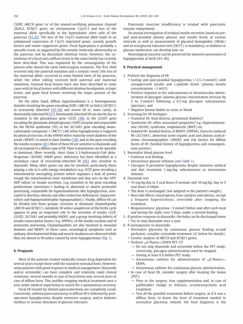
The localization of focal forms can be even improved by the fusion ofthe PET imaging with a CT angiography (Fig. 4). Indeed, the spot ofradiotracer marking the focal form will be located by reference to the
Fig. 4. PET–CT with 3D construction. A: 3D fusion of PET imaging with CT angiography.The focal form appears as an isolated hot spot while the uptake of radiotracer by the restof the pancreas is almost invisible. B: Diffuse form of HI. The uptake of radiotracer ishomogenous all along the pancreas. Both kidneys and the bladder are also visible as theradiotracer is also excreted in urines.
position of the splenic, superior and inferior mesenteric vessels, thevenous confluent and the portal vein. As the focal lesion can be locatedanywhere (50% are located in the head of the pancreas), the collectedinformationwill be of the utmost importance for the surgeon to be ableto find the focal lesion and to perform themost limited pancreatectomy.
Several regions of interest could be defined over the pancreas tocalculate standardized uptake values (SUV). Then, ratios between eachSUV region and the mean pancreatic SUV were calculated. For focalforms, the 18F-fluoro-L-DOPA SUV ratio is higher for the “hot spot”(N1.2), independently of its localization (and b1.1 for diffuse forms).
The PET studies should not be performed under the age of1 month, to exclude patients with transient HI, or in patients with agenetically proven suspected diffuse form (HI/HA syndrome, syn-dromic HI and mutations in GCK, SLC16A1, HNF4A and HADH genes).Indeed, focal forms are observed only in patients who have one singlefatherly inherited mutation in KCNJ11 or in ABCC8 gene (cf. chapter“Genetics”). To conclude, both PET and genetics give informationabout the form (focal or diffuse) of HI, and thus give the indication ofthe type of surgery (partial or subtotal pancreatectomy).
6. Surgical treatment
When medical and dietary therapies are ineffective, surgicaltreatment is required.
The focal form, which accounts for 40% of the patients treatedsurgically, is defined as a focal hyperplasia of the pancreatic β-cells.The lesion measures 2.5 to 7.5 mm in diameter and differs from theadult-type pancreatic adenoma which is clearly delimited tumor, sothat most of the time the focal lesion cannot be macroscopically seenduring the surgery. In diffuse HI, all the β-cells of the whole pancreashave abnormal nuclei and large cytoplasm. The patients thought tohave a focal lesion at PET–CT undergo surgery [29], as they can bedefinitively cured afterwards. In patients in whom a diffuse form isstrongly suspected, the surgery is performed only if they resist or donot tolerate medical treatment. This decision of performing a near-total pancreatectomy or an intensive medical treatment is difficultand may take into consideration the experience of the medical team,the psychological or social context of the parents, and the technolog-ical possibilities in a given country (e.g. the possibility to performsafely a continuous enteral feeding at home).
Before surgery, some precautions are necessary: i) stop medica-tions several days before surgery (5 days before for diazoxide and2 days before for octreotide) as they may interfere with theperoperative pathological analysis, ii) screen for gallbladder stoneswith a abdominal ultrasound, and treat if necessary and iii)supplement systematically with iron to prevent anemia.
The first period of the surgical time confirms the diagnosis of focalor diffuse form suspected on the PET imaging and the genetics. Alaparoscopic approach can be disccused [30]. Pancreatic samples arecollected from the head, the isthmus, the body and the tail of thepancreas and immediately examined microscopically. This intrao-perative histology will confirm definitively the form of HI, and then, incase of a suspected focal form, additional samples will be taken tolocalize the lesion, guided by the PET–CT. Once the focal form ishistologically observed, the pathologist will guide the surgeon tocomplete the resection of the focal abnormality. At the end of thesurgery, a final series of samples are examined to ensure the normalityof the resection edges. The complexity of this surgery requires thepatient suspected with a focal form to be referred to a surgical andpathological team trained in the surgery of focal forms.
A near-total pancreatectomy is performed for diffuse lesions.
7. Genetics
Focal islet-cell hyperplasia is associated with hemi- or homozy-gosity of a paternally inherited mutation of the sulfonylurea receptor
292 J.-B. Arnoux et al. / Early Human Development 86 (2010) 287–294
(SUR1 ABCC8 gene) or of the inward-rectifying potassium channel(Kir6.2, KCNJ11 gene) on chromosome 11p15.1 and loss of thematernal allele specifically in the hyperplastic islets cells of thepancreas [31,32]. The loss of the 11p15 maternal allele leads to anunbalanced expression of 11p15 imprinted genes, namely growthfactors and tumor suppressor genes. Focal hyperplasia is probably asporadic event, as suggested by the somatic molecular abnormality inthe pancreas and by discordant identical twins. However, the co-existence of a focal and a diffuse lesion in the same family has recentlybeen described. This was explained by the consanguinity of theparents who shared the same heterozygous mutation. The first childreceived only the paternal mutation and a somatic event (the loss ofthe maternal allele) occurred in some limited islets of his pancreas,while the other sibling received both paternal and maternalmutations. Unusual focal lesions have also been described in somecases with bi-focal lesions with different deletion breakpoints, ectopiclesion, and giant focal lesions involving the major portion of thepancreas.
On the other hand, diffuse hyperinsulinism is a heterogeneousdisorder involving the genes encoding SUR1 (ABCC8) or Kir6.2 (KCNJ11)in recessively inherited [33–36] and severe HI or, more rarely, indominantly inheritedHI [37]. Dominantly inheritedHI can alsobedue tomutation in the glucokinase gene (GCK) [38], in the GLUD1 geneencoding the glutamate dehydrogenase (GDH) enzyme and responsiblefor the HI/HA syndrome [39], in the SLC16A1 gene encoding mono-carboxylate transporter 1 (MCT1) [40] when hypoglycemia is triggeredby physical exercise, in the HNF4Awhenmaturity-onset diabetes of theyouth (MODY) is noted in their families [10], and in the gene encodingthe insulin receptor [41]. Most of these HI are sensitive to diazoxide andall correspond to a diffuse type of HI. Their transmission can be sporadicor dominant. More recently, short chain L-3-hydroxyacyl-CoA dehy-drogenase (SCHAD, HADH gene) deficiency has been identified as asecondary cause of recessively-inherited HI [42], also sensitive todiazoxide. Many other genes can also be involved, particularly thoseplaying a role in β-cells energy metabolism (e.g. UCP2 gene encoding amitochondrial uncoupling protein which regulates a leak of protontrough the mitochondrial inner membrane and thus acts on the ATP/ADP ration) or insulin secretion, (e.g. mutation in the gene encodingprohormone convertase-1 leading to abnormal or absent proinsulinprocessing, responsible for hyperinsulinemic-like hypoglycemia, asso-ciated to diarrhea, obesity and other endocrine deficiencies (hypocorti-solism and hypogonadotrophic hypogonadism)). Finally, diffuse HI canbe divided into three groups: recessive or dominant channelopathy(ABCC8 and KCNJ11), metabolic HI where anaplerosis of the Krebs cycleappears to play an important role in the secretion of insulin (GCK,GLUD1, SLC16A1 and probably HADH), and a group involving defects ofinsulin transcription factors (e.g. HNF4A), of the insulin receptor and ofproinsulin processing. Channelopathies are also involved in neonataldiabetes and MODY. In these cases, neurological symptoms such asepilepsy, developmental delay andmuscleweakness are observedwhilethey are absent in HI unless caused by sever hypoglycemias (Fig. 1).
8. Prognosis
Most of the patients treated medically remain drug dependent forseveral years except those with the transient neonatal form. However,some patientswith good response tomedical management (diazoxideand/or octreotide) can have complete and relatively rapid clinicalremission: several months in case of focal lesion and, several years incase of diffuse form. This justifies stopping medical treatment once ayear under medical supervision to search for a spontaneous recovery.
Focal HI treated by limited pancreatectomy are completely cured.Conversely, subtotal pancreatectomy in diffuse HI is followed by post-operative hypoglycemia, despite extensive surgery, and/or diabetesmellitus or serious alteration of glucose tolerance.
Pancreatic exocrine insufficiency is treated with pancreaticenzyme replacement.
An annual investigation of residual insulin secretion, based on pre-and post-prandial plasma glucose and insulin levels at variousintervals as well as measurement of glycated hemoglobin (HbAIc)and an oral glucose tolerance test (OGTT), is mandatory, as diabetes orglucose intolerance can develop later on.
Neurological outcome can be preserved by intensive prevention ofhypoglycemia at birth [43–46].
9. Practical management
1. Perform the diagnosis of HI:• Fasting and post-prandial hypoglycemia (b2.5–3 mmol/l) withunsuppressed insulin and c-peptide levels (plasma insulinconcentrations N1 mU/l),
• Positive response to the subcutaneous or intramuscular admin-istration of glucagon (plasma glucose concentration increase by2 to 3 mmol/l following a 0.5 mg glucagon subcutaneousinjection), and
• Negative ketone bodies in urine or blood.2. Screening for HI etiologies
• Transient HI: fetal distress, gestational diabetes?• Syndromic HI: other associated symptoms? e.g. Hyperammone-mia (HI/HA) syndrome, omphalocele (BWS) etc.
• Isolated HI: familial history of MODY (HNF4A), Exercise-inducedHI (SLC16A1), abnormal urine organic acid and plasma acylcar-nitine chromatographies (HADH) and risk factors for diffuseforms of HI (familial history of hypoglycemia and consanguin-eous parents).
3. Normalize blood glucose level• Continue oral feeding.• Intravenous glucose infusion (see Table 1).• Glucagon if persistent hypoglycemia despite intensive medicaland diet treatment 1 mg/day subcutaneous or intravenousinfusion.
4. Diazoxide test• 15 mg/kg/day in 3 oral doses if neonate and 10 mg/kg/ day in 3oral doses if infant.
• The dose is unchanged (not adapted to the patient's weight).• Rare side effects (neutropenia, liver cytolysis and edema), excepta frequent hypertrichosis, reversible after stopping thetreatment.
• Responsiveness: glycemia N3 mmol/l before and after each mealand during the night, over 5 days, under a normal feeding.
5. If positive response to diazoxide: the baby can be discharged home• Try to stop diazoxide once a year.
6. If unresponsive to diazoxide:• Normalize glycemia by continuous glucose feeding or/andperfusion; consider octreotide treatment (cf. below for details).
• Genetic analysis of ABCC8 and KCNJ11 genes.• Perform 18F-fluoro-L-DOPA PET–CT→ Do not stop diazoxide and octreotide before the PET study;
conversely, glucagon administration must be stopped.→ Fasting at least 6 h before PET study.→ Intravenous catheter for administration of 18F-fluoro-L-
DOPA.→ Intravenous catheter for continuous glucose administration.
• In case of focal HI, consider surgery after locating the lesion(PET):→ Prior to the surgery: iron supplementation and, in case of
gallbladder sludge or lithiasis, ursodesoxycholic acidtreatment.
→ Test all the possible treatments before surgery, as if it was adiffuse form, to know the level of treatment needed tonormalize glycemia. Indeed, the final diagnosis is the

293J.-B. Arnoux et al. / Early Human Development 86 (2010) 287–294
peroperative histological analysis, and genetics and PET–CTmay have wrongly suspected a focal form. If the pathologyfinally diagnoses a diffuse form, a subtotal pancreatectomywill be performed only if all the medications previouslytested were inefficient.
→ Stop the medications several days before surgery (as not tointerfere with the histological analysis).
• In case of diffuse HI, test octreotide in combination withdiazoxide (PET):→ Octreotide:
o 10 µg/kg/day in 3 injections or continuous iv.o Increase the dose every 48 h until 50 µg/kg/day.o Perform abdominal ultrasound to search for gallbladder
sludge; and treat with ursodesoxycholic acid.→ If persistent hypoglycemia: nifedipine 0.5–2.0 mg/kg/day in
2 oral doses.→ In case of negative response to all medical and dietary
(continuous enteral feeding) treatment: subtotal pancreatectomy.o Stop the medications several days before surgery.o Prior to the surgery: iron supplementation and, in case of
gallbladder sludge or lithiasis, ursodesoxycholic acidtreatment. Search for gallbladder sludge or lithiasis; andursodesoxycholic acid.
References
[1] Stanley CA. Hyperinsulinism in infants and children. Ped Clin North Am 1997;44:363–74.
[2] Bruining GJ. Recent advances in hyperinsulinism and the pathogenesis of diabetesmellitus. Curr Opin Pediatr 1990;2:758–65.
[3] Dunne MJ, Cosgrove KE, Shepherd RM, Aynsley-Green A, Lindley KJ. Hyperinsu-linism in infancy from basic science to clinical disease. Physiol Rev 2004;84:239–75.
[4] de Lonlay P, Cormier-Daire V, Amiel J, Touati G, Goldenberg A, Fournet JC, et al.Facial appearance in persistent hyperinsulinemic hypoglycemia. Am J Med Genet2002;111:130–3.
[5] de Lonlay P, Cuer M, Vuillaumier-Barrot S, Beaune G, Castelnau P, Kretz M, et al.Hyperinsulinemic hypoglycemia as a presenting sign in phosphomannoseisomerase deficiency: a newmanifestation of carbohydrate-deficient glycoproteinsyndrome treatable with mannose. J Pediatr 1999;135:379–83.
[6] Raizen DM, Brooks-Kayal A, Steinkrauss L, Tennekoon GI, Stanley CA, Kelly A.Central nervous system hyperexcitability associated with glutamate dehydroge-nase gain of function mutations. J Pediatr 2005;146:388–94.
[7] Thomas Jr CG, Underwood LE, Carney CN, Dolcourt JL, Whitt JJ. Neonatal andinfantile hypoglycemia due to insulin excess: new aspects of diagnosis andsurgical management. Ann Surg 1977;185:505–17.
[8] Hussain K. Diagnosis and management of hyperinsulinaemic hypoglycemia ofinfancy. Horm Res 2008;69:2–13.
[9] Otonkoski T, Kaminen N, Ustinov J, Lapatto R, Meissner T, Mayatepek E, et al.Physical exercise-induced hyperinsulinemic hypoglycemia is an autosomal-dominant trait characterized by abnormal pyruvate-induced insulin release.Diabetes 2003;52:199–204.
[10] Pearson ER, Boj SF, Steele AM, Barrett T, Stals K, Shield JP, et al. Macrosomia andhyperinsulinaemic hypoglycemia in patients with heterozygous mutations in theHNF4A gene. PLoS Med 2007;4:e118.
[11] Touati G, Poggi-Travert F, Ogier de Baulny H, Rahier J, Brunelle F, Nihoul-Fekete C,et al. Long-term treatment of persistent hyperinsulinaemic hypoglycemia ofinfancy with diazoxide: a retrospective review of 77 cases and analysis of efficacy-predicting criteria. Eur J Pediatr 1998;157:628–33.
[12] Thornton PS, Alter CA, Katz LE, Baker L, Stanley CA. Short- and long-term use ofoctreotide in the treatment of congenital hyperinsulinism. J Pediatr 1993;123:637–43.
[13] Shilyanski J, Fisher S, Cutz E, Perlman K, Filler RM. Is 95% pancreatectomy theprocedure of choice for treatment of persistent hyperinsulinemic hypoglycemia ofthe neonate? J Pediatr Surg 1997;32:342–6.
[14] de Lonlay-Debeney P, Poggi-Travert F, Fournet JC, Sempoux C, Vici CD, Brunelle F,et al. Clinical features of 52 neonates with hyperinsulinism. N Engl J Med1999;340:1169–75.
[15] Sempoux C, Guiot Y, Lefevre A, Nihoul-Fékété C, Jaubert F, Saudubray JM,et al. Neonatal hyperinsulinemic hypoglycemia: heterogeneity of thesyndrome and keys for differential diagnosis. J Clin Endocrinol Metab1998;83:1455–61.
[16] Rahier J, Sempoux C, Fournet JC, Poggi F, Brunelle F, Nihoul-Fekete C, et al. Partialor near-total pancreatectomy for persistent neonatal hyperinsulinaemic hypo-glycemia: the pathologist's role. Histopathology 1998;32:15–9.
[17] Goossens A, Gepts W, Saudubray JM, Bonnefont JP, Nihoul-Fekete, Heitz PU, et al.Diffuse and focal nesidioblastosis. A clinicopathological study of 24 patients with
persistent neonatal hyperinsulinemic hypoglycemia. Am J Surg Pathol 1989;13:766–75.
[18] Rahier J, Fält K, Müntefering H, Becker K, Gepts W, Falkmer S. The basic structurallesion of persistent neonatal hypoglycemia with hyperinsulinism: deficiency ofpancreatic D cells or hyperactivity of B cells? Diabetologia 1984;26:282–9.
[19] Stanley CA, Thornton PS, Ganguly A, MacMullen C, Underwood P, Bhatia P, et al.Preoperative evaluation of infants with focal or diffuse congenital hyperinsulinismby intravenous acute insulin response tests and selective pancreatic arterialcalcium stimulation. J Clin Endocrinol Metab 2004;90:189.
[20] Giurgea I, Laborde K, Touati G, Bellanné-Chantelot C, Nassogne MC, Sempoux C,et al. Acute insulin responses to calcium and tolbutamide do not differentiatefocal from diffuse congenital hyperinsulinism. J Clin Endocrinol Metab 2004;89:925–9.
[21] Otonkoski T, Näntö-Salonen K, Seppänen M, Veijola R, Huopio H, Hussain K, et al.Noninvasive diagnosis of focal hyperinsulinism of infancy with [18F]-DOPApositron emission tomography. Diabetes 2006;55:13–8.
[22] Ribeiro MJ, De Lonlay P, Delzescaux T, Boddaert N, Jaubert F, Bourgeois S, et al.Characterization of hyperinsulinism in infancy assessed with PET and 18F-fluoro-L-DOPA. J Nucl Med 2005;46:560–6.
[23] Barthlen W, Blankenstein O, Mau H, Koch M, Höhne C, Mohnike W, et al.Evaluation of (18F)FDOPA PET–CT for surgery in focal congenital hyperinsulinism.J Clin Endocrinol Metab 2008;93(3):869–75 [Epub 2007 Dec 11].
[24] Ribeiro MJ, Boddaert N, Delzescaux T, Valayannopoulos V, Bellanné-Chantelot C,Jaubert F, et al. Functional imaging of the pancreas: the role of [18F]fluoro-L-DOPA PET in the diagnosis of hyperinsulinism of infancy. Endocr Dev 2007;12:55–66.
[25] Hardy OT, Hernandez-Pampaloni M, Saffer JR, Suchi M, Ruchelli E, Zhuang H, et al.Diagnosis and localization of focal congenital hyperinsulinism by 18F-fluorodopaPET scan. J Pediatr 2007;150:140–5.
[26] Brunelle F, Negre V, Barth MO, Fekete CN, Czernichow P, Saudubray JM, et al.Pancreatic venous samplings in infants and children with primary hyperinsulin-ism. Pediatr Radiol 1989;19:100–3.
[27] Dubois J, Brunelle F, Touati G, Sebag G, Nuttin C, Thach T, et al. Hyperinsulinismin children: diagnostic value of pancreatic venous sampling correlated withclinical, pathological and surgical outcome in 25 cases. Pediatr Radiol 1995;25:512–6.
[28] Delonlay P, Simon A, Galmiche-Rolland L, Giurgea I, Verkarre V, Aigrain Y, et al.Neonatal hyperinsulinism: clinicopathologic correlation. Hum Pathol 2007;38:387–99.
[29] Crétolle C, Fékété CN, Jan D, Nassogne MC, Saudubray JM, Brunelle F, et al. Partialelective pancreatectomy is curative in focal form of permanent hyperinsulinemichypoglycemia in infancy: a report of 45 cases from 1983 to 2000. J Pediatr Surg2002;37:155–8.
[30] Bax KN, van der Zee DC. The laparoscopic approach toward hyperinsulinism inchildren. Semin Pediatr Surg 2007;16:245–51.
[31] de Lonlay P, Fournet JC, Rahier J, Gross-Morand MS, Poggi-Travert F, Foussier V,et al. Somatic deletion of the imprinted 11p15 region in sporadic persistenthyperinsulinemic hypoglycemia of infancy is specific of focal adenomatoushyperplasia and endorses partial pancreatectomy. J Clin Invest 1997;100:802–7.
[32] Verkarre V, Fournet JC, de Lonlay P, Gross-Morand MS, Devillers M, Rahier J, et al.Paternal mutation of the sulfonylurea receptor (SUR1) gene and maternal loss of11p15 imprinted genes lead to persistent hyperinsulinism in focal adenomatoushyperplasia. J Clin Invest 1998;102:1286–91.
[33] Thomas PM, Cote GJ, Wohllk N, Haddad B, Mathew PM, Rabl W, et al. Mutations inthe sulfonylurea receptor gene in familial persistent hyperinsulinemic hypogly-cemia of infancy. Science 1995;268:426–9.
[34] Nestorowicz A, Wilson BA, Schoor KP, Inoue H, Glaser B, Landau H, et al. Mutationsin the sulfonylurea receptor gene are associated with familial hyperinsulinism inAshkenazi Jews. Hum Mol Genet 1996;5:1813–22.
[35] Thomas P, Ye Y, Lightner E. Mutation of the pancreatic islet inward rectifier Kir6.2also leads to familial persistent hyperinsulinemic hypoglycemia of infancy. HumMol Genet 1996;5:1809–12.
[36] Nestorowicz A, Inagaki N, Gonoi T, Schoor KP, Wilson BA, Glaser B, et al. Anonsense mutation in the inward rectifier potassium channel gene, Kir6.2, isassociated with familial hyperinsulinism. Diabetes 1997;46:1743–8.
[37] Huopio H, Otonkoski T, Vauhkonen I, Reimann F, Ashcroft FM, Laakso M. A newsubtype of autosomal dominant diabetes attributable to a mutation in the gene forsulfonyllurea receptor 1. Lancet 2003;361:301–7.
[38] Glaser B, Kesavan P, Heyman M, Davis E, Cuesta A, Buchs A, et al. Familialhyperinsulinism caused by an activating glucokinase mutation. N Engl J Med1998;338:226–30.
[39] Stanley CA, Lieu Y, Hsu BY, Burlina AB, Greenberg CR, Hopwood NJ, et al.Hyperinsulinemia and hyperammonemia in infants with regulatory mutations ofthe glutamate dehydrogenase gene. N Engl J Med 1998;338:1352–7.
[40] Otonkoski T, Jiao H, Kaminen-Ahola N, Tapia-Paez I, Ullah MS, Parton LE, et al.Physical exercise-induced hypoglycemia caused by failed silencing of mono-carboxylate transporter 1 in pancreatic beta cells. Am J Hum Genet 2007;81:467–74.
[41] Højlund K, Hansen T, Lajer M, Henriksen JE, Levin K, Lindholm J, et al. A novelsyndrome of autosomal-dominant hyperinsulinemic hypoglycemia linked to amutation in the human insulin receptor gene. Diabetes 2004;53:1592–8.
[42] Clayton PT, Eaton S, Aynsley-Green A, Edginton M, Hussain K, Krywawych S, et al.Hyperinsulinism in short-chain L-3-hydroxyacyl-CoA dehydrogenase deficiencyreveals the importance of beta-oxidation in insulin secretion. J Clin Invest2001;108:457–65.

294 J.-B. Arnoux et al. / Early Human Development 86 (2010) 287–294
[43] Mazor-Aronovitch K, Gillis D, Lobel D, Hirsch HJ, Pinhas-Hamiel O, Modan-MosesD, et al. Long-term neurodevelopmental outcome in conservatively treatedcongenital hyperinsulinism. Eur J Endocrinol 2007;157:491–7.
[44] Hussain K, Blankenstein O, De Lonlay P, Christesen HT. Hyperinsulinaemichypoglycemia: biochemical basis and the importance of maintaining normogly-caemia during management. Arch Dis Child 2007;92:568–70.
[45] Menni F, de Lonlay P, Sevin C, Touati G, Peigné C, Barbier V, et al. Neurologicoutcomes of 90 neonates and infants with persistent hyperinsulinemic hypogly-cemia. Pediatrics 2001;107:476–9.
[46] Filan PM, Inder TE, Cameron FJ, Kean MJ, Hunt RW. Neonatal hypoglycemia andoccipital cerebral injury. J Pediatr 2006;148:552–5.
Copyright © 2022 FDOKUMEN