Congenital hyperinsulinism
6
Case Report Congenital hyperinsulinism Indrė Petraitienė a, *, Giedrius Barauskas b , Antanas Gulbinas b,c , Dalius Malcius d , Khalid Hussain e , Gilvydas Verkauskas f , Rasa Verkauskienė a a Institute of Endocrinology, Medical Academy, Lithuanian University of Health Sciences, Kaunas, Lithuania b Department of Surgery, Medical Academy, Lithuanian University of Health Sciences, Kaunas, Lithuania c Institute for Digestive Research, Medical Academy, Lithuanian University of Health Sciences, Kaunas, Lithuania d Department of Pediatric Surgery, Medical Academy, Lithuanian University of Health Sciences, Kaunas, Lithuania e Great Ormond Street Hospital for Children NHS Trust and Institute of Child Health, University College London, London, UK f Children's Hospital, Vilnius University Hospital Santariskiu Klinikos, Vilnius, Lithuania 1. Introduction Hyperinsulinism in infancy is a condition characterized by severe hypoglycemia related to inappropriate insulin secretion in neonatal period or infancy [1,2]. This condition is particu- larly dangerous because glucose is the main energetic source of the brain. The infants' brain is especially sensitive to hypoglycemia and neuroglucopenia can cause seizures [1]. If episodes of hypoglycemia are frequent and/or long lasting, they can disturb brain development and cause severe irreversible neurological sequelae [1,2]. Prompt recognition and management of hypoglycemia are very important avoid- ing these consequences [1–3]. m e d i c i n a x x x ( 2 0 1 4 ) x x x – x x x a r t i c l e i n f o Article history: Received 19 December 2013 Accepted 3 June 2014 Available online xxx Keywords: Hyperinsulinism Hypoglycemia PET scan 18 F-DOPA Gene mutations a b s t r a c t Hyperinsulinism is the most common cause of hypoglycemia in infants. In many cases conservative treatment is not effective and surgical intervention is required. Differentiation between diffuse and focal forms and localization of focal lesions are the most important issues in preoperative management. We present a case of persistent infancy hyperinsulinism. Clinical presentation, conser- vative treatment modalities, diagnostic possibilities of focal and diffuse forms, and surgical treatment, which led to total recovery, are discussed. # 2014 Lithuanian University of Health Sciences. Production and hosting by Elsevier Urban & Partner Sp. z o.o. All rights reserved. Peer review under responsibility of Lithuanian University of Health Sciences. * Corresponding author at: Institute of Endocrinology, Medical Academy, Lithuanian University of Health Sciences, Eiveniu 2, 50009 Kaunas, Lithuania. E-mail address: [email protected] (I. Petraitienė). MEDICI-27; No. of Pages 6 Please cite this article in press as: Petraitienė I, et al. Congenital hyperinsulinism. Medicina (2014), http://dx.doi.org/10.1016/j. medici.2014.08.006 Available online at www.sciencedirect.com ScienceDirect journal homepage: http://www.elsevier.com/locate/medici http://dx.doi.org/10.1016/j.medici.2014.08.006 1010-660X/# 2014 Lithuanian University of Health Sciences. Production and hosting by Elsevier Urban & Partner Sp. z o.o. All rights reserved.
-
Upload
independent -
Category
Documents
-
view
0 -
download
0
Transcript of Congenital hyperinsulinism
MEDICI-27; No. of Pages 6
Case Report
Congenital hyperinsulinismIndrė Petraitienė a,*, Giedrius Barauskas b, Antanas Gulbinas b,c, Dalius Malcius d,Khalid Hussain e, Gilvydas Verkauskas f, Rasa Verkauskienė a
a Institute of Endocrinology, Medical Academy, Lithuanian University of Health Sciences, Kaunas, LithuaniabDepartment of Surgery, Medical Academy, Lithuanian University of Health Sciences, Kaunas, Lithuaniac Institute for Digestive Research, Medical Academy, Lithuanian University of Health Sciences, Kaunas, LithuaniadDepartment of Pediatric Surgery, Medical Academy, Lithuanian University of Health Sciences, Kaunas, LithuaniaeGreat Ormond Street Hospital for Children NHS Trust and Institute of Child Health, University College London, London, UKfChildren's Hospital, Vilnius University Hospital Santariskiu Klinikos, Vilnius, Lithuania
m e d i c i n a x x x ( 2 0 1 4 ) x x x – x x x
a r t i c l e i n f o
Article history:
Received 19 December 2013
Accepted 3 June 2014
Available online xxx
Keywords:
Hyperinsulinism
Hypoglycemia
PET scan18F-DOPA
Gene mutations
a b s t r a c t
Hyperinsulinism is the most common cause of hypoglycemia in infants. In many cases
conservative treatment is not effective and surgical intervention is required. Differentiation
between diffuse and focal forms and localization of focal lesions are the most important
issues in preoperative management.
We present a case of persistent infancy hyperinsulinism. Clinical presentation, conser-
vative treatment modalities, diagnostic possibilities of focal and diffuse forms, and surgical
treatment, which led to total recovery, are discussed.
# 2014 Lithuanian University of Health Sciences. Production and hosting by Elsevier
Urban & Partner Sp. z o.o. All rights reserved.
Available online at www.sciencedirect.com
ScienceDirect
journal homepage: http://www.elsevier.com/locate/medici
1. Introduction
Hyperinsulinism in infancy is a condition characterized bysevere hypoglycemia related to inappropriate insulin secretionin neonatal period or infancy [1,2]. This condition is particu-larly dangerous because glucose is the main energetic source
Peer review under responsibility of Lithuanian University of Health S
* Corresponding author at: Institute of Endocrinology, Medical AcademyLithuania.
E-mail address: [email protected] (I. Petraitienė).
Please cite this article in press as: Petraitienė I, et al. Congenmedici.2014.08.006
http://dx.doi.org/10.1016/j.medici.2014.08.0061010-660X/# 2014 Lithuanian University of Health Sciences. Productreserved.
of the brain. The infants' brain is especially sensitive tohypoglycemia and neuroglucopenia can cause seizures [1]. Ifepisodes of hypoglycemia are frequent and/or long lasting,they can disturb brain development and cause severeirreversible neurological sequelae [1,2]. Prompt recognitionand management of hypoglycemia are very important avoid-ing these consequences [1–3].
ciences.
, Lithuanian University of Health Sciences, Eiveniu 2, 50009 Kaunas,
ital hyperinsulinism. Medicina (2014), http://dx.doi.org/10.1016/j.
ion and hosting by Elsevier Urban & Partner Sp. z o.o. All rights

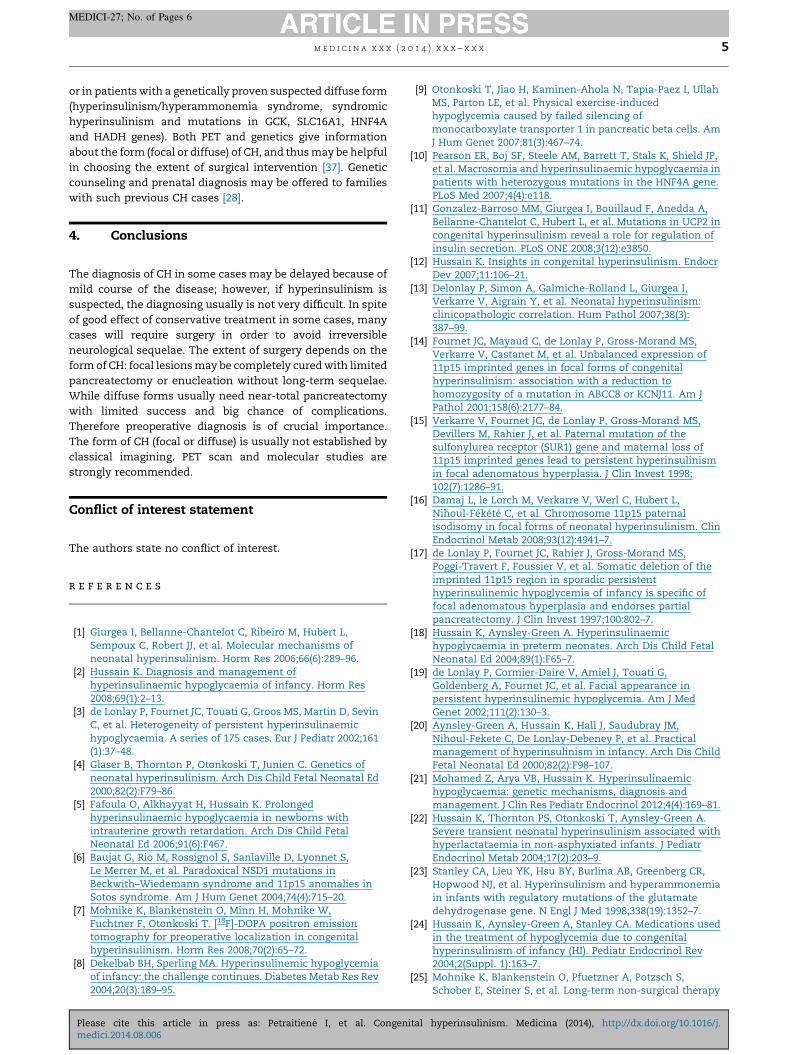
Fig. 1 – During surgery: nodular lesion in the head ofpancreas.
m e d i c i n a x x x ( 2 0 1 4 ) x x x – x x x2
MEDICI-27; No. of Pages 6
2. Case report
We are reporting a case of a boy with normal perinatal history(delivery at 38 weeks of gestation with birth weight (3760 g)and birth length (53 cm) appropriate for gestational age andgender), who was admitted to the intensive care unit at3 months of age because of severe hypoglycemia withgeneralized seizures. At the time of hypoglycemia(0.9 mmol/L) insulin and C-peptide levels were increased(insulin, 15.88 mU/mL; C-peptide, 2.2 ng/mL), leading to thediagnosis of hyperinsulinism. Regular feeding every 2 h withincreased amount of food was sufficient to maintain normo-glycemia. The patient was discharged and an out-patientfollow-up was instituted without any treatment.
The boy was admitted again at 5 months of age because offever, diarrhea, viral upper respiratory tract infection, irrita-bility, accompanied by recurrent episodes of hypoglycemia.The treatment was started with glucose infusions andDiazoxide 10 mg/kg/day with increasing dosage up to 25 mg/kg/day. This treatment was not effective and repeatedepisodes of hypoglycemia were observed 2–3 times a day.Hypoglycemia was accompanied by relatively high insulinsecretion (glycemia, 1.09 mmol/L and insulin, 7.5 mU/mL;glycemia, 1.9 mmol/L and insulin, 4 mU/mL). Hydrochlorothia-zide was added without substantial improvement. Onlycombined treatment with intramuscular injections of Octreo-tide (40 mg/kg/day) maintained glycemia within normal rangefor several days, yet it had to be discontinued because of sideeffects (persistent vomiting, functional invagination). Othermedications (Hydrocortisone and Nifedipine) had no substan-tial effect on glycemic profile. Episodes of hypoglycemiabecame more frequent and intravenous glucose requirementincreased up to 12 mg/kg/min to maintain normoglycemia.Therefore, surgical treatment possibilities and the extent ofoperation (total or partial pancreatectomy) were discussed.The main issue was differentiation between diffuse and focallesion as abdominal MRI showed a normal pancreas and thePET scan was not available in Lithuania at that time. The 95%pancreatectomy was scheduled as there were no pancreaticnodular lesions on MRI, and diffuse nesidioblastosis wassuspected. At laparotomy no nodular lesions in liver or otherabdominal organs were identified. The body and the tail of thepancreas were exposed and examined. No nodular lesionswere identified. Kocher maneuver was used to mobilize theduodenum and the head of the pancreas. At this point a 10 mmnodular lesion was identified at the level of junction of theright gastroepiploic and the middle colic vein (Fig. 1). Thetumor was enucleated and sent for frozen section. Noassociation with the main pancreatic duct was identified. Assoon as the tumor was removed glycemia increased up to11 mmol/L (prior to that the patient was on glucose infusion),and remained at the level of 6–7 mmol/L after glucose infusionwas stopped. Four days later, glycemia was within normalrange without any treatment. The patient was discharged after3 months of hospitalization. The patient was reconsulted atthe age of 6 and 12 months: his psychomotor development wasevaluated as appropriate for age so far. Analysis of all codingand exon/intron boundaries of the KCNJ11 and ABCC8 genes(NM_000525.3, U63421 and L78208) was performed by Sanger
Please cite this article in press as: Petraitienė I, et al. Congenmedici.2014.08.006
sequencing. The boy was found to be heterozygous for anABCC8 nonsense mutation, p.W232*. A second ABCC8 muta-tion has not been found and sequencing analysis of the KCNJ11gene did not identify a change from the normal sequence. Thisresult is consistent with a diagnosis of focal hyperinsulinismdue to a (presumed) paternally inherited ABCC8 mutation.Testing of the boy's father was recommended to confirm hiscarrier status.
3. Literature review
3.1. Epidemiology
Incidence of congenital hyperinsulinism (CH) is reported in 1out of 40 000–50 000 live births in general population [4].Frequency is estimated to be much higher in consanguineousunions (1 in 2500 live births) [4].
3.2. Pathophysiology
Hyperinsulinemic hypoglycemia can be transient, persistentor as an accompanying symptom in several syndromes.Usually, transient hyperinsulinemic hypoglycemia is second-ary (e.g., caused by increased pancreatic b-cells functionbecause of maternal diabetes mellitus) [2]. In these conditionshypoglycemia usually settles within a few days after deliveryand rarely requires treatment with Diazoxide and prolongsseveral months [5]. Hyperinsulinemic hypoglycemia may alsobe present in several overgrowth syndromes (Beckwith–Wiedemann, Perlman, Sotos, Kabuki, Usher, Timothy, Cost-ello, Trisomy 13, mosaic Turner) [2,6].
CH is the most common cause of hypoglycemia in infants[2,7,8]. Mutations of several genes responsible for CH areidentified (Table) [1,2,9–11].
However, in approximately 50% of the cases genesmutations are not established (or are not currently known)[1,2]. The most common causes of CH are ‘‘channelopathies’’,which refer to the pancreatic b-cell ATP-sensitive potassium
ital hyperinsulinism. Medicina (2014), http://dx.doi.org/10.1016/j.
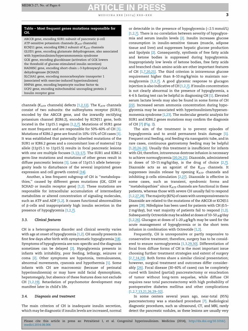
Table – Most frequent genes mutations responsible forCH.
ABCC8 gene, encoding SUR1 subunit of pancreatic b-cellATP-sensitive potassium channels (KATP channels)KCNJ11 gene, encoding KIR6.2 subunit of KATP channelsGLUD1 gene, encoding glutamate dehydrogenase, also associatedwith hyperinsulinism/hyperammonemia syndromeGCK gene, encoding glucokinase (activation of GCK lowersthe threshold of glucose-stimulated insulin secretion)HADHSC gene, encoding short-chain L-3-hydroxyacyl-CoAdehydrogenase (SCHAD)SLC16A1 gene, encoding monocarboxylate transporter 1(associated with exercise-induced hyperinsulinism)HNF4A gene, encoding hepatocyte nuclear factor 4aUCP2 gene, encoding mitochondrial uncoupling protein 2Insulin receptor gene
m e d i c i n a x x x ( 2 0 1 4 ) x x x – x x x 3
MEDICI-27; No. of Pages 6
channels (KATP channels) defects [1,2,12]. The KATP channelsconsist of two subunits: the sulfonylurea receptor (SUR1),encoded by the ABCC8 gene, and the inwardly rectifyingpotassium channel (KIR6.2), encoded by KCNJ11 gene, bothlocated in the 11p15.1 region [1,2,7]. Mutations of SUR1 geneare most frequent and are responsible for 50%–60% of CH [1].Mutations of KIR6.2 gene are found in 10%–15% of CH cases [1].It was established that paternally inherited mutations of theSUR1 or KIR6.2 genes and a concomitant loss of maternal 11pallele (11p15.1 to 11p15.5) results in focal pancreatic lesionswith one ore multiple focuses [1,13–17]. The SUR1 and KIR6.2germ-line mutations and mutations of other genes result indiffuse pancreatic lesions [1]. Loss of 11p15.5 allele heterozy-gosity leads to disturbance of the several imprinted genesexpression and cell growth control [14].
Another, a less frequent subgroup of CH is ‘‘metabolopa-thies,’’ caused by different genes mutations (GK, GDH orSCHAD or insulin receptor gene) [1,2]. These mutations areresponsible for intracellular accumulation of intermediarymetabolites or altered concentration of signaling molecules,such as ATP and ADP [1,2]. It causes functional abnormalitiesof b-cells and inappropriately high insulin secretion in thepresence of hypoglycemia [1,2,7].
3.3. Clinical features
CH is a heterogeneous disorder and clinical severity varieswith age at onset of hypoglycemia [1,2]. CH usually presents infirst few days after birth with symptomatic hypoglycemia [18].Symptoms of hypoglycemia are non-specific and the diagnosissometimes can be delayed [2]. Hypoglycemia presents ininfants with irritability, poor feeding, lethargy, seizures orcoma [2]. Other symptoms are hypotonia, tremulousness,abnormal movements, cyanosis and hypothermia [1]. Someinfants with CH are macrosomic (because of perinatalhyperinsulinemia) or may have mild facial dysmorphism,hepatomegaly, but absence of these features does not excludeCH [1,2,19]. Retardation of psychomotor development maymanifest later in child's life.
3.4. Diagnosis and treatment
The main criterion of CH is inadequate insulin secretion,which may be diagnostic if insulin levels are increased, normal
Please cite this article in press as: Petraitienė I, et al. Congenmedici.2014.08.006
or detectable in the presence of hypoglycemia (<2.5 mmol/L)[1,2,7]. There is no correlation between severity of hypoglyce-mia and serum insulin levels [2]. Insulin increases glucoseconsumption in insulin-sensitive tissues (muscle, adiposetissue and liver) and suppresses hepatic glucose productionand lipolysis [2]. Consequently, synthesis of free fatty acidsand ketone bodies is suppressed during hypoglycemia.Inappropriately low levels of ketone bodies, free fatty acidsand branched chain amino acids are other important featuresof CH [1,7,20,21]. The third criterion is intravenous glucoserequirement higher than 8–10 mg/kg/min to maintain nor-moglycemia [1,2,7]. A good glycemic response to glucagoninjection is also indicative of CH [1,2,7]. If insulin concentrationis not clearly abnormal in the presence of hypoglycemia, a4–6 h fasting test can be helpful in diagnosing CH [1]. Elevatedserum lactate levels may also be found in some forms of CH[22]. Increased serum ammonia concentration during hypo-glycemia may be associated with hyperinsulinism/hyperam-monemia syndrome [1,23]. The molecular genetic analysis forSUR1 and KIR6.2 genes mutations may confirm the diagnosisof CH [7] (Fig. 2).
The aim of the treatment is to prevent episodes ofhypoglycemia and to avoid permanent brain damage [1].Frequent oral feeding, oral feeding via nasogastric tube, and inrare cases, continuous gastrostomy feeding may be helpful[1,20,24–26]. Usually this treatment is insufficient for infantswith CH; therefore, intravenous dextrose infusion is necessaryto achieve normoglycemia [20,24,25]. Diazoxide, administeredin doses of 10–15 mg/kg/day, is the drug of choice [2,7].Diazoxide acts as an agonist of the KATP channels. Itsuppresses insulin release by opening KATP channels andinhibiting b-cells stimulation [2,27]. Diazoxide is effective insome cases, such as transient, syndromic forms and‘‘metabolopathies’’ since KATP channels are functional in thesepatients, whereas those with severe CH usually fail to respond[1,2,27,28]. The majority (82%) of cases with unresponsiveness toDiazoxide are related to the mutations of the ABCC8 or KCNJ11genes [28]. Nifedipine has been used for patients with CH (0.5–2 mg/day), but vast majority of patients fail to respond [1,2].Subsequently Octreotide may be added at doses of 10–50 mg/day[1,2,26]. Glucagon at doses of 1–20 mg/kg/h may be used for theacute management of hypoglycemia or in the short terminfusion in combination with Octreotide [1,2].
Frequently, CH is unresponsive or partly responsive toconservative treatment; therefore, surgery has to be consid-ered to ensure normoglycemia [1,3,29,30]. Differentiation offocal from diffuse forms of CH is the most important issuechoosing further treatment strategies and extent of surgery[2,7,14,29]. Both forms share a similar clinical presentation;however, surgical treatment and outcomes differ consider-ably [26]. Focal disease (30–60% of cases) can be completelycured with limited (partial) pancreatectomy or enucleationof tumor without long-term sequelae, while diffuse CHrequires near total pancreatectomy with high probability ofpostoperative diabetes mellitus and other complications[1,2,7,13,21,26,29–32].
In some centers several years ago, near-total (95%)pancreatectomy was a standard procedure [7]. Radiologicaldiagnostic procedures, such as ultrasound, CT, and MRI, rarelydetect the pancreatic nodules, as these lesions are usually very
ital hyperinsulinism. Medicina (2014), http://dx.doi.org/10.1016/j.
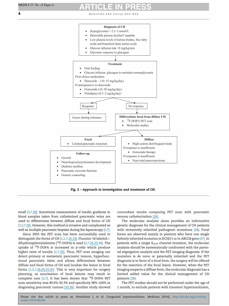
Diagnosis of CH • Hypoglycemia < 2.5–3 mmol/L • Detectable plasma insulin/C-peptide • Low plasma levels of ketone bodies, free fatty
acids and branched chain amino acids • Glucose infusion rate >8 mg/kg/min • Glycemic response to glucagon
Treatment • Oral feeding • Glucose infusion, glucagon to maintain normoglycemia
First choice medication: • Diazoxide – (10–15 mg/kg/day)
If unresponsive to diazoxide: • Octreotide (10–50 mg/kg/day) • Nifedipine (0.5–2 mg/kg/day)
Response No response
Assess fasting tolerance Differentiate focal from diffuse CH • 18 F-DOPA PET scan • Molecular studies
Focal • Limited pancreatic resection
Diffuse • High-calorie diet/frequent feeds
If response is insufficient: • Octreotide therapy
If response is insufficient: • Near-total pancreatectomy
Follow-up • Growth • Neurological/psychomotor development • Diabetes mellitus • Pancreatic exocrine function • Genetic counseling
Fig. 2 – Approach to investigation and treatment of CH.
m e d i c i n a x x x ( 2 0 1 4 ) x x x – x x x4
MEDICI-27; No. of Pages 6
small [1,7,26]. Sometimes measurement of insulin gradients inblood samples taken from catheterized pancreatic veins areused to differentiate between diffuse and focal forms of CH[1,2,7,26]. However, this method is invasive and complicated aswell as multiple pancreatic biopsies during the laparoscopy [2,7].
Since 2003 the PET scan has been successfully used todistinguish the forms of CH [1,2,7,26,33]. Fluorine-18 labeled L-dihydroxyphenylalanine (18F-DOPA) is used [2,7,26,33,34]. Theuptake of 18F-DOPA is increased in b-cells which producehigher rates of insulin [1,7,35]. Thus, PET scan imaging candetect primary or metastatic pancreatic tumors, hyperfunc-tional pancreatic islets and allows differentiate betweendiffuse and focal forms of CH and localize the lesion in focalforms [1,2,7,26,29,33,35]. This is very important for surgeryplanning, as enucleation of local lesions may result incomplete cure [2,7]. It has been shown that 18F-DOPA PETscan sensitivity was 89.0%–92.3% and specificity 98%–100% indiagnosing pancreatic tumors [33,36]. Another study showed
Please cite this article in press as: Petraitienė I, et al. Congenmedici.2014.08.006
concordant results comparing PET scan with pancreaticvenous catheterization [26].
The molecular analysis alone provides an informativegenetic diagnosis for the clinical management of CH patientswith recessively inherited pathogenic mutations [28]. Focalforms are observed mainly in patients who have one singlefatherly inherited mutation in KCNJ11 or in ABCC8 gene [37]. Inpatients with a single KATP channel mutation, the molecularanalysis should be systematically confronted with the paren-tal segregation analysis and the PET imaging diagnosis. If themutation is de novo or paternally inherited and the PETdiagnosis is in favor of a focal form, the surgery will be offeredfor the resection of the focal lesion. However, when the PETimaging suspects a diffuse form, the molecular diagnosis has alimited added value for the clinical management of CHpatients [28].
The PET studies should not be performed under the age of1 month, to exclude patients with transient hyperinsulinism,
ital hyperinsulinism. Medicina (2014), http://dx.doi.org/10.1016/j.
m e d i c i n a x x x ( 2 0 1 4 ) x x x – x x x 5
MEDICI-27; No. of Pages 6
or in patients with a genetically proven suspected diffuse form(hyperinsulinism/hyperammonemia syndrome, syndromichyperinsulinism and mutations in GCK, SLC16A1, HNF4Aand HADH genes). Both PET and genetics give informationabout the form (focal or diffuse) of CH, and thus may be helpfulin choosing the extent of surgical intervention [37]. Geneticcounseling and prenatal diagnosis may be offered to familieswith such previous CH cases [28].
4. Conclusions
The diagnosis of CH in some cases may be delayed because ofmild course of the disease; however, if hyperinsulinism issuspected, the diagnosing usually is not very difficult. In spiteof good effect of conservative treatment in some cases, manycases will require surgery in order to avoid irreversibleneurological sequelae. The extent of surgery depends on theform of CH: focal lesions may be completely cured with limitedpancreatectomy or enucleation without long-term sequelae.While diffuse forms usually need near-total pancreatectomywith limited success and big chance of complications.Therefore preoperative diagnosis is of crucial importance.The form of CH (focal or diffuse) is usually not established byclassical imagining. PET scan and molecular studies arestrongly recommended.
Conflict of interest statement
The authors state no conflict of interest.
r e f e r e n c e s
[1] Giurgea I, Bellanne-Chantelot C, Ribeiro M, Hubert L,Sempoux C, Robert JJ, et al. Molecular mechanisms ofneonatal hyperinsulinism. Horm Res 2006;66(6):289–96.
[2] Hussain K. Diagnosis and management ofhyperinsulinaemic hypoglycaemia of infancy. Horm Res2008;69(1):2–13.
[3] de Lonlay P, Fournet JC, Touati G, Groos MS, Martin D, SevinC, et al. Heterogeneity of persistent hyperinsulinaemichypoglycaemia. A series of 175 cases. Eur J Pediatr 2002;161(1):37–48.
[4] Glaser B, Thornton P, Otonkoski T, Junien C. Genetics ofneonatal hyperinsulinism. Arch Dis Child Fetal Neonatal Ed2000;82(2):F79–86.
[5] Fafoula O, Alkhayyat H, Hussain K. Prolongedhyperinsulinaemic hypoglycaemia in newborns withintrauterine growth retardation. Arch Dis Child FetalNeonatal Ed 2006;91(6):F467.
[6] Baujat G, Rio M, Rossignol S, Sanlaville D, Lyonnet S,Le Merrer M, et al. Paradoxical NSD1 mutations inBeckwith–Wiedemann syndrome and 11p15 anomalies inSotos syndrome. Am J Hum Genet 2004;74(4):715–20.
[7] Mohnike K, Blankenstein O, Minn H, Mohnike W,Fuchtner F, Otonkoski T. [18F]-DOPA positron emissiontomography for preoperative localization in congenitalhyperinsulinism. Horm Res 2008;70(2):65–72.
[8] Dekelbab BH, Sperling MA. Hyperinsulinemic hypoglycemiaof infancy: the challenge continues. Diabetes Metab Res Rev2004;20(3):189–95.
Please cite this article in press as: Petraitienė I, et al. Congenmedici.2014.08.006
[9] Otonkoski T, Jiao H, Kaminen-Ahola N, Tapia-Paez I, UllahMS, Parton LE, et al. Physical exercise-inducedhypoglycemia caused by failed silencing ofmonocarboxylate transporter 1 in pancreatic beta cells. AmJ Hum Genet 2007;81(3):467–74.
[10] Pearson ER, Boj SF, Steele AM, Barrett T, Stals K, Shield JP,et al. Macrosomia and hyperinsulinaemic hypoglycaemia inpatients with heterozygous mutations in the HNF4A gene.PLoS Med 2007;4(4):e118.
[11] Gonzalez-Barroso MM, Giurgea I, Bouillaud F, Anedda A,Bellanne-Chantelot C, Hubert L, et al. Mutations in UCP2 incongenital hyperinsulinism reveal a role for regulation ofinsulin secretion. PLoS ONE 2008;3(12):e3850.
[12] Hussain K. Insights in congenital hyperinsulinism. EndocrDev 2007;11:106–21.
[13] Delonlay P, Simon A, Galmiche-Rolland L, Giurgea I,Verkarre V, Aigrain Y, et al. Neonatal hyperinsulinism:clinicopathologic correlation. Hum Pathol 2007;38(3):387–99.
[14] Fournet JC, Mayaud C, de Lonlay P, Gross-Morand MS,Verkarre V, Castanet M, et al. Unbalanced expression of11p15 imprinted genes in focal forms of congenitalhyperinsulinism: association with a reduction tohomozygosity of a mutation in ABCC8 or KCNJ11. Am JPathol 2001;158(6):2177–84.
[15] Verkarre V, Fournet JC, de Lonlay P, Gross-Morand MS,Devillers M, Rahier J, et al. Paternal mutation of thesulfonylurea receptor (SUR1) gene and maternal loss of11p15 imprinted genes lead to persistent hyperinsulinismin focal adenomatous hyperplasia. J Clin Invest 1998;102(7):1286–91.
[16] Damaj L, le Lorch M, Verkarre V, Werl C, Hubert L,Nihoul-Fékété C, et al. Chromosome 11p15 paternalisodisomy in focal forms of neonatal hyperinsulinism. ClinEndocrinol Metab 2008;93(12):4941–7.
[17] de Lonlay P, Fournet JC, Rahier J, Gross-Morand MS,Poggi-Travert F, Foussier V, et al. Somatic deletion of theimprinted 11p15 region in sporadic persistenthyperinsulinemic hypoglycemia of infancy is specific offocal adenomatous hyperplasia and endorses partialpancreatectomy. J Clin Invest 1997;100:802–7.
[18] Hussain K, Aynsley-Green A. Hyperinsulinaemichypoglycaemia in preterm neonates. Arch Dis Child FetalNeonatal Ed 2004;89(1):F65–7.
[19] de Lonlay P, Cormier-Daire V, Amiel J, Touati G,Goldenberg A, Fournet JC, et al. Facial appearance inpersistent hyperinsulinemic hypoglycemia. Am J MedGenet 2002;111(2):130–3.
[20] Aynsley-Green A, Hussain K, Hall J, Saudubray JM,Nihoul-Fekete C, De Lonlay-Debeney P, et al. Practicalmanagement of hyperinsulinism in infancy. Arch Dis ChildFetal Neonatal Ed 2000;82(2):F98–107.
[21] Mohamed Z, Arya VB, Hussain K. Hyperinsulinaemichypoglycaemia: genetic mechanisms, diagnosis andmanagement. J Clin Res Pediatr Endocrinol 2012;4(4):169–81.
[22] Hussain K, Thornton PS, Otonkoski T, Aynsley-Green A.Severe transient neonatal hyperinsulinism associated withhyperlactataemia in non-asphyxiated infants. J PediatrEndocrinol Metab 2004;17(2):203–9.
[23] Stanley CA, Lieu YK, Hsu BY, Burlina AB, Greenberg CR,Hopwood NJ, et al. Hyperinsulinism and hyperammonemiain infants with regulatory mutations of the glutamatedehydrogenase gene. N Engl J Med 1998;338(19):1352–7.
[24] Hussain K, Aynsley-Green A, Stanley CA. Medications usedin the treatment of hypoglycemia due to congenitalhyperinsulinism of infancy (HI). Pediatr Endocrinol Rev2004;2(Suppl. 1):163–7.
[25] Mohnike K, Blankenstein O, Pfuetzner A, Potzsch S,Schober E, Steiner S, et al. Long-term non-surgical therapy
ital hyperinsulinism. Medicina (2014), http://dx.doi.org/10.1016/j.

m e d i c i n a x x x ( 2 0 1 4 ) x x x – x x x6
MEDICI-27; No. of Pages 6
of severe persistent congenital hyperinsulinism withglucagon. Horm Res 2008;70(1):59–64.
[26] Ribeiro MJ, Boddaert N, Bellanne-Chantelot C, Bourgeois S,Valayannopoulos V, Delzescaux T, et al. The added value of[18F]fluoro-L-DOPA PET in the diagnosis of hyperinsulinismof infancy: a retrospective study involving 49 children. Eur JNucl Med Mol Imaging 2007;34(12):2120–8.
[27] Suchi M, MacMullen CM, Thornton PS, Adzick NS, GangulyA, Ruchelli ED, et al. Molecular and immunohistochemicalanalyses of the focal form of congenital hyperinsulinism.Mod Pathol 2006;19(1):122–9.
[28] Bellanne-Chantelot C, Saint-Martin C, Ribeiro MJ, Vaury C,Verkarre V, Arnoux JB, et al. ABCC8 and KCNJ11 molecularspectrum of 109 patients with diazoxide-unresponsivecongenital hyperinsulinism. J Med Genet 2010;47(11):752–9.
[29] Brunelle F, Ribeiro M, Boddaert N, Nihoul-Fekete C,Jaubert F, Rahier J, et al. Hyperinsulinism in children: newconcepts – the role of imaging. Bull Acad Natl Med 2008;192(1):59–70. discussion 1–2.
[30] Pierro A, Nah SA. Surgical management of congenitalhyperinsulinism of infancy. Semin Pediatr Surg 2011;20(1):50–3.
[31] Sperling MA. PET scanning for infants with HHI: a smallstep for affected infants, a giant leap for the field. J Pediatr2007;150(2):122–4.
Please cite this article in press as: Petraitienė I, et al. Congenmedici.2014.08.006
[32] Fekete CN, de Lonlay P, Jaubert F, Rahier J, Brunelle F,Saudubray JM. The surgical management of congenitalhyperinsulinemic hypoglycemia in infancy. J Pediatr Surg2004;39(3):267–9.
[33] Treglia G, Mirk P, Giordano A, Rufini V. Diagnosticperformance of fluorine-18-dihydroxyphenylalaninepositron emission tomography in diagnosing and localizingthe focal form of congenital hyperinsulinism: a meta-analysis. Pediatr Radiol 2012;42(11):1372–9.
[34] Otonkoski T, Nanto-Salonen K, Seppanen M, Veijola R,Huopio H, Hussain K, et al. Noninvasive diagnosis offocal hyperinsulinism of infancy with [18F]-DOPApositron emission tomography. Diabetes 2006;55(1):13–8.
[35] Yang J, Yuan L, Meeks JK, Zhang N, Li C, Hao R. 18F-DOPApositron emission tomography/computed tomographyapplication in congenital hyperinsulinism. J PediatrEndocrinol Metab 2012;25(7/8):619–22.
[36] Becherer A, Szabo M, Karanikas G, Wunderbaldinger P,Angelberger P, Raderer M, et al. Imaging of advancedneuroendocrine tumors with (18)F-FDOPA PET. J Nucl Med2004;45(7):1161–7.
[37] Arnoux JB, de Lonlay P, Ribeiro MJ, Hussain K, Blankenstein O,Mohnike K, et al. Congenital hyperinsulinism. Early Hum Dev2010;86(5):287–94.
ital hyperinsulinism. Medicina (2014), http://dx.doi.org/10.1016/j.