Conflict of Interest Disclosure Statement
16
1 Crest ® Oral-B ® at dentalcare.com Continuing Education Course, February 3, 2011 Oropharyngeal Candidiasis: Etiology, Epidemiology, Clinical Manifestations, Diagnosis, and Treatment This continuing education course is intended for dentists and dental hygienists. It presents the etiology, epidemiology, clinical manifestations, diagnosis, and treatment of candidal infections relevant to dentistry. Conflict of Interest Disclosure Statement • Dr. Terézhalmy does consulting work for P&G. • Dr. Huber reports no conflicts of interest associated with this course. ADA CERP The Procter & Gamble Company is an ADA CERP Recognized Provider. ADA CERP is a service of the American Dental Association to assist dental professionals in identifying quality providers of continuing dental education. ADA CERP does not approve or endorse individual courses or instructors, nor does it imply acceptance of credit hours by boards of dentistry. Concerns or complaints about a CE provider may be directed to the provider or to ADA CERP at: http://www.ada.org/prof/ed/ce/cerp/index.asp Overview Candidal infections commonly affect the dental profession’s anatomical area of responsibility and the diagnosis and management of such infections, to a great extent, fall in the purview of oral healthcare providers. To administer competent care to patients with candidal infections, clinicians must understand the disease, its treatment, the impact the disease or its treatment may have on the patient, and the extent to which the presence of a candidal infection may impact on the clinical process. Géza T. Terézhalmy, DDS, MA; Michaell A. Huber, DDS Continuing Education Units: 2 hours
-
Upload
independent -
Category
Documents
-
view
0 -
download
0
Transcript of Conflict of Interest Disclosure Statement

1
Crest® Oral-B® at dentalcare.com Continuing Education Course, February 3, 2011
Oropharyngeal Candidiasis: Etiology, Epidemiology, Clinical Manifestations,
Diagnosis, and Treatment
This continuing education course is intended for dentists and dental hygienists. It presents the etiology, epidemiology, clinical manifestations, diagnosis, and treatment of candidal infections relevant to dentistry.
Conflict of Interest Disclosure Statement• Dr. Terézhalmy does consulting work for P&G.• Dr. Huber reports no conflicts of interest associated with this course.
ADA CERPThe Procter & Gamble Company is an ADA CERP Recognized Provider.
ADA CERP is a service of the American Dental Association to assist dental professionals in identifying quality providers of continuing dental education. ADA CERP does not approve or endorse individual courses or instructors, nor does it imply acceptance of credit hours by boards of dentistry.
Concerns or complaints about a CE provider may be directed to the provider or to ADA CERP at:http://www.ada.org/prof/ed/ce/cerp/index.asp
OverviewCandidal infections commonly affect the dental profession’s anatomical area of responsibility and the diagnosis and management of such infections, to a great extent, fall in the purview of oral healthcare providers. To administer competent care to patients with candidal infections, clinicians must understand the disease, its treatment, the impact the disease or its treatment may have on the patient, and the extent to which the presence of a candidal infection may impact on the clinical process.
Géza T. Terézhalmy, DDS, MA; Michaell A. Huber, DDSContinuing Education Units: 2 hours

2
Crest® Oral-B® at dentalcare.com Continuing Education Course, February 3, 2011
Course Contents• Introduction• Etiology and Epidemiology• Clinical Manifestations of Oropharyngeal
Candidiasis • Pseudomembranous Candidiasis • Erythematous Candidiasis • Hyperplastic Candidiasis • Candida-associated Denture Stomatitis • Other Candidal Infections
• Diagnosis• Preventive and Therapeutic Strategies • Prevention • Antifungal Chemotherapy: Mild
Oropharyngeal Candidiasis Nystatin Oral Suspension Clotrimazole Troches • Antifungal Chemotherapy: Moderate to
Severe Oropharyngeal Infections Fluconazole • Antifungal Chemotherapy: Fluconazole-
refractory Disease Itraconazole, Posaconazole, and
Voriconazole Caspofungin, Micafungin, and
Anidulafungin Amphotericin B
• Conclusion• Course Test• References• About the Authors
IntroductionFungi are free-living, eukaryotic organisms that exist as yeasts (round fungi), molds (filamentous fungi), or a combination of these two (dimorphic fungi). While there are over 100,000 fungal
species, only a few are intrinsically pathogenic in humans. Most pathogenic fungi associated with human disease are saprophytic members of the soil microbial flora, i.e., they live on decaying organic matter. For these organisms, the lungs are the most common portals of entry. Some fungi are commensal members of the normal human flora, i.e., they obtain benefit without causing harm to their host. In humans, these are typically found on oral, vaginal, and gastrointestinal mucosa; or as residents of the skin and at times of the respiratory epithelium. In persons with altered homeostatic mechanisms, commensal organisms may become pathogenic. Mycoses of interest to oral healthcare providers are presented in Table 1.
The diagnosis and treatment of most mycoses is the responsibility of physicians. Infections caused by the candida spp. represent the most common mycoses of the oropharyngeal area and the diagnosis and treatment of some of these infections fall, to a great extent, in the purview of the dental profession. To provide timely and competent care to patients with oropharyngeal candidiasis, clinicians must understand the disease, its treatment, the impact the disease or its treatment may have on the patient, and the extent to which the presence of a candidal infection may impact on the clinical process.
Etiology and EpidemiologyAs noted earlier, the Candida spp. are commensal opportunistic organisms typically found on oropharyngeal tissues.1-6 The reported carriage rate in healthy individuals varies from 15 to 75 percent, depending on the population
Learning ObjectivesUpon the completion of this course, the dental professional should be able to:• Discuss the etiology and epidemiology of oropharyngeal candidiasis.• Recognize the clinical manifestations of oropharyngeal candidiasis. • Pseudomembranous candidiasis. • Erythematous candidiasis. • Hyperplastic candidiasis. • Candida-associated denture stomatitis. • Other candidal infections (medial rhomboid glossitis, angular cheilitis, keratinized primary lesions with
candidal infections, candidal esophagitis, and chronic mucocutaneous candidiasis).• Diagnose oropharyngeal candidiasis.• Develop strategies for the treatment and prevention of oropharyngeal candidiasis.

3
Crest® Oral-B® at dentalcare.com Continuing Education Course, February 3, 2011
sampled and the sensitivity of the sampling technique.3 Candida spp. are seldom capable of initiating serious infections in immunocompetent patients. Since the pathogenesis of fungal infections is based on an interplay between host homeostatic mechanisms and the pathogenicity of the Candida spp., patients with acquired and therapeutic immunosuppression (HIV infection, cytotoxic drugs, corticosteroids), endocrinopathies (diabetes mellitus, thymoma, polyendocrinopathy syndrome type 1, hypoparathyroidism, hypoadrenalism, pregnancy with secondary infection of the infant), nutritional deficiencies, a high carbohydrate diet, extended use of broad-spectrum antibacterial agents, qualitative and quantitative changes in salivary flow (drug-induced, radiotherapy, Sjogren’s syndrome), poor oral hygiene, the presence of dental prostheses, age, and smoking are at increased risk of carriage and infection.7-17
In one recent study, 81.1% of 122 consecutive HIV-infected patients on antiretroviral therapy were colonized by Candida spp. and 33.3% had clinical infection.17 In another study, among 210 HIV-positive patients at the time of diagnosis, 62% had clinical oropharyngeal candidiasis.7 Other investigators found (1) that diabetic subjects have significantly higher candidal colony forming units (CFU) than nondiabetic subjects, (2) a significant positive correlation between salivary glucose levels and candidal CFU, and (3) clinical evidence
of oropharyngeal candidiasis in 4% of uncontrolled diabetic patients with CFU >8,000/mL.15
C. albicans is the most common isolate from both healthy individuals and from those at risk of opportunistic infections. However, an increase in the prevalence of other members of the Candida spp. (Table 1) has been reported and a person may harbor several members of the Candida spp. at a time.2,5,17,18 Virulence of the Candida spp., i.e., the ability to cause invasive mycosis, is predicated on such factors as their ability to adhere to epithelial and endothelial cells, their ability to change phenotype (undergo yeast-to-hyphae transformation), and to secrete proteases that facilitate cellular penetration.19 C. albicans appears to be the most virulent member of the Candida spp. This virulence is attributed largely to C. albicans’ ability to express a number of structures that mediate epithelial adherence that provides protection from the continuous flushing action of saliva.3,4 In addition, it is postulated that the hyphal form of C. albicans is important not only for epithelial tissue penetration; but also for escape, following internalization, from phagocytic cells.3,20,21
Clinical Manifestations of Oropharyngeal CandidiasisOropharyngeal candidiasis may appear in a variety of clinical forms. It is often asymptomatic, but patients may relate a burning sensation

4
Crest® Oral-B® at dentalcare.com Continuing Education Course, February 3, 2011
and an altered sense of taste. A classification system based on clinical criteria provides a clear distinction between primary oropharyngeal candidiasis and the oral manifestations of systemic mucocutaneous candidiasis.22
Pseudomembranous CandidiasisPseudomembranous candidiasis (PC) is characterized by the presence of a thick whitish/yellow velvety pseudomembrane or individual plaques on oropharyngeal tissues, which may be wiped away, leaving an erythematous, painful base that readily bleeds (Figures 1-4).1,23,24 The buccal mucosa is the most frequent site, but all oropharyngeal tissues may be affected. PC is frequently observed in neonates, and in patients with acquired or therapeutic immunosuppression (HIV infection, cytotoxic
drugs, corticosteroids).7,14,17 In immunocompetent persons, PC is most likely a consequence of the extended use of broad-spectrum antibacterial agents.24
Erythematous CandidiasisErythematous candidiasis (EC), acute and chronic, appears as a red patch or patches usually on the palate or dorsum of the tongue with concurrent loss of the papillae (Figures 5 & 6).23,24 EC may affect other oral sites such as the buccal mucosa and is often asymptomatic. It is frequently observed with acquired or therapeutic immunosuppression (HIV infection, cytotoxic drugs, corticosteroids).7,17,22 In immunocompetent patients, EC is most likely a consequence of the extended use of broad-spectrum antibacterial agents.22
Figure 3. Pseudomembranous candidiasis as a consequence of chronic use of inhaled corticosteroids in the management of asthma.
Figure 1. Pseudomembranous candidiasis as a consequence of the extended use of a broad-spectrum antibacterial agent.
Figure 4. Pseudomembranous candidiasis as a consequence of chronic use of inhaled corticosteroids in the management of asthma.
Figure 2. Pseudomembranous candidiasis as a consequence of the extended use of a broad-spectrum antibacterial agent.

5
Crest® Oral-B® at dentalcare.com Continuing Education Course, February 3, 2011
Hyperplastic CandidiasisHyperplastic candidiasis is an uncommon clinical infection characterized by persistent (chronic), white papules or plaques (Figure 7), which typically affect the buccal mucosa at the commissural area of a smoker and, less frequently, the tongue (Figure 8).1,22-24 This form infiltrates the epithelium, but when stripped away does not cause severe pain or bleeding. Also known as candidal leukoplakia, it is associated with a malignant transformation rate of up to 15%.1,22,23,25
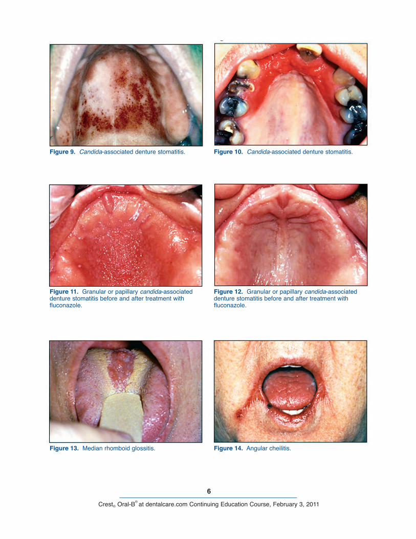
Candida-associated Denture Stomatitis Candida-associated denture stomatitis appears as an erythematous area beneath a denture-bearing surface. These lesions are asymptomatic and are frequently associated with poor oral hygiene.1,22,25 The clinical presentation ranges from areas of focal erythema (Figure 9), to generalized erythema (Figure 10), to granular or papillary
erythema (Figures 11 & 12). Candida-associated denture stomatitis is commonly observed with angular cheilitis.
Other Candidal InfectionsMedian rhomboid glossitis appears as an elliptical or rhomboid erythematous area of depapillation confined to the dorsal aspect of the tongue located anterior to the circumvallate papillae (Figure 13).22,25 Angular cheilitis appears as erythematous fissures at the commissures of the lips. It represents a mixed infection of C. albicans and coagulase-positive Staphylococcus aureus. Predisposing conditions include immunosuppression, poor oral hygiene, a decrease in the intermaxillary space, and nutritional deficiencies (Figure 14).1,22
Keratinized primary lesions with candidal infection include leukoplakia, lichen planus, and lupus erythematosus.1,26 The presence
Figure 5. Erythematous candidiasis before and after treatment with nystatin oral suspension.
Figure 7. Hyperplastic candidiasis (candidal leukoplakia) of the buccal mucosa and tongue as a consequence of heavy smoking.
Figure 6. Erythematous candidiasis before and after treatment with nystatin oral suspension.
Figure 8. Hyperplastic candidiasis (candidal leukoplakia) of the buccal mucosa and tongue as a consequence of heavy smoking.
6
Crest® Oral-B® at dentalcare.com Continuing Education Course, February 3, 2011
Figure 9. Candida-associated denture stomatitis.
Figure 11. Granular or papillary candida-associated denture stomatitis before and after treatment with fluconazole.
Figure 13. Median rhomboid glossitis.
Figure 10. Candida-associated denture stomatitis.
Figure 12. Granular or papillary candida-associated denture stomatitis before and after treatment with fluconazole.
Figure 14. Angular cheilitis.
7
Crest® Oral-B® at dentalcare.com Continuing Education Course, February 3, 2011
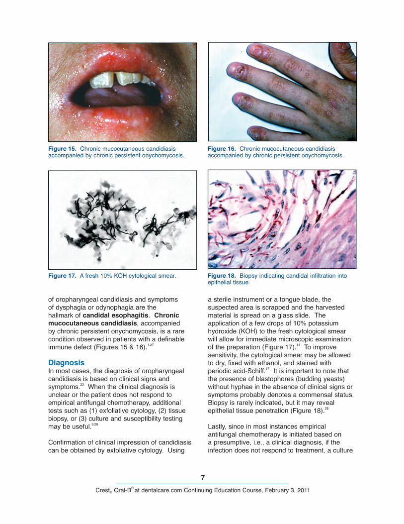
of oropharyngeal candidiasis and symptoms of dysphagia or odynophagia are the hallmark of candidal esophagitis. Chronic mucocutaneous candidiasis, accompanied by chronic persistent onychomycosis, is a rare condition observed in patients with a definable immune defect (Figures 15 & 16).1,27
DiagnosisIn most cases, the diagnosis of oropharyngeal candidiasis is based on clinical signs and symptoms.22 When the clinical diagnosis is unclear or the patient does not respond to empirical antifungal chemotherapy, additional tests such as (1) exfoliative cytology, (2) tissue biopsy, or (3) culture and susceptibility testing may be useful.9,28
Confirmation of clinical impression of candidiasis can be obtained by exfoliative cytology. Using
a sterile instrument or a tongue blade, the suspected area is scrapped and the harvested material is spread on a glass slide. The application of a few drops of 10% potassium hydroxide (KOH) to the fresh cytological smear will allow for immediate microscopic examination of the preparation (Figure 17).14 To improve sensitivity, the cytological smear may be allowed to dry, fixed with ethanol, and stained with periodic acid-Schiff.17 It is important to note that the presence of blastophores (budding yeasts) without hyphae in the absence of clinical signs or symptoms probably denotes a commensal status. Biopsy is rarely indicated, but it may reveal epithelial tissue penetration (Figure 18).28
Lastly, since in most instances empirical antifungal chemotherapy is initiated based on a presumptive, i.e., a clinical diagnosis, if the infection does not respond to treatment, a culture
Figure 15. Chronic mucocutaneous candidiasis accompanied by chronic persistent onychomycosis.
Figure 17. A fresh 10% KOH cytological smear.
Figure 16. Chronic mucocutaneous candidiasis accompanied by chronic persistent onychomycosis.
Figure 18. Biopsy indicating candidal infiltration into epithelial tissue.

8
Crest® Oral-B® at dentalcare.com Continuing Education Course, February 3, 2011
and susceptibility test may provide a definitive diagnosis and identify the presence of resistant organisms.17 More rapid, nonculture-based methods for early diagnosis, such as polymerase chain reaction (PCR), western blot, antigen detection, and identification of fungal metabolites are still considered investigational.
Preventive and Therapeutic StrategiesFungal and human cells, because of their phylogenetic similarity are both eukaryotic and have homologous metabolic pathways for energy production, protein synthesis and cell division. Consequently, only a small number of unique targets for antifungal agents have been identified. One such target is ergosterol, an essential structural and functional component of fungal plasma membranes. This sterol is structurally different from cholesterol, the predominant sterol found in mammalian cell membranes. Another unique target for antifungal chemotherapy is the fungal cell wall. The cell wall contains a number of structural proteins, including glucan, absent in mammalian cells. A third target is fungal adhesion. Adhesion to host cells is mediated by binding of a fungal adhesion to a host cell receptor. Following successful adhesion, pathogens are able to invade the colonized
surface, proliferate in deep tissue, and potentially reach the systemic circulation. Compounds that antagonize adhesive interactions between fungal and mammalian cells are currently under investigation.
The current mainstay of oropharyngeal candidal chemotherapy are (1) drugs that inhibit ergosterol synthesis, i.e., azoles; (2) drugs that disrupt fungal plasma membrane structure and function by binding to ergosterol, i.e., polyenes; and (3) drugs that inhibit the synthesis of β (1, 3)-D-glucan, an essential component of the fungal cell wall, i.e., echinocandins (Figure 19).29 Two other antifungal agents with some clinical utility include flucytosine and griseofulvin. Flucytosine is converted to 5-flurouracil (5-FU), which inhibits thymidylate syntase and prevents DNA synthesis. While the Candida spp. are susceptible to flucytosine, 5-FU is cytotoxic in humans. Dose-dependent toxicity includes bone marrow suppression and hepatic dysfunction. Griseofulvin, inhibits the assembly of microtubules and the activity of accessory proteins essential for the formation of the mitotic spindle. Following oral administration, griseofulvin accumulates in the keratinized layer of cells, making it useful against dermatophytes.
Figure 19. Mechanisms of action of antifungal agents.

9
Crest® Oral-B® at dentalcare.com Continuing Education Course, February 3, 2011
through pore-forming mechanisms increase membrane permeability, effects leakage of essential cellular components, and promotes cell death. To reduce the risk of relapse, treatment generally should be continued for at least 48 hours after the elimination of all signs and symptoms associated with the infection. The oral suspension may also be used as holding solution for prostheses when they are removed from the oral cavity.31 Common adverse effects include contact mucositis and Stevens-Johnson syndrome. Serious adverse effects with nystatin oral suspension are remote as the formulation is not absorbed systemically.
Clotrimazole TrochesClotrimazole is an azole antifungal agent.29 It blocks 14α-sterol demethylase, a fungus-specific cytochrome P450 enzyme that initiates the conversion of lanosterol to ergosterol. This leads to structural and functional plasma membrane damage and cell death. Clotrimazole troches may be effective in the treatment of mild oropharyngeal candidiasis refractory to nystatin. However, since clotrimazole troches contain sucrose, their long-term use may be a problem in caries-prone patients. Common adverse effects with clotrimazole include pruritus and a burning sensation. Serious adverse effects (see
PreventionAppropriate medical treatment of the many predisposing systemic factors and local measures such as meticulous oral hygiene, management of xerostomia, and the maintenance of optimally functioning and clean prostheses may prevent or minimize the incidence of clinical oropharyngeal candidiasis. These measures should include proper brushing of all oral tissues and all surfaces of prostheses, removing prostheses at regular intervals to allow for normal circulation in the supporting tissues, and periodic evaluation of prostheses for proper tissue adaption. For denture-related candidiasis (see below), disinfection of the denture, in addition to antifungal chemotherapy is recommended.30
Antifungal Chemotherapy: Mild Oropharyngeal CandidiasisNystatin oral suspension and clotrimazole troches are the recommended drugs for the treatment of mild (uncomplicated) oropharyngeal candidiasis (Table 2).27,30 Initially, most patients respond to these agents, but relapses occur more frequently than with fluconazole (see below).27
Nystatin Oral SuspensionNystatin is a polyene antifungal agent.29 It binds to ergosterol in fungal plasma membrane and

10
Crest® Oral-B® at dentalcare.com Continuing Education Course, February 3, 2011
three echinocandins appear to be similar in their efficacy. Common adverse effects include pruritus, rash, gastrointestinal disturbances, headache, and fever. Since human cells lack a cell wall and enzymes involved in glucan synthesis are absent, the echinocandins have no serious adverse effect in humans.
Amphotericin BAmphotericin B is a polyene antifungal agents.29 It binds to ergosterol in fungal plasma membrane and through pore-forming mechanism increases membrane permeability, effect leakage of essential cellular components and promotes cell death. Amphotericin B is an important drug in the treatment of systemic candidal infections and it is the preferred drug for the treatment of deep fungal infections during pregnancy (Table 2).27,30 Common adverse effects include gastrointestinal disturbances and weight loss. Serious adverse effects associated with amphotericin B include (1) an immediate systemic reaction or “cytokine storm” (release of TNF-α and IL-1), characterized by fever, chills, and hypotension; (2) renal toxicity, characterized by afferent arteriole constriction leading to renal ischemia; and (3) hematologic toxicity, characterized by anemia secondary to decreased production of erythropoietin. The newer lipid formulations of amphotericin B (amphotec, abelcet, and ambisone) appear to limit renal toxicity.
ConclusionAdvances in medical and pharmacological treatment have resulted in greater life expectancy for patients with acquired or therapeutic immunosuppression, certain metabolic disorders, and other systemic conditions that predispose patients to oropharyngeal and systemic candidiasis. Oral healthcare providers must recognize the clinical manifestations of oropharyngeal candidiasis, and must be familiar with antifungal treatment strategies and patient management guidelines. Prompt treatment with an appropriate antifungal agent can eliminate infection and prevent systemic dissemination. This is of critical importance particularly in immunocompromised patients. Practitioners must educate patients to improve treatment compliance and monitor patients’ response to antifungal therapy. If clinical response to first-line antifungal therapy is not optimal, referral to a physician is indicated.
fluconazole below) with clotrimazole troches are remote as the formulation is minimally absorbed.
Antifungal Chemotherapy: Moderate to Severe Oropharyngeal Infections
FluconazoleFluconazole is an azole antifungal agent.29 It blocks 14α-sterol demethylase (the enzyme responsible for the demethylation of lanosterol to egrosterol); and promotes structural and functional plasma membrane damage, and cell death. Oral fluconazole is recommended for the treatment of moderate-to-severe oropharyngeal candidiasis (Table 2).27,30 Its clinical activity is well established against most candida species (C. krusei is intrinsically resistant and C. glabrata is increasingly resistant). Following oral administration fluconazole is well absorbed (nearly 100% bioavailability) and diffuses freely into saliva. Unfortunately, the azoles are not entirely selective for the 14α-sterol demethylase enzyme. They variably inhibit hepatic cytochrome P450 enzymes, and are responsible for many drug-drug interactions. These interactions are less likely with fluconazole than with itraconazole, voriconazole, and posaconazole (see below). Common adverse effects include nausea, vomiting, diarrhea, and abdominal pain. Hepatotoxicity is a rare serious adverse effect of all systemic azole antifungal agents.
Antifungal Chemotherapy: Fluconazole-refractory Disease
Itraconazole, Posaconazole, and VoriconazoleThe azole antifungal agents itraconazole, posaconazole, and voriconazole appear to have a broader spectrum of activity than fluconazole (see above). Fluconazole-refractory infections should be treated initially with itraconazole solution or posaconazole suspension (Table 2).27,30 Voriconazole is recommended when treatment with other azole antifungal agents has failed (Table 2).27,30
Caspofungin, Micafungin, and AnidulafunginCaspofungin, micafungin, and anidulafungin are echinocandins.29 They inhibit the synthesis of β (1, 3)-D-glucan, an essential component of the fungal cell wall. The echinocandins are active against most Candida spp., including those resistant to the azoles (Table 2).27,30 The

11
Crest® Oral-B® at dentalcare.com Continuing Education Course, February 3, 2011
To receive Continuing Education credit for this course, you must complete the online test. Please go to www.dentalcare.com and find this course in the Continuing Education section.
Course Test Preview1. Which of the following statements is/are correct with respect to the Candida spp.?
a. The Candida spp. are commensal opportunistic organisms.b. The carriage rate in healthy individuals varies from 15 to 75 percent, depending on the
population sampled and the sensitivity of the sampling technique.c. Candida spp. is seldom capable of initiating serious infections in immunocompetent patients.d. All of the above.
2. Patients at increased risk of candidal carriage and infection include those with ___________.a. acquired and therapeutic immunosuppressionb. endocrinopathiesc. extended use of broad-spectrum antibacterial agentsd. All of the above.
3. All of the following statements are correct with respect to C. albicans except which one?a. C. albicans is the least common and least virulent isolate from both healthy individuals and from
those at risk of opportunistic infections.b. The virulence of C. albicans is attributed to some extent to its ability to adhere to epithelial and
endothelial cells.c. C. albicans is able to express a number of structures that provide protection from the continuous
flushing action of saliva.d. C. albicans is able to change phenotype, i.e., undergo yeast-to-hyphae transformation, important
for epithelial tissue penetration.
4. All of the following statements are correct with respect to both pseudomembranous and erythematous oropharyngeal candidiasis except which one? Pseudomembranous and erythematous oropharyngeal candidiasis are frequently observed in _______________.a. patients with acquired immunosuppressionb. neonatesc. immunocompetent patients as a consequence of the extended use of broad-spectrum
antibacterial agentsd. patients with therapeutic immunosuppression
5. All of the following statements with respect to hyperplastic candidiasis are correct except which one? Hyperplastic candidiasis _______________.a. is an uncommon infection characterized by persistent white papules or plaquesb. typically affect the buccal mucosa at the commissural area of smokersc. is associated with epithelial infiltration of the Candida organism, which when stripped away
causes severe pain and bleedingd. also known as candidal leukoplakia, is associated with a malignant transformation rate of up
to 15%
6. All of the following statements with respect to denture stomatitis are correct except which one?a. May present as an erythematous area beneath a denture-bearing surface.b. Is often associated with poor oral hygiene.c. Is commonly observed with keratinized primary lesions with candidal infection.d. May present as a granular or papillary erythema.

12
Crest® Oral-B® at dentalcare.com Continuing Education Course, February 3, 2011
7. Dysphagia and/or odynophagia are the hallmark of _______________.a. median rhomboid glossitisb. Candidal esophagitisc. keratinizing primary lesions with candidal infectiond. chronic mucocutaneous candidiasis
8. In most cases, the diagnosis of oropharyngeal candidiasis is based on:a. Clinical signs and symptomsb. Exfoliative cytologyc. Biopsyd. Polymerase chain reaction (PCR)
9. The current mainstay of oropharyngeal candidal chemotherapy include all of the following except which one?a. Drugs that inhibit ergosterol synthesis, i.e., azoles.b. Drugs that disrupt fugal plasma membrane structure and function by binding to ergosterol, i.e.,
polyenes.c. Drugs that inhibit the synthesis of β (1, 3)-D-glucan, an essential component of the fungal cell
wall, i.e., echinocandins.d. Drugs that inhibit thymidylate synthase and prevents DNA, synthesis, e.g., flucytosine.
10. Appropriate measures to minimize or prevent the incidence of clinical oropharyngeal candidiasis include _______________.a. medical treatment of the many predisposing systemic factorsb. local measures such as meticulous oral hygiene and the maintenance of optimally functioning
and clean prosthesesc. management of xerostomiad. All of the above.
11. The recommended drugs for the treatment of mild (uncomplicated) oropharyngeal candidiasis include all of the following drugs except which one?a. Nystatin oral suspensionb. Nystatin pastillesc. Clotrimazole trochesd. Oral fluconazole
12. Which of the following antifungal agents contain(s) sucrose and their long-term use may be a problem with extended use in caries-prone patients?a. Nystatin oral suspensionb. Nystatin pastillesc. Clotrimazole trochesd. B and C
13. All of the following statements are correct with respect to oral fluconazole except which one? Oral fluconazole is...a. recommended for the treatment of moderate-to-severe oropharyngeal candidiasis.b. is well absorbed from the GI tract and diffuses freely into saliva.c. commonly associated with hepatotoxicity.d. the least likely azole to interact with other drugs.

13
Crest® Oral-B® at dentalcare.com Continuing Education Course, February 3, 2011
14. Which of the following drugs is recommended for the treatment of oropharyngeal candidiasis when treatment with other azole antifungal agents has failed?a. Itraconazoleb. Posaconazolec. Voriconazoled. Clotrimazole
15. All of the following statement are correct with respect to the echinocandins except which one? The echinocandins (caspofungin, micafungin, and anidulafungin)...a. are active against most Candida spp., including those resistant to the azoles.b. appear to be similar in their efficacy.c. are the most toxic of all of the antifungal agents.d. are administered IV.

14
Crest® Oral-B® at dentalcare.com Continuing Education Course, February 3, 2011
References1. Ellepola AN, Samaranayake LP. Oral candidal infections and antimycotics. Crit Rev Oral Biol Med.
2000;11(2):172-98.2. Kleinegger CL, Lockhart SR, Vargas K, Soll DR. Frequency, intensity, species, and strains of oral
Candida vary as a function of host age. J Clin Microbiol. 1996 Sep;34(9):2246-54.3. ten Cate JM, Klis FM, Pereira-Cenci T, Crielaard W, de Groot PW. Molecular and cellular
mechanisms that lead to Candida biofilm formation. J Dent Res. 2009 Feb;88(2):105-15.4. Zhu W, Filler SG. Interactions of Candida albicans with epithelial cells. Cell Microbiol. 2010
Mar;12(3):273-82.5. Henriques M, Azeredo J, Oliveira R. Candida species adhesion to oral epithelium: factors involved
and experimental methodology used. Crit Rev Microbiol. 2006 Oct-Dec;32(4):217-26.6. Weindl G, Wagener J, Schaller M. Epithelial cells and innate antifungal defense. J Dent Res. 2010
Jul;89(7):666-75.7. Bhayat A, Yengopal V, Rudolph M. Predictive value of group I oral lesions for HIV infection. Oral
Surg Oral Med Oral Pathol Oral Radiol Endod. 2010 May;109(5):720-3.8. Bunetel L, Bonnaure-Mallet M. Oral pathoses caused by Candida albicans during chemotherapy:
update on development mechanisms. Oral Surg Oral Med Oral Pathol Oral Radiol Endod. 1996 Aug;82(2):161-5.
9. Epstein JB, Polsky B. Oropharyngeal candidiasis: a review of its clinical spectrum and current therapies. Clin Ther. 1998 Jan-Feb;20(1):40-57.
10. Hedderwick S, Kauffman CA. Opportunistic fungal infections: superficial and systemic candidiasis. Geriatrics. 1997 Oct;52(10):50-4,59.
11. Hoppe JE. Treatment of oropharyngeal candidiasis and candidal diaper dermatitis in neonates and infants: review and reappraisal. Pediatr Infect Dis J. 1997 Sep;16(9):885-94.
12. Kirkpatrick CH, Windhorst DB. Mucocutaneous candidiasis and thymoma. Am J Med. 1979 Jun;66(6):939-45.
13. Navazesh M, Kumar SK. Xerostomia: prevalence, diagnosis, and management. Compend Contin Educ Dent. 2009 Jul-Aug;30(6):326-33.
14. Rossie K, Guggenheimer J. Oral candidiasis: clinical manifestations, diagnosis, and treatment. Pract Periodontics Aesthet Dent. 1997 Aug;9(6):635-41.
15. Sashikumar R, Kannan R. Salivary glucose levels and oral candidal carriage in type II diabetics. Oral Surg Oral Med Oral Pathol Oral Radiol Endod. 2010 May;109(5):706-11.
16. Shay K, Truhlar MR, Renner RP. Oropharyngeal candidosis in the older patient. J Am Geriatr Soc. 1997 Jul;45(7):863-70.
17. Thompson GR 3rd, Patel PK, Kirkpatrick WR, Westbrook SD, et al. Oropharyngeal candidiasis in the era of antiretroviral therapy. Oral Surg Oral Med Oral Pathol Oral Radiol Endod. 2010 Apr;109(4):488-95.
18. Li L, Redding S, Dongari-Bagtzoglou A. Candida glabrata: an emerging oral opportunistic pathogen. J Dent Res. 2007 Mar;86(3):204-15.
19. Fidel PL Jr. Candida-host interactions in HIV disease: relationships in oropharyngeal candidiasis. Adv Dent Res. 2006 Apr 1;19(1):80-4.
20. Kumamoto CA, Vinces MD. Contributions of hyphae and hypha-co-regulated genes to Candida albicans virulence. Cell Microbiol. 2005 Nov;7(11):1546-54.
21. Sudbery P, Gow N, Berman J. The distinct morphogenic states of Candida albicans. Trends Microbiol. 2004 Jul;12(7):317-24.
22. Samaranayake LP, Keung Leung W, Jin L. Oral mucosal fungal infections. Periodontol 2000. 2009 Feb;49:39-59.
23. Akpan A, Morgan R. Oral candidiasis. Postgrad Med J. 2002 Aug;78(922):455-9.24. Reichart PA, Samaranayake LP, Philipsen HP. Pathology and clinical correlates in oral candidiasis
and its variants: a review. Oral Dis. 2000 Mar;6(2):85-91.25. Lynch DP. Oral candidiasis. History, classification, and clinical presentation. Oral Surg Oral Med Oral
Pathol. 1994 Aug;78(2):189-93.

15
Crest® Oral-B® at dentalcare.com Continuing Education Course, February 3, 2011
26. Axéll T, Samaranayake LP, Reichart PA, Olsen I. A proposal for reclassification of oral candidosis. Oral Surg Oral Med Oral Pathol Oral Radiol Endod. 1997 Aug;84(2):111-2.
27. Pappas PG, Kauffman CA, Andes D, Benjamin DK Jr, Calandra TF, et al. Clinical practice guidelines for the management of candidiasis: 2009 update by the Infectious Diseases Society of America. Clin Infect Dis. 2009 Mar 1;48(5):503-35.
28. Williams DW, Lewis MA. Isolation and identification of Candida from the oral cavity. Oral Dis. 2000 Jan;6(1):3-11.
29. Armstrong AW, Taylor CR. Pharmacology of fungal infections. In: Golan DE, ed. Principles of Pharmacology. 2nd ed. Philadelphia, PA: Lippincott Williams & Wilkins; 2008.
30. Abramowicz M, ed. Antifungal Drugs. Treatment Guidelines from The Medical Letter. 2009;7:95-102.31. Siegel MA, Silverman Sol Jr, Sollecito TP. Clinician’s Guide Treatment of Common Oral Conditions.
7th Edition. American Academy of Oral Medicine. 2009.
About the Authors
Géza T. Terézhalmy, DDS, MA
Professor and Dean EmeritusSchool of Dental MedicineCase Western Reserve University
Dr. Terézhalmy is Professor and Dean Emeritus, School of Dental Medicine, Case Western Reserve University. In addition, he is a Consultant, Naval Postgraduate Dental School, National Naval Medical Center; and Civilian National Consultant for Dental Pharmacotherapeutics, Department of the Air Force.
Dr. Terézhalmy earned a B.S. degree from John Carroll University; a D.D.S. degree from Case Western Reserve University; an M.A. in Higher Education and Human Development from The George Washington University; and a Certificate in Oral Medicine from the National Naval Dental Center. Dr. Terézhalmy is certified by the American Board of Oral Medicine and the American Board of Oral and Maxillofacial Radiology (Life).
Dr. Terézhalmy has many professional affiliations and over the past 40 years, has held more than 30 positions in professional societies. He has served as editor or contributing editor for several publications, co-authored or contributed chapters for several books and has had over 200 papers and abstracts published. Dr. Terézhalmy has accepted invitations to lecture before many local, state, national, and international professional societies.
E-mail: [email protected]

16
Crest® Oral-B® at dentalcare.com Continuing Education Course, February 3, 2011
Michaell A. Huber, DDS
Associate ProfessorHead, Oral Medicine DivisionDepartment of Dental Diagnostic ScienceThe University of Texas Health Science Center at San Antonio, Dental School
Dr. Michaell A. Huber is an Associate Professor, Head, Division of Oral Medicine, Department of Dental Diagnostic Science, the University of Texas Health Science Center at San Antonio, Dental School, San Antonio, Texas.
Dr. Huber received his DDS from the University of Texas Health Science Center at San Antonio Dental School, San Antonio, Texas in 1980 and a Certificate in Oral Medicine from the National Naval Dental Center, Bethesda, Maryland in 1988. He is certified by the American Board of Oral Medicine. As an officer of the Dental Corps, United States Navy, Dr. Huber’s assignments included numerous ships and shore stations and served as Chairman, Department of Oral Medicine and Maxillofacial Radiology and Director, Graduate Program in Oral Medicine, National Naval Dental Center, Bethesda, Maryland. In addition he served as Specialty Leader for Oral Medicine to the Surgeon General of the United States Navy, Washington, DC; and Force Dental Officer, Naval Air Force Atlantic, Norfolk, Virginia. He has many professional affiliations and over the past 24 years, he has held a variety of positions in professional organizations.
Since joining the faculty in 2002, Dr. Huber has been teaching both pre-doctoral and graduate dental students at the University of Texas Health Science Center Dental School, San Antonio, Texas, and is the Director of the school’s Oral Medicine Tertiary Care Clinic. He is currently serving as the Public Affairs Chairman for the American Academy of Oral Medicine. Dr. Huber has accepted invitations to lecture before many local, state, and national professional organizations. He has been published in numerous journals including: Oral Surgery, Oral Medicine, Oral Pathology, Oral Radiology and Endodontology; Dental Clinics of North America, Journal of the American Dental Association, and Quintessence International.
E-mail: [email protected]