Current methods for cervical spine movement evaluation: a review
Upload
khangminh22Category
view
1download
0
1
Disclosure of Conflict of Interest
It is the policy of CSRS to insure balance, independence, objectivity and scientific rigor in all of their educational activities. In accordance with this policy, CSRS identifies conflicts of interest with instructors, content managers and other individuals who are in a position to control the content of an activity. Conflicts are resolved by CSRS to ensure that all scientific research referred to, reported or used in a CME activity conforms to the generally accepted standards of experimental design, data collection and analysis. Complete faculty disclosures are included in this book.
FDA Statement (United States)
Some drugs and medical devices demonstrated during this course have limited FDA labeling and marketing clearance. It is the responsibility of the physician to be aware of drug or device FDA labeling and marketing status.
Insurance/Liabilities and Disclaimer
CSRS will not be held liable for personal injuries or for loss or damage to property incurred by participants or guests at the Annual Meeting including those participating in tours and social events. Participants and guests are encouraged to take out insurance to cover loss incurred in the event of cancellation, medical expenses or damage to or loss of personal effects when trav-eling outside of their own countries. CSRS cannot be held liable for any hindrance or disruption of the Annual Meeting proceedings arising from natural, political, social or economic events or other unforeseen incidents beyond its control. Registration of a participant or guest implies acceptance of this condition. The materials presented at this Continuing Medical Education activity are made available for educational purposes only. The material is not intended to represent the only, nor necessarily best, methods or procedures appropriate for the medical situations discussed, but rather is intended to present an approach, view, statement or opinion of the faculty that may be helpful to others who face similar situations. CSRS disclaims any and all liability for injury or other damages resulting to any individual attending a scientific meeting and for all claims that may arise out of the use of techniques demonstrated therein by such individuals, whether these claims shall be asserted by a physician or any other person. The presentation material included in this book is included as received from program participants. We apologize for any oversight, deletion, or misspelling. Any such occurrences were unintentional.

2
Table of Contents
Cervical Spine Research Society555 E Wells St. Suite 1100
Milwaukee, WI 53202Phone: 414-918-9834
Fax: 414-276-3349Email: [email protected]
Future Annual Meetings . . . . . . . . . . . . . . . . . . . . . . . . . . . . . . . . . . . . . . . . . . .Inside Front CoverContinuing Education Credit. . . . . . . . . . . . . . . . . . . . . . . . . . . . . . . . . . . . . . . .Inside Front CoverDisclosure of Conflict of Interest. . . . . . . . . . . . . . . . . . . . . . . . . . . . . . . . . . . . . . . . . . . . . . . . . .1FDA Disclaimer . . . . . . . . . . . . . . . . . . . . . . . . . . . . . . . . . . . . . . . . . . . . . . . . . . . . . . . . . . . . . .1Insurance/Liabilities and Disclaimer. . . . . . . . . . . . . . . . . . . . . . . . . . . . . . . . . . . . . . . . . . . . . . .1Annual Meeting Events . . . . . . . . . . . . . . . . . . . . . . . . . . . . . . . . . . . . . . . . . . . . . . . . . . . . . . . .3Origins of the Society. . . . . . . . . . . . . . . . . . . . . . . . . . . . . . . . . . . . . . . . . . . . . . . . . . . . . . . . . .4Officers/Committees . . . . . . . . . . . . . . . . . . . . . . . . . . . . . . . . . . . . . . . . . . . . . . . . . . . . . . . . . .5Scientific Program . . . . . . . . . . . . . . . . . . . . . . . . . . . . . . . . . . . . . . . . . . . . . . . . . . . . . . . . . . . .9E-Poster Catalog . . . . . . . . . . . . . . . . . . . . . . . . . . . . . . . . . . . . . . . . . . . . . . . . . . . . . . . . . . . .33Alphabetical Participant Disclosure List . . . . . . . . . . . . . . . . . . . . . . . . . . . . . . . . . . . . . . . . . . .41Podium Abstracts. . . . . . . . . . . . . . . . . . . . . . . . . . . . . . . . . . . . . . . . . . . . . . . . . . . . . . . . . . . .71E-Poster Abstracts . . . . . . . . . . . . . . . . . . . . . . . . . . . . . . . . . . . . . . . . . . . . . . . . . . . . . . . . . .271Deceased Members. . . . . . . . . . . . . . . . . . . . . . . . . . . . . . . . . . . . . . . . . . . . . . . . . . . . . . . . .359Donation Form. . . . . . . . . . . . . . . . . . . . . . . . . . . . . . . . . . . . . . . . . . . . . . . . . . . . . . . . . . . . .361Future Meeting and Call for Abstracts . . . . . . . . . . . . . . . . . . . . . . . . . . . . . . . .Back Inside CoverDaily Schedule. . . . . . . . . . . . . . . . . . . . . . . . . . . . . . . . . . . . . . . . . . . . . . . . . . . . . . . Back Cover

3
CSRS 2019 Annual Meeting – Events
Wednesday, November 20CSRS Instructional Course Reception5:00 pm – 6:00 pm Westside BallroomOpen to all registered attendees. No prior registration required NuVasive Industry EventCervical Spine Surgery: Why Should I Change? 6:00 pm - 8:00 pmPre-registration is required. For more information, please email [email protected]
Thursday, November 21CSRS Welcome Reception 5:00 pm – 6:30 pm Westside BallroomOpen to all registered attendees. No prior registration required
Medtronic Industry DinnerInnovations in Cervical Spine Surgery: How Technology is Shaping My Practice 6:30 pm - 9:00 pmPre-Registration is required. Visit Medtronic Booth #406 for more information

4
Origins of the Society
The Cervical Spine Research Society is an organization of individuals interested in clinical and research problems of the cervical spine. Its purpose is the exchange and development of ideas and philosophy regarding the diagnosis and treatment of cervical spine injury and disease.
The concept of a sub-specialty group devoted to the cervical spine was first considered in 1966. As interest in this area grew, a preliminary meeting to consider the formation of such an organization was held in Las Vegas, Nevada, in February, 1973, during the annual meeting of the American Academy of Orthopaedic Surgeons.
Present at the meeting were Edward H. Simmons and Ian McNab of Toronto; Richard Rothman and Henry H. Sherk of Philadelphia; Lee H. Riley, Jr. of Baltimore; Alice L. Garrett of West Haverstraw, New York; and Bernard Jacobs and J. William Fielding of New York City.
The name “Cervical Spine Research Society” was agreed upon and annual meetings were planned. The first such meeting was held in New York City in November, 1973. Since that time, yearly meetings have taken place at various locations within the North American continent.
Since the primary purpose of the organization is to carry out research and develop and exchange information on the cervical spine, international participation has been encouraged.
To provide a wide range of interest, it was felt that the composition of the membership should reflect the varying specialties and disciplines dealing with the cervical spine; biomechanical engineering, neurology, neurosurgery, radiology, orthopaedic surgery, and others. Qualifications for membership were to include demonstration of continued interest in the cervical spine and its related structures.
The organization has developed projects and has continued to grow. Current members are encouraged to seek out individuals, with appropriate interests, for membership to ensure the Society’s future.
J. William Fielding, MD
Mission Statement
The Cervical Spine Research Society is a multidisciplinary organization that provides a forum for the exchange of ideas and promotes clinical and basic science research of the cervical spine. The organization values collegial interaction and strong scientific principles.
5
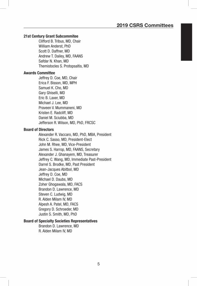
21st Century Grant Subcommitee Clifford B. Tribus, MD, Chair William Anderst, PhD Scott D. Daffner, MD Andrew T. Dailey, MD, FAANS Safdar N. Khan, MD Themistocles S. Protopsaltis, MD
Awards Committee Jeffrey D. Coe, MD, Chair Erica F. Bisson, MD, MPH Samuel K. Cho, MD Gary Ghiselli, MD Eric B. Laxer, MD Michael J. Lee, MD Praveen V. Mummaneni, MD Kristen E. Radcliff, MD Daniel M. Sciubba, MD Jefferson R. Wilson, MD, PhD, FRCSC
Board of Directors Alexander R. Vaccaro, MD, PhD, MBA, President Rick C. Sasso, MD, President-Elect John M. Rhee, MD, Vice-President James S. Harrop, MD, FAANS, Secretary Alexander J. Ghanayem, MD, Treasurer Jeffrey C. Wang, MD, Immediate Past-President Darrel S. Brodke, MD, Past President Jean-Jacques Abitbol, MD Jeffrey D. Coe, MD Michael D. Daubs, MD Zoher Ghogawala, MD, FACS Brandon D. Lawrence, MD Steven C. Ludwig, MD R. Alden Milam IV, MD Alpesh A. Patel, MD, FACS Gregory D. Schroeder, MD Justin S. Smith, MD, PhD
Board of Specialty Societies Representatives Brandon D. Lawrence, MD R. Alden Milam IV, MD
2019 CSRS Committees
6
2019 CSRS Committees
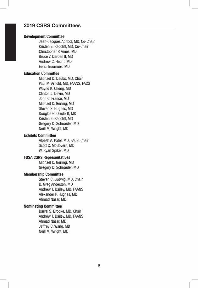
Development Committee Jean-Jacques Abitbol, MD, Co-Chair Kristen E. Radcliff, MD, Co-Chair Christopher P. Ames, MD Bruce V. Darden II, MD Andrew C. Hecht, MD Eeric Truumees, MD
Education Committee Michael D. Daubs, MD, Chair Paul M. Arnold, MD, FAANS, FACS Wayne K. Cheng, MD Clinton J. Devin, MD John C. France, MD Michael C. Gerling, MD Steven S. Hughes, MD Douglas G. Orndorff, MD Kristen E. Radcliff, MD Gregory D. Schroeder, MD Neill M. Wright, MD
Exhibits Committee Alpesh A. Patel, MD, FACS, Chair Scott C. McGovern, MD W. Ryan Spiker, MD
FOSA CSRS Representatives Michael C. Gerling, MD Gregory D. Schroeder, MD
Membership Committee Steven C. Ludwig, MD, Chair D. Greg Anderson, MD Andrew T. Dailey, MD, FAANS Alexander P. Hughes, MD Ahmad Nassr, MD
Nominating Committee Darrel S. Brodke, MD, Chair Andrew T. Dailey, MD, FAANS Ahmad Nassr, MD Jeffrey C. Wang, MD Neill M. Wright, MD

7
Program Committee Gregory D. Schroeder, MD, Co-Chair Justin S. Smith, MD, PhD, Co-Chair Jacob M. Buchowski, MD, MS Jeffrey D. Coe, MD Matthew W. Colman, MD Andrew T. Dailey, MD, FAANS William F. Donaldson III, MD Andrew C. Hecht, MD Han-Jo Kim, MD Craig A. Kuhns, MD Brian K. Kwon, MD, PhD, FRCSC Ronald A. Lehman, MD Sergio A. Mendoza-Lattes, MD Addisu Mesfin, MD R. Alden Milam IV, MD Thomas E. Mroz, MD Ahmad Nassr, MD Themistocles S. Protopsaltis, MD Jeffrey A. Rihn, MD Carlo Santaguida, MD, FRCSC Jason W. Savage, MD W. Ryan Spiker, MD Brian W. Su, MD Eeric Truumees, MD Barrett I. Woods, MD
Publications Committee Alpesh A. Patel, MD, FACS, Chair Rick C. Sasso, MD Gregory D. Schroeder, MD Daniel M. Sciubba, MD Jefferson R. Wilson, MD, PhD, FRCSC
Research Committee Zoher Ghogawala, MD, FACS, Chair Nitin N. Bhatia, MD Brandon D. Lawrence, MD Clifford B. Tribus, MD
2019 CSRS Committees

8
Resident Fellow Grant Subcommittee Nitin N. Bhatia, MD, Chair Jesse E. Bible, MD Samuel K. Cho, MD Carlo Santaguida, MD, FRCSC Richard L. Skolasky, ScD Michael P. Stauff, MD
Seed Starter Grant Subcommittee Brandon D. Lawrence, MD, Chair Wellington K. Hsu, MD Peter G. Passias, MD Jason W. Savage, MD Gregory D. Schroeder, MD W. Ryan Spiker, MD P. Justin Tortolani, MD
Special Projects Committee Gregory D. Schroeder, MD, Chair Erica F. Bisson, MD, MPH Samuel K. Cho, MD Zoher Ghogawala, MD, FACS Addisu Mesfin, MD Thomas E. Mroz, MD Alpesh A. Patel, MD, FACS Barrett I. Woods, MD Neill M. Wright, MD Jim A. Youssef, MD
Traveling Fellowship John M. Rhee, MD, Chair Ilyas Aleem, MD, MS, FRCSC Ivan Cheng, MD John G. Heller, MD Addisu Mesfin, MD Themistocles S. Protopsaltis, MD
2019 CSRS Committees

9
Scientific Program
10
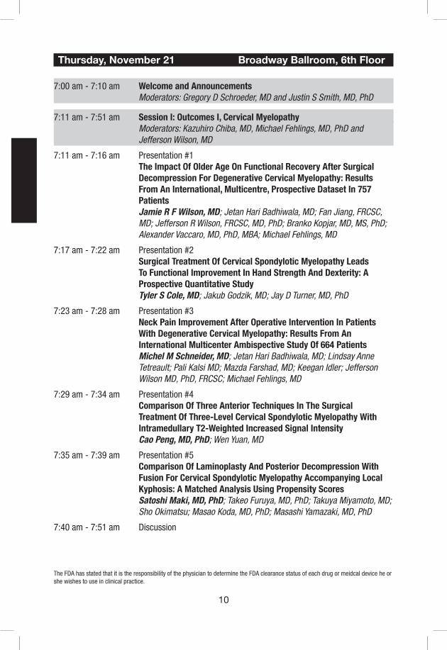
Thursday, November 21 Broadway Ballroom, 6th Floor
The FDA has stated that it is the responsibility of the physician to determine the FDA clearance status of each drug or meidcal device he or she wishes to use in clinical practice.
7:00 am - 7:10 am Welcome and Announcements Moderators: Gregory D Schroeder, MD and Justin S Smith, MD, PhD
7:11 am - 7:51 am Session I: Outcomes I, Cervical MyelopathyModerators: Kazuhiro Chiba, MD, Michael Fehlings, MD, PhD and Jefferson Wilson, MD
7:11 am - 7:16 am Presentation #1 The Impact Of Older Age On Functional Recovery After Surgical
Decompression For Degenerative Cervical Myelopathy: Results From An International, Multicentre, Prospective Dataset In 757 PatientsJamie R F Wilson, MD; Jetan Hari Badhiwala, MD; Fan Jiang, FRCSC, MD; Jefferson R Wilson, FRCSC, MD, PhD; Branko Kopjar, MD, MS, PhD; Alexander Vaccaro, MD, PhD, MBA; Michael Fehlings, MD
7:17 am - 7:22 am Presentation #2 Surgical Treatment Of Cervical Spondylotic Myelopathy Leads
To Functional Improvement In Hand Strength And Dexterity: A Prospective Quantitative StudyTyler S Cole, MD; Jakub Godzik, MD; Jay D Turner, MD, PhD
7:23 am - 7:28 am Presentation #3 Neck Pain Improvement After Operative Intervention In Patients
With Degenerative Cervical Myelopathy: Results From An International Multicenter Ambispective Study Of 664 PatientsMichel M Schneider, MD; Jetan Hari Badhiwala, MD; Lindsay Anne Tetreault; Pali Kalsi MD; Mazda Farshad, MD; Keegan Idler; Jefferson Wilson MD, PhD, FRCSC; Michael Fehlings, MD
7:29 am - 7:34 am Presentation #4 Comparison Of Three Anterior Techniques In The Surgical
Treatment Of Three-Level Cervical Spondylotic Myelopathy With Intramedullary T2-Weighted Increased Signal IntensityCao Peng, MD, PhD; Wen Yuan, MD
7:35 am - 7:39 am Presentation #5 Comparison Of Laminoplasty And Posterior Decompression With
Fusion For Cervical Spondylotic Myelopathy Accompanying Local Kyphosis: A Matched Analysis Using Propensity ScoresSatoshi Maki, MD, PhD; Takeo Furuya, MD, PhD; Takuya Miyamoto, MD; Sho Okimatsu; Masao Koda, MD, PhD; Masashi Yamazaki, MD, PhD
7:40 am - 7:51 am Discussion
11
Thursday, November 21 Broadway Ballroom, 6th Floor
Individual Disclosures can be found in the Disclosure Index pages 41-69.
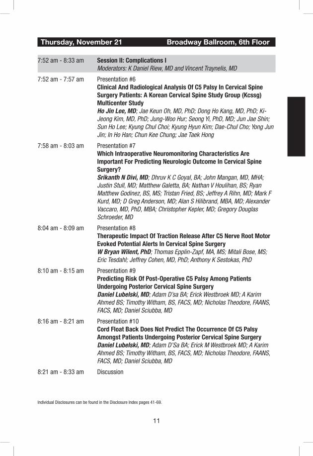
7:52 am - 8:33 am Session II: Complications IModerators: K Daniel Riew, MD and Vincent Traynelis, MD
7:52 am - 7:57 am Presentation #6 Clinical And Radiological Analysis Of C5 Palsy In Cervical Spine
Surgery Patients: A Korean Cervical Spine Study Group (Kcssg) Multicenter StudyHo Jin Lee, MD; Jae Keun Oh, MD, PhD; Dong Ho Kang, MD, PhD; Ki-Jeong Kim, MD, PhD; Jung-Woo Hur; Seong Yi, PhD, MD; Jun Jae Shin; Sun Ho Lee; Kyung Chul Choi; Kyung Hyun Kim; Dae-Chul Cho; Yong Jun Jin; In Ho Han; Chun Kee Chung; Jae Taek Hong
7:58 am - 8:03 am Presentation #7 Which Intraoperative Neuromonitoring Characteristics Are
Important For Predicting Neurologic Outcome In Cervical Spine Surgery?Srikanth N Divi, MD; Dhruv K C Goyal, BA; John Mangan, MD, MHA; Justin Stull, MD; Matthew Galetta, BA; Nathan V Houlihan, BS; Ryan Matthew Godinez, BS, MS; Tristan Fried, BS; Jeffrey A Rihn, MD; Mark F Kurd, MD; D Greg Anderson, MD; Alan S Hilibrand, MBA, MD; Alexander Vaccaro, MD, PhD, MBA; Christopher Kepler, MD; Gregory Douglas Schroeder, MD
8:04 am - 8:09 am Presentation #8 Therapeutic Impact Of Traction Release After C5 Nerve Root Motor
Evoked Potential Alerts In Cervical Spine SurgeryW Bryan Wilent, PhD; Thomas Epplin-Zapf, MA, MS; Mitali Bose, MS; Eric Tesdahl; Jeffrey Cohen, MD, PhD; Anthony K Sestokas, PhD
8:10 am - 8:15 am Presentation #9 Predicting Risk Of Post-Operative C5 Palsy Among Patients
Undergoing Posterior Cervical Spine SurgeryDaniel Lubelski, MD; Adam D’sa BA; Erick Westbroek MD; A Karim Ahmed BS; Timothy Witham, BS, FACS, MD; Nicholas Theodore, FAANS, FACS, MD; Daniel Sciubba, MD
8:16 am - 8:21 am Presentation #10 Cord Float Back Does Not Predict The Occurrence Of C5 Palsy
Amongst Patients Undergoing Posterior Cervical Spine SurgeryDaniel Lubelski, MD; Adam D’Sa BA; Erick M Westbroek MD; A Karim Ahmed BS; Timothy Witham, BS, FACS, MD; Nicholas Theodore, FAANS, FACS, MD; Daniel Sciubba, MD
8:21 am - 8:33 am Discussion

12
Thursday, November 21 Broadway Ballroom, 6th Floor
The FDA has stated that it is the responsibility of the physician to determine the FDA clearance status of each drug or meidcal device he or she wishes to use in clinical practice.
8:34 am - 9:15 am Session III: TraumaModerators: James Harrop, MD and Daniel Sciubba, MD
8:34 am - 8:39 am Presentation #11 Early Versus Late Surgical Decompression For Acute Spinal Cord
Injury: A Pooled Analysis Of 1,548 PatientsJetan H Badhiwala, MD; Christopher Witiw, MD; Jefferson R Wilson, FRCSC, MD, PhD; Michael Fehlings, MD
8:40 am - 8:45 am Presentation #12 Clinical Outcomes Of Acute Cervical Spinal Cord Injury Depending
On The Timing Of SurgeryKyung Jin Song; Jong Hyun Ko, MD; Tae Young Kwon
8:46 am - 8:51 am Presentation #13 Early Versus Late Surgical Decompression For Central Cord
Syndrome: A Propensity Score-Matched AnalysisJetan H Badhiwala, MD; Christopher Witiw, MD; Jefferson R Wilson, FRCSC, MD, PhD; Michael Fehlings, MD
8:52 am - 8:57 am Presentation #14 Can The Proposed Posterior Ligament-Bone Injury Classification
And Severity Score Predict The Failure Of Anterior-Only Surgery For Subaxial Cervical Facet Dislocations?Jun-song Yang, MD; Ding-Jun Hao; Tuan-Jiang Liu
8:58 am - 9:03 am Presentation #15 Cervical Spine Fractures: Who Really Needs Ct Angiography?
Mitchell S Fourman MD, MPhil; Jeremy Dewitt Shaw, MD, MS; Nicholas Vaudreuil, MD; Malcolm Dombrowski, MD; Richard Wawrose, MD; Lorraine Boakye, MD; Louis Alarcon, MD; Joon Yung Lee, MD; William F Donaldson III, MD
9:03 am - 9:15 am Discussion
9:15 am - 9:45 am Exhibit Hall, Westside Ballroom, 5th Floor Break
9:46 am - 10:49 am Symposium I: Spine TraumaModerators: Michael Fehlings, MD, PhD and Alexander Vaccaro, MD, PhD, MBA
9:46 am - 9:56 am Where Do We Stand with Pharmacologic Treatment for SCI?Jefferson Wilson, MD
9:57 am - 10:07 am Lateral Mass and Facet Fractures: When to OperateGregory D Schroeder, MD
10:07 am - 10:17 am Discussion

13
Thursday, November 21 Broadway Ballroom, 6th Floor
Individual Disclosures can be found in the Disclosure Index pages 41-69.
10:18 am - 10:28 am Which Patients with Central Cord Need Urgent Surgery?W Ryan Spiker, MD
10:29 am - 10:39 am Management of Cervical Spine Fractures in an Ankylosed SpineAddisu Mesfin, MD
10:39 am - 10:49 am Discussion
10:50 am - 11:25 am Presidential Introduction and Address
10:50 am – 10:55 am Introduction of CSRS PresidentRick Sasso, MD
10:55 am - 11:25 am Presidential AddressAlexander Vaccaro, MD, PhD, MBA
Industry WorkshopsLunch Available for Workshop Attendees Only, Prior Registration Not Required, No CME Credits
11:30 am - 1:30 pm Workshop 1: Medtronic Shubert/Uris, 6th Floor Circumferential Fixation in the Complex Corpectomy Patient
Workshop 2: NuVasive Majestic/Music Box, 6th Floor Cervical Spine Surgery: Why Should I Change? Featuring Advanced Materials Science.
Workshop 3: DePuy Synthes Ziegfield, 4th Floor The F.A.T.E. of Complex Cervical Surgery featuring the SYMPHONY™ OCT System
Workshop 4: Globus Medical Oneill, 4th Floor Cervical Corpectomy for Spondylotic Myelopathy
Workshop 5: Zimmer Biomet Wilder, 4th Floor 30% at 10 years? The Experts Discuss Techniques & Technology to Reduce Adjacent Segment Disease: A Discussion on How to Protect Against Adjacent Level Cervical Pathology
Workshop 6: Stryker Odets, 4th Floor Management of Complex Cervical Pathologies: An Interactive Panel Discussion
1:35 pm - 2:38 pm Symposium II: Pearls from the ExpertsModerators: Andrew Dailey, MD, FAANS and Justin S Smith, MD, PhD
1:35 pm - 1:45 pm How to Achieve Good Outcomes with 4-Level ACDFAlan S Hilibrand, MD
1:46 pm - 1:56 pm How and When to Use Cervical Pedicle ScrewsSang Hun Lee, MD
1:56 pm - 2:06 pm Discussion

14
Thursday, November 21 Broadway Ballroom, 6th Floor
The FDA has stated that it is the responsibility of the physician to determine the FDA clearance status of each drug or meidcal device he or she wishes to use in clinical practice.
2:07 pm - 2:17 pm Pearls for Instrumentation and Reconstructive Techniques in the Pediatric Cervical SpineHeiko Koller, PhD, MD
2:18 pm - 2:28 pm En Bloc Cervical Tumor Resection: Surgical Technique and Pearls for Optimizing OutcomesChristopher Ames, MD
2:28 pm - 2:38 pm Discussion
2:39 pm - 3:24 pm Session IV: Cervical DeformityModerators: Jacob Buchowski, MD and Jason Savage, MD
2:39 pm - 2:44 pm Presentation #16 What Drives Disability In Cervical Deformity: Novel Patient
Generated Outcome Versus Legacy HrqlNicholas D Stekas, MS; Ethan W Ayres, MPH; Mohamed A Moawad, MPH; Brooke K O’Connell; Dainn Woo, BS; Michael L Smith, MD; Yong H Kim, MD; Aaron James Buckland, FRACS, MBBS; Themistocles S Protopsaltis, MD
2:45 pm - 2:50 pm Presentation #17 Intraoperative Alignment Goals For Severe Cervical Deformity To
Achieve Optimal Improvements In Health-Related Quality Of Life MeasuresSohrab Virk, MD; Peter Gust Passias, MD; Renaud Lafage; Eric O Klineberg, MD; Gregory Michael Mundis Jr, MD; Themistocles Stavros Protopsaltis, MD; Christopher I Shaffrey, MD; Robert Shay Bess, MD; Han Jo Kim, MD; Christopher Ames, MD; Frank J Schwab, MD; Justin S Smith, MD; Virginie Lafage, PhD
2:51 pm - 2:56 pm Presentation #18 Improvement In Cervical Lordosis And Sagittal Alignment After
Vertebral Body Sliding Osteotomy In Patients With Spondylotic Cervical Myelopathy And KyphosisDong-Ho Lee, MD; Jae Hwan Cho; Jae-Woo Park, MD; Chul-Gie Hong; Jung-Gi Ha
2:57 pm - 3:02 pm Presentation #19 Pre-Operative Extension Lateral Cervical Radiographs Are
Associated With Osteotomy Type, Approach And Post-Operative Cervical Alignment Following Cervical Deformity SurgeryEric O Klineberg, MD; Renaud Lafage; Munish C Gupta, MD; Peter Gust Passias, MD; Virginie Lafage, PhD; Justin S Smith, MD; Han Jo Kim, MD; Themistocles Stavros Protopsaltis, MD; Douglas C Burton, MD; Gregory Michael Mundis Jr, MD; Frank J Schwab, MD; Robert A Hart, MD; Christopher I Shaffrey, MD; Christopher Ames, MD
15
Thursday, November 21 Broadway Ballroom, 6th Floor
Individual Disclosures can be found in the Disclosure Index pages 41-69.
3:03 pm - 3:08 pm Presentation #20 Simulated Corrections Of Cervical Deformity Using In-Construct
Measures Demonstrate That Insufficient Corrections Result In DjkThemistocles S Protopsaltis, MD; Dainn Woo, BS; Anand Segar, MD; Renaud Lafage; Gregory Michael Mundis Jr, MD; Justin S Smith, MD; Eric O Klineberg, MD; Peter Gust Passias, MD; Robert Shay Bess, MD; Christopher I Shaffrey, MD; Frank J Schwab, MD; Virginie Lafage, PhD; Christopher Ames, MD
3:09 pm - 3:14 pm Presentation #21 Comparison Of Perioperative Complications Following Posterior
Column Osteotomies Versus Posterior Based Three Column Osteotomy For Correction Of Severe Cervical Sagittal Deformity In 95 Patients: A Single Center StudyDarryl Lau, MD; Vedat Deviren, MD; Christopher Ames, MD
3:14 pm - 3:24 pm Discussion
3:25 pm - 3:55 pm Exhibit Hall, Westside Ballroom, 5th Floor Break
3:56 pm - 4:59 pm Session V: Complications IIModerators: Alan S Hilibrand, MD and Alpesh Patel, MD, FACS
3:56 pm - 4:01 pm Presentation #22 Effect Of Local Retropharyngeal Steroids On Fusion Rate After
Anterior Cervical Discectomy And FusionSapan D Gandhi, MD; Steven Wahlmeier, MD; Philip Louie, MD; Ryan Sauber, MD; Trevor Tooley, BS; Kevin C Baker, PhD; Daniel K Park, MD
4:02 pm - 4:07 pm Presentation #23 Effect Of Topical Steroid On Swallowing Following Acdf: Results Of
A Prospective Randomized Double Blind Control TrialDaniel Stein, BS; Han Jo Kim, MD; Darren Richard Lebl, MD; Russel C Huang, MD; Renaud Lafage; Todd J Albert, MD
4:08 pm - 4:13 pm Presentation #24 A Prospective Cohort Study Of Dysphagia After Subaxial Cervical
SurgeryKenichiro Sakai, MD, PhD; Toshitaka Yoshii; Takashi Hirai; Yoshiyasu Arai, MD, PhD; Atsushi Okawa, MD, PhD
4:14 pm - 4:19 pm Presentation #25 Association Between The Severity Of Dysphagia And Various
Parameters Of The Cervical Spine; Videofluoroscopic Analysis In Neutral And Retraction Position Of The Normal VolunteersJae Taek Hong, MD, PhD; Seong Hoon Lim; Dong Hoon Lee; Jun Seong Kim
16
Thursday, November 21 Broadway Ballroom, 6th Floor
The FDA has stated that it is the responsibility of the physician to determine the FDA clearance status of each drug or meidcal device he or she wishes to use in clinical practice.
4:19 pm - 4:27 pm Discussion
4:28 pm - 4:33 pm Presentation #26 Same Day Surgical Intervention Dramatically Minimizes
Complication Occurrence And Optimizes Peri-Operative Outcomes For Central Cord SyndromePeter Passias, MD; Cole Bortz, BA; Avery Eugene Brown; Haddy Alas, BS; Katherine E Pierce; M Burhan Janjua, MD; Paul Park, MD; Charles Wang, MD; Alexandra Soroceanu, MD; Rafael De La Garza Ramos, MD; Daniel Sciubba, MD; Anthony Frempong-Boadu, MD; Dennis Vasquez-Montes, MS; Bassel Diebo, MD; Michael C Gerling, MD
4:34 pm - 4:39 pm Presentation #27 Prospective Risk Factor Analysis Of Surgery-Related Complications
In Primary Cervical Spine Surgery For Degenerative DiseasesShota Takenaka, MD; Takahiro Makino, MD, MSc; Yusuke Sakai; Hideki Yoshikawa, MD; Takashi Kaito, MD, PhD
4:40 pm - 4:45 pm Presentation #28 The Influence Of Frailty Of Patients On The Incidence Of Surgical
Site Infection After Spine Surgery –The Analysis Of Over 1000 CasesTomoya Yoshikawa, MD; Shuichi Kaneyama, MD, PhD; Masatoshi Sumi, MD, PhD; Koichi Kasahara, MD, PhD; Aritetsu Kanemura, MD, PhD; Hiroaki Hirata, MD, PhD
4:46 pm - 4:51 pm Presentation #29 Upper Cervical Surgery, Increased Signal Intensity Of The Spinal
Cord, And Hypertension As Risk Factors For Dyspnea After Multilevel Anterior Cervical Discectomy And FusionJae Keun Oh, MD, PhD
4:51 pm - 4:59 pm Discussion
5:00 pm - 6:30 pm Exhibit Hall, Westside Ballroom, 5th Floor Welcome Reception
17
Friday, November 22 Broadway Ballroom, 6th Floor
Individual Disclosures can be found in the Disclosure Index pages 41-69.
7:00 am - 7:10 am Welcome and AnnouncementsModerators: Gregory D Schroeder, MD and Justin S Smith, MD, PhD
7:11 am - 7:58 am Session VI: Outcomes IVModerators: Howard S An, MD and Kris Radcliff, MD
7:11 am - 7:16 am Presentation #30 Nonoperative Management Of Asymptomatic Cervical Spinal
Stenosis: A Long-Term Follow-Up StudyMichael P Kelly, MD, MSc; Lukas Peter Zebala, MD; K Daniel Riew, MD
7:17 am - 7:22 am Presentation #31 Prospective Evaluation Of Degenerative Cervical Myelopathy In
Asymptomatic Patients Over 60 YearsSamuel Adams, MD; Sara Holmes; Letterio Salvatore Politi, MD; Patrick J Connolly, MD; Michael Paul Stauff, MD
7:23 am - 7:28 am Presentation #32 Home Versus Standard Physical Therapy After Acdf Surgery:
Preliminary Results Of Health-Related Quality Of Life Outcomes From A Randomized Controlled TrialSrikanth Divi, MD; Dhruv K C Goyal, BA; Matthew Galetta, BA; Justin Stull, MD; John Mangan, MD, MHA; Jeffrey A Rihn, MD; Mark F Kurd, MD; D Greg Anderson, MD; Barrett Ivory Woods, MD; Kristen E Radcliff, MD; Ian Kaye, MD; Alan S Hilibrand, MBA, MD; Alexander Vaccaro, MD, PhD, MBA; Christopher Kepler, MD; Gregory D Schroeder, MD
7:29 am - 7:34 am Presentation #33 Effectiveness Of Surgical Treatment In Reducing Falls And Fall-
Related Neurological Deterioration In Patients With Degenerative Cervical Myelopathy: A Multi-Institutional Prospective StudyAtsushi Kimura, MD, PhD; Hirokazu Inoue, MD, PhD; Yasuyuki Shiraishi; Katsushi Takeshita, MD, PhD; Atsushi Okawa
7:35 am - 7:40 am Presentation #34 Improvement In Cervicogenic Headaches After Laminoplasty: A
Single Institutional Study Of 143 Adult PatientsOwoicho Adogwa, MD, MPH; K Daniel Riew, MD; Gaurang Gupte; Maksim Shlykov, MD; Lukas Peter Zebala, MD; Colleen M Peters, MA; Jacob M Buchowski, MD, MS; Michael Patrick Kelly, MD

18
Friday, November 22 Broadway Ballroom, 6th Floor
The FDA has stated that it is the responsibility of the physician to determine the FDA clearance status of each drug or meidcal device he or she wishes to use in clinical practice.
7:41 am - 7:46 am Presentation #35 Improvements In Neck Pain And Disability Following C1-C2
Posterior Cervical Instrumentation And Fusion For Atlanto-Axial OsteoarthritisOwoicho Adogwa, MD, MPH; Jacob M Buchowski, MD, MS; John Sielatycki, MD; Alexander Theologis, MD; Maksim Aleksandrovich Shlykov, MD, MS; James D Lin, MD, MS; K Daniel Riew, MD
7:46 am - 7:58 am Discussion
7:59 am - 8:38 am Session VII: Basic ScienceModerators: Brian Kwon, MD and Thomas Mroz, MD
7:59 am - 8:04 am Presentation #36 Lotus Over Expression Enhanced The Motor Function Recovery
Following Ips-Ns/Pc TransplantationShuhei Ito, MD; Narihito Nagoshi; Osahiko Tsuji, MD, PhD; Morio Matsumoto, MD; Masaya Nakamura, MD
8:05 am - 8:10 am Presentation #37 Involvement Of Autophagy In Human Cervical Spine Degenerated
And Herniated DiscsTakashi Yurube, MD, PhD; Yuji Kakiuchi; Masaaki Ito; Yoshiki Takeoka; Yutaro Kanda, MD; Ryu Tsujimoto; Kenichiro Kakutani, MD; Toru Takada; Kunihiko Miyazaki; Shingo Miyazaki, MD; Zhongying Zhang; Ryosuke Kuroda, MD; Kotaro Nishida
8:11 am - 8:16 am Presentation #38 Inhibiting Spinal Phospholipase A2 Prevents Pain & Attenuates
Spinal Neuron Activity After Nerve Root CompressionSonia Kartha, BA; Julia Cecilia Hotek, PhD; Beth A Winkelstein, PhD
8:17 am - 8:22 am Presentation #39 Electrospun Synthetic Bone Graft Promotes Msc Function And
Spinal FusionDerek G Ju, MD; Juliane D Glaeser, PhD; Linda E A Kanim, MA; Khosrowdad Salehi, BS; Phillip H Behrens IV, MD; Melodie Metzger, PhD; Dmitriy Sheyn, PhD; Hyun W Bae, MD
8:23 am - 8:28 am Presentation #40 Intraarticular Mmp-1 Is Sufficient To Induce Pain & Substance P
Regulation In Drg Afferents Absent Any Structural DamageMeagan Eleanor Ita, MS; Beth A Winkelstein, PhD
8:28 am - 8:38 am Discussion
19
Friday, November 22 Broadway Ballroom, 6th Floor
Individual Disclosures can be found in the Disclosure Index pages 41-69.
8:39 am - 9:20 am Research Grant SessionBrandon Lawrence, MD and Zoher Ghogawala, MD
8:39 am - 8:48 am Announcement - 2019 Research Grant Winners
8:49 am - 8:50 am Introduction- Research Grant Updates
8:51 am- 8:53 am 2018 21st Century Grant New Approaches in Salamanders as Platforms for Uncovering Spinal Cord Regeneration FactorsHani Singer
8:54 am - 8:56 am 2018 Medtronic Grant Spinal Cord Perfusion Pressure Management in Acute Cervical SCI
Brian Kwon, MD, PhD, FRCSC
8:56 am - 8:59 am 2018 Resident Fellow Grant Advanced Quantitative MRI to Measure Cervical Cord Tissue Injury
and Predict OutcomesMuhammad Ali Akbar, MD
9:00 am - 9:02 am 2018 Resident Fellow Grant Evaluating the Influence of Impaired Cervical Cord Blood Perfusion
on Clinical Severity of Cervical Myelopathy Using Intravoxel Incoherent Motion MRIShuo Niu, MD, PhD
9:03 am - 9:05 am 2018 Seed Starter Grant Morphological, Biochemical and Biomechanical Characterization of
Human Cervical Endplate in Degenerated DiscYongren Wu, PhD
9:06 am - 9:08 am 2018 Seed Starter Grant Is vaporized nicotine as detrimental to spinal fusions as cigarette
smoke?Jesse Bible, MD
9:09 am - 9:11 am 2018 Seed Starter Grant CT Osteoabsorbptiometry assessment of subchondral bone density
predicts intervertebral subsidence in a human cadaver modelMatthew Colman, MD
9:12 am - 9:14 am 2018 Seed Starter Grant Reorganization of Brain Architecture and Networks as Biomarkers
for Cervical MyelopathyAmmar Hawasli, MD, PhD

20
Friday, November 22 Broadway Ballroom, 6th Floor
The FDA has stated that it is the responsibility of the physician to determine the FDA clearance status of each drug or meidcal device he or she wishes to use in clinical practice.
9:15 am - 9:17 am 2018 Seed Starter Grant Assessment of Variation in Preoperative Expectations and
Subsequent Fulfillment for Patients Undergoing Elective Cervical Spine SurgeryInamullah Khan, MBBS
9:20 am - 9:54 am Exhibit Hall, Westside Ballroom, 5th Floor Break
9:55 am - 11:00 am Presidential Guest Lecture
9:55 am - 10:00 am Introduction of Presidential Guest SpeakerAlexander Vaccaro, MD, PhD, MBA
10:00 am - 10:45 am Henry Bohlman Presidential Guest Speaker: “Healthcare in America”Steve Forbes, Chairman and Editor-in-Chief of Forbes Media
10:45 am - 11:01 am Discussion
11:02 am - 12:05 pm Symposium III: Concussions, Spine Injury, and Return to PlayModerators: Wellington Hsu, MD and Alexander Vaccaro, MD, PhD, MBA
11:02 am - 11:09 am Prevention and Evaluation of Concussions in the NFLStephen Stache, MD
11:10 am - 11:17 am Sideline Evaluation: When Does a Player Need to be Removed From Play?Andrew Dossett, MD
11:17 am - 11:25 am Discussion
11:26 am - 11:33 am Two- and Three-Level Disease: When Can a Patient Return to Play after Multilevel Anterior and Posterior Procedures?Robert Watkins III, MD
11:34 am - 11:41 am Asymptomatic Spinal Cord Compression: Is Surgery Necessary to Allow Return to Play?Andrew Hecht, MD
11:41 am - 11:49 am Discussion
11:49 am - 12:05 pm Review of CSRS Survey on Return to Play and a Modified Delphi Method Establishing CSRS Return to Play GuidelinesGregory D Schroeder, MD
12:05 pm - 1:05 pm Exhibit Hall, Westside Ballroom, 5th Floor Lunch

21
Friday, November 22 Broadway Ballroom, 6th Floor
Individual Disclosures can be found in the Disclosure Index pages 41-69.
1:10 pm - 1:36 pm Traveling Fellowship Report and Meeting PreviewsJohn Rhee, MD
1:10 pm - 1:18 pm Traveling Fellowship ReportIlyas S. Aleem, BSc, MD, MS, FRCSC and Lee A. Tan, M.D.
1:19 pm - 1:24 pm Preview CSRS 2020 Annual Meeting in Las Vegas, NVMichael Daubs, MD, 2020 Meeting Local Host
1:25 pm - 1:30 pm Preview CSRS Asia Pacific Section 2020 Annual MeetingKyung-Soo Suk, MD, President CSRS - AP
1:31 pm - 1:36 pm Preview CSRS European Section 2020 Annual MeetingBjörn Zoëga, MD, President CSRS - EU
1:37 pm - 2:24 pm Session VIII: Motion PreservationModerators: Frank Phillips, MD and Rick Sasso, MD
1:37 pm - 1:42 pm Presentation #41 Two-Level Cervical Disc Arthroplasty Vs. Anterior Cervical
Discectomy And Fusion: Ten- Year Outcomes Of A Prospective, Randomized Ide Clinical TrialJeffrey R McConnell, MD; Matthew F Gornet, MD; Todd Hopkins Lanman, MD, FACS; J Kenneth Burkus, MD; Randall F Dryer, MD; Scott D Hodges, DO; Francine Schranck, RN, BSN
1:43 pm - 1:48 pm Presentation #42 Single-Level Cervical Arthroplasty With Prodisc-C Artificial Disc:
10-Year Follow-Up Results In One CenterYanbin Zhao, MD; Yu Sun, MD
1:49 pm - 1:54 pm Presentation #43 Unintended Fusion In Cervical Artificial Disc Replacement: A
Prospective Study On Heterotopic Ossification, Evolution Through Time, And Clinical Outcome, With 5 Years Follow-UpCatarina Marques, MD; Anna Marianne Mac Dowall, MD; Martin Skeppholm; Nuno Canto Moreira, MD, PhD; Claes Olerud, MD
1:55 pm - 2:00 pm Presentation #44 Does Positioning Of Cervical Disc Arthroplasty Implant Affect
Postoperative OutcomeYahya Azhar Othman Othman; Philip York, MD; Russel C Huang, MD; Avani Vaishnav, MBBS; Steven McAnany, MD; Sravisht Iyer, MD; Todd J Albert, MD; Catherine Himo Gang, MPH; Sheeraz Qureshi, MD

22
Friday, November 22 Broadway Ballroom, 6th Floor
The FDA has stated that it is the responsibility of the physician to determine the FDA clearance status of each drug or meidcal device he or she wishes to use in clinical practice.
2:01 pm - 2:06 pm Presentation #45 The Effect Of Acdf Or Arthroplasty On Cervicogenic Headaches: A
Post-Hoc Analysis Of A Prospective, Multicenter Study With 10-Year Follow-UpK Daniel Riew, MD; Matthew F Gornet, MD; Todd Hopkins Lanman, MD, FACS; Jeffrey Ross McConnell, MD; Randall F Dryer, MD; J Kenneth Burkus, MD
2:07 pm - 2:12 pm Presentation #46 Comparison Of Three Fda-Approved Artificial Cervical Discs: A
Finite Element StudyHoon Choi, MD, MS; Yuvaraj Purushothaman, MS; Jamie Lynn Baisden, MD; Narayan Yoganandan, MD
2:12 pm - 2:24 pm Discussion
2:25 pm - 2:55 pm Broadway Ballroom Foyer, 6th Floor Break
2:56 pm - 3:58 pm Session IX: Outcomes IIModerators: Eric O Klineberg, MD and Jeffrey Wang, MD
2:56 pm - 3:01 pm Presentation #47 Mid-Term Surgical Outcome Of Posterior Decompression With
Instrumented Fusion For K-Line Negative Type Cervical Opll -Minimum 5 Years Follow-UpTakeo Furuya, MD, PhD; Satoshi Maki, MD, PhD; Takuya Miyamoto, MD; Sho Okimatsu; Masao Koda, MD, PhD; Masashi Yamazaki, MD, PhD
3:02 pm - 3:07 pm Presentation #48 Does Pre- Or Post-Operative Cervical Sagittal Alignment Correlate
With The Outcomes Of Cervical Laminoplasty? A Minimum Of One-Year Follow-Up StudyShuo Niu, MD, PhD; Albert Anastasio, BA; Kevin Xavier Farley, BA; John JM Rhee, MD
3:08 pm - 3:13 pm Presentation #49 The Effect Of Duration Of Symptoms On Clinical Outcomes Following
Anterior Cervical Discectomy And FusionJoon Sung Yoo, BA; Dil V Patel, BS; Eric H Lamoutte, BS; Sailee S Karmarkar, BS; Kern Singh, MD; Nathaniel W Jenkins, MS; James M Parrish, MPH

23
Friday, November 22 Broadway Ballroom, 6th Floor
Individual Disclosures can be found in the Disclosure Index pages 41-69.
3:14 pm - 3:19 pm Presentation #50 The Impact Of Post-Operative Physical Therapy On Patient-Reported
Outcomes At 1-Year After Cervical Spine SurgeryKristin R Archer, PhD; Emily Oleisky; Jacquelyn S Pennings, PhD; Inamullah Khan, MBBS; Rogelio Adrian Coronado, PhD, PT; Clinton J Devin, MD
3:19 pm - 3:27 pm Discussion
3:28 pm - 3:33 pm Presentation #51 Asymptomatic Acdf Non-Unions Underestimate The True Prevalence
Of Radiographic PseudoarthrosisCharles Hopkins Crawford III, MD; Leah Yacat Carreon, MD; Praveen V Mummaneni; Steven D Glassman, MD
3:34 pm - 3:39 pm Presentation #52 Does Chronic Preoperative Opioid Use Affect Patient Outcomes After
Acdf SurgeryJohn Mangan, MD, MHA; Srikanth Divi, MD; Dhruv K C Goyal, BA; Justin Stull, MD; Matthew Galetta, BA; Jeffrey A Rihn, MD; Mark F Kurd, MD; D Greg Anderson, MD; Barrett Ivory Woods, MD; Kristen E Radcliff, MD; Ian Kaye, MD; Alan S Hilibrand, MBA, MD; Alexander Vaccaro, MD, PhD, MBA; Christopher Kepler, MD; Gregory Douglas Schroeder, MD
3:40 pm - 3:45 pm Presentation #53 Can We Predict A Patient’s Arm Pain, Neck Pain, And Disability Level
One Year After Cervical Spine Surgery For Radiculopathy?Ahilan Sivaganesan, MD; Inamullah Khan, MBBS; Hui Nian, PhD; Frank E Harrell Jr, PhD; Jacquelyn S Pennings, PhD; Mohamad Bydon, MD; Anthony Asher; Kristin Archer, PhD; Clinton J Devin, MD
3:46 pm - 3:51 pm Presentation #54 Preliminary Results From The Multi-Center Prospective,
Randomized Csm-S Study: Overall Quality Of Life Improvement, Complications, And Return To WorkZoher Ghogawala, MD, FACS; Adam Kanter, MD; Praveen V Mummaneni; Erica Fay Bisson, MD, MPH; James S Harrop, MD; Subu Magge; Robert F Heary, FAANS, MD; Michael P Steinmetz, MD; Michael Fehlings, MD; Todd J Albert, MD; Paul M Arnold, MD, FACS; K Daniel Riew, MD; Marjorie Wang, FAANS, MD, MPH; John G Heller, MD; Edward Benzel, MD
3:51 pm - 3:59 pm Discussion
4:00 pm - 4:30 pm CSRS Membership Meeting

24
Saturday, November 23 Broadway Ballroom, 6th Floor
The FDA has stated that it is the responsibility of the physician to determine the FDA clearance status of each drug or meidcal device he or she wishes to use in clinical practice.
7:00 am - 7:10 am Welcome and AnnouncementsModerators: Gregory D Schroeder, MD and Justin S Smith, MD, PhD
7:11 am - 8:14 am Session X: Complications III and Outcomes IIIModerators: Darrel Brodke, MD and Kazuhiro Chiba, MD
7:11 am - 7:16 am Presentation #55 Fate Of Anterior Cervical Discectomy (Acdf) Non-Union At 6 Months
K Daniel Riew, MD; Allan D Levi, MD; William Francis Lavelle, MD; Jeffrey E Florman
7:17 am - 7:22 am Presentation #56 Epidemiology Of Iatrogenic Vertebral Artery Injury In Cervical Spine
Surgery: 21 Multicenter StudiesChang-Hyun Lee, MD, MSc; Jae Taek Hong, MD, PhD; Chi Heon Kim, MD, PhD; Chun Kee Chung, MD; Jae Keun Oh, MD, PhD; Dong Ho Kang, MD, PhD; Hojin Lee; Seung-Jae Hyun, MD, PhD; Seong Yi, PhD, MD; Jun Ho Lee, MD; Dae-Chul Cho, MD, PhD; Jun-Jae Shin, MD, PhD; Yong Jun Jin, MD, PhD; Geun Sung Song, MD, PhD
7:23 am - 7:28 am Presentation #57 Is Facet Joint Distraction A Cause Of Postoperative Axial Neck Pain
After Acdf Surgery?Srikanth N Divi, MD; Dhruv K C Goyal, BA; John Mangan, MD, MHA; Justin Stull, MD; Nathan V Houlihan, BS; Matthew Galetta, BA; Jeffrey A Rihn, MD; Mark F Kurd, MD; D Greg Anderson, MD; Alan S Hilibrand, MBA, MD; Alexander Vaccaro, MD, PhD, MBA; Christopher Kepler, MD; Gregory Douglas Schroeder, MD; Joseph K Lee, MD
7:29 am - 7:34 am Presentation #58 Degenerative Cervical Spondylolisthesis: Does Adjacent Level
Surgical Stabilization Result In Progressive Listhesis?Philip K Louie, MD; Jannat Khan; Hollis Johanson; Jacob Tomas Emerson, BA; Bryce A Basques, MD; Michael T Nolte, MD; Dino Samartzis, PhD; Howard An
7:34 am - 7:42 am Discussion
7:43 am - 7:48 am Presentation #59 Crossing The Cervicothoracic Junction During Laminectomy
And Posterior Spinal Fusion Surgery For Cervical Spondylotic Myelopathy Is Associated With Superior Cervical Radiographic OutcomesAndrew Kai-Hong Chan, MD; Ryan Badiee, BA, BS; Joshua Rivera; Chih-Chang Chang, MD; Leslie C Robinson, MD, PharmD, MBA; Ratnesh N Mehra DO; Lee A Tan, MD; Aaron J Clark MD, PhD; Sanjay S Dhall MD; Dean Chou, MD; Praveen V Mummaneni, MD

25
Saturday, November 23 Broadway Ballroom, 6th Floor
Individual Disclosures can be found in the Disclosure Index pages 41-69.
7:49 am - 7:54 am Presentation #60 Assessing Radiographic Fusion Rates Following A Stand-Alone
Interbody Cage Versus An Anterior Plate Construct For Adjacent Segment Disease After Anterior Cervical Discectomy And FusionSapan D Gandhi, MD; Adam Fahs, MD; Steven Wahlmeier, MD; Philip Louie, MD; Daniel Robert Possley, DO; Jad Khalil, MD; Kevin C Baker, PhD; Daniel K Park, MD
7:55 am - 8:00 am Presentation #61 Sagittal Parameters Poorly Predict Clinical Outcomes Following
Anterior Cervical Discectomy And FusionBryce A Basques, MD; Jannat Khan; Michael T Nolte, MD; Philip Louie, MD; Kamran Movassaghi, MD; Jonathan S Markowitz, BS; Edward Jay Goldberg, MD; Howard An
8:01 am - 8:06 am Presentation #62 Discrepancies In The Surgical Management Of Central Cord
Syndrome: Assessment Of Non-Operative, Surgical, And Crossover To Surgery PatientsPeter Passias, MD; Cole Bortz, BA; Katherine E Pierce; Haddy Alas, BS; Avery Eugene Brown; Nicholas Shepard, MD; M Burhan Janjua, MD; John Buza, MD; Alexandra Soroceanu, MD; Rafael De La Garza Ramos, MD; Daniel Sciubba, MD; Dennis Vasquez-Montes, MS; Bassel Diebo, MD; Michael C Gerling, MD
8:06 am - 8:14 am Discussion
8:15 am - 9:18 am Session XI: Surgical Techniques and Imaging ParametersModerators: Paul Arnold, MD and John Rhee, MD
8:15 am - 8:20 am Presentation #63 Microendoscopic Laminotomy Versus Conventional Laminoplasty
For Cervical Spondylotic Myelopathy - A 5-Year Follow-Up StudyAkihito Minamide, MD, PhD; Andrew K Simpson, MD; Yukihiro Nakagawa; Motohiro Okada; Hiroshi Iwasaki; Shunji Tsutsui, MD, PhD; Masanari Takami, MD; Ryo Taiji; Shizumasa Murata; Takuhei Kozaki Jr, MRCPCH, MRCPe, MRCSEd, MS, MSA, MSc, MSN; Hiroshi Hashizume, MD; Yasutsugu Yukawa; Munehito Yoshida; Hiroshi Yamada, MD
8:21 am - 8:26 am Presentation #64 Effect Of C3 Laminectomy In Cervical Laminoplasty For
Degenerative Cervical Myelopathy: C3 Laminectomy Vs. C3 LaminoplastyKoji Nakajima, MD; Toru Doi; So Kato, MD; Sakae Tanaka, MD, PhD; Yasushi Oshima, MD, PhD
26
Saturday, November 23 Broadway Ballroom, 6th Floor
The FDA has stated that it is the responsibility of the physician to determine the FDA clearance status of each drug or meidcal device he or she wishes to use in clinical practice.
8:27 am - 8:32 am Presentation #65 Minimally-Invasive Posterior Cervical Foraminotomy As An
Alternative To Anterior Cervical Discectomy And Fusion For Unilateral Cervical RadiculopathyNikhil Sahai, MD; Stuart Changoor, MD; Conor Dunn, MD, MS; Michael Faloon, MD; Kumar Gautam Sinha, MD; Ki S Hwang, MD; Arash Emami, MD
8:33 am - 8:38 am Presentation #66 Optimizing Cervicothoracic Junction Biomechanics After C7 Pedicle
Subtraction Osteotomy: A Cadaveric Study Of Stability And Rod StrainJakub Godzik, MD; Jennifer Lehrman, BS, MS; Bernardo de Andrada Pereira; Christopher Ames, MD; Heiko Koller, MD; Kevin Fun Lee, MS; Anna Sawa Newcomb, MS; Jay D Turner, MD, PhD; Brian P Kelly, PhD
8:38 am - 8:46 am Discussion
8:47 am - 8:52 am Presentation #67 A Deep Learning Model For Detection Of Cervical Spinal Cord
Compression In Mri ScansZamir Merali, MD; Jetan Hari Badhiwala, MD; Christopher Witiw, MD; Jefferson R Wilson, FRCSC, MD, PhD; Michael Fehlings, MD
8:53 am - 8:58 am Presentation #68 Intervertebral Foramen Width Is An Important Factor In Deciding
Additional Uncinate Process Resection In Acdf -- A Retrospective StudyYang Liu, MD
8:59 am - 9:04 am Presentation #69 A New Index For Making Decisions Regarding C2 Lamina
Decompression In Cervical Ossification Of The Posterior Longitudinal Ligament: The R-LineJin Hoon Park, MD, PhD; Lee Byung-Jou; Seong Kyun Jeong; Subum Lee, MD; Myeongjong Kim
9:05 am - 9:10 am Presentation #70 Can C7 Slope Be Used As A Substitute For T1 Slope? A Radiographic
AnalysisJun Sup Kim, MD; Ivan Ye; Ray Tang, BA; Samuel Johnston Waring White, BA; Samuel Kang-Wook Cho, MD
9:10 am - 9:18 am Discussion

27
Saturday, November 23 Broadway Ballroom, 6th Floor
Individual Disclosures can be found in the Disclosure Index pages 41-69.
9:19 am - 10:22 am Symposium IV: Cervical Deformity and Complex Cervical ReconstructionModerators: Christopher Ames, MD and Han Jo Kim, MD
9:19 am - 9:29 am Alignment Planning for Deformity CorrectionThemistocles S Protopsaltis, MD
9:30 am - 9:40 am Management of Cervical Deformity Resulting from TraumaHeiko Koller, PhD, MD
9:40 am - 9:50 am Discussion
9:51 am - 10:01 am Three-Column Osteotomies for Deformity Correction: Technique and Video DemonstrationChristopher Ames, MD
10:02 am - 10:12 am Complications Associated with Complex Cervical Reconstruction: Occurrence and Techniques for AvoidancePeter Passias, MD
10:12 am - 10:22 am Discussion
10:22 am - 10:28 am Announcement of Abstract and E-Poster Award WinnersJeffrey D Coe, MD, Awards Committee Chair
10:28 am - 10:33 am Presentation of CSRS Medallion to Incoming President, Rick Sasso, MDAlexander Vaccaro, MD, PhD, MBA
10:34 am - 11:14 am Session XII: Economics, Safety, and Health PolicyModerators: Michael P Kelly, MD, MSc and Michael Steinmetz, MD
10:34 am - 10:39 am Presentation #71 Effect Of Opioid-Limiting Legislation On Postoperative Prescription
Patterns Following Anterior Cervical Decompression And FusionDaniel B C Reid, MD, MPH; Kalpit Nimish Shah, MD; Benjamin Shapiro, MS; Jack H Ruddell, BA; Edward Akelman, MD; Mark A Palumbo, MD; Alan H Daniels, MD
10:40 am - 10:45 am Presentation #72 Perioperative Spending In Single-Level Anterior Cervical
Discectomy And Fusion For Degenerative PathologyMajd Marrache, MD; Andrew Harris, BS; Varun Puvanesarajah, MD; Micheal Raad, MD; Amit Jain, MD
28
Saturday, November 23 Broadway Ballroom, 6th Floor
The FDA has stated that it is the responsibility of the physician to determine the FDA clearance status of each drug or meidcal device he or she wishes to use in clinical practice.
10:46 am - 10:51 am Presentation #73 The Fault Within Our “Drgs”: Refining Risk-Adjustment For Bundled
Payment Models In Cervical FusionsAzeem Tariq Malik, MBBS; Frank M Phillips, MD; Sheldon Michael Retchin, MD, MSPH; Wendy Xu; Elizabeth M Yu, MD; Jeffery D Kim, MD; Safdar N Khan, MD
10:52 am - 10:57 am Presentation #74 Anterior Cervical Discectomy And Fusions (Acdfs) At Physician
Owned Hospitals – Is It Time To Reconsider The Sanctions Of The Affordable Care Act (Aca)?Azeem Tariq Malik, MBBS; Sheldon Michael Retchin, MD, MSPH; Wendy Xu; Frank M Phillips, MD; Safdar N Khan, MD
10:58 am - 11:03 am Presentation #75 No S.C.A.R.E Protocol: A Streamlined Safety Protocol
Tyler J Jenkins, MD; Ryan D Snowden, MD; Joseph Douglas Smucker, MD; Wellington K Hsu, MD; K Daniel Riew, MD; Rick C Sasso, MD
11:03 am - 11:14 am Discussion
11:15 am - 11:29 am Broadway Ballroom Foyer, 6th Floor Break
11:29 am - 12:44 pm Session XIII: Focused Podium PresentationsModerators: Michael Daubs, MD and Robert Hart, MD
11:29 am - 11:31 am Presentation #76 Anterior Cord Compression Is Associated With Neurologic Deficit
In Patients With Degenerative Cervical Myelopathy. Does It Have Evidence?Kyung Chung Kang, MD; Jung-Hee Lee, MD; Ki Young Lee; Hyoungmin Kim; Sang Kyu Im; Jong-Beom Park
11:32 am - 11:34 am Presentation #77 Cervical Bone Mineral Density Measured By Qct In Patients
Undergoing Anterior Cervical Spine SurgeryStephan N Salzmann, MD; Courtney Ortiz Miller, BA; Ichiro Okano, MD; Fabian Winter; Jennifer Shue, MS; John Anthony Carrino, MD; Andrew A Sama, MD; Frank P Cammisa Jr, MD; Federico P Girardi, MD; Alexander P Hughes, MD
11:35 am - 11:37 am Presentation #78 Continuous Optical Monitoring Of Spinal Cord Hemodynamics
During The First 7 Days Post-Injury In A Porcine Model Of Acute Spinal Cord InjuryBrian K Kwon, MD, PhD, FRCSC; Neda Manouchehri; So Kitty; Cheung Amanda; Streijger Femke; Macnab Andrew; Shadgan Babak

29
Saturday, November 23 Broadway Ballroom, 6th Floor
Individual Disclosures can be found in the Disclosure Index pages 41-69.
11:38 am - 11:40 am Presentation #79 Risk Factors Associated With Vertebral Artery Anomalies In The
Subaxial Cervical SpineErika Chiapparelli Harb, MD; Stephan N Salzmann, MD; Colleen Rentenberger; Jennifer Shue, MS; Ichiro Okano, MD; Andrew A Sama, MD; Federico P Girardi, MD; Frank P Cammisa Jr, MD; Alexander P Hughes, MD
11:41 am - 11:43 am Presentation #80 Therapeutic Impact Of Grafted Oligodendrogenic-Neural Progenitor
Cells Combined With Sustained Delivery Of Chondroitinase Abc Using An Innovative Methylcellulose Biomaterial For Chronic Spinal Cord InjurySatoshi Nori, MD, PhD; Mohamad Khazaei, PhD; Kazuya Yokota, MD, PhD; Jan-Eric Ahlfors; Shinsuke Shibata, MD, PhD; Morio Matsumoto, MD; Masaya Nakamura, MD; Molly Shoichet; Michael Fehlings, MD
11:43 am - 11:53 am Discussion
11:54 am - 11:56 am Presentation #81 Establishing Maximal Medical Improvement Following Anterior
Cervical Discectomy And FusionDil V Patel, BS; Joon Sung Yoo, BA; Benjamin Khechen, BA; Anirudh K Gowd; Eric H Lamoutte, BS; Sailee S Karmarkar, BS; Joseph Liu, MD; Kern Singh, MD; Nathaniel W Jenkins, MS; James M Parrish, MPH
11:57 am - 11:59 am Presentation #82 Does Neurologic Diagnosis Affect Improvement In Neck Pain After
Acdf Surgery?Justin Stull, MD; Srikanth Divi, MD; John Mangan, MD, MHA; Dhruv K C Goyal, BA; Matthew Galetta, BA; Jeffrey A Rihn, MD; Mark F Kurd, MD; D Greg Anderson, MD; Barrett Ivory Woods, MD; Kristen E Radcliff, MD; Ian Kaye, MD; Alexander Vaccaro, MD, PhD, MBA; Christopher Kepler, MD; Gregory Douglas Schroeder, MD; Alan S Hilibrand, MBA, MD
12:00 pm - 12:02 pm Presentation #83 Predictive Models For Long-Term Patient-Reported Outcomes After
Cervical Spine Surgery For Myelopathy: A National Study Of 2717 PatientsInamullah Khan, MBBS; Hui Nian, PhD; Frank E Harrell Jr, PhD; Jacquelyn S Pennings, PhD; Mohamad Bydon, MD; Anthony Asher; Kristin Archer, PhD; Clinton J Devin, MD

30
Saturday, November 23 Broadway Ballroom, 6th Floor
The FDA has stated that it is the responsibility of the physician to determine the FDA clearance status of each drug or meidcal device he or she wishes to use in clinical practice.
12:03 pm - 12:05 pm Presentation #84 How Much Do Patients With Predominantly Neck Pain Improve After
Acdf Surgery For Cervical Radiculopathy?Srikanth N Divi, MD; Justin Stull, MD; John Mangan, MD, MHA; Dhruv K C Goyal, BA; Matthew Galetta, BA; Jeffrey A Rihn, MD; Mark F Kurd, MD; D Greg Anderson, MD; Barrett Ivory Woods, MD; Alan S Hilibrand, MBA, MD; Ian Kaye, MD; Alexander Vaccaro, MD, PhD, MBA; Christopher Kepler, MD; Gregory Douglas Schroeder, MD; Kristen E Radcliff, MD
12:06 pm - 12:08 pm Presentation #85 Functional Range Of Motion Of The Cervical Spine In Posterior
Cervical Discectomy And Fusion Patients During Activities Of Daily LivingSebastian Murati; Abenezer Alemu; Marcus Allen; Riffitts Michelle; Anna Bailes; Malcolm Dombrowski, MD; Joon Yung Lee, MD; William F Donaldson III, MD; William W Clark, PhD; Kevin Michael Bell, PhD
12:09 pm - 12:19 pm Discussion
12:20 pm - 12:22 pm Presentation #86 Which Topical Agent Is More Efficacious In Diminishing
Postoperative Drainage Following Multi-Level Cervical Laminoplasty/Laminectomy: Tranexamic Acid- Versus Thrombin-Soaked Collagen Sponge?Jiwon Park, MD; Haolin Zheng; Jin-Sup Yeom, MD, PhD; Jae Won Lee; Ho-Joong Kim, MD, PhD; Sang-Min Park; Bong-Soon Chang, MD, PhD; Choon-Ki Lee, MD, PhD
12:23 pm - 12:25 pm Presentation #87 Pseudoarthrosis Rates After Anterior Cervical Discectomy And
Fusion (Acdf) Using Polyetheretherketone (Peek) Or Structural Allograft For Interbody Grafting: Minimum 2-Year Follow UpMinghao Wang, MD, PhD; Dean Chou, MD; Chih-Chang Chang, MD; YILIN LIU, MD; Ankit Hirpara; Praveen V Mummaneni
12:26 pm - 12:28 pm Presentation #88 CTA In Addition To MRI May Not Be Necessary In Detecting Subaxial
Vertebral Artery Anomalies In Anterior Cervical Spine SurgeryIchiro Okano, MD; Stephan N Salzmann, MD; Colleen Rentenberger; Jennifer Shue, MS; Andrew A Sama, MD; Federico P Girardi, MD; Frank P Cammisa Jr, MD; Alexander P Hughes, MD

31
Saturday, November 23 Broadway Ballroom, 6th Floor
Individual Disclosures can be found in the Disclosure Index pages 41-69.
12:29 pm - 12:31 pm Presentation #89 Can Reinforcing Lateral Mass Screw With Multiple Points Of
Fixation Improve Anchoring Performance And Reduce Screw Pullout?Franck Le Naveaux, PhD; Bahe Hachem; Kevin Fun Lee, MS; Jean-Marc Mac-Thiong, MD; Hyun W Bae, MD; Christopher Ames, MD; Masashi Neo, MD, PhD; Julien Clin
12:32 pm - 12:34 pm Presentation #90 The Effect Of Arthrodesis On Neuroforaminal Area
Clarissa Levasseur, MS; Samuel William Pitcairn, BS; Jeremy Dewitt Shaw, MD, MS; William F Donaldson III, MD; Joon Yung Lee, MD; William Anderst, PhD
12:34 pm - 12:44 pm Discussion
12:45 pm - 12:46 pm Closing RemarksModerators: Gregory D Schroeder, MD and Justin S Smith, MD, PhD
12:47 pm - 12:48 pm Adjourning NoticesRick Sasso, MD

32
33
Individual Disclosures can be found in the Disclosure Index pages 41-69.
E-Poster Catalog
Disclosure information submitted to the AAOS Orthopaedic Disclosure Program.
34
The FDA has stated that it is the responsibility of the physician to determine the FDA clearance status of each drug or meidcal device he or she wishes to use in clinical practice.
E-Poster #1Bariatric Surgery Diminishes Spinal Symptoms In A Morbidly Obese Population: A 2-Year Survivorship Analysis Of Cervical And Lumbar PathologiesPeter Passias, MD; Haddy Alas, BS; Avery Eugene Brown; Cole Bortz, BA; Katherine E Pierce; Dennis Vasquez-Montes, MS; Dainn Woo, BS; Bassel Diebo, MD; Carl B Paulino, MD; Michael C Gerling, MD
E-Poster #2Appropriate Risk Stratification And Accounting For Age-Adjusted Reciprocal Changes In The Thoracolumbar Spine Reduces The Incidence And Magnitude Of Distal Junctional Kyphosis In Cervical Deformity SurgeryPeter Passias, MD; Cole Bortz, BA; Renaud Lafage; Virginie Lafage, PhD; Eric O Klineberg, MD; Han Jo Kim, MD; Alan H Daniels, MD; Gregory Michael Mundis Jr, MD; Themistocles S Protopsaltis, MD; Robert Shay Bess, MD; Frank J Schwab, MD; Christopher I Shaffrey, MD; Justin S Smith, MD; Christopher Ames, MD; International Spine Study Group
E-Poster #3Effect Of Myelopathy On Outcomes After Cervical Disc Replacement: A Study Of A Local Patient Cohort And A Large National CohortAndre Samuel, MD; Harold Gregory Moore, BS; Avani Vaishnav, MBBS; Steven McAnany, MD; Sravisht Iyer, MD; Todd J Albert, MD; Catherine Himo Gang, MPH; Sheeraz Qureshi, MD
E-Poster #4Objective Swallowing Abnormalities In Patients With Dysphagia Following Anterior Cervical Spine SurgeryPope Rodnoi, BS; John Wuellner, MD; Adam Wegner, MD, PhD; Shumon Dhar MD; Machelle Wilson; Peter Belafsky, MD, PhD; Eric O Klineberg, MD
E-Poster #5A Predictive Model And Nomogram For Predicting Return To Work At 3 Months After Cervical Spine Surgery: An Analysis From The Quality Outcome DatabaseClinton J Devin, MD; Mohammed Ali Alvi, MBBS; Panagiotis Kerezoudis, MD; Inamullah Khan, MD; Ahilan Sivaganesan, MD; Matthew J McGirt, MD; Kristin R Archer, PhD, DPT; Kevin T Foley, MD; Praveen Mummaneni, MD; Andrew K Chan, MD; Erica Bisson, MD, MPH; Jian Guan, MD; John J Knightly, MD; Christopher Shaffrey, MD; Anthony L Asher, MD; Mohamad Bydon, MD
E-Poster #6An Evaluation Of Surgeon Ability To Predict Ossification Of The Posterior Longitudinal Ligament (Opll) Using Mri And X-Rays AloneJoseph A Osorio, MD, PhD; Nathan John Lee, MD; Meghana Vulapalli, BS; Meghan Cerpa, MPH; James D Lin, MD, MS; Simon Morr, MD, MPH; Richard Menger; Griffin Richard Baum, MD, MSc; Jae Hong Ha, MD; Hyoungmin Kim; Louis F Amorosa, MD; Marc D Dyrszka, MD; Patrick Charles Reid, MD; Zeeshan Sardar, MD; K Daniel Riew, MD
E-Poster Catalog
35
Individual Disclosures can be found in the Disclosure Index pages 41-69.
E-Poster #7Comparison Between Japanese Orthopaedic Association Score (Joa Score) And Patient-Reported Joa Score (Pro-Joa Score) For Evaluating Surgical Outcomes Of Cervical MyelopathyYasushi Oshima, MD, PhD; So Kato, MD; Toru Doi; Koji Nakajima, MD; Shima Hirai, MD; Sakae Tanaka, MD, PhD
E-Poster #8Proton Pump Inhibitor Use Affects Pseudarthrosis Rates And Influences Patient Reported OutcomesJohn Mangan MD, MHA; Srikanth Divi, MD; James C McKenzie, MD; Justin Stull, MD; David Casper, MD; Dhruv K C Goyal, BA; Scott Wagner, MD; Mark F Kurd, MD; Alan S Hilibrand, MBA, MD; Jeffrey A Rihn, MD; D Greg Anderson, MD; Gregory Douglas Schroeder, MD; Alexander Vaccaro, MD, PhD, MBA; Christopher Kepler, MD
E-Poster #9C2 Versus C3 As The Upper Instrumented Vertebra For Patients Undergoing Long Segment Posterior Cervical FusionAndrew Kai-Hong Chan, MD; Joshua Rivera; Chih-Chang Chang, MD; Dean Chou, MD; Praveen V Mummaneni; Lee A Tan, MD
E-Poster #10A Prospective, Psychometric Validation Of Nih Promis Physical Function, Pain Interference And Upper Extremity Cat In Cervical Spine Patients: Successes And Key LimitationsSravisht Iyer, MD; Jayme Koltsov, PhD; Michael Steinhaus, MD; Thomas Ross, RN; Kelsey Young; dan stein; Jingyan Yang, PhD; Virginie Lafage, PhD; Todd J Albert, MD; Han Jo Kim, MD
E-Poster #11Modified Frailty Index Predicts Readmission Rates And Extended Length Of Stay Following Acdf Surgery For Degenerative DiseaseBrian L Dial, MD; Valentine Rae Esposito, BS; Richard Michael Danilkowicz, MD; Jeffrey O’Donnell; Norah Anne Foster, MD; Isaac Obiri Karikari, MD; Sergio Andres Mendoza-Lattes, MD; Melissa Maria Erickson, MD
E-Poster #12Promis-29 Validity And Conversion Equation To Neck Disability Index (Ndi) Using A National Sample Of Cervical Spine Surgery PatientsJacquelyn Sue Pennings, PhD; Claudia A Davidson; Inamullah Khan, MBBS; Mohamad Bydon, MD; Anthony Asher; John P Wanner, MD; Daniel Verhotz, MD; Clinton J Devin, MD; Kristin Archer, PhD
E-Poster Catalog
36
The FDA has stated that it is the responsibility of the physician to determine the FDA clearance status of each drug or meidcal device he or she wishes to use in clinical practice.
E-Poster #13Underweight Patients Are The Highest Risk Body Mass Index Group For Perioperative Adverse Events Following Posterior Cervical Spine SurgeryTaylor Ottesen, BS; Paul S Bagi, BS, MD; Rohil Malpani, BS; Anoop Raj Galivanche, BS; Arya Giri Varthi, MD; Jonathan N Grauer, MD
E-Poster #14Is It Safe To Perform Anterior Foraminotomy Using High Speed Burrs During Anterior Cervical Discectomy And Fusion?: Evaluation On The Risk Of Vertebral Artery Injury And The Safe Margin Of The Anterior ForaminotomyJae Jun Yang, MD; Kwang-Sup Song, MD, PhD; Dong-Gune Chang; Sang-Min Park; Youngbae Kim
E-Poster #15Promis Physical Function Is More Relevant For Lumbar Than For Cervical Spinal DisordersAvani Vaishnav, MBBS; Steven McAnany, MD; Sravisht Iyer, MD; Todd J Albert, MD; Catherine Himo Gang, MPH; Sheeraz Qureshi, MD
E-Poster #16Are Preoperative Phq-9 Scores Predictive Of Postoperative Outcomes Improvement Following Anterior Cervical Discectomy And Fusion?Joon Sung Yoo, BA; Dil V Patel, BS; Brittany Haws, MD; Benjamin Khechen, BA; Sailee S Karmarkar, BS; Eric H Lamoutte, BS; Kern Singh, MD; Nathaniel W Jenkins, MS; James M Parrish, MPH
E-Poster #17Evaluation Of Postoperative Mental Health Outcomes In Patients Based On Promis Physical Function Following Anterior Cervical Discectomy And FusionJoon Sung Yoo, BA; Dil V Patel, BS; Sailee S Karmarkar, BS; Eric H Lamoutte, BS; Kern Singh, MD; Nathaniel W Jenkins, MS; James M Parrish, MPH
E-Poster #18Correlations Among Promis-29 Domains Before And After Cervical Spine SurgeryJacquelyn Sue Pennings, PhD; Claudia A Davidson; Inamullah Khan, MBBS; Mohamad Bydon, MD; Anthony Asher; John P Wanner, MD; Daniel Verhotz, MD; Clinton J Devin, MD; Kristin Archer, PhD
E-Poster #19Analysis Of Anticipatory Postural Adjustments Between Normal And Cervical Myelopathy PatientHaruki Funao, MD; Tatsuya Igawa; Masaru Matsuzawa; Kodai Yoshida; Norihiro Isogai, MD; Yutaka Sasao, MD; Makoto Nishiyama; Ken Ishii, MD, PhD
E-Poster Catalog

37
Individual Disclosures can be found in the Disclosure Index pages 41-69.
E-Poster Catalog
E-Poster #20Obesity Is Not A Risk Factor For Worse Postoperative Outcomes Following Anterior Cervical Discectomy And FusionJoon Sung Yoo, BA; Dil V Patel, BS; Eric H Lamoutte, BS; Sailee S Karmarkar, BS; Kern Singh, MD; Nathaniel W Jenkins, MS; James M Parrish, MPH
E-Poster #21Preoperative Promis Scores Can Predict Patient Satisfaction Following Surgery For Cervical DegenerationAlvaro Ibaseta, MS; Rafa Rahman; Nicholas S Andrade, BS; Lee H Riley, III MD; David Bradford Cohen, MD; Daniel Sciubba, MD; Brian J Neuman, MD
E-Poster #22Cervical Stiffness Disability Index (Csrs-Csdi): A Novel Cervical Scoring System Quantifying The Effect Of Post-Arthrodesis Stiffness On Patient Quality Of LifeAndrew S Jack, MD, MSc, FRCSC; Jens R Chapman, MD; Rod J Oskouian Jr, MD; Robert A Hart, MD
E-Poster #23Promis Pf In The Evaluation Of Postoperative Outcomes In Workers’ Compensation Patients Following Anterior Cervical Discectomy And FusionJoon Sung Yoo, BA; Dil V Patel, BS; Sailee S Karmarkar, BS; Eric H Lamoutte, BS; Kern Singh, MD; Nathaniel W Jenkins, MS; James M Parrish, MPH
E-Poster #24Anterior Cervical Ossified Posterior Longitudinal Ligament En Bloc Resection: The Efficacy And Advantages Of A Novel Surgical Technique For The Treatment Of Cervical Ossification Of The Posterior Longitudinal Ligament With MyelopathyXiongsheng Chen, MD; Yin Zhao; Yifan Tang
E-Poster #25Treatment Algorithm Of Dens Fractures In The Geriatric PopulationAmelie Kanovsky, MD; Ernst Josef Mueller
E-Poster #26Should Hospital Magnet Designation Influence Patient Preference For Choice Of Facility Selection In Cervical Spine Surgery? Piyush Kalakoti, MD; Nicholas Bedard, MD; Alan G Shamrock, MD; Alexander Joel Volkmar, BS; Cosma Calderaro, MD; Nathan Hendrickson, MD; Andrew James Pugely, MD

38
The FDA has stated that it is the responsibility of the physician to determine the FDA clearance status of each drug or meidcal device he or she wishes to use in clinical practice.
E-Poster #27Opioid Consumption After Anterior Cervical Spine Surgery: What Is The Appropriate Minimum Quantity?Francis Lovecchio, MD; Ajay Premkumar, MD, MPH; Michael Steinhaus, MD; Jeffrey Gei-Hun Stepan, MD, MSc; Dianna Mejia, BA; Alex Koo; Joon Sung Yoo, BA; Virginie Lafage, PhD; Sravisht Iyer, MD; Darren Richard Lebl, MD; Russel C Huang, MD; Han Jo Kim, MD; Kern Singh, MD; Todd J Albert, MD
E-Poster #28Demographic Disparities Between Outcomes Of Acdf And Cdf: Analysis Of Nsqip DatabaseMorenikeji Ayodele Buraimoh, MD; Farooq Usmani, BS, MS; Jael E Camacho-Matos, MD; Bailey Howard, BS; Steven C Ludwig, MD
E-Poster #29“Reverse Roussouly:” Ratios Of Cervical To Thoracic Shape Curvature In An Adult Cervical Deformity PopulationPeter G Passias; Haddy Alas, BS; Avery Eugene Brown; Katherine E Pierce; Cole Bortz, BA; Bassel Diebo, MD; Renaud Lafage; Virginie Lafage, PhD
E-Poster #30Does Extension Dysfunction Affect Postoperative Loss Of Cervical Lordosis In Patients Who Undergo Laminoplasty?Dongwuk Son, MD, PhD; Su Hun Lee, MD; Jun Seok Lee; Geun Sung Song, MD, PhD
E-Poster #31Cervical And Spinal Sagittal Alignment Deviation In The General Elderly Population: A Japanese Cohort Survey Randomly Sampled From A Basic Resident RegistryMasashi Uehara, MD; Jun Takahashi, MD; Shota Ikegami; Ryosuke Tokida; Hikaru Nishimura; Noriko Sakai; Hiroyuki Kato, MD
E-Poster #32Promis Physical Health Domain Scores Are Related To Cervical Deformity SeverityPeter Passias, MD; Katherine E Pierce; Haddy Alas, BS; Avery Eugene Brown; Cole Bortz, BA; Brooke K O’Connell; Dennis Vasquez-Montes, MS; Dainn Woo, BS; Renaud Lafage, MS; Virginie Lafage, PhD
E-Poster #33Predicting The Magnitude Of Djk Following Cervical Deformity Correction Ethan W Ayres, MPH; Themistocles S Protopsaltis, MD; Renaud Lafage; Gregory Michael Mundis Jr, MD; Justin S Smith, MD; D Kojo Hamilton; Eric O Klineberg, MD; Daniel Sciubba, MD; Robert Shay Bess, MD; Christopher I Shaffrey, MD; Frank J Schwab, MD; Virginie Lafage, PhD; Christopher Ames, MD
E-Poster Catalog
39
Individual Disclosures can be found in the Disclosure Index pages 41-69.
E-Poster #34In Cervical Degenerative Spondylosis, The Evaluation Of C2-7Angle, Spinal Canal Stenosis Increases On The Dynamic Mri When The Difference Between Extension Position And Neutral Position Is Larger Than 15.4 Degrees.Jong Beom Lee, MD; Jae Taek Hong; Il sup Kim; Jung Jae Lee; Jong-Hyeok Park
E-Poster #35Mri Phenotype Profile And Its Association With The Development Of Cervical Spondylotic Myelopathy Philip Louie, MD; Jannat Khan; YOUPING TAO, MD; Bryce A Basques, MD; Sapan D Gandhi, MD; Garrett Harada, BA; Fabio Galbusera, MSc; Frank Niemeyer; Hans-Joachim Wilke; Howard An; Dino Samartzis, PhD
E-Poster #36Is Obtaining A Ct Prior To Offering Anterior Cervical Disc Arthroplasty (Acda) Necessary? The Results Of Surgeons Predicting Acda Candidacy From Mris And X-Rays AloneJoseph A Osorio, MD, PhD; Meghana Vulapalli, BS; Nathan John Lee, MD; Meghan Cerpa, MPH; James D Lin, MD, MS; Simon Morr, MD, MPH; Richard Menger; Griffin Richard Baum, MD, MSc; Jae Hong Ha, MD; Kyung-Chung Kang; Louis F Amorosa, MD; Marc D Dyrszka, MD; Patrick Charles Reid, MD; Zeeshan Sardar, MD; K Daniel Riew, MD
E-Poster #37Reconsideration Of Patient Selection For Cervical Disc Replacement (Cdr) Based On Minimum 10-Year Follow-Up ResultsFeifei Zhou, MD; Yanbin Zhao, MD; Yu Sun, MD
E-Poster #38Comparison Of Cervical Artificial Discs Mri ArtifactsPierce D Nunley, MD; Domagoj Coric, MD; Olivier Clerk-Lamalice, MD, MSc; Kelly Frank, MS; Marcus Stone, PhD
E-Poster #39A Comparison Of In-Hospital Complications Following Anterior Cervical Spine Surgery Between Cases With And Without Parkinson’S Disease: A Propensity Score Matched AnalysisAnoop Raj Galivanche, BS; Taylor Ottesen, BS; Schneble, C; Jonathan N Grauer, MD; Arya Giri Varthi, MD
E-Poster #40Preliminary Results Of Randomized Controlled Trial Investigating The Role Of Psychological Distress On Cervical Spine Surgery Outcomes: A Baseline AnalysisPeter Passias, MD; Samantha Horn, BA; Sherri Weiser, PhD; Marco A Campello, PhD; Mohamed A Moawad, MPH; Cole Bortz, BA; Frank Anthony Segreto, BS; Avery Eugene Brown; Haddy Alas, BS; Katherine E Pierce; Rivka Ihejirika; Renaud Lafage; Virginie Lafage, PhD
E-Poster Catalog
40
The FDA has stated that it is the responsibility of the physician to determine the FDA clearance status of each drug or meidcal device he or she wishes to use in clinical practice.
E-Poster #41Correlation And Profile Of Quality Of Life And Functional Outcome Measures For Cervical Spondylotic Myelopathy After SurgeryFeifei Zhou, MD; Yu Sun, MD
E-Poster #42Surgical Outcome Of Cervical Spine Metastasis: A Prospective Study Of 45 CasesYutaro Kanda, MD; Kenichiro Kakutani, MD; Takashi Yurube, MD, PhD; Zhongying Zhang; Yuji Kakiuchi; Yoshiki Takeoka; Tsujimoto Ryu; Kunihiko Miyazaki; Toru Takada; Shingo Miyazaki, MD; Ryosuke Kuroda, MD; Kotaro Nishida
E-Poster #43Defining Clinically Relevant Improvement For Patients Following Cervical Spine Surgery: Percent Reduction Vs. McidKristin R Archer, PhD; Jacquelyn S Pennings, PhD; Inamullah Khan, MBBS; Anthony M Asher; Anthony L Asher; Clinton J Devin, MD
E-Poster #44Free-Hand Placement Of C7 Laminar Screws: Accuracy And Safety In 43 Consecutive PatientsJiwon Park, MD; K Daniel Riew, MD; Ho-Joong Kim, MD, PhD; Bong-Soon Chang, MD, PhD; Choon-Ki Lee, MD, PhD; Jin-Sup Yeom, MD, PhD
E-Poster Catalog

41
Alphabetical Participant Disclosure
ListDisclosure information submitted to the AAOS Orthopaedic Disclosure Program.

42
AC = Awards Committee • C = CSRS Staff • EC = Education Committee • IC = Instructional Course Faculty • M = ModeratorPC = Program Committee • RC = Research Committee • RS = Research Session • S = Symposium Presenter • SP = Special Presenter
Alphabetical Participant Disclosure List
Name Disclosure Information Presentations E-Posters Role
Adams, Samuel (This individual reported nothing to disclose); Submitted on: 04/10/2019
31
Adogwa, Owoicho (This individual reported nothing to disclose); Submitted on: 04/05/2019
34, 35
Akbar, Muhammad (This individual reported nothing to disclose); Submitted on: 04/17/2019
RS
Albert, Todd Submitted on: 04/11/2019; ASIP: Stock or stock Options; Biomet: IP royalties; Biometrix: Stock or stock Options; Breakaway Imaging: Stock or stock Options; Crosstree: Stock or stock Options; DePuy, A Johnson & Johnson Company: IP royalties; Paid consultant; FacetLink: Paid consultant; Stock or stock Options; Gentis: Paid consultant; Stock or stock Options; In ViVo Therapeutics: Stock or stock Options; Invuity: Stock or stock Options; Jay Pee: Publishing royalties, financial or material support; Journal of Bone and Joint Surgery - American: Editorial or governing board; Nuvasive: Paid consultant; Paradigm Spine: Stock or stock Options; PMIG: Stock or stock Options; Saunders/Mosby-Elsevier: Publishing royalties, financial or material support; Scoliosis Research Society: Board or committee member; Spine: Editorial or governing board; Spine Deformity Journal: Editorial or governing board; Spinicity: Stock or stock Options; Thieme: Publishing royalties, financial or material support; Vertech: Stock or stock Options
23
Ames, Christopher Submitted on: 04/09/2019; Biomet Spine: IP royalties; Biomet Zimmer Spine: Paid consultant; DePuy, A Johnson & Johnson Company: IP royalties; Paid consultant; Research support; Global Spine Analytics - Director: Other financial or material support; International Spine Study Group (ISSG): Research support; International Spine Study Group (ISSG) - Executive Committee: Other financial or material support; K2M: IP royalties; Paid consultant; Medicrea: IP royalties; Paid consultant; Medtronic: Paid consultant; Next Orthosurgical: IP royalties; Nuvasive: IP royalties; Operative Neurosurgery - Editorial Board: Other financial or material support; Scoliosis Research Society (SRS) - Grant Funding: Other financial or material support; Stryker: IP royalties; Paid consultant; Titan Spine: Research support
S, M

43
Alphabetical Participant Disclosure List
Name Disclosure Information Presentations E-Posters Role
An, Howard S. Submitted on: 05/01/2019; American Journal of Orthopedics: Editorial or governing board; Articular Engineering LLC: Stock or stock Options; Bioventis Inc.: Paid consultant; Medyssey Inc.: Stock or stock Options; Spinalcyte Inc.: Research support; Spine: Editorial or governing board; U & I Inc.: IP royalties; Zimmer: IP royalties
M
Anderst, William Submitted on: 04/03/2019; Journal of Biomechanics: Editorial or governing board; Journal of Orthopaedic Research: Editorial or governing board; Smith & Nephew: Research support
90
Archer, Kristin Submitted on: 04/11/2019; American Physical Therapy Association: Board or committee member; Foundation for Physical Therapy: Board or committee member; NeuroPoint Alliance, Inc: Paid consultant; Pacira: Paid consultant; Palladian Health: Paid consultant; Physical Therapy: Editorial or governing board
50 43
44
AC = Awards Committee • C = CSRS Staff • EC = Education Committee • IC = Instructional Course Faculty • M = ModeratorPC = Program Committee • RC = Research Committee • RS = Research Session • S = Symposium Presenter • SP = Special Presenter
Alphabetical Participant Disclosure List
Name Disclosure Information Presentations E-Posters Role
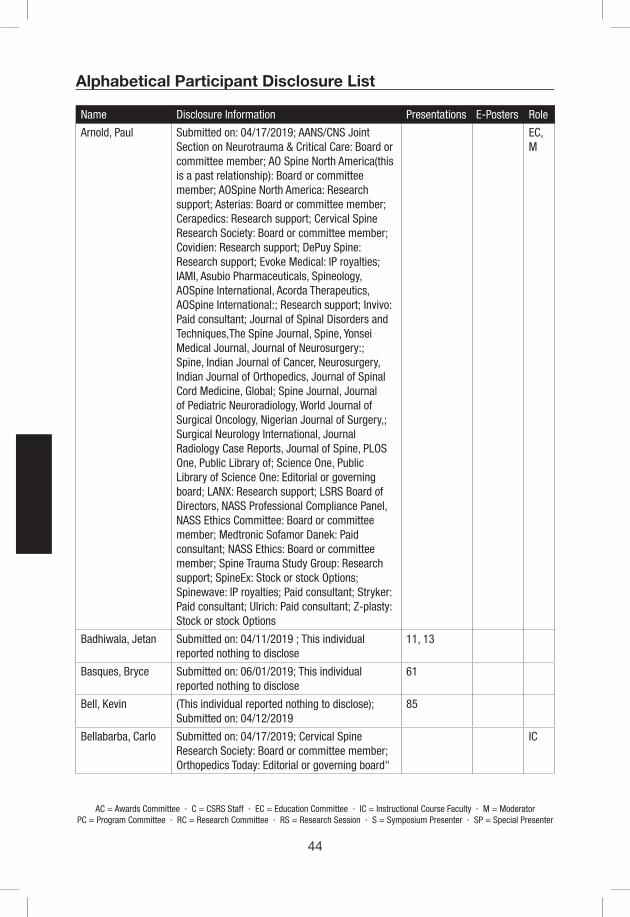
Arnold, Paul Submitted on: 04/17/2019; AANS/CNS Joint Section on Neurotrauma & Critical Care: Board or committee member; AO Spine North America(this is a past relationship): Board or committee member; AOSpine North America: Research support; Asterias: Board or committee member; Cerapedics: Research support; Cervical Spine Research Society: Board or committee member; Covidien: Research support; DePuy Spine: Research support; Evoke Medical: IP royalties; IAMI, Asubio Pharmaceuticals, Spineology, AOSpine International, Acorda Therapeutics, AOSpine International:; Research support; Invivo: Paid consultant; Journal of Spinal Disorders and Techniques,The Spine Journal, Spine, Yonsei Medical Journal, Journal of Neurosurgery:; Spine, Indian Journal of Cancer, Neurosurgery, Indian Journal of Orthopedics, Journal of Spinal Cord Medicine, Global; Spine Journal, Journal of Pediatric Neuroradiology, World Journal of Surgical Oncology, Nigerian Journal of Surgery,; Surgical Neurology International, Journal Radiology Case Reports, Journal of Spine, PLOS One, Public Library of; Science One, Public Library of Science One: Editorial or governing board; LANX: Research support; LSRS Board of Directors, NASS Professional Compliance Panel, NASS Ethics Committee: Board or committee member; Medtronic Sofamor Danek: Paid consultant; NASS Ethics: Board or committee member; Spine Trauma Study Group: Research support; SpineEx: Stock or stock Options; Spinewave: IP royalties; Paid consultant; Stryker: Paid consultant; Ulrich: Paid consultant; Z-plasty: Stock or stock Options
EC, M
Badhiwala, Jetan Submitted on: 04/11/2019 ; This individual reported nothing to disclose
11, 13
Basques, Bryce Submitted on: 06/01/2019; This individual reported nothing to disclose
61
Bell, Kevin (This individual reported nothing to disclose); Submitted on: 04/12/2019
85
Bellabarba, Carlo Submitted on: 04/17/2019; Cervical Spine Research Society: Board or committee member; Orthopedics Today: Editorial or governing board"
IC

45
Alphabetical Participant Disclosure List
Name Disclosure Information Presentations E-Posters Role
Bhatia, Nitin Submitted on: 03/29/2019; Alphatec Spine: IP royalties; Paid consultant; Paid presenter or speaker; Biomet: IP royalties; Paid consultant; Paid presenter or speaker; Cervical Spine Research Society: Board or committee member; DiFusion: Paid consultant; Stock or stock Options; North American Spine Society: Board or committee member; OKO: Editorial or governing board; Orthofix, Inc.: Paid presenter or speaker; Seaspine: IP royalties; Paid consultant; Paid presenter or speaker; Spineart: IP royalties; Paid presenter or speaker; Spineart, Zimmer: Paid consultant; SpineLine: Editorial or governing board; Stryker: IP royalties; Paid consultant; Paid presenter or speaker; Western Orthopaedic Association: Board or committee member
RC
Bible, Jesse (This individual reported nothing to disclose); Submitted on: 12/06/2018
RS
Bisson, Erica Submitted on: 04/06/2019 AANS Ethics, AANS/CNS Spine SPC: Board or committee member Journal of Neurosurgery: Spine: Editorial or governing board MiRus: Paid consultant nView: Paid consultant; Stock or stock Options
AC
Brodke, Darrel Submitted on: 05/07/2019; Amedica: IP royalties; AOSpine: Board or committee member; Cervical Spine Research Society: Board or committee member; Clinical Orthopaedics and Related Research: Editorial or governing board; Lumbar Spine Research Society: Board or committee member; Medtronic: IP royalties; Vallum: Paid consultant
IC, M

46
AC = Awards Committee • C = CSRS Staff • EC = Education Committee • IC = Instructional Course Faculty • M = ModeratorPC = Program Committee • RC = Research Committee • RS = Research Session • S = Symposium Presenter • SP = Special Presenter
Alphabetical Participant Disclosure List
Name Disclosure Information Presentations E-Posters Role
Buchowski, Jacob Submitted on: 07/29/2019; AAOS: Board or committee member; American Spinal Injury Association: Board or committee member; Association of Bone and Joint Surgeons: Board or committee member; Cervical Spine Research Society: Board or committee member; FOSA: Board or committee member; Globus Medical: IP royalties; Journal of Bone and Joint Surgery - American: Editorial or governing board; K2M: IP royalties; Lumbar Spine Research Society: Board or committee member; North American Spine Society: Board or committee member; Scoliosis Research Society: Board or committee member; Spine Deformity: Editorial or governing board; Wolters Kluwer Health - Lippincott Williams & Wilkins: Publishing royalties, financial or material support
PC, M
Bydon, Mohamad Submitted on: 04/08/2019; This individual reported nothing to disclose
IC
Chan, Andrew Submitted on: 07/08/2019; Orthofix, Inc.: Other financial or material support
59 9
Changoor, Stuart (This individual reported nothing to disclose); Submitted on: 08/22/2019
65
Chen, Xiongsheng (This individual reported nothing to disclose); Submitted on: 04/11/2019
24
Cheng, Wayne Submitted on: 09/03/2019; Cervical Spine Research Society: Board or committee member; DePuy, A Johnson & Johnson Company: Paid consultant; international journal of spine surgery: Editorial or governing board; Medtronic: Research support; Presidyan: Stock or stock Options; radius: Paid presenter or speaker
EC

47
Alphabetical Participant Disclosure List
Name Disclosure Information Presentations E-Posters Role
Chiba, Kazuhiro Submitted on: 08/28/2019; Asahi Kasei Pharma Corp.: Research support; Astellas Pharma Inc.: Research support; Cervical Spine Research Society Asia-Pacific Section: Board or committee member; Chug Pharmaceutical Co. Ltd.: Research support; Chugai Co. Ltd: Paid presenter or speaker; Daiichi Sankyo Co. Ldt: Research support; Daiichi-Sankyo Co. Ltd.: Paid presenter or speaker; Eastern Japan Association of Orthopedics and Traumatology: Board or committee member; Eisai Co. Ltd.: Paid presenter or speaker; Research support; Eli Lilly: Research support; Hisamitsu Pharmaceutical Co., Inc.: Research support; International Society for the Study of the Lumbar Spine: Board or committee member; Japanese Orthopedic Association: Board or committee member; Japanese Society for Spine Surgery and Related Research: Board or committee member; Journal of Orthopaedic Science: Editorial or governing board; Kaken Pharmaceutical Co. Ltd.: Paid presenter or speaker; Research support; Kanto Society of Orthopedics and Traumatology: Board or committee member; Medtronic Sofamor Danek: Research support; MSD Co. Ltd.: Research support; Nippon Zoki Pharmaceutial Co. Ltd.: Research support; Pfeizer Japan Inc.: Paid presenter or speaker; Pfizer: Research support; Seikagaku Corp.: Paid consultant; Research support; Shionogi & Co. Ltd.: Research support; Smith & Nephew: Research support; Spine: Editorial or governing board; Spine Surgery and Related Research: Editorial or governing board; Stryker: Research support; Taisho Pharmaceutical Co. Ltd.: Paid presenter or speaker; Research support; Teijin Pharma Ltd.: Research support; The Japanese Society of Lumbar Spine Disorders: Board or committee member; The Study Group for Nerve and Spine: Board or committee member; Tiejin Pharma Ltd.: Paid presenter or speaker; Zimmer: Research support
M
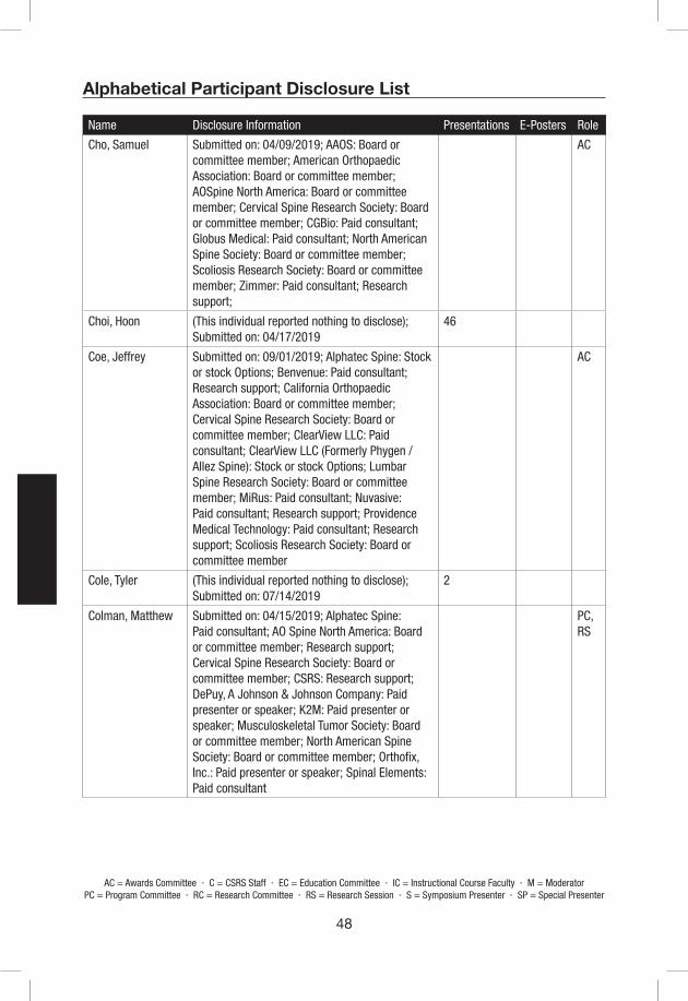
48
AC = Awards Committee • C = CSRS Staff • EC = Education Committee • IC = Instructional Course Faculty • M = ModeratorPC = Program Committee • RC = Research Committee • RS = Research Session • S = Symposium Presenter • SP = Special Presenter
Alphabetical Participant Disclosure List
Name Disclosure Information Presentations E-Posters Role
Cho, Samuel Submitted on: 04/09/2019; AAOS: Board or committee member; American Orthopaedic Association: Board or committee member; AOSpine North America: Board or committee member; Cervical Spine Research Society: Board or committee member; CGBio: Paid consultant; Globus Medical: Paid consultant; North American Spine Society: Board or committee member; Scoliosis Research Society: Board or committee member; Zimmer: Paid consultant; Research support;
AC
Choi, Hoon (This individual reported nothing to disclose); Submitted on: 04/17/2019
46
Coe, Jeffrey Submitted on: 09/01/2019; Alphatec Spine: Stock or stock Options; Benvenue: Paid consultant; Research support; California Orthopaedic Association: Board or committee member; Cervical Spine Research Society: Board or committee member; ClearView LLC: Paid consultant; ClearView LLC (Formerly Phygen / Allez Spine): Stock or stock Options; Lumbar Spine Research Society: Board or committee member; MiRus: Paid consultant; Nuvasive: Paid consultant; Research support; Providence Medical Technology: Paid consultant; Research support; Scoliosis Research Society: Board or committee member
AC
Cole, Tyler (This individual reported nothing to disclose); Submitted on: 07/14/2019
2
Colman, Matthew Submitted on: 04/15/2019; Alphatec Spine: Paid consultant; AO Spine North America: Board or committee member; Research support; Cervical Spine Research Society: Board or committee member; CSRS: Research support; DePuy, A Johnson & Johnson Company: Paid presenter or speaker; K2M: Paid presenter or speaker; Musculoskeletal Tumor Society: Board or committee member; North American Spine Society: Board or committee member; Orthofix, Inc.: Paid presenter or speaker; Spinal Elements: Paid consultant
PC, RS

49
Alphabetical Participant Disclosure List
Name Disclosure Information Presentations E-Posters Role
Coric, Domagoj Submitted on: 04/12/2019; AANS/CNS Joint Section on Spine & Peripheral Nerves: Board or committee member; Aesculap/B.Braun: Paid presenter or speaker; DiscGenics: Research support; Globus Medical: IP royalties; Paid consultant; Paid presenter or speaker; Research support; Integrity Implants: IP royalties; ISASS: Board or committee member; ISASS Journal: Editorial or governing board; Medtronic: IP royalties; Paid consultant; Paid presenter or speaker; NuTech: Research support; Premia Spine: Paid consultant; Research support; Stock or stock Options; RTI Surgical: IP royalties; Southern Neurosurgical Society: Board or committee member; Spine Wave: IP royalties; Paid consultant; Paid presenter or speaker; Stock or stock Options; Stryker: IP royalties; Paid consultant; Paid presenter or speaker
IC
Crawford, Charles H III
Submitted on: 04/06/2019; Alphatec Spine: IP royalties; Paid consultant; DePuy, A Johnson & Johnson Company: Paid consultant; Medtronic: Paid consultant; Medtronic Sofamor Danek: Paid presenter or speaker; North American Spine Society: Board or committee member; Nuvasive: Paid consultant; Paid presenter or speaker; Scoliosis Research Society: Board or committee member; Springer: Publishing royalties, financial or material support
51
Dailey, Andrew Submitted on: 08/28/2019; Biomet: IP royalties; Paid consultant; Cervical Spine Research Society: Board or committee member; K2M: Research support; Lumbar Spine Research Society: Board or committee member
M, IC, PC
Daubs, Michael Submitted on: 04/16/2019; AOSpine North America: Board or committee member; Cervical Spine Research Society: Board or committee member; DePuy, A Johnson & Johnson Company: IP royalties; lumbar spine research society: Board or committee member; Pfizer: Research support; The Spine Journal: Editorial or governing board
EC, M
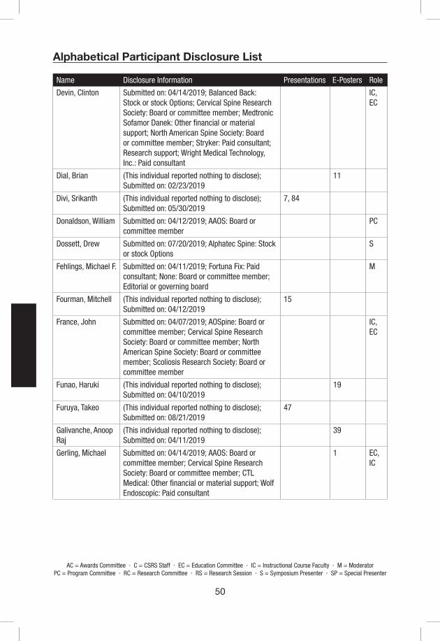
50
AC = Awards Committee • C = CSRS Staff • EC = Education Committee • IC = Instructional Course Faculty • M = ModeratorPC = Program Committee • RC = Research Committee • RS = Research Session • S = Symposium Presenter • SP = Special Presenter
Alphabetical Participant Disclosure List
Name Disclosure Information Presentations E-Posters Role
Devin, Clinton Submitted on: 04/14/2019; Balanced Back: Stock or stock Options; Cervical Spine Research Society: Board or committee member; Medtronic Sofamor Danek: Other financial or material support; North American Spine Society: Board or committee member; Stryker: Paid consultant; Research support; Wright Medical Technology, Inc.: Paid consultant
IC, EC
Dial, Brian (This individual reported nothing to disclose); Submitted on: 02/23/2019
11
Divi, Srikanth (This individual reported nothing to disclose); Submitted on: 05/30/2019
7, 84
Donaldson, William Submitted on: 04/12/2019; AAOS: Board or committee member
PC
Dossett, Drew Submitted on: 07/20/2019; Alphatec Spine: Stock or stock Options
S
Fehlings, Michael F. Submitted on: 04/11/2019; Fortuna Fix: Paid consultant; None: Board or committee member; Editorial or governing board
M
Fourman, Mitchell (This individual reported nothing to disclose); Submitted on: 04/12/2019
15
France, John Submitted on: 04/07/2019; AOSpine: Board or committee member; Cervical Spine Research Society: Board or committee member; North American Spine Society: Board or committee member; Scoliosis Research Society: Board or committee member
IC, EC
Funao, Haruki (This individual reported nothing to disclose); Submitted on: 04/10/2019
19
Furuya, Takeo (This individual reported nothing to disclose); Submitted on: 08/21/2019
47
Galivanche, Anoop Raj
(This individual reported nothing to disclose); Submitted on: 04/11/2019
39
Gerling, Michael Submitted on: 04/14/2019; AAOS: Board or committee member; Cervical Spine Research Society: Board or committee member; CTL Medical: Other financial or material support; Wolf Endoscopic: Paid consultant
1 EC, IC

51
Alphabetical Participant Disclosure List
Name Disclosure Information Presentations E-Posters Role
Ghiselli, Gary Submitted on: 09/09/2019; Colorado Medical Society: Board or committee member; Colorado Orthopedic Society: Board or committee member; New Era Orthopedics: IP royalties; North American Spine Society: Board or committee member
AC
Ghogawala, Zoher Submitted on: 07/12/2019; AANS-CNS Joint Spine Section: Board or committee member; Cervical Spine Research Society: Board or committee member; North American Spine Society: Board or committee member
54 M, IC, RC
Groves Sausser, Lindsey
(This individual reported nothing to disclose); Submitted on: 06/28/2019
C
Harrop, James Submitted on: 04/15/2019; AlaMab: Other financial or material support; Avvie: Other financial or material support; Cervical Spine Research Society: Board or committee member; DePuy, A Johnson & Johnson Company: Paid consultant; Paid presenter or speaker; ethicon: Paid consultant; Globus Medical: Paid presenter or speaker; LSRS: Board or committee member; PNS: Board or committee member; Spine Universe, CNS quarterly, Congress of Neurosurgeons Execuative Board, CSRS,PNS, Jefferson University Physicians, LSRS, COSSS: Editorial or governing board
M
Hart, Robert Submitted on: 04/10/2019; American Orthopaedic Association: Board or committee member; Cervical Spine Research Society: Board or committee member; depuy: Research support; DePuy, A Johnson & Johnson Company: IP royalties; Paid consultant; Paid presenter or speaker; Globus Medical: IP royalties; Paid consultant; Paid presenter or speaker; International Spine Study Group: Board or committee member; ISSLS Textbook of the Lumbar Spine: Editorial or governing board; Medtronic: Paid consultant; Paid presenter or speaker; Misonix: Research support; North American Spine Society: Board or committee member; Orthofix, Inc.: Paid consultant; Paid presenter or speaker; Scoliosis Research Society: Board or committee member; SeaSpine: IP royalties; Spine Connect: Stock or stock Options; Western Ortho Assn: Board or committee member
M
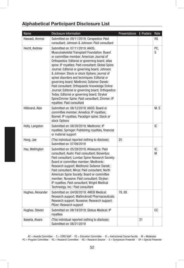
52
AC = Awards Committee • C = CSRS Staff • EC = Education Committee • IC = Instructional Course Faculty • M = ModeratorPC = Program Committee • RC = Research Committee • RS = Research Session • S = Symposium Presenter • SP = Special Presenter
Alphabetical Participant Disclosure List
Name Disclosure Information Presentations E-Posters Role
Hawasli, Ammar Submitted on: 09/11/2019; Cerapedics: Paid consultant; Johnson & Johnson: Paid consultant
RS
Hecht, Andrew Submitted on: 07/11/2019; AAOS, Musculoskeletal Transplant Foundation: Board or committee member; American Journal of Orthopedics: Editorial or governing board; atlas spine: IP royalties; Paid consultant; Global Spine Journal: Editorial or governing board; Johnson & Johnson: Stock or stock Options; journal of spinal disorders and techniques: Editorial or governing board; Medtronic Sofamor Danek: Paid consultant; Orthopaedic Knowledge Online Journal: Editorial or governing board; Orthopedics Today: Editorial or governing board; Stryker SpineZimmer Spine: Paid consultant; Zimmer: IP royalties; Paid consultant
PC, S
Hilibrand, Alan Submitted on: 08/12/2019; AAOS: Board or committee member; Amedica: IP royalties; Biomet: IP royalties; Paradigm spine: Stock or stock Options
M, S
Holly, Langston Submitted on: 08/28/2019; Medtronic: IP royalties; Springer: Publishing royalties, financial or material support
IC
Hong, Jae (This individual reported nothing to disclose); Submitted on: 07/09/2019
25
Hsu, Wellington Submitted on: 05/28/2019; Allosource: Paid consultant; Asahi: Paid consultant; Bioventus: Paid consultant; Lumbar Spine Research Society: Board or committee member; Medtronic: Research support; Medtronic Sofamor Danek: Paid consultant; Mirus: Paid consultant; North American Spine Society: Board or committee member; Nuvasive: Paid consultant; Stryker: IP royalties; Paid consultant; Wright Medical Technology, Inc.: Paid consultant
IC, M
Hughes, Alexander Submitted on: 04/08/2019; 4WEB Medical: Research support; Mallinckrodt Pharmaceuticals: Research support; Nuvasive: Research support; Pfizer: Research support
79, 88
Hughes, Steven Submitted on: 08/19/2019; Globus Medical: IP royalties
EC
Ibaseta, Alvaro (This individual reported nothing to disclose); Submitted on: 08/21/2019
21
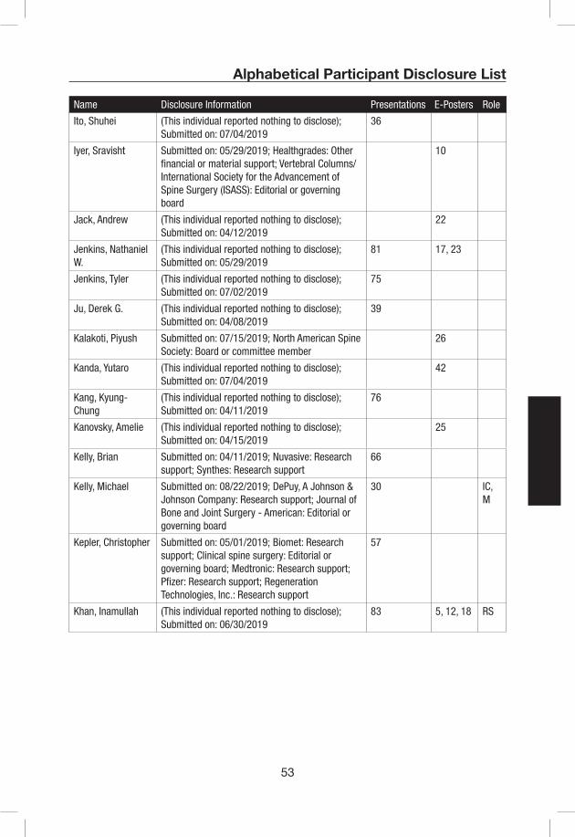
53
Alphabetical Participant Disclosure List
Name Disclosure Information Presentations E-Posters Role
Ito, Shuhei (This individual reported nothing to disclose); Submitted on: 07/04/2019
36
Iyer, Sravisht Submitted on: 05/29/2019; Healthgrades: Other financial or material support; Vertebral Columns/International Society for the Advancement of Spine Surgery (ISASS): Editorial or governing board
10
Jack, Andrew (This individual reported nothing to disclose); Submitted on: 04/12/2019
22
Jenkins, Nathaniel W.
(This individual reported nothing to disclose); Submitted on: 05/29/2019
81 17, 23
Jenkins, Tyler (This individual reported nothing to disclose); Submitted on: 07/02/2019
75
Ju, Derek G. (This individual reported nothing to disclose); Submitted on: 04/08/2019
39
Kalakoti, Piyush Submitted on: 07/15/2019; North American Spine Society: Board or committee member
26
Kanda, Yutaro (This individual reported nothing to disclose); Submitted on: 07/04/2019
42
Kang, Kyung-Chung
(This individual reported nothing to disclose); Submitted on: 04/11/2019
76
Kanovsky, Amelie (This individual reported nothing to disclose); Submitted on: 04/15/2019
25
Kelly, Brian Submitted on: 04/11/2019; Nuvasive: Research support; Synthes: Research support
66
Kelly, Michael Submitted on: 08/22/2019; DePuy, A Johnson & Johnson Company: Research support; Journal of Bone and Joint Surgery - American: Editorial or governing board
30 IC, M
Kepler, Christopher Submitted on: 05/01/2019; Biomet: Research support; Clinical spine surgery: Editorial or governing board; Medtronic: Research support; Pfizer: Research support; Regeneration Technologies, Inc.: Research support
57
Khan, Inamullah (This individual reported nothing to disclose); Submitted on: 06/30/2019
83 5, 12, 18 RS

54
AC = Awards Committee • C = CSRS Staff • EC = Education Committee • IC = Instructional Course Faculty • M = ModeratorPC = Program Committee • RC = Research Committee • RS = Research Session • S = Symposium Presenter • SP = Special Presenter
Alphabetical Participant Disclosure List
Name Disclosure Information Presentations E-Posters Role
Kim, Han Jo Submitted on: 04/11/2019; AAOS: Board or committee member; AO SPINE: Board or committee member; Cervical Spine Research Society: Board or committee member; HSS Journal, Asian Spine Journal: Editorial or governing board; ISSGF: Research support; K2M: IP royalties; Scoliosis Research Society: Board or committee member; Zimmer: IP royalties
IC, M, PC
Kimura, Atsushi (This individual reported nothing to disclose); Submitted on: 04/07/2019
33
Klineberg, Eric Submitted on: 04/11/2019; AO Spine: Paid presenter or speaker; Research support; DePuy Synthes Spine: Research support; DePuy, A Johnson & Johnson Company: Paid consultant; K2M: Paid presenter or speaker; Medicrea: Paid consultant; OREF: Research support; Stryker: Paid consultant
19 M
Ko, Jong-Hyun (This individual reported nothing to disclose); Submitted on: 09/04/2019
12
Koller, Heiko (This individual reported nothing to disclose); Submitted on: 04/12/2019
S
Kuhns, Craig Submitted on: 08/29/2019; Alphatec Spine: Paid consultant; Stock or stock Options; Nuvasive: Paid consultant; Stock or stock Options; Spineology: IP royalties; Paid consultant
PC
Kwon, Brian Submitted on: 04/16/2019; Nervgen: Paid consultant; Versapeutics: Paid consultant
78 PC, M, RS
Lafage, Renaud Submitted on: 09/11/2019; Nemaris: Stock or stock Options
32
Lafage, Virginie Submitted on: 04/11/2019; DePuy, A Johnson & Johnson Company: Paid presenter or speaker; Research support; Globus Medical: Paid consultant; International Spine Study Group: Board or committee member; K2M: Paid presenter or speaker; Medtronic: Research support; Nemaris Inc: Stock or stock Options; Nuvasive: Research support; Scoliosis Research Society: Board or committee member; Stryker: Research support
29, 40
Lau, Darryl (This individual reported nothing to disclose); Submitted on: 07/08/2019
21

55
Alphabetical Participant Disclosure List
Name Disclosure Information Presentations E-Posters Role
Lawrence, Brandon Submitted on: 04/26/2019; AO Spine Fellowship Committee: Board or committee member; AO Spine North America: Paid presenter or speaker; Cervical Spine Research Society: Board or committee member; K2M: Paid presenter or speaker
IC, M, RC
Laxer, Eric Submitted on: 06/03/2019; Cervical Spine Research Society: Board or committee member; Cutting Edge Spine: Paid consultant; Lumbar Spine Research Society: Board or committee member; Nuvasive: Other financial or material support
AC
Le Naveaux, Franck
Submitted on: 04/15/2019; DePuy, A Johnson & Johnson Company: Paid consultant; Spinologics Inc.: Employee
89
Lee, Chang (This individual reported nothing to disclose); Submitted on: 06/20/2012
56
Lee, Dong Ho (This individual reported nothing to disclose); Submitted on: 08/28/2019
18
Lee, Ho Jin (This individual reported nothing to disclose); Submitted on: 04/10/2019
6
Lee, Jong Beom (This individual reported nothing to disclose); Submitted on: 04/13/2019
34
Lee, Michael Submitted on: 06/03/2019; DePuy, A Johnson & Johnson Company: Paid consultant; Globus Medical: Paid consultant; Stryker: Paid consultant
AC
Lee, Sang Hun Submitted on: 06/02/2019; CG Bio: Paid consultant; Medtronic: Paid presenter or speaker
S
Lee, Su Hun (This individual reported nothing to disclose); Submitted on: 07/12/2019
30
Lehman, Ronald Submitted on: 04/04/2019; AOSpine: Board or committee member; Associate Editor - Spine Deformity: Editorial or governing board; Cervical Spine Research Society: Board or committee member; Deputy Editor for Deformity - The Spine Journal: Editorial or governing board; Medtronic: Paid consultant; North American Spine Society: Board or committee member; Scoliosis Research Society: Board or committee member; Wolters Kluwer Health - Lippincott Williams & Wilkins: Publishing royalties, financial or material support
PC
Lemke, Denise (This individual reported nothing to disclose); Submitted on: 04/30/2019
C
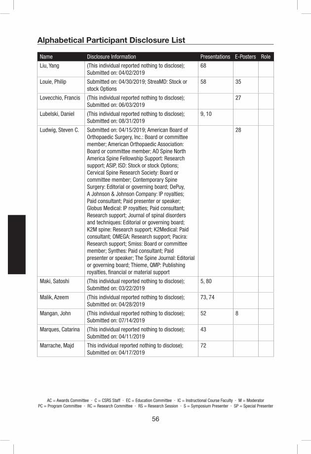
56
AC = Awards Committee • C = CSRS Staff • EC = Education Committee • IC = Instructional Course Faculty • M = ModeratorPC = Program Committee • RC = Research Committee • RS = Research Session • S = Symposium Presenter • SP = Special Presenter
Alphabetical Participant Disclosure List
Name Disclosure Information Presentations E-Posters Role
Liu, Yang (This individual reported nothing to disclose); Submitted on: 04/02/2019
68
Louie, Philip Submitted on: 04/30/2019; StreaMD: Stock or stock Options
58 35
Lovecchio, Francis (This individual reported nothing to disclose); Submitted on: 06/03/2019
27
Lubelski, Daniel (This individual reported nothing to disclose); Submitted on: 08/31/2019
9, 10
Ludwig, Steven C. Submitted on: 04/15/2019; American Board of Orthopaedic Surgery, Inc.: Board or committee member; American Orthopaedic Association: Board or committee member; AO Spine North America Spine Fellowship Support: Research support; ASIP, ISD: Stock or stock Options; Cervical Spine Research Society: Board or committee member; Contemporary Spine Surgery: Editorial or governing board; DePuy, A Johnson & Johnson Company: IP royalties; Paid consultant; Paid presenter or speaker; Globus Medical: IP royalties; Paid consultant; Research support; Journal of spinal disorders and techniques: Editorial or governing board; K2M spine: Research support; K2Medical: Paid consultant; OMEGA: Research support; Pacira: Research support; Smiss: Board or committee member; Synthes: Paid consultant; Paid presenter or speaker; The Spine Journal: Editorial or governing board; Thieme, QMP: Publishing royalties, financial or material support
28
Maki, Satoshi (This individual reported nothing to disclose); Submitted on: 03/22/2019
5, 80
Malik, Azeem (This individual reported nothing to disclose); Submitted on: 04/28/2019
73, 74
Mangan, John (This individual reported nothing to disclose); Submitted on: 07/14/2019
52 8
Marques, Catarina (This individual reported nothing to disclose); Submitted on: 04/11/2019
43
Marrache, Majd This individual reported nothing to disclose); Submitted on: 04/17/2019
72
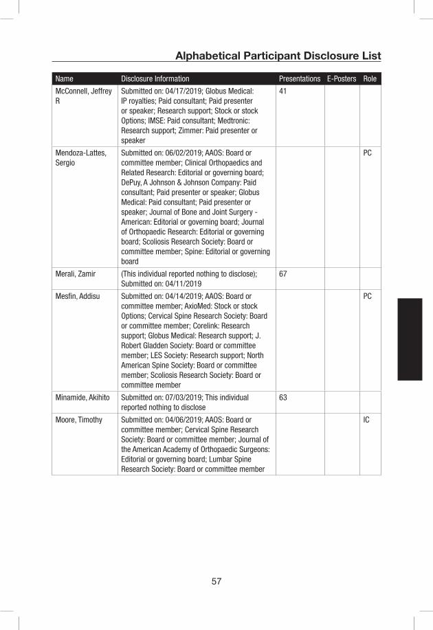
57
Alphabetical Participant Disclosure List
Name Disclosure Information Presentations E-Posters Role
McConnell, Jeffrey R
Submitted on: 04/17/2019; Globus Medical: IP royalties; Paid consultant; Paid presenter or speaker; Research support; Stock or stock Options; IMSE: Paid consultant; Medtronic: Research support; Zimmer: Paid presenter or speaker
41
Mendoza-Lattes, Sergio
Submitted on: 06/02/2019; AAOS: Board or committee member; Clinical Orthopaedics and Related Research: Editorial or governing board; DePuy, A Johnson & Johnson Company: Paid consultant; Paid presenter or speaker; Globus Medical: Paid consultant; Paid presenter or speaker; Journal of Bone and Joint Surgery - American: Editorial or governing board; Journal of Orthopaedic Research: Editorial or governing board; Scoliosis Research Society: Board or committee member; Spine: Editorial or governing board
PC
Merali, Zamir (This individual reported nothing to disclose); Submitted on: 04/11/2019
67
Mesfin, Addisu Submitted on: 04/14/2019; AAOS: Board or committee member; AxioMed: Stock or stock Options; Cervical Spine Research Society: Board or committee member; Corelink: Research support; Globus Medical: Research support; J. Robert Gladden Society: Board or committee member; LES Society: Research support; North American Spine Society: Board or committee member; Scoliosis Research Society: Board or committee member
PC
Minamide, Akihito Submitted on: 07/03/2019; This individual reported nothing to disclose
63
Moore, Timothy Submitted on: 04/06/2019; AAOS: Board or committee member; Cervical Spine Research Society: Board or committee member; Journal of the American Academy of Orthopaedic Surgeons: Editorial or governing board; Lumbar Spine Research Society: Board or committee member
IC

58
AC = Awards Committee • C = CSRS Staff • EC = Education Committee • IC = Instructional Course Faculty • M = ModeratorPC = Program Committee • RC = Research Committee • RS = Research Session • S = Symposium Presenter • SP = Special Presenter
Alphabetical Participant Disclosure List
Name Disclosure Information Presentations E-Posters Role
Mroz, Thomas Submitted on: 04/12/2019; Cervical Spine Research Society: Board or committee member; North American Spine Society: Board or committee member; Pearl Diver, Inc: Stock or stock Options; SpineLine, EditorGlobal Spine Journal, Deputy Editor: Editorial or governing board; Stryker: IP royalties; Paid consultant
PC, M
Mummaneni, Praveen
Submitted on: 04/06/2019 AANS/CNS Spine Section and Scoliosis Research Society: Board or committee member American Association of Neurological Surgeons: Board or committee member AOSpine: Paid presenter or speaker Cervical Spine Research Society: Board or committee member Congress of Neurological Surgeons: Board or committee member DePuy, A Johnson & Johnson Company: IP royalties; Paid consultant Global Spine Journal: Editorial or governing board Globus Medical: Paid consultant International Spine Study Group: Research support Neurosurgery: Editorial or governing board NREF: Research support Spinal Deformity: Editorial or governing board Spineart: Paid presenter or speaker Spinicity/ISD: Stock or stock Options Springer: Publishing royalties, financial or material support Stryker: Paid consultant Taylor and Francis: Publishing royalties, financial or material support Thieme: Publishing royalties, financial or material support World Neurosurgery: Editorial or governing board
AC
Muscarella, Joe (This individual reported nothing to disclose); Submitted on: 08/03/2019
IC
Nakajima, Koji (This individual reported nothing to disclose); Submitted on: 07/12/2019
64
Nassr, Ahmad Submitted on: 05/16/2019; American Orthopaedic Association: Board or committee member; AO Spine: Research support; Cervical Spine Research Society: Board or committee member; DePuy, A Johnson & Johnson Company: Paid consultant; Lumbar spine research society: Board or committee member; Pfizer: Research support; Premia Spine: Research support; Scoliosis Research Society: Board or committee member; Techniques in Orthopedics: Editorial or governing board
PC
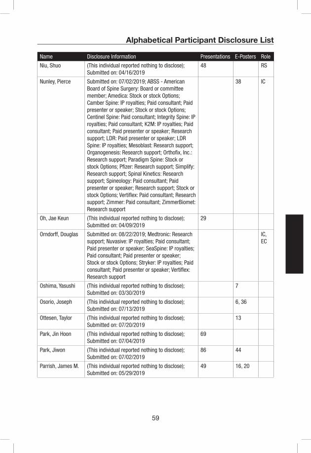
59
Alphabetical Participant Disclosure List
Name Disclosure Information Presentations E-Posters Role
Niu, Shuo (This individual reported nothing to disclose); Submitted on: 04/16/2019
48 RS
Nunley, Pierce Submitted on: 07/02/2019; ABSS - American Board of Spine Surgery: Board or committee member; Amedica: Stock or stock Options; Camber Spine: IP royalties; Paid consultant; Paid presenter or speaker; Stock or stock Options; Centinel Spine: Paid consultant; Integrity Spine: IP royalties; Paid consultant; K2M: IP royalties; Paid consultant; Paid presenter or speaker; Research support; LDR: Paid presenter or speaker; LDR Spine: IP royalties; Mesoblast: Research support; Organogenesis: Research support; Orthofix, Inc.: Research support; Paradigm Spine: Stock or stock Options; Pfizer: Research support; Simplify: Research support; Spinal Kinetics: Research support; Spineology: Paid consultant; Paid presenter or speaker; Research support; Stock or stock Options; Vertiflex: Paid consultant; Research support; Zimmer: Paid consultant; ZimmerBiomet: Research support
38 IC
Oh, Jae Keun (This individual reported nothing to disclose); Submitted on: 04/09/2019
29
Orndorff, Douglas Submitted on: 08/22/2019; Medtronic: Research support; Nuvasive: IP royalties; Paid consultant; Paid presenter or speaker; SeaSpine: IP royalties; Paid consultant; Paid presenter or speaker; Stock or stock Options; Stryker: IP royalties; Paid consultant; Paid presenter or speaker; Vertiflex: Research support
IC, EC
Oshima, Yasushi (This individual reported nothing to disclose); Submitted on: 03/30/2019
7
Osorio, Joseph (This individual reported nothing to disclose); Submitted on: 07/13/2019
6, 36
Ottesen, Taylor (This individual reported nothing to disclose); Submitted on: 07/20/2019
13
Park, Jin Hoon (This individual reported nothing to disclose); Submitted on: 07/04/2019
69
Park, Jiwon (This individual reported nothing to disclose); Submitted on: 07/02/2019
86 44
Parrish, James M. (This individual reported nothing to disclose); Submitted on: 05/29/2019
49 16, 20

60
AC = Awards Committee • C = CSRS Staff • EC = Education Committee • IC = Instructional Course Faculty • M = ModeratorPC = Program Committee • RC = Research Committee • RS = Research Session • S = Symposium Presenter • SP = Special Presenter
Alphabetical Participant Disclosure List
Name Disclosure Information Presentations E-Posters Role
Passias, Peter "Submitted on: 07/15/2019; Allosource: Other financial or material support; Cervical Scoliosis Research Society: Research support; Globus Medical: Paid presenter or speaker; Medicrea: Paid consultant; SpineWave: Paid consultant; Zimmer: Paid presenter or speaker"
26, 62 S, IC
Patel, Alpesh "Submitted on: 05/04/2019; Alphatec Spine: IP royalties; Amedica: IP royalties; Paid consultant; Stock or stock Options; American Orthopaedic Association: Board or committee member; AO Spine North America: Board or committee member; Cervical Spine Research Society: Board or committee member; Cytonics: Stock or stock Options; DePuy, A Johnson & Johnson Company: Paid consultant; EndoLuxe: Stock or stock Options; International Society for the Advancement of Spine Surgery: Board or committee member; Journal of the American Academy of Orthopaedic Surgeons: Editorial or governing board; Publishing royalties, financial or material support; Kuros Biosciences: Paid consultant; Lumbar Spine Research Society: Board or committee member; Nocimed: Stock or stock Options; North American Spine Society: Board or committee member; Nuvasive: IP royalties; Paid consultant; nView Medical Inc: Stock or stock Options; Springer: Publishing royalties, financial or material support; Surgical Neurology International: Editorial or governing board; Tissue Differentiation Intelligence: Stock or stock Options; Vital5: Stock or stock Options; Wolters Kluwer Health - Lippincott Williams & Wilkins: Editorial or governing board; Zimmer: IP royalties; Paid consultant"
IC
Peng, Cao (This individual reported nothing to disclose); Submitted on: 08/29/2019
4

61
Alphabetical Participant Disclosure List
Name Disclosure Information Presentations E-Posters Role
Phillips, Frank Submitted on: 04/12/2019; Int. Spine Journal: Editorial or governing board; ISASS: Board or committee member; Mainstay: Stock or stock Options; Medtronic: IP royalties; Nuvasive: IP royalties; Paid consultant; Stock or stock Options; PearDiver: Stock or stock Options; Providence: Stock or stock Options; SI Bone: Paid consultant; Stock or stock Options; Society of Minimally invasive Spine Surgery: Board or committee member; Spinal Simplicity: Stock or stock Options; Stryker: IP royalties; Paid consultant; Surgio: Stock or stock Options; Theracell: Stock or stock Options; Vertiflex: Stock or stock Options; Vital 5: Stock or stock Options
M
Protopsaltis, Themistocles
Submitted on: 08/23/2019; Globus Medical: Paid consultant; Innovasis: Paid consultant; K2M: Paid consultant; Medicrea International: Paid consultant; Nuvasive: Paid consultant; Torus Medical: Stock or stock Options
16, 20 33 S, IC, PC
Radcliff, Kristen Submitted on: 04/11/2019; 4 Web Medical: Stock or stock Options; AAOS: Board or committee member; Cervical Spine Research Society: Board or committee member; Globus Medical: IP royalties; Paid consultant; Innovative Spine Devices: IP royalties; ISASS: Board or committee member; Lilly USA: Other financial or material support; Medtronic: Paid consultant; NEXXT Spine: Other financial or material support; North American Spine Society: Board or committee member; Orthofix, Inc.: Research support; Orthopedic Sciences, Inc: IP royalties; Paid consultant; Pacira pharmaceuticals: Research support; Rothman Institute: Stock or stock Options; Simplify Medical: Research support; SMISS: Board or committee member; Stryker: Other financial or material support; Paid consultant; Zimmer: Unpaid consultant; Zimmer Biomet: Other financial or material support;
AC, M, EC
Reid, Daniel Submitted on: 04/17/2019; Cerapedics: Other financial or material support; Stryker: Other financial or material support
71

62
AC = Awards Committee • C = CSRS Staff • EC = Education Committee • IC = Instructional Course Faculty • M = ModeratorPC = Program Committee • RC = Research Committee • RS = Research Session • S = Symposium Presenter • SP = Special Presenter
Alphabetical Participant Disclosure List
Name Disclosure Information Presentations E-Posters Role
Rhee, John Submitted on: 04/16/2019; Alphatec Spine: Stock or stock Options; Biomet: IP royalties; BiometDepuy: Paid presenter or speaker; Biometsynthes: Paid consultant; Cervical Spine Research Society: Board or committee member; DePuy, A Johnson & Johnson CompanyKineflexMedtronic: Research support; Phygen: Stock or stock Options; Stryker: IP royalties; Wolters Kluwer Health - Lippincott Williams & Wilkins: Publishing royalties, financial or material support; Zimmer: Paid presenter or speaker
M, IC
Riew, K. Daniel Submitted on: 06/04/2019; Advanced Medical: Other financial or material support; Amedica: Stock or stock Options; AO Spine: Other financial or material support; AOSpine: Board or committee member; Research support; AxioMed: Stock or stock Options; Benvenue: Stock or stock Options; Biomet: IP royalties; Paid consultant; Paid presenter or speaker; Clinics in orthopedics: Editorial or governing board; DePuy, A Johnson & Johnson Company: Paid presenter or speaker; European Spine Journal: Editorial or governing board; Expanding Orthopedics, PSD: Stock or stock Options; global spine journal: Editorial or governing board; Invasive: Paid presenter or speaker; Medtronic: Paid consultant; Paid presenter or speaker; Neurosurgery: Editorial or governing board; Nuvasive: IP royalties; Paid consultant; Osprey: Stock or stock Options; Paradigm Spine: Stock or stock Options; Spinal Kinetics: Stock or stock Options; Spine: Editorial or governing board; spine surgery today: Editorial or governing board; Spineology: Stock or stock Options; Vertiflex: Stock or stock Options; Zeiss: Other financial or material support
45, 55 M, IC
Rihn, Jeffrey Submitted on: 04/12/2019; Cervical Spine Research Society: Board or committee member; Globus Medical: Paid consultant; The Spine Journal: Editorial or governing board; XTANT Medical: Stock or stock Options
PC
Rodnoi, Pope (This individual reported nothing to disclose); Submitted on: 08/21/2019
4
Sakai, Kenichiro (This individual reported nothing to disclose); Submitted on: 04/04/2019
24
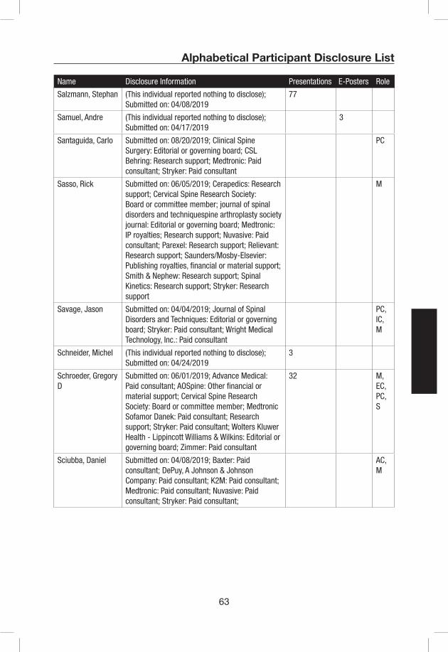
63
Alphabetical Participant Disclosure List
Name Disclosure Information Presentations E-Posters Role
Salzmann, Stephan (This individual reported nothing to disclose); Submitted on: 04/08/2019
77
Samuel, Andre (This individual reported nothing to disclose); Submitted on: 04/17/2019
3
Santaguida, Carlo Submitted on: 08/20/2019; Clinical Spine Surgery: Editorial or governing board; CSL Behring: Research support; Medtronic: Paid consultant; Stryker: Paid consultant
PC
Sasso, Rick Submitted on: 06/05/2019; Cerapedics: Research support; Cervical Spine Research Society: Board or committee member; journal of spinal disorders and techniquespine arthroplasty society journal: Editorial or governing board; Medtronic: IP royalties; Research support; Nuvasive: Paid consultant; Parexel: Research support; Relievant: Research support; Saunders/Mosby-Elsevier: Publishing royalties, financial or material support; Smith & Nephew: Research support; Spinal Kinetics: Research support; Stryker: Research support
M
Savage, Jason Submitted on: 04/04/2019; Journal of Spinal Disorders and Techniques: Editorial or governing board; Stryker: Paid consultant; Wright Medical Technology, Inc.: Paid consultant
PC, IC, M
Schneider, Michel (This individual reported nothing to disclose); Submitted on: 04/24/2019
3
Schroeder, Gregory D
Submitted on: 06/01/2019; Advance Medical: Paid consultant; AOSpine: Other financial or material support; Cervical Spine Research Society: Board or committee member; Medtronic Sofamor Danek: Paid consultant; Research support; Stryker: Paid consultant; Wolters Kluwer Health - Lippincott Williams & Wilkins: Editorial or governing board; Zimmer: Paid consultant
32 M, EC, PC, S
Sciubba, Daniel Submitted on: 04/08/2019; Baxter: Paid consultant; DePuy, A Johnson & Johnson Company: Paid consultant; K2M: Paid consultant; Medtronic: Paid consultant; Nuvasive: Paid consultant; Stryker: Paid consultant;
AC, M
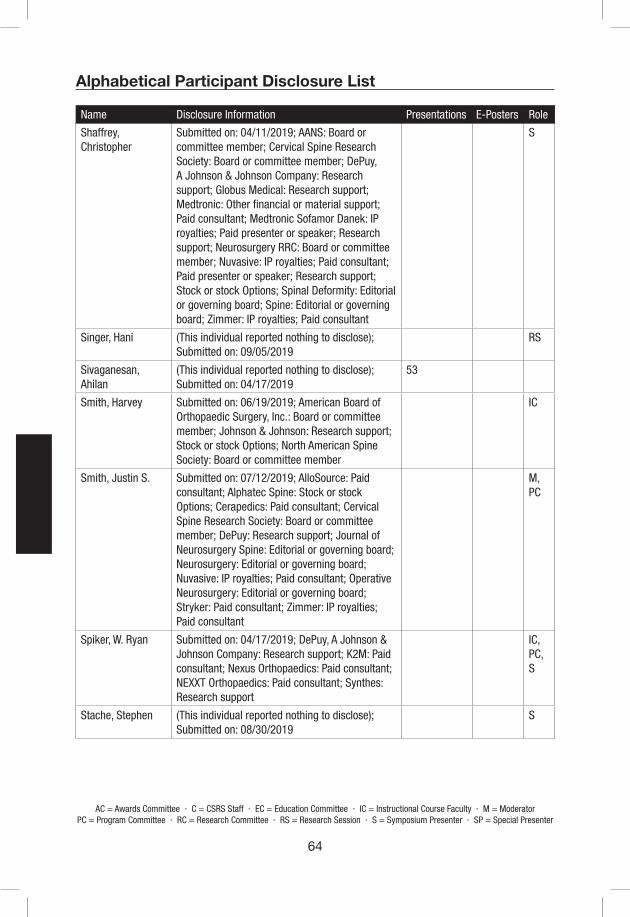
64
AC = Awards Committee • C = CSRS Staff • EC = Education Committee • IC = Instructional Course Faculty • M = ModeratorPC = Program Committee • RC = Research Committee • RS = Research Session • S = Symposium Presenter • SP = Special Presenter
Alphabetical Participant Disclosure List
Name Disclosure Information Presentations E-Posters Role
Shaffrey, Christopher
Submitted on: 04/11/2019; AANS: Board or committee member; Cervical Spine Research Society: Board or committee member; DePuy, A Johnson & Johnson Company: Research support; Globus Medical: Research support; Medtronic: Other financial or material support; Paid consultant; Medtronic Sofamor Danek: IP royalties; Paid presenter or speaker; Research support; Neurosurgery RRC: Board or committee member; Nuvasive: IP royalties; Paid consultant; Paid presenter or speaker; Research support; Stock or stock Options; Spinal Deformity: Editorial or governing board; Spine: Editorial or governing board; Zimmer: IP royalties; Paid consultant
S
Singer, Hani (This individual reported nothing to disclose); Submitted on: 09/05/2019
RS
Sivaganesan, Ahilan
(This individual reported nothing to disclose); Submitted on: 04/17/2019
53
Smith, Harvey Submitted on: 06/19/2019; American Board of Orthopaedic Surgery, Inc.: Board or committee member; Johnson & Johnson: Research support; Stock or stock Options; North American Spine Society: Board or committee member
IC
Smith, Justin S. Submitted on: 07/12/2019; AlloSource: Paid consultant; Alphatec Spine: Stock or stock Options; Cerapedics: Paid consultant; Cervical Spine Research Society: Board or committee member; DePuy: Research support; Journal of Neurosurgery Spine: Editorial or governing board; Neurosurgery: Editorial or governing board; Nuvasive: IP royalties; Paid consultant; Operative Neurosurgery: Editorial or governing board; Stryker: Paid consultant; Zimmer: IP royalties; Paid consultant
M, PC
Spiker, W. Ryan Submitted on: 04/17/2019; DePuy, A Johnson & Johnson Company: Research support; K2M: Paid consultant; Nexus Orthopaedics: Paid consultant; NEXXT Orthopaedics: Paid consultant; Synthes: Research support
IC, PC, S
Stache, Stephen (This individual reported nothing to disclose); Submitted on: 08/30/2019
S

65
Alphabetical Participant Disclosure List
Name Disclosure Information Presentations E-Posters Role
Steinmetz, Michael Submitted on: 07/25/2019; AANS/CNS Section on Disorders of the Spine and Peripheral Nerves: Board or committee member; Biomet: IP royalties; Counicl of State Neurosurgical Societies: Board or committee member; Elseveir: Publishing royalties, financial or material support; Globus Medical: Paid consultant; Paid presenter or speaker; Intellirod: Paid presenter or speaker; Journal of Neurosurgery Spine: Editorial or governing board; Stryker: Paid presenter or speaker; The Spine Journal: Editorial or governing board; World Neurosurgery and Operative Neurosurgery: Editorial or governing board
M
Stull, Justin (This individual reported nothing to disclose); Submitted on: 04/09/2019
82
Su, Brian Submitted on: 09/02/2019; Cervical Spine Research Society: Board or committee member; North American Spine Society: Board or committee member; Stryker: Paid consultant
PC
Takenaka, Shota (This individual reported nothing to disclose); Submitted on: 08/21/2019
27
Traynelis, Vincent Submitted on: 07/22/2019; Journal of Spinal Disorders and Techniques: Editorial or governing board; Medtronic: IP royalties; Paid consultant; Medtronic Sofamor Danek: IP royalties; Paid consultant; Neurosurgery: Editorial or governing board; Nuvasive: Paid consultant; Spine: Editorial or governing board; Spine Surgery Today: Editorial or governing board; Surgical Neurology International Spine: Editorial or governing board; Thompson Surgical: Paid consultant; World Neurosurgery: Editorial or governing board
IC, M

66
AC = Awards Committee • C = CSRS Staff • EC = Education Committee • IC = Instructional Course Faculty • M = ModeratorPC = Program Committee • RC = Research Committee • RS = Research Session • S = Symposium Presenter • SP = Special Presenter
Alphabetical Participant Disclosure List
Name Disclosure Information Presentations E-Posters Role
Tribus, Clifford Submitted on: 11/24/2018; Amedica and Spineology: Stock or stock Options; Cervical Spine Research Society: Board or committee member; Clinical Spine Surgery: Editorial or governing board; Journal of the American Academy of Orthopaedic Surgeons: Editorial or governing board; Lumbar Spine Research Society: Board or committee member; McGraw-Hill: Publishing royalties, financial or material support; Medicrea: IP royalties; Medtronic: Research support; Scoliosis Research SocietyAAOS: Board or committee member; Spine: Editorial or governing board; Spineology: IP royalties; Paid consultant; Stryker: IP royalties; Other financial or material support; Paid consultant; Paid presenter or speaker; Zimmer: IP royalties; Paid consultant
RC
Truumees, Eeric Submitted on: 04/25/2019; AAOS: Board or committee member; AAOS Now: Editorial or governing board; American Orthopaedic Association: Board or committee member; Dova Pharmaceuticals: Research support; Journal of Bone and Joint Surgery - American: Editorial or governing board; Journal of the American Academy of Orthopaedic Surgeons: Editorial or governing board; Medtronic Sofamor Danek: Research support; North American Spine Society: Board or committee member; Publishing royalties, financial or material support; Pfizer: Research support; Relievant: Research support; Spine: Editorial or governing board; Stryker: Research support; The Spine Journal: Editorial or governing board; Vertex Pharmaceuticals: Research support
PC
Uehara, Masashi (This individual reported nothing to disclose); Submitted on: 07/02/2019
31

67
Alphabetical Participant Disclosure List
Name Disclosure Information Presentations E-Posters Role
Vaccaro, Alexander Submitted on: 07/15/2019; Advanced Spinal Intellectual Properties: Stock or stock Options; Aesculap: IP royalties; Atlas Spine: IP royalties; Paid consultant; Avaz Surgical: Stock or stock Options; Bonovo Orthopaedics: Stock or stock Options; Clinical Spine Surgery: Editorial or governing board; Computational Biodynamics: Stock or stock Options; Cytonics: Stock or stock Options; DePuy, A Johnson & Johnson Company: Paid consultant; Dimension Orthotics LLC: Stock or stock Options; Electrocore: Stock or stock Options; Elsevier: Publishing royalties, financial or material support; Flagship Surgical: Stock or stock Options; FlowPharma: Stock or stock Options; Franklin Bioscience: Stock or stock Options; Globus Medical: IP royalties; Paid consultant; Stock or stock Options; Innovative Surgical Design: Paid consultant; Stock or stock Options; Insight Therapeutics: Stock or stock Options; Jaypee: Publishing royalties, financial or material support; Medtronic: IP royalties; Paid consultant; none: Other financial or material support; Nuvasive: Paid consultant; Stock or stock Options; Orthobullets: Paid consultant; Paradigm Spine: Stock or stock Options; Parvizi Surgical Innovations: Stock or stock Options; Prime Surgeons: Stock or stock Options; Progressive Spinal Technologies: Stock or stock Options; Replication Medica: Stock or stock Options; Spine Medica: Stock or stock Options; SpineWave: IP royalties; Paid consultant; Spinology: Stock or stock Options; Stout Medical: Paid consultant; Stock or stock Options; Stryker: IP royalties; Paid consultant; Taylor Franics/Hodder & Stoughton: Publishing royalties, financial or material support; Thieme: Editorial or governing board; Publishing royalties, financial or material support; Vertiflex: Stock or stock Options
M
Vaishnav, Avani (This individual reported nothing to disclose); Submitted on: 07/14/2019
15
Virk, Sohrab (This individual reported nothing to disclose); Submitted on: 07/03/2019
17
Wahlmeier, Steven (This individual reported nothing to disclose); Submitted on: 07/14/2019
22, 60
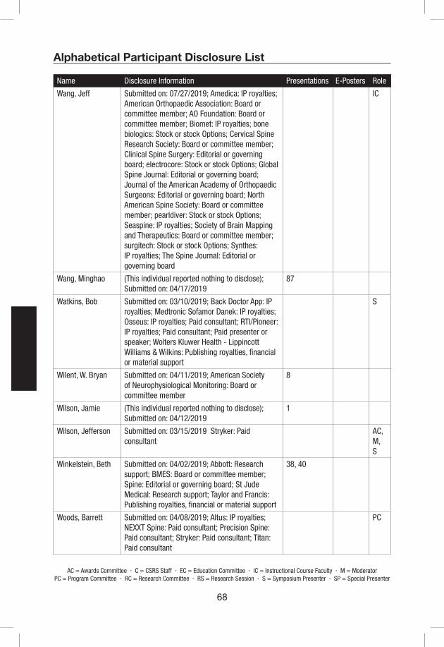
68
AC = Awards Committee • C = CSRS Staff • EC = Education Committee • IC = Instructional Course Faculty • M = ModeratorPC = Program Committee • RC = Research Committee • RS = Research Session • S = Symposium Presenter • SP = Special Presenter
Alphabetical Participant Disclosure List
Name Disclosure Information Presentations E-Posters Role
Wang, Jeff Submitted on: 07/27/2019; Amedica: IP royalties; American Orthopaedic Association: Board or committee member; AO Foundation: Board or committee member; Biomet: IP royalties; bone biologics: Stock or stock Options; Cervical Spine Research Society: Board or committee member; Clinical Spine Surgery: Editorial or governing board; electrocore: Stock or stock Options; Global Spine Journal: Editorial or governing board; Journal of the American Academy of Orthopaedic Surgeons: Editorial or governing board; North American Spine Society: Board or committee member; pearldiver: Stock or stock Options; Seaspine: IP royalties; Society of Brain Mapping and Therapeutics: Board or committee member; surgitech: Stock or stock Options; Synthes: IP royalties; The Spine Journal: Editorial or governing board
IC
Wang, Minghao (This individual reported nothing to disclose); Submitted on: 04/17/2019
87
Watkins, Bob Submitted on: 03/10/2019; Back Doctor App: IP royalties; Medtronic Sofamor Danek: IP royalties; Osseus: IP royalties; Paid consultant; RTI/Pioneer: IP royalties; Paid consultant; Paid presenter or speaker; Wolters Kluwer Health - Lippincott Williams & Wilkins: Publishing royalties, financial or material support
S
Wilent, W. Bryan Submitted on: 04/11/2019; American Society of Neurophysiological Monitoring: Board or committee member
8
Wilson, Jamie (This individual reported nothing to disclose); Submitted on: 04/12/2019
1
Wilson, Jefferson Submitted on: 03/15/2019 Stryker: Paid consultant
AC, M, S
Winkelstein, Beth Submitted on: 04/02/2019; Abbott: Research support; BMES: Board or committee member; Spine: Editorial or governing board; St Jude Medical: Research support; Taylor and Francis: Publishing royalties, financial or material support
38, 40
Woods, Barrett Submitted on: 04/08/2019; Altus: IP royalties; NEXXT Spine: Paid consultant; Precision Spine: Paid consultant; Stryker: Paid consultant; Titan: Paid consultant
PC

69
Alphabetical Participant Disclosure List
Name Disclosure Information Presentations E-Posters Role
Wright, Neill Submitted on: 08/14/2019; Cerapedics: Paid presenter or speaker; Cervical Spine Research Society: Board or committee member; Nuvasive: IP royalties; Ulrich Medical USA: Paid consultant; Vertebral Technologies, Inc.: Stock or stock Options; Zimmer: Paid presenter or speaker
EC
Wu, Yongren (This individual reported nothing to disclose); Submitted on: 09/04/2019
RS
Yang, Jae (This individual reported nothing to disclose); Submitted on: 07/06/2019
14
Yang, Junsong (This individual reported nothing to disclose); Submitted on: 08/22/2019
14
Ye, Ivan (This individual reported nothing to disclose); Submitted on: 07/10/2019
70
York, Philip (This individual reported nothing to disclose); Submitted on: 08/05/2019
44
Yoshikawa, Tomoya (This individual reported nothing to disclose); Submitted on: 08/23/2019
28
Yurube, Takashi (This individual reported nothing to disclose); Submitted on: 04/11/2019
37
Zhao, Yanbin (This individual reported nothing to disclose); Submitted on: 03/04/2019
42
Zhou, Feifei (This individual reported nothing to disclose); Submitted on: 04/09/2019
37, 41

70

71
Individual Disclosures can be found in the Disclosure Index pages 41-69.
PodiumPresentation
Abstracts
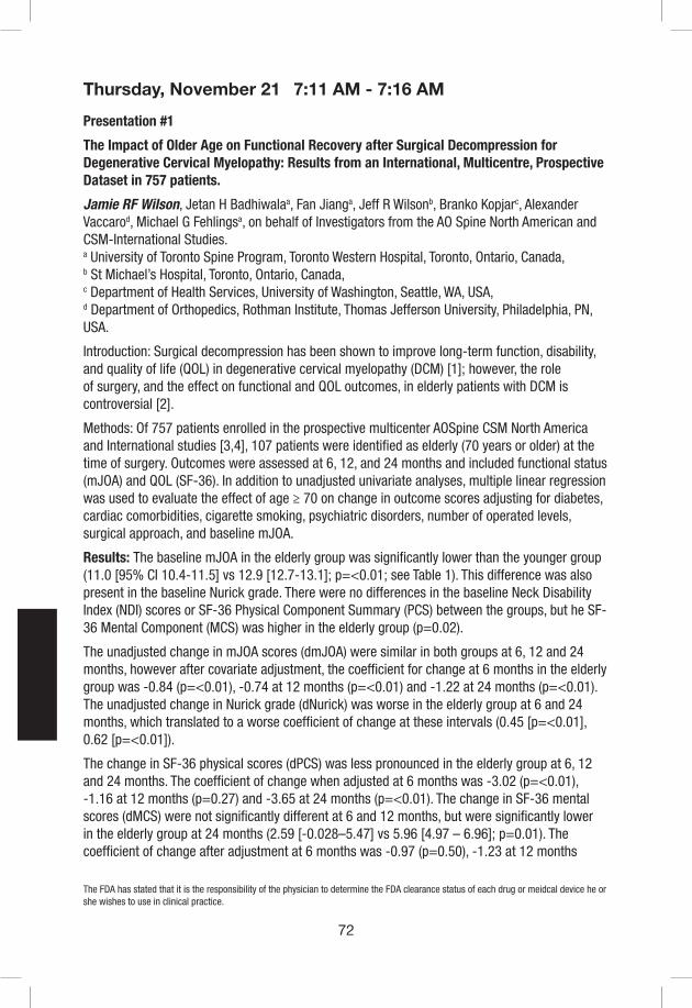
72
Thursday, November 21
The FDA has stated that it is the responsibility of the physician to determine the FDA clearance status of each drug or meidcal device he or she wishes to use in clinical practice.
Presentation #1
The Impact of Older Age on Functional Recovery after Surgical Decompression for Degenerative Cervical Myelopathy: Results from an International, Multicentre, Prospective Dataset in 757 patients.
Jamie RF Wilson, Jetan H Badhiwalaa, Fan Jianga, Jeff R Wilsonb, Branko Kopjarc, Alexander Vaccarod, Michael G Fehlingsa, on behalf of Investigators from the AO Spine North American and CSM-International Studies.a University of Toronto Spine Program, Toronto Western Hospital, Toronto, Ontario, Canada, b St Michael’s Hospital, Toronto, Ontario, Canada, c Department of Health Services, University of Washington, Seattle, WA, USA, d Department of Orthopedics, Rothman Institute, Thomas Jefferson University, Philadelphia, PN, USA.
Introduction: Surgical decompression has been shown to improve long-term function, disability, and quality of life (QOL) in degenerative cervical myelopathy (DCM) [1]; however, the role of surgery, and the effect on functional and QOL outcomes, in elderly patients with DCM is controversial [2].
Methods: Of 757 patients enrolled in the prospective multicenter AOSpine CSM North America and International studies [3,4], 107 patients were identified as elderly (70 years or older) at the time of surgery. Outcomes were assessed at 6, 12, and 24 months and included functional status (mJOA) and QOL (SF-36). In addition to unadjusted univariate analyses, multiple linear regression was used to evaluate the effect of age ≥ 70 on change in outcome scores adjusting for diabetes, cardiac comorbidities, cigarette smoking, psychiatric disorders, number of operated levels, surgical approach, and baseline mJOA.
Results: The baseline mJOA in the elderly group was significantly lower than the younger group (11.0 [95% CI 10.4-11.5] vs 12.9 [12.7-13.1]; p=<0.01; see Table 1). This difference was also present in the baseline Nurick grade. There were no differences in the baseline Neck Disability Index (NDI) scores or SF-36 Physical Component Summary (PCS) between the groups, but he SF-36 Mental Component (MCS) was higher in the elderly group (p=0.02).
The unadjusted change in mJOA scores (dmJOA) were similar in both groups at 6, 12 and 24 months, however after covariate adjustment, the coefficient for change at 6 months in the elderly group was -0.84 (p=<0.01), -0.74 at 12 months (p=<0.01) and -1.22 at 24 months (p=<0.01). The unadjusted change in Nurick grade (dNurick) was worse in the elderly group at 6 and 24 months, which translated to a worse coefficient of change at these intervals (0.45 [p=<0.01], 0.62 [p=<0.01]).
The change in SF-36 physical scores (dPCS) was less pronounced in the elderly group at 6, 12 and 24 months. The coefficient of change when adjusted at 6 months was -3.02 (p=<0.01), -1.16 at 12 months (p=0.27) and -3.65 at 24 months (p=<0.01). The change in SF-36 mental scores (dMCS) were not significantly different at 6 and 12 months, but were significantly lower in the elderly group at 24 months (2.59 [-0.028–5.47] vs 5.96 [4.97 – 6.96]; p=0.01). The coefficient of change after adjustment at 6 months was -0.97 (p=0.50), -1.23 at 12 months
7:11 AM - 7:16 AM

73
Thursday, November 21
Individual Disclosures can be found in the Disclosure Index pages 41-69.
(p=0.38) and -4.53 at 24 months (p=<0.01). The unadjusted change in NDI scores (dNDI) showed no difference at any interval, including after covariate adjustment.
Conclusions: In this combined dataset from 2 large prospectively collected multi-centre studies on DCM surgery, the group aged 70 or greater demonstrated significantly worse functional and QOL recovery when compared to the younger cohort after adjusting for the effect of co-morbidites, number of operated levels, surgical approach and baseline mJOA. Elderly patients undergoing surgery for DCM should therefore be counseled appropriately regarding expectations of surgery.
Table 1:
Adults under 70 Adults 70 years and over Coefficient of Change
n 650 107
mJOA – Baseline (p=<0.01) 12.9 (12.7 – 13.1) 11.0 (10.4 – 11.5)
Unadjusted dmJOA
6 months (p=0.75) 2.21 (2.02 – 2.40) 2.30 (1.71 – 2.88) -0.84 (p=<0.01)
12 months (p=0.30) 2.50 (2.29 – 2.70) 2.79 (2.18-3.41) -0.74 (p=<0.01)
24 months (p=0.77) 2.71 (2.51 – 2.92) 2.63 (1.99 – 3.27) -1.22 (p=<0.01)
Nurick – Baseline (p<0.01) 3.15 (3.06-3.24) 3.80 (3.58-4.02)
Unadjusted dNurick
6 months (p<0.01) -1.27 (-1.38 - -1.17) -0.89 (-1.16 - -0.61) 0.45 (p=<0.01)
12 months (p=0.37) -1.41 (-1.52 - -1.31) -1.28 (-1.57 - -1.00) 0.20 (p=0.19)
24 months (p<0.01) -1.54 (-1.65 - -1.44) -0.98 (-1.31 - -0.64) 0.62 (p=<0.01)
SF-36 PCS – Baseline (p= 0.28) 34.5 (33.8 – 35.2) 33.5 (31.8 – 35.1)
Unadjusted dPCS
6 months (p=<0.01) 5.78 (5.07 – 6.48) 3.14 (1.46 – 4.81) -3.02 (p=<0.01)
12 months (p=0.40) 6.41 (5.67 – 7.16) 5.56 (3.86 – 7.26) -1.16 (p=0.27)24 months (p=<0.01) 6.17 (5.40 – 6.94) 3.43 (1.37 – 5.49) -3.65 (p=<0.01)SF-36 MCS – Baseline (p=0.02) 39.8 (38.7 – 40.8) 43.0 (40.6 – 45.5)
Unadjusted dMCS
6 months (p=0.56) 6.01 (5.05 – 6.97) 5.23 (2.40 – 8.06) -0.97 (p=0.50)12 months (p=0.87) 6.18 (5.22 – 7.14) 5.96 (3.32 – 8.61) -1.23 (p=0.38)24 months (p=0.01) 5.96 (4.97 – 6.96) 2.59 (-0.028 – 5.47) -4.53 (p=<0.01)NDI – Baseline (p=0.98) 39.41 (37.74-41.09) 39.48 (35.65-43.30)
Unadjusted dNDI
6 months (p=0.77) 28.44 (26.86-30.03) 29.04 (25.66-32.42) 1.71 (p=0.40)12 months (p=0.93) 27.53 (25.85-29.22) 27.73 (24.10-31.37) 1.75 (p=0.42)24 months (p=0.61) 27.09 (25.49-28.68) 28.16 (24.63-31.69) 2.36 (p=0.26)
7:11 AM - 7:16 AM

74
Thursday, November 21
The FDA has stated that it is the responsibility of the physician to determine the FDA clearance status of each drug or meidcal device he or she wishes to use in clinical practice.
References:1. Fehlings MG, Tetreault LA, Riew KD, Middleton JW, Aarabi B, Arnold PM, Brodke DS, Burns AS,
Carette S, Chen R, Chiba K, Dettori JR, Furlan JC, Harrop JS, Holly LT, Kalsi-Ryan S, Kotter M, Kwon BK, Martin AR, Milligan J, Nakashima H, Nagoshi N, Rhee J, Singh A, Skelly AC, Sodhi S, Wilson JR, Yee A, Wang JC (2017) A Clinical Practice Guideline for the Management of Patients With Degenerative Cervical Myelopathy: Recommendations for Patients With Mild, Moderate, and Severe Disease and Nonmyelopathic Patients With Evidence of Cord Compression. Global spine journal 7 (3 Suppl):70S-83S. doi:10.1177/2192568217701914
2. Tetreault L, Palubiski LM, Kryshtalskyj M, Idler RK, Martin AR, Ganau M, Wilson JR, Kotter M, Fehlings MG (2018) Significant Predictors of Outcome Following Surgery for the Treatment of Degenerative Cervical Myelopathy: A Systematic Review of the Literature. Neurosurgery clinics of North America 29 (1):115-127 e135. doi:10.1016/j.nec.2017.09.020
3. Fehlings MG, Ibrahim A, Tetreault L, Albanese V, Alvarado M, Arnold P, Barbagallo G, Bartels R, Bolger C, Defino H, Kale S, Massicotte E, Moraes O, Scerrati M, Tan G, Tanaka M, Toyone T, Yukawa Y, Zhou Q, Zileli M, Kopjar B (2015) A global perspective on the outcomes of surgical decompression in patients with cervical spondylotic myelopathy: results from the prospective multicenter AOSpine international study on 479 patients. Spine 40 (17):1322-1328. doi:10.1097/BRS.0000000000000988
4. Fehlings MG, Wilson JR, Kopjar B, Yoon ST, Arnold PM, Massicotte EM, Vaccaro AR, Brodke DS, Shaffrey CI, Smith JS, Woodard EJ, Banco RJ, Chapman JR, Janssen ME, Bono CM, Sasso RC, Dekutoski MB, Gokaslan ZL (2013) Efficacy and safety of surgical decompression in patients with cervical spondylotic myelopathy: results of the AOSpine North America prospective multi-center study. The Journal of bone and joint surgery American volume 95 (18):1651-1658. doi:10.2106/JBJS.L.00589
7:11 AM - 7:16 AM
75
Thursday, November 21
Individual Disclosures can be found in the Disclosure Index pages 41-69.
Presentation #2
Surgical treatment of cervical spondylotic myelopathy leads to functional improvement in hand strength and dexterity: a prospective quantitative study
Tyler S Cole, MD; Jakub Godzik, MD; Jay D Turner, MD, PhD
Introduction: Cervical spondylotic myelopathy (CSM) is the most common cause of spinal cord dysfunction in adults. Diminished hand strength and dexterity in CSM is a major contribution to disability. The goal of this study was to establish the functional impact of CSM severity on hand function using quantitative testing and evaluate the response to intervention.
Methods: 33 consecutive patients planned for surgical treatment of CSM were prospectively enrolled. A licensed occupational therapist conducted three functional hand tests: 1) palmar dynamometry to assess grip strength, 2) hydraulic pinch gauge test to assess pinch strength, and 3) nine-hole peg test to assess upper extremity dexterity. Tests were performed preoperatively and at 6-8 weeks postoperatively. Test results were expressed as 1) percentile relative to age- and sex- stratified norms and 2) achievement of minimum clinically important (MCI) difference. Patients were stratified into mild, moderate, and severe myelopathy based on modified Japanese Orthopedic Association (mJOA) score. Severity of stenosis was graded on preoperative magnetic resonance imaging by three independent physicians using Kang classification.
Results: Primary presenting symptoms were neck pain (33%), numbness (21%), and upper extremity weakness (12%). 61% patients underwent anterior approach decompression with mean of 2.9 ± 1.5 levels treated. Preoperative pinch (p < 0.001) and grip (p = 0.014) strength were lower in moderate and low mJOA patients compared to high mJOA patients. Significant postoperative improvement was observed in all hand function domains with MCI improvement at 6 weeks ranging from 33% of patients in dominant strength tests to 72% of patients in non-dominant dexterity tests; patients with moderate baseline mJOA were more likely to have MCI improvement in dominant grip (58.3%) compared to low (30%) and high (9%) mJOA groups (p = 0.041). Dexterity as measured by mean dominant peg percentile was less than 1 in patients with cord signal change increasing in percentile to 15.7 with only subarachnoid effacement (p = 0.032).
Conclusion: CSM patients achieved significant improvement in strength and dexterity postoperatively. Baseline strength measures correlated most with preoperative mJOA, however baseline dexterity correlated most with severity of stenosis on MRI. The majority of patients experienced MCI improvement in dexterity. Baseline pinch strength correlated with postoperative mJOA MCI improvement, and patients with moderate baseline mJOA were the most likely to have improvement in dominant grip strength postoperatively.
7:17 AM - 7:22 AM

76
Thursday, November 21
The FDA has stated that it is the responsibility of the physician to determine the FDA clearance status of each drug or meidcal device he or she wishes to use in clinical practice.
Presentation #3
Neck Pain Improvement After Operative Intervention In Patients With Degenerative Cervical Myelopathy: Results From An International Multicenter Ambispective Study of 664 Patients
M.M. Schneider1, J. Badhiwala1,5, L.Tetreault1,2, P. Kalsi1, M. Farshad3, R.K. Idler2, J.R. Wilson4, M.G. Fehlings1,5
1 Toronto Western Hospital, Department of Neurosurgery, Canada2 University College Cork, Cork, Ireland3 Balgrist University Hospital, Zurich, Switzerland4 St. Michael’s Hospital, Department of Neurosurgery, Canada 5 University of Toronto, Canada
Introduction: Operative intervention in patients with degenerative cervical myelopathy (DCM) is increasingly recommended as it effectively halts neurological progression and improves functional impairment, disability, and quality of life. Despite the high incidence of neck pain in patients with DCM, there is a paucity of high-quality, prospective studies evaluating the impact of surgery, the role of the surgical approach and the effect of the functional impairment on neck pain outcomes.
Methods: Data was obtained from merging of the AOSpine Cervical Spondylotic Myelopathy North America (CSM-NA) and CSM International (CSM-I) studies. Both prospective studies enrolled 757 patients at 26 global sites from 2005-2011. Participating centers were located in North America (n=12), Latin America (n=3), Asia Pacific (n=6) and Europe (n=5). All patients underwent a surgical intervention of the cervical spine and were assessed at 6, 12 and 24 months postoperatively. A total of 664 patients had complete preoperative neck disability index (NDI) scores and were therefore eligible for this analysis. Patients were asked to rate their neck pain as none, very mild, moderate, fairly severe, very severe or the worst imaginable. Four hundred and ninety-seven patients had neck pain outcomes at the 24-months follow-up. The surgical approach was defined as anterior only (AP), consisting of anterior discectomy, corpectomy, fusion, fixation and/or graft implantation; posterior only (PA), including posterior laminectomy or laminoplasty; posterior instrumentation and laminectomy (PI) and a combined anterior-posterior approach (AP).
Results: Preoperatively, 79.2 % (n=526) of patients indicated neck pain, whereas 20.2% (n=134) rated their pain as very mild, 27.9% (n=185) as moderate, 19.6% (n=130) as fairly severe, 9.6% (n=64) as very severe, and 1.9% (n=13) as worst imaginable. The overall NDI improved significantly between preoperatively (39.37, ±20.6) and the 6-, 12- and 24-months follow-up’s (28.50 ±19.2; 27.47 ±20.2; 27.13 ±19.2; p<0.0001) and reached the minimum clinically important difference (MCID) of 7.5 points at every follow-up. The NDI subscore neck pain also improved significantly from preoperatively (1.83 ±1.3) to all follow-up’s (1.16 ±1.0; 1.04 ±1.0; 0.96 ±1.1, p-value<0.0001). Out of 566 patients with a complete NDI assessment at 6 months, 331 (58.6%) had an anterior approach only (AP), 63 (11.1%) a posterior approach only (PA), 155 (27.3%) a posterior laminectomy and instrumentation (PI), and 17 (3%) a combined anterior-posterior approach (AP). Patients reported no neck pain in 33% after an AA, in 35% after
7:23 AM - 7:28 AM

77
Thursday, November 21
Individual Disclosures can be found in the Disclosure Index pages 41-69.
a PA, in 32% after a PI, and in 29% after an AP approach 6 months after surgery. The neck pain incidence at 24 months postoperatively did not differ in patients with mild (52.9%), moderate (51.7%), or severe DCM (48.1%).
Conclusion: To our knowledge, this is the first multi-center, international study demonstrating significant improvements in neck pain up to 24 months after operative intervention for DCM. The functional impairment or the surgical approach was independent to the pain response. Further studies are needed that evaluate important predictors of improvement in neck pain and to assess the effect of neck pain on the quality-of-life.
References:1. Nouri A, Tetreault L, Singh A, Karadimas SK, Fehlings MG. Degenerative Cervical Myelopathy:
Epidemiology, Genetics, and Pathogenesis. Spine (Phila Pa 1976). 2015;40(12):E675-693.2. Tetreault L, Goldstein CL, Arnold P, et al. Degenerative Cervical Myelopathy: A Spectrum of
Related Disorders Affecting the Aging Spine. Neurosurgery. 2015;77 Suppl 4:S51-673. Baptiste DC, Fehlings MG. Pathophysiology of cervical myelopathy. Spine J. 2006;6(6
Suppl):190S-197S.4. Hughes JT, Brownell B. Necropsy observations on the spinal cord in cervical spondylosis. Riv
Patol Nerv Ment. 1965;86(2):196-204.5. Gore DR, Sepic SB, Gardner GM. Roentgenographic findings of the cervical spine in
asymptomatic people. Spine (Phila Pa 1976). 1986;11(6):521-524.6. Irvine DH, Foster JB, Newell DJ, Klukvin BN. Prevalence of Cervical Spondylosis in a General
Practice. Lancet. 1965;1(7395):1089-1092.7. Fehlings MG, Tetreault LA, Riew KD, et al. A Clinical Practice Guideline for the Management
of Patients With Degenerative Cervical Myelopathy: Recommendations for Patients With Mild, Moderate, and Severe Disease and Nonmyelopathic Patients With Evidence of Cord Compression. Global Spine J. 2017;7(3 Suppl):70S-83S.
8. Fehlings MG, Tetreault LA, Kurpad S, et al. Change in Functional Impairment, Disability, and Quality of Life Following Operative Treatment for Degenerative Cervical Myelopathy: A Systematic Review and Meta-Analysis. Global Spine J. 2017;7(3 Suppl):53S-69S.
9. Fehlings MG, Ibrahim A, Tetreault L, et al. A global perspective on the outcomes of surgical decompression in patients with cervical spondylotic myelopathy: results from the prospective multicenter AOSpine international study on 479 patients. Spine (Phila Pa 1976). 2015;40(17):1322-1328.
10. Fehlings MG, Wilson JR, Kopjar B, et al. Efficacy and safety of surgical decompression in patients with cervical spondylotic myelopathy: results of the AOSpine North America prospective multi-center study. J Bone Joint Surg Am. 2013;95(18):1651-1658.
11. Fehlings MG, Barry S, Kopjar B, et al. Anterior versus posterior surgical approaches to treat cervical spondylotic myelopathy: outcomes of the prospective multicenter AOSpine North America CSM study in 264 patients. Spine (Phila Pa 1976). 2013;38(26):2247-2252.
12. Fehlings MG, Santaguida C, Tetreault L, et al. Laminectomy and fusion versus laminoplasty for the treatment of degenerative cervical myelopathy: results from the AOSpine North America and International prospective multicenter studies. Spine J. 2017;17(1):102-108.
13. Tetreault L, Kopjar B, Cote P, Arnold P, Fehlings MG. A Clinical Prediction Rule for Functional
7:23 AM - 7:28 AM

78
Thursday, November 21
The FDA has stated that it is the responsibility of the physician to determine the FDA clearance status of each drug or meidcal device he or she wishes to use in clinical practice.
Outcomes in Patients Undergoing Surgery for Degenerative Cervical Myelopathy: Analysis of an International Prospective Multicenter Data Set of 757 Subjects. J Bone Joint Surg Am. 2015;97(24):2038-2046.
14. Nouri A, Martin AR, Kato S, Reihani-Kermani H, Riehm LE, Fehlings MG. The Relationship Between MRI Signal Intensity Changes, Clinical Presentation, and Surgical Outcome in Degenerative Cervical Myelopathy: Analysis of a Global Cohort. Spine (Phila Pa 1976). 2017;42(24):1851-1858.
15. Kopjar B, Tetreault L, Kalsi-Ryan S, Fehlings M. Psychometric properties of the modified Japanese Orthopaedic Association scale in patients with cervical spondylotic myelopathy. Spine (Phila Pa 1976). 2015;40(1):E23-28.
16. Tetreault L, Nouri A, Kopjar B, Cote P, Fehlings MG. The Minimum Clinically Important Difference of the Modified Japanese Orthopaedic Association Scale in Patients with Degenerative Cervical Myelopathy. Spine (Phila Pa 1976). 2015;40(21):1653-1659.
17. Benzel EC, Lancon J, Kesterson L, Hadden T. Cervical laminectomy and dentate ligament section for cervical spondylotic myelopathy. J Spinal Disord. 1991;4(3):286-295.
18. Tetreault L, Kopjar B, Nouri A, et al. The modified Japanese Orthopaedic Association scale: establishing criteria for mild, moderate and severe impairment in patients with degenerative cervical myelopathy. European spine journal : official publication of the European Spine Society, the European Spinal Deformity Society, and the European Section of the Cervical Spine Research Society. 2017;26(1):78-84.
19. Carreon LY, Glassman SD, Campbell MJ, Anderson PA. Neck Disability Index, short form-36 physical component summary, and pain scales for neck and arm pain: the minimum clinically important difference and substantial clinical benefit after cervical spine fusion. Spine J. 2010;10(6):469-474.
20. Vernon H, Mior S. The Neck Disability Index: a study of reliability and validity. J Manipulative Physiol Ther. 1991;14(7):409-415.
21. MacDermid JC, Walton DM, Avery S, et al. Measurement properties of the neck disability index: a systematic review. J Orthop Sports Phys Ther. 2009;39(5):400-417.
22. Young BA, Walker MJ, Strunce JB, Boyles RE, Whitman JM, Childs JD. Responsiveness of the Neck Disability Index in patients with mechanical neck disorders. Spine J. 2009;9(10):802-808.
23. Fehlings MG, Skaf G. A review of the pathophysiology of cervical spondylotic myelopathy with insights for potential novel mechanisms drawn from traumatic spinal cord injury. Spine (Phila Pa 1976). 1998;23(24):2730-2737.
24. Karadimas SK, Erwin WM, Ely CG, Dettori JR, Fehlings MG. Pathophysiology and natural history of cervical spondylotic myelopathy. Spine (Phila Pa 1976). 2013;38(22 Suppl 1):S21-36.
25. Cavanaugh JM, Lu Y, Chen C, Kallakuri S. Pain generation in lumbar and cervical facet joints. J Bone Joint Surg Am. 2006;88 Suppl 2:63-67.
26. Wilson JR, Tetreault LA, Kim J, et al. State of the Art in Degenerative Cervical Myelopathy: An Update on Current Clinical Evidence. Neurosurgery. 2017;80(3S):S33-S45.
27. Abduljabbar FH, Teles AR, Bokhari R, Weber M, Santaguida C. Laminectomy with or Without Fusion to Manage Degenerative Cervical Myelopathy. Neurosurg Clin N Am. 2018;29(1):91-105.
7:23 AM - 7:28 AM

79
Thursday, November 21
Individual Disclosures can be found in the Disclosure Index pages 41-69.
Table 1:
Neck pain response to surgery at 6, 12 and 24 months
Pre-operative 6 mo 12 mo 24 mo
n % n % n % n %
no pain (NP) 138 20.8 186 32.9 213 39.7 238 47.9
pain 526 79.2 380 67.1 324 60.3 259 52.1
mild 134 20.2 187 33.0 166 30.9 109 21.9
moderate 185 27.9 133 23.5 102 19.0 101 20.3
fairly severe 130 19.6 42 7.4 37 6.9 32 6.4
very severe 64 9.6 12 2.1 14 2.6 14 2.9
worst imaginable 13 1.9 6 1.1 5 0.9 3 0.6
Total 664 566 537 497
Table 1: Descriptive statistics illustrating the proportion of patients with NP, MIP and MAP at 6, 12, and 24 months after operative intervention.
Table 2:
NDI at 6 months and surgical approach
AA PA PI AP
NDI subscore at 6 monthsn=566
n % n % n % n %
No pain 109 33% 22 35% 50 32% 5 29%
Mild 113 34% 24 38% 46 30% 4 24%
Moderate 73 22% 12 19% 42 27% 6 35%
fairly severe 23 7% 4 6% 13 8% 2 12%
very severe 9 3% 1 2% 2 1% 0 0%
worst imaginable 4 1% 0 0% 2 1% 0 0%
Total 331 58.6% 63 11.1% 155 27.3% 17 3%
Table 2: The NDI scores at 6 months for a total of 566 patients were similar for all surgical approaches (AP: anterior approach only, PA: posterior approach only, PI posterior instrumentation and laminectomy, AP: combined anterior and posterior approach).
7:23 AM - 7:28 AM
80
Thursday, November 21
The FDA has stated that it is the responsibility of the physician to determine the FDA clearance status of each drug or meidcal device he or she wishes to use in clinical practice.
Presentation #4
Comparison of three anterior techniques in the surgical treatment of three-level cervical spondylotic myelopathy with intramedullary T2-weighted increased signal intensity
Peng Cao, Wen Yuan
Department of Orthopaedics, Shanghai Changzheng Hospital, Second Military Medical University, Shanghai, China
Objective: To compare the clinical and radiological outcomes of three anterior surgical techniques in the treatment of three-level cervical spondylotic myelopathy (CSM) and the evolution of intramedullary T2-weighted increased signal intensity (ISI).
Methods: A total of 98 consecutive patients (61 males, 37 females) with three-level CSM who underwent anterior cervical surgery from January 2006 to January 2016 were retrospectively enrolled. Based on different anterior reconstructive techniques, the patients were divided into three groups: anterior cervical discectomy and fusion (ACDF), anterior cervical corpectomy and fusion (ACCF) and hybrid decompression and fusion (HDF) groups. The Japanese Orthopedic Association (JOA) score and its recovery rate were used to evaluate the clinical outcomes. The cervical alignment and range of motion (ROM) were used to assess radiological outcomes. In addition, the signal change ratio (SCR) and length of ISI were used to quantify the ISI.
Results: No statistical differences in preoperative factors were found among three groups (P > 0.05). Although the HDF group had moderate surgery time and blood loss than other groups (P < 0.05), it achieved better postoperative JOA score and recovery rate than other groups (P < 0.05). The postoperative C2-C7 lordotic angle and postoperative ROM in the HDF group was similar to the ACDF group (P > 0.05), which were both higher than that in the ACCF group (P < 0.05). The incidence of complications in the HDF group was similar to the ACDF group (P > 0.05), which were both lower than that in the ACCF group (P < 0.05). Besides, the postoperative SCR in the HDF group was lower compared with other groups (P < 0.05). And the postoperative length of ISI in the HDF group was similar to the ACCF group (P > 0.05), which was both shorter than that in the ACDF group (P < 0.05).
Conclusions: For three-level CSM patients with ISI on T2-weighted MRI, HDF can be considered as the optimal technique, which achieves better clinical and radiological outcomes than ACDF or ACCF procedure. HDF also has a better postoperative regression of ISI than ACDF or ACCF procedure, which may be possibly an important indicator for better surgical outcomes.
Key Words: Cervical spondylotic myelopathy; Increased signal intensity; Anterior cervical discectomy and fusion; Anterior cervical corpectomy and fusion; Hybrid decompression and fusion.
Conflict of interest: None of the authors has any potential conflict of interest.
Funding: This study was supported by Science and Technology Commission of Shanghai Municipality (No. 17140901500).
7:29 AM - 7:34 AM
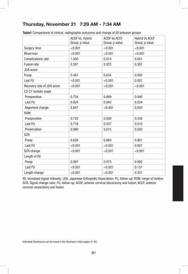
81
Thursday, November 21
Individual Disclosures can be found in the Disclosure Index pages 41-69.
Table1 Comparisons of clinical, radiographic outcomes and change of ISI between groups
ACDF Vs. Hybrid Group, p value
ACDF Vs ACCF Group, p value
Hybrid Vs ACCF Group, p value
Surgery time <0.001 <0.001 <0.001
Blood loss <0.001 <0.001 <0.001
Complications rate 1.000 0.014 0.041
Fusion rate 0.387 0.023 0.387
JOA score
Preop 0.487 0.634 0.942
Last FU <0.001 <0.001 0.001
Recovery rate of JOA score <0.001 <0.001 <0.001
C2-C7 lordotic angle
Preoperative 0.734 0.889 0.940
Last FU 0.924 0.043 0.034
Alignment change 0.847 <0.001 0.003
ROM
Preoperative 0.720 0.939 0.538
Last FU 0.718 0.037 0.010
Preservation 0.999 0.015 0.042
SCR
Preop 0.628 0.864 0.891
Last FU <0.001 <0.001 0.001
SCR change <0.001 <0.001 <0.001
Length of ISI
Preop 0.997 0.973 0.992
Last FU <0.001 <0.001 0.157
Length change <0.001 <0.001 0.331
ISI, increased signal intensity; JOA, Japanese Orthopedic Association; FU, follow-up; ROM, range of motion; SCR, Signal change ratio; FU, follow-up; ACDF, anterior cervical discectomy and fusion; ACCF, anterior cervical corpectomy and fusion.
7:29 AM - 7:34 AM

82
Thursday, November 21
The FDA has stated that it is the responsibility of the physician to determine the FDA clearance status of each drug or meidcal device he or she wishes to use in clinical practice.
Table 2 Comparisons of postoperative complications between groups
ACDF group(n=38)
Hybrid group(n=24)
ACCF group(n=36)
Surgery-related complications
Dysphagia 3 (10.5%) 2 (8.3%) 4 (11.1%)
C5 palsy 1 (2.6%) 1 (4.2%) 1 (2.8%)
Cerebral fluid leakage 2 (5.3%) 0 1 (2.8%)
Epidural hematoma 0 0 1 (2.8%)
Infection 0 0 1 (2.8%)
Total 6 (15.8%) 3 (12.5%) 8 (22.2%)
Instrumentation- and/or graft-related complications
Graft dislodgement 0 0 2
Hardware breakage 0 0 0
Subsidence 0 1 (4.2%) 5
Total 0 1 (4.2%) 7 (19.4%)
7:29 AM - 7:34 AM

83
Thursday, November 21
Individual Disclosures can be found in the Disclosure Index pages 41-69.
Presentation #5
Comparison of laminoplasty and posterior decompression with fusion for cervical spondylotic myelopathy accompanying local kyphosis: a matched analysis using propensity scores
S. Maki1, T. Furuya1, T. Miyamoto1, M. S. Okimatsu, M. Koda2, M. Yamazaki2
1. Department of Orthopaedic Surgery, Chiba University Graduate School of Medicine, Japan2. Department of Orthopaedic Surgery, Faculty of Medicine, University of Tsukuba
Background: Laminoplasty for patients with cervical spondylotic myelopathy (CSM) accompanying local kyphosis is considered to have poor surgical outcomes. However, few studies have compared results with surgical procedures other than laminoplasty. We sought to compare the results of laminoplasty with those of posterior decompression fusion for CSM accompanying local kyphosis using propensity score matching.
Methods: We reviewed 188 patients with CSM who underwent surgery and were followed up for at least 1 year after surgery. Patients with a history of cerebral palsy, ossification of the posterior longitudinal ligament, rheumatoid arthritis, dropped head syndrome, destructive spondyloarthropathy, and those who had previously undergone cervical spinal surgery were excluded from the study. Thirty-eight patients who presented local kyphosis exceeding 10° were enrolled. Their Japanese Orthopaedics Association (JOA) scores were evaluated before and 1 year after surgery and the score recovery rate was calculated. We adjusted characteristics including the age, sex, preoperative JOA score, and local kyphosis angle of the laminoplasty (LP) group and the posterior decompression and instrumented fusion (PDF) group using the propensity score, and compared the JOA score recovery rate, operative time, and estimated blood loss between the two groups. Radiographic parameters including the C2–C7 Cobb angle, local kyphosis angle, and C2–C7 sagittal vertical axis (SVA) were also measured.
Result: In the 188 patients with CSM who underwent surgery, 38 (20%) had local kyphosis exceeding 10°. Among them, 23 patients underwent LP and 15 patients underwent PDF. These 38 patients were included in propensity score calculation, and 1-to-1 matching resulted in 11 pairs of LP and PDF cases. The LP group and the PDF group had comparable baseline covariates. The preoperative JOA score and postoperative JOA score in the LP group versus the PDF group was 7.5 versus 7.0 and 11.0 versus 12.2, respectively, with no significant difference between the two groups. The mean recovery rate of the JOA score in the LP group was 32.4% versus 52.4% in the PDF group (p = 0.028). The recovery rate of those in the PDF group was significantly better than that of those in the LP group. The mean operative time for LP was 139.5 minutes and for PDF was 246.3 minutes being significantly longer in the PDF group than in the LP group. However, there was no significant difference in estimated blood loss between the groups (141.7 g versus 164.1 g). Although it did not reach significance, local kyphosis progressed postoperatively from 13.1° to 17.5° (p = 0.065) in the LP group, whereas it only changed from 12.9° to 12.7° in the PDF group (not significant). C2–C7 Cobb angle decreased significantly (LP: 7.4 to 0.6, PDF: 8.2 to 0.4) in both groups and C2–C7 SVA tended to increase (LP: 28.2 to 33.5 mm, PDF: 26.1 to 32.5) postoperatively.
7:35 AM - 7:39 AM
84
Thursday, November 21
The FDA has stated that it is the responsibility of the physician to determine the FDA clearance status of each drug or meidcal device he or she wishes to use in clinical practice.
Conclusions: The prevalence of the local kyphosis exceeding 10° in the patients with CSM was 20%. For patients with CSM accompanying local kyphosis, PDF was associated with longer operative time, but offered greater neurological improvement than LP.
7:35 AM - 7:39 AM
85
Thursday, November 21
Individual Disclosures can be found in the Disclosure Index pages 41-69.
Presentation #6
Clinical and Radiological Analysis of C5 Palsy in Cervical Spine Surgery Patients: A Korean Cervical Spine Study Group (KCSSG) Multicenter Study
Ho Jin Lee,1 Jae Keun Oh,2 Dong Ho Kang,3 Ki-Jeong Kim,4 Chi Heon Kim5, Chun Kee Chung,5 Jung-Woo Hur,6 Seong Yi,7 Jun Jae Shin,8 Sun Ho Lee,9 Kyung Chul Choi,10 Kyung Hyun Kim,11 Dae-Chul Cho,12 Yong Jun Jin,13 In Ho Han,14 Jae Taek Hong15
1Department of Neurosurgery, St. Vincent’s Hospital, College of medicine, The Catholic University of Korea, Suwon 2Department of Neurosurgery, Hallym University Sacred Heart Hospital, Anyang, 3Department of Neurosurgery, Gyeongsang National university School of Medicine, Jinju, 4Department of Neurosurgery, Spine Center, Seoul National University Bundang Hospital, Seoul National University College of Medicine, Seongnam, 5Department of Neurosurgery, Seoul National University Hospital, Seoul National University College of Medicine, Neuroscience Research Institute, Seoul National University Medical Research Center, Clinical Research Institute, Seoul National University Hospital, Seoul, 6Department of Neurosurgery, Spine center, Seoul St. Mary’s Hospital, College of Medicine, The Catholic University of Korea, 7Department of Neurosurgery, Yonsei University College of Medicine, Severance Hospital, Seoul, 8Department of Neurosurgery, Sanggye Paik Hospital, Inje University, 9Department of Neurosurgery, Samsung Medical Center, Sungkynkwan University School of Medicine, Seoul, 10Department of Neurosurgery, The Leon Wiltse Memorial Hospital, Anyang, 11Department of Neurosurgery, Yonsei University College of Medicine, Gangnam Severance Hospital, 12Department of Neurosurgery, Kyungpook National University Hospital, Daegu, 13Department of Neurosurgery, Inje University Seoul Paik Hospital, Seoul, 14Department of Neurosurgery, Pusan National University Hospital, Pusan, 15Department of Neurosurgery, Eun Pyeong St. Mary’s Hospital, The Catholic University of Korea, Seoul, South Korea
Introduction: The development of C5 palsy (deltoid muscle paralysis) is a serious, well-documented complication after cervical spine surgery that can arise from either anterior or posterior surgery. The aims of the present study were to evaluate the characteristics of C5 palsy using a (KCSSG) database.
Material & Methods: A retrospective review for clinical and radiological data was performed for 87 patients (68 males, 19 females, mean age 61.2 years) who had C5 palsy among 15582 cases of cervical spine surgeries in 21 institutes. C5 palsy was defined as deltoid muscle impairment of one or more grades in a manual muscle test (MMT) conducted within 6 weeks after surgery. This study was approved by the Ethics Committee of each KCCSG member institution. We investigated multiple clinical factors (BMI, DM, disease category, pre-operative symptoms, surgical method, symptom duration, operation method, side of C5 palsy, C5 palsy grade and onset time, course of recovery of C5 palsy, and Japanese Orthopedic Association score) and radiological factors [C4–5 foramen diameter, occupying ratio, T2HIZ (high intensity zone in T2 weighted MRI image), cervical spine alignment, and cord rotation] using pre- and post-operative X-rays, CT, and MRI (Figure 1). We also conducted sub-group analyses to identify clinical and radiological differences according to focused clinical factors.
Results: In the whole cohort of C5 palsy patients, the mean motor grade value was 2.8, which
7:52 AM - 7:57 AM

86
Thursday, November 21
The FDA has stated that it is the responsibility of the physician to determine the FDA clearance status of each drug or meidcal device he or she wishes to use in clinical practice.
recovered to 4.0 after 4.4 months. The average onset time of C5 palsy was 1.8 days after surgery. Forty-eight (55%) out of 87 patients had a positive of cord signal change, and a small C4–5 foramen diameter (right: 2.1mm, left: 2.0mm) (Table 1). Symptom duration was closely related to the type of main pathology [OPLL ≥ HNP (herniated nucleus pulposus) or degenerative spondylosis]. The delayed onset group (2 days after surgery) had shorter symptom duration and more severe paralysis than the early onset group. Elderly patients had worse symptoms than younger patients. The mild (G3–4) C5 palsy group had a shorter onset time and much greater degree of recovery compared with the severe (G1–2) group. The severity of the C5 palsy grade increased as the T2HIZ distribution became clearer (Table 2).
Conclusion: The overall clinical and radiological profiles of a large sample of C5 palsy patients have been presented in this study. We found a close correlation between the degree of paralysis and C4–5 FD. The delayed onset group had shorter symptom duration and much worse C5 palsy grades than the early onset group. Moreover, the clinical course and degree of C5 palsy can be influenced by other multiple radiological and clinical factors. Elderly patients had a worse degree of C5 palsy than younger patients and the mild C5 palsy group had shorter onset time to C5 palsy and a much better degree of recovery than the severe group. In addition, degree of T2HIZ clarity was statistically related to the severity of C5 palsy symptoms.
Figure 1. Measurement and analysis of radiographic data. A; C2-7 angle and Ishihara’s curvature index, B; transforaminal diameter of C4-5, C; occupying ratio and T2HIZ, D; cord rotation.
7:52 AM - 7:57 AM

87
Thursday, November 21
Individual Disclosures can be found in the Disclosure Index pages 41-69.
Table 1 Demographics of C5 palsy patient and their clinical and radiological findings
Mean ± SD
Age (years) 61.2 ± 11.9
Sex (n) Female (19) / Male (68)
Disease category (n) Degenerative (84) / trauma (2) / tumor (1)
BMI 24.3 ± 2.9
DM (n) 26 (positive) / 62 (negative)
Existence of T2HIZ (n) 48 (positive) / 39 (negative)
Bilateral C5 palsy vs Unilateral C5 palsy (n) 12 vs 75
Palsy side after open-door laminoplasty (n) Open side 17 / Hinge side 3
C4-5 FD (mm), right 2.1 ± 1.2
C4-5 FD (mm), left 2.0 ± 1.3
C2-7 angle (pre-OP) ( ˚) 8.5 ± 12.8
C2-7 angle (post-OP)( ˚) 11.0 ± 11.3
T1-slope (pre-OP) (˚) 22.6 ± 10.5
T1-slope (post-OP) (˚) 18.6 ± 19.1
Ishihara index (pre-OP) 0.1 ± 0.1
Ishihara index (post-OP) 0.1 ± 0.1
Occupying ratio (%) 53.1 ± 10.0
Cord rotation (˚) 4.6 ± 4.3
Symptom duration (months) 13.2 ± 19.1
Main symptoms (radiculopathy vs myelopathy) (n) Radiculopathy (46) / Myelopathy (41)
JOA score (pre-OP) 12.1 ± 3.0
JOA score (post-OP) 13.8 ± 2.1
Onset of C5 palsy 1.8 ± 2.8
MMT grade of C5 palsy 2.8 ± 2.0
MMT grade after C5 palsy recovery 4.0 ± 1.0
Recovery period (months) 4.4 ± 4.2
7:52 AM - 7:57 AM

88
Thursday, November 21
The FDA has stated that it is the responsibility of the physician to determine the FDA clearance status of each drug or meidcal device he or she wishes to use in clinical practice.
Table 2 Sub-group analysis according to variable comparison factors (clinical and radiographic difference)Age Sex Surgical approach Main symptom Laterality of C5 palsy Posterior operation
Comparison factors and classification criteria
<60 yrs(n=40)
≥ 60 yrs(n=47)
Female(n=19)
Male(n=68)
Anterior(n=27)
Posterior(n=57)
Myelopathy(n=41)
Radiculopathy(n=46)
Unilateral(n=75)
Bilateral(n=12)
Decompression(n=23)
Fusion(n=34)
T2HIZ (positive/total)(P-value)
19 / 40 28 / 47(0.10)
12 / 19 35 / 68(0.43)
9 / 27 36 / 57(0.01)†
27 / 41 20/ 46(0.03)†
39 / 76 8 / 12(0.33)
13 / 23 21 / 34(0.89)
C4-5 FD (mm), right(P-value)
2.1 2.0(0.72)
1.8 2.1(0.19)
1.7 2.2(0.11)
2.1 1.9(0.47)
2.0 2.1(0.78)
2.4 2.0(0.22)
C4-5 FD (mm), left(P-value)
2.1 1.8(0.26)
2.0 1.9(1.00)
1.9 1.9(0.65)
1.9 2.0(0.70)
2.0 1.7(0.53)
2.0 1.9(0.63)
C2-7 angle (pre-OP) ˚(P-value)
5.1 11.5(0.03)†
7.9 8.5(0.73)
4.8 9.8(0.13)
11.5 5.8(0.06)†
9.0 5.2(0.37)
12.2 7.8(0.26)
C2-7 angle (post-OP) ˚(P-value)
7.3 13.9(0.01)†
7.8 12.0(0.18)
10.6 10.7(0.97)
12.4 9.3(0.23)
11.7 7.1(0.21)
14.3 7.7(0.06)
T1-slope (pre-OP) ˚(P-value)
22.7 22.4(0.49)
19.2 23.5(0.91)
23.4 23.8(0.85)
20.5 23.8(0.75)
22.3 24.8(0.86)
23.9 23.7(0.95)
T1-slope (post-OP) ˚(P-value)
19.0 18.0(0.83)
10.1 21.8(0.04)†
18.0 20.0(0.80)
16.8 20.1(0.50)
17.0 26.4(0.37)
19.3 25.9(0.06)
Ishihara index (pre-OP)(P-value)
0.0 0.1(0.01)†
0.1 0.1(0.81)
0.01 0.13(0.06)
0.05 0.09(0.01)†
0.13 0.07(0.06)
0.13 0.12(0.57)
Ishihara index (post-OP)(P-value)
0.0 0.1(0.01)†
0.0 0.1(0.03)†
0.01 0.10(0.43)
0.11 0.09(0.29)
0.11 0.07(0.17)
0.10 0.10(0.87)
Occupying ratio %(P-value)
53.0 53.1(0.57)
52.7 53.2(0.87)
54.3 52.5(0.38)
54.9 52.5(0.10)
52.5 57.0(0.15)
49.4 53.3(0.69)
Cord rotation ˚(P-value)
4.1 5.0(0.29)
3.8 4.8(0.51)
5.2 4.1(0.35)
4.6 4.5(0.28)
4.7 3.5(0.27)
4.7 3.9(0.27)
Symptom duration (months)(P-value)
12.1 14.1(0.29)
21.2 11.0(0.11)
13.9 11.3(0.57)
14.2 12.1(0.82)
12.1 19.9(0.08)
5.5 16.0(0.06)
JOA score (pre-OP)(P-value)
12.9 11.1(0.03)†
10.0 12.6(0.01)†
14.0 11.6(0.02)†
10.8 13.3(0.01)†
12.1 11.7(0.65)
12.8 10.4(0.01)†
JOA score (post-OP)(P-value)
14.4 13.0(0.04)†
13.8 13.7(0.83)
14.6 13.4(0.16)
13.1 14.3(0.13)
13.7 14.0(0.85)
13.7 13.4(0.82)
Onset of C5 palsy(P-value)
1.8 1.6(0.75)
1.3 1.8(0.40)
2.1 1.6(0.26)
2.3 1.2(0.12)
1.8 1.2(0.97)
2.1 1.3(0.59)
C5 palsy grade(P-value)
3.0 2.5(0.04)†
2.8 2.7(0.76)
2.7 2.8(0.49)
4.0 4.0(0.71)
2.7 3.1(0.15)
3.2 2.6(0.15)
C5 palsy recovery(P-value)
4.1 3.8(0.26)
4.3 3.9(0.06)
4.2 3.9(0.50)
5.0 3.6(0.41)
4.0 3.8(0.78)
4.0 3.8(0.82)
Diagnosis Onset of C5 palsy T2HIZ grade C5 palsy grade Occupying ratio
Comparison factors and classification criteria
HNP /Spondylosis
OPLL Early( < 2 day)
Delayed(≥ 2days)
Grade 0(n=36)
Grade 1(n=15)
Grade 2(n=36)
Severe (G1-2)(n=26)
Mild (G3-4)(n=58)
Low (<50)(n=28)
High (≥50)(n=57)
T2HIZ (positive/total)(P-value)
27 / 52 17 / 33(0.98)
24 / 50 23 / 35(0.10)
15/ 26 29 / 57(0.44)
15 / 28 32 / 57(0.82)
C4-5 FD (mm), right(P-value)
2.0 2.1(0.79)
2.0 2.1(0.68)
2.3 2.0 1.8(0.29)
1.6 2.2(0.03)†
1.5 2.3(<0.01)†
C4-5 FD (mm), left(P-value)
1.9 2.1(0.28)
2.0 1.7(0.17)
2.2 1.6 1.8(0.20)
1.4 2.2(0.01)†
2.1 1.9(0.52)
C2-7 angle (pre-OP) ˚(P-value)
6.0 12.5(0.03)†
6.9 10.4(0.24)
6.4 13.2 7.8(0.30)
11.7 7.2(0.17)
4.9 9.5(0.07)
C2-7 angle (post-OP) ˚(P-value)
9.7 12.9(0.26)
10.1 12.1(0.46)
9.3 15.7 9.8(0.21)
11.4 10.5(0.97)
7.5 11.8(0.12)
T1-slope (pre-OP) ˚(P-value)
23.0 22.1(0.46)
22.4 22.8(0.57)
23.2 22.3 21.5(0.97)
24.5 22.0(0.84)
19.3 23.8(0.27)
T1-slope (post-OP) ˚(P-value)
17.4 19.4(0.33)
20.8 15.1(0.41)
21.0 18.3 19.7(0.92)
15.7 19.3 (0.37)
11.6 24.3(0.01)†
Ishihara index (pre-OP)(P-value)
0.10 0.13(0.12)
0.12 0.12(0.94)
0.09 0.12 0.14(0.21)
0.12 0.10(0.74)
0.10 0.11(0.41)
Ishihara index (post-OP)(P-value)
0.10 0.10(0.72)
0.10 0.10(0.56)
0.09 0.11 0.11(0.69)
0.09 0.10(0.80)
0.07 0.10(0.19)
Occupying ratio %(P-value)
50.5 57.2(0.01)†
52.8 53.5(0.75)
52.1 54.0 53.7(0.73)
50.5 54.3(0.11)
41.6 58.7(<0.01)†
Cord rotation ˚(P-value)
4.5 4.8(0.29)
4.8 4.3(0.35)
5.1 3.0 4.7(0.47)
2.4 5.5(<0.01)†
3.5 5.1(0.17)
Symptom duration (months)(P-value)
10.9 17.1(0.01)†
16.8 8.6(0.01)†
13.4 6.6 16.0(0.13)
11.9 14.1(0.28)
17.9 11.2(0.68)
JOA score (pre-OP)(P-value)
12.1 11.8(0.69)
11.6 12.7(0.18)
12.3 11.5 11.9(0.79)
12.6 11.7(0.30)
12.2 11.9(0.42)
JOA score (post-OP)(P-value)
14.0 13.5(0.61)
13.4 14.1(0.35)
13.9 13.2 13.7(0.70)
13.8 14.0(0.38)
14.3 13.4(0.19)
Onset of C5 palsy(P-value)
1.8 1.4(0.27)
0.1 3.9(<0.01)†
1.2 2.2 1.9(0.59)
2.7 1.3(0.03)†
2.0 1.5(0.45)
C5 palsy grade(P-value)
3.9 4.2(0.95)
3.0 2.5(0.03)†
3.1 2.9 2.5(0.03)†
1.6 3.4(<0.01)†
2.9 2.7(0.70)
C5 palsy recovery(P-value)
3.9 4.2(0.16)
4.2 3.7(0.02)†
4.2 4.1 3.8(0.36)
3.4 4.3(<0.01)†
4.1 3.9(0.32)
†: statistical significance ( P<0.05)
7:52 AM - 7:57 AM
89
Thursday, November 21
Individual Disclosures can be found in the Disclosure Index pages 41-69.
Presentation #7
Which Intraoperative Neuromonitoring Characteristics Are Important for Predicting Neurologic Outcome in Cervical Spine Surgery?
Srikanth N Divi MD, Dhruv KC Goyal BA, John J Mangan MD, Justin D Stull MD, Matthew S Galetta BA, Nathan V Houlihan BS, Ryan M Godinez MS, Tristan Fried BA, Jeffrey A Rihn MD, Mark F Kurd MD, D Greg Anderson MD, Alan S Hilibrand MD, Alexander R Vaccaro MD PhD MBA, Christopher K Kepler MD MBA, Gregory D Schroeder MD Rothman Institute, Thomas Jefferson University Hospital, Philadelphia, PA
Introduction: The use of multimodal intraoperative neuromonitoring (IONM) during cervical spine surgery can alert the surgeon to a potentially catastrophic neurologic event. Many intraoperative characteristics must be interpreted by the surgeon and the neurophysiologist simultaneously that may have an impact on a patient’s neurologic status. The aim of this study is to identify correlation between characteristics of IONM events and postoperative neurologic outcomes.
Materials/Methods: Patients undergoing anterior or posterior cervical spine surgery for cervical myelopathy or radiculopathy were retrospectively identified at a single center, academic institution. Patients with a recognized intraoperative neuromonitoring (IONM) abnormality with loss of intraoperative motor evoked potential (MEP) baseline amplitude were identified as event positive (P) and those without any IONM abnormality were labelled as event negative (N). For patients with a positive event, the proportion of patients who subsequently recovered 50% and 90% of their MEP signal were identified. For all patients, intraoperative variables such as positioning and pre- and postoperative upper extremity motor strength at 1-year follow-up were compared with univariate analysis. Correlation analysis was conducted to identify associations with patient characteristics and immediate or residual postoperative weakness.
Results: A total of 293 patients were included in the cohort, with 71 patients in the neuromonitoring event positive group (P) and 222 patients in the event negative (N) group. Mean age for the cohort was 57.9 [56.6, 59.2] years. Patients in the P group had a higher average number of operative levels (2.99 [2.61, 3.37] vs. 2.14 [1.99, 2.29], p < 0.001) and had 25.7% of patients that underwent posterior decompression and fusion surgery compared to none in the N group (p < 0.001). In the P group, 22 patients eventually recovered 50% of baseline amplitude and all but 4 of these patients also eventually recovered 90% of baseline amplitudes, leaving 32 patients (51.6%) that did not achieve 90% recovery. There was a trend towards significance with a slightly higher proportion of immediate postoperative weakness in the P group (p = 0.094). At final follow-up, patients in the P group had higher rates of residual weakness (p = 0.049). Using bivariate correlation with Spearman’s rank, intraoperative positioning (prone, supine/prone) positively correlated with immediate postoperative weakness (p < 0.001), residual weakness (p = 0.007), and number of motor groups affected (p = 0.002). Number of operative levels were significantly correlated with immediate postoperative weakness (p = 0.003) and number of motor groups affected (p = 0.033). In addition, 50% and 90% recovery were strongly correlated (rho 0.854, p < 0.001). Results can be found in Table 1.
Conclusion: In patients with a positive IONM alert, roughly half of patients did not achieve 90% of baseline MEP amplitudes. These patients showed a trend towards increased immediate
7:58 AM - 8:03 AM
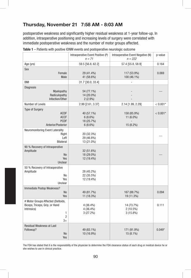
90
Thursday, November 21
The FDA has stated that it is the responsibility of the physician to determine the FDA clearance status of each drug or meidcal device he or she wishes to use in clinical practice.
postoperative weakness and significantly higher residual weakness at 1-year follow-up. In addition, intraoperative positioning and increasing levels of surgery were correlated with immediate postoperative weakness and the number of motor groups affected.
Table 1 – Patients with positive IONM events and postoperative neurologic outcome
Intraoperative Event Positive (P)n = 71
Intraoperative Event Negative (N)n = 222
p-value
Age (yrs) 59.5 [56.8, 62.2] 57.4 [55.8, 58.9] 0.164
SexFemale
Male29 (41.4%)41 (58.6%)
117 (53.9%)100 (46.1%)
0.069
BMI 31.7 [30.0, 33.4] -
DiagnosisMyelopathy
RadiculopathyInfection/Other
54 (77.1%)14 (20.0%)2 (2.9%)
---
---
Number of Levels 2.99 [2.61, 3.37] 2.14 [1.99, 2.29] < 0.001*
Type of SurgeryACDFACCFPCDF
Anterior/Posterior
40 (57.1%)6 (8.6%)
18 (25.7%)6 (8.6%)
158 (85.9%)11 (6.0%)
-15 (8.2%)
< 0.001*
Neuromonitoring Event LateralityRight
LeftBilateral
20 (32.3%)29 (46.8%)13 (21.0%)
---
90 % Recovery of Intraoperative Amplitude
NoYes
Unclear
32 (51.6%)18 (29.0%)12 (19.4%)
---
50 % Recovery of Intraoperative Amplitude
NoYes
Unclear
28 (45.2%)22 (35.5%)12 (19.4%)
Immediate Postop Weakness?NoYes
49 (81.7%)11 (18.3%)
167 (88.7%)19 (11.3%)
0.094
# Motor Groups Affected (Deltoids, Biceps, Triceps, Grip, or Hand Intrinsics)
12
3+
4 (36.4%)4 (36.4%)3 (27.2%)
14 (73.7%)2 (10.5%)3 (15.8%)
0.111
Residual Weakness at Last Followup?
NoYes
49 (83.1%)10 (16.9%)
171 (91.9%)15 (8.1%)
0.049*
7:58 AM - 8:03 AM
91
Thursday, November 21
Individual Disclosures can be found in the Disclosure Index pages 41-69.
Presentation #8
Therapeutic Impact of Traction Release after C5 Nerve Root Motor Evoked Potential Alerts in Cervical Spine Surgery
W. Bryan Wilent, PhD1, Thomas Epplin-Zapf, MS,MA1, Mitali Bose, BS1, Eric Tesdahl, PhD1, Jeffrey Cohen, MD, PhD1, Anthony Sestokas PhD1
1Speciatly Care, Brentwood, TN
Keywords: MEPs, nerve root, C5, spinal cord, intraoperative neuromonitoring
Introduction: The goal of intraoperative neuromonitoring in cervical spine surgery is to reduce the risk of iatrogenic neurologic injury. Motor evoked potentials (MEP) have been shown to have excellent diagnostic accuracy for predicting immediate-onset C5 palsies, but additional data are needed to assess the therapeutic impact of intraoperative interventions prompted by C5 nerve root MEP alerts.
Materials/Methods: A total of 40,919 cervical procedures monitored with MEPs, somatosensory evoked potentials, and spontaneous electromyography from Jan-2016 to Jan-2018 were reviewed retrospectively from a multi-institutional IONM database. Procedures with MEP alerts limited to the upper extremities were identified, and alerts were categorized based on the innervation pattern of the affected muscles. Specifically, C5 MEP alerts were defined as those isolated to attenuation of the deltoid and/or biceps muscle MEPs. C5 MEP alerts were further defined as fully resolved or unresolved (no improvement or partial improvement) based on the status of the signals at closure. A retrospective review of case documentation was used to determine the timing of the alerts (before or after exposure completion) and the category of intervention (anesthetic or blood pressure change, surgical intervention, or release of positional traction). Clinical outcomes were based on neurologic assessment in the immediate postoperative period. Odds ratios (OR) for new neurologic deficits were calculated according to Altman.
Results: 1.1% of procedures had an isolated C5 MEP alert. Relative to procedures with no MEP alerts, the odds of a new neurologic deficit greatly increased if there was a C5 MEP alert (OR=60.4, CI:[40.9-89.2]), and increased further if the alert remained unresolved through closure (OR=102.2, CI:[67.9,153.7]). Importantly, relative to procedures with an unresolved C5 MEP alert, the risk of deficit was significantly decreased if a C5 MEP alert was resolved by closure (OR=0.09, CI:[0.03-0.29]). All OR data were associated with p values <0.0001. 41.5% of all C5 MEP alerts were fully resolved (n=185) by closure, and the documentation of those cases was reviewed in detail to determine the timing of the alert and the effective intervention. 22.8% of those alerts occurred after initial patient positioning but prior to the completion of exposure and 77.2% occurred after exposure was complete. For alerts occurring prior to completion of exposure, the most common effective intervention was release of positional traction (90.9%). For alerts occurring post-exposure, the effective intervention varied. 19.2% were resolved after changes in the anesthetic regimen and/or increases in the mean arterial pressure, 30.3% after surgical action such as further decompression, removal of graft, or vertebral distraction release, 36.3% after positional traction release, and 14.1% after positional traction release combined
8:04 AM - 8:09 AM
92
Thursday, November 21
The FDA has stated that it is the responsibility of the physician to determine the FDA clearance status of each drug or meidcal device he or she wishes to use in clinical practice.
with further surgical action; thus, the majority of effective interventions involved the release of positional traction (50.4%).
Conclusions: When evolving C5 nerve root dysfunction is diagnosed perioperatively via MEPs, the timely release of positional traction is a common intervention that can facilitate MEP recovery and may reduce risk of post-operative C5 palsy.
8:04 AM - 8:09 AM

93
Thursday, November 21
Individual Disclosures can be found in the Disclosure Index pages 41-69.
Presentation #9
Predicting risk of post-operative C5 palsy among patients undergoing posterior cervical spine surgery.
Zach Pennington BS¹, Dan Lubelski MD¹†, Adam D’Sa BA¹, Erick M. Westbroek MD¹, A. Karim Ahmed BS¹, Timothy F. Witham MD¹, Nicholas Theodore MD¹, Daniel M. Sciubba MD¹
¹Department of Neurosurgery, Johns Hopkins School of Medicine, Baltimore
Introduction: Nearly 20,000 patients undergo posterior cervical spine surgery annually. Post-operative complications occur in a non-trivial proportion of cases and include mechanical and non-mechanical varieties. Post-operative C5 palsy – characterized by new onset deltoid weakness with or without biceps weakness and sensory symptoms in the C5 dermatome – occurs in 8-15% of cases. Given the ability of C5 palsy to worsen patient outcomes and increase total costs to the patient, we attempted to identify radiographic and surgical risk factors for this complication.
Materials/Methods: We reviewed all patients treated with posterior cervical spine surgery at our institution for a degenerative spine pathology. Included patients had full medical records including pre-operative MR, neurological examination, and surgical history. Patients were excluded if they had fewer than 3 months of follow-up. We performed univariable analyses with χ² analyses, Fisher exact test, and Mann-Whitney U tests. Multivariable logistic regression was performed for the variables to identify independent, significant anatomic and surgical predictors of post-operative C5 palsy.
Results: Of 468 patients identified, 364 had full medical records for review. Of these, 52 experienced a post-operative C5 palsy. Patients experiencing a C5 palsy had a higher average Kang score (p<0.01), more extensive compression (p=0.02), narrower C4/5 foramina (p<0.01), narrower canals (p<0.01), and more extensive bone stenosis at the C4/5 level (p<0.01). Those experiencing palsy were also more likely to have decompression ≥1- (p<0.01), or ≥2-levels (p<0.01) above and below the C4/5 space and were more likely to have fusion ≥1- (p=0.02), or ≥2-levels (p<0.01) above and below the C4/5 space. The rate of foraminotomy use did not differ between groups. On multivariable regression, mean foraminal diameter, fusion length, and performance of C4 and C5 laminectomies were significant predictors of experiencing a post-operative C5 palsy.
Conclusion: Both anatomic and surgical considerations influence a patient’s risk of C5 palsy. Longer decompressions, tighter C4/5 foramina, and greater baseline compression of the spinal cord at the C4/5 level appear to all increase a patient’s risk for a post-operative C5 palsy. The performance of C4/5 foraminotomies was not observed to increase the risk of post-operative C5 palsy among our cohort.
8:10 AM - 8:15 AM

94
Thursday, November 21
The FDA has stated that it is the responsibility of the physician to determine the FDA clearance status of each drug or meidcal device he or she wishes to use in clinical practice.
Presentation #10
Cord float back does not predict the occurrence of C5 palsy amongst patients undergoing posterior cervical spine surgery
Zach Pennington BS¹, Dan Lubelski MD¹, Adam D’Sa BA¹, Erick M. Westbroek MD¹, A. Karim Ahmed BS¹, Timothy F. Witham MD¹, Nicholas Theodore MD¹, Daniel M. Sciubba MD¹
¹Department of Neurosurgery, Johns Hopkins School of Medicine, Baltimore, MD
Introduction: C5 palsy is a common complication of cervical spine surgery characterized by acute-onset deltoid weakness with or without sensory changes in the C5 dermatome and weakness of the biceps brachii. Despite its relative frequency among those undergoing posterior cervical surgery, the exact mechanism of its occurrence is unknown. Here we sought to investigate one proposed mechanism, namely that increased cord float back increases the risk of C5 palsy.
Materials/Methods: We retrospectively reviewed all patients treated at our institution between January 2013 and December 2017 who underwent posterior cervical spine surgery crossing the C4/5 junction. Included patients had pre- and post-operative MRI, CT scan, and lateral X-rays acquired within 3 months of surgery, a full neurological exam, a complete operative note, and complete surgical and medical records. Patients were excluded if they were less than 18 years of age, had fewer than 3 months of follow-up, or had previously undergone posterior cervical spine surgery.
Results: We identified 468 total patients, of whom 257 had undergone index operations. Of these a total of 37 patients had sufficient imaging for inclusion with a mean age of 62.2 years and mean follow-up of 17.8 months. Nine patients (24.3%) in the cohort experience a post-operative C5 palsy with a mean time to onset of 3.7 days and a mean duration of 33 weeks. Patients experiencing a post-operative palsy were noted to have greater stenosis at the C4/5 level as measured by canal sagittal diameter (p=0.02) and canal cross-sectional area (p=0.03), as well as greater cord compression, noted by a higher baseline Kang score. Small C4/5 foraminal diameter, greater C4 laminectomy width, and greater C5 laminectomy width were also noted in those experiencing a post-operative palsy, but these differences were not significant (p=0.08-0.11). Notably, none of the following were associated with post-operative palsy: post-operative distance between the spinal cord and posterior longitudinal ligament at C4/5, post-operative cord float back at the C4/5 level, or change in cervical lordosis.
Conclusion: Our results suggest that cord float back at the C4/5 level does not influence the risk of experiencing a post-operative C5 palsy. The observation that wider laminectomies at C4 and C5 as well as greater baseline compression of the C5 roots and spinal cord at the C4/5 level suggest that the occurrence of C5 palsy may be related to baseline damage to the spinal cord and roots. However, evaluation using larger, multicenter cohorts is necessary to provide more conclusive insight into this complication.
8:16 AM - 8:21 AM

95
Thursday, November 21
Individual Disclosures can be found in the Disclosure Index pages 41-69.
Presentation #11
Early Versus Late Surgical Decompression For Acute Spinal Cord Injury: A Pooled Analysis Of 1,548 Patients
Jetan H. Badhiwala, MD1; Christopher D. Witiw, MD, MSc2; Jefferson R. Wilson, MD, PhD2; Michael G. Fehlings, MD, PhD1
1Division of Neurosurgery and Spinal Program, Department of Surgery, Krembil Neuroscience Center, Toronto Western Hospital, Toronto, Ontario, Canada2Division of Neurosurgery, Department of Surgery, University of Toronto, St. Michael’s Hospital, Toronto, Ontario, Canada
INTRODUCTION: There is a well-founded biological rationale for early decompressive surgery following acute spinal cord injury (SCI).1,2 However, results relating to the effect of early (< 24 hrs) versus late (≥ 24 hrs) surgical decompression on clinical outcomes are mixed, and the quality of evidence is variable.3 To that end, we sought to leverage the statistical power derived from pooling four high-quality prospective datasets in order to compare sensorimotor recovery with early versus late surgical decompression for acute SCI.
METHODS: Patients with acute traumatic SCI who underwent surgical decompression were identified from four independent prospective, multi-center SCI datasets: NACTN SCI Registry;4 STASCIS; 5 Sygen;6 and NASCIS III.7 Patients were dichotomized into early (< 24 hrs) and late (≥ 24 hrs) surgery groups.5 The primary end point was change in ASIA motor score (AMS) at 1-year. Secondary outcomes included AIS grade and change in ASIA light touch and pin prick scores at 1-year. One-stage meta-analyses for each outcome were performed by hierarchical mixed-effects regression (linear for AMS, light touch score, and pin prick score; ordinal for AIS grade) using a stratified intercept to account for clustering of patients within studies. Fixed-effect covariates were specified for baseline score as well as age, mechanism of injury (fall vs. MVC vs. sports vs. other), baseline AIS grade, neurological level (cervical vs. thoracic vs. lumbosacral), and administration of steroids. The treatment (early vs. late surgery) was specified as a random-effect. As a sensitivity analysis, two-stage meta-analyses were conducted for each outcome, wherein treatment effects were first calculated for each individual study and subsequently pooled in random-effects meta-analyses.8 Subgroup analyses for the primary outcome were conducted stratified by age (< 50 vs ≥ 50 yrs), mechanism of injury, AIS grade, neurological level, and administration of steroids. Effect sizes were summarized by mean difference (MD) for continuous outcomes and common (proportional) odds ratio (cOR) for AIS grade.
RESULTS: A total of 1,548 patients were eligible. Mean age was 39.1 years. The early surgery group experienced greater improvement than the late surgery group at 1-year for AMS (MD 4.0, 95% CI 1.7-6.2, P=0.001), light touch score (MD 4.6, 95% CI 1.9-7.2, P=0.001), and pin prick score (MD 4.2, 95% CI 1.5-6.9, P=0.003). Further, on ‘shift analysis’, the early surgery group achieved a more favorable distribution of AIS grades at 1-year compared to the late surgery group (Figure 1) (cOR 1.46, 95% CI 1.14-1.87, P=0.003). Two-stage meta-analyses confirmed findings of improved sensorimotor recovery with early surgery, producing similar effect sizes to one-stage meta-analyses (Figure 2). Subgroup analyses did not reveal any significant interactions of the treatment effect. The effect of early surgery was strongest for cervical SCI (P=0.003);
8:34 AM - 8:39 AM

96
Thursday, November 21
The FDA has stated that it is the responsibility of the physician to determine the FDA clearance status of each drug or meidcal device he or she wishes to use in clinical practice.
however, we observed a trend toward improved recovery with early versus late surgery for thoracic SCI as well (MD 5.2, 95% CI -0.8-11.2, P=0.088).
CONCLUSION: In an individual patient data meta-analysis adjusting for potential confounders, we found early surgery, within 24 hours of injury, to be associated with superior sensorimotor recovery at 1-year following acute SCI, as compared to late surgery.
Figure 1. Pooled distribution of AIS grades at 1-year stratified by treatment group. Early surgery was associated with a ‘shift’ toward more favorable AIS grade compared to late surgery (cOR 1.46, 95% CI 1.14-1.87, P=0.003).
Figure 2. Forest plots for two-stage meta-analyses for 1-year outcomes: A) change in AMS; B) change in ASIA light touch score; C) change in ASIA pin prick score; D) AIS grade.
8:34 AM - 8:39 AM

97
Thursday, November 21
Individual Disclosures can be found in the Disclosure Index pages 41-69.
References.1. Guha A, Tator CH, Endrenyi L, Piper I. Decompression of the spinal cord improves recovery after
acute experimental spinal cord compression injury. Paraplegia 1987; 25(4): 324-39.2. Dimar JR, 2nd, Glassman SD, Raque GH, Zhang YP, Shields CB. The influence of spinal canal
narrowing and timing of decompression on neurologic recovery after spinal cord contusion in a rat model. Spine (Phila Pa 1976) 1999; 24(16): 1623-33.
3. Wilson JR, Tetreault LA, Kwon BK, et al. Timing of Decompression in Patients With Acute Spinal Cord Injury: A Systematic Review. Global Spine J 2017; 7(3 Suppl): 95S-115S.
4. Grossman RG, Toups EG, Frankowski RF, Burau KD, Howley S. North American Clinical Trials Network for the Treatment of Spinal Cord Injury: goals and progress. J Neurosurg Spine 2012; 17(1 Suppl): 6-10.
5. Fehlings MG, Vaccaro A, Wilson JR, et al. Early versus delayed decompression for traumatic cervical spinal cord injury: results of the Surgical Timing in Acute Spinal Cord Injury Study (STASCIS). PLoS One 2012; 7(2): e32037.
6. Geisler FH, Coleman WP, Grieco G, Poonian D, Sygen Study G. The Sygen multicenter acute spinal cord injury study. Spine (Phila Pa 1976) 2001; 26(24 Suppl): S87-98.
7. Bracken MB, Shepard MJ, Holford TR, et al. Administration of methylprednisolone for 24 or 48 hours or tirilazad mesylate for 48 hours in the treatment of acute spinal cord injury. Results of the Third National Acute Spinal Cord Injury Randomized Controlled Trial. National Acute Spinal Cord Injury Study. JAMA 1997; 277(20): 1597-604.
8. DerSimonian R, Laird N. Meta-analysis in clinical trials. Control Clin Trials 1986; 7(3): 177-88.
8:34 AM - 8:39 AM
98
Thursday, November 21
The FDA has stated that it is the responsibility of the physician to determine the FDA clearance status of each drug or meidcal device he or she wishes to use in clinical practice.
Presentation #12
Clinical Outcomes of Acute Cervical Spinal Cord Injury Depending on the Timing of Surgery
Kyung Jin Song, Jong Hyun Ko, Tae Young Kwon, Hyung Gik Kim
Department of Orthopedic Surgery, Chonbuk National University Medical School, Research Institute of Clinical Medicine of Chonbuk National University-Biomedical Research Institute of Chonbuk National University Hospital
Introduction: There have been lots of prior studies that show optimal surgical time for acute cervical spinal cord injury (SCI). Most studies reported that early surgery had better results than late surgery. However, the timing of surgery remains controversial and the cutoff to classify the early and late surgery varies. A previous study determined the cutoff time of early surgery was 8 hours after injury. Others said 24 hours or 72 hours was the cutoff time. The purpose of this study is to evaluate the clinical outcomes of acute cervical SCI depending on the timing of surgery.
Material and Methods: We analyzed patients whose surgery was performed for acute cervical spinal cord injury from 2007 to 2017. The primary outcome depending on timing of surgery was analyzed with AIS (ASIA Impairment Scale) change between initial assessment and 6 months after the surgery. We defined the 1 degree improvement of AIS was clinical significant. We applied various cutoff time of early surgery. We also analyzed the relation of AIS change and age, gender, neurologic level of injury and usage of mega-dose steroid administration. Secondary outcomes were analyzed with complications, such as neurologic bladder, need for ICU care, respiratory distress requiring ventilator care and other newly onset medical problems, and mortality. The statistical analysis was performed by R language version 3.3.3 (Vienna, Austria) and Chi-squared or Fisher’s exact test and logistic regression analysis.
Results: A total of 151 patients with acute cervical SCI were enrolled. Of these, 14 patients underwent surgery within 12 hours of the injury and 34 patients, 33 patients and 21 patients underwent surgery within 24 hours, 48 hours, 72 hours of the injury respectively. Forty nine (49) patients underwent surgery after 72 hours. As primary outcome, the improvement of AIS was shown between early surgery group (≤24hr) and late surgery group (>24hr) (p=0.041). The details of AIS improvement were 44.8% in the patients, which underwent surgery within 24 hours of the injury and 31% (24~72hr) and 24.1% (>72hr). In univariate binary logistic regression test, 24 hours was also significant cutoff time of early surgery (p=0.044). In addition, 12hrs vs 72hrs and 24hrs vs 72hrs were possible cutoff time for early and late surgery (p=0.039 and 0.044). In multivariate binary logistic regression test, the only selectable cutoff time was 24hrs for early surgery (p=0.016), but there was no statistical difference between ≤12 hours group and 12~24 hours group (p=0.08). Usage of mega-dose steroid and the AIS improvement was not shown significant relation statistically (p=0.061). Age, gender, and neurologic level of injury (NLI) had no relation with AIS improvement statistically also. As secondary outcome, complications were 34.6% in the patients who underwent surgery within 24 hours of injury, and 28.5% (24~72hr) and 24.4% (>72hr) respectively, however, there was no statistical significance.
Conclusions: In acute SCI, better clinical outcome can be expected in early surgery than in late surgery. One of the selectable cutoff time for early surgery is 24 hour after the injury.
8:40 AM - 8:45 AM

99
Thursday, November 21
Individual Disclosures can be found in the Disclosure Index pages 41-69.
Presentation #13
Early Versus Late Surgical Decompression For Central Cord Syndrome: A Propensity Score-Matched Analysis
Jetan H. Badhiwala, MD1; Christopher D. Witiw, MD, MSc2; Jefferson R. Wilson, MD, PhD2; Michael G. Fehlings, MD, PhD1
1Division of Neurosurgery and Spinal Program, Department of Surgery, Krembil Neuroscience Center, Toronto Western Hospital, Toronto, Ontario, Canada, 2Division of Neurosurgery, Department of Surgery, University of Toronto, St. Michael’s Hospital, Toronto, Ontario, Canada
INTRODUCTION: The role of early surgical decompression, within 24 hours of injury, for central cord syndrome (CCS) remains unclear, with the current evidence base being derived exclusively from small retrospective case series with sample sizes of less than 20 patients undergoing early surgery.1 Yet, with the continued aging of the global population, CCS is soon expected to become the most common form of acute traumatic spinal cord injury (SCI),2,3 making the identification of treatment strategies that mitigate disability and improve functional status in this vulnerable population a key public health priority.4 To that end, we sought to perform a head-to-head comparison of the neurological and functional outcomes of early (< 24 hrs) versus late (≥ 24 hrs) surgical decompression in patients with CCS.
METHODS: Patients who underwent surgery for CCS, defined by American Spinal Injury Association (ASIA) Impairment Scale (AIS) grade C or D and a ≥ 5-point difference between ASIA lower (LEMS) and upper extremity motor score (UEMS) (LEMS – UEMS ≥ 5), were identified from three prospective, multi-center SCI datasets: NACTN SCI Registry;5 STASCIS;6 and NASCIS III.7 Propensity scores were calculated as the probability of undergoing early (< 24 hrs) versus late (≥ 24 hrs) surgery using the logit method with age (continuous), mechanism of injury (categorical; fall vs. MVC vs. sports vs. other), data source (categorical), initial ASIA motor score (continuous), initial AIS grade (categorical; C vs. D), and initial ASIA neurological level of injury (NLI) (continuous) as covariates. Propensity score matching was performed in a 1:1 ratio using the ‘optimal matching’ technique to minimize the average absolute distance across all matched pairs. The primary outcome was ASIA motor score at 1-year. Secondary outcomes were Functional Independence Measure (FIM) motor score as well as scores for each of the thirteen FIM motor subdomains at 1-year. Outcomes were compared between study groups by t-test. Effect sizes for each outcome measure were summarized by mean differences (MDs) and associated 95% confidence intervals (CIs).
RESULTS: Three-hundred patients fulfilled eligibility criteria and had complete baseline data available for analysis. Propensity score matching produced a final study cohort of 186 patients, 93 undergoing early surgery (< 24 hrs) and 93 receiving late surgery (≥ 24 hrs). Patient demographic, injury, and treatment characteristics at baseline were balanced between matched early and late surgery groups (Table 1). Table 2 presents outcomes for each time to surgery group. ASIA motor score at 1-year was significantly higher in the early surgery (mean: 91.8) compared to late surgery (mean: 87.1) group (MD 4.64, 95% CI 0.19 to 9.09, P=0.04). Similarly, patients who underwent early (mean: 84.2) as opposed to late (mean: 77.2) surgery achieved better functional status at 1-year, as measured by the FIM motor score (MD 7.04, 95% CI 0.59 to
8:46 AM - 8:51 AM
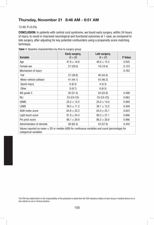
100
Thursday, November 21
The FDA has stated that it is the responsibility of the physician to determine the FDA clearance status of each drug or meidcal device he or she wishes to use in clinical practice.
13.49, P=0.03).
CONCLUSION: In patients with central cord syndrome, we found early surgery, within 24 hours of injury, to result in improved neurological and functional outcomes at 1-year, as compared to late surgery, after adjusting for key potential confounders using a propensity score matching technique.
Table 1: Baseline characteristics by time to surgery group
VariableEarly surgery,
N = 93Late surgery,
N = 93 P Value
Age 47.8 ± 16.8 48.0 ± 15.5 0.935
Female sex 27 (29.0) 18 (19.4) 0.123
Mechanism of injury 0.762
Fall 37 (39.8) 40 (43.0)
Motor vehicle collision 41 (44.1) 43 (46.2)
Sports injury 6 (6.5) 4 (4.3)
Other 9 (9.7) 6 (6.5)
AIS grade C 20 (21.5) 24 (25.8) 0.490
NLI C5 (C4-C5) C5 (C4-C5) 0.863
UEMS 25.2 ± 12.5 25.0 ± 14.0 0.895
LEMS 39.5 ± 11.2 38.1 ± 12.2 0.400
ASIA motor score 64.8 ± 22.2 63.0 ± 25.1 0.623
Light touch score 91.9 ± 24.3 90.2 ± 27.1 0.666
Pin prick score 86.1 ± 28.9 86.2 ± 28.9 0.986
Administration of steroids 58 (62.4) 53 (57.0) 0.455
Values reported as mean ± SD or median (IQR) for continuous variables and count (percentage) for categorical variables
8:46 AM - 8:51 AM

101
Thursday, November 21
Individual Disclosures can be found in the Disclosure Index pages 41-69.
Table 2: Outcomes at 1-year by time to surgery group
VariableEarly surgery,
N = 93*Late surgery,
N = 93*Difference(95% CI) P Value
ASIA motor score 91.8 87.1 4.64 (0.19 to 9.09) 0.041*
FIM motor score 84.2 77.2 7.04 (0.59 to 13.49) 0.033*
Eating 6.6 6.2 0.43 (-0.07 to 0.92) 0.088
Grooming 6.5 6.0 0.42 (-0.14 to 0.98) 0.136
Bathing 6.3 5.8 0.53 (-0.12 to 1.18) 0.110
Dressing, upper body 6.5 6.0 0.48 (-0.10 to 1.07) 0.101
Dressing, lower body 6.3 5.7 0.62 (-0.03 to 1.27) 0.060
Toileting 6.5 5.9 0.63 (0.02 to 1.24) 0.042*
Bladder management 6.6 6.1 0.57 (-0.03 to 1.16) 0.061
Bowel management 6.7 6.2 0.51 (-0.04 to 1.06) 0.069
Bed, chair, wheelchair transfers 6.6 6.1 0.49 (-0.09 to 1.08) 0.097
Toilet transfers 6.6 6.1 0.55 (-0.01 to 1.11) 0.056
Tub, shower transfers 6.4 6.0 0.44 (-0.15 to 1.04) 0.141
Walking, wheelchair 6.6 6.1 0.43 (-0.09 to 0.95) 0.105
Stairs 6.2 5.3 0.98 (0.26 to 1.70) 0.008*
*Values reported are means
References.1. Anderson KK, Tetreault L, Shamji MF, et al. Optimal Timing of Surgical Decompression for Acute
Traumatic Central Cord Syndrome: A Systematic Review of the Literature. Neurosurgery. 2015;77 Suppl 4:S15-32.
2. Thompson C, Mutch J, Parent S, Mac-Thiong JM. The changing demographics of traumatic spinal cord injury: An 11-year study of 831 patients. J Spinal Cord Med. 2015;38(2):214-223.
3. Fehlings MG, Tetreault L, Nater A, et al. The Aging of the Global Population: The Changing Epidemiology of Disease and Spinal Disorders. Neurosurgery. 2015;77 Suppl 4:S1-5.
4. Badhiwala JH, Ahuja CS, Fehlings MG. Time is spine: a review of translational advances in spinal cord injury. J Neurosurg Spine. 2018;30(1):1-18.
5. Grossman RG, Toups EG, Frankowski RF, Burau KD, Howley S. North American Clinical Trials Network for the Treatment of Spinal Cord Injury: goals and progress. J Neurosurg Spine. 2012;17(1 Suppl):6-10.
6. Fehlings MG, Vaccaro A, Wilson JR, et al. Early versus delayed decompression for traumatic cervical spinal cord injury: results of the Surgical Timing in Acute Spinal Cord Injury Study (STASCIS). PLoS One. 2012;7(2):e32037.
7. Bracken MB, Shepard MJ, Holford TR, et al. Administration of methylprednisolone for 24 or 48 hours or tirilazad mesylate for 48 hours in the treatment of acute spinal cord injury. Results of the Third National Acute Spinal Cord Injury Randomized Controlled Trial. National Acute Spinal Cord Injury Study. JAMA. 1997;277(20):1597-1604.
8:46 AM - 8:51 AM
102
Thursday, November 21
The FDA has stated that it is the responsibility of the physician to determine the FDA clearance status of each drug or meidcal device he or she wishes to use in clinical practice.
Presentation #14
Can the proposed posterior ligament-bone injury classification and severity score predict the failure of anterior-only surgery for subaxial cervical facet dislocations?
Jun-Song Yang1 M.D, Tuan-Jiang Liu1 M.D, Ding-Jun Hao1, M.D
Department of Spine Surgery, Honghui Hospital, Xi’an Jiaotong University, Xi’an, Shaanxi, China.
Background: The approach selection for subaxial cervical fracture dislocations (SCFD) is controversial. While increasing spinal surgeons prefer a single anterior approach to address the SCFD patients, the complication of development of postoperative cervical kyphosis is worrisome. The question of how to predict the risk of implant failure after a single anterior approach based on radiological characters of these patients. This study was aim to verify whether the novel posterior ligament-bone injury classification and severity (PLICS) score can be generally utilized for the treatment of the SCFD to predict the failure of anterior-only surgery.
Methods: 394 patients with SCFD who underwent anterior surgery between January 2002 and December 2017 were enrolled. Clinical evaluation criteria included visual analogue score (VAS), the Neck Disability Index (NDI) and the American Spinal Injury Association (ASIA) impairment scale. The PLICS is based on the injury severity of ligament-bone structure of three elements at the posterior column of cervical spine (table.1 and 2).
Results: The reduction and decompression was satisfactory at the postoperative MRI for all patients. Among 354 patients (89.8%) fulfilled the follow-up, average follow-up duration was 18±3.5 months. 15 patients (4.2%) experienced progressive cervical kyphosis postoperatively. The patients were divided into successful (15 patients) and unsuccessful (339 patients) groups. The average VAS score in the unsatisfactory group decreased from 6.7+0.8 to 1.3+0.6; the average VAS score in the satisfactory group decreased from 5.9+0.8 to 0.3+0.5; there was statistical difference in the preoperative and postoperative VAS score between these two groups (P< .05, respectively). The average NDI score was statistically low in the satisfactory group (6.2+2.1 vs 15.5+3.8, P= .028). At least one grade improvement in the ASIA scale was observed in 88.5% of the patients in the satisfactory group and in 66.7% of the patients in the satisfactory group. Except the only one patient with a PLICS score of 6 (figure.1), the PLICS score for the other 14 patients who experienced postoperative hardware failure was ≥7. However, the PLICS score for the other 339 patients (except for 2 cases) who obtained satisfactory radiological outcome was <7 (figure.2). A significantly difference was detected in the PLICS score between the two groups (P< .001).
Conclusion: When a PLICS score is > 7 or = 7 accompanied by severe lateral mass fracture, the risk of postoperative failure after an anterior-only reconstruction is high and supplemental posterior strengthening can be considered.
Keywords: Subaxial cervical spine injury; Anterior approach; implant failure; fracture dislocation
8:52 AM - 8:57 AM

103
Thursday, November 21
Individual Disclosures can be found in the Disclosure Index pages 41-69.
FIGURE.1
FIGURE.2
Table 1. The score to assess severity of injury to the posterior element
Classification Scores
Intact of PLC 0
Mild injury of PLC 1
Moderate injury of PLC 2
Severe injury of PLC 3
PLC: posterior ligamentous complex
Table 2. The score to assess severity of injury to the lateral element
Ligamentous structure Scores
Normal alignment of facet joints 0
Subluxation 1
Dislocation 2
Bony structure
Without lateral mass fracture 0
Stable lateral mass fracture 1
Unstable lateral mass fracture 2
Extremely unstable lateral mass fracture 3
8:52 AM - 8:57 AM

104
Thursday, November 21
The FDA has stated that it is the responsibility of the physician to determine the FDA clearance status of each drug or meidcal device he or she wishes to use in clinical practice.
Presentation #15
Cervical Spine Fractures: Who Really Needs CT Angiography?
Mitchell S Fourman MD, M Phil; Jeremy Dewitt Shaw, MD, MS; Nicholas Vaudreuil, MD; Malcolm Dombrowski, MD; Richard Wawrose, MD; Lorraine Boakye, MD; Louis Alarcon, MD; Joon Yung Lee, MD; William F Donaldson III, MD
Introduction: Computed tomography angiography (CTA) screening permits the rapid detection of blunt cerebrovascular injuries (BCVI) following cervical spine fractures. Gold standard screening criteria, such as the American College of Surgeons (ACS) advanced trauma and life support guidelines and the expanded Denver Criteria, were derived from high energy trauma patients 1,2. Correlation between low grade (Denver I) BCVI and posterior circulation strokes in the low energy trauma population remains unclear. This discrepancy is significant, as the low sensitivity of CTAs (<80% in pooled analyses3) predicts an unacceptable false positive rate, thereby over-indicating invasive angiography and endovascular procedures if used as a screening modality in lower risk populations. The purpose of this study was to derive a simple algorithm for indicating CTA in patients with cervical spine fractures.
Materials/Methods: A novel two-step metric for indicating CTA screening was derived with historical data and compared with the ACS guidelines and the expanded Denver Criteria. Four years (2012-2016) of consecutive patients presenting with cervical spine fractures to a tertiary level 1 trauma center were used to validate this novel algorithm. A CTA was indicated if the patient met two absolute criteria, or one absolute and one fracture criteria (fig 1). BCVI and stroke detection with the novel proposed criteria was compared with the ACS and Denver Criteria. Statistical analysis was performed using Fisher’s Exact Test.
Results: A total of 721 patients with cervical fractures were included in the analysis, of which 417 underwent CTAs (57.8%, fig 2). Sixty-eight BCVIs and seven strokes were diagnosed in this cohort. Importantly, this novel algorithm outperformed both ACS and Denver criteria for detection of stroke (7 / 7, 100% with the novel metric, 6 / 7, 85.7% with the ACS and Denver Criteria, p = 1.0) and detected an equivalent number of BCVIs (52 with the novel metric, 54 with the ACS and Denver Criteria, p = .84). Notably fewer scans would have been needed with the proposed screening algorithm compared with the ACS or Denver Criteria. (261 / 721, 36.2% with our criteria vs. 413 / 721, 57.3% with the ACS standard and 417 / 721 (57.8%) with the Denver Criteria, p < .0002 for each). No strokes occurred in patients not indicated for BCVI by the novel criteria, and all BCVI in this group were Denver I.
Conclusion: This novel two-step patient specific algorithm is a useful adjunct for identifying patients at risk of BCVI and stroke after cervical spine fractures. The current model outperforms the current gold standard ACS and Denver Criteria. This novel algorithm may also have additional utility in pediatric populations that are currently not covered by the ACS or Denver Criteria. Prospective analyses are required prior to any recommendation of widespread clinical adoption.
References: 1. Geddes AE, Burlew CC, Wagenaar AE, et al. Expanded screening criteria for blunt
cerebrovascular injury: a bigger impact than anticipated. American journal of surgery
8:58 AM - 9:03 AM

105
Thursday, November 21
Individual Disclosures can be found in the Disclosure Index pages 41-69.
2016;212:1167-74.2. Shafafy R, Suresh S, Afolayan JO, et al. Blunt vertebral vascular injury in trauma patients: ATLS((R)) recommendations and review of current evidence. Journal of spine surgery (Hong Kong) 2017;3:217-25.
3. Roberts DJ, Chaubey VP, Zygun DA, et al. Diagnostic accuracy of computed tomographic angiography for blunt cerebrovascular injury detection in trauma patients: a systematic review and meta-analysis. Annals of surgery 2013;257:621-32.
Figure 1 – Absolute and Fracture Criteria
Absolute Selection Criteria:1. High Energy Trauma2. Loss of Consciousness at Time of Trauma3. Altered Mental State or Unable to Protect the Airway during Initial EvalFracture Selection Criteria:1. Transverse Foramen Involvement2. Combined C1-C2 Fx3. Bilateral Facet Fx4. Jump Facet / Facet Dislocation
Figure 2 – Patient Eligibility and Outcomes based on the Novel Criteria
8:58 AM - 9:03 AM
106
Thursday, November 21
The FDA has stated that it is the responsibility of the physician to determine the FDA clearance status of each drug or meidcal device he or she wishes to use in clinical practice.
Presentation #16
Cervical deformity, Health-related quality of life, Patient reported outcome metrics. What Drives Disability in Cervical Deformity: Novel Patient Generated Outcome versus Legacy HRQL
Nicholas Stekas MS1, Ethan W Ayres MPH1, Mohamed A Moawad MPH1, Brooke K O’Connell MS1, Dainn Woo BS1, Michael L Smith MD1, Yong H Kim MD1, Aaron J. Buckland MBBS, FRACS1, Themistocles S. Protopsaltis MD1
1Department of Orthopaedic Surgery, NYU Langone Orthopaedic Hospital, New York, NY
Introduction: Existing health outcome (HRQL) metrics do not adequately capture disability from cervical deformity (CD) and do not correlate with cervical malalignment. In the novel Patient Generated Index (PGI) patients report their greatest difficulties related to their CD. These results were used to determine items that should be included in a CD-specific HRQL. The purpose of this study was to utilize the PGI to reveal the aspects of CD disability not captured by existing HRQLs
Methods: This was a retrospective review of a prospective CD database. CD patients completed the PGI by describing aspects of their disability that bother them the most. The responses were weighted and scored. PGI responses were categorized into domains: Sagittal discomfort/range of motion(ROM), Activities of Daily Living(ADL), and Social Life/Hobbies. Legacy metrics included the NDI, mJOA, and EQ-5D questionnaires. PGI scores and legacy HRQL metrics were correlated with alignment, pain, age, sex, BMI, and medical comorbidities. Significant drivers for each HRQL metric were identified using stepwise regression and R2 values were reported for each model.(table 1)
Results: 45 CD patients (mean cSVA: 51mm) including 12 PGI patients (mean cSVA: 62mm) were included for analysis. PGI scores were found to be driven significantly by age and C2 Slope (r2=0.50). NDI was driven significantly by neck pain, back pain, and BMI (r2=0.32). mJOA was driven significantly by Charlson Comorbidity Score (CCI), back pain and weight (r2=0.33). EQ5D was significantly driven by CBVA, age, and T1 Slope (r2=0.78). When examining PGI domains, Sagittal Discomfort/ROM score was driven significantly by cSVA and age (r2=0.54). ADL score was driven by CBVA and a medical history of neuromuscular disease (r2=0.87). Social Life/Hobbies score was driven by Charlson Comorbidity Scores, a medical history of ankylosing spondylitis, and a medical history of connective tissue disease (r2=1.0). Horizontal Gaze/Walking Safety, Pain, and Neurologic Complaints did not correlate significantly with alignment, pain, demographic info or past medical history.
Conclusions: Legacy HRQLs do not adequately capture CD disability and do not correlate with cervical malalignment. In a cohort of CD patients, PGI scores and EQ5D scores were driven significantly by sagittal alignment. However, mJOA and NDI were primarily driven by pain and medical comorbidities.
2:39 PM - 2:44 PM
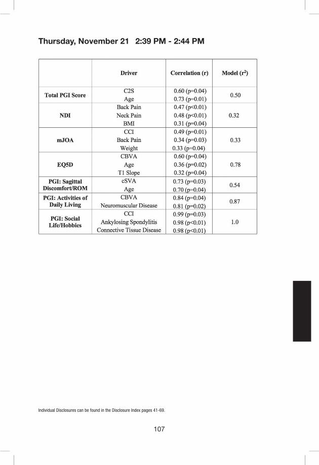
107
Thursday, November 21
Individual Disclosures can be found in the Disclosure Index pages 41-69.
2:39 PM - 2:44 PM
108
Thursday, November 21
The FDA has stated that it is the responsibility of the physician to determine the FDA clearance status of each drug or meidcal device he or she wishes to use in clinical practice.
Presentation #17
Intraoperative Alignment Goals for Severe Cervical Deformity to Achieve Optimal Improvements in Health-Related Quality of Life Measures
Sohrab Virk MD1; Peter Passias MD2; Renaud Lafage MS1; Eric Klineberg MD3; Gregory Mundis MD4; Themistocles Protopsaltis MD2; Christopher Shaffrey MD5; Shay Bess MD6; Han Jo Kim MD1; Christopher Ames MD7; Frank Schwab MD1; Justin Smith MD PhD8; Virginie Lafage PhD1 ; International Spine Study Group9
1Department of Orthopedics, Hospital for Special Surgery, New York, New York2Department of Orthopedics, NYU Langone Orthopedic Hospital, New York, New York3Department of Orthopaedic, University of California, Davis, Sacramento, California4Scripps Clinic, San Diego, California5Department of Neurosurgery, Duke University Medical Center, Durham, NC6International Spine Center, Presbyterian St. Luke‘s/Rocky Mountain Hospital for Children, Denver, CO7Department of Neurological Surgery, University of California, San Francisco, San Francisco, California8Department of Neurosurgery, University of Virginia Medical Center, Charlottesville, Virginia9International Spine Study Group Foundation, Denver, Colorado, USA
Introduction: Patients with severe cervical deformity (CD) often have profound deficits in numerous activities of daily living. Association between health status and post-operative radiographic goals is difficult to quantify. We aimed to investigate the radiographic characteristics of patients who achieved optimal HRQOLs following surgery for CD
Methods: Surgical patients with severe CD were isolated based upon a previously presented combination of preop cSVA, T1 slope, maximum focal kyphosis in extension, C2 slope in extension, and number of kyphotic levels in extension. Those with available preop and 1-year postop HRQL data were included. Based on previous study, patients were grouped into 3 distinct patterns of CD: focal deformity (FD), flatneck (FN=large TS-CL and lack of compensation), or cervicothoracic (CT). Post-operative outcomes were defined as “good” if a patient had >2 of the 3 following criteria (1) NDI <20 or meeting MCID, (2) mild myelopathy (mJOA≥13), and (3) NRS-Neck ≤5 or improved by >2 points from baseline. Within each distinct deformity group, patients with good outcomes were compared to those with poor outcomes (i.e not meeting the criteria for good) for differences in demographics, HRQL scores, and alignment, via Chi-squared or student’s t-tests.
Results: Overall, 83/153 patients met the criteria of severe CD and 40 patients had complete 1 year follow-up of clinical/radiographic data. Patient breakdown by deformity pattern was as follows: CT (N=13), FN (N=17), and FD (N=17), with 7 patients meeting criteria for both FD and FN deformities. Within the FD cohort, maximal focal kyphosis (ie kyphosis at one level) was better corrected in patients with a “good” outcome (p = 0.03). In the FN cohort, patients who went on to have “good” outcomes presented with worse horizontal gaze (McGregor Slope 21° vs 6°, p=0.061) and cSVA (72mm vs 60mm, p=0.030). “Good” outcome FN patients showed significantly greater postop correction of horizontal gaze (-25° vs -5°, p = 0.031). In the CT
2:45 PM - 2:50 PM
109
Thursday, November 21
Individual Disclosures can be found in the Disclosure Index pages 41-69.
cohort, patients with “good” outcomes had superior global alignment both pre- (SVA: -17mm vs 108mm, p <0.001) and post-operatively (50mm vs 145mm, p=0.001). CT patients with “good” outcomes also had better postop cervical alignment (cSVA 35mm vs 49mm, p=0.030), and less kyphotic segments during extension (p=0.011). In the FD cohort, there were no differences between “good” and “poor” outcomes patients in preoperative alignment; however, “good” outcome patients showed superior changes in postoperative focal kyphosis (-2° vs 5°, p=0.030). Within all three deformity pattern categories, there were no differences between “good” and “poor” outcome patients with respect to demographics or surgical parameters (levels fused, surgical approach, decompression, osteotomy, all p>0.050).
Conclusions: The results of this study highlight intra-operative goals for three distinct sagittal morphotypes of severe CD: cervicothoracic, flatneck, and focal deformity. Distinct deformity specific intra-operative goals include obtaining proper sagittal global/cervical alignment for cervicothoracic patients, correcting maximal focal kyphosis in focal deformity patients, and correcting horizontal gaze for flatneck patients.
2:45 PM - 2:50 PM
110
Thursday, November 21
The FDA has stated that it is the responsibility of the physician to determine the FDA clearance status of each drug or meidcal device he or she wishes to use in clinical practice.
Presentation #18
Improvement in cervical lordosis and sagittal alignment after vertebral body sliding osteotomy in patients with spondylotic cervical myelopathy and kyphosis
Dong-Ho Lee, Jae Hwan Cho, Jae-Woo Park, Chul-Gie Hong, Jung-Gi Ha, and Kun-Bo Park
Department of Orthopedic Surgery, Asan Medical CenterUniversity of Ulsan College of Medicine, Seoul, Korea
Introduction: Vertebral body sliding osteotomy (VBSO) is a novel technique for safer anterior decompression in patients with multilevel cervical myelopathy and kyphosis. Another advantage of VBSO may be the restoration of cervical lordosis through the preserved vertebral body and anterior discectomy and fusion at the above and below osteotomy level. This study aimed to evaluate the improvement in cervical lordosis and sagittal alignment after VBSO compared with the traditional anterior cervical corpectomy and fusion (ACCF).
Materials and Methods: We included 34 patients who underwent VBSO and 31 patients who underwent ACCF for cervical spondylotic myelopathy and kyphosis. The mean age was 52 (27 to 77) and 52 (46 to 80) years in the VBSO and ACCF groups, respectively. In the ACCF group, all operations were 1-level corpectomy, and 19 patients in the VBSO group underwent a 2-level operation (Fig 1). Preoperative, postoperative, and final postoperative 2-years follow-up radiographs were used to evaluate the improvement in cervical lordosis and sagittal alignment. The Japanese Orthopaedic Association (JOA) score was used to assess the degree of disability.
Results: There was no difference in the preoperative radiologic parameters between groups. Postoperative C2-C7 (11.6˚±10.9˚) and segmental (15.0˚±10.3˚) lordosis in the VBSO group were higher than those (6.4˚±8.1˚, 9.3˚, respectively) in the ACCF group (p=0.032, 0.011, respectively). In the ACCF group, although the final C2-C7 (7.9˚±7.8˚, p=0.022) and segmental (7.2˚±7.6˚, p=0.012) lordosis were improved compared with preoperative value, segmental lordosis at the final follow-up decreased compared with a postoperative value (p=0.045). After VBSO, however, not only C2-C7 (13.0˚±9.9˚, p<0.001) and segmental (15.2˚±8.5˚, p<0.001) lordosis but also C0-C2 lordosis (39.3˚±7.2˚, p=0.049) and C2-C7 sagittal vertical axis (SVA) (18.4±7.9mm, p=0.038) showed significant improvement at the final follow-up compared with the preoperative value. There was no difference in preoperative and postoperative JOA scores between groups. In the subgroup analysis of 1-level VBSO and ACCF, the final segmental lordosis was larger in VBSO. The postoperative segmental lordosis was larger in 2-level VBSO in the comparison of 1-level and 2-level VBSO
Conclusion: VBSO was a superior technique in terms of improving global and segmental cervical lordosis compared with ACCF. The preserved vertebral body support the graft or cage more effectively and the multiple trapezoidal shape cage more increases the cervical lordosis (Fig 2). C0-C2 lordosis and C2-C7 SVA improved only after VBSO. VBSO is a reliable technique for patients who require simultaneous correction of kyphosis and anterior decompression.
2:51 PM - 2:56 PM

111
Thursday, November 21
Individual Disclosures can be found in the Disclosure Index pages 41-69.
Fig 1. A 27-year-old man with cervical spondylotic myelopathy due to ossification of the posterior longitudinal ligament (OPLL) and cervical kyphosis underwent vertebral body sliding osteotomy at C5 and C6. In the preoperative radiographic examination, the segmental lordosis between C4 and C7 was -16°, and OPLL was noted on computed tomography. After the operation, the segmental lordosis between C4 and C7 was increased to 4° and the OPLL in the spinal canal moved outward. At 2 years after the index operation, complete union was noted and the segmental lordosis between C4 and C7 was maintained at 7°.
Fig 2. During anterior cervical corpectomy and fusion (ACCF), the height of the vertebral body is the primary concern, and there may be a little change in cervical lordosis. However, the height of the vertebral body is maintained because the body just slides anteriorly during vertebral body sliding osteotomy (VBSO), and the cervical lordosis increase due to the cervical discectomy and fusion with a trapezoidal wedge cage above and below the VBSO level.
2:51 PM - 2:56 PM

112
Thursday, November 21
The FDA has stated that it is the responsibility of the physician to determine the FDA clearance status of each drug or meidcal device he or she wishes to use in clinical practice.
Presentation #19
Pre-Operative Extension Lateral Cervical Radiographs are Associated with Osteotomy Type, Approach and Post-Operative Cervical Alignment Following Cervical Deformity Surgery
Eric Klineberg, MD1 ; Renaud Lafage, MS2 ; Munish Gupta, MD3 ; Peter Passias, MD4 ; Virginie Lafage, PhD2 ; Justin Smith, MD PhD5 ; Han Jo Kim, MD2 ; Themistocles Protopsaltis, MD4 ; Douglas Burton, MD6 ; Gregory Mundis, MD7 ; Frank Schwab, MD2 ; Robert Hart, MD8 ; Christopher Shaffrey, MD9 ; Christopher Ames, MD10 ; International Spine Study Group11
1Department of Orthopaedic Surgery, University of California, Davis, Sacramento, California 2Department of Orthopedics, Hospital for Special Surgery, New York, New York 3Department of Orthopaedic Surgery, Washington University, St Louis, MO4Department of Orthopedics, NYU Langone Orthopedic Hospital, New York, New York5Department of Neurosurgery, University of Virginia Medical Center, Charlottesville, Virginia6Department of Orthopaedics, University of Kansas Medical Center, Kansas City, Kansas7Scripps Clinic, San Diego, California8Department of Orthopaedic Surgery, Swedish Neuroscience Institute, Seattle, Washington9Department of Neurosurgery, Duke University Medical Center, Durham, NC 10Department of Neurological Surgery, University of California, San Francisco, School of Medicine, San Francisco, California11International Spine Study Group Foundation, Denver, Colorado
Introduction: Choosing the surgical approach and osteotomy type is challenging when surgically treating cervical deformity. Pre-operative assessment of flexibility and final alignment would be useful for surgeons treating this complex problem. Currently extension lateral XR are inconstantly taken for the management of cervical deformity for planning purposes. The purpose of the current study was to investigate the relationship of surgical procedure choice and the pre-operative flexibility of the cervical spine. Determine if extension lateral XR (ELXR) can also predict the final post-operative alignment.
Material/Methods: This study is a retrospective review of prospective, consecutively enrolled multicenter cervical deformity database. Inclusion was cervical kyphosis>10°, cervical scoliosis>10°, C2-C7 SVA>4cm or chin-brow vertical angle>25°. Patients were excluded if they did not have neutral and ELXR or did not have deformity limited to the Cervical or Cervicothoracic spine. The ELXR was compared to pre-operative neutral lateral, and 3 mo alignment XR and type of surgical osteotomy based upon the Ames Classification. Statistical analysis included t-test and chi-squared.
Results: 106/164 patients met the inclusion criteria. Mean age of 60 yo, with 58% females. 43.4% of patients had prior cervical surgery. The evaluation of ELXR in patients who received grade 1-2 osteotomies (as compared to those who received grade 5,6 or 7), shows they have statistically lower T1S (23.06 vs 35.46; p=.021), lower T1S-CL (20.90 vs 36.29; p=.033), lower cSVA (24.66 vs 48.35; p<.001) and lower C2Slope (18.69 vs 37.46; p=.008) .
Subsequent analysis revealed the anterior approach chosen over the post approach when, in
2:57 PM - 3:02 PM
113
Thursday, November 21
Individual Disclosures can be found in the Disclosure Index pages 41-69.
extension, patients achieved more normal radiographic alignment T1S (21.17 vs 31.59; p=.018), lower cSVA (11.18 vs 36.44; p<.001), lower T1S-CL (13.68 vs 28.13; p=.014) and lower C2 slope (8.49 vs 28.28; p<.001).
Patients were more likely to have a post approach when they had larger than normativeT1S-CL>17 in the ELXR (46.77% vs 36.36%; p=.033). Overall, while surgery created a significant change in all radiographic parameters; the 3month lateral XR and baseline ELXR were statistically similar for T1S-CL (26.04 vs 24.83; p=.542) and C2Slope (23.27 vs 22.87; p=.839).
Conclusion: Pre-operative ELXR had a significant association with surgical approach and grade of osteotomy. The ELXR was statistically similar to the post-operative 3-month lateral XR and should be used to predict final cervical alignment. Obtaining ELXR is a critical alignment tool, and should be obtained as a part of the pre-operative surgical plan.
2:57 PM - 3:02 PM
114
Thursday, November 21
The FDA has stated that it is the responsibility of the physician to determine the FDA clearance status of each drug or meidcal device he or she wishes to use in clinical practice.
Presentation #20
Simulated corrections of cervical deformity using in-construct measures demonstrate that insufficient corrections result in DJK
Themistocles S. Protopsaltis MD1, Dainn Woo BS1, Anand Segar MD PhD1, Ethan W. Ayres MPH1, Renaud Lafage MSc2, Gregory M. Mundis Jr MD3, Justin S. Smith MD PhD4, Eric O. Klineberg MD5, Peter G. Passias MD1, Robert A. Hart MD6, Shay Bess MD7, Christopher I. Shaffrey MD4, Frank J. Schwab MD2, Virginie Lafage PhD2, Christopher P. Ames MD8, and the International Spine Study Group (ISSG)9
1 Department of Orthopaedic Surgery, NYU Langone Orthopaedic Hospital, New York, NY2 Department of Orthopaedic Surgery, Hospital for Special Surgery, New York, NY3 San Diego Center for Spinal Disorders, La Jolla, CA 4 Department of Neurological Surgery, University of Virginia Medical Center, Charlottesville, VA5 Department of Orthopaedic Surgery, University of California, Davis, Davis, CA6 Department of Orthopaedic Surgery, Swedish Neuroscience Institute, Seattle, WA7 Rocky Mountain Scoliosis and Spine, Denver, CO8 Department of Neurological Surgery, University of California, San Francisco, San Francisco, CA9 ISSGF, Littleton, CO
Introduction: Distal junctional kyphosis (DJK) is a major concern following cervical deformity (CD) correction, leading to failed realignment and revision surgery. Undercorrection may be the major factor associated with DJK, however it is difficult to assess because the DJK erodes the correction and worsens traditional measures like cSVA. In-construct measures have been proposed that measure alignment within the fusion separate from the DJK and subjacent compensation. The purpose of this study was to simulate cervical deformity corrections using surgical planning software to determine if undercorrection results in DJK.
Materials and Methods: This was a retrospective analysis of a prospective, multicenter, cervical deformity (CD) database. Operative CD patients with preoperative and postoperative full-spine radiographs were analyzed for severe DJK (kyphosis change >20° in LIV to LIV-2) and traditional DJK (change >10°). C2-LIV-Tilt (angle of a line from the centroid of C2 to the centroid of the lower instrumented vertebra and a line along the posterior vertebral body wall of the LIV) was measured postoperatively and the correction was simulated in the preoperative x-ray to match the C2-LIV-Tilt using planning software.(fig 1 & 2) Linear regression analysis using C2 pelvic angle (CPA) and Pelvic Tilt (PT) determined the simulated PT to match the virtual CPA. SVA measures were compared in patients with severe and traditional DJK and no DJK. Linear regression analysis was used to determine the C2-T4 and C2-T10 Tilts that correspond to DJK=10° and cSVA=4cm.
Results: 69 CD patients (mean age 61, 60% female) were included. Severe and traditional DJK occurred in 11 (16%) and 22 (32%) patients; 3 (4%) required DJK revision. Simulated corrections demonstrated that severe and traditional DJK pts had worse alignments compared to no DJK pts: cSVA (42.5 vs 33.0 vs 23.4mm, p<.001), C2-T3 SVA (74.5 vs 61.8 vs 41.4mm, p<.001), C2-LIV SVA (68.9 vs 57.3 vs 36.8mm, p<.001). Linear regression revealed a predictive relationship between in-construct measures (C2T4-Tilt and C2T10-Tilt) and cSVA and change in DJK (all
3:03 PM - 3:08 PM

115
Thursday, November 21
Individual Disclosures can be found in the Disclosure Index pages 41-69.
R>.57, p<.001). cSVA=4cm corresponded to C2T4-Tilt of 10.4° and C2T10-Tilt of 28.0°. DJK change=10° corresponded to C2T4-Tilt of 5.8° and C2T10-Tilt of 20.1°. Severe DJK pts had the worst post-op alignment by all measures including cSVA, TSCL, CPA, C2LIV-Tilt, (all p<.001).
Conclusions: Simulated cervical deformity corrections demonstrated that severe DJK patients have insufficient corrections compared to patients without DJK. In-construct measures assess sagittal alignment within the fusion separate from DJK and subjacent compensation. They can be useful as intraoperative tools to gauge the adequacy of CD correction.
3:03 PM - 3:08 PM

116
Thursday, November 21
The FDA has stated that it is the responsibility of the physician to determine the FDA clearance status of each drug or meidcal device he or she wishes to use in clinical practice.
3:03 PM - 3:08 PM

117
Thursday, November 21
Individual Disclosures can be found in the Disclosure Index pages 41-69.
Presentation #21
Comparison of Perioperative Complications Following Posterior Column Osteotomies versus Posterior Based Three Column Osteotomy for Correction of Severe Cervical Sagittal Deformity in 95 Patients: A Single Center Study
Darryl Lau, MD, Ceclia L Dalle Ore, BA , Vedat Deviren, MD, Christopher P. Ames, MD
Introduction: Correction of severe cervical sagittal deformity with osteotomies can be challenging and associated with significant morbidity. The difference in high- and low-grade osteotomy complication profile and risk factors has yet to be defined.
Methods: A retrospective comparison of complication profile between posterior based 3CO and PCO was performed in a single surgeon experience from 2011 to 2018 of all patients with cSVA of >4 cm who underwent correction for cervical deformity. Multivariate analysis was utilized.
Results: 95 patients were included: 49 3CO and 46 PCO. 12 of PCO had anterior releases. Mean age was 63.2 years and 60.0% were female. Preoperative and postoperative parameters: cSVA (6.2 cm and 3.5 cm, p<0.001), cervical lordosis (-6.8 degrees (kyphosis) and 7.5 degrees, p<0.001), and T1-slope (40.9 degrees and 35.2 degrees, p=0.026). Complication rate was 37.9% and neurologic deficit was 16.8%. Surgical and medical complication rates were 17.9% and 23.2%. Overall, surgical, and neurologic complication rate was higher with 3CO compared to PCO but this was not significant (42.9% vs. 32.6%, p=0.304, 18.4% vs. 10.9%, p=0.303, and 20.4% vs. 13.0%, p=0.338). Medical complication rates were similar (23.9% vs. 22.4%, p=0.866). Independent risk factors for surgical complication were male gender (OR 10.88, p=0.014) and cSVA >8 cm (OR 10.36, p=0.037). Anterior-posterior surgery was independently associated with medical complications (OR 10.30, p=0.011). Kyphosis >20 degrees was an independent risk factor for neurological deficit (OR 2.08, p=0.011).
Conclusion: There was no significant difference in complication rates between 3CO and PCO. Preoperative cSVA > 8 cm and kyphosis >20 degrees are risk factors for surgical and neurologic complications, respectively. Larger prospective studies are needed.
3:09 PM - 3:14 PM
118
Thursday, November 21
The FDA has stated that it is the responsibility of the physician to determine the FDA clearance status of each drug or meidcal device he or she wishes to use in clinical practice.
Presentation #22
Effect of Local Retropharyngeal Steroids on Fusion Rate after Anterior Cervical Discectomy and Fusion
Sapan D. Gandhi M.D.a,b, Steven T. Wahlmeier M.D.a, Philip Louie M.D.b, Ryan Sauber M.D.a,c, Trevor R. Tooley BSd, Kevin C. Baker PhDa, Daniel K. Park M.D.a
aDepartment of Orthopaedic Surgery, William Beaumont Hospital, Royal Oak, MIbDepartment of Orthopaedic Surgery, Rush University, Chicago, ILcDepartment of Orthopaedic Surgery, Allegheny Health Network, Pittsburg, PAdOakland University William Beaumont School of Medicine, Rochester Hills, MI
Introduction: Anterior cervical discectomy and fusion (ACDF) is considered the gold standard surgical intervention for myelopathy or radiculopathy secondary cervical spondylosis. In an attempt to decrease the incidence and severity of dysphagia, retropharyngeal steroids have been utilized at the time of surgery. Although utilization of retropharyngeal steroids has been successful in decreasing dysphagia, there is an unknown effect on the rate of arthrodesis after surgery as well as long term complications. The purpose of this study is to examine the effect of retropharyngeal steroids on fusion rate and clinical outcomes after ACDF.
Methods: After IRB approval, a retrospective case control study by a single surgeon was conducted (n = 122). The experimental group consisted patients who had a collagen sponge soaked with 40mg of methylprednisone over the anterior cervical plate just prior to closure at the end of their ACDF (n = 42). The control group consisted of matched cases based on number of levels and age at an approximate 1:2 case to control ratio (n = 80). Demographic variables, medical comorbidities, surgical data, peri-operative and immediate post-operative complications, and reoperations were reviewed and recorded. In addition, fusion status was determined at a minimum of 9 months post-operatively on lateral flexion extension plain film radiographs and/or computed tomography (CT). Fusion on plain-films was defined as flowing bone in the interbody space, with less than 2mm of motion between spinous process between the flexion and extension views, as described by Cannada et al. The senior author prefers to obtain cervical spine CT scans for all patients at 1 year post-operatively to assess fusion status, but patients did not have to undergo CT scan if they were doing well clinically and were solidly fused radiographically. Steroid and non-steroid groups were compared statistically.
Results: There were no statistically significant differences between the case and control groups in terms of number of levels (2.14 vs 2.08 levels, respectively; p=0.667) and age (51.2 vs 52.6 years, respectively; p=0.493). In both the case and control groups, patients underwent 1, 2, or 3 level ACDF. 1 patient in the steroid group developed an esophageal rupture and retropharyngeal abscess, requiring surgical irrigation, debridement, and repair 8 months postoperatively. The steroid group had a patient fusion rate of 81%, while the control group had a rate of 93% (p=0.025). When analyzed by attempted fusion levels, the steroid group had a rate of 64.7%, while the control group had a fusion rate of 91% (p=0.005). There was no difference in all cause re-operation (3/42 in steroid group, 9/80 in control group, p=0.457).
Conclusions: Although previous studies have reported that local retropharyngeal steroids are
3:56 PM - 4:01 PM

119
Thursday, November 21
Individual Disclosures can be found in the Disclosure Index pages 41-69.
effective at decreasing dysphagia in multi-level ACDF, our series shows that compared to a controlled cohort, there may be a lower rate of radiographic fusion. Esophageal rupture and retropharyngeal abscess may also be a rare, but significant complication of local steroid use that has been reported in three other cases in the literature.
3:56 PM - 4:01 PM
120
Thursday, November 21
The FDA has stated that it is the responsibility of the physician to determine the FDA clearance status of each drug or meidcal device he or she wishes to use in clinical practice.
Presentation #23
Effect of Topical Steroid on Swallowing following ACDF: Results of a Prospective Randomized Double Blind Control Trial
Daniel Stein, BS1 ; Han Jo Kim, MD1 ; Darren Lebl, MD1 ; Russel Huang, MD1 ; Renaud Lafage, MS1 ; Todd Albert, MD1
1. Department of Orthopedics, Hospital for Special Surgery, New York, New York
Introduction: Dysphagia is a common complication in the setting of ACDF surgery. There is controversy in the literature regarding the effectiveness of Local Intraoperative Corticosteroids (LIC) in reducing post-operative dysphagia. This study aims to evaluate the effectiveness of LIC in decreasing the severity of swallowing difficulty following ACDF.
Material/Methods: Patients who sustained a multilevel ACDF were enrolled and randomized in a double blinded fashion. Arm S (Steroid) received 1ml (40mg) of methylprednisolone delivered with an absorbable hemostatic matrix (vehicle) to the retro-esophageal space prior to closure. The control arm (C) only received the vehicle prior to closure. Dysphagia specific instruments (EAT-10 and the SWAL-QOL) were collected pre-operatively, and at day-1 (POD1), day-2 (POD2), and 1 month (M1) post-operatively. Potential randomization biases were assessed by comparing the pre-operative characteristics and surgical treatment of the 2 arms. A Friedman test was used to investigate change over time in PRO; a Mann-Whitney U test was performed to compare the median PRO scores (S vs C) at each time points.
Results: Out of the 106 enrolled patients, 95 (90%) had complete dataset and were included in the analysis. The overall mean age was 57.6 yo, for a BMI of 29.4kg/m2, and 48.4% of female patients. The most common diagnosis were cervical spondylosis (63.2%), radiculopathy (48.4%), myeloradiculopathy (30.5%) and myelopathy (24.2%). The comparison of the C arm (n=47) and S arm (n=48) revealed no significant difference in demographics, diagnosis, or surgical information in terms of number of levels fused (1.5±0.6 vs 1.4±0.6, p = 0.521), Op time (150min±37 vs 136min±33 p = 0.065), or EBL (100cc±43 vs 91cc±52 p = 0.358). Pre-operatively, there was also no significant difference in PRO, with the exception of the Eat-Desire domain (p=0.049, but similar median, 25th and 75th percentile). Overall and for each randomized arm, significant change in dysphagia scores were observed from pre to post-op. The comparison of post-operative PRO across the study arms revealed that the S arm had significantly better dysphagia scores than the control arm (table 1). At POD1, the S arm had better SWALL-QOL in Food selection (p=0.049, 87.5 [50-100] vs. 75 [37.5-100]), and Fear (p=0.027, 100 [89-100] vs 93.7 [75-100]); at POD2 the S arm had better dysphagia scores in Burden (p=0.02), Eat Duration (p=0.008), Fear (p = 0.017), Fatigue (p=0.047), and modified Eat-10 (p=0.013). Finally, better dysphagia scores were maintained at M1 in terms of Eat Desire (p = 0.015), East duration (p=0.046), Fear Swallow (p=0.016), and Fatigue (p=0.003)
Conclusion: Both arms demonstrated similar pre-operative characteristics and underwent similar surgical procedure. Our study demonstrated the benefit of LIC with this delivery method to prophylactically reduce dysphagia following ACDFs. Early Post-op results were superior for treatment group, especially post-op days 2, and maintained at 1 month.
4:02 PM - 4:07 PM

121
Thursday, November 21
Individual Disclosures can be found in the Disclosure Index pages 41-69.
Table 1: PRO across the study arms
4:02 PM - 4:07 PM
122
Thursday, November 21
The FDA has stated that it is the responsibility of the physician to determine the FDA clearance status of each drug or meidcal device he or she wishes to use in clinical practice.
Presentation #24
A Prospective Cohort Study of Dysphagia after Subaxial Cervical Surgery
Kenichiro Sakai1), Toshitaka Yoshii2), Takashi Hirai2), Yoshiyasu Arai1), Astushi Okawa2)
1) Department of Orthopedic Surgery, Saiseikai Kawaguchi General Hospital2) Department of Orthopedic Surgery, Tokyo Medical and Dental University
Introduction: Dysphagia after anterior cervical surgery is widely known; however, even after posterior surgery, it occasionally occurs. The purpose of this study is to investigate dysphagia after subaxial cervical surgery prospectively.
Materials/Methods: The study included a total of 191 consecutive patients (132 male, 59 female; mean age 64.9 years) who underwent subaxial cervical spine surgery for degenerative diseases at our hospital and completed 1-year follow-up. Anterior decompression with fusion (ADF) was performed in 87 cases, posterior decompression with fusion (PDF) in 21 cases and laminoplasty (LAMP) in 83 cases. Dysphagia was evaluated by self-administered questionnaire of Bazaz dysphagia score before, 6-month and 1-year after surgery. In addition, diagnosis, level of operative segment, number of operative segment, CL (C2-7 lordotic angle), OC2A (O-C2 angle), ROM (C2-7 range of motion), C-JOA score and neck pain (VAS) were investigated.
Results: Thirty-two patients (16.8%: 29 Pts in mild grade, 3 Pts in moderate grade) had dysphagia before surgery. The incidence of dysphagia after surgery, defined as a case which the Bazaz dysphasia score worsened more than one grade postoperatively, was higher in ADF group at 6-month after surgery; however, there was no differences between ADF and PDF group at 1-year (Figure 1). By univariate analysis, anterior surgery (P< 0.05), fusion surgery (P< 0.01), large postoperative CL (P< 0.01), increased CL after surgery (P< 0.01) and small postoperative ROM (P< 0.05) were detected as risk factors of dysphagia after surgery. By multivariate analysis, increased CL after surgery was a risk factor (Table 1). The cut-off value by ROC analysis was an increased CL of 5°, which was associated with 74.3% sensitivity and 84.6% specificity, for predicting dysphagia after surgery.
Conclusion: The incidence of dysphagia after surgery was no differences between ADF and PDF group at 1-year after surgery. We should pay attention to the possibility of dysphagia caused by alignment change in posterior fusion surgery as well as anterior surgery.
4:08 PM - 4:13 PM
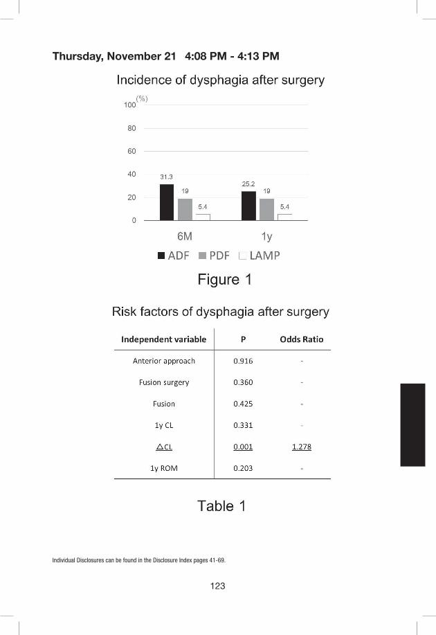
123
Thursday, November 21
Individual Disclosures can be found in the Disclosure Index pages 41-69.
4:08 PM - 4:13 PM

124
Thursday, November 21
The FDA has stated that it is the responsibility of the physician to determine the FDA clearance status of each drug or meidcal device he or she wishes to use in clinical practice.
Presentation #25
Association between the severity of dysphagia and various parameters of the cervical spine; Videofluoroscopic analysis in neutral and retraction position of the normal volunteers
Jae Taek Hong, Seong Hoon Lim, Dong Hoon Lee, Jun Seong Kim
Department of Neurosurgery, Catholic University of Korea, Eunpyeong St. Mary’s Hospital
Introduction: Occipitocervical fixation (OCF) is associated with several potential complications. Malalignment of craniovertebral junction (CVJ) and associated dysphagia have been well described in the literature. However, there has been little attention given to investigate the association between the degree of swallowing dysfunction and various patient’s parameters. So, the purpose of this study was to investigate the relationship between the severity of dysphagia and various parameters of the subjects.
Methods: Thirty-nine healthy asymptomatic volunteers, 18 males and 21 females, were enrolled in this study. Based on video-fluorographic swallowing study (VFSS), two scoring systems of swallowing dysfunction (DRS; dysphagia rating scale, DOSS; dysphagia outcome and severity scale) were measured in neutral and retraction position. We calculated DRS based on the seven radiological findings and five clinical symptoms of dysphagia during barium swallowing study, with a total score of 0 to 12 (Table 1).
We also evaluated DOSS to systemically rate the function severity of dysphagia based on VFSS. DOSS is a 7-point comprehensive ordinal scale that combines treatment and diet (Table 2).
Multiple linear regression analysis was performed to evaluate the relationship between the degree of swallowing dysfunction and various factors of the subjects such as radiological and clinical parameters.
Results: There was statistically significant correlation between DRS and DOSS (r=-0.354, P< 0.05). Multiple regression analysis showed that there was a significant association of the dysphagia severity (DRS and DOSS) with the percentile change (%dn) of the narrowest OD (r=0.121, p<0.01 and r=0.020, p<0.01, respectively).
Percentile change of OD (neutral and retraction position) was positively associated with the difference of C0-2 angle (r=1.676, p<0.01).
None of the other variables such as age, sex, C0-1 angle, C1-2 angle and C2-7 angle were significantly associated with the degree of dysphagia or %dn oropharyngeal space.
Conclusion: This study demonstrated that there was a strong relationship between OD change and the degree of swallowing dysfunction. The change of OD is significantly associated with the C0-2 angle change. These data suggest that severity of dysphagia is dependent on the OD change and the C0-2 angle change has considerable impact on the narrowing of OD after OCF. Therefore, intraoperative control of C0-2 angle could be the most critical step during OCF to prevent not only the stricture of oropharyngeal space but also postoperative dysphagia.
4:14 PM - 4:19 PM

125
Thursday, November 21
Individual Disclosures can be found in the Disclosure Index pages 41-69.
Table 1. Measurements of Dysphagia Severity Scale (DRS = Clinical Score + Radiological Score)A. Clinical Score
Clinical findings Yes=1, No=0(Total Score = 0-5)
Foreign body sensation
Residual sensation after swallowing
Coughing
Aspiration
Swallowing difficulty
B. Radiological Score
Radiological findings Yes=1, No=0(Total Score = 0-7)
Pharyngeal transit time prolongation
Valleculae retention
Pyriformis sinus retention
Aspiration
Pharynx coating
Supraglottic penetration (SGP)
choking
Table 2. Dysphagia outcome and severity scale (DOSS)
DOSS Definition
7 Within normal limits No symptoms of dysphagia
6 Minimal problems Some symptoms of dysphagia but no need for rehabilitation or exercise
5 Oral problems Significant symptoms in the pre-oral anticipatory stage or oral stage without aspiration
4 Occasional aspiration Possible aspiration or aspiration is suspected due to pharyngeal residue
3 Water aspiration Aspiration of thin liquids; change in food consistency is effective
2 Food aspiration Food aspiration with no effect from compensatory techniques or food consistency change
1 Saliva aspiration Unstable medical condition due to severe saliva aspiration
4:14 PM - 4:19 PM

126
Thursday, November 21
The FDA has stated that it is the responsibility of the physician to determine the FDA clearance status of each drug or meidcal device he or she wishes to use in clinical practice.
Presentation #26
Same Day Surgical Intervention Dramatically Minimizes Complication Occurrence and Optimizes Peri-operative Outcomes for Central Cord Syndrome
Cole Bortz BA, Avery Brown BS, Haddy Alas BS, Katherine E Pierce BS, M. Burhan Janjua MD, Paul Park MD, Charles Wang MD, Alex Soroceanu MD, FRCS(C), MPH, Rafael De la Garza Ramos MD, Daniel M. Sciubba MD, Anthony Frempong-Boadu MD, Dennis Vasquez-Montes MS, Bassel G. Diebo MD, Michael C. Gerling MD, Peter G Passias MD
Introduction: As surgery is increasingly recommended for patients with neurologic deterioration secondary to central cord syndrome (CCS), it is important to investigate the relationship between time to surgery and patient outcomes. The merits of early vs delayed surgical treatment remain controversial in the literature. This study aims to investigate associations between time to surgical intervention and surgical outcomes for CCS patients.
Methods: CCS patients (ICD-9 codes 952.03, 952.08, 952.13, 952.18) were isolated in the NIS database 2005-2013. Operative patients were grouped by time to intervention: same day as admission, 1-day delay, 2-day, 3-day, 4-7 days, 8-14 days, and >14 days. As appropriate, analysis of variance and chi-squared tests compared demographics, Charlson Comorbidity Index (CCI) scores, surgical details, LOS, discharge status, periop complications, and total charges across patient groups. Controlling for age, CCI, and concurrent traumatic fractures, binary logistic regression assessed surgical timing associated with increased odds of periop complication, using same-day patients as a reference group (Odds Ratio [95% confidence interval]).
Results: Included: 6,734 CSS patients (59±16yrs, 26%F, mean CCI: 1.2±1.6). Overall rate of surgical treatment was 64.1%, with rates of surgery increasing from 2005 (50.0%) to 2013 (73.0%, p<0.001). The most common mechanisms of injury were falls (30.3%), pedestrian accidents (6.9%), assault (3.2%), and bicycle-related injury (2.7%). Of the patients that underwent surgery, 52.0% underwent fusion (62.4% 2-3 levels, 33.2% eight-levels, 0.5% 9+ levels), 30.1% discectomy, and 13.8% other exploration/decompression of the spinal canal. Breakdown by time to procedure was: 38.5% same day, 16.1% 1 day, 9.6% 2 days, 7.5% 3 days, 16.2% 4-7 days, 8.3% 8-14 days, and 2.7% >14 days. Timing groups did not differ in trauma status at admission (shock or hemorrhage, p=0.261); however, age differed between groups (min: 1 day [58±15 years], max: >14 days [63±13 years], p<0.001). Relative to other groups, same-day patients had the lowest LOS (7.7±9.8 days vs 9.4-37.3 days, p<0.001) highest rates of home discharge (42.09% vs 30.9%-14.4%, p<0.001). Same-day patients showed a trend of lower periop neurologic complications (0.4%) than 1-day (0.6%) and 2-day (1.0%) patients, while patients delayed 3+ days had the lowest (0.1%, p=0.144). Patients delayed >14 days to surgery had increased odds of periop cardiac (7.0 [1.6-30.0]) and infection (6.1 [2.2-16.3]) complications. All timing groups beyond 3 days showed increased odds of VTE: 4-7 days (3.0 [1.6-5.5]), 8-14 days (3.0 [1.4-6.3]), 14+ days (5.6 [2.3-13.6]). Same-day surgery was also associated with lower total hospital charges than delayed surgery ($87,741 vs $118,815-$272,901, p<0.001).
Conclusions: Patients undergoing surgery for central cord syndrome on the same day as admission showed significantly lower odds of complication, hospital charges, and higher rates
4:28 PM - 4:33 PM

127
Thursday, November 21
Individual Disclosures can be found in the Disclosure Index pages 41-69.
of discharge to home than patients that experienced a delay to operation. In contrast, patients delayed >14 days to surgery were associated with inferior outcomes, including increased odds of cardiac complication and infection.
Time to Procedure
Same-day 1-day 2-day 3-day 4-7 day 8-14 day >14 day p-value
Number of patients (%) 1664 (39%)
695 (16%)
414 (10%)
324 (8%)
699 (16%)
360 (8%)
118 (3%)
Baseline Patient Characteristics
Age (years) 58.65 58.44 58.77 60.71 62.56 62.32 62.76 *<0.001
CCI 1.14 1.29 1.22 1.29 1.51 1.67 1.95 *<0.001
Baseline trauma: shock (%) 1.0% 1.9% 0.5% 0.6% 1.3% 1.9% 0.8% 0.261
Clinical and Cost Outcomes
LOS (days) 7.67 10.37 9.48 11.72 14.08 19.93 37.32 *<0.001
Discharge to home (%) 42.0% 29.1% 30.9% 25.3% 21.6% 15.6% 14.4% *<0.001
Total charges ($) 76,832 11,5473 11,4738 12,6091 138,332 170,844 239,611 *<0.001
Complication Outcomes
Neurologic Complication 0.4% 0.6% 1.0% 0.0% 0.0% 0.3% 0.0% 0.144
Cardiac Complication 0.3% 0.7% 1.0% 0.3% 0.1% 0.3% 2.5% *0.009
Infection Complication 0.8% 1.0% 1.2% 0.3% 0.6% 1.4% 5.1% *<0.001
Sepsis Complication 2.0% 2.7% 2.2% 2.2% 4.1% 8.6% 11.0% *<0.001
VTE 1.1% 2.9% 1.2% 2.2% 3.3% 3.3% 5.9% *<0.001
Mortality 3.4% 3.4% 3.2% 4.2% 5.2% 4.4% 6.7% 0.382
4:28 PM - 4:33 PM
128
Thursday, November 21
The FDA has stated that it is the responsibility of the physician to determine the FDA clearance status of each drug or meidcal device he or she wishes to use in clinical practice.
Presentation #27
Prospective risk factor analysis of surgery-related complications in primary cervical spine surgery for degenerative diseases
Shota Takenaka, MD, DMSc, Takahiro Makino, MD, DMSc, Yusuke Sakai, MD, Hideki Yoshikawa, MD, PhD, Takashi Kaito, MD, PhD
Department of Orthopaedic Surgery, Osaka University Graduate School of Medicine
Introduction: Cervical spine surgery is performed in various pathological conditions in a diverse patient population. Several studies have examined the occurrence of surgery-related complications after cervical spine surgery. However, many of these studies have been done using large national databases or retrospective analysis. We used a registry of prospectively collected multicenter data to explore the risk factors for surgery-related complications in primary cervical spine surgery for degenerative diseases.
Materials/Methods: From 2012 to 2018, 5027 patients (3270 men and 1757 women) with cervical degenerative diseases underwent primary cervical spine surgery at our 25 affiliate institutes. Revision surgeries were excluded. The average age was 66.8 years for men and 70.2 years for women. A detailed preoperative diagnosis revealed cervical spondylotic myelopathy (CSM) in 74%, ossification of the posterior longitudinal ligament (OPLL) in 12%, cervical disc herniation in 6%, and others in 8%. The surgical procedures consisted of laminoplasty in 80%, posterior fusion in 9%, anterior fusion in 7% and others in 4%. As subcategories, the presence of cervical kyphosis exceeding 10°, the presence of OPLL with an occupying ratio of 50% or more, and concomitant foraminotomy were also chosen. The examined surgery-related complications were postoperative upper limb palsy (ULP) with a manual muscle test grade of 0 to 2, other neurologic deficits, dural tear, cerebrospinal fluid leakage (CFL), surgical site infection (SSI) requiring reoperation, postoperative hematoma requiring reoperation. Multivariate logistic regression was performed.
Results: ULP, other neurologic deficits, dural tear, CFL, SSI and hematoma occurred in 3.6%, 0.6%, 1.5%, 0.6%, 0.7% and 0.5%, respectively. Risk factors for ULP were: OPLL (odds ratio [OR] = 2.23, 95% confidence interval [CI] = 1.53–3.23, p < 0.001), concomitant foraminotomy (OR = 4.22, CI = 2.55–7.00, p < 0.001), older age (p = 0.035), non-fusion surgery (OR = 0.64 CI = 0.44–0.92, p = 0.015), disc herniation (OR = 0.17 CI = 0.04–0.71 p = 0.015). In particular, posterior fusion was a significant risk factor for ULP in the CSM patients. OPLL was a risk factor for other neurologic deficits (OR = 5.41 CI = 2.55–11.5, p < 0.001), dural tear (OR = 1.82 CI = 1.03–3.23, p = 0.040) and CSL (OR = 2.89 CI = 1.33–6.32, p = 0.008). In the OPLL patients, CSL was significantly observed in those with OPLL with an occupying ratio of 50% or more. Male sex was the only independent risk factor for SSI. As to hematoma, no independent risk factor was identified. Cervical kyphosis exceeding 10° was not significantly associated with surgery-related complications.
Conclusion: This study demonstrated that OPLL increased the risk for various surgery-related complications (ULP, other neurologic deficits, dural tear and CFL). In particular, patients with OPLL with an occupying ratio of 50% or more should be carefully treated. Cervical kyphosis by itself
4:34 PM - 4:39 PM

129
Thursday, November 21
Individual Disclosures can be found in the Disclosure Index pages 41-69.
did not increase the risk of surgery-related complications, whereas posterior fusion to correct cervical alignment was associated with an increased risk of ULP. Concomitant foraminotomy also increased the risk of ULP.
4:34 PM - 4:39 PM
130
Thursday, November 21
The FDA has stated that it is the responsibility of the physician to determine the FDA clearance status of each drug or meidcal device he or she wishes to use in clinical practice.
Presentation #28
The influence of frailty of patients on the incidence of surgical site infection after spine surgery –the analysis of over 1000 cases
Tomoya Yoshikawa, Shuichi Kaneyama, Masatoshi Sumi, Koichi Kasahara, Aritetsu Kanemura, Hiroaki Hirata.
Department of Orthopaedic Surgery, Kobe Rosai Hospital, Kobe Japan
Purpose: Surgical site infection (SSI) after spine surgery is not necessarily rare and can result in increased morbidity and mortality. Recently, “Frailty”, which is defined as a decrease in the physiological reserves as well as multisystem impairments that are separate from the normal process of aging has been focused as a risk of poor surgical outcome, and the modified frailty index (mFI) has been applied for prediction of postoperative adverse events in various surgical fields. This study aimed to investigate the applicability of the mFI as a predictor of SSI after spine surgery.
Methods: A total of 1059 cases who underwent spine surgery from 2015 to 2017 was reviewed. The incidence of SSI within 6 months after surgery and the risk factor of the SSI were investigated: as the “patient” aspect; age, gender, BMI, smoking, comorbidity, medication, blood test, and modified Frailty Index (mFI), and as the “surgery” aspect; number of involved vertebral level, surgical approach, duration of surgery, instrumentation, emergency, multiple operation, blood transfusion, and dural tear. The mFI was previously described by Saxton and Velanovich by giving individuals a score based on 11 variables present in the Canadian Study of Health and Aging Frailty Index. The score is calculated by dividing the number of variables by the total number assessed (n/11). The 11 variables were (1) a history of diabetes mellitus, (2) functional status (independent or not independent), (3) COPD or pneumonia, (4) congenital heart failure, (5) myocardial infarction, (6) percutaneous coronary intervention, stenting, or angina, (7) hypertension requiring medication, (8) peripheral vascular disease or ischemic rest pain, (9) impaired sensorium, (10) transient ischemic attack or (11) cerebrovascular accident with neurological deficits.
Results: SSI occurred in 20 cases (1.9%). The mFI in the patients who experienced SSI was 0.16, which was higher in the patients who did not experience SSI (0.11) (p<0.05). The incidence of SSI was higher in the patients with 0.18 or more mFI (12/345 cases: 3.5%) than with mFI under 0.18 (8/714 cases: 1.1%) (p<0.01). In addition, postoperative serum albumin level (under 2.9g/dL: 5.4%, over 3.0g/dL: 1.1%, p<0.05), vertebral level involved in surgery (over 5 levels: 3.0%, under 4 levels: 1.3%, p<0.05), operative duration (over 3 hours: 4.5%, under 3 hours: 0.6%, p<0.01), instrumentation (with: 3.8%, without: 0.9%, p<0.01) and blood transfusion (did: 5.4%, not: 1.3%, p<0.01) showed significant difference of the incidence of SSI between subject and control group.
Discussion: Our results demonstrated that mFI 0.18 or more (the patient had more than one variables) was a risk factor of SSI after spine surgery. Since most of the patients who need spine surgery are independent, meaning they have already one variable, they are prone to meet that condition by having just one comorbidity. Furthermore, high invasive surgery was indicated to
4:40 PM - 4:45 PM
131
Thursday, November 21
Individual Disclosures can be found in the Disclosure Index pages 41-69.
increase the risk of SSI. The mFI was applicable as a predictor of SSI after spine surgery and careful precaution against SSI should be taken for high invasive surgery for the patients with high mFI.
4:40 PM - 4:45 PM
133
Friday, November 22
Individual Disclosures can be found in the Disclosure Index pages 41-69.
Presentation #30
Nonoperative Management Of Asymptomatic Cervical Spinal Stenosis: A Long-Term Follow-Up Study
Michael P. Kelly, MD, MSc, Lukas P. Zebala, MD, K. Daniel Riew, MD
Introduction: The natural history of patients with asymptomatic cervical spondylotic stenosis is not known. As such, the treatment of these patients remains controversial. Some surgeons recommend prophylactic decompression to prevent a catastrophic neurological injury, while others recommend observation alone. There are no long-term natural history studies of patients with asymptomatic cervical stenosis to provide data for the shared-decision making process. This is a follow-up study from a prior mid-term report.
Methods: Adult patients with an AP spinal canal diameter < 9 mm on MRI without T2-signal intensity and no signs nor symptoms of myelopathy were enrolled in a prospective, observational cohort study. Exclusion criteria were: age over 80 years, pathologic or traumatic compression, instability requiring stabilization. Enrolled patients were recommended to have repeat physical exams and MRIs annually. At last contact patients completed Neck Disability Index and mJOA outcomes questionnaires. Any patients developing myelopathy or recalcitrant radiculopathy underwent surgery. The incidence density was calculated to estimate the rate of surgery in this population.
Results: 86 patients were enrolled. Follow-up was between 2-19.6 years (Mean 9.9yrs; cumulative follow-up of 758.7 patient*yrs). 5 Patients were deceased and 21 were otherwise lost to follow-up(with 4 having surgery prior to loss). No patients became para or tetraplegic. 20 Patients (25%) underwent surgery at a mean follow-up of 5.2 yrs; incidence density rate of 2.6%. Mean NDI at last FU 18.6 (Interquartile range 7.5-32); mean mJOA 15 (IQR 14-16). For patients treated with surgery mean NDI was 22.5 (IQR 8-34) and mJOA 14 (12 – 16).
Conclusions: At a mean of almost 10yr follow-up, 25% of patients presenting with asymptomatic stenosis had undergone surgery with an annual rate of 2.6%. No neurological catastrophes occurred. mJOA scores indicated mild myelopathy at last followup for all patients and mild-moderate disease for patients treated surgically. These data suggest that observation with serial exams is safe and can lead to good outcomes. However, continued followup is necessary as 11% of patients had undergone surgery at 4yr followup versus 25% at 10yrs.
7:11 AM - 7:16 AM

134
Friday, November 22
The FDA has stated that it is the responsibility of the physician to determine the FDA clearance status of each drug or meidcal device he or she wishes to use in clinical practice.
Presentation #31
Prospective Evaluation of Degenerative Cervical Myelopathy In Asymptomatic Patients Over 60 Years
Ryan Schiedo MD, Samuel Adams MD, Sara Holmes BS, Letterio Politi MD, Patrick Connolly MD, Michael Stauff MD
Introduction: Degenerative Cervical Myelopathy (DCM) is caused by narrowing of the cervical spinal canal resulting in compression of the spinal cord. The diagnosis of DCM is confirmed by history, physical exam findings, and advanced imaging showing cord compression. Radcliff et al. reported the incidence of undiagnosed DCM in hip fracture patients (18%) and elective hip replacement patients (0%) based on history, modified Japanese Orthopedic Association (mJOA) score, and physical exam.[1] Other investigators have studied the specificity and sensitivity of physical exam findings for diagnosing DCM and found it to be imperfect, with 21% of patients diagnosed with DCM lacking obvious clinical signs.[2] The purpose of this study was to determine the prevalence of undiagnosed DCM in patients over 60 years by combining history, physical exam, and MRI.
Materials/Methods: Patients 60 years and older were prospectively enrolled from primary care practices. Patients were excluded if they had a history of debilitating neck pain, inability to have a cervical MRI, neurologic disease, cognitive impairment, or prior cervical spine surgery. A cervical MRI was performed using a 3.0 Tesla imaging system (Philips Achieva) and a cervical coil. Neck Disability Index (NDI) and mJOA scores were collected for all patients, as well as a thorough history and physical exam. MRI results were reviewed independently by an Orthopedic Spine surgeon and Neuroradiologist. Central cervical canal stenosis was graded 0-3, with 0 indicating no canal stenosis and 3 indicating spinal cord compression with myelomalacia.[3] Patients were diagnosed with DCM if they had myelopathic history (mJOA <16), physical exam consistent with myelopathy (upper motor neuron (UMN) signs), and MRI showing cord compression (Figure 1).
Results: Fifty-two patients were included in the study, 24 Males (46%) and 28 Females (54%). The average age was 69.4 years old (range 60–90 years). The mean NDI was 5.3%, range 0-24%. The mean mJOA score for the cohort was 17.6 (range 14-18). Two patients (3.8%) had an mJOA score of <16. Eight (15.4%) had hyperreflexia grade 3 out of 4. Both reviewers agreed that 2 patients had myelomalacia on MRI, while one reviewer identified an additional 3 patients as having myelomalacia. Spinal cord compression with deformity was identified by both reviewers in 25 patients (48.1%), Figure 2. Overall agreement was 92.86% with a kappa coefficient of 0.89 [0.85, 0.93]. One (1.9%) patient was diagnosed with DCM. Five (9.6%) patients had cord compression and hyperreflexia or ataxia, but mJOA score was >16. Three (5.8%) patients had myelomalacia on MRI but were asymptomatic.
Conclusion: In our study the prevalence of cervical spinal cord compression in patients 60 years and older with minimal neck pain was found by MRI to be 48.1%. Among these patients one was diagnosed with DCM, while three others had myelomalacia with a normal physical exam. Our data supports the work of other researchers and shows that patients may have MRI findings of myelomalacia but remain asymptomatic. This study confirms a small but important prevalence of spinal stenosis and spinal cord compression in patients over 60 who have minimal or no symptoms.
7:17 AM - 7:22 AM
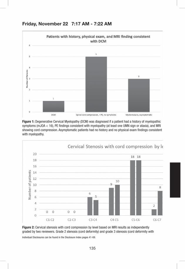
135
Friday, November 22
Individual Disclosures can be found in the Disclosure Index pages 41-69.
Figure 1: Degenerative Cervical Myelopathy (DCM) was diagnosed if a patient had a history of myelopathic symptoms (mJOA < 16), PE findings consistent with myelopathy (at least one UMN sign or ataxia), and MRI showing cord compression. Asymptomatic patients had no history and no physical exam findings consistent with myelopathy.
Figure 2: Cervical stenosis with cord compression by level based on MRI results as independently graded by two reviewers. Grade 2 stenosis (cord deformity) and grade 3 stenosis (cord deformity with
7:17 AM - 7:22 AM

136
Friday, November 22
The FDA has stated that it is the responsibility of the physician to determine the FDA clearance status of each drug or meidcal device he or she wishes to use in clinical practice.
myelomalacia) are combined. Data represents all levels of all patients with cord compression.
References:1. Radcliff, K.E., et al., High Incidence of Undiagnosed Cervical Myelopathy in Patients With Hip
Fracture Compared With Controls. J Orthop Trauma, 2016. 30(4): p. 189-93.2. Rhee, J.M., et al., Prevalence of physical signs in cervical myelopathy: a prospective, controlled
study. Spine (Phila Pa 1976), 2009. 34(9): p. 890-5.3. Kang, Y., et al., New MRI grading system for the cervical canal stenosis. AJR Am J Roentgenol,
2011. 197(1): p. W134-40.
7:17 AM - 7:22 AM

137
Friday, November 22
Individual Disclosures can be found in the Disclosure Index pages 41-69.
Presentation #32
Home versus Standard Physical Therapy after ACDF Surgery: Preliminary Results of Health-Related Quality of Life Outcomes from a Randomized Controlled Trial
Srikanth N Divi MD, Dhruv KC Goyal BA, Matthew S Galetta BA, Justin D Stull MD, John J Mangan MD, Jeffrey A Rihn MD, Mark F Kurd MD, D Greg Anderson MD, Barrett I Woods MD, Kristen E Radcliff MD, Ian D Kaye MD, Alan S Hilibrand MD, Alexander R Vaccaro MD PhD MBA, Christopher K Kepler MD MBA, Gregory D Schroeder MD
Rothman Institute, Thomas Jefferson University Hospital, Philadelphia, PA
Introduction: Recent literature has suggested home physical therapy (PT) is equally efficacious as standard physical therapy in patients who require orthopaedic procedures; however, this has not been studied in patients undergoing spine surgery. The goal of this prospective, randomized controlled trial was to determine whether home PT is equivalent to standard PT in terms of patient reported outcome measures (PROM) after anterior cervical discectomy and fusion (ACDF) surgery.
Methods: Patients undergoing 1-2 level ACDF surgery were prospectively enrolled at a high-volume academic institution starting on January 1st, 2017. Patients who required surgical intervention to address to address malignant tumor, infections, traumatic pathology, or revision of a previous cervical surgery were excluded. Patients were randomized and split into one of two groups: home-based PT (Home PT group) vs standard, in-person PT (Standard PT group) starting four weeks after surgery. Preoperative, 2 week, and 3 month outcome scores—including Neck Disability Index (NDI), Visual Analogue Scale Neck (VAS Neck) and Arm (VAS Arm) pain, and the Short Form-12 Physical Component Score (PCS-12) and Mental Component Score (MCS-12)—were recorded and compared between groups with univariate analysis and multiple linear regressions. A recovery ratio and the percentage of patients reaching the minimum clinically important difference (%MCID) were calculated at the 3-month mark.
Results: To date, a total of 34 patients are enrolled with 20 in the Home PT and 14 in the Standard PT group. The average age was 55 [50, 59], the number of males was 21 (61.8%), and the number of one- and two-level fusions were 11 (32.4%) and 23 (67.6%), respectively. Overall, patients participated in physical therapy for an average of 7.1 [5.9, 8.3] weeks with a mean frequency of 3.5 [2.9, 4.2] sessions a week. Both groups demonstrated significant improvement in VAS Neck and Arm pain scores (p < 0.01), but only the Home PT group improved in terms of PCS-12 scores (p = 0.006). Neither groups exhibited significant improvement in terms of NDI or MCS-12 scores. There were no significant differences between groups in terms pre- or postoperatively for any outcome measure. Furthermore, multiple linear regression analysis indicated that physical therapy group was not a significant predictor of patient outcomes
Conclusion: Overall, patients undergoing Home and Standard PT demonstrated similar improvement in VAS Neck and Arm scores. Patients in the Home PT group also showed significant improvement in PCS-12 scores compared to the standard PT group. Analysis at the 1-year point from this randomized controlled trial will provide further insight into the utility of home vs. standardized PT.
7:23 AM - 7:28 AM

138
Friday, November 22
The FDA has stated that it is the responsibility of the physician to determine the FDA clearance status of each drug or meidcal device he or she wishes to use in clinical practice.
Table 2: Patient Reported Outcome Comparisons Between PT Group (3 Month) (n=34)
Standard PT Group (n=14)
Paired Samples
t-Test
Home PT Group (n=20)
Paired Samples
t-Test
Mann-Whitney U Test
Multiple Linear Regression (β
[95% CI], p-value)
NDI
Preoperative 41.0 [32.0, 50.0]
0.932
35.0 [25.0, 45.0]
0.319
0.312
-4.422 [-17.063, 8.219], 0.475
2 Week 46.0 {34.0, 58.0] 42.0 [34.0, 49.0] 0.381
3 Month 41.0 [28.0, 54.0] 33.0 [25.0, 40.0] 0.255
Delta 3.0 [-9.0, 14.0] 4.0 [-6.0, 15.0] 0.958
RR 0.8%—
6.3%—
0.400
%MCID 21.4% 20.0% 0.622
VAS Neck
Preoperative 5.9 [4.6, 7.1]
0.002*
5.3 [4.1, 6.6]
<0.001*
0.525
-0.510 [-1.993, 0.972], 0.482
2 Week 4.0 [3.0, 5.0] 3.0 [2.0, 4.0] 0.236
3 Month 3.0 [2.0, 5.0] 2.0 [1.0, 3.0] 0.233
Delta -2.2 [-4.1, -0.3] -2.0 [-3.6, -0.4] 0.624
RR 40.9%—
45.4%—
0.972
%MCID 57.1% 40.0% 0.324
VAS Arm
Preoperative 5.6 [3.7, 7.5]
0.004*
4.9 [3.3, 6.5]
0.003*
0.661
-0.305 [-2.027, 1.416], 0.716
2 Week 3.0 [2.0, 5.0] 3.0 [2.0, 4.0] 0.547
3 Month 3.0 [2.0, 4.0] 2.0 [1.0, 3.0] 0.423
Delta -2.4 [-4.4, -0.4] -1.5 [-3.3, 0.3] 0.441
RR 14.6%—
36.5%—
0.551
%MCID 64.3% 35.0% 0.092
PCS-12
Preoperative 37.8 [31.1, 44.5]
0.355
36.9 [32.5, 41.3]
0.006*
0.936
1.051 [-5.182, 7.284], 0.730
3 Month 39.4 [33.4, 45.4] 42.6 [38.2, 46.9] 0.382
Delta 4.3 [-2.2, 10.9] 9.3 [3.4, 15.3] 0.441
RR 4.4%—
12.1%—
0.363
%MCID 42.9% 45.0% 0.901
MCS-12
Preoperative 47.3 [40.0, 54.6]
0.409
49.3 [43.3, 55.3]
0.178
0.522
6.617 [-1.203, 14.437], 0.093
3 Month 45.6 [39.1, 52.0] 52.8 [48.4, 57.3] 0.054
Delta 1.6 [-9.8, 13.1] 8.5 [-0.2, 17.2] 0.248
RR 3.2%—
9.5%—
0.248
%MCID 35.7% 45.0% 0.588
Univariate Analysis: 1Paired Samples t-Test, 2Mann Whitney U Test, or Pearson Chi-Square Analysis. PROMs: Neck Disability Index (NDI), Short Form-12 Survey Physical Component Score (PCS-12) and Mental Component Score (MCS-12), Visual Analogue Scale Neck (VAS Neck) and Arm (VAS Arm) pain scores. Radiographic measurements: C2 Tilt, C2 Slope, C2-C7 Lordosis, Sagittal Vertical Axis (SVA), T1 Slope, and C2-C7 ROM (Extension – Flexion). RR = Recovery Ratio, defined as: [Delta Outcome Score/(Optimal Outcome Score – Observed Outcome Score)], where the following Optimal Outcome Scores were used: 100 (PCS-12 and MCS-12) or 0 (NDI). The percentage of patients achieving the Minimum Clinically Important Difference (%MCID) was determined using the following MCID cutoff values: PCS-12 – 8.1 points, MCS-12 – 4.7 points, and NDI – 15 points; VAS Neck – 2.5; VAS Arm – 2.5 points. *Indicates significance (p<0.05)
7:23 AM - 7:28 AM

139
Friday, November 22
Individual Disclosures can be found in the Disclosure Index pages 41-69.
PROMs: Neck Disability Index (NDI), Visual Analogue Scale Neck (VAS Neck) and Arm (VAS Arm) pain scores. Delta scores defined as: 3 Month Outcome Score – Preoperative Outcome Score. RR = Recovery Ratio, defined as: [Delta Outcome Score/(Optimal Outcome Score – Observed Outcome Score)], where the following Optimal Outcome Scores were used: 100 (PCS-12 and MCS-12) or 0 (NDI). The percentage of patients achieving the Minimum Clinically Important Difference (%MCID) was determined using the following MCID cutoff values: PCS-12 – 8.1 points, MCS-12 – 4.7 points, and NDI – 15 points; VAS Neck – 2.5; VAS Arm – 2.5 points. Multiple Linear Regression analysis conducted to predict whether home physical therapy was a significant predictor of change in outcomes over time—controlling for factors including age, sex, BMI, smoking status, and baseline scores. *Indicates significance (p<0.05)
7:23 AM - 7:28 AM
140
Friday, November 22
The FDA has stated that it is the responsibility of the physician to determine the FDA clearance status of each drug or meidcal device he or she wishes to use in clinical practice.
Presentation #33
Effectiveness of surgical treatment in reducing falls and fall-related neurological deterioration in patients with degenerative cervical myelopathy: a multi-institutional prospective study
Atsushi Kimura, MD,1 Hirokazu Inoue, MD,1 Yasuyuki Shiraishi, MD,1 Katsushi Takeshita, MD,1 Atsushi Okawa, MD2
1Department of Orthopaedics, Jichi Medical University, Tochigi, Japan2Department of Orthopedic Surgery, Tokyo Medical and Dental University, Tokyo, Japan
Introduction: Patients with degenerative cervical myelopathy (DCM) commonly experience unsteady walking and frequent falls. Even minor trauma to the neck in patients with DCM can cause neurological deterioration, ranging widely from transient sensory aggravation to permanent motor paralysis. Clinicians empirically recommend surgical treatment for patients with DCM to reduce the risk of fall-induced neurological deterioration. However, data are limited on the effectiveness of surgical treatment for reducing the risk of neurological deterioration. The purpose of this study was to investigate the effectiveness of surgical treatment in preventing falls and fall-related neurological deterioration in a prospective cohort of patients with DCM.
Methods: Patients with DCM scheduled for surgical treatment were enrolled prospectively from October 2016 through December 2017 at eight participating institutes. Exclusion criteria were: comorbidity impairing physical functions (e.g., cerebral infarction, cerebral palsy, or severe rheumatoid arthritis); bedridden status or full dependence on a wheelchair before surgery because of severe cervical myelopathy; or difficulty completing a questionnaire because of cognitive impairment. At the time of enrollment, participants were given diaries to record the details of falls (occurrence time, circumstances, and consequences), to be returned at a postoperative 1-year follow-up visit. In the fall diary, patients were asked whether they had experienced any deterioration in symptoms because of falls. For affirmative responses, they were asked to check the answer most accurately describing the symptom deterioration as follows: 1) only deterioration of sensory function in the arms or legs; or 2) deterioration of motor deficits in the arms or legs (e.g., weakness, fine motor skill difficulties, and walking difficulty). Functional outcome was assessed using the Japanese Orthopaedic Association (JOA) score and Neck Disability Index at enrollment, admission, and 1 year after surgery.
Results: From the initial sample of 168 participants, 159 completed the 1-year follow-up; 132 fall diaries were retrieved and analyzed. Patient characteristics and baseline functions are shown in Table 1. A total of 96 falls were reported during 18.0 person-years of preoperative observation. After surgery, a total of 119 falls were reported during 112.2 person-years. Thus, the fall rate decreased significantly from 5.3 (96/18.0) to 1.1 (119/112.2) per person-year after surgery (P < 0.0001). Time-course analysis revealed that the incidence of falls was highest within 1 month before surgery (Figure 1). The incidence rate of fall-induced motor deterioration also significantly decreased from 0.33 (32/96) to 0.07 (8/119) per fall after surgery (P < 0.0001). Seventeen patients who experienced preoperative fall-induced motor deterioration showed a significantly lower JOA score compared with those who did not experience deterioration at the 1-year follow-up (P < 0.02).
7:29 AM - 7:34 AM

141
Friday, November 22
Individual Disclosures can be found in the Disclosure Index pages 41-69.
Conclusion: Surgical treatment for DCM significantly reduced not only the frequency of falls but also the risk of fall-induced deterioration of motor deficits. Because fall-induced neurological deterioration had a negative impact on the surgical outcome in patients with DCM, particular attention should be given to fall prevention before surgery.
Table 1. Patient characteristics and baseline functions
Characteristic Number (N = 132)
Age (years) 69.4 ± 10.1
Sex (M/F) 83/49
Etiology of myelopathy
Spondylosis 80 (61)
OPLL 52 (39)
Surgical approach
Anterior 28 (21)
Posterior 86 (65)
Combined 18 (14)
Preoperative JOA score 11.0 ± 2.5
Preoperative NDI score 37.7 ± 19.2
Follow-up period (month)
Preoperative 1.8 ± 1.6
Postoperative 12.5 ± 1.5
Data are shown as mean ± standard deviation or number (%). OPLL, ossification of the posterior longitudinal ligament; JOA, Japanese Orthopaedic Association; NDI, Neck Disability Index.
Figure 1. Time-course of fall incidence and consequences of falls
7:29 AM - 7:34 AM
142
Friday, November 22
The FDA has stated that it is the responsibility of the physician to determine the FDA clearance status of each drug or meidcal device he or she wishes to use in clinical practice.
Presentation #34
Improvement in Cervicogenic Headaches After Laminoplasty: A Single Institutional Study of 143 Adult Patients
Owoicho Adogwa M.D.M.P.H. 1, K. Daniel Riew M.D.2, Gaurang Gupte B.S. 1, Max Shlykov M.D.1, Lukas P. Zebala M.D.1 , Colleen Peters B.S. M.S1, Jacob Buchowski M.D.M.S1, Michael P. Kelly M.D. M.S. 1
Corresponding Author: Michael P. Kelly M.D. M.S., Washington University School of Medicine 1Department of Orthopeadic Surgery, Washington University School of Medicine, St. Louis, MO2Department of Orthopeadic Surgery, NY Presbytyerian Hospital/Columbia University School of Medicine, NYC, NY
Introduction: Cervicogenic headache(CeH) is a common cause of chronic headache that is often unrecognized or misdiagnosed. Prevalence estimates range from 1% to 3% of the general population to 15% to 20% of patients with chronic headaches. While the underlying pathophysiology and source of pain for this condition remains unclear, a growing body of work suggest a strong relationship with degenerative conditions of the cervical spine. Anterior decompressive procedures are associated with improvement in CeH. Whether a motion sparing posterior decompression surgery will offer improvement is not known. The aim of this study is to investigate whether cervical laminoplasty is associated with a reduction in cervicogenic headache.
Methods: Clinical records of patients with degenerative cervical stenosis undergoing laminoplasy between 2008 and 2016 were reviewed. Patient and surgical data, complications, as well as the response to Neck Disability Index (NDI) questionnaire was collected. Cervicogenic headache (CeH) was assessed based on patient response to the “Headache Question” of the NDI questionnaire(Q 5). Moderate CeH was defined by a score of 2 or 3 to the headache question of the NDI questionnaire, and severe CeH a score greater than 3. The primary outcome was improvement in cervicogenic headache after surgery.
Results: A total of 143 patients were included in the study. The mean±standard deviation age and BMI was 61.12±12.12 years and 29.62±6.60 Kg/M2, respectively. 62% were male, 10% smokers and 27% had a history of diabetes. The mean score to the headache question of the NDI was 1.40 at baseline, which improved to 0.90 at 1-year(p=0.01) and 0.97(p=0.04) at 2-years after surgery. At baseline, 20.3% of patients reported moderate cervicogenic headache, which improved to 9.8% at 1-year and 6.99% at 2-years. 7.7% of patients reported severe cervicogenic headache prior to surgery, which decreased to 1.4% at 1-year(p=0.01) and 0.69% at 2-years(p=0.01). Laminoplasty was associated with a 82% relative risk reduction in severe cervicogenic headache at 1-year and 92% relative risk reduction at 2-years.
Conclusion: This study suggests that laminoplasty may be associated with a significant improvement in cervicogenic headache.
7:35 AM - 7:40 AM
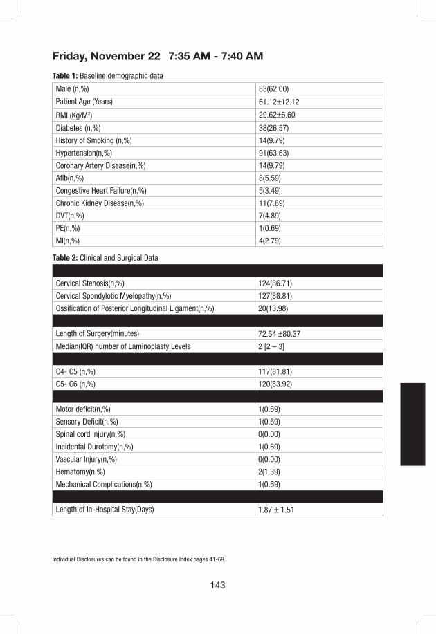
143
Friday, November 22
Individual Disclosures can be found in the Disclosure Index pages 41-69.
Table 1: Baseline demographic data
Male (n,%) 83(62.00)
Patient Age (Years) 61.12±12.12
BMI (Kg/M2) 29.62±6.60
Diabetes (n,%) 38(26.57)
History of Smoking (n,%) 14(9.79)
Hypertension(n,%) 91(63.63)
Coronary Artery Disease(n,%) 14(9.79)
Afib(n,%) 8(5.59)
Congestive Heart Failure(n,%) 5(3.49)
Chronic Kidney Disease(n,%) 11(7.69)
DVT(n,%) 7(4.89)
PE(n,%) 1(0.69)
MI(n,%) 4(2.79)
Table 2: Clinical and Surgical Data
Primary Diagnosis
Cervical Stenosis(n,%) 124(86.71)
Cervical Spondylotic Myelopathy(n,%) 127(88.81)
Ossification of Posterior Longitudinal Ligament(n,%) 20(13.98)
Intra-operative Variables
Length of Surgery(minutes) 72.54 ±80.37
Median(IQR) number of Laminoplasty Levels 2 [2 – 3]
Most Common Laminoplasty Levels 8(5.59)
C4- C5 (n,%) 117(81.81)
C5- C6 (n,%) 120(83.92)
Post-Operative Complications(n,%) 14(9.79)
Motor deficit(n,%) 1(0.69)
Sensory Deficit(n,%) 1(0.69)
Spinal cord Injury(n,%) 0(0.00)
Incidental Durotomy(n,%) 1(0.69)
Vascular Injury(n,%) 0(0.00)
Hematomy(n,%) 2(1.39)
Mechanical Complications(n,%) 1(0.69)
Length of in-Hospital Stay(Days) 1.87 ± 1.51
7:35 AM - 7:40 AM

144
Friday, November 22
The FDA has stated that it is the responsibility of the physician to determine the FDA clearance status of each drug or meidcal device he or she wishes to use in clinical practice.
Table 3: Baseline and Change in Patient Reported Outcomes Scores
Baseline 6 Months 12-Months 24-Months p-Value
NDI 33.50 ±18.70 21.29 ±15.38 23.50 ±19.10 25.20±19.23 0.01
JOA 12.59 ±2.97 12.78 ± 3.06 13.91 ±3.23 14.80 ±2.33 0.02
PROMIS Physical Function 34.99 ±7.68 37.93 ±7.09 40.68 ±8.02 42.64 ±7.01 0.01
PROMIS Pain 64.16 ± 8.30 60.21 ± 6.78 56.98 ± 10.46 58.23 ± 9.04 0.04
PROMIS Depression 51.64 ± 11.33 48.79 ± 9.68 46.97 ± 9.24 46.58 ± 7.63 0.07
PROMIS Anxiety 61.87 ± 8.78 54.60 ± 12.62 50.86 ± 12.37 51.16 ± 8.09 0.01
Table 4: Baseline and change in patient response to headache question of NDI
Baseline 6 Months 12-Months 24-Months p-Value
Mean score to Headache Question of NDI Questionnaire
1.40 ±1.39 0.78 ±0.99 0.90 ±1.11 0.97 ±1.04 0.04
Patients Reporting Moderate Headache(n,%)
29(20.27) 12(8.39) 14(9.79) 10(6.99) 0.01
Patients Reporting Severe Headache(n,%)
11(7.69) 0(0.00) 2(1.40) 1(0.69) 0.01
Figure 1:Post-operative change in percentage of patients reporting improvement in moderate and severe cervigogenic headaches.
7:35 AM - 7:40 AM

145
Friday, November 22
Individual Disclosures can be found in the Disclosure Index pages 41-69.
Presentation #35
Improvements in neck pain and disability following C1-C2 posterior cervical instrumentation and fusion for atlanto-axial osteoarthritis
Owoicho Adogwa M.D. M.P.H. 1, J. Alex Sielatycki M.D.2, Maksim A. Shlykov M.D. M.S.1, Alekos A. Theologis M.D. 1, James Lin M.D. M.S.2, Travis CreveCoeur B.S.1 , Colleen Peters M.A.1, Jacob M. Buchowski M.D. M.S. 1, K. Daniel Riew M.D.2
Corresponding Author: Owoicho Adogwa, M.D. M.P.H Washington University School of Medicine, St. Louis, MO 1Department of Orthopeadic Surgery, Washington University School of Medicine, St. Louis, MO2Department of Orthopeadic Surgery, NY Presbyterian Hospital/Columbia University School of Medicine, NYC, NY
Introduction: Symptomatic atlanto-axial (C1-C2) osteoarthritis (AAOA) is a relatively common phenomenon in elderly patients, however is frequently overlooked as a cause of severe neck and occipital pain. There is a paucity of data on the effectiveness posterior atlanto-axial fusion (PAAF) for this condition. The aim of this study is to assess changes in patient-reported outcomes and neck-related disability in adult patients undergoing posterior atlanto-axial fusion (PAAF) for symptomatic Atlanto-axial (C1-C2) osteoarthritis (AAOA).
Methods: Clinical records of consecutive patients with symptomatic AAOA who underwent PAAF (2004-2017) by a single surgeon were reviewed. Patients with rheumatoid arthritis, prior surgery at C1-2 and/or trauma were excluded. Patient demographics, comorbidities, intra- and post-operative variables, and complication rates were collected. Neck Disability Index scores (NDI) were recorded at baseline, 6 weeks, 6-months, 1-year and 2-years post-operatively. Primary outcome was change in NDI scores at 1- and 2-years after operation.
Results: Forty-two patients (average age: 72.04±8.56 years, 26.19% were male) met inclusion criteria. In the cohort, 19.04% had prior subaxial cervical spine surgery, 35.71% had a history of smoking (all had stopped smoking prior to surgery), and 11.90% had type II diabetes. At baseline, the majority of patients (64%) had normal neurological examinations, 19% were myelopathic, and 17% had radiculopathy. NDI scores were available for 76% of the study sample at baseline, 59% at 1-year, and 36% at 2-years. Average pre-operative NDI score was 26.88±24.85, which improved to 10.59±14.88 and 13.20±14.96 at 1-and 2-years follow-up, respectively (p=0.004). At baseline, 18% of patient reported severe disability based on NDI scores and this decreased to 2% of patients at 1 year and none at 2 years (p=0.01). Importantly, there were a high percentage (11.90% 5 out of 42) patients that had prior sub-axial cervical fusions for their pain, due to a mistaken diagnosis for this condition, without symptom relief.
Conclusion: This study suggests that in appropriately selected patients, PAAF may decrease neck pain and improve functional disability in patients with AAOA. Future prospective longitudinal studies are needed to corroborate the findings.
Key Words: Cervical osteoarthritis, C1-C2 osteoarthritis, Neck disability.
7:41 AM - 7:46 AM

146
Friday, November 22
The FDA has stated that it is the responsibility of the physician to determine the FDA clearance status of each drug or meidcal device he or she wishes to use in clinical practice.
Table 1. Baseline characteristics of patients with atlanto-axial (C1-C2) osteoarthritis who underwent C1-C2 posterior instrumentation and fusion.
Characteristic Value %
Total 42
Male 11 26.19
Age (years) 72.04±8.56 -
BMI (Kg/M2) 26.88±4.91 -
Diabetes 5 11.90
Smoking 15 35.71
Prior Subaxial Surgery 5 11.90
Conservative Therapies Trialed Prior to Surgery
Physical Therapy 24 57.14
Epidural Injections 21 50.00
Facet Injections 2 4.76
Narcotics 23 54.76
NSAIDS 27 64.28
Muscle Relaxants 16 38.09
Massage Therapy 17 40.47
Radiofrequency Ablation 3 7.14
Trigger Point Injections 6 14.28
Clinical Presentation
Normal Neurological Exam 27 64.28
Myelopathy 8 19.04
Radiculopathy 7 17.00
* NSAIDs: non-steroidal anti-inflammatory
7:41 AM - 7:46 AM
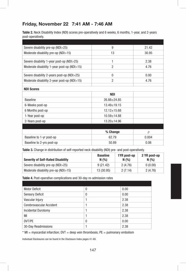
147
Friday, November 22
Individual Disclosures can be found in the Disclosure Index pages 41-69.
Table 2. Neck Disability Index (NDI) scores pre-operatively and 6 weeks, 6 months, 1-year, and 2-years post-operatively.
Severity of Self-Rated Disability N %
Severe disability pre-op (NDI>25) 9 21.42
Moderate disability pre-op (NDI>15) 13 30.95
Severe disability 1-year post-op (NDI>25) 1 2.38
Moderate disability 1-year post-op (NDI>15) 2 4.76
Severe disability 2-years post-op (NDI>25) 0 0.00
Moderate disability 2-year post-op (NDI>15) 2 4.76
NDI Scores
NDI
Baseline 26.88±24.85
6-Weeks post-op 13.49±19.15
6 Months post-op 12.12±15.68
1-Year post-op 10.59±14.88
2-Years post-op 13.20±14.96
Change in NDI Scores
% Change p
Baseline to 1-yr post-op 62.79 0.004
Baseline to 2-yrs post-op 50.89 0.06
Table 3. Change in distribution of self-reported neck disability (NDI) pre- and post-operatively.
Severity of Self-Rated DisabilityBaseline
N (%)1YR post-op
N (%)2 YR post-op
N (%)
Severe disability pre-op (NDI>25) 9 (21.42) 2 (4.76) 0 (0.00)
Moderate disability pre-op (NDI>15) 13 (30.95) 2 (7.14) 2 (4.76)
Table 4. Post-operative complications and 30-day re-admission rates
Complication Patients (N) %
Motor Deficit 0 0.00
Sensory Deficit 0 0.00
Vascular Injury 1 2.38
Cerebrovascular Accident 1 2.38
Incidental Durotomy 1 2.38
MI 1 2.38
DVT/PE 0 0.00
30-Day Readmissions 1 2.38
* MI = myocardial infarction; DVT = deep vein thrombosis; PE = pulmonary embolism
7:41 AM - 7:46 AM
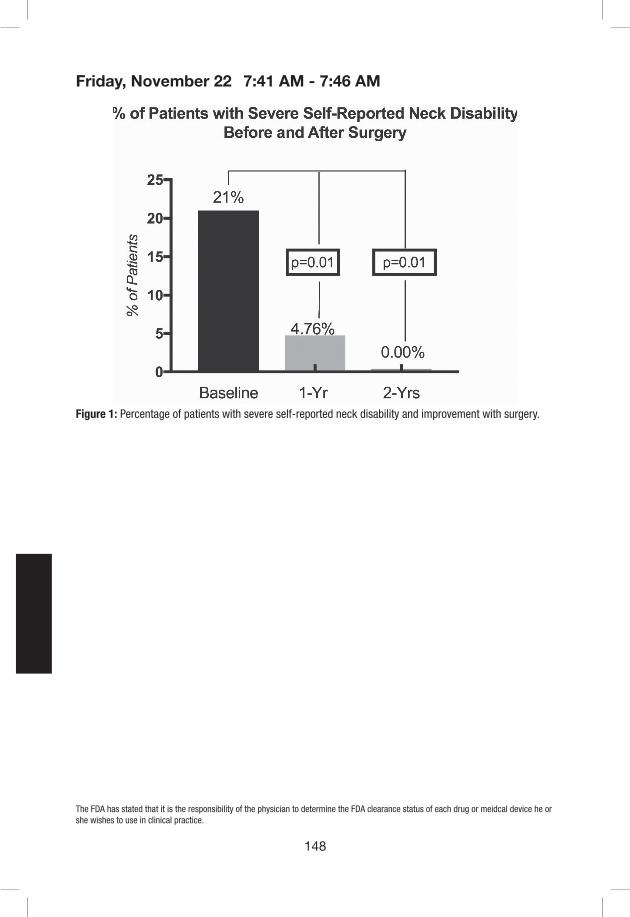
148
Friday, November 22
The FDA has stated that it is the responsibility of the physician to determine the FDA clearance status of each drug or meidcal device he or she wishes to use in clinical practice.
Figure 1: Percentage of patients with severe self-reported neck disability and improvement with surgery.
7:41 AM - 7:46 AM
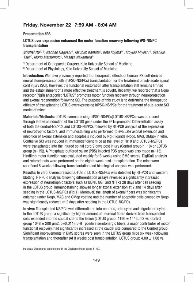
149
Friday, November 22
Individual Disclosures can be found in the Disclosure Index pages 41-69.
Presentation #36
LOTUS over expression enhanced the motor function recovery following iPS-NS/PC transplantation
Shuhei Ito1), 2), Narihito Nagoshi1), Yasuhiro Kamata1), Kota Kojima1), Hiroyuki Miyoshi1), Osahiko Tsuji1), Morio Matsumoto1), Masaya Nakamura1)
1) Department of Orthopaedic Surgery, Keio University School of Medicine2) Department of Physiology, Keio University School of Medicine
Introduction: We have previously reported the therapeutic effects of human iPS cell-derived neural stem/precursor cells (hiPSC-NS/PCs) transplantation for the treatment of sub-acute spinal cord injury (SCI). However, the functional restoration after transplantation still remains limited and the establishment of a more effective treatment is sought. Recently, we reported that a Nogo receptor (NgR) antagonist “LOTUS” promotes motor function recovery through neuroprotection and axonal regeneration following SCI. The purpose of this study is to determine the therapeutic efficacy of transplanting LOTUS overexpressing hiPSC-NS/PCs for the treatment of sub-acute SCI model of mice.
Materials/Methods: LOTUS overexpressing hiPSC-NS/PCs(LOTUS-NS/PCs) was produced through lentiviral induction of the LOTUS gene under the EF1αpromoter. Differentiation assay of both the control-NS/PCs and LOTUS-NS/PCs followed by RT-PCR analysis of the expression of neurotrophic factors, and immunostaining was performed to evaluate axonal extension and inhibition of axonal extension and apoptosis induced by NgR ligands (Nogo, MAG, OMgp) in vitro. Contusive SCI was induced in immunodeficient mice at the level of Th10 and LOTUS-NS/PCs were transplanted into the injured spinal cord 9 days post injury (Control group(n=10) or LOTUS group (n=15)). A Phosphate buffered saline (PBS) injected PBS group was also made (n=15). Hindlimb motor function was evaluated weekly for 8 weeks using BMS scores. DigiGait analysis and rotarod tests were performed on the eighth week post-transplantation. The mice were sacrificed 8 weeks following transplantation and histological analysis was performed.
Results: In vitro: Overexpressed LOTUS in LOTUS-NS/PCs was detected by RT-PCR and western blotting. RT-PCR analysis following differentiation assays revealed a significantly increased expression of neurotrophic factors such as BDNF, NGF and NTF-3 28 days after cell seeding in the LOTUS group. Immunostaining showed longer axonal extension at 2 and 14 days after seeding in the LOTUS-NS/PCs (Fig.1). Moreover, the length of axonal fibers was significantly enlarged under Nogo, MAG and OMgp coating and the number of apoptotic cells caused by Nogo was significantly reduced at 2 days after seeding in the LOTUS-NS/PCs.
In vivo: Transplanted NS/PCs well differentiated into neurons, astrocytes and oligodendrocytes. In the LOTUS group, a significantly higher amount of neuronal fibers derived from transplanted cells extended into the caudal site to the lesion (LOTUS group; 4196 ± 1442μm2 vs. Control group 1048 ± 208 μm2, p=0.011). 5-HT positive serotonergic fibers, a major contributor of motor functional recovery, had significantly increased at the caudal site compared to the Control group. Significant improvements in BMS scores were seen in the LOTUS group mice six week following transplantation and thereafter (At 8 weeks post transplantation: LOTUS group; 4.00 ± 1.08 vs.
7:59 AM - 8:04 AM
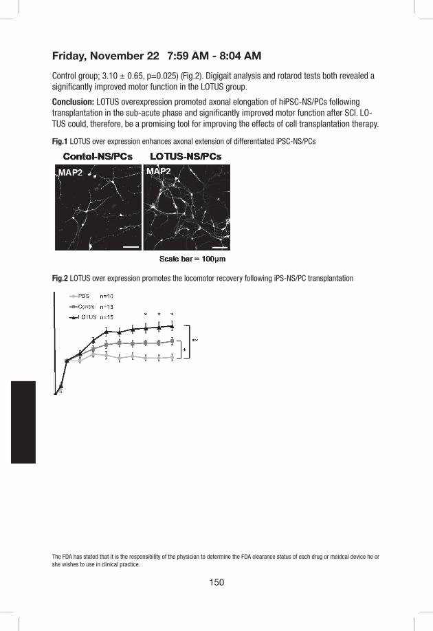
150
Friday, November 22
The FDA has stated that it is the responsibility of the physician to determine the FDA clearance status of each drug or meidcal device he or she wishes to use in clinical practice.
Control group; 3.10 ± 0.65, p=0.025) (Fig.2). Digigait analysis and rotarod tests both revealed a significantly improved motor function in the LOTUS group.
Conclusion: LOTUS overexpression promoted axonal elongation of hiPSC-NS/PCs following transplantation in the sub-acute phase and significantly improved motor function after SCI. LO-TUS could, therefore, be a promising tool for improving the effects of cell transplantation therapy.
Fig.1 LOTUS over expression enhances axonal extension of differentiated iPSC-NS/PCs
Fig.2 LOTUS over expression promotes the locomotor recovery following iPS-NS/PC transplantation
7:59 AM - 8:04 AM

151
Friday, November 22
Individual Disclosures can be found in the Disclosure Index pages 41-69.
Presentation #37
Involvement of autophagy in human cervical spine degenerated and herniated discs1Takashi Yurube, 1Yuji Kakiuchi, 1Masaaki Ito, 1Yoshiki Takeoka, 1Yutaro Kanda, 1Ryu Tsujimoto, 1Kenichiro Kakutani, 2Toru Takada, 1Kunihiko Miyazaki, 1Shingo Miyazaki, 1Zhongying Zhang, 1Ryosuke Kuroda, 1Kotaro Nishida1. Department of Orthopaedic Surgery, Kobe Graduate School of Medicine, Kobe, Japan2. Department of Orthopaedic Surgery, Kobe Hokuto Hospital, Kobe, Japan
Introduction: Cervical spine intervertebral disc pathologies can cause neck pain and neurological complications. The disc is the largest avascular organ in the body. Disc cells are expected subject to limited nutrition. Autophagy, the intracellular process by self-digestion and recycling damaged components, is an important cell survival mechanism under stress, primarily nutrient deprivation. We thus hypothesized that resident cells would utilize autophagy to cope with the harsh disc environment. However, clinical relevance of disc cellular autophagy is unknown. Therefore, an in-vivo study was designed to elucidate the involvement of autophagy in human cervical spine degenerated and herniated discs.
Materials/Methods: Human disc nucleus pulposus (NP) and annulus fibrosus (AF) tissues of total 44 patients who underwent anterior discectomy and fusion surgery for cervical spondylosis (n=32; age, 54.8±19.2 [22–83] yr; male 16, female 16; Pfirrmann degeneration grade, 3.4±0.8 [2–5]) and herniation (n=12; age, 49.3±19.0 [21–85] yr; male 8, female 4; Pfirrmann degeneration grade, 3.1±0.9 [2–4])were carefully dissected for protein extraction. Western blotting for an autophagy marker, LC3-II, and autophagy substrate, p62/SQSTM1, was performed. The α-tubulin was used as a loading control. Degeneration severity-, age-, and disease-dependent expression levels of autophagy-related LC3-II and p62/SQSTM were analyzed.
Results: First, we assessed expression of autophagy-related proteins based on the severity of human disc degeneration. In 12 patient samples of grades-2, -3, -4, and -5 disc NP tissues, expression levels of autophagic LC3-II and p62/SQSTM1 were both highest in discs with the Pfirrmann grades 3 and 4 (p=0.049 in LC3-II between grades 2 versus 4) (Fig. 1).
Thus, to understand roles of autophagy in human disc aging, we analyzed 20 consecutive patient samples of grades-3 and -4 disc NP and AF tissues. Human disc NP tissues in varying ages showed that LC3-II and p62/SQSTM1 expression transiently increased in middle ages of 40–70 but subsequently decreased in older ages of >70 (LC3-II R2=0.424, p<0.01; p62/SQSTM1 R2=0.392, p=0.01). Human disc AF-tissues presented a similar trend (LC3-II R2=0.546, p<0.01; p62/SQSTM1 R2=0.614, p<0.01) (Fig. 2).
Finally, to find disease-specific characteristics of autophagy in human disc degeneration and herniation, we compared age- and degeneration severity (grades 2, 3, and 4)-matched 12 patients with unstable degenerated discs requiring fusion and 12 those with symptomatic herniated discs requiring discectomy. There were no significant differences in LC3-II expression between disc degeneration and herniation (p=0.332) while p62/SQSTM1 expression levels declined in degenerated discs compared to herniated discs (p=0.046).
Conclusion: This is the first report to demonstrate that autophagy is clinically involved in
8:05 AM - 8:10 AM

152
Friday, November 22
The FDA has stated that it is the responsibility of the physician to determine the FDA clearance status of each drug or meidcal device he or she wishes to use in clinical practice.
intervertebral disc degeneration and herniation. Autophagy levels transiently increased in human middle-aged and moderately degenerated discs, potentially by stress response. However, autophagy levels subsequently decreased in human older-aged and severely degenerated discs, cells of which might lose the reaction potential of autophagy. In this study, disc cellular autophagy was degeneration severity- and age- but not disease-dependent, which would be important in the patient selection for autophagy-modulating molecular therapies. Autophagy modulation could be suitable for treating grades-3 and -4 disc diseases in middle-aged patients.
Fig. 1. Western blotting for autophagy marker LC3-II and autophagy substrate p62/SQSTM1 in human disc degeneration.
Fig. 2. Western blotting for autophagy marker LC3-II and autophagy substrate p62/SQSTM1 in human disc aging.
8:05 AM - 8:10 AM

153
Friday, November 22
Individual Disclosures can be found in the Disclosure Index pages 41-69.
Presentation #38
Inhibiting Spinal Phospholipase A2 Prevents Pain & Attenuates Spinal Neuron Activity After Nerve Root Compression
Sonia Kartha,a Julia Q Hoteka & Beth A Winkelsteina,b
aBioengineering, University of Pennsylvania, Philadelphia, PennsylvaniabNeurosurgery, University of Pennsylvania, Philadelphia, Pennsylvania
INTRODUCTION: The cervical nerve root is a common source of chronic pain when compressed from trauma and/or disc disease [1]. Nerve root compression (NRC) induces inflammatory mediators, including pro-inflammatory cytokines and phospholipase A2 (sPLA2), in the spinal cord that initiate and maintain neuronal hyperexcitability and pain [2-6]. sPLA2 releases arachidonic acid, is involved in production of prostaglandins, and modulates neuronal regulation of nociception and excitatory signaling [6-8]. Although spinal hyperexcitability is evident after NRC when pain persists, and is attenuated by neuromodulatory drugs that provide pain relief [9,10], it is unknown if that hyperexcitability is modulated by sPLA2. This study investigated whether inhibiting spinal sPLA2 modulates pain and/or spinal neuron hyperexcitability after injury in a rat model and probed its effects when given later after pain is established.
METHODS: Male Holtzman rats underwent a unilateral C7 dorsal NRC [9]. Immediately after compression, rats received the sPLA2 inhibitor thioetheramide-PC (Cayman) dissolved in PBS (0.25mg/kg) intrathecally by lumbar puncture (40-60µL) (NRC+TXT n=7) [7]. Additional rats underwent root compression only (NRC; n=3) or sham procedures (SHAM; n=3) with root exposure only. In a separate group (NRC+TXT7 n=4), the inhibitor was given at day 7 after NRC. Behavioral sensitivity was measured as the paw withdrawal threshold (PWT) to von Frey filaments before (baseline) and after surgery [9], and normalized to baseline. Electrophysiologic recordings were acquired at days 1, 7 and 14 in the C6/C7 spinal cord [9], under sodium pentobarbital (45mg/kg; i.p.) anesthesia. After identifying mechanosensitive neurons, non-noxious and noxious von Frey filaments were applied to the paw [9]. Voltage potentials were measured during stimulation, spike-sorted and counted using Spike2. PWT and evoked neuronal responses were compared by separate ANOVAs with Tukey’s HSD test.
RESULTS: Inhibiting spinal sPLA2 at NRC prevents pain at day 1, with higher PWT (p<0.01) than NRC and no difference from sham (Fig. 1). Although NRC reduces (p<0.01) the PWT at day 1 from baseline, withdrawal thresholds for NRC+TXT at day 1 are not different from baseline (Fig. 1). PWTs remain at baseline levels (p>0.99) until day 7 (Fig. 1). Similarly, treatment at day 7 attenuates pain by day 8 (p<0.01), with higher PWTs than untreated and at baseline until day 14 (p>0.96). Dorsal horn neuron activity (511±245µm below the pial surface) is reduced by inhibitor treatment (Fig. 2). At day 1, sPLA2 inhibitor (37 neurons) decreases (p<0.03) neuron firing for the noxious filaments, returning to SHAM levels (34 neurons). That trend is preserved at day 7, with significant decreases (p<0.05) from NRC and no difference from normal (16 neurons) or SHAM (Fig. 2). At day 14 after day 7 treatment, activity on the side of the injury (14 neurons) matches that of the uninjured contralateral side (6 neurons).
CONCLUSION: Inhibiting sPLA2 at NRC prevents the development and maintenance of pain and
8:11 AM - 8:16 AM

154
Friday, November 22
The FDA has stated that it is the responsibility of the physician to determine the FDA clearance status of each drug or meidcal device he or she wishes to use in clinical practice.
spinal neuron hyperexcitability (Figs. 1 & 2). Treating after injury, when pain is present, abolishes it and equalizes spinal neuronal activity to uninjured levels. These findings suggest sPLA2 has a role in early nociceptive transmission and central sensitization.
Fig. 1. Treatment increases the PWT significantly (#p<0.01) above NRC, keeping it at baseline levels for day 1. Further, treatment prevents pain until day 7 when given at NRC injury and immediately abolishes pain when given at day 7, with significant increases (*p<0.01) and no difference (p>0.96) in PWT baseline.
Fig. 2. Representative spinal neuron recordings at day 1 show decreased activity with treatment. Quantification of activity for each von Frey stimulus shows NRC at day 1 is increased above normal (*p<0.05) for all stimuli, and above sham (*p<0.03), treatment with NRC and recording at day 1 (^p<0.03), and for recordings at day 7 after treatment and NRC($p<0.05) for noxious stimuli.
References:[1] Abbed K et al, Neurosurgery 60:S28-S34, 2007. [2] Rothman S et al., J Pain 10:90-9, 2009.
[3] Nicholson K et al, Neurosci 209:187-95, 2012. [4] Vasquez E et al, J Neurosci 21:9001-8, 2001. [5] Kartha S et al, Neurosci 388:393-404, 2018. [6] Svensson C et al, Neurosci 133:543-53, 2005. [7] Morioka N et al, J Neurochem 80:989-97, 2002. [8] DeCoster M et al, J Neurosci Res 67:634-45, 2002. [9] Nicholson K et al, J Neurosci Res 92:116-29, 2014. [10] Nicholson K et al, J Neurosurg:Spine 20:751-62, 2014.
8:11 AM - 8:16 AM
155
Friday, November 22
Individual Disclosures can be found in the Disclosure Index pages 41-69.
Presentation #39
Supported by the Sharpe Foundation and the NIH (R01NS199892; T32NS043126).Electrospun Synthetic Bone Graft Promotes MSC Function and Spinal Fusion
Derek G. Ju MD, Juliane D Glaeser PhD, Khosrowdad Salehi BS, Linda E. A. Kanim MA, Phillip H Behrens MD, Melodie F Metzger PhD, Dmitriy Sheyn PhD, Hyun W. Bae MD
Cedars-Sinai Spine Center, Los Angeles, CA
Introduction: Synthetic bone grafts are being developed to lessen the need for autograft and allograft during spinal fusion surgery. Electrospun synthetic bone grafts (ESBG) are a novel scaffold with a highly porous, 3D nanofibrous structure. This allows for a large surface area-to-volume ratio, potentially improving its osteoconductive and osteoinductive properties. Here, we investigate the potential of ESBG to stimulate mesenchymal stem cell (MSC) function in vitro, and to facilitate BMP-2 mediated spinal fusion in a rat model.
Materials/Methods: Adhesion, proliferation, and osteogenic differentiation of MSCs seeded with and without ESBG for 7 days was analyzed in vitro. For an In-vivo analysis, 36 Lewis rats underwent posterolateral spine fusions in the following groups: (I) ESBG+bone marrow aspirate (BMA), (II) ESBG+BMA+BMP-2 (low-dose), and (III) BMA. Rats were followed for 3 months and the fusion masses were characterized with manual palpation, biomechanical tests, micro-CT and histological evaluation.
Results: 90% of cultured MSCs adhered to ESBG. When seeded in ESBG, there was a significant increase in MSC proliferation (0.18 to 0.43, p<0.01, Fig. 1A, B) and osteoblastic activity (2.3 to 5.2; p<0.02, Fig. 1C). In the rat model, fusion rates at 3 months were 29% (group I), 100% (group II), and 0% (group III, Fig. 2). Biomechanical stiffness was highest in group II vs I (286 vs. 198 N/mm, p<0.05). On micro-CT, group II had increased bone volume (216.0 vs. 133.9), bone volume/total volume (0.085 vs. 0.01) and increased connectivity density (1.26 vs 0.28; all p<0.05). Histological evaluation demonstrated new bone formation within the graft only in group II.
Conclusion: This is the first reported characterization of ESBG for spinal applications. ESBGs provide a novel scaffold that supports MSC binding, proliferation, and osteogenic differentiation. In a rat spine fusion model, ESBG impregnated with BMA and low-dose BMP-2 allowed for 100% fusion with strong biomechanical properties. ESBGs represent a promising synthetic graft and should be further investigated for clinical feasibility.
8:17 AM - 8:22 AM

156
Friday, November 22
The FDA has stated that it is the responsibility of the physician to determine the FDA clearance status of each drug or meidcal device he or she wishes to use in clinical practice.
Fig. 1. ESBG allows for adhesion and proliferation of MSCs and stimuates osteogenic differentiation. A. Confocal image of adhesion of MSCs to ESBG. B. hMSC proliferation on ESBG over time. C. ALP (osteoblastic) activity in the presence and absence of ESBG.
Fig. 2 Radiographic evidence of new bone formation at the implantation side at 8 and 12 weeks post-surgery.
8:17 AM - 8:22 AM

162
Friday, November 22
The FDA has stated that it is the responsibility of the physician to determine the FDA clearance status of each drug or meidcal device he or she wishes to use in clinical practice.
Presentation #42
Single-level cervical arthroplasty with ProDisc-C artificial disc: 10-year follow-up results in one center
Zhao Yanbin, Sun Yu
Department of Orthopaedic Surgery, Peking University Third Hospital, Beijing, China
Keywords: cervical disc arthroplasty; heterotopic ossification; adjacent segment disease
Objectives: The aim of this study was to evaluate the long-term clinical and radiographic outcomes of cervical arthroplasty using the ProDisc-C prosthesis.
Methods: Clinical and radiographic evaluations, including dynamic flexion-extension lateral images, were performed at baseline and at 10-year follow-up.
Results: Twenty-seven patients who had single-level ProDisc-C arthroplasty were followed up for a mean period of 123 months. The range of motion at the operated level was 8.9±3.9° at baseline and 6.6±3.5° at final follow-up. Twenty of 27 levels (74%) developed heterotopic ossification. According to McAfee’s classification, one level was classified as grade Ⅰ, 4 levels were classified as grade Ⅱ, 12 levels were classified as grade III and 3 levels as grade IV. Three patients developed recurrent cervical radiculopathy or myelopathy due to adjacent segment disease and received the reoperations. The reoperations included two cases of cervical arthroplasty at adjacent segments and one case of cervical laminoplasty.
Conclusions: ProDisc-C arthroplasty had acceptable clinical and radiographic results at 10-year follow-up. Heterotopic ossification was common after ProDisc-C arthroplasty, which decreased the range of motion.
1:43 PM - 1:48 PM

163
Friday, November 22
Individual Disclosures can be found in the Disclosure Index pages 41-69.
Presentation #43
Unintended Fusion In Cervical Artificial Disc Replacement: A Prospective Study On Heterotopic Ossification, Evolution Through Time, And Clinical Outcome, With 5 Years Follow-Up
Catarina Marques, MD1*, Anna MacDowall, MD, PhD1, Martin Skeppholm, MD, PhD2, Nuno Canto Moreira, MD, PhD3, Claes Olerud, MD, PhD1
*Corresponding author. Adress: Entrance 61, 6th floor, Department of Surgical Sciences, Uppsala University, Sweden
1Department of Surgical Science, Uppsala University Hospital, Sweden2Department for Learning, Informatics, Management and Ethics, Karolinska institutet, Stockholm, Sweden3Pediatric Radiology Section, Department of Clinical Neuroscience (CNS), K8, Karolinska Universitetssjukhuset Solna, Stockholm, Sweden
Introduction: ACDF is the gold standard surgical treatment in patients with cervical degenerative disc disease with radiculopathy. Implants for artificial disc replacement (ADR) were developed as an alternative to ACDF. ADR was expected to preserve motion, decrease the incidence of adjacent segment pathology and consequently lead to better clinical outcomes.
Heterotopic ossification (HO) is an unintended event that may lead to spontaneous fusion of the ADR, thus failing to preserve motion.
Our study aims to determine the prevalence, predisposing factors and rate of development of HO, as well as its effect on clinical outcomes.
Materials/Methods: Analysis of a multicentre prospective RCT. 153 patients randomized to ADR or ACDF, maximum two disc levels. All ADR patients received NSAID to prevent HO.
Of 83 ADR patients, we included the 59 (79 prosthesis) that had good-quality radiological follow-up studies at 5 years.
HO was graded on X-ray 2- and 5 years postoperatively (2 viewers, modified McAfee classification). Baseline characteristics and several parameters on image studies (CT, MRI and X-ray) were analyzed. Worsening of HO grade between 2- and 5-years follow-up was evaluated and eventual predisposing factors for worsening where analyzed.
The clinical outcome measure was NDI.
Results: HO was found in 92% of the prosthesis after 5 years.
Severe HO (grade 3 or 4) existed in 71% and complete fusion (grade 4) in 27% after 5 years.
Significantly less HO, and of lower grades, was found in women. Age and BMI seemed to be predisposing factors for HO, but lost significance in a multivariate analysis.
Nearly 2/3 of the prosthesis did not increase in HO grade between 2- and 5-years follow-up. No statistically significant predisposing factors could be identified in those patients whose HO worsened, though smoking could be a factor (odds ratio=3.95, p=0,055).
1:49 PM - 1:54 PM
164
Friday, November 22
The FDA has stated that it is the responsibility of the physician to determine the FDA clearance status of each drug or meidcal device he or she wishes to use in clinical practice.
No difference in NDI was found between patients with severe HO and those with none or mild HO.
Conclusion: Disc prosthesis are meant to move, in order to be an alternative to fusion regarding the potential prevention of adjacent segment pathology.
Despite NSAID treatment, almost all patients have HO after 5 years, more than 2/3 of prosthesis have severe HO, and over 1/4 actually spontaneously fuse. However, clinical outcomes were not affected by severe HO. Thus, having a functioning arthroplasty does not have a clinical impact.
Male gender is a clear predisposing factor for HO.
HO develops during the first two post-operative years, and then stabilizes in nearly 2/3 of the prosthesis. No clear predisposing factors were found in the patients whose HO continues to aggravate. Smoking, nearly reaching statistic significance, is however a factor that should be considered in future research.
1:49 PM - 1:54 PM

165
Friday, November 22
Individual Disclosures can be found in the Disclosure Index pages 41-69.
Presentation #44
Does Positioning of Cervical Disc Arthroplasty Implant Affect Postoperative Outcome
Yahya Othman1, Philip York, MD1, Russel Huang, MD1,2, Avani Vaishnav, MBBS1, Steven McAnany, MD1,2, Sravisht Iyer, MD1,2, Todd Albert, MD1,2, Catherine Gang, MPH1, Sheeraz Qureshi, MD, MBA1,2
1Hospital for Special Surgery, New York, NY 2Weill Cornell Medical College, New York, NY
Introduction: Cervical disc arthroplasty (CDA) is a surgical option for patients with cervical radiculopathy symptoms that have failed conservative management. CDA has been shown to be non-inferior to Anterior Cervical Decompression and Fusion (ACDF) surgery in terms of patient outcomes. Furthermore, CDA has a lower incidence of adjacent segment disease as well as reoperation rates. Proper surgical technique is critical for success of CDA surgery. The aim of this study was to investigate whether implant positioning has an impact on the outcomes of CDA surgery.
Materials/Methods: Following local IRB approval, a retrospective study was conducted on consecutive patients undergoing CDA with the Mobi-C implant (Zimmer Biomet, IN) between January 2016 and December 2018. Immediately postoperative lateral and AP radiographs were assessed by three spine surgeons for implant positioning. Our scoring criteria included rotational, sagittal and coronal placement as well as size matching. AP and lateral radiographs were scored independently and the overall postoperative score was calculated as the sum of the two scores. The overall postoperative score was correlated with 5 patient reported outcomes measures (PROMS); Visual Analogue Scale (VAS) arm, VAS neck, Neck Disability Index (NDI), and Short Form 12 Physical health (SF12-P) and mental health (SF12-M). Correlation between the overall radiographic score and patient reported outcomes was assessed using the Pearson correlation coefficient. Inter-rater reliability was assessed using the intraclass correlation coefficient.
Results: Radiographs of 85 patients, and 110 levels, that underwent cervical disc arthroplasty were assessed. Of those, forty-one patients were excluded from the patient reported outcomes measures analysis due to inadequate follow-up. Interrater reliability for radiographic assessment was good to excellent. Mean follow-up was 8 months and ranged from 6 to 24 months. Patients achieved significant improvement in all reported outcomes at latest follow up. At early follow-up, implant positioning success did not significantly correlate with patient reported outcomes measures. However, the overall implant positioning scores showed strong correlation with NDI and SF12-P outcomes measures at latest follow up (p=0.007, p=0.028, respectively). The overall score did not correlate with VAS arm and VAS neck (p=0.948 p=0.131, respectively).
Conclusions: Proper surgical technique is essential for long term success of CDA surgery. Qualitative assessment of radiographs is a reliable way to assess implant positioning success. Although not evident in the immediate stage, proper implant sizing and implant rotational and translational positioning have a significant impact on functional and disability outcomes of patients undergoing CDA surgery. Further research is warranted to investigate other parameters that affect the outcomes of CDA surgery.
1:55 PM - 2:00 PM

166
Friday, November 22
The FDA has stated that it is the responsibility of the physician to determine the FDA clearance status of each drug or meidcal device he or she wishes to use in clinical practice.
Presentation #45
The Effect of ACDF or Arthroplasty on Cervicogenic Headaches: A Post-hoc Analysis of a Prospective, Multicenter Study with 10-Year Follow-up
INTRODUCTION: To our knowledge, there is no large, prospective study that has examined the efficacy of cervical spine surgery for relieving headaches associated with radiculopathy or myelopathy at 10 years post-op. The purpose of this study is to determine the effectiveness of a 2-level ACDF or Cervical Disc Arthroplasty (CDA) at relieving headaches associated with cervical radiculopathy or myelopathy at 10 years post-op.
METHODS: This was a post-hoc analysis of a prospective FDA-IDE study for the Prestige-LP vs ACDF for radiculopathy or myelopathy due to 2-levels. Pre-operatively and out to 10 years, their NDI documented if they had headaches (0: No headaches; 1: Infrequent slight; 2: Infrequent Moderate; 3: Frequent Moderate; 4: Frequent Severe; 5: Nearly constant).
RESULTS: 397 patients were randomized to CDA (209) or ACDF (188). Pre-operatively 86% had headaches and 55.9% (52.2% of CDA, 60.1% of ACDFs) had frequent moderate, severe or nearly constant headache (grades 3-5). By 6 weeks post-op, 64.4% had headaches and only 12.5% had Grades 3-5 headaches (9.3% of CDA and 16% of ACDFs). The benefit lasted to the 10-year follow-up such that 60.3% had any headaches and 16.8% had Grades 3-5 headaches (10.9% CDA; 24.3% ACDF).
2:01 PM - 2:06 PM
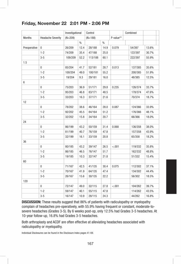
167
Friday, November 22
Individual Disclosures can be found in the Disclosure Index pages 41-69.
Investigational Control Combined
Months Headache Severity (N=209) (N=188) P-value**
% %
Preoperative 0 26/209 12.4 28/188 14.9 0.079 54/397 13.6%
1-2 74/209 35.4 47/188 25.0 122/397 30.7%
3-5 109/209 52.2 113/188 60.1 222/397 55.9%
1.5
0 85/204 41.7 52/181 28.7 0.013 137/385 35.6%
1-2 100/204 49.0 100/181 55.2 200/385 51.9%
3-5 19/204 9.3 29/181 16.0 48/385 12.5%
6
0 75/203 36.9 51/171 29.8 0.235 126/374 33.7%
1-2 95/203 46.8 83/171 48.5 178/374 47.6%
3-5 33/203 16.3 37/171 21.6 70/374 18.7%
12
0 78/202 38.6 46/164 28.0 0.087 124/366 33.9%
1-2 92/202 45.5 84/164 51.2 176/366 48.1%
3-5 32/202 15.8 34/164 20.7 66/366 18.0%
24
0 86/199 43.2 50/159 31.4 0.068 136/358 38.0%
1-2 81/199 40.7 76/159 47.8 157/358 43.9%
3-5 32/199 16.1 33/159 20.8 65/358 18.2%
36
0 80/185 43.2 39/147 26.5 <.001 119/332 35.8%
1-2 86/185 46.5 76/147 51.7 162/332 48.8%
3-5 19/185 10.3 32/147 21.8 51/332 15.4%
60
0 71/167 42.5 41/135 30.4 0.075 112/302 37.1%
1-2 70/167 41.9 64/135 47.4 134/302 44.4%
3-5 26/167 15.6 30/135 22.2 56/302 18.5%
120
0 72/147 49.0 32/115 27.8 <.001 104/262 39.7%
1-2 59/147 40.1 55/115 47.8 114/262 43.5%
3-5 16/147 10.9 28/115 24.3 44/262 16.8%
DISCUSSION: These results suggest that 86% of patients with radiculopathy or myelopathy complain of headaches pre-operatively, with 55.9% having frequent or constant, moderate-to-severe headaches (Grades 3-5). By 6 weeks post-op, only 12.5% had Grades 3-5 headaches. At 10-year follow-up, 16.8% had Grades 3-5 headaches.
Both arthroplasty and ACDF are often effective at alleviating headaches associated with radiculopathy or myelopathy.
2:01 PM - 2:06 PM

168
Friday, November 22
The FDA has stated that it is the responsibility of the physician to determine the FDA clearance status of each drug or meidcal device he or she wishes to use in clinical practice.
Presentation #46
Comparison of Three FDA-Approved Artificial Cervical Discs: A Finite Element Study
Hoon Choi, MD1, Yuvaraj Purushothaman, MS1, Jamie Baisden, MD1, Narayan Yoganandan, PhD1
1 Medical College of Wisconsin, Milwaukee, WI
Introduction: Cervical arthroplasty is an increasingly popular alternative to arthrodesis. Studies have focused on comparing cervical arthroplasty with cervical arthrodesis. However, there is a lack of literature on understanding how the design of the artificial cervical disc influences the spine biomechanics. Moreover, commercially available FDA-approved artificial cervical discs vary widely in design. The purpose of the current study was to evaluate and compare the biomechanics in the index and adjacent levels after cervical arthroplasty using three different artificial cervical discs.
Materials/Methods: A previously validated C2-T1 osteoligamentous finite element model was used to perform virtual C5-6 arthroplasty using three different FDA-approved artificial cervical discs (Fig 1). Motion-controlled moment loading protocol was used. Moment was varied until Bryan, Prodisc C, and Prestige LP models displayed the same total range of motion across C3 to C7 as the intact spine model at 2 Nm of pure moment loading. Range of motion (ROM) and facet force (FF) were recorded in the index level. ROM, FF, and intradiscal pressure (IDP) were recorded in the adjacent levels.
Results: Prodisc C and Prestige LP led to supraphysiologic ROM and FF in the index level while decreasing ROM and FF in the adjacent levels. In contrast, Bryan reduced ROM and FF in the index level. Bryan increased ROM and FF in the adjacent levels in flexion, but decreased ROM and FF in the adjacent levels in extension. Prodisc C decreased IDP in the adjacent levels. Bryan reduced IDP in extension only. Prestige LP increased adjacent level IDP.
Conclusions: The unencumbered open multi-piece design with a fixed core but small permissible translation of the center of rotation (semi-constrained) found in Prodisc C and Prestige LP led to increased motion and stress in the facet joints in the index levels, while doing the opposite in the index levels. This is consistent with its intended purpose of protecting the adjacent levels while preserving motion in the index level. This is also consistent with the contraindications of facet arthropathy and spinal instability. Polymer-on-metal design of Prodisc C led to reductions in disc pressure in the adjacent levels. Bryan, with its encapsulated design and limited flexion, demonstrated subphysiologic motion preservation in the index level and shielded the facet joints. This could indicate its potential use in specific cases with concurrent facet arthropathy that still demand motion preservation. Not all artificial cervical discs were created equal, and it is important to understand that design influences biomechanics.
2:07 PM - 2:12 PM

169
Friday, November 22
Individual Disclosures can be found in the Disclosure Index pages 41-69.
Figure 1. C5-6 Arthroplasty was simulated with three different artificial cervical discs, (from left to right), Prodisc C, Bryan, and Prestige LP.
2:07 PM - 2:12 PM

170
Friday, November 22
The FDA has stated that it is the responsibility of the physician to determine the FDA clearance status of each drug or meidcal device he or she wishes to use in clinical practice.
Presentation #47
Mid-term Surgical Outcome of Posterior Decompression with Instrumented Fusion for K-line negative type Cervical OPLL -minimum 5 years follow-up
Takeo Furuya, 1, Satoshi Maki, 1, Takuya Miyamoto, 1, Sho Okimatsu, 1, Masao Koda, 1, 2, Masashi Yamazaki, 1, 2
1: Dept. Orthopaedic Surgery, Chiba University Graduate School of Medicine, Chiba, Japan.2: Dept. of Orthopedic Surgery, Faculty of Medicine, University of Tsukuba, Japan
Introduction: We have reported a concept of K-line for making decisions regarding the surgical approach for cervical ossification of the posterior longitudinal ligament (OPLL). K-line is the line that connects the midpoints of the spinal canal at C2-7 at the lateral view of the cervical radiograph in the neutral position. By using the K-line, we can evaluate the alignment of the cervical spine and the size of OPLL in one parameter. When the OPLL exceeds the K-line, the OPLL is classified into a K-line negative type. We previously reported poor surgical outcome of laminoplasty alone and an advantage of additional instrumented fixation for K-line negative type cervical OPLL. The purpose of this study was to assess mid-term surgical outcome after posterior decompression with instrumented fusion (PDF) in patients with K-line negative type cervical OPLL over five years.
Methods: A retrospective review of 34 patients with PDF from 2002 to 2013 for K-line (-)-type cervical OPLL at our institute was conducted (PDF group). Seven patients with laminoplasty from 2000 to 2002 for K-line negative type cervical OPLL at our institute was set as control (LMP group). Patients without 5 years postoperative follow-up were excluded. The surgical outcomes were evaluated using the Japanese Orthopaedic Association score (JOA score) and recovery rate. Preoperative and postoperative segmental range of motion at the maximum spinal cord compression level, C2-7 angle and CGH-C7 SVA were measured for radiographic examination.
Results: No statistical difference was seen between the two groups for preoperative clinical data. Postoperative follow-up period showed statistically difference between the two groups. Preoperative and postoperative JOA scores were 8.1±2.0 and 11.8±2.4 in the PDF group and 9.1±3.0 and 10.9±3.0 in the LMP group respectively. The average recovery rate was 39.5% in the PDF group and 15.5% in the LMP group at final follow-up (P<0.05). The segmental range of motion at the maximum spinal cord compression level was 0 degree in the PDF group in average and 4 degrees in the LMP group in average at final follow-up (P<0.05). The C2-7 angle and CGH-C7 SVA showed 1 degree (ranged 0 to 2 degrees) increase of kyphosis and 10mm (-5 to 35mm) off-balance postoperatively in the PDF group, whereas 8 degrees (ranged 2 to 18 degrees) increase of kyphosis and 13.7mm (ranged -30 to 60mm) off-balance postoperatively in the LMP group.
Discussion: The PDF group showed better surgical outcome compared with the LMP group. The addition of posterior instrumented fusion can control and maintain the local stabilization. However, the instrumentation cannot prevent progression of off-balance. The reason may come from wide exposure of the surgical site and muscle damage when we performed relatively long instrumented fusion. Those data suggest that the benefit of instrumentation is mainly control of
2:56 PM - 3:01 PM
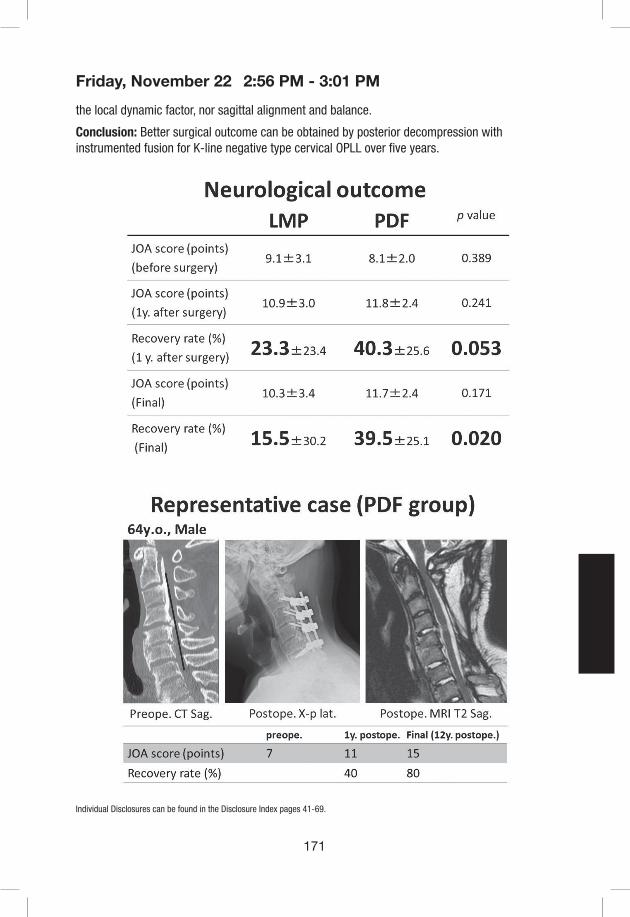
171
Friday, November 22
Individual Disclosures can be found in the Disclosure Index pages 41-69.
the local dynamic factor, nor sagittal alignment and balance.
Conclusion: Better surgical outcome can be obtained by posterior decompression with instrumented fusion for K-line negative type cervical OPLL over five years.
2:56 PM - 3:01 PM
172
Friday, November 22
The FDA has stated that it is the responsibility of the physician to determine the FDA clearance status of each drug or meidcal device he or she wishes to use in clinical practice.
Presentation #48
Does pre- or post-operative cervical sagittal alignment correlate with the outcomes of cervical laminoplasty? A minimum of one-year follow-up study
Shuo Niu, MD, PhD, Emory Orthopaedics & Spine Center, Atlanta, GAAlbert T. Anastasio, BA, Emory School of Medicine, Atlanta, GAKevin X. Farley, BA, Emory School of Medicine, Atlanta, GAJohn M. Rhee, MD, Emory Orthopaedics & Spine Center, Atlanta, GA
Introduction: Cervical laminoplasty is a common surgical treatment for myelopathy. However, it is still under debate how pre- or post-operative cervical sagittal alignment affects the outcomes of cervical laminoplasty, such as neurological improvement and patient-reported outcomes (PRO). The purpose of this study is therefore to determine whether there is any correlation between cervical sagittal alignment, either pre- or post-operative, and the outcomes of laminoplasty.
Materials/Methods: This is a retrospective case-control study of 144 consecutive patients between 2007 and 2017 undergoing multi-level (≥3) posterior cervical laminoplasty for cervical myelopathy at a single academic spine center. The severity of myelopathy was accessed by modified Japanese Orthopedic Association (mJOA) scores. Total pain was measured by the visual analog scale. PRO included neck disability index (NDI) and 12-item short form survey (SF-12). Radiographic measures of cervical sagittal alignment on x-rays consisted of C2-C7 angle, T1 slope, C2-C7 sagittal vertical axis (C2-C7 SVA; distance between C2 plumb line and superoposterior corner of C7), and C2-C7 forward pitch (C2-C7 FP; distance between C2 plumb line and superoanterior corner of C7). Patients were also divided into two groups based on the post-operative C2-C7 SVA (C2-C7 SVA ≥ 40 mm and < 40 mm). Mann-Whitney U-test was used to determine the differences between pre- and post-operative with respect to surgical outcomes and radiographic measures. Spearman’s correlation coefficient was used to determine the correlations between PRO and each of radiographic measures. P<0.05 was considered significant.
Results: The demographics of the patient cohort was 43.1% female, with a mean age of 63.2 (range, 22-97) years and a mean follow up 23.7 (range, 12-105) months. Laminoplasty yielded improvement in functionality as evidence by significantly increased mJOA scores, decreased total pain and NDI scores at the final follow-up (Table 1). There was a loss of sagittal balance post-operatively, as demonstrated by significantly increased C2-C7 SVA and FP (~7-8mm increase, Table 1). However, there was no correlation between any pre-operative sagittal alignment measures and PRO (Table2, all p>0.05). And no correlation between most of post-operative sagittal alignment and PRO (Table 2), except for a significantly negative correlation between post-operative C2-C7 FP and SF-PCS (Spearsman’s r=-0.328, p=0.011). When the difference between pre- and post-operative metrics was calculated (delta value), there was no correlation between change in sagittal alignment and change (either detrimental or beneficial) in PRO. Lastly, when the group with C2-C7 SVA ≥ 40 mm (n=60) was compared to the group with C2-C7 SVA< 40 mm (n=84), there was no significant difference with regard to post-operative mJOA and PRO scores (all p>0.05).
Conclusion: Cervical laminoplasty yields satisfactory neurological and functional improvement
3:02 PM - 3:07 PM
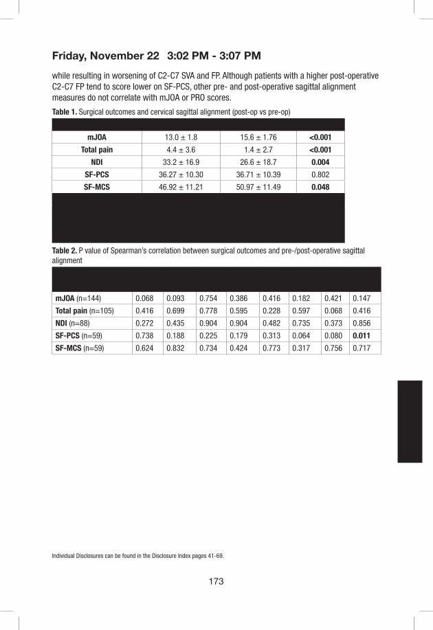
173
Friday, November 22
Individual Disclosures can be found in the Disclosure Index pages 41-69.
while resulting in worsening of C2-C7 SVA and FP. Although patients with a higher post-operative C2-C7 FP tend to score lower on SF-PCS, other pre- and post-operative sagittal alignment measures do not correlate with mJOA or PRO scores.
Table 1. Surgical outcomes and cervical sagittal alignment (post-op vs pre-op)
Pre-op Post-op p value
mJOA 13.0 ± 1.8 15.6 ± 1.76 <0.001
Total pain 4.4 ± 3.6 1.4 ± 2.7 <0.001
NDI 33.2 ± 16.9 26.6 ± 18.7 0.004
SF-PCS 36.27 ± 10.30 36.71 ± 10.39 0.802
SF-MCS 46.92 ± 11.21 50.97 ± 11.49 0.048
C2-C7 angle (°) 11.6 ± 10.2 10.1 ± 12.1 0.455
T1 slope (°) 30.9 ± 9.7 32.2 ± 9.8 0.245
C2-C7 SVA (mm) 32.4 ± 16.8 39.7 ± 19.3 0.001
C2-C7 FP (mm) 13.1 ± 16.2 21.3 ± 19.6 <0.001
Table 2. P value of Spearman’s correlation between surgical outcomes and pre-/post-operative sagittal alignment
C2-C7 angle T1 slope C2-C7 SVA C2-C7 FP
Pre-op Post-op Pre-op Post-op Pre-op Post-op Pre-op Post-op
mJOA (n=144) 0.068 0.093 0.754 0.386 0.416 0.182 0.421 0.147
Total pain (n=105) 0.416 0.699 0.778 0.595 0.228 0.597 0.068 0.416
NDI (n=88) 0.272 0.435 0.904 0.904 0.482 0.735 0.373 0.856
SF-PCS (n=59) 0.738 0.188 0.225 0.179 0.313 0.064 0.080 0.011
SF-MCS (n=59) 0.624 0.832 0.734 0.424 0.773 0.317 0.756 0.717
3:02 PM - 3:07 PM
174
Friday, November 22
The FDA has stated that it is the responsibility of the physician to determine the FDA clearance status of each drug or meidcal device he or she wishes to use in clinical practice.
Presentation #49
The Effect of Duration of Symptoms on Clinical Outcomes Following Anterior Cervical Discectomy and Fusion
James M. Parrish, MPH, Joon S. Yoo, BA, Dil V. Patel, BS, Eric H. Lamoutte, BS, Sailee S. Karmarkar, BS, Kern Singh, MD
Objective: There is a paucity of literature regarding the influence of preoperative symptom duration on patient-reported outcomes (PROs) following ACDF. To examine whether the time spanning from symptom onset to operative treatment has an effect on postoperative clinical improvement in patients undergoing anterior cervical discectomy and fusion (ACDF).
Methods: Patients who underwent primary, single-level ACDF were retrospectively reviewed and stratified according to preoperative symptom duration (<12 months and ≥12 months). Demographic and perioperative characteristics were compared using Chi-squared analysis and linear regression. Differences in PRO scores, including Neck Disability Index (NDI), Visual Analogue Scale (VAS) neck pain, VAS arm pain, and 12-Item Short-Form Physical Component Score (SF-12 PCS), at each postoperative timepoint were compared using linear regression. Achievement of minimal clinically important difference (MCID) for PROs was compared using Chi-square analysis.
Results: 115 patients underwent primary, single-level ACDF: 77 had a DOS <12 months and 38 had a DOS ≥12 months. When comparing PROs preoperatively, the shorter DOS cohort had significantly worse VAS arm pain. However, there was no preoperative difference in NDI, VAS neck pain, and SF-12 PCS. In the postoperative period, there were no significant differences in improvement of PROs throughout the 12-month timepoint (Table 1). The shorter DOS cohort had a comparable number of patients achieving MCID for NDI, VAS neck pain, VAS leg pain, and SF-12 PCS (Table 2).
Conclusion: This investigation assessed the influence of preoperative DOS in patients undergoing a primary, single-level ACDF. Patients with DOS shorter than 12 months exhibited significantly worse VAS arm pain scores at the time of surgery. However, these patients demonstrated similar clinical outcomes postoperatively regardless of preoperative symptom duration. Our findings suggest that delayed surgical intervention may not necessarily lead to impaired functional recovery in patients with degenerative cervical disease.
3:08 PM - 3:13 PM

175
Friday, November 22
Individual Disclosures can be found in the Disclosure Index pages 41-69.
Table 1. Improvement in patient-reported outcomes
DOS <12 Months(N=77)
DOS ≥12 Months(N=38)
†p-value*
Neck Disability Index (mean ± SD)
Preoperative 43.7 ± 19.3 37.6 ± 16.4 0.097
6-week 31.0 ± 20.6 30.6 ± 18.7 0.918
3-month 25.6 ± 20.1 29.2 ± 20.4 0.407
6-month 20.9 ± 19.4 27.1 ± 23.5 0.195
12-month 25.2 ± 24.5 28.0 ± 20.8 0.701
VAS Neck Pain (mean ± SD)
Preoperative 6.4 ± 2.3 5.9 ± 2.4 0.266
6-week 3.4 ± 2.8 3.2 ± 2.4 0.694
3-month 2.9 ± 2.7 2.8 ± 2.2 0.822
6-month 2.7 ± 2.8 2.8 ± 2.6 0.784
12-month 3.3 ± 3.1 3.2 ± 2.3 0.883
VAS Arm Pain (mean ± SD)
Preoperative 6.3 ± 2.6 5.2 ± 2.7 0.049
6-week 2.6 ± 3.6 2.5 ± 2.2 0.860
3-month 2.4 ± 2.8 3.4 ± 3.0 0.113
6-month 2.5 ± 3.0 3.0 ± 2.8 0.385
12-month 3.4 ± 3.4 4.0 ± 2.9 0.532
SF-12 Physical Component Score (mean ± SD)
Preoperative 34.1 ± 8.2 36.4 ± 8.8 0.213
6-week 35.8 ± 8.5 35.9 ± 8.8 0.969
3-month 38.4 ± 11.5 40.1 ± 11.5 0.593
6-month 43.5 ± 9.8 40.5 ± 9.1 0.265
12-month 42.9 ± 11.0 41.1 ± 12.5 0.618
SD = Standard Deviation; VAS = Visual Analog Scale; SF-12 = 12-Item Short-Form*Boldface indicate statistical significance†p-values calculated for each category using linear regression
3:08 PM - 3:13 PM

176
Friday, November 22
The FDA has stated that it is the responsibility of the physician to determine the FDA clearance status of each drug or meidcal device he or she wishes to use in clinical practice.
Table 2. Achievement of minimal clinically important difference (MCID)
DOS <12 Months(N=77)
DOS ≥12 Months(N=38)
†p-value*
Neck Disability Index (mean ± SD) 87.0% (67) 84.2% (32) 0.683
VAS Neck Pain (mean ± SD) 93.5% (72) 89.5% (34) 0.449
VAS Arm Pain (mean ± SD) 81.8% (63) 76.3% (29) 0.488
SF-12 Physical Component Score (mean ± SD) 85.7% (66) 89.5% (34) 0.573
SD = Standard Deviation; VAS = Visual Analog Scale; SF-12 = Short-FormMCID values derived from Parker et al.: NDI=9.40, VAS Neck Leg=2.6, VAS Neck Arm=4.1; SF-12 PCS=8.1†p-values calculated for each category using Chi-square analysis
3:08 PM - 3:13 PM
177
Friday, November 22
Individual Disclosures can be found in the Disclosure Index pages 41-69.
Presentation #50
The Impact of Post-Operative Physical Therapy on Patient-Reported Outcomes at 1-year After Cervical Spine Surgery
Kristin R. Archer,1 Emily Oleisky,1 Jacquelyn S. Pennings,1 Inamulah Khan,1 Rogelio A. Coronado,1 Clinton J. Devin1,2
1Vanderbilt University Medical Center, Nashville, TN, 2Steamboat Orthopaedic and Spine Institute, Steamboat Springs, CO
Introduction: Pain and disability can persist after spinal surgery for which physical therapy (PT) is commonly prescribed. Currently, there is limited evidence to support the effectiveness of postoperative PT following spinal surgery. The purpose of this study was to examine the association between attending outpatient PT during the postoperative period and patient-reported outcomes at 1 year following cervical spine surgery.
Materials/Methods: This study was a retrospective evaluation of prospectively collected data between October 2010 and June 2018 from a single-center, spine registry. 767 participants undergoing anterior cervical discectomy and fusion (ACDF) or posterior laminectomy with or without fusion for a degenerative condition were included. The primary outcomes for this study were disability (Neck Disability Index: NDI), quality of life (EQ-5D), and neck and arm pain (11-point Numeric Rating Scale: NRS).
Participants were enrolled into a spine registry prior to surgery and completed a preoperative assessment. Follow-up assessments occurred at 3 months and 1 year after surgery. A categorical variable to describe PT over the 1 year period was created (No PT (reference group), PT 0-3 months only, PT 0-3 and 3-12 months, PT 3-12 months only). Linear mixed-effects models were used to examine the effect of PT group on outcomes over time (3 months and 1 year). All analyses controlled for preoperative outcome scores, time, age, gender, race, smoking status, insurance type, body mass index, ambulation status, comorbidities, duration of symptoms, surgery type, revision, discharge status, number of levels, ASA grade, and preoperative depression/anxiety and narcotic use. Significance was set at p < .05.
Results: Over the 1-year period, 351 patients had no PT (46%), 193 had PT from 0-3 months only (25%), 138 had PT from 0-3 and 3-12 months (18%), and 85 had PT from 3-12 months only (11%). The mixed-effects models found no significant relationship between PT 0-3 months and all patient-reported outcomes at 1-year compared to the No PT group (p >.05). Patients who had PT between 3-12 months only had NDI scores 5.8-points higher, EQ-5D scores 0.03-points lower, and neck and arm pain scores 0.98-points and 0.68-points higher than the No PT group (p < .01). Finally, the PT 0-3 and 3-12 months group had NDI scores 4.1-points higher, EQ-5D scores 0.03-points lower, and neck and arm pain scores 0.54-points and 0.40-points higher than the No PT group (p < .05).
Conclusions: Results suggest that there is no difference in 1-year patient-reported outcomes between patients who utilize PT during the first 3 months only and patients who have No PT after cervical spinal surgery. However, attending postoperative PT later in recovery, between 3 and 12 months, appears to result in increased disability and pain at 1-year after surgery, after
3:14 PM - 3:19 PM

178
Friday, November 22
The FDA has stated that it is the responsibility of the physician to determine the FDA clearance status of each drug or meidcal device he or she wishes to use in clinical practice.
accounting for patient and clinical characteristics. While the differences between groups are statistically significant, they do not appear to be clinically significant based on established MCID values. Additional research is needed to determine subgroups of patients who might benefit from traditional PT.
3:14 PM - 3:19 PM

179
Friday, November 22
Individual Disclosures can be found in the Disclosure Index pages 41-69.
Presentation #51
Asymptomatic ACDF Non-unions Underestimate the True Prevalence of Radiographic Pseudoarthrosis
Charles H Crawford III, MD; Leah Y Carreon, MD, Praveen Mummaneni, MD, Steven D Glassman, MD
Introduction: It is unclear whether radiographic nonunion after ACDF is a relevant problem as its true prevalence is unknown. Nonunion may be under-observed, as some patients are not symptomatic enough to justify radiographic evaluation. Long-term follow-up is difficult to obtain in the ACDF population, as minimally symptomatic post-op patients are the norm. The purpose of this study id to determine if patients with radiographic nonunion after ACDF have worse Patient Reported Outcomes (PROs) compared to patients with a solid fusion.
Materials and Methods: This is a Secondary analysis of the 345 subjects in the control Anterior Cervical Discectomy and Fusion (ACDF) arm of Medtronic Investigational Device Exemption (IDE) trials for cervical disc arthroplasty (CDA) who had single-level ACDF with allograft and plate with 24-month data. Using the 24-month post-op evaluation, subjects were divided into those who had radiographic fusion and those who did not using strict radiographic study criteria. SF-36, NDI, Neck and Arm pain NRS were collected at 6 weeks, 3, 6, 12, 24, 36, 48, 60, 72, and 84 months post-op. Last observation carried-forward method was used in the analysis for secondary surgery cases, such that the scores immediately prior to a secondary surgery were used for all future events.
Results: 44 (13%) patients had radiographic non-union and 301 (87%) were fused at 24 months post-op. At 24 months, NDI, Neck and Arm pain NRS were similar between the patients with radiographic non-union and those with radiographic union. Seven patients in the Nonunion group (16%) and 10 (3%) in the Fused group had additional surgery at the index level prior to the 24-month follow-up (p=0.003). Over the 84-month follow-up a total of 9 patients in the Nonunion group (21%) and 22 (7%) in the Fused group had additional surgery at the index level (p=0.009).
Conclusion: While the radiographic non-union rate at 24 months was 13%, PROs show that many of the radiographic non-unions were asymptomatic. Although a majority of patients with radiographic non-union did not undergo additional surgery, index level re-operation was significantly higher (21% vs. 7%) in the radiographic non-union group.
3:28 PM - 3:33 PM

180
Friday, November 22
The FDA has stated that it is the responsibility of the physician to determine the FDA clearance status of each drug or meidcal device he or she wishes to use in clinical practice.
Presentation #52
Does Chronic Preoperative Opioid Use Affect Patient Outcomes after ACDF Surgery
John J Mangan MD, Srikanth N Divi MD, Dhruv KC Goyal BA, Justin D Stull MD, Matthew S Galetta BA, Jeffrey A Rihn MD, Mark F Kurd MD, D Greg Anderson MD, Barrett I Woods MD, Kristen E Radcliff MD, Ian D Kaye MD, Alan S Hilibrand MD, Alexander R Vaccaro MD PhD MBA, Christopher K Kepler MD MBA, Gregory D Schroeder MD
Rothman Institute, Thomas Jefferson University Hospital, Philadelphia, PA
Introduction: Chronic opioid use has been found to lead to worse outcomes after spine surgery. However, the influence of such preoperative opioid use on patient reported outcome measurements (PROMs) after anterior cervical discectomy and fusion (ACDF) surgery is not well defined. The goal of the present study was to further elucidate this association in the degenerative cervical population.
Methods: A retrospective cohort was identified on a prospectively maintained database at a high volume, academic institution. Patients over the age of 18 that underwent 1-3 level ACDF to address cervical degenerative pathology were included. Patients undergoing surgery for trauma, tumor, infection, or revision were excluded. Perioperative opioid prescribing patterns from one year before to one year postoperatively — including opioid formulation prescribed, number of prescriptions received, total number of pills dispensed, total duration of opioids consumed — were queried from the Pennsylvania Drug Prescription Monitoring Program (PDMP) and used to separate patients into two groups based on their preoperative opioid use: naïve opioid users (N) defined as patients who consumed opioids for a total of ≤ 7 days in the 60 days prior to surgery, and chronic opioid users (C) defined as patients who consumed opioids for > 7 days in the 60 days prior to ACDF. Opioid consumption patterns and patient reported outcomes—including NDI, PCS-12, MCS-12, and VAS Neck/Arm Pain—were compared between groups using univariate and multiple linear regression analyses.
Results: A total of 92 patients were included, with 38 in the C group and 54 in the N group. The C group received a significantly greater average number of opioid scripts than the N group (10 [7, 13] vs 3 [2, 4], p < 0.001) and a significantly larger mean number of tablets consumed (342 [205, 480] vs 155 [77, 233], p = 0.041) up to the one-year follow-up mark. Patients in all groups improved significantly after surgery (p<0.05) in all outcome measures. Patients in the C group were found to have a significantly greater change in NDI scores (p = 0.044) and a higher percentage of patients who reached the minimum clinically important difference (% MCID) (p = 0.041) after ACDF. Furthermore, multiple linear regression analysis indicated that chronic opioid users demonstrated significantly greater improvement in terms of NDI (p=0.006), PCS-12 (p=0.004), and MCS-12 (p=0.040) scores than opioid naive patients after ACDF surgery. Outcome comparisons between C and N groups can be located in Table 1.
Conclusion: While the C group was found to have obtained a greater number of opioid scripts and consumed a larger quantity of opioid tablets than the N group, this group demonstrated greater improvement in NDI, PCS-12, and MCS-12 scores than the group of opioid naïve patients after ACDF.
3:34 PM - 3:39 PM
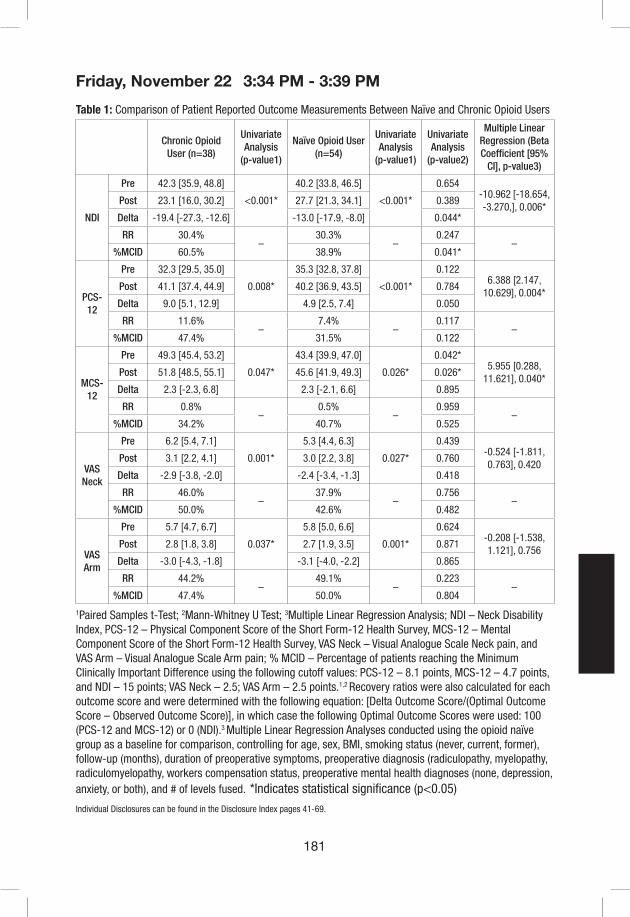
181
Friday, November 22
Individual Disclosures can be found in the Disclosure Index pages 41-69.
Table 1: Comparison of Patient Reported Outcome Measurements Between Naïve and Chronic Opioid Users
Chronic Opioid User (n=38)
Univariate Analysis
(p-value1)
Naïve Opioid User (n=54)
Univariate Analysis
(p-value1)
Univariate Analysis
(p-value2)
Multiple Linear Regression (Beta Coefficient [95%
CI], p-value3)
NDI
Pre 42.3 [35.9, 48.8]
<0.001*
40.2 [33.8, 46.5]
<0.001*
0.654-10.962 [-18.654, -3.270,], 0.006*
Post 23.1 [16.0, 30.2] 27.7 [21.3, 34.1] 0.389
Delta -19.4 [-27.3, -12.6] -13.0 [-17.9, -8.0] 0.044*
RR 30.4%–
30.3%–
0.247–
%MCID 60.5% 38.9% 0.041*
PCS-12
Pre 32.3 [29.5, 35.0]
0.008*
35.3 [32.8, 37.8]
<0.001*
0.1226.388 [2.147,
10.629], 0.004*Post 41.1 [37.4, 44.9] 40.2 [36.9, 43.5] 0.784
Delta 9.0 [5.1, 12.9] 4.9 [2.5, 7.4] 0.050
RR 11.6%–
7.4%–
0.117–
%MCID 47.4% 31.5% 0.122
MCS-12
Pre 49.3 [45.4, 53.2]
0.047*
43.4 [39.9, 47.0]
0.026*
0.042*5.955 [0.288,
11.621], 0.040*Post 51.8 [48.5, 55.1] 45.6 [41.9, 49.3] 0.026*
Delta 2.3 [-2.3, 6.8] 2.3 [-2.1, 6.6] 0.895
RR 0.8%–
0.5%–
0.959–
%MCID 34.2% 40.7% 0.525
VAS Neck
Pre 6.2 [5.4, 7.1]
0.001*
5.3 [4.4, 6.3]
0.027*
0.439-0.524 [-1.811, 0.763], 0.420
Post 3.1 [2.2, 4.1] 3.0 [2.2, 3.8] 0.760
Delta -2.9 [-3.8, -2.0] -2.4 [-3.4, -1.3] 0.418
RR 46.0%–
37.9%–
0.756–
%MCID 50.0% 42.6% 0.482
VAS Arm
Pre 5.7 [4.7, 6.7]
0.037*
5.8 [5.0, 6.6]
0.001*
0.624-0.208 [-1.538, 1.121], 0.756
Post 2.8 [1.8, 3.8] 2.7 [1.9, 3.5] 0.871
Delta -3.0 [-4.3, -1.8] -3.1 [-4.0, -2.2] 0.865
RR 44.2%–
49.1%–
0.223–
%MCID 47.4% 50.0% 0.804
1Paired Samples t-Test; 2Mann-Whitney U Test; 3Multiple Linear Regression Analysis; NDI – Neck Disability Index, PCS-12 – Physical Component Score of the Short Form-12 Health Survey, MCS-12 – Mental Component Score of the Short Form-12 Health Survey, VAS Neck – Visual Analogue Scale Neck pain, and VAS Arm – Visual Analogue Scale Arm pain; % MCID – Percentage of patients reaching the Minimum Clinically Important Difference using the following cutoff values: PCS-12 – 8.1 points, MCS-12 – 4.7 points, and NDI – 15 points; VAS Neck – 2.5; VAS Arm – 2.5 points.1,2 Recovery ratios were also calculated for each outcome score and were determined with the following equation: [Delta Outcome Score/(Optimal Outcome Score – Observed Outcome Score)], in which case the following Optimal Outcome Scores were used: 100 (PCS-12 and MCS-12) or 0 (NDI).3 Multiple Linear Regression Analyses conducted using the opioid naïve group as a baseline for comparison, controlling for age, sex, BMI, smoking status (never, current, former), follow-up (months), duration of preoperative symptoms, preoperative diagnosis (radiculopathy, myelopathy, radiculomyelopathy, workers compensation status, preoperative mental health diagnoses (none, depression, anxiety, or both), and # of levels fused. *Indicates statistical significance (p<0.05)
3:34 PM - 3:39 PM

182
Friday, November 22
The FDA has stated that it is the responsibility of the physician to determine the FDA clearance status of each drug or meidcal device he or she wishes to use in clinical practice.
3:34 PM - 3:39 PM

183
Friday, November 22
Individual Disclosures can be found in the Disclosure Index pages 41-69.
Presentation #53
Can We Predict a Patient’s Arm Pain, Neck Pain, and Disability Level One Year After Cervical Spine Surgery for Radiculopathy?
Ahilan Sivaganesan MD2, Inamullah Khan MD1,2, Hui Nian PhD4, Frank E. Harrell Jr. PhD4, Jacquelyn S. Pennings, PhD1, Mohamad Bydon MD5, Anthony L. Asher MD6, Kristin R. Archer PhD, DPT1,7, Clinton J. Devin MD1,2,3
1Department of Orthopedic Surgery, Vanderbilt University School of Medicine, Nashville, TN2Department of Neurological Surgery, Vanderbilt University School of Medicine, Nashville, TN3Steamboat Orthopaedics and Spine Institute, Steamboat Springs, CO4Department of Biostatistics, Vanderbilt University School of Medicine, Nashville, TN5Department of Neurological Surgery, Mayo Clinic, Rochester, MN6Carolina Neurosurgery & Spine Associates, Carolinas Healthcare System, Charlotte, NC7Department of Physical Medicine & Rehabilitation, Vanderbilt University Medical Center,Nashville, TN
Introduction: With the advent of value-based health care reform, the efficacy of various spine procedures has come under scrutiny. This is no surprise, as variation in patient-reported outcomes (PROs) after spine surgery is considerable. Surgeons are therefore in need of tools which allow them make individualized forecasts of long-term outcomes for their patients. This would allow surgeons to set meaningful expectations, based on which they could make treatment plans with patients through a process of shared decision-making. The aim of this study was to develop and validate one such tool – a set of predictive models for disability, arm pain, and neck pain twelve months after elective cervical spine surgery for radiculopathy.
Materials/Methods: A retrospective analysis was performed using prospectively collected data from the cervical module of a national spine registry – the Quality and Outcomes Database (QOD). All patients undergoing cervical spine surgery for radiculopathy were included in this study. Proportional odds ordinal regression models were developed for the following outcomes: Neck Disability Index (NDI), Numeric Rating Scale for arm pain (NRS-AP), and neck pain (NRS-NP). The following covariates were included: age, gender, BMI, race, education, smoking status, diabetes, anxiety/depression, symptom duration, motor deficit or numbness, spondylolisthesis, employment status, workers’ compensation, insurance, ambulation, baseline PROs, and surgical factors such as number of levels, arthrodesis, and anterior vs. posterior approach. International validation was performed using bootstrap resampling.
Results: A total of 5,076 patients who underwent surgery for cervical radiculopathy were included in this study. Overall, there was a significant improvement in all three 12-month PROs (p<0.001). The most important predictors of outcomes, in descending order, were: baseline PROs, symptom duration, workers’ compensation, age, surgical approach, employment, education, insurance, and spondylolisthesis. Figures 1 and 2 display adjusted effects of each covariate and their relative impact on variation, respectively. The models’ discriminative performance (measured by the overfitting-corrected c-index) were: 0.69 for NDI, 0.67 for neck pain, and 0.65 for arm pain at twelve months.
3:40 PM - 3:45 PM
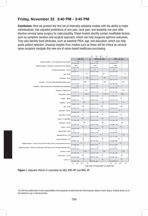
184
Friday, November 22
The FDA has stated that it is the responsibility of the physician to determine the FDA clearance status of each drug or meidcal device he or she wishes to use in clinical practice.
Conclusion: Here we present the first set of internally validated models with the ability to make individualized, risk-adjusted predictions of arm pain, neck pain, and disability one year after elective cervical spine surgery for radiculopathy. These models identify certain modifiable factors, such as symptom duration and surgical approach, which can help surgeons optimize outcomes. They also identify fixed attributes, such as baseline PROs, age, and education, which can help guide patient selection. Drawing insights from models such as these will be critical as cervical spine surgeons navigate this new era of value-based healthcare purchasing.
Figure 1. Adjusted effects of covariates for NDI, NRS-NP and NRS-AP
3:40 PM - 3:45 PM

185
Friday, November 22
Individual Disclosures can be found in the Disclosure Index pages 41-69.
Figure 2. Proportions of overall variation explained by each covariate
3:40 PM - 3:45 PM

186
Friday, November 22
The FDA has stated that it is the responsibility of the physician to determine the FDA clearance status of each drug or meidcal device he or she wishes to use in clinical practice.
Presentation #54
Preliminary Results from the Multi-Center Prospective, Randomized CSM-S Study: Overall Quality of Life Improvement, Complications, and Return to Work
Zoher Ghogawala, MD, Adam Kanter, MD, Praveen Mummaneni, MD, Erica Bisson, MD, James Harrop, MD, Subu N. Magge, MD, Robert Heary, MD, Michael Steinmetz, MD, Michael Fehlings, MD, Todd Albert, MD, Paul Arnold, MD, K. Daniel Riew, MD, Marjorie Wang, MD, Robert G. Whitmore, MD, John Heller, MD, Jill Curran, MS, Edward Benzel, MD
Introduction: The Cervical Spondylotic Myelopathy-Surgical study is a randomized prospective study conducted to compare the effectiveness of ventral versus dorsal (fusion or laminoplasty) surgery for patients with multi-level CSM.
Materials/Methods: A multi-center prospective, randomized clinical trial was conducted on patients aged 45-80 years with multi-level CSM. Patients were screened and enrolled over a 4 year period (2014-2018). Patients were randomized to ventral or dorsal surgery (2:3 randomization) and dorsal surgical approach (dorsal fusion or laminoplasty) was at the discretion of surgeon and patient. Outcome assessments (SF-36, NDI, mJOA, and EQ-5D) along with patient work status were obtained pre-operatively, 3 months, 6 months, and 1 year post-operatively. Complications were assessed by an independent study coordinator.
Results: A total of 15 sites randomized 163 patients. 63 (38.7%) were randomized to ventral surgery and 100 (61.3%) to dorsal. Average age was 62.2 years and 49% were male. Baseline characteristics were comparable between ventral and dorsal groups. After randomization there was a 3% crossover rate. A total of 151 (93%) patients provided 1 year follow-up. Analysis as randomized demonstrated no difference in improvement in SF-36 PCS at 1 year between ventral and dorsal surgery (5.5 Ventral vs. 6.3 Dorsal; P=0.6). We conducted a planned analysis of patients as treated. 66 patients ultimately underwent ventral fusion (VF) and 97 (69 dorsal fusion (DF) and 28 dorsal laminoplasty (DL)) underwent dorsal surgery. Patients, regardless of strategy, demonstrated significant improvements in NDI (14.8 VF, 10.8 DF, 17.5 DL), mJOA (2.2 VF, 2.0 DF, 2.4 DL), and EQ-5D (0.14 VF, 0.12 DF, 0.19 DL) over a one year period post-operatively. DL had superior outcomes in primary outcome SF-36 PCS when compared with VF (P=0.04) and DF (P=0.05) (Figure 1). Patients undergoing VF and DF surgeries experienced a greater number of complications at 1 month post-operatively compared to DL patients (54.5% VF vs. 46.4% DF vs. 14.3% DL; P=0.001). A total of 68 patients (27 VF, 29 DF, 12 DL) were working either part or full-time pre-operatively. A larger percentage of DL patients returned to work at 1 month compared with VF or DF (54.6% DL vs. 29.6% VF vs. 24.1% DF; P=0.17). At 1 year, the proportion of patients who returned to work was similar among the three groups (85.2% VF vs. 75.9% DF vs. 90.9% DL) (Figure 2).
Conclusion: Patients undergoing surgery for CSM demonstrate improved overall quality of life and return to work rates by 1 year follow-up. Dorsal laminoplasty surgery is associated with greater improvements in health-related quality of life and fewer complications.
Figure 1. SF-36 PCS scores pre-operatively and at 3, 6, and 12 months post-operatively for all three surgical groups. (Laminoplasty superior at 1 year, P<0.05)
3:46 PM - 3:51 PM

187
Friday, November 22
Individual Disclosures can be found in the Disclosure Index pages 41-69.
Figure 2. Percentage of patients who return to work at 1, 3, 6, and 12 months following surgery for CSM.
3:46 PM - 3:51 PM

188
Saturday, November 23
The FDA has stated that it is the responsibility of the physician to determine the FDA clearance status of each drug or meidcal device he or she wishes to use in clinical practice.
Presentation #55
If an Anterior Cervical Discectomy has not healed at six months, will it ever heal and when?
Riew, KD, Levi AD, Lavelle WF, Florman JE
Introduction: One of the most common questions from patients who have not healed their ACDF by 6 months is if it will ever heal and when. There are few large-scale prospective studies where the fusion assessment was done by radiologists uninvolved in the care of the patient. We utilized the data from a prospective arthrodesis versus Bryan arthroplasty study to determine the fate of an ACDF that is not fused at six months.
Methods: 213 patients underwent a single-level ACDF and were followed up. Radiographs were analyzed at 6, 12, 24, 48, 60, 84 and 120 months. Two independent radiologists and a 3rd adjudicator determined the fusion status using previously published criteria. To determine the radiographic fate of those who were un-fused at 6 months, we excluded patients without 6-month radiographs as well as those who were solidly fused at six months. We used imaging results at subsequent visits to determine if these patients ever fused and, if so, when the fusion took place.
Results: Of the 213 patients who had undergone an arthrodesis procedure, 53 patients did not have radiographs at six months. Seventy-seven out of the 160 patients were solidly fused, leaving 83 with a nonunion at six months (52%). Three of these nonunion patients had revision operations at or less than six months, leaving 80 patients in our study group. Eleven of these were lost to further follow-up, leaving 69 who had follow-up at 12 months or more. By 12 months post-operatively, 35 (50.7%) out of these 69 patients achieved a solid fusion. Four patients had no radiographs at 12 months but had radiographs later. By 24 months, 52 (75%) of the 69 patients were fused. Of the 34 patients who were not fused at 12 months or had no radiographs to document their fusion status at 12 months, 17 (50 %) were solidly fused, 6 were documented as not having a fusion, 2 had revision surgery and 9 patients had no radiographs. By 84 months, 61 (76%) of the 80 who were not fused at six months ultimately were documented as having a fusion. Two had revision surgery and 17 (11, 2, 3 and 1 after 6, 12, 24 and 48 months, respectively) were lost to follow-up before documentation of their fusion. 76% of patients without a fusion at six months ultimately fused by their final follow-up at 120 months.
Discussion: The majority of the patients who are not fused at six months following single level ACDF will ultimately go on to having a solid fusion by 120 months. For patients who are not fused at six months, it appears that roughly 50 percent will fuse by 12 months. Of the patients who are not fused at 12 months, roughly 50 percent will fuse by 24 months. Patients can be reassured that the majority of patients with a single level ACDF will ultimately fuse even if they have not achieved solid arthrodesis at 6 months postoperatively.
7:11 AM - 7:16 AM

189
Saturday, November 23
Individual Disclosures can be found in the Disclosure Index pages 41-69.
Presentation #56
Epidemiology of iatrogenic vertebral artery injury in cervical spine surgery: 21 multicenter studies
Chang-Hyun Lee,1 Dong Ho Kang,2 Ki-Jeong Kim,3 Sang-Woo Kim,4 Seok Won Kim,5 Young Jin Kim,6 Chun Kee Chung,1,7-10 Jun Jae Shin,11 Jae Keun Oh,12 Seong Yi,13 Jung Kil Lee,14 Jun Ho Lee,15 Ho Jin Lee,16 Hyoung-Joon Chun,17 Dae-Chul Cho,18 Yong Jun Jin,19 Kyung-Chul Choi,20 In Ho Han,21 Seung-Jae Hyun,3 Jung-Woo Hur,22 Jae Taek Hong22 and Geun Sung Song23
Corresponding author: Geun Sung Song and Jae Taek Hong contributed equally as the corresponding authors
Geun Sung Song, M.D., Ph.D. Professor Department of Neurosurgery, Pusan National University Yangsan Hospital, School of Medicine, Pusan National University, Yangsan, The Republic of Korea
Co-corresponding author: Jae Taek Hong, M.D., Ph.D. Professor Department of Neurosurgery, Eunpyeong St. Mary’s Hospital, College of medicine, The Catholic University of Korea, Seoul, The Republic of Korea 1Department of Neurosurgery, Seoul National University Hospital, Seoul National University College of Medicine, Seoul, 2Department of Neurosurgery, Gyeongsang National university School of Medicine, Jinju, 3Department of Neurosurgery, Spine Center, Seoul National University Bundang Hospital, Seoul National University College of Medicine, Seongnam, 4Department of Neurosurgery, Yeungnam University Medical Center, Daegu, 5Department of Neurosurgery, Chosun University Hospital, Gwangju, 6Department of Neurosurgery, Dankook University Hospital, Chonan, 7Neuroscience Research Institute, Seoul National University Medical Research Center, Clinical Research Institute, Seoul National University Hospital, 8Neuroscience Research Institute, Seoul National University Medical Research Center, 9Clinical Research Institute, Seoul National University Hospital, 10Department of Brain and Cognitive Sciences, Seoul National University College of Natural Sciences, Seoul, 11Department of Neurosurgery, Sanggye Paik Hospital, Inje University, Seoul, 12Department of Neurosurgery, Hallym University Sacred Heart Hospital, Anyang, 13Department of Neurosurgery, Yonsei University College of Medicine, Severance Hospital, Seoul, 14Department of Neurosurgery, Chonnam University Hospital, Chonnam University College of Medicine, Gwangju, 15Department of Neurosurgery, Kyung Hee University Medical Centre, Kyung Hee University college of Medicine, Seoul, 16Department of Neurosurgery, Incheon St. Maria Hospital, Incheon, 17Department of Neurosurgery, Hanyang University Seoul Hospital, Seoul, 18Department of Neurosurgery, School of Medicine, Kyungpook National University, Kyungpook National University Hospital, Daegu, 19Department of Neurosurgery, Inje University Seoul Paik Hospital, Seoul, 20Department of Neurosurgery, The Leon Wiltse Memorial Hospital, Anyang, 21Department of Neurosurgery, Pusan National University Hospital, Pusan, 22Department of Neurosurgery, Eunpyeong St.Mary’s Hospital, The Catholic University of Korea, Seoul, 23Department of Neurosurgery, Pusan National University Yangsan Hospital, Yangsan, The Republic of Korea
INTRODUCTION: In cervical spine surgeries (CSS), the overall incidence of iatrogenic vertebral artery injury (IVAI) was reported to be 0.07–1.4%. Although IVAI occurred during C1-2 fusion,
7:17 AM - 7:22 AM

190
Saturday, November 23
The FDA has stated that it is the responsibility of the physician to determine the FDA clearance status of each drug or meidcal device he or she wishes to use in clinical practice.
there is no accurate information regarding the surgery-specific risk of IVAI. This study aimed to stratify the incidence of IVAI by surgical method and evaluate the correlation between IVAI and its sequelae.
MATERIALS/METHODS: We performed a retrospective, multicenter study involving clinical and radiological evaluations for IVAI. We included all CSS between 2012 and 2016, and excluded neck-mass excision and pain intervention. We collected data about patient characteristics and complications, including diagnosis, surgical technique, and the presence of IVAI. In IVAI cases, the technique details, characteristics, and sequelae were investigated.
RESULTS: In total, 14,722 patients, with 15,582 CSS in 21 centers, were included; IVAIs were identified in 13 (0.08%) patients. Surgery-specific incidence of IVAI was 1.35% in cases involving C1–2 posterior fixation and 0.20% in cases involving C3–6 posterior fixation. Common injury mechanisms were screw-in (31%) and high-speed drilling (23%). Screw-related IVAI occurred in nine (69%) patients, and IVAI of the C1 lateral-mass and C2 pedicle screws occurred in four and three patients as shown in Fig. 1, respectively. Among the 13 cases of IVAI, three (23%) involved cerebellar or stem infarction; the infarction had no substantial correlation with injury grade or dominancy in Fig 2.
CONCLUSION: Overall incidence of IVAI in CSS was 0.08%. C1–2 posterior fixation had the highest incidence of IVAI at 1.35%. Although clinical results of IVAI can be highly variable, controlling the risk factors of IVAI is important.
Figure 1. Iatrogenic vertebral artery injury (IVAI) stratified by injury mechanism.
7:17 AM - 7:22 AM
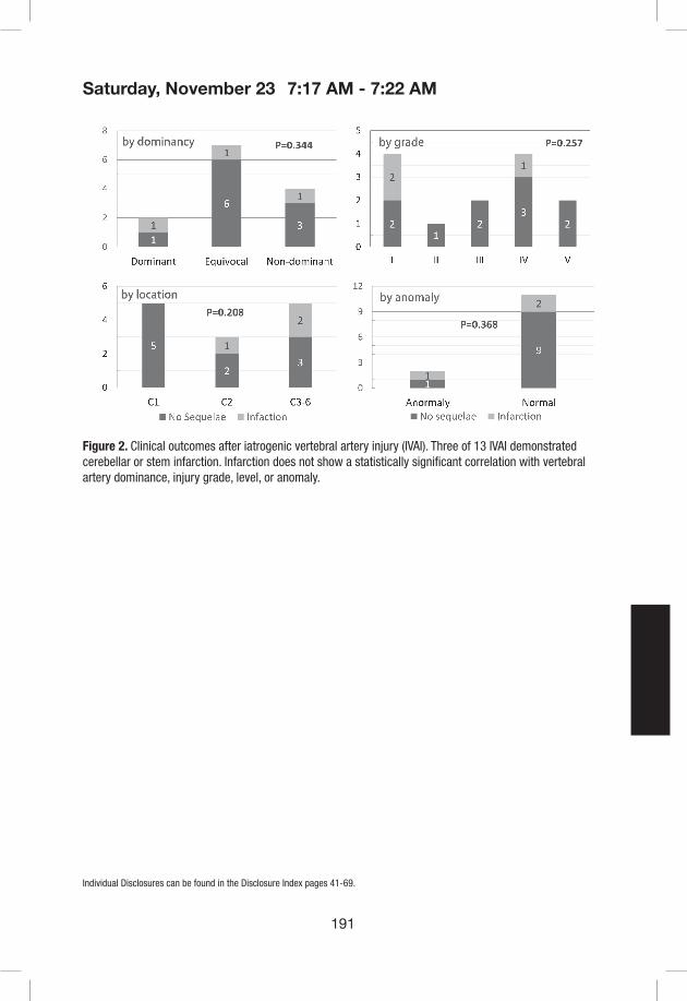
191
Saturday, November 23
Individual Disclosures can be found in the Disclosure Index pages 41-69.
Figure 2. Clinical outcomes after iatrogenic vertebral artery injury (IVAI). Three of 13 IVAI demonstrated cerebellar or stem infarction. Infarction does not show a statistically significant correlation with vertebral artery dominance, injury grade, level, or anomaly.
7:17 AM - 7:22 AM

192
Saturday, November 23
The FDA has stated that it is the responsibility of the physician to determine the FDA clearance status of each drug or meidcal device he or she wishes to use in clinical practice.
Presentation #57
Is Facet Joint Distraction a Cause of Postoperative Axial Neck Pain after ACDF Surgery?
Srikanth N Divi MD, Dhruv KC Goyal BA, John J Mangan MD, Justin D Stull MD, Nathan V Houlihan BS, Matthew S Galetta BA, Jeffrey A Rihn MD, Mark F Kurd MD, D Greg Anderson MD, Alan S Hilibrand MD, Alexander R Vaccaro MD PhD MBA, Christopher K Kepler MD MBA, Gregory D Schroeder MD, Joseph K Lee MD
Rothman Institute, Thomas Jefferson University Hospital, Philadelphia, PA
Introduction: Intervertebral distraction leads to indirect nerve decompression, but has been postulated to distract the degenerative facet joints posteriorly and increase postoperative axial pain and disability following an anterior cervical discectomy and fusion (ACDF). The aim of this study is to determine if there is a correlation between the amount of facet distraction and postoperative patient reported outcomes.
Methods: A retrospective cohort analysis of patients undergoing one to three level ACDF for degenerative pathologies at a high-volume academic center was performed. Each patient received standing, lateral cervical spine x-rays at the immediate postoperative time point and were split into groups based on the amount of inter-facet distance (facet distraction) measured on these films: Group A: < 1.5 mm distraction; Group B: 1.5-2.0 mm distraction; and Group C: > 2.0 mm distraction. For patients with multilevel surgery, the level with the greatest facet distraction was used. Patients reported outcome measures—including NDI, PCS-12, MCS-12, VAS Neck, and VAS Arm pain scores—were obtained preoperatively and at a minimum of 1-year postoperatively. Univariate and multivariate analyses were performed to compare outcomes between groups and receiver operator characteristic (ROC) curves were generated and area under the curves (AUC) were calculated for failure to achieve minimal clinically important difference (MCID) for VAS Neck scores.
Results: A total of 229 patients were included with an average follow-up of 19.8 [19.0, 20.7] months with a mean facet joint distraction of 1.7mm [1.6, 1.8]. Stratifying into groups, there were 87 patients in Group A (mean distraction 1.20mm [1.17, 1.24]), 76 patients in Group B (mean distraction 1.71mm [1.68, 1.74]), and 66 patients in Group C (mean distraction 2.40mm [2.31, 2.50]). Patients significantly improved across all outcome measures from baseline to postoperatively (p < 0.05). There was no difference between groups at any time point with respect to outcome scores (p > 0.05). In addition, the recovery ratio and the % of patients achieving MCID were not significantly different between groups. Multiple regression analysis did not identify increasing distraction as a significant predictor of patient outcomes. ROC curves for each group’s failure to achieve MCID in VAS Neck showed that patients in Group C had the highest area under the curve, indicating that this group was the best predictor of postoperative neck pain (Figure 1).
Conclusions: In this cohort, there were no significant differences between patient outcomes and the amount of facet distraction after ACDF surgery. Multivariate analysis did not find a correlation between facet distraction and overall HRQOL outcome; however, more than 2.0mm of facet distraction was identified as a risk factor for failure to reach the MCID for VAS Neck pain.
7:23 AM - 7:28 AM

193
Saturday, November 23
Individual Disclosures can be found in the Disclosure Index pages 41-69.
Figure 1.
Figure 1 – a) Group A: < 1.5mm, AUC 0.680 [0.566, 0.793]. b) Group B: 1.5mm – 2.0mm, AUC 0.726 [0.609, 0.844], c) Group C: > 2.0mm, AUC 0.740 [0.620, 0.860]
7:23 AM - 7:28 AM
194
Saturday, November 23
The FDA has stated that it is the responsibility of the physician to determine the FDA clearance status of each drug or meidcal device he or she wishes to use in clinical practice.
Presentation #58
Degenerative Cervical Spondylolisthesis: Does Adjacent Level Surgical Stabilization Result In Progressive Listhesis?
Philip K. Louie, MD1; Jannat M. Khan, BS1; Hollis E. Johanson, BS1; Jacob T. Emerson, BS1; Bryce A. Basques, MD1; Michael Nolte, MD1; Dino Samartzis, DSc1; Howard S. An, MD1
1Department of Orthopaedic Surgery, Rush University Medical Center, Chicago, IL
Introduction: Patients with cervical spondylotic myelopathy (CSM) often present with multi-level disease and may experience spondylolisthesis within or adjacent to the levels of clinical pathology. The progression of an unfused degenerative cervical spondylolisthesis (DCS) segment remains unclear when it is not included within the surgical construct. We sought to evaluate if unfused DCS segments, adjacent to the fusion, develop worsening instability that requires surgery.
Method: A retrospective cohort study was performed for twenty-three consecutive patients, who presented with CSM, had radiographs revealing DCS at one or more levels, and underwent surgical intervention. Exclusion criteria included less than 12 months of follow-up, evidence of clinical symptoms present at the DCS level and surgery at adjacent levels. On lateral cervical plain radiographs, anterolisthesis was measured in millimeters (mm). Spondylolisthesis was identified as greater than 2mm of anterolisthesis, and progression was deemed as greater than 0.5mm increase from initial listhesis. Reoperation and patient reported outcomes (PRO) was assessed. Several radiographic parameters were measured pre-operatively and at the last follow-up: amount of listhesis, cervical lordosis, sagittal vertebral axis, and T1 slope at the pathologic level. Baseline patient characteristics were compared using chi-squared analysis and independent sample t-tests for categorical and continuous data, respectively. Bivariate and multivariate regressions were subsequently used to compare radiographic outcomes. Multivariate analysis was used to find independent risk factors for progression of disease.
Results: DCS was present at C2-3 in three cases (13.0%), C3-4 nine cases (39.1%), C4-5 seven cases (30.4%), C5-6 two cases (8.7%), C6-7 no cases (0%), C7-T1 two cases (8.7%). The average pre-operative slip was 2.7mm±0.5mm. At final follow-up, five (21.7%) demonstrated progression (>0.5mm) of their slip, 18 (78%) remained stable (within 0.5mm) or improved. Older age and male sex were associated with progression, but did not increase the risk of reoperation. Overall, PRO scores significantly improved post-operatively when evaluating a disability index as well as neck and arm pain. Two patients (8.7%) with DCS underwent revision surgery, one for symptomatic pseudoarthrosis, and one for myeloradiculopathy due to progression of the adjacent DCS.
Conclusions: Despite the presence of altered stress at the DCS level with the adjacent surgical intervention, the majority of patients did not experience DCS progression at final follow-up nor require further surgical intervention. Given our findings, surgeons may not need to extend a proposed cervical fusion for CSM to include the adjacent asymptomatic DCS level.
7:29 AM - 7:34 AM
195
Saturday, November 23
Individual Disclosures can be found in the Disclosure Index pages 41-69.
Presentation #59
Crossing the cervicothoracic junction during laminectomy and posterior spinal fusion surgery for cervical spondylotic myelopathy is associated with superior cervical radiographic outcomes1Andrew K. Chan MD, 1Ryan Badiee BS, 1Joshua Rivera, 2,3Chih-Chang Chang MD, 1Leslie C. Robinson MD, PharmD, MBA, 1Ratnesh Mehra DO, 1Lee Tan MD, 1Aaron J. Clark MD, PhD, 1Sanjay S. Dhall MD, 1Dean Chou MD, 1Praveen V. Mummaneni MD.1Department of Neurological Surgery, University of California, San Francisco, CA2Department of Neurosurgery, Neurological Institute, Taipei Veterans General Hospital, Taipei.3School of Medicine, National Yang-Ming University, Taipei.
INTRODUCTION: In laminectomy and posterior spinal fusion (LPSF) surgery for cervical spondylotic myelopathy (CSM), the evidence is unclear as to whether the lower instrumented vertebrae (LIV) should cross the cervicothoracic junction (CTJ). This study analyzes cervical radiographic parameters following LPSF for CSM and compares outcomes between those with and without LIV crossing the CTJ.
MATERIALS/METHODS: A consecutive series of adults undergoing LPSF for CSM from 2012 through 2018 with a minimum of 12-month follow up were identified. LPSF with sub-axial upper instrumented vertebrae and LIV between C6 and T2 were included. Clinical and radiographic outcomes were compared. Multivariate models were constructed to assess the influence of crossing the CTJ on cervical radiographic parameters, correcting for factors reaching p<0.20 on univariate analyses.
RESULTS: 79 patients were included: 46 (58.2%) who crossed the CTJ (crossed-CTJ; LIV T1-2) and 33 (41.8%) who did not cross the CTJ (not-crossed CTJ; LIV C6-7). Crossed-CTJ trended toward older age than non-crossed CTJ (63.7±10.8 vs. 59.1±11.4,p=0.08). There were a similar proportion of males (not-crossed CTJ 54.6% vs. crossed-CTJ 56.5%,p=0.99). Mean follow-up was 21.0 months (minimum 12 months). Baseline VAS neck pain, VAS arm pain, Nurick, mJOA, NDI, and EQ-5D did not differ significantly (p>0.05). Crossed-CTJ was associated with higher blood loss (414.7±364.3 vs. 201.5±107.9 ml,p<0.001), longer operative times (213.2±48.9 vs. 166.0±34.1 min,p<0.001), but similar hospital stays (5.2±3.9 vs. 4.0±1.8 days,p=0.08) and discharge dispositions [67.4% (crossed-CTJ) vs.69.7% to home,p=0.99]. The reoperation rate was 5.1% overall and did not differ significantly between crossed-CTJ and not-crossed CTJ (2.2% vs. 9.1%, respectively,p=0.39). For not-crossed CTJ, there were 3 reoperations (9.1%): 2 irrigation and debridements for surgical site infection and 1 case requiring a spinal cord stimulator for failed neck surgery syndrome. There were no cases of pseudarthrosis or hardware misplacement/failure requiring reoperation for not-crossed CTJ. For crossed CTJ, there was 1 reoperation (2.2%) involving a C3-T1 LPSF complicated by pseudarthrosis requiring an anterior cervical discectomy and fusion. Postoperative VAS neck pain, VAS arm pain, mJOA, and Nurick scores were similar (p>0.05).
Crossed-CTJ had a higher mean preoperative C2-7 sagittal vertical axis (SVA) (33.3±16.0 vs. 23.8±12.4 mm,p=0.01), but similar preoperative cervical lordosis (CL) and C1 minus T1-slope
7:43 AM - 7:48 AM
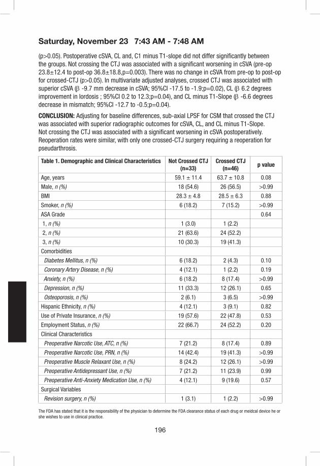
196
Saturday, November 23
The FDA has stated that it is the responsibility of the physician to determine the FDA clearance status of each drug or meidcal device he or she wishes to use in clinical practice.
(p>0.05). Postoperative cSVA, CL and, C1 minus T1-slope did not differ significantly between the groups. Not crossing the CTJ was associated with a significant worsening in cSVA (pre-op 23.8±12.4 to post-op 36.8±18.8,p=0.003). There was no change in cSVA from pre-op to post-op for crossed-CTJ (p>0.05). In multivariate adjusted analyses, crossed CTJ was associated with superior cSVA (β -9.7 mm decrease in cSVA; 95%CI -17.5 to -1.9;p=0.02), CL (β 6.2 degrees improvement in lordosis ; 95%CI 0.2 to 12.3;p=0.04), and CL minus T1-Slope (β -6.6 degrees decrease in mismatch; 95%CI -12.7 to -0.5;p=0.04).
CONCLUSION: Adjusting for baseline differences, sub-axial LPSF for CSM that crossed the CTJ was associated with superior radiographic outcomes for cSVA, CL, and CL minus T1-Slope. Not crossing the CTJ was associated with a significant worsening in cSVA postoperatively. Reoperation rates were similar, with only one crossed-CTJ surgery requiring a reoperation for pseudarthrosis.
Table 1. Demographic and Clinical Characteristics Not Crossed CTJ(n=33)
Crossed CTJ(n=46)
p value
Age, years 59.1 ± 11.4 63.7 ± 10.8 0.08
Male, n (%) 18 (54.6) 26 (56.5) >0.99
BMI 28.3 ± 4.8 28.5 ± 6.3 0.88
Smoker, n (%) 6 (18.2) 7 (15.2) >0.99
ASA Grade 0.64
1, n (%) 1 (3.0) 1 (2.2)
2, n (%) 21 (63.6) 24 (52.2)
3, n (%) 10 (30.3) 19 (41.3)
Comorbidities
Diabetes Mellitus, n (%) 6 (18.2) 2 (4.3) 0.10
Coronary Artery Disease, n (%) 4 (12.1) 1 (2.2) 0.19
Anxiety, n (%) 6 (18.2) 8 (17.4) >0.99
Depression, n (%) 11 (33.3) 12 (26.1) 0.65
Osteoporosis, n (%) 2 (6.1) 3 (6.5) >0.99
Hispanic Ethnicity, n (%) 4 (12.1) 3 (9.1) 0.82
Use of Private Insurance, n (%) 19 (57.6) 22 (47.8) 0.53
Employment Status, n (%) 22 (66.7) 24 (52.2) 0.20
Clinical Characteristics
Preoperative Narcotic Use, ATC, n (%) 7 (21.2) 8 (17.4) 0.89
Preoperative Narcotic Use, PRN, n (%) 14 (42.4) 19 (41.3) >0.99
Preoperative Muscle Relaxant Use, n (%) 8 (24.2) 12 (26.1) >0.99
Preoperative Antidepressant Use, n (%) 7 (21.2) 11 (23.9) 0.99
Preoperative Anti-Anxiety Medication Use, n (%) 4 (12.1) 9 (19.6) 0.57
Surgical Variables
Revision surgery, n (%) 1 (3.1) 1 (2.2) >0.99
7:43 AM - 7:48 AM

197
Saturday, November 23
Individual Disclosures can be found in the Disclosure Index pages 41-69.
Table 2. Radiographic Variables Not Crossed CTJ(n=33)
Crossed CTJ(n=46)
p value
C6-7 central stenosis, baseline, n (%) 26 (78.8) 34 (73.9) 0.62
C7-T1 central stenosis, baseline, n (%) 4 (12.1) 20 (43.5) 0.003**
C6-7 foraminal stenosis, baseline, n (%) 22 (66.6) 32 (69.6) 0.78
C7-T1 foraminal stenosis, baseline, n (%) 2 (6.1) 17 (37.0) 0.002**
cSVA, baseline, mm 23.8 ± 12.4 33.3 ± 16.0 0.01**
CL, baseline, degrees 8.7 ± 11.1 9.7 ± 11.7 0.71
T1-Slope, baseline, degrees 27.4 ± 7.7 28.1 ± 8.8 0.74
CL minus T1-Slope, baseline, degrees 19.5 ± 10.2 18.2 ± 9.9 0.60
SVA, baseline, mm 39.3 ± 27.2 38.4 ± 40.3 0.92
PT, baseline, degrees 19.8 ± 9.0 20.7 ± 11.3 0.75
PI minus LL, baseline, degrees 11.8 ± 8.1 12.5 ± 10.3 0.78
cSVA, postoperative, mm 36.8 ± 18.8 37.2 ± 17.1 0.92
CL, postoperative, degrees 5.0 ± 11.6 8.0 ± 9.9 0.25
T1-Slope, postoperative, degrees 27.9 ± 9.6 30.3 ± 8.8 0.28
CL minus T1-Slope, postoperative, degrees 24.0 ± 11.7 21.8 ± 10.5 0.39
SVA, postoperative, mm 36.5 ± 36.1 37.8 ± 45.3 0.91
PT, postoperative, degrees 22.9 ± 6.8 21.5 ± 9.4 0.54
PI minus LL, postoperative, degrees 13.2 ± 10.6 16.5 ± 17.6 0.40
7:43 AM - 7:48 AM

198
Saturday, November 23
The FDA has stated that it is the responsibility of the physician to determine the FDA clearance status of each drug or meidcal device he or she wishes to use in clinical practice.
Presentation #60
Assessing Radiographic Fusion Rates Following A Stand-Alone Interbody Cage Versus An Anterior Plate Construct For Adjacent Segment Disease After Anterior Cervical Discectomy And Fusion
Sapan D. Gandhi, MD; Adam Fahs, MD; Steven Wahlmeier, MD; Philip Louie, MD; Daniel Possley, DO; Jad Khalil, MD; Kevin C. Baker, PhD; Daniel K. Park, MD
Introduction: Anterior cervical discectomy and fusion (ACDF) is considered the gold standard surgical intervention for myelopathy or radiculopathy secondary cervical spondylosis. Although ACDF has excellent outcomes, symptomatic adjacent segment disease (ASD) is a common occurrence, and can often times require surgical intervention. Stand-alone cages provide the potential benefits of lower profile hardware and shorter operative time, as previous constructs at adjacent levels do not need to be removed. However, the rate of fusion when stand-alone interbody cages are used for ASD is previously unreported.
Methods: After IRB approval, a retrospective case control study by two surgeons were conducted (n = 86). The case group consisted patients who a 1-level ACDF for ASD (n=46) subdivided into those with a stand-alone interbody cage (Anchor-C, Stryker, Kalamazoo, MI, n=29) and anterior plate construct (n=19). The control group consisted of 40 consecutive patients undergoing primary 1-level ACDF with a plate and screw construct. Fusion status was determined at a minimum of 9 months post-operatively on lateral flexion extension plain film radiographs or computed tomography (CT). Fusion on plain-films was defined as flowing bone in the interbody space, with less than 2mm of motion between spinous process between the flexion and extension views, as described by Cannada et al. The senior surgeons (DKP and JK) prefer to obtain cervical spine CT scans for all patients at 1 year post-operatively to assess fusion status, but patients did not have to undergo CT scan if they were doing well clinically and radiographically. Revision surgery for pseudarthrosis was also reviewed and recorded. The stand-alone group and plate and screw construct group were compared using Fischer Exact Test.
Results: 86 patients were retrospectively reviewed. Patients undergoing primary ACDF had lower age and ASA score as well as shorter operative time. Fusion rate was higher for primary ACDF compared to all patients who underwent ACDF for adjacent segment disease (95% vs 74%, p=0.009). When compared to primary ACDF, patients with a stand-alone cage construct had significantly lower fusion rate and (69% vs 95%, p=0.006) and higher re-operation rate (14% vs 0%, p=0.02). There were no significant differences in fusion rate between those undergoing primary 1-level ACDF, versus 1-level ACDF for ASD with anterior plate construct (95% vs 82.3%, p=0.153). There were no significant differences in anterior plate construct versus stand-alone cage construct for ASD in terms of fusion and re-operation (82% vs 69%, p=0.489)
Conclusions: In our series of patients, stand-alone cage for ASD after ACDF achieved fusion at a lower rate and more often required revision surgery for pseudarthrosis compared to our control series of primary, 1-level ACDF. We found no differences in stand-alone cage and anterior plate construct in the ASD setting, although our study may be underpowered to detect a difference. Future studies should focus on a larger, more tightly controlled series, as well as examining the biomechanical properties of stand-alone cage for ASD.
7:49 AM - 7:54 AM
199
Saturday, November 23
Individual Disclosures can be found in the Disclosure Index pages 41-69.
Presentation #61
Does Association Exist between Cervical Sagittal Parameters and Clinical Outcomes after Anterior Cervical Discectomy and Fusion?
Bryce A. Basques, MD1; Jannat M. Khan, BS1; Michael Nolte, MD1; Philip K. Louie, MD1; Kamran Movassaghi, MD1; Jonathan Markowitz, BS1; Victor Lei, BS1; Edward J Goldberg, MD1; Howard S An, MD1
1Rush University Medical Center, Department of Orthopaedic Surgery, Chicago, IL
Introduction: Sagittal radiographic measurements of the spine have received significant attention in recent years, and in the lumbar spine, the relationship between sagittal balance, spinopelvic parameters, and clinical outcomes is well-established. While there has also been increasing attention to sagittal parameters in the cervical spine, the relationship between these radiographic measurements and clinical outcomes is less clear. The aim of the present study was to characterize independent associations between cervical spine radiographic parameters and clinical outcomes following anterior cervical discectomy and fusion (ACDF).
Materials and Methods: We performed a retrospective cohort analysis of patients who underwent a single-level or multi-level ACDF for cervical radiculopathy or myelopathy. Patients were excluded from analysis if they were under 18 years of age at the time of surgery, had postoperative follow up less than 6 months or had an ACDF for cervical spine fracture or infection. Radiographic assessments included: C2-C7 lordosis, T1 angle, levels fused, sagittal vertical axis (SVA), fusion mass lordosis, proximal and distal adjacent segment lordosis, and adjacent segment degeneration (ASD). Patient reported outcomes were obtained in the form of Neck Disability Index (NDI) scores and Visual Analog Scales (VAS) scores for the neck and arm. Baseline patient characteristics were compared using chi-squared analysis and independent sample t-tests for categorical and continuous data, respectively. Bivariate and multivariate regressions were subsequently used to compare clinical outcomes between procedure groups. Multivariate analyses controlled for differences in baseline patient characteristics.
Results: A total of 381 patients met inclusion criteria. Average follow-up length was 28 months, average age was 50.2 years, average body mass index was 28.7, and 49.2% of patients were female. Of these patients, 93.7% achieved successful fusion, 6.6% had graft subsidence, and 4.99% had a re-operation. Preoperative and postoperative radiographic measurements and patient-reported outcomes were collected. On multivariate analysis, preoperative radiographic measurements poorly predicted clinical outcomes. Increased preoperative lordosis at the adjacent unfused segment proximal to the eventual fusion mass was found to be associated with increased final NDI (corrected p=0.018), and increased final proximal lordosis was associated with increased final NDI (corrected p=0.049, Table 1). However, no other radiographic parameters were associated with any patient-reported clinical outcomes. Subsidence rates were increased with greater change in proximal lordosis from preoperative to postoperative (OR 1.30, corrected p=0.007) and from preoperative to final follow-up (OR 1.17, corrected p=0.044). Subsidence was negatively associated with increased change in lordosis across the fused segments from preoperative to postoperative (OR 0.88, corrected p=0.032) and from preoperative to final follow-up (OR 0.86, corrected p=0.017, Table 2). No other radiographic parameters were associated
7:55 AM - 8:00 AM

200
Saturday, November 23
The FDA has stated that it is the responsibility of the physician to determine the FDA clearance status of each drug or meidcal device he or she wishes to use in clinical practice.
with subsidence, and no parameters were associated with reoperation or pseudoarthrosis.
Conclusions: The present study found that sagittal parameters were poorly predictive of clinical outcomes. Increased preoperative lordosis at the adjacent segment proximal to the fusion mass was associated with worse final NDI scores, and was the only preoperative radiographic parameter associated with clinical outcomes. Further research should attempt to identify other preoperative factors or radiographic parameters that are more closely associated with clinical outcomes.
Table 1. Measurements and NDI Final
Measurement Outcome Beta p-value Adjusted p-value*
Proximal Lordosis final NDI Final 0.78 0.001 0.018
Proximal Lordosis pre NDI Final 0.77 0.003 0.049
Table 2. Radiographic measurements and Subsidence
Measurement Outcome Odds Ratio p-value Adjusted p-value*
Proximal Lordosis Change Preop-Postop Subsidence 1.3 0.0004 0.007
Fusion Lordosis Change Preop-Final Subsidence 0.86 0.001 0.017
Fusion Lordosis Change Preop-Postop Subsidence 0.88 0.002 0.032
Proximal Lordosis Change Preop-Final Subsidence 1.17 0.003 0.044
7:55 AM - 8:00 AM
203
Saturday, November 23
Individual Disclosures can be found in the Disclosure Index pages 41-69.
Presentation #63
Microendoscopic laminotomy versus conventional laminoplasty for cervical spondylotic myelopathy - A 5-year follow-up study
Akihito Minamide, MD, PhD1; Andrew K. Simpson, MD, MHS2; Yukihiro Nakagawa, MD, PhD1; Motohiro Okada, MD, PhD1; Hiroshi Iwasaki, MD, PhD1; Shunji Tsutsui, MD, PhD1; Masanari Takami, MD, PhD1; Ryo Taiji, MD1; Shizumasa Murata, MD1; Takuhei Kozaki, MD1; Hiroshi Hashizume, MD, PhD1; Yasutsugu Yukawa, MD, PhD1; Munehito Yoshida, MD, PhD1; Hiroshi Yamada, MD, PhD1
1Department of Orthopaedic Surgery, Wakayama Medical University, Wakayama, Japan2Microendoscopic Spine Institute and Brigham and Women’s Hospital, Harvard Medical School, Boston, MA
Background: The cervical spondylotic myelopathy (CSM) consists of a pincer mechanism due to a bulging disc and a hypertrophied ligamentum flavum. The concept of cervical microendoscopic laminotomy (CMEL) is the relief of pincer mechanism due to remove the dorsal compressive elements of the articular segment. CMEL is a minimally invasive spine surgery using endoscpe, and may provide some benefit with regard to maintaining lordosis and decreasing axial symptoms. The goal of this study was to characterize the long-term clinical and radiological results of articular segmental decompression surgery using endoscopy (CMEL) for CSM and to compare outcomes to conventional expansive laminoplasty (ELAP).
Methods: Consecutive patients with CSM who required surgical treatment were enrolled. All enrolled patients (n = 78) underwent CMEL or ELAP. All patients were followed postoperatively for more than 5 years. The preoperative and 5-year follow-up evaluations included neurological assessment (Japanese Orthopaedic Association [JOA] score), JOA recovery rates, axial neck pain (using a visual analog scale), the SF-36, and cervical sagittal alignment (C2–7 subaxial cervical angle).
Results: Sixty-one patients were included for analysis, 31 in the CMEL group and 30 in the ELAP group. The mean preoperative JOA score was 10.1 points in the CMEL group and 10.9 points in the ELAP group (p > 0.05). The JOA recovery rates were similar, 57.6% in the CMEL group and 55.4% in the ELAP group (p > 0.05). The axial neck pain in the CMEL group was significantly lower than that in the ELAP group (p < 0.01). At the 5-year follow-up, cervical alignment was more favorable in the CMEL group, with an average 2.6° gain in lordosis (versus 1.2° loss of lordosis in the ELAP group [p < 0.05]) and lower incidence of postoperative kyphosis.
Conclusions: CMEL is a novel, less invasive technique that allows for multilevel posterior cervical decompression for the treatment of CSM. This 5-year follow-up data demonstrates that after undergoing CMEL, patients have similar neurological outcomes to conventional laminoplasty, with significantly less postoperative axial pain and improved subaxial cervical lordosis when compared with their traditional ELAP counterparts.
8:15 AM - 8:20 AM

204
Saturday, November 23
The FDA has stated that it is the responsibility of the physician to determine the FDA clearance status of each drug or meidcal device he or she wishes to use in clinical practice.
Presentation #64
Effect of C3 laminectomy in cervical laminoplasty for degenerative cervical myelopathy: C3 laminectomy vs. C3 laminoplasty
Koji Nakajima, MD; Toru Doi, MD, PhD; So Kato, MD; Sakae Tanaka, MD, PhD; Yasushi Oshima, MD, PhD
The University of Tokyo, Orthopaedic Surgery
Introduction: Interlaminar bony fusion sometimes occurs after cervical laminoplasty (LP), especially at C2–3 and may cause loss of cervical range of motion (ROM). In recent years, C3 laminectomy (LN) has been performed to prevent C2–3 interlaminar fusion and can improve postoperative cervical ROM. However, a positive effect of C3 LN in surgical outcomes, in terms of the Japanese Orthopaedic Association (JOA) score or Short-Form 12 (SF-12) score, has not been reported. This study aimed to evaluate the surgical outcomes after C3 LN and C3 LP for degenerative cervical myelopathy patients.
Materials/Methods: Patients who underwent cervical double-door LP in our hospital between 2012 and 2017 were enrolled in this study. The inclusion criteria were as follows: (1) patients with 1–2-year follow-up, 2) having undergone primary surgery for cervical spine, and 3) experienced myelopathy caused by spondylosis or ossification of posterior longitudinal ligament. We collected data regarding backgrounds of the patients, operative factors, radiographic measurement, and preoperative/postoperative outcomes (JOA score, SF-12 Physical Component Summary [PCS], SF-12 Mental Component Summary [MCS], and neck pain assessed using a numerical rating scale [NRS]).
Results: Forty-seven patients who underwent LP fulfilled the inclusion criteria, including 23 patients with C3 LN and 24 patients with C3 LP. There were no significant differences between the background characteristics of the two groups. The operative time and estimated blood loss were significantly lower in the C3 LN group than in the C3 LP group (130.0 min vs. 155.2 min, p < 0.01; 64.4 g vs. 265.4 g, p < 0.01). C2–3 interlaminar bony fusion was observed only in the C3 LP group (C3 LP 64% vs. C3 LN 0%, p < 0.01); however, ΔC2–7 ROM (Δ = postoperative – preoperative) was not significantly different between the two groups (p = 0.57). The recovery rate of the JOA score (JOA-RR) and ΔSF-12 PCS of the C3 LN group were superior to those of the C3 LP group (p = 0.04 and p = 0.03 respectively). The two groups did not significantly differ in terms of ΔSF-12 MCS and Δneck NRS.
Conclusions: Our study revealed that JOA-RR and ΔSF-12 PCS were better in the C3 LN group than in the C3 LP group of patients undergoing cervical LP.
8:21 AM - 8:26 AM

205
Saturday, November 23
Individual Disclosures can be found in the Disclosure Index pages 41-69.
Table 1. Patients backgrounds and operative factors
C3 Laminectomy (N=23) C3 Laminoplasty (N=24) p Value
Age 65.6±13.9 68.3±11.6 0.47
Sex (M/F) 14 / 9 20 / 4 0.09
BMI 26.3±4.2 24.8±4.3 0.23
Diseases (CSM/OPLL) 12 / 11 15 / 9 0.47
Diabetes mellitus 8 (35%) 10 (42%) 0.63
Operative time (min.) 130.0±25.0 155.2±33.1 <0.01
Estimated blood loss (g) 64.4±55.7 265.4±213.0 <0.01
CSM; Cervical Spondylotic Myelopathy, OPLL; Ossification of Posterior Longitudinal Ligament
Table 2. Radiographic and clinical outcomes
C3 Laminectomy (N=22) C3 Laminoplasty (N=18) p Value
Preop C2-7 angle 13.6±10.7 7.8±9.0 0.08
Preop C2-7 ROM 31.7±13.8 27.8±8.6 0.29
Preop T1 slope (CT) 23.6±12.8 19.2±9.9 0.24
Postop C2-7 angle 9.3±11.3 5.9±8.1 0.30
Postop C2-7 ROM 27.7±12.2 21.5±10.5 0.10
ΔC2-7 angle -4.3±6.7 -1.9±8.9 0.33
ΔC2-7 ROM -4.1±13.1 -6.3±10.8 0.57
C2-3 interlaminar bony fusion 0 (0.0%) 7 (39%) <0.01
Preop JOA score* 11.0±2.4 10.7±2.1 0.72
Postop JOA score* 13.8±2.5 12.2±2.4 0.08
JOA recovery rate* 49.8±32.5 26.3±24.8 0.04
ΔSF-12 PCS** 9.4±12.7 -1.2±12.8 0.03
ΔSF-12 MCS** -0.7±8.7 2.0±10.5 0.45
ΔNeck pain (NRS)*** 0.3±3.2 1.4±2.7 0.27
* C3 Laminectomy N=17, C3 Laminoplasty N=13** C3 Laminectomy N=14, C3 Laminoplasty N=17*** C3 Laminectomy N=17, C3 Laminoplasty N=20
8:21 AM - 8:26 AM

206
Saturday, November 23
The FDA has stated that it is the responsibility of the physician to determine the FDA clearance status of each drug or meidcal device he or she wishes to use in clinical practice.
Presentation #65
Minimally Invasive Posterior Cervical Foraminotomy as an Alternative to Anterior Cervical Discectomy and Fusion for Unilateral Cervical Radiculopathy
Nikhil Sahai, M.D.1, Stuart Changoor, M.D.1, Conor Dunn, M.D.1, Michael Faloon, M.D.1, Kumar Sinha, M.D.1, Ki Soo Hwang, M.D.1, Arash Emami, M.D.1
1St. Joseph’s University Medical Center, Paterson, NJ
Introduction: Unilateral cervical radiculopathy refractory to conservative treatment is one of the most common pathologies that spine surgeons treat surgically. Anterior cervical discectomy and fusion (ACDF) has long been considered standard treatment for cervical radiculopathy secondary to lateral disc herniation or foraminal stenosis. Recent studies suggest minimally-invasive posterior cervical foraminotomy (MI-PCF) to be an alternative to ACDF, but not without concern for reoperation and the ability to achieve similar improvement in patient outcomes. Therefore, the purpose of this study was to compare patient outcomes, reoperation rate and complication rate of MI-PCF to those of ACDF by analyzing the current literature.
Methods: 3 electronic databases were queried using terms related to MI-PCF. MINORS scoring was used to assess study quality. A total of 14 studies were included in this analysis. Analyses for heterogeneity and publication bias were performed. Clinical outcome scores (NDI, VAS-neck and VAS-arm), reoperation proportion, and complications were assessed. Each outcome measure was compared to those of ACDF from two previously published studies. A random-effects model of meta-analysis was used for heterogenous groups, and a fixed-effects model was used for groups that were not. Overlap of 95% confidence intervals suggests no significant difference at the p<0.05 level.
Results: 14 studies were included, with outcome data of 1216 patients. The mean age of the study population was 51.57 years, who were 61.8% male and had a mean follow-up period of 30 months. The mean improvement in NDI was 20.30 (95% CI, 18.81-21.79) and 16.85 (95% CI, 14.96-19.10) for the MI-PCF and ACDF groups, respectively. The mean improvement in VAS-neck was 4.16 (95% CI, 2.70-5.61) and 2.47 (95% CI, 2.09-2.84) for the MI-PCF and ACDF groups, respectively. The mean improvement in VAS-arm was 5.31 (95% CI, 4.50-6.12) and 2.27 (95% CI, 1.82-2.70) for the MI-PCF and ACDF groups, respectively. 6% (95% CI, 3%-12%) of patients who underwent MI-PCF required a re-operation compared to 3.9% (95% CI, 2.77%-5.46%) of those who underwent ACDF. Complications were noted in 4% (95% CI, 3%-7%) of MI-PCF patients and 7.79% (95% CI, 5.54%-10.85%) of ACDF patients. The most common complications were transient neuropraxia, wound-related, and durotomy.
Conclusion: MI-PCF resulted in a greater improvement in VAS-arm scores compared to ACDF, supporting its effectiveness as a procedure for lateral pathology. There were no other significant differences in patient reported clinical outcome scores, proportion of patients requiring re-operation, or complications between patients who underwent MI-PCF and those who underwent ACDF. Thus, MI-PCF may be utilized as a safe and effective alternative to ACDF in patients with unilateral cervical radiculopathy without myelopathy, without concern for increased reoperations or complications, and with advantages of preservation of cervical motion and quicker recovery.
8:27 AM - 8:32 AM
207
Saturday, November 23
Individual Disclosures can be found in the Disclosure Index pages 41-69.
Presentation #66
Optimizing Cervicothoracic Junction Biomechanics after C7 Pedicle Subtraction Osteotomy: a Cadaveric Study of Stability and Rod Strain.
Jakub Godzik, MD1, Jennifer Lehrman, MS2, Bernardo de Andrada Pereira, MD2, Christopher Ames, MD3, Heiko Koller, MD4, Kevin Lee, MS5 , Anna Newcomb, MS2, Jay Turner, MD, PhD1, Brian P. Kelly, PhD2
1Department of Neurosurgery Barrow Neurological Institute St. Joseph’s Hospital and Medical Center Phoenix, Arizona2Spinal Biomechanics Laboratory Barrow Neurological Institute St. Joseph’s Hospital and Medical Center Phoenix, Arizona3Department of Neurological Surgery University of California San Francisco, California4Department of Neurosurgery Technical University of Munich Munich, Germany5DePuy Synthes Spine Inc. Raynham, Massachusetts
Introduction: Cervical pedicle subtraction osteotomy (PSO) is a powerful corrective technique for severe cervical deformity that requires a sound biomechanical construct to avoid instrumentation failure and rod fracture. However, little evidence exists regarding optimal construct design in regards to maximizing stability and minimizing posterior rod strain. The current biomechanical study aimed to compare range of motion stability (ROM) and rod strain in uniform, tapered, and accessory rod constructs in a human cadaveric C7 PSO model.
Materials and Methods: Fourteen human cadaveric (C2-T4) specimens were prepared and potted at C2 and T4. Specimens were tested intact and divided into two statistically equivalent groups. Specimens were instrumented with pars screws at C2, cervical lateral mass screws at C3-C6, pedicle screws at T1-T3, and a 25° PSO performed at C7. The first specimen group was instrumented with 3.5-5.5mm titanium tapered rods (TR). The second group received uniform titanium 4.0mm rods (UR). The UR group was additionally tested with lateral accessory rods (C5-T2) (UR+AR) (Figure 1). All conditions were tested with pure moments of 2.0Nm in flexion (F), extension (E), left-right lateral bending (LLB, RLB), left-right axial rotation (LAR, RAR), and a compressive load (C) of 70N. Intervertebral ROM, posterior (pRS), and lateral (lRS) rod strains were measured at C2-C3, T2-T3, and PSO level. Statistical comparisons were performed using one-way ANOVA and paired Holm-Sidak tests (p<0.05).
Results: TR demonstrated significantly reduced mean ROM compared to UR in RAR at the PSO level (0.25° versus 0.35°, p=0.041) and in LAR at the T2-T3 level (0.18° versus 0.38°, p=0.023). No other differences were found in global or segmental ROM (p>0.08). At the PSO level, pRS was significantly greater using TR compared to UR+AR in F, E, and RAR (p<0.018) (Figure 2). lRS was highest at the PSO level with significant differences between TR and UR+AR, in LAR and RAR (p<0.027).
Conclusion: The C7 PSO is a highly destabilizing condition. When fixation is performed from C2 to T3, maximal rod strain concentrated across the PSO fixation site. Differences in stability between TR and UR were small. UR+AR provided the greatest reduction of pRS and lRS.
8:33 AM - 8:38 AM

208
Saturday, November 23
The FDA has stated that it is the responsibility of the physician to determine the FDA clearance status of each drug or meidcal device he or she wishes to use in clinical practice.
Figure 1. Schematic of rod constructs tested in-vitro.
Figure 2. Posterior rod strains recorded at C2-C3 and PSO levels during flexion and extension.
8:33 AM - 8:38 AM

209
Saturday, November 23
Individual Disclosures can be found in the Disclosure Index pages 41-69.
Presentation #67
A Deep Learning Model For Detection of Cervical Spinal Cord Compression in MRI Scans
Zamir Merali, M.D1, Jetan Badhiwala, M.D1, Christopher D. Witiw, M.D, MSc., FRCSC1,2, Jefferson Wilson, M.D, PhD., FRCSC2, Michael G. Fehlings, M.D, PhD., FRCSC3
Division of Neurosurgery, University of Toronto, Toronto, Ontario, CanadaDivision of Neurosurgery, St. Michael’s Hospital, Toronto, Ontario, CanadaDivision of Neurosurgery, Toronto Western Hospital, Toronto, Ontario, Canada
Introduction: Magnetic Resonance Imaging(MRI) evidence of spinal cord compression plays a central role in the diagnosis of degenerative cervical myelopathy(DCM)[1]. There is growing recognition that deep learning models may assist in addressing the increasing volume of medical imaging data and provide initial interpretation of images gathered in a primary-care setting[2]. We aimed to develop and validate a deep learning model for detection of cervical spinal cord compression in MRI scans. We applied this model to predict DCM disease severity.
Materials and Methods: Patients undergoing surgery for DCM as a part of the AOSpine CSM-NA or CSM-I trials were included in our study[3,4]. Healthy non-myelopathic patients from a separate study were also included[5]. The axial T2-weighted(aT2w) sequence for each patient was anonymized and extracted for model training. Patient demographics, acquisition parameters, and the modified Japanese Orthopedic Association(mJOA) score was collected. We defined two patient cohorts: 1)The ‘training cohort’ was used to train the deep learning model, 2)The ‘testing cohort’ consisted of patients from different geographic regions and was used for model testing and external validation.
Two specialist physicians independently reviewed the training cohort and identified axial images showing partial or circumferential spinal cord compression(Figure 1). Spinal cord compression was defined as any indentation, flattening, torsion, or circumferential compression of the spinal cord parenchyma from extrinsic tissues[6]. Inter-rater reliability was assessed with the Cohen’s kappa coefficient(κ). A deep convolutional neural network(CNN) model using the ResNet50 architecture with ImageNet weights was trained using images from the training cohort with TensorFlow(v1.12) and a gradient-descent optimization algorithm[7,8].
We assessed external validity by comparing the deep learning model’s prediction for each image in the testing cohort with labels assigned by a human labeler. We determined model accuracy for each patient and presented summary statistics stratified by patient characteristics and MRI scanner type (Table 1). We used the output of the deep learning model to predict patient disease severity with a logistic regression model and presented classification accuracy and area-under-the-curve(AUC).
Results: The training cohort included 110 patients with 5635 images and the testing cohort included 219 patients. Two human labelers independently labeled the training cohort with κ = 0.82.
After training for 75 epochs the deep learning model achieved 93% accuracy at detecting spinal cord compression in individual aT2w images in the training cohort. The deep learning model achieved a median predictive accuracy on patients in the testing cohort of 80%(Table 1). The
8:47 AM - 8:52 AM
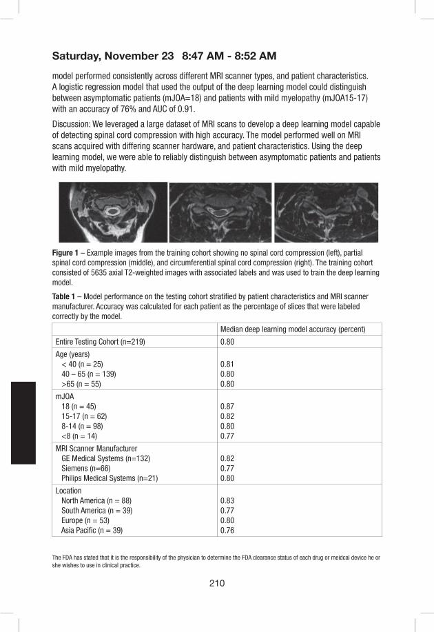
210
Saturday, November 23
The FDA has stated that it is the responsibility of the physician to determine the FDA clearance status of each drug or meidcal device he or she wishes to use in clinical practice.
model performed consistently across different MRI scanner types, and patient characteristics. A logistic regression model that used the output of the deep learning model could distinguish between asymptomatic patients (mJOA=18) and patients with mild myelopathy (mJOA15-17) with an accuracy of 76% and AUC of 0.91.
Discussion: We leveraged a large dataset of MRI scans to develop a deep learning model capable of detecting spinal cord compression with high accuracy. The model performed well on MRI scans acquired with differing scanner hardware, and patient characteristics. Using the deep learning model, we were able to reliably distinguish between asymptomatic patients and patients with mild myelopathy.
Figure 1 – Example images from the training cohort showing no spinal cord compression (left), partial spinal cord compression (middle), and circumferential spinal cord compression (right). The training cohort consisted of 5635 axial T2-weighted images with associated labels and was used to train the deep learning model.
Table 1 – Model performance on the testing cohort stratified by patient characteristics and MRI scanner manufacturer. Accuracy was calculated for each patient as the percentage of slices that were labeled correctly by the model.
Median deep learning model accuracy (percent)
Entire Testing Cohort (n=219) 0.80
Age (years) < 40 (n = 25) 40 – 65 (n = 139) >65 (n = 55)
0.810.800.80
mJOA 18 (n = 45) 15-17 (n = 62) 8-14 (n = 98) <8 (n = 14)
0.870.820.800.77
MRI Scanner Manufacturer GE Medical Systems (n=132) Siemens (n=66) Philips Medical Systems (n=21)
0.820.770.80
Location North America (n = 88) South America (n = 39) Europe (n = 53) Asia Pacific (n = 39)
0.830.770.800.76
8:47 AM - 8:52 AM
211
Saturday, November 23
Individual Disclosures can be found in the Disclosure Index pages 41-69.
References:1. Martin AR, Tadokoro N, Tetreault L, Arocho-Quinones E V, Budde MD, Kurpad SN, et al: Imaging
Evaluation of Degenerative Cervical Myelopathy: Current State of the Art and Future Directions. Neurosurg Clin N Am 29:33–45, 2018
2. Wang S, Summers RM. Machine learning and radiology. Med Image Anal 2012; 16(5):933-9513. Fehlings MG, Kopjar B, Arnold PM, Yoon SW, Vaccaro AR, Shaffrey CI, et al. The AOSpine North
America Cervical Spondylotic Myelopathy Study: 2-Year Surgical Outcomes of a Prospective Multicenter Study in 280 Patients922. Neurosurgery 2010;67:543.
4. Fehlings MG, Wilson JR, Kopjar B, Yoon ST, Arnold PM, Massicotte EM, et al. Efficacy and safety of surgical decompression in patients with cervical spondylotic myelopathy: results of the AOSpine North America prospective multi-center study. J Bone Joint Surg Am 2013;95:1651–8. doi:10.2106/JBJS.L.00589.
5. Martin AR, De Leener B, Cohen-Adad J, Can microstructural MRI detect subclinical tissue injury in subjects with asymptomatic cervical spinal cord compression? A prospective cohort study. BMJ Open 2018;13;8(4)
6. Harrop JS, Naroji S, Maltenfort M, Cervical myelopathy: a clinical and radiographic evaluation and correlation to cervical spondylotic myelopathy. Spine. 2010;35(6):620-624
7. Ding Y, Sohn JH, Kawczynski MG, et al. A deep learning mdoel to predict a diagnosis of Alzheimer disease by using 18F-FDG PET of the brain. Radiology. 2019;290(2):456-464
8. Kaiming He, Xiangyu Z, Shaoquing R, Jian S. Deep residual learning for image recognition. arXiv:1512.03385
8:47 AM - 8:52 AM

215
Saturday, November 23
Individual Disclosures can be found in the Disclosure Index pages 41-69.
Presentation #69
A new index for making decisions regarding C2 lamina decompression in cervical ossification of the posterior longitudinal ligament: The R-line
Byung-Jou Lee, MDa, Seong Kyun Jeong, MDb, Myeongjong Kim, MDb, Subum Lee, MDb, Jin Hoon Park, MD, PhDb
Corresponding authors: Jin Hoon Park, MD, PhD Department of Neurological Surgery, Asan Medical Center University of Ulsan College of Medicine, Songpa-gu, Seoul 05505, South Korea a Department of neurosurgery, Inje University Ilsan Paik Hospital, Neuroscience & Radiosurgery Hybrid Research Center, College of Medicine, 170, Juwharo,Ilsan Seogu, Goyang ,Gyeonggi 10380, Republic of Koreab Department of Neurosurgery, Asan Medical Center, University of Ulsan College of Medicine, Songpa-gu, Seoul 05505, South Korea
Background: Determining the degree of C2 lamina decompression in OPLL extending to the C2 level is difficult.
To analyze the association between radiographic factors and postoperative C2 cord shifting and evaluate the usefulness of the R-line (rostral line) as a new index for determining the degree of C2 lamina decompression in ossification of the posterior longitudinal ligament (OPLL) extending to the C2 level.
Methods: We established the R-line to determine the degree of C2 lamina decompression in relation to factors associated with C2 cord shifting. We divided 36 consecutive patients into the incomplete and complete decompression groups and compared the correspondence between the degree of C2 lamina decompression using the R-line and actual degree of decompression in each group.
Results: The maximal degree of cord compression by OPLL and C2–3 local segment lordotic angle correlated with postoperative C2 cord shifting. The R-line was defined as the line that moves backward by normal cord diameter at the posterior edge of the OPLL of maximal compressed cord level by OPLL and parallel to the line passing through the center of the C2–3 vertebral body. In all patients in the incomplete decompression group, the actual degree of decompression was insufficient compared with the degree of C2 lamina decompression using the R-line.
Conclusions: If the R-line touches the upper half of the posterior C2 lamina, total decompression of C2 lamina should be performed. The R-line is a practical tool for making decisions regarding C2 lamina decompression in OPLL extending to the C2 level.
Key words: C2, laminectomy, ossification of the posterior longitudinal ligament, posterior decompression, R-line
Running title: R-line for determining degree of C2 decompression
8:59 AM - 9:04 AM
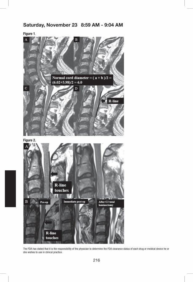
216
Saturday, November 23
The FDA has stated that it is the responsibility of the physician to determine the FDA clearance status of each drug or meidcal device he or she wishes to use in clinical practice.
Figure 1.
Figure 2.
8:59 AM - 9:04 AM
217
Saturday, November 23
Individual Disclosures can be found in the Disclosure Index pages 41-69.
Fig. 1 Definition of the R-line. A: Normal cord diameter, 6.0 mm. B: Moves backward by 6.0 mm. C: Line passing through the center of the C2–3 vertebral body. D: R-line, parallel line to the line passing through the center of the C2–3 vertebral body.
Fig. 2 Deciding the degree of C2 lamina decompression. A: R-line does not touch the upper half of the posterior C2 lamina. Postoperative MRI shows complete decompression after undercutting of the C2 lamina. B: R-line touches the upper half of the posterior C2 lamina. Immediate postoperative MRI shows incomplete decompression and patient had complete paralysis after undercutting of the C2 lamina. Therefore, emergency reoperation (C2 total laminectomy) is performed, and MRI after reoperation shows complete decompression. However, it has also been shown that syrinx or myelomalacia is produced by nerve injury caused by cord compression
8:59 AM - 9:04 AM
218
Saturday, November 23
The FDA has stated that it is the responsibility of the physician to determine the FDA clearance status of each drug or meidcal device he or she wishes to use in clinical practice.
Presentation #70
Can C7 Slope Be Used As A Substitute for T1 Slope? A Radiographic Analysis
Zoe Cheung, Jun Kim, Ivan Ye, Ray Tang, Samuel White, Samuel Cho
Introduction: Sagittal imbalance in the cervical spine is a major cause of neck pain, headache, fatigue, and disability. While parameters such as C2-C7 lordosis and C2-C7 sagittal vertical axis have been extensively studied, they do not fully characterize cervical sagittal balance. T1 is an important new parameter of both cervical as well as global spinal sagittal balance. However, the T1 superior endplate can be difficult to visualize on standard lateral radiographs due to overlying anatomical structures. C7 slope has therefore been proposed as a potential substitute for T1 slope when the T1 superior endplate is not well visualized. The objectives of this study were: (1) to assess the correlation between C7 slope and T1 slope on upright lateral cervical spine radiographs, and (2) to evaluate the inter-rater reliability of C7 slope.
Material/Methods: Cervical spine radiographs taken between December 2017 and June 2018 at a single institution were reviewed. Only radiographs with visible C7 superior and inferior endplates, and T1 superior endplate were included. Radiographs with cervical instrumentation were excluded. Two independent observers measured upper C7 slope, lower C7 slope, and T1 slope. The correlations between upper C7 slope and T1 slope, as well as between lower C7 slope and T1 slope were evaluated. Linear regression analyses were also performed. Inter-rater reliability of C7 slope as assessed.
Results: A total of 650 radiographs were reviewed. The superior endplate of C7, inferior endplate of C7, and superior endplate of T1 were visible in 72.9%, 50.2%, and 31.2% of these radiographs, respectively. After applying our exclusion criteria, 152 patients remained and were included in our analysis. The average age was 48.1 years, with 70.4% females. The average upper C7 slope, lower C7 slope, and T1 slope was 23.5°±9.1°, 22.9°±9.0°, and 27.5°±8.7°, respectively. There was a strong correlation between upper C7 slope and T1 slope (r=0.91, p<0.001), as well as between lower C7 slope and T1 slope (r=0.90, p<0.001). Linear regression analyses showed that T1 slope could be estimated from C7 slope based on the equation, T1 slope = 0.87 x C7 slope + 7, with an overall model fit of R2=0.8. There was strong inter-rater reliability for upper (ICC=0.95, p<0.001) and lower (ICC=0.96, p<0.001) C7 slope.
Conclusion: Both the upper and lower C7 slope are strongly correlated with T1 slope and can be used as a substitute to estimate T1 slope when the superior endplate of T1 is not well visualized.
9:05 AM - 9:10 AM
219
Saturday, November 23
Individual Disclosures can be found in the Disclosure Index pages 41-69.
Presentation #71
Effect of Opioid-Limiting Legislation on Postoperative Prescription Patterns Following Anterior Cervical Decompression and Fusion
Daniel B.C. Reid, MD, MPH1; Kalpit N. Shah, MD1; Benjamin H. Shapiro, MS2; Jack H. Ruddell, BA2; Edward Akelman, MD1; Mark Palumbo, MD1; Alan H. Daniels, MD1
Corresponding Author: Daniel B.C. Reid, MD, MPH Department of Orthopaedic Surgery Warren Alpert Medical School of Brown University 2 Dudley Street Providence, RI 1: Department of Orthopaedics, Warren Alpert Medical School of Brown University2: Warren Alpert Medical School of Brown University
Institutional Review Board Approval was obtained at all participating institutions for collection and analysis of patient data.
Key Words: opioid; law; legislation; trauma; hip; tibia; distal radius; femur; ankle; pilon
Introduction: Since 2016, at least 28 U.S. states have enacted legislation setting mandatory dose limitations on prescribers of opioids. The impact of such laws following anterior cervical discectomy and fusion (ACDF) remains unknown. This study evaluated post-operative opioid prescription patterns in a population of patients undergoing elective ACDF before and after implementation of a statewide opioid-limiting law.
Materials and Methods: Patient medical records in conjunction with state prescription drug monitoring (PDMP) data were utilized to collect demographic, medical, surgical, clinical, and pharmacological data. Eligible patients included those undergoing primary elective 1-3 level ACDF procedures. Patients undergoing ACDF for trauma were excluded. Patients requiring corpectomy, undergoing other simultaneous procedures, or undergoing ACDF as part of a staged procedures were excluded. 211 patients (101 pre-law, 110 post-law) were evaluated. Pre-law and post-law patients were compared in terms of all demographic, medical, surgical factors. Postoperative opioid utilization in terms of total morphine milligram equivalents (MMEs) was compared between groups at initial, 1-month, 2-month, 3-month, and 4-month postoperative timepoints. Secondary analysis compared cohorts again after stratification by preoperative opioid use. Additionally, outcomes including 30 and 90-day rates for emergency department (ED) visits, readmissions, and reoperations were evaluated. Multiple linear regression was performed to determine factors independently associated with increased 30-day postoperative opioid use. Predictors of chronic (>90day) opioid utilization were evaluated with multiple logistic regression.
Results: Pre-law and post-law patient groups were similar terms of age, sex, surgeon specialty, insurance type, ASA score, preoperative opioid and benzodiazepine use, number of levels fused, site of initial presentation, and discharge disposition (all p>0.05). Post-law, ACDF patients were prescribed significantly less opioids in their first postoperative prescription (26.65 vs. 62.08 pills, p<.001; 202.23 vs. 549.18 MMEs, p<.001). This decrease in opioid utilization was observed for the first 30 postoperative days (cumulative 30-day MMEs 444.14 vs. 877.87, p<.001). When stratified by opioid-tolerance, both opioid-naïve (363.54 vs. 632.20 MMEs, p<.001) and opioid-tolerant (730.08 vs. 1,122.90 MMEs, p=0.022) patients filled less total 30-day MMEs in the post-law cohort. After controlling for confounders, factors independently associated with increased
10:34 AM - 10:39 AM

220
Saturday, November 23
The FDA has stated that it is the responsibility of the physician to determine the FDA clearance status of each drug or meidcal device he or she wishes to use in clinical practice.
30-day opioid utilization included pre-law status, preoperative opioid exposure, preoperative benzodiazepine exposure, and number of levels fused (all p<.05). A non-significant trend toward increased 30-day utilization in patients with Medicaid insurance or worker’s compensation was noted. There was no association between pre/post-law status and chronic (>90 day) opioid requirements. There were no differences between pre- and post-law patients in terms of 30 or 90-day ED visits, readmissions, or reoperations (all p>0.05).
Conclusion: Implementation of mandatory opioid limits effectively decreased 30-day postoperative opioid utilization following ACDF without any associated increase in ED visits or unplanned readmissions for pain. While no effect on chronic opioid utilization was noted, the substantial decline in early 30-day opioid requirements seen (nearly 50%) in this population may decrease the risk of opioid diversion and improve public health.
10:34 AM - 10:39 AM

221
Saturday, November 23
Individual Disclosures can be found in the Disclosure Index pages 41-69.
Table 1. Population Characteristics
VariablePre-law Patients
(n=101)Post-law Patients
(n=110)All Patients
(N=211) p-value
Age, mean (95% CI) 51.4 (49.6-53.2) 52.6 (50.5-54.6) 52.0 (50.6-53.4) 0.377
Sex (Female), n (%) 58 (57.4%) 55 (50.5%) 113 (53.8%) 0.312
Surgeon Specialty
Orthopaedic Surgery 53 (52.5%) 61 (55.5%) 114 (54.0%) 0.664
Neurosurgery 48 (47.5%) 49 (44.6%) 97 (46.0%)
Insurance, n (%)
Private 62 (61.4%) 68 (61.8%) 130 (61.6%) 0.27
Medicare 16 (15.8%) 18 (16.4%) 34 (16.1%)
Medicaid 6 (5.9%) 13 (11.8%) 19 (9.0%)
Worker’s Compensation 17 (16.8%) 11 (10.0%) 28 (13.3%)
Incarcerated 1 (1.0%) 0 (0.0%) 1 (0.5%)
ASA Score
ASA 1 5 (5.0%) 5 (4.6%) 10 (4.7%) 0.113
ASA 2 62 (61.4%) 82 (74.6%) 144 (68.3%)
ASA 3 34 (33.7%) 22 (20.0%) 56 (26.5%)
ASA 4 0 (0.0%) 1 (0.9%) 1 (0.5%)
Preoperative Opioid Use, n (%)
30d Preop Period 38 (37.6%) 33 (30.0%) 71 (33.7%) 0.242
90d Preop Period 51 (50.5%) 47 (42.7%) 98 (46.5%) 0.258
Preoperative Benzodiazepine Use n(%)
30d Preop Period 19 (18.8%) 16 (14.6%) 35 (16.6%) 0.405
90d Preop Period 27 (26.7%) 22 (20.0%) 49 (23.2%) 0.247
Number of Levels Fused
1 Level 56 (55.5%) 61 (55.5%) 117 (55.5%) 0.718
2 Levels 41 (40.6%) 42 (38.2%) 83 (39.3%)
3 Levels 4 (4.0%) 7 (6.4%) 11 (5.2%)
Presentation
Clinic 98 (97.0%) 108 (98.2%) 206 (97.6%) 0.583
Emergency Department 3 (2.97%) 2 (1.82%) 5 (2.37%)
Discharged Disposition
Home 100 (99.0%) 108 (98.2%) 208 (98.6%) 0.612
Skilled Nursing Facility 1 (1.0%) 2 (1.8%) 3 (1.4%)
First Postoperative Opioid Prescription
oxycodone 100 (91.9%) 100 (90.9%) 181 (85.2%) 0.135
hydrocodone 11 (10.9%) 5 (4.6%) 16 (7.6%)
hydromorphone 1 (1.0%) 0 (0.0%) 1 (0.5%)
none 8 (7.9%) 5 (4.6%) 13 (6.2%)
10:34 AM - 10:39 AM
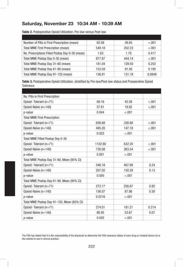
222
Saturday, November 23
The FDA has stated that it is the responsibility of the physician to determine the FDA clearance status of each drug or meidcal device he or she wishes to use in clinical practice.
Table 2. Postoperative Opioid Utilization, Pre-law versus Post-law
Variable Pre-law (n=101) Post-law (n=110) p value
Number of Pills in First Prescription (mean) 62.08 26.65 <.001
Total MME First Prescription (mean) 549.18 202.23 <.001
No. Prescriptions Filled Postop Day 0-30 (mean) 1.63 1.70 0.417
Total MME Postop Day 0-30 (mean) 877.87 444.14 <.001
Total MME Postop Day 31-60 (mean) 181.04 129.50 0.252
Total MME Postop Day 61-90 (mean) 153.59 91.93 0.195
Total MME Postop Day 91-120 (mean) 136.81 131.18 0.0848
Table 3. Postoperative Opioid Utilization, stratified by Pre-law/Post-law status and Preoperative Opioid Tolerance
Variable Pre-law (n=101) Post-law (n=110) p value
No. Pills in First Prescription
Opioid- Tolerant (n=71) 69.16 42.58 <.001
Opioid-Naïve (n=140) 57.81 19.82 <.001
p-value 0.044 <.001
Total MME First Prescription
Opioid- Tolerant (n=71) 638.68 330.68 <.001
Opioid-Naïve (n=140) 495.20 147.18 <.001
p-value 0.022 <.001
Total MME Filled Postop Day 0-30
Opioid- Tolerant (n=71) 1122.90 632.20 <.001
Opioid-Naïve (n=140) 730.08 363.54 <.001
p-value 0.001 <.001
Total MME Postop Day 31-60, Mean (95% CI)
Opioid- Tolerant (n=71) 546.18 407.88 0.24
Opioid-Naïve (n=140) 207.02 150.29 0.13
p-value 0.005 <.001
Total MME Postop Day 61-90, Mean (95% CI)
Opioid- Tolerant (n=71) 272.17 226.67 0.82
Opioid-Naïve (n=140) 126.07 87.86 0.30
p-value 0.0216 <.001
Total MME Postop Day 91-120, Mean (95% CI)
Opioid- Tolerant (n=71) 274.01 181.21 0.214
Opioid-Naïve (n=140) 80.95 53.67 0.07
p-value 0.002 <.001
10:34 AM - 10:39 AM

223
Saturday, November 23
Individual Disclosures can be found in the Disclosure Index pages 41-69.
Table 4a. Multiple Linear Regression; Variables Associated with Total 30-Day Postoperative Opioid Dosing
Variables CoefficientRobust Std. Err. t p-value
95% Confidence interval
Post-Law -394.72 71.48 -5.52 <.001 -535.66 -253.78
Preoperative Opioid Exposure (30d) 311.32 81.05 3.84 <.001 151.51 471.13
Preoperative Benzodiazepine Exposure (30d) 335.45 109.91 3.05 0.003 118.73 552.18
Orthopaedic Surgeon (vs. Neurosurgeon) -167.85 67.14 -2.5 0.013 -300.24 -35.46
Number of Levels Fused 113.54 54.33 2.09 0.038 6.41 220.66
Worker’s Compensation (vs. Private Insurance) 134.81 79.72 1.69 0.092 -22.38 292.00
Medicaid (vs. Private Insurance) 274.12 143.36 1.91 0.057 -8.55 556.80
Constant 578.13 98.79 5.85 <.001 383.32 772.93
*Backwards stepwise linear regression with robust standard error performed. Other non-significant variables analyzed included age, sex, medicare insurance, ASA score, presentation site, and discharge disposition
Table 4b. Multiple Logistic Regression; Independent Predictors of Chronic Opioid Utilization
Variables Odds RatioRobust Std. Err. z p-value
95% Confidence interval
Orthopaedic Surgeon (vs. Neurosurgeon) 0.42 0.17 -2.19 0.029 0.20 0.91
Preoperative Opioid Exposure (30d) 4.42 1.76 3.73 <.001 2.02 9.63
Preoperative Benzodiazepine Exposure (30d) 2.12 0.96 1.65 0.098 0.87 5.16
Constant 0.20 0.05 -6.11 <.001 0.12 0.33
*Backwards stepwise logistic regression with robust standard error performed. Other non-significant variables analyzed included pre-low/post-law time period, number of levels fused, insurance type, age, sex, ASA score, presentation site, and discharge disposition
Table 5. Outcome Variables
Outcome VariablesPre-law (n=101)
Post-law (n=110)
All Patients (N=211) p-value
ED Visit Within 30 Days of Discharge, n (%) 8 (7.9%) 9 (8.2%) 17 (8.1%) 0.945
ED Visit Within 90 Days of Discharge, n (%) 10 (9.9%) 10 (9.1%) 20 (9.5%) 0.841
Unplanned Readmission Within 30 Days of Discharge, n (%) 4 (4.0%) 5 (4.6%) 9 (4.3%) 0.934
Unplanned Readmission Within 90 Days of Discharge, n (%) 4 (4.0%) 6 (5.5%) 10 (4.7%) 0.610
Reoperation Within 30 Days of Discharge, n (%) 0 (0.0%) 1 (0.9%) 1 (0.5%) 0.337
Reoperation Within 90 Days of Discharge, n (%) 1 (1.0%) 1 (0.9%) 2 (1.0%) 0.952
10:34 AM - 10:39 AM
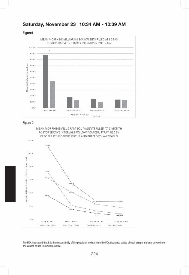
224
Saturday, November 23
The FDA has stated that it is the responsibility of the physician to determine the FDA clearance status of each drug or meidcal device he or she wishes to use in clinical practice.
Figure1
Figure 2
10:34 AM - 10:39 AM

225
Saturday, November 23
Individual Disclosures can be found in the Disclosure Index pages 41-69.
Presentation #72
Perioperative Spending in Single-Level Anterior Cervical Discectomy and Fusion for Degenerative Pathology
Majd Marrache, 1 Andrew B. Harris, 1 Varun Puvanesarajah, 1 Micheal Raad, 1 Amit Jain1
1Department of Orthopaedic Surgery, The Johns Hopkins University, Baltimore, MD
Introduction: Anterior cervical discectomy and fusion (ACDF) is a commonly performed cervical spine procedure. The gain in popularity of bundled payments for surgical procedures has driven the need for spine surgeons to identify the drivers of spending and trends of resource utilization during the perioperative period. The purpose of this study was to identify the distribution of spending by private payers in the 1-year period surrounding single-level ACDF for degenerative pathology.
Methods: A private insurance claims database was retrospectively queried to identify patients that underwent single-level ACDF from 2011 to 2015. Patients were identified using CPT codes for single level ACDF and International Classification of Diseases, Version 9 (ICD- 9CM) and ICD-10CM codes for cervical disc pathologies. Gross payments for health services were identified from 6 months pre- to 6 months post-operatively. Categories of perioperative spending included: outpatient health services (Office visits, Injections, tests performed, medical supplies and devices and Emergency Department visits), prescription pain medications, and admission to inpatient facilities. All dollar values were adjusted to the 2016 Consumer Pricing Index (CPI), and are expressed as median (interquartile range (IQR)). Significance was set at p<0.05.
Results: In our cohort containing 32,454 patients (mean age was 50 years old, 54% were female), the index hospital admission accounted for the majority of overall spending ($1.1 Billion, 74% overall). Preoperative spending accounted for 12% ($173 Million) and postoperative spending accounted for 14% ($207 Million). The median (IQR) perioperative spending, 6 months prior and 6 months following surgery, was $42,662 ($32,093 – 58,195). Imaging (35%), office visits (17%) and ED visits (12%) were the highest contributors to preoperative spending with a median spending of $110 ($43- 222), $102 ($73 – 141) and $57 ($21- 163) per patient. Median spending on hospital admission for surgery was $29,920 ($22,725 - $40,208) per patient. Postoperatively, admissions to inpatient facilities accounted for 42% of postoperative spending and 6% of total perioperative spending. Imaging (12%) and medical supplies (10%) were the second and third highest contributors of postoperative spending, respectively. Only 64 (0.2%) of patients were discharged to a rehabilitation facility following surgery. Total postoperative spending for patients readmitted following surgery was significantly higher than those who were not ($24,544 vs. $1,002, p<0.001). Being discharged to a rehabilitation facility had significantly higher postoperative spending ($19,658) compared to patients discharged home ($1,121), home with home care ($2,305), to another facility ($4,535), p<0.001
Conclusion: While surgery accounted for the greatest costs, other sources of cost before and after ACDF surgery are not trivial. This data helps provide insight into potential targets for cost reduction
10:40 AM - 10:45 AM

226
Saturday, November 23
The FDA has stated that it is the responsibility of the physician to determine the FDA clearance status of each drug or meidcal device he or she wishes to use in clinical practice.
Figure 1. Proportion of spending on outpatient services, prescription pain medications, hospital admission and readmission for 32,454 patients, 6 months prior and 6 months following surgery.
10:40 AM - 10:45 AM
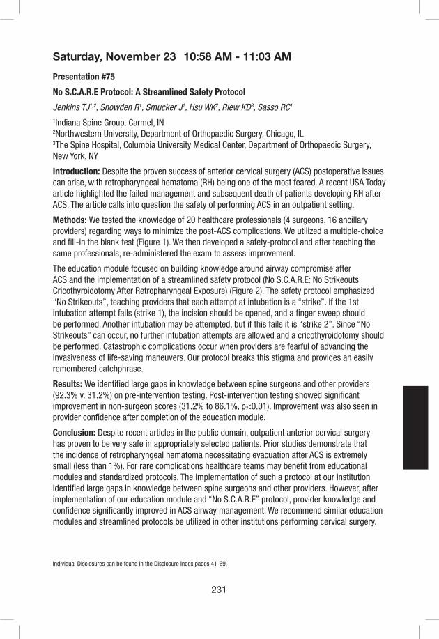
231
Saturday, November 23
Individual Disclosures can be found in the Disclosure Index pages 41-69.
Presentation #75
No S.C.A.R.E Protocol: A Streamlined Safety Protocol
Jenkins TJ1,2, Snowden R1, Smucker J1, Hsu WK2, Riew KD3, Sasso RC1
1Indiana Spine Group. Carmel, IN2Northwestern University, Department of Orthopaedic Surgery, Chicago, IL3The Spine Hospital, Columbia University Medical Center, Department of Orthopaedic Surgery, New York, NY
Introduction: Despite the proven success of anterior cervical surgery (ACS) postoperative issues can arise, with retropharyngeal hematoma (RH) being one of the most feared. A recent USA Today article highlighted the failed management and subsequent death of patients developing RH after ACS. The article calls into question the safety of performing ACS in an outpatient setting.
Methods: We tested the knowledge of 20 healthcare professionals (4 surgeons, 16 ancillary providers) regarding ways to minimize the post-ACS complications. We utilized a multiple-choice and fill-in the blank test (Figure 1). We then developed a safety-protocol and after teaching the same professionals, re-administered the exam to assess improvement.
The education module focused on building knowledge around airway compromise after ACS and the implementation of a streamlined safety protocol (No S.C.A.R.E: No Strikeouts Cricothyroidotomy After Retropharyngeal Exposure) (Figure 2). The safety protocol emphasized “No Strikeouts”, teaching providers that each attempt at intubation is a “strike”. If the 1st intubation attempt fails (strike 1), the incision should be opened, and a finger sweep should be performed. Another intubation may be attempted, but if this fails it is “strike 2”. Since “No Strikeouts” can occur, no further intubation attempts are allowed and a cricothyroidotomy should be performed. Catastrophic complications occur when providers are fearful of advancing the invasiveness of life-saving maneuvers. Our protocol breaks this stigma and provides an easily remembered catchphrase.
Results: We identified large gaps in knowledge between spine surgeons and other providers (92.3% v. 31.2%) on pre-intervention testing. Post-intervention testing showed significant improvement in non-surgeon scores (31.2% to 86.1%, p<0.01). Improvement was also seen in provider confidence after completion of the education module.
Conclusion: Despite recent articles in the public domain, outpatient anterior cervical surgery has proven to be very safe in appropriately selected patients. Prior studies demonstrate that the incidence of retropharyngeal hematoma necessitating evacuation after ACS is extremely small (less than 1%). For rare complications healthcare teams may benefit from educational modules and standardized protocols. The implementation of such a protocol at our institution identified large gaps in knowledge between spine surgeons and other providers. However, after implementation of our education module and “No S.C.A.R.E” protocol, provider knowledge and confidence significantly improved in ACS airway management. We recommend similar education modules and streamlined protocols be utilized in other institutions performing cervical surgery.
10:58 AM - 11:03 AM
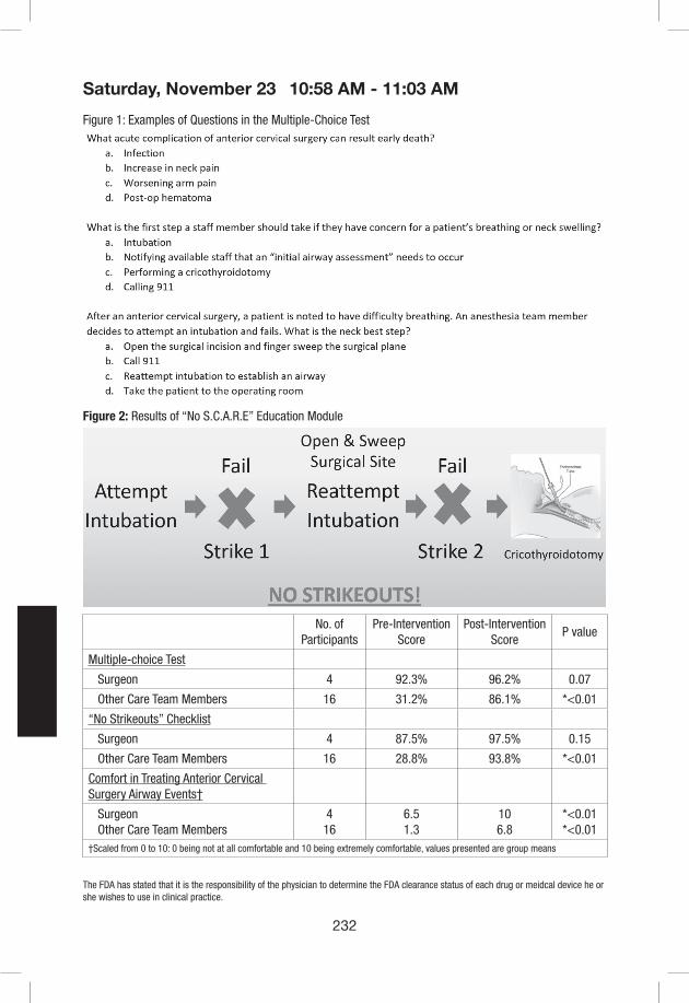
232
Saturday, November 23
The FDA has stated that it is the responsibility of the physician to determine the FDA clearance status of each drug or meidcal device he or she wishes to use in clinical practice.
Figure 1: Examples of Questions in the Multiple-Choice Test
Figure 2: Results of “No S.C.A.R.E” Education Module
No. of Participants
Pre-InterventionScore
Post-InterventionScore
P value
Multiple-choice Test
Surgeon 4 92.3% 96.2% 0.07
Other Care Team Members 16 31.2% 86.1% *<0.01
“No Strikeouts” Checklist
Surgeon 4 87.5% 97.5% 0.15
Other Care Team Members 16 28.8% 93.8% *<0.01
Comfort in Treating Anterior Cervical Surgery Airway Events†
Surgeon Other Care Team Members
416
6.51.3
106.8
*<0.01*<0.01
†Scaled from 0 to 10: 0 being not at all comfortable and 10 being extremely comfortable, values presented are group means
10:58 AM - 11:03 AM

233
Saturday, November 23
Individual Disclosures can be found in the Disclosure Index pages 41-69.
Presentation #76
Anterior cord compression is associated with neurologic deficit in patients with degenerative cervical myelopathy. Does it have evidence?
Kyung-Chung Kang M.D., Jung-Hee Lee M.D., Ki-Young Lee M.D., Sang-Kyu Im M.D. and Hyoungmin Kim1
Reprints (Corresponding author): Dr. K.C.Kang, MD, Department of Orthopaedic Surgery, Kyung Hee University Medical Center, 23 Kyungheedaero, Dongdaemun-gu, Seoul, 02447, Republic of Korea.
Department of Orthopaedic Surgery, Kyung Hee University Hospital, College of Medicine, Kyung Hee University, Seoul, Republic of Korea.1Department of Orthopaedic Surgery, Seoul National University Hospital, College of Medicine, Seoul National University, Seoul, Republic of Korea.
Introduction: Degree of cervical cord compression is considered to be associated with severity of myelopathy. In the literatures, patients with low anteroposterior compression ratio (< 0.4) or flattening shape of spinal cord showed severe neurologic deficit. However, anterior protruded disc and/or kyphotic alignment are usually associated with anterior portion of spinal cord that contains anterior horn and motor neuron and anterior spinal artery. Therefore, anterior cord compression seems to have more important roles in patients with neurologic deficit. Until now, there was a lack of evidence for evaluating isolated anterior cord compression or proving its clinical importance.
Materials & Methods: We retrospectively analyzed 211 patients who underwent surgical decompression for degenerative cervical myelopathy between May 2015 and Nov. 2017 at our institution. Using PACS system, axial MR images at maximal cord compression were magnified by 7 or 8 times and analyzed. Anterior length, posterior length and width of spinal cord were measured respectively (Figure 1). To evaluate clinical importance of anterior cord compression, correlation analysis between degree of anterior cord compression (anterior/width ratio and anterior length) and JOA score (each 4 section and total) was performed and cut-off values that shows most significant differences of total JOA scores were analyzed.
Results: In this study, 133 men and 78 women were included (mean age: 59.6). At the level of maximal cord compression, mean anterior and posterior length of spinal cord are 0.8±1.2 mm and 4.5±1.0 mm and mean width is 14.3±1.8 mm. Mean anterior length/width, posterior length/width, and anteroposterior compression ratio were 0.06±0.09, 0.32±0.06 and 0.38±0.10, respectively. Mean total JOA score is 13.2±3.2 (I:2.5±1.1, II:3.2±1.2, III:4.8±1.3 and IV:2.7±0.6). Anteroposterior compression ratio showed significant correlations with JOA-I, JOA-II, JOA-III and JOA-total(r=0.217, 0.146, 0.163 and 0.232, respectively). Anterior length and anterior length/width ratio showed similar or more significant correlations with JOA-I, JOA-II, JOA-III, JOA-IV and JOA-total (anterior length, r=0.212, 0.182, 0.148, 0.149 and 0.254, respectively and anterior length/width, r=0.213, 0.172, 0.151, 0.139 and 0.245, respectively). However, posterior length and posterior length/width ration didn’t show any correlations with JOA score (Table 1). Anterior length showed significant correlation with anterioposterior compression ratio (r=0.786, p<0.001),
11:29 AM - 11:31 AM

234
Saturday, November 23
The FDA has stated that it is the responsibility of the physician to determine the FDA clearance status of each drug or meidcal device he or she wishes to use in clinical practice.
but posterior length did not(r=0.076, p=0.272). Meanwhile, highest cut-off values for significant differences of JOA-total are 0.1 mm(anterior length) and 0.13(anterior length/width ratio).
Conclusion: In patients with degenerative cervical myelopathy, we confirmed that anterior cord compression showed significant correlations with anteroposterior compression ratio and neurologic deficit, but posterior did not. Patients with anterior length (< 1 mm) or anterior/width ratio (<0.13) in axial MR images will be associated with more severe neurologic deficit.
Key words: cervical myelopathy, anterior cord compression, anteroposterior compression ratio
Figure 1. Cervical MR images of 49 year-old male patient with cervical spondylotic myelopathy. He complained severe hand clumsiness and whole body numbness. At C4-C5 level, measured values were 1.27 mm(A), 3.53mm(P) and 11.6mm(W). Anterior length(A)/Width(W) is 0.11 and anteroposterior compression ratio is 0.41. The patient’s JOA-total score was 13 (I-0, II-4, III-6 and IV-3). The patient showed more than 0.4 of anteroposterior compression ratio, but had severe neurologic deficit, especially in upper extremities.
Table 1. Correlation analysis between measured values and JOA score
CorrelationCoefficient
JOA-I JOA-II JOA-III JOA-IV JOA-total
A+P/W .217** .146* .163* .094 .232**
A .212** .182** .148* .149* .254**
P -.028 .010 -.011 -.037 -.008
A/W .213** .172* .151* .139* .245**
P/W .060 -.020 .015 -.062 .009A: anterior length, P: posterior length, W: width (at the level of maximum cord compression)**: p<0.001, *: p<0.05
11:29 AM - 11:31 AM

235
Saturday, November 23
Individual Disclosures can be found in the Disclosure Index pages 41-69.
Presentation #77
Cervical Bone Mineral Density Measured by QCT in Patients undergoing Anterior Cervical Spine Surgery
Stephan N. Salzmann1, Courtney Ortiz Miller1, Ichiro Okano1, Fabian Winter1, Jennifer Shue1,
John A. Carrino1, Andrew A. Sama1, Frank P. Cammisa1, Federico P. Girardi1, Alexander P. Hughes1
1Hospital for Special Surgery, New York, NY
INTRODUCTION: Surgery to the anterior column of the cervical spine is commonly performed for a variety of spinal pathologies. Clinically, the association between bone mineral density (BMD) and surgical instrumentation performance is well recognized. Although several studies quantified the BMD of the human lumbar spine, comprehensive BMD data for the cervical spine is limited. The few available studies mainly included young and healthy patient samples, which are contrary to the typical cervical fusion patient. Currently no large scale study provides detailed BMD information of the cervical and first thoracic vertebrae in patients undergoing anterior cervical spine surgery. The objective of this study was to determine the BMD of the cervical and the first thoracic vertebrae in patients undergoing anterior cervical discectomy and fusion (ACDF).
MATERIALS/METHODS: Patients that underwent ACDF from 2015 to 2018 at a single, academic institution were included in this study. Subjects with previous cervical instrumentation or missing/incomplete preoperative cervical spine CT imaging were excluded. Asynchronous quantitative computed tomography (QCT) measurements of the lateral masses of C1 and the C2-T1 vertebral bodies were performed. For this purpose, an elliptical region of interest (ROI) that consisted exclusively of trabecular bone was selected. Any apparent sclerotic levels that might affect trabecular QCT measurements were excluded from the final analysis. Pairwise comparison of BMD was performed and correlations between the various cervical levels were evaluated. The statistical significance level was set at p<0.05.
RESULTS: 194 patients (men, 62.9%) met inclusion criteria. The patient population was 91.2% Caucasian with a mean age of 55.9 years and mean BMI of 28.2 kg/m2. The trabecular BMD was highest in the mid-cervical spine (C4) and decreased in the caudal direction (C1 = 253.3 mg/cm3, C2 = 276.6 mg/cm3, C3 = 272.2 mg/cm3, C4 = 283.5 mg/cm3, C5 = 265.1 mg/cm3, C6 = 235.3 mg/cm3, C7 = 216.8 mg/cm3, T1 = 184.4 mg/cm3) (fig 1). The BMD of C7 and T1 was significantly less than those of all other levels. Nonetheless, significant correlations in BMD among all measured levels were observed, with a Pearson’s correlation coefficient ranging from 0.507 to 0.885.
CONCLUSIONS: To the authors’ knowledge this is currently the largest study assessing cervical BMD by QCT. The patient sample consisted of patients undergoing ACDF, which clearly adds to the clinical relevance of the findings. Knowledge of BMD variation in the cervical spine might be useful to surgeons utilizing anterior cervical spine plate and screw systems. Due to the significant variation in cervical BMD, procedures involving instrumentation at caudal levels might possibly benefit from a modification in instrumentation or surgical technique to achieve results similar to more cephalad levels.
11:32 AM - 11:34 AM

236
Saturday, November 23
The FDA has stated that it is the responsibility of the physician to determine the FDA clearance status of each drug or meidcal device he or she wishes to use in clinical practice.
Figure 1:
11:32 AM - 11:34 AM

237
Saturday, November 23
Individual Disclosures can be found in the Disclosure Index pages 41-69.
Presentation #78
Continuous optical monitoring of spinal cord hemodynamics during the first 7 days post-injury in a porcine model of acute spinal cord injury
Brian K. Kwon1,2, Neda Manouchehri2, Kitty So2, Amanda Cheung2, Femke Streijger2, Andrew Macnab3, Babak Shadgan1,2
1. Department of Orthopaedics, University of British Columbia, Vancouver, Canada2. International Collaboration on Repair Discoveries (ICORD), Vancouver, Canada3. Department of Pediatrics, University of British Columbia, Vancouver, Canada
Introduction: Current clinical practice guidelines for acute spinal cord injury (SCI) patients recommend augmenting the mean arterial pressure (MAP) to 85-90 mmHg to increase spinal cord perfusion and potentially improve neurologic function1,2”type”:”article-journal”,”volume”:”31”},”uris”:[“http://www.mendeley.com/documents/?uuid=7584178f-a61b-4786-bc29-f6c0d6391ef8”]},{“id”:”ITEM-2”,”itemData”:{“DOI”:”10.1089/neu.2014.3778”,”ISBN”:”0897-7151\\r1557-9042”,”ISSN”:”0897-7151”,”PMID”:”25669633”,”abstract”:”Current guidelines for the care of patients with acute spinal cord injuries (SCIs. However, it is difficult for clinicians to hemodynamically manage acute SCI patients without real-time physiologic information about the effect of MAP augmentation within the injured cord. A non-invasive method for monitoring physiologic parameters inside the injured spinal cord would greatly enhance the ability of clinicians to optimize the hemodynamic management of acute SCI. To this end, we developed an implantable optical sensor, based on Near Infrared Spectroscopy (NIRS), for non-invasive, real-time and continuous monitoring of regional spinal cord tissue oxygenation and hemodynamics after acute SCI3. NIRS is an optical technique that uses light in the near infrared spectrum to monitor changes in the concentrations of the oxygenated and deoxygenated hemoglobin, from which changes in tissue oxygenation and perfusion can be inferred4. In this study, we investigated the feasibility and validity of using a customized NIRS sensor to continuously monitor spinal cord oxygenation and hemodynamics during the first 7 days post-injury in a porcine model of acute SCI.
Methods: Six Yucatan mini-pigs weighing between 25-31 kg underwent a dorsal laminectomy at the T5 to L1 levels to expose the dura, and spinal cord received a weight-drop T10 contusion-compression injury. A multi-wavelength NIRS system with a custom-made miniaturized optical sensor was placed directly onto the dura at T9 to non-invasively measure tissue oxygenation and hemodynamics within the spinal cord (Fig.1). Using NIRS, the spinal cord tissue oxygenation percentage (TOI%) and concentrations of oxygenated (O2Hb), deoxygenated (HHb) and total hemoglobin (THb) were monitored before and after SCI. To validate the NIRS measures, an invasive intraparenchymal (IP) combined PO2/blood flow (SCBF) sensor was inserted directly into the spinal cord adjacent to the NIRS sensor at T11. Using norepinephrine and nitroprusside, MAP was increased and decreased by 20 mmHg for 30- or 60-minute periods. Episodes of MAP alterations and hypoxia were performed acutely after injury, 2 days post-injury, and 7 days post-injury to simulate the types of hemodynamic changes SCI patients experience after injury.
Results: Non-invasive NIRS monitoring identified changes in spinal cord hemodynamics and oxygenation levels during episodes of MAP alterations throughout the first 7 days post-injury.
11:35 AM - 11:37 AM

238
Saturday, November 23
The FDA has stated that it is the responsibility of the physician to determine the FDA clearance status of each drug or meidcal device he or she wishes to use in clinical practice.
Changes of O2Hb and TOI% followed similar patterns of oxygenation changes measured by the IP PO2 sensor and changes of THb shows strong correlations with IP SCBF (Fig.2).
Conclusions: Our novel NIRS sensor is feasible as a non-invasive technique to monitor real-time changes in spinal cord oxygenation and hemodynamics 7 days post-injury. Further development of this method would allow a clinically applicable device spine surgeons could place on the dura at the time of surgical decompression to monitor spinal cord tissue hemodynamics post-injury. While this experimental study utilized a porcine model of thoracic injury, the NIRS sensor that we have developed will certainly be applicable to the injured cervical spinal cord in humans, where aggressive hemodynamic management is an important aspect of early care.
FIGURES:Fig 1. The custom-made MW- NIRS sensor placed and fixed on the spinal cord using a cross connector, and invasive IP catheters inserted into the spinal cord. The sensor is connected to the NIRS system using a wire for real-time data collection, storage and visualization on a laptop computer. An articulating sensor holder that is attached to the titanium bars positions the impact device.
11:35 AM - 11:37 AM

239
Saturday, November 23
Individual Disclosures can be found in the Disclosure Index pages 41-69.
Fig 2. Changes of noninvasive NIRS-based THb and invasive IP-based SCBF during episodes of MAP increase on Day 1 and Day 7. Note that even though the animal has had the NIRS probe applied for 7 days (and has been moving within its cage), the NIRS probe continues to be able to monitor SCBF and ThB in a similar fashion on Day 7 to what was achieved on Day 1.
REFERENCES:1. Consortium for Spinal Cord Medicine. (2008). Early acute management in adults with spinal
cord injury: a clinical practice guideline for health-care professionals. J. Spinal Cord Med. 31, 403–479.
2. Hawryluk, G., Whetstone, W., Saigal, R., Ferguson, A., Talbott, J., Bresnahan, J., Dhall, S., Pan, J., Beattie, M., and Manley, G. (2015). Mean Arterial Blood Pressure Correlates with Neurological Recovery after Human Spinal Cord Injury: Analysis of High Frequency Physiologic Data. J. Neurotrauma 32, 1958–1967.
3. Shadgan, B., Kwon, B.K., Streijger, F., Manouchehri, N., So, K., Shortt, K., Cripton, P.A., and Macnab, A. (2017). Optical monitoring of spinal cord hemodynamics, a feasibility study., in: Optical Diagnostics and Sensing XVII: Toward Point-of-Care Diagnostics. pp. 100720T.
4. MacNab, A. (2009). Biomedical applications of near infrared spectroscopy. Amsterdam, Netherlands: IOS Press, pp. 355-402.
11:35 AM - 11:37 AM
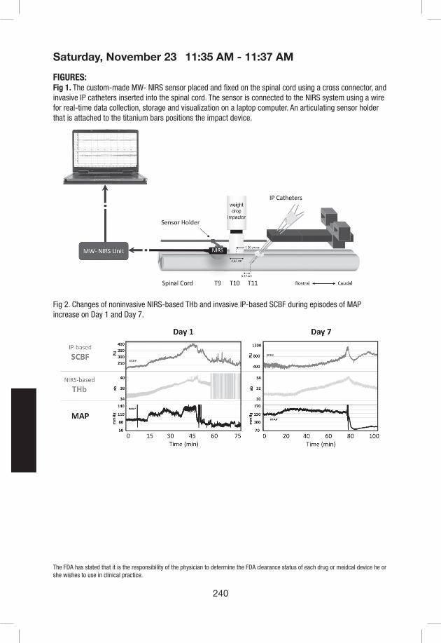
240
Saturday, November 23
The FDA has stated that it is the responsibility of the physician to determine the FDA clearance status of each drug or meidcal device he or she wishes to use in clinical practice.
FIGURES:Fig 1. The custom-made MW- NIRS sensor placed and fixed on the spinal cord using a cross connector, and invasive IP catheters inserted into the spinal cord. The sensor is connected to the NIRS system using a wire for real-time data collection, storage and visualization on a laptop computer. An articulating sensor holder that is attached to the titanium bars positions the impact device.
Fig 2. Changes of noninvasive NIRS-based THb and invasive IP-based SCBF during episodes of MAP increase on Day 1 and Day 7.
11:35 AM - 11:37 AM

241
Saturday, November 23
Individual Disclosures can be found in the Disclosure Index pages 41-69.
Presentation #79
Risk Factors Associated with Vertebral Artery Anomalies in the Subaxial Cervical Spine
Fabian Winter, MD1, Erika Chiapparelli Harb, MD1, Stephan N. Salzmann, MD1, Colleen Rentenberger, MD1, Jennifer Shue, MS1, Ichiro Okano, MD1, Andrew A. Sama, MD1, Federico P. Girardi, MD1, Frank P. Cammisa, MD1, and Alexander P. Hughes, MD1
1Department of Orthopaedic Surgery, Hospital for Special Surgery, New York City, NY
Introduction: Vertebral artery (VA) variations, congenital or acquired, can increase the risk of iatrogenic injuries during anterior cervical spine surgery. The majority of VA variations are asymptomatic. In previous case reports of iatrogenic VA injuries an abnormal course of the VA has not been diagnosed before the occurrence of the complication. Little is known about possible risk factors for VA variations. The aim of our study is to provide risk factors of VA anomalies in the subaxial cervical spine utilizing the data of consecutive patients with computed tomography angiographs (CTA).
Methods: We reviewed 217 consecutive preoperative CTAs at a single academic institution between 2007 and 2018. Potential dangerous VA variations were defined as medial loops (tortuosity of the VA towards the vertebral body), asymmetrical transverse foramen entry points (at least two levels), extraforaminal medial VA course (medial displacements before the entry into the transverse foramen), occlusions (decrease in arterial caliber), bilateral bony erosions, and duplications. Potential risk factors including age, sex, and previous cervical spine surgeries were reviewed. Statistical analysis was conducted utilizing the Fishers Exact test for categorical variables and the Student T test for continuous variables; statistical significance was set at p < 0.05.
Results: A total of 217 patients were included. The mean age was 57.5, 52% were female, 30% had a previous cervical spine surgery. 45 (21%) had 53 reported VA anomalies. The most common abnormality were medial loops (17/217, 8%), followed by asymmetrical entries into the transverse foramen (13/217, 6%). In addition 7 bilateral erosions (3%), 7 extraforaminal medial displacements (3%), 5 occlusions (2%), and 4 duplications (2%) were detected. Regarding potential risk factors, medial loops were significantly associated with female patients (82%, p = 0.028). No other significant association was observed between VA anomalies and included risk factors.
Conclusion: VA variations are not uncommon in the subaxial cervical spine. The most common are VA loops towards the midline of the vertebral body, which was observed significantly more among female patients. The different anatomic VA anomalies should be considered in the preoperative evaluation to prevent iatrogenic injuries during cervical surgery.
11:38 AM - 11:40 AM

242
Saturday, November 23
The FDA has stated that it is the responsibility of the physician to determine the FDA clearance status of each drug or meidcal device he or she wishes to use in clinical practice.
Fig 1. Illustration of three vertebral artery abnormalities, (Left) bilateral erosion, (Middle) duplication of the left vertebral artery, (Right) occlusion of the right vertebral artery.
11:38 AM - 11:40 AM

247
Saturday, November 23
Individual Disclosures can be found in the Disclosure Index pages 41-69.
Presentation #81
Establishing Maximal Medical Improvement Following Anterior Cervical Discectomy and Fusion
Nathaniel W. Jenkins, MS, Dil V. Patel, BS1, Joon S. Yoo, BA1, Benjamin Khechen, BA1, Anirudh K. Gowd, BS1, Eric H. Lamoutte, BS, Sailee S. Karmarkar, BS1, Joseph N. Liu, MD1, Kern Singh, MD1
1Department of Orthopaedic Surgery, Rush University Medical Center, Chicago, IL
Introduction: The purpose of this study is to identify the time point at which patients undergoing primary, single-level anterior cervical discectomy and fusion (ACDF) can be considered to have reached maximal medical improvement (MMI).
Methods: A surgical database was retrospectively reviewed between 2014-2017 for patients undergoing ACDF for degenerative pathology. Neck Disability Index (NDI) scores were collected preoperatively and at 6-week, 3-month, 6-month, and 12-month follow-up. The minimal clinically important difference (MCID) was calculated using distribution-based methodology at the 1-year interval and validated using a nonparametric receiver operating characteristic (ROC) curve. Area under curve (AUC) analysis was used to validate predictive power. The time period at which patients achieved MCID was determined and plotted using a Kaplan-Meier survivorship curve. MMI was defined as the interval at which 90% of patients achieved MCID. A Cox hazard proportion model was used to determine whether age, body mass index (BMI), and Charlson Comorbidity Index (CCI) were associated with time to achieve MCID.
Results: A total 69 patients were included in the analysis. Average NDI scores were 37.9 preoperatively, 30.7 at 6-weeks, 25.1 at 3-months, 21.3 at 6-months, and 22.0 at 12-months postoperatively. The change in NDI reflective of MCID was calculated to be -11.0 (AUC=100%). In total, 51 patients (73.9%) achieved MCID within the study period, with median time to achievement being 3-months. Of these patients, 95.7% achieved MCID by 6-months postoperatively, thus allowing the establishment of MMI at this time point. No association was identified between age (HR: 1.014, p=0.606), BMI (HR: 0.984, p=0.552), or CCI (HR: 0.932, p=0.719), and time to achieve MCID.
Conclusion: Majority of patients undergoing ACDF achieved MCID at 3-months follow-up, while over 95% of patients who achieved MCID reached this threshold by 6 months following surgery. As such, future outcome reporting in patients undergoing ACDF should include follow-up to 6 months postoperatively. Providers can also utilize this information to better counsel patients regarding postoperative expectations following ACDF.
11:54 AM - 11:56 AM

248
Saturday, November 23
The FDA has stated that it is the responsibility of the physician to determine the FDA clearance status of each drug or meidcal device he or she wishes to use in clinical practice.
11:54 AM - 11:56 AM
249
Saturday, November 23
Individual Disclosures can be found in the Disclosure Index pages 41-69.
Presentation #82
Does Neurologic Diagnosis Affect Improvement in Neck Pain after ACDF Surgery?
Justin D Stull MD, Srikanth N Divi MD, John J Mangan MD, Dhruv KC Goyal BA, Matthew S Galetta BA, Jeffrey A Rihn MD, Mark F Kurd MD, D Greg Anderson MD, Barrett I Woods MD, Kristen E Radcliff MD, Ian D Kaye MD, Alexander R Vaccaro MD PhD MBA, Christopher K Kepler MD MBA, Gregory D Schroeder MD, Alan S Hilibrand MD
Rothman Institute, Thomas Jefferson University Hospital, Philadelphia, PA
Introduction: While anterior cervical discectomy and fusion (ACDF) is performed primarily for the treatment of cervical myelopathy or radiculopathy, patients often experience a concurrent improvement in axial neck pain. Currently, there is a paucity of literature examining the difference in improvement of neck pain after ACDF for cervical myelopathy or radiculopathy.
Materials/Methods: A retrospective analysis of prospectively collected data was conducted to determine whether there are any differences with respect to healthcare related quality of life (HRQOL) outcomes in patients who underwent ACDF at a single institution. Patients over the age of 18 who underwent 1-3 level primary ACDF for the treatment of cervical myelopathy or radiculopathy were included. Patients undergoing surgery for trauma, tumor, infection, or revision were excluded. Anyone with less than one year of clinical follow up was excluded from the analysis. Patients were split into groups based on whether they had purely radiculopathy (R) or myelopathy (M), or a combination of myeloradiculopathy (MR). Visual Analog Score (VAS) Neck was defined as the primary outcome. Secondary outcomes of interest included: Short Form-12 (SF-12) Physical Component Score (PCS-12) and Mental Component Score (MCS-12), Neck Disability Index (NDI), and VAS Arm scores. One-way ANOVA with Bonferroni post-hoc analysis, Chi-square analysis, and multiple linear regression analysis were used to compare differences between groups.
Results: A total of 251 patients were included in the cohort (Table 1), with 121 patients in the radiculopathy (R) only group, 58 in the myelopathy (M) only group, and 72 in the myeloradiculopathy (MR) group. Patients in the M group were significantly older (57 years old, p = 0.003) than patients in the R group (50 years) and the MR group (54 years). Patients in the M group also had a higher proportion of 3 level ACDF (p < 0.001). Preoperative VAS Neck pain was significantly higher at baseline in the R group (6.3, p = 0.005) compared to the M (5.0) and MR (5.3). Postoperatively, the VAS Neck was significantly improved (p < 0.001) in all groups, and independent of diagnosis (Δ VAS Neck, -2.9 M group, -2.3 R group, -2.3 MR group, p = 0.351). Recovery ratio and % Minimal Clinically Important Difference (MCID) were similar for all groups (p = 0.928 and p = 0.112, respectively). In addition, patients in the M group had significantly more improvement in MCS-12 scores (p = 0.032). Multiple linear regression analysis found that the presence of radiculopathy or myelopathy were not significant predictors of patient outcomes.
Conclusion: Patients undergoing an ACDF have a statistically significant improvement in VAS neck pain and this is not affected by underlying neurologic diagnosis. All patients improved to a similar degree with no significant difference in neck pain postoperatively.
Level of Evidence: III
11:57 AM - 11:59 AM

250
Saturday, November 23
The FDA has stated that it is the responsibility of the physician to determine the FDA clearance status of each drug or meidcal device he or she wishes to use in clinical practice.
Table 1: Health-Related Quality of Life Outcome Measurement Comparisons Between Groups
Radiculopathy (n=121)
Myelopathy (n=58)Myeloradiculopathy (n=72)
One-Way ANOVAp-value
Multiple Linear Regression Analysis, β-coefficient [95% CI], p-value
PCS-12
Pre 33.2 [31.9, 34.5] 31.9 [29.6, 34.2] 34.0 [32.1, 36.0] 0.321
R: 1.014 [-2.212, 4.241], 0.536MR: 3.001 [-0.412, 6.413], 0.085
Post 40.4 [38.3, 42.5] 38.5 [35.6, 41.4] 42.9 [40.4, 45.3] 0.083
∆ 7.1 [5.3, 8.9] 6.1 [3.9, 8.4] 9.0 [6.7, 11.2] 0.221
RR 0.10 [0.08, 0.13] 0.08 [0.05, 0.11] 0.13 [0.10, 0.16] 0.158
% MCID 43.8% 36.2% 54.2% 0.115
MCS-12
Pre 46.7 [44.5, 48.9] 45.1 [42.2, 47.9] 43.1 [40.2, 45.9] 0.126
R: -3.648 [-7.494, 0.198], 0.063MR: -3.012 [-7.037, 1.013], 0.142
Post 48.3 [46.2, 50.4] 51.6 [49.0, 54.3] 48.7 [45.8, 51.6] 0.188
∆ 1.4 [-1.1, 3.9] 6.6 [2.9, 10.3] 5.7 [2.3, 9.1] 0.032**
RR -0.01 [-0.05, 0.04] 0.08 [0.02, 0.15] 0.07 [0.02, 0.13] 0.024**
% MCID 35.5% 56.9% 45.8% 0.023*
NDI
Pre 44.9 [41.5, 48.3] 39.3 [34.0, 44.6] 41.6 [37.0, 46.2] 0.170
R: 2.257 [-4.030, 8.544], 0.480MR: -0.009 [-6.605, 6.587], 0.998
Post 28.5 [24.2, 32.7] 22.9 [17.5, 28.4] 23.1 [18.1, 28.0] 0.155
∆ -16.3 [-19.9, -12.7] -16.2 [-20.9, -11.4] -18.9 [-24.0, -13.9] 0.633
RR 0.37 [0.29, 0.46] 0.35 [0.21, 0.48] 0.39 [0.22, 0.55] 0.916
% MCID 49.6% 48.3% 50.0% 0.980
VAS Neck
Pre 6.3 [5.9, 6.7] 5.0 [4.1, 5.9] 5.2 [4.5, 6.0] 0.005*
R: 0.354 [-0.554, 1.263], 0.443MR: 0.245 [-0.698, 1.188], 0.609
Post 3.4 [2.9, 3.9] 2.8 [2.1, 3.5] 3.0 [2.3, 3.6] 0.356
∆ -2.9 [-3.4, -2.3] -2.3 [-3.1, -1.4] -2.3 [-3.1, -1.5] 0.351
RR 0.42 [0.32, 0.52] 0.45 [0.27, 0.63] 0.41 [0.23, 0.58] 0.928
% MCID 52.9% 37.9% 41.7% 0.112
VAS Arm
Pre 5.4 [4.8, 5.9] 4.7 [3.8, 5.7] 5.2 [4.5, 5.9] 0.473
R: 0.793 [-0.144, 1.731], 0.097MR: -0.028 [-1.012, 0.956], 0.955
Post 3.1 [2.5, 3.6] 2.5 [1.8, 3.2] 2.4 [1.8, 3.0] 0.204
∆ -2.2 [-2.9, -1.5] -2.3 [-3.2, -1.4] -2.9 [-3.7, -2.0] 0.488
RR 0.51 [0.42, 0.60] 0.47 [0.31, 0.63] 0.52 [0.31, 0.73] 0.926
% MCID 43.8% 48.3% 48.6% 0.760
Preoperative, postoperative, and delta (postoperative-preoperative) reported outcome scores, recovery ratios (RR), and the percentage of patients achieving the minimum clinically important difference (% MCID) reported as: Mean [95% CI]. Short-Form-12 Physical Component Score (PCS-12) and Mental Component Score (MCS-12), Neck Disability Index (NDI), Visual Analogue Score (VAS) Neck pain (VAS Neck), Arm pain (VAS Arm), and Recovery Ratio (RR). *Indicates statistical significance (p < 0.05). **Indicates no significant difference on Bonferroni post-hoc analysis. Multiple Linear Regression analysis performed using myelopathy as a baseline for comparison
REFERENCES1. Radcliff K, Davis R, Hisey M, et al. Long-term Evaluation of Cervical Disc Arthroplasty with the
Mobi-C© Cervical Disc: A Randomized, Prospective, Multicenter Clinical Trial with Seven-Year Follow-up. Int J Spine Surg. 2017;11(5):244-262. doi:10.14444/4031
2. Parker SL, Godil SS, McGirt MJ. Determination of Minimum Clinically Important Difference in Pain, Disability and Quality of Life After Anterior Cervical Discectomy and Fusion. Spine J.
11:57 AM - 11:59 AM

251
Saturday, November 23
Individual Disclosures can be found in the Disclosure Index pages 41-69.
2012;12(9):S120. doi:10.1016/j.spinee.2012.08.3233. Carreon LY, Glassman SD, Campbell MJ, Anderson PA. Neck Disability Index, short form-
36 physical component summary, and pain scales for neck and arm pain: the minimum clinically important difference and substantial clinical benefit after cervical spine fusion. Spine J. 2010;10(6):469-474. doi:10.1016/j.spinee.2010.02.007
4. Ibm.com. (2019). IBM SPSS Software. [online] Available at: https://www.ibm.com/analytics/spss-statistics-software [Accessed 10 Jan. 2019].
11:57 AM - 11:59 AM

252
Saturday, November 23
The FDA has stated that it is the responsibility of the physician to determine the FDA clearance status of each drug or meidcal device he or she wishes to use in clinical practice.
Presentation #83
Predictive Models for Long-Term Patient-Reported Outcomes After Cervical Spine Surgery for Myelopathy: A National Study of 2717 Patients
Ahilan Sivaganesan MD2, Inamullah Khan MD1,2, Hui Nian PhD4, Frank E. Harrell Jr. PhD4, Jacquelyn S. Pennings, PhD1, Mohamad Bydon MD5, Anthony L. Asher MD6, Kristin R. Archer PhD, DPT1,7, Clinton J. Devin MD1,2,3
1Department of Orthopedic Surgery, Vanderbilt University School of Medicine, Nashville, TN2Department of Neurological Surgery, Vanderbilt University School of Medicine, Nashville, TN3Steamboat Orthopaedics and Spine Institute, Steamboat Springs, CO4Department of Biostatistics, Vanderbilt University School of Medicine, Nashville, TN5Department of Neurological Surgery, Mayo Clinic, Rochester, MN6Carolina Neurosurgery & Spine Associates, Carolinas Healthcare System, Charlotte, NC7Department of Physical Medicine & Rehabilitation, Vanderbilt University Medical Center,Nashville, TN
Introduction: In this current era of healthcare reform, surgeons are now expected to demonstrate the effectiveness of their interventions. Patient-reported outcomes (PROs) are useful metrics, however in the field of cervical spine surgery, there is immense patient-level variation in every major outcome for a given procedure. This is certainly true for the surgical treatment of myelopathy, where surgeons find it particularly difficult to forecast how a patient will be doing long after surgery. The aim of this study was to develop and validate a set of predictive models for myelopathy, arm pain, neck pain, and disability twelve months after cervical spine surgery for myelopathy.
Materials/Methods: A retrospective analysis was performed using prospectively collected data from the cervical module of a national spine registry – the Quality and Outcomes Database (QOD). All patients undergoing cervical spine surgery for myelopathy were included in this study. Proportional odds ordinal regression models were developed for the following outcomes: modified Japanese Orthopedic Association (mJOA) score for myelopathy, Numeric Rating Scale for arm pain (NRS-AP), neck pain (NRS-NP), and Neck Disability Index (NDI). The following covariates were included: age, gender, BMI, race, education, smoking status, diabetes, anxiety/depression, symptom duration, motor deficit or numbness, spondylolisthesis, employment status, workers’ compensation, insurance, ambulation, baseline PROs, and surgical factors such as number of levels, arthrodesis, and anterior vs. posterior approach. International validation was performed using bootstrap resampling.
Results: A total of 2717 patients who underwent surgery for cervical myelopathy were included in this study. Overall, there was a significant improvement in all three 12-month PROs (p<0.001). The most important predictors of outcomes, in descending order, were: baseline PROs, age, employment, symptom duration, ambulation, workers’ compensation, surgical approach, and education. Figures 1 and 2 display the adjusted effects of each covariate and their relative impact on variation, respectively. The models’ discriminative performance (measured by the overfitting-corrected c-index) were: 0.70 for NDI, 0.68 for neck pain, and 0.67 for arm pain, and 0.72 for myelopathy (mJOA) at twelve months.
12:00 PM - 12:02 PM
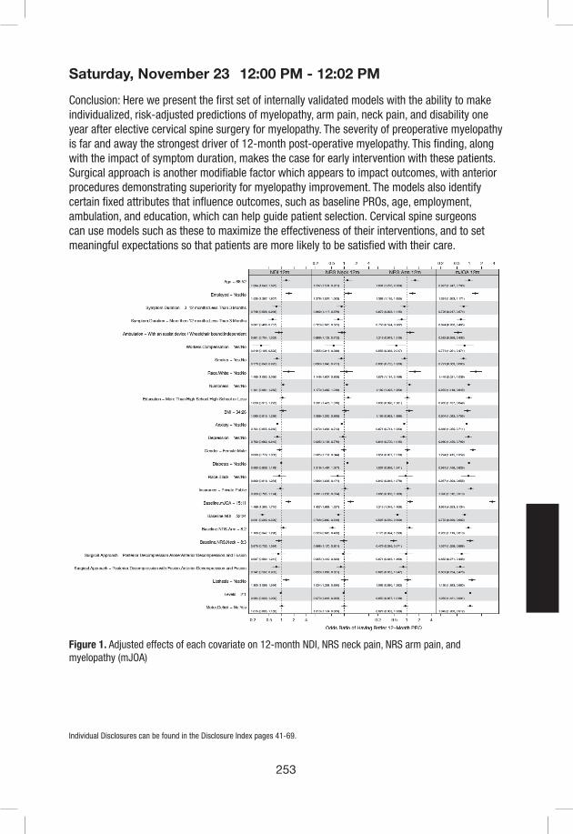
253
Saturday, November 23
Individual Disclosures can be found in the Disclosure Index pages 41-69.
Conclusion: Here we present the first set of internally validated models with the ability to make individualized, risk-adjusted predictions of myelopathy, arm pain, neck pain, and disability one year after elective cervical spine surgery for myelopathy. The severity of preoperative myelopathy is far and away the strongest driver of 12-month post-operative myelopathy. This finding, along with the impact of symptom duration, makes the case for early intervention with these patients. Surgical approach is another modifiable factor which appears to impact outcomes, with anterior procedures demonstrating superiority for myelopathy improvement. The models also identify certain fixed attributes that influence outcomes, such as baseline PROs, age, employment, ambulation, and education, which can help guide patient selection. Cervical spine surgeons can use models such as these to maximize the effectiveness of their interventions, and to set meaningful expectations so that patients are more likely to be satisfied with their care.
Figure 1. Adjusted effects of each covariate on 12-month NDI, NRS neck pain, NRS arm pain, and myelopathy (mJOA)
12:00 PM - 12:02 PM

254
Saturday, November 23
The FDA has stated that it is the responsibility of the physician to determine the FDA clearance status of each drug or meidcal device he or she wishes to use in clinical practice.
Figure 2. Relative importance of each covariate in explaining variation in 12-month NDI, NRS neck pain, NRS arm pain, and myelopathy (mJOA)
12:00 PM - 12:02 PM

255
Saturday, November 23
Individual Disclosures can be found in the Disclosure Index pages 41-69.
Presentation #84
How Much Do Patients With Predominantly Neck Pain Improve After ACDF Surgery for Cervical Radiculopathy?
Srikanth N Divi MD, Justin D Stull MD, John J Mangan MD, Dhruv KC Goyal BA, Matthew S Galetta BA, Jeffrey A Rihn MD, Mark F Kurd MD, D Greg Anderson MD, Barrett I Woods MD, Alan S Hilibrand MD, Ian D Kaye MD, Alexander R Vaccaro MD PhD MBA, Christopher K Kepler MD MBA, Gregory D Schroeder MD, Kristen E Radcliff MD
Rothman Institute, Thomas Jefferson University Hospital, Philadelphia, PA
Introduction: Anterior Cervical Discectomy and Fusion (ACDF) is a successful surgery for patients presenting with symptoms of radiculopathy. However, many of these patients have concurrent neck pain that often can be more symptomatic than their arm pain. The primary goal of this study is to determine how much patients with predominantly neck pain improve after ACDF compared to patients with predominantly arm pain.
Methods: A retrospective review of a prospectively maintained database on patients at a single, high volume institution was conducted. Patients over the age of 18 who underwent 1-3 level ACDF for a diagnosis of cervical radiculopathy between January 2014 and December 2015 were included. Those with less than one year of clinical follow-up and those who received operative intervention to address tumors, infection, trauma or revision were excluded. Patients were split into three groups based on preoperative Visual Analog Score (VAS) for neck and arm pain. Using a minimally clinically important difference (MCID) threshold of 1.0 point, patients were split into the Neck (N) pain dominant group (VAS Neck ≥ VAS Arm by 1.0 point), the Arm (A) pain dominant group (VAS Arm ≥ VAS Neck by 1.0 point), or the Neutral (NE) group (VAS Neck and VAS Arm within 1.0 point).1 Additional patient reported outcome measurements were collected and compared using univariate and multivariate analysis: Neck Disability Index (NDI), Short Form-12 (SF-12) Mental Component Score (MCS-12) and Physical Component Score (PCS-12).
Results: A total of 122 patients were included in the final cohort. Average preoperative VAS Neck scores were significantly higher in the N and NE groups (p < 0.001) and average preoperative VAS Arm scores were significantly higher in the NE and A groups (p < 0.001). All groups significantly improved postoperatively with respect to PCS-12 and NDI scores (p<0.001), except for PCS-12 scores in the A group, which exhibited a trend towards improvement (p=0.064). None of the groups improved postoperatively with respect to MCS-12 scores. In addition, there were no significant differences in terms of baseline or postoperative scores for all outcome measures between groups. The magnitude of improvement, recovery ratio (RR), and % minimal clinically important difference (MCID) also improved to the same degree regardless of arm or neck dominant symptoms. Using multivariate analysis, both the NE and A groups were significantly more likely to improve with respect to NDI scores (Beta Coeff: -7.86 [-15.4, -0.30], p = 0.042 and Beta Coeff: -10.6 [-21.0, -0.14], p = 0.047, respectively). Outcome comparisons between N, NE, and A groups can be located in Table 1.
Conclusion: All groups showed a similar magnitude of functional improvement (PCS-12) except for patients with arm-dominant pain. In addition all three groups significantly improved with
12:03 PM - 12:05 PM

256
Saturday, November 23
The FDA has stated that it is the responsibility of the physician to determine the FDA clearance status of each drug or meidcal device he or she wishes to use in clinical practice.
respect to neck disability scores. However, when using multivariate analysis, patients in the NE and A groups were more likely to improve in NDI scores compared to the N group. Further studies are needed to clarify improvement in axial neck pain after ACDF surgery.
Table 1: VAS Arm vs Neck Pain Dominance Outcomes
NECK (n=46) Neutral (n=57) ARM (n=19)
Univariate Analysis (p-value)Multivariate
Regression (Beta Coefficient [95%
CI], p-value)
Paired Samples
t-Test
Kruskal-Wallis H test or
Chi-Square Analysis
PCS-12
Pre 32.7 [31.0, 34.5] 32.9 [30.9, 34.8] 35.3 [30.7, 39.9] N: <0.001*NE: <0.001*
A: 0.064
0.762NE: 2.380
[-1.578, 6.338], 0.236
A: -0.403 [-5.860, 5.055], 0.884
Post 39.4 [35.9, 42.8] 41.4 [38.5, 44.4] 40.0 [34.1, 46.0] 0.617
Delta 6.6 [3.3, 10.0] 8.3 [6.0, 10.6] 4.7 [-0.3, 9.8] 0.323
RR 9.6% 12.1% 6.8%—
0.419
% MCID 39.1% 50.9% 36.8% 0.382
MCS-12
Pre 47.6 [43.8, 51.4] 46.4 [43.0, 49.7] 46.2 [40.8, 51.6] N: 0.401NE: 0.076A: 0.318
0.836NE: 3.899
[-0.361, 8.158], 0.072
A: 4.990 [-0.813, 10.793], 0.091
Post 45.8 [42.4, 49.2] 49.6 [46.7, 52.5] 49.8 [43.0, 56.6] 0.072
Delta -1.8 [-6.0, 2.4] 2.8 [-0.7, 6.4] 3.6 [-3.8, 11.1] 0.161
RR -7.7% 1.9% 4.3%—
0.130
% MCID 26.1% 40.4% 42.1% 0.255
NDI
Pre 45.0 [41.0, 50.0] 47.0 [41.0, 52.0] 37.0 [28.0, 46.0] N: <0.001*NE: <0.001*A: <0.001*
0.177 NE: -7.826 [-15.352, -0.300],
0.042*A: -10.582
[-21.026, -0.139], 0.047*
Post 33.6 [26.6, 40.5] 27.5 [20.9, 34.1] 19.7 [10.6, 28.8] 0.063
Delta -11.6 [-17.4, -5.9] -19.3 [-25.0, -13.5] -17.4 [-25.8, -9.0] 0.330
RR 25.6% 40.0% 51.7%—
0.106
% MCID 45.7% 50.9% 52.6% 0.825
VAS Neck
Pre 6.5 [6.0, 7.0] 6.8 [6.2, 7.4] 4.3 [3.0, 5.5] N: <0.001*NE: <0.001*
A: 0.025*
<0.001*NE: -1.147
[-2.264, -0.030], 0.044*
A: -1.282 [-2.912, 0.348], 0.122
Post 4.2 [3.3, 5.1] 3.2 [2.4, 3.9] 2.5 [1.2, 3.7] 0.065
Delta -2.3 [-3.2, -1.4] -3.6 [-4.4, -2.7] -1.8 [-3.4, -0.2] 0.082
RR 34.1% 51.1% 29.1%—
0.197
% MCID 47.8% 59.6% 42.1% 0.302
VAS Arm
Pre 3.0 [2.2, 3.9] 6.8 [6.2, 7.4] 6.3 [5.1, 7.5] N: 0.362NE: <0.001*
A: 0.001*
<0.001*NE: -1.758
[-3.157, -0.358], 0.014*
A: -1.683 [-3.394, 0.027], 0.054
Post 3.5 [2.6, 4.5] 2.8 [2.0, 3.6] 3.1 [1.6, 4.6] 0.541
Delta 0.5 [-0.6, 1.7] -4.0 [-4.8, -3.2] -3.1 [-4.8, -1.5] <0.001*
RR 36.2% 58.7% 51.1%—
0.263
% MCID 19.6% 63.2% 42.1% <0.001*
Revision Surgery?
NoYes
42 (91.3%)4 (8.7%)
51 (89.5%)6 (10.5%)
17 (89.5%)2 (10.5%)
— 0.947 —
References:1. Macdowall A, Skeppholm M, Robinson Y, Olerud C. Validation of the visual analog scale in the
cervical spine. J Neurosurg Spine. 2018;28(3):227-235.2. Radcliff K, Davis R, Hisey M, et al. Long-term Evaluation of Cervical Disc Arthroplasty with the
Mobi-C© Cervical Disc: A Randomized, Prospective, Multicenter Clinical Trial with Seven-Year Follow-up. Int J Spine Surg. 2017;11(5):244-262. doi:10.14444/4031
12:03 PM - 12:05 PM

257
Saturday, November 23
Individual Disclosures can be found in the Disclosure Index pages 41-69.
3. Carreon LY, Glassman SD, Campbell MJ, Anderson PA. Neck Disability Index, short form- 36 physical component summary, and pain scales for neck and arm pain: the minimum clinically important difference and substantial clinical benefit after cervical spine fu.
4. Parker SL, Godil SS, Shau DN, Mendenhall SK, Mcgirt MJ. Assessment of the minimum clinically important difference in pain, disability, and quality of life after anterior cervical discectomy and fusion: clinical article. J Neurosurg Spine. 2013;18(2):154-6.
12:03 PM - 12:05 PM

258
Saturday, November 23
The FDA has stated that it is the responsibility of the physician to determine the FDA clearance status of each drug or meidcal device he or she wishes to use in clinical practice.
Presentation #85
Functional Range of Motion of the Cervical Spine in Posterior Cervical Discectomy and Fusion Patients During Activities of Daily Living
Adrianna Oh, BS, Pittsburgh, PA, Sebastian Murati, BS, Pittsburgh, PA, Abenezer Alemu, BS, , Pttsburgh, PA, Marcus Allen, BS, Pittsburgh, PA, Michelle Riffitts, BS, Pittsburgh, PA, Anna Bailes, BS, Pittsburgh, PA, Malcolm Dombrowski, MD, Pittsburgh, PA, Joon Y. Lee, MD, Pittsburgh, PA, William F. Donaldson, PhD, Pittsburgh, PA, William W. Clark, PhD, Pittsburgh, PA, Kevin Bell, PhD, Pittsburgh, PA
INTRODUCTION: Posterior cervical discectomy and fusion (PCDF) is indicated for stenosis, radiculopathy, myelopathy, and acute instability. Cervical range of motion (CROM) is reduced post-PCDF, which may be associated with worse pain and functional status.1 However, it is unknown how post-fusion reduction in total CROM impacts ADLs, which use a mean 19% of total CROM,2 and whether this has a significant impact on a patient’s quality of life.3 We hypothesize that maximal and functional CROM during ADLs will be reduced post-fusion and that functional CROM will be negatively correlated with pain, disability, and fear behaviors.
METHODS: Sixteen control subjects (57.7±8.5 years, 3M, 13F) and fourteen fusion subjects post 4- and 5-segment PCDF (53.7±9.9 years, 6M, 8F, post-op 1.13±0.54 years; C3-C7, C4-C7, or C5-T1 level fusions) were recruited to participate in an IRB approved study. A 10-point pain scale, Fear Avoidance Belief Questionnaire (FABQ), and Neck Disability Index (NDI) survey data were collected. A motion tracking system based on inertial measurement units (IMUs) was used to measure CROM. Five cycles of maximum CROM and ADLs (bending to pick up an object, backing up a car, using a phone, tying shoes, squatting to pick up an object, climbing up and down stairs, moving from a standing to sitting position, moving from a supine to standing position, and walking) were recorded in flexion/extension (FE), lateral bending (LB), axial rotation (AR) planes. Output data was analyzed using a custom MATLAB program. Two-sided t-test was used to identify age differences between groups; Chi-square test was used to identify sex differences. Two-sided Welch’s t-test was performed to identify differences between CROM in control and fusion groups. Pearson correlation coefficient calculations were used to correlate pain score, FABQ, and NDI. Statistical significance was established at p<0.05.
RESULTS: There is no significant difference in age or sex between control and fusion groups. There is an increase in FABQ and NDI in fusion subjects compared to controls (p<0.01). There is no difference in pain (Figure 1A). Maximum CROM is decreased (p<0.01) in fusion (N=14) subjects compared to controls (N=16) in all directions except AR to the right (Figure 1B). Functional ROM was decreased in PCDF subjects compared to control subjects in backing up a car, using a phone, tying shoes, putting on socks, sitting to standing, climbing stairs, moving from supine to standing, and walking in at least one direction (Table 1). Functional ROM during backing up a car, using a phone, tying shoes, sitting to standing, putting on socks, and moving from supine to standing are negatively correlated with FABQ, pain, and NDI (Table 1).
CONCLUSION: Previous research has quantified the reduction in total CROM in healthy individuals, however no research has identified the impact of fusion on functional CROM during ADLs in a patient population. This study confirms that for some ADLs, functional CROM is
12:06 PM - 12:08 PM
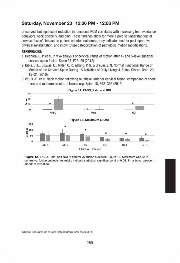
259
Saturday, November 23
Individual Disclosures can be found in the Disclosure Index pages 41-69.
preserved, but significant reduction in functional ROM correlates with increasing fear avoidance behaviors, neck disability, and pain. These findings allow for more a precise understanding of cervical fusion’s impact on patient-oriented outcomes, may indicate need for post-operative physical rehabilitation, and imply future categorization of pathologic motion modifications.
REFERENCES: 1. Bechara, B. P. et al. In vivo analysis of cervical range of motion after 4- and 5-level subaxial
cervical spine fusion. Spine 37, E23–29 (2012).2. Bible, J. E., Biswas, D., Miller, C. P., Whang, P. G. & Grauer, J. N. Normal Functional Range of
Motion of the Cervical Spine During 15 Activities of Daily Living: J. Spinal Disord. Tech. 23, 15–21 (2010).
3. Wu, X.-D. et al. Neck motion following multilevel anterior cervical fusion: comparison of short-term and midterm results. J. Neurosurg. Spine 18, 362–366 (2013).
12:06 PM - 12:08 PM
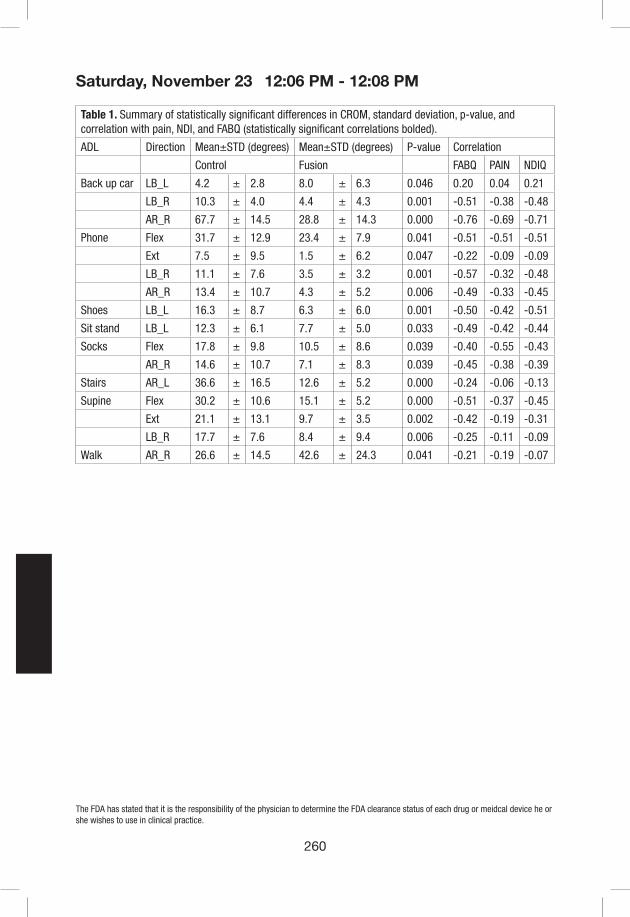
260
Saturday, November 23
The FDA has stated that it is the responsibility of the physician to determine the FDA clearance status of each drug or meidcal device he or she wishes to use in clinical practice.
Table 1. Summary of statistically significant differences in CROM, standard deviation, p-value, and correlation with pain, NDI, and FABQ (statistically significant correlations bolded).
ADL Direction Mean±STD (degrees) Mean±STD (degrees) P-value Correlation
Control Fusion FABQ PAIN NDIQ
Back up car LB_L 4.2 ± 2.8 8.0 ± 6.3 0.046 0.20 0.04 0.21
LB_R 10.3 ± 4.0 4.4 ± 4.3 0.001 -0.51 -0.38 -0.48
AR_R 67.7 ± 14.5 28.8 ± 14.3 0.000 -0.76 -0.69 -0.71
Phone Flex 31.7 ± 12.9 23.4 ± 7.9 0.041 -0.51 -0.51 -0.51
Ext 7.5 ± 9.5 1.5 ± 6.2 0.047 -0.22 -0.09 -0.09
LB_R 11.1 ± 7.6 3.5 ± 3.2 0.001 -0.57 -0.32 -0.48
AR_R 13.4 ± 10.7 4.3 ± 5.2 0.006 -0.49 -0.33 -0.45
Shoes LB_L 16.3 ± 8.7 6.3 ± 6.0 0.001 -0.50 -0.42 -0.51
Sit stand LB_L 12.3 ± 6.1 7.7 ± 5.0 0.033 -0.49 -0.42 -0.44
Socks Flex 17.8 ± 9.8 10.5 ± 8.6 0.039 -0.40 -0.55 -0.43
AR_R 14.6 ± 10.7 7.1 ± 8.3 0.039 -0.45 -0.38 -0.39
Stairs AR_L 36.6 ± 16.5 12.6 ± 5.2 0.000 -0.24 -0.06 -0.13
Supine Flex 30.2 ± 10.6 15.1 ± 5.2 0.000 -0.51 -0.37 -0.45
Ext 21.1 ± 13.1 9.7 ± 3.5 0.002 -0.42 -0.19 -0.31
LB_R 17.7 ± 7.6 8.4 ± 9.4 0.006 -0.25 -0.11 -0.09
Walk AR_R 26.6 ± 14.5 42.6 ± 24.3 0.041 -0.21 -0.19 -0.07
12:06 PM - 12:08 PM
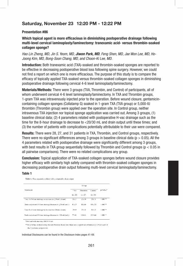
261
Saturday, November 23
Individual Disclosures can be found in the Disclosure Index pages 41-69.
Presentation #86
Which topical agent is more efficacious in diminishing postoperative drainage following multi-level cervical laminoplasty/laminectomy: tranexamic acid- versus thrombin-soaked collagen sponge?
Hao Lin Zheng, MD, Jin S. Yeom, MD, Jiwon Park, MD, Feng Shen, MD, Jae Won Lee, MD, Ho-Joong Kim, MD, Bong-Soon Chang, MD, and Choon-Ki Lee, MD.
Introduction: Both tranexamic acid (TXA)-soaked and thrombin-soaked sponges are reported to be effective in decreasing postoperative blood loss following spine surgery. However, we could not find a report on which one is more efficacious. The purpose of this study is to compare the efficacy of topically applied TXA-soaked versus thrombin-soaked collagen sponges in diminishing postoperative drainage following cervical 4-6 level laminoplasty/laminectomy.
Materials/Methods: There were 3 groups (TXA, Thrombin, and Control) of participants, all of whom underwent cervical 4-6 level laminoplasty/laminectomy. In TXA and Thrombin groups, 1-gram TXA was intravenously injected prior to the operation. Before wound closure, gentamicin-containing collagen sponges (Collatamp G) soaked in 1-gram TXA (TXA group) or 5,000-IU thrombin (Thrombin group) were applied over the operation site. In Control group, neither intravenous TXA injection nor topical sponge application was carried out. Among 3 groups, (1) baseline clinical data; (2) 4 parameters related with postoperative H-vac drainage such as the time for the 8-hour drainage to decrease to <20/30 mL and drain output until these times; and (3) the number of patients with complications potentially attributable to their use were compared.
Results: There were 28, 27, and 31 patients in TXA, Thrombin, and Control groups, respectively. There were no significant differences among 3 groups in baseline clinical data (p > 0.05). All the 4 parameters related with postoperative drainage were significantly different among 3 groups, with best results in TXA group sequentially followed by Thrombin and Control groups (p < 0.05 in all pairwise comparisons). There were no related complications any group.
Conclusion: Topical application of TXA-soaked collagen sponges before wound closure provides higher efficacy with similarly high safety compared with thrombin-soaked collagen sponges in decreasing postoperative drain output following multi-level cervical laminoplasty/laminectomy.
Table 1
12:20 PM - 12:22 PM
262
Saturday, November 23
The FDA has stated that it is the responsibility of the physician to determine the FDA clearance status of each drug or meidcal device he or she wishes to use in clinical practice.
Presentation #87
Pseudoarthrosis rates after anterior cervical discectomy and fusion (ACDF) using polyetheretherketone (PEEK) or structural allograft for interbody grafting: minimum 2-year follow up
Minghao Wang, MD, PhD; Dean Chou, MD; Chih-chang Chang MD; Ankit Hirpara, BS; Yilin Liu, MD; Praveen Mummaneni, MD
Department of Neurosurgery, University of California, San Francisco
Objective: Both structural allograft and PEEK have been described to provide good fusion rates and few complications during ACDF. Recent data, however, has shown higher pseudoarthrosis rates with PEEK than structural allograft. We wished to evaluate pseudoarthrosis rates between PEEK and allograft in single and multi-level ACDF. We also wished to evaluate the fusion rates among different graft materials (e.g. autograft or allograft).
Method: We performed a retrospective review of patients treated with ACDF alone at our North American spine center from 2005-2017. Exclusion criteria were: concomitant posterior surgery (staged or not), adjacent level corpectomies, infection, tumor, pediatric patients, or lack of minimum 2-year follow up. Data collection included demographic variables, levels fused, interbody type (PEEK cage versus structural allograft), graft material (e.g. endplate shavings, demineralized bone matrix (DBM), or hybrid), pseudoarthrosis rates, and revision surgery rates for pseudoarthrosis. These data were analyzed by Pearson’s chi-square test and Fisher’s exact test according to the sample size. Pearson’s correlation test was also used to evaluate the relationship between the pseudoarthrosis rate and number of fused levels.
Result: Using inclusion criteria, 214 patients with minimum 2-year follow up were analyzed. There were no significant differences in age, gender and time of follow-up between the PEEK cohort and structural allograft cohort. Pseudoarthrosis rates (PR) were 1-level (PEEK 12.8%, allograft 4.9%, p>0.05), 2-level (PEEK 12.7%, allograft10.4%, p>0.05), and ≥3 level (PEEK 15.4%, allograft12.3%, p>0.05). For 1-level and 2-level ACDF with structural allograft, although the sample size was quite small, using DBM or DBM mixed with local autograft significantly increased the PR compared to using structural allograft packed with local autograft alone (p<0.05). For 2 or more level ACDF, using a PEEK cage was more likely to require revision surgery for pseudoarthrosis than using structural allograft (p<0.05). Collectively with both cohorts, there was a strong correlation between the number of surgical levels and the PR (Pearson’s coefficient=0.975).
Conclusion: In ACDF procedures involving the same number of fusion levels, there was no statistically significant difference in PR between PEEK cages and structural allograft. However, for ≥ 2-level ACDF, the use of PEEK cages increased the probability of revision surgery, potentially from the patients being more symptomatic than structural allograft pseudoarthrosis cases. With regards to structural allograft in 1 and 2-level cases, using DBM packed had a higher PR than packing with local autograft alone. Although the PR rates between PEEK and structural allograft are not different, PR in PEEK cases may be more symptomatic, resulting in a higher revision surgery rate.
12:23 PM - 12:25 PM

263
Saturday, November 23
Individual Disclosures can be found in the Disclosure Index pages 41-69.
Table 1 Comparison of PEEK cage and structural allograft in fusion, pseudoarthrosis, and revision surgery (RS) rate for different level(s) of ACDF.
Fusion Level(s) Interbody DeviceOutcome
TotalFusion
Pseudoarthrosis without RS
Pseudoarthrosis with RS
1-level
PEEK Cage 41 (87.2%) 6 (12.8%) 0 47
Structural Allograft 58 (95.1%) 3 (4.9%) 0 61
1-level Total 99 (91.7%) 9 (8.3%) 0 108
2-level
PEEK Cage 48 (87.3%) 3 (5.5%) 4 (7.2%) 55
Structural Allograft 60 (89.6%) 7 (10.4%) 0 67
2-level Total 108 (88.5%) 10 (8.2%) 4 (3.3%) 122
multi-level (>=3)
PEEK Cage 11 (84.6%) 0 2 (16.4%) 13
Structural Allograft 50 (87.7%) 7 (12.3%) 0 57
multi-level Total 61 (87.1%) 7 (10%) 2 (2.9%) 70
Table 2 Classification statistics of cage types and filler types for different level(s) of ACDF
Fusion Level(s) Interbody Device Filler TypeOutcome
TotalFusion Pseudoarthrosis
1-level (108)
PEEK (47)Autograft 34 (94.4%) 6 (5.6%) 36
DBM/Hybrid 7 (100%) 0 7
Structural Allograft (61)Autograft 54 (98.2%) 1 (1.8%) 55
DBM/Hybrid 4 (66.7%) 2 (33.3%) 6
2-level (122)
PEEK (55)Autograft 46 (86.8%) 7 (13.2%) 53
DBM/Hybrid 2 (100%) 0 2
Structral Allograft (67)Autograft 60 (92.3%) 5 (7.7%) 65
DBM/Hybrid 0 2 (100%) 2
multi-level (>=3) (70)
PEEK (13)Autograft 9 (81.8%) 2 (18.2%) 11
DBM/Hybrid 2 (100%) 0 2
Structral Allograft (57)Autograft 44 (88%) 6 (12%) 50
DBM/Hybrid 6 (85.7%) 1 (14.3%) 7
Table 3 Comparison of outcome using stand-alone designed PEEK cages and PEEK cages fixed with plate and screw for 1-level ACDF
Cage TypeOutcome (1-level)
TotalFusion Pseudoarthrosis
PEEK (Plate+Screw) 33 (84.6%) 6 (15.4%) 39
PEEK (Stand-alone) 8 (100%) 0 8
Total 41 (87.2%) 6 (12.8%) 47
12:23 PM - 12:25 PM
264
Saturday, November 23
The FDA has stated that it is the responsibility of the physician to determine the FDA clearance status of each drug or meidcal device he or she wishes to use in clinical practice.
Presentation #88
CTA in Addition to MRI may not be necessary in Detecting Subaxial Vertebral Artery Anomalies in Anterior Cervical Spine Surgery
Fabian Winter, MD1, Ichiro Okano, MD1, Stephan N. Salzmann, MD1, Colleen Rentenberger, MD1, Jennifer Shue, MS1, Andrew A. Sama, MD1, Federico P. Girardi, MD1, Frank P. Cammisa, MD1, and Alexander P. Hughes, MD1
1Department of Orthopaedic Surgery, Hospital for Special Surgery, New York City, NY
Introduction: Iatrogenic injuries to the vertebral artery (VA) are often a result of variable courses of the vessel along the cervical vertebrae. VA injuries can potentially cause permanent neurologic deficits and death from exsanguination. Computed tomography angiography (CTA) is considered the most reliable imaging modality for preoperative vascular evaluation. However, a CTA is not usually performed prior to subaxial cervical spine surgeries, whereas the majority of patients have preoperative magnetic resonance imaging (MRI). The purpose of this study is to evaluate the necessity of CTA in detecting VA anomalies in the subaxial cervical spine.
Methods: This cross-sectional retrospective study reviewed 191 patients who had a CTA and MRI of the cervical spine at a single academic institution between 2007 and 2018. An axial VA position classification was used to grade VA anomalies in the subaxial cervical spine on both CTAs and MRIs (Fig 1). The classification consists of a number (0-4) that characterizes the location of the VA from the medio-lateral (ML) aspect of the vertebral body. The risk of a VA injury is increased if the VA is displaced towards the midline of the vertebral body, indicated by the grades of the classification. A “0” indicates no displacement of the VA towards the midline of the vertebral body; a “1” describes a midline migration of 0-25%; “2” 25-50%; “3” 50-75%; and “4” 75-100%, respectively.
Results: The weighed κ-value of the CTA and MRI agreement for all disc levels was 0.87 (95% CI: 0.87-0.87). For VAs after entering the transverse foramen (intraforaminal), the κ-value was 0.84 (95% CI: 0.84-0.84), and for VAs before entering the transverse foramen (extraforaminal), 0.91 (95% CI: 0.91-0.91). For intraforaminal VA midline migrations (ML Grade ≥ 1) the κ-value was 0.80 (95% CI: 0.72-0.89), and for extraforaminal VA midline migrations, the κ-value was 0.85 (95% CI: 0.73-0.97). MRI underestimations (false negative ML grades) occurred in 1.4% of intra- and extraforaminal VAs.
Conclusion: MRIs are acceptable to evaluate the anatomic course of the VA with a reliable classification. In previous reports the majority of VA variations were retrospectively detected after the occurrence of iatrogenic VA injuries. Our study suggests that careful evaluation of routine preoperative MRIs is sufficient to prevent the majority of iatrogenic VA injuries. Further larger, prospective studies are needed to assess the generalizability of our results in the clinical setting.
12:26 PM - 12:28 PM
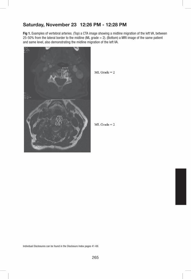
265
Saturday, November 23
Individual Disclosures can be found in the Disclosure Index pages 41-69.
Fig 1. Examples of vertebral arteries. (Top) a CTA image showing a midline migration of the left VA, between 25-50% from the lateral border to the midline (ML grade = 2). (Bottom) a MRI image of the same patient and same level, also demonstrating the midline migration of the left VA.
12:26 PM - 12:28 PM

266
Saturday, November 23
The FDA has stated that it is the responsibility of the physician to determine the FDA clearance status of each drug or meidcal device he or she wishes to use in clinical practice.
Presentation #89
Can Reinforcing Lateral Mass Screw with Multiple Points of Fixation Improve Anchoring Performance and Reduce Screw Pullout?
Franck Le Naveaux, Bahe Hachem, Kevin Lee, Jean-Marc Mac-Thiong, Hyun Bae, Christopher Ames, Masashi Neo, Julien Clin
Introduction: While Lateral Mass (LM) screw fixation has become the standard of care for posterior cervical spine stabilization, fixation failure remains a significant complication risk. Post-operatively, patient’s daily activities generate loads on the screws, which can translate to bone deformation around the screw threads and reduce fixation strength. Does adding multiple points of fixation to a LM screw improve anchoring performance? Multipoint (MP) fixation is enabled by an implant that locks to the top of a LM screw. It has attachment points extended towards the bone for two smaller MP screws to anchor laterally to the same LM (fig. 1). Using finite element analysis, the amount of off-loading on the LM screw by the addition of MP fixation was evaluated.
Materials & Methods: The first step was to determine the force magnitudes and vectors on each screw in a C3-T2 construct. The instrumented levels followed current standard of care fixation techniques with C3-C6 LM screws and T1-T2 pedicle screws; interconnected by 3.5 mm Titanium rods. A C2-T2 finite element model (FEM), validated based on published cadaveric non-linear load-displacement data, was subjected to 2 Nm of head flexion moment on C2 with T2 fixed (fig.1). In the second step, a refined FEM of C3 accounting for the non-linear behavior of bone with material properties of trabecular bone (E= 43 MPa) and 1.6 mm of cortical bone layer (E= 2144 MPa) was used. The force vector previously determined on the C3 left screw was applied on the screw head, in the following two conditions 1) standalone LM screw and 2) LM screw plus MP fixation. Bone loading and bone deformation at the bone/screw interface were compared.
Results: Head flexion moment generated the highest load on the construct’s most proximal LM screw (at C3) with an equivalent force of 39N (fig.1). By adding MP fixation, the load was shared across multiple screws decreasing the bone loading by 38% at the LM screw interface (fig.2). Furthermore, the bone deformation around the LM screw threads was reduced by 34% with MP fixation (average strain of 0.55%) when compared to standalone LM screw (0.84%).
Conclusion: Reinforcing a LM screw with MP fixation increases anchoring performance by distributing bone loading and reducing bone deformation, which may help preserve implant fixation and prevent screw pull-out. Further investigations are required to assess the anchoring performance improvement of the MP fixation in different bone qualities.
12:29 PM - 12:31 PM

267
Saturday, November 23
Individual Disclosures can be found in the Disclosure Index pages 41-69.
Fig. 1. Two step finite element analysis approach used to compare the bone loading resulting from the forces sustained by the screws during a head flexion movement
Fig. 2. Reduction of the bone loading at the LM screw interface with the MP fixation.
12:29 PM - 12:31 PM
268
Saturday, November 23
The FDA has stated that it is the responsibility of the physician to determine the FDA clearance status of each drug or meidcal device he or she wishes to use in clinical practice.
Presentation #90
The Effect Of Arthrodesis On Neuroforaminal Area
Clarissa LeVasseur, Samuel Pitcairn, Jeremy Shaw, William Donaldson, Joon Lee, and William Anderst
Department of Orthopedic Surgery, University of Pittsburgh, Pittsburgh, PA
INTRODUCTION: One proposed mechanism of pain relief following anterior cervical discectomy and fusion (ACDF) is increased neuroforaminal area1. The objective of this study was to address the paucity of in vivo data describing cervical neuroforaminal area by determining the effects of age, motion, symptomatic pathology, and ACDF on neuroforaminal area. We hypothesized that the bony neuroforamen area would decrease with age, spine extension, and symptomatic pathology, and the area would increase with flexion and after ACDF.
METHODS: This analysis included 28 cervical arthrodesis patients (15M, 13F; average age: 50±5 years), 19 middle-aged controls (7M, 12F; average 45±6 years) and 29 young adult controls (15M, 14F; average 26±4 years) who consented to participate in IRB-approved studies. Arthrodesis patients had symptoms resulting from degenerative spondylosis, symptomatic cervical disc herniation, or symptomatic disc degeneration, and received either single or two-level ACDF at C56 and/or C67 via an anterior approach with rigid plate fixation with an average graft height of 7mm. Biplane radiographs of the cervical spine were collected from all participants in neutral, full flexion, and full extension positions. Biplane radiographs of arthrodesis patients were collected before (PRE) and 1 year after (1YR-POST) surgery. Three-dimensional vertebral positions were determined using a validated registration process that matched subject-specific 3D bone models created from CT to the biplane radiographs with sub-millimeter accuracy2. A custom computer algorithm automatically calculated the neuroforamen cross-sectional area bilaterally using the 3D bone models3. Left and right side neuroforamen areas were averaged at each vertebral level prior to analysis. The effects of age, motion, and symptomatic pathology on neuroforamen area were evaluated using analysis of variance. Side-to-side differences in symptomatic-level neuroforamen area, and changes in index and adjacent level neuroforamen area from PRE to 1YR-POST, were identified using paired t-tests.
RESULTS: Neuroforaminal area was smaller at all vertebral levels in patients with symptomatic pathology than in middle-aged and young adult controls (Figure 1). Neuroforamen area was smaller in the lower cervical motion segments of middle-aged adults compared to younger adults (Figure 1). Moving from flexion to neutral to extension progressively decreased neuroforamen area, and these effects of motion on neuroforamen area were more pronounced in lower cervical motion segments. Neuroforamen area increased from PRE to 1YR-POST at the inferior adjacent segment, but no changes in neuroforamen area were identified at the arthrodesis site or the superior adjacent segment (Figure 2). Prior to surgery, the bony foramen area at the index level was smaller on the symptomatic side (26.7 mm2) compared to the asymptomatic side (30.5 mm2) (p = 0.005).
CONCLUSION: The results indicate that ACDF patients have a smaller neuroforamen area over the entire subaxial cervical spine, not only the symptomatic levels. This provides evidence
12:32 PM - 12:34 PM

269
Saturday, November 23
Individual Disclosures can be found in the Disclosure Index pages 41-69.
supporting systemic, rather than localized, spinal degeneration. Although the symptomatic neuroforamen was smaller than the asymptomatic side, the data do not support the notion that ACDF relieves pain by increasing bony neuroforamen area. Symptom resolution is likely due to disc removal, stabilization of the motion segment, and prevention of dynamic extension-based foraminal stenosis.
Figure 1. Neuroforamen area in the static neutral position at each vertebral level in arthrodesis patients, middle-aged controls and young controls. Error bars indicate ± 1 standard deviation. ** denotes p < 0.01, *** denotes p < 0.001.
12:32 PM - 12:34 PM

270
Saturday, November 23
The FDA has stated that it is the responsibility of the physician to determine the FDA clearance status of each drug or meidcal device he or she wishes to use in clinical practice.
Figure 2. Neuroforamen area in the static neutral position at the arthrodesis site as well as the superior and inferior adjacent segments before (PRE) and 1 year after (1YR-POST) arthrodesis. Error bars indicate ± 1 standard deviation.
REFERENCES 1. Albert TJ, Smith MD, Bressler E, et al. 1997. An in vivo analysis of the dimensional changes of
the neuroforamen after anterior cervical diskectomy and fusion: a radiologic investigation. Journal of spinal disorders 10:229-233.
2. Anderst WJ, Baillargeon E, Donaldson WF, 3rd, et al. 2011. Validation of a noninvasive technique to precisely measure in vivo three-dimensional cervical spine movement. Spine 36:E393-400.
3. Anderst WJ. 2012. Automated measurement of neural foramen cross-sectional area during in vivo functional movement. Comput Methods Biomech Biomed Engin 15:1313-1321.
12:32 PM - 12:34 PM
271
E-PosterAbstracts

272
CSRS-2019E-Poster #1
Bariatric Surgery Diminishes Spinal Symptoms in a Morbidly Obese Population: A 2-Year Survivorship Analysis of Cervical and Lumbar Pathologies
Peter G Passias MD, Haddy Alas BS, Avery Brown BS, Cole Bortz BA, Katherine E Pierce BS, Dennis Vasquez-Montes MS, Dainn Woo BS, Bassel G. Diebo MD, Carl B. Paulino MD, Michael C. Gerling MD
Introduction: Bariatric surgery for morbid obesity helps to address common comorbidity burdens, including decreasing rates of myocardial infarction and stroke. The increased mechanical stresses on the spine caused by morbid obesity predispose patients to various spinal pathologies and are concerning for spinal surgeons, with previous studies suggesting poorer outcomes than the general population. The effects of bariatric surgery on diminishing spinal complaints or symptoms have yet to be elucidated in the literature. The study assessed the rate in which various spinal symptoms diminish after bariatric surgery.
Materials/Methods: Retrospective analysis of the prospectively collected (NYSID) years 2004-2013. Patient linkage codes allow identification of multiple and return inpatient stays within the time-frame analyzed (720 days). Inclusion criteria were bariatrics surgery patients with one or more visits prior to and after bariatric procedure(excluding pts with <30 days f/u, spine surgery, or new post-bariatric spine pathology) for one of the following common cervical or lumbar spinal diagnoses, queried with ICD-9CM codes: herniation, stenosis, spondylosis, disc degeneration, and spondylolisthesis. Time from bariatric surgery until the patient’s respective spinal diagnosis was no longer present was considered resolution of spinal symptoms. Kaplan-Meier survivorship curves assessed rates of resolution within each spinal diagnosis cohort.
Results: 4,351 bariatric surgery pts with a pre-op spinal diagnosis by ICD-9 were analyzed. Lumbar pts: 1,049 had stenosis, 774 spondylosis, 648 degeneration, 249 spondylolisthesis, 72 disc herniation. Cervical pts: 581 disc herniation, 376 had stenosis, 366 spondylosis, 236 degeneration. Cumulative resolution rates at 90-day, 180-day, 360-day, and 720-day f/u were as follows: lumbar stenosis (48%,67.6%,79%,91%), lumbar herniation (61%,77%,86%,93%), lumbar spondylosis (47%,65%,80%,93%), lumbar spondylolisthesis (37%,58%,70%,87%), lumbar degeneration (37%,56%,72%,86%). By cervical region: cervical stenosis (48%,70%,84%,94%), cervical herniation (39%,58%,74%,87%), cervical spondylosis (46%, 70%,83%, 94%), cervical degeneration (44%,64%,78%,89%). Lumbar herniation pts saw significantly higher 90d-resolution than cervical herniation pts(p<0.001). Cervical vs lumbar degeneration resolution rates did not differ @90d(p=0.058), but did @180d(p=0.034). Cervical and lumbar stenosis resolution was similar @90d & 180d, but cervical showed greater resolution by 1Y(p=0.036).
Conclusions: Over 50% of bariatric patients diagnosed with a cervical or lumbar pathology before weight-loss surgery no longer sought inpatient care for their respective spinal diagnosis by 180 days post-op. Lumbar herniation had significantly higher resolution than cervical herniation by 90d, whereas cervical degeneration and stenosis resolved at higher rates than corresponding lumbar pathologies by 180d and 1Y f/u, respectively.
273
CSRS-2019E-Poster #2
Appropriate Risk Stratification and Accounting for Age-Adjusted Reciprocal Changes in the Thoracolumbar Spine Reduces the Incidence and Magnitude of Distal Junctional Kyphosis in Cervical Deformity Surgery
Peter G Passias MD, Cole Bortz BA, Renaud Lafage MS, Virginie Lafage PhD, Eric O. Klineberg MD, Han Jo Kim MD, Alan H Daniels MD, Gregory M Mundis MD, Themistocles S. Protopsaltis MD, Shay Bess MD, Frank J. Schwab MD, Christopher I. Shaffrey MD, Justin S Smith MD, PhD, Christopher P. Ames MD, International Spine Study Group
Background: For surgical cervical deformity (CD) patients, it is unclear whether distal junctional kyphosis (DJK) develops as compensation for mal-correction of sagittal deformity in the thoracic curve. Furthermore, there is limited understanding of other drivers of DJK, especially for different DJK types (severe, progressive, and clinically symptomatic). The purpose of this study is to identify factors associated with DJK occurrence and magnitude; assess differences in these factors across DJK types.
Methods: Patients with pre-and postop (3M, 6M, 1Y, or 2Y) clinical/radiographic data. Excluded: patients with previous fusion to L5 or below. DJK was defined per surgeon note or radiographically as DJK angle (kyphosis from LIV to LIV-2)<-10°, and pre- to postop change in DJK angle by<-10°. Age-specific target LL-TK alignment was calculated as published. Postop offset from target LL-TK was correlated to DJK magnitude and inclination. DJK types were: severe (DJK<-20°), progressive (DJK increase>4.4°, accounting for published measuring error), and symptomatic (associated with reop or published disability thresholds of NDI>24 or mJOA<14). Random forest analysis identified top patient, surgical, and radiographic factors associated with DJK and DJK types. Means comparison tests assessed differences in factors associated with DJK types.
Results: Included: 136 CD patients(61±10yrs, 61%F). Overall DJK rate was 30%. Though there were no differences in DJK rates across patients under-corrected, over-corrected, and matching age-specific LL-TK targets, postop offset from ideal LL-TK correlated with greater DJK angle(r=0.428) and inclination of the distal end of the fusion construct(r=0.244, both p<0.02). Seven of the top 15 factors associated with DJK were radiographic (CL, C2-T3 lordosis, CTPA, cSVA, C2 slope, C0-C2 Cobb, TLPA), four were surgical (# partial facet joint resections, combined approach, Smith-Petersen osteotomy, # posterior osteotomies), four were clinical (any comorbidity, any tumor, BMI, preop NDI). For DJK patients, breakdown by DJK type was: severe (22%), progressive (24%), symptomatic (61%). Symptomatic DJK patients had more posterior osteotomies than asymptomatic (p=0.018). Severe DJK patients presented with worse NDI scores, upper-cervical deformity (CL, C2 slope, C0-C2), and had more posterior osteotomies than non-severe (all p<0.01). Progressive DJK patients presented with greater malalignment—globally (CTPA) and in the cervical spine (CL, cSVA, C2 slope, C0-C2, C2-T3 lordosis, all p<0.03)—than static DJK patients. Of the top 10 factors associated with symptomatic DJK, 60% were clinical, 20% radiographic, and 20% surgical. For severe DJK, 60% were radiographic, 20% were clinical, and 20% were surgical. For progressive, 80% were radiographic and 20% were surgical. Of the top factors associated with DJK reop, 50% were clinical, 30% surgical, and 20% radiographic.

274
CSRS-2019
Conclusions: Postop offset from age-specific LL-TK alignment is associated with greater DJK and a more anterior distal construct inclination, suggesting DJK may develop as a result of inappropriate cervical and global realignment. Additionally, preop clinical and radiographic factors are associated with symptomatic and progressive DJK, suggesting the need for concomitant preop risk stratification.
Rank Factor Associated with DJK Type of Factor
1 Number of partial facet joint resections Surgical
2 Combined surgical approach Surgical
3 Any Smith-Petersen osteotomy (SPO) Surgical
4 Baseline C2-C7 lordosis Radiographic
5 Baseline C2-T3 lordosis Radiographic
6 Baseline cervical-thoracic pelvic angle (CTPA) Radiographic
7 Any comorbidity Clinical
8 Any tumor (per Charlson Comorbidity Index) Clinical
9 Number of posterior osteotomies Surgical
10 Baseline C2-C7 sagittal vertical axis (SVA) Radiographic
11 Baseline body mass index (BMI) Clinical
12 Baseline C2 slope Radiographic
13 Baseline Neck Disability Index (NDI) score Clinical
14 Baseline C0-C2 Cobb Radiographic
15 Baseline T1–L1 pelvic angle Radiographic
Table 1. Top 15 baseline radiographic, clinical, and surgical factors associated with distal junctional kyphosis (DJK), as selected from Random Forest analysis.

275
CSRS-2019E-Poster #3
Effect of myelopathy on outcomes after cervical disc replacement: A study of a local patient cohort and a large national cohort
Andre M. Samuel, M.D. 1, Harold G. Moore, B.S 2., Avani Vaishnav, M.B.B.S. 1, Steven McAnany, M.D. 1, Sravisht Iyer, M.D. 1, Todd Albert, M.D. 1, Catherine Gang, M.P.H. 1, Sheeraz Qureshi, M.D., M.B.A. 1
1 Hospital for Special Surgery, 535 East 70th Street, New York, NY 10021
2 Weill Cornell Medical College, 407 E 61st St, New York, NY 10065
Introduction: Previous research has shown increased perioperative morbidity after anterior cervical discectomy and fusion (ACDF) for patients with myelopathy. However the association of myelopathy with outcomes after CDR has not yet been shown.
Materials/Methods: Consecutive patients undergoing CDR by a single surgeon were identified and patients undergoing CDR in the 2015 and 2016 National Surgical Quality Improvement Program (NSQIP) database were identified. Patients with a preoperative diagnosis of cervical myelopathy were identified in both cohort, and perioperative outcomes and short-term postoperative outcomes were compared between patients with and without myelopathy. Comparisons were also controlled based on the number of levels treated.
Results: A total of 27 patients were identified in the institutional cohort, 12 patients (44.4%) with myelopathy. A total of 3,023 patients were identified in the national cohort, 411 (13%) with myelopathy. In the institutional cohort (Table 1), the non-myelopathy group saw significant improvements in neck disability index (NDI), and visual analog scale (VAS) neck and arm pain at both 2 weeks and 6 weeks post-operatively. The myelopathy group only saw a significant improvement in NDI at 6 weeks (-13.1± 4.1, P < 0.05)) but not at 2 weeks (P > 0.05). In the national cohort (Table 2), myelopathy was associated with longer operative time and length of stay, even after controlling for the number of levels treated (P < 0.05). However, there was no significant difference in perioperative complications (P > 0.05).
Conclusions: Myelopathy is not associated with increased perioperative morbidity and complications after CDR. Significant improvement in patient reported outcomes is seen at 6 weeks in myelopathy patients, although more rapid improvement is seen in patients without myelopathy.

276
CSRS-2019
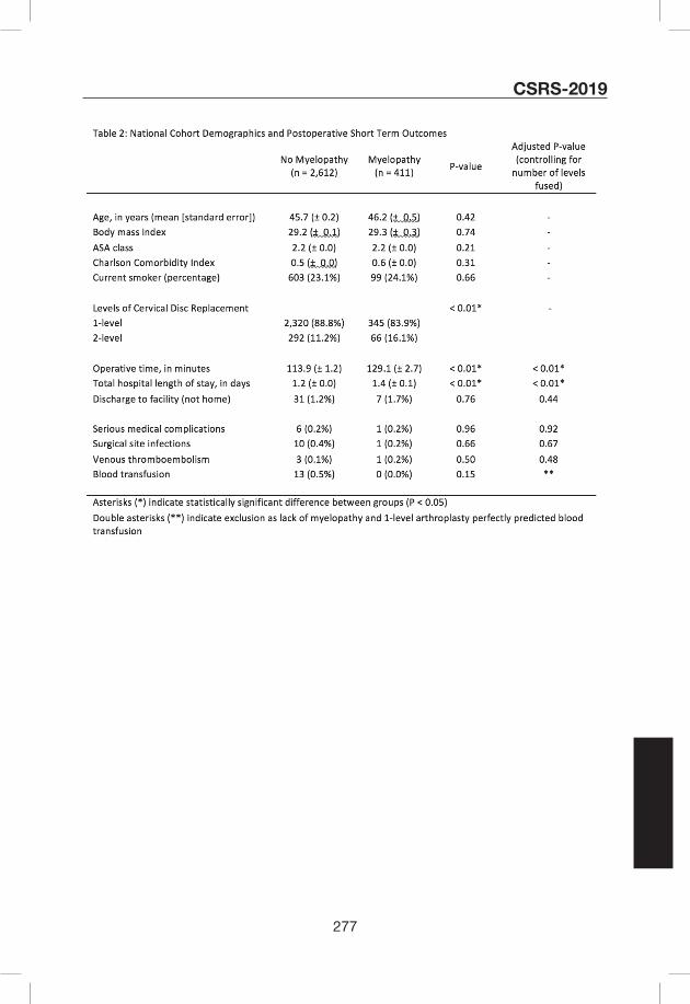
277
CSRS-2019
278
CSRS-2019E-Poster #4
Objective swallowing abnormalities in patients with dysphagia following anterior cervical spine surgery
Pope Rodnoi BS, John C Wuellner MD, Adam M Wegner MD, Shumon Ian Dhar MD, Machelle D. Wilson PhD, Peter C Belafsky MD PhD, Eric O Klineberg MD1. Center for Voice and Swallowing, Department of Otolaryngology-Head & Neck Surgery, University of California, Davis Sacramento, USA (SID, PCB) 2. University of California Davis, Department of Orthopaedic Surgery, Sacramento, CA, USA (PR, JCW, EOK)3. University of California Davis, Department of Public Health Sciences, Sacramento, CA, USA (MDW)4. Washington University in Saint Louis, Department of Orthopedic Surgery Saint Louis, MO, USA (AMW)
Introduction: The most common complication after anterior cervical spine (ACS) surgery is dysphagia. It may result from pharyngeal edema, structural changes, or nerve injury, which can impair vocal fold mobility, pharyngeal sensation, or pharyngeal function. We report the largest series and most detailed characterization of swallowing parameters in patients with dysphagia after ACS.
Methods: A case series of dysphagic patients following ACS who were referred to an otolaryngologist at a single center for dysphagia evaluation between 01/2012 and 12/2017 was analyzed. Parameters from video fluoroscopic swallow studies including lateral upper esophageal sphincter opening (UES-L), pharyngeal constriction ratio (PCR), and penetration aspiration scale (PAS) were measured. Number of levels fused, type of plate used, and primary versus revision surgery was abstracted.
Results: 129 ACS patients with dysphagia were age and gender matched to control patients with dysphagia who did not have ACS surgery. Mean age was 63 (+/- 11 years) and 54% were female. 72.9% had anterior cervical discectomy and fusion (ACDF) with a plate, 11.6% had a no profile implant, 2.3% had disc arthroplasty (DA), and 12.4% had hybrid surgery (ACDF + DA). Mean number of levels fused was 2.2 (+/- 0.9). 11.6% (15/129) were revision surgeries. Mean time from ACS to swallow study was 58.3 months (+/- 63.2). 8% (10/129) had endoscopic vocal fold immobility. 7% of patients exhibited aspiration (penetration aspiration scale (PAS) > 5). Mean PCR for ACS patients was 0.12 (+/- 0.12) vs. control 0.08 (+/- 0.08) (p = 0.01), indicating significant pharyngeal weakness (Figure 1). There was no difference in mean lateral UES-L opening (0.84 (+/- 0.23) vs. control 0.86 (+/- 0.22)) (p = 0.52).
Discussion and Conclusion: Swallowing dysfunction in most patients after ACS is related to pharyngeal weakness and not vocal fold immobility, aspiration, or diminished UES opening. Preventive measures should focus on the relationship between surgical approach, plate morphology, and etiology of pharyngeal dysfunction.

279
CSRS-2019
Figure 1. Regression plot of EAT-10 and PCR

280
CSRS-2019E-Poster #5
A Predictive Model and Nomogram for Predicting Return to Work at 3 Months After Cervical Spine Surgery: An Analysis from the Quality Outcome Database
Clinton J. Devin, MD1; Mohamad Bydon, MD2; Mohammed Ali Alvi, MBBS2; Panagiotis Kerezoudis,MD2; Inamullah Khan, MD1 ; Ahilan Sivaganesan, MD1; Matthew J. McGirt,MD3; Kristin R. Archer, PhD, DPT4; Kevin T. Foley, MD5; Praveen Mummaneni,MD6; Andrew K. Chan, MD6; Erica Bisson, MD,MPH7; Jian Guan, MD7; John J. Knightly,MD8; Christopher Shaffrey,MD9; Anthony L. Asher, MD3
1. Department of Orthopedic Surgery and Neurological Surgery, Vanderbilt Spine Center, Vanderbilt University Medical Center, Nashville, Tennessee2. Department of Neurologic Surgery, Mayo Clinic, 200 First Street SW, Rochester, Minnesota3. Department of Neurological Surgery, Carolina Neurosurgery and Spine Associates and Neurological Institute, Carolinas Healthcare System, Charlotte, North Carolina4. Department of Physical Medicine and Rehabilitation, Vanderbilt University Medical Center, Nashville, Tennessee5. Department of Neurosurgery, University of Tennessee Health Sciences Center, Semmes Murphey Neurologic and Spine Institute, Memphis, Tennessee6. Department of Neurological Surgery, University of California, San Francisco, California7. Department of Neurologic Surgery, University of Utah, Salt Lake City, Utah8. Atlantic Neurosurgical Specialists, Morristown, New Jersey9. Department of Neurosurgery, University of Virginia Medical Center, Charlottesville, Virginia
Introduction: Neck pain is one of the most common causes of work-loss due to disability. Due to recent changes in healthcare policies, return to work (RTW) has been increasingly prioritized by physicians and hospitals to optimize healthcare delivery. In this manuscript, we utilized a national spine registry to identify clinical factors associated with RTW at 3 months among patients undergoing a cervical spine surgery.
Methods: We queried the Quality-Outcomes-Database registry for preoperatively employed patients undergoing cervical spine surgery for degenerative spine disease. Multiple-imputations were used for missing values and multivariable (MV) logistic regression analysis was employed to identify factors associated with higher odds of returning to work. A nomogram was constructed using the results of the MV model.
Results: A total of 4689 patients were analyzed, of which 82.2 %(n=3854) returned to work at 3-months postoperatively. Among previously employed and working patients, 88.3% (n=3443) patients returned to work compared to 53.3% (n=411) among those who were employed but not working (p<.001). On MV-analysis we found that patients who were less likely to RTW were older (Age>56-65:OR 0.69, 95% CI= 0.57-0.85,p<0.001;>65: OR 0.65, 95%CI= 0.43-0.97,p=0.02), were employed but on leave (OR 0.24, 95%CI: 0.20-0.29,p<0.001), were employed part-time (OR0.56, 95% CI=0.42-0.76, p<0.001), had a heavy (OR 0.42, 95% CI: 0.32- 0.54,p<0.001) or medium (OR 0.59, 95% CI:0.46-0.76,p<0.001) intensity occupation, had worker’s compensation (OR0.38, 95%CI:0.28-0.53,p<0.001), had a higher NDI score at baseline (OR 0.60, 95%CI:0.51-0.70,p=0.017), more likely to present with myelopathy (OR 0.52,95%CI:0.42-0.63,p< 0.001) and had more levels fused (3-5 levels:OR 0.46, 95%CI 0.35-0.61,p<0.001). We then constructed a
281
CSRS-2019
nomogram to predict RTW which was found to have an area under the curve (AUC) of 0.812 and good validity.
Conclusion: Multiple factors are predictive of 3-month RTW following cervical spine surgery including active employment, low-intensity occupation, and non-worker’s compensation status. Construction of nomogram to predict RTW was associated with good validity. The results from this study could help the surgeons identify at-risk patients so that preoperative expectations could more comprehensively be discussed.

282
CSRS-2019E-Poster #6
An Evaluation of Surgeon Ability to Predict Ossification of the Posterior Longitudinal Ligament (OPLL) using MRI and X-Rays Alone
Joseph Osorio, Meghana Vulapalli, Nathan Lee, Meghan Cerpa, James Lin, Simon Morr, Richard Menger, Griffin Baum, Jae Hong Ha, Hyoungmin Kim, Louis Amorosa, Marc Dyrszka, Patrick Reid, Zeeshan Sardar, K. Daniel Riew
Introduction: Ossification of the posterior longitudinal ligament (OPLL) is associated with an increased risk of dural tears during anterior cervical surgery. Most experienced cervical spine surgeons have had the experience of operating on what they thought was a cervical disc herniation, only to find OPLL intraoperatively. While CT scans can easily identify OPLL, due to the radiation, routine use of CT in surgical patients is not recommended. Therefore, we sought to determine if an X-ray and MRI alone were sufficient at identification of OPLL.
Materials/Methods: 162 cervical levels in 103 consecutive anterior cervical surgery candidates without prior history of cervical spine surgery and with a CT scan of their cervical spine were included. All patients had plain radiographs and MRI, in addition to CT. 10 spine surgeons assessed radiographs and MRIs at two separate time points to determine if OPLL was present/suspected. Patients with OPLL were further categorized as either mild (7), moderate (8), or severe (2). R software was used for statistical analysis.
Results: CT scans identified OPLL in 16.5% of cases (17 of 103) Table 1. The intra-observer (k = 0.42), and inter-observer reliability (k = 0.26) were both poor at predicting OPLL using X-ray and MRIs alone. The diagnostic specificity and sensitivity of the surgeon raters was 93.19% and 26.84% respectively. When specifically observing the three categories of OPLL, raters were able to identify OPLL 10% of the time in mild cases, 34.70% of the time in patients with moderate OPLL and surprisingly, only 72.27% of the time, even in severe cases Figure 1 demonstrates two OPLL cases where surgeon raters identified moderate OPLL only 40% of the time in the first example (A-E), and 70% of the time in the second example of a patient with severe OPLL (F-J).
Conclusion: CT scans identified OPLL in 16.5% of cases that were initially planned for anterior cervical surgery. Spine surgeon assessments of X-ray and MRIs alone for identifying OPLL was unreliable with weak intra-observer and inter-observer reliability. When observing mild, moderate and severe cases of OPLL, the surgeon raters were unable to identify OPLL in the mild and moderate cases with only MRI and X-rays available. However, even in severe cases, OPLL was only identified in 72.27% of cases. This suggests that, in the absence of identifiable characteristic on MRI and plain radiographs, CT scans might be necessary in more cases, in order to identify OPLL pre-operatively.

283
CSRS-2019
Table 1. Patient Demographics and Levels.
Male sex total (percentage) 73 (71%)
Age mean (SD) 46.0 yr (11.1)
Levels total 162
Levels per patient mean (SD) 1.4 (0.6)
OPLL 17
Mild OPLL total (percentage of OPLL) 7 (41%)
Moderate OPLL total (percentage of OPLL) 8 (47%)
Severe OPLL total (percentage of OPLL) 2 (11%)
Figure 1. Two OPLL patient examples. Patient 1 (A-E) is a patient with moderate OPLL that 40% of surgeon raters were able to identify OPLL using the X-ray and MRI alone. Patient 2 (F-J) is a patient with severe OPLL that 70% of surgeon raters were able to identify OPLL using the X-ray and MRI alone.

284
CSRS-2019E-Poster #7
Comparison between Japanese Orthopaedic Association score (JOA score) and patient-reported JOA score (PRO-JOA score) for evaluating surgical outcomes of cervical myelopathy
Yasushi Oshima, So Kato, Toru Doi, Koji Nakajima, Shima Hirai, Sakae Tanaka
Department of Orthopaedic Surgery, The University of Tokyo
Introduction: The Japanese Orthopedic Association (JOA) score is a widely used disease-specific outcome tool that can provide a quantitative measure for patients with cervical compression myelopathy. However, JOA score is based on the physicians’ point of view and can be biased by surgeons, as opposed to patient-reported outcomes (PROs), such as EuroQOL (EQ-5D), neck disability index (NDI), and short form-12 (SF-12). Although JOA score can more accurately evaluate neurological improvement as compared to general PROs, it is necessary for the physicians to personally examine the patients to provide an accurate JOA score. We questioned whether JOA score can function as a PRO, wherein the patients can respond to the JOA written questionnaire, similar to other PROs. Therefore, this study aims to compare the JOA score provided by patients (PRO-JOA score) with the JOA score provided by physicians.
Methods: Between 2009 and 2013, a consecutive series of patients undergoing posterior decompression surgery for cervical compressive myelopathy at a single academic institution were retrospectively reviewed. Patients with rheumatoid arthritis, tumors, or a history of trauma were excluded. Patients responded to the questionnaires included in SF-12, EQ5D, NDI, and PRO-JOA pre- and >12 months postoperatively. The scores and outcomes between JOA and PRO-JOA were compared using Spearman’s rho and Bland–Altman analyses to investigate their correlations.
Results: This retrospective analysis included 75 patients, whose preoperative JOA and PRO-JOA scores (10.8 and 10.6, respectively) were not statistically significant (p = 0.45), with a correlation coefficient (Spearman’s rho) of 0.74. Likewise, the postoperative JOA and PRO-JOA scores (13.3 and 12.9, respectively) were not statistically significant (p = 0.06), with a correlation coefficient of 0.68. However, the recovery rates for JOA and PRO-JOA scores (42% and 27%, respectively) were significantly different (p<0.01), with a correlation coefficient of 0.45. Compared with other PROs, JOA and PRO-JOA scores showed moderate correlations. Bland–Altman analyses revealed that limits of agreement (LOA) were −4.3 to 4.7 for preoperative score, −3.4 to 4.3 for postoperative score, and −75% to 106% for recovery rate.
Conclusion: This study identified that PRO-JOA score can also be used as a disease-specific scoring measure because JOA and PRO-JOA scores had strong correlations both pre- and postoperatively and further demonstrates moderate correlation with other PROs. Therefore, PRO-JOA score can be used, for example, to determine the neurological status of patients who reside distantly from the hospital and are unable to visit their physicians postoperatively. However, compared with JOA score, our results identified that PRO-JOA score showed slightly lower postoperative values and recovery rates. Furthermore, the large LOA by Bland–Altman analysis indicates that PRO-JOA and JOA should not be used interchangeably. Although both measures demonstrate a similar trend as a group analysis, a detailed interpretation of patients whose JOA and PRO-JOA scores show discrepancy can provide valuable information. Taken together, PRO-JOA score can be used to measure outcomes in patients with cervical myelopathy.

285
CSRS-2019E-Poster #8
Proton Pump Inhibitor Use Affects Pseudarthrosis Rates and Influences Patient Reported Outcomes
John J Mangan, MD, MHA1, Srikanth Divi, MD1; James C. McKenzie, MD1; Justin Stull MD1; David Casper MD; Dhruv K.C. Goyal, BA1; Mark F. Kurd MD1; Alan S. Hilibrand MD1; Scott Wagner MD2; Jeffery A Rihn MD1; D. Greg Anderson MD1; Gregory D. Schroeder MD1; Alexander R. Vaccaro MD, PhD, MBA1; Christopher K. Kepler MD, MBA1
1Rothman Institute at Thomas Jefferson University Hospital, Philadelphia, PA
2Walter Reed Medical Center, Bethesda, MD
Introduction: Cervical pseudarthrosis is a frequent cause of need for revision ACDF and may lead to worse patient reported outcomes. Well-established risk factors for cervical pseudarthrosis include diabetes, smoking, and malnutrition. However, the effect of proton pump inhibitors (PPI), which are widely prescribed medications that affect bone healing and density, on cervical fusion rates are unknown. The purpose of this study was to determine if patients taking PPIs have higher rates of non-union after ACDF.
Methods: A retrospective cohort review was performed to compare patients who were taking PPIs preoperatively with those not taking PPIs prior to ACDF. Patients younger than 18 years of age, those with less than one-year follow-up, and those undergoing surgery for trauma, tumor, infection, or revision were excluded. The rates of clinically diagnosed pseudarthrosis and radiographic pseudarthrosis were compared between PPI groups. Patient outcomes (NDI, PCS-12, MCS-12, and VAS Neck and Arm scores), pseudarthrosis rates, and revision rates were compared between PPI groups using either multiple linear or logistic regression analysis, controlling for demographic and operative variables.
Results: Out of a total of 264 patients, 58 patients were in the PPI group and 206 were in the non-PPI group. A total of 23 (8.71%) patients were clinically diagnosed with pseudarthrosis with a significant difference between PPI and non-PPI groups (p = 0.009). Using multiple linear regression, PPI use was not found to significantly affect any patient reported outcome measure. However, based on logistic regression, PPI use was found to increase the odds of clinically diagnosed pseudarthrosis (Odds Ratio 3.552, p = 0.014). Additionally, clinically diagnosed pseudarthrosis negatively influenced improvement in PCS-12 scores (β = -4.865 [-9.025, -0.706], p = 0.022).
Conclusions: PPI use was found to be a significant predictor of clinically diagnosed pseudarthrosis following ACDF surgery. Furthermore, clinically diagnosed pseudarthrosis negatively influenced improvement in PCS-12 scores. Currently, PPI use is widespread and the surgeon should be aware of patients taking this type of medication preoperatively. Further studies are needed for a more comprehensive understanding of the relationship between PPI use and outcomes following ACDF surgery.
286
CSRS-2019E-Poster #9
C2 versus C3 as the Upper Instrumented Vertebra for Patients Undergoing Long Segment Posterior Cervical Fusion
Ryan K. Badiee BS BA, Andrew K. Chan MD, Joshua Rivera, Chih-Chang Chang MD, Dean Chou MD, Praveen V. Mummaneni MD, Lee A. Tan MD.
Introduction: Posterior cervical laminectomy and fusion (PCLF) is a common procedure for treatment of multilevel cervical degenerative disease. For patients who require long segment posterior cervical construct, the evidence is unclear as to whether C2 vs C3 would be the ideal upper instrumented vertebra (UIV). This study analyzes postoperative outcomes for PCLF comparing constructs with UIV at C2 and C3.
Materials/Methods: Adult patients undergoing PCLF from 2012 to 2018 at a single center were identified. Patients with UIV at either C2 or C3 and an LIV at or above T2 were included. Demographic data, surgical characteristics, and clinical outcomes were compared. Subgroup analyses stratified patients by whether the construct crossed the cervicothoracic junction, preoperative cSVA (> or ≤ 40 mm), or preoperative cervical lordosis minus T1 slope ((> or ≤ 15 degrees). To identify risk factors associated with UIV, univariate analysis was performed, using Student’s t test for continuous variable, and chi-squared or Fisher exact tests for categorical variables. Multivariate logistic regression analyses were utilized to determine the effect of UIV on clinical and radiographic outcomes.
Results: A total of 132 patients were included, of whom 50 (37.8%) had a UIV at C2 and 82 (62.2%) had a UIV at C3. Overall, there was no difference in postoperative radiographic or clinical outcomes in C2 vs C3 UIV groups. There were no significant differences in complication rate (p=0.56), reoperation (p=0.27), postoperative cSVA (p=0.42) or postoperative Nurick score (p=0.96). For patients with lower instrumented vertebra (LIV) at lower cervical spine (not crossing CTJ), C2 UIV group had fewer complications (OR: 0.006, p=0.01) and an increased rate of discharge home (p=0.049). Neck and arm pain were not significantly different between C2 and C3 UIV groups (p=0.54 and p=0.80 respectively). Extending fusion distally to upper thoracic spine (LIV at T1 or T2) attenuated these benefits, with no significant difference in complication rate (p=0.33) or discharge home (p=0.09). In patients with a preoperative cSVA < 40 mm, a C2 UIV was associated with decreased in postoperative length of stay of 0.6 days compared to those with UIV of C3 (p=.005). Subgroups with normal vs high preoperative CL-T1 had no significant differences in outcomes.
Conclusion: After adjusting for other factors, there was no significant difference in postoperative clinical and radiographic outcomes in C2 vs C3 UIV groups. Subgroup analyses reveal that C2 UIV is associated with fewer complications and higher rates of discharge to home in patients with LIV at lower cervical spine, and shorter hospital stays in patients with preoperative cSVA <40mm.

287
CSRS-2019E-Poster #10
A Prospective, Psychometric Validation of NIH PROMIS Physical Function, Pain Interference and Upper Extremity CAT in Cervical Spine Patients: Successes and Key Limitations
Sravisht Iyer, MD1; Jayme C.B. Koltsov, PhD2; Michael Steinhaus, MD1; Thomas Ross, RN1; Kelsey Young, BA1;Daniel Stein, BS1; Jingyan Yang, PhD1; Virginie Lafage, PhD1; Todd Albert, MD1; Han Jo Kim, MD1
1Hospital for Special Surgery, New York, NY2Department of Orthopedic Surgery, Stanford University, Palo Alto, CA
Introduction: Current patient-reported outcomes in cervical pathology have substantial limitations. Patient Reported Outcomes Measurement Information System (PROMIS) Computer Adaptive Tests (CATs) offer the potential for improved psychometric properties with reduced questionnaire burden. The purpose of this study was to validate PROMIS CATs against existing patient-reported outcomes in patients undergoing surgery of the cervical spine.
Methods: Adult patients undergoing cervical spine surgery at a single institution between 2016-2018 were prospectively enrolled. Patients undergoing surgery for instability due to trauma were excluded. Patients completed questionnaires (SF-36, NDI, VAS arm/neck, and PROMIS Pain Interference (PI), Physical Function (PF), and Upper extremity (UE) CATs) preoperatively and at 6 months postoperatively. Demographic data, diagnosis, and procedural data were recorded. Validation of the instruments was completed with a Rasch Model as well as measurement of coverage, efficiency, test-retest reliability, responsiveness, and convergent validity.
Results: Of 164 eligible patients, 137 had 6-month follow-up data. The most common diagnoses were radiculopathy (37.6%), myeloradiculopathy (30.5%), and myelopathy (28.4%). All studied instruments had acceptable fit to a Rasch model. PROMIS CATs demonstrated improved average time to completion for PI (39s), PF (47s), and UE (54s), compared to NDI (117s) and SF-36 PCS (175s). Responsiveness for PROMIS CATs was similar to NDI and SF-36. Test-retest reliability was lower for PI (ICC:0.68), PF (0.70), and UE (0.59), compared to NDI (0.86) and PCS (0.85). For convergent validity, PI was strongly correlated to NDI and PF to SF-36 PCS (Figure 1). There were no significant floor or ceiling effects for the PROMIS domains, although UE had postoperative clustering (n=18) at a high score (56.4) and PI had postoperative clustering (n=27) at a low score (38.7) (Figure 2).
Conclusions: PROMIS CATs demonstrate several advantages, including improved efficiency and comparable responsiveness and convergent validity with legacy instruments in cervical spine patients. Nevertheless, CATs had lower test-retest reliability and had significant clustering at higher levels of function for the PI and UE CATs. These limitations must be considered before broad adoption of CATs in cervical spine patients.

288
CSRS-2019
Figures
Figure 1. Correlation between post-operative PROMIS and legacy outcome measures. Note that as with the pre-operative scores, the PROMIS PI is most strongly correlated with the NDI and that the PF is most strongly correlated with the SF-36 PCS.rs=Spearmans Correlation Coefficient; PROMIS=Patient Reported Outcomes Measurement Information System; PI = Pain Interference; PF = Physical Function; UE=Upper Extremity; NDI=Neck Disability Index; SF-36= Short Form 36 survey; PCS = Physical Components Subscore

289
CSRS-2019
Figure 2. Pre- and Post-operative scores of the various outcome measures. Note the clustering at the lower range for PROMIS PI and at the upper extremes for PROMIS UE (both represent higher levels of function). Although not a true ”floor” effect, these suggest some difficulty differentiating between patients at higher levels of function using these instruments. All patients in the ”clustered” group had reached a maximum of 12 questions.PROMIS=Patient Reported Outcomes Measurement Information System; PI= Pain Interference; PF=Physical Function; UE=Upper Extremity; NDI=Neck Disability Index; SF-36= Short Form 36 survey; PCS=Physical Components Subscore; MCS=Mental Components Subscore; VAS=Visual Analog Scale

290
CSRS-2019E-Poster #11
Modified Frailty Index Predicts Readmission Rates and Extended Length of Stay Following ACDF Surgery for Degenerative Cervical Disease
Brian L. Dial, MD, Valentine R. Esposito, MD, Richard M. Danilkowicz, MD, Jeffrey O’Donnell, MD, Norah Foster, MD, Isaac Karikari, MD, Sergio Mendoza-Lattes, MD, Melissa Erickson, MD
Introduction: The modified frailty index 11-point scale (mFI-11) based on the Canadian Study of Health and Aging Frailty Index (CSHA-FI) is a validated predictor of patient frailty, and correlates with 30-day post-operative complications following spine surgery. The purpose of this study was to investigate the mFI-11 as a perioperative risk assessment tool to identify patients at increased risk for 90-day hospital readmission or extended length of stay (LOS) following elective anterior cervical decompression and fusion (ACDF). The hypothesis was that patients with increased frailty would correlate with increased hospital readmission rate and extended length of hospitalization.
Materials/Methods: The cohort included elective ACDFs at a single institution from 2013 to 2017. Patients meeting inclusion criteria were eligible for chart review. The mFI-11 was calculated for each patient. Hospital LOS and 90-day hospital readmissions were determined for each patient. Extended length of stay was defined as patient being discharged after post-operative day one. Regression analysis was performed to correlate the mFI-11 to readmission rates and LOS. Multivariate analysis was performed to assess whether the mFI-11 was a significant predictor of hospital readmission or LOS when controlling for age > 65 years, BMI, number of levels fused, ASA class, and indication for surgery (myelopathy vs. radiculopathy).
Results: The cohort included 1,896 patients. 120 patients (6.3%) required a 90-day hospital readmission, and 265 patients (14%) required an extended LOS. The mFI-11 strongly linearly correlated with 90-day readmission rates (r2=0.99), and strongly exponentially correlated with extended LOS (r2=0.93) (Figures 1 and 2). The 90-day readmission rate was 3.72% for patients with a mFI-11, and increased to 19.4% when the mFI-11 was greater than or equal to 0.27. The most common indications for readmission were dysphagia, pain control, and respiratory complications. In the multivariate analysis the following variables were statistically associated with 90-day readmissions: mF-11 0.09 (OR 1.94, 95% CI 1.24-3.08), mF-11 0.18 (OR 2.77, 95% CI 1.49-5.06), mF-11 0.27 (OR 6.02, 95% CI 2.02-15.9), and radiculopathy as the indication for surgery (OR 0.60, 95% CI 0.36-0.95). In the multivariate analysis the following variables were statistically associated with an extended length of hospitalization: mF-11 0.18 (OR 2.03, 95% CI 1.43-3.25), mFI-11 0.27 (OR 6.62, 95% CI 2.86-15.69), mFI-11 0.36 (OR 18.45, 95% CI 2.4-275.24), and age > 65 (OR 2.08, 95% CI 1.52-2.84).
Conclusion: The mFI-11 was an independent predictor of both 90-day hospital readmission rate and extended length of hospitalization. The mFI-11 strongly correlates in a linear fashion for 90-day hospital readmission rates, and in an exponential fashion for extended length of stay.

291
CSRS-2019
Figures:Modified Frailty Index vs. Rate of Readmission
Modified Frailty Index vs. Extended Length of Stay
292
CSRS-2019E-Poster #12
PROMIS-29 Validity and Conversion Equation to Neck Disability Index (NDI) using a National Sample of Cervical Spine Surgery Patients
Jacquelyn S. Pennings, Claudia A. Davidson, Inamullah Khan, Mohamad Bydon, Anthony L. Asher, Clinton J. Devin, John Paul Wanner, Daniel Verhotz, Kristin R. Archer
Introduction: Patient Reported Outcomes Measures (PROMs) have become a vital tool for evaluating success of spine surgery. Commonly used PROMs, like the Neck Disability Index (NDI) used to assess pain related disability, tend to be narrow in scope and used for specific populations. The Patient Reported Outcomes Measurement Information System (PROMIS) domains, recommended by the NIH, offers an alternative that is not condition-specific. PROMIS is easily accessible, free, includes multiple domains, and offers two modes of delivery. Despite these benefits, researchers and clinicians are hesitant to use PROMIS and literature is sparse regarding how these measures perform in cervical surgery patients. The purpose was to examine the validity of the PROMIS-29 to better understand it’s use for cervical spine surgery patients. A secondary objective was to calculate a conversion equation between PROMIS-29 and NDI, to allow researchers and clinicians to determine a predicted NDI.
Methods: We conducted a retrospective analysis of prospectively collected national registry data, the Quality Outcomes Database (QOD), which is designed to evaluate risk-adjusted outcomes for the most commonly performed spinal surgical procedures. We queried the QOD registry for patients with PROMIS-29 and NDI responses. Of 619 patients, 41% were female, 87% Caucasian, and 13% had revision surgery. The mean age was 58 years (SD=12).
The PROMIS-29 (v2.0) includes 7 short-form domains with 4 items each rated on a 5-point scale: Physical Function (PF), Depression (DEP), Anxiety, Fatigue, Sleep Disturbance (SD), Ability to Participate in Social Roles and Activities (SR), Pain Interference (Pain), and a single pain intensity (PI) item rated from 0 (no pain) to 10 (worst pain imaginable). The NDI contains 10 questions measuring disability in patients with neck pain. Outcomes were collected prior to surgery and 3 and 12 months post-surgery.
Validity of the PROMIS-29 was assessed using Cronbach’s α, ceiling/floor effects, and principle axis factoring. Multivariable regression predicting NDI scores from PROMIS-29 domains used data from all three timepoints. Predicted NDI scores, derived from PROMIS-29 domains, were plotted against NDI scores to determine how well PROMIS-29 domains predicted NDI. A conversion equation was created from PROMIS regression coefficients.
Results: Results showed good reliability (Cronbach’s α>0.80) and validity of PROMIS-29 in patients undergoing cervical surgery: convergent (r>0.65) and discriminant (r=0.49-0.63) validity, low/high floor/ceiling effects (≤2%), and unidimensional domains (factor loading>0.64). The conversion equation used 6 PROMIS-29 domains (NDIpercent=7.928-0.527*[PFraw] + 1.570*[PAINraw] + 0.995*[SDraw] + 1.101[PIraw] + 0.392[Fatigueraw] – 0.409 [SRraw]). Correlations between predicted and actual NDI scores at each timepoint were: r=0.84 at baseline, r=0.76 at 3 months, and r=0.84 at 12 months, suggesting that the equation predicted NDI scores that are strongly correlated with actual NDI scores (Figure 1).
Conclusions: Good reliability and validity support the use of PROMIS-29 in cervical surgery
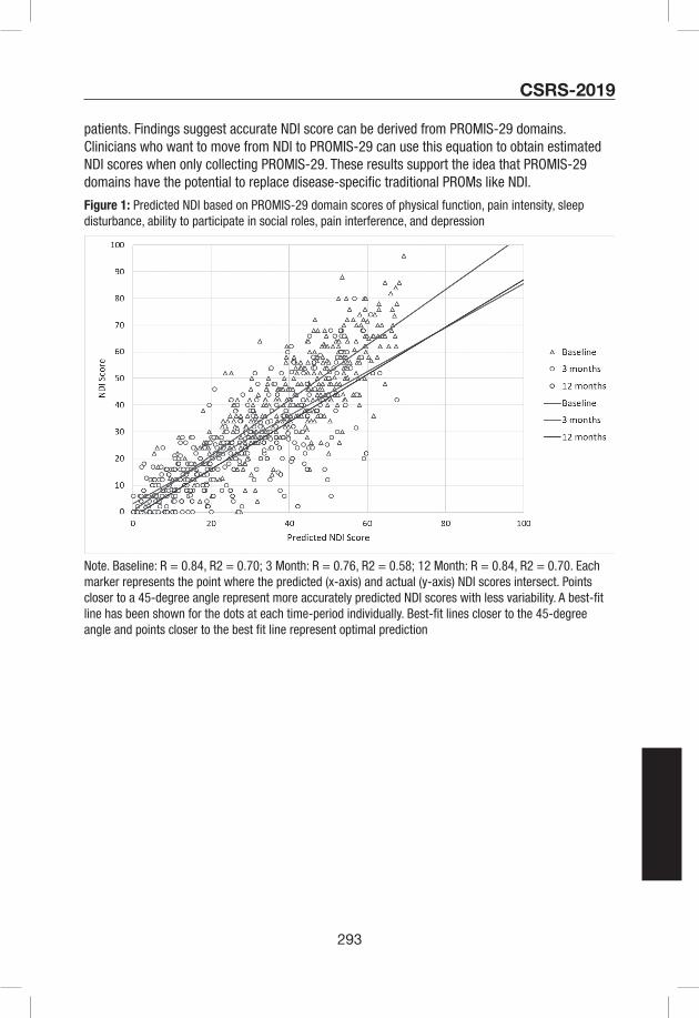
293
CSRS-2019
patients. Findings suggest accurate NDI score can be derived from PROMIS-29 domains. Clinicians who want to move from NDI to PROMIS-29 can use this equation to obtain estimated NDI scores when only collecting PROMIS-29. These results support the idea that PROMIS-29 domains have the potential to replace disease-specific traditional PROMs like NDI.
Figure 1: Predicted NDI based on PROMIS-29 domain scores of physical function, pain intensity, sleep disturbance, ability to participate in social roles, pain interference, and depression
Note. Baseline: R = 0.84, R2 = 0.70; 3 Month: R = 0.76, R2 = 0.58; 12 Month: R = 0.84, R2 = 0.70. Each marker represents the point where the predicted (x-axis) and actual (y-axis) NDI scores intersect. Points closer to a 45-degree angle represent more accurately predicted NDI scores with less variability. A best-fit line has been shown for the dots at each time-period individually. Best-fit lines closer to the 45-degree angle and points closer to the best fit line represent optimal prediction

294
CSRS-2019E-Poster #13
Underweight Patients are the Highest Risk Body Mass Index Group for Perioperative Adverse Events Following Posterior Cervical Spine Surgery
Taylor D. Ottesen, B.S., Paul S. Bagi, MD, Rohil Malpani, B.S., Anoop R. Galivanche, B.S., Arya G. Varthi, MD, Jonathan N. Grauer, MD.
Introduction: Body Mass Index (BMI) is a weight-for-height metric that is used to quantify tissue mass and weight levels. Past studies have mainly focused on the association of high BMI on spine surgery outcomes and shown variable conclusions. Prior results may have varied due to insufficient power or inconsistent categorical separation of BMI groups (e.g. underweight, overweight, or obese). Additionally, few studies have considered outcomes of patients with low BMI. The aim of the current study was to analyze patients along the entirety of the BMI spectrum and to establish specific granular BMI categories for which patients become at risk for complication and mortality following posterior cervical spine surgery.
Methods: Patients undergoing elective posterior spine surgery were abstracted from the National Surgical Quality Improvement Program (NSQIP) databases 2005-2016. Patients that had a primary diagnosis of trauma, tumor, or infection were excluded from analysis. Patients were then aggregated into BMI categories and adverse outcomes were normalized to average risk of normal-weight subjects (BMI 20.0-24.9 kg/m2). Risk-adjusted multivariate regressions were performed controlling for all patient demographics and overall health.
Relative risk of adverse outcomes, normalized to average risk of normal weight subjects (bins BMI 20-24.9), was calculated. Bins were then aggregated BMI categories with underweight being classified as BMI<18.5 kg/m2, normal weight as BMI 18.5-24.9 kg/m2, overweight as BMI 25-29.9 kg/m2, obese as BMI 30-39.9 kg/m2, morbidly obese as BMI 40-49.9 kg/m2 and super morbidly obese as BMI>50 kg/m2. In order to control for patient demographics (age, sex, functional status) and overall health as measured by the American Society of Anesthesiologists (ASA) classification, risk adjusted multivariate regressions were then undertaken.
Results: A total of 16,806 patients met inclusion criteria. Odds for adverse events for underweight patients (BMI<20.0 kg/m2) were the highest among any category of patients along the BMI spectrum. These patients experienced increased odds of any adverse event (Odds Ratio (OR)=1.67, p=0.008, major adverse events (OR=2.08, p=0.001), or major post-operative infection (OR=1.95, p=0.002) when matched to patients in the normal-weight category. Additionally, a protective effect against any adverse event, major adverse event, and mortality were found in overweight and obese patients (Table 1, Figure 1).
Conclusions: The current study finds underweight patients to have the highest risk of adverse events and post-operative infection than any other BMI category, including the super morbidly obese (BMI>50 kg/m2). The low BMI group may represent a compromised population due to possible poor nutrition, comorbid medical conditions, and failure to thrive. Underweight patients are an extremely at-risk population that has not been previously focused on as an at-risk BMI category. Increased pre-surgical planning and asset allocation in this population should be considered by physicians and healthcare systems, as is often already done for patients on the other end of the BMI spectrum.

295
CSRS-2019

296
CSRS-2019E-Poster #14
Is it safe to perform anterior foraminotomy using high speed burrs during anterior cervical discectomy and fusion?: Evaluation on the risk of vertebral artery injury and the safe margin of the anterior foraminotomy
Jae Jun Yang1, Kwang-Sup Song2, Dong-Gune Chang3, Sang-Min Park4, Youngbae B Kim5
1Department of Orthopedic Surgery, Dongguk University Ilsan Hospital, Goyang, Republic of Korea
2Department of Orthopedic Surgery, Chung-Ang University Hospital, College of Medicine, Chung-Ang University, Seoul, Republic of Korea
3Department of Orthopedic Surgery, Sanggye Paik Hospital, College of Medicine, Inje University, Seoul, Republic of Korea
4Department of Orthopedic Surgery, Seoul National university College of Medicine and Seoul National University Bundang Hospital, Seongnam, Republic of Korea
5Department of Orthopedic Surgery, VHS medical center, Seoul, Republic of Korea
Introduction: Anterior foraminotomy (AF) during anterior cervical discectomy and fusion (ACDF) may risk injury to the vertebral artery (VA). To avoid this complication, surgical techniques using Kerrison punches or total uncinate resection have been proposed. However, foraminotomy using Kerrison punches may cause inadequate decompression and total uncinate resection has instability and subsidence issues. High speed burrs can be efficiently used for AF if avoidance of VA injury can be guaranteed. The purposes of this study were to evaluate the risk of VA injury and propose a guideline for secure AF using high speed burrs.
Materials/Methods: Angio-computed tomography (CT) images of the cervical spine preoperatively obtained from thirty patients were evaluated. Surgical simulation of AF with ACDF on the axial CT images was set as follows (Fig 1): mediolateral exposure between bilateral uncovertebral joints using a 40mm self-retractor, longitudinal axis of burrs located 6.5mm and 4mm medial at the top of the retractor considering the radius of hand-pieces (10mm and 5mm, respectively) and a 3mm burr tip (A), and a burr tip targeting the lateral margin of the superior cortex of the pedicle (B). To assess the risk of VA injury, length from the anterior tip to drilling point on the uncinate process (TDL), resection length of the uncinate process (RL), and distance from VA to the burr tip (DVA) were measured. To assess the safe margin of the AF, length from the anterior tip to drilling point 3mm away from the VA on the uncinate process (TD3L) was measured (Fig 2).
Results: The averages of TDLs in the simulation using a burr with 10mm hand-piece (simulation1) were 0.4, 1.7, and 2.9mm in C45, C56, and C67, respectively. The average of TDLs in the simulation using a burr with 5mm hand-piece (simulation 2) were 0.9, 2.1, and 3.1mm in C45, C56, C67, respectively. The averages of RLs were 7.3, 7.0, and 7.7mm in C45, C56, and C67, respectively. The averages of DVAs in simulation 1 were 1.8, 2.6, and 5.2mm in C45, C56, and C67, respectively. The averages of DVAs in simulation 2 were 2.0, 2.8, and 5.5mm in C45, C56, and C67, respectively. The averages of TD3Ls in simulation 1 were 2.2 and 2.8mm in C45 and C56, respectively. The averages of TD3Ls in simulation 2 were 2.4 and 2.9mm in C45 and C56, respectively. When a secure drilling point is set at 3mm away from the anterior uncinate tip based on the averages of TD3Ls, outliers (TD3L more than 3mm) in simulation 1 were 32% and 37% in C45 and C56, respectively. Outliers in simulation 2 were 37% and 40% in C45 and
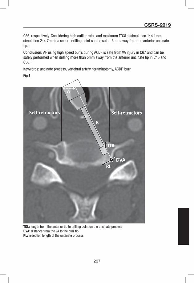
297
CSRS-2019
C56, respectively. Considering high outlier rates and maximum TD3Ls (simulation 1: 4.1mm, simulation 2: 4.7mm), a secure drilling point can be set at 5mm away from the anterior uncinate tip.
Conclusion: AF using high speed burrs during ACDF is safe from VA injury in C67 and can be safely performed when drilling more than 5mm away from the anterior uncinate tip in C45 and C56.
Keywords: uncinate process, vertebral artery, foraminotomy, ACDF, burr
Fig 1
TDL: length from the anterior tip to drilling point on the uncinate processDVA: distance from the VA to the burr tipRL: resection length of the uncinate process
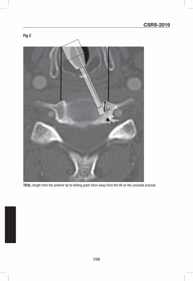
298
CSRS-2019
Fig 2
TD3L: length from the anterior tip to drilling point 3mm away from the VA on the uncinate process
299
CSRS-2019E-Poster #15
PROMIS Physical Function is more relevant for lumbar than for cervical spinal disorders
Avani Vaishnav, MBBS1, Steven McAnany, MD1,2, Sravisht Iyer, MD1,2, Todd Albert, MD1,2, Catherine Gang, MPH1, Sheeraz Qureshi, MD, MBA1,2
1Hospital for Special Surgery
2Weill Cornell Medical College
Introduction: As the focus in spine surgery shifts from radiographic to patient-centric outcomes, patient-reported outcomes measures(PROMs) are becoming increasingly important. They are linked to patient satisfaction, and are used to assess healthcare expenditure, determine compensation and evaluate cost-effectiveness. Thus, PROMs are important to various stakeholders, including patients, physicians, payers and healthcare institutions. While Oswestry Disability Index (ODI) and Neck Disability Index (NDI) are most commonly used, PROMIS is a new outcome measure that is increasingly being implemented. The purpose of this study was to evaluate if PROMIS Physical Function (PROMIS-PF) is a good surrogate for disease-specific outcome measures in common spinal conditions.
Methods: This was a retrospective review of prospectively collected data and patients undergoing minimally invasive spine surgery (MIS) for degenerative spinal conditions were included. PROMs (ODI, NDI, PROMIS-PF) were collected at each time-point and analyzed. Pearson product-moment correlation was used for correlations between ODI and PROMIS-PF in lumbar surgery, and between NDI and PROMIS-PF in cervical surgery. Fisher Z-transformation was used to compare the strength of correlations at each time-point.
Results: Of the 408 patients included, 325 underwent lumbar and 83 underwent cervical spinal procedures for degenerative conditions of the spine. In lumbar patients, ODI decreased from 40.80+19.24 pre-operatively to 18.97+16.72 at 6 months; and PROMIS-PF improved from 35.28+7.81 to 45.37+9.25. In cervical patients, NDI decreased from 36.70+19.45 pre-operatively to 21.61+17.71 at 6 months; and PROMIS-PF improved from 39.65+7.66 to 46.85 + 8.81.
As seen in Table 1, A statistically significant negative correlation was seen at all time-points in both cohorts (p<0.0001), which was strong between ODI and PROMIS-PF (r ranged from -0.645 to -0.774), but moderate between NDI and PROMIS-PF (r ranged from -0.483 to -0.645).
As seen in Table 2A comparison of the strengths of the correlations showed that the correlation between ODI and PROMIS-PF was stronger than that between NDI and PROMIS-PF at all time-points, with this difference being statistically significant at 6 weeks (p=0.020), and approaching statistical significance pre-operatively (p=0.055) and at 6 months (p=0.056). It is likely that these difference may have been statistically significant at other time-points with a larger sample size.
Conclusion: These findings indicate that ODI and PROMIS-PF are highly correlated, but NDI and PROMIS-PF are only moderately correlated. Thus, PROMIS-PF appears to adequately capture functional impairment due to lumbar disease, but not that due cervical disease. Prior studies have shown PROMIS to be correlated with legacy measures, and computer adaptive testing allows for faster administration and reduced patient burden. Despite these advantages, it is important to keep in mind that PROMIS was designed to be used across diseases and it may
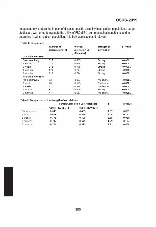
300
CSRS-2019
not adequately capture the impact of disease-specific disability in all patient populations. Larger studies are warranted to evaluate the utility of PROMIS in common spinal conditions, and to determine in which patient populations it is truly applicable and relevant.
301
CSRS-2019E-Poster #16
Are Preoperative PHQ-9 Scores Predictive of Postoperative Outcomes Improvement Following Anterior Cervical Discectomy and Fusion?
James M. Parrish, MPH, Joon S. Yoo, BA, Dil V. Patel, BS, Brittany E. Haws, MD, Benjamin Khechen, BA, Sailee S. Karmarkar, BS, Eric H. Lamoutte, BS, Kern Singh, MD
Introduction: Few studies have quantified depression symptoms in the preoperative period using a validated metric such as Patient Health Questionnaire-9 (PHQ-9) and have tracked patient-reported outcomes (PROs) following common spinal procedures, such as an anterior cervical discectomy and fusion (ACDF). This study aims to determine if there is an association between preoperative depression, as quantified by PHQ-9, and postoperative improvement in pain and disability after an ACDF.
Method: Patients undergoing a primary, one- to three-level ACDF were retrospectively reviewed and stratified by their preoperative PHQ-9 score: minimal to no depression (<5) or mild to severe depression (≥5). PROs, including Neck Disability Index (NDI), Visual Analogue Scale (VAS) neck and arm pain, and 12-Item Short Form (SF-12) Physical Component Score (PCS), were measured preoperatively and at 6-week, 3-month, 6-month, and 1-year postoperatively. PRO scores were analyzed amongst PHQ-9 cohorts using multivariate linear regression. Achievement of minimum clinically important difference (MCID) was compared using chi-square analysis.
Results: 119 patients were included: 51 had a preoperative PHQ-9 score <5 and 68 had a PHQ-9 score ≥5. Higher PHQ-9 scores were associated with increased preoperative NDI, VAS neck, and VAS arm scores and significantly lower SF-12 PCS scores preoperatively. Cohorts experienced similar VAS pain scores up to 1-year following surgery, except for VAS neck pain at 3-months when patients with greater depression symptoms had more pain. High PHQ-9 patients had higher NDI values at 6-week and 3-month marks, however, they had similar NDI scores at 6-month and 1-year compared to non-depressed patients. Similarly, SF-12 PCS scores were lower for patients with a higher PHQ-9 score at 3-months and 6-months, however, both groups had similar scores at 1-year follow-up. A greater percentage of the high PHQ-9 cohort achieved MCID for NDI, however, there were no differences in MCID achievement for VAS neck, VAS arm, or SF-12 PCS.
Conclusion: In this study, we identified patients with mild to severe depression symptoms using PHQ-9 and compared outcomes following ACDF to patients with no depression symptoms. Patients with worse preoperative mental health reported significantly greater preoperative disability and pain. However, both cohorts demonstrated similar clinical recovery at 1-year follow-up. These findings suggest patients with worse preoperative mental health can expect significant improvements in PROs following surgery.

302
CSRS-2019

303
CSRS-2019E-Poster #17
Evaluation of Postoperative Mental Health Outcomes in Patients Based on PROMIS Physical Function Following Anterior Cervical Discectomy and Fusion
Nathaniel W. Jenkins, MS, Joon S. Yoo, BA, Dil V. Patel, BS, Sailee S. Karmarkar, BS, Eric H. Lamoutte, BS, Kern Singh, MD
Object: Few investigations have evaluated the relationship between preoperative physical health and improvement in mental health following common spinal procedures such as anterior cervical discectomy and fusion (ACDF). This investigation aims to assess the relationship of preoperative physical function, as measured by Patient-Reported Outcome Measurement Information System Physical Function (PROMIS PF), to improvement in mental health, as evaluated by Short Form- 12 Mental Component Summary (SF-12 MCS) following ACDF.
Methods: Patients undergoing primary, one- to three-level ACDF were retrospectively reviewed and stratified based on preoperative PROMIS PF scores: <40 points (severe disability) and ≥40 points (mild disability). PROMIS PF cohorts was tested for an association with demographic characteristics and perioperative variables using Chi-squared analysis and multivariate linear regression. Multivariate linear regression was utilized to determine the association between PROMIS PF cohorts and improvement in SF-12 MCS following ACDF.
Results: 129 one- to three-level ACDF patients were included: 73 had PROMIS PF <40 and 56 had PROMIS PF ≥40. Patients with worse physical function were more likely to be diabetic.
There were no other significant differences in demographic and perioperative characteristics (Table 1). Patients with PROMIS PF <40 reported worse mental health than patients with PROMIS PF ≥40 at the preoperative period and at all postoperative timepoints except for 1-year. Furthermore, both cohorts had similar changes in mental health from baseline through the 6-month follow-up. However, at the 1-year postoperative timepoint, patients with PROMIS PF <40 had a statistically greater change in mental health score compared to patients with PROMIS PF ≥40 mild disability (Table 3).
Conclusion: In this retrospective investigation, we identified patients with varying physical function using PROMIS PF and compared mental health outcomes as assessed by the SF-12 MCS survey following ACDF. Patients with worse preoperative physical function reported significantly worse preoperative and postoperative mental health. However, both cohorts demonstrated similar clinical recovery at the 1-year follow-up. In addition, patients with worse preoperative physical function made significantly greater improvements in mental health from baseline. These findings suggest that patients with worse preoperative physical function can still expect significant improvements in mental health following surgery and should be counseled to expect a similar long-term postoperative outcome compared to patients with better preoperative physical function.
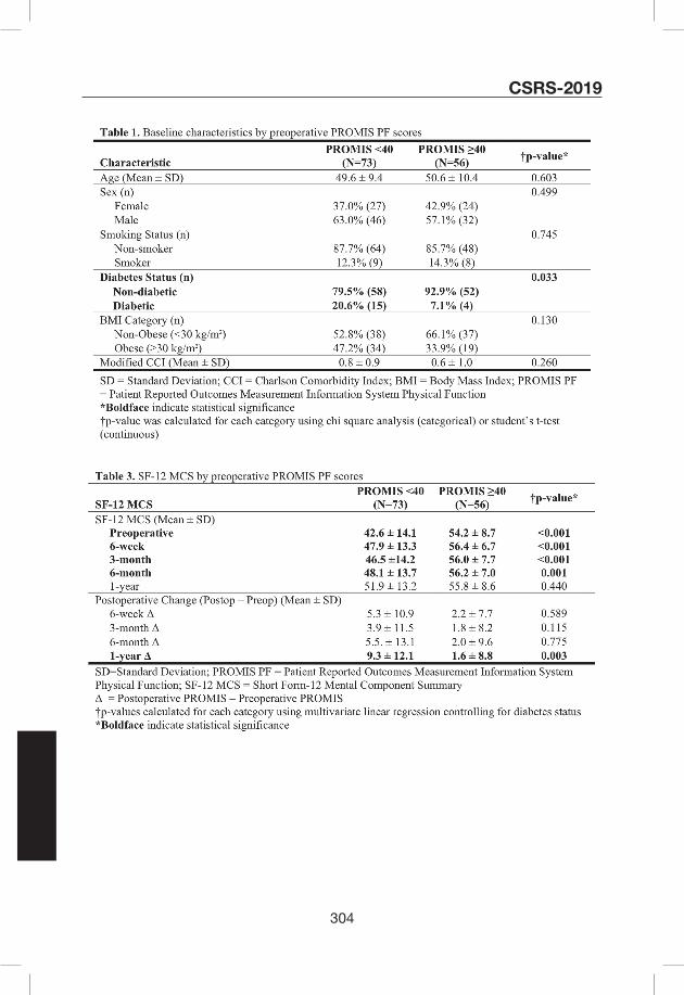
304
CSRS-2019
305
CSRS-2019E-Poster #18
Correlations Among PROMIS-29 Domains Before and After Cervical Spine Surgery
Jacquelyn S. Pennings, Claudia A. Davidson, Inamullah Khan, Mohamad Bydon, Anthony L. Asher, Clinton J. Devin, John Paul Wanner, Daniel Verhotz, Kristin R. Archer
Introduction: Although numerous reviews have been published about which patient reported outcome measures (PROMs) best assess spine surgery outcomes, consensus still does not exist. Most recommendations suggest reporting on outcome domains including physical functioning, pain, and quality of life. The Patient-Reported Outcomes Measurement Information System (PROMIS) tools offer an alternative to legacy measures such as the Neck Disability Index (NDI), which has been recommended by The National Institutes of Health (NIH) as part of the standards for research on chronic spine pain. PROMIS offers many benefits: easy accessibility, free, includes multiple domains, and two modes of delivery including computer adaptive tests and short forms, and providing t-scores that allow for easy comparisons to population averages. Despite these benefits, many researchers and clinicians are hesitant to incorporate PROMIS into research and practice. The purpose was to evaluate the pattern of correlations within the 8 PROMIS-29 domains at baseline and 12-months after surgery in a sample of cervical spine patients.
Methods: The current study is a retrospective analysis of prospectively collected national registry data, the Quality Outcomes Database (QOD), which is designed to evaluate risk-adjusted outcomes for the most commonly performed spinal surgical procedures. We queried the QOD registry for patients with PROMIS-29. 619 patients from 16 US hospitals were included. Of these 41% were female, 87% Caucasian, and 13% had revision surgery. The mean age was 58 years (SD=12).
The PROMIS-29 (v2.0) is comprised of 7 short-form domains with 4 items each rated on a 5-point scale: Physical Function, Depression, Anxiety, Fatigue, Sleep Disturbance, Ability to Participate in Social Roles and Activities, Pain Interference, and one pain intensity item rated from 0 (no pain) to 10 (worst pain imaginable).
Spearman’s correlation coefficients were calculated for the relationship between each pair of domain scores at baseline and 12-months postoperatively. In addition, principle axis factoring (PAF) was used to determine whether there was an underlying factor structure for the 8 domains.
Results: Results show statistically significant correlations between each pair of domain scores at baseline and also at 12-months after surgery (all p-values<.001). At baseline, correlation magnitudes ranged from ρ=-0.32 for Physical Function and Sleep Disturbance to ρ=0.76 for Depression and Anxiety. At 12 months, correlation magnitudes ranged from ρ=0.44 for Depression and Sleep Disturbance to ρ=0.86 for Pain Intensity and Pain Interference. PAF revealed that domain scores factored into one factor with loadings ranging from .88 for pain interference to .71 for sleep disturbance.
Conclusions: The strength of the relationships among PROMIS-29 domains range in magnitude from moderate (.32) to very strong (0.86) and are stronger at 12 months than at baseline. Factor analysis suggests that the 8 domains may be categorized into one higher order factor representing overall health and functioning for cervical spine patients. Pain interference accounted for the most variance in this higher-order factor. This research adds information on

306
CSRS-2019
PROMIS-29 domain scores in cervical spine surgery patients and can aid in researcher’s and clinician’s decisions of whether to use PROMIS-29.
Table 1. Spearman’s correlations among PROMIS-29 domain scores before and 12 months after cervical spine surgery
Physical function
Pain interference
Pain intensity
Participation in social roles
Sleep disturbance
Fatigue Depression
Baseline (N=403)
Pain interference -0.65
Pain intensity -0.44 0.64
Participation in social roles 0.71 -0.76 -0.43
Sleep disturbance -0.32 0.55 0.47 -0.47
Fatigue -0.58 0.61 0.37 -0.63 0.50
Depression -0.44 0.43 0.35 -0.45 0.37 0.56
Anxiety -0.42 0.45 0.35 -0.47 0.42 0.51 0.76
12 Months (N=84)
Pain interference -0.81
Pain intensity -0.76 0.86
Participation in social roles 0.81 -0.82 -0.71
Sleep disturbance -0.45 0.50 0.49 -0.48
Fatigue -0.63 0.64 0.64 -0.65 0.57
Depression -0.60 0.60 0.56 -0.65 0.44 0.56
Anxiety -0.55 0.57 0.54 -0.57 0.45 0.45 0.85
Note. All correlation coefficients p < 0.001.

307
CSRS-2019E-Poster #19
Analysis of anticipatory postural adjustments between normal and cervical myelopathy patient
Haruki Funao1,2, Tatsuya Igawa3, Masaru Matsuzawa3, Kodai Yoshida2, Norihiro Isogai1,2, Yutaka Sasao1,2, Makoto Nishiyama1,2, Ken Ishii1,2
1Department of Orthopedic Surgery, School of Medicine, International University of Health and Welfare (IUHW)
2Department of Orthopedic Surgery & Spine and Spinal Cord Center, IUHW Mita Hospital
3Department of Rehabilitation, IUHW Mita Hospital
BACKGROUND: Patients with cervical myelopathy (CM) are at a risk of fall, and commonly experienced fall-related deterioration. The anticipatory postural adjustments (APAs) are unconscious muscular activities to control the position of center of mass using trunk and lower extremity muscles minimizing loss of equilibrium. However, to date, there were no reports investigating the APA in CM patients. The purpose of this study was to investigate postural control between normal and CM patients according to the APA analysis.
METHODS: A total of 39 subjects; 15 patients (8 male/7 female) who underwent cervical laminoplasty for CM, and 24 age- and sex- matched normal subjects (12 male/12 female) (control group), were enrolled in this study. Both CM group and control groups were required to turn around by side-step. Reflective markers were placed at 18 different points (Fig.1), and three-dimensional motion capture system and 6 force plates were used for the analysis. The preoperative Japanese Orthopaedic Association (JOA) score, turning time, step length, and % step length were measured in both groups. Student t test and Pearson correlation coefficient were used for statistical analyses.
RESULTS: The average age was 66.2 years old in CM group, and 68.4 years old in the control group. The average height/weight were 159.6 cm/60.9 kg in CM group, and 161.5 cm/61.9 kg in the control group. There were no significant differences in age, height, and body weight between the two groups. The APA phase, turning time, step length, and % step length were 0.46 seconds, 2.27 seconds, 310 mm, and 19.5 % in CM group, and 0.38 seconds, 1.95 seconds, 364 mm, and 22.6 % in the control group. The APA phase was significantly longer in CM group (p<0.05). Turning time tended to be longer in CM group (p=0.10), whereas step length and % step length tended to be shorter in CM group (p=0.06, p=0.10). The mean preoperative JOA score of motor function of the upper extremity, that of the lower extremity, and total JOA score in CM group were 2.5, 2.0, and 9.9, respectively. The correlation coefficients between preoperative JOA score of motor function of the lower extremity and the APA phase, turning time, step length, and % step length were -0.35 (p=0.20), -0.53 (p<0.05), 0.48 (p=0.08), and 0.39 (p=0.16). The patients with lower JOA scores of motor function of the lower extremity had a significantly longer duration of turning time, and tended to have longer duration of APA phase, and shorter step length and % step length.
CONCLUSIONS: Patients with CM had a significantly longer duration of APA phase, and this phenomenon tended to appear in patients with lower JOA scores of the lower extremity motor function. It has been difficult to visualize and quantify the postural control and counterbalance the

308
CSRS-2019
perturbation. The APA phase may enable us to quantify postural control (Fig.2), and it would be useful in evaluating lower extremity function and risk of fall in CM patients.
Fig.1
Fig.2
309
CSRS-2019E-Poster #20
Obesity is Not a Risk Factor for Worse Postoperative Outcomes Following Anterior Cervical Discectomy and Fusion
James M Parrish, MPH, Joon S. Yoo, BA, Dil V. Patel, BS, Eric H. Lamoutte, BS, Sailee S. Karmarkar, BS, Kern Singh, MD
Introduction: There is a scarcity in the literature in evaluating clinical outcomes of obese patients following anterior cervical spine surgery. To evaluate the effects of obesity on postoperative improvements in pain and physical function in patients undergoing an anterior cervical discectomy and fusion (ACDF).
Methods: Patients undergoing a primary, one- to three-level ACDF were retrospectively reviewed and stratified based on predefined BMI thresholds: Non-Obese (BMI <30.0 kg/m2) and Obese (BMI ≥30.0 kg/m2). Demographic and perioperative characteristics were compared amongst BMI cohorts using Chi-squared analysis and linear regression. Achievement of minimum clinically important difference (MCID) was compared using Chi-squared analysis. Improvements in patient-reported outcomes were calculated using linear regression.
Results: 122 patients were included: 74 patients were in the non-obese cohort and 48 patients were in the obese cohort. The obese cohort had more males than females; however, there were no significant differences in other demographic and perioperative variables (Table 1). There was no difference in preoperative NDI, VAS neck and arm, and SF-12 PCS between the cohorts. Although the obese patients reported worse scores in all outcome measures through the 6-month time point, this did not reach statistical significance (Table 3). The obese cohort had similar rates of MCID achievement compared to the non-obese cohort with respect to NDI (p=0.605), VAS neck pain (p=0.882), VAS arm pain (p=0.118), and SF-12 PCS (p=0.301).
Conclusion: This retrospective investigation compared clinical outcomes between obese and non-obese cohorts following an ACDF using VAS neck and arm pain, NDI, and SF-12 PCS scores. In contrast to prior investigations, our results demonstrated that patients with a higher BMI report similar preoperative pain and disability and may benefit from similar postoperative improvements through 6 months. Therefore, our study established that BMI may not necessarily be an accurate predictor of the recovery course of patients following ACDF.

310
CSRS-2019

311
CSRS-2019E-Poster #21
Preoperative PROMIS Scores can Predict Patient Satisfaction Following Surgery for Cervical Degeneration
Alvaro Ibaseta MS, Rafa Rahman BS, Nicholas Andrade BS, Richard L. Skolasky ScD, Khaled M. Kebaish MD, Daniel M. Sciubba MD, David B. Cohen MD MPH, Brian J. Neuman MD
From the Department of Orthopaedic Surgery, The Johns Hopkins University School of Medicine, Baltimore, MD
Introduction: Satisfaction with treatment is a primary goal of health care providers. However, factors contributing to patient satisfaction in cervical spine surgery are poorly understood. Preoperative identification of patients likely to be satisfied with surgery can help guide treatment, and set appropriate patient expectations. This study examines whether preoperative PROMIS scores can predict postoperative satisfaction with treatment in patients undergoing surgery for cervical degeneration.
Methods: Patients undergoing surgery for cervical degeneration in 2015-2018 with minimum 1-year follow-up were evaluated. PROMIS questionnaires were obtained preoperatively and at 1 year postoperatively. NASS Patient Satisfaction Index (PSI) scores were obtained at the postoperative encounter. To determine whether preoperative data could be used to predict patient satisfaction at 1-year postoperatively, we performed a logistic regression analysis using with NASS PSI as outcome, including all preoperative PROMIS scores as well as age, gender and race. PROMIS domains assessed were pain interference, physical function, anxiety, depression, fatigue, sleep disturbance and satisfaction with participation in social roles.
Results: 86 patients undergoing surgery for cervical degeneration (mean age 63.6 years) were included. At 12 months, 56% (48/86) reported that “the treatment met their expectations”, as outlined by NASS PSI scores. Logistic regression analysis showed that low preoperative PROMIS Depression (OR=0.96, CI=[0.93, 0.99], p=0.008) and high preoperative PROMIS Satisfaction with Participation in Social Roles led to increased NASS PSI (OR= 1.03, CI=[1.01, 1.07], p=0.04) scores. Also, white race was found to lead to higher NASS PSI scores (OR= 8.69, CI=[1.49, 50.6], p=0.02). No significant relationships were found with respect to preoperative PROMIS Pain Interference, Physical Function, Anxiety, Sleep Disturbance, age or gender.
Conclusion: Low preoperative PROMIS Depression and high preoperative PROMIS Satisfaction with Participation in Social Roles can predict patient satisfaction with treatment following surgery for cervical degeneration, as measured by NASS PSI. Thus, patients with few depressive symptoms and a strong social support system preoperatively are most likely to be satisfied with surgical treatment. Also, white race can predict higher patient satisfaction. By assessing preoperative PROMIS scores and demographic data, clinicians may be better equipped to identify patients likely to be satisfied, and inform point-of-care shared decision-making.

312
CSRS-2019E-Poster #22
Cervical Stiffness Disability Index (CSRS-CSDI): A Novel Cervical Scoring System Quantifying the Effect of Post-Arthrodesis Stiffness on Patient Quality of Life
Andrew S Jack, MD, MSc, FRCSC1, Jens R Chapman, MD1, Rod J Oskouian, MD, FAANS1, Robert A Hart, MD, MA, MHCDS1
1Swedish Neuroscience Institute (SNI), Seattle, WA
Introduction: Cervical arthrodesis is a common treatment employed for patients with myelopathy/radiculopathy.[1] Arthrodesis, however, will result in increased stiffness and decreased range of motion which may decrease quality of life.[2] Current outcome measures such as the NDI focus on pain,[3] and to our knowledge no scoring system exists quantifying how a patient’s quality of life is affected by stiffness postoperatively.
Materials and Methods: The Cervical Spine Research Society-Cervical Spine Disability Index (CSRS-CSDI) was created through a modified Delphi process.[4] Expert spine panel consensus was achieved through a five-step iterative method occurring between September 2018 and March 2019. A panel of experts were asked to rank (1-5) the impact of cervical stiffness on various patient functions. The first 4 rounds consisted of both anonymous survey responses and in-person, live-discussion from 5 local spine surgeons and 6 fellows. In order for a statement or idea to be included in the final CSRS-CSDI, it had to score ≥3/5 by 80% of the panel. The final round consisted of presenting the questionnaire to a group of external spine surgeons to gain insight from outside institutions regarding their cervical arthrodesis patient experience. The finalized CSRS-CSDI consisted of 10 questions.
Results: After literature review and panel discussion, 24 initial questions were ranked with a goal of a final questionnaire limited to 10-items to maintain both simplicity and consistency with the LSDI (previously published outcome measure assessing patient function post-lumbar arthrodesis).[2, 5] Questions scoring highest as most likely to be affected by increased stiffness included a patient’s ability to: 1. Dress his/herself (100% respondents ranking this to be at least significantly affected, ≥3/5), 2. Pick up a small object off of the floor, 3. Turn and attend to multi-directional conversation or stimuli 4. Look down while walking (all with 82% ranking this to be at least significantly affected, ≥3/5), and 5. Look up and attend to items overhead (added in round 5 after external input and consensus for its inclusion). Questions scored least likely to be affected included a patient’s ability to: 1. Sit down/stand-up from a chair (100% ranking this to not be affected or in a minor way, <3/5), 2. Use a computer/hand-held electronic device (91% ranking this to not be affected or in a minor way, <3/5), 3. Get in and out of a bed, 4. Sleep flat in bed (both with 82% ranking this to not be affected or in a minor way, <3/5), and 5. Read independently (73% ranking this to not be affected or in a minor way, <3/5).
Conclusion: This is the first study to create a patient outcome measure addressing the impact of cervical stiffness post-arthrodesis similar to that of the lumbar spine. The CSRS-CSDI is comprised of 10 questions related to BADL, IADL, and functions affecting patient quality of life. Given a patient-driven need for better information regarding functional limitations due to stiffness post-cervical fusion, the CSRS-CSDI may prove useful in counseling patients regarding their expected outcomes. Further investigation demonstrating its validity and utility is currently underway.

313
CSRS-2019
References:1. Bohlman HH, Emery SE, Goodfellow DB, Jones PK. Robinson anterior cervical discectomy and
arthrodesis for cervical radiculopathy. Long-term follow-up of one hundred and twenty-two patients. J Bone Joint Surg Am. 1993;75(9):1298-307.
2. Hart RA, Gundle KR, Pro SL, Marshall LM. Lumbar Stiffness Disability Index: pilot testing of consistency, reliability, and validity. Spine J. 2013;13(2):157-61.
3. MacDermid JC, Walton DM, Avery S, et al. Measurement properties of the neck disability index: a systematic review. J Orthop Sports Phys Ther. 2009;39(5):400-17.
4. Eubank BH, Mohtadi NG, Lafave MR, et al. Using the modified Delphi method to establish clinical consensus for the diagnosis and treatment of patients with rotator cuff pathology. BMC Med Res Methodol. 2016;16:56.
5. Hart RA, Pro SL, Gundle KR, Marshall LM. Lumbar stiffness as a collateral outcome of spinal arthrodesis: a preliminary clinical study. Spine J. 2013;13(2):150-6.

314
CSRS-2019E-Poster #23
PROMIS PF in the Evaluation of Postoperative Outcomes in Workers’ Compensation Patients Following Anterior Cervical Discectomy and Fusion
Nathaniel W. Jenkins, MS, Joon S. Yoo, BA, Dil V. Patel, BS, Sailee S. Karmarkar, BS, Eric H. Lamoutte, BS, Kern Singh, MD
Introduction: To our knowledge, there has not been a study to evaluate clinical outcomes of WC patients utilizing the PROMIS PF survey. To determine the improvement of clinical outcomes in Workers’ Compensation (WC) patients compared to non-WC patients utilizing Patient-Reported Outcome Measurement Information System Physical Function (PROMIS PF) following anterior cervical discectomy and fusion (ACDF).
Methods: Patients undergoing a primary, one- to three-level ACDF were retrospectively reviewed and stratified according to insurance (WC and non-WC). Demographic and perioperative characteristics were compared using Chi-squared analysis and independent t-tests. Change in PROMIS PF scores was calculated using paired t-tests. Differences in postoperative PROMIS PF scores and changes in PROMIS PF from baseline were compared using linear regression.
Results: 177 one- to three-level ACDF patients were included: 45 had WC insurance and 132 had non-WC insurance. WC patients were younger and more likely to be obese. WC patients reported significantly lower PROMIS PF scores preoperatively and reported less improvement through 6-months. However, both cohorts reported comparable PROMIS PF scores at the 1-year timepoint. WC patients demonstrated similar improvements from baseline through 1-year postoperatively compared to non-WC patients (Table 3). For the non-WC cohort, the change in the postoperative PROMIS PF score from baseline was significant at 3-months, 6-months, and 1-year. However, in both cohorts, the change in the postoperative PROMIS PF score from baseline was not significantly different at 6-weeks (Table 4).
Conclusion: In our study, WC patients had worse baseline physical function as indicated by lower preoperative PROMIS PF scores and reported lower PROMIS PF scores postoperatively. However, there were no significant differences when comparing the postoperative change from baseline between the cohorts. Both cohorts experienced significant postoperative improvements from baseline. This study established that PROMIS PF is an effective tool to evaluate recovery of WC patients following ACDF.
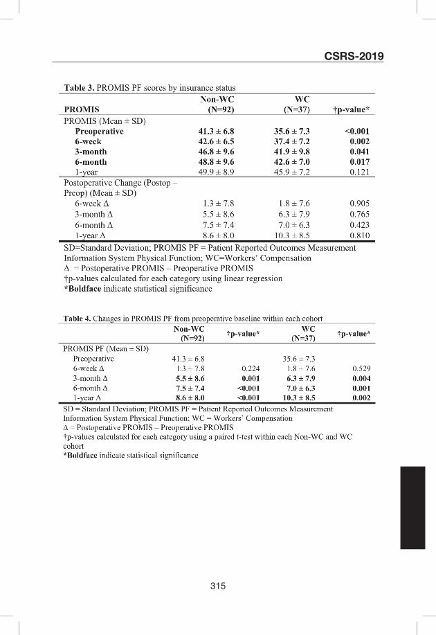
315
CSRS-2019

316
CSRS-2019E-Poster #24
Anterior Cervical Ossified Posterior Longitudinal Ligament En Bloc Resection: the Efficacy and Advantages of a Novel Surgical Technique for the Treatment of Cervical Ossification of the Posterior Longitudinal Ligament with Myelopathy
Xiongsheng Chen, MD; Yin Zhao; Yifan Tang
Introduction: For cervical ossification of posterior longitudinal ligament (OPLL) with spinal canal occupation ratio (COR) ≥ 50%, anterior cervical corpectomy and fusion (ACCF) is better in clinical efficacy than the posterior decompression. However, traditional ACCF meaning cervical ossified posterior longitudinal ligament piecemeal resection (ACOP) is difficult in decompression operation and may be accompanied by the high incidences of spinal cord injury (SCI) and cerebrospinal fluid leakage (CSFL). In this retrospective comparative study between ACOP and anterior cervical ossified posterior longitudinal ligament en bloc resection (ACOE), we aim to evaluate the clinical efficacy and advantages of ACOE in the treatment of cervical OPLL.
Material and Methods: Eighty-six patients suffered from cervical OPLL with COR ≥ 50% and underwent ACCF from July 2009 to January 2017. The clinical data with more than two years follow-up were retrospectively analyzed, including 50 males and 36 females, average (52.9±7.4) years (31~67 years). Of these patients, 45 cases received the ACOP from July 2009 to December 2013, and 41 cases received the ACOE (Fig. 1) from January 2014 to January 2017. There was no period of overlap during which both procedures were used for these patients. There were no important differences in age, sex ratio, body mass index (BMI), and COR between the two groups. The clinical efficacy of the two procedures was evaluated by comparing the JOA scores of pre-operation, one day, three months, six months, one year and two years postoperatively. The advantages of ACOE were analyzed by comparing the operation time, the intraoperative blood loss, and the complication incidences including SCI, CSFL, C5 nerve root palsy, dysphagia, hoarseness, hematoma, Horner syndrome, implant complication and infection.
Results: In the final follow-up ranged from 24 to 90 months (35.1±6.4 months), the JOA scores in the two groups all rose (Table 1). There was no clinically important difference in JOA scores between cervical OPLL treated with the ACOE and those treated with the ACOP at pre-operation. At one day, three months, six months, one year and two years postoperatively, the mean JOA scores in the ACOE group were all higher than which in the ACOP group (P<0.05). The mean JOA scores in the ACOE group decreased at one day postoperatively (P<0.05). Before and after surgery, there was no important difference in JOA scores between single-corpectomy and double-corpectomy in the groups of ACOE and ACOP. The operation time and the intraoperative blood loss in the ACOE group were both lower than those in the ACOP group (P<0.05). A patient with a COR of more than 90% had SCI after accepting ACOP. There was no incidence difference in all complications between the two groups. One case of dysphagia remained 2 years postoperatively. Seven years postoperatively, the case of SCI had the JOA score of 11, which was still lower than the preoperative JOA score of 15. The other complications (13/15, 86.67%) were significantly improved after proper treatments.
Conclusion: Comparing to ACOP, the ACOE is more efficient in decompression operation and safer with better neurological improvement. The neurological deterioration caused by the ACOP operation was difficult to be controlled.

317
CSRS-2019
Figure 1. The surgical procedure schematic of anterior cervical ossified posterior longitudinal ligament en bloc resection (ACOE)

318
CSRS-2019
Table 1. JOA scores in two groups
Parameters ACOE group ACOP group Difference (95% CI) P value
Pre-operation
Single-corpectomy 9.6±2.0 (5~14) 9.5±1.2 (5~15) 0.1 (-0.79~0.99) 0.82
Double-corpectomy 9.7±1.5 (4~15) 9.9±1.3 (6~14) -0.2 (-1.22~0.82) 0.69
Difference (95% CI) -0.1 (-1.33~1.13) -0.4 (-1.17~0.37)
P value 0.87 0.30
One day postoperatively
Single-corpectomy 10.4±1.7 (6~14) 8.3±1.3 (3~13) 2.1 (1.28~2.92) <0.01*
Double-corpectomy 10.2±1.9 (5~16) 8.9±1.6 (4~13) 1.3 (0.02~2.59) 0.05*
Difference (95% CI) 0.2 (-0.98~1.38) -0.6 (-1.48~0.28)
P value 0.73 0.18
Three months postoperatively
Single-corpectomy 15.1±0.9 (13~17) 10.5±0.8 (4~14) 4.6 (4.14~5.06) <0.01*
Double-corpectomy 14.8±1.1 (11~17) 10.8±1.7 (6~15) 4.0 (2.92~5.08) <0.01*
Difference (95% CI) 0.3 (-0.35~0.95) -0.3 (-1.05~0.45)
P value 0.35 0.43
Six months postoperatively
Single-corpectomy 15.3±1.2 (13~17) 13.5±0.9 (8~16) 1.8 (1.23~2.37) <0.01*
Double-corpectomy 14.9±1.2 (12~17) 13.7±1.1 (10~15) 1.2 (0.35~2.05) <0.01*
Difference (95% CI) 0.4 (-0.40~1.20) -0.2 (-0.81~0.41)
P value 0.32 0.51
One year postoperatively
Single-corpectomy 15.4±1.1 (13~17) 14.3±1.5 (10~17) 1.1 (0.39~1.81) <0.01*
Double-corpectomy 15.0±1.0 (13~17) 14.0±0.8 (12~16) 1.0 (0.34~1.66) <0.01*
Difference (95% CI) 0.4 (-0.31~1.11) 0.3 (-0.50~1.10)
P value 0.26 0.45
Two years postoperatively
Single-corpectomy 15.5±1.0 (13-17) 14.7±1.1 (11-17) 0.8 (0.23~1.37) <0.01*
Double-corpectomy 15.3±1.2 (13-17) 14.2±0.9 (12-17) 1.1 (0.33~1.87) <0.01*
Difference (95% CI) 0.2 (-0.51~0.91) 0.5 (-0.14~1.14)
P value 0.57 0.12
P values were determined with unpaired t-test. * Significant difference at α=0.05 (two-tailed)

319
CSRS-2019
Table 2. The main complication incidences in two groups
No. (%)Hazard ratio 95% CI P valueComplications ACOE group
(n=41)ACOP group
(n=45)
SCI 0 (0.00) 1 (2.22) 1.02 0.98~1.07 0.34
CSFL 1 (2.44) 2 (4.44) 1.02 0.94~1.11 0.61
C5 nerve root palsy 1 (2.44) 1 (2.22) 1.00 0.94~1.07 0.95
Dysphagia 1 (2.44) 2 (4.44) 1.02 0.94~1.11 0.61
Hoarseness 1 (2.44) 1 (2.22) 1.00 0.94~1.07 0.95
Hematoma 0 (0.00) 1 (2.22) 1.02 0.98~1.07 0.34
Horner syndrome 1 (2.44) 1 (2.22) 1.00 0.94~1.07 0.95
Implant complications 1 (2.44) 0 (0.00) 0.98 0.93~1.02 0.29
Infection 0 (0.00) 0 (0.00) NA NA NA
Total 6 (14.63) 9 (20.00) 1.07 0.88~1.30 0.51
P values were determined with the χ2 test.
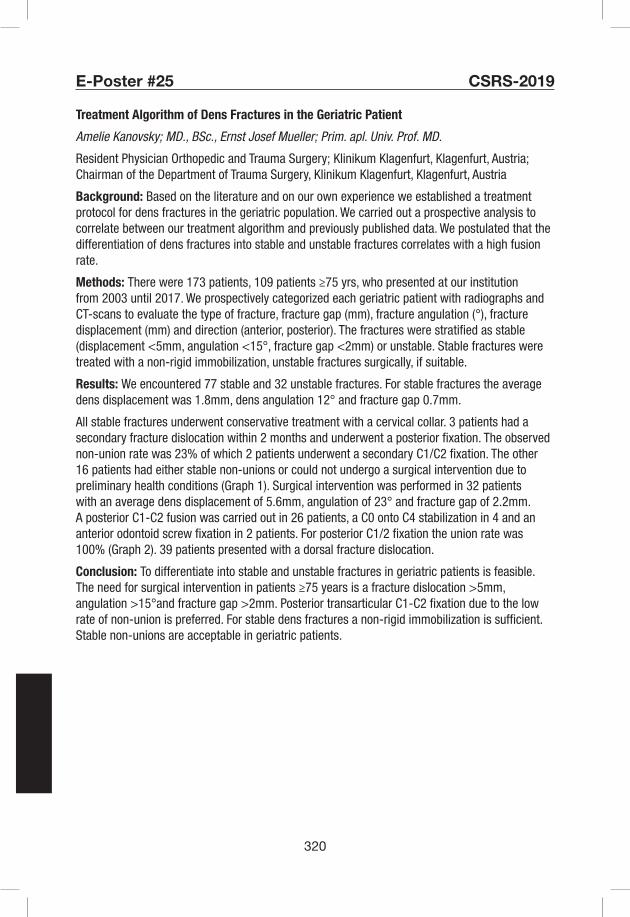
320
CSRS-2019E-Poster #25
Treatment Algorithm of Dens Fractures in the Geriatric Patient
Amelie Kanovsky; MD., BSc., Ernst Josef Mueller; Prim. apl. Univ. Prof. MD.
Resident Physician Orthopedic and Trauma Surgery; Klinikum Klagenfurt, Klagenfurt, Austria; Chairman of the Department of Trauma Surgery, Klinikum Klagenfurt, Klagenfurt, Austria
Background: Based on the literature and on our own experience we established a treatment protocol for dens fractures in the geriatric population. We carried out a prospective analysis to correlate between our treatment algorithm and previously published data. We postulated that the differentiation of dens fractures into stable and unstable fractures correlates with a high fusion rate.
Methods: There were 173 patients, 109 patients ≥75 yrs, who presented at our institution from 2003 until 2017. We prospectively categorized each geriatric patient with radiographs and CT-scans to evaluate the type of fracture, fracture gap (mm), fracture angulation (°), fracture displacement (mm) and direction (anterior, posterior). The fractures were stratified as stable (displacement <5mm, angulation <15°, fracture gap <2mm) or unstable. Stable fractures were treated with a non-rigid immobilization, unstable fractures surgically, if suitable.
Results: We encountered 77 stable and 32 unstable fractures. For stable fractures the average dens displacement was 1.8mm, dens angulation 12° and fracture gap 0.7mm.
All stable fractures underwent conservative treatment with a cervical collar. 3 patients had a secondary fracture dislocation within 2 months and underwent a posterior fixation. The observed non-union rate was 23% of which 2 patients underwent a secondary C1/C2 fixation. The other 16 patients had either stable non-unions or could not undergo a surgical intervention due to preliminary health conditions (Graph 1). Surgical intervention was performed in 32 patients with an average dens displacement of 5.6mm, angulation of 23° and fracture gap of 2.2mm. A posterior C1-C2 fusion was carried out in 26 patients, a C0 onto C4 stabilization in 4 and an anterior odontoid screw fixation in 2 patients. For posterior C1/2 fixation the union rate was 100% (Graph 2). 39 patients presented with a dorsal fracture dislocation.
Conclusion: To differentiate into stable and unstable fractures in geriatric patients is feasible. The need for surgical intervention in patients ≥75 years is a fracture dislocation >5mm, angulation >15°and fracture gap >2mm. Posterior transarticular C1-C2 fixation due to the low rate of non-union is preferred. For stable dens fractures a non-rigid immobilization is sufficient. Stable non-unions are acceptable in geriatric patients.

321
CSRS-2019
Graph 1. Summarized data and treatment algorithm of stable, conservatively treated dens fractures in the elderly.
Graph 2. Summarized data and treatment algorithm of unstable and surgically treated dens fractures in the elderly.

322
CSRS-2019E-Poster #26
Should Hospital Magnet Designation Influence Patient Preference for the Choice of Facility Selection in Cervical Spine Surgery? 1Piyush Kalakoti, 1Nicholas A. Bedard, 1Alan G. Shamrock, 1Alexander J. Volkmar, Cosma Calderaro, 1Nathan R. Hendrickson, 1Andrew J. Pugely1Spine Surgery, Department of Orthopedics, The University of Iowa
Introduction: Public reporting of outcomes has been the cornerstone of the paradigm shift witnessed in improving transparency and adding value to the national healthcare delivery. Several notable online platforms—including the CMS Hospital Compare, the Magnet Recognition Program, and those hosted by private/non-profit organizations (ProPublica; Consumers’ Checkbook) provide hospital rankings contingent on defined outcome metrics. The initiatives, while championing for improved transparency in healthcare reporting and potentially influencing patient’s choice for healthcare facility selection are often associated with intrinsic methodological biases. The American Nursing Credentialing Center (ANCC) Magnet Recognition Program epitomizes nursing excellence and conferred to hospitals that have demonstrated a healthy work environment contingent upon periodic quadrennial reviews.
In context to spine surgery, outcomes are often driven by the interplay of patient-specific comorbidity status, intraoperative events and surgeon expertise. The current study investigates whether hospitalization at a Magnet-accredited facility is associated with improved inpatient outcomes after cervical spine surgery.
Materials/Methods: Hospital-level data on ANCC Magnet accreditation (https://www.nursingworld.org/magnet) for 2005-2011 was merged with the Nationwide Inpatient Sample for respective years. Adult patients [18-90 years] undergoing elective cervical spine surgery were extracted. Primary endpoints were case-fatality, disposition status, length of hospital stay (LOS), resource utilization (inflation adjusted charges/costs), and postoperative complications. Multivariable regression techniques, generalized estimating equation (to control for clustering at the facility-level) and models adjusted with propensity scores (to mimic effect of a randomized trial), were controlled for potential confounders to investigate the differences in primary endpoints across patients operated at Magnet-certified hospitals versus non-Magnet hospitals.
Results: Overall, 127,303 adults (mean age: 52.09 ± 12.02 years; 52.4% were women) underwent elective cervical spine surgery across 968 hospitals. Of these, 16.4% (n=20830) were hospitalized at centers designated with Magnet-accreditation. Compared to non-Magnet hospitals, patients hospitalized at Magnet-accredited centers were relatively sicker (Charlson comorbidity score: 0.68 vs 0.66; p<0.001), privately insured (63.8% vs 56.0%; p<0.001), high-income status (37.4% vs 29.1%; p<0.001). [Fig. 1]
In multivariable regression models adjusted for potential confounders, Magnet-accredited hospitals were not superior to non-Magnet hospitals in terms of case-fatality (p=0.922), discharge disposition status (p=0.300) and LOS (p=0.472). On the contrary, likelihood of postoperative complications including thromboembolic event (OR:1.34; p<0.001), acute renal failure (OR:1.57; p<0.001) and infections (OR:1.76; p=0.004) were notably higher in cervical spine patients operated at Magnet-designated facility. The increased complications at Magnet-

323
CSRS-2019
accredited centers plausibly translated to increased resource utilization as reflected by higher charges (+$2,994; p<0.001) and costs (+$2,034; p<0.001) compared to non-Magnet centers. [Fig. 2]
Conclusion: The study notes that over three-quarters (83.6%) of patients undergoing cervical spine surgery will be hospitalized at a non-Magnet facility while approximately 16% will choose a Magnet-accredited center. Using robust-statistical techniques, the data reflects that Magnet-designation is not associated with superior inpatient outcomes after cervical spine surgery. Hospital and provider selection by patients seeking surgical care for cervical disorders should be independent of Magnet designation. Future investigations elucidating the clinical benefits distinguishing centers based upon Magnet status is recommended.
Figure 1: Patient characteristics and outcomes across patients hospitalized for cervical spine surgery at centers with Magnet accreditation versus non-Magnet hospitals
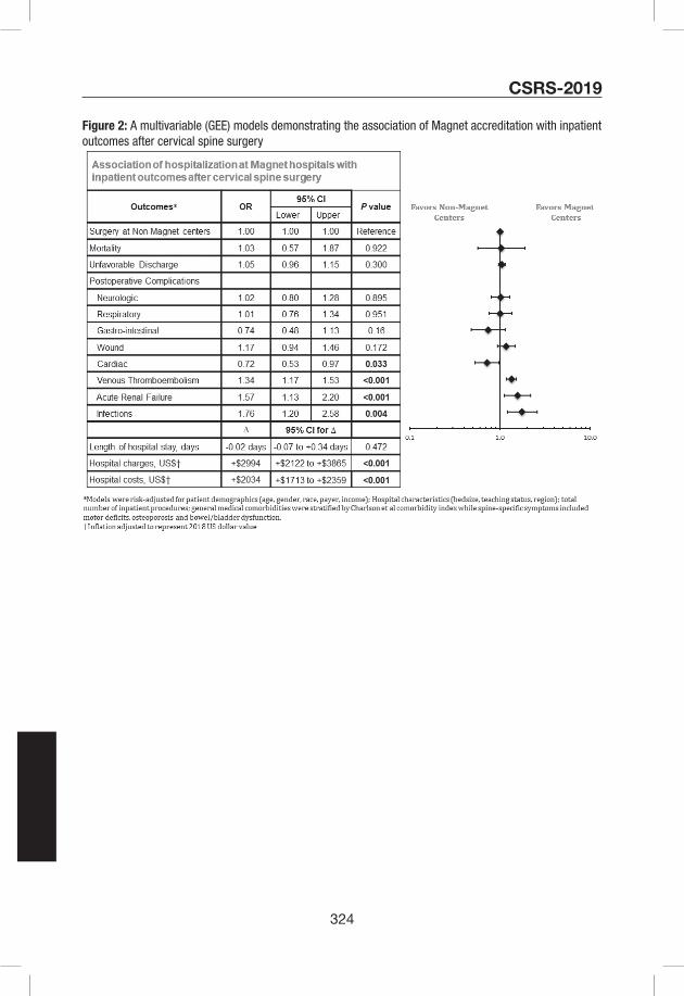
324
CSRS-2019
Figure 2: A multivariable (GEE) models demonstrating the association of Magnet accreditation with inpatient outcomes after cervical spine surgery

325
CSRS-2019E-Poster #27
Opioid consumption after anterior cervical spine surgery: what is the appropriate minimum quantity?
Francis Lovecchio, MD; Ajay Premkumar, MD, MPH; Michael Steinhaus, MD; Jeffrey Stepan, MD, MSc; Dianna Mejia, BA; Alexander Koo, BS; Joo S. Yoon, BA; Virginie Lafage, PhD; Sravisht Iyer, MD; Russel Huang, MD; Sheeraz Qureshi, MD, MBA; Han Jo Kim, MD; Kern Singh, MD; Todd Albert, MD
Introduction: As cervical spine procedures move into the outpatient setting, providers will be left without inpatient narcotic consumption data on which to base an outpatient prescription. There is a lack of quantitative data to inform opioid prescribing guidelines after ACDF or CDA. The purpose of this study was to record daily opioid use and pain levels after one or two level ACDF or CDA through an automated text-messaging based data collection system.
Methods: Forty-two adult patients undergoing one- or two-level ACDF or CDA with one of six fellowship trained spine surgeons were enrolled at two participating institutions between February 2018 and March 2019. To ensure a generalizable patient cohort, only patients with chronic opioid dependence (daily opioid use in the six months before surgery) were excluded. Daily opioid use and NRS pain levels were collected through a HIPAA-compliant, automated text-messaging system. To facilitate clinical applications, opioid use was converted from oral morphine equivalents (OME) into “pills” (oxycodone 5 mg equivalents). After six weeks or upon patient-reported cessation of opioid use, final survey questions were asked. Refill and prescription data was verified from the state narcotic prescription registry. Risk factors were compared between patients in the top and bottom half of opioid consumption.
Results: Average age of the patient sample was 52.1±11.5 years, with a BMI of 27.6±4.3 kg/m2 (Table 1). Thirty-two patients (76.8%) underwent ACDF and ten underwent CDA (23.8%). The majority of patients underwent one-level surgery (61.9% one-level, 38.1% two-level). Approximately one quarter (23.8%) of patients used opioids on a non-daily basis in the six months before surgery. Opioid use was widely variable, ranging from 0-229.6 “pills” (oxycodone 5mg equivalents) (0-1722 OME). Median use was 28 pills (IQR 16-46.8). Opioid use did not vary between the one- and two-level groups (median pill consumption, 29 IQR [18.6-58.5] vs. 26.7 IQR [13.3-50], respectively, p=0.223). Daily opioid consumption and pain scores decreased steadily over the first week (Figure 1); half ceased opioids by POD7.5. Eight patients (19.0%) completed their initial prescription, and six patients (14.2%) obtained a refill. Only 7 patients (17.5%) took opioids past POD14. Preoperative intermittent opioid use trended toward an association with the top half of opioid consumption (11% vs. 33%, p=0.082).
Conclusions: A prescription for 28 oxycodone 5 mg pills should be sufficient for most patients undergoing one- or two-level anterior cervical surgery. A quantitative prescription guideline must include didactic components on narcotic use, pain expectations, and appropriate pill disposal to mitigate the wide variation in postoperative opioid use and difficulty in predicting consumption.

326
CSRS-2019
Table 1. Characteristics of study sample
N (42) % or range
Age (yrs)* 52.1 (11.5) 27-77
BMI* 27.6 (4.3) 19.8-36.8
Gender
Male 29 69.0%
Female 13 31.0%
Surgery
ACDF 32 76.2%
CDA 10 23.8%
Number of Levels 0.0%
1 26 61.9%
2 16 38.1%
Length of stay (days)
≤1 38 90.5%
2 3 7.1%
3 1 2.4%
History of psychiatric disorder† 9 21.4%
History of intermittent opioid use‡ 10 23.8%
Current smoker 3 7.1%
Marijuana use 4 9.5%
Greater than 7 drinks/week 6 14.3%
Type of opioid prescribed
Tramadol 8 19.0%
Oxycodone 28 66.7%
Hydrocodone-acetaminophen 4 9.5%
Other 2 4.8%
Initial Prescription Size (oxycodone 5 mg equivalents)
0-10 1 2.4%
10-20 3 7.1%
20-30 7 16.7%
30-40 11 26.2%
40-50 3 7.1%
50-60 5 11.9%
60-70 5 11.9%
70+ 7 16.7%
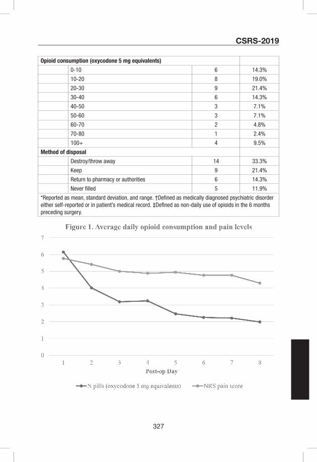
327
CSRS-2019
Opioid consumption (oxycodone 5 mg equivalents)
0-10 6 14.3%
10-20 8 19.0%
20-30 9 21.4%
30-40 6 14.3%
40-50 3 7.1%
50-60 3 7.1%
60-70 2 4.8%
70-80 1 2.4%
100+ 4 9.5%
Method of disposal
Destroy/throw away 14 33.3%
Keep 9 21.4%
Return to pharmacy or authorities 6 14.3%
Never filled 5 11.9%
*Reported as mean, standard deviation, and range. †Defined as medically diagnosed psychiatric disorder either self-reported or in patient’s medical record. ‡Defined as non-daily use of opioids in the 6 months preceding surgery.

328
CSRS-2019E-Poster #28
Demographic Disparities between Outcomes of ACDF and CDF: Analysis of NSQIP Database
Morenikeji Ayodele Buraimoh MD, Mohammad Farooq Usmani MD, MSc, Jael Camacho MD, Bailey Howard BS, Steven C. Ludwig MD
Department of Orthopaedics, University of Maryland Medical Center, Baltimore, MD
Background: Anterior cervical discectomy and fusion (ACDF) and Cervical disc arthroplasties (CDA) are common procedures for radiculopathy and myelopathy. Studies have observed socioeconomic disparities in the rates of CDA vs ACDFs. The aim of this study is to compare the utilization of CDA and ACDF among different gender, racial, and payer type population to after 2010 and evaluate for persistent healthcare disparity in CDA utilization. We hypothesize that patients from lower socioeconomic backgrounds will be less likely to get CDA compared with ACDF.
Methods: We conducted a retrospective cohort study with the use of prospectively collected data as part of the National Surgical Quality Improvement Program (NSQIP). Medical records and operative reports are used by onsite trained surgical clinical reviewers to prospectively collect preoperative, intraoperative, and postoperative variables. Patients are characterized by CPT codes and data are gathered through a 30-day postoperative period. Patients were identified based on CPT code of the primary procedures of either CDA (CPT code 22856 and 0092T) or ACDF (CPT codes 63075, 22554, or 22551). Demographics data, patient medical history, pre-operative characteristics, operative details, and post-operative complications were obtained for each of the patients identified based on the CPT codes. For bi-variate analysis, unpaired T-test was used for continuous variables that were normally distributed. For bi-variate analysis, unpaired T-test was used for continuous variables that were normally distributed. Multivariate nominal logistic regression was created using a backwards stepwise selection procedure.
Results: A total of 45,704 patients were included in the analysis and the majority (n=43,487, 95.15%) had an ACDF. Patients with ACDF were statistically older, taller, and more likely to be female than patients who received CDAs. African Americans made up 10.14% of ACDFs population compared with 5.86% of CDA population (p<0.0001). Patient co-morbidities did not seem to play an important role in determining the likelihood that patients will get a CDA. Patients who had a CDA had shorter operative time (110 minutes vs 127 minutes), had shorter length of stay (1 day vs 2 days), and were discharged more quickly after the operation (1.07 days vs 1.80 days). The rate of complications between ACDF and CDA group was comparable. In the multivariate model, age, height and weight had a minimal impact. However, compared to whites, African American race had a negative association, indicating that they were less likely to get a CDA compared to ACDF.
Conclusions: Inequalities concerning medical treatment have been recognized in various settings and should be addressed in order to provide the optimal care to patients. The study highlights that African Americans are less likely to get CDA relative to whites. The disparity could be due to access, education, or exposure. Access to different procedures is often correlated with type of insurance. Education and exposure are also key to patients adapting new techniques and procedures. Physicians and patients both are responsible for exposure and education. Additional

329
CSRS-2019
studies will determine effective strategies for closing the disparities in access to CDA.
Figure 1: Odds ratio and 95% confidence interval for complications for patients who undergo CDA over ACDF. SSI, Surgical Site Infection; MI, Myocardial Infarction; UTI, Urinary Tract Infection; DVT, Deep Vein Thrombosis.

330
CSRS-2019

331
CSRS-2019E-Poster #29
“Reverse Roussouly:” Ratios of Cervical to Thoracic Shape Curvature in an Adult Cervical Deformity Population
Peter G Passias MD, Haddy Alas BS, Avery Brown BS, Katherine E Pierce BS, Cole Bortz BA, Edem J Abotsi BA, Renaud Lafage MS, Virginie Lafage PhD
Introduction: The high mobility of the cervical spine exists in stark contrast to the natural stiffness of the thoracic region below. Understanding how cervical, CTJ, and thoracic curves coexist at baseline (BL) can provide important clues to patient symptomatology and surgical outcomes following surgical correction. The purpose of our study was thus to explore baseline normative curvature ratios of the cervico-thoracic spine and establish radiographic thresholds for severe myelopathy and disability within the context of shape.
Methods: Patients undergoing cervical fusion with a diagnosis of cervical deformity (CD; C2-C7 Cobb>10°, CK>10°, cSVA>4cm, or CBVA>25°), stenosis, spondylolisthesis, or myelopathy with available baseline(BL) radiographic data were included. Cervical lordosis(CL) was measured using C2-C7 Cobb angle and thoracic kyphosis(TK) using T2-12, with negative values indicating kyphotic angles. A mathematical ratio was calculated for CL:TK ranging from -1 to +1, and this ratio was correlated to cSVA(>or<40mm), SVA(>or<40mm), and mJOA scores at BL(>or<14, severe) using Pearson bivariate r. Univariate analyses including independent samples t-tests analyzed differences in presence of severe myelopathy(mJOA>14) or NDI>40 across CL:TK curve ratio groups.
Results: 63 cervical pts(55.2yrs,56%F) met inclusion criteria. Mean C2-C7 CL at BL was 3.77±11.6°, mean T2-T12 was -43.50±12.8, mean C2-C7 SVA was 21.65±10.1mm, and mean C7-S1 SVA was -7.18±49.5mm. All patients had a kyphotic(negative) thoracic curvature at BL. In terms of CL:TK ratio, 37 had a negative ratio (more lordotic c-spine/kyphotic t-spine), and 26 had a positive ratio (more kyphotic c-spine/kyphotic t-spine). More positive CL:TK significantly correlated to greater TS-CL(r=0.655,p<0.001), increased upper cervical (C0-2) lordosis (r=0.454, p=0.001), increased McGregor’s slope (r=0.292,p=0.032), and decreased T1-slope (r=-0.304, p-.016), in addition to increased pelvic tilt (r=0.274, p=0.034). mJOA scores correlated to the CL:TK ratio, with positive CL:TK ratios having higher mJOA scores and negative CL:TK ratios having higher rates of moderate myelopathy(43.2% vs 15.4%, p=0.028). CL:TK did not correlate to NDI scores. Conditional Tree Analysis analyzed all CL:TK curvatures to establish cSVA and TS-CL thresholds predictive of severe myelopathy or neck disability at BL. Conditional forward regression revealed negative CL:TK ratios with a cSVA>38mm had increased odds of NDI>40 by at least 2.94x, and positive CL:TK ratios with a cSVA>30mm or TS-CL>36° had increased odds of NDI>40 by an average of 6.42x. (Table 1)
Conclusions: Patients with an increased CL:TK ratio, indicating cervical and thoracic kyphotic curves, had higher rates of myelopathy and neck disability at baseline. Specific thresholds for cervical sagittal vertical axis(>38mm or >30mm) and T1-slope minus cervical lordosis(>36o) predicted neck disability scores depending on baseline shape curvature.
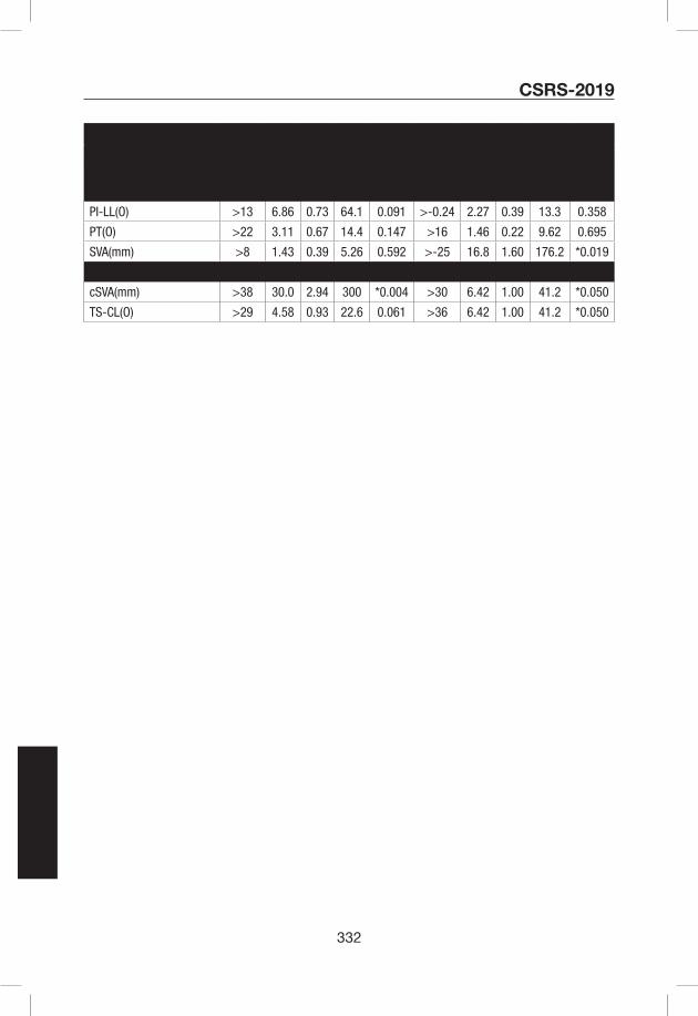
332
CSRS-2019
Predictors of Moderate to Severe Myelopathy & Neck Disability
Negative CL:TK Positive CL:TK
Mod-Severe Myelopathy (mJOA<14)
Cut-off OR LI UI p Cut-off OR LI UI p
PI-LL(O) >13 6.86 0.73 64.1 0.091 >-0.24 2.27 0.39 13.3 0.358
PT(O) >22 3.11 0.67 14.4 0.147 >16 1.46 0.22 9.62 0.695
SVA(mm) >8 1.43 0.39 5.26 0.592 >-25 16.8 1.60 176.2 *0.019
Neck Disability (NDI>40) Cut-off OR LI UI p Cut-off OR LI UI p
cSVA(mm) >38 30.0 2.94 300 *0.004 >30 6.42 1.00 41.2 *0.050
TS-CL(O) >29 4.58 0.93 22.6 0.061 >36 6.42 1.00 41.2 *0.050

333
CSRS-2019E-Poster #30
Does Extension Dysfunction Affect Postoperative Loss of Cervical Lordosis in Patients Who Undergo Laminoplasty?
Dongwuk Son, Suhun Lee, Junseok Lee, Geunsung Song
Pusan National University Yangsan Hospital
Introduction: Laminoplasty is an effective surgical method for treating cervical degenerative disease. However, the loss of cervical lordosis (LCL) after laminoplasty is associated with a decrease in the patient’s quality of life. The T1 slope (T1s) is particularly assumed to be an important factor for kyphotic change after laminoplasty. However, another study reported that the less LCL in high T1s patients after laminoplasty. Previous studies have focused on the injury of the posterior neck muscular-ligament complex (PMLC) itself, which are important factors in maintaining cervical sagittal balance; however, this muscle injury model could not explain the less kyphotic change in high T1s patients. We focused on the PMLC constriction reservoir rather than on the injury itself. This individual reservoir difference can explain the phenomenon of less kyphotic change even in high T1s patients. To predict the PMLC constriction reservoir, we focused on the extension function (EF) of the cervical spine, which is defined as the degree of extensibility in the neutral cervical position (Figure 1A, B). The purpose of this study was to investigate the effect of EF on LCL after laminoplasty.
Materials/Methods: We retrospectively analyzed 50 consecutive patients who underwent open-door laminoplasty (>1-year follow-up). EF is defined as extension C2–7 Cobb’s angle (CA) minus neutral C2–7 CA. LCL is defined as follow-up CA minus preoperative CA [CA (FU)-CA (PRE)], and significant kyphotic change was defined as LCL< 10°.
Results: The distribution of LCL was -3.70±7.98 and the significant kyphotic change occurred in 20% of the patients (10/50). Correlation analysis revealed that LCL was not related to the CA (PRE) and C7 slope (PRE). EF, C2-7 sagittal vertical axis (PRE), and C2 slope (PRE) were found to be risk factors for LCL by multiple linear regression analysis. The receiver operating characteristic curve analysis revealed that EF could predict the significant kyphotic change well. The cutoff value of EF was 14°. All significant kyphotic change occurred at EF<14°. Upon limiting the number of patients with preoperative straight curvature (n=28), no significant kyphotic change occurred in any patient whose EF was ≥14° (Table 1).
Conclusion: We have identified a new factor, EF, which could predict LCL after laminoplasty. All significant kyphotic changes after laminoplasty occurred particularly when the EF was <14°. In addition, no significant kyphotic change occurred in patients with straight curvature when the EF was ≥14°.
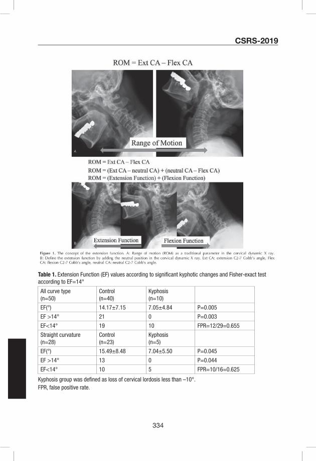
334
CSRS-2019
Table 1. Extension Function (EF) values according to significant kyphotic changes and Fisher-exact test according to EF=14°
All curve type(n=50)
Control(n=40)
Kyphosis(n=10)
EF(°) 14.17±7.15 7.05±4.84 P=0.005
EF >14° 21 0 P=0.003
EF<14° 19 10 FPR=12/29=0.655
Straight curvature(n=28)
Control(n=23)
Kyphosis(n=5)
EF(°) 15.49±8.48 7.04±5.50 P=0.045
EF >14° 13 0 P=0.044
EF<14° 10 5 FPR=10/16=0.625
Kyphosis group was defined as loss of cervical lordosis less than –10°. FPR, false positive rate.

335
CSRS-2019E-Poster #31
Cervical and spinal sagittal alignment deviation in the general elderly population: A Japanese cohort survey randomly sampled from a basic resident registry
Masashi Uehara1, Jun Takahashi1, Shota Ikegami1, Ryosuke Tokida2, Hikaru Nishimura2, Noriko Sakai3, Hiroyuki Kato1
1Department of Orthopaedic Surgery, Shinshu University School of Medicine
2Rehabilitation Center, Shinshu University Hospital
3Department of Orthopaedic Surgery, New Life Hospital
Background: It is widely recognized that sagittal spinal alignment changes with age. However, there are presently no clear benchmarks for such values or those for the cervical spine in the general population. Quality epidemiological studies are needed to establish standards for spinal alignment deviation. In this study of an aged Japanese population, we employed random sampling from the basic resident registry of a rural town for subject selection to determine reference values of sagittal spinal alignment including the cervical spine.
Methods: Registered citizens of 50 to 89 years old were targeted for this survey. We established 8 groups based on age (50’s, 60’s, 70’s, and 80’s) and gender (male and female) after random sampling from the resident registry of Obuse town in 2014. A total of 413 people (203 males and 210 females) were enrolled. Radiographic parameters of sagittal spinal alignment of the cohort were measured and analyzed. For the purposes of examining to what extent deviation would occur and from which age, comparisons between age groups on the basis of 50’s age group reference values were performed using multiple comparisons based on the Dunnett test.
Results: Each spinal alignment parameter stratified by age and gender was presented. Weighted values were based on the population composition ratio of Japan and represented the reference values for Japanese over 50 years old. Global spinal alignments became more misaligned with age for both genders. Sagittal vertical axis (SVA) forward shift was significantly more frequent in 80’s males and 70’s females, and SVA in 80’s females was a mean of 66 mm forward of that of 50’s females. Forward movement of the cervical spine was especially prominent in men. Cervical protrusion was markedly greater in 60’s males onwards. C2-7 SVA was large at all ages in males, and T1 slope increased from their 60’s. In women, lumbar lordosis and posterior pelvic inclination were noticeable from a younger age than in men. The amount of pelvic tilt misalignment in female subjects was approximately 10 years earlier than their male counterparts.
Conclusions: This first resident cohort of Japanese individuals determined average spinal alignment parameters by age and gender. Spinal balance generally shifts forward as age increases. A forward shift in the upper cervical spine occurs first in men, while lumbopelvic alignment shift occurs first in women.

336
CSRS-2019E-Poster #32
PROMIS Physical Health Domain Scores are Related to Cervical Deformity Severity
Katherine E Pierce BS, Haddy Alas BS, Avery Brown BS, Cole Bortz BA, Brooke K O’Connell MS, Dennis Vasquez-Montes MS, Dainn Woo BS, Renaud Lafage MS, Virginie Lafage PhD, Peter G Passias MD
Introduction: Computer adaptive testing is becoming increasingly common in assessing patient outcomes. The Patient Reported Outcome Measurement Information System (PROMIS) instruments of Physical Function, Pain Intensity and Pain Interference have been shown to correlate with established patient outcome metrics. There is a lack of studies investigating the relationship between PROMIS physical health domain metrics and established Ames cervical deformity (CD) radiographic classification. The purpose was to evaluate the association of available cervical alignment components via the Ames cervical deformity classification parameters with PROMIS physical domains.
Materials/Methods: Surgical CD patients(C2-C7 Cobb angle >10° or C2-C7 sagittal vertical axis >4cm or TS-CL>15°) ≥18 years old with available baseline(BL) radiographic and PROMIS data were isolated in the Spine Quality Database(Quality). Patients were classified according to Ames CD modifiers for cSVA and TS-CL (non-deformed[Non], moderate deformity[Mod], severe deformity[Sev]). Descriptives and univariate analyses compared population-weighted PROMIS scores for Pain Intensity (PI), Physical Function (PF), and Pain Interference across CD deformity modifiers. Conditional Tree Analysis with logistic regression sampling were performed to determine a possible threshold of PROMIS scores for which the correlation with Ames radiographic cut-offs was most significant. Reported cut off values for Mod(cSVA: 4-8cm; TS-CL: 15-20°) and Sev(cSVA: >8cm; TS-CL: >20°) disability were used.
Results: 208 pts(58.8yrs, 51%F, 29.6kg/m2, mean Charlson Comorbidity score: 1.19) met inclusion criteria. By surgical approach: 79.3% posterior, 5.7% anterior, 14.9% combined; mean levels fused: 3.7±4.2. Baseline cervical cSVA modifier by severity: 83.2% Non, 16.8% Mod. No patients met criteria for severe cSVA modifier. Baseline TS-CL modifier by severity: 18.8% Non, 22.1% Mod, 59.1% Sev. Mean PI score: 89.6±15.4, mean PF score: 11.9±13.1, mean Int score: 56.9±6.8. PI did not differ between cSVA or TS-CL modifier severity. Mod cSVA patients and Mod/Sev TS-CL modifier groups both trended towards lower PF scores and higher pain interference scores though this was not significant (P>0.05). Conditional tree analysis determined thresholds for PROMIS scores that were independent predictors of modifier severity. A PI score>96(OR: 0.658[0.303-1.430]), a PF score<14 (OR:1.864[0.767-4.531])and an interference score of >57.4 (OR: 1.712[0.811-3.616]) were predictors of Mod cSVA. A PI score of >87(OR: 1.428[0.767-2.659]), a PF score <14 (OR: 1.551[0.851-2.827]), and a pain interference score of >56.3 (OR:1.656[0.946-2.897]) were predictors of Sev TS-CL.
Conclusions: PROMIS physical health domain metrics of Pain Intensity, Physical Function, and Pain Interference were related to cervical malalignment parameters of the Ames cervical deformity classifications system. Certain baseline PROMIS thresholds can be connected to severity of cervical deformity.
337
CSRS-2019E-Poster #33
Predicting The Magnitude Of DJK Following Cervical Deformity Correction
Ethan W. Ayres MPH1, Themistocles S. Protopsaltis MD1, Renaud Lafage MS2, Gregory M. Mundis Jr. MD3, Justin S. Smith MD PhD4, D. Kojo Hamilton MD5, Eric O. Klineberg MD6, Daniel M. Sciubba MD7, Robert A. Hart MD8, Shay Bess MD9, Christopher I. Shaffrey MD4, Frank J Schwab MD2, Virginie Lafage PhD2, Christopher P. Ames MD10, and the International Spine Study Group (ISSG)11
1Department of Orthopaedic Surgery, NYU Langone Orthopaedic Hospital, New York, NY2Department of Orthopaedic Surgery, Hospital for Special Surgery, New York, NY3San Diego Center for Spinal Disorders, La Jolla, CA 4Department of Neurological Surgery, University of Virginia Medical Center, Charlottesville, VA5Department of Neurological Surgery, University of Pittsburgh School of Medicine, Pittsburgh, PA 6Department of Orthopaedic Surgery, University of California, Davis, Davis, CA7Department of Neurosurgery, Johns Hopkins University School of Medicine, Baltimore MD 8Department of Orthopaedic Surgery, Swedish Neuroscience Institute, Seattle, WA9Rocky Mountain Scoliosis and Spine, Denver, CO10Department of Neurological Surgery, University of California, San Francisco, San Francisco, CA11ISSGF, Littleton, CO
Keywords: Distal Junctional Kyphosis, Cervical Deformity, Deformity surgery, outcomes
Introduction: Severe cervical deformity (CD) can be a source of severe disability. Surgical correction of the deformity can markedly improve a patient’s quality of life and functional status. However, high rates of complications have been reported, one of the most serious being distal junctional kyphosis (DJK). Although researchers have identified risk factors for DJK, no model has been proposed that predicts DJK magnitude. Therefore, this study aims to develop a model that can accurately predict the postoperative DJK angle using radiographic measurements obtained pre- and intra-operatively.
Methods: This is a retrospective review of a prospective database of operative CD with clinical and radiographic data available preoperatively and at any postoperative interval (3m, 6m, 1y, 2y). Patients with a history of fusion below L4 were excluded. The DJK angle was defined as the Cobb from LIV to LIV-2. Traditional DJK (tDJK) was a DJK angle change >10°; severe DJK (sDJK) was a change >20°. Models were developed using a randomly-selected 66.6% of patients. Pre and intra-operative radiographic parameters significantly associated with DJK were identified and ranked in order of importance via a Conditional Variable Importance Table that used a non-replacement sampling set of 4,000 conditional inference trees. Linear regression models were developed using the factors most strongly associated with the post-operative DJK angle. The models were validated in the remaining 33.3% of patients.
Results: 131 patients were included (60 ±10y, 62% female). Mean follow-up was 14±8 months. The most common LIV was T2 (20.6%) with an LIV below T2 in 44%. The mean postop DJK angle was 14.6°±14°. 35% of patients developed DJK (11% sDJK). 5 patients underwent a subsequent revision surgery due to DJK. The model development subset included 84 patients and did not differ significantly from the validation subset in terms of radiographic or clinical parameters. The variables identified as being most significant were: Intraop change of CL (ΔCL) and C2-LIV
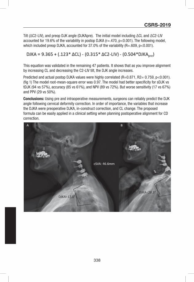
338
CSRS-2019
Tilt (ΔC2-LIV), and preop DJK angle (DJKApre). The initial model including ΔCL and ΔC2-LIV accounted for 19.6% of the variability in postop DJKA (r=.470, p<0.001). The following model, which included preop DJKA, accounted for 37.0% of the variability (R=.609, p<0.001).
This equation was validated in the remaining 47 patients. It shows that as you improve alignment by increasing CL and decreasing the C2-LIV tilt, the DJK angle increases.
Predicted and actual postop DJKA values were highly correlated (R=0.871, R2= 0.759, p<0.001).(fig 1) The model root-mean-square error was 0.97. The model had better specificity for sDJK vs tDJK (94 vs 57%), accuracy (85 vs 61%), and NPV (89 vs 72%). But worse sensitivity (17 vs 67%) and PPV (29 vs 50%).
Conclusions: Using pre and intraoperative measurements, surgeons can reliably predict the DJK angle following cervical deformity correction. In order of importance, the variables that increase the DJKA were preoperative DJKA, in-construct correction, and CL change. The proposed formula can be easily applied in a clinical setting when planning postoperative alignment for CD correction.
339
CSRS-2019E-Poster #34
In cervical degenerative spondylosis, the evaluation of C2-7angle, spinal canal stenosis increases on the Dynamic MRI when the difference between extension position and neutral position is larger than 15.4 degrees.
Jong Beom Lee, Il Sup Kim, Jung Jae Lee, Jong-Hyeok Park, Jae Taek Hong
BACKGROUND: Dynamic MRI is useful to find cervical hidden pathology. Previous study, our results show that the dynamic MRI provides more detailed radiological information of dynamic cord compression and aids surgical planning for cervical myelopathy, especially in CDS patients. And positional changes in Muhle`s grades in the CDS group were significantly higher than in the OPLL group.
PURPOSE: The purpose of this study was to estimate the radiologic parameters of the CDS patients, to find out cut off value of spinal canal stenosis changes when exam of dynamic MRI.
MATERIAL AND METHODS: 112 patients who underwent cervical dynamic MRI were included. Patients with a cervical ossification of posterior longitudinal ligament, congenital anomaly, trauma, instability, tumor, ankylosing spondylitis, or reoperation were excluded. There were 39 males and 24 females. C2-7 angle was measured in cervical spine x-ray lateral image of neutral (NX), flexion (FX) and extension position (EX), and cervical dynamic MRI. C7 slope angle was measured in cervical x-ray lateral image of neutral. Muhle’s grades were determined in neutral and dynamic MR images from C2-3 to C7-T1 in extension, flexion, and neutral neck positions. In addition, we divide two groups, two grades were increased or maximum grade was increased; stenosis group (S group) and others; maintenance group (M group). Statistical analysis was performed using SPSS software and statistical significance was accepted for p values < 0.05.
RESULTS: There were 24 cases in the S group and 39 cases in the M group. The C2-7 angle of EX was 33.23 ° in the S group and 22.26 ° in the M group (p=0.002). The difference C2-7 angle of EX-NX was 22.74 ° in the S group and 8.35 ° in the M group (p=0.000). The C7 slope angle of NX was 18.06 ° in the S group and 24.59 ° in the M group (p=0.002). With respect to the above, the cut-off value was obtained using the ROC curve, EX was 29.1 degrees and difference C2-7 angle of EX-NX was 15.4 degrees.
CONCLUSION: If the C2-7 angle of EX is 29.1 ° and difference C2-7 angle of EX-NS is 15.4 ° or more, these are suggested that the number of canal stenosis may increase further when exam of dynamic MRI.

340
CSRS-2019E-Poster #35
MRI Phenotype Profile and its Association with the Development of Cervical Spondylotic Myelopathy 1Philip Louie, 1Jannat Khan, 2Youping Tao, 1Bryce Basques, 1Sapan Gandi, 1Garrett Harada, 2Fabio Galbusera, 2Frank Niemeyer, 2Hans-Joachim Wilke, 1Howard S. An, 1Dino Samartzis1 Department of Orthopaedic Surgery, RUSH University Medical Center, Chicago, IL, USA2 Institute of Orthopaedic Research and Biomechanics, University of Ulm, Ulm, Germany
Introduction: Cervical spondylotic myelopathy (CSM) is characterized by spinal cord compression secondary to degenerative changes. In CSM, early diagnosis can optimize management outcomes. Spinal phenotypes on MRI are observable traits, whereby certain patterns and severity have been associated with low back pain development and intensity. However, patterns of MRI phenotypes of the cervical spine in relation to CSM remain largely unknown. As such, this study addressed the presence of various cervical spine MRI phenotypes and their association with the development of CSM.
Methods: A retrospective study design with prospectively collected data was performed consisting of patients who presented with neck pain, with/without upper extremity symptoms from 2008-2015. Sagittal 1.5T cervical spine MRI was assessed. Cervical MRI phenotypes were evaluated, which consisted of detailed topographical patterning of disc degeneration, disc space narrowing, disc bulge/extrusion, high intensity zones, structural endplate abnormalities, Modic changes, osteophyte formation and ossified posterior longitudinal ligament from C2-T1. Cumulative scores of each phenotype were determined. A Cumulative Phenotype Index (CPI) was obtained, representing a summation of phenotype scores per level. Three individuals assessed MRI phenotypes, whereby inter- and intra-rator reliability assessments were noted to be good to excellent (k>0.80). Standing plain radiographs were used to assess C2-C7 lordosis, T1-slope and sagittal vertical axis. Patient demographics were also evaluated. Patients were stratified into two Groups. Group A consisted of CSM (with or without radiculopathy) and Group B of non-CSM individuals. Univariate and multivariate regression modeling were performed to identify risk factors related to CSM. Receiver operative curve analyses was used to obtain cut-off values of relevant phenotypes in relation to CSM.
Results: There were 337 patients (males:50.4%), with an overall mean age of 50 years (SD:11.2). Group A (n=98) were older (p<0.001), and females (p=0.001) in comparison to Group B (n=239). Table 1 notes the prevalence of MRI phenotypes. Level specific, caudal and rostral motion segmental variations were also found between groups. Type II Modic changes were the predominant Modic within both groups. The mean CPIs for Group A and B were 17.3 (SD:10.1) and 12.7 (SD:7.1), respectively (p<0.001). Adjusting for patient demographics/imaging phenotypes and following multiple regression model scenarios, (Model 1) structural endplate abnormalities (OR:3.22, 95% CI:1.79-5.81) and (Model 2) CPI (OR:1.05, 95% CI:1.02-1.09) demonstrated the most significant associations with CSM. A CPI linear dose response was noted to increase risk of CSM: CPI (ref 0-15) of 16-29 (OR:2.18, 95% CI:1.95-3.99) and 30 or more (OR:4.19, 95% CI:1.20-14.67).
Conclusions: This study is the first to note significance of structural endplate involvement and

341
CSRS-2019
overall severity index of degenerative spinal phenotypes as imaging markers in the development of CSM. A dose-response of degenerative changes was identified in relation to CSM. Being able to identify patients at high risk for the development of CSM can be a cost-effective approach to facilitate screening and identifying novel pathways for disease development. This study underlines the clinical relevance of MRI phenotypes and the need for machine-learning approaches to assist in standardization for future multi-center studies and time reduction in imaging assessments.
Cervical MRI Phenotypes Group A (CSM) Group B (non-CSM) p-value
Disc Degeneration 90.8% 88.3% 0.500
Disc Space Narrowing 64.3% 52.5% 0.048
Disc Bulge/Extrusion 99.0% 98.7% 1.000
High Intensity Zones 11.2% 10.5% 0.837
Endplate Abnormalities 57.1% 35.1% <0.001
Modic Changes 33.7% 43.1% 0.109
Osteophytes 78.6% 73.2% 0.304
OPLL 2.0% 0.4% 0.204
Table 1: Overall prevalence of MRI phenotypes of the cervical spine from C2-T1 in relation to CSM and non-CSM patients. Statistical tests were chi-square or Fisher’s Exact tests, where applicable. CSM=Cervical Spondylotic Myelopathy, OPLL=Ossified Posterior Longitudinal Ligament
Biomarkers, such as genetic factors, have been identified to be significantly related to certain MRI spinal phenotypes (e.g. structural endplate abnormalities) as well as CSM and may mediate biomarker associations such as age, sex-type, body mass index (BMI), smoking, and diabetes mellitus (DM)

342
CSRS-2019E-Poster #36
Is Obtaining a CT Prior to Offering Anterior Cervical Disc Arthroplasty (ACDA) Necessary? The Results of Surgeons Predicting ACDA Candidacy from MRIs and X-Rays Alone
Joseph Osorio, Meghana Vulapalli, Nathan Lee, Meghan Cerpa, James Lin, Simon Morr, Richard Menger, Griffin Baum, Jae Hong Ha, Kyung-Chung Kang, Louis Amorosa, Marc Dyrszka, Patrick Reid, Zeeshan Sardar, K. Daniel Riew
Introduction: Moderate or severe spondylosis at the level being treated (significant bridging osteophytes), and the presence of ossification of the posterior longitudinal ligament (OPLL) are contraindications to ACDA. Although the CT is the gold standard for identifying these conditions, some surgeons proceed with ACDA surgery with only an MRI and plain radiographs. We sought to determine if an X-ray and MRI alone were adequate for assessing bridging osteophytes and OPLL, when considering ACDA candidates.
Materials/Methods: 121 cervical levels in 86 consecutive anterior cervical surgery candidates were included. None had prior cervical surgery, all were being considered for ADCA prior to obtaining a CT scan, and all were being considered for 1-2 levels of pathology. 10 spine surgeons rated X-rays and MRIs to determine if the patients were suitable candidates for ACDA. Ratings were registered into our database and analysis was performed using Fleiss’ Kappa and sensitivity and specificity calculated through R statistical analysis software.
Results: Of the 86 patients included (Table 1), CT scans changed the initial planned arthroplasty (ACDA) to fusion (ACDF) in 17% of cases (15 of 86) because of contraindications that included significant bridging osteophytes and/or OPLL. 10 surgeons rated scans from 86 patients over 2 separate sessions using X-ray and MRIs. The intra-rater reliability (k = 0.44), and inter-rater reliability (k = 0.24) demonstrated weak predictability in identifying which patients would be found on CT to have significant bridging osteophytes and/or OPLL. Additionally, the raters were found to have a diagnostic sensitivity and specificity of 69.59% and 52.51% respectively. Figure 1 demonstrates an example of an ACDA candidate that the majority of surgeon raters opted for an ACDA, although the CT scan found OPLL vs retrovertebral osteophyte, making this a contraindication to ACDA.
Conclusion: CT scans changed operative management in 17% of cases initially planned for ACDA to ACDF. Spine surgeon assessments of X-ray and MRIs alone for ACDA were highly unreliable with significantly weak intra-rater and inter-rater reliability, further emphasizing the need for obtaining CT scans on all ACDA candidates.
Table 1. Patient Demographics and Levels.
Male sex total (percentage) 62 (72%)
Age mean (SD) 44.3 yr (10.0)
Levels total 121
Levels per patient mean (SD) 1.4 (0.6)
C3-4 total (percentage) 2 (2%)
C4-5 total (percentage) 18 (15%)
C5-6 total (percentage) 53 (44%)
C6-7 total (percentage) 48 (40%)

343
CSRS-2019
Figure 1. Patient example of a C4-6 ACDA candidate that went on to have an ACDF after the preoperative CT demonstrated OPLL. The majority of surgeon raters opted for ACDA in this example. (A) shows the X-ray, (B) sagittal MRI, (C) sagittal CT, (D) axial MRI, and (E) axial CT. (C) and (E) demonstrate significant osteophytes and OPLL vs retrovertebral osteophyte on the CT images.
344
CSRS-2019E-Poster #37
Reconsideration of patient selection for cervical disc replacement (CDR) based on minimum 10-year follow-up results
Feifei Zhou1 MD, Yanbin Zhao1 MD, Yu Sun1 MD1 Department of Orthopaedic Surgery, Peking University Third Hospital, Beijing, China
Introduction: Cervical Disc Replacement (CDR) can be complicated by heterotopic ossification (HO) and unwanted ankylosis at the index level, which some authors consider an inevitable consequence of cervical non-fusion surgery. Through our experience with CDR, we noticed that patients with more severe preoperative cervical spondylosis had higher rates of postoperative ossification. Most of the indications for the various cervical artificial prostheses were described qualitatively, but lack of quantitative criteria. The objective of our study was to identify specific factors that quantified the amount of preoperative degenerative changes in an effort to identify ideal candidates for CDR and minimize the incidence of HO based on a minimum of 10-year follow-up data.
Methods: A retrospective review was performed on 54 patients (66 levels) who underwent Bryan disc replacements by a single group within one institution between 12/2003 and 8/2008. All patients had at least 10 years of clinical follow-up. Postoperative bone formation at the index level was graded on lateral cervical spine radiographs using the McAfee classification. Pre-operative degeneration of the index level was measured by quantitative scoring system based on neutral lateral radiographs including disc height loss, anterior osteophytes with respect to the AP diameter of the corresponding vertebral body, and endplate sclerosis. The appearance of ossification of anterior longitudinal ligament (OALL), the range of motion (ROM) at the target level, age, and gender were also collected. After univariate analysis, the significant factors were then analyzed with the multifactor Logistic regression analysis. The receiver operating characteristic curve (ROC) was conducted to identify the optimal cut-off points for the significant factors.
Results: The study patients had an average age of 44 years, with a mean follow-up of 120.3 months. The overall incidence of postoperative bone formation was 68.2% (45/66 levels). According to the univariate analysis, gender, disc height, anterior osteophyte, and endplate sclerosis were included for multivariate analysis. The multivariate Logistic regression identified disc height, anterior osteophyte and endplate sclerosis as the independent risk factors for postoperative bone formation. The ROC curve determined that the optimal cut-off point for disc height, anterior osteophyte and endplate sclerosis were 0.5, 1.5, and 1.5 respectively.
Conclusion: The overall incidence of postoperative bone formation after CDR was relatively high when patients are followed for greater than 10 years. Most notably, the degree of preoperative degeneration of the target level plays an important role in postoperative ossification. Rigorous indication criteria, including a nearly normal disc height and no more than mild anterior osteophytes and endplate sclerosis, should be applied when considering Bryan disc replacement.

345
CSRS-2019
Table 1 Scoring system of cervical disc degeneration based on neutral lateral radiographs (Walraevens J, Liu B, Sloten JV, Goffin J. Qualitative and quantitative assessment of degeneration of cervical intervertebral discs and facet joints. Eur Spine J,2009,18:358-369.)
346
CSRS-2019E-Poster #38
Comparison of Cervical Artificial Discs MRI Artifacts
Pierce Nunley, MD1; Domagoj Coric, MD2; Olivier Clerk-Lamalice, MD-MSc, FRCPC, FIPP3; Kelly Frank, MS1; Marcus Stone, PhD1
1Spine Institute of Louisiana; 2 Carolina Neurosurgery & Spine Associates; 3 University of Alberta
Introduction: The first PEEK/ceramic cervical disc arthroplasty (CDA) device is currently undergoing clinical trials approved through the Food and Drug Administration (FDA) to evaluate safety and clinical performance. CDA devices currently on the market have significant metallic components that lead to MR image artifacts. Magnetic field variations can be very rapid close to metal objects; the magnetization within a single imaged voxel may process at varying rates leading to signal loss. In images, this manifests as a black area without diagnostic information. These artifacts can then obstruct important anatomy (e.g. neural foramen and the spinal cord), causing difficult post-surgical diagnosis. Recently, spinal implants constructed of PEEK gained favor, in part, due to reduced artifacts. To our knowledge, a comparison of MR artifact from the investigational PEEK/ceramic CDA device to other FDA-approved CDA devices under clinical conditions has not previously been conducted.1
Materials/Methods: This is a retrospective review of patients who received a CDA device at one or two contiguous levels and received a post-operative MRI. A total of 46 CDA devices were evaluated at three centers participating in the PEEK/ceramic CDA device Investigational Device Exemption (IDE) clinical trials. Devices evaluated included four commercially available devices comprised of titanium/polyurethane (5 levels), titanium/UHMWPE (10 levels), CCM/UHMWPE (15 levels), and titanium ceramic composite (5 levels), as well as the investigational PEEK/ceramic composition (11 levels). Axial T1-weighted, axial T2-weighted, sagittal STIR, sagittal T1-weighted, and sagittal T2-weighted MR images of patients with CDA devices were collected. All imaging was performed on 1.5 T MRI. CDA MRI artifacts were measured by a radiologist blinded to the device type. Based on ASTM F2119, artifact was defined as the distance between the device boundary and the fringe of the artifact in the anterior-posterior (AP) direction; this was calculated as half the difference between the maximum measured AP distance of the artifact minus the AP footprint dimension of the CDA device.
Results: The AP artifacts induced by the investigational PEEK/ceramic device were statistically significantly smaller (p<.05) than those induced by all other evaluated devices for all MR sequences evaluated (Fig 1). The mean artifacts for other devices ranged from 3.2mm to 6.3mm, whereas the mean artifacts for the PEEK/ceramic device were 0.8mm or less.
Conclusion: The investigational PEEK/ceramic CDA device had statistically smaller MRI artifacts in comparison to other approved CDA devices evaluated (titanium/polyurethane, titanium/UHMWPE, CCM/UHMWPE, Ti ceramic composite). PEEK/ceramic allows improved visualization of the surrounding spinal/neural structures and neural foramen on post-operative MRI.
1 MR artifact is not a clinical endpoint in the IDE clinical trials.
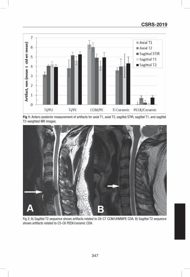
347
CSRS-2019
Fig 1: Antero-posterior measurement of artifacts for axial T1, axial T2, sagittal STIR, sagittal T1, and sagittal T2-weighted MR images.
Fig 2: A) Sagittal T2 sequence shows artifacts related to C6-C7 CCM/UHMWPE CDA. B) Sagittal T2 sequence shows artifacts related to C5-C6 PEEK/ceramic CDA.

348
CSRS-2019E-Poster #39
A Comparison of In-Hospital Complications Following Anterior Cervical Spine Surgery Between Cases with and without Parkinson’s Disease: A Propensity Score Matched Analysis
Anoop R. Galivanche, BS,1 Taylor D. Ottesen, BS,1 Jonathan N. Grauer, MD,1 Arya G. Varthi, MD1
1Department of Orthopaedics and Rehabilitation, Yale School of Medicine, New Haven, CT
Introduction: Patients with Parkinson’s Disease (PD), the second most prevalent neurodegenerative disorder (surpassed only by Alzheimer’s disease), may present with cervical spine conditions. With anterior cervical fusion surgery (ACF) commonly performed, the potential impact of PD on perioperative outcomes is of interest. The current study used the large sample size of the National Inpatient Sample (NIS) database to compare anterior cervical fusion perioperative complications of those with and without PD.
Materials and Methods: The 2007-2014 National Inpatient Sample (NIS) database was queried for cases undergoing anterior cervical spine procedures. Those with circumferential surgery and emergent cases were excluded.
Age and sex were directly extracted from the database. Cases who were obese or morbidly obese were further identified based on diagnostic codes. Overall comorbidity burden of each case was approximated using Elixhauser’s Comorbidity Index (ECI) and grouped into the following bins: 0 comorbidities, 1-6 comorbidities, and greater than 6 comorbidities. Cases with Parkinson’s disease (PD) were then identified using diagnostic codes.
Length of hospital stay (LOS) was also directly extracted from the database. Outcome data was collected on the following adverse events: surgical site infection, pneumonia, urinary tract infection, sepsis, renal injury, unplanned reintubation, venothrombolic events, cardiac arrest, myocardial infarction, and stroke. Outcomes were aggregated into serious, minor, and any adverse event. Prolonged LOS was defined as LOS greater than the median.
After propensity score matching on the basis of age, sex, BMI, and overall comorbidity burden, multivariate logistic regression analysis was used to compare outcome measures between PD cases and non-PD cases.
Results: A total of 231,999 cases were included in the study population, of whom 754 (0.33%) had Parkinson’s disease. Cases with Parkinson’s disease tended to be older, male, less obese, and have a higher comorbidity burden. Prior to propensity score matching, cases in the PD group had a significantly longer length of hospital stay (mean of 4.5 days compared with 2.3 days, p<0.001). After 1:1 propensity score matching based on demographic and comorbidity variables, there were no longer differences between the cohorts.
Based on multivariate analysis of the propensity matched cohorts, PD cases had greater odds of any (OR=2.25), serious (OR=1.60), and minor adverse events (OR=2.48) (p<0.05 for each) but not statistically different rates of prolonged hospital stay or mortality (Fig 1).
Conclusion: Overall, anterior cervical fusion cases with Parkinson’s disease are at greater risk of in-hospital adverse events, even when controlling for demographic and comorbidity variables. Specific considerations could be given to this at-risk patient population.

349
CSRS-2019

350
CSRS-2019E-Poster #40
Preliminary Results of Randomized Controlled Trial Investigating the Role of Psychological Distress on Cervical Spine Surgery Outcomes: A Baseline Analysis
Peter G Passias MD, Samantha R Horn BA, Sherri Weiser-Horwitz, PhD, Marco Campello, PT, PhD, Mohamed A Moawad MPH, Cole Bortz BA, Frank A. Segreto BS, Avery Brown, BS, Haddy Alas, Katherine Pierce, Dennis Vasquez-Montes MS, Rivka Ihejirika MD, Karan Patel MD, Renaud Lafage MS, Virginie Lafage PhD
Introduction: Recent studies have suggested that for patients with neck pain, both psychological and physical symptoms need to be addressed. Psychological distress risk factors are associated with poor outcomes in the fear avoidance model, where individuals passively cope through avoidance behaviors and disuse. Cognitive behavioral therapy (CBT) works to address risk factors through education about pain, modification of maladaptive beliefs, and increasing patient’s self-efficacy. Thus, this study determined the effectiveness of brief psychological intervention on outcomes in cervical spine surgery.
Materials/Methods: 35 patients age >18 with symptomatic cervical degenerative disease have been enrolled in the study. If patients met psychological distress criteria, they were in the treatment group: DRAM >17 and <33, FABQ >49/66, PCS >30/52 or OEQ ≤2 (randomized to CBT or placebo). CBT and sham treatment groups had 6 sessions prior to surgery. The control group had no intervention prior to surgery. Baseline and 3M changes were assessed for all outcome measures.
Results: 35 patients were enrolled (age 53.9 years, BMI 28.7 kg/m2). 22 patients met psychological distress criteria and were randomized into a treatment group (13 CBT vs. 9 placebo). 13 patients were in the control group, with 5 having too high of DRAM scores to be CBT candidates. At enrollment, the average DRAM score was 34.55 ± 13.87, with the DRAM observational group, Placebo, and CBT groups all having higher scores than control patients (45.6 vs 13.4, P<0.001, table a). Treatment patients had higher baseline FABQ scores than controls (45.9 vs 20.4, P=0.004). The overall OEQ score was 3.78 ± 1.18, with all control patients scoring a 5 and CBT and Placebo patients answering 3-4 out of 5 on the scale. At 3M post op, all groups showed improved outcomes in all measurements. Between CBT, placebo, and control patients, CBT patients exhibited greater postop improvement in all psychological questionnaires compared to non treatment groups ( DRAM: 34.9 30.8 CBT, 35.2 22.4 placebo, 11 10 no risk controls, FABQ: 40.8 35.2 CBT, 38.3 37.8 placebo, 19 21 no risk controls, PCS: 31.8 17.4 CBT, 32.8 11.6 placebo, 15 6 no risk controls, OEQ: Disagree Strongly Agree CBT, Disagree Agree placebo, Strongly Agree Strongly Agree, no risk controls).
Conclusions: Preliminary results of this randomized controlled study on cervical degenerative surgery patients showed that patients who received cognitive behavioral treatment before surgery had better improvement in all psychological related questionnaires compared to non treatment patients. Long-term follow up will assess the changes in psychological and pain related outcomes in these groups to assess the impact of psychological intervention for at risk patients.
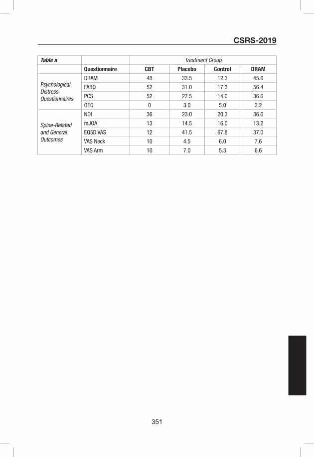
351
CSRS-2019
Table a Treatment Group
Questionnaire CBT Placebo Control DRAM
Psychological Distress Questionnaires
DRAM 48 33.5 12.3 45.6
FABQ 52 31.0 17.3 56.4
PCS 52 27.5 14.0 36.6
OEQ 0 3.0 5.0 3.2
Spine-Related and General Outcomes
NDI 36 23.0 20.3 36.6
mJOA 13 14.5 16.0 13.2
EQ5D VAS 12 41.5 67.8 37.0
VAS Neck 10 4.5 6.0 7.6
VAS Arm 10 7.0 5.3 6.6

352
CSRS-2019E-Poster #41
Correlation and profile of quality of life and functional outcome measures for cervical spondylotic myelopathy after surgery
Feifei Zhou1 M.D, Yu Sun1 M.D
1Department of Orthopaedic Surgery, Peking University Third Hospital, Beijing, China
Introduction: Patients with cervical spondylotic myelopathy (CSM) always exhibit a pattern of neurologic dysfunctions, and surgical decompression has been confirmed its efficacy in preventing disease progression. The aim of this study is to investigate functional outcomes with the modified Japanese Orthopaedic Association (mJOA) and quality of life outcomes with the SF-36, and try to investigate the correlation between quality of life and functional outcome measures at different follow-up times.
Methods: A prospective cohort enrolled 355 patients with CSM treated in our hospital form February 2008 to September 2014. We used mJOA and SF-36 questionnaires to assess the patients at different time points before the surgery and during the process of more than 2-year follow-up. Wilcoxon rank-sum test was performed t analyze postoperative changes in mJOA and SF-36, and maximum recovery time point was determined by the peak recovery time point. After that we used Spearman rank correlation analysis to investigating the correlation between the two health status measures (HSMs). Finally, in order to assess each HSMs’ ability to discriminate HTI, receiver operating characteristic (ROC) curve was created, and the area under the curve (AUC) was calculated to evaluate the accuracy of the ROC, as well as calculated AUC of the ROC curve and correlations between responses to the HTI by Spearman rank correlation analysis.
Results: The mean age of study patients was 57.4±10.2 years, 56.6% (201/355) were male and 43.4% (154/355) were female, and the mean follow-up was 50.2 months after surgery. Evaluation items in the mJOA assessment exhibited various degrees of improvement, and upper extremity sensation got the highest persistence rate (0.69). After surgery, sensory function had a quicker improvement than motor function (3 months, 63.3% vs 33.5%), but became nearly the same at one years and final follow-up (one year, 67.5% vs 65.7%; final follow-up, 69.1% vs 68.5%). The maximum recovery time point of the mJOA score was 16.1 months. As for the QOL, PCS reached the significant improvement at 3 months postoperative (maximum recovery, 20.3 months) while MCS reached at the time point of one year after surgery (maximum recovery, 23.9 months). Between mJOA and SF-36, different correlations were found at different time point after surgery. Using the ROC curve, AUC and correlation coefficient of PCS was the highest at three months after surgery (AUC=0.968), and mJOA at one year after surgery (AUC=0.928), as well as MCS at final follow-up (AUC=0.948).
Conclusion: Surgical treatment can improve the QOL and symptoms of CSM patients, and can help the patients achieving significant improvement in neurological function. mJOA, PCS, and MCS reached their maximal recovery in turn postoperatively, and the most responsive measure of therapeutic effectiveness varies depending on the follow-up timepoint.

353
CSRS-2019E-Poster #42
Surgical outcome of cervical spine metastasis: a prospective study of 45 cases1Yutaro Kanda, 1Kenichiro Kakutani, 1Takashi Yurube, 1Zhongying Zhang, 1Yuji Kakiuchi, 1Yoshiki Takeoka, 1Ryu Tsujimoto, 1Kunihoko Miyazaki, 2Toru Takada, 1Shingo Miyazaki 1Ryosuke Kuroda, 1Kotaro Nishida1. Department of Orthopaedic Surgery, Kobe Graduate School of Medicine, Kobe, Japan2. Department of Orthopaedic Surgery, Kobe Hokuto Hospital, Kobe, Japan
Introduction: Symptomatic spinal metastasis is characterized by severe pain and neurological deficits. Since cervical spine metastasis often causes tetraplegia, intractable pain, and respiratory failure, the patient’s performance status (PS) and quality of life (QOL) were severely deteriorated. However, few studies addressed the surgical outcome of cervical spine metastasis. The aim of this study is firstly to investigate the outcome of surgery for cervical spine metastasis, and secondary to identify the risk factor of poor surgical outcome, especially focusing on PS and QOL.
Materials/Methods: We prospectively analyzed 45 patients with cervical spine metastasis who undergo palliative surgery from 2013 to 2016. Surgery is based on posterior decompression and stabilization surgery using laminectomy and instrumentation. The indications for surgery were (1) progressive neurological deficits, or (2) intractable pain resistant to conservative care. Tokuhashi score was investigated to assess the severity of spine metastasis. Eastern Cooperative Oncology Group Performance Status (ECOGPS), EQ5D and Frankel classification were used to assess PS, QOL, and neurological function, respectively. Clinical follow-up was performed at one, three, and six months after surgery. The survival times and complications were also collected. Furthermore, these patients were divided into three groups based on main lesion level (Group U; C1-2, Group M; C3-6, Group L; C7, T1). The chronological changes between the three groups were identified by using Kruskal-Wallis test and Scheffe post hoc test. The poor surgical outcome was defined as no improvement or deterioration after improvement of ECOGPS or EQ5D within six months. Chi-square test was performed between the three groups. A P value of < 0.05 represented significance.
Results: There were 30 males and 15 females, with a mean age of 68.4 ± 10.3 years. The mean surgical time and blood loss were 191 ± 76 (min) and 285 ± 301 (g), respectively. Postoperative complications were occurred in 10 patients (22.2%). The median of ECOGPS was PS3 at baseline, improved to PS2 at one month and PS1 at three and six months after surgery. The mean value of EQ5D score was 0.168 at baseline, improved to 0.646 at one month and 0.759 at three months (Fig. 1). Except for 15 neurologically normal patients (Frankel E), Frankel classification was improved in 22 patients (48.9%), whereas deteriorated in 2 patients (4.4%). Interestingly, all 5 patients with recurrence of neurological deficit were belonged to Group L. There were no significant differences in ECOGPS and EQ5D between the three groups at all end points. However, the rate of poor outcome trended to be higher in Group L (12 of 20, 60.0%) than Group U (2 of 9, 22,2%) and Group M (4 of 16, 25.0%) (Table. 1) (p = 0.09).
Conclusion: In the current study, posterior decompression and stabilization for cervical spine metastasis greatly improved PS and QOL. Interestingly, posterior surgery for C7 or T1 metastasis might be likely to result in poor outcome. To our knowledge, no previous reports referred to poor

354
CSRS-2019
outcome of lower cervical spine metastasis. However, there was no significant difference and further analysis is needed.
Figure 1. The chronological change of PS and EQ5D
Table 1. Surgical outcome according to the main lesion level
355
CSRS-2019E-Poster #43
Defining Clinically Relevant Improvement for Patients Following Cervical Spine Surgery: Percent Reduction VS. MCID
Kristin R. Archer,1 Jacquelyn S. Pennings,1 Inamulah Khan,1 Anthony M. Asher,1 Anthony L. Asher,2
Clinton J. Devin1,3
1Vanderbilt University Medical Center, Nashville, TN; 2Carolina Neurosurgery & Spine Associates, Charlotte, NC; 3Steamboat Orthopaedic and Spine Institute, Steamboat Springs, CO
Introduction: The literature suggests that an absolute change from baseline (i.e., minimal clinically important difference: MCID) may not be a reliable marker of response to treatment for patients with a low or high baseline patient-reported outcome (PRO) score. An alternative to MCID is a threshold of clinical relevance defined by a percent reduction from baseline PRO score. The purpose of this study was to determine whether a percent reduction of 30% in disability and pain scores is a valid method for determining clinical improvement at 12 months after cervical spine surgery.
Materials/Methods: The study was a retrospective evaluation of prospectively collected data from a national surgical spine registry, the Quality Outcomes Database (QOD). 9,662 participants undergoing elective anterior cervical discectomy and fusion (ACDF) or posterior laminectomy with or without fusion for a cervical degenerative condition were included in the analysis. Outcomes were disability (NDI), neck and arm pain (NRS-NP/AP), and satisfaction (NASS scale). Participants completed a preoperative assessment and follow-up assessment at 1-year. The change in NDI and NRS-NP/AP scores were categorized as met a percent change ≥ 30% (clinically relevant) or a percent change < 30% (not clinically relevant) and by achieving MCID or not achieving MCID. MCID was based on established values of 17.3 for NDI, 2.6 for NRS-NP, and 4.1 for NRS-AP.1 The relationship between the 30% reduction and MCID categorizations and satisfaction were compared with logistic regression models and receiver operating characteristic (ROC) curves. Analyses were conducted in the entire sample and by surgical procedure (ACDF, posterior decompression, posterior decompression with fusion). In addition, the ability of 30% reduction and MCID to predict satisfaction was examined by severity of disability and pain. Significance was set at p < .05.
Results: A 30% reduction in NDI and NRS-NP/AP predicted satisfaction with 74.6%, 71.6%, and 68.9% accuracy, respectively, whereas MCID thresholds predicted satisfaction with 69.8%, 69.1%, and 66.7% accuracy (p < .001). Differences were similar by surgical procedure. The largest difference in predictive power for 30% reduction compared to MCID NDI was found for patients with bed-bound disability (NDI 81-100%: 75.7% vs. 58%) and minimal disability (NDI 0-20%: 70.3% vs. 52.6%). For pain, the largest difference in predictive power for 30% reduction compared to MCID was found for patients with no/low pain (NRS-NP 0-4: 68.1% vs. 59.1%; NRS-AP 0-4: 66.6% vs. 50%). However, for severe arm pain the accuracy was higher for MCID compared to 30% reduction (74.4% vs. 69.9%).
Conclusions: A 30% PRO reduction may be a more clinically relevant method for identifying response to treatment compared to MCID. It can be applied to broad spine surgery populations and takes into account the baseline PRO score. Furthermore, a 30% reduction appears to

356
CSRS-2019
outperform established MCID thresholds for patients with either low or high preoperative disability scores and low neck and arm preoperative pain scores.
References: 1Parker SL, Godil SS, Shau DN, Mendenhall SK, McGirt MJ. Assessment of the minimum clinically important difference in pain, disability, and quality of life after anterior cervical discectomy and fusion: clinical article. Journal of neurosurgery Spine. 2013;18(2):154-60.

357
CSRS-2019E-Poster #44
Free-hand Placement of C7 Laminar Screws: Accuracy and Safety in 43 Consecutive Patients
Jiwon Park, MD (Seoul, Korea), K. Daniel Riew, MD (New York, NY), Ho-Joong Kim, MD, Bong-Soon Chang, MD, Choon-Ki Lee, MD, and Jin S. Yeom, MD (Seoul, Korea)
Introduction: Although pedicle screws have been regarded as the first-line choice for fixation of C7, they may require radiographic or fluoroscopic guidance, take time for placement, and have a potential risk of neurovascular complications. Given such limitations, C7 laminar screw might serve as a viable alternative. However, while laminar screws are widely used for C2 fixation, reports on the clinical experience of C7 laminar screws are quite limited. The purpose of this study was to determine the accuracy and safety of C7 laminar screw placement with a free-hand technique.
Materials/Methods: All patients who underwent posterior cervical fixation with C7 laminar screws by the last author were chosen. All screws were placed with a free-hand technique without radiographic or fluoroscopic guidance. The operating time for each screw placement was approximately 1-2 minutes. Clinical information and radiologic data of the patients were analyzed. Using postoperative CT scans, which were taken in all patients, the accuracy of screw placement was evaluated by two orthopedic surgeons by assessing the direction (dorsal versus ventral) and degree of laminar cortical breach.
Results: Forty-three consecutive patients were enrolled. There were 26 males and 17 females, and the age averaged 59 years (range, 14-82). A total of 61 C7 laminar screws were used: twenty-five patients underwent unilateral C7 laminar screw fixation, and eighteen patients underwent bilateral fixation (Figs 1 and 2). All the laminar screws were 3.5 mm in diameter and 20 to 26 mm in length (3 20-mm, 13 22-mm, 38 24-mm, and 7 26-mm screws).
Of the 61 screws, 14 screws (23%) breached the laminar cortical wall, including 3 dorsal and 11 ventral breaches. Of those 14 screws, 11 screws (18%) breached by less than 50% of screw diameter and 3 screws (5%) breached by more than 50% but less than 100% of screw diameter. Nine screws were one of the screws placed bilaterally, and 5 screws were unilaterally placed. No intraoperative neurovascular injury was observed, and none of the screws with cortical breaches resulted in worsening of neurologic symptoms. None of the patients required reoperation for any reason. Over the follow-up period of 20 months, mechanical failure such as loosening or fractures of screw-rod system has not been observed.
Conclusion: To our knowledge, this is the largest clinical study ever performed on C7 laminar screw fixation. Although laminar cortical breach was common (25%) with our free-hand technique, most were trivial and none had clinically relevant neurovascular complications nor mechanical failures. Therefore, C7 laminar screws, which can be placed quickly and easily without the need of radiographic or fluoroscopic guidance, may provide a valuable alternative to pedicle screws for C7 fixation in terms of efficacy and safety. However, long-term outcomes, including fusion status, remains to be evaluated with further follow-up.

358
CSRS-2019
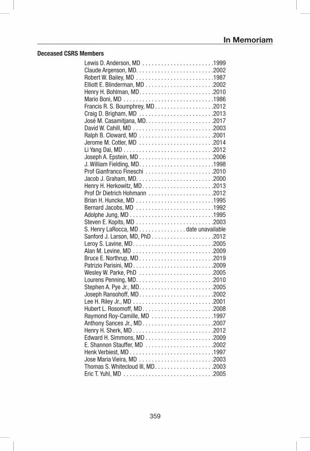
359
In Memoriam
Deceased CSRS Members Lewis D. Anderson, MD . . . . . . . . . . . . . . . . . . . . . . .1999Claude Argenson, MD. . . . . . . . . . . . . . . . . . . . . . . . .2002Robert W. Bailey, MD . . . . . . . . . . . . . . . . . . . . . . . . .1987Elliott E. Blinderman, MD . . . . . . . . . . . . . . . . . . . . . .2002Henry H. Bohlman, MD. . . . . . . . . . . . . . . . . . . . . . . .2010Mario Boni, MD . . . . . . . . . . . . . . . . . . . . . . . . . . . . .1986Francis R. S. Boumphrey, MD . . . . . . . . . . . . . . . . . . .2012Craig D. Brigham, MD . . . . . . . . . . . . . . . . . . . . . . . .2013José M. Casamitjana, MD. . . . . . . . . . . . . . . . . . . . . .2017David W. Cahill, MD . . . . . . . . . . . . . . . . . . . . . . . . . .2003Ralph B. Cloward, MD . . . . . . . . . . . . . . . . . . . . . . . .2001Jerome M. Cotler, MD . . . . . . . . . . . . . . . . . . . . . . . .2014Li Yang Dai, MD . . . . . . . . . . . . . . . . . . . . . . . . . . . . .2012Joseph A. Epstein, MD . . . . . . . . . . . . . . . . . . . . . . . .2006J. William Fielding, MD. . . . . . . . . . . . . . . . . . . . . . . .1998Prof Gianfranco Fineschi . . . . . . . . . . . . . . . . . . . . . .2010Jacob J. Graham, MD. . . . . . . . . . . . . . . . . . . . . . . . .2000Henry H. Herkowitz, MD . . . . . . . . . . . . . . . . . . . . . . .2013Prof Dr Dietrich Hohmann . . . . . . . . . . . . . . . . . . . . .2012Brian H. Huncke, MD . . . . . . . . . . . . . . . . . . . . . . . . .1995Bernard Jacobs, MD . . . . . . . . . . . . . . . . . . . . . . . . .1992Adolphe Jung, MD . . . . . . . . . . . . . . . . . . . . . . . . . . .1995Steven E. Kopits, MD . . . . . . . . . . . . . . . . . . . . . . . . .2003S. Henry LaRocca, MD . . . . . . . . . . . . . . . date unavailableSanford J. Larson, MD, PhD . . . . . . . . . . . . . . . . . . . .2012Leroy S. Lavine, MD. . . . . . . . . . . . . . . . . . . . . . . . . .2005Alan M. Levine, MD . . . . . . . . . . . . . . . . . . . . . . . . . .2009Bruce E. Northrup, MD . . . . . . . . . . . . . . . . . . . . . . . .2019Patrizio Parisini, MD. . . . . . . . . . . . . . . . . . . . . . . . . .2009Wesley W. Parke, PhD . . . . . . . . . . . . . . . . . . . . . . . .2005Lourens Penning, MD. . . . . . . . . . . . . . . . . . . . . . . . .2010Stephen A. Pye Jr., MD. . . . . . . . . . . . . . . . . . . . . . . .2005Joseph Ransohoff, MD. . . . . . . . . . . . . . . . . . . . . . . .2002Lee H. Riley Jr., MD . . . . . . . . . . . . . . . . . . . . . . . . . .2001Hubert L. Rosomoff, MD. . . . . . . . . . . . . . . . . . . . . . .2008Raymond Roy-Camille, MD . . . . . . . . . . . . . . . . . . . .1997Anthony Sances Jr., MD . . . . . . . . . . . . . . . . . . . . . . .2007Henry H. Sherk, MD . . . . . . . . . . . . . . . . . . . . . . . . . .2012Edward H. Simmons, MD . . . . . . . . . . . . . . . . . . . . . .2009E. Shannon Stauffer, MD . . . . . . . . . . . . . . . . . . . . . .2002Henk Verbiest, MD . . . . . . . . . . . . . . . . . . . . . . . . . . .1997Jose Maria Vieira, MD . . . . . . . . . . . . . . . . . . . . . . . .2003Thomas S. Whitecloud III, MD. . . . . . . . . . . . . . . . . . .2003Eric T. Yuhl, MD . . . . . . . . . . . . . . . . . . . . . . . . . . . . .2005

360

361
CSRS Donation Form
Name
Address
City State
Zip Country
Office Phone Email
Total Donation Amount $ I would like my donation to go to:
21st Century Research & Education Grant Fund $
Henry Bohlman Educational Endowment Fund $
General $
Recognition Information
I wish to be recognized as (e.g., Dr. or Dr. & Mrs., etc.)
I wish for my donation to remain anonymous
Payment Information
CREDIT CARD Please charge my credit card in the amount of $ in (partial / full) payment.
I pledge to pay the balance by donating $ per year for years to be paid before December 31st of each year.
Card type: VISA MasterCard American Express Discover
Card Number Exp. Date
Name on Card
CHECK Enclosed is my check in the amount of $ in (partial / full) payment of my pledge.
I pledge to pay the balance by donating $ per year for years to be paid before December 31st of each year.
Please return this form with your donation to CSRS by email to [email protected] or mail completed form and check to Cervical Spine Research Society, 555 E. Wells St., Ste. 1100, Milwaukee, WI 53202.

362
Copyright © 2022 FDOKUMEN