Communicative capacities in Middle Pleistocene humans from the Sierra de Atapuerca in Spain

Journal of Human Evolution 53 (2007) 6e25
Metric and morphological study of the upper cervical spine fromthe Sima de los Huesos site (Sierra de Atapuerca, Burgos, Spain)
Asier Gomez-Olivencia a,b,*, Jose Miguel Carretero a,b, Juan Luis Arsuaga b,c,Laura Rodrıguez-Garcıa a,b, Rebeca Garcıa-Gonzalez a,b, Ignacio Martınez b,d
a Laboratorio de Evolucion Humana. Dpto. de Ciencias Historicas y Geografıa. Universidad de Burgos.
Edificio IþDþi. Plaza Misael de Ba~nuelos s/n. 09001 Burgos, Spainb Centro UCM-ISCIII de Investigacion sobre Evolucion y Comportamiento Humanos, c/Sinesio Delgado, 4 (Pabellon 14), 28029 Madrid, Spain
c Departamento de Paleontologıa, Facultad de Ciencias Geologicas, Universidad Complutense de Madrid, Ciudad Universitaria s/n, 28040 Madrid, Spaind Departamento de Geologıa, Universidad de Alcala, Edificio de Ciencias, Campus Universitario, 28871 Alcala de Henares, Spain
Received 22 February 2006; accepted 21 December 2006
Abstract
In this article, the upper cervical spine remains recovered from the Sima de los Huesos (SH) middle Pleistocene site in the Sierra de Ata-puerca (Burgos, Spain) are described and analyzed. To date, this site has yielded more than 5000 human fossils belonging to a minimum of 28individuals of the species Homo heidelbergensis. At least eleven individuals are represented by the upper cervical (C1 and C2) specimens: sixadults and five subadults, one of which could represent an adolescent individual. The most complete adult vertebrae (three atlases and three axes)are described, measured, and compared with other fossil hominins and modern humans. These six specimens are associated with one another andrepresent three individuals. In addition, one of these sets of cervical vertebrae is associated with Cranium 5 (Individual XXI) from the site. Themetric analysis demonstrates that the Sima de los Huesos atlases and axes are metrically more similar to Neandertals than to our modern humancomparative sample. The SH atlases share with Neandertals a sagittally elongated canal. The most remarkable feature of the SH (and Neandertal)axes is that they are craniocaudally low and mediolaterally wide compared to our modern male sample. Morphologically, the SH sample shareswith Neandertals a higher frequency of caudally projected anterior atlas arch, which could reflect greater development of the longus colli muscle.In other features, such as the frequency of weakly developed tubercles for the attachment of the transverse ligament of the atlas, the Sima de losHuesos fossils show intermediate frequencies between our modern comparative samples and the Neandertals, which could represent the prim-itive condition. Our results are consistent with the previous phylogenetic interpretation of H. heidelbergensis as an exclusively European species,ancestral only to H. neanderthalensis.� 2007 Elsevier Ltd. All rights reserved.
Keywords: Atlas; Axis; Cervical vertebrae; Middle Pleistocene; Sima de los Huesos
Introduction
The Sima de los Huesos (SH) site is approximately 0.5 kmfrom the Cueva Mayor entrance, well inside the Cueva MayoreCueva del Silo cave system in the Sierra de Atapuerca in
* Corresponding author. Laboratorio de Evolucion Humana, Dpto. de Cien-
cias Historicas y Geografıa, Universidad de Burgos, Edificio IþDþI, Plaza
Misael de Ba~nuelos s/n, 09001 Burgos, Spain. Tel.: þ34 947 25 93 24.
E-mail address: [email protected] (A. Gomez-Olivencia).
0047-2484/$ - see front matter � 2007 Elsevier Ltd. All rights reserved.
doi:10.1016/j.jhevol.2006.12.006
northern Spain (Arsuaga et al., 1997b). To date, more than5000 fossil human remains have been recovered from the site(Arsuaga and Martınez, 2004) in the excavations directed byone of us (JLA). Based on dental evidence, these remains be-long to a minimum number of 28 individuals (Bermudez deCastro et al., 2004) of both sexes and diverse ages. In addition,thousands of carnivore bones have been recovered mixed withand stratigraphically above the human fossils (Garcıa et al.,1997; Garcıa, 2002). All anatomical parts of the skeleton arerepresented among the human remains, suggesting that
7A. Gomez-Olivencia et al. / Journal of Human Evolution 53 (2007) 6e25
complete corpses were accumulated at this site. The age-at-death distribution suggests that a nonattritional demographicevent affected this living population (Bocquet-Appel andArsuaga, 1999; Bermudez de Castro et al., 2004). The originof the human accumulation is most likely to be anthropo-genic (Arsuaga et al., 1997b). A recently discovered hand-axe has been interpreted as evidence of symbolic behavior inthese early humans (Carbonell et al., 2003).
A recently found in situ speleothem (SRA-3), which sealsthe human-fossil-bearing sediments throughout the site, hasbeen dated. There is a hiatus in the speleothem growth at about4 cm below the top. This upper portion shows a linear growthrate of about 1 cm per 32,000 years. Ten dates have been ob-tained in the lower 10 cm of speleothem below the hiatus, allof which indicate a minimum age of 350 ka, although thisthickness could represent a significant amount of time beyondthis date. Thus, a range of 400e500 ka has been proposed forthe human remains (Bischoff et al., 2003). These dates arecompatible with both the micro- and macromammalian assem-blages (Cuenca-Bescos et al., 1997; Garcıa et al., 1997; Garcıa,2002). Bischoff et al. (2006) recently published a reanalysisof six samples of SRA-3 using inductively coupled plasma-multicollector mass-spectrometry (ICP-MS), which yieldednew dates that cluster around 600 ka, with an estimated mini-mum age of the speleothem, and thus of the underlying humanfossils, of 530 ka.
The human remains from this site have been assigned toHomo heidelbergensis. This species, in our view, is exclu-sively European, and is ancestral only to the later Neandertals(Arsuaga et al., 1991, 1997c; Carretero et al., 1997; Martınezand Arsuaga, 1997).
The record of the upper cervical vertebral column is rela-tively abundant for Homo neanderthalensis and late Pleisto-cene Homo sapiens, but with respect to the rest of the genusHomo, it is scarce or nonexistent.1 The virtual absence of a fos-sil record of the upper cervical spine for the middle Pleisto-cene underscores the importance of the SH specimensdescribed and analyzed here.
Regarding the Neandertals, the most conspicuous traitsdescribed for the atlas vertebra (C1) are (1) weakly developedtubercles for the insertion of the transverse ligament and (2)a caudal projection of the anterior tubercle (Boule, 1911e1913; Martin, 1923; Heim, 1976; Arensburg, 1991); for the Ne-andertal axis (C2) no trait or pattern has been highlighted exceptits great morphological variability (Piveteau, 1966). In his studyof the cervical spine of the Kebara 2 Neandertal individual,Arensburg (1991) concluded that, except for the horizontal spi-nous process of the C6 and C7, the cervical column seemed tobe within the range of variation of modern human populations.Nevertheless, the study of the middle Pleistocene SH uppercervical vertebrae (C1 and C2) may reveal some previously
1 The exceptions are the specimens from Dmanisi (Meyer, 2005), Gran Do-
lina (Carretero et al., 1999), and Koobi Fora (KNM-ER 1808; Walker et al.,
1982; Leakey and Walker, 1985) for the early Pleistocene and, for the middle
Pleistocene, the atlas from the Zhoukoudian I1 individual (Boaz et al., 2004).
undocumented morphological features and/or patterns of varia-tion within the Neandertal evolutionary lineage.
The first part of the study comprises the inventory of all theatlases and axes, with the determination (if possible) of the ageat death (Tables 3 and 4) and the minimum number of individ-uals represented among the remains. A brief description of themost complete adult vertebrae is also provided. In the secondpart, we perform a metrical analysis of the adult vertebrae andcompare the anatomical features present in the SH specimenswith those found in other samples of Homo, especially H. ne-anderthalensis and H. sapiens.
Materials
The SH vertebral sample comprises 455 fossils that repre-sent at least 180 vertebrae. The cervical sample consists of116 fossils (Gomez-Olivencia, 2005), including 22 first cervi-cal vertebrae (atlas) and 16 second cervical vertebrae (axis).The present study includes the atlas (C1) and axis (C2) re-mains recovered up through the 2004 field season. An inven-tory and photographic documentation of all the fossilmaterial, as well as short descriptions and metrical data ofthe most complete adult vertebrae, are provided.
Descriptions of a few of the cervical vertebrae [includingthe atlas VC3 (AT-1554) and a general description of the SHcervical vertebrae] have been published previously (Carreteroet al., 1999; Gomez et al., 2005). The present study providesa detailed analysis of the SH upper cervical spine. Appendix1 provides information on the labeling of the SH vertebrae.
For comparative purposes we have studied a large sample ofmodern human skeletons and fossil hominin specimens fromthe following species: H. antecessor, H. neanderthalensis,and late Pleistocene H. sapiens (Table 1). Although remainsof the atlas and axis are also known from the Mousterian siteof Qafzeh (Vandermeersch, 1981), their fragmentary naturemakes comparison with these specimens difficult. Data forthe following specimens have been taken from the literature:Kebara 2 (Arensburg et al., 1990; Arensburg, 1991), Regour-dou 1 (Piveteau, 1966), Shanidar 2 and 4 (Stewart, 1962;Trinkaus, 1983), Subalyuk (Pap et al., 1996), and Dolnı Vesto-nice 14 (Sladek et al., 2000).
Methods
We used standard anthropometric techniques and instru-ments to take all measurements. The metric variables are illus-trated in Fig. 1. Following Meyer (2005), the areas of vertebralcanals were measured on scaled digital images and cross-checked for accuracy by comparing imaged linear measure-ments to physical dimensions measured with digital calipers.This method avoids the considerable error of area estimationby simply multiplying the dorsoventral and transverse diame-ters of the neural canal (Meyer, 2005). Vertebral-canal areaswere measured on cranial (atlas) and caudal (axis) photographsusing CAD software and cross-checked using the canal’s max-imum transverse diameter (M11). For the atlas, the photographwas taken in superior view.

8 A. Gomez-Olivencia et al. / Journal of Human Evolution 53 (2007) 6e25
Table 1
Comparative specimens and samples of atlases and axes measured by the authors
Specimen/sample Species Sex Original/cast Location
ATD6-90 (C1) H. antecessor Female Original Museo de Burgos, Burgos (Spain)
Krapina (C1, n¼ 3; C2, n¼ 3) H. neanderthalensis ? Original Croatian Natural History Museum,
Zagreb (Croatia)
La Chapelle-aux-Saints (C1 and C2) H. neanderthalensis Male Original Musee de l’Homme, Paris (France)
La Ferrassie 1 (C1 and C2) H. neanderthalensis Male Original Musee de l’Homme, Paris (France)
Shanidar 2 (C1 and C2) H. neanderthalensis Male Cast Musee de l’Homme, Paris (France)
Skhul V (C1 and C2) H. sapiens Male Original Peabody Museum of Archaeology and
Ethnology, Cambridge (MA, USA)
Arcy-sur-Cure, Grotte des Fees (C1 and C2) H. sapiens (?)1 ? Original (C1) Musee de l’Homme, Paris (France) (C1)
Cast (C2) Institut de Paleontologie Humaine,
Paris (France) (C2)
Cro-Magnon (C1) H. sapiens Male Original Musee de l’Homme, Paris (France)
Carolingian2 (C2, n¼ 4) H. sapiens ? Original Musee de l’Homme, Paris (France)
Neolithic3 (C2, n¼ 2) H. sapiens ? Original Musee de l’Homme, Paris (France)
Afalou-Bou-Rhummel4 (C1, n¼ 12; C2, n¼ 10) H. sapiens ? Original Institut de Paleontologie Humaine,
Paris (France)
Taforalt5 (C1, n¼ 8; C2, n¼ 9) H. sapiens ? Original Institut de Paleontologie Humaine,
Paris (France)
Burgos6 (n¼ 40) H. sapiens Males Original Laboratorio de Evolucion Humana-University
of Burgos, Burgos (Spain)
Hamman-Todd7 (n¼ 101) H. sapiens 50 males/51 females Original Cleveland Museum of Natural History,
Cleveland (OH, USA)
1 The Arcy-sur-Cure atlas was found in 1860 in the lower level of the Grotte des Fees, (Yonne, France). The axis was found in 1898 in the clearings of older
excavations (Leroi-Gourhan, 1958). Leroi-Gourhan (1958) identified both specimens as Neandertal. In the case of the axis, the taxonomic assignment was based on
the surface color of the fossil; Leroi-Gourhan pointed out that this specimen is within the modern human range of variation but that it resembles Neandertals in its
weak cervical curvature. In the case of the atlas, he did not find any trait to distinguish it from modern humans. Due to the problematic provenience of both spec-
imens and the fact that these fossils are morphologically more similar to our modern human comparative samples than to Neandertals, they should be cautiously
considered as representing H. sapiens.2 The Carolingian sample comes from the Saint-Germain-des-Pres cementery (Paris, France).3 The Neolithic sample comes from a cave site in the Petit Morin Valley (France).4 The Afalou-Bou-Rhummel sample was recovered from the homonymous rock-shelter in Algeria. This sample and the Taforalt sample are dated to >10,000 BP
(see Irish, 2000, and references therein).5 The Taforalt sample was recovered from the homonymous cave site in Morocco.6 The Burgos sample comprises 40 contemporary adult (estimated age at death is 20e45 years) male individuals from Burgos, Spain.7 The Hamann-Todd sample comprises 100 North American adults (50 Euro-Americans and 50 Afro-Americans, with equal sexual representation) from the
Hamann-Todd Osteological Collection.
Univariate comparative analysis was performed on all of thevariables measured in the atlases and axes. Bivariate analysiswas performed on the vertebral-canal variables (M10 andM11) in both C1 and C2, and the M1a and STrD measurementsin the axes. We used nonparametric methods in cases where oneor more of the groups had a sample size of n< 10. For the uni-variate analysis, we performed a KruskaleWallis test to com-pare the differences between the SH, Neandertal, and Burgosmodern human samples. When a significant difference( p< 0.05) was found in a variable, we performed a ManneWhitney test between all possible pairs of samples to determinewhich ones were significantly different. We adjusted the p-values for these comparisons using the Bonferroni methodand have reported all the cases in which p< 0.10.
Inventory
Here we provide an inventory of all the atlas and axis re-mains. These fossils are listed in Tables 2 and 3 and depictedin Figs. 2e5. The SH upper cervical sample comprises 22 atlasspecimens and 16 axis specimens.
Age at death
The age at death has been estimated for the SH cervicalvertebrae (Tables 2 and 3) based on modern human patternsof maturation (i.e., fusion of the principal centers of ossifica-tion, presence/absence of the epiphyseal rings, and degree ofobliteration of the metaphyseal scars). However, since theSH hominins had a shorter period of dental growth (RamırezRozzi and Bermudez de Castro, 2004), the ages at death mightbe overestimated if tooth formation is correlated with somaticdevelopment, as proposed by Smith (1991).
Given the difficulty of assigning a precise age at death basedon the ossification patterns of the atlas (except in limited cir-cumstances), these specimens are classified as adult or sub-adult. The atlas’s principal centers of ossification are fusedby the sixth year of life (Scheuer and Black, 2000). Additionalattention was given to the surface of the articular facets for thedetermination of the age at death, since immature individualsshow a more porous surface. In the Atapuerca sample, theVC3 atlas shows a partially obliterated metaphyseal scar onits transverse processes. This feature is consistent with the

9A. Gomez-Olivencia et al. / Journal of Human Evolution 53 (2007) 6e25
age at death estimated (21e25 years) for the associated verte-bra VC4(C2). The left mass AT-2078 exhibits the immatureappearance on the surface of both articular facets, while VC17only exhibits this appearance in the upper articular facet.
In contrast to the atlas, the complex ossification pattern of theaxis allows for more precise age-at-death estimates. The axisossifies from five primary centers: one for the body, one foreach half of the dens, and one for each half of the neural arch.The halves that form the dens are already fused at birth, whereasthe neural arch is fused by the third or fourth year and the bodyis joined to the other elements by the fifth or sixth year of life.
Fig. 1. Some of the dimensions used in the osteometric analysis, as defined in
the text: (a) atlas (C1) in cranial view; (b) axis (C2) in craniodorsal view; (c)
axis (C2) in lateral view. Drawings modified from Gray (1959). (1) Diameters
in major axis of upper articular facets (UAFMAD) (right-left); (2) diameter at
a right angle to the major axis of upper articular facets (UAFTrD) (right-left);
anterior arch thickness at the level of the anterior tubercle (AATh); craniocau-
dal diameter of laminae (CCDLam) (right-left); total vertebral dorsal height
(DoH); distance between the tubercles for the attachment of the transverse lig-
ament (DTub); inferior transverse diameter (ITrD); lower articular facet dorso-
ventral diameter (LAFDvD) (right-left); total vertebral ventral height (M1a);
body craniocaudal dorsal diameter (M2); body inferior transverse diameter
(M8); canal dorsoventral maximum diameter (M10); canal transverse maxi-
mum diameter (M11); spine length (M13); maximum dorsoventral diameter
(MDvD); maximum transverse diameter (MTrD); posterior arch thickness at
the level of the posterior tubercle (PATh); superior transverse diameter
(STrD); transverse diameter of the facet for the axis (TrDFA); maximum trans-
verse diameter of the tip of the spinous process (TrDTSP). The following vari-
ables have not been drawn: body inferior anteroposterior diameter (M5), taken
perpendicular to M8; upper articular facet dorsoventral diameter of the axis
(UAFDvD) (right-left); upper articular facet transverse diameter of the axis
(UAFTrD) (right-left); lower articular facet transverse diameter (LAFTrD)
(right-left), taken perpendicular to LAFDvD; thickness of the laminae
(ThLam), taken perpendicular to CCDLam; anterior arch height at the level
of the anterior tubercle (AAH), taken perpendicular to AATh; maximum
height of lateral masses (right-left) (MHLM), taken at the lateral edges of
the lateral masses of the atlas; posterior arch height at the level of the posterior
tubercle (PAH), taken perpendicular to PATh; height of the posterior arch in
the groove for the vertebral artery (HPAG) (right-left). M# refers to the Martin
number (Brauer, 1988) and variable definition. In the axes, M10 has been mea-
sured on the caudal aspect of the vertebral foramen.
The axis has five secondary centers of ossification: two for thetransverse processes; the ossiculum terminale, which fuses atthe end of the dens by the twelfth year; one for the spine; andan inferior epiphyseal ring, which fuses to the caudal part ofthe body (Scheuer and Black, 2000). Buikstra et al. (1984) re-ported that the fusion of the inferior epiphyseal ring commencesby 17e19 years and is completed by the twenty-fifth year intheir female sample from the Terry Collection. Both VC4 andVC28 show a partial fusion of the caudal epiphyseal ring. Nev-ertheless, VC4 shows a more advanced state of fusion, so itcould belong to an individual who was older than VC28.
Minimum number of individuals
A minimum of 28 individuals, based on the dental remains,have been identified among the SH human fossils (Bermudezde Castro et al., 2004). Regarding the atlas, a minimum of 10individuals are represented, based on the repetition of the leftmass. In addition, AT-3985 (a right lateral mass) represents an-other individual due to its incompatibility with any of the leftlateral masses. These 11 individuals represent six adults (oneof them less than 25 years of age, represented by VC3) andfive subadults, one of them possibly an adolescent, representedby VC17 (Table 2). Among the axes, based on the repetition ofthe most common element (the dens of the axis), at least 10 in-dividuals are recognizable: four adults (one of them less than25 years of age), one late adolescent or young adult, one imma-ture individual between 12 and 16 years of age, and four indi-viduals older than 12 years. All these ages at death are fullycompatible with those based on dental (Bermudez de Castroet al., 2004), cranial (Arsuaga et al., 1997c), and postcranialevidence (Carretero et al., 1997).
Sex determination
Sex determination of the three most complete SH atlasesand axes, which represent three individuals, was attemptedbased on multiple regression equations and discriminant func-tions (Marino, 1995; Wescott, 2000). The results suggest thatthese three sets of upper cervical vertebrae all represent maleindividuals.
Additionally, a discriminant function was generated fromdata taken from atlas specimens in the Hamann-Todd Osteolog-ical Collection, and we calculated the a posteriori probability ofthe most complete SH atlases (VC3 and VC7) being classifiedas belonging to male individuals (Table 4). These two SHatlases show a posteriori probabilities of being from male indi-viduals higher than 0.80. Due to the incompleteness of VC16, itwas not possible to apply the discriminant function, but usingan additional discriminant function in which only ITrD isused, VC16 is classified as being from a male with an a poste-riori probability of 0.79.
Marino’s (1995), Wescott’s (2000), and our formulae wereused to estimate the sex of several additional fossil atlases andaxes. The results agree with the sex determinations based onother postcranial features with one exception: the Regourdou1 individual is classified as being from a male using the

10 A. Gomez-Olivencia et al. / Journal of Human Evolution 53 (2007) 6e25
axis, while its sex has been regarded as indeterminate by Van-dermeersch and Trinkaus (1995). On the other hand, Krapina98 and 104, of unknown sex, were classified as belonging tofemales. The classification of the Gran Dolina ATD6-90 stillagrees with the previous sex determination by Carreteroet al. (1999), but the result should be interpreted more cau-tiously due to its low a posteriori probability (0.67).
The results of the sex determination of the VC7 atlas andVC8 axis are also congruent with the sex assignment of Cra-nium 5 based on the associated mandible AT-888 (Rosaset al., 2002; Bermudez de Castro et al., 2004). Although Cra-nium 5 appears to be a small male or a female when the cranialmeasurements are compared with other middle Pleistocenefossils and Neandertals, the facial skeleton is large and thuslikely that of a male (Arsuaga et al., 1997c).
Brief description of the most complete atlases and axes
Atlases
The most complete adult atlases are shown in Fig. 2.Specimen VC3(C1) is the most complete nonmodern atlas
of a fossil hominin yet found. It preserves the transverse
Table 2
Inventory of the atlas (C1) remains from the SH site (as of the 2004 field
season)
Specimen
number
Year Preservation Age at death Figure
VC31 1995 Complete vertebra Adult 2a,b,c
VC72 2000 Complete vertebra Adult 2d,e,f
VC163 1994, 1997 Masses and
posterior arch
Adult 2g,h
VC174 1995, 2000 Anterior arch
and left mass
Adolescent (?) 3a
AT-269 1989 Left mass Subadult 3b
AT-326 1990 Left mass 3c
AT-1818 1996 Left mass Adult 3d
AT-2078 1997 Left mass Subadult 3e
AT-2130 1997 Anterior arch 3l
AT-2264 1997 Posterior arch >5 years 3p
AT-2584 1998 Left mass,
posterior arch
3f
AT-2852 1998 Anterior arch 3m
AT-3003 1999 Left mass 3g
AT-3013 1999 Right mass,
posterior arch
fragment
Adult 3h
AT-3687 2000 Posterior arch Subadult (?) 3q
AT-3691 2000 Posterior arch Subadult (?) 3r
AT-3693 2000 Anterior arch 3n
AT-3694 2000 Right mass Subadult 3i
AT-3971 1994 Right mass Subadult 3j
AT-3985 ? Right mass Adult 3k
AT-3992 1992 Posterior arch 3s
AT-4037 2000 Anterior arch
fragment
3o
1 VC3¼AT-1554.2 VC7¼AT-3339þAT-3340þAT-3341þAT-3688.3 VC16¼AT-1140þAT-2201.4 VC17¼AT-3374þAT-3973þAT-3991.
processes, which show an incompletely obliterated metaphy-seal line. This vertebra was found in anatomical connectionwith the VC4 axis. The anterior bar of the left transverseprocess is not fused to the posterior bar, and this specimenexhibits subtle tubercles for the attachment of the transverseligament. The transverse processes show a triangular crosssection, making the dorsal surface quite vertical.
Specimen VC7(C1) is slightly larger and more robust thanVC3. It lacks the transverse processes. The sulcus for the rightvertebral artery is covered by a bony arch, while that on theleft side is only partially covered. Such foramina are not un-usual and have been reported in the atlas of the Shanidar 2Neandertal (Stewart, 1962; Trinkaus, 1983). Like VC3, thisspecimen exhibits subtle tubercles for the attachment of thetransverse ligament.
Specimen VC16(C1) lacks the anterior arch and the trans-verse processes. It exhibits severe postmortem erosion to thesuperior articular facets and inferior left articular facet. It issmaller than the VC3 and VC7 atlases, although the size ofits posterior arch falls between that of VC3 and VC7. It also
Table 3
Inventory of the axis (C2) remains from the SH site (as of the 2004 field
season)
Specimen
number
Year Preservation Age at death Figure
VC21 1998 Complete vertebra >25 4a,b,c
VC42 1995 Complete vertebra 17e25
(21e25)?
4d,e,f
VC83 2000, 2001 Complete vertebra >25 4g,h,i
VC284 2003 Complete except
right transverse
process
17e25
(17e20)?
5a
AT-150 1988 Right lamina,
spinous process
>25 5d
AT-1573 1995 Dens, body,
right art. facet,
frag. upper
left art. facet
12e16 5b
AT-2289 1997 Dens, frag.
body, frag.
upper right facet
12e16 5c
AT-2883 1998 Dens >12 5f
AT-3696 2000 Dens >12 5g
AT-3741 2000 Left superior
art. facet
>6 5k
AT-3979 1998 Dens >12 5h
AT-4046 1994 Frag. laminae,
spinous process
4-25 5j
AT-4051 1998 Frag. right
lamina, right
lower art. facet
>4 5l
AT-4187 2003 Frag. body,
right upper
art. facet
6e16 5m
AT-4314 2003 Dens >12 5i
AT-4662 1995 Tip of the
spinous process
<25 (16e25)? 5e
1 VC2¼AT-2465.2 VC4¼AT-1555.3 VC8¼AT-3680þAT-3840.4 VC28¼AT-4634þAT-4643.

11A. Gomez-Olivencia et al. / Journal of Human Evolution 53 (2007) 6e25
Fig. 2. Complete or nearly complete atlases from Sima de los Huesos. VC3 atlas in cranial (a), caudal (b), and ventral (c) views. VC7 atlas in cranial (d), caudal (e),
and ventral (f) views. VC16 atlas in cranial (g), caudal (h), and ventral views. Scale bar¼ 5 cm.
exhibits more strongly developed tubercles for the attachmentof the transverse ligament.
All of the SH atlases possess straight medial edges of the in-ferior articular facets, a feature that is also found in the Nean-dertal specimens from La Chapelle-aux-Saints and La Ferrassie1, while most atlases in our modern human comparative samplefrom Burgos possess rounded edges. We cannot evaluate thistrait in other fossil hominins because this anatomical regionsuffers erosion [e.g., the Gran Dolina ATD6-90 and KNM-ER 1808 atlases (see Leakey and Walker, 1985)].
Axes
The most complete adult axes are shown in Fig. 4.Specimen VC2(C2) is a nearly complete axis that lacks
only the transverse processes and is eroded at the dorsoinferior(posteroinferior) part of the vertebral body, at both lateral endsof the superior articular surfaces, and at the spinous process. Itis attributed to a fully adult individual, as the inferior epiphy-seal ring is completely fused. Its size is intermediate betweenthe large axes (VC4 and VC8) and the small axis VC28.
Specimen VC4(C2) is a complete axis that is only slightlyeroded on the dorsoinferior (posteroinferior) part of the body,at the tip of the spine, and at the lateral ends of the transverseprocesses and inferior articular facets. The secondary center ofossification of the spinous process is still unfused, and the in-ferior epiphyseal ring is in a more advanced state of fusionthan VC28. This specimen was found in anatomical connec-tion in the excavation with the VC3 atlas.
Specimen VC8(C2) lacks the transverse processes and bonechips from the vertebral body at the base of the dens, from thetip of the spinous process, and from the ventralmost part of theright superior articular facet. It is slightly worn at the dorso-inferior (posteroinferior) part of the body. It is similar insize to VC4, albeit slightly more robust. The spinous processis clearly bifid.
All of the spinous processes of the SH axes are robust. Theaxis specimens in the modern human samples that we have stud-ied (Burgos and Hamann-Todd) possess spinous processes witha triangular shape in dorsal view and crowned with a narrowridge at the cranial end. On the other hand, all the spinous pro-cesses of the SH axes possess more vertically oriented lateralwalls, and therefore the whole spinous process has a ‘‘trapezoi-dal’’ and massive appearance. Neandertals that preserve thisregion show the same pattern [e.g., La Ferrassie 1 (Gomez-Olivencia, personal observation) and Krapina 104 (seeFigure 177 in Radovcic et al., 1988: 72)], as do some late Pleis-tocene modern humans [e.g., Taforalt III (Gomez-Olivencia,personal observation) and Dolnı Vestonice 15 (see Figure 14.4in Holliday, 2006: 244)]. This pattern could be related to strongdevelopment of the nuchal muscles M. rectus capitis posteriormajor and M. obliquus capitis inferior.
Associations of vertebrae
Among the atlas and axis remains from the SH site, it hasbeen possible to recognize three associated sets of vertebrae

12 A. Gomez-Olivencia et al. / Journal of Human Evolution 53 (2007) 6e25
Fig. 3. Sima de los Huesos fragmentary atlases. Cranial view of (a) VC17, (b) AT-269, (c) AT-326, (d) AT-1818, (e) AT-2078, (f) AT-2584, (g) AT-3003, (h) AT-
3013, (i) AT-3694, (j) AT-3971, (k) AT-3985. Dorsal view of (l) AT-2130, (m) AT-2852, (n) AT-3693, (o) AT-4037. Cranial view of (p) AT-2264, (q) AT-3687, (r)
AT-3691, (s) AT-3992. Scale bar¼ 5 cm.
based on age at death, size, and anatomical compatibility(articular congruence). These sets are listed below:
� VC3(C1) and VC4(C2): These specimens are the first twocervical vertebrae of a young adult individual of about21e25 years of age at death based on the degree of fusionof the epiphyses. The atlas was found articulated with theaxis.� VC7(C1) and VC8(C2): These vertebrae belong to an
adult individual older than 25 years of age at death basedon the complete fusion of the inferior epiphyseal ring. Inaddition, VC7 (and hence VC8) has been associatedwith Cranium 5 (Individual XXI; see Bermudez de Castroet al., 2004) based on size, articular congruence and sim-ilar degree of osteophytosis (see below). This associationis the first between cranial and postcranial remains fromthis site.� VC16(C1) and VC2(C2): These vertebrae belong to an
adult individual of more than 25 years of age at deathbased on the complete fusion of the inferior epiphysealring. They exhibit a comparable degree of ossification ofthe insertion of the articular capsules between C1 and C2.
Morphology and metrics of the atlas
Metrics
The metric dimensions and indices of the SH atlases andcomparative data from Neandertal and modern human samplesare provided in Tables 5e7. Results of the KruskaleWallis testand ManneWhitney U-test are presented in Table 5.
The SH atlases are metrically closer to Neandertals than toour modern human comparative samples; they show a sagittallyelongated canal, sagittally expanded inferior articular facets,and a broader inferior transverse diameter. In Neandertals,the sagittal elongation of the atlas is more extreme than inthe SH vertebrae. The significantly broader inferior transversediameter of the SH atlases (ITrD) (compared with both mod-ern human samples) has its counterpart in the significantlybroader superior transverse diameter (STrD) of the SH axes(see below).
All the SH indices are well within the ranges of variation ofour modern human comparative samples (Table 7). In contrast,Neandertals show a high shape index due to their significantlylarger maximum dorsoventral diameter.

13A. Gomez-Olivencia et al. / Journal of Human Evolution 53 (2007) 6e25
Fig. 4. Sima de los Huesos complete adult axes. VC2 axis in cranial (a), caudal (b), and ventral (c) views. VC4 axis in cranial (d), caudal (e), and ventral (f) views.
VC8 axis in cranial (g), caudal (h), and ventral (i) views. Scale bar¼ 5 cm.
Size and shape of the vertebral foramen
The vertebral foramen of the atlas does not accurately re-flect the size of the spinal cord (this trait will be discussedin the axis section and in the discussion below) because theventral half is occupied by the dens of the axis. A ManneWhitney U-test indicates that the SH sample has a significantlylarger dorsoventral canal diameter (M10) than the Burgosmodern human sample. The Neandertals have a larger canal,in both dorsoventral (M10) and transverse (M11) diameters,than the Burgos modern human sample. This larger dorsoven-tral diameter results in a high canal-shape index in Neandertals(Table 7), which could be related to the dorsoventral elonga-tion of the foramen magnum, a proposed Neandertal autapo-morphy (Rak et al., 1994, 1996; but see Creed-Miles et al.,1996). It appears that the length, rather than the shape of theforamen magnum, is primarily responsible for distinguishingNeandertals from modern humans (Rak et al., 1996).
The values for the dorsoventral diameter of the foramenmagnum (M7) of SH crania are similar to those of Neander-tals, and both of these values are significantly larger than those
for our human male comparative samples (Table 8). Unfortu-nately, the number of associated fossil atlases and cranial ba-ses is small. Figure 6 compares the dorsoventral diameter ofthe atlas with that of the foramen magnum in the VC7 atlas(associated with Cranium 5), La Ferrassie 1, Skhul V, DolnıVestonice 14, Cro-Magnon, and two recent human samples.The SH specimen is well above the means for the modernhuman sample, but it is still inside their ranges. The fossilH. sapiens specimens from Skhul V and Cro-Magnon arejust within the limits of the 95% equiprobability ellipse ofthe recent human samples. In contrast, the La Ferrassie 1 Ne-andertal and the Dolnı Vestonice 14 modern human exhibitmuch larger values in both dimensions and are clearly outliers.When the variables are considered separately (Tables 6 and 8),the SH and Neandertal specimens lie between the recenthuman sample and the extreme La Ferrassie 1 individual.
The canal-shape index calculated for all the SH atlases iswell within the recent human range of variation (Table 7). Incontrast, the Neandertal mean canal-shape index is above therange of variation of the Burgos male sample, but it is still en-compassed within the Hamann-Todd range of variation. When

14 A. Gomez-Olivencia et al. / Journal of Human Evolution 53 (2007) 6e25
Fig. 5. Sima de los Huesos fragmentary and/or immature axes. (a) Cranial view of VC28. Dorsal views of (b) AT-1573 and (c) AT-2289. Cranial views of (d) AT-
150 and (e) AT-4662. Ventral views of (f) AT-2883, (g) AT-3696, (h) AT-3979, and (i) AT-4314. Cranial views of (j) AT-4046, (k) AT-3741, (l) AT-4051, and (m)
AT-4187. Scale bar¼ 5 cm.
Table 4
Sex assignments for Atapuerca (SH) and other fossil atlases and axes
Sex assignment
of individual
Atlas Axis
Sex using Marino’s
(1995) formulae1Discriminant analysis2
(sex; posterior probability)
Sex using Wescott’s
(2000) formulae3
VC3(C1), VC4(C2) M (14) M; 0.81 M (5/4)
VC7(C1), VC8(C2) M (14) M; 0.90 M (5/3)
VC16(C1), VC2(C2) M (4) M (5/5)
ATD6-90 (Gran Dolina) F* F (14) F; 0.67
Kebara 2 My M (8)
Krapina 98 F (6)
Krapina 104 F (1/1)
La Chapelle M# M(6)
La Ferrassie 1 M** M(14) M; 0.78 M (5/5)
Regourdou 1 ? M (5/4)
Shanidar 2 Mx M (14) M; 0.95 M (1/1)
Subalyuk 1 F{ F (6)
Percentage correct classification d 75e85% 72.3% 81.7e83.4%
* Sex assignment is from Carretero et al. (1999).
y Sex assignment is from Rak and Arensburg (1987).
# Sex assignment is from Boule (1911e1913).
** Sex assignment is from Heim (1976).
? The sex of the Regourdou 1 specimen was undetermined by Vandermeersch and Trinkaus (1995), but discriminant analysis performed by Carretero (1994) on the
clavicle sexed this individual as a male with a probability of 99%, and Churchill and Formicola (1997) considered it to be from a male in their analysis.
x Sex assignment is from Trinkaus (1983).
{ Sex assignment is from Pap et al. (1996).1 Marino (1995) proposed 14 different formulae (7 multiple regression equations and 7 discriminant functions). The number of these formulae that we have used
is in parentheses. The formulae used different measurements, so the number of formulae used was contingent on the preservation of the fossil and/or the number of
measurements available from the literature. All of the results were consistent with each other.2 Five variables were entered into the forward stepwise discriminant analysis (MDvD, STrD, ITrD, M10, M11); the variables used to calculate the discriminant
function are: MDvD, STrD, ITrD.3 Wescott proposed five different discriminant equations. The number of formulae that we used and how many of them resulted in the proposed sex are in
parentheses. Two equations in the case of VC8 and one in the case of VC4 and Regourdou 1 classified these specimens as females but with a result close to
the sectioning point. In contrast, three of the formulae classified all of the SH specimens, La Ferrassie 1, and Regourdou 1 as males with a value well-above
the mean for the male sample used by Wescott (2000).

15A. Gomez-Olivencia et al. / Journal of Human Evolution 53 (2007) 6e25
Table 5
Raw dimensions (in mm) of the Sima de los Huesos atlases and results of the ManneWhitney U-test for differences between sample means in SH, Neandertals, and
recent humans
Variable VC3 VC7 VC16 SH-N-BU SH-BU SH-N BU-N
K-W M-W M-W (B) M-W M-W M-W (B)
Maximum dorsoventral diameter (MDvD)# 47.4 48.1 * * yMaximum transverse diameter (MTrD) 80.0
Superior transverse diameter (STrD) x 49.7 51.5 (47.5)
Inferior transverse diameter (ITrD) x 48.6 50.4 (48.3) * * *
Canal dorsoventral maximum diameter (M10) 34.8 33.3 (32.5) ** * y ** *
Canal transverse maximum diameter (M11) 30.1 33.7 29.2
Distance between the tubercles for the attachment
of the transverse ligament (DTub)
18.6 20.3 16.5
Anterior arch height at the level of the
anterior tubercle (AAH)
10.5 12.2
Anterior arch thickness at the level of the
anterior tubercle (AATh)
6.6 6.3
Transverse diameter of the facet for the axis (TrDFA) 11.8 11.8 ** * *
Maximum height of lateral masses (MHLM) 19.5/20.0 22.6/22.6
Posterior arch height at the level of the
posterior tubercle (PAH)
8.5 9.3 9.5
Posterior arch thickness at the level of the
posterior tubercle (PATh)
5.9 7.6 7.6 * * y
Height of the posterior arch in the groove
for the vertebral artery (HPAG)
4.1/5.5 d/5.3 4.3/4.9 X/** /** /* /*
Diameters in major axis of upper articular
facets (UAFMAD)
22.7/22.1 (23.1)/24.7
Diameters in right angle to major axis of
upper articular facets (UAFTrD)
11.4/11.5 11.9/11.5 **/ */ */
Lower articular facet dorsoventral diameter (LAFDvD) 18.9/(18.0) 19.1/18.4 17.7/d **/** */* */ **/** */*
Lower articular facet transverse diameter (LAFTrD) 15.9/15.4 16.0-16.5 16.1/d
Values in parentheses are estimated. Cells that contain two entries are for the right and left sides (right/left).
# Maximum anteroposterior diameter along the sagittal plane (McCown and Keith, 1939).
x Maximum transverse diameter measured to the lateral margins of the superior or inferior articular facets (Trinkaus, 1983).
Abbreviations are as follows: SH¼ Sima de los Huesos; BU¼Burgos males; N¼Neandertals; HT¼Hamann-Todd males; K-W refers to the KruskaleWallis test
performed on SH, BU, and N samples; M-W refers to the ManneWhitney U-test performed on different pairs of samples; M-W (B) refers to the ManneWhitney
U-test adjusted using the Bonferroni method; * p< 0.05; ** p< 0.01; y 0.05< p< 0.10; X¼ the analysis was not performed because one of the samples is of size
n¼ 0 (K-W) or n< 2 (M-W).
Table 6
Comparison of linear atlas measurements (mm) between the SH specimens, Neandertals, and fossil and recent H. sapiens
Sima de los Huesos Neandertals1 Burgos males H-T males
n Mean SD n Mean SD n Mean SD Mean SD
MDvD 2 47.8 0.5 2 50.9 3.3 35 44.8 2.8 46.6 3.3
MTRD 1 80.0 32 78.3 3.9 78.1 5.3
STrD 3 49.6 2.0 5 49.1 2.6 35 48.6 2.4 50.0 3.6
ITrD 3 49.1 1.1 5 47.5 2.0 36 45.5 2.5 46.4 2.7
M10 3 33.5 1.2 3 35.2 1.6 35 30.2 1.9 31.5 2.4
M11 3 31.0 2.4 5 30.1 2.2 36 29.0 2.1 28.5 2.4
DTub 3 18.5 1.9 2 17.3 1.5 36 16.0 2.0
AAH 2 11.4 1.2 3 11.0 1.9 37 11.0 1.1 11.1 8.5
AATh 2 6.4 0.2 5 5.7 1.8 38 6.3 0.8 10.1 6.6
TrDFA 2 11.8 0.0 3 13.0 1.2 38 10.1 1.4
MHLM 2/2 21.1/21.3 2.2/1.8 1/2 19.3/20.5 d/1.5 38/36 21.5/21.6 1.9/1.8
PAH 3 9.1 0.5 2 8.2 2.5 35 9.9 2.0
PATh 3 7.0 1.0 2 3.6 0.6 35 7.8 2.1
HPAG 2/3 4.2/5.2 0.1/0.3 0/2 d/5.2 d/0.1 35/33 4.2/4.0 0.9/0.6
UAFMAD 2/2 22.9/23.4 0.3/1.8 6/6 23.6/24.1 2.2/2.2 37/36 23.7/23.5 1.8/1.7
UAFTrD 2/2 11.6/11.5 0.4/0.0 6/5 12.2/11.4 1.4/0.7 38/37 10.4/10.5 1.2/1.0
LAFDvD 3/2 18.6/18.2 0.8/0.3 5/5 18.1/18.2 0.9/1.4 37/37 16.3/16.2 1.3/1.2
LAFTrD 3/2 16.0/16.0 0.1/0.8 4/5 14.3/15.6 1.2/1.6 37/38 15.5/15.8 1.0/1.2
Cells that contain two entries are for the right and left sides (right/left).1 Neandertal sample (n¼ 9; MNI¼ 8) includes: Kebara 2 (Arensburg, 1991); Krapina 98, 99, and 100; La Chapelle-aux-Saints; La Ferrassie 1; Shanidar 2;
Subalyuk 1 (Pap et al., 1996); and Tabun C1 (McCown and Keith, 1939).
16 A. Gomez-Olivencia et al. / Journal of Human Evolution 53 (2007) 6e25
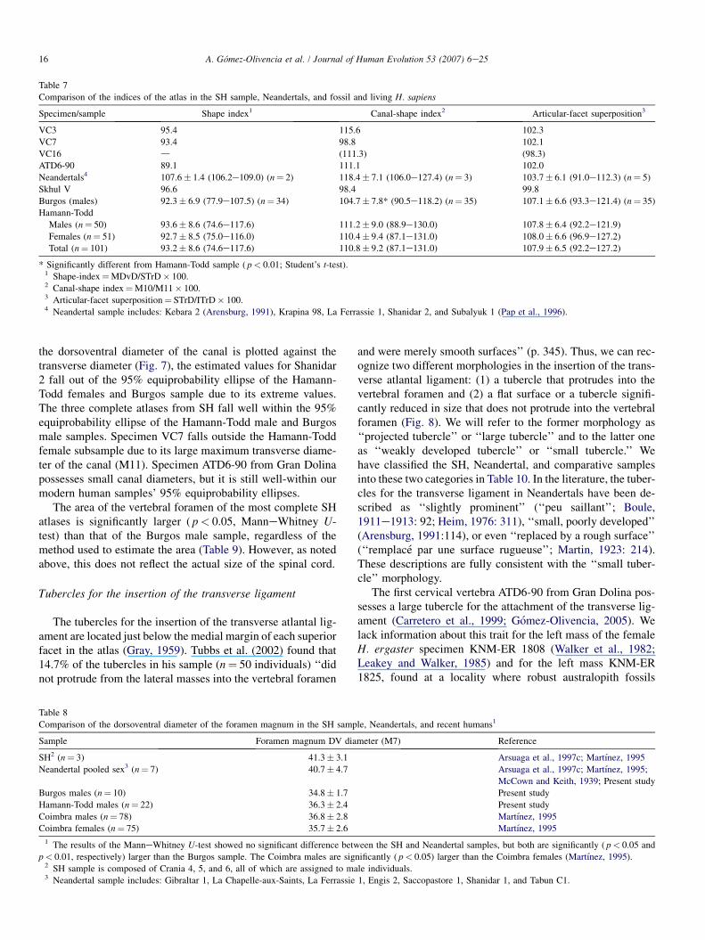
Table 7
Comparison of the indices of the atlas in the SH sample, Neandertals, and fossil and living H. sapiens
Specimen/sample Shape index1 Canal-shape index2 Articular-facet superposition3
VC3 95.4 115.6 102.3
VC7 93.4 98.8 102.1
VC16 d (111.3) (98.3)
ATD6-90 89.1 111.1 102.0
Neandertals4 107.6� 1.4 (106.2e109.0) (n¼ 2) 118.4� 7.1 (106.0e127.4) (n¼ 3) 103.7� 6.1 (91.0e112.3) (n¼ 5)
Skhul V 96.6 98.4 99.8
Burgos (males) 92.3� 6.9 (77.9e107.5) (n¼ 34) 104.7� 7.8* (90.5e118.2) (n¼ 35) 107.1� 6.6 (93.3e121.4) (n¼ 35)
Hamann-Todd
Males (n¼ 50) 93.6� 8.6 (74.6e117.6) 111.2� 9.0 (88.9e130.0) 107.8� 6.4 (92.2e121.9)
Females (n¼ 51) 92.7� 8.5 (75.0e116.0) 110.4� 9.4 (87.1e131.0) 108.0� 6.6 (96.9e127.2)
Total (n¼ 101) 93.2� 8.6 (74.6e117.6) 110.8� 9.2 (87.1e131.0) 107.9� 6.5 (92.2e127.2)
* Significantly different from Hamann-Todd sample ( p< 0.01; Student’s t-test).1 Shape-index¼MDvD/STrD� 100.2 Canal-shape index¼M10/M11� 100.3 Articular-facet superposition¼ STrD/ITrD� 100.4 Neandertal sample includes: Kebara 2 (Arensburg, 1991), Krapina 98, La Ferrassie 1, Shanidar 2, and Subalyuk 1 (Pap et al., 1996).
the dorsoventral diameter of the canal is plotted against thetransverse diameter (Fig. 7), the estimated values for Shanidar2 fall out of the 95% equiprobability ellipse of the Hamann-Todd females and Burgos sample due to its extreme values.The three complete atlases from SH fall well within the 95%equiprobability ellipse of the Hamann-Todd male and Burgosmale samples. Specimen VC7 falls outside the Hamann-Toddfemale subsample due to its large maximum transverse diame-ter of the canal (M11). Specimen ATD6-90 from Gran Dolinapossesses small canal diameters, but it is still well-within ourmodern human samples’ 95% equiprobability ellipses.
The area of the vertebral foramen of the most complete SHatlases is significantly larger ( p< 0.05, ManneWhitney U-test) than that of the Burgos male sample, regardless of themethod used to estimate the area (Table 9). However, as notedabove, this does not reflect the actual size of the spinal cord.
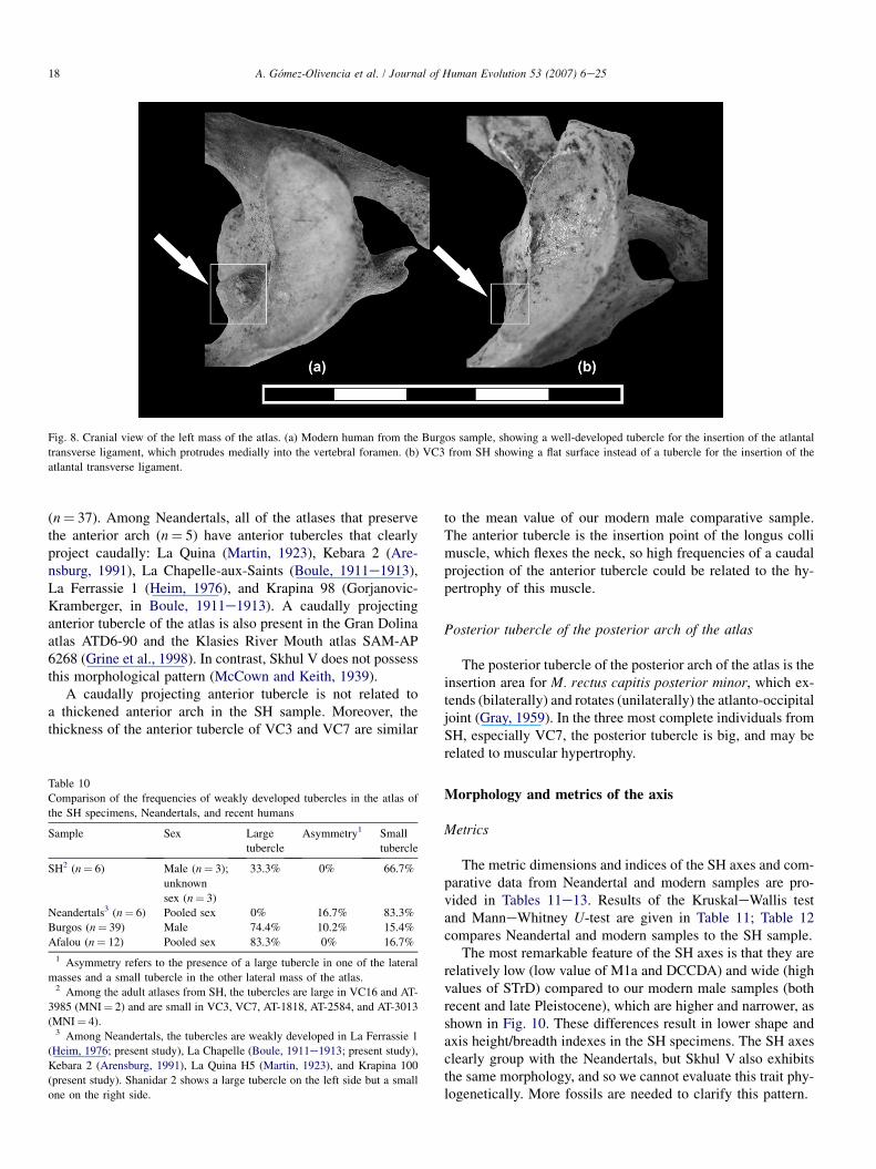
Tubercles for the insertion of the transverse ligament
The tubercles for the insertion of the transverse atlantal lig-ament are located just below the medial margin of each superiorfacet in the atlas (Gray, 1959). Tubbs et al. (2002) found that14.7% of the tubercles in his sample (n¼ 50 individuals) ‘‘didnot protrude from the lateral masses into the vertebral foramen
and were merely smooth surfaces’’ (p. 345). Thus, we can rec-ognize two different morphologies in the insertion of the trans-verse atlantal ligament: (1) a tubercle that protrudes into thevertebral foramen and (2) a flat surface or a tubercle signifi-cantly reduced in size that does not protrude into the vertebralforamen (Fig. 8). We will refer to the former morphology as‘‘projected tubercle’’ or ‘‘large tubercle’’ and to the latter oneas ‘‘weakly developed tubercle’’ or ‘‘small tubercle.’’ Wehave classified the SH, Neandertal, and comparative samplesinto these two categories in Table 10. In the literature, the tuber-cles for the transverse ligament in Neandertals have been de-scribed as ‘‘slightly prominent’’ (‘‘peu saillant’’; Boule,1911e1913: 92; Heim, 1976: 311), ‘‘small, poorly developed’’(Arensburg, 1991:114), or even ‘‘replaced by a rough surface’’(‘‘remplace par une surface rugueuse’’; Martin, 1923: 214).These descriptions are fully consistent with the ‘‘small tuber-cle’’ morphology.
The first cervical vertebra ATD6-90 from Gran Dolina pos-sesses a large tubercle for the attachment of the transverse lig-ament (Carretero et al., 1999; Gomez-Olivencia, 2005). Welack information about this trait for the left mass of the femaleH. ergaster specimen KNM-ER 1808 (Walker et al., 1982;Leakey and Walker, 1985) and for the left mass KNM-ER1825, found at a locality where robust australopith fossils
Table 8
Comparison of the dorsoventral diameter of the foramen magnum in the SH sample, Neandertals, and recent humans1
Sample Foramen magnum DV diameter (M7) Reference
SH2 (n¼ 3) 41.3� 3.1 Arsuaga et al., 1997c; Martınez, 1995
Neandertal pooled sex3 (n¼ 7) 40.7� 4.7 Arsuaga et al., 1997c; Martınez, 1995;
McCown and Keith, 1939; Present study
Burgos males (n¼ 10) 34.8� 1.7 Present study
Hamann-Todd males (n¼ 22) 36.3� 2.4 Present study
Coimbra males (n¼ 78) 36.8� 2.8 Martınez, 1995
Coimbra females (n¼ 75) 35.7� 2.6 Martınez, 1995
1 The results of the ManneWhitney U-test showed no significant difference between the SH and Neandertal samples, but both are significantly ( p< 0.05 and
p< 0.01, respectively) larger than the Burgos sample. The Coimbra males are significantly ( p< 0.05) larger than the Coimbra females (Martınez, 1995).2 SH sample is composed of Crania 4, 5, and 6, all of which are assigned to male individuals.3 Neandertal sample includes: Gibraltar 1, La Chapelle-aux-Saints, La Ferrassie 1, Engis 2, Saccopastore 1, Shanidar 1, and Tabun C1.

17A. Gomez-Olivencia et al. / Journal of Human Evolution 53 (2007) 6e25
have been collected (Leakey and Walker, 1985). The area inwhich the tubercles should be present has suffered mild dam-age to the left articular process of the Australopithecus afaren-sis atlas A.L. 333-83 (Lovejoy et al., 1982).
The atlases from the Sima de los Huesos site show higherfrequencies of small tubercles than do our two modern humancomparative samples, but lower frequencies than are observedin Neandertals. Although we cannot completely rule out thepossibility that the condition is a general reflection of robustic-ity, the tubercle for the insertion of the transverse ligamentdoes not appear to be remodeled by physical stress and thusthe degree of development could be epigenetic (Tubbs et al.,
Fig. 6. Dorsoventral diameter of the foramen magnum versus the dorsoventral
diameter of the atlas in several fossil specimens and recent humans. The 95%
equiprobability ellipses for the Burgos male and the Hamann-Todd samples
are shown.
Table 9
Comparison of the vertebral-foramen area (mm2) the SH and recent human
atlases
Specimen/sample Vertebral-foramen
area 1*
Vertebral-foramen
area 2**
VC3 726.7 697.3
VC7 781.6 749.7
VC16 (697.3) (626.8)
Burgos1 (males) 603.5� 63.2
(478.2e735.2) (n¼ 30)
594.6� 67.5
(458.7e723.9) (n¼ 30)
* Imaged area measures.
** Cross-checked for accuracy by comparing imaged linear measures to phys-
ical dimensions measured with digital calipers.1 The two mean values of the vertebral-foramen area in the Burgos sample
are not statistically different.
2002), which implies that this trait could be useful as a phylo-genetic character. Moreover, the percentages of small tuber-cles in the two modern human populations (Burgos andAfalou) are similar, despite their different chronology, geogra-phy, and lifestyles. The presence of large tubercles in theATD6-90 atlas from Gran Dolina suggests that high frequen-cies of well-developed tubercles could be the primitive condi-tion within the genus Homo, which has been preserved in latePleistocene H. sapiens, as represented in the atlases SAM-AP6268 from Klasies River Mouth (Grine et al., 1998) and SkhulV, and in the modern human samples (see above). In contrast,high frequencies of weakly developed tubercles would be thederived condition of H. neanderthalensis, and the middlePleistocene European populations of H. heidelbergensiswould represent an intermediate stage. Alternatively, wecould hypothesize a polymorphic primitive condition thatled to high frequencies of small tubercles in the EuropeanH. heidelbergensiseH. neanderthalensis lineage and to highfrequencies of large tubercles in H. sapiens.
Anterior tubercle of the anterior arch of the atlas
In the Sima de los Huesos atlases, all of the anterior arches(n¼ 7), regardless of their age at death, display a projecting an-terior tubercle. The anterior tubercle of the anterior arch of theatlas (Fig. 9) projects caudally in 48.6% of the individuals inthe modern human male comparative sample from Burgos
Fig. 7. Dorsoventral diameter of the canal vs. the maximum transverse diam-
eter of the canal in the atlas in several fossil specimens, the terminal Pleisto-
cene sample from the sites of Afalou and Taforalt, and several recent human
samples. The 95% equiprobability ellipses are given for the Afalou/Taforalt
and recent human samples. DV 14 refers to Dolnı Vestonice 14.
18 A. Gomez-Olivencia et al. / Journal of Human Evolution 53 (2007) 6e25
Fig. 8. Cranial view of the left mass of the atlas. (a) Modern human from the Burgos sample, showing a well-developed tubercle for the insertion of the atlantal
transverse ligament, which protrudes medially into the vertebral foramen. (b) VC3 from SH showing a flat surface instead of a tubercle for the insertion of the
atlantal transverse ligament.
(n¼ 37). Among Neandertals, all of the atlases that preservethe anterior arch (n¼ 5) have anterior tubercles that clearlyproject caudally: La Quina (Martin, 1923), Kebara 2 (Are-nsburg, 1991), La Chapelle-aux-Saints (Boule, 1911e1913),La Ferrassie 1 (Heim, 1976), and Krapina 98 (Gorjanovic-Kramberger, in Boule, 1911e1913). A caudally projectinganterior tubercle of the atlas is also present in the Gran Dolinaatlas ATD6-90 and the Klasies River Mouth atlas SAM-AP6268 (Grine et al., 1998). In contrast, Skhul V does not possessthis morphological pattern (McCown and Keith, 1939).
A caudally projecting anterior tubercle is not related toa thickened anterior arch in the SH sample. Moreover, thethickness of the anterior tubercle of VC3 and VC7 are similar
Table 10
Comparison of the frequencies of weakly developed tubercles in the atlas of
the SH specimens, Neandertals, and recent humans
Sample Sex Large
tubercle
Asymmetry1 Small
tubercle
SH2 (n¼ 6) Male (n¼ 3);
unknown
sex (n¼ 3)
33.3% 0% 66.7%
Neandertals3 (n¼ 6) Pooled sex 0% 16.7% 83.3%
Burgos (n¼ 39) Male 74.4% 10.2% 15.4%
Afalou (n¼ 12) Pooled sex 83.3% 0% 16.7%
1 Asymmetry refers to the presence of a large tubercle in one of the lateral
masses and a small tubercle in the other lateral mass of the atlas.2 Among the adult atlases from SH, the tubercles are large in VC16 and AT-
3985 (MNI¼ 2) and are small in VC3, VC7, AT-1818, AT-2584, and AT-3013
(MNI¼ 4).3 Among Neandertals, the tubercles are weakly developed in La Ferrassie 1
(Heim, 1976; present study), La Chapelle (Boule, 1911e1913; present study),
Kebara 2 (Arensburg, 1991), La Quina H5 (Martin, 1923), and Krapina 100
(present study). Shanidar 2 shows a large tubercle on the left side but a small
one on the right side.
to the mean value of our modern male comparative sample.The anterior tubercle is the insertion point of the longus collimuscle, which flexes the neck, so high frequencies of a caudalprojection of the anterior tubercle could be related to the hy-pertrophy of this muscle.
Posterior tubercle of the posterior arch of the atlas
The posterior tubercle of the posterior arch of the atlas is theinsertion area for M. rectus capitis posterior minor, which ex-tends (bilaterally) and rotates (unilaterally) the atlanto-occipitaljoint (Gray, 1959). In the three most complete individuals fromSH, especially VC7, the posterior tubercle is big, and may berelated to muscular hypertrophy.
Morphology and metrics of the axis
Metrics
The metric dimensions and indices of the SH axes and com-parative data from Neandertal and modern samples are pro-vided in Tables 11e13. Results of the KruskaleWallis testand ManneWhitney U-test are given in Table 11; Table 12compares Neandertal and modern samples to the SH sample.
The most remarkable feature of the SH axes is that they arerelatively low (low value of M1a and DCCDA) and wide (highvalues of STrD) compared to our modern male samples (bothrecent and late Pleistocene), which are higher and narrower, asshown in Fig. 10. These differences result in lower shape andaxis height/breadth indexes in the SH specimens. The SH axesclearly group with the Neandertals, but Skhul V also exhibitsthe same morphology, and so we cannot evaluate this trait phy-logenetically. More fossils are needed to clarify this pattern.

19A. Gomez-Olivencia et al. / Journal of Human Evolution 53 (2007) 6e25
Fig. 9. (a) A modern atlas from the Burgos sample that lacks the anterior tubercle caudal projection. (b) Ventral view of VC7, showing the caudal projection of the
anterior arch tubercle (arrow).
Size and shape of the vertebral foramen
The SH axes have canal-size dimensions and canal-shapeindices that are well within our modern human sample rangeof variation (Tables 11e13; Fig. 11). Moreover, the area ofthe vertebral foramen calculated for the SH specimens is closeto the mean of our Burgos male sample (Table 14). AmongNeandertals, the remarkably large transverse diameter of thecanal (M11) in Shanidar 2 and the large dorsoventraldiameter of the canal (M10) in Krapina 105 place these verte-brae out of the 95% equiprobability ellipse of the modern hu-man comparative sample from Burgos (Fig. 11).
Finally, we have compared the vertebral-canal surface area ofthe axes to their articular surface areas (as a proxy for size). TheSH specimens’ mean falls close to that of the Burgos humansample (Burgos mean¼ 97.9� 18.8, n¼ 29; SH mean¼
99.6� 19.5, n¼ 3). Moreover, within the Burgos male sample,there is no evidence of a correlation between these two variables.
Pathological lesions
Pathology in the vertebral column has been described inseveral hominins (Cook et al., 1983; Dawson and Trinkaus,1997; Trinkaus, 1985; Ogilvie et al., 1998), including someof the SH hominins (Perez, 2003). We have found two kindsof pathology in the SH upper cervical vertebrae: a developmen-tal defect and degenerative pathology.
In the VC3 atlas, the anterior bar of the transverse processremains unfused to the posterior bar of the transverse process,and thus the foramen transversarium is not completely de-limited. The fusion of the anterior bar to the posterior bar of
Table 11
Raw dimensions (in mm) of the SH axes and results of the ManneWhitney U-test for differences between sample means in SH, Neandertals, and recent humans
Variable VC2 VC4 VC8 SH-N-BU SH-BU SH-N BU-N
K-W M-W M-W(B) M-W M-W M-W(B)
Maximum dorsoventral diameter (MDvD) # (50.0) 50.1 48.9
Maximum transverse diameter (MTrD) x (60.0)
Superior transverse diameter (STrD) x (48.0) 50.5 51.1 ** ** *
Inferior transverse diameter (ITrD) 46.0 (49.6) 45.4
Canal dorsoventral maximum diameter (M10) 17.4 18.3 15.5
Canal transverse maximum diameter (M11) 23.7 22.9 23.9 * * yBody craniocaudal dorsal diameter (M2) 19.0 17.1 18.0 * * *
Total vertebral ventral height (M1a) (34.5) (36.2) (36.0) ** * ** **
Total vertebral dorsal height (DoH) 31.5 32.2 31.9 * * *
Body inferior anteroposterior diameter (M5) (15.5) (16.1)
Body inferior transverse diameter (M8) 18.4 16.4 18.4
Cranial articular facet dorsoventral diameter (UAFDvD) 17.0/17.7 16.1/17.0 d/18.2
Cranial articular facet transverse diameter (UAFTrD) 19.2/d 15.9/15.1 d/19.6 /X
Caudal articular facet dorsoventral diameter (LAFDvD) 10.4/d 10.0/d 11.9/10.9 /X
Caudal articular facet transverse diameter (LAFTrD) 12.6/d 12.3/d 10.2/10.1 /X
Laminae: craniocaudal diameter (CCDLam) 12.0/11.2 10.8/11.7 11.8/11.6 */* **/* */*
Laminae: thickness (ThLam) 4.9/4.5 5.8/6.6 5.2/7.0
Spine length (M13) (16.5) 17.0 18.4
Maximum transverse diameter of the tip of the
spinous process (TrDTSP)
12.8 (16.3) 18.5
Values in parentheses are estimated. Cells that contain two entries are for the right and left sides (right/left).
# Maximum anteroposterior diameter along the sagittal plane (McCown and Keith, 1939).
x Maximum transverse diameter measured to the lateral margins of the superior or inferior articular facets (Trinkaus, 1983).
Abbreviations are as follows: SH¼ Sima de los Huesos; BU¼Burgos males; N¼Neandertals; HT¼Hamann-Todd males; K-W refers to the KruskaleWallis test
performed on SH, BU, and N samples; M-W refers to the ManneWhitney U-test performed on different pairs of samples; M-W (B) refers to the ManneWhitney
U-test adjusted using the Bonferroni method; * p< 0.05; ** p< 0.01; y 0.05< p< 0.10; X¼ the analysis was not performed because one of the samples is of size
n¼ 0 (K-W) or n< 2 (M-W).

20 A. Gomez-Olivencia et al. / Journal of Human Evolution 53 (2007) 6e25
Table 12
Comparison of linear axis measurements (mm) in the SH specimens, Neandertals, and fossil and recent H. sapiens
Sima de los Huesos Neandertals1 Burgos males H-T males (n¼ 50)
n Mean SD n mean SD n Mean S.D Mean S.D
MDvD 3 49.7 0.7 4 51.2 4.0 35 49.6 2.3 51.6 2.8
MTRD 1 60.0 d 2 51.8 0.4 35 54.7 4.4 55.2 4.0
STrD 3 49.9 1.6 8 47.8 2.7 39 45.2 2.3
ITrD 3 47.0 2.3 2 51.8 3.9 38 47.2 2.4 47.8 2.9
M10 3 17.1 1.4 6 18.0 1.6 39 16.5 1.5
M11 3 23.5 0.5 8 24.5 1.3 39 23.1 1.3 23.4 1.9
M2 3 18.0 1.0 6 16.2 2.7 37 19.0 1.5
M1a 3 35.6 0.9 9 34.3 2.8 38 37.9 2.3 38.9 2.6
DCCDA 3 31.9 0.4 5 30.7 3.2 38 34.0 2.1
M5 2 15.8 0.4 7 16.2 2.1 39 15.1 1.1 16.7 1.6
M8 3 17.7 1.1 8 19.3 1.6 35 18.1 1.4 19.8 2.3
UAFDvD 2/3 16.6/17.6 0.6/0.6 3/2 17.9/18.3 1.1/3.0 37/39 17.7/18.1 1.2/1.4
UAFTrD 2/2 17.6/17.3 2.3/3.1 1/0 15.0/d d/d 36/39 16.4/16.3 1.3/1.4
LAFDvD 3/1 10.8/10.9 1.0/d 2/0 11.8/d 0.6/d 36/35 10.1/10.1 1.4/1.4
LAFTrD 3/1 11.7/10.1 1.3/d 2/0 12.4/d 0.2/d 35/34 11.2/11.2 1.2/1.5
CCDLam 3/3 11.5/11.5 0.6/0.3 7/6 10.4/10.5 1.1/1.1 39 11.9/11.8 1.1/1.1
ThLam 3/3 5.3/6.0 0.4/1.3 7/5 5.5/5.3 0.1/0.6 39 5.7/5.6 1.3/1.0
M13 3 17.3 1.0 4 17.3 1.8 36 18.9 2.7
TrDTSP 3 15.9 2.9 1 14.2 d 32 14.1 4.0
Cells that contain two entries are for the right and left sides (right/left).1 The Neandertal sample (n¼ 11) comprises the following individuals: Kebara 2 (Arensburg et al., 1990; Arensburg, 1991), Krapina 103, Krapina 104, Krapina
105, La Chapelle-aux-Saints, La Ferrassie 1, La Quina H5 (Martin, 1923), Regourdou 1 (Piveteau, 1966), Shanidar 2, Shanidar 4 (Stewart, 1962; Trinkaus, 1983),
and Tabun C1 (McCown and Keith, 1939).
the transverse process in modern humans occurs at about 3e4years of age (Scheuer and Black, 2000).
The VC7 atlas shows a slight osteophytosis along the edgesof its superior articular facets. This very slight osteophytosisfinds its counterpart in the occipital condyles of Cranium 5,with which it is associated. Cranium 5’s age at death hasbeen estimated to be in excess of 35 years based on toothwear, and thus it represents one of the oldest individuals inthe SH sample, consistent with the appearance of this pathol-ogy. The VC16 atlas shows porosity in the middle-dorsal partof its superior articular facets. It exhibits abnormal prolifera-tion of bone on the edge of the lower articular facet, which iscongruent with the abnormal porous bone present on the
associated VC2 axis at the edge of the superior articular facet.Moreover, the VC2 axis shows osteophytosis along the ventraledges of the superior articular facet (the only part of the facetthat is preserved) and on the inferior articular facets. Thiswould be consistent with early stages of degenerative joint dis-ease (DJD). Finally, even if it cannot be considered technicallypathological, the axis AT-2289 shows a rugosity on the cranio-lateral end of the dens on the alar ligament’s insertion points.This condition could be related to a slight ossification of theligamentous attachment point (enthesophyte).
In general, the level of DJD present in the SH upper cervicalsample is not very severe, with the VC16-VC2 association be-ing the most strongly affected. Degenerative joint disease is
Table 13
Comparison of the indices of the axis in the SH sample, Neandertals, and fossil and living H. sapiens
Specimen/sample Shape index2 Canal-shape
index3Articular-facet
superposition4Axis height/width
index5
VC2 (104.2) 73.4 (104.3) (71.9)*
VC4 92.2* 79.9 (101.8) (71.7)*
VC8 95.7* 64.9 112.6* (70.5)*
Neandertals1 109.5� 11.3
(100.0e125.8) (n¼ 4)
73.8� 9.5
(65.2e91.3) (n¼ 6)
93.0� 1.9
(91.6e94.3) (n¼ 2)
71.1*� 8.3
(57.1e83.5) (n¼ 8)
Skhul V 107.0 69.3 d 67.6*
Burgos (males) 110.4� 5.6
(99.7e122.0) (n¼ 35)
70.9� 4.8
(61.1e81.4) (n¼ 36)
95.5� 4.5
(87.3e104.8) (n¼ 37)
84.1� 4.7
(75.2e94.3) (n¼ 38)
Values in parentheses are estimated.
* Value is out of the Burgos recent human sample range.1 The Neandertal sample (n¼ 8) includes the following specimens: Kebara 2 (Arensburg et al., 1990; Arensburg, 1991), Krapina 103, Krapina 104, Krapina 105,
La Chapelle-aux-Saints, La Ferrassie 1, Regourdou 1 (Piveteau, 1966), and Shanidar 2.2 Shape index¼MDvD/STrD� 100.3 Canal-shape index¼M10/M11� 100.4 Articular-facet superposition¼ STrD/ITrD� 100.5 Axis height/width index¼M1a/STrD.

21A. Gomez-Olivencia et al. / Journal of Human Evolution 53 (2007) 6e25
age-progressive (Aufderheide and Rodrıguez-Martın, 1998). In80% of the cases, no cause is evident, and in other cases, thecause may be physical, infectious, or metabolic, among otherfactors.
Bocquet-Appel and Arsuaga (1999) demonstrated that thereis a dearth of mature adult individuals at the SH site. Based onthe study of dental wear, Bermudez de Castro et al. (2004)found only three individuals who were older than 35 years(one male and two of indeterminate sex). Of these three indi-viduals, one could also be represented by Pelvis 1 (older than35) and another, represented by the isolated pubis AT-2500,could be more than 45 years.
Bermudez de Castro and Perez (1995) studied the enamelhypoplasia in the SH sample to determine the level of biolog-ical stress that affected the development of these hominins.They found that this population probably suffered a lower levelof biological stress than did Neandertal populations (see alsoCunha et al., 2004).
In summary, the appearance of different degrees of DJD intwo SH adult individuals represented by the upper cervicalvertebrae could indicate that these vertebrae belong to someof the older individuals represented by the dental material.
Fig. 10. Axis total vertebral height (y) vs. superior transverse diameter in the
Burgos sample and several fossil atlases. The 95% equiprobability ellipse for
the Burgos male sample and the regression line for the Burgos sample (solid
line) and the Neandertals (dotted line) are shown. For the Burgos modern male
sample, y¼ 0.4879xþ 15.8829 (n¼ 38). For the Neandertal sample, y¼0.3415xþ 18.369 (n¼ 8). The extremely low value for the total vertebral
height in the Kebara 2 axis could be due to an underestimation of this measure-
ment by Arensburg et al. (1990). On the other hand, McCown and Keith
(1939) underscore the smallness of Skhul V axis.
In one case, this is confirmed by the association of VC7(C1)and VC8(C2) with Cranium 5 (age at death� 35; Bermudezde Castro et al., 2004).
Discussion
Phylogenetic evidence suggests that the SH sample and allEuropean middle Pleistocene hominins represent populationsthat were ancestral to the Neandertal populations, as they arecharacterized by a mixture of shared primitive features andNeandertal apomorphies (Arsuaga et al., 1997c; Carretero
Fig. 11. Axis canal dorsoventral diameter (y) vs. canal transverse maximum
diameter in the Burgos sample and several fossil atlases. The 95% equiprob-
ability ellipses for the Burgos male sample and for the Iberomaurusian pooled
sex sample are given. DV 13 refers to Dolnı Vestonice 13. LF1 refers to La
Ferrassie 1. The value of M10 reported by McCown and Keith (1939) for
the Skhul V axis is 24.6 mm, 0.1 mm above the value reported by Stewart
(1962), which is 6 mm above our value. This extraordinary difference could
be due to differences in measurement method (see Fig. 1).
Table 14
Comparison of the vertebral-foramen area (mm2) of the SH and recent human
axes
Specimen/sample Vertebral-foramen
area 1*
Vertebral-foramen
area 2**
VC2 (306.3) (302.0)
VC4 329.1 331.4
VC8 295.4 314.0
Burgos1 (males) 301.9� 43.1
(234.5e401.2) (n¼ 30)
302.9� 47.5
(233.8e415.2) (n¼ 30)
Values in parentheses are estimated.
* Imaged linear measures.
** Cross-checked for accuracy by comparing imaged linear measures to phys-
ical dimensions measured with digital calipers.1 The two mean values of the vertebral-foramen area in the Burgos sample
are not statistically different.

22 A. Gomez-Olivencia et al. / Journal of Human Evolution 53 (2007) 6e25
et al., 1997; Martınez and Arsuaga, 1997). In the SH upper cer-vical vertebral sample, we have found some features that couldbe of phylogenetic significance but whose polarity is difficult toascertain due to the scarcity of hominin vertebrae. Within thisgroup we can mention: (1) the development of the tubercle forthe attachment of the transverse ligament of the atlas and (2)the height/breadth index of the axis. We should note that theSH specimens are metrically more similar to Neandertalsthan to our modern human comparative samples. Moreover,we have found that (1) the atlases from the Sima de los Huesossite exhibit a percentage of weakly developed tubercles for theattachment of the transverse ligament that is intermediate be-tween modern human populations and Neandertals, and (2)the SH axes exhibit a height/breadth index similar to that ofthe Neandertals. These findings are fully compatible with thephylogenetic position proposed for these hominins (i.e., thatH. heidelbergensis is an exclusively European species, ances-tral only to H. neanderthalensis; Arsuaga et al., 1997c; Carre-tero et al., 1997; Martınez and Arsuaga, 1997).
Biomechanics
The atlanto-occipital articulation allows only for flexionand extension (Bogduk and Mercer, 2000). It acts as a first-class lever in which the occipital condyles act as the fulcrum,lying between the nuchal muscles and the mass of the head(Escuredo et al., 2002). In all other respects, the head and atlasmove essentially as a single unit. Few muscles act directly onthe atlas and, in fact, its movements are governed by the mus-cles that act on the head (Bogduk and Mercer, 2000). The SHhominins show a degree of prognathism (as measured bybasioneprosthion length) that is similar to that of the Neander-tals (Arsuaga et al., 1997c) and well-above the prognathism ofmodern humans (Martınez, 1995). The SH atlases show en-larged insertion areas for M. rectus capitis posterior minorand the SH axes have robust spinous processes that could re-late to development of M. obliquus capitis inferior and M. rec-tus capitis posterior major. While these features could indicatemuscular force acting at the atlanto-occipital joint to counterthe aforementioned prognathism, we should recall that othermuscles also act to extend the head (e.g., the M. semispinaliscapitis), which could also play an important biomechanicalrole in this joint. Alternatively, these enlarged muscular-at-tachment areas could simply reflect a generally robust bodybuild and/or high activity levels, factors that could produce in-creased caudal projection of the anterior tubercle, whichwould agree with the high body mass calculated for thesehominins (Arsuaga et al., 1999; Carretero et al., 2004).
The upper cervical spine of the SH hominins is character-ized by a mediolaterally expanded atlantoaxial joint, repre-sented by the ITrD of the atlas and the STrD of the axis, anda short craniocaudal dimension of the axis. During lateral incli-nation of the head, there is no movement in the atlantoaxialjoint (Kapandji, 1998) and we hypothesize that a mediolateralexpansion would further stabilize this joint. The close relation-ship between neck biomechanics and head movement and thefact that the head is the final link in an open kinematic chain
that includes the cervical and the upper thoracic vertebrae(Winters and Peles, 1990) make it necessary, if we want to fullyassess the biomechanics of this anatomical region, to take intoaccount both the upper and lower cervical vertebrae and cranialmorphology, which we plan to do in a future publication.
Size of the vertebral canal and its implications
Much attention has been devoted to vertebral-canal size andits relationship to spoken language. One factor in the evolutionof human language that would be reflected in vertebral-canalmorphology is increased breath control (MacLarnon, 1993;MacLarnon and Hewitt, 1999, 2004). Modern humans havean enlarged thoracic vertebral canal, reflecting a larger amountof gray matter. Based on the morphology of the KNM-WT15000 individual, a narrower thoracic canal has been proposedfor Homo ergaster, indicating that this species may only havebeen capable of short, unmodulated utterances, such as thoseused by extant nonhuman primates (MacLarnon and Hewitt,1999). However, significant abnormalities have been foundin the KNM-WT 15000 individual (Latimer and Ohman,2001), which could indicate some form of axial dysplasia,and so the small canal may be a reflection of a neural-canalstenosis associated with the pathology. In contrast, Schiesset al. (2006) argued that the diagnosis of a congenital dysplasiais not supported, indicating that the pathological lesions in theKNM-WT 15000 individual may not be as severe as previ-ously reported. Moreover, the Dmanisi vertebrae (Meyer,2005; Meyer et al., 2006), which are the oldest known forthe genus Homo, follow the modern human pattern in all re-gions, as the raw and relative sizes of the vertebral canalsfall well within the human range, indicating that these homi-nins may have had fine control of the respiratory muscles in-volved in spoken language (Meyer, 2005; Meyer et al., 2006).
Arsuaga et al. (1997a) showed that the mean cranial capac-ity of SH’s three most complete crania (1245 cm3) (Arsuagaet al., 1993, 1997c) is slightly less than that of two compara-tive samples from the Hamann-Todd Osteological Collection.However, given the large body-weight estimates for thesehominins, their encephalization quotients are below both mod-ern human or Neandertal values (Arsuaga et al., 1999). In Ne-andertals, higher encephalization quotients are reached byexpansion of the cranial capacity, while in modern humans itis mainly achieved by a reduction in body mass (Arsuagaet al., 1999; Carretero et al., 2004). In addition to the paralleltrends in encephalization in these two lineages, the absolutesize of the bony vertebral canal in the upper cervical spinereached modern human values by the middle Pleistocene. Pre-liminary studies (Carretero et al., 1999; Gomez et al., 2004;Gomez-Olivencia, 2005) have shown that the SH lower cervi-cal spine’s canal had a similar size compared to modern hu-mans, but a full assessment of this anatomical region willnot be possible until larger sets of cervical and thoracic verte-brae are associated. In any case, as demonstrated by Martınezet al. (2004), the SH hominins had the skeletal characteristicsof the outer and middle ear that support the perception of spo-ken language.
23A. Gomez-Olivencia et al. / Journal of Human Evolution 53 (2007) 6e25
Summary and conclusions
Study of the SH upper cervical spine leads us to identifya minimum of 11 individuals represented by these fossils: 6adults and 5 subadults. Three sets of associated atlases andaxes, probably belonging to male individuals, have been iden-tified: two older adults (one of them associated with Cranium5) and one young adult. Metrical and morphological attributesreveal that SH atlases and axes are more similar to Neandertalhomologues than to our modern male comparative samples.The SH upper cervical spine is characterized by: (1) a largemaximum dorsoventral diameter of the atlas’s canal, whichmay be related to the large dorsoventral diameter of the fora-men magnum; (2) dorsoventrally large lower facets of the at-las; (3) a mediolaterally expanded atlantoaxial joint; (4)a craniocaudally short axis; (5) a caudally projecting anteriortubercle of the anterior arch of the atlas; and (6) lateral massesof the atlas that possess weakly developed tubercles for theattachment of the transverse ligament at frequencies that liebetween those of modern humans and Neandertals. Future as-sociations of more cervical elements from SH will clarify theanatomy of this region and will improve our understanding ofthe biology of these humans.
Acknowledgements
Special thanks go to our colleagues and friends A. Gracia,C. Lorenzo, N. Garcıa, R. Quam, and A. Esquivel for theirgreat work at the Sima de los Huesos site. M.C. Ortega re-stored the fossils for us. We are grateful to Jakov Radovcic(Croatian Natural History Museum), Bruce Latimer and Yo-hannes Haile-Selassie (Cleveland Natural History Museum),Philippe Mennecier (Musee de l’Homme), Dominique Gri-maud-Herve (Departement de PrehistoiredMuseum nationald’Histoire naturelle), Jose Marıa Bermudez de Castro (Centrode Investigacion de Evolucion Humana), Belen Castillo (Mu-seo de Burgos), and Michele Morgan (Peabody Museum ofArchaeology and Ethnology) for providing access to the skel-etal collections in their care. We are also indebted to AurelieFort, Lyman Jellema, Stephanie Renault, and Olivia Herschen-sohn for curatorial assistance. Further thanks go to our col-leagues at the Laboratorio de Evolucion Humana (LEH) ofthe Universidad de Burgos and at the Centro UCM-ISCIII deInvestigacion sobre Evolucion y Comportamiento Humanos.Special thanks go to Aimara for her help in the determinationof the vertebral-canal area, with the figures, and all her com-ments, which greatly improved this manuscript. Luis Caboprovided helpful advice regarding statistics. Antoine Balzeau,Isabelle de Groote, J.E. Gonzalez Urquijo, and Jeremiah Scottprovided helpful comments on different parts of the manu-script. We are indebted to Marc R. Meyer discussing ideasdealing with vertebrae. We are grateful to Rolf Quam andCiaran Brewster (P.D.), who helped with the English transla-tion. Angelica Torres kindly revised the English in an earlierversion of the manuscript. Rolf Quam also provided helpfulcomments that greatly improved this paper. We are indebtedto William H. Kimbel for his thorough edit of the manuscript
and his helpful comments, as well as those provided by theassociate editor and three anonymous referees, which greatlyimproved the manuscript.
The first author is supported by a grant from the Ministeriode Educacion y Ciencia. Laura Rodrıguez is supported bya grant from the Fundacion Siglo para las Artes en Castillay Leon. This research was supported by the Ministerio deCiencia y Tecnologıa, Proyecto BOS2003-08938-C03-01.Funding for the fieldwork came from the Junta de Castilla yLeon and Fundacion Atapuerca. Help in the field from theGrupo Espeleologico Edelweiss was essential.
Appendix 1. Labeling of the SH vertebrae
Every human fossil from this site is identified by the fieldlabel ‘‘AT-’’ followed by the inventory number. For example,AT-333 is a distal epiphysis of a left adult humerus (see Car-retero et al., 1997) and AT-121 is a frontal bone (Arsuagaet al., 1991). The fossils that consist of several fragmentsand labels have a second identification number with the initialletter of the bone and a Roman numeral. For example, H-I isHumerus I, a distal epiphysis of an immature individual com-posed of AT-741 and AT-791 (Carretero et al., 1997). In thecase of anatomical parts that consist of different bones, likecrania, a third inventory number is used. When significantparts of the calvaria (including different bones) are represented(with or without the face), the term ‘‘cranium’’ is used to label it,followed by an Arabic number. For example, Cranium 1 com-prises AT-40, AT-63a-b, AT-65, AT-86, AT-122, AT-177a-e,AT-206, AT-216, AT-223, AT-472, and AT-937 (Arsuagaet al., 1997c), which includes Occipital III [described as anisolated cranial bone by Arsuaga et al. (1991)]. Finally, basedon dental evidence, 28 individuals have been identified(Bermudez de Castro et al., 2004) and are labeled with Romannumerals. For example, the individual XXI includes Cranium5 and the mandible AT-888.
We present a somewhat different way of labeling the SHvertebrae from that used for other bones in the same collectiondue to the large number of vertebral elements within eachspine and the difficulty in assessing the exact anatomical posi-tion of vertebrae in some cases. The most complete vertebraeare also labeled with a ‘‘V’’ (for vertebra) and the initial letterof the vertebral region to which they belong (‘‘C’’ for cervical,‘‘T’’ for thoracic, and ‘‘L’’ for lumbar). Arabic numbers indi-cate the ‘‘inventory’’ number within each vertebral region. Forexample, VC16 is formed by AT-1140 and AT-2201, andamong the most complete cervical vertebrae, it is the sixteenthlabeled element (Table 2). Specimen VC15 is formed only byAT-2582, but this vertebra is complete enough to warrant a VClabel, and among the most complete cervical vertebrae, it isthe fifteenth labeled element. To make the discussion in thetext easier to follow, the anatomical position within the regionis indicated in parentheses. For example, VC1(C7) (formerlyknown as C7-I in Carretero et al., 1999) is formed by AT-321, AT-1556, AT-1569, and AT-1609, and it is the first labeledcervical of the sample (Table 2); the specimen is a seventh

24 A. Gomez-Olivencia et al. / Journal of Human Evolution 53 (2007) 6e25
cervical vertebra. Specimen VT27(T4-8) is formed by AT-1135 and AT-4008 and is the twenty-seventh thoracic elementlabeled, and its anatomical position lies between the fourthand the eighth thoracic vertebrae. For other elements, even ifthe anatomical determination can be made with complete ac-curacy we use the ‘‘AT-’’ field label due to the incompletenessof the fossil. For example, AT-2883 is the dens of an axis (C2)(Table 3). Future reconstructions of ‘‘AT-’’ labeled fragmentswill yield new complete VC specimens.
References
Arensburg, B., 1991. The vertebral column, thoracic cage and hyoid bone. In:
Bar-Yosef, O., Vandermeersch, B. (Eds.), Le squelette mousterien de Ke-
bara 2. Editions du CNRS, Paris, pp. 113e147.
Arensburg, B., Schepartz, L.A., Tillier, A.M., Vandermeersch, B., Rak, Y.,
1990. A reappraisal of the anatomical basis for speech in Middle Palaeo-
lithic hominids. Am. J. Phys. Anthropol. 83, 137e146.
Arsuaga, J.L., Carretero, J.M., Lorenzo, C., Gracia, A., Martınez, I., Bermudez
de Castro, J.M., Carbonell, E., 1997a. Size variation in middle Pleistocene
humans. Science 277, 1086e1088.
Arsuaga, J.L., Martınez, I., Gracia, A., Carretero, J.M., Lorenzo, C.,
Garcia, N., Ortega, A.I., 1997b. Sima de los Huesos (Sierra de Atapuerca,
Spain). The site. J. Hum. Evol. 33, 109e127.
Arsuaga, J.L., Martınez, I., Gracia, A., Lorenzo, C., 1997c. The Sima de
los Huesos crania (Sierra de Atapuerca, Spain). A comparative study.
J. Hum. Evol. 33, 219e281.
Arsuaga, J.L., Carretero, J.M., Martınez, I., Gracia, A., 1991. Cranial remains
and long bones from Atapuerca/Ibeas (Spain). J. Hum. Evol. 20, 191e230.
Arsuaga, J.L., Lorenzo, C., Carretero, J.M., Gracia, A., Martınez, I.,
Garcıa, N., Bermudez de Castro, J.M., Carbonell, E., 1999. A complete hu-
man pelvis from the middle Pleistocene of Spain. Nature 399, 255e258.
Arsuaga, J.L., Martınez, I., 2004. Atapuerca y la Evolucion Humana. Fundacio
Caixa Catalunya, Barcelona.
Arsuaga, J.L., Martınez, I., Gracia, A., Carretero, J.M., Carbonell, E., 1993.
Three new human skulls from the Sima de los Huesos middle Pleistocene
site in Sierra de Atapuerca, Spain. Nature 362, 534e537.
Aufderheide, A.C., Rodrıguez-Martın, C., 1998. The Cambridge Encyclopedia
of Human Paleopathology. Cambridge University Press, Cambridge.
Bermudez de Castro, J.M., Perez, P.J., 1995. Enamel hypoplasia in the middle
Pleistocene hominids from Atapuerca (Spain). Am. J. Phys. Anthropol. 96,
301e314.
Bermudez de Castro, J.M., Martinon-Torres, M., Lozano, M., Sarmiento, S.,
Muela, A., 2004. Paleodemography of the AtapuercaeSima de los Huesos
hominin sample: a revision and new approaches to the paleodemography of
the European middle Pleistocene population. J. Anthropol. Res. 60, 5e26.
Bischoff, J.L., Shamp, D.D., Aramburu, A., Arsuaga, J.L., Carbonell, E., Ber-
mudez de Castro, J.M., 2003. The Sima de los Huesos hominids date to
beyond U/Th equilibrium (>350 kyr) and perhaps to 400e500 kyr: new ra-
diometric dates. J. Archaeol. Sci. 30, 275e280.
Bischoff, J.L., Williams, R.W., Rosenbauer, R.J., Aramburu, A., Arsuaga, J.L.,
Garcıa, N., Cuenca-Bescos, G., 2006. High-resolution U-series dates from
the Sima de los Huesos hominids yields 600 þN/�66 kyrs: implications
for the evolution of the early Neanderthal lineage. J. Archaeol. Sci. 34,
763e770.
Boaz, N.T., Ciochon, R.L., Xu, Q., Liu, J., 2004. Mapping and taphonomic
analysis of the Homo erectus loci at Locality 1 Zhoukoudian, China.
J. Hum. Evol. 46, 519e549.
Bocquet-Appel, J.P., Arsuaga, J.L., 1999. Age distributions of hominid sam-
ples at Atapuerca (SH) and Krapina could indicate accumulation by catas-
trophe. J. Archaeol. Sci. 26, 327e338.
Bogduk, N., Mercer, S., 2000. Biomechanics of the cervical spine. I: Normal
kinematics. Clin. Biomech. 15, 633e648.
Boule, M., 1911e1913. L’homme fossile de la Chapelle-aux-Saints. Ann.
Paleontol. 6, 111e172. 7, 21e56, 85e192; 8, 1e70.
Brauer, G., 1988. Osteometrie. In: Knussmann, R. (Ed.), Anthropologie. Hand-
buch der vergleichenden Biologie des Menschen. Gustav Fischer, Stuttgart,
pp. 160e232.
Buikstra, J.E., Gordon, C.C., St. Hoyme, L., 1984. The case of severed skulls.
Individuation in forensic anthropology. In: Rathbun, T., Buikstra, J.E.
(Eds.), Human Identification: Case Studies in Forensic Anthropology.
Charles C. Thomas Pub. Ltd., pp. 121e135.
Carbonell, E., Mosquera, M., Olle, A., Rodrıguez, X.P., Sala, R., Verges, J.M.,
Arsuaga, J.L., Bermudez de Castro, J.M., 2003. Les premiers comporte-
ments funeraires auraient-ils pris place a Atapuerca, il y a 350 000 ans?
L’Anthropologie 107, 1e14.
Carretero, J.M., 1994. Estudio del esqueleto de las dos cinturas y el miembro
superior de los homınidos de la Sima de los Huesos, Sierra de Atapuerca,
Burgos. Ph.D. Dissertation, Universidad Complutense de Madrid.
Carretero, J.M., Arsuaga, J.L., Lorenzo, C., 1997. Clavicles, scapulae and
humeri from the Sima de los Huesos Site (Sierra de Atapuerca, Spain).
J. Hum. Evol. 33, 357e408.
Carretero, J.M., Arsuaga, J.L., Martınez, I., Quam, R., Lorenzo, C., Gracia, A.,
Ortega,A.I., 2004. Los humanos de la Sima de los Huesos (Sierrade Atapuerca)
y la evolucion del cuerpo en el genero Homo. In: Baquedano, E., Rubio, S.
(Eds.), Miscelanea en Homenaje a Emiliano Aguirre. Volumen III. Paleoantro-
pologıa. Museo Arqueologico Regional, Alcala de Henares, pp. 120e135.
Carretero, J.M., Lorenzo, C., Arsuaga, J.L., 1999. Axial and appendicular skel-
eton of Homo antecessor. J. Hum. Evol. 37, 459e499.
Churchill, S.E., Formicola, V., 1997. A case of marked bilateral asymmetry in
the upper limbs of an Upper Palaeolithic male from Barma Grande (Ligu-
ria), Italy. Int. J. Osteoarchaeol. 7, 18e38.
Cook, D.C., Buikstra, J.E., DeRousseau, C.J., Johanson, D.C., 1983. Vertebral pa-
thology in the Afar australopithecines. Am. J. Phys. Anthropol. 60, 83e102.
Creed-Miles, M., Rosas, A., Kruszynski, R., 1996. Issues in the identification
of Neandertal derivative traits at early post-natal stages. J. Hum. Evol. 30,
147e153.
Cuenca-Bescos, G., Laplana, C., Canudo, J.I., Arsuaga, J.L., 1997. Small
mammals from Sima de los Huesos. J. Hum. Evol. 33, 175e190.
Cunha, E., Ramirez Rozzi, F.V., Bermudez de Castro, J.M., Martinon-
Torres, M., Wasterlain, S.N., Sarmiento, S., 2004. Enamel hypoplasias
and physiological stress in the Sima de los Huesos middle Pleistocene
hominins. Am. J. Phys. Anthropol. 125, 220e231.
Dawson, J.E., Trinkaus, E., 1997. Vertebral osteoarthritis of the La Chapelle-
aux-Saints 1 Neanderthal. J. Archaeol. Sci. 24, 1015e1021.
Escuredo, B., Sanchez, J.M., Borras, F.X., Serrat, J., 2002. Estructura y fun-
cion del cuerpo humano. McGraw-Hill, Interamericana, Madrid.
Garcıa, N., 2002. Los carnıvoros de los yacimientos Pleistocenos de la Sierra
de Atapuerca. Ph.D. Dissertation, Universidad Complutense de Madrid.
Garcıa, N., Arsuaga, J.L., Torres, T., 1997. The carnivore remains from the
Sima de los Huesos middle Pleistocene site (Sierra de Atapuerca, Spain).
J. Hum. Evol. 33, 155e174.
Gomez, A., Carretero, J.M., Arsuaga, J.L., Martınez, I., Quam, R., Lorenzo, C.,
Gracia, A., Garcıa, N., Ortega, A.I., Rodrıguez, L., 2004. El raquis de los
humanos del yacimiento de la Sima de los Huesos (Sierra de Atapuerca,
Burgos). In: Egocheaga, J.E. (Ed.), Biologıa de Poblaciones Humanas: Di-
versidad, Tiempo, Espacio. Dpto. de Biologıa de Organismos y Sistemas,
Antropologıa Fısica, Universidad de Oviedo, Oviedo, pp. 283e293.
Gomez, A., Carretero, J.M., Rodrıguez, L., Garcıa, R., Arsuaga, J.L., 2005.
The cervical vertebrae from the Sima de los Huesos site (Sierra de Ata-
puerca, Burgos, Spain). Am. J. Phys. Anthropol. 40 (Suppl.), 107.
Gomez-Olivencia, A., 2005. Estudio de la columna cervical de los humanos
de la Sima de los Huesos (Sierra de Atapuerca, Burgos). Master’s Thesis,
Universidad Complutense de Madrid.
Gray, H., 1959. Anatomy of the Human Body. Lea and Febiger, Philadelphia.
Grine, F.E., Pearson, O.M., Klein, R.G., Rightmire, G.P., 1998. Additional
human fossils from Klasies River Mouth, South Africa. J. Hum. Evol.
35, 95e107.
Heim, J.-L., 1976. Les Hommes fossiles de la Ferrassie. I. Le Gisement. Les
Squelettes Adultes (Crane et Squelette du Tronc). Masson, Paris.
Holliday, T.W., 2006. The vertebral columns. In: Trinkaus, E., Svoboda, J.
(Eds.), Early Modern Human Evolution in Central Europe. Oxford Univer-
sity Press, Oxford, pp. 242e294.

25A. Gomez-Olivencia et al. / Journal of Human Evolution 53 (2007) 6e25
Irish, J.D., 2000. The Iberomaurusian enigma: North African progenitor or
dead end? J. Hum. Evol. 39, 393e410.
Kapandji, A.I., 1998. Fisiologıa Articular. 3. Tronco y Raquis. Editorial
Medica Panamericana. Maloine, Madrid.
Latimer, B., Ohman, J.C., 2001. Axial dysplasia in Homo erectus. J. Hum.
Evol. 40, A12.
Leakey, R.E.F., Walker, A.C., 1985. Further hominids from the Plio-Pleisto-
cene of Koobi Fora, Kenya. Am. J. Phys. Anthropol. 67, 135e163.
Leroi-Gourhan, A., 1958. Etude des restes humains fossiles provenant des
grottes d’Arcy-sur-Cure. Ann. Paleontol. 44, 87e148.
Lovejoy, C.O., Johanson, D.C., Coppens, Y., 1982. Elements of the axial skel-
eton recovered from the Hadar formation: 1974e1977 collections. Am. J.
Phys. Anthropol. 57, 631e635.
MacLarnon, A.M., 1993. The vertebral canal. In: Walker, A., Leakey, R.E.F.
(Eds.), The Nariokotome Homo erectus Skeleton. Springer-Verlag, Berlin,
pp. 359e390.
MacLarnon, A.M., Hewitt, G.P., 1999. The evolution of human speech:
The role of enhanced breathing control. Am. J. Phys. Anthropol. 109,
341e363.
MacLarnon, A.M., Hewitt, G.P., 2004. Increased breathing control: another
factor in the evolution of human language. Evol. Anthropol. 13, 181e197.
Marino, E.A., 1995. Sex estimation using the first cervical vertebra. Am. J.
Phys. Anthropol. 97, 127e133.
Martin, H., 1923. L’Homme fossile de la Quina. Librairie Octave Doin, Paris.
Martınez, I., 1995. La base del craneo y el hueso temporal en la evolucion de
los homınidos, con especial referencia a los fosiles de la Sierra de Ata-
puerca (Burgos). Ph.D. Dissertation, Universidad Complutense de Madrid.
Martınez, I., Arsuaga, J.L., 1997. The temporal bones from Sima de los Hue-
sos middle Pleistocene site (Sierra de Atapuerca, Spain). A phylogenetic
approach. J. Hum. Evol. 33, 283e318.
Martınez, I., Rosa, M., Arsuaga, J.L., Jarabo, P., Quam, R., Lorenzo, C.,
Gracia, A., Carretero, J.M., Bermudez de Castro, J.M., Carbonell, E.,
2004. Auditory capacities in middle Pleistocene humans from the Sierra
de Atapuerca in Spain. Proc. Natl. Acad. Sci. U.S.A. 101, 9976e9981.
McCown, T.D., Keith, A., 1939. The Stone Age of Mount Carmel. The Fossil
Human Remains from the Levalloiso-Mousterian. Clarendon Press,
Oxford.
Meyer, M.R., 2005. Functional biology of the Homo erectus axial skeleton
from Dmanisi, Georgia. Ph.D. Dissertation, University of Pennsylvania.
Meyer, M.R., Lordkipanitze, D., Vekua, 2006. Evidence for the anatomical ca-
pacity for spoken language in Homo erectus. Am. J. Phys. Anthropol. 42
(Suppl.), 130.
Ogilvie, M.D., Hilton, C.E., Ogilvie, C.D., 1998. Lumbar anomalies in the
Shanidar 3 Neandertal. J. Hum. Evol. 35, 597e610.
Pap, I., Tillier, A.-M., Arensburg, B., Chech, M., 1996. The Subalyuk Nean-
derthal remains (Hungary): a re-examination. Annales Historico-Naturales
Musei Nationalis Hungarici 88, 233e270.
Perez, P.-J., 2003. Recopilacion de diagnosticos paleopatologicos en fosiles
humanos, con casos relativos a homınidos de Atapuerca. In: Isidro, A.,
Malgosa, A. (Eds.), Paleopatologıa. La Enfermedad no Escrita. Masson,
Barcelona.
Piveteau, J., 1966. La Grotte de Regourdou (Dordogne). Paleontologie hu-
maine. Ann. Paleontol. 52, 163e194.
Radovcic, J., Smith, F.H., Trinkaus, E., Wolpoff, M.H., 1988. The Krapina
Hominids. An Illustrated Catalog of Skeletal Collection. Mladost, Croatian
Natural History Museum, Zagreb.
Rak, Y., Arensburg, B., 1987. Kebara 2 Neanderthal pelvis: first look at a com-
plete inlet. Am. J. Phys. Anthropol. 73, 227e231.
Rak, Y., Kimbel, W.H., Hovers, E., 1994. A Neandertal infant from Amud
Cave, Israel. J. Hum. Evol. 26, 313e324.
Rak, Y., Kimbel, W.H., Hovers, E., 1996. On Neandertal autapomorphies dis-
cernible in Neandertal infants: a response to Creed-Miles et al. J. Hum.
Evol. 30, 155e158.
Ramırez Rozzi, F.V., Bermudez de Castro, J.M., 2004. Surprisingly rapid
growth in Neanderthals. Nature 428, 936e939.
Rosas, A., Bastir, M., Martınez-Maza, C., Bermudez de Castro, J.M., 2002.
Sexual dimorphism in the Atapuerca-SH hominids: the evidence from
the mandibles. J. Hum. Evol. 42, 451e474.
Scheuer, L., Black, S., 2000. Developmental Juvenile Osteology. Academic
Press, San Diego.
Schiess, R., Haeusler, M., Langenegger, E., 2006. How pathological is the Nar-
iokotome boy KNM-WT 15000 (Homo erectus)? Am. J. Phys. Anthropol.
42 (Suppl.), 159.
Sladek, V., Trinkaus, E., Hillson, S.W., Holliday, T.W., 2000. The People of
the Pavlovian. Skeletal Catalogue of the Gravettian Fossil Hominids
from Dolnı V�estonice and Pavlov. Academy of Sciences of the Czech Re-
public, Institute of Archaeology in Brno, Brno.
Smith, B.H., 1991. Dental development and the evolution of life history in
Hominidae. Am. J. Phys. Anthropol. 86, 157e174.
Stewart, T.D., 1962. Neanderthal cervical vertebrae with special attention to
the Shanidar Neanderthals from Iraq. Bibl. Primatol. 1, 130e154.
Trinkaus, E., 1983. The Shanidar Neandertals. Academic Press, New York.
Trinkaus, E., 1985. Pathology and the posture of the La Chapelle-aux-Saints
Neandertal. Am. J. Phys. Anthropol. 67, 19e41.
Tubbs, R.S., Wellons, J.C., Banks, J., Blount, J.P., Oakes, W.J., 2002. Quanti-
tative anatomy of the transverse ligament tubercles. J. Neurosurg. Spine
97, 343e345.
Vandermeersch, B., 1981. Les hommes fossiles de Qafzeh (Israel). CNRS, Paris.
Vandermeersch, B., Trinkaus, E., 1995. The postcranial remains of the Regour-
dou 1 Neandertal: the shoulder and arm remains. J. Hum. Evol. 28, 439e476.
Walker, A., Zimmerman, M.R., Leakey, R.E.F., 1982. A possible case of
hypervitaminosis A in Homo erectus. Nature 296, 248e250.
Wescott, D.J., 2000. Sex variation in the second cervical vertebra. J. Forensic
Sci. 45, 462e466.
Winters, J.M., Peles, J.D., 1990. Neck muscle activity and 3-D head kinemat-
ics during quasi-static and dynamic tracking movements. In: Winters, J.M.,
Woo, S.L.-Y. (Eds.), Multiple Muscle Systems. Biomechanics and Move-
ment Organization. Springer-Verlag, New York, pp. 461e480.
Copyright © 2022 FDOKUMEN