Conditions in Occupational Therapy - Www Aiu Edu Login Asp
398
-
Upload
khangminh22 -
Category
Documents
-
view
1 -
download
0
Transcript of Conditions in Occupational Therapy - Www Aiu Edu Login Asp

LWBK942-FM.qxd 6/25/11 8:45 AM Page x
Conditions in Occupational Therapy
Effect on Occupational Performance

Conditions in Occupational Therapy
Effect on Occupational Performance
Fourth Edition
Ben J. Atchison, PhD, OTR/L, FAOTAProfessorDepartment of Occupational TherapyWestern Michigan UniversityKalamazoo, Michigan
Diane K. Dirette, PhD, OTProfessorDepartment of Occupational TherapyWestern Michigan UniversityKalamazoo, Michigan
Acquisitions Editor: Kelley SquazzoProduct Manager: Kristin RoyerMarketing Manager: Allison PowellDesign Coordinator: Joan WendtArt Director: Jennifer ClementsManufacturing Coordinator: Margie OrzechProduction Service: SPi Global
Copyright © 2012 by Lippincott Williams & Wilkins, a Wolters Kluwer business
351 West Camden Street Two Commerce SquareBaltimore, MD 21201 2001 Market Street Philadelphia, PA 19103
4th Edition
All rights reserved. This book is protected by copyright. No part of this book may be reproduced in any form by any means, including photocopying, or utilized by any information storage and retrieval system without written permission from the copyright owner, except for brief quotations embodied in critical articles and reviews. Materials appearing in this book prepared by individuals as part of their official duties as U.S. government employees are not covered by the above-mentioned copyright.
Printed in China.
Library of Congress Cataloging-in-Publication DataConditions in occupational therapy: effect on occupational performance / [edited by] Ben Atchison, Diane Dirette. — p. ; cm. Includes bibliographical references and index. ISBN 978-1-60913-507-2 1. Occupational therapy. 2. Occupational therapy—Case studies. I. Atchison, Ben. II. Dirette, Diane K. [DNLM: 1. Occupational Therapy. 2. Mental Disorders. 3. Nervous System Diseases. WB 555]
RM735.C66 2011 615.8'515—dc23
2011017373
Care has been taken to confirm the accuracy of the information presented and to describe generally accepted practices. However, the authors, editors, and publisher are not responsible for errors or omissions or for any consequences from application of the information in this book and make no warranty, expressed or implied, with respect to the currency, completeness, or accuracy of the contents of the publication. Application of the information in a particular situation remains the professional responsibility of the practitioner.
The authors, editors, and publisher have exerted every effort to ensure that drug selection and dosage set forth in this text are in accordance with current recommendations and practice at the time of publication. However, in view of ongoing research, changes in government regulations, and the constant flow of infor-mation relating to drug therapy and drug reactions, the reader is urged to check the package insert for each drug for any change in indications and dosage and for added warnings and precautions. This is particularly important when the recommended agent is a new or infrequently employed drug.
Some drugs and medical devices presented in the publication have Food and Drug Administration (FDA) clearance for limited use in restricted research settings. It is the responsibility of the health care provider to ascertain the FDA status of each drug or device planned for use in their clinical practice.
To purchase additional copies of this book, call our customer service department at (800) 638-3030 or fax orders to (301) 223-2320. International customers should call (301) 223-2300.
Visit Lippincott Williams & Wilkins on the Internet: at LWW.com. Lippincott Williams & Wilkins customer service representatives are available from 8:30 am to 6 pm, EST.
10 9 8 7 6 5 4 3 2 1

To my wife, Marcia, my best friend.— Ben Atchison
To Mom and Hervey, from whom I learned the meaning of hard work.
— Diane Dirette

vii
Preface
The goals of this textbook were the same in previous editions: to provide a framework for students to learn about common conditions seen by occupational therapists and to facilitate the teaching and learning of conditions from an occupational therapy perspective. We thank Dr. Ruth Hansen for her significant contribu-tions to the development of this framework and her significant contributions as first editor in the first and second editions of this textbook.
The original goals of this book have not changed in this fourth revised edition of Condi-tions in Occupational Therapy: Effect on Occupa-tional Performance. Although not all conditions that an occupational therapist will encounter are included, we discuss those most common to our practice.
All chapters have the same basic structure, including sections on etiology, incidence and prevalence, signs and symptoms, course and prognosis, and medical/surgical management. The information is synthesized from an occu-pational performance perspective, using lan-guage included in the Occupational Therapy Practice Framework. It is important to begin the occupational therapy process with an understanding of client factors, including body structures and functions associated with a given condition, and to examine the potential effect on the occupational performance areas.
In this edition, the Occupational Therapy Practice Framework language, which is the most current “language of the profession,” is inserted. We are pleased to announce the
addition of several new chapters in this edition. These include Developmental Trauma Disor-der, Infectious Diseases, Low Vision Disor-ders, and Muscular Dystrophy, as well as major revisions across all chapters to ensure current information on all aspects of these selected conditions. Case studies have been updated and are included for each chapter.
There is a continuing discussion in our pro-fession about whether occupational therapists “treat diagnoses.” We do not propose that there be an emphasis on the treatment of a diagno-sis. We understand and support a patient-first philosophy. We do, however, argue that there are specific factors that impact on the ability to perform occupational roles and functions that are unique to a given condition. These factors must be understood and analyzed regarding their relative impact on the patient’s ability to participate and engage in daily activity.
Each chapter in this fourth edition provides the authors’ interpretations of the effects of the condition on occupational performance. This analysis is not absolute. Those who use this book may disagree about the importance of various disabilities and the secondary changes that might occur. That process, however, is the key to our goal for publishing this book. We expect it to be a starting point for discussion and analysis of the condition and its impact on occupational performance.
Ben AtchisonDiane Dirette

ix
Contributors
Jennifer L. Forgach, MS, OTR/LChildren’s Hospital of MichiganDetroit, Michigan
Joyce Fraker, MS, OTRDepartment of PsychiatryAnn Arbor VA Medical CenterAnn Arbor, Michigan
Paula W. Jamison, PhD, OTR/LProfessor EmeritusDepartment of Occupational TherapyWestern Michigan UniversityKalamazoo, Michigan
Laura V. Miller, MS, OTR/L, CDI, CDRSPrivate PracticeLivonia, Michigan
Brandon G. Morkut, MS, OTR/LVanBuren Intermediate School DistrictLawrence, MichiganClinical FacultyDepartment of Occupational TherapyWestern Michigan UniversityKalamazoo, Michigan
Karin J. Opacich, PhD, MHPE, OTR/L, FAOTASchool of Public HealthUniversity of IllinoisChicago, Illinois
David P. Orchanian, MPA, OTRMaster Clinical Faculty SpecialistDepartment of Occupational TherapyWestern Michigan UniversityKalamazoo, Michigan
Sharon L. Pavlovich, M.A.M, COTA/LDepartment of Occupational TherapyLoma Linda UniversityLoma Linda, California
Ben J. Atchison, PhD, OTR/L, FAOTAProfessorDepartment of Occupational TherapyWestern Michigan UniversityKalamazoo, Michigan
Ann Chapleau, DHS, OTR/LAssistant ProfessorDepartment of Occupational TherapyWestern Michigan UniversityKalamazoo, Michigan
Carla Chase, EdD, OTR/L, CAPSAssociate ProfessorDepartment of Occupational TherapyWestern Michigan UniversityKalamazoo, Michigan
Gerry E. Conti, PhD, OTR/LAssistant ProfessorDepartment of Occupational TherapyWayne State UniversityDetroit, Michigan
Diane K. Dirette, PhD, OTProfessorDepartment of Occupational TherapyWestern Michigan UniversityKalamazoo, Michigan
Heather Javaherian-Dysinger, OTD, OTR/LAssistant ProfessorDepartment of Occupational TherapySchool of Allied Health ProfessionsLoma Linda UniversityLoma Linda, California
Joanne Estes, MS, OTR/LAssistant ProfessorDepartment of Occupational TherapyXavier UniversityCincinnati, Ohio

x C O N T R I B U T O R S
Elizabeth L. Phillips, PhD, RNBronson School of NursingWestern Michigan UniversityKalamazoo, Michigan
Kathryn M. Shangraw, MA, CCC-SLPGrand Rapids Public SchoolsGrand Rapids, Michigan
Michelle A. Suarez, PhD c, OTR/LAssistant ProfessorDepartment of Occupational TherapyWestern Michigan UniversityKalamazoo, Michigan
Christine K. Urish, PhD, OTR/L, BCMH, FAOTAProfessorDepartment of Occupational TherapySt. Ambrose UniversityDavenport, Iowa
Andrea L. Washington, BS, OTR/LChildren’s Hospital of MichiganDetroit, Michigan
Mary Steichen Yamamoto, MS, OTR/LPrivate PracticeAnn Arbor, MichiganDavenport, Iowa

xixi
Preface vii
Contributors ix
1 Thinking Like an OT 1 ■ Diane Dirette, and Ben Atchison
2 Cerebral Palsy 9 ■ Mary Steichen Yamamoto
3 Autism Spectrum Disorders 23 ■ Kathryn Shangraw
4 Intellectual Disability 47 ■ Michelle Suarez , and Ben Atchison
5 Mood Disorders 59 ■ Ann Chapleau
6 Schizophrenia and Other Psychotic Disorders 69 ■ Ann Chapleau
7 Anxiety Disorders 79 ■ Christine K. Urish
8 Dementia 99 ■ Joyce Fraker
9 Cerebrovascular Accident 127 ■ David P. Orchanian, and Paula W. Jamison
10 Cardiopulmonary Disorders 153 ■ Carla Chase
11 Diabetes 165 ■ Joanne Estes
12 Acquired Brain Injury 179 ■ Gerry E. Conti
Contents
xii C O N T E N T S
13 Burns 199 ■ Elizabeth L. Phillips
14 Progressive Neurological Disorders 209 ■ Diane K. Dirette
15 Rheumatic Diseases 225 ■ David P. Orchanian
16 Spinal Cord Injury 257 ■ Laura V. Miller
17 Orthopedics 285 ■ Heather Javaherian-Dysinger, and Sharon Pavlovich
18 Low Vision Disorders 301 ■ Diane K. Dirette
19 Infectious Diseases 311 ■ Karin J. Opacich
20 Developmental Trauma Disorder 323 ■ Ben Atchison, and Brandon Morkut
21 Muscular Dystrophy 337 ■ Jennifer L. Forgach, and Andrea L. Washington
Glossary 347
Index 367
1
Thinking Like an OT
■ D i an e D ire t t e ■ B e n At c h i s o n
1
It is more important to know what kind of person has the disease than what kind of disease the person has.
—Sir William Osler (Address at Johns Hopkins University, February 1905)
L indsey is finishing her course work in occupational therapy and is now beginning her first level II fieldwork experience. Throughout her education, she has learned the importance of evidence-based practice to guide her treatment decisions. Her challenge now is to
develop her clinical reasoning skills to merge the science she has learned with the art of practice. To achieve this, she must understand the person’s diagnosis, analyze the person’s unique set of problems based on the per-son’s individual characteristics and determine the impact on occupational performance. The first step of this process is the referrals she receives. Each referral gives her some basic information about the person including the person’s diagnosis. Her job is to decide what to do next.
How does a student learn to correlate general information about a diagnosis with the needs of a particular person and to identify the prob-
lems that require occupational therapy intervention? How does a staff therapist set priorities for problems and decide which require immediate attention? How much problem identification can be done before the therapist actually sees the patient? How does a supervisor know when a student or therapist is doing a “good job” of screening referrals and anticipating the dysfunction that the patient might be experi-encing? These are precursors to the actual intervention process and are essential to effective and efficient clinical reasoning (Benamy, 1996).
The clinical reasoning procedure used by each health care professional is some-what different. The information that is the main focus of intervention for a speech therapist will differ from that of a psychologist or a nurse. What makes occupa-tional therapy unique among health care professions is that practitioners gather and use information to help people become self-sufficient in their daily activities. Such
K E Y T E R M S
AltruismClient factors
ContextDignity
EqualityFreedom
JusticePerformance in areas of
occupationPerformance patterns
Performance skillsPerson-first language
PrudenceTruth
2 C O N D I T I O N S I N O C C U P A T I O N A L T H E R A P Y
data gathering and analysis provide the therapist with the foundation for a treatment plan through a prioritized list of anticipated problems or dysfunc-tions for an individual.
To comprehend the unique aspects of occu-pational therapy requires an understanding of the core values, philosophical assumptions, and domain of concern of the profession, as well as the lan-guage that is used to communicate information clearly and precisely.
CORE VALUES OF OCCUPATIONAL THERAPYThe core values of occupational therapy are set forth in the document “Core Values and Attitudes of Occupa-tional Therapy Practice” (Kanny, 1993). Seven have been identified: altruism, dignity, equality, freedom, justice, truth, and prudence.
1. Altruism is the unselfish concern for the welfare of others. This concept is reflected in actions and atti-tudes of commitment, caring, dedication, responsive-ness, and understanding.
2. Dignity emphasizes the importance of valuing the inherent worth and uniqueness of each person. This value is demonstrated by an attitude of empathy and respect for self and others.
3. Equality requires that all individuals be perceived as having the same fundamental human rights and opportunities. This value is demonstrated by an atti-tude of fairness and impartiality.
4. Freedom allows the individual to exercise choice and to demonstrate independence, initiative, and self- direction.
5. Justice places value on the upholding of such moral and legal principles as fairness, equity, truthfulness, and objectivity.
6. Truth requires that we be faithful to facts and real-ity. Truthfulness or veracity is demonstrated by being accountable, honest, forthright, accurate, and authen-tic in our attitudes and actions.
7. Prudence is the ability to govern and discipline oneself through the use of reason. To be prudent is to value judiciousness, discretion, vigilance, moderation, care, and circumspection in the management of one’s affairs, to temper extremes, make judgments, and respond on the basis of intelligent reflection and rational thought (Kanny, 1993).
These values are the foundation of the belief system that occupational therapists use as a moral guide when making clinical decisions.
PHILOSOPHICAL ASSUMPTIONSThe philosophical assumptions of the profession guide occupational therapists in providing client-centered ther-apy that meets the needs of the client and society. These assumptions express our basic beliefs about the client and the context in which the client functions (Mosey, 1996). These assumptions are as follows:
■ Each individual has a right to a meaningful existence: the right to live in surroundings that are safe, support-ive, comfortable, and over which he or she has some control; to make decisions for himself or herself; to be productive; to experience pleasure and joy; to love and be loved.
■ Each individual is influenced by the biologic and social nature of the species.
■ Each individual can only be understood within the context of his or her family, friends, community, and membership in various cultural groups.
■ Each individual has the need to participate in a variety of social roles and to have periodic relief from partici-pation.
■ Each individual has the right to seek his or her poten-tial through personal choice, within the context of accepted social constraints.
■ Each individual is able to reach his or her potential through purposeful interaction with the human and nonhuman environment.
■ Occupational therapy is concerned with promot-ing functional interdependence through interactions directed toward facilitating participation in major social roles (areas of occupational performance); and development of biologic, cognitive, psychological, and social components (client factors) fundamental to such roles.
■ The extent to which intervention is focused on the context, the areas of occupational performance, or on the client factors depends on the needs of the particu-lar individual at any given time.
LANGUAGEAlthough many language systems and mechanisms are available, we will discuss language from two perspec-tives. First is a philosophical discussion of using person-first language. Second is the use of the Occupational therapy practice framework: domain and process, 2nd edition (American Occupational Therapy Associa-tion [AOTA], 2008), which presents the professional language and the occupational therapy domain of concern.
3Chapter 1 : Th ink ing L ike an OT
Person-First Language
In many cases the literature and the media, both popu-lar and professional, describe a person with a given con-dition as the condition—the arthritic, the C.P. kid, the schizophrenic, the alcoholic, the burn victim, the men-tally retarded. All of these terms label people as mem-bers of a large group rather than as a unique individual. The use of person-first language requires that the person be identified first and the disease used as a secondary descriptor. For example, a woman, who is a physicist, is active in her church and has arthritis; the fourth-grade boy, who is a good speller, loves baseball and has cerebral palsy. The condition does not and should not be the pri-mary identity of any person.
Consider the following: a father is introducing his son to his coworkers. Which of the following is the best intro-duction:
“Hey, everyone, this is my retarded son, John.”
“Hey, everyone, this is my son, John, who is retarded and loves soccer and video games.”
“Hey, everyone, this is my son, John. He loves soccer and video games.”
Of course, the third statement is the best choice. Yet it is common when describing a person who has a disability to emphasize the disability first. The consequence is a labe-ling process. “Although such shorthand language is com-monplace in clinics and medical records, it negates the individuality of the person. Each of us is a person, with a variety of traits that can be used to describe aspects of our personality, behavior, and function. To use a disease or condition as the adjective preceding the identifying noun negates the multiple dimensions that make the person a unique individual” (Hansen, 1998).
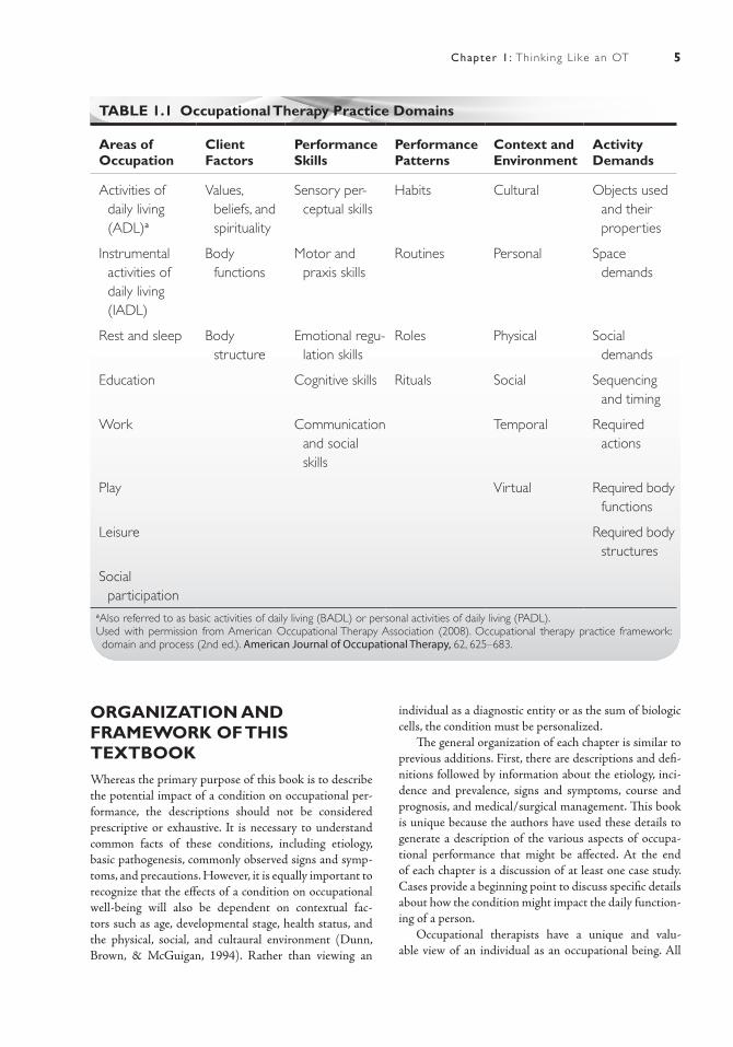
THE OCCUPATIONAL THERAPY PRACTICE FRAMEWORKThe professional language for the profession of occu-pational therapy was revised in 2008 and presented in a document titled the “Occupational Therapy Practice Framework: Domain and Process,” second edition (AOTA, 2008). The Practice Framework outlines the language and constructs that describe the occupational therapy profession’s domain of concern. The domain defines the area of human activity to which the occupational therapy process is applied. The process facilitates engagement in occupation to support participation in life. The focus of the process is on the use of and the enhancement of engagement in occupation. The specific aspects of the domain are outlined in Table 1.1.
The Framework is organized into six aspects—per-formance in areas of occupation, performance skills, performance patterns, context, activity demands, and client factors. Performance in areas of occupation are broad categories of human activity that are typically part of daily life. The areas include activities of daily living, instrumental activities of daily living, education, work, and play, leisure, and social participation. Performance skills are features of what a person does during an activ-ity. These skills are separated into the categories of motor skills, process skills, and communication/interaction skills. Performance patterns are the habits, routines, and roles that a person adopts. Context refers to the condi-tions that surround the person. Those conditions include cultural, physical, social, personal, spiritual, temporal and virtual contexts. Activity demands are the aspects of the task that influence the performance by the person. These demands include the objects used and their properties, space demands, social demands, sequencing and timing, required actions, required body functions and required body structures. Client factors are the body functions and the body structures that reside within the person (Fig. 1.1.) See Table 1.1 for an overview of the practice framework domains.
Each of these aspects has a relationship with and influence on the others. The outcome is, of course, the ability to function and engage in occupations. Although at a given time you may focus on areas of occupation or client factors, the ultimate concern is whether the individ-ual is able to perform necessary and desired tasks in daily life. For example, a therapist may evaluate a person’s atten-tion span, but not in isolation. Attention span is evaluated within the realm of the performance patterns and context of the person—attention span required to work on an assembly line, to drive a car, to learn a card game, or to conduct a business meeting.
Once you know the diagnosis and age of the person, you can use this Practice Framework to examine sys-tematically the deficits that occur in the client factors (described in Figure 1.1) as well as how these particular deficits can and do alter the person’s ability to complete tasks in relevant areas of occupational performance. In other instances, you may focus primarily on the area of occupational performance or the contextual factors for the individual, without paying much attention to the underlying client factors that influence the performance areas.
EVIDENCE-BASED PRACTICEThere has been a call to action in the health professions to practice health care based on evidence of the effectiveness of each treatment approach (Gutman, 2010). High levels of evidence are based on studies that compare groups of

4 C O N D I T I O N S I N O C C U P A T I O N A L T H E R A P Y
people, usually with similar diagnoses. Evidence, espe-cially high levels of evidence, on which to base one’s prac-tice, however, might be limited (Dirette, Rozich & Vau, 2009). First, it is limited by an insufficient number of resources to support specific treatment approaches with specific diagnoses. Second, it might be limited by the fact that groups of people with “average” results do not always represent the unique situation of the person with whom the therapist is working.
Therefore, while we support the idea of evidence-based practice in general, there is clearly a need for thera-
pists to develop clinical reasoning skills that will not only help them decide which evidence to use with par-ticular diagnoses but also help them decide what to do with the unique individual with whom they are working. Understanding the diagnosis with which the individual presents is often the first step in the clinical reasoning process. This textbook provides information about com-mon diagnoses seen by occupational therapists and pro-vides the first steps in the clinical reasoning process by providing ideas about the potential impact on occupa-tional performance.
Values, Beliefs,and
Spirituality
CLIENTFACTORS
BodyStructures
BodyFunctions
VALUES: Principles, standards, or qualities by a person, organization, or population
BELIEFS: Cognitive content held as true by a person, organization, or population
SPIRITUALITY: “Personal quest for understanding answers to ultimate questionsabout life, about meaning, and the sacred” (Moyers and Dale, 2007, p 28)
BODYFUNCTIONS
BODYSTRUCTURES
Includes mental, sensory, neuromusculoskeletal, cardiovascular, hematological, immunological, respiratory, voice and speech, digestive, metabolic, endocrine, genitourinary, reproductive, and skin and related functions.
Structures of the nervous system, and those related to eyes, ear, voice and speech, cardiovascular, immunological, respiratory, digestive, metabolic, endocrine, genitourinary, reproductive, movement, and skin.
Source: American Occupational Therapy Association (2008). Occupational therapy practice framework: domain and process, (2nd ed.). American Journal of Occupational Therapy, 62, 625 – 683.
Figure 1.1 Client factors.
5Chapter 1 : Th ink ing L ike an OT
ORGANIZATION AND FRAMEWORK OF THIS TEXTBOOKWhereas the primary purpose of this book is to describe the potential impact of a condition on occupational per-formance, the descriptions should not be considered prescriptive or exhaustive. It is necessary to understand common facts of these conditions, including etiology, basic pathogenesis, commonly observed signs and symp-toms, and precautions. However, it is equally important to recognize that the effects of a condition on occupational well-being will also be dependent on contextual fac-tors such as age, developmental stage, health status, and the physical, social, and cultaural environment (Dunn, Brown, & McGuigan, 1994). Rather than viewing an
individual as a diagnostic entity or as the sum of biologic cells, the condition must be personalized.
The general organization of each chapter is similar to previous additions. First, there are descriptions and defi-nitions followed by information about the etiology, inci-dence and prevalence, signs and symptoms, course and prognosis, and medical/surgical management. This book is unique because the authors have used these details to generate a description of the various aspects of occupa-tional performance that might be affected. At the end of each chapter is a discussion of at least one case study. Cases provide a beginning point to discuss specific details about how the condition might impact the daily function-ing of a person.
Occupational therapists have a unique and valu-able view of an individual as an occupational being. All
TABLE 1.1 Occupational Therapy Practice Domains
Areas of Occupation
Client Factors
Performance Skills
Performance Patterns
Context and Environment
Activity Demands
Activities of daily living (ADL)a
Values, beliefs, and spirituality
Sensory per-ceptual skills
Habits Cultural Objects used and their properties
Instrumental activities of daily living (IADL)
Body functions
Motor and praxis skills
Routines Personal Space demands
Rest and sleep Body structure
Emotional regu-lation skills
Roles Physical Social demands
Education Cognitive skills Rituals Social Sequencing and timing
Work Communication and social skills
Temporal Required actions
Play Virtual Required body functions
Leisure Required body structures
Social participation
aAlso referred to as basic activities of daily living (BADL) or personal activities of daily living (PADL).Used with permission from American Occupational Therapy Association (2008). Occupational therapy practice framework: domain and process (2nd ed.). American Journal of Occupational Therapy, 62, 625–683.

6 C O N D I T I O N S I N O C C U P A T I O N A L T H E R A P Y
of us attach meaning to our lives and the lives of others through the activities and occupations that are part of our daily existence. Occupation, then, means more than just work. It is a much broader concept that refers to human involvement in activities that will result in productive and purposeful outcomes. It also includes participation in leisure, rest, and self-care activities that some may not consider productive and purposeful. For example, the occupations of a 3-month-old infant include those that could be categorized under the general headings of play or activities of daily living. Activities such as play explo-ration, socialization, and functional communication are critical at this age.
The complexity of occupation changes dramatically as the infant progresses toward preschool and school age. It is interesting to observe the rapid addition of new occu-pational roles and expectations as the child enters school. Many aspects of occupational development are emerging. For example, a 7-year-old child participating in classroom activities is involved in a type of work. Being on time, turning in assignments that are completed properly, good
grooming, and getting along with others are all behaviors that will be important as the child approaches adulthood.
Adults are expected to assume, independently pur-sue, and maintain relevant occupations. In general, adults spend the greater portion of their waking hours engaged in some type of work or instrumental activities of daily living. These occupations may be a job or vocation that is done for pay, organized volunteer activities, or home management. The percentage of time spent in each area is largely determined by the role the individual assumes. In addition, adults spend a portion of their time exploring and performing leisure and social activities. Activities of daily living, sexual expression, grooming, and eating are also important for adults.
The basic tenets regarding occupational performance are that these tasks are critical and must be performed by the person or by others to survive. By engaging in various occupations, the person develops, learns adaptive mechanisms, and meets individual needs. It is important to understand the influence of culture on adaptation. Cultural influences, such as institutions, rules, values,
TABLE 1.2 Most Frequently Cited Conditions Treated by Occupational Therapists
Neurologic Stroke 24% Dementia 16% Cerebral Palsy 12%
Traumatic Brain Injury 12%
Parkinson’s 9%
Low Vision 5%
Development Dev. delay 22.6%
Sensory inte-grative disor-der 18.5%
Mental retardation 12.1%
Learning disorder 11%
Visual processing deficit 6.1%
Cardiopul-monary
Congestive heart fail-ure 26.1%
Chronic obstructive pulmonary disease 25.7%
Myocardial infarction 25.6%
Orthopedic Fractures 29%
Joint replace-ment 22%
Osteoarthri-tis 17%
Upper and lower ext. amputa-tions 12%
General Medical
Decond. debilit. 23%
Diabetes 20% Cancer 16% RA 15%
Psychosocial ASD 17% Behavior disorders 17%
ADD HD 16%
Anxiety disorder 14%
Mood disor-der 8.4%
Schizo-phrenia 7.1%
From National Board for Certification of Occupational Therapists (2008). Executive summary: NBCOT 2008 practice analysis. Retrieved January 3, 2011, from http://www.nbcot.org/pdf/Executive-Summary-for-the-Practice-Analysis-Study-OTR.pdf
7Chapter 1 : Th ink ing L ike an OT
architectural design, art, history, and language, affect the ways and the extent to which a person uses adaptive mechanisms.
Conversely, illness, trauma, or injury can cause vary-ing degrees of occupational dysfunction. The individual receiving occupational therapy is most often experiencing permanent, long-term changes in the ability to engage in everyday activities. The continuum between health and illness is dynamic. The individual’s state of health or ill-ness can be judged by the ability to engage in activities that meet both immediate and long-range needs, and to assume desired roles. Illness or disability is considered in relation to its effects on occupational performance and, therefore, the degree of occupational dysfunction that is experienced.
These precepts are the foundation for the reasoning process described in this book. The combination of these assumptions or beliefs and the occupational performance structure is the frame that provides a unique occupational therapy perspective.
This book of course cannot cover every condition that an occupational therapist will encounter in practice. In the three previous editions, conditions were selected con-ditions based on the AOTA Member Data Survey gath-ered in the late 1980s and on feedback we received from individuals who read and used the first three editions of this textbook. We selected conditions representing the broad range of occupational therapy practice— mental health, physical rehabilitation, geriatrics, and pediatrics. The most current published survey data regarding the highest frequency of conditions treated by occupational therapists were published in 2008 by the National Board for Certification in Occupational Therapy (NBCOT), which conducted a practice analysis survey. A total of
1,283 surveys were sent to practitioners across all practice settings jurisdictions, and 1,156 respondents completed the survey indicating a response rate of 90% with 96% of the respondents actively in practice.
Respondents were asked to indicate their top three diagnoses for six different client condition categories: neurologic, developmental, musculoskeletal/orthope-dic, cardiopulmonary, and psychosocial conditions. The results of this survey were consistent with our selection of common conditions for this current edition. In addi-tion, the results from a survey were similar between the data obtained by surveys of certified occupational therapy assistants, citing similar frequencies in diagnoses treated (NBCOT, 2008).
As an instructional tool, this book provides an oppor-tunity to examine each condition closely. The reader is urged to use the information as a springboard for further study of the conditions included here and the many other conditions that occupational therapists encounter in practice. The analysis of the impact on occupational per-formance for a particular condition is dynamic, and the identification of the most important areas of dysfunction and, therefore, treatment will vary from practitioner to practitioner. In addition, factors such as secondary health problems, age, gender, family background, and culture contribute greatly to the development of a unique occu-pational performance profile for each individual served.
The occupational performance approach to the identi-fication of dysfunction described in this book can be used to examine the effects of any condition on a person’s daily life. This process will enable the therapist to identify and set a priority for problems in occupational performance, which, in turn, will serve as the foundation for creating an effective intervention plan.
R E F E R E N C E S
American Occupational Therapy Association (2008). Occupational therapy practice framework: domain and process (2nd ed.). American Journal of Occupa-tional Therapy, 62, 625–683.
Benamy, B. C. (1996). Developing clinical reasoning skills. San Antonio, TX: Therapy Skill Builders.
Dirette, D., Rozich, A., & Viau, S. (2009). The Issue Is: Is there enough evidence for evidence-based practice in occupational therapy? The American Journal of Oc-cupational Therapy, 63, 782–786.
Dunn, W., Brown, C., & McGuigan, A. (1994). Ecology of human performance: a framework for considering the effect of context. American Journal of Occupational Therapy, 48(7), 595–607.
Gutman, S.A. (2010). From the desk of the editor: AJOT publication priorities. The American Journal of Occupational Therapy, 64(5), 679–681.
Hansen, R. A. (1998). Ethical implications. In J. Hinojosa & P. Kramer (Eds.), Evaluation: obtaining and inter-preting data. Bethesda, MD: American Occupational Therapy Association, p. 203.
Kanny, E. (1993). Core values and attitudes of occupa-tional therapy practice. American Journal of Occupa-tional Therapy, 47, 1085–1086.
Mosey, A. C. (1996). Applied scientific inquiry in the health professions: an epistemological orientation (2nd ed.). Bethesda, MD: American Occupational Therapy Association.
National Board for Certification in Occupational Therapy (2008). Executive summary: NBCOT 2008 practice analysis. Retrieved January 3, 2011, from http://www.nbcot.org/pdf/Executive-Summary-for-the-Practice-Analysis-Study-OTR.pdf

9
Cerebral Palsy
■ Mar y St e i c h e n Yam am o t o
2
Jill’s parents, who had been trying to conceive a child for several years, were thrilled when a family friend asked if they would be interested in adopting a baby girl that had just been born to a young unmarried
woman in her church. The baby was born 6 weeks early and her weight was 4 pounds, but she appeared to be healthy. After initiating the paper-work for a private adoption, they brought the baby home and named her Jill. By the time of Jill’s 6-month well-baby visit, her parents had become concerned. She appeared to be a bright baby who smiled and cooed and enjoyed reaching for and playing with toys, but her legs seemed stiff and she was not yet rolling over. They spoke with their family doctor about their concerns, but he assured them that Jill was developing normally and that they had nothing to be concerned about.
By the time of Jill’s 9-month well-baby visit, her parent’s concerns were only growing. Jill was still not sitting up and had not yet learned to roll over or crawl. Her doctor decided to refer Jill to the county early intervention program for a developmental assessment. Jill was assessed by the early intervention team that included an occupational therapist, physical therapist, and speech and language pathologist. The occupational therapist noted some mildly increased tone and incoordination in her upper extremities, which resulted in about a 2- to 3-month delay in fine motor and self-help skills. The physical therapist noted that Jill had hypertonicity and retained primitive reflexes in her lower extremities, which was causing significant delay in the acquisition of gross motor skills. The speech and language therapist found Jill’s cognitive, language, and social skills to be at age level. The team suggested to the parents that they have a pediatric neurologist assess Jill, as she was demonstrating some of the signs and symptoms of cerebral palsy. Both the occupational and physi-cal therapist recommended that therapy services begin as soon as possible. An IFSP (Individualized Family Service Plan) was developed at a subsequent meeting and Jill began weekly physical and occupational therapy sessions.
Jill’s parents took her to a pediatric neurologist who diagnosed her with spastic diplegia, a type of cerebral palsy. Her parents were initially overwhelmed and devastated by the diagnosis. The next year was very difficult as they grieved the loss of so many dreams that they had for Jill and so much uncertainty about her future. They waded through an array
K E Y T E R M S
AstereognosisAtaxia
Athetoid (dyskinetic)Bronchopulmonary dysplasia
ClonusContracture
DiplegiaDysarthria
EquinovalgusEquinovarus
Gastroesophageal refluxGraphesthesia
HemiplegiaHomonymous hemianopsia
HyperreflexiaHypertonicity (spasticity)
HypotonicityHypoxemiaKernicterus
KyphosisLordosis
NystagmusPrimitive reflexes
QuadriplegiaScoliosis
StrabismusStretch reflex
Topagnosia
10 C O N D I T I O N S I N O C C U P A T I O N A L T H E R A P Y
of possible therapy approaches and medical and surgical interventions that were recommended try-ing to decide which would be right for Jill and their family. They struggled to find time to work on home exercises that had been prescribed for Jill. The strain became so great that they even separated for a while but eventually reconciled. By the time Jill turned 3 years old, she was walking with a walker and able to sit in a chair independently, although she needed assistance with changing positions. She was feeding herself but not yet dressing herself. They enrolled her in a preschool special education classroom where she received therapy services. By kindergarten, Jill was in a regular education classroom with a parae-ducator for safety and support. Jill was a happy child, who had many friends and did well academically. Jill most likely will continue to need some type of addi-tional support in order to be an independent adult, but all involved were optimistic about her future.
DESCRIPTION AND DEFINITIONSigmund Freud, in his monograph entitled “Infantile Cer-ebral Paralysis,” points out that a well-known painting by Spanish painter Jusepe Ribera (1588–1656), which depicts a child with infantile hemiplegia, proves that cer-ebral paralysis existed long before medical investigators began paying attention to it in the mid-1800s (Freud, 1968). Freud’s work as a neurologist is not generally well known and at the time that his monograph was published in Vienna in 1897, he was already deep into his work in the area of psychotherapy. However, he was recognized at the time as the prominent authority on the paralyses of chil-dren. Today, cerebral paralysis is known as cerebral palsy.
Cerebral palsy is not one specific condition but rather a grouping of clinical syndromes that affect movement, muscle tone, and coordination as a result of an injury or lesion of the immature brain. It is not considered a disease. It is considered a developmental disability since it occurs early in life and interferes with the development of motor and sometimes cognitive skills. Historically, cerebral palsy has been classified as and is still sometimes diagnostically referred to as a static encephalopathy (Brooke, 2010). This is now considered inaccurate due to recognition of the fact that the neurological manifestations of cerebral palsy often change or progress over time. Static encepha-lopathy is permanent and unchanging damage to the brain and includes other developmental problems such as fetal alcohol syndrome, mental impairments, and learning disabilities. Many children and adults with cerebral palsy perform well academically and vocationally without any signs of cognitive dysfunction, which is associated with the term encephalopathy ( Johnson, 2004).
A child is considered to have cerebral palsy if all of the following characteristics apply:
1. The injury or insult occurs when the brain is still developing. It can occur anytime during the prenatal, perinatal, or postnatal periods. There is some disa-greement about the upper age limit for a diagnosis of cerebral palsy during the postnatal period, but it typically is up to 2 or 3 years of age (Thorogood & Alexander, 2009; United Cerebral Palsy Research and Educational Foundation, 1995).
2. It is nonprogressive. Once the initial insult to the brain has occurred, there is no further worsening of the child’s condition or further damage to the central nerv-ous system. However, the characteristics of the dis-abilities affecting an individual often change over time.
3. It always involves a disorder in sensorimotor develop-ment that is manifested by abnormal muscle tone and stereotypical patterns of movement. The severity of the impairment ranges from mild to severe.
4. The sensorimotor disorder originates specifically in the brain. The muscles themselves and the nerves connecting them with the spinal cord are nor-mal. Although some cardiac or orthopaedic prob-lems can result in similar postural and movement abnormalities, they are not classified as cerebral palsy.
5. It is a lifelong disability. Some premature babies dem-onstrate temporary posture and movement abnormal-ities that look similar to patterns seen in cerebral palsy but resolve typically by 1 year of age. For children with cerebral palsy, these difficulties persist (Little, 1862).
ETIOLOGYHistorically, birth asphyxia was considered the major cause of cerebral palsy. When British surgeon William Little first identified cerebral palsy in 1860, he suggested that a major cause was a lack of oxygen during the birth process. In 1897, Sigmund Freud disagreed, suggesting that the disorder might sometimes have roots earlier in life. Freud wrote, “Difficult birth, in certain cases, is merely a symptom of deeper effects that influence the development of the fetus” (1968). Although Freud made these observations in the late 1800s, it was not until the 1980s that research supported his views (Freeman & Nelson, 1988; Illingworth, 1985). Extensive research conducted in the United States has shown that only 5% to 10% of the cases of cerebral palsy were a result of birth complications (National Institute of Neurologi-cal Disorders and Stroke [NINDS], 2001). The birth complications resulting in cerebral palsy are related to hypoxemia because of a reduction of umbilical or uterine blood flow (Blickstein, 2003). Table 2-1 lists specific risk factors related to intrapartum hypoxemia.
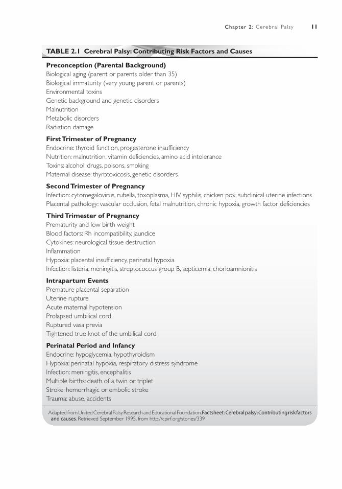
11Chapter 2 : Cerebr a l Pa l sy
TABLE 2.1 Cerebral Palsy: Contributing Risk Factors and Causes
Preconception (Parental Background)Biological aging (parent or parents older than 35)Biological immaturity (very young parent or parents)Environmental toxinsGenetic background and genetic disordersMalnutritionMetabolic disordersRadiation damage
First Trimester of PregnancyEndocrine: thyroid function, progesterone insufficiencyNutrition: malnutrition, vitamin deficiencies, amino acid intoleranceToxins: alcohol, drugs, poisons, smokingMaternal disease: thyrotoxicosis, genetic disorders
Second Trimester of PregnancyInfection: cytomegalovirus, rubella, toxoplasma, HIV, syphilis, chicken pox, subclinical uterine infectionsPlacental pathology: vascular occlusion, fetal malnutrition, chronic hypoxia, growth factor deficiencies
Third Trimester of PregnancyPrematurity and low birth weightBlood factors: Rh incompatibility, jaundiceCytokines: neurological tissue destructionInflammationHypoxia: placental insufficiency, perinatal hypoxiaInfection: listeria, meningitis, streptococcus group B, septicemia, chorioamnionitis
Intrapartum EventsPremature placental separationUterine ruptureAcute maternal hypotensionProlapsed umbilical cordRuptured vasa previaTightened true knot of the umbilical cord
Perinatal Period and InfancyEndocrine: hypoglycemia, hypothyroidismHypoxia: perinatal hypoxia, respiratory distress syndromeInfection: meningitis, encephalitisMultiple births: death of a twin or tripletStroke: hemorrhagic or embolic strokeTrauma: abuse, accidents
Adapted from United Cerebral Palsy Research and Educational Foundation. Factsheet : Cerebral palsy: Contributing risk factors and causes. Retrieved September 1995, from http://cpirf.org/stories/339
12 C O N D I T I O N S I N O C C U P A T I O N A L T H E R A P Y
It is now known that in the majority of cases, the damage that results in cerebral palsy occurs prenatally. Congenital cerebral palsy that results from brain injury during intrauterine life has been reported to be responsi-ble for approximately 70% to 80% of children who have cerebral palsy ( Johnson, 2004; United Cerebral Palsy Research and Education Foundation Factsheet, 2001). In most cases, however, the specific cause is not known (United Cerebral Palsy Research and Education Founda-tion Factsheet, 2001). There are a large number of risk factors that can result in cerebral palsy, and the interplay between these factors is often complex, making it difficult to identify the specific cause. The presence of risk factors does not always result in a subsequent diagnosis of cer-ebral palsy. The presence of one risk factor may not result in cerebral palsy unless it is present to an overwhelm-ing degree. Current thought is that often two or more risk factors may interact in such a way as to overwhelm natural defenses, resulting in damage to the developing brain. The strongest risk factors include prematurity and low birth weight (Lawson & Badawi, 2003). During the postpartum period, premature and low birth weight infants are at greater risk for developing complications, especially in the circulatory and pulmonary systems. These complications can lead to brain hypoxia and result in cerebral palsy. Intraventricular- periventricular bleed-ing and hypoxic infarcts that occur during this period also place the premature infant at increased risk ( United Cerebral Palsy Research and Educational Foundation, 1995). The most common type of ischemic injury in the premature infant is periventricular leukomalacia, which occurs in the white matter adjacent to the ventricles. Sixty percent to 100% of premature infants with perive-ntricular leukomalacia later show signs of cerebral palsy (Zach, 2010). Additional risk factors that have been more recently identified include intrauterine exposure to infection and disorders of coagulation (NINDS, 2001). Maternal infection is a critical risk factor for cerebral palsy, both during prenatal development and at the time of delivery. The infection does not necessarily produce signs of illness in the mother, which can make it difficult to detect. In a study conducted in the mid-1990s, it was determined that mothers with infections at the time of birth had a higher risk of having a child with cerebral palsy (Grether & Nelson, 1997). Table 2-1 shows a more thorough list of risk factors.
In approximately 10% of children with cerebral palsy in the United States, the condition was acquired after birth (United Cerebral Palsy Research and Educational Foundation, 2001). The most common causes in the perinatal and early childhood periods include cerebro-vascular accidents (CVAs), infections such as meningitis or encephalitis, poisoning, trauma such as near-drown-ing and strangulation, child abuse, and illnesses such as endocrine disorders (Grether & Nelson, 1997; Marmer,
1997; United Cerebral Palsy Research and Educational Foundation, 2001). Closed-head injury that occurs dur-ing this period is now classified as traumatic brain injury, even though the resulting impairments are very similar to cerebral palsy (United Cerebral Palsy, 2009). The cause remains unknown in 20% to 30% of cases with an early onset of symptoms postnatally (Grether & Nelson, 1997; United Cerebral Palsy, 2009).
INCIDENCE AND PREVALENCEEstimates of the incidence of cerebral palsy in the United States range from 1.5 to 4 per 1,000 live births (United Cerebral Palsy, 2006). The Centers for Disease Control and Prevention (CDC) has been monitoring the incidence of cerebral palsy in children in the Atlanta, Georgia area since the mid-1980s. In 2000, the prevalence was 3.1 per 1,000 children or 1 in 323 children (CDC, 2004). In the CDC study as well as in two other geographically diverse counties where data were collected, there was a 30% higher rate among African American children as compared to non-Hispanic white children. There was a 70% increase in prevalence in middle- and low-income areas as compared with upper-class areas (CDC; Cerebral Palsy Interna-tional Research Foundation, 2009). The United Cerebral Palsy Association estimated in 2001 that 764,000 chil-dren and adults in the United States show one or more symptoms of cerebral palsy. They estimated that each year, 8,000 infants and 1,200 to 1,500 preschool-age children are diagnosed with cerebral palsy (United Cerebral Palsy Research and Educational Foundation, 2001).
There has been considerable advancement in obstetric and neonatal care during the past 2 to 3 decades. Many hoped these advancements would reduce the incidence of cerebral palsy. Unfortunately, the rate has remained relatively stable. This is probably a result of increased survival rates of very low birth weight and premature infants. Another fac-tor may be the use of fertility treatments by older women that have resulted in an increase in the number of multi-ple births. Multiple births tend to result in infants who are smaller and premature and are at greater risk for health problems. On the average, they are half the weight of other babies at birth and arrive 7 weeks earlier (United Cerebral Palsy Research and Educational Foundation, 2001). There is a 400% increase in the probability of cerebral palsy in twin births than in a single birth (United Cerebral Palsy Research and Educational Foundation, 1997).
SIGNS AND SYMPTOMSThe early signs and symptoms common to all types of cerebral palsy are muscle tone, reflex and postural abnor-malities, delayed motor development, and atypical motor performance (United Cerebral Palsy, 2006).
13Chapter 2 : Cerebr a l Pa l sy
Tone Abnormalities
Tone abnormalities include hypertonicity, hypo tonicity, and fluctuating tone. Fluctuating tone shifts in varying degrees from hypotonic to hypertonic. Muscle tone can be characterized as the degree of resistance when a muscle is stretched. For instance, when there is hypotonicity and the elbow is passively extended, there will be little to no resistance to the movement and hypermobility in the elbow joint. With hypertonicity, there will be increased resistance and it may be difficult to pull the elbow into full extension if the tone is strong. Most infants with cerebral palsy ini-tially demonstrate hypotonia. Later, the infant may develop hypertonicity, fluctuating tone, or continue to demonstrate hypotonia, depending on the type of cerebral palsy.
Reflex Abnormalities
With hypertonicity, reflex abnormalities such as hyperre-flexia, clonus, overflow, enhanced stretch reflex, and other signs of upper motor neuron lesions are present (United Cerebral Palsy, 2006). Retained primitive infantile reflexes and a delay in the acquisition of righting and equilibrium reactions occur in conjunction with all types of abnormal tone. When hypotonia is present, there may be areflexia, or an absence of primitive reflexes. These reflexes should be present during the first several months of life and it is of concern when they are not.
Atypical Posture
The presence of primitive reflexes and tone abnor-malities causes the child to have atypical positions at rest and to demonstrate stereotypical and uncontrol-lable postural changes during movement. For instance, a child with hypertonicity in the lower extremities often lies supine with the hips internally rotated and adducted and the ankles plantar flexed. This posture is caused by a combination of hypertonicity in the affected muscles and the presence of the crossed exten-sion reflex. A child with hypotonicity typically lies with the hips abducted, flexed, and externally rotated because of low muscle tone, weakness in the affected muscles, and the influence of gravity.
Delayed Motor Development
Cerebral palsy is always accompanied by a delay in the attainment of motor milestones. One of the signs that often alert the pediatrician to the problem is a delay in the child’s ability to sit independently. While cerebral palsy is present at birth in all but the approximately 10% of cases, it is often not recognized until the child fails to achieve these early motor milestones.
Atypical Motor Performance
The way in which a child moves when performing skilled motor acts is also affected. Depending on the type of cer-ebral palsy, the child may demonstrate a variety of motor abnormalities such as asymmetrical hand use, unusual crawling method or gait, uncoordinated reach, or diffi-culty sucking, chewing, and swallowing.
TYPES OF CEREBRAL PALSYTypes of cerebral palsy are classified neurophysiologically into three major types: spastic, athetoid, and ataxia.
1. Spastic is characterized by hypertonicity. Deep ten-don reflexes are present in affected limbs and motor control is affected by the hypertonicity. This type is the most common and accounts for approximately 70% to 80% of the cases of cerebral palsy (NINDS, 2001; Thorogood & Alexander, 2009). The spasticity is a result of upper motor neuron involvement (Porter & Kaplan, 2009). Within this category, types are fur-ther subdivided anatomically according to the parts of the body that are affected.
2. Athetoid, also known as dyskinetic type, is charac-terized by involuntary and uncontrolled movements. These movements are typically slow and writhing. This type accounts for approximately 10% to 20% of the cases of cerebral palsy (NINDS, 2001). This type results from basal ganglia involvement (Porter & Kaplan, 2009).
3. Ataxia is characterized by unsteadiness and difficul-ties with balance, particularly when ambulating. It results from involvement of the cerebellum or its path-ways (Porter & Kaplan, 2009). It is much less com-mon than the other two types, occurring in only about 5% to 10% of the cases of cerebral palsy (NINDS, 2001).
It is common for there to be mixed forms where two of the types occur together as a result of diffuse brain dam-age. The most common is combination is spastic with athetoid features. Persons with this type have signs of athetosis, and postural tone that fluctuates from hyperto-nicity to hypotonia. Athetoid combined with ataxia is less common (Porter & Kaplan, 2009).
Spastic
Spastic cerebral palsy is characterized by hypertonicity, retained primitive reflexes in affected areas of the body, and slow, restricted movement. The impact on motor function can range from a mild impairment that does not interfere with functional skills, such as not having isolated finger movement, to a severe impairment, where there is an inability to reach and grasp. Contractures,
14 C O N D I T I O N S I N O C C U P A T I O N A L T H E R A P Y
which are caused by permanent shortening of a muscle or joint leading to deformities, are common. It is catego-rized anatomically by the area of the body that is affected. Spastic hemiplegia, spastic diplegia, and spastic quadri-plegia are the most common types. Spastic triplegia has similar features to spastic quadriplegia with three limbs involved. Typically, it is both lower extremities and one upper extremity. There are often mild coordination dif-ficulties in the noninvolved upper extremity. Monoplegia, where one limb is affected, is rare and when it does occur is typically mild (Molnar & Alexander, 1998)
Spastic HemiplegiaSpastic hemiplegia involves one entire side of the body, including the head, neck, and trunk. Usually, the upper extremity is most affected. Early signs include asymmetri-cal hand use during the first year or dragging one side of the body when crawling or walking. The initial hypotonic stage is short-lived, with spasticity developing gradually (United Cerebral Palsy, 2006). Most children begin walk-ing after 18 months of age, with nearly all children walk-ing by their third birthday (United Cerebral Palsy, 2006). When walking, the child typically hyperextends the knee and the ankle in equinovarus or equinovalgus posi-tion on the involved side. The child often lacks righting and equilibrium reactions on the involved side and will avoid bearing weight on this side. The shoulder is held in adduction, internal rotation; the elbow is flexed; the forearm is pronated; the wrist is flexed and ulnar devi-ated; and the fingers are flexed. Spasticity increases dur-ing physical activities and emotional excitement. Arm and hand use is limited on the involved side, depending on the severity. The child may use more primitive patterns of grasping and lacks precise and coordinated movement. In more severe cases, the child may totally neglect the involved side or use it only as an assist during bilateral activities. Parietal lobe damage occurs in about 50% of cases and results in impaired sensation, including astere-ognosis, loss or lack of kinesthesia, diminished two-point discrimination, decreased graphesthesia, and topagnosia (United Cerebral Palsy, 2006).
Spastic DiplegiaSpastic diplegia involves both lower extremities, with mild incoordination, tremors, or less severe spasticity in the upper extremities. It is most often attributed to pre-mature birth and low birth weight and is, therefore, on the rise as more infants born prematurely survive as a result of medical advances. The ability to sit independently can be delayed up to 3 years of age or older because of inadequate hip flexion and extensor and adductor hypertonicity in the legs (Bobath, 1980). Frequently, the child will rely on the arms for support. The young child will move forward on the floor by pulling along with flexed arms while the
legs are stiffly extended. Getting up to a creeping position is difficult because of spasticity in the lower extremities. Similarly, standing posture and gait are affected to vary-ing degrees, depending on severity. Because of a lack of lower extremity equilibrium reactions, excessive trunk and upper extremity compensatory movements are used when walking. Lumbar lordosis, hip flexion and internal rotation (scissoring), plantar flexion of the ankles, and dif-ficulty shifting weight when walking are common. Many of these problems result in contractures and deformities, including dorsal spine kyphosis, lumbar spine lordosis, hip subluxation or dislocation, flexor deformities of hips and knees, and equinovarus or equinovalgus deformity of the feet (Bobath). Approximately 80% to 90% of children with diplegia will walk independently, some requiring assistive devices such as crutches or a walker to do so (Sala & Grant, 1995). Walking will be slower and more labored with a crouched gait sometimes developing.
Spastic QuadriplegiaWith spastic quadriplegia, the entire body is involved. The arms typically demonstrate spasticity in the flexor muscles, with spasticity in the extensor muscles in the lower extremities. Because of the influence of the tonic labyrinthine reflex (TLR), shoulder retraction and neck hyperextension are common, particularly in the supine position. This results in difficulty with transitional move-ments such as rolling or coming up to sitting. In the prone position, there is increased flexor tone, also a result of TLR influence, causing difficulty with head raising and bearing weight on the arms. Independent sitting and standing are difficult for the child because of hyperto-nicity, the presence of primitive reflex involvement, and a lack of righting and equilibrium reactions. Only a small percentage of children with quadriplegia are able to walk independently, and less than 10% ever walk in the com-munity after adolescence (Bleck, 1975; Sala & Grant, 1995). Oral musculature is usually affected, with result-ing dysarthria, eating difficulties, and drooling (Molnar & Alexander, 1998). Individuals are susceptible to con-tractures and deformities, particularly hip dislocation and scoliosis, and must be closely monitored.
Athetosis
Athetosis is the most common type of dyskinesia or dystonia, characterized by slow, writhing involuntary movements of the face and extremities or the proximal parts of the limbs and trunk. Abrupt, jerky distal move-ments (choreiform) may also appear. The movements increase with emotional tension and are not present dur-ing sleep. Head and trunk control is often affected as is the oral musculature, resulting in drooling, dysarthria,
15Chapter 2 : Cerebr a l Pa l sy
and eating difficulties. Whereas spasticity is character-ized by hypertonicity in the affected muscle groups and restricted movement, athetosis is characterized by fluctu-ating tone and excessive movement. Contractures are rare, but hypermobility may be present because of fluctuating hypotonicity.
Ataxia
Ataxia is characterized by a wide-based, staggering, unsteady gait. Children with ataxia often walk quickly to compensate for their lack of stability and control. Con-trolled movements are clumsy. Intention tremors may be present. The ability to perform refined movements such as handwriting is affected. Hypotonicity is often present (Low, 2010).
ASSOCIATED DISORDERSThere are a number of disorders and difficulties associ-ated with cerebral palsy, in addition to the motor impair-ment that can significantly affect functional abilities. In some cases, associated disorders can have a more signifi-cant impact on function than the motoric aspects of cer-ebral palsy.
Cognitive Impairment
Of all the associated disorders with cerebral palsy, cog-nitive impairment has the most significant impact upon functional outcomes. Estimates of the incidence of cogni-tive or intellectual impairment with cerebral palsy range from 30% to 50% (Molnar & Alexander, 1998). In about one-third of these instances, the cognitive impairment is mild. The most significant impairments most often occur with mixed types and severe spastic quadriplegia. Athetoid-type cerebral palsy has the least occurrence of cognitive impairment. Many children with spastic hemi-plegia and diplegia have average intelligence (Molnar & Alexander; Porter & Kaplan, 2009).
Seizure Disorder
Reports of the incidence of seizures in people with cer-ebral palsy range from 25% to 60% (Porter & Kaplan, 2009). The incidence varies across the diagnostic catego-ries. It is most common in spastic hemiplegia and quadri-plegia, and rare with spastic diplegia and athetosis (CDC, 2004; Johnson, 2004; Molnar & Alexander, 1998; Porter & Kaplan). A population-based study published in 2003 found that the frequency of epilepsy in children with cerebral palsy was 38%. Partial seizures were the most
common type and children with a cognitive impairment had a higher frequency of a seizure disorder (Carlsson, Hagberg, & Olsson, 2003).
Visual and Hearing Impairments
Visual and hearing impairments occur at a higher rate with cerebral palsy than in the general population. Stra-bismus is the most common visual defect, occurring in 20% to 60% of children with cerebral palsy, with the highest rates in spastic diplegia and quadriplegia (Hiles, Wallar, & McFarlane, 1975). Other visual and ocu-lar abnormalities include nystagmus, homonymous hemianopsia associated with spastic hemiplegia, and difficulties with visual fixation and tracking (Molnar & Alexander, 1998). Some children with athetosis have paralysis of upward gaze, which is a clinical manifestation of kernicterus (Porter & Kaplan, 2009). Ataxia can be associated with nystagmus and problems with depth per-ception (Russman & Gage, 1989).
Hearing impairments most often include sensorineu-ral hearing loss, due to congenital nervous system infec-tions ( Johnson, 2004). Conductive hearing losses, caused by persistent fluid in the ears and middle ear infections, occur when there is severe motor involvement in children who spend a lot of time lying down (Blackman, 1997).
Oral Motor
If the oral musculature is affected, the individual with cerebral palsy may have significant difficulty with speak-ing and eating. Dysarthria, if it is severe, may affect func-tional communication resulting in the need for alternative forms of communication. Eating difficulties can result in increased risk of aspiration, limited amount of foods consumed, and difficulty with chewing and swallowing. Drooling is a significant problem in about 10% of the cases of cerebral palsy (Stanley & Blair, 1994). It can be due to many factors including oral motor muscular con-trol, impaired oral sensation, inefficient and infrequent swallowing, poor lip closure and jaw control, and poor head control.
Dental problems occur frequently in individuals with cerebral palsy. Motor problems and oral sensitivity can make tooth brushing more difficult. The combination of enamel dysplasia, mouth breathing, and poor hygiene leads to increased tooth decay and periodontal diseases (Molnar & Alexander, 1998).
Gastrointestinal
Gastrointestinal difficulties occur frequently in cer-ebral palsy. Gastroesophageal reflux can create much
16 C O N D I T I O N S I N O C C U P A T I O N A L T H E R A P Y
discomfort and can result in refusal to eat or difficulty transitioning to solid foods. It requires medical interven-tion such as medication or in more serious cases surgery. Constipation is common due to decreased mobility and exercise as well as inadequate intake of water or unusual diets due to difficulties with oral motor control (Molnar & Alexander, 1998).
Pulmonary
Individuals with more severe motor impairments such as spastic quadriplegia often develop scoliosis or other spi-nal deformities that can impact respiration. The respira-tory muscles themselves may be affected, which results in poor respiration. These individuals are prone to fre-quent upper respiratory infections that can significantly impact their health. When difficulty chewing and swal-lowing is associated with poor breathing and inadequate or decreased ability to cough, this can result in increased aspiration pneumonia ( Johnson, 1994). A barium swal-low study can be conducted to rule out aspiration as a cause of frequent pneumonia. In cases where there is aspiration, a gastric feeding tube may need to be inserted. Premature infants who had bronchopulmonary dyspla-sia also have compromised pulmonary systems (Molnar & Alexander, 1998).
DIAGNOSISNo definitive test will diagnose cerebral palsy. Several factors must be considered. Physical evidence includes a history of delayed achievement of motor milestones; however, delayed motor development can occur with a host of other developmental disabilities and genetic syn-dromes. The quality of movement is the factor that helps provide a differential diagnosis. The findings of atypical or stereotypical movement patterns and the presence of infantile reflexes and abnormal muscle tone point toward a diagnosis of cerebral palsy. However, other causes must be ruled out, such as progressive neurological disorders, mucopolysaccharidosis, muscular dystrophy, or a spinal cord tumor. Many of these disorders can be ruled out by laboratory tests, although some must be differentiated by clinical or pathological criteria. A magnetic resonance imaging (MRI) or computed tomography (CT) scan may provide evidence of hydrocephalus, help determine the location and extent of structural lesions, and help rule out other conditions. However, these scans are not defini-tive as far as making a diagnosis of cerebral palsy and are not predictive as far as the child’s functioning (Miller & Bachrach, 2006). The yield of finding an abnormal CT scan in a child with cerebral palsy is 77% and for MRIs it is about 89% (Ashwal, Russman, Blasco, & Miller,
2004). An electroencephalogram (EEG) should not be used to determine the cause of cerebral palsy but should be obtained if there is an indication that the child may be having seizures. Because of the high occurrence of asso-ciated conditions, children with cerebral palsy should be screened for cognitive, visual, and hearing impairments, as well as speech and language disorders.
Cerebral palsy often is not evident during the first few months of life and is rarely diagnosed that early. Most cases, however, are detected by 12 months and nearly all can be diagnosed by 18 months (Miller & Bachrach, 2006). In some cases, early postural and tonal abnormalities in pre-mature infants can resemble cerebral palsy, but the signs are transient with normal subsequent development.
COURSE AND PROGNOSISThe course of cerebral palsy varies depending on type, severity, and the presence of associated problems. With mild motor involvement, the child will continue to make motor gains and compensate for motor difficulties. With more severe forms, little progress may be made in attain-ing developmental milestones and performing functional tasks. As the child grows older, secondary problems such as contractures and deformities will become more com-mon, especially with spasticity. Adults with cerebral palsy experience musculoskeletal difficulties and loss of func-tion at an earlier age than their nondisabled peers. One study found that 75% of individuals with cerebral palsy had stopped walking by age 25 due to fatigue and walk-ing insufficiency (Murphy, Molnar, & Lankasky, 1995). Another study of young adults found clinical evidence of arthritis in 27% of the subjects with cerebral palsy as compared to 4% in the general population (Cathels & Reddihough, 1993).
The survival rate for adults with cerebral palsy is good but lower than the general population. A study in Great Britain found there was an 86% survival rate at age 50 for adults with cerebral palsy compared to 96% for the gen-eral population. After age 50, the relative risk of death was only slightly higher in women as compared to the general population. The risk for men with cerebral palsy was the same as the general population. Adults with cerebral palsy were more likely to die of respiratory disease than the general population but less likely to die from an accident or injury (Hemming, Hutton, & Pharoah, 2006).
MEDICAL/SURGICAL MANAGEMENTBecause of the complexity and diversity of difficulties affecting the individual with cerebral palsy, medical man-agement requires a team approach using the skills of many
17Chapter 2 : Cerebr a l Pa l sy
professionals. Depending on the type of cerebral palsy and the presence of associated problems, team mem-bers typically include an occupational therapist, physical therapist, speech pathologist, educational psychologist, nurse, and social worker. The emphasis of intervention is usually on helping the child gain as much motor control as possible; positioning the child to minimize the effects of abnormal muscle tone; instructing the parents and car-egivers on handling techniques and ways to accomplish various activities of daily living (ADLs); recommending adaptive equipment and assistive technology to increase the child’s ability to perform desired activities; providing methods to improve feeding and speech if difficulties are present; and helping parents manage behavioral concerns and family stresses.
The primary physician treats the usual childhood dis-orders and helps with prevention of many health prob-lems. Physicians with various medical specialties may also be involved. The usual specialists include a neurologist to assess neurological status and help control seizures, if present; an orthopaedist to prescribe orthotic devices and any necessary surgeries; and an ophthalmologist to assess and treat any visual difficulties.
Medical management includes both surgical and nonsurgical approaches, with much of the focus on tech-niques to decrease spasticity. Oral medications such as diazepam (Valium), dantrolene (Dantrium), and baclofen have been used to reduce spasticity in severe cases with mixed results (Albright, 1996; Johnson, 2004). Intrathe-cal caclofen infusion (ITB) administered through a pump implanted in the abdominal wall to the spinal cord fluid has shown to be more effective than oral medications in reducing severe spasticity and dystonia in cerebral palsy. There is, however, the potential for serious side effects and the long-term consequences are not yet known (Stempien & Tsai, 2000). Another treatment more widely used in recent years is the injection of botulinum toxin (Botox) into muscles. Spasticity is reduced for a period of 3 to 6 months after injection. Botox is injected into specific muscles, which, in addition to reducing tone, increases range of motion and reduces deformities as well as pro-vides an opportunity to work on muscle strengthening. Minimal side effects have been reported; however, its long term effectiveness on function has not been demonstrated (Steinbok, 2006).
Orthotics and splinting are used to improve function and prevent contractures and deformities. Upper extrem-ity resting or night splints are used to maintain range of motion. Soft splints, dynamic splints, and those allow-ing movement of the fingers and thumb are used during waking hours and functional activities to reduce tone and promote more typical patterns of movement. Ankle-foot orthosie (AFO) and variants are often prescribed to con-trol spastic equinus, promote alignment of the hind foot,
and control mid foot and excessive knee extension when standing (Cusick, 1990; Molnar & Alexander, 1998). Inhibitory and progressive casting has gained acceptance as an alternative to bracing in recent years (Hanson & Jones, 1989; Lannin, Novak, & Cusick, 2007). A molded footplate is constructed that inhibits the primitive reflexes, thus reducing spasticity. The footplate is surrounded by a snug, bivalve below the knee cast. Inhibitory and progres-sive casting also is used with the upper extremities.
Surgical approaches are used to improve the func-tion and appearance of affected areas of the body and to prevent or correct deformities. Tendon lengthening to increase range of motion and tendon transfers to decrease spastic muscle imbalances are done. These procedures, commonly used on the lower extremities, are performed more selectively in the upper extremities (Steinbok, 2006). Selective dorsal rhizotomy (SDR) is a neurosurgical tech-nique that is used to reduce spasticity and improve func-tion in carefully selected individuals (Berman, Vaughan, & Peacock, 1990; Cleveland Clinic Health Information Center, 2010; Kinghorn, 1992; Peackock & Staudt, 1992). The procedure involves dividing the lumbosacral posterior nerve root into four to seven rootlets. Each root-let is stimulated electrically. The dorsal rootlets causing spasticity are cut, leaving the normal rootlets intact. This approach is highly successful for individuals who meet the selection criteria (Berman et al., 1990; Cleveland Clinic Health Information Center; Kinghorn, 1992). The most likely candidates are children with either diplegia or severe quadriplegia (Berman et al.; Cleveland Clinic Health Information Center; Kinghorn; Peacock & Staudt, 1991). For children with diplegia, the goal is to improve gait and leg function. For children with spastic quadriplegia who have very limited movement, the goal is to increase their independence by allowing them to sit for longer periods of time enabling them to use a wheelchair or potty chair as well as making daily care easier for their caregivers by reducing spasticity, which makes dressing and other daily living tasks more manageable (Cleveland Clinic Health Information Center, 2010). An essential part of this treatment approach includes intensive post-surgical physical and occupational therapy for a period of several weeks.
IMPACT ON OCCUPATIONAL PERFORMANCEVirtually all of the body function categories can be affected in the individual who has cerebral palsy. Which of the categories are affected depends on the type of cer-ebral palsy, the severity of the condition, and the presence of associated disorders. Milder forms of cerebral palsy may have limited impact upon occupational performance.
18 C O N D I T I O N S I N O C C U P A T I O N A L T H E R A P Y
Some individuals will require physical assistance, addi-tional training, or assistive technology to participate fully in occupational performance areas, while individuals with severe forms of cerebral palsy will be limited in their per-formance of all areas of occupation.
The body function category that is always affected in individuals with cerebral palsy is neuromusculoskeletal and movement-related function. If spasticity is present, it affects joint mobility and results in limited active or pas-sive range of motion or both. Joint stability is affected in all types of cerebral palsy. With spastic type, uneven muscle pull affects stability and joint cocontraction; fluc-tuating movement and tone affects stability in athetoid type; and ataxic type is characterized by a lack of joint stability. Underlying the tone abnormalities is decreased muscle power. Tone abnormalities affect muscle endur-ance. It requires much more effort to complete motor tasks with both hypotonia and hypertonia and endurance is often diminished. Endurance is also decreased if respira-tory muscles are affected. In movement functions, primi-tive motor reflexes such as the stretch reflex, asymmetrical tonic neck reflex (ATNR), or grasp reflex are retained in the child with spasticity and continue to influence move-ment throughout life. Individuals with all types of cerebral palsy have impaired involuntary movement reactions such as decreased righting and equilibrium reactions. Involun-tary movements impact individuals with athetoid cerebral palsy. Some individuals with ataxia have intention trem-ors. Gait patterns are affected in all types of cerebral palsy; however, in milder cases they may have little effect on occupational performance.
All sensory functions can be affected. All types of cerebral palsy have associated visual difficulties such as strabismus and visual tracking and fixation or hear-ing impairments. Some individuals with cerebral palsy demonstrate sensory processing difficulties with either hypersensitivity to sensory input or with their ability to discriminate sensory input.
Mental functions, both global and specific, can be affected, particularly if there is an associated learning dis-ability, attention deficit hyperactive disorder, or cognitive impairment. Voice and speech functions can be affected if the oral motor and respiratory muscles have tonal abnor-malities. Functions of the cardiovascular and respiratory system can be affected in a variety of ways, such as associ-ated spinal deformities that can compromise respiration or decreased physical endurance or stamina as a result of the amount of effort that it takes to move. The digestive system can be affected by a number of different medical and physical factors such as reflux and feeding difficul-ties. Urinary functions are impacted by decreased control of muscles used in urination and in some cases cognitive factors.
It is important for the occupational therapist to be aware of all the client factors that can be affected in indi-viduals with cerebral palsy but to not make any assump-tions based upon the type of cerebral palsy and known associated disorders but to directly assess each factor and its impact on occupational performance. Each individual is unique and will have their own set of strengths and challenges.
Case Illustrations
Case 1A.K. is a 2-year-old girl who lives with her parents and older brother. She was born at 37 weeks gestation at a birth weight of 5 pounds 10 ounces. Pregnancy and birth were unremarkable. She was healthy at birth but by her well-baby visit at 9 months of age she was not yet rolling, crawling, or sitting independently. Her pediatrician referred her to a pediatric neurologist who diagnosed her with spastic diplegia at 11 months of age. The neurologist referred her to a physiatrist (a physician and rehabilitation physician) at the local children’s hospital as well as the local school district for early intervention services. The physiatrist signed a referral for her to the orthotics department to fit her with AFOs to help her with standing and walk-ing. He has also recommended Botox injections and selective posterior rhizotomy for consideration as
future treatments. Through the early intervention program, she received a multidisciplinary team assess-ment that included physical, occupational, and speech and language assessments. Delays were noted in gross motor, fine motor, and self-help skills. Speech and lan-guage, social, and cognitive skills were all determined to be at age level. Weekly occupational and physical therapy home-based services were recommended.
Affected performance skills are in the motor area, which includes posture, mobility, coordination, and strength/effort. She demonstrates spasticity in all lower extremities and her trunk. Her movement patterns reflect significant spasticity in her legs. Her joint range of motion is significantly limited in her hamstrings and hip adductors with mild limitations in her heel cords bilaterally. A.K. can sit independently; however, it is difficult for her to sit on the floor with her legs
19Chapter 2 : Cerebr a l Pa l sy
extended in front of her as a result of hamstring and hip adductor tightness. She requires assistance mov-ing in and out of sitting. In prone, she can push up to hands and knees and can crawl for short distances. She bears weight on her legs in supported standing with knees in a slightly flexed position, hips adducted, and feet plantar flexed and pronated. She has begun ambulating with a walker for short distances.
In her upper extremities, there is mildly increased muscle tone bilaterally as well as some incoordination. A.K. grasps pegs and small blocks and releases objects into a container. She can place a peg in a pegboard but is not yet able to stack objects or complete a shape sorter. Areas of occupation that are affected include ADLs and play. Because she is only 2 years old, instrumental ADLs, student, work, and leisure areas of occupational are not yet relevant areas for her. In ADL, because of her age she would not yet be expected to be independent. Given this, A.K.’s affected ADLs include bathing, dressing, feeding, and functional mobility. A.K. needs assistance with dressing skills such as undressing and removing shoes and socks. She is independent in feeding with some adaptations. She requires assistance with maintaining a stable sitting position in the bathtub and requires assistance getting in and out of the bath tub. She needs assistance with functional mobility, such as getting in and out of chairs and moving from one place to another.
Exploratory play skills are affected by A.K.’s difficulty with moving about her environment to obtain toys she wants to play with. Participation is affected by the need for a stable position in which to free up her upper extremities to manipulate toys. She uses a bench with a pelvis stabilizer and tray for refined fine motor tasks.
Case 2L.N. is a 54-year-old woman with cerebral palsy, spas-tic quadriplegic type. She has lived alone in an apart-ment complex for the elderly and disabled for the past 15 years. She supports herself on supplemental security income (SSI) and disability payments from the state. A personal-care attendant provided by the Department of Social Services comes in each morn-ing and evening to assist her with ADLs, such as meal preparation, bathing, and dressing. L.N. has never been employed but has done volunteer work. She writes articles for a newsletter on her computer and has worked in her church’s Sunday school. She has no family support but has many friends. She enjoys learn-ing and taking classes through continuing education.
Spasticity, fluctuating tone, and retained primitive reflexes severely restrict L.N.’s purposeful movement.
She has limited range of motion in her left upper extremity and both lower extremities. When reach-ing with the left arm, she cannot bring it to shoulder height or behind her back. She has a gross grasp in her right upper extremity and can grasp a joystick to operate her electric wheelchair. She cannot write or perform other activities requiring fine motor dexter-ity. Her left upper extremity is used as an assist for bilateral activities, with no grasping ability present. She can maintain an upright position in sitting, but her weight is shifted to the left (with resulting scoliosis). She can bring her head to an upright position, but neck flexion increases with activities requiring effort. Oral motor muscles are affected, resulting in severe dysarthria, drooling, and difficulty eating. Endurance is a problem, and L.N. becomes easily fatigued.
Communication/interaction skills are also affected. Articulation and modulation when speaking is affected by L.N.’s oral motor control. Limited dexter-ity and restrictions in movement limit her ability to use gestures and to orient her body in relation to others when engaged in social interactions.
All areas of occupation are affected. In ADLs, L.N. needs assistance with bathing, personal hygiene and grooming, toilet hygiene, and dressing. She brushes her teeth and performs light hygiene, such as wash-ing her face, independently. She can transfer herself between her wheelchair and her bed. She needs assistance transferring to the shower seat she uses for bathing. She can transfer on and off the toilet in her apartment with grab bars and the toilet seat at the proper height and position, although it takes her a while to do this. In eating, L.N. can feed herself with adaptations if the food is setup for her, but the process is slow and messy. She drinks from a straw. She takes her own medications if they are set out for her.
In instrumental ADLs, L.N. needs assistance in clothing care, cleaning her apartment, household maintenance, and meal preparation. She can use a handheld portable vacuum cleaner for small cleanups. She has a cat that she cares for. She shops indepen-dently but needs assistance in getting money out of her wallet at the cash register. All areas of ADLs are affected except socialization. L.N. uses a computer for written communication. She uses a speaker phone for telephone communication. If she falls or is in danger at home, she has an emergency alert system that she can activate. Because her speech is difficult to understand, she has an augmented output device for communication but uses it infrequently. She uses a motorized wheelchair for mobility. In the community, L.N. uses public transportation with no difficulty. She has some difficulty transferring herself to and from
20 C O N D I T I O N S I N O C C U P A T I O N A L T H E R A P Y
the toilet when using public restrooms, which some-times results in incontinence.
In work activities, L.N. has never been employed but has worked as a volunteer for the past several years in the religious education program at her church. She enjoys the interaction with the children that are in the classes.
In leisure activities, L.N. has varied interests. She is an avid reader and enjoys computer games. Social activities include getting together with friends fre-quently and going out into the community, either alone or with friends. L.N. participates in church retreats as well as community-based trips through an independent living center.
R E C O M M E N D E D L E A R N I N G R E S O U R C E S
Domans, J., & Pellegrano, L. (1998). Caring for children with cerebral palsy: A team-based approach. Baltimore, MD: Brooks Publishing. An interdisciplinary reference for team-based, collaborative care of children with cerebral palsy.
Geralis, E. (1997). Children with cerebral palsy—A par-ent’s guide (2nd ed.). Bethesda, MD: Woodbine House. Good reference for therapists and parents for young children with cerebral palsy. Contains com-prehensive information on cerebral palsy including medical treatment information, and the effects of cerebral palsy on the child’s development and edu-cation.
Levitt, S. (2003). Treatment of cerebral palsy and motor delay (4th ed.). Oxford, England: Blackwell Sci-ence LTP. Book was written for occupational and physical therapists working with children with cerebral palsy. Good discussion of treatment approaches, principles of treatment, and description of procedures.
Morris, S., & Klein, M. (1987). Pre-feeding skills. Tucson, AZ: Therapy Skill Builders. Excellent reference for oral-motor and feeding therapy for children. Very
thorough and good overall approach to feeding issues.
United Cerebral Palsy National Office1660 L Street, NW Suite 700Washington, DC 20036Tel: (202) 776-0406; toll free: (800) 872-5827www.ucp.orgLeading source of information on cerebral palsy and
national advocacy group.Children’s Hemiplegia and Stroke Association (CHASA)4101 West Green Oaks Blvd.PMB #149Arlington, TX 76016Tel: (817) 492–4325www.hemikids.orgA comprehensive, practical resource related to children
with hemiplegia type cerebral palsy.National Institute of Neurological Disorders and
Stroke (NINDS). Cerebral palsy information page. Retrieved from www.ninds.nih.gov/disorders/cerebral_palsy. Lists resources including organiza-tions, publications, links, and general information about cerebral palsy.
Albright, A. (1996). Intrathecal baclofen in cerebral palsy movement disorders. Journal of Child Neurology, 11, S29.
Ashwal, S., Russman, B., Blasco, G., & Miller, A. (2004). Practice parameter: Diagnostic assessment of the child with cerebral palsy. Neurology, 62, 851–863.
Berman, B., Vaughan, C., & Peacock, W. J. (1990). The effect of rhizotomy on movement in patients with cerebral palsy. American Journal of Occupational Therapy, 44, 6.
Blackman, J. (1997). Medical aspects of developmental dis-abilities in children birth to three (3rd ed.). Iowa City, IA: The University of Iowa.
Bleck, E. (1975). Locomotor prognosis in cerebral palsy. Developmental Medicine and Child Neurology, 17, 18–25.
Blickstein, A. (2003). Cerebral palsy: A look at etiology and new task force conclusions. OBG Management, 15, 5.
Bobath, K. A. (1980). Neurological basis for the treatment of cerebral palsy. Philadelphia, PA: JB Lippincott.
Brooke, H. (2010). Cerebral palsy as a cause of static encelop-athy in infants. Retrieved February 2010, from http://www.associatedcontent.com/article/2688287/ cerebral_palsy_as_a_cause_of_static.html?cat = 5
Carlsson, M., Hagberg, G., & Olsson, I. (2003). Clinical and etiological aspects of epilepsy in children with
R E F E R E N C E S
21Chapter 2 : Cerebr a l Pa l sy
cerebral palsy. Developmental Medicine and Child Neurology, 45, 371–376.
Cathels, B., & Reddihough, D. (1993). The health care of young adults with cerebral palsy. The Medical Journal of Australia, 159, 444–446.
Centers for Disease Control and Prevention. (2004). Metropolitan Atlanta Developmental Disabilities Sur-veillance Program. Retrieved August 4, 2004, from http://www.cdc.gov/ncbddd/dd/cp3.htm#common
Cerebral Palsy International Research Foundation. (2009). Racial disparities in the prevalence of cerebral palsy. Retrieved May 2009, from http://cpirf.org/stories/478
Cleveland Clinic Health Information Center. (2010). Selective dorsal rhizotomy. Retrieved June 17, 2010 from http://my.clevelandclinic.org/services/ selective_dorsal_ rhizotomy/ns_overview.aspx
Cusick, B. (1990). Progressive casting and splinting for low-er extremity deformities in children with neuromuscular dysfunction. Tucson, AZ: Therapy Skill Builders.
Freeman, J., & Nelson, K. (1988). Intrapartum asphyxia and cerebral palsy. Pediatrics, 82, 240–249.
Freud, S. (1968). Infantile cerebral paralysis. Coral Gables, FL: University of Miami Press. (Original work pub-lished in 1897).
Grether, J., & Nelson, K. (1997). Maternal infection and cerebral palsy in infants of normal birth weight. The Journal of the American Medical Association, 278, 3.
Hanson, C., & Jones, L. (1989). Gait abnormalities and inhibitive casts in cerebral palsy: Literature review. Journal of the American Podiatric Medical Association, 79, 53–59.
Hemming, K., Hutton, J. L., & Pharoah, P. O. (2006). Long-term survival for a cohort of adults with cerebral palsy. Developmental Medicine and Child Neurology, 48, 90–95.
Hiles, D., Wallar, P., & McFarlane, F. (1975). Current concepts in the management of strabismus in children with cerebral palsy. Annals of Ophthalmology, 7, 789.
Illingworth, R. (1985). A pediatrician asks—why is it called birth injury? British Journal of Obstetrics and Gnyaecology: An International Journal of Obstetrics & Gynaecology, 92, 122–130.
Johnson, M. (2004). Encephalopathies. In R. Behrman, R. Kliegman, & H. Jenson (Eds.), Nelson’s textbook of pediatrics (17th ed.). Philadelphia, PA: Saunders.
Kinghorn, J. (1992). Upper extremity functional changes fol-lowing selective posterior rhizotomy in children with cere-bral palsy. American Journal of Occupational Therapy, 4, 6.
Lannin, N., Novak, I., & Cusack, A. (2007). A system-atic review of upper extremity casting for children and adults with central nervous system disorders. Clinical Rehabilitation, 21(11), 963-976.
Lawson, R. D., & Badawi, N. (2003). Etiology of cerebral palsy. Hand Clinics, 19(4), 547–556.
Little, W. (1862). On the influence of abnormal parturition, difficult labor, premature birth and physi-cal condition of the child, especially in relation to deformities. Transactions of the Obstetrical Society of London, 3, 243–344. Retrieved August 15, 2010 from http://www.neonatology.org/classics/little.html
Low, J. (2010). Ataxic cerebral palsy. Retrieved September 26, 2010, from www.disabled-world.com/health/neurology/cerebral-palsy/ataxic.php
Marmer, L. (1997). ACDC tracks disability in kids ages 3 to 10. Advance for occupational therapists. King of Prussia, PA: Merion Publications, Inc.
Miller, F., & Bachrach, S. (2006). Cerebral palsy: A complete guide for caregiving. Baltimore, MD: John Hopkins Press.
Molnar, G., & Alexander, M. (Eds.). (1998). Pediatric re-habilitation (3rd ed.). University of California Davis, CA: Hanley & Belfus.
Murphy, K., Molnar, G., & Lankasky, K. (1995). Medi-cal and functional status of adults with cerebral palsy. Developmental Medicine and Child Neurology, 37, 1075–1084.
National Institute of Neurological Disorders and Stroke. (2001). Cerebral palsy: Hope through research. Re-trieved July 1, 2001, from http://www.ninds.nih.gov/ health_and_medical/ pubs/cerebral_palsyhtr.htm
Peacock, W., & Staudt, L. (1991). Functional outcomes following selective posterior rhizotomy in children with cerebral palsy. Journal of Neurosurgery, 74, 380–385.
Porter, R., & Kaplan, M. (Eds.). The Merck Manual Online. Rahway, NJ: Merck Sharp & Dohme Corp. Retrieved May 2009, from http://www.merck.com/mmpe/sec19/ ch283/ch283b.html
Russman, B., & Gage, J. (1989). Cerebral palsy. Current Problems in Pediatrics, 19, 65–111.
Sala, D., & Grant, A. (1995). Prognosis for ambulation in cerebral palsy. Developmental Medicine and Child Neurology, 37, 1020–1026.
Stanley, F., & Blair, E. (1994). Cerebral palsy. In I. B. Press (Ed.), The epidemiology of childhood disorders, New York, NY: Oxford University Press.
Steinbok, B. (2006). Selection of treatment modalities in children with spastic cerebral palsy: Management options. Neurosurgical Focus, 21(2). Retrieved Sep-tember 4, 2010 from http://www.medscape.com/viewarticle/550745_2.
Stempien, L., & Tsai, T. (2000). Intrathecal baclofen pump use for spasticity: A clinical survey. American Journal of Physical Medicine & Rehabilitation, 6, 536–541.
Thorogood, C., & Alexander, M. (2009). Cerebral palsy. Retrieved March 11, 2009, from http://emedicine.medscape.com/article/310740-overview
United Cerebral Palsy Research and Educational Foun-dation. (1995). Factsheet: Cerebral palsy: Contributing

22 C O N D I T I O N S I N O C C U P A T I O N A L T H E R A P Y
risk factors and causes. Retrieved September 1995, from http://cpirf.org/stories/339
United Cerebral Palsy Research and Educational Foun-dation. (1997). Multiple births and developmental brain damage. Retrieved May 1997, from http://cpirf.org/stories/364
United Cerebral Palsy Research and Education Foundation Factsheet. (2001). Cerebral palsy facts and
figures. Retrieved October 2001, from http://www.ucp.org/ucp_channeldoc.cfm/ 1/11/10427/10427–10427/447
Zach, T. (2010) Periventricular leukomalacia. Retrieved March 18, 2010, from http://emedicine.medscape.com/article/975728-overview
23
Autism Spectrum Disorders
■ Kat h r y n Sh an g raw
3
Rachel was the beautiful first-born daughter to her parents, and dur-ing her first year of life, she appeared to be a typically developing child. She responded to people and objects in her environment with interest and enjoyment. Though slightly delayed in learning to
walk, she mastered this skill between 14 and 16 months of age. In contrast, many of her language skills developed strongly during her first year. Rachel was exposed to three different languages in her home and learned several vocabulary words in each language by the time she was 14 months old.
When Rachel was approximately 14 months old, her parents noticed a startling change in her behavior. Eye contact became rare, and she no longer turned in response when her mother called her name. Because of this decreased responsiveness, her parents worried she was losing her hear-ing. However, an audiologic test indicated normal hearing abilities. Dressing this little girl also became a challenge. Suddenly, she did not easily tolerate the sensation of clothes against her skin. She became intensely distressed when her mother tried to brush her hair or clip her nails. The vocabulary of approximately 30 words that she had previously developed was replaced by silence or babbling. Upon reflection, her mother realized Rachel had never used specific communicative gestures such as waving to greet others or pointing. Engaging Rachel in daily activities became increasingly difficult for she appeared frustrated, unable to express her needs and wants, and unable to leave familiar, preferred activities without becoming highly agi-tated. During these episodes of tantrums, Rachel would hit herself or bang her head repeatedly against a wall while crying. Her mother described that overall, “she no longer seemed happy.” When Rachel was 2 years and 6 months old, her parents brought their concerns to the attention of early intervention specialists. As a result, Rachel began receiving occupational therapy, speech-language therapy, and special education services. During her preschool class and therapy sessions, these specialists observed the same concerning characteristics her parents had described. Additionally, they noted she did not appear interested in the other children in her preschool class. Transitioning from room to room consistently distressed her, causing “meltdowns” during which she flung herself on the floor and cried inconsolably. She used only a few real words or echoed words of
K E Y T E R M S
Ambiguous hand preferenceAmygdala
Asperger’s disorder/Asperger’s syndrome
Auditory processingAutistic disorder
CaseinCerebellum
Childhood disintegrative disorder
EcholaliaEmotional lability
Frontal lobesGastrointestinal (GI) disorder
Gaze shiftGluten
HippocampusHyperlexia
Inferior oliveJoint attentionLimbic systemNeurobiologic
NeuronPervasive developmental disorder
Purkinje cellsRett’s disorderRote memory
Tactile defensivenessTheory of mind
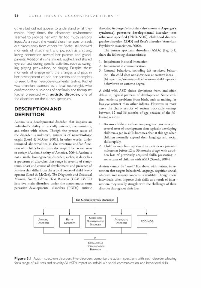
24 C O N D I T I O N S I N O C C U P A T I O N A L T H E R A P Y
others but did not appear to understand what they meant. Many times, the classroom environment seemed to provide her with far too much sensory input. As a result, she would close her eyes or seek out places away from others. Yet Rachel still showed moments of attachment and joy, such as a strong, loving connection toward her parents and grand-parents. Additionally, she smiled, laughed, and shared eye contact during specific activities, such as swing-ing, playing peek-a-boo, or singing. Despite these moments of engagement, the changes and gaps in her development caused her parents and therapists to seek further neurodevelopmental testing. Rachel was therefore assessed by a local neurologist, who confirmed the suspicions of her family and therapists: Rachel presented with autistic disorder, one of the disorders on the autism spectrum.
DESCRIPTION AND DEFINITIONAutism is a developmental disorder that impacts an individual’s ability to socially interact, communicate, and relate with others. Though the precise cause of the disorder is unknown, autism is of neurobiologic origin (Lord & McGee, 2001). In other words, unde-termined abnormalities in the structure and/or func-tion of a child’s brain cause the atypical behaviors seen in autism (Autism Society of America, 2004). Autism is not a single, homogeneous disorder; rather, it describes a spectrum of disorders that range in severity of symp-toms, onset and course of development, and presence of features that differ from the typical course of child devel-opment (Lord & McGee). The Diagnostic and Statistical Manual, Fourth Edition, Text Revision (DSM IV-TR) lists five main disorders under the synonymous term pervasive developmental disorders (PDDs): autistic
disorder, Asperger’s disorder (also known as Asperger’s syndrome), pervasive developmental disorder—not otherwise specified (PDD-NOS), childhood disinte-grative disorder (CDD) and Rett’s disorder (American Psychiatric Association, 2000).
The autism spectrum disorders (ASDs) (Fig. 3.1) share the following characteristics:
1. Impairment in social interaction2. Impairment in communication3. Unusual behaviors, including (a) restricted behav-
ior—the child does not show new or creative ideas—(b) repetitive/stereotyped behavior—a child repeats a behavior to an extreme degree.
A child with ASD shows deviations from, and often delays in, typical patterns of development. Some chil-dren evidence problems from birth, such as making far less eye contact than other infants. However, in most cases the characteristics of autism noticeably emerge between 12 and 36 months of age because of the fol-lowing reasons:
1. Because children with autism progress more slowly in several areas of development than typically developing children, a gap in skills becomes clear at this age when children normally expand their language and social skills rapidly.
2. Children may have appeared to meet developmental milestones before 12 to 36 months of age, with a sud-den loss of previously acquired skills, presenting in some cases of children with ASD (Strock, 2004).
Autism cannot be “cured.” For those with autism, inter-vention that targets behavioral, language, cognitive, social, adaptive, and sensory concerns is available. Though these individuals often improve their skills as a result of inter-vention, they usually struggle with the challenges of their disorder throughout their lives.
THE AUTISM SPECTRUM DISORDERS
CHILDHOOD
DISINTEGRATIVE
DISORDER
ASPERGER’S
DISORDERPDD-NOS
RETT’S
DISORDER
AUTISTIC
DISORDER
SOCIAL SKILLS
COMMUNICATION
BEHAVIOR
Figure 3.1 Autism spectrum disorders. Five disorders comprise the autism spectrum, with each disorder allowing for a range of skill sets and severity. All ASDs impact an individual’s social, communication, and behavioral skills.
25Chapter 3 : Aut i sm Spectr um Disorder s
THE MANY NAMES OF AUTISMThe terms used in the autism field are confusing for many people, since some terms are used interchangeably or inconsistently in literature and by professionals. The most accurate and generally agreed-upon definitions are summarized as follows.
■ The terms autism, ASDs, and PDDs are synonymous. Many professionals prefer using the term ASDs rather than PDDs, believing it more accurately reflects the variability that occurs both among the disorders and within an individual diagnostic category.
■ This chapter will use autism and ASDs interchange-ably, noting the term PDDs only when quoting another source that uses this phrase.
■ Note that the similar term autistic disorder actually refers to one specific diagnostic category on the autism spectrum.
ETIOLOGYThough researchers believe that certain factors (e.g., genetics) are more likely than others to cause autism, no one etiologic factor has clearly emerged. Therefore, autism is diagnosed based on observed behaviors, not by its cause. It was first described in literature in 1943 by Dr. Leo Kanner. Initial hypotheses that autism was caused by “cold” or unresponsive parents (Bettelheim, 1967) were dismissed as scientific evidence revealed that autism resulted from neurologic differences rather than the misguided assumption of aloof parenting (Lord & McGee, 2001).
Abnormalities in Brain Structure/Function
Children with autism develop, process, and react differ-ently to their world than children who are developing typically. This diversion from normal development means that children with autism begin life with brains that are physically different from nonautistic brains. Because the brain functions differently, the way in which life experi-ences are mapped onto the brain is altered as a result of the physical differences (Siegel, 2003). The precise dis-tinctions are not yet entirely clear, since autism is not caused by a single obvious lesion in the brain. In fact, studies have indicated subtle differences in several areas of the brain, though researchers are not yet entirely certain which area, or combination of areas, results in the mani-festation of autism (United Kingdom Medical Research Council, 2001).
Through the study of postmortem brain tissue and imaging studies such as positron emission tomography
scan and magnetic resonance imaging (MRI), consisten-cies have emerged in the research of brain abnormalities.
1. The brain is made up of about 100 billion neurons, or single cells, that interconnect and communicate infor-mation among the various brain regions and from other areas of the body. The behaviors of autism may result from abnormalities in the neural networking between the multiple areas of the brain, rather than in one area, as the brain processes complex information (Piven, Saliba, Bailey, & Arndt, 1997). With several areas of the brain affected, a constellation of deficits and neurologic differences occur, which would explain why many areas of a child’s development are affected (Minshew, 2009). For instance, a recent study revealed lowered connectivity across networks relating to lan-guage, working memory, social cognition or percep-tion, and problem solving (Fig. 3.2) (Levy, Mandell, & Schultz, 2009).
2. Though children with autism are born with normal head circumferences, acceleration in head growth often occurs between 9 and 12 months, with sig-nificant deceleration at 12 months. Interestingly, for children with autism, the gap in developmental skills often widens from 12 to 24 months and beyond, just following this period of noted head growth (Minshew, 2009). Meanwhile, individuals with Rett’s disorder evidence smaller-than-usual brains (Bauman & Kem-per, 1996).
3. There is a growing body of research that suggests that dysfunction of the cerebellum is a likely contributor to autism. This area of the brain has been traditionally known for its role in the coordination of movement. But the cerebellum, which sits over the brainstem, may also play a role in sensory discrimination, atten-tion, emotions, mental imagery, problem solving, some aspects of language processing, visual-spatial orienta-tion, spatial orientation, visuomotor function, and the speed, consistency, and appropriateness of mental and cognitive processes (Bauman, 2004; Kemper & Bauman, 1998). Neurons in the cerebellum known as Purkinje cells form a layer near the surface of the cerebellum and convey signals away from the cer-ebellum. In autistic individuals, an important devia-tion consistently noted is a decrease in the Purkinje cell number.
4. In the brainstem, the neurons in the inferior olive of individuals with autism also showed deviations depending on age. Cells were initially larger than nor-mal but typical in appearance in young children; in adulthood, these cells were unusually small and pale (Kemper & Bauman, 1998; United Kingdom Medical Research Council, 2001).
5. Inferior olives are connected to Purkinje cells by climbing fibers, and this bond from the inferior olive

26 C O N D I T I O N S I N O C C U P A T I O N A L T H E R A P Y
0.8
0.7
0.6
0.5
0.4
L.DLPFC
Ant.paracingulate
L. intraparietalsulcus
L. inf.extrastriate
BrocaÕs(L. IFG)
Autistic
Control
Additionally, some areas of the frontal lobe appear markedly larger in children with autism compared with typically developing children (Carper & Courchesne, 2005). The frontal lobe’s function is voluntary control of the body’s movements. Specific regions of this lobe are also responsible for social behavior, spontaneous production of language, initiation of motor activity, processing sensory stimuli, and planning reactions as a result of the sensory input, abstract thinking, problem solving, and judgment (Kemper & Bauman, 1998). How the abnormalities affect the frontal lobe’s ability to conduct these roles remains unclear, though many symptoms of autism appear related to problems in frontal lobe functioning (Fig. 3.3).
7. Within the limbic system, a network of structures that regulates emotion, structures known as the amygdala and hippocampus (Fig. 3.4), appear abnormal in the brains of individuals with autism. “Memories, the desire to produce language, feelings, and the emotional color-ing of thought are all mediated by the limbic system. Anatomical systems necessary for cognitive functions, such as language, spatial concepts, understanding of meaning in life, and so forth are all intimately linked to the limbic system” (Helm-Estabrooks & Albert, 1991, p. 15). Since the functioning of these abilities is impacted in individuals with autism, it has been a logi-cal step to focus research in this area. Thus far, anoma-lies include decreased size of the neurons that make up the structures of the limbic system, with a higher num-ber of these neuronal cells packed into their respective spaces (Kemper & Bauman, 1998).
Scientists are persistently closing in on the differences in the brain structure and function of this population. In the meantime, the question arises: What has happened
Figure 3.2 Autism: connectivity in the brain. The behaviors of autism may result from reduced connec-tivity among the different regions of the brain. This fig-ure illustrates reliably lower functional connectivity for autism participants between pairs of key areas during sentence comprehension tasks. Darker end of scale denotes lower connectivity. (Slide courtesy of Marcel Just, Carnegie Mellon University.)
Frontal lobe
Parietal lobe
Occipital lobe
Temporal lobe
Brainstem
Cerebellum
Figure 3.3 Cerebral cortex. Left lateral view of the brain, including the four lobes of the cerebrum and the cerebellum (From Bickley, L. S., & Szilagyi. [2003], P. Bates’ guide to physical examination and history taking [8th ed.]. Philadelphia: Lippincott, Williams & Wilkins).
to the Purkinje cell is made at 28 to 30 weeks’ gesta-tion. Once this union occurs, if a Purkinje cell dies, the inferior olive will also deplete. As previously noted, Purkinje cells are reduced in number in chil-dren with autism, but the cells of the inferior olive are normal in quantity. The implication is that damage to the Purkinje cells in children with autism would have likely occurred in utero, prior to 30 weeks’ gesta-tion, before the bond was established (Bauman, 2004; Kemper & Bauman, 1998).
6. Imaging studies have indicated reduced activation of the frontal lobe in individuals diagnosed with autism (United Kingdom Medical Research Council, 2001).
27Chapter 3 : Aut i sm Spectr um Disorder s
in a child’s system to cause these deviations in the brain to occur?
Hypothesized Causes of Autism
Parents of a child with autism, upon hearing the child’s diagnosis, often exhaustively search for potential causes of this disability. The professionals who work with these parents must act as guides to discuss the most recent and accurate information in the autism field. Parents are likely to question professionals about many of the factors that will be discussed in the following sections. The scientific community has been delving into possible reasons for this disorder’s occurrence, particularly as more and more chil-dren receive the diagnosis. It is important to share with parents those theories that are based on current scientific research and gently steer them away from invalid “trends.”
GeneticsThe differences in the neurobiology of children with autism are most likely accounted for by genetics. Genes are composed of DNA and, through heredity, deter-mine each individual’s unique characteristics. Genes are responsible for carrying the instructions for the brain. If these instructions are wrong, the brain deviates from a course of typical development (Siegel, 2003). No single gene has been found to cause these disorders; therefore, current research is focused on finding a combination of genes that may be responsible for an individual’s suscep-tibility to autism (United Kingdom Medical Research
Council, 2001; Veenstra- VanDerWeele, Cook, & Lom-broso, 2003). Rett’s disorder is the only spectrum dis-order that has a confirmed genetic mutation as its cause, occurring on the methyl-CpG- binding protein 2 gene (Webb & Latif, 2001).
Support for Genetic Etiology ■ In sibling studies where one monozygotic twin was
diagnosed with autism, the other was also diagnosed somewhere on the spectrum in up to 95% of cases. For dizygotic (fraternal) twins, both children received an ASD diagnosis in only 3% to 8% of cases studied (Centers for Disease Control and Prevention [CDC], 2010; Robledo & Ham-Kucharski, 2005). These sta-tistics imply that shared genetic codes significantly increase the likelihood of autism (Rapin & Katzman, 1998; Veenstra-VanDerWeele et al., 2003).
■ In the general population, an individual has an esti-mated 1% chance of having autism (CDC, 2010). Sib-lings of children with autism, however, are at a higher risk for presenting somewhere on the autism spec-trum. For parents, the probability of having a second child with autism is 3% to 7% if the first child is male and autistic. If the first child is a female with autism, the likelihood of a second child with autism increases to approximately 7% to 14% (Rapin & Katzman, 1998). A higher rate of speech-language disorders has also been found among these families (Rutter, Silberg, O’Connor, & Simonoff, 1999), suggesting that these siblings received some, but not all, of the genes respon-sible for autism (Siegel, 2003).
Brain stem andcerebellum removed
and brainrotated slightly
Amygdala(beneath overlying
cortex)Hippocampus
(beneath overlyingcortex)
Figure 3.4 Amygdala and hippocampus. The limbic sys-tem is a group of subcortical structures in the brain that are responsible for learning, mem-ory, and emotion. Deviations in the hippocampus and amyg-dala may contribute to autistic behaviors. (From Bear, M. F., Connors, B. W., & Parasido, M. A. [2001]. Neuroscience: Exploring the brain (2nd ed.). Philadelphia: Lippincott, Williams & Wilkins).
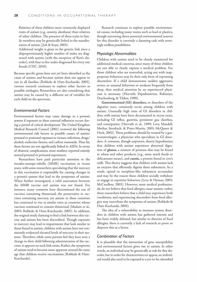
28 C O N D I T I O N S I N O C C U P A T I O N A L T H E R A P Y
■ Relatives of these children more commonly displayed traits of autism (e.g., anxiety, aloofness) than relatives of other children. The presence of these traits in fam-ily members may be genetically linked to the manifes-tation of autism ( Jick & Kaye, 2003).
■ Additional weight is given to the genetic link since a disproportionately higher number of males are diag-nosed with autism (with the exception of Rett’s dis-order), with four to five males diagnosed for every one female (CDC, 2010).
Because specific genes have not yet been identified as the cause of autism, and because autism does not appear to run in all families (Robledo & Ham-Kucharski, 2005), current research continues to explore other factors as possible etiologies. Researchers are also considering that autism may be caused by a different set of variables for each child on the spectrum.
Environmental FactorsEnvironmental factors may cause damage to a person’s system if exposure to these external influences occurs dur-ing a period of critical development The United Kingdom Medical Research Council (2001) reviewed the following environmental risk factors as possible causes of autism: prenatal or postnatal exposure to viruses, infections, drugs/alcohol, endocrine factors, and carbon monoxide. Thus far, these factors are not significantly linked to ASDs. In terms of obstetric complications, most mothers reported entirely normal prenatal or perinatal experiences.
Researchers have paid particular attention to the measles-mumps-rubella (MMR) vaccination in recent years, with some researchers speculating that the mercury in this vaccination is responsible for causing changes in a person’s system that lead to the symptoms of autism. When further investigated, a valid association between the MMR vaccine and autism was not found. For instance, many countries have discontinued the use of vaccines containing thimerosal, the preservative in vac-cines containing mercury, yet autism in these countries has continued to rise at similar rates as countries whose vaccines continued to contain thimerosal (Madsen et al., 2003; Robledo & Ham-Kutcharski, 2005). In addition, the original study claiming to find a link between this vac-cine and autism has been discredited. Though exposure to mercury may lead to impairments that look similar to those found in autism, children with autism have not con-sistently evidenced elevated levels of mercury in their sys-tems. Therefore, while some parents feel they have seen a change in their child following administration of the vac-cines, it appears no such link exists. Rather, the symptoms of autism tend to become more apparent around the same age that children receive vaccinations (Robledo & Ham-Kutcharski).
Research continues to explore possible environmen-tal causes, including many toxins such as lead or plastics, though narrowing down potential environmental sources for this disorder is currently a daunting task with seem-ingly endless possibilities.
Physiologic AbnormalitiesChildren with autism need to be closely monitored for additional medical concerns, since many of these children are not able to clearly express a medical problem. For those children who are nonverbal, acting out with inap-propriate behaviors may be their only form of expressing discomfort. If a child demonstrates sudden aggressive, severe, or unusual behaviors or awakens frequently from sleep, then medical attention by an experienced physi-cian is necessary (Horvath, Papadimitriou, Rabsztyn, Drachenberg, & Tildon, 1999).
Gastrointestinal (GI) disorders, or disorders of the digestive tract, commonly occur among children with autism. Unusually high rates of GI disorders in chil-dren with autism have been documented in recent years, including GI reflux, gastritis, persistent gas, diarrhea, and constipation (Horvath et al., 1999; Levy, Mandell, Merhar, Ittenbach, & Pinto-Martin, 2003; McQueen & Heck, 2002). These problems should be treated by a gas-troenterologist, a physician who specializes in GI disor-ders. A common, though unproven, theory hypothesizes that children with autism experience abnormal diges-tion of gluten, a mixture of proteins that may be found in wheat and other products (e.g., some snack foods or delicatessen meats), and casein, a protein found in cow’s milk. This theory suggests that children with autism lack an enzyme that efficiently digests these substances. As a result, opioid or morphine-like substances accumulate and may be the reason these children socially withdraw or engage in repetitive behaviors (Levy & Hyman, 2003; McCandless, 2003). However, most medical profession-als do not believe that food allergies cause autism; rather, these researchers believe that a child may experience both conditions, and experiencing discomfort from food aller-gies may exacerbate the symptoms of autism (Robledo & Ham-Kucharski, 2005).
The idea of a vulnerability to immune system disor-ders in children with autism has gathered interest and has been widely debated, but similar to theories of food allergies, there is currently a lack of research to prove or disprove this as a factor.
Combination of FactorsIt is plausible that the interaction of gene susceptibility and environmental factors gives rise to autism. In other words, an individual may be genetically at risk for this dis-order, but in order for characteristics to appear, an individ-ual would also need to be exposed to a yet-to-be-identified
29Chapter 3 : Aut i sm Spectr um Disorder s
environmental factor (United Kingdom Medical Research Council, 2001).
PREVALENCE AND INCIDENCEThe CDC (2010) estimates that 1 in 110 children in the United States has an ASD, diagnosed with autistic dis-order, Asperger’s syndrome, or PDD-NOS. CDD and Rett’s disorder are less common, with CDD occurring in 0.1 to 0.6 children per 10,000 and Rett’s disorder occur-ring in 1 in 10,000 to 1 in 15,000 children (United King-dom Medical Research Council, 2001; Webb & Latif, 2001). Prevalence information for the adult population on the autism spectrum is not known.
Prevalence of autism has been on the rise since the early 1990s. Though this increase has raised speculation of a possible epidemic, the rise in prevalence results, at least in part, from better identification and availability of services. In addition, changes in diagnostic criteria now allow more children to fit the diagnosis of autism (Fom-bonne, 1999; Jick & Kaye, 2003; Rapin, 2002).
Incidence, or the measure of new cases per year, is best studied in disorders that have a clear onset. Because the age of onset in autism is usually unclear, accurate incident rates are difficult to measure. The incidence of ASDs is likely to be within the range of 30 to 60 cases per 10,000, which represents a huge increase over the original estimate 40 years ago of 4 cases for every 10,000 individuals (Rutter, 2005).
Autism affects children across all racial, ethnic, and socioeconomic groups. It is four times more likely to occur in boys than in girls, with the exception of Rett’s disorder (CDC, 2010).
SIGNS AND SYMPTOMSEach ASD is related to the other disorders in social, language, and behavioral deviations, and each differs in terms of age of onset, severity of symptoms, and presence of other features such as cognitive impairment (Lord & McGee, 2001). However, these core behaviors do not nearly encompass the entire picture of these complex human beings. Motor abnormalities, sensory process-ing disorders, and co-occurring medical disorders (e.g., seizures, sleep disturbances, GI problems) are only some of the additional concerns that arise in this population. Every child with autism displays a separate matrix of strengths and challenges; even two children who fall into the same diagnostic category on the spectrum (e.g., Asperger’s syndrome) will exhibit individual differences resulting from personality and experiences (United King-dom Medical Research Council, 2001).
The first of the core symptoms, difficulty in social interaction, includes limited use of eye contact, facial
expressions, social gestures (e.g., pointing, waving, reach-ing arms up to be lifted by parent), and body postures. Additionally, children on the autism spectrum are usually challenged to accurately interpret the body language and facial expressions of other people. A child with ASD may not seek out others to share enjoyment, share interest in the same objects, or look for approval or reassurance from parents. Individuals may not frequently gaze shift; that is, sharing attention with another person by alternating gaze between an object and a person or following the gaze of another person. Maintaining continuous back-and-forth play or conversation with another person is often diffi-cult for those with autism (Greenspan & Wieder, 2006). Moreover, individuals experience challenges in develop-ing friendships with same-age peers. These obstacles range from showing a lack of interest in others to desir-ing friends but having difficulty understanding how to relate appropriately to peers. Problems with interaction may exist because children with autism are hypothesized to have limitations in theory of mind, which is the abil-ity to understand another person’s thoughts, feelings, or intentions. It is how an individual “reads” someone’s thoughts, understands another person’s perspective on an issue, and predicts another’s feelings (Leslie & Frith, 1987). Without this ability, a person is not able to predict or understand the actions of others (Strock, 2004). Other social disturbances that manifest in children with autism include limited or repetitive play routines and limited or absent pretend play skills. A child may experience intense emotional outbursts if confused, frustrated, anxious, or overstimulated by the environment (Strock).
Language skills for those with autism are marked by abnormal characteristics and delays. Typically devel-oping children progress through several communica-tion milestones in their first few years. During a child’s first year of life, a toddler gazes at others with interest, begins babbling, and reacts to sounds and voices. By 12 months, gestures and first words emerge, and a child shows recognition of his name and some familiar words and phrases. By 2 years of age, a child understands longer phrases and directions, has built a substantial vocabulary, and combines words into two- to three-word phrases (Strock, 2004). However, children with autism frequently show delays in learning to compre-hend language and to speak.
Comprehension of a word must occur in order to produce that word in a meaningful way. In addition, as children develop, they begin to understand not just words but expressions, gestures, and social contexts. Many indi-viduals with autism show challenges in comprehension, evidenced by
■ Echoing back a speaker’s words rather than showing understanding of the speaker
■ Difficulty responding to questions or comments

30 C O N D I T I O N S I N O C C U P A T I O N A L T H E R A P Y
■ Not responding to directions or requests to partici-pate in an activity
■ Not attending to things that others are pointed out in the environment
■ Not showing, looking at, or getting objects when asked
■ Showing confusion, anxiety, and “falling apart” emo-tionally (dysregulation)
■ Not engaging, relating, or communicating as we might expect (Gerber, 2009)
■ Struggling to understand social cues such as sarcasm, humor, and facial expressions (Stark, 2004)
In the area of language production, many children with autism are delayed in talking; some are unable to use words until their school-age years. In severe cases of autism, spoken language may not be acquired at all, with recent studies estimating that about 20% to 25% of those with autism remain nonverbal throughout their lives (Tager-Flusberg, Paul, & Lord, 2005). Social impairments further affect these children’s communica-tion because they may not be motivated to communicate with others, are typically challenged to use or understand social cues such as intonation and facial expressions to interpret meaning, and have difficulty maintaining the “give and take” of conversations (e.g., a child may discuss only his topic of interest without allowing others to take a turn).
For those children who develop spoken language, conversation may appear peculiar because of the pres-ence of echolalia, pronoun reversal, out of context words, and odd vocal intonation patterns. Echolalia is speech in which the child repeats what he has previ-ously heard, including intonation similar to the speak-er’s (Luyster, Kadlec, Carter, & Tager-Flusberg, 2008; Tager-Flusberg et al., 2005). Echolalia in not exclusive to autism, as most young children use echolalia as they are learning language. However, children with autism retain this characteristic and often apply it in abnormal ways. It likely results from poor auditory processing (a child’s ability to respond to sounds and comprehend verbal language) while auditory memory remains intact (Siegel, 1996). Echolalia may be immediate, meaning that it occurs just after another speaker’s utterance, or delayed, occurring a significant period of time after hear-ing the spoken words (e.g., reciting an entire children’s book from memory an hour or day later) (Siegel, 1996). Though echolalia may appear unusual in conversation, children with autism frequently use this inappropriate device in meaningful ways (e.g., a child may recite lines from his favorite movie as a self-calming strategy when he feels anxious or to help aid processing of what the child just heard). Because children with autism often fail to understand language, pronouns (e.g., “you” and
“me”) are also often used incorrectly. Other times, these individuals may use language that seems out of place for the context. For instance, a child may say a pet’s name while pushing a toy train. Unusual intonation patterns may include a monotone speech pattern, meaning that the usual melody of speech sounds “flat” or unemotional. Other children with autism may use a sing-song pattern in their speech. Poor volume control and higher fre-quency of whispering for children who echo have also been reported (Tager-Flusberg et al.).
In the area of repetitive behaviors, individuals with ASD may exhibit an abnormal or intense preoccupa-tion with routines or patterns. For instance, a child may line up blocks or toy cars for hours without playing with others or using these toys in pretend play. Rigidity in this type of play may occur; for instance, the child may scream or cry in reaction to someone joining his play or moving the lined-up toys. Obsessions with specific topics are common, such as showing an unusual inter-est in elevators or numbers and letters. Individuals with autism are often successful with routines; however, dis-ruption to that routine may be extremely disturbing to that person (Strock, 2004). Many people diagnosed with autism display abnormal, stereotyped behaviors such as repetitive hand-flapping, body rocking, or look-ing at objects/people out of the corner of their eyes. Another common feature of autism includes unusual, nonfunctional preoccupation with parts of an object, such as spinning the wheels of a toy car without showing interest in any other type of play with this toy (Ameri-can Psychiatric Association, 2000; Strock).
All children with autism demonstrate some combina-tion of these social, language, and behavioral symptoms. Additionally, specific characteristics are associated with each disorder on the autism spectrum and are discussed in the following sections.
Autistic Disorder
Autistic disorder is diagnosed when severe challenges occur in each area of social, language, and behavioral char-acteristics. Marked delays and deviations are noted in lan-guage development, which are the most common initial concerns of parents and pediatricians. Approximately half of children with autistic disorder remain nonverbal or struggle with severely impaired speech as adults (Rapin, 2002). Because both the comprehension and the use of language are affected, these individuals are additionally challenged to compensate for lack of verbal language with another mode of communication, such as mean-ingful social gestures (pointing or acting out what they want/need) or sign language (Lord & McGee, 2001). Even those who develop verbal language may struggle in
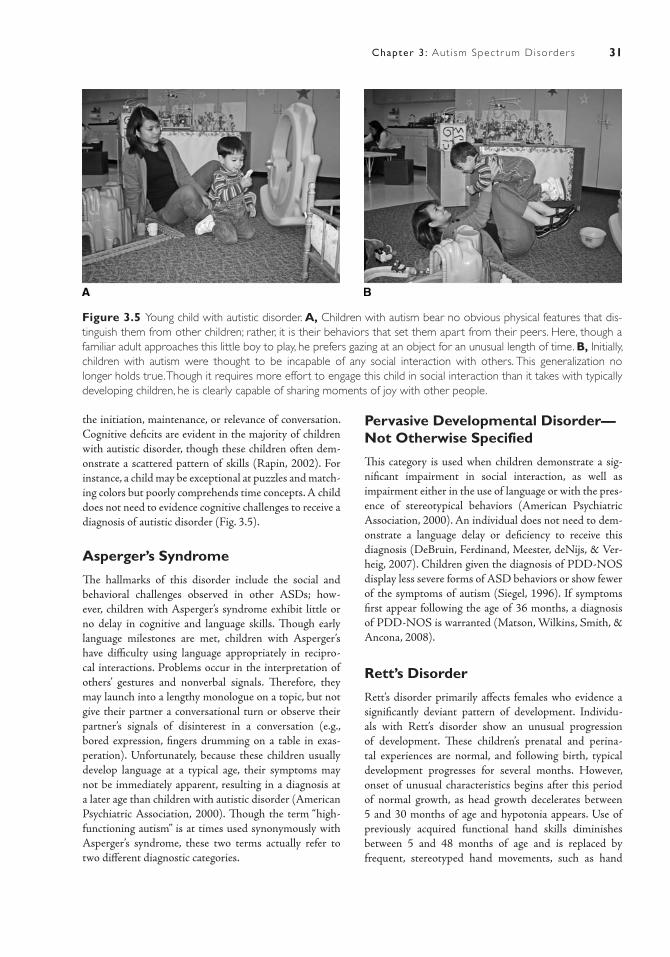
31Chapter 3 : Aut i sm Spectr um Disorder s
the initiation, maintenance, or relevance of conversation. Cognitive deficits are evident in the majority of children with autistic disorder, though these children often dem-onstrate a scattered pattern of skills (Rapin, 2002). For instance, a child may be exceptional at puzzles and match-ing colors but poorly comprehends time concepts. A child does not need to evidence cognitive challenges to receive a diagnosis of autistic disorder (Fig. 3.5).
Asperger’s Syndrome
The hallmarks of this disorder include the social and behavioral challenges observed in other ASDs; how-ever, children with Asperger’s syndrome exhibit little or no delay in cognitive and language skills. Though early language milestones are met, children with Asperger’s have difficulty using language appropriately in recipro-cal interactions. Problems occur in the interpretation of others’ gestures and nonverbal signals. Therefore, they may launch into a lengthy monologue on a topic, but not give their partner a conversational turn or observe their partner’s signals of disinterest in a conversation (e.g., bored expression, fingers drumming on a table in exas-peration). Unfortunately, because these children usually develop language at a typical age, their symptoms may not be immediately apparent, resulting in a diagnosis at a later age than children with autistic disorder (American Psychiatric Association, 2000). Though the term “high-functioning autism” is at times used synonymously with Asperger’s syndrome, these two terms actually refer to two different diagnostic categories.
Pervasive Developmental Disorder—Not Otherwise Specified
This category is used when children demonstrate a sig-nificant impairment in social interaction, as well as impairment either in the use of language or with the pres-ence of stereotypical behaviors (American Psychiatric Association, 2000). An individual does not need to dem-onstrate a language delay or deficiency to receive this diagnosis (DeBruin, Ferdinand, Meester, deNijs, & Ver-heig, 2007). Children given the diagnosis of PDD-NOS display less severe forms of ASD behaviors or show fewer of the symptoms of autism (Siegel, 1996). If symptoms first appear following the age of 36 months, a diagnosis of PDD-NOS is warranted (Matson, Wilkins, Smith, & Ancona, 2008).
Rett’s Disorder
Rett’s disorder primarily affects females who evidence a significantly deviant pattern of development. Individu-als with Rett’s disorder show an unusual progression of development. These children’s prenatal and perina-tal experiences are normal, and following birth, typical development progresses for several months. However, onset of unusual characteristics begins after this period of normal growth, as head growth decelerates between 5 and 30 months of age and hypotonia appears. Use of previously acquired functional hand skills diminishes between 5 and 48 months of age and is replaced by frequent, stereotyped hand movements, such as hand
Figure 3.5 Young child with autistic disorder. A, Children with autism bear no obvious physical features that dis-tinguish them from other children; rather, it is their behaviors that set them apart from their peers. Here, though a familiar adult approaches this little boy to play, he prefers gazing at an object for an unusual length of time. B, Initially, children with autism were thought to be incapable of any social interaction with others. This generalization no longer holds true. Though it requires more effort to engage this child in social interaction than it takes with typically developing children, he is clearly capable of sharing moments of joy with other people.
32 C O N D I T I O N S I N O C C U P A T I O N A L T H E R A P Y
wringing (American Psychiatric Association, 2000). At the same time, dementia, autistic features, and possible onset of seizures appear (Dunn & McLeod, 2001). Poor coordination may be noted in gait or trunk movements. The pattern of social development is quite unusual. These children lose their previous ability to socially engage with others; however, social interaction is often later reacquired in a child’s development. Expressive language, recep-tive language, and psychomotor skills are significantly impaired in this population (American Psychiatric Asso-ciation, 2000; Dunn & McLeod). Rett’s disorder stands alone as the only ASD with a known etiology caused by a single gene disorder (Webb & Latif, 2001).
Childhood Disintegrative Disorder
In this rare disorder, children with CDD develop normally for a minimum of 2 years following birth. Prior to 10 years
of age, a loss of skills is noted in at least two of the follow-ing areas of development: receptive/expressive language, social/adaptive functioning, bowel/bladder control, play abilities, and motor skills (American Psychiatric Asso-ciation, 2000). Skills disintegrate until the child exhibits features consistent with those in autism. However, before the loss of skills has occurred, children with CDD have usually acquired far more words, phrases, and even sen-tences than children with autistic disorder or PDD-NOS, meaning that the loss of language is more obvious in CDD than in autistic disorder (Siegel, 1996) (Table 3-1).
Co-Occurring Conditions
Autism has been associated with medical or genetic con-ditions in approximately 10% of its population. These conditions include, but are not limited to, Fragile X syn-drome, tuberous sclerosis, metabolic disorders, rubella
TABLE 3.1 The Autism Spectrum Disorders
Disorder Characteristics
Autistic disorder (classic autism)
Presence of ≥6 of 12 potential deficits involving all three behavioral domains that define the autistic spectrum
≥ 2 deficits in sociability, empathy, and insight into other person’s feelings and agendas
≥ 1 deficit in communicative language and imagination≥ 1 deficit in behavioral and cognitive flexibilityDetectable before the age of 3 yearsDiagnosis not excluded by the level of cognitive competence or
the existence of other handicaps
Asperger’s disorder Troublesome social ineptnessBehavioral inflexibility with a narrow range of interests IQ ≥70
(affected children may be normally intelligent or gifted)No delay in the emergence of speechOften demonstrates clumsiness
Pervasive developmental disorder—not otherwise specified
Applies to less severely affected children who do not meet criteria for either autistic disorder or Asperger’s disorder
Childhood disintegrative disorder
Early development entirely normal, including speechSevere regression between the ages of 2 and 10 years, affecting
language, sociability, cognition, and competence in skills of daily life
Rett’s disorder Severe global regression in infant girls (rarely in boys), resulting in lifelong severe mental retardation, lack of language and purposeful hand use, and other neurologic deficits
33Chapter 3 : Aut i sm Spectr um Disorder s
syndrome, and Haemophilus influenzae meningitis (Fom-bonne, 1999).
Seizure disorders are not specific to autism but occur more frequently than in the general population. An esti-mated one-third of children are reported to have expe-rienced at least two seizures before reaching adulthood. The probability of experiencing seizures is highest during adolescence and has been linked to causing or worsening cognitive and motor impairment (Rapin, 1997).
Diagnosis
Autism is now diagnosed at a younger age than ever before since characteristics of autism have become more defined and better recognized in recent years. Receiving a diagnosis at an early age is optimal because the sooner the disorder is recognized, the more likely the child can expe-rience dramatic gains in learning and reduction in symp-toms (Lord & McGee, 2001; Strock, 2004). In addition, parents are able to receive valuable support and education from professionals and other families.
Researchers have begun focusing on differences as early as the first year of life using families’ home movies. One study that examined children diagnosed with autism reviewed home videotapes from their first birthdays and revealed interaction and behavioral differences clearly at 12 months of age. Consistent signs among these children included lack of the following skills: pointing, showing objects to others, looking to others, and responding to their names (Osterling & Dawson, 1994). In other stud-ies that examined retrospective home videotapes, infants with autism also displayed hypotonia, poor visual atten-tion, mouthing objects with extreme frequency, aver-sion to social touch, decreased or lacking babbling, and absence of shifting their attention from people to objects (Maestro et al., 2002). Researchers are now wondering if these initial characteristics may show that not as many children as originally thought have a “sudden onset” of autism but rather show these subtle signs early in life, with more significant signs appearing after 12 months (Osterling & Dawson, 1994).
A child is usually referred for an assessment because those who interact closely with him (e.g., family mem-bers, pediatrician, teachers) may observe warning signs either specific to autism or to a development delay in one or more areas. When these concerns become appar-ent, the child is initially screened, usually by primary care providers or early child care professionals, to look for the “red flags” that may indicate autistic behaviors. Published screening instruments for children with autism include
■ The Checklist for Autism in Toddlers (Baron-Cohen, Allen, & Gillberg, 1992)
■ The Autism Screening Questionnaire (Berumet, Rut-ter, Lord, Pickles, & Bailey, 1999)
■ The Screening Tool for Autism in Two-Year-Olds (Stone, Coonrod, & Ousley, 2000)
■ Australian Scale for Asperger’s syndrome (Garnett & Attwood, 1998)
■ Pervasive Developmental Disorders Screening Test—Stage 1 (Siegel, 1998a)
■ The Modified Checklist for Autism in Toddlers (Rob-ins, Fein, Barton, & Green, 2001)
If a child does not pass the screening, enough warn-ing signs of autism are present to warrant a thorough assessment for autism. Since each child on the ASD displays a unique profile of strengths and weaknesses, no two children on the autism spectrum will look the same. Therefore, critical to an accurate diagnosis is an assessment with clinicians are experienced in identify-ing the characteristics of autism. Additionally, a thor-ough diagnosis with a team of professionals can gather insight into each child’s skills across several areas of development, which, in turn, helps with intervention planning. The following elements should be included in every sensitive, comprehensive evaluation of a child with autism:
1. History: Though autism is not known to result from complications during pregnancy, it is important to discuss any unusual pre- or perinatal events to rule out other disorders. Because of autism’s genetic implications, it is important to determine if other family members have been diagnosed with autism, psychiatric concerns, or developmental disorders. As noted previously, a child’s history of development and behaviors is key in diagnosing this disorder. Therefore, questioning about autism-specific behaviors, includ-ing changes over time, in the areas of social, language, behavioral, play, cognitive, and sensory processing abilities is critical (Rapin, 1997).
2. Medical history: During this portion of the assess-ment, parents report when their child’s developmen-tal milestones were reached (e.g., what age their child said his first word, learned to walk), if regression of developmental skills occurred at any point, or if any other medical problems are occurring (e.g., psy-chiatric, sleeping, or eating problems) (Filipek et al., 1999).
3. Physical/neurologic examination: Other illnesses such as Fragile X syndrome, tuberous sclerosis, fetal alcohol syndrome, or congenital rubella need to be ruled out, since these disorders may look similar to autism (Rapin, 1997). The physician also checks for other medical illnesses (e.g., GI disorders, ear infec-tions), measures head circumference, gives a general physical examination, examines mental status, verifies

34 C O N D I T I O N S I N O C C U P A T I O N A L T H E R A P Y
that cranial nerves function normally, and performs a motor examination (Filipek et al., 1999).
4. Parent interviewing: Many diagnostic tools are available to gain parents’ insight into their child’s autism-specific behaviors. A clinician should also ask parents about their overall impression of their child, since more general questions may reveal further insight beyond the scope of these tools (Filipek et al., 1999; Lord & McGee, 2001). Parent interviewing tools include
■ The Autism Diagnostic Interview: Revised (Lord, Rutter, & LeCouteur, 1994)
■ Functional Emotional Assessment Scale (Greenspan, DeGangi, & Wieder, 1999)
■ The Gilliam Autism Rating Scale (Gilliam, 1995) ■ The Pervasive Developmental Disorders Screen-
ing Test—Stage 2 (Siegel, 1998b)
5. Tests in developmental areas, including language, social, motor, cognitive, and adaptive skills.
6. Informal observation of the child interacting with oth-ers. This is considered one of the most valuable pieces of the evaluation process, since it typically reveals the qualitative impairments of the child (such as lack of eye contact, limited initiation of interaction, or diffi-culty transitioning between tasks).
7. Audiologic testing: An audiologic evaluation is nec-essary to rule out hearing disorders. Children with autism are often unresponsive to verbal auditory stim-uli, and it is important to determine that this behavior is not caused by a hearing impairment. If hearing loss co-occurs with autism, language comprehension may be further impacted than with a diagnosis of autism alone (Filipek et al., 1999).
8. Other testing: Certain tests may prove beneficial to specific circumstances. For instance, an electroen-cephalograph may be needed if the child is suspected of having seizures (Filipek et al.). Some children are assessed through university studies, and MRIs are typically included in these autism assessments (Bau-man, 2004; Filipek et al.). Genetics testing may be appropriate for parents who are considering having another child. Some parents are also concerned about heavy metal contamination and wish to pursue a lead screening for their child.
During the comprehensive assessment, a diagnostic instrument should be used that examines autistic behav-iors while the clinician observes the child’s interests and interactions with others.
The instruments that are currently available include
■ The Autism Diagnostic Observation Schedule—Generic (Lord, Rutter, Goode, Heemsbergen, & Jor-dan, 1989)
■ The Childhood Autism Rating Scale (Schopler, Reichler, & Renner, 1998)
COURSE AND PROGNOSISBecause each category in the autism spectrum presents with a range of abilities, the course of the disorder during one’s lifetime depends on the child’s diagnosis and indi-vidual differences. In addition, a diagnosis can change for an individual. For instance, a child with autistic disorder may show improvement in his symptoms, thereby display-ing characteristics more similar to PDD-NOS (Siegel, 1996). Most individuals with autism tracked by longitudi-nal studies have retained ASD symptoms to some degree throughout their lives (Lord & McGee, 2001).
Autistic Disorder
A child displays traits of autism before the age of 3 years, and these symptoms are generally most apparent follow-ing the child’s second birthday. In some cases, deviations in a child’s developmental and interaction skills are noted shortly following birth (e.g., hypersensitivities, limited eye contact, difficulty imitating facial expressions), while other infants appear to develop normally and seem to lose skills between 12 and 36 months of age (Tager-Flusberg et al., 2005). Though gains may be made in developmen-tal and social skills, these children are typically not able to catch up with their peers and remain autistic once the loss has occurred (Rapin, 1991). In some cases, behavio-ral challenges increase as a child reaches school age, while these challenges may decrease in other children. Longitu-dinal studies have found that few individuals with autistic disorder reach full independence as adults; many are not able to live on their own or find employment. Even those who evidence higher skills in this category (known as high-functioning autism) frequently struggle with social interactions throughout their lives. In fact, it is older chil-dren with higher-functioning autistic disorders who often make more attempts to socialize and may feel greater dis-tress and loneliness than younger or lower-functioning children with ASD when they are unsuccessful in recip-rocal interactions (Rapin, 1991). For the many individu-als who do not reach independence in adulthood, families must continue to provide support or these adults may enter residential programs. These programs offer special education, developmental, and behavioral techniques to teach new skills and generalize those skills into functional community activities (Holmes, 1990).
Asperger’s syndrome
A child with Asperger’s syndrome typically exhibits strong verbal skills in the school-age years; however, profession-als and peers should not let these skills be misleading in terms of the child’s social impairments. These children may show a strong interest in having friends but lack the

35Chapter 3 : Aut i sm Spectr um Disorder s
understanding of the social rules that apply to these inter-actions. As a result, these individuals may appear more comfortable with others either older or younger than themselves. Longitudinal studies suggest that prognosis is better for Asperger’s syndrome than autistic disorder because more individuals are able to function indepen-dently in employment and self-sufficient living (American Psychiatric Association, 2000).
Pervasive Developmental Disorder—Not Otherwise Specified
Children with PDD-NOS are typically diagnosed between the ages of 3 and 4 years old. Some individu-als demonstrate an intellectual disability, and those with lower IQ scores show more severely impaired social development (Matson et al., 2008). However, those children with higher developmental skills do not always walk an easier path. Increased awareness of their own differences from their peers can cause individuals with PDD-NOS to feel isolated, anxious, and/or depressed. During adolescence, children with PDD-NOS face confusing hormonal changes while trying to understand the intricate social subtleties their peers seem to have mastered. Both friendships and dating may be difficult for these individuals. A counselor with training in ASDs may help these adolescents feel supported and under-stood and can navigate teenagers through some of their social challenges. As an adult, if an individual with this disorder experiences only mild symptoms, he will likely be independent and able to live on his own. For some with PDD-NOS, marriage, parenting, and friendship may be too complex and overwhelming, but others have found success in these relationships despite the addi-tional challenges of the disorder (Autism Speaks, 2010).
Rett’s Disorder
Rett’s disorder is evident before a child reaches 4 years of age and most frequently presents in the first or second year of life following a period of typical development. Challenges in communication and behaviors are typi-cally continuous throughout the individual’s life. Gains in developmental skills are usually not significant in later childhood or adolescence, though the child may show a heightened desire to socially interact with others (Ameri-can Psychiatric Association, 2000).
Childhood Disintegrative Disorder
Symptoms appear in this disorder following the age of 2 years but prior to the age of 10 years, and children pro-gress through typical developmental milestones before
the onset of this disorder. Following the onset, a signifi-cant loss of skills is noted. If this disorder occurs in isola-tion, the loss of skills ceases and minimal improvement is observed later in life. This disorder may co-occur with a progressive neurologic condition, in which case, the child will continue to lose skills (American Psychiatric Associa-tion, 2000).
General Prognostic Indicators
Overall, children’s language levels and IQ scores are the strongest indicators related to prognosis in independence and appropriate functioning in adulthood, particularly in the area of verbal IQ (Lord & McGee, 2001).
In terms of communication skills, a child’s ability to spontaneously, meaningfully, and consistently combine words into phrases or sentences before 5 years of age is a good prognostic indicator of cognitive, language, adaptive, and academic achievement measures (Lord & McGee, 2001). It is important to observe that the child is able to spontaneously construct sentences rather than echo others’ utterances (echolalia) or use memorized chunks of language to communicate. A child’s use of joint attention has been found to be a predictor of language outcome. Joint attention is the ability to share experi-ences with others through social communication, includ-ing eye contact, gesture, and verbal language. Children who fail to use early gestural joint attention (e.g., failing to point at an object and to turn to his mother to deter-mine if his mother shares his interest) seem to struggle in the development of meaningful language. In Lord and McGee’s review of autism research (2001), one longitu-dinal research finding implied that early joint attention, symbolic play, and receptive language were strong predic-tors of a child’s future outcomes. Another study in this review examined severity of repetitive, stereotyped behav-iors and social symptoms and found that the severity later predicted adaptive functioning.
Early success in hand-eye coordination may predict vocational abilities later in life. Fine motor skills also pre-dicted later leisure pursuits. Children with autism who dis-played a definite hand preference performed significantly better on motor, language, and cognitive tasks (Hauck & Dewey, 2001). The ability to imitate body movements has been linked to expressive language development, and imitation of actions with objects predicted later levels of play abilities. The more established behavioral challenges become without intervention, the more these problems persist and worsen (Lord & McGee, 2001).
Medical/Surgical Management
Overall, treatment for autism does not heavily rely on medical intervention, and surgical interventions are not
36 C O N D I T I O N S I N O C C U P A T I O N A L T H E R A P Y
practiced for this disorder. Intensive early intervention is most effective in reducing problem behaviors while increasing language, social, sensory, motor, and cogni-tive skills. A variety of approaches are available, ranging from highly structured to naturalistic. Because no two children with autism present with the same set of symp-toms, no one treatment plan is successful for all children with autism. Effective intervention must account for the child’s individual strengths and challenges and must con-sider functional skills to be generalized across a variety of settings in the child’s life. Substantial literature supports early intervention for children younger than 3 years old as the most beneficial time to connect new pathways for more appropriate functioning and behavior, though an individual may continue to make substantial gains fol-lowing this period of development (Lord & McGee, 2001; Rapin, 1997; Rogers, 1996). Because it is beyond the scope of this book to discuss these approaches, this chapter focuses on current knowledge of medical inter-ventions meant to accompany intervention techniques.
Pharmacologic TherapiesUse of medication does not cure the core social, language, and repetitive behavioral deficits of autism because “in most cases, the brain has undergone atypical cellular development dating from the earliest embryonic stages” (Rapin, 2002, p. 303). Much of the research in this area is presently limited or inconclusive. In addition, many parents and clinicians are cautious because use of phar-macotherapy with children presents the risk of harmful side effects. Lindsay and Aman (2003) reviewed existing literature on pharmacologic intervention and found that when certain medications are used appropriately, behav-ioral dysregulations such as hyperactivity, irritability, anxiety, and perseveration may be reduced. For instance, risperidone, an atypical antipsychotic agent, shows prom-ising results in emerging research for reducing tantrums, irritability, aggression, and self-injurious and repeti-tive behaviors (Gordon, 2002; Lindsay & Aman, 2003; Pediatric Psychopharmacology Autism Network, 2002). Because autism is likely a genetic disorder determined before the child is born, pharmacologic treatments could probably not “undo” this disorder. Therefore, these treat-ments are not intended to replace educational services but rather to supplement them. For those children who respond successfully to medication, behavioral and edu-cational intervention may be even more beneficial since they do not struggle as greatly with challenging behaviors.
Medical ConditionsMedical intervention is necessary for any co-occurring medical conditions, such as seizures or GI disorders. Children with autism need to be monitored closely
for behaviors that may reflect a medical condition and should receive a complete medical workup and treatment through a physician who specializes in the child’s medical condition.
Complementary and Alternative MedicineComplementary and alternative medicine has gained popularity in the past decade as a supplementary treat-ment to educational services. Complementary and alter-native medicine is defined as “a broad domain of healing resources that encompasses all health systems modali-ties and practices and their accompanying theories and beliefs, other than those intrinsic to the politically domi-nant health system” (Panel of Definition and Descrip-tion, 2002, as cited in Levy et al., 2003, p. 418). Those who support complementary and alternative medicine believe the methods target underlying medical diffi-culties, such as GI and sleep disorders, which are not addressed through educational intervention. The goal of many of the complementary and alternative medi-cine treatments is to aid in the associated problems of autism, rather than claim to cure the disorder. Statistics reveal that 30% to 50% of children with autism in the United States are using complementary and alternative medicine; however, approximately 9% are using poten-tially harmful treatments and 11% are using multiple complementary and alternative medicine treatments (Levy et al., 2003). Several studies are currently under way to examine the effectiveness and the risks of these treatments since many parents have reported a decrease in associated problems (e.g., GI problems) and an increase in developmental skills. Table 3-2 summarizes the potentially harmful side effects of these methods. The following sections describe commonly used com-plementary and alternative medicine treatments.
SupplementsThe use of vitamins and minerals to address ASD con-cerns purports that because children with autism expe-rience GI inflammation and intestinal disorders, their ability to absorb nutrients is thereby reduced. As a result, development that relies on nutrients such as vitamins A, B1, B3, B5; biotin; selenium; zinc; and magnesium is altered. Frequently used supplements include vitamin C, cod liver oil (for vitamins A and D), and the combina-tion of vitamin B6 with magnesium (Autism Society of America, 2004).
Gluten-Free, Casein-Free DietParents and professionals who support the theory that gluten and casein negatively impact a child’s development recommend a diet that completely eliminates these prod-ucts (McCandless, 2003). Parent reports of improvement
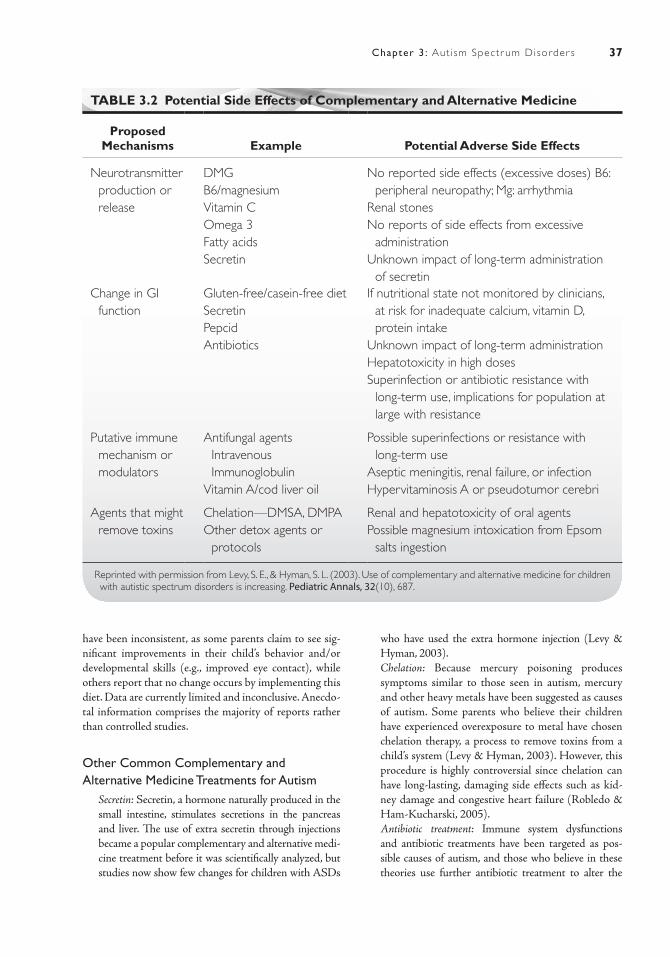
37Chapter 3 : Aut i sm Spectr um Disorder s
TABLE 3.2 Potential Side Effects of Complementary and Alternative Medicine
Proposed Mechanisms Example Potential Adverse Side Effects
Neurotransmitter production or release
DMGB6/magnesiumVitamin COmega 3Fatty acidsSecretin
No reported side effects (excessive doses) B6: peripheral neuropathy; Mg: arrhythmia
Renal stonesNo reports of side effects from excessive
administrationUnknown impact of long-term administration
of secretinChange in GI
functionGluten-free/casein-free dietSecretinPepcidAntibiotics
If nutritional state not monitored by clinicians, at risk for inadequate calcium, vitamin D, protein intake
Unknown impact of long-term administrationHepatotoxicity in high dosesSuperinfection or antibiotic resistance with
long-term use, implications for population at large with resistance
Putative immune mechanism or modulators
Antifungal agents Intravenous Immunoglobulin
Vitamin A/cod liver oil
Possible superinfections or resistance with long-term use
Aseptic meningitis, renal failure, or infectionHypervitaminosis A or pseudotumor cerebri
Agents that might remove toxins
Chelation—DMSA, DMPAOther detox agents or
protocols
Renal and hepatotoxicity of oral agentsPossible magnesium intoxication from Epsom
salts ingestion
Reprinted with permission from Levy, S. E., & Hyman, S. L. (2003). Use of complementary and alternative medicine for children with autistic spectrum disorders is increasing. Pediatric Annals, 32(10), 687.
have been inconsistent, as some parents claim to see sig-nificant improvements in their child’s behavior and/or developmental skills (e.g., improved eye contact), while others report that no change occurs by implementing this diet. Data are currently limited and inconclusive. Anecdo-tal information comprises the majority of reports rather than controlled studies.
Other Common Complementary and Alternative Medicine Treatments for Autism ■ Secretin: Secretin, a hormone naturally produced in the
small intestine, stimulates secretions in the pancreas and liver. The use of extra secretin through injections became a popular complementary and alternative medi-cine treatment before it was scientifically analyzed, but studies now show few changes for children with ASDs
who have used the extra hormone injection (Levy & Hyman, 2003).
■ Chelation: Because mercury poisoning produces symptoms similar to those seen in autism, mercury and other heavy metals have been suggested as causes of autism. Some parents who believe their children have experienced overexposure to metal have chosen chelation therapy, a process to remove toxins from a child’s system (Levy & Hyman, 2003). However, this procedure is highly controversial since chelation can have long-lasting, damaging side effects such as kid-ney damage and congestive heart failure (Robledo & Ham-Kucharski, 2005).
■ Antibiotic treatment: Immune system dysfunctions and antibiotic treatments have been targeted as pos-sible causes of autism, and those who believe in these theories use further antibiotic treatment to alter the
38 C O N D I T I O N S I N O C C U P A T I O N A L T H E R A P Y
course of the symptoms in autism (Levy & Hyman, 2003).
■ Antifungal treatment: Yeast overgrowth in the colon is hypothesized to cause many medical disorders, including autism, with a low-sugar diet and the use of probiotic agents (which encourage helpful intestinal bacteria) used as treatments (Levy & Hyman, 2003).
Conclusion of Complementary and Alternative Medicine TreatmentsThe effectiveness of complementary and alternative medi-cine stands largely unproven and highly controversial. Attempts are under way for further studies since many parents report positive results by using these methods. Complementary and alternative medicine treatments are intended to supplement educational services rather than to replace them. Clinicians are currently encouraged to use an empathetic stance, with families providing com-plementary and alternative medicine to their children, though no alternative method should be administered without the guidance of an experienced physician.
IMPACT OF CONDITIONS ON CLIENT FACTORS
Global Impairment
Autism is considered a global impairment, meaning that it does not reflect damage as a result of one specific lesion in the brain. Since autism likely affects several regions of the brain, this global impact means that autism impairs multiple areas of a child’s development. As a result, even a child’s ability to regulate basic functions is impaired, such as consistent sleep patterns or emotional stability.
Irregular sleeping patterns are common in children with autism, particularly in the quality rather than the quantity of their sleep. Disturbances include problems falling asleep, waking throughout the night, disoriented awakening, difficulty sleeping in an unfamiliar place, and nightmare-type disturbances such as screaming. Chil-dren may exhibit sleep disturbances as early as 2 years of age, and higher occurrence of sleep disorders seems to exist in children with more severe forms of autism (Hering, Epstein, Elroy, Iancu, & Zelnic, 1999; Hoshino, Watanabe, Yashima, Kaneko, & Kumashiro, 1984; Schreck & Mulick, 2000).
Children with autism often have difficulty regulat-ing their own emotions and interpreting the emotions of others. These children often experience depression, a sense of loneliness, and a limited range of emotions that results in a flattened affect. For example, they may show they are happy or angry, but more subtle emotions such as worry, jealousy, or disappointment are less commonly
seen in these children. Additionally, difficulty in read-ing facial expressions, body postures, and intonation of voices to infer another’s feelings are common challenges. An unusual characteristic of autism is emotional labil-ity, which is a child’s use of laughing or crying for unclear reasons or during inappropriate situations. Some parents may describe their children as “laid back” or less reactive to emotional stimuli, while other parents note a tendency to frequently cry inconsolably. Heightened anxiety is evident in many children, as are temper tantrums that often result from disrupted routines (Rapin, 1991). Other children show a lack of fear during dangerous situations (Lord & McGee, 2001). Aggressive behaviors such as hitting, bit-ing, self-injury (head banging, hitting self ), or self-stimu-latory behaviors (e.g., body rocking) may occur when the child does not get his way or his routine is disturbed.
Specific Mental Functions
Each child with autism manifests the symptoms in differ-ent ways; therefore, children with an ASD show various combinations of concerns in their mental skills. Attention deficits, however, are nearly universal in this population. Unique patterns of attention include distractibility, dis-organization, intense preoccupation for preferred, self-initiated activities for unusual lengths of time, and lack of boredom for repeating same action or play schema (Rapin, 1991). The ability to focus jointly on an activity with another person is significantly impaired.
A child with autism often shows a scattered pattern of memory functions (Hill, Berthoz, & Frith, 2004). Overall, “memory performance of individuals with autism becomes increasingly impaired as the complexity of the material increases” (Minshew & Goldstein, 2001, p. 1099). Children with autism also use fewer organiza-tional strategies, relying on stereotyped rules regardless of the task’s complexity. Therefore, a child with autism per-forms more poorly on tasks with higher complexity. Word recall is more significantly impacted than digit recall, and these children often have difficulty recalling activities in which they have recently participated (Boucher, 1981). However, certain areas of memory remain intact, particu-larly in the areas of visual and rote memory. Rote mem-ory describes the memorization and use of previously heard chunks of language rather than the spontaneous generation of language.
In the area of perceptual functioning, individuals with ASDs perceive sensory stimuli but often process and react abnormally to it. Integration of perceptual and sensori-motor information allows individuals to respond appro-priately with physical and emotional responses. In autism, this integration does not occur in the same efficient way; therefore, this population of individuals has difficulty responding with typical emotional and physical responses
39Chapter 3 : Aut i sm Spectr um Disorder s
to the sensory stimuli around them. Between 30% and 100% of individuals with autism demonstrate deviant sensory-perceptual abilities (Dawson & Watling, 2000).
Cognitive impairments are common in, though not universal to, the autism population. Approximately 75% of these individuals have IQ scores below 70 (American Psychiatric Association, 2000), indicating intellectual disability. Those with Asperger’s syndrome, however, show IQ scores within the normal range. Individuals with cognitive deficits typically demonstrate scattered skills; in other words, they may present with strong skills in some areas with significant concerns in other areas. These children often have difficulty sequencing a series of items, imitating the actions and words of others meaning-fully, generalizing concepts across a variety of situations, demonstrating theory of mind, and playing with toys appropriately and symbolically (Lord & McGee, 2001). In her 1991 literature review of autistic features, Rapin described that children with autism tend to have better visual-spatial skills than auditory verbal skills on IQ tests. Children in this population may show above-average skills in very specific areas, such as calculating numbers, completing puzzles, or demonstrating rote verbal mem-ory, while demonstrating overall cognitive impairment. Some children with autism are able to read at a young age with minimal instruction, but they have little or no understanding of what they read. This unusual occur-rence is known as hyperlexia.
Studies examining higher level cognitive functions have revealed that executive functioning skills such as forward planning, cognitive flexibility, and the use of assistive strate-gies (e.g., creating a mnemonic such as a rhyme to assist in remembering information) in learning are impacted for those who have autism (Gordon, 2002).
As previously described, children with autism typi-cally show language delay, with the exception of those with Asperger’s syndrome. Deviations from typical lan-guage are noted across all diagnostic categories. Children on the autism spectrum typically have limitations in using language in appropriate contexts and for social purposes.
Sensory Functions and Pain
“The experience of being human is imbedded in the sensory events of everyday life. When we observe how people live their lives, we discover they characterize their experiences from a sensory point of view” (Dunn, 2001, p. 608). If a child’s sensory system does not interpret stimuli in a typical way, it is easy to understand why this individual may react to the world differently. These sensory disturbances appear to occur more commonly in children than adults, and since the severity of sensory impairment seems to be related to the severity of stereotypic and behavioral abnormalities, sensory processing problems should be addressed through
early intervention to reduce these abnormal behaviors (Dawson & Watling, 2000; Lord & McGee, 2001).
The prevalence of sensory impairments in children with autism is significantly higher than in the population of typically developing children, though each child with autism shows a different profile of hyper- and hyposen-sitivity to sensory stimulation. Despite the variability in sensory thresholds from child to child, some general pat-terns have emerged in research.
Visual perception is usually an area of relative strength and may be used to compensate for challenging areas. For instance, the integration of vestibular, visual, and soma-tosensory afferent systems is needed to maintain upright postural stability. Molloy, Dietrich, and Bhattacharya (2003) measured the postural stability in children with autism and found that these children relied on visual cues to help them maintain stability. When these visual cues were omitted, children had difficulty maintaining their upright balance and reducing their sway. In addition, children may use their visual processing strengths to help learn routines through the use of a picture schedule, or they may communicate using picture symbols if verbal language is challenging (Prizant, Wetherby, Rubin, Lau-rent, & Rydell, 2006). One area of visual processing that is consistently impaired is integrating details of a figure into a whole (Deruelle, Rondan, Gepner, & Tardif, 2004). For instance, if given a line drawing of a house made up of geometric shapes, these children focus on the shapes rather than seeing the image of a house. Children with autism often twirl themselves or spin objects obsessively while staring intently at the object; they may be fascinated with the visual effects these actions create (Hewetson, 2002). They may seek out areas of visual interest, such as lines on the floor of a gymnasium. Some show hypersen-sitivities to a busy environment or bright light.
Auditory processing is often a significantly challeng-ing area for children with autism. Frequently, children with ASDs are unresponsive to some auditory stimuli while being oversensitive to others. For instance, a child may not respond when her name is called but screams in response to hearing a vacuum cleaner running. Further-more, children with autism may be distracted or irritable or have difficulty functioning in activities in noisy envi-ronments (Kientz & Dunn, 1997).
These children also show mixed patterns of craving and/or avoiding vestibular sensations. Some children appear to seek out, others withdraw from, and some show mixed preferences for certain types of these sensations. Common vestibular movements include swinging, spin-ning on a sit-and-spin or merry-go-round, and playing with spinning toys (Kientz & Dunn, 1997).
Food tastes and textures are difficult for many chil-dren on the autism spectrum. Many parents note their child with autism has unusual taste preferences. Due to
40 C O N D I T I O N S I N O C C U P A T I O N A L T H E R A P Y
sensory issues, these individuals are frequently “picky eat-ers” and may have a limited repertoire of food preferences (Williams, Dalrymple, & Neal, 2000). They may be sensi-tive to certain textures, such as avoiding wet (e.g., oranges in fruit syrup), “goopy” (e.g., macaroni and cheese), and/or mixed textured foods (e.g., a casserole). Though nutritional intake generally appears adequate (United Kingdom Medical Research Council, 2001), it is often stressful for families to meet nutritional needs of these children who have very specific food preferences. Parents and clinicians typically observe these individuals to crave strong tastes, such as spicy or salty foods, and crunchy foods that give the child significant sensory input.
As with taste, many individuals with autism have abnormal smell interests. Unusual behaviors are observed in the person’s inappropriate smelling of objects or unu-sual attachment to or aversion of certain smells (Rapin, 1991; Williams et al., 2000).
Reports of clumsiness are common for children who are higher functioning on the autism spectrum (e.g., chil-dren with milder cognitive impairment, such as those with Asperger’s syndrome) (Ghaziuddin & Butler, 1998). Another study suggested that in children with Asperger’s, this observed clumsiness may result from a deficit in proprioception, not from a motor disturbance (Weimer, Schatz, Lincoln, Ballantyne, & Trauner, 2001).
Tactile defensiveness is a hypersensitivity to certain touch situations that most people find nonthreaten-ing. Children with this symptom frequently exhibit an avoidance-withdrawal response (e.g., rubbing/scratching, avoiding certain textures, or crying when exposed to a spe-cific texture) (Baranek & Berkson, 1994). The presence of tactile defensiveness appears to co-occur with stereotyped behaviors such as rocking of the body, hand flapping, and unusual interest or focus on objects (Baranek, Foster, & Berkson, 1997). A significant relationship also appears between tactile defensiveness and rigid behaviors (e.g., child has difficulty breaking out of own agenda), repetitive verbalizations, visual stereotypes, and abnormally focused affections. Though these children experience significant tactile hypersensitivities, many also appear to have a reduced response to temperature or pain (Rapin, 1991).
Functions Related to the Digestive System
Children with autism may be at a higher risk for expe-riencing GI problems such as reflux or gastritis, with persistent gas, diarrhea, and constipation also fre-quently reported (Horvath et al., 1999; Levy et al., 2003; McQueen & Heck, 2002). Additionally, hypotheses exist regarding functioning of the digestive system, such as increased intestinal permeability that allows absorption of morphine-like compounds from gluten and casein.
The buildup of these substances theoretically results in the social withdrawal and stereotypical behaviors seen in autism, but as previously described, this theory is contro-versial and has not been proven (Levy & Hyman, 2003).
Urinary and Reproductive Functions
In children with autism, the urinary tract is usually typical in its structure and function. However, for those children with cognitive and sensory impairments, toilet training is often complicated. Children with autism often learn to toilet train at a later age than typically developing children; display problems such as fear, pain, confusion, frustration, and constipation when learning to train; uri-nate or defecate in inappropriate places; and experience difficulty when a change of routine occurs or when enter-ing an unfamiliar bathroom (Dalrymple & Ruble, 1992).
Because individuals with autism have difficulty inter-acting, inappropriate sexual behavior is also a concern. Close relationships with others are often challenging for these individuals; therefore, fewer person-oriented behaviors may be noted. Additionally, discouraging inap-propriate sexual behaviors may be difficult. The most fre-quently reported inappropriate sexual behaviors include public masturbation or public touching of one’s own pri-vate parts (Ruble & Dalrymple, 1993; Van Bourgondien, Reichle, & Palmer, 1997).
Gross and Fine Motor Skills
Though individuals with ASDs often show relative strengths in movement functions, some abnormalities in motor skills have been observed, including overall motor joint laxity, hypotonia, clumsiness, apraxia, and chronic toe-walking (Rapin, 1997). Persistent toe-walking may be from hypersensitivity to certain walking surfaces (e.g., grass, sand) or to provide children with additional pro-prioceptive feedback as their muscles contract (Robledo & Ham-Kucharski, 2005). Females with Rett’s disor-der frequently demonstrate poor coordination in gait or trunk movements. In children with Asperger’s syndrome, “clumsiness” has been more specifically defined as poor performance on tests of apraxia, tandem gait, one-leg bal-ance with eyes closed, and repetitive finger-thumb apposi-tion (Weimer et al., 2001).
Further fine and gross motor impairments include problems in skilled movement, hand-eye coordina-tion, speed, praxis and imitation, posture, and balance (Dawson & Watling, 2000). A particularly debilitating abnormality in individuals with autism includes deficits in motor imitation skills despite intact perceptual and motor capacities, which is most apparent in younger groups of children (Williams, Whiten, & Singh, 2004).
41Chapter 3 : Aut i sm Spectr um Disorder s
Furthermore, these children perform poorly on tasks of executing a sequence of movements, such as a sequence of hand or facial actions in imitation (Hughes, 1996). Reduced stride lengths, increased stance times, increased hip flexion at toe-off, reduced knee extension at initial ground contact, abnormal heel strikes, and decreased knee extension and ankle dorsiflexion at ground contact were noted in an earlier study of children with autism (Vilensky, Damasio, & Maurer, 1981).
In addition, a disproportionate number of children with autism display ambiguous hand preference (∼40%) long past the age that dominant hand preference typi-cally develops. A person who is ambidextrous will usu-ally choose one hand for a specific task (e.g., left hand for writing, right hand for throwing a football). An ambigu-ous hand preference, however, refers to switching hands within the same activity (Hauck & Dewey, 2001). When this behavior persists into the school-age years, it may indicate abnormal functioning of the brain.
Motor stereotypes are common in those on the autism spectrum and often manifest through hand flapping,
pacing, spinning, running in circles, and flipping light switches. More severe, self-injurious forms include biting, hitting oneself, or head banging (Rapin, 1997).
Brain Structure
The most significant abnormalities in a child’s body struc-ture include the anomalies of the brain that are currently under investigation. Likely caused by genetic abnormali-ties, these deviations in the brains of individuals with autism are not yet clearly defined, nor is it clear precisely how the differences cause the characteristics of autism. Studies have found consistent abnormalities in head growth, the cerebellum, frontal lobes, and limbic system (Bauman, 2004; Bauman & Kemper, 1996; Carper & Courchesne, 2005; Helm- Estabrooks & Albert, 1991; Kemper & Bauman, 1998; Piven et al., 1997; United Kingdom Medical Research Council, 2001). Research is also examining potential deviations in the way different regions of the brain communicate (Piven et al.).
Case IllustrationChild with AutismAt 1 year of age, Jacob was brought to an early inter-vention clinic because of delays in motor and cognitive skills. An initial evaluation conducted by an occupa-tional therapist, educator, and speech-language pathol-ogist confirmed these concerns. In the area of gross motor skills, Jacob was delayed in learning to walk and demonstrated moderate hypotonia in his trunk. Fine motor concerns included tactile defensiveness of wet or sticky substances and delays in grasping and manipulating objects appropriately. Cognitively, Jacob showed little interest in playing with toys or imitat-ing the words and actions of others. Language impair-ments were not yet observed since Jacob frequently vocalized, babbled, and expressed his feelings through behaviors such as smiling or crying. At this young age, ASD was not initially suspected. Jacob was clearly a delightful little boy who showed a strong attachment to his parents and was interested in watching children and other adults in his environment.
Jacob was placed in a playgroup at the clinic where his occupational therapist and educator worked directly with him, while a speech-language patholo-gist monitored his language. As time passed, further concerns became evident. His language skills failed to further develop; as a result, first words did not emerge. Nor was he using gestures to indicate what he wanted. At the age of 14 months, he evidenced
limitations in language comprehension and speech production, and his use of eye contact decreased. As the months progressed, he showed frustration more frequently through crying, banging his head, throwing himself on the ground, and arching his back to pull away from a person trying to hold him. His feeding skills were limited since he was not able to bring his hand to his mouth, and he demonstrated extreme sensitivities to many tastes and textures. He did not show an interest in other self-help skills such as learn-ing to bathe or dress himself. While other children his age learned to imitate motor movements in songs, Jacob seemed to content to only listen to the songs. He did not demonstrate typical play skills for his age such as exploring how toys worked, taking toys in or out of a container, or taking turns with others. How-ever, musical or flashing toys captured his attention for long periods of time. While he was engrossed with these toys, he evidenced unusual, repetitive behaviors such as rocking his body back and forth.
Through early intervention by his therapists and parents, Jacob’s gross motor skills improved during the following months, and he successfully learned to walk at 18 months. He delighted in walking through his home, through the early intervention center, and outside. His eye contact improved, becoming more spontaneous and consistent. His repertoire of sounds increased, and he vocalized to take a turn in songs or
42 C O N D I T I O N S I N O C C U P A T I O N A L T H E R A P Y
games; in addition, he began signing “more” to request something desirable to happen again. He learned to play with toys in a more functional manner, including stacking rings and blocks and taking toys in and out of containers. Yet other skills continued to be challenging. When he was left to play on his own, he repetitively turned the pages of books or walked aimlessly around a room. He showed hypersensitivities to touching or mouthing certain textures, limited motor or imitation skills, difficulty understanding others, a lack of verbal words, and repetitive behaviors during play.
Currently, at slightly older than 2 years of age, Jacob continues to struggle in several areas of devel-opment. However, he has made steady progress in these developmental skills, and his family and team are encouraged that he will continue to make signifi-cant gains. More importantly, despite being faced with more challenges than typically developing children, Jacob is a young boy who is often able to share and express joy with others.
Adult With AutismPatrick is a 38-year-old man who was diagnosed with autistic disorder at 2½ years of age. His mother was first concerned he had a hearing impairment, as he did not seem to understand what people said to him, did not respond to a fire truck siren, placed his ear close to the refrigerator, and acted out with negative behaviors. Behavioral and developmental concerns persisted after testing ruled out a hearing impairment. A psychologist specializing in autism assessed Patrick’s developmental skills and behaviors and diagnosed Pat-rick with autistic disorder.
Patrick communicated nonverbally for several years, usually by grabbing a person’s hand and leading the per-son to an item he wanted. He had a very limited diet as a young child, eating only peanut butter, raisins, and yogurt. In time, he increased his repertoire of foods. When Patrick was 3, he began attending a local school for children with autism; at the age of 8, he attended a school for children with a variety of special needs. Patrick began using words at the age of 9. He trans-ferred to a local public high school at the age of 18, participating in the school’s autism program, and work-ing in the school’s kitchen to learn to work with others. He graduated high school when he was 26, receiving a diploma of completion for the school’s autism program.
Following high school, Patrick moved into a con-dominium with two other men diagnosed with autism; he currently maintains this living situation. Pat-rick and his roommates receive 24-hour supervision. He also works 5 days per week at a therapy center that encourages functional skill-building. He works
alongside eight or nine other adults with autism and receives supervision in his daily tasks, which include shredding legal documents and inserting newspapers into plastic sleeves. To prevent overwhelming stimuli, the room is kept dim and without extraneous sound.
As with all individuals diagnosed with autism, Pat-rick shows personal strengths and unique challenges, with his own personality shining through.
Patrick has achieved several goals in activities of daily living and continues to work toward indepen-dence. He dresses himself, with the occasional need for help with fasteners. He toilets independently, though requires assistance with wiping after a bowel move-ment. He washes himself with verbal prompting from a supervisor and climbs in and out of the tub indepen-dently. He applies deodorant and cologne and combs his hair when directed. Once a supervisor has prepared his toothbrush, Patrick brushes his teeth for 2 minutes, needing occasional reminders to not swallow the toothpaste. He carries groceries into the house, takes out the trash, and wipes the table after meals.
Patrick displays unusual and stereotypical behaviors, such as rapidly bouncing or tapping a small ball on a tabletop, spinning lids, and repeatedly blowing up bal-loons. He eats his food very quickly and needs verbal reminders to eat at a slower pace. In the car, he often bends down to play with the hardware beneath his seat. Recently, Patrick has shown negative behaviors, such as destruction of property or soiling himself. His family and staff believe these behaviors are likely Patrick’s way of showing grief from his father’s death last year.
Socially, Patrick gets along well with his family, supervisors, and roommates, though he has few verbal interactions with his roommates. His family frequently brings him to community outings such as sports events; Patrick shows interest and enjoyment during these outings. He reciprocates another person’s smile easily. He occasionally initiates and maintains eye con-tact with another person, but not with typical dura-tion or frequency. He often responds to a speaker, but does not engage in back-and-forth conversation.
Regarding other developmental skills, Patrick cur-rently understands more language than he uses. He follows familiar one-step directions, though some-times needs repetition from the speaker to com-plete the request. He comprehends simple questions that relate to his interests and daily routines. Patrick communicates through single words and phrases. He names familiar people and objects, answers simple questions, and requests desired items and actions. He greets when prompted by another person. In the area of fine motor skills, Patrick does not demon-strate a hand preference. He writes his first name, last
43Chapter 3 : Aut i sm Spectr um Disorder s
name, and the word “love” to sign letters and cards. Patrick shows many strengths in his gross motor skills. He enjoys playing a variety of sports, including swimming and throwing a ball back and forth with a partner. He loves to shoot basketball hoops (drib-bling is more difficult for him), which was a favorite activity he had shared with his father. He experiences some difficulty with coordination and motor plan-ning. Cognitively, Patrick has recently learned the
days of the week and loves to look at calendars. He can recite the alphabet and recognizes a few familiar written words. He does not yet tell time.
Patrick lives a very active life, filled with family, sup-portive staff, and opportunities to contribute to his community. His mother and professional team contin-uously work together to create new goals for Patrick, and he meets these challenges. He is a delightful and admirable individual.
R E C O M M E N D E D L E A R N I N G R E S O U R C E S
OrganizationsAutism Society of America7910 Woodmont Ave., Suite 300Bethesda, MD 20814-3067www.autism-society.org/site/PageServerAutism Speakshttp://www.autismspeaks.orgWeb site includes a “100 day kit” for families
whose children have recently been diagnosed with autism.
Centers for Disease Control and Preventionhttp://www.cdc.gov/ncbddd/autism/index.htmlWeb site includes fact sheets and other helpful informa-
tion for families, individuals with autism, healthcare providers, etc.
First Signshttp://www.firstsigns.org/Web site includes valuable information on autism and
an ASD video glossaryNational Alliance of Autism Research99 Wall Street, Research ParkPrinceton, NJ 08540www.naar.orgNational Institute of Mental Health Office of Commu-
nications6001 Executive BlvdRoom 8184, MSC 9663Bethesda, MD 20892-9663www.nimh.nih.gov/publicat/autism.cfm
Books on Autism Spectrum DisordersAttwood, T. (1998). Asperger’s syndrome: A guide for par-
ents and professionals. London, England: Jessica King-sley.
Greenspan, S. I., & Wieder, S. (2006). Engaging autism: Using the Floortime approach to help children relate,
communicate, and think. Cambridge, MA: Da Capo Press.
Lord, C., & McGee, J. P. (Eds.). (2001). Educating children with autism. Washington, DC: National Academy Press.
Ozonoff, S., Dawson, G., & McPartland, J. (2002). A par-ent’s guide to Asperger syndrome and high functioning autism. New York, NY: Guilford Press.
Quill, K. A. (2000). Do-Watch-Listen-Say: Social and communication intervention for children with autism. Baltimore, MD: Paul H. Brookes Publishing.
Robledo, S. J., & Ham-Kucharski, D. (2005). The autism book: Answers to your most pressing questions. New York, NY: Penguin Group.
Siegel, B. (2003). Helping children with autism learn: Treat-ment approaches for parents and professionals. New York, NY: Oxford University Press.
Volkmar, F. R. (Ed.). (2005). Handbook of autism and per-vasive developmental disorders (pp. 335–364). Hobo-ken, NJ: John Wiley & Sons, Inc.
Wing, L. (1996). The autistic spectrum: A guide for par-ents and professionals. London, England: Constable & Company Limited.
Books on Sensory Integration DisorderAyres, A. J. (1998). Sensory integration and the child. Los
Angeles, CA: Western Psychological Services.Kranowitz, C. S. (1998). The out-of-sync child: Recognizing
and coping with sensory integration dysfunction. New York, NY: Perigree.
Diagnostic Guidelines OnlineFilipek, P. (2006). Autism diagnostic guidelines. Screening
and Diagnosis of Autism. Retrieved from http://www.neurology.org/cgi/reprint/55/4/468.pdf

44 C O N D I T I O N S I N O C C U P A T I O N A L T H E R A P Y
American Psychiatric Association. (2000). Diagnostic and statistic manual of mental disorders (4th ed., text revi-sion). Washington, DC: Author.
Autism Society of America. (2004). Autism info [Fact sheets]. Retrieved April 11, 2004, from http://www.autism-society.org
Autism Speaks. (2010). PDD-NOS. Retrieved from http://www.autismspeaks.org/navigating/pdd_nos.php
Baranek, G. T., & Berkson, G. (1994). Tactile defen-siveness in children with developmental disabilities: Responsiveness and habituation. Journal of Autism and Developmental Disorders, 24(4), 457–471.
Baranek, G. T., Foster, L. G., & Berkson, G. (1997). Tac-tile defensiveness and stereotyped behaviors. Ameri-can Journal of Occupational Therapy, 51(2), 91–95.
Baron-Cohen, S., Allen, J., & Gillberg, C. (1992). Can autism be detected at 18 months? The needle, the hay-stack, and the CHAT. The British Journal of Psychiatry, 161, 839–843.
Bauman, M., & Kemper, T. (1996). Neuroanotomic ob-servations of the brain in autism: a review and future directions. International Journal of Developmental Neu-roscience, 23(2–3), 183–187.
*Bauman, M. Innovative interventions in autism/non-verbal learning disabilities. Conference presentation, December 3–4, 2004, Seattle, WA.
Berument, S. K., Rutter, M., Lord, C., Pickles, A., & Bailey, A. (1999). Autism screening questionnaire: Diagnostic validity. The British Journal of Psychiatry, 175, 444–451.
Bettelheim, B. (1967). The empty fortress: Infantile autism and the birth of the self. New York, NY: Free Press.
Boucher, J. (1981). Memory for recent events in autis-tic children. Journal of Autism and Developmental Disorders, 11, 293–302.
Carper, R. A., & Courchesne, E. (2005). Localized enlargement of the frontal cortex in early autism. Biological Psychiatry, 57(2), 126–133.
Centers for Disease Control and Prevention. (2010). Autism spectrum disorders (ASDs). Retrieved from http://www.cdc.gov/ncbddd/autism/index.html
Dalrymple, N. J., & Ruble, L. (1992). Toilet training and behaviors of people with autism: Parent views. Journal of Autism and Developmental Disorders, 22(2), 265–275.
Dawson, G., & Watling, R. (2000). Interventions to fa-cilitate auditory, visual, and motor integration in autism: A review of the evidence. Journal of Autism and Developmental Disorders, 30(5), 415–421.
DeBruin, E. I., Ferdinand, R. F., Meester, S., deNijs, P. F. A., & Verheig, F. (2007). High rates of psychiatric co-morbidity in PDD-NOS. Journal of Autism and Developmental Disorders, 37(5), 877–886.
Deruelle, C., Rondan, C., Gepner, B., & Tardif, C. (2004). Spatial frequency and face processing in children with autism and Asperger syndrome. Journal of Autism and Developmental Disorders, 34(2), 199–210.
Dunn, H. G., & MacLeod, P. M. (2001). Rett syndrome: Review of biological abnormalities. Canadian Journal of Neurological Sciences, 28(1), 16–29.
Dunn, W. (2001). The sensations of everyday life: Empirical, theoretical, and pragmatic considerations. American Journal of Occupational Therapy, 55(6), 608–620.
Filipek, P. A., Accardo, P. J., Baranek, G. T., Cook, E. C., Jr., Dawson, G., Gordon, G., et al. (1999). The screen-ing and diagnosis of autistic spectrum disorders. Journal of Autism and Developmental Disorders, 29(6), 439–484.
Fombonne, E. (1999). The epidemiology of autism: A review. Psychological Medicine, 29:769–786.
Garnett, M. S., & Attwood, A. J. (1998). Australian scale for Asperger’s syndrome. In: T. Attwood (Ed.) Asperger’s syndrome: A guide for parents and professionals. London, England: Jessica Kingsley.
Gerber, S. (2009). Understanding the development and derailment of comprehension: The impact on engag-ing, relating, and communicating. Proceedings from Interdisciplinary Council on Developmental and Learn-ing Disorders 2009. Autism spectrum disorders: What works and why? Bethesda, MD.
Ghaziuddin, M., & Butler, E. (1998). Clumsiness in autism and Asperger syndrome: A further report. Journal of Intellectual Disability Research, 42, 43–48.
Gilliam, G. E. (1995). Gilliam autism rating scale. Austin, TX: Pro-Ed.
Gordon, B. (2002). Autism and autistic spectrum disor-ders. In A. K. Asbury, G. M. McKhann, W. I. McDon-ald, and P. Dyck. (Eds.). Diseases of the nervous system (3rd ed) (Vol. I, pp. 406–418). Cambridge, England: Cambridge University Press.
Greenspan, S. I., DeGangi, G, & Wieder, S. (1999). The functional emotional assessment scale for infancy and early childhood: A manual. Madison, WI: Internation-al Universities Press.
Greenspan, S. I., & Wieder, S. (2006). Engaging autism: Using the Floortime approach to help children relate, communicate, and think. Cambridge, MA: Da Capo Press.
Hauck, J. A., & Dewey, D. (2001). Hand preference and motor functioning in children with autism. Journal of Autism and Developmental Disorders, 31(3), 265–277.
Helm-Estabrooks, N., & Albert, M. L. (1991). Manual of aphasia therapy. Austin, TX: Pro-Ed.
R E F E R E N C E S
45Chapter 3 : Aut i sm Spectr um Disorder s
Hering, E., Epstein, R., Elroy, S., Iancu, D. R., & Zelnic, N. (1999). Sleep patterns in autistic children. Jour-nal of Autism and Developmental Disorders, 29(2), 143–147.
Hewetson, A. (2002). The stolen child: Aspects of autism and Asperger’s syndrome. Westport, CT: Bergin & Garvey.
Hill, E., Berthoz, S., & Frith, U. (2004). Cognitive pro-cessing of own emotions in individuals with autistic spectrum disorder and in their relatives. Journal of Autism and Developmental Disorders, 34(2), 229–235.
Holmes, D. L. (1990). Community-based services for children and adults with autism: The Eden fam-ily programs. Journal of Autism and Developmental Disorders, 20, 339–351.
Horvath, K., Papadimitriou, J. C., Rabsztyn, A., Drachenberg, C., & Tildon, J. (1999). Gastrointesti-nal abnormalities in children with autistic disorder. Journal of Pediatrics, 135(5), 559–563.
Hoshino, Y., Watanabe, H., Yashima, Y., Kaneko, M., & Kumashiro, H. (1984). An investigation on sleep disturbance of autistic children. Folia Psychiatrica et Neurologica Japonica, 38(1), 45–51.
Hughes, C. (1996). Brief report: Planning problems in autism at the level of motor control. Journal of Autism and Developmental Disorders, 26(1), 99–107.
Jick, H., & Kaye, J. A. (2003). Epidemiology and pos-sible causes of autism. Pharmacotherapy, 23(12), 1525–1530.
Kanner, L. (1943). Autistic disturbances of affective con-tact. Nervous Child, 2, 17–250.
Kemper, T. L., & Bauman, M. (1998). Neuropathol-ogy of infantile autism. Journal of Neuropathology & Experimental Neurology, 57(7), 645–652.
Kientz, M. A., & Dunn, W. (1997). A comparison of the performance of children with and without autism on the sensory profile. American Journal of Occupational Therapy, 51(7), 530–537.
Leslie, A. M., & Frith, U. (1987). Metarepresentation and autism: How not to lose one’s marbles. Cognition, 27(3), 291–294.
Levy, S. E., & Hyman, S. L. (2003). Use of complemen-tary and alternative medicine for children with autis-tic spectrum disorders is increasing. Pediatric Annals, 32(10), 685–691.
Levy, S. E., Mandell, D. S., Merhar, S., Ittenbach, R. F., & Pinto-Martin, J. A. (2003). Use of complementary and alternative medicine among children recently diagnosed autistic spectrum disorder. Journal of Developmental & Behavioral Pediatrics, 24(6), 418–423.
Levy, S. E., Mandell, D. S., & Schultz, R. T. (2009). Autism. Lancet, 372, 1627–1638.
Lindsay, R. L., & Aman, M. G. (2003). Pharmacologic therapies aid treatment for autism. Pediatric Annals, 32(10), 671–676.
Lord, C., & McGee, J. P. (Eds.). (2001). Educating children with autism. Washington, DC: National Academy Press.
Lord, C., Rutter, M., Goode, S., Heemsbergen, J., & Jordan, H. (1989). Autism diagnostic observation schedule: A standardized observation of commu-nicative and social behavior. Journal of Autism and Developmental Disorders, 19, 185–212.
Lord, C., Rutter, M., & LeCouteur, A. (1994). Autism diagnostic interview-revised: A revised version of a diagnostic interview for caregivers of individuals with possible pervasive developmental disorders. Journal of Autism and Developmental Disorders, 24, 659–685.
Luyster, R. J., Kadlec, M. B., Carter, A., & Tager-Flusberg, H. (2008). Language assessment and development in toddlers with autism spectrum disorders. Journal of Autism and Developmental Disorders, 38, 1426–1438.
Madsen, K. M., Lauritsen, M. B., Pedersen, C. B., Thorsen, P., Plesner, A., Andersen, P. H., & Mortensen, P. B. (2003). Thimerosal and the occurrence of autism: Negative ecological evidence from Danish popula-tion-based data. Pediatrics, 112(3), 604–606.
Maestro, S., Muratori, F., Cavallaro, M. C., Pei, F., Stern, D., Golse, B., et al. (2002). Attentional skills during the first 6 months ofage in autism spectrum disor-der. Journal of the American Academy of Child and Adolescent Psychiatry, 41, 1239–1245.
Marcel, J. Reliably lower functional connectivity for au-tism participants between pairs of key areas during sentence comprehension. Slide courtesy of Dr. Marcel Just, Carnegie Mellon University.
Matson, J. L.,Wilkins, J., Smith, K., & Ancona, M. (2008). PDD-NOS symptoms in adults with intellectual dis-ability: Toward an empirically oriented diagnostic model. Journal of Autism and Developmental Disorders, 38(3), 530–537.
McCandless, J. (2003). Children with starving brains: A medical treatment guide for autism spectrum disorder (2nd ed.). Paterson, NJ: Bramble Books.
McQueen, J. M., & Heck, A. M. (2002). Secretin for the treatment of autism. The Annals of Pharmacotherapy, 36, 305–311.
Minshew, N. (2009). The details are in the connections: Deciphering heterogeneity in ASD. Proceedings from Interdisciplinary Council on Developmental and Learning Disorders 2009: Autism spectrum disorders: What works and why? Bethesda, MD.
Minshew, N. J., & Goldstein, G. (2001). The pattern of intact and impaired memory functions in autism. The Journal of Child Psychology and Psychiatry, 42(8), 1095–1101.
Molloy, C. A., Dietrich, K., & Bhattacharya, A. (2003). Postural stability in children with autism spec-trum disorder. Journal of Autism and Developmental Disorders, 33(6), 643–652.
46 C O N D I T I O N S I N O C C U P A T I O N A L T H E R A P Y
Osterling, J., & Dawson, G. (1994). Early recognition of children with autism: A study of first birthday home videotapes. Journal of Autism and Developmental Disorders, 24(3), 247–257.
Panel of Definition and Description. Defining and describing complementary and alternative medicine. Paper presented at CAM Research Methodology con-ference, April 7–9, Washington, DC.
Pediatric Psychopharmacology Autism Network. (2002). Risperidone in children with autism and serious be-havioral problems. New England Journal of Medicine, 347(5), 314–321.
Piven, J., Saliba, K., Bailey, J., & Arndt, S. (1997). An MRI study of autism: The cerebellum revisited. Neurology, 49(2), 546–551.
Prizant, B. M., Wetherby, A. M., Rubin, E., Laurent, A. C., & Rydell, P. J. (2006). The SCERTS model: A comprehensive educational approach for children with autism spectrum dis-orders. Baltimore, MD: Paul H. Brookes Publishing Co.
Rapin, I. (1991). Autistic children: Diagnosis and clinical features. Pediatrics, 87, 751–760.
Rapin, I. (1997). Autism. New England Journal of Medicine, 337, 97–104.
Rapin, I. (2002). The autistic spectrum disorders. New England Journal of Medicine, 347(5), 302–303.
Rapin, I., & Katzman, R. (1998). Neurobiology of autism. Annals of Neurology, 43, 7–14.
Robins, D. L., Fein, D., Barton M. L., & Green, J. A. (2001). The modified checklist for autism in toddlers: An initial study investigating the early detection of au-tism and pervasive developmental disorders. Journal of Autism and Developmental Disorders, 21, 131–144.
Robledo, S. J., & Ham-Kucharski, D. (2005). The au-tism book: Answers to your most pressing questions. New York, NY: Penguin Group.
Rogers, S. J. (1996). Brief report: Early intervention in au-tism. Journal of Autism and Developmental Disorders, 26(2), 243–246.
Ruble, L. A., & Dalrymple, N. J. (1993). Social/sexual awareness of persons with autism: A parental per-spective. The Archives of Sexual Behavior, 22, 229–240.
Rutter, M. (2005). Incidence of autism spectrum dis-orders: Changes over time and their meaning. Acta Paediatrica, 94(1), 2–15.
Rutter, M., Silberg, J., O’Connor, T., & Simonoff, E. (1999). Genetics and child psychiatry: II Empirical research findings. The Journal of Child Psychology and Psychiatry, 40(1), 19–55.
Schopler, E., Reichler, R. J., & Renner, B. R. (1998). The childhood autism rating scale. Los Angeles: Western Psychological Services.
Schreck, K. A., & Mulick, J. (2000). Parental report of sleep problems in children with autism. Journal of Autism and Developmental Disorders, 30(2), 127–135.
Siegel, B. (1996). The world of the autistic child. Oxford, England: Oxford University Press.
Siegel, B. (1998a). Early screening and diagnosis in autism spectrum disorders: The Pervasive Developmental Disor-ders Screening Test (PDDST). Paper presented at the NIH State of the Science in Autism Screening and Diagnosis Working Conference.
Siegel, B. (1998b). Pervasive developmental disorders screening test—Stage 2. Bethesda, MD.
Siegel, B. (2003). Helping children with autism to learn. Oxford, England: Oxford University Press.
Stone, W. L., Coonrod, E. E., & Ousley, O. Y. (2000). Brief report: Screening tool for autism in two-year-olds (STAT): Development and preliminary data. Journal of Autism and Developmental Disorders, 30, 607–612.
Strock, M. (2004). Autism spectrum disorders (pervasive developmental disorders). Retrieved from http://eric.ed.gov/
Tager-Flusberg, H., Paul, R., & Lord, C. (2005). Language and communication in autism. In F. R. Volkmar (Ed.), Handbook of autism and pervasive developmental dis-orders (pp. 335–364). Hoboken, NJ: John Wiley & Sons, Inc.
United Kingdom Medical Research Council. (December, 2001). MRC review of autism research: Epidemiology and causes. Retrieved July 25, 2004, from http://www.mrc.ac.uk/pdf- autism-report.pdf
Van Bourgondien, M. E., Reichle, N. C., & Palmer, A. (1997). Sexual behavior in adults with autism. Journal of Autism and Developmental Disorders, 27(2), 113–125.
Veenstra-VanDerWeele, J., Cook, E., & Lombroso, P. J. (2003). Genetics of childhood disorders: XLVI. Autism, Part 5: Genetics of autism. Journal of the American Academy of Child and Adolescent Psychiatry, 42(2), 116–118.
Vilensky, J. A., Damasio, A. R., Maurer, R. G. (1981). Gait disturbances in patients with autistic behavior: A preliminary study. Archives of Neurology, 38(10), 646–649.
Webb, T., & Latif, F. (2001). Rett syndrome and the MECP2 gene. Journal of Medical Genetics, 38, 217–223.
Weimer, A. K., Schatz, A. M., Lincoln, A., Ballantyne, A. O., & Trauner, D. A. (2001). “Motor” impairment in Asperger syndrome: Evidence for a deficit in proprioception. Journal of Developmental Be-havioral Pediatrics, 22(2), 92–101.
Williams, J. H. G., Whiten, A., & Singh, T. (2004). A sys-tematic review of action imitation in autistic spec-trum disorder. Journal of Autism and Developmental Disorders, 34(3), 285–299.
Williams, P. G., Dalrymple, N., & Neal, J. (2000). Eat-ing habits of children with autism. Pediatric Nursing, 26(3), 259–264.
47
Intellectual Disability
■ Mi c h e l l e Su are z ■ B e n At c h i s o n
4
Jeffrey, aged 3 years, was brought to Early On at the local school district for a developmental evaluation. His parents report that his pediatri-cian is concerned about his language development. He smiled and
interacted with examiners but was unable to express his wants and needs or follow single step directions. An interview with his parents revealed that Jeffrey was unable to dress or undress and was not self-feeding. He was diagnosed with an intellectual disability and enrolled in early childhood special education where he is working toward language and self-care goals. His family hopes he will eventually attend mainstream kindergarten with special education support.
Michael, now 2, was diagnosed by his pediatrician shortly after his birth as having Down syndrome. He exhibits the physical characteristics such as a round face, flattened nose bridge, abnormally small head, low-set ears, short limbs, and abnormally shaped fingers. His motor development is delayed, and his low muscle tone has made learning to walk difficult. Intel-lectual disability in children with Down syndrome is inevitable but varies in the degree of severity. Michael’s early intervention program provides services for Michael as well as parent support and education.
Kelly, now in second grade, is pleasant and likable. Teachers have been concerned about her inability to write letters or sound out words since kindergarten, and as the curriculum becomes more challenging, she is falling further behind. She has trouble maintaining friendships and often struggles to join peers in play. School staff conducted multidisciplinary team evaluation and diagnosed Kelly with an intellectual disability. She was placed in a special education classroom where the material is appropriate for her cognitive ability and learning pace.
K E Y T E R M S
Cortical atrophyCraniostenosis
CytomegalovirusDown syndrome
Fragile X syndromeHydrocephaly
HyperphenylalaninemiaHypoxia
Muscular dystrophyPICA
Spina bifidaTay-Sachs disease
TeratogenicToxemia
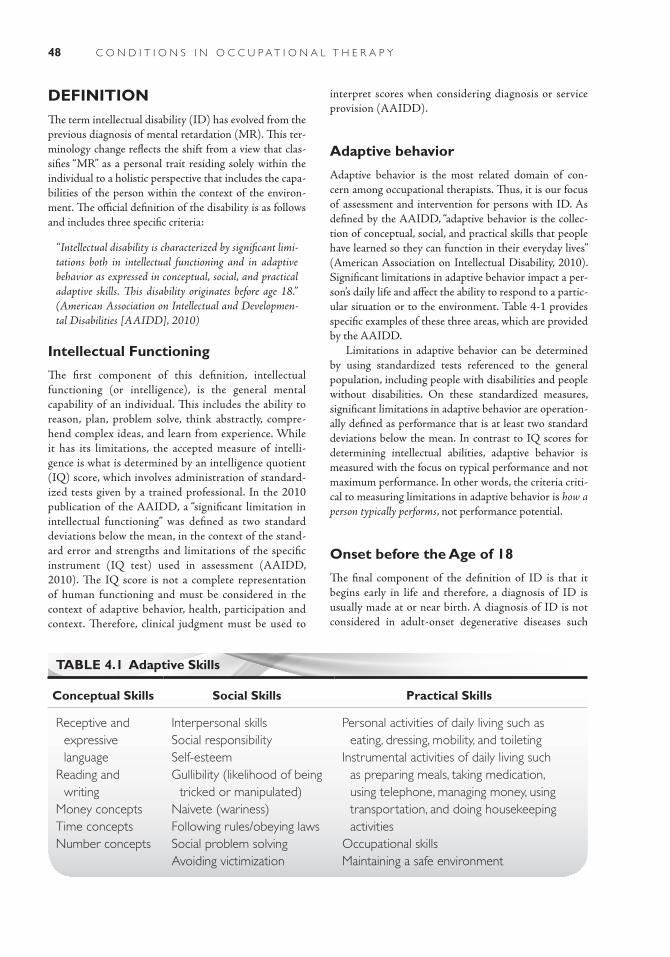
48 C O N D I T I O N S I N O C C U P A T I O N A L T H E R A P Y
DEFINITIONThe term intellectual disability (ID) has evolved from the previous diagnosis of mental retardation (MR). This ter-minology change reflects the shift from a view that clas-sifies “MR” as a personal trait residing solely within the individual to a holistic perspective that includes the capa-bilities of the person within the context of the environ-ment. The official definition of the disability is as follows and includes three specific criteria:
“Intellectual disability is characterized by significant limi-tations both in intellectual functioning and in adaptive behavior as expressed in conceptual, social, and practical adaptive skills. This disability originates before age 18.” (American Association on Intellectual and Developmen-tal Disabilities [AAIDD], 2010)
Intellectual Functioning
The first component of this definition, intellectual functioning (or intelligence), is the general mental capability of an individual. This includes the ability to reason, plan, problem solve, think abstractly, compre-hend complex ideas, and learn from experience. While it has its limitations, the accepted measure of intelli-gence is what is determined by an intelligence quotient (IQ) score, which involves administration of standard-ized tests given by a trained professional. In the 2010 publication of the AAIDD, a “significant limitation in intellectual functioning” was defined as two standard deviations below the mean, in the context of the stand-ard error and strengths and limitations of the specific instrument (IQ test) used in assessment (AAIDD, 2010). The IQ score is not a complete representation of human functioning and must be considered in the context of adaptive behavior, health, participation and context. Therefore, clinical judgment must be used to
interpret scores when considering diagnosis or service provision (AAIDD).
Adaptive behavior
Adaptive behavior is the most related domain of con-cern among occupational therapists. Thus, it is our focus of assessment and intervention for persons with ID. As defined by the AAIDD, “adaptive behavior is the collec-tion of conceptual, social, and practical skills that people have learned so they can function in their everyday lives” (American Association on Intellectual Disability, 2010). Significant limitations in adaptive behavior impact a per-son’s daily life and affect the ability to respond to a partic-ular situation or to the environment. Table 4-1 provides specific examples of these three areas, which are provided by the AAIDD.
Limitations in adaptive behavior can be determined by using standardized tests referenced to the general population, including people with disabilities and people without disabilities. On these standardized measures, significant limitations in adaptive behavior are operation-ally defined as performance that is at least two standard deviations below the mean. In contrast to IQ scores for determining intellectual abilities, adaptive behavior is measured with the focus on typical performance and not maximum performance. In other words, the criteria criti-cal to measuring limitations in adaptive behavior is how a person typically performs, not performance potential.
Onset before the Age of 18
The final component of the definition of ID is that it begins early in life and therefore, a diagnosis of ID is usually made at or near birth. A diagnosis of ID is not considered in adult-onset degenerative diseases such
TABLE 4.1 Adaptive Skills
Conceptual Skills Social Skills Practical Skills
Receptive and expressive language
Reading and writing
Money conceptsTime conceptsNumber concepts
Interpersonal skillsSocial responsibilitySelf-esteemGullibility (likelihood of being
tricked or manipulated)Naivete (wariness)Following rules/obeying lawsSocial problem solvingAvoiding victimization
Personal activities of daily living such as eating, dressing, mobility, and toileting
Instrumental activities of daily living such as preparing meals, taking medication, using telephone, managing money, using transportation, and doing housekeeping activities
Occupational skillsMaintaining a safe environment
49Chapter 4 : Inte l lectua l Disab i l i ty
as dementia or those associated with traumatic brain injury.
In considering this definition, the AAIDD notes that there are five assumptions that need to be considered in the diagnostic process (American Association on Intellectual Disability, 2010):
1. Limitations in present functioning must be consid-ered within the context of community environments typical of the individual’s age peers and culture.
2. Valid assessment considers cultural and linguistic diversity as well as differences in communication, sen-sory, motor, and behavioral factors.
3. Within an individual, limitations often coexist with strengths.
4. An important purpose of describing limitations is to develop a profile of needed supports.
5. With appropriate personalized supports over a sus-tained period, the life functioning of the person with ID generally will improve.
ETIOLOGYThe causes of ID may be classified according to when they occurred in the developmental cycle (prenatally, perinatally, or postnatally) or by their origin (biomedi-cal vs. environmental) (American Association on Mental Retardation, 1992; Yeargin-Allsopp, Murphy, Cordero, Decouflé, & Hollowell, 1997). There are hundreds of causes of ID (The Arc, 1993). Despite knowing the many factors that contribute to ID, in a large propor-tion of cases the cause remains unknown. The ability to determine the cause is highly correlated with the level of the retardation. The etiology of ID is much less likely to be known with individuals who are mildly intellectually disabled (IQs of 50–70) than with those who are severely affected (IQs of <50) (American Association on Mental Retardation, 2002; Matilainen, Airaksinen, & Monomen, 1995; Yeargin- Allsopp et al., 1997).
In a large U.S. population–based study describing probable causes of ID in school-aged children, the follow-ing results were obtained (Matilainen et al., 1995):
■ No defined cause, 78.0% ■ Prenatal conditions, 12.4% ■ Genetic, 7.1% ■ Perinatal conditions, 5.9% ■ Intrauterine/intrapartum, 5.2% ■ Postneonatal events, 3.6% ■ Teratogenic, 2.9% ■ Central nervous system (CNS) birth defects, 1.5% ■ Other birth defects, 0.8% ■ Neonatal, 0.7%
Prenatal factors that can cause ID include genetic aber-rations, birth defects that are not genetic in origin,
environmental influences, or a combination of factors (Matilainen et al., 1995). Up to 50% of the individuals diagnosed with ID may have more than one causal factor (American Association on Mental Retardation, 2002). With genetic aberrations, the problem is with either the genes, which are the basic unit of heredity, or the chro-mosomes, which carry the genes. Each nongerm cell (cells other than the ovum and spermatozoa) contains 23 pairs of chromosomes, including one pair of sex chromosomes that determine the sex of the person. Males have an X and a Y chromosome, and females have two X chromo-somes.
In many cases of ID, the gene or chromosome that has caused the condition can be identified specifically. In fact, more than 350 inborn errors of metabolism that result from genetic changes have been identified. Many of these metabolic errors lead to ID (Scriver, 1995). The two most common genetic causes of ID are Down syndrome and fragile X syndrome (The Arc, 1996). Down syndrome is generally caused by an extra 21st chromosome, and fragile X syndrome is the result of a mutation at what is known as the fragile site on the X chromosome (Kaplan, Sadock, & Grebb, 1994). In other cases, the specific genetic aber-ration has not been identified. Factors such as higher incidences of a condition in specific families or increased recurrence rates among siblings suggest that the defect is genetic (Matilainen et al., 1995).
Birth defects that are not considered genetic in origin also can contribute to or cause ID. These could include such things as malformation of parts of the CNS (corti-cal atrophy, hydrocephaly, spina bifida, craniostenosis) (Yeargin-Allsopp et al., 1997), congenital cardiac anoma-lies (Rogers et al., 1995), or metabolic disorders not asso-ciated with a genetic defect (hypothyroidism) (Reuss, Paneth, Pinto-Martin, Lorenz, 1996).
Environmental factors may also be involved in pre-natal development of ID. They may include exposure to chemical agents, such as alcohol or nonprescription drugs ingested by the mother during the pregnancy; maternal conditions such as hyperphenylalaninemia ( Jardim, Palma-Dias, Silva, Ashston-Prolla, & Guigliani, 1996), toxemia, hypertension, and diabetes; or congenital infec-tions such as cytomegalovirus, rubella, and syphilis (Yeargin-Allsopp et al., 1997).
Genetic Causes
Genetic causes can be divided into two types: single gene dis-orders and chromosomal abnormalities. In single gene disor-ders, there is a problem with the quality of the genetic material; a specific gene is defective. In chromosomal abnormalities, the problem is with the quantity of the material. There is either too much or too little genetic material in a specific chromo-some (Gror & Shekleton, 1979).
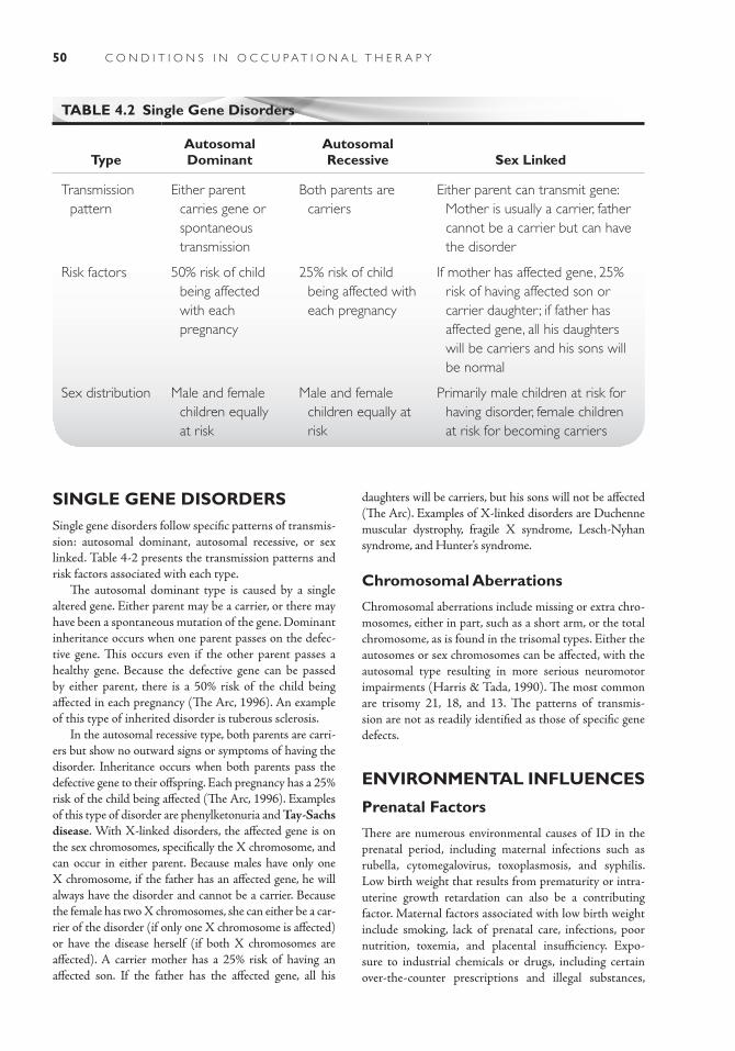
50 C O N D I T I O N S I N O C C U P A T I O N A L T H E R A P Y
SINGLE GENE DISORDERSSingle gene disorders follow specific patterns of transmis-sion: autosomal dominant, autosomal recessive, or sex linked. Table 4-2 presents the transmission patterns and risk factors associated with each type.
The autosomal dominant type is caused by a single altered gene. Either parent may be a carrier, or there may have been a spontaneous mutation of the gene. Dominant inheritance occurs when one parent passes on the defec-tive gene. This occurs even if the other parent passes a healthy gene. Because the defective gene can be passed by either parent, there is a 50% risk of the child being affected in each pregnancy (The Arc, 1996). An example of this type of inherited disorder is tuberous sclerosis.
In the autosomal recessive type, both parents are carri-ers but show no outward signs or symptoms of having the disorder. Inheritance occurs when both parents pass the defective gene to their offspring. Each pregnancy has a 25% risk of the child being affected (The Arc, 1996). Examples of this type of disorder are phenylketonuria and Tay-Sachs disease. With X-linked disorders, the affected gene is on the sex chromosomes, specifically the X chromosome, and can occur in either parent. Because males have only one X chromosome, if the father has an affected gene, he will always have the disorder and cannot be a carrier. Because the female has two X chromosomes, she can either be a car-rier of the disorder (if only one X chromosome is affected) or have the disease herself (if both X chromosomes are affected). A carrier mother has a 25% risk of having an affected son. If the father has the affected gene, all his
daughters will be carriers, but his sons will not be affected (The Arc). Examples of X-linked disorders are Duchenne muscular dystrophy, fragile X syndrome, Lesch-Nyhan syndrome, and Hunter’s syndrome.
Chromosomal Aberrations
Chromosomal aberrations include missing or extra chro-mosomes, either in part, such as a short arm, or the total chromosome, as is found in the trisomal types. Either the autosomes or sex chromosomes can be affected, with the autosomal type resulting in more serious neuromotor impairments (Harris & Tada, 1990). The most common are trisomy 21, 18, and 13. The patterns of transmis-sion are not as readily identified as those of specific gene defects.
ENVIRONMENTAL INFLUENCES
Prenatal Factors
There are numerous environmental causes of ID in the prenatal period, including maternal infections such as rubella, cytomegalovirus, toxoplasmosis, and syphilis. Low birth weight that results from prematurity or intra-uterine growth retardation can also be a contributing factor. Maternal factors associated with low birth weight include smoking, lack of prenatal care, infections, poor nutrition, toxemia, and placental insufficiency. Expo-sure to industrial chemicals or drugs, including certain over-the-counter prescriptions and illegal substances,
TABLE 4.2 Single Gene Disorders
TypeAutosomal Dominant
Autosomal Recessive Sex Linked
Transmission pattern
Either parent carries gene or spontaneous transmission
Both parents are carriers
Either parent can transmit gene: Mother is usually a carrier, father cannot be a carrier but can have the disorder
Risk factors 50% risk of child being affected with each pregnancy
25% risk of child being affected with each pregnancy
If mother has affected gene, 25% risk of having affected son or carrier daughter ; if father has affected gene, all his daughters will be carriers and his sons will be normal
Sex distribution Male and female children equally at risk
Male and female children equally at risk
Primarily male children at risk for having disorder, female children at risk for becoming carriers
51Chapter 4 : Inte l lectua l Disab i l i ty
also can affect birth weight, particularly during the first trimester of pregnancy.
Perinatal Factors
Two major causative factors of ID in the perinatal period are mechanical injuries at birth and perinatal hypoxia. Mechanical injuries are caused by difficulties of labor because of malposition, malpresentation, dis-proportion, or other labor complications that result in tears of the meninges, blood vessels, or other substances of the brain. Factors that cause perinatal hypoxia or anoxia include premature placental separation, mas-sive hemorrhage from placenta previa, umbilical cord wrapped around the baby’s neck, and meconium aspi-ration. Very premature infants also may have impaired respiration or an intracranial hemorrhage that can result in brain damage.
If a mother has an active case of herpes simplex II and is shedding the virus at the time of delivery, the baby can acquire the infection in the birth canal, which can cause severe developmental disability. This can be avoided by testing to determine whether the mother has an active case and, if so, delivering by cesarean section.
Postnatal Factors
Traumas or infections that result in injury or a lack of oxygen to the brain are a major cause of ID during the postnatal period. Traumas include near-drowning or strangulation, child abuse, and closed head injuries. Early severe psychosocial deprivation (i.e., attachment disorder, removal from the family home) can be part of the cause in the ID etiology (Shevell, 2008). Infections include encephalitis and meningitis. ID that results from menin-gitis caused by Haemophilus influenzae is now preventable, however, with the introduction of the H. influenzae type b (Hib) vaccine (Baraff, Lee, & Schriger, 1993).
Another major postnatal factor is sociodemographic characteristics or environmental influences. When an analysis of the relationship of sociodemographic charac-teristics and ID was completed for a large population of children with ID, it was found that boys, children with two or more older siblings, African American children, children whose mothers had not completed high school, and children of older mothers (with Down syndrome fac-tored out) were more likely to experience ID (American Association on Mental Retardation, 1992).
INCIDENCE AND PREVALENCEID is the most frequently occurring developmental dis-ability. Estimates of the prevalence of ID in the United States range from 1% to 3%. Most professionals associ-
ated with the AAIDD accept a prevalence of 2.5%, and they recognize that the prevalence varies with chrono-logic age (The Arc, 1996). A review of prevalence studies found that 2.5% to 3% is probably an accurate estimate of distribution in the general population (Frayers, 1993). Boys are 1.5 times more likely to experience MR than girls (Hauser & Ratey, 1994), which may be related to the sex-linked genetic disorders that result in MR (American Association on Mental Retardation, 2002).
SIGNS AND SYMPTOMSID often occurs in tandem with, or as a secondary mani-festation of, another diagnosis. One study’s results found that two-thirds of the children with severe ID (IQ < 50) had an additional neurologic diagnosis; <20% of children with mild ID were found to have an additional neurologic diagnosis. These diagnoses included conditions such as cerebral palsy, epilepsy, and hearing and visual impair-ments (The Arc, 2005). With certain genetic conditions, such as Down syndrome, MR is one of the clinical signs of the condition.
ID is defined by the AAIDD as a condition that is present from childhood (age 18 or younger), with (IQ) two standard deviations below the mean as measured on a standardized test and significant limitations in adaptive skills. Adaptive skills must be two standard deviations below the mean on a standardized test in conceptual, social, or practical skill areas (AAIDD, 2010). As previ-ously stated, adaptive skill areas include communication, self-care, home living, social/interpersonal skills, leisure, health and safety, self- direction, functional academics, use of community resources, and work. Adaptive skills should be assessed in all of the individual’s performance contexts. Someone with limited intellectual function who does not have adaptive skill deficits is not considered mentally retarded (AAIDD).
There are currently two major systems of classifica-tion for ID. The American Psychiatric Association uses the older, more traditional system of classifying ID based on performance on standardized intelligence tests using somewhat arbitrary cutoffs to assign levels of function. It is essentially used for diagnostic purposes (American Association on Mental Retardation, 1992). In 1992, the AAIDD (previously called the American Association on Mental Retardation) introduced a system of classifica-tion based on adaptive skills levels and supports needed to function (American Association on Mental Retarda-tion, 2002). The AAIDD system, because it focuses on function and supports needed in adaptive skills across performance contexts, is more useful to the practice of occupational therapy.
In the traditional system, ID is classified according to the severity of the impairment in intellectual function-ing. This is determined through standardized intelligence
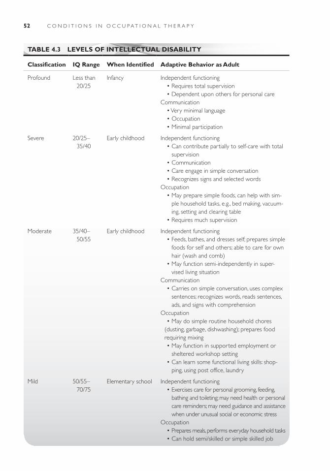
52 C O N D I T I O N S I N O C C U P A T I O N A L T H E R A P Y
TABLE 4.3 LEVELS OF INTELLECTUAL DISABILITY
Classification IQ Range When Identified Adaptive Behavior as Adult
Profound Less than 20/25
Infancy Independent functioning • Requires total supervision • Dependent upon others for personal careCommunication • Very minimal language • Occupation • Minimal participation
Severe 20/25–35/40
Early childhood Independent functioning • Can contribute partially to self-care with total
supervision • Communication • Care engage in simple conversation • Recognizes signs and selected wordsOccupation • May prepare simple foods, can help with sim-
ple household tasks, e.g., bed making, vacuum-ing, setting and clearing table
• Requires much supervision
Moderate 35/40–50/55
Early childhood Independent functioning • Feeds, bathes, and dresses self; prepares simple
foods for self and others; able to care for own hair (wash and comb)
• May function semi-independently in super-vised living situation
Communication • Carries on simple conversation, uses complex
sentences; recognizes words, reads sentences, ads, and signs with comprehension
Occupation • May do simple routine household chores
(dusting, garbage, dishwashing); prepares food requiring mixing
• May function in supported employment or sheltered workshop setting
• Can learn some functional living skills: shop-ping, using post office, laundry
Mild 50/55–70/75
Elementary school Independent functioning • Exercises care for personal grooming, feeding,
bathing and toileting; may need health or personal care reminders; may need guidance and assistance when under unusual social or economic stress
Occupation • Prepares meals, performs everyday household tasks • Can hold semi/skilled or simple skilled job
53Chapter 4 : Inte l lectua l Disab i l i ty
testing. To be considered intellectually disabled, the per-son’s performance on these tests must be two standard deviation units or more below the mean. The levels of ID as identified by IQ tests are mild, moderate, severe, and profound. Approximately 85% of individuals with ID are in the mild range, 10% are in the moderate range, 3.5% are in the severe range, and 1.5% are in the profound range of function (Hauser & Ratey, 1994; Silka & Hauser, 1997). Table 4-3 presents the classifications, IQ levels, and gen-eral level of functioning as an adult. This information is adapted from The Diagnostic and Statistical Manual of Mental Disorders, Fourth Edition (DSM-IV), which is the diagnostic standard for mental health care professionals in the United States. The DSM-IV classifies four differ-ent degrees of MR: mild, moderate, severe, and profound. These categories are based on the functioning level of the individual (American Association on Mental Retarda-tion, 1992, 2002; Kaplan et al., 1994; Reiss, Goldberg, & Ryan, 2006). It is important to remember that not all individuals in a particular classification will function at exactly that level. The DSM 5, currently in development, is projected to use diagnostic criteria more in line with the American Association for Intellectual and Develop-mental Disabilities (American Psychological Association, 2011).
The classification system developed by the AAIDD involves a three-step process. The first step is to have a qualified person administer standardized intelligence and adaptive skills assessments that are appropriate for the individual’s age, communication abilities, and cultural experience. The second step is to describe the individual’s strengths and weaknesses across the dimen-sions of (1) intellectual and adaptive behavior skills, (2) psychological/emotional considerations, (3) physical/health/etiologic considerations, and (4) environmental considerations. The third step is to have the interdiscipli-nary team determine needed supports across these four dimensions. Supports are classified based on the level of intensity and include intermittent, limited, extensive, and pervasive. Intermittent support is provided on an “as-needed” basis. Limited support occurs over a limited time span. Extensive support is assistance provided on a daily basis in a life area. Pervasive support refers to the need for support in all life areas across all environments on a daily basis (American Association on Mental Retar-dation, 2002).
In addition to the performance deficits produced by ID and the associated conditions already mentioned, a high proportion of individuals with ID also have some form of mental illness. Estimates of prevalence of mental illness among people with ID range from 10% to 20% (Reiss et al., 2006) to 40% to 70% (Silka & Hauser, 1997). Some of the common types of mental illness seen in people with ID include personality disorders, affective
disorders, psychotic disorders, avoidant disorders, paranoid personality disorders, severe behavior prob-lems that may include self-injurious behavior (Zigman, Schupf, Zigman, & Silverman, 1993), and dementia associated with Down syndrome (Behrman, Kliegman, & Nelson, 1992). Several misconceptions about people with ID may complicate or prevent appropriate care for their mental illness, including the beliefs that peo-ple who are mentally retarded cannot also be mentally ill, do not experience normal feelings and emotions, and are not affected by changes in their environment. Sub-stance abuse problems, especially with alcohol, may be overlooked, and there is controversy about the benefit of using antipsychotic drugs with individuals who are mentally retarded (Hauser & Ratey, 1994). Because of limited communication skills and limitations in abstract thinking caused by the ID, the diagnosis of mental ill-ness and mental health problems can be a very difficult process and is frequently inexact. Good communication with caregivers and significant others in the life of the individual with ID is essential.
COURSE AND PROGNOSISID is generally considered a lifelong condition, but the course and prognosis will vary depending upon the cause(s) of the retardation (Beers & Berkow, 1997; Behr-man et al., 1992). In terms of life expectancy, people with mild ID live as long as the general population (Shevell, 2008). However, people with more profound ID are less likely to reach old age. This is likely due to more serious neurologic deficits and associated disorders.
Most cases of ID are nonprogressive; that is, once the initial insult to the brain occurs, there is no further damage (Beers & Berkow, 1997; Behrman et al., 1992). The emphasis is on managing the medical aspects of the condition and helping individuals to achieve their highest potential. However, certain genetic conditions (e.g., mus-cular dystrophy and Tay-Sachs disease) are progressive, with incremental loss of function and, in some cases, associated early death. The goal for these individuals is to help them achieve the highest level of independence and maintain it as long as possible. Those with Down syndrome experience degenerative changes in the brain, beginning at about age 40, that eventually result in pro-gressive dementia similar to Alzheimer’s disease (Zigman et al., 1993). On a positive note, it is possible for individu-als with mild ID to gain adaptive behavior skills through remedial programs to the extent that they no longer meet the diagnostic criteria for being intellectually disabled, although their intellectual function has probably not changed significantly (American Association on Mental Retardation, 1992).
54 C O N D I T I O N S I N O C C U P A T I O N A L T H E R A P Y
DIAGNOSISAn evaluation must be performed to determine whether a person meets the criteria for being mentally retarded. Besides ascertaining that the onset of the condition occurred before age 18, there are two main aspects to this process. The first part involves administration of appropriate standardized intelligence tests by a quali-fied individual. The selection of the specific standardized instrument should be based on factors like the individu-al’s social, linguistic, and cultural background (American Association on Mental Retardation, 1992). The individu-als’ IQ is interpreted in the context of their level of adap-tive behavior (AAIDD, 2010).
The second aspect of the process is the evaluation of adaptive behavior as it related to the targeted adaptive life skill areas. Adaptive functioning is an individual’s ability to cope with life demands and meet societal expectations for independence depending on age group (American Association on Mental Retardation, 1992). The skills needed for adaptive behavior become more complex and varied as the person ages. For instance, eating and dressing independently are major skills for the young child, but the child does not need to be able to use a telephone or man-age money. Evidence for deficits and strengths in adaptive function should be gained from one or more independent, reliable sources who are familiar with the individual’s abil-ities in different performance contexts. This information should be used to complete a standardized scale designed to provide a composite “picture” of the individual’s adap-tive function. As with the selection of an intelligence test, care should be taken that the adaptive behavior scale chosen is appropriate for the individual’s sociocultural background, education, associated handicaps, motivation, and cooperation level (American Association on Mental Retardation, 2002). There are more than 200 adaptive behavior measures and scales. The most common scale is the Vineland Adaptive Behavior Scales—Second Edi-tion (VAB-II) (Sparrow, Balla, & Cicchetti, 2005), which purports to assess the personal and social skills needed for everyday living. It is an indirect assessment in that the respondent is not the individual in question but someone familiar with the individual’s behavior. The VAB-II meas-ures four domains: communication, daily living skills, socialization, motor skills, and an optional maladaptive behavior index. An adaptive behavior composite is a com-bination of the scores from the four domains. A second scale frequently used to assess adaptive behavior is the Diagnostic Adaptive Behavior Scale (DABS) formerly titled the Adaptive Behavior Scale (AAIDD, 2010). This measure was developed by the AAIDD to assess concep-tual, social, and practical skills and focuses on the critical “cutoff area” for the purpose of ruling in or ruling out a diagnosis of ID.
MEDICAL/SURGICAL MANAGEMENTThere is no drug treatment for the condition of ID; how-ever, medications may be needed for some of the condi-tions that may occur in tandem. Concomitant mental health problems such as affective or psychotic disorders and severe behavior problems would all benefit from appropriate medical intervention; seizure disorders would require drug therapy. Neuromuscular aberrations (spastic-ity, rigidity, etc.) seen in cerebral palsy may also be helped by medication.
IMPACT OF CONDITIONS ON CLIENT FACTORS AND OCCUPATIONAL PERFORMANCEVirtually all areas of occupational performance and many client factors can be affected by ID, depending on the cause and severity of the ID. As stated previously, the diagnostic criteria for individuals with ID included three categories of adaptive behavior (conceptual, social, and practical skills) (AAIDD, 2010). These areas (commu-nication, language, interpersonal skills, social responsibil-ity, recreation, friendships, daily living skills, work, and travel) impact all occupational performance areas includ-ing (basic) activities of daily living, instrumental activities of daily living, work, play, leisure, and social participation.
Although all of the occupational performance areas and client factors can be influenced by ID, those that are affected will depend on factors such as the presence of additional medical diagnoses and the severity of the diag-nosis. It is imperative that the clinician be informed about the specific diagnosis that accompanies the identification of ID to determine the associated client factors that are involved. For example, occupational therapists often work with persons who have Down syndrome. Babies with Down syndrome often have hypotonia or poor muscle tone. Because they have a reduced muscle tone, which results in oral motor dysfunction such as protruding tongue, feeding babies with Down syndrome is often dif-ficult. Hypotonia may also affect the muscles of the diges-tive system, in which case constipation may be a problem. Atlantoaxial instability, a malformation of the upper part of the spine located under the base of the skull, is present in some individuals with Down syndrome. This condition can cause spinal cord compression as well as craniover-tebral instability (Brockmeyer, 1999). Additionally, half of children with Down syndrome are diagnosed with a congenital heart. In addition to hearing disorders, visual problems also may be present early in life. Cataracts occur in approximately 3% of children with Down syndrome but can be surgically removed.
55Chapter 4 : Inte l lectua l Disab i l i ty
Approximately half of the children with Down syn-drome have congenital heart disease and associated early onset of pulmonary hypertension or high blood pres-sure in the lungs. Echocardiography may be indicated to identify any congenital heart disease. If the defects have been identified before the onset of pulmonary hyper-tension, surgery has provided favorable results. Seizure disorders, though less prevalent than some of the other associated medical conditions, still affect between 5% and 13% of individuals with Down syndrome, a 10-fold greater incidence than in the general population. Con-genital hypothyroidism, characterized by a reduced basal metabolism, an enlargement of the thyroid gland, and
disturbances in the autonomic nervous system, occurs slightly more frequently in babies with Down syndrome. Recent studies indicate that 66% to 89% of children with Down syndrome have a hearing loss of >15 to 20 deci-bels in at least one ear, due to the fact that the external ear and the bones of the middle and inner ear may develop differently in children with Down syndrome (National Institutes of Health [NIH], 2006). Given that 85% of all ID cases fall in the mild range of cognitive impairment, global and specific mental functions will be most affected with this population. The following case studies illus-trate how ID affects an individual’s area of occupational performance in different stages of the life cycle.
Case IllustrationCase 1K.R. is a 21-year-old young woman with Down syn-drome. Despite the fact that she has reached the legal age of adulthood, she is still in the process of transi-tioning from the developmental stage of adolescence to young adulthood. She still receives special educa-tion services but is also working on developing work and job skills through traditional vocational services. Her “disability” is expected to be lifelong.
K.R.’s condition was identified at birth. She has received continuous support and direct services to facilitate development of her abilities since then. She has always lived at home with her parents and still does so. She has her own room and bathroom at home and generally has exclusive use of the family room for her leisure pursuits. Her parents are profes-sionals who are actively involved in their professions and the community. They are very realistic about her abilities and extremely supportive, allowing her to make most of her life choices. K.R. has a younger sis-ter who is now away at college. K.R. has always been exposed to and involved in many social and cultural opportunities in the community, both with her family and on her own. Her social circle includes friends with and without disabilities, and she has several close friends as well as many social acquaintances.
K.R. has recently declared her life goals to be getting a job, getting an apartment of her own, and spending leisure time at the local community drop-in/recreational center for individuals with disabilities. Her mother feels that, with appropriate supports, all of these goals are attainable.
K.R. is independent in most areas of personal care. As a result of limited fine motor coordination that seems to be complicated by visual perceptual deficits, she needs assistance with regulating the water tem-perature for her bath, fastening zippers and buttons,
and tying her shoes. She also occasionally needs reminders to straighten her clothing and brush the back of her hair. She has a speech impediment that makes it difficult for individuals who are unfamiliar with her to initially understand her. K.R. has learned to adapt to this limitation and works very hard to make people understand what she is trying to com-municate. When cued, she will slow down and work on enunciating her words. She is independent in func-tional mobility, but because of problems with depth perception, she is very cautious when climbing steps and negotiating between different surface levels. She is able to travel independently in community areas she is familiar with and can ride the community “Dial-a-Ride” bus if the ride is arranged for her. Her mother feels that K.R. would be aware of danger in her home, such as a fire, and would get out of the house. She does not consistently answer the phone when it rings, although she is capable of doing so. For this reason, she is generally not allowed to be at home alone because her parents have no way of checking in with her if she does not answer the phone.
K.R. is able to perform many home management tasks and has recently become motivated to attempt more activities given her desire to have an apartment of her own. She generally dislikes “housework” but knows it is necessary in order to be a good room-mate. She folds laundry and puts it away and is learn-ing to sort light and dark clothing and operate the washing machine. Her mother questions her ability to make judgments about what should or should not go into the dryer, however. She is able to sweep, vacuum, and dust but is not thorough, and this may be a result of her lack of interest and/or her visual-perceptual impairment. She keeps her bathroom clean and makes her bed. She is currently dependent in meal preparation, and this is the area that will probably
56 C O N D I T I O N S I N O C C U P A T I O N A L T H E R A P Y
require the most support for her to live in her own apartment. She cannot safely regulate stove and oven temperatures because of her fine motor problems. At the current time, K.R. attends school in a self-contained special education classroom in the local high school for half a day, focusing on vocational and prevocational skills, and spends the other half-day at Goodwill Indus-tries in a work adjustment trial placement. K.R. has told her mother that she no longer enjoys going to school and would rather go to Goodwill. In addition to school and vocational activities, K.R. volunteers at the local community theater and at her church office doing clerical tasks. She is very interested in obtain-ing employment and would prefer to work at a video store, a music store, the mall, or Pizza Hut. She is very interested in clerical tasks and has been working at her father’s medical practice putting monthly billings in envelopes to be mailed. K.R. has many leisure interests and activities. She enjoys music and videos, likes to eat out, participates in Special Olympics, goes to a commu-nity center for structured activities, and socializes with her friends. She prefers not to do strenuous physical activities, probably because of the difficulty she has as a result of generalized hypotonia. She has participated in team sports at the center but does so mostly for the social interaction. K.R. is very aware of her limitations and takes herself out of situations that she knows will be difficult or where she might not succeed.
Case 2F.B. is a 60-year-old man who is intellectually disabled. He has a history of behavior problems, including PICA and obsessive-compulsive type behaviors of picking at his skin and clothing and stuffing toilets. He was retired from a sheltered workshop approximately 18 months ago when the emphasis of the program shifted to work readiness for community placement. It was determined that he would not be a good can-didate for community work because of his age and lack of necessary supports (e.g., transportation). He currently attends a day program for social/leisure activities. F.B. has no known family and has spent most of his life in public residential institutions or in adult foster care (AFC) homes. He currently has a court-appointed guardian who makes most significant life decisions for him, including those regarding medi-cal care and living arrangements. His guardian sup-ports and encourages F.B. to make his wishes known about how he would like to spend his leisure time and allowed him input into his last housing change. In addition to his intellectual limitations, F.B. also expe-riences fairly frequent medical problems related to his obsessive-compulsive behaviors (e.g., skin infec-tions). Medical problems, which may be age related,
are emphysema and frequent fractures of bones in his lower extremities. F.B. currently lives in an AFC with 11 other adults who are developmentally disabled. His home is only required by law to provide basic care, and so it does not offer training in or support for par-ticipation in many home management tasks. His social groups generally consist of other adults with develop-mental disabilities or paid paraprofessional staff, either at home or the day program. His cultural experiences have been very limited because of his background.
F.B. is independent in most self-care tasks but needs supervision when using the bathroom because of his history of stuffing the toilet. He needs assistance and supports for taking medication owing to his cogni-tive limitations. He also needs very close monitoring of health status because he has a very high pain tolerance. He tends to prefer solitary activities but has become more verbal, social, and outgoing since going to the day program. He generally seeks out staff to interact with, and this usually takes the form of teasing. He will share activities with day program peers if prompted and has shown protective behaviors toward clients who are more limited and vulnerable than he is. Although he is verbal, he has a speech impediment that makes it difficult for people who are unfamiliar with him to understand what he is saying. He is independently ambulatory; however, he has reduced endurance as a result of an old hip fracture and neuropathy of the right lower extremity caused by a degenerative dis-ease in the lumbar spine. He is dependent for all com-munity mobility as a result of cognitive limitations and lack of experience and training. It is not clear whether he understands emergency situations, but he is coop-erative with emergency drill procedures at the day program. It is likely that he would need ongoing super-vision to maintain his personal safety.
Because of a lack of experience and opportunity, F.B. is dependent in all home management tasks. He has no responsibility for caring for others but seems to be very aware when one of his peers needs assis-tance or protection and alerts staff to these needs. He has been retired from the vocational arena for 18 months and now attends a day program that emphasizes social and leisure activities.
F.B. is generally not open to exploring new activi-ties and has to be coaxed and teased by staff to try them. He generally prefers solitary activities and appears to enjoy assembly activities that result in a finished product like picture puzzles and building with Erector-Set components. He does not seem to be very interested in watching television or listening to music but does enjoy going on automobile rides with his guardian, especially when she drives her convert-ible with the top down.
57Chapter 4 : Inte l lectua l Disab i l i ty
American Association on Intellectual Disability. (2010). Intellectual disability: Definition, classifica-tion, and systems of supports (11th ed.). Washing-ton, DC: Author.
Baker, B., Brightman, A., Blacher, J., & Heifetz, L. (2004). Steps to independence: Teaching everyday skills to children with special needs (4th ed.). Baltimore, MD: Paul H. Brookes.
Behrman, R., & Kliegman, R. (Eds.) (2002) Nelson essentials of pediatrics (4th ed.). Philadelphia, PA: WB Saunders.
Case-Smith, J. (2005). Occupational therapy for children (5th ed.). St. Louis, MO: Mosby.
American Journal on Intellectual and Developmental Disabilities
American Association on Intellectual and Developmental Disabilities
444 North Capitol St., NW, Suite 846Washington, DC 20001Education and Training in Mental Retardation and
Developmental DisabilitiesDivision on Mental Retardation & Developmental
DisabilitiesThe Council for Exceptional Children1920 Association DriveReston, VA 22091–1589Journal of the Association for Persons with Severe
HandicapsDivision on Mental Retardation & Developmental
DisabilitiesThe Council for Exceptional Children1920 Association DriveReston, VA 22091
Advocacy ResourcesAmerican Association on Intellectual and Developmen-
tal Disabilities (AAIDD)444 North Capitol St., NW, Suite 846Washington, DC 20001
Tel: +1-202-387-1968; toll free: +1-800-424-3688;Fax: +1-202-387-2193The Arc500 E. Border St., Suite 300Arlington, TX 76010Tel: +1-817-261-6003; TTY: +1-817-277-0553;Fax: +1-817-277-3941The Association for Persons with Severe Handicaps
(TASH)11201 Greenwood Ave.North Seattle, WA 98133Tel: +1-206-361-8870Division on Mental Retardation & Developmental Dis-
abilitiesThe Council for Exceptional Children1 920 Association DriveReston, VA 22091–1589Tel: +1-703-620-3660National Down Syndrome Congress1605 Chantilly Dr., Suite 250Atlanta, GA 30324Tel: +1-800-232-NDSCNational Down Syndrome Society666 BroadwayNew York, NY 10012Tel: +1-212-460-9330; toll free: +1-800-221-4602People First International (self-advocacy group)1340 Chemeketa St., NE Salem, OR 97301Tel: +1-503-588-5288President’s Committee for People with Intellectual Dis-
abilitiesU.S. Department of Health & Human Services330 Independence Ave., SWWashington, DC 20201Tel: +1-202-619-0634Special Olympics International, Inc.1350 New York Ave., NW, Suite 500Washington, DC 20005Tel: +1-202-628-3630
R E C O M M E N D E D L E A R N I N G R E S O U R C E S
R E F E R E N C E S
American Association of Intellectual and Developmental Disabilities (AAIDD). (2010). Diagnostic adaptive behavior scale. Retrieved August 5, 2010, from http://www.aamr.org/content_106.cfm?navid=23
American Association on Intellectual Disability. (2010). Intellectual disability: Definition, classification, and sys-tems of supports (11th ed.). Washington, DC: Author.
American Association on Mental Retardation. (1992). Mental retardation: Definition, classification, and sys-tems of supports (9th ed.). Washington, DC: Author.
American Association on Mental Retardation. (2002). Mental retardation: Definition, classification, and systems of supports (10th ed.). Washington, DC: Author.
58 C O N D I T I O N S I N O C C U P A T I O N A L T H E R A P Y
American Psychiatric Association (2011). DSM-5 devel-opment: a intellectual developmental disorder. Retrieved May 18, 2011 from http://www.dsm5.org/propose-drevision/pages/proposedrevision.aspx?rid=384
Baraff, L., Lee, S., & Schriger, D. (1993). Outcomes of bacterial meningitis in children: A meta-analysis. Pe-diatric Infectious Disease Journal, 12, 389–394.
Beers, M., & Berkow, R., (Eds.). (1997). The Merck man-ual of diagnosis and therapy (17th ed.). Rahway, NJ: Merck Sharp & Dohme Research Laboratories.
Behrman, R., Kliegman, R., & Nelson, W. (Eds.). (1992). Nelson’s textbook of pediatrics (14th ed.). Philadelphia, PA: WB Saunders.
Brockmeyer, D. (1999). Down syndrome and craniover-tebral instability: Topic review and treatment recom-mendations. Pediatric Neurosurgery, 31(2), 71–77.
Frayers, T. (1993). Epidemiological thinking in mental retardation: Issues in taxonomy and population fre-quency. International Review of Research in Mental Re-tardation, 19, 97–133.
Gror, M., & Shekleton, M. (1979). Basic pathophysiology: A conceptual approach. St. Louis, MO: CV Mosby.
Harris, S., & Tada, W. (1990). Genetic disorders. In D. Umphred (Ed.). Neurological rehabilitation. St. Louis: CV Mosby.
Hauser, M., & Ratey, J. (1994). The patient with men-tal retardation. In S. Hyman & G. Tesar (Eds.). The manual of psychiatric emergencies (pp. 104–109). Boston: Little Brown & Co. Retrieved December 27, 2005, from http://www.psychiatry.com
Jardim, L., Palma-Dias, R., Silva, L., Ashston-Prol-la, P., & Guigliani, R. (1996). Maternal hyperphe-nylalaninemia as a cause of microcephaly and mental retardation. Acta Paediatrica, 85, 943–946.
Kaplan, H., Sadock, B., & Grebb, J. (1994). Synopsis of psychiatry (7th ed.). Baltimore, MA: Williams & Wilkins.
Matilainen, R., Airaksinen, E., & Monomen, T. (1995). A population-based study on the causes of mild and severe mental retardation. Acta Paediatrica, 84, 261–266.
National Institutes of Health. (2006). Facts about Down syndrome. Retrieved February 12, 2006, from http://www.nichd.nih.gov/publications/pubs/downsyn-drome/down
Reiss, S., Goldberg, B., & Ryan, R. (1993). Mental illness in persons with mental retardation. Retrieved January 16, 2006, from http://www.psychiatry.com
Reuss, M., Paneth, M., Pinto-Martin, J., & Lorenz, J. (1996). The relation of transient hypothyroxinemia in preterm infants to neurologic development at two years of age. New England Journal of Medicine, 334, 821–827.
Rogers, B., Msall, M., Buck, G., Lyon, N., Norris, M., Roland et al. (1995). Neurodevelopmental outcome of infants with hypoplastic left heart syndrome. Jour-nal of Pediatrics, 126, 496–498.
Scriver, C. (1995). The metabolic and molecular bases of in-herited disease (7th ed.). New York, NY: McGraw-Hill.
Shevell, M. (2008). Global developmental delay and mental retardation or intellectual disability: Concep-tualization, evaluation and etiology. Pediatric Clinics of North America, 55, 1071–1084.
Silka, V., & Hauser, M. (1997). Psychiatric assessment of the person with mental retardation. Psychiatric An-nals, 27, 3.
Sparrow, S., Balla, D., & Cicchetti, D. (2005). Vineland adaptive behavior scales. San Antonio, TX: PsychCorp.
The Arc. (1993). Introduction to mental retardation. Re-trieved January 10, 2006, from http://www.thearc.org
The Arc. (1996). Genetic causes of mental retardation. Retrie ved December 19, 2005, from http://www.thearc.org
Yeargin-Allsopp, M., Murphy, C., Cordero, J., Decouflé, P., & Hollowell, J. (1997). Reported biomedical causes and associated medical conditions for mental retardation among 10-year-old children, metropolitan Atlanta, 1985 to 1987. Developmental Medicine and Child Neurology, 39, 142–147.
Zigman, W., Schupf, N., Zigman, A., & Silverman, W. (1993). Aging and Alzheimer disease in people with mental retardation. International Review of Research in Mental Retardation, 19, 41–70.
59
Mood Disorders
■ Ann C h ap l e au
5
Helen sits in her rocking chair in her small living room, smoking a cigarette. The room is dimly lit by the television screen. Her silver hair is disheveled and her sweat suit is stained and dirty. She has several days’ worth of Meals on Wheels containers on
the coffee table, mostly uneaten. A picture of her deceased husband is on the end table, as are various photos of her grown daughter, who lives in another state. Helen has not left her home in over 6 months, except for doctor visits. She has early-stage emphysema, but has not been able to stop smoking. She no longer knits or bakes, two of her favorite hobbies. At her last doctor’s visit, her physician asked her if she was feeling depressed, but Helen was too ashamed to tell him how sad and lonely she had been feeling for months. She wanted to tell him that there wasn’t any point in living anymore, that her life doesn’t have any meaning. But she remained silent. At home, she is overwhelmed by feelings of guilt. She had been raised to keep a tidy home and be active, and not to feel sorry for oneself, but she feels too tired to do anything except to watch television.
DESCRIPTION AND DEFINITIONMood disorders are among the most disabling and prevalent illnesses worldwide. The World Health Organization (WHO) estimates that major depressive disor-
der (MDD) and bipolar disorder (BPD) are among the leading cause of disability, and that MDD is expected to become the second leading cause of disability by 2020, second only to cardiovascular disease (WHO, 2008). Despite the prevalence and impact on global health care costs, as well as quality of life for individuals and their families, mood disorders often go undetected and untreated.
Mood disorders represent a spectrum of mood disturbances, from the extremely low mood of depression to the extremely elevated mood of mania (Figure 5.1). The American Psychi-atric Association (APA) provides a classification system (APA, 2000) to identify symptoms using universal terminology. Depressive disorders include MDD and dysthymic disorder, which are often referred to as unipolar depression, as they do not include mood variances
K E Y T E R M S
AffectAnhedonia
AvolitionCyclothymia
DysphoriaEuphoria
Flight of ideasGrandiosity
Hypomanic episodeMajor depressive episode
ManiaManic episodeMixed episode
ProdromalPsychomotor agitation
Psychomotor retardationPsychosis
Unipolar depression
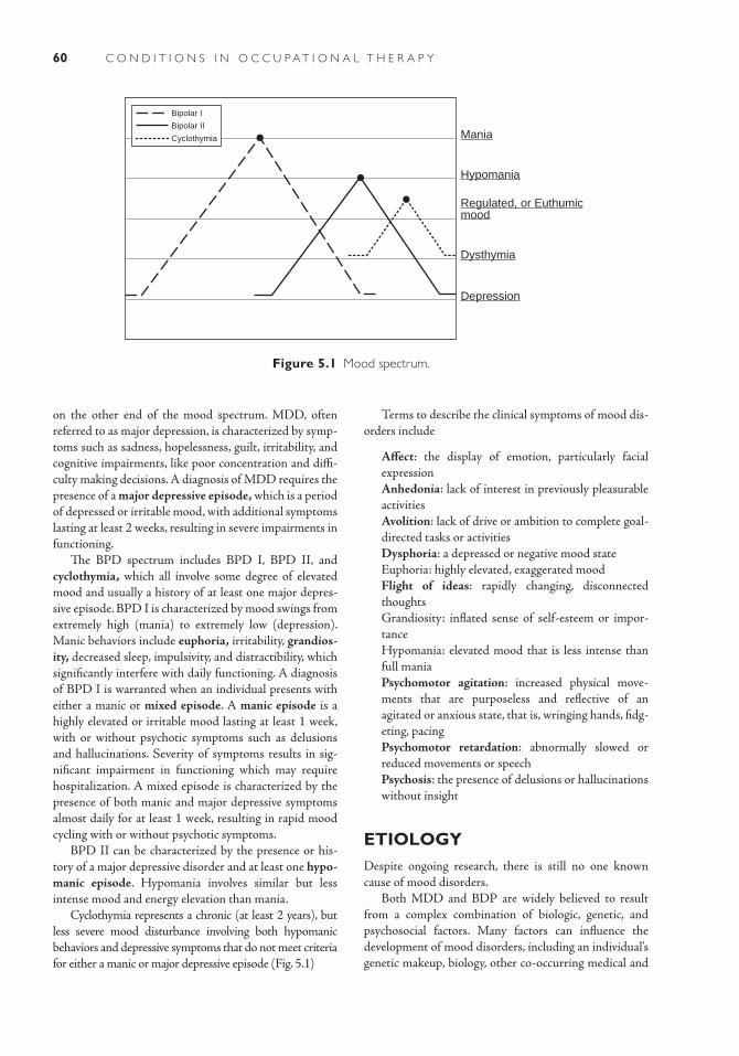
60 C O N D I T I O N S I N O C C U P A T I O N A L T H E R A P Y
on the other end of the mood spectrum. MDD, often referred to as major depression, is characterized by symp-toms such as sadness, hopelessness, guilt, irritability, and cognitive impairments, like poor concentration and diffi-culty making decisions. A diagnosis of MDD requires the presence of a major depressive episode, which is a period of depressed or irritable mood, with additional symptoms lasting at least 2 weeks, resulting in severe impairments in functioning.
The BPD spectrum includes BPD I, BPD II, and cyclothymia, which all involve some degree of elevated mood and usually a history of at least one major depres-sive episode. BPD I is characterized by mood swings from extremely high (mania) to extremely low (depression). Manic behaviors include euphoria, irritability, grandios-ity, decreased sleep, impulsivity, and distractibility, which significantly interfere with daily functioning. A diagnosis of BPD I is warranted when an individual presents with either a manic or mixed episode. A manic episode is a highly elevated or irritable mood lasting at least 1 week, with or without psychotic symptoms such as delusions and hallucinations. Severity of symptoms results in sig-nificant impairment in functioning which may require hospitalization. A mixed episode is characterized by the presence of both manic and major depressive symptoms almost daily for at least 1 week, resulting in rapid mood cycling with or without psychotic symptoms.
BPD II can be characterized by the presence or his-tory of a major depressive disorder and at least one hypo-manic episode. Hypomania involves similar but less intense mood and energy elevation than mania.
Cyclothymia represents a chronic (at least 2 years), but less severe mood disturbance involving both hypomanic behaviors and depressive symptoms that do not meet criteria for either a manic or major depressive episode (Fig. 5.1)
Terms to describe the clinical symptoms of mood dis-orders include
■ Affect: the display of emotion, particularly facial expression
■ Anhedonia: lack of interest in previously pleasurable activities
■ Avolition: lack of drive or ambition to complete goal-directed tasks or activities
■ Dysphoria: a depressed or negative mood state ■ Euphoria: highly elevated, exaggerated mood ■ Flight of ideas: rapidly changing, disconnected
thoughts ■ Grandiosity: inflated sense of self-esteem or impor-
tance ■ Hypomania: elevated mood that is less intense than
full mania ■ Psychomotor agitation: increased physical move-
ments that are purposeless and reflective of an agitated or anxious state, that is, wringing hands, fidg-eting, pacing
■ Psychomotor retardation: abnormally slowed or reduced movements or speech
■ Psychosis: the presence of delusions or hallucinations without insight
ETIOLOGYDespite ongoing research, there is still no one known cause of mood disorders.
Both MDD and BDP are widely believed to result from a complex combination of biologic, genetic, and psychosocial factors. Many factors can influence the development of mood disorders, including an individual’s genetic makeup, biology, other co-occurring medical and
Mania
Hypomania
Regulated, or Euthumicmood
Dysthymia
Depression
Bipolar I
Bipolar II
Cyclothymia
Figure 5.1 Mood spectrum.
61Chapter 5 : Mood Disorder s
psychiatric conditions, cognitive abilities, personality, support systems, personal history, particularly exposure to stress, and coping strategies. Additionally, mood dis-orders can be triggered by childbirth, seasonal changes, substance use, and other medical conditions.
Biologic Factors
Much of the research over the past 50 years has focused on biologic changes in brain function (U.S. Department of Health and Human Services, 1999). Current research using advanced technologies of brain imaging to exam-ine structures and brain activity provides evidence that mood disorders are disorders of the brain. Magnetic reso-nance imaging (MRI) and positron emission tomography (PET) studies reveal abnormal functioning in regions of the brain that regulate mood, sleep, thinking, appetite, and behavior.
In studies of MDD, functional abnormalities have been found in the limbic system, including the amygdala, hippocampus, insula, regions of the anterior cingulate cortex, and the dorsolateral prefrontal cortex—brain regions that represent centers of emotion (Cole et al., 2010; Gos et al., 2010; McKinnon, Yucel, Nazarov, & MacQueen, 2009). Functional abnormalities of the limbic system have also been found in studies of BPD (Kruger, Seminowicz, Goldapple, Kennedy, & Mayberg, 2003). These abnormalities are consistent with studies in which both MDD and BPD subjects demonstrated blunted or decreased behavioral and physiologic reactivity to sad or negative stimuli, such as pictures and videos (Foti, Olvet, Klein, & Hajcak, 2010; Miklowski & Johnson, 2005; Rottenberg, 2005).
The role of neurotransmitters, such as serotonin, ace-tylcholine, and melatonin, in mood disorders has been studied for several decades. Neuroimaging and genetic studies have focused on the role of genes that may predis-pose an individual to dysregulated levels of neurotrans-mitters. Current research focuses on neurotransmitter systems, such as the effects of dysregulated dopamine and serotonin transporters and receptors in the brain (Miklowski & Johnson, 2005).
Biologic abnormalities of the brain, however, may not necessarily be the cause of a mood disorder, but can develop as a result of the disorder. For example, some researchers believe that chronic stress can trigger depres-sion, as the body continually responds to stress through neurochemical changes (Hennessy, Schiml-Webb, & Deak, 2009; Miller & Blackwell, 2005). One such stress response is the excessive secretion of cortisol, which can interfere with the limbic-cortical systems. The challenge for researchers is to differentiate which abnormalities are causal factors and which develop in response to environ-mental factors that trigger neurochemical changes.
Both MDD and BPD are known to run in families as heritable traits, but researchers have had minimal success in isolating any specific, responsible genes. Genetic varia-tions associated with both MDD and BPD support the growing belief among researchers that mood disorders involve a combination of altered genes that interact with environmental factors, such as stress, to explain why some family members with a known genetic risk develop either MDD or BPD while other family members do not (Caspi et al., 2003; McMahon et al., 2010).
Twin studies also support the genetic link in mood disorders. In studies of MDD, heritability was found to be 38%, with higher concordance rates among women than men (Kendler, Gatz, Gardner, & Pedersen, 2005). In studies of BPD, heritability is even higher; identical twins have a concordance rate of 57%, while the concordance rate for fraternal twins is 14% (Miklowitz & Johnson, 2005).
Psychosocial Risk Factors
In addition to the role of biologic and genetic factors in the development of mood disorders, the role of stressful life events has been widely studied. For example, traumatic events, such as childhood sexual, physical, or emotional abuse, increase the risk of both MDD and BPD (Hammen, 2005; Maniglio, 2010; Miklowitz & Johnson, 2005). Other risk factors include exposure to war, disaster, displacement, parental mental illness, domestic violence, physical or sexual assault, involvement in a serious accident, death of a loved one, chronic work stress, and caregiver stress (Man-iglio, 2010; Wingo et al., 2010). However, it is important to remember that the majority of people who experience life stressors do not develop a mood disorder and that not all mood disorders are triggered by a traumatic event. For those who experience severe life events, it is estimated that 20% will develop MDD, perhaps higher for those who experience more profound trauma (Monroe & Reid, 2009).
INCIDENCE AND PREVALENCEMDD is both common and widespread, with a lifetime prevalence of one in five, or 20%, and a relapse rate of more than 80% (Gotlib & Hamilton, 2008). The lifetime prevalence for BPD is much lower. Community samples can range from.4% to 1.5% (APA, 2000), or from 3.9% to 10%, based on inclusion criteria of the full bipolar spec-trum (Miklowitz & Johnson, 2005).
In any given year, 20.9 million (9.5%) adults in the United States will experience a mood disorder, with 14.8 million suffering from MDD and 5.7 million suffering from BPD (Kessler et al., 2005). Women are more fre-quently diagnosed with MDD than men (Gavin et al.,
62 C O N D I T I O N S I N O C C U P A T I O N A L T H E R A P Y
2010; Kessler et al., 2003). Additionally, lower socio-economic status is associated with both MDD and BPD (Everson, Maty, Lynch, & Kaplan, 2002; Schoeyen et al., 2010).
SIGNS AND SYMPTOMSBoth MDD and BPD are marked by a complex set of symptoms that interfere with daily functioning. Moreo-ver, both disorders are associated with high rates of sui-cide (Moller & Henkel, 2005), making a proper diagnosis essential for treatment purposes.
Major Depressive Disorder
A diagnosis of MDD can be determined when an indi-vidual has experienced at least one major depressive epi-sode, in the absence of any manic, mixed, or hypomanic episodes (APA, 2000). Symptoms must represent a sig-nificant change in typical functioning that interferes with daily living. According to the APA, a depressive episode must include at least five of the following symptoms dur-ing a 2-week period, and at least one of the symptoms observed must be either depressed mood or loss of inter-est or pleasure (APA, 2000).
■ Depressed mood ■ Anhedonia ■ Weight loss ■ Altered sleep ■ Change in psychomotor behavior ■ Fatigue/loss of energy ■ Feelings of worthlessness or guilt ■ Impaired cognition ■ Thoughts of death or suicide
Children with depressive disorders may not exhibit the same set of symptoms as adults. Behaviors can include irritability, negativity, acting clingy or overly needy, behav-ioral problems in school, refusing to attend school, and fear of a parent’s death (U.S. Department of Health and Human Services, 2008).
Bipolar Disorder
The bipolar disorders can be differentiated by the experi-ence of either a depressive or manic episode. A diagnosis of BPD I is warranted when an individual has experienced at least one manic or mixed episode, with manic mood as the dominant presentation. The individual may have had at least one major depressive episode, as well. A classifica-tion of BPD II is determined when the individual has had at least one depressive episode and at least one hypomanic episode, but no manic or mixed episode. BPD II features a
predominantly depressed mood. As in MDD, symptoms of BPD must represent a change from previous level of func-tioning. The disturbed mood must last a minimum of 1 week and include at least three of the following symptoms (APA, 2000):
■ Grandiosity ■ Minimal need for sleep ■ Excessively talkative or having pressured speech ■ Racing thoughts or flight of ideas ■ Distractibility ■ Excessive goal-directed activity or psychomotor
agitation ■ Impulsivity or participation in dangerous or risky
activities
COURSE AND PROGNOSISThe course of illness and prognosis of both MDD and BPD is varied and is complicated by co-occurring con-ditions such as anxiety, cardiovascular and pulmonary disease, diabetes, and obesity, which can be related to life-style, limited access to care, and the effects of long-term psychotropic medication use (Carnethon et al., 2007; Colton & Manderscheid, 2005; Egede & Zheng, 2003). In addition, individuals with mood disorders are more likely to abuse alcohol and/or drugs, which can negatively impact prognosis (Boschloo et al., 2010; Conway, Comp-ton, Stinson, & Grant, 2005).
Major Depressive Disorder
Average age of onset for MDD is in the mid- to late-20s (Beesdo, et al., 2009). Approximately one-third of those diagnosed with MDD will develop a chronic course of the illness, often lasting throughout their lifetime (Arnow & Constantino, 2003). Although MDD is commonly regarded as an adult illness, there is growing evidence that depressive symptoms meeting diagnostic criteria can be present in adolescence, childhood, and even early child-hood/preschool years (Hammen, 2009; Luby, 2010). Childhood depression carries a higher risk for recurrence (30% to 50%) during adolescent years and early adult-hood (Costello et al., 2002) and can also be a risk fac-tor for more severe depression in adulthood (Weissman et al., 1999).
During adolescence, rates for depression are equally common among boys and girls, but after age 13, the rate doubles or triples for girls. This higher rate continues into adulthood for women until after middle age (Cos-tello et al., 2002). Adolescents who experience an initial depressive or manic episode are at a higher risk of conver-sion to BPD (Beesdo et al., 2009).
63Chapter 5 : Mood Disorder s
MDD among older adults is not uncommon. While the prevalence of diagnosed depression appears to decline with age, risk of depression and suicide increases when co-occurring physical illness or pain affects ability to function (National Institute of Mental Health, 2007). About one in three older adults with MDD develop a chronic course of the illness (Licht-Strunk et al., 2007).
Bipolar Disorder
Age of onset for BPD is earlier than in MDD, with an average age of 17.5 years (Beesdo, et al., 2009). BPD can occur in childhood and early adolescence, as well. A prodromal period, ranging from 1.8 to 7.3 years, before full onset of BPD, has been noted in a number of studies (Skjelstad et al., 2010). Symptoms of dysregulated mood and fluctuations in energy increase in intensity during this period.
Longitudinal studies of BPD indicate that a full, func-tional recovery from a first episode is uncommon. There is a high risk of relapse, recurring episodes, and mood cycling during the first 2 years of the illness. In addition, suicidal behavior is higher during this early phase (Salva-tore et al., 2007). Sixty percent of people with BPD will experience a recurrence within the first 2 years, while as many as 75% will experience recurrences within the first 5 years. In addition, many will suffer residual symptoms, such as depression, between these episodes (Miklowitz, 2007).
Those who experience an initial depressive or mixed episode are at greater risk for more severe depression and morbidity later in the illness than those who initially expe-rience a manic episode. In general, the course of the BPD is highly variable, but early treatment is associated with a better prognosis (Goldberg & Harrow, 2004).
Rates of suicide among people with BPD are higher than in any other psychiatric condition; four times higher than among those with MDD and 15 times higher than among the general population. As many as 50% of people with BPD attempt suicide at least once during the course of the illness and 11% die as a result of suicide (Angst, et al., 2005; Miklowitz & Johnson, 2006).
MEDICAL/SURGICAL MANAGEMENT
Pharmacology for Major Depressive Disorders
Antidepressant medications work to regulate neuro-transmitters, particularly serotonin and norepineph-rine. The newest and most widely prescribed category is the “second-generation” antidepressants called selective
serotonin reuptake inhibitors (SSRIs) and serotonin and norepinephrine reuptake inhibitors (SNRIs). SSRIs and SNRIs are the current medications of choice due to lesser side effects than seen with “first-generation” tricyclics and monoamine oxidase inhibitors (MAOIs).
Unfortunately, side effects of all antidepressants can range from minor to life threatening depending on the individual, and can result in medication noncompliance. Side effects can include nausea, headache, agitation, sex-ual dysfunction or loss of drive, dry mouth, constipation, blurred vision, and sedation.
MAOIs require strict adherence to diet and medica-tion restrictions, as certain foods and medicines can inter-act with the MAOIs, resulting in increased risk of stroke.
Pharmacology for Bipolar Disorder
Medications for mood stabilization can vary based on the need to treat acute symptoms versus maintenance treat-ment. Monitoring of medication use is important to ensure that the mood stabilizer does not cause an episode at the opposite end of the spectrum. Lithium was the first approved medication for BPD and has been the most com-monly used medication for mood stabilization for decades. Extensive research has supported its efficacy (Miklowski & Johnson, 2005).
Other mood stabilizers found to be effective include Valproic acid or divalproex sodium (known as Depakote), and anticonvulsant medications, such as lamotrigine (Lam-ictal), gabapentin (Neurontin), topiramate (Topamax), and oxcarbazepine (Trileptal). Potential side effects include sedation, weight gain, tremors, dry mouth, excessive thirst, or polydipsia, restlessness, acne, gastric irritation, and kid-ney problems.
A treatment regime for acute mania may consist of a combination of either lithium, divalproex, or carbamaz-epine, and an atypical antipsychotic medication (McIntyre & Konarski, 2005). Atypical antipsychotic medication side effects can include sedation, dizziness, blurred vision, skin rash, and sun sensitivity (Table 5.1).
Electroconvulsive Therapy
Convulsive, or shock, therapy has been used in psychia-try for decades. Early use was often associated with tragic results, including permanent severe blunting of affect, permanent memory loss, and even death. Electrocon-vulsive therapy (ECT) has evolved over time, as medical advances have increased our knowledge of the intricacies of the brain. Currently, ECT is considered a safe, effec-tive treatment for MDD and BPD, but is primarily used with those who are treatment resistant to pharmacol-ogy (Taylor, 2007). The mortality rate for ECT is about
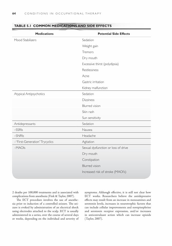
64 C O N D I T I O N S I N O C C U P A T I O N A L T H E R A P Y
2 deaths per 100,000 treatments and is associated with complications from anesthesia (Fink & Taylor, 2007).
The ECT procedure involves the use of anesthe-sia prior to induction of a controlled seizure. The sei-zure is evoked by administration of an electrical shock using electrodes attached to the scalp. ECT is usually administered in a series, over the course of several days or weeks, depending on the individual and severity of
symptoms. Although effective, it is still not clear how ECT works. Researchers believe the antidepressive effects may result from an increase in monoamines and serotonin levels, increases in neurotrophic factors that can include cellular improvements and norepinephrine and serotonin receptor expression, and/or increases in anticonvulsant action which can increase opioids (Taylor, 2007).
TABLE 5.1 COMMON MEDICATIONS AND SIDE EFFECTS
Medications Potential Side Effects
Mood Stabilizers ■ Sedation
■ Weight gain
■ Tremors
■ Dry mouth
■ Excessive thirst (polydipsia)
■ Restlessness
■ Acne
■ Gastric irritation
■ Kidney malfunction
Atypical Antipsychotics ■ Sedation
■ Dizziness
■ Blurred vision
■ Skin rash
■ Sun sensitivity
Antidepressants ■ Sedation
–SSRIs ■ Nausea
–SNRIs ■ Headache
–“First-Generation” Trycyclics ■ Agitation
–MAOIs ■ Sexual dysfunction or loss of drive
■ Dry mouth
■ Constipation
■ Blurred vision
■ Increased risk of stroke (MAOI’s)
65Chapter 5 : Mood Disorder s
Repetitive Transcranial Magnetic Stimulation
Repetitive transcranial magnetic stimulation (rTMS) is a newly emerging intervention for treatment-resistant depression. rTMS involves a noninvasive procedure of creating a magnetic field that passes through the skull inducing electrical currents in the brain that activate specific nerve cells. The procedure can be repeated daily over a period of days or weeks. It has proven successful in reducing depressive symptoms and improving remission rates (George et al., 2010; Gross, Nakamura, Pascual-Leone, & Fregni, 2007; Holtzheimer et al., 2010). Based on recent successful clinical trials, an rTMS device for the treatment of MDD has been recently approved by the U.S. Food and Drug Administration.
IMPACT ON OCCUPATIONAL PERFORMANCEMood disorders, particularly those at the extreme ends of the mood spectrum, can significantly interfere with daily functioning. Symptoms of both MDD and BPD inter-fere with one’s ability to work, socialize, recreate, sleep, eat, and learn. The impact on occupational performance, however, can vary greatly based on factors such as sever-ity of symptoms, responsiveness to treatment, number of episodes, co-occurring conditions and age of onset. For example, an adult with a high level of premorbid func-tioning, who experiences a single episode and responds well to intervention, may successfully return to work and family roles. For many individuals, however, there are numerous barriers to occupational performance once symptoms occur.
Basic Activities of Daily Living
The ability to care for personal health such as eating well, following a healthy diet, and sleeping and exercis-ing regularly can be impaired. Previously maintained daily routines can be disrupted. Symptoms of avolition and anhedonia seen in depressive episodes can decrease motivation for exercise, proper nutrition, grooming, and hygiene. Symptoms of distractibility, impulsivity, and psy-chomotor disturbances that are characteristic of a manic or hypomanic episode can interfere with the quality of self-care. Additionally, sleep patterns are often disturbed, with individuals either not getting adequate sleep, sleep-ing excessively, or experiencing poor sleep quality. Weight gain or loss is common, and can be related to either changes in daily habits or long-term medication use.
Instrumental Activities of Daily Living
Early onset in childhood or adolescence, a time of developing skills and interests, coupled with high rates of recurrence throughout adulthood, can limit one’s ability to develop educational, social, and work skills. For individuals who continue to experience residual symptoms between episodes, it can be even more dif-ficult to reengage in meaningful life roles. The individ-ual dealing with recurring depressed, manic, or cycling moods has great difficulty managing daily tasks that can include work, school, parenting/caregiving, home maintenance, and healthy leisure.
Many individuals with mood disorders lack the educational and training requirements needed for a successful career. Moreover, persons suffering from depressive episodes are likely to experience symptoms of anhedonia, avolition, and cognitive difficulties, which decrease motivation and ability to work. Stud-ies reveal that those diagnosed with MDD or BPD are less likely to work outside the home, either full- or part-time (Substance Abuse and Mental Health Ser-vices Administration, 2009; Suppes et al., 2001), and those who do work, experience lowered productivity and increased absenteeism (Cook, 2005; Kessler et al., 2005).
Children and adolescents with early onset of a mood disorder may experience symptoms that can negatively impact school performance, such as lower scores on achievement tests and impaired peer interactions, includ-ing social withdrawal or fighting and disruptive behav-ior. Studies have shown that high school students with depression, particularly females, are more likely to drop out of high school and are less likely to enroll in college (Fletcher, 2007).
Involvement in healthy leisure activities is usually limited, as well. Those with MDD are likely to socially isolate and cease participation in activities due to symp-toms of anhedonia and avolition. During a manic or hypomanic episode, an individual may experience an initial burst of creative, goal-directed energy, but is more likely to engage in risky or dangerous activities, and, over time, to become more unable to organize time and activities.
In general, those who experience recurring episodes, limited treatment success, co-occurring medical or psy-chiatric conditions, and lack of healthy support sys-tems are more likely to demonstrate an overall decline in functioning. Early intervention and ongoing access to treatment and other support networks can improve one’s ability to maintain meaningful and productive roles and to experience a greater quality of life through social participation.
66 C O N D I T I O N S I N O C C U P A T I O N A L T H E R A P Y
Case IllustrationsCase 1Phyllis spent excessive hours at her new human resources job, planning how to take over the com-pany. She was visibly irritated with what she perceived as organizational inefficiencies and spoke about her concerns to her employers. Her grandiosity was evi-dent by all her coworkers. She drafted numerous versions of detailed proposals to “take the company to the next level.” Despite working long hours at her computer each day, she failed to complete the tasks she was hired to do. She was continually distracted by her big plans. In addition, she began shopping for new clothes that she felt would be in keeping with her elevated status. She ran up large credit card bills and was in financial trouble.When she shared her business ideas with family members, they gave her feedback about how unre-alistic she was being and urged her to focus on her job. She responded defensively, unable to recognize how her behavior was negatively affecting her work. Although she was highly talented in her field, she was fired after 3 months on the job.
Case 2Justin is a 9-year-old boy who was admitted to the child and adolescent psychiatric inpatient unit at the local community mental health center. He had been living in a foster home since being removed from his biological parents’ home at age 7, due to neglect.His foster parents describe Justin as quiet and passive in his interactions with others. He avoids eye contact and wears his hair long, over his eyes. Justin spends most of his free time in his room, alone, watching television or playing video games. Schoolwork is a daily struggle for Justin’s foster parents, as they indicate he is disinterested in school and avoids completing his assignments. They report he “throws temper tan-trums” when they make him do homework. He fre-quently complains of having an upset stomach when it is time to get ready for school in the morning. During the initial interview with the occupational therapist, Justin shared that he felt he was “bad” and that was why he had been removed from his biological parents’ custody.
American Psychiatric Association. (2000). Diagnostic and statistical manual of mental disorders (4th ed., rev.). Washington, DC: Author.
Arnow, B. A., & Constantino, M. J. (2003). Effective-ness of psychotherapy and combination treatment for chronic depression. Journal of Clinical Psychology, 59, 893–905.
Beesdo, K., Hofler, M., Leibenluft, E., Lieb, R., Bauer, M., Pfenning, A. (2009). Mood episodes and mood
disorders: Patterns of incidence and conversion in the first three decades of life. Bipolar Disorders, 11, 537–549.
Boschloo, L., Vogelzangs, N., Smit, J. H., van den Brink, W., Veltman, D. J., Beekman, A. T. F., et al. The performance of the Alcohol Use Disorder Identi-fication Test (AUDIT) in detecting alcohol abuse and dependence in a population of depressed or anxious persons. Journal of Affective Disorders, 125, 441–445.
R E C O M M E N D E D L E A R N I N G R E S O U R C E S
National Alliance on Mental Illness (NAMI)3803 N. Fairfax Dr., Ste. 100Arlington, VA 22203Phone: 703-524-7500Fax: 703-524-9094www.nami.orgNational Institute of Mental Health (NIMH)Science Writing, Press, and Dissemination Branch5001 Executive Boulevard, Room 8184, MSC 9553Bethesda, MD 20892-9553
Phone: 1-855-515-5454Fax: 301-443-4279www.nimh.nih.govNARSAD50 Cutter Mill Road, Suite 404Great Neck, NY 11021Phone: 515-829-0091Fax: 515-487-5930www.narsad.org
R E F E R E N C E S
67Chapter 5 : Mood Disorder s
Carnethon, M. R., Biggs, M. L., Barzilay, J. I., Smith, N. L., Vaccarino, V., Bertoni, A. G., et al. (2007). Longitudinal association between depressive symptoms and incident type 2 diabetes mellitus in older adults: The cardiovascular health study. Archives of Internal Medicine, 157, 802–807.
Caspi, A., Sugden, K., Moffitt, T. E., Taylor, A., Craig, I. W., Harrington, H., et al. (2003). Influence of life stress on depression: Moderation by a polymorphism in the 5-HTT gene. Science, 301, 385–389.
Cole, J., Toga, A. W., Hojatkashani, C., Thompson, P., Costafreda, S. G., Cleare, A. J., et al. (2010). Subre-gional hippocampal deformations in major depressive disorder. Journal of Affective Disorders, 125, 272–277.
Colton, C. W., & Manderscheid, R. W. (2005). Congruen-cies in increased mortality rates, years of potential life lost, and causes of death among public mental health clients in eight states. Preventing Chronic Disease, 3(2), 1–14.
Conway, K. P., Compton, W., Stinson, F. S., & Grant, B. F. (2005). Lifetime comorbidity of DSM-IV mood and anxiety disorders and specific drug use disorders: Results from the National Epidemiologic Survey on Alcohol and Related Conditions. Journal of Clinical Psychiatry, 57, 247–257.
Cook, J. A. (2005). Employment barriers for persons with psychiatric disabilities: Update of a report for the President’s Commission. Psychiatric Services, 57, 1391–1405.
Costello, E. J., Pine, D. S., Hammen, C., March, J. S., Plotsky, P. M., Weissman, M. M., et al. (2002). Devel-opment and natural history of mood disorders. Society of Biological Psychiatry, 52, 529–542.
Egede, L. E., & Zheng, D. (2003). Independent factors associated with major depressive disorder in a nation-al sample of individuals with diabetes. Diabetes Care, 25, 104–111.
Everson, S. A., Maty, S. C., Lynch, J. W., & Kaplan, G. A. (2002). Epidemiologic evidence for the relation between socioeconomic status and depression, obesity, and dia-betes. Journal of Psychiatric Research, 53, 891–895.
Fink, M., & Taylor, M. A. (2007). Electroconvulsive ther-apy: Evidence and challenges. JAMA, 298, 330–332.
Fletcher, J. M. (2007). Adolescent depression: Diagno-sis, treatment, and educational attainment. Journal of Health Economics, 17, 1215–1235.
Foti, D., Olvet, D. M., Klein, D. N., & Hajcak, G. (2010). Reduced electrocortical response to threatening faces in major depressive disorder. Depress Anxiety, 27, 813–820. doi: 10.1002/da.20712
Gavin, A. R., Walton, E., Chae, D. H., Alegria, M., Jack-son, J. S., & Takeuchi, D. (2010). The associations between socio-economic status and major depressive disorder among Blacks, Latinos, Asians and non-Hispanic Whites: Findings from the Collaborative Psychiatric Epidemiology Studies. Psychological Medi-cine, 40, 51–51.
George, M. S., Lisanby, S. H., Avery, D., McDonald, W. M., Durkalski, V., Pavlicova, M., et al. (2010). Daily left prefrontal transcranial magnetic stimulation ther-apy for major depressive disorder. Archives of General Psychiatry, 57, 507–515.
Goldberg, J., & Harrow, M. (2004). Consistency of re-mission and outcome in bipolar and unipolar mood disorders: A ten-year prospective follow-up. Journal of Affective Disorders, 81, 123–131.
Gos, T., Krell, D., Bielau, H., Steiner, J., Mawrin, C., Trubner, K., et al. (2010). Demonstration of disturbed activity of the lateral amygdaloid nucleus projection neurons in depressed patients by the AgNOR staining method. Journal of Affective Disorders, 125, 402–410.
Gotlib, I. H., & Hamilton, J. P. (2008). Neuroimaging and depression: Current status and unresolved issues. Cur-rent Directions in Psychological Science, 17, 159–153.
Gross, M., Nakamura, L., Pascual-Leone, A., & Fregni, F. (2007). Has repetitive transcranial magnetic stimula-tion (rTMS) treatment for depression improved? A systematic review and meta-analysis comparing the recent vs. the earlier rTMS studies. Acta Psychiatrica Scandinavica, 115, 155–173.
Hammen, C. (2005). Stress and depression. Annual Review of Clinical Psychology, 1, 293–319.
Hammen, C. (2009). Adolescent depression: Stressful interpersonal contexts and risk for recurrence. Current Directions in Psychological Science, 18, 200–204.
Hennessy, M. B., Schiml-Webb, P. A., & Deak, T. (2009). Separation, sickness, and depression: A new perspec-tive on an old animal model. Current Directions in Psy-chological Science, 18, 227–231.
Holtzheimer, P. E., McDonald, W. M., Mufti, M., Kelley, M. E., Quinn, S., Corso, G., et al. (2010). Accelerated repeti-tive transcranial magnetic stimulation for treatment-resis-tant depression. Depression and Anxiety, 27, 950–953.
Kendler, K. S., Gatz, M., Gardner, C. O., & Pedersen, N. L. (2005). A Swedish national twin study of life-time major depression. American Journal of Psychiatry, 153, 109–114.
Kessler, R. C., Akiskal, H. S., Ames, M., Birnbaum, H., Greenberg, P., Hirschfeld, R. M. A., et al. (2005). Prevalence and effects of mood disorders on work perfor-mance in a nationally representative sample of U.S. work-ers. American Journal of Psychiatry, 153, 1551–1558.
Kessler, R., Berglund, P., Demler, O., Jin, R., Koretz, D., Merikangas, K., Rush, A., Wang, P. S. (2003). The epidemiology of major depressive disorder: Results from the National Co-Morbidity Survey Replication. JAMA, 289, 3095–3105.
Kessler, R. C., Chiu, W. T., Demler, O., & Walters, E. E. (2005). Prevalence, severity, and comorbidity of twelve-month DSM-IV disorders in the National Comorbidity Survey Replication (NCS-R). Archives of General Psychiatry, 52, 517–527.
68 C O N D I T I O N S I N O C C U P A T I O N A L T H E R A P Y
Kruger, S., Seminowicz, S., Goldapple, K., Kennedy, S. H., & Mayberg, H. S. (2003). State and trait influ-ences on mood regulation in bipolar disorder: Blood flow differences with an acute mood challenge. Biol Psychiatry, 54, 1274–1283.
Licht-Strunk, E., van der Windt, D. A. W. M., van Mar-wijk, H. W. J., de Haan, M., & Beekman, A. T. F. (2007). The prognosis of depression in older patients in general practice and the community: A systematic review. Family Practice, 24, 158–180.
Luby, J. L. (2010). Preschool depression: The importance of identification of depression early in development. Current Directions in Psychological Science, 19, 91–95.
Maniglio, R. (2010). Child sexual abuse in the etiology of depression: a systematic review of reviews. Depression and Anxiety, 27, 531–542. doi: 10.1002/da.20587
McIntyre, R. S., & Konarski, J. (2005). Tolerability pro-files of atypical antipsychotics in the treatment of bi-polar disorder. Journal of Clinical Psychiatry, 55, 28–35.
McKinnon, M. C., Yucel, K., Nazarov, A., MacQueen, G.M. (2009). A meta-analysis examining clinical predictors of hippocampal volume in patients with major depressive disorder. Journal of Psychiatry & Neuroscience, 34, 41–54.
McMahon, F. J., Akula, N., Schulze, T. G., Muglia, P., Tozzi, F. Detera-Wadleigh, S.D., et al. (2010). Meta-analysis of genome-wide association data identifies a risk locus for major mood disorders on 3p21.1. Nature Genetics, 42, 128–131. doi: 10.1038/ng.523
Miklowitz, D. J. (2007). The role of the family in the course and treatment of bipolar disorder. Current Di-rections in Psychological Science, 16, 192–196.
Miklowitz, D. J. & Johnson, S. L. (2006). The psycho-pathology and treatment of bipolar disorder. Annual Review of Clinical Psychology, 2, 199–235.
Miller, G. E., & Blackwell, E. (2005). Turning up the heat: Inflammation as a mechanism linking chronic stress, depression, and heart disease. Current Directions in Psychological Science, 15, 259–272.
Moller, H. J., & Henkel, V. (2005). What are the most effective diagnostic and therapeutic strategies for the management of depression in specialist care? Copen-hagen: WHO Regional Office for Europe. Retrieved November 5, 2010, from http://www.euro.who.int/en/home.
Monroe, S. M., & Reid, M. W. (2009). Life stress and major depression. Current Directions in Psychological Science, 18, 58–72.
National Institute of Mental Health. (2007). Older adults: Depression and suicide facts (NIH Publication No. 4593). Retrieved from http://www.nimh.nih.gov/health/publications/older-adults-listing.shtml
Rottenberg, J. (2005). Mood and emotion in major depression. Current Directions in Psychological Science, 14, 157–170.
Salvatore, P., Tohen, M., Khalsa, H. M., Baethge, C., Tondo, L., & Baldessarini, R. J. (2007). Longitudinal research on bipolar disorders. Epidemiologia e Psichi-atria Sociale, 15, 109–117.
Schoeyen, H. K., Birkenaes, A. B., Vaaler, A., Auestad, B. H., Malt, U. F., Andreassen, O. A., et al. (2010). Bipolar disorder patients have similar levels of edu-cation but lower socio-economic status than the gen-eral population. Journal of Affective Disorders, doi: 10.1015/j. jad.2010.08.012
Schneck C. D., Miklowitz, D. J., Calabrese, J. R., Al-len, M. H., Thomas, M. R., Wisniewski, S. R., et al. (2004). Phenomenology of rapid-cycling bipolar disorder: Data from the first 500 participants in the Systematic Treatment Enhanced Program. American Journal of Psychiatry, 10, 1902–1908.
Skjelstad, D. V., Malt, U. F., & Holte, A. (2010). Symp-toms and signs of the initial prodrome of bipolar disorder: A systematic review. Journal of Affective Dis-orders, 125, 1–13.
Substance Abuse and Mental Health Services Adminis-tration. (2009). The NSDUH Report: Employment and major depressive episode. Retrieved from http://www.oas.samhsa.gov/2k9/152/Employment.htm
Suppes, T., Leverich, G. S., Keck, P. E. Nolen, W. A., Denicoff, K. D., Altshuler, L.L., et al. (2001). The Stan-ley Foundation Bipolar Treatment Outcome Network. II. Demographics and illness characteristics of the first 251 patients. Journal of Affective Disorders, 57, 45–59.
Taylor, S. (2007). Electroconvulsive therapy: A review of history, patient selection, technique, and medica-tion management. Southern Medical Journal, 100, 494–498.
U.S. Department of Health and Human Services, National Institutes of Health, & National Institute of Mental Health. (2008). Depression (NIH Publi-cation No. 08–3551). Retrieved from http://www.nimh.nih.gov/health/publications/depression/nimhdepression.pdf
U.S. Department of Health and Human Services, Substance Abuse and Mental Health Services Administration, Center for Mental Health Services, National Institutes of Health, & National Institute of Mental Health (1999). Mental health: A report of the Surgeon General. Retrieved from http://www.sur-geongeneral.gov/library/mentalhealth/home.html
Weissman, M. M., Wolk, S., Goldstein, R. B., Moreau, D., Adams, P., Greenwald, S., et al. (1999). Depressed adolescents grown up. JAMA, 281, 1701–1713.
Wingo, A. P., Wrenn, G., Pelletier, T., Gutman, A. R., Bradley, B., & Ressler, K. J. (2010). Moderating effects of resilience on depression in individuals with a history of childhood abuse or trauma exposure. Jour-nal of Affective Disorders, 125, 411–414.
69
Schizophrenia and Other Psychotic Disorders
■ Ann C h ap l e au
6
Joe, a thin, middle-aged man, can often be seen walking about downtown, wearing a cap fashioned out of aluminum foil, pulling a hand cart loaded with assorted household items and clothing. He is
well-known to the mental health community and is a familiar sight to local residents. Joe grew up in a middle-class family, the only child of parents who were both accountants. His parents described him as a happy baby and a bright student, who had attended college to study engineering. But things began to change for Joe and his family when he moved one hour away to the state university when he was 19. Although his teen years were marked by a gradual withdrawal from friends and family, he began to demonstrate more socially bizarre behaviors while at college and when home on weekends, such as dressing in dark, heavy, hooded clothing even in warm weather, and an intense preoccupation with watching the CSPAN television channel. His parents were concerned about his behavior, but hoped it was a temporary phase of adapting to his changing life.
During winter break of his sophomore year in college, while his par-ents were vacationing in Mexico, Joe was seen by neighbors in the park near his family home, wearing camouflage and sleeping in a small pup tent. Police were called, and Joe hid in his tent, refusing to speak to them. He was forcibly removed and taken to the local psychiatric hospital for emergency admission. During his evaluation by the on-call psychiatrist, he revealed that he had moved out of his home because he had discovered “bugs” planted throughout the house. He believed that he was under surveillance by the Central Intelligence Agency (CIA) and was worried that his parents had
been arrested. He reported that he had been suspicious of a plot against him for quite some time, but that this final “discovery” was the confirmation he was waiting for. When the staff arranged for him to speak to his parents, who reassured him they were only on vacation, Joe was convinced that CIA agents were impersonating their voices. A physical exam including blood testing was conducted to rule out any medical or
K E Y T E R M S
AffectAlogia
AnhedoniaAntipsychotic medication
AssocialityAvolition
Catatonic behaviorDelusions
Disorganized behaviorDisorganized thinking
DysphoriaExtrapyramidal syndrome
HallucinationsNegative symptoms
ParanoiaPositive symptoms
Premorbid functioningProdromal phase
PsychosisTardive dyskinesia
70 C O N D I T I O N S I N O C C U P A T I O N A L T H E R A P Y
drug-related cause of the psychosis. His parents were interviewed to obtain pertinent social and medical history. They shared that he had experienced some problems over the past 2 years in getting along with his roommates and was struggling with getting to all of his classes, resulting in low grades. Joe was diag-nosed with paranoid-type schizophrenia and admit-ted to the inpatient program under court order. He was given antipsychotic medications and discharged to his parents upon their return home. He was not able to return to college as planned. He attempted to enroll at the local community colleges for various classes but was never able to complete the courses.
Joe has been in and out of the psychiatric hospital numerous times in the past 25 years. His admissions are typically preceded by his stopping his medications, which results in a return of delusions that he is under CIA surveillance. He has never been employed and has never had a significant-other rela-tionship. He is suspicious of others, and so he will not consent to living in a room and board facility or group home. His attempts at independent living in community apartments are usually short-lived. He has been evicted for inability to pay rent as well as for unsafe conditions in the residence, such as hoard-ing and not cleaning. He resides for brief periods with his parents and has had numerous episodes of street homelessness.
DESCRIPTION AND DEFINITIONSchizophrenia is one of the most severe, complex, and debilitating of all mental health disorders. It is among the top 10 most disabling conditions worldwide (World Health Organization [WHO], 2008). Schizophrenia is a progressive disorder that can be treated, but not cured. This lifelong brain disorder is characterized by periods of psychosis, which is the presence of delusions or halluci-nations without insight. It is also marked by a progressive decline in daily living skills, including work and education skills, social/relationship skills, and basic self-care abili-ties. To better understand the complex disorder of schizo-phrenia, one must understand the terms that describe the clinical symptoms.
■ Delusions: Fixed beliefs that, even in the face of contradictory evidence, are typically due to a misin-terpretation of an event or experience. People with schizophrenia may experience more than one type of delusion at a time or at different points in time during the course of the illness. Delusions are cat-egorized as bizarre or nonbizarre. Bizarre delusions
are characterized by beliefs of events that are clearly impossible and not related to everyday life experi-ences, such as a belief that aliens have impregnated the person while he or she was asleep. An example of a nonbizarre delusion is the belief that all coworkers are talking about the person. The individual may inter-pret that all private conversations observed at work to be about oneself. Delusional content can include any of the following types: ■ Persecutory: The most common of all delusions;
one believes himself or herself to be victimized, ridiculed, or placed under surveillance by known or unknown persons. Some people who suffer from persecutory delusions, such as paranoia, believe that someone is attempting to poison them, lead-ing to a refusal to eat or drink. Another example is thought broadcasting, in which the individual is convinced that outside forces are able to transmit, or broadcast to others, their inner thoughts.
■ Referential: These are also fairly common forms of delusions; one believes that common cues from the environment, such as facial expressions of celebrities on television, casual comments in daily conversations, or newspaper stories are specifically targeted to the individual, holding special meaning or a message for him or her.
■ Somatic: This form of delusion is marked by beliefs that involve the person’s body. For example, a per-son may believe they have received a secret opera-tion while they were under anesthesia or that they are pregnant, despite evidence to the contrary.
■ Religious: People who suffer from religious delu-sions may believe that they are Jesus Christ or are acting out direct orders from God.
■ Grandiose: Examples include believing that one is all-powerful or important, that one is acting on secret orders from the president of the United States, or believing oneself to be a genius or mul-timillionaire.
■ Hallucinations: The experience of particular sensa-tions that are not real to others and that are experi-enced while awake. The two most common forms include ■ Auditory hallucinations: Hearing voices or sounds.
The individual may perceive voices in the external environment, or inside his or her head, often more than one voice conversing or speaking directly to the person. Voices can be described as taunting and cruel, sometimes commanding the individual to perform certain acts, such as harming oneself or others, or can be familiar voices, which are per-ceived as friendly companions.
■ Visual hallucinations: Seeing images of people or objects in the environment. Individuals may
71Chapter 6 : Sch izophren ia and Other Psychot ic Disorder s
describe seeing shadowy figures or the image of a dead body.
■ Other, less common forms of hallucinations include olfactory, tactile, and gustatory hallucina-tions.
■ Disorganized thinking: Speech, which can provide clues about thought processes, may encompass any of the following: ■ Loose associations: Answers that begin to veer “off
track” of the original questions. ■ Tangential: Unrelated comments or answers. ■ Incoherent: Often referred to as “word salad,” seen
in more severe cases. ■ Disorganized behavior: Unpredictable, socially inap-
propriate behaviors that interfere with daily activities. Examples include agitated or angry outbursts with no known provocation; sexually acting out in public, that is, masturbation; and difficulties performing goal-directed tasks such as meal preparation or grooming.
■ Catatonic behavior: Loss of responsiveness to envi-ronmental cues. The individual may assume rigid or bizarre postures and resist attempts made to move or reposition him or her. Excessive, nonpurposeful motor activity may also be observed. In extreme cases, the individual appears to be completely unresponsive, as in a catatonic stupor.
ETIOLOGYThere is no one single factor found to be the cause of schizophrenia. There are a number of models that attempt to explain the multiple factors that can contrib-ute to the development of the disorder. Current research supports both a genetic vulnerability present at birth (Boos et al., 2007) and environmental triggers (Leask, 2004; McDonald & Murray, 2000). Findings from advanced neuropsychological and brain imaging suggest the presence of genetic markers, reduced brain activity in the frontal and temporal regions, and structural abnor-malities in all regions of the brain, including enlarged ventricles, reduced volume of gray matter in the cerebral cortex, and decreased size of the hippocampus and thala-mus (Lawrie & Abukmeil, 1998; Lieberman et al., 2008; Shenton, Dickey, Frumin, & McCarley, 2001; Velak-oulis et al., 2006). The role of key neurotransmitters, including dopamine, glutamate, and serotonin, is being explored to determine their role in genetic alterations (Sawa & Snyder, 2002).
Complications in prenatal development or during delivery, affecting brain development, have also been associated with schizophrenia (Cannon, Jones, & Mur-ray, 2002; Walker, Kestler, Bollini, & Hochman, 2004). Additionally, complications in later brain development, including chronic cannabis use, exposure to trauma, or
other stress, have also been linked to schizophrenia (Veen et al., 2004).
Recent longitudinal studies have focused on the pat-tern of structural changes in the brain and have found pro-gressive anatomical changes both prior to and following onset of the illness (Arango et al., 2008; Kempton, Stahl, Williams, & DeLisi, 2010; Sun et al., 2009; Velakoulis et al., 2006).
Despite major progress in research, the cause of schizophrenia and other psychotic disorders remains a mystery. With new technology including molecular genetics, in vivo brain imaging, and advancements in psy-chopharmacology, there is hope for discovering the cause or causes of schizophrenia that can ultimately lead to advancements in prevention and treatment.
INCIDENCE AND PREVALENCEThe incidence of schizophrenia is low, with a median value of 15.2 per100,000 people. The lifetime prevalence is 4.0 per 1,000 (McGrath, Saha, Chant, & Welham, 2008). Schizophrenia crosses all racial, geographical, and socioeconomic boundaries. Some studies, however, indi-cate that migrant status, lower economic status, residing in a higher latitude, and male gender are factors associ-ated with a higher incidence and prevalence (McGrath et al., 2008).
SIGNS AND SYMPTOMSDespite scientific advancements that reveal neurobiologi-cal abnormalities of the brain, schizophrenia continues to be misunderstood by the general public. Negative ste-reotypes are reinforced by the unique nature of the signs and symptoms of this disorder. Schizophrenia affects the brain, which controls impulse, judgment, affect, and social skills. Perhaps most important, schizophrenia affects one’s insight. As a result, people with schizophre-nia are more likely to have difficulty recognizing and accepting that they have the disorder. This lack of insight can lead to resistance to treatment that can help reduce or eliminate symptoms.
Symptoms of schizophrenia vary greatly, but include the categories of positive, negative, cognitive, and affective symptoms, which are present to some degree in each per-son diagnosed with the disease ( Jibson, Glick, & Tandon, 2004).
Positive Symptoms
Delusions, hallucinations, and disorganized thoughts are referred to as “positive” symptoms, as they involve an excess of behavior or experiences. Delusions are the most
72 C O N D I T I O N S I N O C C U P A T I O N A L T H E R A P Y
common type of positive symptoms observed, affecting 65% of people with schizophrenia, while hallucinations and disorganized thoughts each affect 50% of those with schizophrenia ( Jibson et al., 2004). Positive symptoms may be continuously or intermittently present, but rarely abate completely.
Negative Symptoms
Flattened affect (facial and other nonverbal expression), alogia (impoverished speech), avolition (decreased moti-vation), anhedonia (loss of pleasure in previously enjoyed activities), and associality (decreased interest in socializa-tion and maintenance of relationships) are referred to as “negative” symptoms, as they represent an absence of func-tion or experience.
Cognitive Symptoms
Memory, attention, language, and executive function, such as abstract reasoning and planning skills, are affected by schizophrenia. In tests of sensory information processing, individuals with schizophrenia demonstrate reduced abil-ity to process visual stimuli and to respond to environ-mental stimuli, due to motor skills deficits (Walker et al., 2004). Use of neuroimaging and cognitive tests have also revealed impaired neurocognitive functioning in com-pleting tasks requiring use of frontal and temporal lobes (Lieberman et al., 2008).
Mean IQ scores range from 80 to 85, significantly lower than the norm of 100 for the general population ( Jibson et al., 2004). People with schizophrenia also lack skills in identifying social cues and solving social problems. In general, research has not yielded any spe-cific cognitive deficit present in all individuals with schizophrenia. Impairments appear generalized and vary greatly from person to person.
Affective Symptoms
Flattened or inappropriate affect is a common symptom of schizophrenia, and people with schizophrenia are more likely to demonstrate difficulties in identifying and expressing emotions. This, coupled with a limited abil-ity to identify social cues, can lead to maladaptive social functioning.
Mood disturbances are also commonly seen. Dys-phoria or depressed mood can be present during or fol-lowing a psychotic episode and may require intervention. Individuals who develop insight into the severity of their illness are at greater risk of becoming depressed or experi-encing demoralization.
The types and degree of symptoms present in an individual guide the diagnosis of one of five subtypes of schizophrenia: paranoid, disorganized, catatonic, undif-ferentiated, and residual types (American Psychiatric Association [APA], 2000). Paranoid-type schizophrenia is characterized by the presence of hallucinations or delusions, without disorganized thoughts or behavior or socially inappropriate affect. Persons with paranoid-type schizophrenia are believed to have a better prognosis. Disorganized type presents with disorganized behavior and speech and inappropriate affect. Those diagnosed with catatonic type, the rarest type, primarily experience abnormal posture and movement, with minimal speech or echolalia. A diagnosis of undifferentiated type is given when the individual does not meet criteria for any of the other types while still meeting the general diagnostic criteria for schizophrenia. Residual type reflects an absence of current psychosis, but continued negative symptoms or more mild impairments in social skills, affect, and thinking.
COURSE AND PROGNOSISOnset of symptoms can be gradual or acute, but there are usually earlier signs of dysfunction, in both premorbid functioning, the period from birth to the prodromal phase, and the prodromal phase, which can range from weeks or months to years before full onset of symptoms. Studies of premorbid functioning reveal subtle signs of problems in motor development, affect, and school per-formance. Impairments in cognitive functioning are often documented in school records and formal testing much earlier in life (Russell, Munro, Jones, Hemsley, & Murray, 1997). Some individuals do not present with significant premorbid “clues,” but begin to show early signs of psycho-sis and unusual behavior in the prodromal phase. Non-specific clinical symptoms seen in the prodromal phase can include affective changes such as depressed or anxious mood, irritability, insomnia, and cognitive changes such as impaired concentration and difficulty attending to tasks. Late-emerging symptoms in the prodromal phase can include suspiciousness, brief hallucinations, and per-ceptual difficulties (Kulhara, Banerjee, & Dutt, 2008).
Age of onset for schizophrenia is between 16 and 30 and is typically earlier for males than females. Diagno-sis after age 45 is unusual (National Institute of Mental Health [NIMH], 2009). However, early-onset schizo-phrenia can be diagnosed in childhood or adolescence. Childhood onset refers to diagnosis of schizophrenia before the age of 13 and is characterized by an insidious onset, rather than a clear first episode, with multiple neu-rodevelopmental impairments present prior to the onset of psychotic or negative symptoms (Arango et al., 2008). Adolescent onset usually has a clear first episode, with a mean onset age of 15 (Arango et al.).
73Chapter 6 : Sch izophren ia and Other Psychot ic Disorder s
The criteria for a diagnosis of schizophrenia require the presence of symptoms for a period of at least 1 month, with some clinical signs present for at least 6 months (APA, 2000). The course of the illness varies greatly, with multiple episodes with changeable symptoms. The APA provides a classification of the longitudinal course that can be applied 1 year after initial onset (APA). The course can be classified as a single episode, episodic, continuous, or in partial or full remission.
Some individuals are able to maintain independent living and work competitively, whereas others experience a chronic decline in functioning, resulting in the need for 24-hour supervision and care. Approximately 10% to 20% of individuals are able to maintain remission for 5 years after the first psychotic episode, but the majority experience continued relapses ( Jibson et al., 2004). Those who do not comply with an antipsychotic medication regime are five times more likely to relapse than those who are medication compliant. Relapses, which are asso-ciated with deterioration in daily functioning, can also be triggered by noncompliance with psychosocial treatment, environmental stressors, and substance abuse ( Jibson et al.). Over time, with multiple psychotic episodes, negative and cognitive symptoms may become more prominent while positive symptoms are more likely to decrease in intensity in late middle age (Lieberman et al., 2008).
Other factors that can negatively impact prognosis include male gender, having a gradual and early onset of symptoms, a family history of schizophrenia, poor pre-morbid functioning, dysfunctional family relationships, and experience of abuse or neglect ( Jibson et al., 2004; Walker et al., 2004).
Even when medication has been shown to reduce or even stop positive symptoms such as hallucinations and delusions, cognitive impairments, such as executive functions and abstract reasoning, are more likely to per-sist. Individuals with more severe cognitive loss are apt to experience decreased occupational performance and quality of life than those who demonstrate cognitive improvements ( Jibson et al., 2004).
People with schizophrenia die, on average, 25 years earlier than those in the general population, related to pulmonary, cardiac, and infectious diseases (Colton & Manderscheid, 2006). Complications from antipsychotic medications, self-neglect, fear of health institutions, lack of access to health care, and increased suicide rate (Melt-zer, 2002) are all contributing factors to this disparity in life expectancy.
Quality of life is also significantly lower for people with schizophrenia, as they represent lower socioeconomic sta-tus, are typically unemployed or underemployed, and are at higher risk of homelessness and incarceration (WHO, 2009). They are more likely to be victims of crime, espe-cially violent crimes, and to abuse alcohol and/or drugs.
Approximately 50% of all people with schizophrenia have a co-occurring diagnosis of substance abuse or depend-ence (Volkow, 2009). Smoking rates are also high among those with schizophrenia, estimated as high as four times the general population (Kumari & Postma, 2005).
MEDICAL/SURGICAL MANAGEMENTThe focus of treatment is to reduce or eliminate symp-toms and to provide environmental supports to enhance quality of life. The primary treatment for symptom reduc-tion continues to be medication, although technological advances show promise for other medical or surgical approaches to treatment.
Pharmacologic Treatment
Prior to the 1950s, treatment consisted of institution-alization, including physical restraints and seclusion, even lobotomy. There was no medication for relief of clinical symptoms. Thorazine was the first antipsychotic medica-tion introduced in the 1950s. Other similar medications, referred to as “first-generation” or “typical” antipsychotics, were also quickly developed. This first group of medica-tions, which included Haldol, Prolixin, and Navane, proved effective in decreasing positive symptoms of schizophre-nia by blocking dopamine receptors. Unfortunately, the side effects were often severe, including extrapyramidal syndrome (abnormal movements similar to Parkinson’s disease), tardive dyskinesia (motor abnormalities such as writhing movements), cardiac problems, and heavy sedation.
The first atypical antipsychotics were introduced in the late 1980s. Atypical antipsychotics differ from the typical or first-generation antipsychotics in that they occupy different neurotransmitter receptors. Clozapine (Clozaril) was introduced in 1989, receiving FDA approval in 1990. It is considered to be highly effective in treating psychotic symptoms for individuals who were previously unresponsive to other first-generation medi-cations. A major drawback, however, is the potential for serious side effects, including development of agranulo-cytosis, which requires regular blood work to monitor white blood cell count. Despite its effectiveness, it is not a favored medication among people with schizophrenia due to the medical risk, additional side effects, inconvenience, and excessive cost ( Jibson et al., 2004).
Other atypical antipsychotics introduced since the 1990s include Risperdal (risperidone), Zyprexa ( olanzapine), Seroquel (quetiapine), Geodon (ziprasi-done), Aripiprazole (Abilify), and Paliperidone (Invego). They also are less likely to cause motor abnormalities
74 C O N D I T I O N S I N O C C U P A T I O N A L T H E R A P Y
such as extrapyramidal effects, but there can be other side effects including sedation, mild hypotension, weight gain, akathesia (restlessness), dry mouth, and constipation (Lieberman et al., 2005; NIMH, 2009).
Repetitive Transcranial Magnetic Stimulation
While medications have been successful in reducing posi-tive symptoms of schizophrenia, there has been no proven treatment for negative symptoms. Transcranial magnetic stimulation (TMS) was developed in the 1980s to study brain function. It involves creating a magnetic field that passes through the skull, creating a current in the brain that activates nearby nerve cells. A coil of wire, wrapped in plastic, is held to the head while a capacitor is discharged to create the magnetic field. Researchers learned that repeated applications (repetitive transcranial magnetic stimulation [rTMS]) over the course of several days appear to affect brain activity. The magnetic field can be targeted to spe-cific regions of the brain where nerve cells are associated with psychiatric symptoms. Recent studies of rTMS, while inconclusive, show promise in reducing negative symptoms of schizophrenia (Diabac-de Lange, Knegtering, & Ale-man, 2010; Matheson, Green, Loo, & Carr, 2010). Other studies of rTMS have also shown effectiveness in reduc-ing positive symptoms of auditory hallucinations, when used in conjunction with antipsychotic medication (Bagati, Haque Nizamie, & Prakash, 2009).
Electroconvulsive Therapy
Electroconvulsive therapy (ECT) is a medical procedure that consists of inducing a seizure by administration of electrical shock using electrodes attached to the scalp. Anesthesia is used for this brief procedure. It is gener-ally administered in a series, over the course of days or weeks. The mortality rate for ECT is approximately 2 deaths per 100,000 treatments and is associated with anesthesia complications (Fink & Taylor, 2007). ECT is used for treating severe symptoms that do not respond to medication, particularly catatonia. Studies indicate some short-term benefits in global functioning when used in conjunction with antipsychotic medication (Tharyan & Adams, 2005). Side effects include confusion experienced immediately after the procedure as well as short-term memory loss for about 1 to 2 weeks.
IMPACT ON OCCUPATIONAL PERFORMANCEBecause schizophrenia typically presents in early adult-hood, a period of developing new roles and responsibilities
such as career, life partnership, independent living, and parenthood, its lifelong effects are profound. Functional abilities vary greatly, but can be positively affected by envi-ronmental factors such as the presence of social support systems, financial assistance, and opportunities for housing or work.
Basic Activities of Daily Living
The ability to manage personal health is significantly compromised. Antipsychotic medications can cause weight gain, cardiac problems, and other health risks. Many individuals have impaired motor functioning and visual processing deficits, which, coupled with avolition and anhedonia symptoms, can limit interest or ability to participate in traditional exercise, nutrition, or smoking cessation programs.
Avolition, anhedonia, and disorganized behavior can also affect one’s ability to initiate and complete self-care tasks such as grooming and hygiene. Impaired visual processing and difficulty interpreting visual stimuli can be associated with excessive application of makeup or ineffective attempts to bathe or shave thoroughly.
Instrumental Activities of Daily Living
The majority of people with schizophrenia do not live or work independently. The ability to work or succeed in school; manage a household including cooking, cleaning, laundry, and budgeting; or function as a caregiver requires multiple skills such as higher level cognitive processes, sen-sorimotor skills, and social nteraction skills, all of which can be affected by the positive, negative, affective, and cog-nitive symptoms of schizophrenia. Those who experience repeated relapses over time, which often involve hospi-talization, demonstrate an overall decline in functioning, which limits opportunities for independent living.
Education
There is some evidence of impaired academic performance in premorbid functioning as well as during the prodromal phase, with standardized testing and general IQ scores in a significantly lower range than the general popula-tion (Fuller et al., 2002). Many individuals experience their first acute onset in young adulthood, which can derail plans for post–high school education or gradua-tion. In fact, worldwide, about one-third of all people with schizophrenia do not graduate from high school (WHO, 2009). While antipsychotic medication can be effective in treating positive symptoms, there is no intervention that has been shown to create meaningful improvements in cognitive functioning. For the vast majority of those with

75Chapter 6 : Sch izophren ia and Other Psychot ic Disorder s
schizophrenia, ongoing cognitive impairments persist and can have a major impact on academic success.
Work
Competitive work situations require multiple job-specific skills, the ability to learn new information quickly, and the ability to interact effectively with coworkers, customers, and superiors. Individuals with schizophrenia are more likely to lack college education or other technical or skilled trade certification. Even with supported employment assistance, they are more likely to be placed in minimum wage jobs such as fast food work, which requires the abil-ity to process and respond very quickly while maintain-ing effective communication with coworkers. These work environments are often too demanding for the individual with active symptoms of schizophrenia. Successful work environments provide structured tasks, additional time allotted to complete work, the ability to work part-time hours, and opportunities for job coaching, particularly when new expectations are introduced.
Leisure and Social Participation
Symptoms of anhedonia and avolition can play a crucial role in limiting involvement in leisure activities. Psychoso-cial functioning may be impaired by both associality and affective symptoms, such as flattened affect and the abil-ity to recognize and reciprocate appropriate social cues. People with schizophrenia typically have less financial and transportation resources for recreation, fewer social contacts, and less interest in leisure activities. As a result, they are more likely to be socially isolated.
OTHER PSYCHOTIC DISORDERSIn addition to schizophrenia, there are several other psy-chotic disorders as classified by the APA (2000). Schizo-phrenia and the following disorders all share the common feature of active psychosis.
Schizophreniform Disorder
Clinical features are nearly identical to schizophrenia with the exception of two differences: (1) Total dura-tion of the illness is more than 1 month but <6 months, and (2) occupational performance deficits may not be present. About one-third of all people diagnosed with schizophreniform disorder will experience remission within a 6-month period, but the remaining two-thirds are likely to eventually be diagnosed with either schizo-phrenia or schizoaffective disorder.
Schizoaffective Disorder
Clinical symptoms of schizophrenia are present, includ-ing delusions or hallucinations, but at some point in the course of the illness, a major depressive, manic, or mixed episode occurs. There are two subtypes of schizoaffective disorder, depending on the mood presentation: bipo-lar type, which includes a manic or mixed episode, and depressive type. Age of onset is typically in early adult-hood but can range from adolescence to late life.
Delusional Disorder
Symptoms include the presence of nonbizarre delusions lasting at least 1 month. Auditory or visual hallucinations may be present, but are not prominent. Olfactory or tac-tile hallucinations may be both present and prominent if they relate to the delusion. For example, if a person believes that he or she has an unknown infectious dis-ease, he or she may experience related body sensations or odors. Activities of daily living skills are not signifi-cantly impaired, although work, social, and relationship problems can occur as a result of the delusional beliefs. For example, those who experience jealous or persecutory delusions may demonstrate angry or violent behavior. Age of onset for delusional disorders is highly variable, from adolescence to late life. The course of the illness can also be variable, from chronic to full remission within several months.
Brief Psychotic Disorder
Age of onset is typically in adolescence or early adult-hood, with a sudden onset of positive psychotic symp-toms or highly disorganized or catatonic behavior lasting between 1 day and 1 month. The brief episode is followed by a return to premorbid functioning. There may or may not be a precipitating stressor, such as death of a loved one or a traumatic experience in war combat. Individu-als experience great confusion and dramatic mood shifts. Occupational performance can be significantly impaired, and there is an increased risk of suicide due to impulsivity.
Shared Psychotic Disorder
In this rarely seen disorder, the individual comes to believe in a nonbizarre or somewhat bizarre delusion held by a significant other, close friend, or family member. Although usually seen between two adults, it can occur among groups, particularly family members. For exam-ple, children may adopt a parent’s delusional beliefs. The course of the illness can be varied but is usually chronic due to its occurrence in long-term relationships. When
76 C O N D I T I O N S I N O C C U P A T I O N A L T H E R A P Y
Case IllustrationCase 1Megan is a 12-year-old girl, in the sixth grade at mid-dle school. She was born in Texas, but moved with her mother and four siblings to Michigan when she was 5. Megan’s father was jailed in Texas for running a methamphetamine laboratory in their family home. The family moved in with Megan’s grandmother in the Detroit area, and her mother was able to find part-time employment in hotel housekeeping. Megan had difficulty adjusting to her new living situation and starting school for the first time. She had trouble fol-lowing classroom rules such as sharing supplies and toys. In the second and third grades, she struggled with learning to read and write. Her IQ score was 85. She did not develop any close friendships. She struggled
academically with most subjects, but excelled in art, often drawing or painting very dark and strange images that she could not explain. She became increasingly withdrawn and disinterested in peer-related activities such as sports and going to the movies.
When Megan was 12, she was referred for psy-chological testing, which revealed that she had clinical symptoms of psychosis. Megan revealed to the psy-chologist that Satan had appeared to her on multiple occasions. She was tormented by voices telling her that she was evil and should commit suicide. She was seen by a psychiatrist who diagnosed her with early onset schizophrenia, admitting her to a local child and adolescent psychiatric hospital for medication and psychotherapy.
the individual is separated from the other person, the delusions abate.
Psychotic Disorder Due to a Medical Condition or Substance Use
Delusions or hallucinations are a direct result of a gen-eral medical condition, such as epilepsy, brain lesions, Huntington’s disease, hepatic or renal disease, lupus, or auditory or visual nerve injuries. The course can be varied, from a single episode to recurrent. Even when the underlying medical condition is resolved, psychotic symptoms can continue, particularly in cases of brain injury.
Substance-Induced Psychotic Disorder
Clinical features include hallucinations or delusions directly due to effects of a drug or exposure to a toxin. In cases of intoxication or withdrawal from an abused drug, the hallucinations and delusions are more severe and present well beyond what would be expected during the intoxication/withdrawal/detoxification stages.
Psychotic Disorder NOS
Psychotic symptoms are present but do not meet the cri-teria for any specific psychotic disorder.
R E C O M M E N D E D L E A R N I N G R E S O U R C E S
Torrey, E. F. (2001). Surviving schizophrenia: A manual for families, consumers, and providers. New York, NY: Harper Collins Publishing.
OrganizationsNational Alliance on Mental Illness (NAMI)3803 N. Fairfax Dr., Ste. 100Arlington, VA 22203Tel: (703) 524-7600Fax: (703) 524 9094www.nami.org
National Institute of Mental Health (NIMH)Science Writing, Press, and Dissemination Branch6001 Executive Boulevard, Room 8184, MSC 9663Bethesda, MD 20892-9663Tel: (866) 615-6464Fax: (301) 443-4279www.nimh.nih.govNARSAD60 Cutter Mill Road, Suite 404Great Neck, NY 11021Tel: (516) 829-0091Fax: (516) 487-6930www.narsad.org
77Chapter 6 : Sch izophren ia and Other Psychot ic Disorder s
R E F E R E N C E S
American Psychiatric Association. (2000). Diagnostic and statistical manual of mental disorders (Revised 4th ed.). Washington, DC: Author.
Arango, C., Moreno, C., Martinez, S., Parellada, M., Desco, M., Moreno, D., et al. (2008). Longitudinal brain changes in early-onset psychosis. Schizophrenia Bulletin, 34, 341–353.
Bagati, D., Haque Nizamie, S., & Prakash, R. (2009). Effect of augmentatory repetitive transcranial mag-netic stimulation on auditory hallucinations in schizo-phrenia: Randomized controlled study. Australian and New Zealand Journal of Psychiatry, 43, 386–392.
Boose, E., Ellison, A. M., Osterweil, L. J., Podorozhny, R., Clarke, R., Wise, A., Hadley, J.L., & Foster, D. R. (2007). Ensuring reliable datasets for environ-mental models and forecasts. Ecological Informatics, 2, 237–247.
Cannon, M., Jones, P. B., & Murray, R. M. (2002). Ob-stetric complications and schizophrenia: Historical and met-analytic review. The American Journal of Psy-chiatry, 159, 1080–1092.
Colton, C. W., & Manderscheid, R. W. (2006). Congru-encies in increased mortality rates, years of potential life lost, and causes of death among public mental health clients in eight states. Preventing Chronic Dis-ease, 3. Retrieved from http://www.cdc.gov/pcd/ issues/2006/apr/05_0180.htm
Diabac-de Lange, J. J., Knegtering, R., & Aleman, A. (2010). Repetitive transcranial magnetic stimulation for negative symptoms of schizophrenia: Review and metanalysis. The Journal of Clinical Psychiatry, 71, 411–418.
Fink, M., & Taylor, M. A. (2007). Electroconvulsive therapy: Evidence and challenges. The Journal of the American Medical Association, 298, 330–332.
Fuller, R., Nopoulos, P., Arndt, S., O’Leary, D., Ho, B. C., & Andreason, N. C. (2002). Longitudinal assessment of premorbid cognitive functioning in patients with schizophrenia through examination of standardized scholastic test performance. The American Journal of Psychiatry, 159, 1183–1189.
Jibson, M. D., Glick, I. D., & Tandon, R. (2004). Schizophrenia and other psychotic disorders. Focus, 2(1), 17–30.
Kempton, M. J., Stahl, D., Williams, S. C. R., & DeLisi, L. E. (2010). Progressive lateral ventricular enlarge-ment in schizophrenia: A meta-analysis of longitudi-nal MRI studies. Schizophrenia Research, 120, 54–62.
Kulhara, P., Banerjee, A., & Dutt, A. (2008). Early inter-vention in schizophrenia. The Indian Journal of Psy-chiatry, 50, 128–134.
Kumari, V., & Postma, P. (2005). Nicotine use in schizo-phrenia: The self-medication hypotheses. Neurosci-ence & Biobehavioral Reviews, 29, 1021–1034.
Lawrie, S. M., & Abukmeil, S. S. (1998). Brain abnor-mality in schizophrenia: A systematic and quantita-tive review of volumetric magnetic resonance imaging studies. The British Journal of Psychiatry, 172, 110–120.
Leask, S. J. (2004). Environmental influences in schizo-phrenia: The known and the unknown. Advances in Psychiatric Treatment, 10, 323–330.
Lieberman, J. A., Drake, R. E., Sederer, L. I., Belger, A., Keefe, R., Perkins, D., et al. (2008). Science and recov-ery in schizophrenia. Psychiatric Services, 59, 487–496.
Lieberman, J. A., Stroup, S. T., McEvoy, J. P., Swartz, M. S., Rosenheck, R. A., Perkins, D. O., et al. (2005). Effectiveness of antipsychotic drugs in patient with chronic schizophrenia. The New England Journal of Medicine, 353, 1209–1223.
Matheson, S. L., Green, M. J., Loo, C., & Carr, V. J. (2010). A change in the conclusions of a recent sys-tematic meta-review: Repetitive transcranial magnetic stimulation is effective for the negative symptoms of schizophrenia. Schizophrenia Research. Advance on-line publication. doi:10.1016/j.schres 2010.05 029
McDonald, C., & Murray, R. M. (2000). Early and late environmental risk factors for schizophrenia. Brain Research Reviews, 31, 130–137.
McGrath, J., Saha, S., Chant, D., & Welham, J. (2008). Schizophrenia: A concise overview of incidence, preva-lence, and mortality. Epidemiologic Reviews, 30, 67–76.
Meltzer, H. Y. (2002). Suicidality in schizophrenia: A review of the evidence for risk factors and treatment options. Current Psychiatry Reports, 4, 279–283.
National Institute of Mental Health. (2009). Schizophre-nia (NIH Publication No. 09-3517). Bethesda, MD: Author.
Pantelis, C., Velakoulis, D., Wood, S. J., Suckling, J., Phillips, L. J., Yung, A. R., et al. (2003). Neuroana-tomical abnormalities before and after onset of psy-chosis: A cross-sectional and longitudinal MRI com-parison. Lancet, 25, 281–288.
Russell, A. J., Munro, J. C., Jones, P. B., Hemsley, D. R., & Murray, R. M. (1997). Schizophrenia and the myth of intellectual decline. The American Journal of Psychiatry, 154, 635–639.
Sawa, A., & Snyder, S. H. (2002). Schizophrenia: Diverse approaches to a complex disease. Science, 296, 692–695.
Shenton, M. E., Dickey, C. C., Frumin, M., & McCarley, R. W. (2001). A review of MRI findings in schizo-phrenia. Schizophrenia Research, 49, 1–52.
78 C O N D I T I O N S I N O C C U P A T I O N A L T H E R A P Y
Sun, D., Phillips, L., Velakoulis, D., Yung, A., McGorry, P. D., Wood, S. J., et al. (2009). Progressive brain structural changes mapped as psychosis develops in ‘at risk’ individuals. Schizophrenia Research, 108, 85–92.
Tharyan, P., & Adams, C. E. (2005). Electroconvulsive therapy for schizophrenia. Cochrane Database of Sys-tematic Reviews (2), CD000076.
Veen, N. D., Selten, J. P., van der Tweel, I., Feller, W. G., Hoek, H. W., & Kahn, R. S. (2004). Cannabis use and age at onset of schizophrenia. The American Jour-nal of Psychiatry, 161, 501–506.
Velakoulis, D., Wood, S. J., Wong, M. T., McGorry, P. D., Yung, A., Phillips, L., et al. (2006). Hippocampal and amygdala volumes according to psychosis stage
and diagnosis: A magnetic resonance imaging study of chronic schizophrenia, first-episode psychosis, and ultra-high-risk individuals. Archives of General Psy-chiatry, 63, 139–149.
Volkow, N. D. (2009). Substance use disorders in schizophrenia—clinical implications of comorbidity. Schizophrenia Bulletin, 35, 469–472.
Walker, E., Kestler, L., Bollini, A., & Hochman, K. M. (2004). Schizophrenia: Etiology and course. The An-nual Review of Psychology, 55, 401–430.
World Health Organization. (2008). The global burden of disease: 2004 update. Geneva: Author.
World Health Organization. (2009). Discussion paper: Mental health, poverty and development. Geneva: Author.
79
Anxiety Disorders
■ C h r i s t in e K . Ur i sh
7
As the occupational therapist arrives on the inpatient psychiat-ric unit, she finds April pacing up and down the hallway. April was admitted last evening as a result of her significant functional decline in all areas of occupational performance. She has been
diagnosed with generalized anxiety disorder by her psychiatrist. “I know my psychiatrist has written a referral for me to attend occupational therapy, I know all about occupational therapy,” she states. I have worked in this hospital on the pediatrics unit for quite some time.” I know the occupa-tional therapists come to the pediatrics unit and play with the children and try to get them to move and interact with their parents.” I don’t need to play.” I am so anxious. I worry all the time and it seems as if I worry about everything. I don’t really think occupational therapy and play will help me at all.” I cannot sleep or rest and I am very worried that I will be fired because I’m not functioning as I should because I am anxious and tired.”
The occupational therapist suggests April come with her to discuss occupational therapy services and to complete an initial interview. The occupational therapist wants to determine how April’s anxiety is impacting her ability to do everyday things. As the occupational therapist walks down the hall, April responds by saying, “I’m not able to be a nurse anymore, then what will I do?” “This play therapy is not going to help my anxiety, I cannot do anything right and I am certain this is not going to help me.” “What in the world was my psychiatrist thinking when he ordered me to attend occupational therapy groups?” “How in the world is this going to help me at all?”
April is obviously thinking that occupational therapy is one-dimensional. In her mind, occupational therapy is about play. The occupational therapist knows that she will have to explain to April how occupational therapy services on the pediatrics unit differ from an inpatient psychiatric setting. She also knows that she will have to do her best to try and convince April that occupational therapy can be beneficial to her. The occupational therapist feels that this information could assist April in improving her performance skills and decreasing her anxiety.
K E Y T E R M S
AgoraphobiaAminobutyric acid (GABA)
Anxiety disorderCompulsions
Generalized anxiety disorderObsessions
Obsessive-compulsive disorderPanic attack
Panic disorderPhobia
Posttraumatic stress disorderSocial phobia
80 C O N D I T I O N S I N O C C U P A T I O N A L T H E R A P Y
our day-to-day existence we experience anxiety, whether or not we recognize it as anxiety. Anxiety can motivate us into action. For example, “I’m anxious about my per-formance review at work at the end of the month, so I will go in early or stay late this week to make sure I am caught up on things,” or “I think my clothing is get-ting tighter, I seem to have put on a few pounds. I need to spend more time exercising to lose some weight and improve my physical appearance.” Anxiety, however, can also be pathological, when we worry incessantly about things that we cannot control or change. When this incessant worry begins to negatively impact our ability to work, learn, or socialize, anxiety may be considered pathologic. Anxiety symptoms may vary from individual to individual (Sadock & Sadock, 2007).
Classification of Anxiety Disorders
The Diagnostic and Statistical Manual of Mental Disorders IV-Text Revised (DSM IV-TR) has established criteria to determine if anxiety or anxiety-related conditions are pathologic (American Psychiatric Association [APA], 2000). The criteria present both physical and psycho-logical symptoms that must be met for a diagnosis to be made. Anxiety disorders are considered under Axis I in the DSM IV-TR multiaxial system. There are 11 anxiety disorders as classified by the DSM IV-TR. They include panic disorder, panic disorder with agoraphobia, agora-phobia without history of panic disorder, specific phobia (formerly known as simple phobia), social phobia (also known as social anxiety disorder), obsessive-compul-sive disorder (OCD), posttraumatic stress disorder (PTSD), acute stress disorder, generalized anxiety disor-der, anxiety disorder due to a general medical condition that is specified, and substance-induced anxiety disorder. In this chapter the anxiety disorders of panic disorder, phobia, social phobia, OCD, PTSD, and generalized anx-iety disorder will be presented in a comprehensive fashion.
Occupational therapists treat clients diagnosed with these disorders in a variety of mental health settings including inpatient, partial hospitalization, and outpa-tient settings. Occupational therapists also treat clients in a variety of nonmental health settings in which they observe anxiety symptoms in the clients they treat across the lifespan. Although occupational therapists do not make psychiatric diagnoses, a thorough understanding of the criteria for making such a diagnosis is important for the clinician to facilitate clinical observations.
Panic Disorder
Panic disorder includes short, sudden attacks of fear; fear of losing control; or terror ( John Hopkins, 2011).
The occupational therapist looks forward to the challenge of working with April both individually and in a group of other patients diagnosed with men-tal illness. If April will learn relaxation techniques and develop her skill at using them, through participation in occupational therapy services, her level of anxiety may decrease. She may find that she is able to sleep at night, and thus her energy level and concentration may improve. The occupational therapist also has a feeling that April may be so focused on her success and per-formance at work that April’s leisure lifestyle may be limited. As an occupational therapist she is concerned about April’s ability to balance her work, activities of daily living, instrumental activities of daily living (IADLs), sleep and rest, leisure, and social participation. Although April thinks that all occupational therapy includes is play, this therapist will work with April to assist her in understanding that a balance of occupations and occu-pational performance is essential to being productive.
INTRODUCTIONThis chapter discusses anxiety disorders: how they are classified, diagnosed, and differ from “normal” feelings. Evidence-based research on intervention will be presented throughout the chapter. The impact of anxiety disorders upon performance skills in occupation will be examined through the detailed case studies at the end of the chapter. Learning resources for development of additional knowl-edge are provided at the end of the chapter as well.
DEFINITION AND DESCRIPTIONAnxiety is defined as “apprehension of danger, and dread accompanied by restlessness, tension, tachycar-dia, and dyspnea unattached to a clearly identifiable stimulus” (Dirckx, 2001). It is important to distinguish fear from anxiety. Fear is similar in that it is an alerting response to a known, external, definite threat. Anxi-ety is a response to a threat that is unknown, vague, and internal and it can lead to conflicted feelings (Sad-ock & Sadock, 2007). It is normal to have some degree of anxiety in our lives: “Will I get a raise during my review with my boss?” “Will my dress be appropriate for the social occasion to which I am driving?” “Will I get a good grade on the test I recently completed?” Most often, even if we are nervous or anxious about a number of life events, we are able to perform daily activities and occupations without incident (Grey House Publishing, 2004). In
81Chapter 7 : Anxiety Disorder s
fears of tunnels, bridges, using public transportation, flying in an airplane, and being in closed places. Situational phobias are usually more common in adults than children. Natural environment phobias include fear of natural occurrences such as lightening, thunder, heights, and deep water. These fears often present in childhood. Blood-injection injury phobias focus on the fear of receiving an injection or treatment, which requires some invasive bod-ily procedure. The specific phobia of animals is the fourth most frequently seen phobia in adult clinical populations. This phobia includes insects in addition to animals. The final type of specific phobia is categorized as other type that includes loud sounds, falling, contracting an illness, and choking. A fear of costume characters in children is also considered in this category (Fadem, 2004).
The diagnosis of social phobia, also known as social anxiety, is a marked, persistent fear in the presence of strangers. People fear being negatively judged by others. The individual experiences anxiety regarding his or her social interaction or performance and feels that his or her performance will be scrutinized by these people (Fadem, 2004). The person fears that he or she will act in a way that will embarrass or humiliate himself or herself. The person is concerned others will perceive him or her as weak, crazy, or stupid. If public speaking is an activity the individual must perform, he or she may fear others will see his or her hands and voice tremble and fear that he or she will appear inarticulate (APA, 1994). When an individual is exposed to the feared social or performance situation, he or she experiences anxiety that may appear as a situation-ally bound or situationally predisposed panic attack. The individual acknowledges and recognizes that his or her fear is excessive or unreasonable, yet feared performance or social situations are avoided. If the individual must attend a social event or perform, he or she endures this experience with a great deal of distress. Consequently, people with this disorder experience a marked impact on the quantity and quality of their daily activities. The individual expresses much distress about having the social phobia condition.
In persons younger than age 18, the duration of this condition must exist for at least 6 months for a diagnosis to be made. For the diagnosis of social phobia to be made, the fear or avoidance of the performance or social situ-ations cannot be attributed to the effects of a substance (medication reaction or drug abuse), a general medical condition, or another mental disorder. In considering the diagnosis of social phobia, the consideration of avoidant personality disorder should be critically examined.
Obsessive-Compulsive Disorder
To be diagnosed with OCD, an individual must experi-ence either obsessions or compulsions or both that he or
The diagnosis of panic disorder without agoraphobia (“fear of the marketplace”) includes having at least four panic attacks within the last month, an ongoing concern of having additional attacks or worry about the implica-tions of having additional attacks (such as concern over health, having a “heart attack,” or “going crazy”), and a change in behavior as a result of the panic attacks. The absence of agoraphobia and a panic attack cannot be a result of the physiologic effects of substance(s) and other general medical conditions or be attributed to another mental disorder such as social phobia or OCD, PTSD, or separation anxiety disorder (APA, 2000).
Phobia
Phobia refers to irrational fears that lead individuals to often avoid certain objects and specific situations all together (Substance Abuse and Mental Health Service Administration [SAMHSA], 2011). The diagnosis of phobia includes the individual presenting with marked and persistent fear, which is considered excessive or unrea-sonable in the face of, or when considering the anticipation of, a specific object (animals, seeing blood) or specific situ-ation (preparing to fly on an airplane, having blood drawn, receiving an immunization or shot) (APA, 2000). Expo-sure to the feared stimulus produces an anxiety response that may present in the form of a situationally bound or situationally predisposed panic attack. According to one of the DSM IV-TR diagnostic criteria, individuals with phobia should be able to see the fear that they are experi-encing is excessive or unreasonable. Individuals may often avoid environments or situations in which the feared stimulus will be present. If they feel compelled to put themselves in uncomfortable situations, they will endure the environment or situation with a great deal of anxiety and distress (APA, 2000). The avoidance of the feared stimulus, anticipation, or distress experienced as a result of the feared stimulus interferes with the individual’s daily routine including occupational or academic functioning, social activities, or interpersonal relationships, and there is increased distress surrounding having the phobia.
In persons younger than 18 years of age, the duration of the phobia must be present for at least 6 months. Addition-ally, the anxiety present in phobias and the accompanying panic attacks (or phobic avoidance with a situation or object) cannot be better accounted for by another mental disorder such as OCD, PTSD, separation anxiety disorder, social phobia, or panic disorder with or without agoraphobia.
There are five different types of specific phobias found in adult clinical populations (Fadem, 2004). These phobias, in descending order of frequency seen in clinical settings, include situational, natural environment, blood-injection injury, animal, and other types. Situational phobias include
82 C O N D I T I O N S I N O C C U P A T I O N A L T H E R A P Y
4. Psychological distress that is intense when experienc-ing internal or external cues that are symbolic of or are similar to a part of the traumatic event
5. Physiologic reactions upon exposure to internal or external cues that are symbolic of or are similar to a part of the traumatic event (APA, 2000)
Individuals with PTSD usually avoid stimuli associated with the traumatic event. They may appear numb, in an overall way, and demonstrate at least three of the follow-ing seven characteristics:
1. Attempts to avoid thinking about, feeling emotions related to, or discussing the trauma or anything asso-ciated to the trauma
2. Avoidance of activities, locations, or individuals who may facilitate recollection of the traumatic experience
3. Inability to recall events or aspects of the trauma4. Significant decrease in interest or participation in
activities that were once identified as meaningful5. Feelings of estrangement or detachment from other
people6. Limitations in the individual’s affective range7. Limited ability to view future or viewing the future as
shortened (APA, 2000)
Additionally, individuals may present with persistent symptoms of increased arousal as demonstrated by two or more of the following symptoms:
■ Difficulty attaining or maintaining sleep ■ Irritability or angry outbursts ■ Concentration difficulties ■ Hypervigilance ■ Startle response, which is exaggerated
The duration of symptoms presented must be longer than 1 month, and the disturbance must cause significant func-tional impairment in all areas deemed important to the individual (APA, 2000).
Generalized Anxiety Disorder
This disorder is diagnosed when an individual has excessive worry and anxiety, which occurs more often than not for a period of at least 6 months. This excessive worry or anxiety can include a number of events or activities such as school or work performance or family concerns (APA, 2000). Individuals have difficulty controlling their worry, and they characteristically alsohave three or more of the following symptoms:
■ Feelings of being on edge, restless, or keyed up ■ Becoming easily fatigued ■ Feeling as if his or her mind is going blank and
difficulty with concentration
she realizes are unreasonable, unnecessary, intrusive, and irresistible (APA, 2000; John Hopkins, 2011). Obses-sions are recurrent persistent thoughts, impulses, or images that are experienced as intrusive and inappropri-ate and cause significantly increased anxiety and distress. The thoughts, images, or impulses are not excessive wor-ries about day-to-day problems or events. The individual with OCD may attempt to suppress or ignore these thoughts, images, or impulses or try to counteract these thoughts with other thoughts or actions. The individual is aware that the obsessive thoughts, images, or impulses are created through his or her own mind, as opposed to thought insertion.
Compulsions are repetitive acts such as hand-washing, straightening, or checking or mental activi-ties such as prayer or silently repeating words (APA, 2000). The individual feels compelled to perform these acts in response to an obsession or according to specific rules that are typically rigidly applied. The behaviors or mental acts performed by the individual are targeted at preventing or decreasing distress or to prevent a negative (or dreaded) event or situation. Unfortunately, the thoughts and behaviors do not logically relate to the objects of distress the individual holds in his or her mind.
In addition to causing marked distress, the obses-sions or compulsions must consume at least 1 hour or more of time daily and interfere in a significant manner with the individual’s routine occupational functioning. The obsessive-compulsive behavior is not a result of a general medical condition or a result of the physiologic effects of a substance (APA, 2000).
Posttraumatic Stress Disorder
PTSD is a psychological stress disorder from exposure to traumatic events such as natural disasters, violent crime (e.g., rape, child abuse, and murder), torture, accidents, or war ( John Hopkins, 2011). The diagnosis of PTSD is made when an individual has been exposed to a traumatic event in which he or she experienced, witnessed, or was confronted with situations that threatened or involved death or injury or a threat of death or serious injury to himself or herself or to others (APA, 2000). The individ-ual’s response to the event involved intense fear, helpless-ness, and horror. This traumatic event is reexperienced in a persistent fashion in one of five ways:
1. Intrusive, recurrent, and distressing recollections of the event that may include images, thoughts, or per-ceptions
2. Recurrent dreams that cause distress3. Experiencing hallucinations, reliving the experience,
illusions, or dissociative experiences
83Chapter 7 : Anxiety Disorder s
must consider the powerful interaction between biology, cognitive/emotional influences, and stress. The likelihood of developing an anxiety disorder includes a combina-tion of life experiences, psychological traits, and genetic factors. Anxiety disorders, such as panic disorder, have a stronger genetic basis than other disorders, although at this time specific genes have not been identified (United States Surgeon General [USSG], n.d.). Women are more at risk for being diagnosed with anxiety disorders than men, although it is not clear as to why. Some research-ers suggest the role of gonadal steroid. Other researchers suggest women’s response to stress and their exposure to a wider range of life events is different from those experi-enced by men (USSG, n.d.).
There are three major etiologic considerations in examining anxiety disorders. These include biological factors, genetic factors, and psychosocial factors. Three major schools of psychosocial theory have contributed to the understanding of the causes of anxiety: psycho-analytic, behavioral, and existential. Each of these frame-works presents conceptual and practice applications for the treatment of anxiety disorders.
According to the biological theories of anxiety, an excessive autonomic reaction is present with increased sympathetic tone. Increased catecholamines are released in addition to increased production of norepinephrine metabolites. Aminobutyric acid (GABA) levels are decreased, and this causes central nervous system hyperac-tivity. Additionally, a decrease in serotonin causes anxiety, and increased dopaminergic activity is related to anxi-ety. The temporal cortex activity in those with anxiety is decreased. The center of the brain, locus ceruleus, is hyper-active when anxiety is present, especially during a panic attack (Sadock & Sadock, 2001). Magnetic resonance imaging evidence indicates that individuals with panic dis-order have pathology in the temporal lobes of the brain, in particular, in the hippocampus (Sadock & Sadock, 2007).
Research into the neurotransmitters of those diag-nosed with OCD has indicated that dysregulation of serotonin is associated with formation of obsessions and compulsions. When critically examining data from brain imaging studies of individuals diagnosed with OCD, one can see altered neurocircuitry between the orbitofrontal cortex, caudate, and thalamus. Positron emission tomog-raphy scans have shown increased metabolism and blood flow in the basal ganglia and frontal lobe. Research indi-cates an altered noradrenergic system in individuals diag-nosed with PTSD (Sadock & Sadock, 2001). Research has also suggested the possibility of opioid system hyper-regulation in individuals with PTSD. Biological research into the etiology of generalized anxiety disorder has focused on GABA and serotonin receptors. There are also indications that genetics may play a role in the etiol-ogy of anxiety disorders.
■ Irritability ■ Tension in muscles ■ Difficulty with sleep, which can include falling asleep,
staying asleep, or restless sleep
The concern of the individual with generalized anxiety dis-order is not related to another DSM IV-TR Axis I disor-der such as OCD (anxiety about contamination) or weight gain as in anorexia nervosa (APA, 2000). The anxiety, worry, and associated physical symptoms cause the indi-vidual difficulty in all areas of functioning important to the individual. The generalized anxiety disorder cannot be a result of the physiologic effects of substances or a general medical condition. This diagnosis cannot occur only dur-ing a mood disorder, pervasive developmental disorder, or a psychotic disorder. The DSM IV-TR does have a char-acterization for anxiety disorder due to a general medical condition as well as substance-induced anxiety disorder and anxiety disorder not otherwise specified (APA, 2000).
Anxiety disorders may coexist with other disorders such as depression, substance abuse, eating disorders, schizophrenia, personality disorders, or other anxiety dis-orders (National Institute of Mental Health [NIMH], 2011). Comorbidity of another mental illness was high when individuals were diagnosed with social phobia and generalized anxiety disorder (Brown, Campbell, Lehman, Grisham, & Mancil, 2001). Per the data from the Epide-miologic Catchment Area survey, there are high rates of comorbidity among Axis I disorders, including 46.5% phobic disorder, 31.7% major depression, and 24.1% substance abuse (most commonly alcohol abuse) (Karno, Golding, & Sorenson, 1998). Individuals diagnosed with social phobia also had high levels of comorbidity with avoidant personality disorder (Rettew, 2000). Data from the National Comorbidity Survey suggested close to 50% of persons diagnosed with PTSD also had three or more additional diagnoses, most commonly mood disorders, another anxiety disorder, and substance misuse (Kessler, Sonnega, Bromet, & Hughes, 1995). When considering anxiety disorders from a comprehensive perspective, it is important to note that anxiety disorders may coexist with cancer and heart disease as well (NIMH, 2011). It is important for clinicians to consider comorbidity to provide the best intervention for individuals with anxiety disorders and other coexisting conditions in everyday practice.
Etiology
The understanding of the causes of anxiety has increased in recent years as neurochemical and physical pathways of fear and anxiety have been critically examined (Brantley, 2007). Despite this, more research is needed as we are far from having a clear understanding of these complex con-ditions. In examining the causes of anxiety disorders, one
84 C O N D I T I O N S I N O C C U P A T I O N A L T H E R A P Y
conditioning. Learning theory is of significance in the treatment of phobias and provides clear explanation for many symptoms experienced by the phobic individual.
When examining OCD through the template of learning theory, obsessions are viewed as conditioned stimuli and are paired through fear and anxiety with an event that is noxious or anxiety producing. Compulsions are viewed as mechanisms that reduce anxiety attached to an obsessive thought. Over time, the compulsions become less effective in reducing the anxiety (Sadock & Sadock, 2007).
From a cognitive behavioral perspective an individual with PTSD cannot process or rationalize the traumatic experience that caused the diagnosis. The individual con-tinues to experience and reexperience the extreme stress and ineffectively use avoidance as a mechanism for deal-ing with the stressful experience. Some individuals with PTSD may obtain secondary gain related to their diag-nosis such as monetary compensation, increased atten-tion, and others fulfilling their dependency needs. These gains need to be critically examined by the health care practitioner.
According to the cognitive behavioral school of thought, an individual with generalized anxiety disorder has an incorrect and inaccurate perception of danger. This inaccuracy is facilitated by selective attention to negative information within the environment, distortion in infor-mation processing, and an inability to cope.
From an existential perspective, there is no one spe-cifically identifiable stimulus that facilitates the feeling of chronic anxiety in an individual. Anxiety, according to an existential approach, occurs when the individual becomes aware of profound feelings of a lack of meaning in his or her life. This lack of meaning for some individuals is more fear provoking than thoughts of death. With increased concerns of bioterrorism and nuclear attacks, existential concerns in society have been noted to be increasing (Sad-ock & Sadock, 2007). Occupational therapy professionals are well suited to address anxiety from an existential per-spective as we critically examine meaning and occupation in an individual’s day-to-day existence.
Stress is the prime cause of PTSD. However, not every individual will experience PTSD after exposure to a traumatic event. The stress experience in and of itself is not sufficient to cause the disorder. Preexist-ing biological and psychological factors before, during, and after the trauma must be taken into consideration (Sadock & Sadock, 2007). The cumulative and long-term effects of stress contribute to the development of anxiety and anxiety disorders. Chronic stress places an individual at serious risk for physical illness, emotional, social, and spiritual dysfunction. Clearly, unmanaged chronic stress needs to be identified and addressed (Brantley, 2007).
Despite the fact that well-controlled research studies into panic disorder are limited, available genetic research has indicated nearly half the individuals with panic dis-order have at least one relative affected with an anxiety disorder (Sadock & Sadock, 2007). Approximately 5% of individuals with high levels of anxiety have a variant of the gene associated with serotonin metabolism (Sad-ock & Sadock, 2001). Phobias also seem to be more common among family members. More specifically the blood-injection-type phobia has a significantly high familial tendency. Further, first-degree relatives with social phobia are three times more likely to be diagnosed with social phobia than those who have first-degree relatives without a mental disorder (Sadock & Sadock, 2007). When considering a genetic link in the diagnosis of OCD, available research supports the hypothesis that the disorder has a genetic component. However, the data at this time do not pinpoint the heritable factors from the influence of behavior and culture.
From a psychoanalytic perspective, anxiety is viewed as developmentally related to childhood fears of disinte-gration and is related to the fear of loss of a loved one or an object or the fear of castration. Clinicians critically examine possible triggers when working with individuals with anxiety disorders. In people diagnosed with pho-bias, from a psychoanalytic perspective, the individual attempts to repress. When this fails, other defense mech-anisms such as avoidance are called upon (Sadock & Sadock, 2007).
Despite the significant biological underpinning of the diagnosis of OCD, one must consider the psychodynamic meaning associated with the obsessions and compulsions and the secondary gain that the individual may experi-ence as a result of his or her behavior. Additionally, one must recognize the situations and experiences that pre-cipitate or exacerbate the individual’s obsessions and compulsions. In examining PTSD from a psychoanalytic perspective, the traumatic experience reactivates an unre-solved psychological conflict (Sadock & Sadock, 2007). This results in the use of defenses including repression, regression, denial, splitting, dissociation, guilt, reaction formation, and undoing. Individuals diagnosed with gen-eralized anxiety disorder, according to the psychoanalytic perspective, have unresolved unconscious conflicts.
Behavioral theories propose that anxiety is a response that is learned from exposure to parental behavior or through the process of classical conditioning. Anxiety disorders include faulty, distorted, or counterproduc-tive thinking patterns (Sadock & Sadock, 2001). The success of treating individuals with anxiety disorders using a behavioral approach lends to the credence of this approach in intervention (Sadock & Sadock, 2007). Anxiety is acquired through classical conditioning and observational learning and maintained through operant
85Chapter 7 : Anxiety Disorder s
specific phobia being the second most common mental disorder (Sadock & Sadock, 2007). Despite the fact that phobias are the most common mental disorder, a significant number of persons do not seek help for their phobia(s) or are misdiagnosed upon seeking medi-cal attention (Sadock & Sadock, 2007). In considering social phobia, 15 million American adults are diagnosed with this disorder. Men and women are equally likely to experience social phobia that usually begins in childhood or early adolescence (NIMH, 2011).
Obsessive-Compulsive Disorder
This diagnosis can occur in children, adolescents, or early adulthood. The National Institute of Mental Health (2011) estimates that more than 2% of the U.S. popu-lation or 1 in 40 individuals will experience OCD at some point during their lives. In adulthood both men and women are likely to be diagnosed with OCD. In adolescence, however, boys appear to be more frequently affected than girls. OCD has a male-to-female ratio of 1:1. The mean age of diagnosis for OCD is 20 years of age. However, this diagnosis can occur as young as 2 years of age. Individuals who are single are more frequently affected with OCD than individuals who are married. Limitations in the access to health care may be a factor as to why OCD occurs less often in blacks than whites (Sadock & Sadock, 2007).
Generalized Anxiety Disorder
Studies indicate 6.8 million American adults experience this disorder (NIMH, 2011), with twice as many men as women impacted (Sadock & Sadock, 2007). The lifetime prevalence for this diagnosis is 5%. In anxiety disorder clin-ics approximately 25% of the clients treated are diagnosed with generalized anxiety disorder (Sadock & Sadock, 2007).
Posttraumatic Stress Disorder
The diagnosis of posttraumatic stress disorder is esti-mated to occur between 2 and 9% of the population (NIMH, 2011). Approximately 30% of individuals who served in the Vietnam War experienced PTSD (Sadock & Sadock, 2007). This is of significant concern given recent events, such as the Gulf War and the extended United States conflicts in Afghanistan and Iraq. Men’s and women’s experiences differ in relation to the trauma to which they are exposed. The lifetime prevalence of PTSD is higher for women than men. Individuals who are diagnosed with PTSD are more likely to be single, divorced or widowed, withdrawn socially, and have low socioeconomic status. The most significant risk factor for
INCIDENCE AND PREVALENCEIn the United States more than 18.1% of adults are diag-nosed with an anxiety disorder in 1 year. Another 48.4% of adults experience mood (9.5%), substance abuse (3.8%), impulse control (8.9%), or other mental disorder (26.2%). When considering comorbid disorders, 55% of individuals experienced one disorder whereas 22% experienced two diagnoses and 23% experienced three or more diagnoses (Kessler, Chiu, Demler, & Walters, 2005). Less than 30% of individuals who suffer from anxiety disorders, however, seek treatment (Lepine, 2002). When considering gender, women are 60% more likely to experience the diagnosis of an anxiety disor-der than are men (Kessler et al., 2005). The annual cost of anxiety disorders is estimated to be $42 to $47 bil-lion dollars per year (DuPont et al., 1996). Of the total cost, $23 billion (54% of cost) is spent in nonpsychiatric medical costs (physician office visits, emergency room costs). Approximately $4.1 billion (10%) is spent in indirect workplace costs, $1.2 billion (3%) in mortality costs, and $0.8 billion (2%) in pharmaceutical (prescrip-tion) costs. Costs to the workplace are attributed more to lost productivity rather than absenteeism. Other than phobia, all anxiety disorders were found to be associated with impairment in work performance.
Panic Disorder
This diagnosis affects 5 million American adults (NIMH, 2011). Women are twice as likely to be diag-nosed with panic disorder as men. An underdiagnosis of panic disorder in men however may skew this distribu-tion (Sadock & Sadock, 2007). The male-to-female ratio for panic disorder without agoraphobia is 1:1 and agora-phobia is 1:2 (Sadock & Sadock, 2001). The ethnic dif-ferences present in panic disorder are small. One social factor contributing to the development of a panic disor-der is relational, for example, experiencing separation or divorce. The mean age for diagnosis of a panic disorder is 25 years of age (Sadock & Sadock, 2007). This disor-der often begins in late adolescence or early adulthood (NIMH, 2011).
Phobia
Overall, phobias are the most common mental disorder in the United States with 19.2 million American adults being diagnosed with a specific phobia (NIMH, 2011). Specific phobia is considered to be more common than social phobia. In women, specific phobia is the most common mental disorder. In men the most common mental disorder is substance-related disorders with
86 C O N D I T I O N S I N O C C U P A T I O N A L T H E R A P Y
have experienced a panic attack. To obtain the diagnosis of panic disorder, an individual must have experienced four attacks within 4 weeks or one attack within the last month with ongoing worry or concern of when another attack will strike (SAMHSA, 2011). Panic attacks typically are short lived but can last up to 10 minutes in duration. Rarely, attacks will last up to 1 hour and can occur while sleeping.
Panic attacks are considered according to three catego-ries: unexpected, situationally bound, and situationally predisposed Copeland, 2003). Unexpected panic attacks typically come “out of the blue” without warning and for no specific reason. Situationally bound panic attacks are just that—situational. Individuals may experience a panic attack each time they are exposed to a particular situation such as driving over a bridge, seeing a snake, or visiting the dentist’s office (APA, 1994; Copeland). Predisposed panic attacks are situations in which an individual may have a panic attack, but the attacks are not consistent. An example of this would be when an individual experiences panic attacks when driving (Copeland). Individuals with panic disorder who reported childhood physical abuse were more likely to have additional comorbid Axis I diag-noses including depression and had a higher likelihood of attempting suicide (Friedman et al., 2002).
Phobia
Symptoms of a phobia include many of the physical symp-toms previously presented when discussing panic disorder, including sweating, increased heart rate, and trembling (SAMHSA, 2010). Phobias are traditionally classified by the specific fear through the use of Greek or Latin prefixes. For example, acrophobia is the fear of heights, ailurophobia is the fear of cats, pyrophobia is the fear of fire, and xenophobia is the fear of strangers (Sadock & Sadock, 2007). Diagnosis of a phobia should indicate which type of phobia (animal, natural environment, blood injection, situational, other) (APA, 2000). If the person experiences extreme anxiety when faced with the feared situation or object, the individu-al’s daily routine, social activities, and interpersonal relation-ships are impacted, and, because of these fears, a diagnosis may be made by a qualified and trained professional. Pho-bias may impact many different areas of occupation depend-ing on the specific feared stimulus of the individual.
In social phobia, the symptoms present are similar to those for specific phobia. In addition the extreme anticipa-tory anxiety regarding performance or social situations neg-atively impacts cognition and leads to actual or perceived poor performance in the situations evoking fear, which only further perpetuates the cycle of anxiety (APA, 1994).
Obsessive-Compulsive Disorder
OCD is characterized by an individual’s repetitive thoughts (obsessions) or behaviors (compulsions), or
this disorder is the duration, severity, and proximity of the trauma to the individual (Sadock & Sadock, 2007). Life-time prevalence rate for anxiety disorder is 28.8% (Kessler et al., 2005).When considering individuals older than age 55, the best estimate prevalence rate based on ECA data for anxiety disorders was 11.4% (USSG, 2005). Preva-lence of simple phobia in those older than 55 years of age was 7.3%, OCD was 1.5%, and panic disorder was 0.5% (USSG, 2005). The prevalence rates reported indicate that anxiety disorders are common in the general popula-tion, with anxiety disorders having the earliest age of onset of mental disorders (Kessler et al., 2005). Prevalence rates for anxiety disorders according to gender are between one third to two thirds higher in women. Rates of anxiety dis-orders in women could be due to the perception women hold regarding stressful life events (USSG, 2005). Fur-ther, the World Health Organization (WHO) identified gender-specific risk factors, which include gender-based violence (including sexual violence), subordinate social status, socioeconomic disadvantages specific to women, and responsibilities to care for others (WHO, n.d.)
SIGNS AND SYMPTOMSIt is important to consider the messages presented by society as we examine the signs and symptoms of anxi-ety disorders. Messages such as “Don’t Worry, Be Happy” abound. It is something that individuals may subscribe to in trying to keep their symptoms “under control,” so no one will recognize they are experiencing a great deal of internal turmoil and distress (Roth & Fonagy, 2004). In making a diagnosis of an anxiety disorder, presenting symptoms are the primary consideration. Many anxiety disorders may present with similar physical symptoms. In some disorders, however, the symptoms are more severe, whereas in others the symptoms are not as severe. Although all individuals may experience some degree of fear, worry, and anxiety, health care providers need to criti-cally examine the diagnostic criteria relative to the degree of symptoms present and the duration of symptoms.
Panic Disorder
The diagnosis of panic disorder includes experiencing a panic attack. Symptoms of a panic attack include heart pounding, increased sweating, feelings of trembling or shakiness, feeling short of breath, feeling as if choking will occur, chest tightness, pain, discomfort, abdominal discomfort, distress or nausea, feeling faint, lightheaded or dizzy, feelings of unreality or depersonalization, fear of going crazy or losing control, fear of death, sensation of tingling or numbness, hot flashes, or chills. Individuals who experience at least four of these symptoms, which appear quickly and peak within 10 minutes, are said to
87Chapter 7 : Anxiety Disorder s
<3 months are considered acute PTSD. Symptoms that last longer than 3 months are considered chronic in nature (APA, 2000). Individuals with PTSD may use alcohol or drugs to self-medicate and try to “forget” about the trau-matic experience. This compounds the problems experi-enced in the areas of occupation for the person as not only are they dealing with the symptoms of PTSD, but they are also dealing with the negative effects of drug and alcohol use. Depression is comorbid with PTSD, and suicide risk associated with this diagnosis is one to which practitioners need to be attentive (Sadock & Sadock, 2000).
Generalized Anxiety Disorder
Symptoms of generalized anxiety disorder include chronic tension, exaggerated worry, and irritability. These symptoms that do not appear to have a cause present as more intense than a situation would warrant. Physical symptoms can include restlessness, difficulty with sleep (both falling asleep and staying asleep), increased headaches, trembling, muscle twitches, tension, and sweating. A diagnosis can be made when an individual has been worried excessively about eve-ryday problems for >6 months (SAMHSA, 2010).
The DSM IV-TR diagnostic criteria are most com-monly used in examining the signs and symptoms of anx-iety disorders to make a diagnosis. Other psychological tests that may be used include Rorschach test, thematic apperception test, Bender-Gestalt, draw a person, the Minnesota multiphasic personality inventory II, state-trait anxiety inventory, Hamilton anxiety rating scale, and the Zung self-rated anxiety scale (Sadock & Sadock, 2001). At present there are no specific laboratory tests that can be used to diagnose anxiety.
COURSE AND PROGNOSIS
Panic Disorder
The age of onset for panic disorder is typically early to mid-dle adulthood (Stekette & Pigott, 2003). However, onset can occur at childhood, early adolescence, or midlife (Sad-ock & Sadock, 2007). Panic disorder is associated with increased risk of agoraphobia and depression. One relation-ship factor identified as contributing to panic disorder is the history of a recent divorce or separation (Sadock & Sadock, 2007). Comorbidity of depression, substance abuse, and other anxiety disorders are associated with poor prognosis.
Considered a chronic condition, the course of panic disorder is variable across clients as well as within a single individual. Studies that have examined intervention for panic disorder are not easily interpreted because of the inability of researchers to control for the effects of treat-ment (Sadock & Sadock, 2007). However, 30% to 40% of individuals have presented as symptom free after long-term follow-up. One half of individuals with this diagnosis
both (SAMHSA, 2010). Individuals may verbalize obses-sions as recurrent thoughts, ideas, or images (which can be aggressive or violent in nature) that occupy the indi-vidual’s consciousness (e.g., arm being attacked by flesh-eating bacteria, obsessing about germs). As much as the individual may try to counteract these thoughts, ideas, or images, they remain ever present. Compulsions are repet-itive behaviors that are ritualistic and performed accord-ing to rules or stereotypical patterns. Excessive behavior (e.g., hand washing to counteract previously presented obsession) may temporarily relieve the tension brought about by the obsessions ( John Hopkins, 2011). Common obsessions include fears of germs and contamination; fear of harm to self or others; excessive concern for orderli-ness; fear of making a mistake or losing a valuable item; constant thoughts of a number, sound, word, or image; or fears of social embarrassment ( John Hopkins, 2011).
Symptoms presented by an individual with OCD can range from mild to severe. Some individuals may only experience obsessions and may be able to control their obsessions and associated compulsions for a short period of time, thus hiding the condition from friends, family, and coworkers. This diagnosis is characterized by symptoms that the client perceives as excessive or unreasonable. How-ever, there is a wide range of insight that individuals have in relation to their obsessive-compulsive symptoms. There-fore, a subtype of OCD, “poor insight,” has been introduced to the DSM IV-TR criteria for the diagnosis (Friedman et al., 2002). Clients who demonstrate poor insight tend to overestimate the likelihood of harm and are less likely to benefit from behavioral interventions but can benefit from pharmacologic interventions (Matsunaga et al., 2002).
Although a common societal misperception exists that OCD focuses exclusively on hand washing, this is only one of many potential compulsive behaviors. Com-mon compulsive behaviors include hand washing and excessive cleanliness of the skin, checking and recheck-ing of appliances to assure they are turned off and of doors to assure they are locked, counting to a specified number repeatedly, repeating words or specific actions several times in a row throughout the day, arranging of items in a rigid and specific order, and hoarding and col-lecting items that are no longer needed such as mail or newspapers ( John Hopkins, 2011).
Posttraumatic Stress Disorder
People with PTSD may relive the traumatic event over and over to the point of becoming emotionally numb. They can experience chronic anxiety, exaggerated startle response, trouble with concentration on tasks, nightmares, and insomnia. Individuals with this diagnosis may vehemently avoid situations that would remind them of the traumatic event as this will cause increased distress and could cause a panic attack (SAMHSA, 2010). Symptoms that last
88 C O N D I T I O N S I N O C C U P A T I O N A L T H E R A P Y
with substance dependence, depression, avoidant person-ality disorder, panic disorder, and generalized anxiety disorder (Lieb et al., 2000).
Social phobia is more common in women than in men. Social phobia has onset before other psychiatric conditions (Long, 2011). Additionally, psychiatric con-ditions complicate approximately one third of those diagnosed with social phobia. Early intervention of social phobia may prevent the onset of other psychiatric conditions (Long). Without intervention the course of social phobia is chronic and unremitting. A significant concern relative to this disorder is the strong tendency of society, including mental health professionals, to trivial-ize this disorder (Lipsitz et al., 1999).
Obsessive-Compulsive Disorder
OCD is typically diagnosed in adolescence or early adult-hood. Over half of the individuals with OCD experience sudden onset of symptoms. Between 50% and 70% of the symptoms occur after a stressful event such as death of a loved one, pregnancy, or sexual problems (Sadock & Sadock, 2007). If untreated, however, as the condi-tion progresses individuals may become so consumed by obsessions and compulsions that they are unable to function in work or complete daily living activi-ties because so much time is spent engaged in rituals (Weissman et al., 1994). Because of the desire of the individual to “hide” their condition, diagnosis and treat-ment are challenging. Individuals with OCD often do not receive intervention until as many as 5 to 10 years after the initial onset of symptoms (Sadock & Sadock, 2007). Between 20% and 30% of individuals can expe-rience significant improvement in their symptoms, 40% to 50% may experience moderate improvement, and the remaining 20% to 40% may have continuation or worsening of symptoms (Sadock & Sadock, 2007). One third of individuals diagnosed with OCD also are diag-nosed with depression. Suicide is a significant risk for this comorbidity (Sadock & Sadock, 2007).
OCD can be accompanied by depression, eating dis-orders, substance abuse, attention deficit hyperactivity disorder, and other anxiety disorders. Effective interven-tion for these conditions is essential for the successful treatment of OCD (NIMH, 2011). Poor prognosis is associated with the following characteristics: yielding to compulsions, childhood onset, bizarre compulsions, delu-sional beliefs, and comorbid personality disorder. The content of an individual’s obsessions does not appear to be related to prognosis (Sadock & Sadock, 2007).
Posttraumatic Stress Disorder
The course of PTSD can be from as short as 1 week after exposure to a trauma to as much as 30 years after exposure.
continue to have symptoms, but these symptoms are mild enough to not significantly impact their lives. Approxi-mately 10% to 20% of individuals continue to have symp-toms despite treatment (Sadock & Sadock, 2007).
It is important that clinicians use a psychometrically sound instrument such as the Panic Disorder Severity Scale to monitor clients and determine if symptoms are reoccur-ring to facilitate treatment modification. By responding to recurrence of symptoms, a complete relapse may be avoided (Rouillon, 1996). Full remission of all symptoms is the goal for individuals diagnosed with panic disorder, and individu-als are considered to have met this standard when they no longer meet the diagnostic criteria in the DSM IV-TR for 6 months or longer (Rouillon).
Phobia
The most common phobia among women is a fear of ani-mals and among men is a fear of heights (Shear & Clark, 1998). Phobias are the most common anxiety disorder. Individuals may commonly avoid the feared stimulus and may go to extreme lengths to avoid the feared stimulus (Sadock & Sadock, 2007). Depression is a comorbid con-dition in approximately one third of people with phobia. Animal phobia, natural environment phobia, and blood- injection phobia appear to peak at childhood. Other pho-bias have a peak onset at early adulthood. Phobias have been found to run in families, especially blood-injection phobia (Sadock & Sadock, 2001).
Limited data exist regarding the course of specific phobia despite being the most common anxiety disor-der. Individuals frequently do not seek treatment for this condition and may live with anxiety for many years. The condition appears to remain constant and does not appear to have the waxing and waning progression that is seen in other anxiety disorders. For individuals who do seek treat-ment, a positive response to intervention was associated with better long-term outcomes (Curtis, Magee, Eaton, Wittchen, & Kessler, 1998). One study that examined outcomes of individuals diagnosed with phobia 10 to 16 years after treatment challenged the notion that recov-ery from this diagnosis is characterized by complete and lasting remission from symptoms (Curtis et al., 1998).
Social Phobia
The most misunderstood and least studied of the anxiety disorders is social phobia (Lipsitz, Mannuzza, Klein, Ross, & Fyer, 1999). The age of onset for individuals with social phobia is childhood or adolescence, and the individual may experience symptoms of the disorder for many years. Parental psychopathology, including social phobia, depres-sion, and parenting style (overprotection or rejection), have been associated with development of social phobia in youth ( Judd, 1994). This disorder is commonly associated
89Chapter 7 : Anxiety Disorder s
targeted at addressing mental health concerns such as national mental illness awareness week, anxiety screen-ing day, and depression screening day as well as through the increasing amount of information regarding mental illness, diagnosis, and treatment available via the Internet (Hwang, 1998; Wang, Berglund, & Kessler, 2000).
Current guidelines for treatment of severe mental ill-ness can be found in four different categories (Richards, Klein, & Carlbring, 2003). These categories vary depend-ing on the score and stringency for which the guidelines rely on research evidence. The categories include
■ Recommendations ■ Comprehensive treatment options ■ Algorithms ■ Expert consensus guidelines
In 1998, comprehensive treatment options were devel-oped for panic disorder by the APA (Mellman et al., 2001). However, the strength of evidence presented in support of these treatment options is less stringent than Patient Outcomes Research Team (PORT) recom-mendations. PORT treatment recommendations for schizophrenia were developed by the U.S. Agency for Health Care Policy and Research. The PORT project critically examined literature, which was then followed by expert review. The PORT recommendations contain very specific evidence of efficacy of treatment interven-tions that supported the utilization of these interven-tions. The APA guidelines developed for panic disorder were by a professional organization and did not require the evidence considered for inclusion to be as stringent as the guidelines used for the development of PORT. As a result, the APA treatment guidelines are less prescriptive than the PORT treatment recommendations. Treatment guidelines have also been developed for PTSD by the International Society for Traumatic Stress Studies. These guidelines strongly endorse the use of specific serotonin reuptake inhibitors (SRIs) as a first-line medication for the treatment of PTSD (Mellman et al., 2001).
Algorithms, a single rule or set of rules, are used when solving a problem. Medication algorithms are considered within practice guidelines. Algorithms provide practi-tioners with a step-by-step approach to clinical decisions considering medications. At present the Texas Medica-tion Algorithm Project has the most extensive collec-tion of medication algorithms for persons with mental illness. Unfortunately, there are no algorithms for anxi-ety disorder treatment. Algorithms do exist, however, for schizophrenia, bipolar disorder, and major depressive dis-order (Mellman et al., 2001).
The last category includes expert consensus guidelines. These are recommendations based upon surveys com-pleted by a comprehensive array of experts in the treat-ment of identified conditions. These guidelines do not rely on critical analysis of research literature. The rationale
Therefore, PTSD can be diagnosed at any age at which an individual experiences an extreme trauma. The male-to-female ratio for this diagnosis is 1:2 (Sadock & Sadock, 2007). Symptoms may vary with time and may be most extreme in cases of increased stress. Without treatment, 30% of individuals who would have been diagnosed with PTSD will recover, whereas 40% will continue to experi-ence mild symptoms, 20% moderate symptoms, and 10% will remain the same or experience a worsening of symp-toms (Sadock & Sadock, 2001). Of individuals diagnosed with PTSD, 50% will recover within 1 year.
Individuals who are very young and individuals who are very old have more difficulty with traumatic events than do those at midlife (Sadock & Sadock, 2007). This is associ-ated with the fact that the young person may not have yet developed effective coping strategies to deal with the trauma and the older individual may have more rigid coping strate-gies that may be ineffective when presented with a traumatic experience. Individuals who experience rapid onset of symp-toms of short duration, who had good premorbid function-ing, and who experience positive social supports are more apt to experience a good prognosis. Furthermore, individuals who do not have a coexisting physical disability, substance-related disorder, or other medical condition are also more likely to have a good prognosis (Sadock & Sadock, 2007).
Generalized Anxiety Disorder
The age of onset for generalized anxiety disorder is variable and difficult to pinpoint. It can occur as early as childhood (Sadock & Sadock, 2007). Individuals, when questioned about their symptoms, often recall feeling anxious as long as they can remember. This condition tends to worsen without treatment and especially dur-ing times of increased stress (Fadem, 2004). One third of individuals with generalized anxiety disorder symp-toms seek psychiatric intervention. More often these individuals present to family practitioners, cardiologists, internists, or gastroenterologists from whom they are seeking relief for the somatic concerns that they are expe-riencing as a result of this condition (Sadock & Sadock, 2007). Because of a high incidence of comorbidity of generalized anxiety disorder with other psychiatric conditions, a specific course and prognosis are difficult to identify. Generalized anxiety disorder can be viewed as a chronic condition in which the individual may experience symptoms that can be lifelong (Sadock & Sadock, 2007).
MEDICAL/SURGICAL MANAGEMENTSociety has become more aware and accepting of identifi-cation and treatment of mental illness over the last several years. This is apparent through public health initiatives
90 C O N D I T I O N S I N O C C U P A T I O N A L T H E R A P Y
a result of the half-life of the medication. Side effects of benzodiazepines include sedation and fatigue, cogni-tive and memory impairments, delayed reaction time, impaired balance and coordination, hangover effects, withdrawal, and abuse potential.
For individuals diagnosed with panic disorder, anti-depressant medications such as paroxetine hydrochloride (Paxil), which is a selective SRI, or benzodiazepines such as alprazolam (Xanax) are approved by the Food and Drug Administration and are often prescribed (Fadem, 2004; Sadock & Sadock, 2007). Paxil has been shown to be effective with individuals diagnosed with social pho-bia. Benzodiazepines may be used for social phobia on a very short-term basis. For the pharmacologic treatment of OCD, antidepressant medications such as fluvoxamine maleate (Luvox) and clomipramine hydrochloride (Anafra-nil) have been shown to be effective interventions (Fadem, 2004). Caution should be exercised when pharmacologic interventions are considered with individuals diagnosed with PTSD as substance abuse disorder can complicate the pharmacologic choices (Read, Brown, & Kahler, 2004). Antidepressants such as sertraline hydrochloride (Zoloft), anticonvulsants such as carbamazepine (Tegretol), and antipsychotic medications such as olanzapine (Zyprexa) are considered for individuals with PTSD (Fadem, 2004). With the use of consistent and ongoing antidepressant therapy, the remission rate of anxiety disorders is improved. However, response to medications is very individualized, and many clients may need extended time in treatment before a benefit is obtained (Kjernisted & Bleau, 2004).
Some research has indicated cognitive behavioral interventions are superior to pharmacologic approaches whereas other research indicates the contrary (Sadock & Sadock, 2007). In considering cognitive behavioral inter-ventions for anxiety disorders, a variety of interventions exist. Findings from one study indicated when treating individuals with panic disorder, progress can be obtained through the use of a self-help workbook and brief thera-pist contact (Hecker, Losee, Robertson-Nay, & Maki, 2004). An emphasis on family and client psychoeducation, focusing on symptomatology, nature and course of panic disorder, and lifestyle modifications have been suggested as evidence-based interventions that cost less (in Aus-tralia) than the cost of drug therapy over a 1-year period of time (Andrews, Oakley-Browne, & Castle, 2003). The efficacy of cognitive behavioral therapy (CBT) has been examined through evidence-based research. It is impor-tant to note that CBT interventions are often coupled with pharmacotherapy; however, within individuals diag-nosed with panic disorder, use of medications may inter-fere with CBT intervention approach (Shearer, 2007).
The APA’s comprehensive treatment guidelines for panic disorder indicate most individuals can be treated on an outpatient basis and may rarely require hospitali-zation (APA, 2009). Cognitive behavioral interventions
provided for the development of these expert guidelines is related to the fact that research literature at times does not address specific points in treatment decision making. At present, expert treatment guidelines exist for panic dis-order (APA, 2009), OCD, and PTSD (Foa, Davidson, & Frances, 1999; March, Frances, Kahn, & Carpenter, 1998). In addition to expert treatment guidelines for prac-titioners, guidelines have been developed for patients and families regarding these diagnoses (March et al., 1998).
People with anxiety disorders are three to five times more likely to seek the care of a physician and six times more likely to be hospitalized for a psychiatric disor-der (Copeland, 2003). From an occupational therapy perspective their functional abilities would be assessed, and it would be determined how their anxiety was impacting activities of daily living, IADLs, sleep/rest, education, work, play, leisure, and social participation (American Occupational Therapy Association [AOTA], 2008). The occupational therapist may consider admin-istering an activity configuration, role checklist, interest checklist, or self-assessment of occupational functioning to determine functional deficits (Reed, 2001).
The most common treatments for anxiety disorders include a combination of pharmacologic and psychologi-cal interventions with the exception of specific phobia for which there is no good pharmacologic treatment (Fadem, 2004). Remission is the ultimate goal for the treatment of anxiety disorders (Kjernistad & Bleau, 2004). Occupational therapy intervention is targeted at changing performance deficits in areas of occupation as a result of the symptoms experienced by the person (Reed, 2001). Occupational therapy intervention may also examine activity demands and modifications that could be made to the activity or environment to assist the indi-vidual in independence in areas of occupation (AOTA, 2008). Individuals with anxiety disorders may experi-ence dysfunction in performance skills: emotional regu-lation, cognitive skills, and communication and social skills (AOTA, 2008). Although some individuals may continue to experience symptoms of anxiety through-out their lives, occupational therapy intervention can be focused toward the development or reestablishment of meaningful daily routines. Engagement in meaningful occupation can serve to facilitate adaptation, which can lead to improved health and wellness, quality of life, and positive life satisfaction. It is important to remain client centered as interventions that may reduce anxiety in one individual may prove ineffective for another.
Medications utilized to treat anxiety disorders include anxiolytics and antidepressants, specifically SRIs (Sadock & Sadock, 2007). One should be cautious when working with clients who are on benzodiazepines who have addic-tion concerns as these medications are highly addictive. Further, older adults are at risk of falls when on benzo-diazepines because of side effects impacting balance as
91Chapter 7 : Anxiety Disorder s
■ Limited screening to assess social phobia is available at present.
■ Assessment and intervention may be directed toward somatic complaints exp ressed by the individual rather than the social phobia syndrome.
■ Physicians lack knowledge of effective treatment options or they trivialize the client’s concerns or view them as unchangeable (Bruce & Saeed, 1999).
Despite these facts, cognitive behavior therapy is useful when treating social phobia (NIMH, 2011). Both cogni-tive therapy and exposure therapy present good efficacy, and the combination presents the largest effect sizes in meta-analytic reviews (Taylor, 1996). There is limited research on the efficacy of social skills training with social phobia.
Obsessive-Compulsive Disorder
Expert treatment guidelines exist for the treatment of OCD (March et al., 1998). These guidelines address spe-cific cognitive behavioral strategies, medications, main-tenance of treatment, treatment of comorbidities, and minimizing medication side effects. Guidelines for fami-lies and patients have also been developed. Treatment for OCD commonly combines medication and behavioral intervention to achieve effectiveness.
Exposure and response prevention has been iden-tified as the most useful intervention in treating this condition. In this approach the individual is deliber-ately and voluntarily exposed to what triggers his or her obsessive thoughts. The individual is then taught tech-niques to avoid performing the compulsive behaviors and how to cope with the associated anxiety (NIMH, 2011). Although exposure and response prevention has been shown to be effective, it is important to note it may facilitate a reduction in symptoms, rather than a removal of symptoms. Some individuals continue to display ongoing distress even after intervention (Roth & Fonagy, 2004).
Posttraumatic Stress Disorder
Medications are used with caution in individuals with PTSD because of the concern of self-medication and the potential for substance abuse. Specific SRIs are sug-gested for individuals with this diagnosis (Fadem, 2004; Mellman et al., 2001). Expert consensus guidelines exist for the treatment of PTSD. These guidelines provide insight into recommended treatment interventions based on the most prominent symptoms present. For example, if an individual experiences intrusive thoughts, exposure therapy is suggested. If irritability and/or angry outbursts are present, cognitive therapy and anxiety management are suggested. Anxiety management training as specified
suggested by the APA include the use of psychoeducation, panic monitoring, breathing monitoring, anxiety manage-ment skill development, cognitive restructuring, and in vivo exposure. Establishing and maintaining a therapeutic alliance with the individual diagnosed with panic disorder is viewed as a key element in successful intervention as well as educating the individual on the early signs of relapse.
Panic Disorder
From an occupational therapy perspective, individuals with panic disorder may need assistance in the area of IADLs. People may be fearful of leaving the house and therefore community mobility may be impaired. System-atic desensitization can be useful, but very stressful for individuals in addressing this fear (Andrews et al., 2003). Relaxation training including deep breathing, progressive muscle relaxation, visualization, and autogenic training are effective interventions utilized by occupational therapy practitioners for individuals with panic disorder and pho-bias (Bonder, 2010). Although relaxation therapies such as visualization, deep breathing, meditation, and progres-sive muscle relaxation have been shown to decrease anxi-ety, there is limited research (Conrad & Roth, 2007).
Phobia
When considering other available interventions for pho-bia, 70% to 85% of individuals responded with clini-cally significant improvement when exposure was used as a behavioral intervention. The addition of cognitive components appeared to add little efficacy (Roth & Fon-agy, 2004). Therapist-directed exposure was suggested rather than self-directed exposure to the feared stimulus. Although some studies have used virtual reality techniques as intervention for the treatment of phobias, this may be cost prohibitive and technically challenging but could pro-vide a clinic with the ability to expose the client to a wide range of feared stimuli (North, North, & Coble, 1998).
Social Phobia
When considering evidence-based intervention for social phobia, five cognitive behavioral treatments including exposure therapy, cognitive restructuring, exposure cou-pled with cognitive restructuring, social skills training, and relaxation were compared and found to be moder-ately effective with no differences obtained at the end of the study or at the follow-up (Federoff & Taylor, 2001). Further, both individual and group interventions were found to be equally beneficial to individuals with this diagnosis. Some obstacles to effective treatment for social phobias are as follows:
■ The individual’s avoidance of treatment because of fear, shame, or stigma.
92 C O N D I T I O N S I N O C C U P A T I O N A L T H E R A P Y
in the guidelines includes relaxation training, breathing retraining, positive thinking and self-talk, assertiveness training, and thought stopping (Foa et al., 1999).
Cognitive therapy focusing on assisting people in modifying their unrealistic assumptions, beliefs, and auto-matic thoughts can be implemented by trained profession-als. Exposure therapy, play therapy, and psychoeducation are examples of effective intervention for individuals with PTSD. Group therapy in which the individual can talk with others who have had similar experiences has been found to be effective intervention. Opportunities to express emotion are valuable. Activities including drawing emotions and experiences can be viewed by the individual as both relaxing and cathartic (Bonder, 2010). Journaling has been found to assist the individual in gaining insight, understanding, and acceptance of the occurrence (Pen-nebaker & Chung, 2007; Ullrich & Lutgendorf, 2002). Individuals may need assistance in reestablishing social relationships, work, and leisure participation.
Limited research has been completed examining the link between sensory defensiveness in adults and increased anxiety (Kinnealey & Fuiek, 1999). It is hypoth-esized that many symptoms of sensory defensiveness may be interchangeable with psychiatric disorders including generalized anxiety disorder. Research has explored the use of sensory integration interventions for decreasing anxiety levels by using deep pressure, tactile, and proprio-ceptive activities (Kinnealey, Oliver, & Wilbarger, 1995).
Generalized Anxiety Disorder
Generalized anxiety disorder can be addressed using cognitive therapy focusing on education and lifestyle alterations focusing on how the external environmen-tal influences internal feelings. Addressing diet (caffeine intake), medi cation use (over-the-counter medications may increase anxiety), and the need for regular exercise can also be helpful in addressing generalized anxiety disorder (Kjernisted & Bleau, 2004). Rational/cognitive approaches that focus on assisting the person in replacing negative self-statements with more positive ones can be effective with individuals diagnosed with generalized anx-iety disorder. Time management activities that assist the individual in prioritizing activities may assist in decreas-ing anxiety as well. Expressive activities such as journal writing, drawing, or other craft activities can provide a mechanism for the individual to communicate his or her feelings and may assist in the development of coping skills (Reed, 2001).
Clinicians should be mindful regarding the length of intervention provided to individuals with anxiety disor-ders. Outpatient sessions between 6 and 7 weeks were deemed too short by clients in two different studies (Prior, 1998; Rosier, Williams, & Ryrie, 1998). Structured
course content should include opportunities for skill development, communication, and practice with ongoing monitoring by the clinician. Cognitive aspects of inter-vention such as relaxation training and assertiveness were found to be most beneficial per client report. An 8-week course was felt to allow more time for learning and prac-tice of the cognitive aspects of the course deemed most important by those diagnosed with anxiety disorders (Rosier, Williams, Ryrie, 1998).
IMPACT OF CONDITIONS ON OCCUPATIONAL PERFORMANCEThe impact of anxiety disorders on client factors is dependent on the specific disorder with which the client is diagnosed. In the area of mental functions, specifically global mental functions, sleep, temperament, and energy can be impacted by the diagnosis of an anxiety disorder. In the area of specific mental functions, the following areas are impacted:
■ Attention ■ Reduced recall (memory) ■ Impaired ability to make associations ■ Time management ■ Problem solving ■ Decision making ■ Emotional functions in the area of self- control (Sad-
ock & Sadock, 2007)
From a sensory perspective, individuals with an anxiety disorder may demonstrate increased startle response. Physical signs present in anxiety disorder from a neuro-muscular and movement-related perspective include feel-ing shaky, muscle tension, backache, headache, and fatigue. From a cardiovascular and respiratory perspective, symp-toms include tachycardia and hyperventilation, which can lead to syncope (passing out). From a gastrointestinal perspective, clients may experience signs of diarrhea and have the feeling of an upset stomach or “butterflies” in the stomach. When considering the genitourinary system and reproductive functions, clients may experience uri-nary frequency and decreased libido, which could relate to reproduction or lack of desire for sexual relationships (APA, 2000; Fadem, 2004; Sadock & Sadock, 2007).
Panic Disorder
The symptoms an individual may experience during a panic attack can have a negative impact upon the many different areas of occupation, including care of others, care of pets, child rearing, community mobility, safety and emergency maintenance, shopping, sleep/rest, formal educational
93Chapter 7 : Anxiety Disorder s
participation, job performance, and leisure and social par-ticipation (AOTA, 2008). A common concern of indi-viduals who experience panic disorder is anticipation of when the next attack will occur. This fear can significantly alter daily roles, habits, routines, and rituals. This can have a negative impact upon their ability to initiate performance in daily activities. The individual who experiences a panic disorder and has children may worry about the well-being of his or her children. He or she may feel as if he or she is dying while having an attack. Those experiencing symp-toms of panic disorder may begin restricting themselves to their residence for fear of having a panic attack while in public, and this behavior can facilitate agoraphobia.
Phobia
Social phobia impacts the areas of educational participa-tion, such as having to get up in front of class and give a presentation. Job performance, as well as other social roles and responsibilities, may be impaired (Fischler & Booth, 1999). Communication skills may be negatively affected because of the individual’s significant level of anxiety. Individuals with social phobia may experience low self esteem due to their inability to perform up to self-imposed standards, yet they frequently do not seek assistance for their concerns. Individuals with this disor-der may be characterized by others as “nervous” or “inef-fective” in social situations.
To increase the potential for individuals with social phobia to experience success, the occupational therapist should work toward establishing environmental control to reduce anxiety regarding the unknown. Individuals may benefit from working alone initially to reduce self-consciousness and at their own pace. Positive feedback for effective performance is essential. Working with individuals diagnosed with social phobia to self-disclose diagnosis to supportive individuals may assist in reducing self-consciousness and embarrassment when symptoms arise (Fischer & Booth, 1999).
Obsessive-Compulsive Disorder
Areas of occupation impacted by OCD can include activities of daily living, IADLs, sleep/rest, education, work, play, leisure, and social participation. OCD has a negative impact upon the individual’s cognitive skills and emotional regulation skills. Motor and praxis skills are not typically affected (Thomsen, 2000). The obsessive and compulsive rituals engaged in by the individual can occur indirectly in any of these areas or can indirectly negatively affect another area. For example, a client who obsesses about turning off all the lights in the house prior to leaving for work may be frequently late to work, which
will most likely have a negative impact on his or her work performance. The obsession and compulsion are related to home management but may negatively impact job performance because of the poor work habit of arriving late. Strategies for occupational success include predict-able and consistent expectations, setting a work pace, the option of working alone if working with others is dis-tracting, and ongoing positive feedback for performance that is well done (Fischler & Booth, 1999). Occupational therapy interventions can assist the individual with OCD to develop improved coping skills and to explore issues related to the obsessive thoughts and compulsive actions. Performance patterns are seriously impacted in individu-als diagnosed with OCD. Occupational therapy interven-tions can work to facilitate change in this area (Thomsen, 2000). Client motivation is a significant factor in the suc-cess of behavioral interventions for OCD (Prior, 1998).
Posttraumatic Stress Disorder
PTSD can impact activities of daily living to the point where the person does not care about his or her per-sonal hygiene and grooming. Sleep/rest is of significant concern as the person may experience nightmares and flashbacks. IADLs can be negatively impacted in the areas of care of others, care of pets, child rearing, finan-cial management (especially if alcohol and drug use/abuse are present), health management/maintenance, home establishment and management, meal preparation and cleanup, religious observance, safety and emergency maintenance, and shopping (AOTA, 2008). Further, for-mal educational participation, job performance, leisure performance, and all aspects of social participation are impacted by this diagnosis. Roles, routines, rituals, and habits can be negatively impacted through flashbacks and reexperiencing the trauma. Cognitive skills and emo-tional regulation skills are negatively impacted because of intrusive thoughts experienced by the individual (Levitt, 2005). Family and friends may notice these problems and try to provide assistance. Denial, however, is common and the person may refuse assistance or treatment.
Generalized Anxiety Disorder
Any area of occupation can be impacted by generalized anxiety disorder. The individual may express exces-sive worry about himself or herself and his or her own health and well-being (IADLs) or his or her educa-tional participation, job performance, as well as social participation. This anxiety disorder is one that many people express as similar to the gray cloud of worry that follows them everywhere and as such impacts every area of occupation.
94 C O N D I T I O N S I N O C C U P A T I O N A L T H E R A P Y
Case IllustrationsGeneralized Anxiety DisorderApril is a 44-year-old registered nurse. She has worked in nursing for 20 years on a hospital pediat-rics unit. For the last 7 months, for more days than not, April finds it difficult to control her level of anxiety and worry. She is worried about getting along with coworkers, pleasing the physicians with whom she works, and interacting appropriately with the parents of the children for whom she is providing care. Addi-tionally, April has been worrying about her children’s school performance and the fact that many compa-nies in her community are downsizing. Although her husband has frequently reassured her that his position is stable, she cannot help but worry that he will lose his job or be demoted. With the economy being on a downhill slide, this serves to increase April’s fears regarding employment and finances.
April also worries about her difficulty attending to her tasks as a nurse. She finds her mind going blank, and she has difficulty concentrating on what a physician is saying to her while she is doing rounds. Three times during the last month she has recorded physician’s orders incorrectly. The unit secretary has caught these errors and brought them to April’s attention. April is demonstrating difficulty respond-ing in an appropriate fashion to safety concerns, which are typically presented in her workplace such as “Code Blue.” She feels others know of her prob-lems. She is concerned about her relationships with her coworkers since she is so preoccupied by her anxiety and she finds herself irritable at work and has “snapped” at a several coworkers over the last few months.
April has been having extreme difficulty falling and staying asleep. She has been awakening 2 to 3 hours early and is not able to fall back to sleep. As a result she feels fatigued and has been experienc-ing increased muscle tension, backaches, and frequent headaches. April describes her ongoing keyed-up feel-ings of restlessness like “walking on eggshells.” April expresses frustration in her lack of ability to relax and inability to decrease her anxiety. April expresses little appetite and has experienced noticeable weight loss over the last 7 months.
At home, April has not been preparing meals for her family as she had in the past, rather has been rely-ing solely on frozen dinners and takeout food for family dining. April picks up the house but does not clean per se. She used to keep her house immaculate, but for the past several months she has verbalized
feelings of fatigue and lack of desire to keep things clean in her home. Although she expresses numer-ous interests including scrapbooking, cooking, reading, and walking, she has not been able to participate in these leisure activities because of her anxiety level and accompanying fatigue. April had previously been active as a volunteer at her church and involved in activities of her son’s football league, but at this time she feels so overwhelmed that she is unable to com-plete these tasks. She has been caring for her children marginally. She has experienced difficulty helping them with their school work and has been distant or short with them in her communication.
April’s husband and friends too have noticed a change in April’s behavior. Although they have been supportive of and encouraging April, they do not know what to do or how to assist her in diminishing her level of worry. April agrees to go on family outings in the community with her husband and children, but at the last minute, she backs out. When asked why, she provides little to no explanation for the last minute change of plans. Infrequently she will speak to friends on the phone but has not gone to social activities with her friends in months.
Financially, April and her family have money in their savings, live comfortably, and have been saving for their children’s college education and their retire-ment. Despite these facts, April spends between 4 and 6 hours each weekend reviewing the financial status of the family and constantly worrying about expenses that are considered by many others to be routine expenditures.
April has seen a psychiatrist who has prescribed medication and supportive psychotherapy 1 week ago. Despite April’s complaints of fatigue, the psy-chiatrist encouraged her to try to walk one to two times per week as physical exercise is beneficial to those with generalized anxiety disorder. Additionally, deep breathing exercises were reviewed in attempts to decrease her level of anxiety. April plans to con-tinue to pursue therapy on an outpatient basis with a psychologist and occupational therapist for relaxation training and to critically examine life stressors and develop effective coping mechanisms to deal with her current level of anxiety.
Obsessive-Compulsive DisorderLisa is a 28-year-old female who ruminates about the potential for her car and other valuable items in her possession to catch fire. In preparing to leave to go
95Chapter 7 : Anxiety Disorder s
anywhere she repeatedly walks around the car and opens and closes the hood several times before get-ting in the car. Once she starts the car, she allows the car to idle for a few minutes but then turns off the car, walks around the car, and then again opens the hood. She repeats this routine at least three times before she is able to drive anywhere. It is not uncommon for Lisa to pull off the road and repeatedly turn her car off and on and open the hood to make sure the car is not on fire. Lisa is constantly late to work and social engagements because of her obsessive and compul-sive routine regarding transportation. Lisa vehemently refuses to ride with others because they will not stop when she wants to stop to allow her to get out and check their car for signs of fire. Recently the police followed Lisa and pulled her over for her erratic behavior in driving and her frequent stops on the side of the road to check her car for signs of fire.
In her home, Lisa is constantly plugging and unplugging electrical appliances because she is fear-ful that if she leaves them plugged in, her home will catch fire. Lisa lives alone in a single-family home. Her family is unaware of her behavior as she is estranged from her parents and is an only child. Lisa frequently stares at light bulbs to make sure that the light is off despite the fact that she had turned off the light switch. This delays Lisa’s ability to leave her home in a timely fashion.
As a result of being home a great deal of time, Lisa has become a compulsive shopper using televi-sion shopping channels for leisure and entertainment. She has numerous items that she has ordered and not taken out of the boxes that are piled in the cor-ner of her living room. Lisa is also experiencing dif-ficulties in the area of financial management because of her compulsive shopping behaviors and her incon-sistent employment status. She has recently begun a new job as a customer service representative for a telemarketing company. She has been recently rep-rimanded several times for spending excessive time speaking with older adult customers who call in to the center.
Lisa does not eat healthy meals and does not like to leave her house to purchase food. As a result she is gaining weight, and her blood pressure is elevated. In the area of self-care, Lisa is meticulous about her personal appearance spending great lengths of time to make sure her hair, makeup, nails, and clothing are coordinated and appropriate for the event.
After 7 years of college, Lisa graduated with a bachelor of arts degree in English from a private col-lege. She has never been able to secure the type of employment she desires, which would be creative
writing. It took Lisa longer to graduate as she would frequently drop classes midsemester if she did not attain the grade that she felt she should have been receiving from the course instructor. Lisa has held seven jobs since she graduated from college 2 years ago. She is often fired for absenteeism, tardiness, or decreased productivity on the job because of her obsessive-compulsive behavior at work.
Socially, Lisa does not participate in community activities, is estranged from her family, and relies on her coworkers as friends. As a result of her frequent job changes she has a limited number of friends. Lisa has alienated herself from college friends and room-mates who have tried to provide caring feedback about her behavior.
Post-traumatic stress disorderRichard is a 35-year-old male who served in the war in Iraq. He is a member of the U.S. Marine Corps Reserve and was responsible for delivering equip-ment and supplies along various routes in Iraq. During his 18 months in Iraq, he was in several convoys that were shot at by insurgents. On one occasion, despite orders to not leave his vehicle, Richard ran to a vehicle in need of emergency assistance. Upon arrival at the vehicle that was engulfed in flames, Richard removed two of the four passengers who were already deceased as a result of an insurgent attack. After this event, Richard began having flashbacks, seeing the faces of his dead comrades in his dreams. Each time Richard would leave his base for a delivery of supplies after the event, he would be hypervigilant because of his fears of his convoy being attacked. While in Iraq, he reported auditory flashbacks of the attack on his con-voy in which he tried to rescue his fellow soldiers. As a result of this experience, Richard reported decreased ability to sleep while in Iraq, and now that he is home he continues to demonstrate an inability to obtain a restful night’s sleep. This is an ongoing complaint as he is extremely tired, and this makes him feel irritable and overwhelmed when faced with daily tasks. After this incident, Richard was noticed to be isolated from the other members of his unit. His superior noticed this behavior and attempted to get assistance for Richard, but he vehemently refused. Upon arrival in the United States, Richard’s family and friends hosted numerous celebrations for him. Prior to his tour of duty in Iraq, Richard was a social, friendly, and outgo-ing individual. Since his return, he does not like to have attention drawn to himself, and his family is concerned about his “loner” behavior as he has been isolative since his return from Iraq. Richard has not been par-ticipating in community activities at this time. Prior to
96 C O N D I T I O N S I N O C C U P A T I O N A L T H E R A P Y
going to Iraq, Richard was active in his church and was a member of the Lion’s club. However, at present, he rarely goes out with “the gang from work,” an activity in which he participated regularly prior to going to Iraq. When friends call and ask him to participate in social activities, he states he will but then fails to follow though and attend these events.
In the area of work, upon his return to civilian life, Richard has returned to his position as an elec-trician. Richard has been home nearly 2 months. The attack he continues to reexperience occurred 4 months ago. He has experienced difficulty on the job in the areas of concentration, exaggerated startle response, and irritability with his coworkers. Richard has been calling in sick as a result of his excessive drinking behavior, has been showing up for work late, and has gotten into arguments with supervi-sors and coworkers who have expressed concerns about his behavior. Richard has not been completing tasks as assigned, rather he is verbalizing “he knows a better way” to do things. Richard has experienced difficulties in the area of safety and emergency main-tenance response during the fulfillment of his duties as an electrician.
Richard is single and lives on his own. Prior to leaving for Iraq, he was in a relationship with Amber ; however, he ended the relationship prior to his departure to Iraq. Amber maintained contact with Richard via email, Facebook, and Skype while he was in Iraq and came to his welcome-home event. She has made attempts to reach out to him, to engage him socially with their group of friends, but he does not return her phone calls and at times will agree to participate in social activities only to show up late and leave early. He refuses to accompany Amber, to attend a social gathering; rather he will drive himself. His parents and siblings reside in the same commu-nity. Richard has expressed his difficulty with sleep to his mother who is a nurse. He states he has dif-ficulty falling asleep and often wakes up with vivid dreams of the attack on his convoy in which his com-rades were killed. Richard has been lifting weights by himself at home. If he does participate in leisure activities, he often does so alone and has a couple of beers while playing darts at a local tavern. After the attack, Richard was noted to be using alcohol on an increased basis to cope with the stressful event he experienced.
R E C O M M E N D E D L E A R N I N G R E S O U R C E S
Bourne, E. J. (2011). The Anxiety and phobia workbook (5th ed.). Oakland, CA: New Harbinger Publications.
Hyman, B. M., & Pedrick, C. (2010). The OCD workbook: Your guide to breaking free from obsessive-compulsive disorder (3rd ed.). Oakland, CA: New Harbinger Publications.
Kase, L., Antony, M. M., & Vitale, J. (2006). Anxious 9 to 5: How to beat worry, stop second guessing yourself and work with confidence. Oakland, CA: New Harbinger Publications.
Neziroglu, F., Bubrick, J., Yaryura-Tobias, J. A., & Perkins, P. B. (2004). Overcoming compulsive hoarding: Why you can save and how you can stop. Oakland, CA: New Harbinger Publications.
Reinecke, M. A. (2010). Little ways to keep calm and carry on: Twenty lessons for managing worry, anxiety and fear. Oakland, CA: New Harbinger Publications.
Anxiety Disorders Association of America. http://www.adaa.org/
Expert Consensus Guidelines Treatment of Obsessive-Compulsive Disorder. http://www.psychguides.com/ocd
Expert Consensus Guidelines Treatment of Obsessive- Compulsive Disorder for Patients and Families. http://www.psychguides.com/content/obsessive-compulsive-disorder-guide-patients-and-families
Expert Consensus Guidelines Treatment of Post-traumatic Stress Disorder. www.psychguides.com/ ptsd
Expert Consensus Guidelines Treatment of Posttrau-matic Stress Disorder for Patients and Families. http://www.psychguides.com/sites/psychguides.com/files/docs/ptsdhe.pdf
International OCD Foundation. http://www.ocfoun-dation.org/
International Society for Traumatic Stress Studies. http://www.istss.org/Home.htm
National Center for PTSD. http://www.ptsd.va.gov/public/ pages/PTSDcoach.asp
T2 Virtual PTSD Experience. http://www.t2health.org/vwproj/
97Chapter 7 : Anxiety Disorder s
R E F E R E N C E S
American Occupational Therapy Association (AOTA). (2008). Occupational therapy practice framework: Domain & process (2nd ed.). Bethesda, MD: Author.
American Psychiatric Association. (1994). Diagnostic and statistical manual of mental disorders (4th ed.). Washington, DC: Author.
American Psychiatric Association. (2000). Diagnostic and statistical manual of mental disorders IV (4th ed., text revision). Washington, DC: Author.
American Psychiatric Association. (2009). Practice guide-line for treatment of patients with panic disorder (2nd ed.). Washington, DC: Author.
Andrews, G., Oakley-Browne, M., & Castle, D. (2003). Summary of guidelines for the treatment of panic disor-der and agoraphobia. Australasia Psychiatry, 11, 29–33.
Bonder, B. (2010). Psychopathology and function (4th ed.). Thorofare, NJ: Slack Inc.
Brantley, J. (2007). Calming your anxious mind (2nd ed.). Oakland, CA: New Harbinger Publications.
Brown, T. A., Campbell, L. A., Lehman, C. L., Grisham, J. R., & Mancil, R. B. (2001). Current and lifetime comorbidity of the DSM-IV anxiety and mood dis-orders in large clinical sample. Journal of Abnormal Psychology, 110, 585–599.
Bruce, T., & Saeed, A. (1999). Social anxiety disorder: A common underrecognized mental disorder. American Family Physician, 60, 2311–2320.
Conrad, A., & Roth, W. T. (2007). Muscle relaxation therapy for anxiety disorders: It works but how? Jour-nal of Anxiety Disorders, 21(3), 243–264.
Copeland, M. E. (2003). The worry control workbook. Dummerston, VT: Peach Press.
Curtis, G. C., Magee, W. J., Eaton, W. W., Wittchen, H. U., & Kessler, R. C. (1998). Specific fears and pho-bias. Epidemiology and classification. British Journal of Psychiatry, 173, 212–217.
Dirckx, J. H. (2001). Stedman’s Concise Medical Dictionary for the Health Professional. Philadelphia, PA: Lippincott Williams & Wilkins.
DuPont, R. L., Rice, D. P., Miller, L. S., Shiraki, S. S., Rowland, C. R., & Harwood, H. J. (1996). Economic costs of anxiety. Anxiety, 2, 167–172.
Fadem, B. (2004). Behavioral science in medicine. Philadelphia, PA: Lippincott Williams & Wilkins.
Federoff, I. C., & Taylor, S. (2001). Psychological and phar-macological treatments of social phobia: A meta analy-sis. Journal of Clinical Psychopharmacology, 21, 311–324.
Fischler, G. L., & Booth, N. (1999). Vocational impact of psychiatric disorders: A guide for rehabilitation profes-sionals. Gaithersburg, MD: Aspen Publication.
Friedman, S., Smith, L., Fogel, D., Paradis, C., Viswanathan, R., Ackerman, R., & Trappler, B. (2002). The incidence
and influence of early traumatic life events in patients with panic disorder: A comparison with other psychiatric outpatients. Journal of Anxiety Disorders, 16(3), 259–272.
Foa, E. B., Davidson, J. R., & Frances, A. (1999). The expert consensus guideline series: Treatment of post-traumatic stress disorder. Journal of Clinical Psychiatry, 60(16 Suppl), 1–69.
Hecker, J. E., Losee, M. C., Roberson-Nay, R., & Maki, K. (2004). Mastery of your anxiety and panic and brief therapist contact in the treatment of panic disorder. Journal of Anxiety Disorders, 18, 111–126.
Grey House Publishing. (2004). The Complete Mental Health Directory. New York: Sedgwick Press.
Hwang, M. Y. (1998). I can’t stop myself: The devastation of obsessive-compulsive disorder. Journal of the Ameri-can Medical Association, 280, 1806.
John Hopkins. (2011). Conditions we treat: Anxiety disorders. Retrieved from: http://www.hopkinsmedicine.org/ psychiatry/specialty_areas/anxiety/conditions.html
Judd, L. L. (1994). Social phobia: A clinical overview. Journal of Clinical Psychiatry, 55(Suppl), 5–9.
Karno, M., Golding J. M., & Sorenson, S. B. (1998). The epidemiology of obsessive-compulsive disorder in five US communities. Archives of General Psychiatry, 45, 1094–1099.
Kessler, R. C., Berglund, P., Demler, O., Jin, R., Merikangas, K. R., & Walters, E. E. (2005). Lifetime prevalence and age-of-onset distributions of DSM-IV disorders in the National Comorbidity Survey Repli-cation. Archives of General Psychiatry, 62(6), 593–602.
Kessler, R. C., Chiu, W. T., Demler, O., & Walters, E. E. (2005). Prevalence, severity, and comorbidity of twelve-month DSM-IV disorders in the National Comorbidity Survey Replication (NCS-R). Archives of General Psychiatry, 62(6), 617–627.
Kessler, R. C., Sonnega, A., Bromet, E., & Hughes, M. (1995). Posttraumatic stress disorder in the national comorbidity survey. Archives of General Psychiatry, 52, 1048–1060.
Kinnealey, M., & Fuiek, M. (1999). The relationship between sensory defensiveness, anxiety, depression and pain in adults. Occupational Therapy International, 6, 195–206.
Kinnealey, M., Oliver, B., & Wilbarger, P. (1995). A phenomenological study of sensory defensiveness in adults. American Journal of Occupational Therapy, 49, 444–451.
Kjernisted, K. D., & Bleau, P. (2004). Long term goals in the management of acute and chronic anxiety disorders. Canadian Journal of Psychiatry, 49(Suppl 1), 51S–63S.
Lepine, J. P. (2002). The epidemiology of anxiety disor-ders: Prevalence and societal costs. Journal of Clinical Psychiatry, 63(Suppl 14), 4–8.
98 C O N D I T I O N S I N O C C U P A T I O N A L T H E R A P Y
Levitt, V. B. (2005). Anxiety disorders. In E. Cara & A. MacRae (Eds.). Psychosocial occupational therapy: A clinical practice (2nd ed). New York: Delmar Learning.
Lieb, R., Wittchen, H. U., Höfler, M., Fuetsch, M., Stein, M. B., & Merikangas, K. R. (2000). Parental psychopa-thology, parenting styles, and the risk of social phobia in offspring: A prospective-longitudinal community study. Archives of General Psychiatry, 57, 859–866.
Lipsitz, J. D., Mannuzza, S., Klein, D. F., Ross, D. C., & Fyer, A. J. (1999). Specific phobia 10–16 years after treatment. Depression & Anxiety, 10, 105–111.
Long, P. W. (2011). Anxiety disorders. Retrieved from: http://www.mentalhealth.com/
March, J. S., Frances, A., Kahn, D. A., & Carpenter, D. (1998). Expert consensus guidelines: Treatment of OCD. Retrieved from: http://www.psychguides.com/content/treatment-obsessive-compulsive-disorder
Matsunaga, H., Kiriike, N., Matsui, T., Oya, K., Iwasaki, Y., Koshimune, K., et al. (2002). Obsessive-compul-sive disorder with poor insight. Comprehensive Psy-chiatry, 43, 150–157.
Mellman, T. A., Miller, A. L., Weissman, E. M., Crismon, M. L., Essock, S. M., & Marder, S. R. (2001). Evidence-based pharmacologic treatment for people with severe mental illness: A focus on guidelines and algorithms. Psychiatric Services, 52, 619–625.
National Institute of Mental Health (NIMH). (2011). Anxiety disorders. Retrieved from: http://www.nimh.nih.gov/health/topics/anxiety-disorders/index.shtml
North, M. M., North, S. M., & Coble, J. R. (1998). Virtual reality therapy: An effective treatment for psychologi-cal disorders. Virtual reality in neuro-psycho-physiology. Amsterdam, the Netherlands: IOS Press.
Pennebaker, J. W., & Chung, C. K. (2007). Expressive writ-ing, emotional upheaveals and health. In H. S. Friedman & R. C. Silver (Eds.). Foundations of health psychology (pp. 263–284). New York: Oxford University Press.
Prior, S. (1998). Determining the effectiveness of short term anxiety management course. British Journal of Occupational Therapy, 61, 207–212.
Read, J., Brown, P., & Kahler, C. (2004). Substance use and posttraumatic stress disorders: Symptom interplay and effects on outcome. Addictive Behaviors. 29:1665–1672.
Reed, K. L. (2001). Quick reference to occupational therapy (2nd ed.). Frederick, MD: Aspen Publishers.
Rettew, D. C. (2000). Avoidant personality disorder, generalized social phobia, and shyness: Putting the personality back into personality disorders. Harvard Review of Psychiatry, 8, 283–297.
Richards, J., Klein, B., & Carlbring, P. (2003). Internet based treatment for panic disorder. Cognitive Behav-ioral Therapy, 32, 125–135.
Rosier, C., Williams, H., & Ryrie, I. (1998). Anxiety man-agement groups in a community mental health team. British Journal of Occupational Therapy, 61, 203–206.
Roth, A., & Fonagy, P. (2004). What works for whom: A critical review of psychotherapy research (2nd ed.). New York: Guilford Press.
Rouillon, F. (1996). Epidemiology of panic disorder. Encephale, 5, 25–34.
Sadock, B. J., & Sadock, V. (2007). Kaplan & Sadock’s syn-opsis of psychiatry: Behavioral science & clinical psychiatry (10th ed.). Philadelphia, PA: Lippincott Williams & Wilkins.
Sadock, B. J., & Sadock, V. A. (2001). Kaplan & Sadock’s pocket handbook of clinical psychiatry (3rd ed.). Philadelphia, PA: Lippincott Williams & Wilkins.
Shear, M. K., & Clark, D. (1998). The road to recovery in panic disorder: Response, remission and relapse. Jour-nal of Clinical Psychiatry, 59(Suppl 8), 4–8.
Shearer, S. L. (2007). Recent advances in the understand-ing and treatment of anxiety disorders. Primary Care Clinics in Office Practice, 34, 475–504.
Stekette, G., & Pigott, T. (2003). Obsessive-compulsive disorder: The latest assessment and treatment strategies. Kansas City, MO: Compact Clinicals.
Substance Abuse and Mental Health Services Admin-istration (SAMHSA). (2010). Handout: Anxi-ety disorders, Retrieved from: http://www.ncsacw.samhsa .gov/f i les/TrainingPackage/MOD3/ AnxietyDisorders.pdf
Taylor, S. (1996). Meta-analysis of cognitive-behavioral treatments for social phobia. Journal of Behavior Ther-apy and Experimental Psychiatry, 27(1), 1–9.
Thomsen, P. H. (2000). Obsessive-compulsive disorder: Pharmacological treatment. European Child and Ado-lescent Psychiatry, 9, 76–84.
Ullrich, P. M., & Lutgendorf, S. K. (2002). Journaling about stressful events: Effects on cognitive process-ing and emotional expression. Annuals of Behavioral Medicine, 24(3), 244–250.
United States Surgeon General (USSG) (n.d.). Men-tal health: A report of the surgeon general. Retrieved: http://www.surgeongeneral.gov/library/mental-health/home.html#topper
Wang, P. S., Berglund, P., & Kessler, R. C. (2000). Recent care of common mental disorders in the United States prevalence and conformance with evidence based rec-ommendations. Journal of General Internal Medicine, 15, 284–292.
Weissman, M. M., Bland, R. C., Canino, G. J., Greenwald, S., Hwu, H. G., et al. (1994). The cross national epi-demiology of obsessive compulsive disorder: The cross national collaborative group. Journal of Clinical Psychiatry, 55(3, Suppl), 5–10.
World Health Organization. (n.d.). Gender and women’s mental health. Retrieved: http://www.who.int/mental_ health/prevention/genderwomen/en/
99
Dementia
■ Joy c e Fra k e r
8
Corrine slowly entered Roland’s room and gently awakened him for his morning activities of daily living (ADLs). Roland opened his eyes and looked at her with an expression of disorientation and fear. Corrine had known him for almost a year, and though
he could never remember her name, he usually greeted her with a smile of recognition. Today was different. Roland appeared tense, with agitated movements of his arms and legs. Corrine sat next to him, stroked his arms rhythmically, and told him her name. “It is such a beautiful day, let’s open the curtain and look at the view,” she told him. She opened the curtains and handed him his glasses. Roland put them on without difficulty and turned his vacant gaze toward the window. Within moments, Roland relaxed, and his limbs calmed.
Corrine said, “Let’s get dressed. I will help you put on your clean pants.” She lightly touched a leg to cue him to lift it so she could place his foot in the pant leg. Roland looked at her, anxious and bewildered. “What do you want me to do?” he cried. Corrine lightly touched his foot, directing him to lift it. Roland tightly gripped his upper thighs and began to shake. He pleaded, louder this time, “I can’t move them, what do you want me to do?” Corrine gently took his hand and began stroking his arm. “Let’s just rest for a moment,” she said. After he had visibly calmed, Corrine gently lifted each foot and placed them into the pant legs. She positioned his walker and said, “You can help us stand.” She lightly guided him by the elbow, and he came to a stand. “Thank you for doing such a good job,” Corrine told him and helped him pull on the pants. “Now all we need are shoes, and then we can get some breakfast.” Roland looked at her, again without recognition, the fear now replaced with calm.
K E Y T E R M S
AgnosiaAmyloid precursor protein
(APP)Analgesic
AnomiaAphasiaApraxia
AsterixisBeta-amyloid plaques
BradykinesiaCerebrospinal fluid assays
CircumlocutionDiffusion-tensor imaging
DisinhibitionDysnomic aphasia
DysphagiaElectroencephalography
Executive functionHyperflexia
HyperoralityHypersexuality
LabilityLong-term memory
Magnetic resonance imaging (MRI)
Magnetic resonance

100 C O N D I T I O N S I N O C C U P A T I O N A L T H E R A P Y
COGNITIVE DISORDERSDementia, as defined by the Diagnostic and Statistical Manual of Mental Disorders (DSM-IV-TR) in 2000, is a condition characterized by multiple cognitive defi-cits, with the main deficit being impairment of memory. Alzheimer’s disease (AD) is the most prevalent form of dementia. Dementia, an often misunderstood disorder, has been variously identified in the past as organic brain syndrome, organic mental disorder, or senile dementia. The American Psychiatric Association no longer classi-fies dementia as an organic disorder, because “organic” implies that only dementia, and not other mental disor-ders, has a biological basis (American Psychiatric Asso-ciation, 2000). Senile dementia is a term that was used when the medical community believed that memory loss was a normal part of the aging process. “Senile” literally means age 65 or older, but the term conjures up nega-tive images of one who is weak and incompetent. Some memory loss may occur as a normal part of aging; this varies from person to person and should not interfere with occupational performance. In fact, well into late old age, individuals can learn new things (Van Wynen, 2001). Memory loss that impairs function is a serious concern that calls for medical investigation.
Serious and persistent memory loss is the hallmark, or most significant symptom, of dementia. However, because memory loss is a symptom seen in other dis-orders as well, it is useful to review cognitive disorders in general. The DSM-IV-TR (American Psychiatric Association, 2000) includes dementia in the chapter “Delirium, Dementia, and Amnestic and Other Cogni-tive Disorders.” These disorders are grouped together because they share the symptom of a significant deficit in cognition or memory that represents a decline from a previous level of occupational function.
Delirium and dementia are the cognitive disorders most commonly seen by occupational therapists. Although they differ greatly in their course and prognosis, they can easily be mistaken for each other. Therefore, the occupa-tional therapist must be able to differentiate between them.
Delirium
The diagnostic criteria for delirium are as follows: (a) disturbed consciousness (reduced level of arousal) with
spectroscopyMagnetic resonance volumetry
Neurofibrillary tanglesNeuroleptic
ParaphasiaParatonia
Personal episodic memoryPositron emission tomography
Procedural memoryProdromal
Recent memoryRemote memory
Semantic memoryShort-term memory
SundowningTau
Topographical orientation
decreased ability to focus, sustain, or shift attention; (b) change in cognition (such as memory loss, disorientation, language disturbance) or the develop-ment of perceptual disturbance (hallucinations, paranoid thoughts) that cannot be explained by a preexisting, estab-lished, or evolving dementia; (c) the disturbance quickly develops, usually within hours or days, and symptoms fluctuate throughout the course of a day; and (d) there is medical evidence that the disturbance is either caused by a medical condition or developed during intoxication or withdrawal from a substance, including alcohol, illegal sub-stances such as cocaine or hallucinogens, or prescription medications (American Psychiatric Association, 2000).
It is not unusual for delirium to occur when a person is being treated, or is in need of treatment, for an acute physical condition. A high fever can bring on delirium, causing the person to be confused, to misinterpret shad-ows, to be disoriented (especially in a hospital environ-ment, which is unfamiliar and possibly frightening), and to be unable to express thoughts and needs clearly. These symptoms might fluctuate, just as the degree of a fever might fluctuate during the course of a day. An asympto-matic urinary tract infection is often suspected when a delirium occurs without clear signs of a medical prob-lem. The delirium ends as the acute medical condition clears.
Etiology of DeliriumDelirium is caused by one or more underlying medical conditions. Although a fever can bring on a delirium, the rise in body temperature is merely a symptom of an underlying medical condition. Medical conditions asso-ciated with delirium are many and varied. They include infection, burns, and metabolic disturbances. Delirium can also be the result of an adverse reaction to medica-tion or the use of a toxic substance, including alcohol or illegal substances. Risk factors for delirium include increased severity and complexity of physical illness, use of many prescription medications, extremes of age (older or younger), and the presence of a baseline cognitive impairment, such as dementia (Hales & Yudofsky, 2003). Delirium can manifest in confusion and disorientation following surgery. In fact, delirium is a frequent finding during recovery from hip fractures, stem cell transplant, (AIDS), and terminal cancer (Hales & Yudofsky).
101Chapter 8 : Dement ia
It should be noted that a person does not die from delirium. For a person who is declining with a terminal illness, it is not uncommon for delirium to develop as the body’s life-sustaining systems begin to fail. It is also important to note that there is a risk for falls that accom-panies delirium. The person hospitalized postoperatively or for a serious illness may attempt to get out of bed even though he or she may not have the needed strength, balance, or coordination. Disturbed thought processes including impulsivity, emotionality, confusion, and fear may also lead a person with delirium to remove lines and tubes necessary for his or her medical treatment or engage in other, potentially dangerous, behaviors (Schneider & Levenson).
Medical Management of DeliriumTreatment for delirium involves treating any under-lying cause or medical condition. It is important to withdraw any sedatives or most medications that act on the central nervous system (CNS). One exception to this rule is when the delirium is related to with-drawal from alcohol or sedatives, in which case the use of anxiolytics is indicated. In other patients with delirium, treatment may include the neuroleptic halo-peridol (Schneider & Levenson, 2008). The person with delirium requires extra supportive physical care, including attention to nutrition and hydration, and maintenance of a safe and quiet environment. If hos-pitalized, it is also helpful for the person to have posi-tive, orienting cues, such as familiar pictures or things nearby, and frequent contact with family or loved ones (Samuals & Neugroschel, 2008).
For a person who is normally alert with intact memory, the symptoms of delirium are significant cause for concern. If the person is recovering from a medical condition that resulted in delirium, care should be taken that long-term decisions, such as guardianship and nursing home placement, be avoided until the delirium has cleared. Consider the possibility of an individual who has a delirium caused by a pre-scribed medication, such as a sedative. If the delirium is not accurately diagnosed and the medication routine changed, the person could be assessed as being unable to live independently. Delirium is a temporary condi-tion whose course usually ends as the person becomes medically stable.
Dementia
Memory impairment is the first and most prominent symptom to emerge in most cases of dementia. Recent memory is affected first, often manifested as the person uncharacteristically misplaces things. In addition to
Incidence and Prevalence of Delirium
Estimating the incidence and prevalence of delirium is difficult because it is often overlooked or left untreated. Neugroschl and Samuels state that delirium is commonly underrecognized by health care workers and may be pre-sent in as many as 1% of community-dwelling adults, 10% of emergency department patients, 40% of terminally ill patients, and 50% of hospital patients (Samuals & Neu-groschel, 2008). According to a more conservative esti-mate, 10% to 15% of inpatients are delirious at some point during hospitalization. For acutely ill geriatric patients, that number increases, to 30% to 50% (American Psychi-atric Association, 2000; Samuals & Neugroschel).
Signs and Symptoms of DeliriumThe prodromal symptoms of delirium can include restlessness, anxiety, sleep disturbance, and irritability. Clinical features of delirium include altered arousal and disturbance of the sleep-wake cycle. The person may be easily awakened, quickly fall asleep, or sleep restlessly. Perception may be altered, with misperceptions, illusions, delusions, or hallucinations. The wrinkled pattern of a blanket may appear to be an object; a shadow in the corner seems to move and is perceived as threatening. The person will likely have decreased attention, impaired memory, disorientation to time or place, and disorganized thinking and speech (Hales & Yudofsky, 2003). Neurologic abnor-malities can include dysgraphia (inability to write a sen-tence); constructional apraxia (evidenced by inability to draw a clock face); dysnomic aphasia (difficulty naming objects); motor abnormalities (tremor, asterixis, or hand-flapping tremor); myoclonus or muscle spasms; and reflex and tone changes (Hales & Yudofsky). Additionally, there can be psychomotor agitation, psychomotor retardation, sadness, irritability, anxiety, anger, or euphoria.
Course and Prognosis of DeliriumDelirium has a rapid onset. For persons who have delir-ium, many meet diagnostic criteria within the same day that they developed their first symptoms, and most meet criteria within 48 hours of emergence of symptoms. The course usually fluctuates. There can be lucid intervals as well as periods of confusion and anxiety within the course of a day or even hours. It is not unusual for symptoms to worsen in the evening or at night, and this phenomenon is known as sundowning (Samuals & Neugroschel, 2008).
Delirium usually has a brief duration of days to weeks. It is a transient condition that ameliorates as the underly-ing medical condition resolves, although it is possible for posttraumatic stress symptoms or persistent cognitive dysfunction to emerge in its wake (Schneider & Leven-son, 2008). The possible outcomes for delirium range from full recovery to death (Hales & Yudofsky, 2003).
102 C O N D I T I O N S I N O C C U P A T I O N A L T H E R A P Y
memory impairment, other cognitive deficits may emerge. These include aphasia, apraxia, agnosia, or a disturbance in executive functioning. For an individual to be diagnosed as having dementia, these deficits must be severe enough to impair occupational performance and represent a decline from a previous functional level (American Psychiatric Association, 2000).
A person who has dementia may experience other cognitive and personality disturbances as well. Topo-graphic orientation may be affected, which, com-pounded by memory loss, can result in the person easily becoming lost. A disturbance in spatial rela-tions creates difficulty with spatial tasks. The person with little or no awareness of memory loss, and who has other impairments, often displays poor judgment or poor insight. One who has awareness of cognitive deficits may become anxious or defensive. Dementia may be associated with gait disturbances that lead to falls, disinhibited behavior such as making inappropri-ate jokes, and psychotic symptoms such as delusional thinking or hallucinations (American Psychiatric Association, 2000).
The DSM-IV-TR (American Psychiatric Associa-tion, 2000) differentiates between the types of dementia based on their etiologies. Table 8-1 lists numerous poten-tial causes of dementia. Dementia of the Alzheimer’s type is more commonly known as Alzheimer’s disease (AD). The cause of AD is the subject of much debate, and the diagnosis is made after ruling out other types of dementia. Vascular dementia was formerly referred to as multi-infarct dementia. Dementias that result from other general medical conditions may be diagnosed when one of the following medical conditions is present: HIV, head trauma, Parkinson’s disease, Huntington’s disease, Pick’s disease, Creutzfeldt-Jakob disease, normal pressure hydrocephalus, hyperthyroidism, brain tumor, vitamin B12 deficiency, and intracranial radiation. Substance-induced dementia may be diagnosed when there is evi-dence that cognitive deficits are related to the effects of substance use, such as a drug abuse or a medication. The DSM-IV-TR (American Psychiatric Association) lists alcohol, inhalants, sedatives, hypnotics, and anxiolytics as substances that may induce dementia.
This chapter will focus on the most commonly seen dementias. According to the Alzheimer’s Association, AD is the most common form of dementia and accounts for about 70% of all cases of dementia in people over the age of 70. Vascular dementia is the second most common form of dementia and comprises about 17% of dementia cases (Alzheimer’s Association, 2009). How-ever, these figures are likely an oversimplification that fail to account for cases of “mixed” AD and vascular demen-tia, which some researchers believe are very common (Szoeke, Campbell, Chiu, & Ames, 2009). This chapter
also briefly discusses frontotemporal dementia (FTD), otherwise known as Pick’s disease, and dementia with Lewy bodies (DLB). Recent research has focused atten-tion on these forms of dementia and the possibility that they may be more commonly occurring than was previ-ously understood (Frank, 2003; Mosimann & McKeith, 2003).
Alzheimer’s Disease
Etiology of Alzheimer’s DiseaseThe cause of AD is poorly understood, and, at present, there is no perfect biological marker that is diagnostic of AD in a living person (Minati, Edinton, Bruzzone, & Giaccone, 2009). The clinical diagnosis is made after ruling out alternate etiologies, such as cardiovascular disease or Parkinson’s disease. The best diagnostic tools for confirming a diagnosis of AD, such as magnetic reso-nance volumetry and magnetic resonance spectroscopy, diffusion-tensor imaging, cerebrospinal fluid assays, positron emission tomography, and electroencepha-lography, can be difficult and expensive. But even these advanced tests yield specificity and sensitivity of between 60% and 90% (Minati et al.). Development and utiliza-tion of superior diagnostic tools has not been a priority to researchers due to the lack of highly effective treat-ment options following a positive diagnosis (Minati et al.). However, much research has focused on associated features and laboratory findings relevant to AD in an attempt to discover its origin(s). This chapter will review some of the major findings now being studied.
Neuropathology of Alzheimer’s DiseaseNeuroimaging tools, such as magnetic resonance imag-ing (MRI), are being used to detect the earliest changes of AD or to differentiate AD from other forms of dementia (DeCarli, 2001). Some of these physical findings are cor-tical atrophy, widened sulci, and ventricular enlargement (Hales & Yudofsky, 2003). This shrinking of brain struc-ture is a result of neuronal loss, which seems to be caused by neurofibrillary tangles and beta-amyloid plaques.
Beta-amyloid plaques are caused by a chemical acci-dent or the defective breakdown of a benign substance known as amyloid precursor protein (APP). APP lives in various parts of the body, including the brain, and its role in cellular function is unknown. As part of its mysteri-ous function, APP regularly gets broken down into much smaller soluble components and is washed away with other decomposed tissues and chemicals (Revesz, et al, 2006). Under some conditions that are not under-stood, the breaking apart does not proceed correctly. The outcome produces sticky, insoluble shards of beta-amyloid. These shards stick to each other, attracting
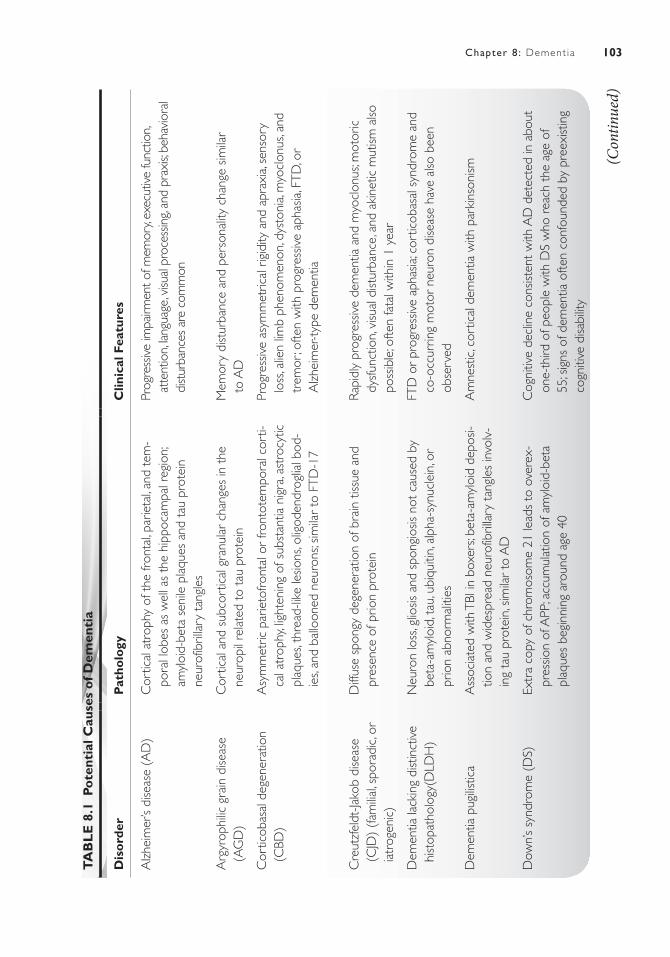
103Chapter 8 : Dement ia
TA
BL
E 8
.1 P
ote
ntia
l Cau
ses
of D
emen
tia
Dis
ord
erP
atho
logy
Clin
ical
Fea
ture
s
Alz
heim
er’s
dise
ase
(AD
)C
ortic
al a
trop
hy o
f the
fron
tal, p
arie
tal, a
nd t
em-
pora
l lob
es a
s w
ell a
s th
e hi
ppoc
ampa
l reg
ion;
am
yloi
d-be
ta s
enile
pla
ques
and
tau
pro
tein
ne
urof
ibril
lary
tan
gles
Prog
ress
ive im
pairm
ent o
f mem
ory,
exec
utive
func
tion,
atte
ntio
n, la
ngua
ge, v
isual
pro
cess
ing,
and
prax
is; b
ehav
iora
l di
stur
banc
es a
re c
omm
on
Arg
yrop
hilic
gra
in d
iseas
e (A
GD
)C
ortic
al a
nd s
ubco
rtic
al g
ranu
lar
chan
ges
in t
he
neur
opil
rela
ted
to t
au p
rote
inM
emor
y di
stur
banc
e an
d pe
rson
ality
cha
nge
simila
r
to A
D
Cor
ticob
asal
deg
ener
atio
n (C
BD)
Asy
mm
etric
par
ieto
front
al o
r fro
ntot
empo
ral c
orti-
cal a
trop
hy, li
ghte
ning
of s
ubst
antia
nig
ra, a
stro
cytic
pl
aque
s, th
read
-like
lesio
ns, o
ligod
endr
oglia
l bod
-ie
s, an
d ba
lloon
ed n
euro
ns; s
imila
r to
FTD
-17
Prog
ress
ive
asym
met
rical
rig
idity
and
apr
axia
, sen
sory
lo
ss, a
lien
limb
phen
omen
on, d
ysto
nia,
myo
clon
us, a
nd
trem
or; o
ften
with
pro
gres
sive
apha
sia, F
TD, o
r
Alz
heim
er-t
ype
dem
entia
Cre
utzf
eldt
-Jako
b di
seas
e (C
JD)
(fam
ilial, s
pora
dic,
or
iatr
ogen
ic)
Diff
use
spon
gy d
egen
erat
ion
of b
rain
tiss
ue a
nd
pres
ence
of p
rion
prot
ein
Rapi
dly
prog
ress
ive
dem
entia
and
myo
clon
us; m
otor
ic
dysf
unct
ion,
visu
al d
istur
banc
e, a
nd a
kine
tic m
utism
also
po
ssib
le; o
ften
fata
l with
in 1
yea
r
Dem
entia
lack
ing
dist
inct
ive
hist
opat
holo
gy(D
LDH
)N
euro
n lo
ss, g
liosis
and
spo
ngio
sis n
ot c
ause
d by
be
ta-a
myl
oid,
tau
, ubi
quiti
n, a
lpha
-syn
ucle
in, o
r pr
ion
abno
rmal
ities
FTD
or
prog
ress
ive
apha
sia; c
ortic
obas
al s
yndr
ome
and
co-o
ccur
ring
mot
or n
euro
n di
seas
e ha
ve a
lso b
een
obse
rved
Dem
entia
pug
ilistic
aA
ssoc
iate
d w
ith T
BI in
box
ers;
beta
-am
yloi
d de
posi-
tion
and
wid
espr
ead
neur
ofib
rilla
ry t
angl
es in
volv
-in
g ta
u pr
otei
n, s
imila
r to
AD
Am
nest
ic, c
ortic
al d
emen
tia w
ith p
arki
nson
ism
Dow
n’s
synd
rom
e (D
S)Ex
tra
copy
of c
hrom
osom
e 21
lead
s to
ove
rex-
pres
sion
of A
PP; a
ccum
ulat
ion
of a
myl
oid-
beta
pl
aque
s be
ginn
ing
arou
nd a
ge 4
0
Cog
nitiv
e de
clin
e co
nsist
ent
with
AD
det
ecte
d in
abo
ut
one-
third
of p
eopl
e w
ith D
S w
ho r
each
the
age
of
55; s
igns
of d
emen
tia o
ften
conf
ound
ed b
y pr
eexi
stin
g co
gniti
ve d
isabi
lity
(Con
tinue
d)

104 C O N D I T I O N S I N O C C U P A T I O N A L T H E R A P YT
AB
LE
8.1
Po
tent
ial C
ause
s o
f Dem
enti
a (C
ontin
ued)
Dis
ord
erP
atho
logy
Clin
ical
Fea
ture
s
Fam
ilial B
ritish
dem
entia
(F
BD)
Wid
espr
ead
cere
bral
am
yloi
d an
giop
athy
, am
yloi
d de
posit
ion
in th
e pa
renc
hym
a, an
d ne
urof
ibril
lary
ta
ngle
s; ca
used
by
auto
som
al d
omin
ant m
utat
ion
on c
hrom
osom
e 13
Prog
ress
ive
dem
entia
, spa
stic
tet
rapl
egia
, and
ata
xia
Fam
ilial D
anish
dem
entia
(F
DD
)Si
mila
r to
FBD
with
neo
cort
ex a
nd p
aren
chym
a m
ore
seve
rely
affe
cted
Begi
ns w
ith c
atar
act a
nd o
cula
r he
mor
rhag
es in
the
third
de
cade
of l
ife fo
llow
ed b
y se
vere
per
cept
ive
hear
ing
loss
10
–20
year
s la
ter;
afte
r ag
e 40
, ata
xia,
psyc
hiat
ric d
istur
-ba
nce,
and
pro
gres
sive
dem
entia
beg
in
Fam
ilial e
ncep
halo
path
y w
ith
neur
oser
pin
incl
usio
n
bodi
es (
FEN
IB)
Four
diff
eren
t ge
netic
mut
atio
ns le
ad t
o ne
uros
er-
pin
incl
usio
n bo
dies
(C
ollin
s bo
dies
) w
ithin
the
ne
uron
s of
the
dee
p co
rtex
and
sub
stan
tia n
igra
(M
irand
a &
Lom
as, 2
006)
Dem
entia
, tre
mor
, sei
zure
s, pr
ogre
ssiv
e m
yocl
onus
, ep
ileps
y, dy
sart
hria
; age
of o
nset
var
ies
from
tee
ns t
o
age
50 d
epen
ding
on
mut
atio
n ty
pe
Fam
ilial o
r sp
orad
ic fa
tal
inso
mni
aTh
alam
ic d
istur
banc
e an
d po
ssib
le c
ereb
ella
r or
ce
rebr
al a
trop
hy c
ause
d by
prio
n pr
otei
nIn
som
nia
and
auto
nom
ic d
ysfu
nctio
n; d
emen
tia la
te in
co
urse
; typ
ical
ly fa
tal w
ithin
13–
15 m
onth
s
HIV
-ass
ocia
ted
dem
entia
Vira
l inf
ectio
n of
mic
rogl
ia a
nd a
stro
cyte
s le
ads
to
neur
oinf
lam
mat
ion,
mye
lin p
allo
r, m
icro
glia
l nod
-ul
es, s
ynap
tic p
runi
ng, a
nd n
euro
n lo
ss p
rimar
ily in
th
e fro
ntal
and
par
ieta
l lob
es
Cog
nitiv
e, m
otor
, and
beh
avio
ral d
ysfu
nctio
n se
en in
ap
prox
imat
ely
10%
of p
eopl
e w
ith H
IV, u
sual
ly in
the
la
te s
tage
s
Incl
usio
n bo
dy m
yopa
thy,
Page
t’s d
iseas
e of
the
bo
ne, a
nd fr
onto
tem
pora
l de
men
tia (
IBM
PFD
)
Aut
osom
al d
omin
ant
mut
atio
n on
chr
omos
ome
9
lead
s to
deg
ener
atio
n of
mus
cle,
bra
in, a
nd
bone
tiss
ue
Prox
imal
and
dist
al m
uscl
e w
eakn
ess
and
atro
phy;
FTD
in
abou
t 30
% o
f affe
cted
indi
vidu
als

105Chapter 8 : Dement ia
Fron
tote
mpo
ral d
emen
tia
and
park
inso
nism
link
ed t
o ch
rom
osom
e 17
(FT
D-1
7)
with
pro
gran
ulin
mut
atio
n
Neu
rona
l, int
ranu
clea
r in
clus
ions
FTD
, pro
gres
sive
apha
sia, c
ortic
obas
al s
yndr
ome,
or
sym
ptom
s sim
ilar
to A
D o
r Pa
rkin
son’
s di
seas
e
Fron
tote
mpo
ral d
emen
tia
and
park
inso
nism
link
ed t
o ch
rom
osom
e 17
(FT
D-1
7)
with
tau
mut
atio
n
Neu
rona
l or
glia
l inc
lusio
ns in
volv
ing
tau
prot
ein;
sp
ecifi
c fe
atur
es v
ary
FTD
, pro
gres
sive
apha
sia, o
r bo
th, b
egin
ning
aro
und
mid
-dl
e ag
e or
soo
ner
(30–
50)
Fron
tote
mpo
ral l
obar
de
gene
ratio
n w
ith u
biq-
uitin
-pos
itive
incl
usio
ns
(FTL
D-U
)
Abn
orm
al n
eurit
es a
nd/o
r cy
topl
asm
ic in
clus
ions
in
the
front
al o
r te
mpo
ral c
orte
x or
den
tate
gra
nule
ce
ll la
yer;
front
otem
pora
l glio
sis a
nd n
euro
nal l
oss
FTD
, pro
gres
sive
apha
sia, o
r bo
th; m
otor
neu
ron
dise
ase
co-o
ccur
s in
abo
ut h
alf o
f cas
es
Ger
stm
ann-
Stra
usse
r- Sc
henk
er s
yndr
ome
(GSS
)Sp
ongi
form
cha
nges
, neu
rona
l los
s, as
trog
liosis
, de
posit
ion
of p
rion
prot
ein
plaq
ues
mai
nly
in t
he
cere
bellu
m
Cer
ebel
lar
dysf
unct
ion
incl
udin
g at
axia
, nys
tagm
us, a
nd d
ys-
arth
ria; c
ours
e m
ay p
rogr
ess
over
sev
eral
yea
rs; d
emen
tia
occu
rs la
te in
cou
rse
Hip
poca
mpa
l scl
eros
is (H
pScl
)N
euro
nal l
oss
and
astr
ocyt
e gl
iosis
of t
he h
ip-
poca
mpu
s, sp
ecifi
cally
the
cor
nu a
mm
onis
and
subi
culu
m; le
sions
may
ext
end
to o
ther
nea
rby
stru
ctur
es
Ofte
n ac
com
pani
es a
noth
er fo
rm o
f dem
entia
, com
pli-
catin
g cl
inic
al p
ictu
re; d
efic
its in
epi
sodi
c m
emor
y an
d la
ngua
ge s
imila
r to
AD
; less
exe
cutiv
e an
d vi
suos
patia
l im
pairm
ent
than
AD
Hun
tingt
on’s
dise
ase
Stria
tal d
egen
erat
ion
and
varia
ble
neoc
ortic
al
atro
phy
Park
inso
nism
, abu
lia, a
kine
sia, e
vent
ual c
hore
a, po
ssib
le
dysf
unct
ion
in p
sych
omot
or s
peed
, att
entio
n/co
ncen
-tr
atio
n, le
arni
ng, a
nd m
emor
y; po
ssib
le d
epre
ssio
n,
hallu
cina
tions
, del
usio
ns, d
isinh
ibiti
on, a
nd b
ehav
iora
l dy
scon
trol
; typ
ical
ly 1
0–20
yea
r co
urse
(Con
tinue
d)
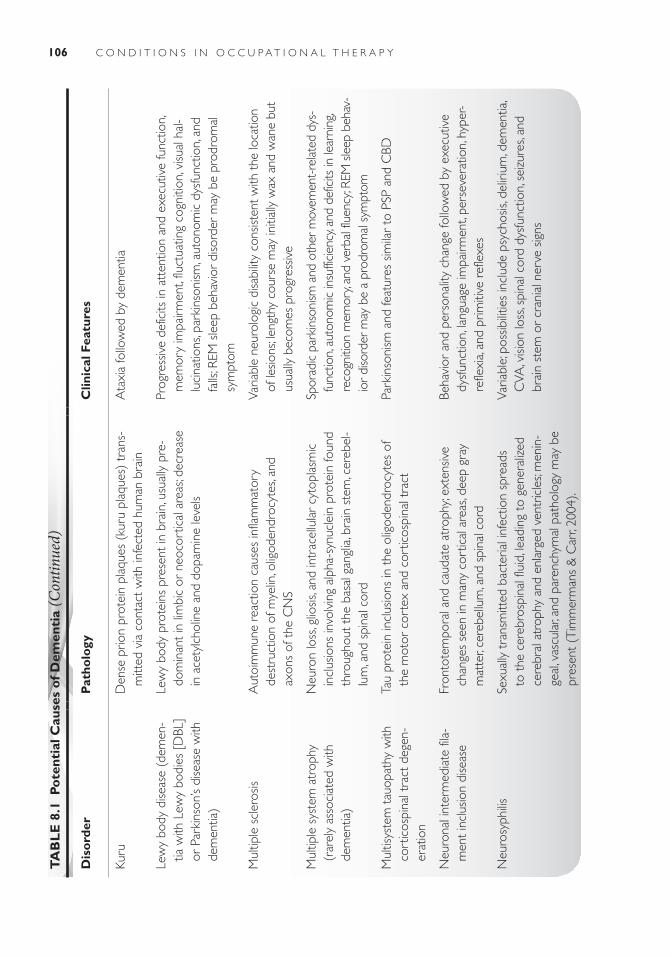
106 C O N D I T I O N S I N O C C U P A T I O N A L T H E R A P YT
AB
LE
8.1
Po
tent
ial C
ause
s o
f Dem
enti
a (C
ontin
ued)
Dis
ord
erP
atho
logy
Clin
ical
Fea
ture
s
Kuru
Den
se p
rion
prot
ein
plaq
ues
(kur
u pl
aque
s) t
rans
-m
itted
via
con
tact
with
infe
cted
hum
an b
rain
Ata
xia
follo
wed
by
dem
entia
Lew
y bo
dy d
iseas
e (d
emen
-tia
with
Lew
y bo
dies
[D
BL]
or P
arki
nson
’s di
seas
e w
ith
dem
entia
)
Lew
y bo
dy p
rote
ins
pres
ent
in b
rain
, usu
ally
pre
-do
min
ant
in li
mbi
c or
neo
cort
ical
are
as; d
ecre
ase
in a
cety
lcho
line
and
dopa
min
e le
vels
Prog
ress
ive
defic
its in
att
entio
n an
d ex
ecut
ive
func
tion,
m
emor
y im
pairm
ent,
fluct
uatin
g co
gniti
on, v
isual
hal
-lu
cina
tions
, par
kins
onism
, aut
onom
ic d
ysfu
nctio
n, a
nd
falls
; REM
sle
ep b
ehav
ior
diso
rder
may
be
prod
rom
al
sym
ptom
Mul
tiple
scl
eros
isA
utoi
mm
une
reac
tion
caus
es in
flam
mat
ory
dest
ruct
ion
of m
yelin
, olig
oden
droc
ytes
, and
ax
ons
of t
he C
NS
Varia
ble
neur
olog
ic d
isabi
lity
cons
isten
t with
the
loca
tion
of le
sions
; leng
thy
cour
se m
ay in
itial
ly w
ax a
nd w
ane
but
usua
lly b
ecom
es p
rogr
essiv
e
Mul
tiple
sys
tem
atr
ophy
(r
arel
y as
soci
ated
with
de
men
tia)
Neu
ron
loss
, glio
sis, a
nd in
trac
ellu
lar
cyto
plas
mic
in
clus
ions
invo
lvin
g al
pha-
synu
clei
n pr
otei
n fo
und
thro
ugho
ut t
he b
asal
gan
glia
, bra
in s
tem
, cer
ebel
-lu
m, a
nd s
pina
l cor
d
Spor
adic
par
kins
onism
and
oth
er m
ovem
ent-
rela
ted
dys-
func
tion,
auto
nom
ic in
suffi
cien
cy, a
nd d
efic
its in
lear
ning
, re
cogn
ition
mem
ory,
and
verb
al fl
uenc
y; RE
M s
leep
beh
av-
ior
diso
rder
may
be
a pr
odro
mal
sym
ptom
Mul
tisys
tem
tau
opat
hy w
ith
cort
icos
pina
l tra
ct d
egen
-er
atio
n
Tau
prot
ein
incl
usio
ns in
the
olig
oden
droc
ytes
of
the
mot
or c
orte
x an
d co
rtic
ospi
nal t
ract
Park
inso
nism
and
feat
ures
sim
ilar
to P
SP a
nd C
BD
Neu
rona
l int
erm
edia
te fi
la-
men
t in
clus
ion
dise
ase
Fron
tote
mpo
ral a
nd c
auda
te a
trop
hy; e
xten
sive
chan
ges
seen
in m
any
cort
ical
are
as, d
eep
gray
m
atte
r, ce
rebe
llum
, and
spi
nal c
ord
Beha
vior
and
per
sona
lity
chan
ge fo
llow
ed b
y ex
ecut
ive
dysf
unct
ion,
lang
uage
impa
irmen
t, pe
rsev
erat
ion,
hyp
er-
refle
xia,
and
prim
itive
ref
lexe
s
Neu
rosy
philis
Sexu
ally
tra
nsm
itted
bac
teria
l inf
ectio
n sp
read
s to
the
cer
ebro
spin
al fl
uid,
lead
ing
to g
ener
alize
d ce
rebr
al a
trop
hy a
nd e
nlar
ged
vent
ricle
s; m
enin
-ge
al, v
ascu
lar,
and
pare
nchy
mal
pat
holo
gy m
ay b
e pr
esen
t (T
imm
erm
ans
& C
arr,
2004
).
Varia
ble;
pos
sibilit
ies
incl
ude
psyc
hosis
, del
irium
, dem
entia
, C
VA, v
ision
loss
, spi
nal c
ord
dysf
unct
ion,
sei
zure
s, an
d br
ain
stem
or
cran
ial n
erve
sig
ns

107Chapter 8 : Dement ia
Nor
mal
pre
ssur
e hy
dro-
ceph
alus
Build
up o
f exc
ess
fluid
in t
he b
rain
; may
be
reve
rsed
with
shu
nt p
lace
men
tD
iffic
ulty
wal
king
, mem
ory
loss
, and
urin
ary
inco
ntin
ence
Pick
’s di
seas
eA
ccum
ulat
ion
of h
yper
phos
phor
ylate
d ta
u pr
otei
n; th
inni
ng o
f affe
cted
gyr
i; lig
hten
ing
of s
ubst
antia
nig
raFT
D, p
rogr
essiv
e ap
hasia
, sem
antic
dem
entia
, or
cort
ico-
basa
l syn
drom
e (r
arel
y)
Prog
ress
ive
supr
anuc
lear
pa
lsy (
PSP)
Neu
rofib
rilla
ry t
angl
es a
nd n
euro
pil t
hrea
ds in
the
ba
sal g
angl
ia, d
ienc
epha
lon,
and
bra
inst
emSu
pran
ucle
ar g
aze
palsy
, pos
tura
l ins
tabi
lity
and
falls
, pa
rkin
soni
sm; d
emen
tia in
mid
dle-
late
sta
ges
Spin
ocer
ebel
-la
r de
gene
ratio
n (M
acha
do-Jo
seph
dise
ase,
ot
her
spin
ocer
ebel
lar
atax
ia t
ypes
, den
tato
rubr
al-
pallid
oluy
sian
atro
phy,
and
Frie
drei
ch’s
atax
ia)
Deg
ener
atio
n of
the
cer
ebel
lum
, bra
inst
em, s
pina
l co
rd, a
nd b
asal
gan
glia
; diff
eren
t pa
thol
ogie
s re
sult
from
var
ious
her
itabl
e ge
netic
mut
atio
ns; o
ther
st
ruct
ures
incl
udin
g th
e th
alam
us a
nd c
ereb
ral
cort
ex m
ay b
e af
fect
ed
Ata
xia,
dysa
rthr
ia, d
ysph
agia
, and
aut
onom
ic d
ysfu
nctio
n;
varia
ble
cogn
itive
sym
ptom
s in
clud
ing
impa
ired
exec
u-tiv
e fu
nctio
ning
, diff
icul
ty w
ith s
patia
l rea
soni
ng, p
erso
n-al
ity c
hang
es, a
nd la
ngua
ge d
efic
its
Syst
emic
lupu
s er
ythm
atos
us
(SLE
)Sm
all f
ocal
lesio
ns o
ften
acco
mpa
nied
by
cort
ical
at
roph
y, ve
ntric
ular
enl
arge
men
t, an
d ce
rebr
al
edem
a am
ong
othe
r fin
ding
s; hi
ghly
var
iabl
e;
rela
ted
to m
icro
vasc
ular
aut
oim
mun
e di
seas
e (K
ozor
o, E
., Han
ley,
J., La
ptiv
a, L.
, & F
illey,
C. M
., 20
08).
Varia
ble
cogn
itive
and
neu
rops
ychi
atric
syn
drom
es
obse
rved
in a
bout
25%
–75%
of i
ndiv
idua
ls w
ith S
LE; d
efi-
cits
in p
roce
ssin
g sp
eed,
att
entio
n, an
d ex
ecut
ive
func
tion;
flu
ctua
ting,
chro
nic
cour
se
Vasc
ular
dem
entia
Cer
ebro
vasc
ular
dise
ase
(ofte
n a
serie
s of
sm
all
stro
kes)
lead
s to
foca
l les
ions
on
the
brai
n an
d ne
urot
rans
mitt
er d
isrup
tion
Cog
nitiv
e de
clin
e sim
ilar
to A
D b
ut o
ften
with
less
sev
ere
mem
ory
invo
lvem
ent;
Gai
t di
stur
banc
e is
com
mon
; ab
rupt
or
step
wise
(ra
ther
tha
n co
ntin
uous
) de
clin
e

108 C O N D I T I O N S I N O C C U P A T I O N A L T H E R A P Y
fragments of dead and dying neurons, and slowly decline into dense, misshapen plaques (Shenk, 2003). Beta-amyloid plaques collect outside and around the neurons. It is believed that this accumulation causes physical damage to axons and that prolonged neuronal response to this injury ultimately leads to the devel-opment of neurofibrillary tangles and neuronal death (Vickers, Dickson, & Adlard, 2000).
Neurofibrillary tangles are made up of another con-taminated protein called tau. Tau normally serves as connectors or “railroad ties” for a track-like structure that transports nutrients and other important molecules throughout the cell body of every neuron ( Josephs, et al., 2005). The contaminated or tangled tau somehow becomes hyperphosphorylated, a corruption resulting in several extra molecules of phosphorous. Without the sta-bilizing railroad ties of normal tau, the tracks bend into a twisted mess. Under the weight of the nutrients being transported, the tracks buckle, and the damage increases. Inside the neuron, this twisted debris gets worse as the filaments of track keep twisting around each other. As cell communication and nourishment is lost, the neuron begins to wither. The cell body, axons, and dendrites dis-integrate, and as a result, thousands of synapses vanish (Bear, Connors, & Paradiso, 2001; Shenk, 2003). It is well documented that the brains of persons with AD have an abundance of the abnormal structures of beta-amyloid plaques and neurofibrillary tangles (Shenk). Filamentous structures known as “Hirano bodies” found primarily in the hippocampus are also associated with the disease (Neugroschl, Kolevzon, Samuels, & Marin, 2004).
The structural changes just described do not cause AD, but are the end product of a pathological process. The challenge for researchers is to discover what causes this degeneration. The loss of neurons occurs through-out the brain but is significant in the cerebral cortex. The cerebral cortex is responsible for higher brain functions, including thinking, judgment, reasoning, speech, and language. Plaques and tangles are also dense in the hip-pocampus, which plays a role in attention and memory (Cohen & Eisdorfer, 2001).
It is possible that amyloid has a function as a repair protein. Amyloid levels have been noted to increase when there is some injury to the brain. Some researchers contend that beta-amyloid plaques do not lead to AD, but in fact might be a by-product of the AD process (Shenk, 2003).
Genetic Predisposition of Alzheimer’s DiseaseUnlike some diseases, AD is not caused by a single gene. More than one gene mutation is seen in AD, and genes on multiple chromosomes are involved. In most cases, genes alone are not sufficient to cause AD. The two types of AD are early onset and late onset. Early onset refers to AD that manifests before the age of 65, and late onset
refers to AD seen at or after age 65. Less than 5% of AD is early onset, and this form of the disease is often inher-ited (Alzheimer’s Disease Education & Referral Center, 2003a; Rocchi, Pellegrini, Siciliano, & Murri, 2003).
Three genes have been identified as responsible for the rare, early-onset, familial form of AD. Mutated chromo-some 21 causes an abnormal APP to be produced. Mutated chromosome 14 causes an abnormal protein called prese-nilin 1 to be produced. Mutated chromosome 1 causes yet another abnormal protein, presenilin 2, to be produced (National Institute on Aging, 2003). It appears that these mutations increase the likelihood that beta-amyloid will be snipped from the APP, forming more of the sticky beta-amyloid (Cohen & Eisdorfer, 2001). Even if only one of these genes inherited from a parent is mutated, the person will almost certainly develop early-onset AD. This means that in these families, children have a 50% chance of devel-oping early-onset AD if one parent has this disease.
In the more common, late-onset AD, a polymor-phism in the apolipoprotein E gene is associated with increased susceptibility (Tanzi & Bertram, 2001). The apolipoprotein E gene is found on chromosome 19. This gene codes for a protein that helps carry cholesterol in the bloodstream. The apolipoprotein E gene comes in several forms, called alleles. The three most common alleles are apolipoprotein E 2, apolipoprotein E 3, and apolipoprotein E 4. A person inherits one apolipopro-tein E allele from each parent. One or two copies of the 4 allele increases the risk of getting AD. However, having the 4 allele does not mean that AD is certain, as some persons with even two of the 4 allele do not develop the disease. The rarer 2 allele may be associated with having a lower risk for AD (Alzheimer’s Disease Education & Referral Center, 2003a; Blazer, Steffens, & Busse 2004). AD may also be linked to mutations in the gene that encodes for insulin-degrading enzyme, which helps to break down beta-amyloid. Other genes related to AD include those involved in intracellular APP trafficking and the creation of apolipoprotein neural receptors and transmembrane proteins that influence calcium levels and beta-amyloid production (Minati et al., 2009).
Down syndrome (DS) is a risk factor for AD. Indi-viduals with DS carry an extra copy of chromosome 21, which contains the APP gene. As a result, research-ers believe that APP processing in neuronal membranes is abnormal, leading to the formation of beta-amyloid plaques and eventual AD neuropathology. In fact, the brains of almost all individuals with DS over the age of 40 show anatomical changes associated with AD, and as many as 66% will develop clinically detectable dementia in their 50s (Beacher et al., 2009). In another study, it was reported that 32.1% of a sample of individuals with DS between the ages of 55 and 59 had dementia, 17.7% between the ages of 50 and 54, and 8.9% between the ages
109Chapter 8 : Dement ia
Other Potential Causes of Alzheimer’s DiseaseOne of the theories of aging suggests that over time, dam-age from a kind of molecule called a free radical can build up in neurons and cause a loss of function. Free radicals can be helpful in fighting infection, but too many can injure cells by changing other nearby molecules, such as those in the neuron’s cell membrane or in DNA. This process can lead to a chain reaction, releasing more free radicals, and causing more damage. This is called oxida-tive damage and may contribute to AD by upsetting the proper flow of substances in and out of cells; by disrupt-ing cell metabolism; and/or by interfering with other cell processes such as protein synthesis, stress response, and enzyme-catalyzed reactions (National Institute on Aging, 2003; Reed, Sultana, & Butterfield, 2010). Some researchers are focusing on a possible connection between oxidative stress and beta-amyloid, the principle compo-nent of plaques found in the brain of the person with AD (Varadarajan, Yatin, & Aksenova, 2000). To support the role of oxidative stress in contributing to AD, researchers have found that antioxidant defense is altered in the brains of persons with mild cognitive impairment (MCI), a con-dition that is often a precursor to AD (Reed et al., 2010).
Researchers are also studying the possible role of inflammation, because cells and compounds that are known to be involved in inflammation are found in AD plaques. Some scientists think that inflammation is harm-ful and sets off a cycle of events that lead to the death of neurons. Others believe that some aspects of the inflam-matory process may be a helpful part of the brain’s natural healing efforts (National Institute on Aging, 2003).
Research is focused on even more factors that may be related to AD, including trace elements, the role of the immune system, and the possibility of a viral connection. The role of numerous misfolded proteins in the neuropa-thology of AD along with other neurodegenerative dis-eases such as Parkinson’s disease, Huntington’s disease, and amyotrophic lateral sclerosis (ALS/Lou Gherig’s dis-ease) is also a topic of scientific inquiry (Kanehisa, Limvi-phuvadh, & Tanabe, 2010).
Incidence and Prevalence
In the next 50 years, the prevalence of AD in the United States is expected to nearly quadruple. This means that 1 in every 45 Americans would meet the diagnostic criteria (Kawas & Brookmeyer, 2001). This phenom-enon is largely a result of the fact that the baby-boom generation will be elderly and that life expectancies are increasing with medical advances. The DSM-IV-TR (American Psychiatric Association, 2000) states that, while the prevalence increases with age, women tend to have a higher incidence of AD. Based on the large-scale, longitudinal “Framingham study,” the estimated lifetime
of 45 and 49 (Coppus et al., 2006). Due to preexisting learning disabilities, AD is more difficult to detect and treat in this population (Beacher et al., 2009).
More recently, a possible link between environmental conditions in early brain development and future mani-festation of AD has been proposed. The “latent early-life associated regulation (LEARn)” model posits that factors such as diet, toxin exposure, and hormones early in one’s development may disrupt DNA structure in a manner that influences the way certain genes are regulated. In support of this model, animal studies have shown that exposure to lead early in life may upregulate the expres-sion of APP in old age (Lahiri, Zawia, Sambamurti, & Maloney, 2008; Wu et al., 2008).
Neurotransmitter Abnormalities in Alzheimer’s DiseaseThere is some evidence that the neurotransmitter acetyl-choline is implicated in the progression of AD. Neurons that contain acetylcholine are known as cholinergic neu-rons, and many of these are bunched together to form tracts. Cholinergic tracts radiate throughout the entire brain. Autopsy shows that for a person with AD, many cholinergic tracts throughout the entire brain have been destroyed. The enzyme choline acetyltransferase (CAT), which is needed to form acetylcholine, is seriously reduced in the brains of persons with AD. This reduction of CAT is greatest in areas of the brain where there is a dense amount of plaques and tangles (Alzheimer’s Disease Education & Referral Center, 2003a). Other neurotransmitter systems found to be altered in people with AD include norepineph-rine, GABA, and glutamate (Neugroschl et al., 2004).
Cardiovascular Risk Factors in Alzheimer’s DiseaseAlthough vascular dementia accounts for 15% to 25% of all dementia cases, there is now a concern that cardiovascu-lar disease is also linked to AD. As people are living longer with AD, and living long enough to be diagnosed with AD at ages 80 and 90, these groups are also at risk for develop-ing cardiovascular disease. Some see this group as having a “mixed dementia” or a combination of AD and vascular disease (Cohen & Eisdorfer, 2001). Epidemiologic stud-ies suggest that risk factors for cardiovascular disease and stroke are associated with cognitive impairment and AD and that the presence of these factors intensifies the sever-ity of symptoms in AD (Alzheimer’s Disease Education & Referral Center, 2003a). In autopsy studies, 60% to 90% of AD cases exhibit variable cerebrovascular pathol-ogy (Kawas & Brookmeyer, 2001). Researchers have also found that the use of statins, the most common type of cholesterol-lowering drug, is associated with a lower risk of developing AD (Rocchi et al., 2003).
110 C O N D I T I O N S I N O C C U P A T I O N A L T H E R A P Y
Signs and Symptoms of Alzheimer’s DiseaseAlthough the hallmark symptom of dementia is memory impairment, the signs and symptoms of AD impact virtually all performance skills. Because AD is a progressive disease, the first signs can be mistaken for normal aging. Even though there are small changes in process skills with normal aging, a healthy older adult can continue to learn and problem solve at any age. Researchers are generating new information on normal aging versus MCI. A person with MCI will have more impairment of process skills than is expected with nor-mal aging, but will not yet meet the diagnostic criteria for dementia.
Defining the progressive stages of AD is an arbitrary process, and various researchers use different ideologies. Reisberg, a prominent researcher in the study of AD, has proposed seven stages (Reisberg & Sclan, 1992). These stages range from stage 1, in which there is no impair-ment, to stage 7, which is very severe decline. Most researchers use a three-stage approach (Goldman, Wise, & Brody, 2004; Samuels & Neugroschl, 2004; Shenk, 2003). The Clinical Dementia Rating (CDR) is used to measure dementia (Hughes, Berg, & Danziger, 1993). This scale defines three levels of dementia as mild, mod-erate, and severe.
This chapter will use the three-stage approach, with the stages being defined as early, middle, and late. We will also briefly review MCI, as it is possibly a precur-sor to dementia. It is important to remember that each person with AD is a unique individual, with unique con-cerns, needs, and strengths. Although such individuals do not always fit into a rigid classification system, using the three stages will give the reader general guidelines for better understanding the problems facing the person with AD and his or her caregivers.
Mild Cognitive ImpairmentMCI is defined as the clinical state of individuals who are memory impaired but are functioning well and do not meet the criteria for dementia (Peterson, Stevens, & Ganguli, 2001). MCI appears to be a prodromal stage of AD. It is a stage of mild but persistent memory loss and is often seen with word naming difficulties. According to Cohen, the diagnosis of MCI includes five criteria:
1. The individual complains of memory problems.2. Memory loss is abnormal for the person’s age.3. ADLs are not affected.4. Other cognitive abilities are intact.5. There is no dementia.
For individuals given a diagnosis of MCI, approximately 15% will progress to AD each year compared with 1% of age-matched controls (Neugroschl et al., 2004).
risk for developing AD is about 1 in 5 for women and 1 in 10 for men. This study also indicates that while the risk of developing dementia between the ages of 65 and 75 is about 1%, it rises sevenfold in the following 10 years and 20-fold by the age of 85. By 2050, it is estimated that between 11 million and 16 million older Americans will have AD unless preventative treatment becomes available (Alzheimer’s Association, 2009).
In addition to age, there are numerous factors that appear to create a higher risk for AD (Hales & Yudofsky, 2003; Neugroschl et al. 2004; Reitz et al., 2010; Shah et al., 2009). These factors include the following:
1. Low educational level 2. History of head trauma with loss of consciousness 3. History of depression 4. Late maternal age 5. Environmental and occupational exposure (e.g., to
aluminum) 6. History of electroconvulsive therapy (ECT) 7. Alcohol abuse 8. Analgesic abuse 9. Long-standing physical inactivity10. Vascular risk factors (e.g., hypertension, high choles-
terol)11. Type II diabetes12. Black or Hispanic ethnicity13. High waist to hip ratio
Minati et al. (2009) state that traumatic brain injury (TBI) is increasingly believed to be a risk factor for the develop-ment of AD due to the accumulation of beta-amyloid and tau pathology that follows neural degeneration. They also indicate that cerebrovascular abnormalities may increase the chances of being diagnosed with AD. However, they assert that the most important risk factors are advanced age and genetic predisposition.
Related to these risk factors, there also appear to be several variables that may reduce the risk of developing AD (Breitner & Albert, 2009; Neugroschl et al., 2004; Shah et al., 2009). These include
1. Exposure to antioxidants (such as vitamin E)2. Regular consumption of fish3. Estrogen replacement therapy in postmenopausal
women4. Use of nonsteroidal antiinflammatory drugs5. Use of specific antihypertensive drugs6. Use of statins7. Use of specific histamine blockers8. Physical activity9. Exposure to education, cognitive training, and mental
stimulation
However, the effectiveness of some of these agents is still a matter of scientific debate.

111Chapter 8 : Dement ia
Early StageThe early and mild stage usually lasts 2 to 3 years (Gauthier, 2002). The cognitive changes in the early stage can be divided into three groups: memory, language, and visuospatial.
Memory Short-term memory is significantly impaired, which makes new learning very difficult. The person forgets tasks, loses the thread of a conversation, and misplaces things. Long-term memory begins to be impaired (Hales & Yudofsky, 2003). The person might be able to remember a phone number long enough to repeat it, but will forget it if there is any delay in using it. Procedural memory, such as knowing how to write, will remain intact. However, other types of memory begin to show deficits, including personal episodic memory, which is time-related information about one’s self, such as where and if one ate breakfast; semantic memory, such as remembering the name of a common object; and general knowledge, such as remembering the name of the highest mountain in the world (Alzheimer’s Disease Education & Referral Center, 2003a).
Language Aphasia, an abnormal neurologic con dition in which language is impaired, appears in the early stage. The person can usually maintain sentence structure, though it becomes less complex. Poor semantic memory leads to difficulty with word retrieval (the ability to name an object when shown a picture of it) and word list generation (e.g., being able to quickly name words in a common category or beginning with the same letter). When forgetting a word, the person may substitute inappropriate words, making sentences incomprehensible (Alzheimer’s Disease Education & Referral Center, 2003a). This would be an example of paraphasia, which means saying the wrong word, substituting a word that sounds alike, or using a word in the same category as the intended word. Anomia is the phenomenon in which a person searches in vain for a word and says “thing-a-ma-jig” or just gives up. Circumlocution may occur, in which the person tries to express an idea by talking around the intended word with extensive description and elaboration (Liu, McDowd, & Lin, 2004).
Visuospatial Skills Visuospatial abilities decline in early AD. The person starts to get lost in a familiar neighborhood or does not recognize a familiar intersection. There may be some disorientation within the home, such as putting the frying pan in the freezer or the wallet in the dishwasher (Bear et al., 2001).
Executive Functions In addition to these classic symptoms, the person in the early stage of AD begins to have difficulty with instrumental activities of daily living (IADLs). Balancing the checkbook is impossible, and bills may be forgotten. The family notices that the person is more rigid and irritable and less spontaneous or adventurous. Problems
with planning, organizing, sequencing, and abstracting become apparent as the person begins to have difficulty in the workplace or following a recipe. The person may begin to prepare a meal, become distracted, and forget to complete it (Hales & Yudofsky, 2003).
The person may recognize that memory has become impaired or may not have the judgment necessary for such insight. It is not unusual to see personality changes. Some persons with AD become suspicious, thinking that one’s things are being stolen or that his or her spouse is being unfaithful. Depression is seen in up to 25% of peo-ple in the early stage of AD. Confusion or anxiety may lead to withdrawal from routine social activity. In some cases, delusional thinking may emerge (Kovach, 1997).
Middle StageIn the middle stage of AD, which can last from 2 to 10 years, there is continued decline in memory, visuospatial skills, and language. All areas of performance skills begin to show deficits, psychiatric symptoms increase, and behavior disturbances arise.
Memory Recent and remote memory worsens. The person may think he or she is back at an earlier stage of life and become focused on a past worry, such as getting the children to school on time. The person is no longer bothered by the memory loss. There is disorientation to place and time, and at times the person may not recognize his or her own face in the mirror, much less friends or family members. New information is not retained for more than a few moments. There is difficulty organizing thoughts and thinking logically and inability to cope with new or unexpected situations. Thinking is concrete, with no ability to take into account ideas or objects that are not present (National Institute on Aging, 2003; Shenk, 2003; Zgola, 1999).
Language Aphasia worsens, and the person loses fluent language. Language is limited to the concerns of the moment or reminiscing about the past. There may be diminished verbal responsiveness, or verbalizations may be impulsive and inappropriate. There is difficulty understanding simple questions or instructions. The person has trouble following a conversation and may be unable to keep track of his or her own thoughts or words (Frank, 2003).
Internal speech is part of the complex process used when an individual makes plans or mentally solves prob-lems. A person who can no longer use words effectively loses this ability of rational planning and problem solving. His or her actions will become impulsive and disorganized, requiring step-by-step direction and supervision for any occupation (Zgola, 1999).
Visuospatial Skills Visuospatial abilities continue to decline. Visual inattention, which is seen in the early stage, begins to seriously limit function in the middle
112 C O N D I T I O N S I N O C C U P A T I O N A L T H E R A P Y
stage (Liu et al., 2004). The person gets lost in familiar environments and is unable to become oriented if he or she moves to a new environment. Constructional skills are compromised, and the person is unable to sort out the arms and legs of garments while trying to get dressed. There is a loss of ability to judge depth and distance. The person may step highly over a mark on the ground or choose to walk around it. He or she may not be able to distinguish furniture from designs on the carpet or interpret changes in flooring. These visuospatial impairments can lead to falls. Judging direction and distance is problematic, resulting in knocking things over when trying to grasp them or grasping at thin air (Zgola, 1999).
Psychiatric Symptoms Psychiatric symptoms that emerged in the early stage worsen. Depression and anxiety are frequently seen, and the presence of depression is associated with increased mortality. The person may lose control over his or her emotions, having outbursts of fear or anger. Visual hallucinations are not uncommon, and auditory hallucinations can also occur. Sleep is disturbed, with increased daytime napping and frequent nighttime wakefulness (Kovach, 1997).
Behavior Disturbances Behavior disturbances increase the likelihood that the individual will need nursing home placement. It is in the middle stage of AD that wandering and agitation become a problem (National Institute on Aging, 2003). The person paces, seemingly without a goal or destination. Pacing could be a sign of stress or anxiety, or perhaps it is the person’s need for activity and exercise. Agitation could be the person’s only way to respond to fear or frustration (Kovach, 1997). Loss of impulse control manifests in sloppy table manners, undressing at inappropriate times or places, and vulgar or rude language (Vickers et al., 2000). There is a loss of social propriety and inhibition, a failure of the filtering system that determines which thoughts to keep to one’s self and which thoughts to act upon. This inability to inhibit impulses can cause offense to others or become dangerous if the person with AD acts upon aggressive impulses (Zgola, 1999).
Late StageThe late stage of AD can last from 8 to 12 years. At this stage, the person with AD is fully dependent on others for basic ADLs, such as bathing, dressing, and eating. Motor skills are affected, and the person becomes immobile and incontinent.
Memory There is no ability to create new memories and little or no recognition of close family members. All
process skills are seriously impaired, and purposeful, goal-directed occupation is lost.
Language Speech is limited to one or two words, or speech and vocabulary may be totally unintelligible. Over time, speech will decline to the point where there is none. The person no longer smiles or communicates with facial expression (Shenk, 2003). There can be instances of moaning or crying, and since these are universal sounds of distress, it is important for caregivers to explore the possible causes of discomfort. Receptive language is also seriously impaired. The person cannot process the meaning of words but may respond to a calm and soothing tone of voice.
Motor Skills The person becomes bed-bound with eventual loss of postural control. Neurologic symptoms develop, including hyperflexia, apraxic gait, and frontal release signs (grasp and snout reflexes). Paratonia is a primitive reflex in which there is involuntary resistance in an extremity in response to a sudden passive movement. Thus, if a caregiver quickly moves the person’s arm, he or she automatically resists the movement (Goldman et al., 2004).
Seizures may occur, and contractures, pressure ulcers, urinary tract infections, and pneumonia may develop from immobility. There is incontinence of bladder and bowel. Appetite decreases, and eventually the person develops dysphagia or the loss of ability to chew and swallow (Kovach, 1997).
Psychiatric Symptoms The sleep cycle is very disturbed as the person spends 60% of time sleeping, including much of the daytime. Hallucinations persist for some (Kovach, 1997).
Course and PrognosisThe course of AD tends to be slowly progressive, with the loss of about 3 points per year on the Mini Men-tal State Examination, a standard cognitive assessment instrument (Neugroschl et al., 2004). The stages out-lined in the previous section of this chapter illustrate the progression of the signs and symptoms of AD. A 2004 study reported the average survival time fol-lowing diagnosis of AD to be 4 to 6 years. On average, those who are newly diagnosed live approximately half as long as people of the same age without AD. However, some people can survive as long as 20 years after the first appearance of symptoms (Alzheimer’s Association, 2009; Varadarajan et al., 2000).
This disease progresses until death. Death is usually a result of complications, such as infection, or the even-tual inability of the individual to maintain nutrition and hydration. Despite suspected underreporting, AD was the sixth leading cause of death in the United States in 2006 (Alzheimer’s Association, 2009).
113Chapter 8 : Dement ia
Medical Management
DiagnosisThere are three primary criteria in the diagnosis of AD. First, there is the development of multiple cognitive defi-cits manifested by both memory impairment and at least one other cognitive disturbance. These cognitive distur-bances include aphasia, which is language disturbance; apraxia, which is impaired ability to plan motor activities despite intact motor function; agnosia, which is failure to recognize or identify objects despite intact sensory function; and disturbance in executive function, such as problems with planning, organizing, sequencing, and abstracting.
The second criterion for AD is the finding that the multiple cognitive deficits cause significant impairment in social or occupational functioning and represent a sig-nificant decline from a previous level of functioning. The third criterion is having a course characterized by gradual onset and continuing cognitive decline (American Psychi-atric Association, 2000).
The DSM-IV-TR (American Psychiatric Associa-tion, 2000) uses the first two criteria in diagnosing all other dementias. In addition to the aforementioned cri-teria, the clinician must eliminate other possible causes of dementia for the diagnosis of AD to be made. A defin-itive diagnosis of AD is possible only after death, when an autopsy can reveal the physical findings of plaques and tangles in the brain.
The diagnosis is a time-consuming process, but an important one, as the thorough medical investigation may reveal a condition that can be treated. The possibility of reversible dementia will be discussed later.
The National Institutes of Health (NIH) (National Institute on Aging, 2003) has outlined the following steps in diagnosing AD:
1. A detailed patient history should include a descrip-tion of how and when the symptoms developed, the patient’s and family’s medical condition and medical history, and an assessment of the patient’s emotional state and living environment.
2. Interviewing family members or close friends, which can provide information on how behavior and person-ality have changed.
3. Physical and neurologic examinations and laboratory tests, which help determine neurologic functioning and identify possible non-AD causes of dementia.
4. A computerized tomography (CT) scan or an MRI test, which can reveal changes in the brain’s structure and function that indicate early AD.
5. Neuropsychologic tests that measure memory, lan-guage skills, and other cognitive functions, which help indicate what kind of cognitive changes are occurring.
General Treatment ApproachGoldman et al. (2004) describe the following four pillars of complete dementia care:
1. Supportive care for the patient2. Supportive care for the family and/or caregiver3. Disease treatment4. Symptom treatment, including cognitive, mental, and
behavioral symptoms
Supportive care includes assessing the environment for aspects of safety as well as looking at how the environ-ment can improve function and well-being. Very early in the disease process, the person with AD needs to discuss financial and medical concerns such as wills and advance directives. It is important to determine the level of care that is needed, who will provide the care, and additional supports such as day programs. The family may need to help make decisions regarding ability to drive and work. Maintaining the person’s dignity and privacy is always important. The person with AD will also need ongoing medical care and evaluation of vision and hearing.
Minati et al. (2009) summarizes a number of evidence-based interventions that may be helpful in the treatment of persons with AD. These include individual and group-based structured activities, psychological therapy, and cognitive rehabilitation. Cognitive strategies based on implicit learning include spaced retrieval, vanishing cues, and errorless learning. However, it is important to note that this type of “learning” depends on implicit aspects of cognition not as rapidly affected by the disease; such treat-ments should be used only to the extent that they are ther-apeutic for the individual. Other cognitive interventions include cognitive stimulation, reminiscence therapies, and external memory aids. These authors assert that “person- centered therapeutic interventions” are helpful in address-ing emotional issues that underlie much of the problem behavior and distress associated with AD for both the patient and the caregiver (Minati et al., 2009).
Support for the caregiver is critical. Approximately 80% of people with dementia are cared for by their families in the community (Neugroschl et al., 2004). Caring for someone who is progressively losing memo-ries and skills can be stressful, sad, and frightening. Care giving can become a 24-hour-a-day job. Complicating the stress of caring for a person with AD is the tendency of caregivers to withhold the diagnosis from friends and family and the social isolation that may result from embarrassing behavior or functional decline (Neugro-schl et al.). The stress of caregiving leads to increased risk of depression, sleep disturbance, and substance abuse in caregivers and may result in patient neglect (Neugroschl et al.). The Alzheimer’s Association can refer caregivers for support, resources, and advocacy.
114 C O N D I T I O N S I N O C C U P A T I O N A L T H E R A P Y
Disease treatment is meant to target the etiology or cause of the progressive decline in AD. The etiology of AD is still unknown. Former President Ronald Reagan brought national attention to the disorder when, in 1994, he announced that he had been diagnosed with AD. Since his death, his widow, Nancy Reagan, has publicly cam-paigned for increasing dementia research funds and for lim-iting obstacles to stem cell research. Until the causes of AD are better understood, medical management of the vascular process continues to be important not just for persons with vascular dementia but also for AD.
Symptom treatment targets cognitive decline, psy-chiatric symptoms (including psychosis or depression), and behavior disturbances. Pharmacotherapy is useful for these symptoms. The use of medications will not stop the disease process, but relieving the associated symptoms can comfort the person with AD and improve the qual-ity of life for both patient and caregivers. In addition to pharmacotherapy, behavioral and environmental strate-gies can provide support for more positive experiences as the disease progresses.
PharmacotherapyPrescription drugs are frequently used in managing symp-toms such as impaired cognition, depression, delusions and hallucinations, and agitation or aggression. Some of the newest medications are those used in treating cog-nitive symptoms and work as cholinesterase inhibitors. These include donepezil (Aricept), rivastigmine (Exelon), and galantamine (Reminyl). Inhibiting acetylcholinest-erase leaves more acetylcholine in the synapse, which facilitates the activity of remaining neurons. The various cholinesterase inhibitors tend to perform equally in com-parison group studies, but it is possible for individuals to respond more favorably to one drug than another (Farlow & Boustani, 2009). These medications do not inhibit cell loss, but research does show that there is modest cogni-tive improvement in about a third of the persons who take them. This improvement is seen in increased function in ADLs and in delayed placement in nursing homes (Peter-son et al., 2001). Some studies show that treatment with cholinesterase inhibitors can restore cognition to the level seen about 6 months prior to the start of treatment (Liu et al., 2004). This means that the person may be able to again do the things he or she could do half a year ago, such as dressing with verbal prompts only. However, these treatments cannot halt the progression of AD, and cogni-tive decline will continue. Common side effects of cho-linesterase inhibitors include nausea, vomiting, diarrhea, and loss of appetite (Farlow & Boustani, 2009).
Memantine (Namenda) is another medication used in an attempt to slow the progression of AD. It works by blocking excess glutamate, a chemical involved in memory function (National Institute on Aging, 2003). Memantine
has few long-term side effects but may cause temporary confusion or sedation while the dosing is being adjusted. A combination of Memantine and a cholinesterase inhib-itor is the preferred treatment for moderate to severe AD. However, as AD progresses and the person is no longer able to share meaningful interactions, medication may be discontinued (Farlow & Boustani, 2009).
At the present, pharmacologic advances in the treat-ment of AD include large-scale clinical trials of an anti-histamine drug called Dimebon (Latrepirdine). This agent appears to have mitochondrial stabilization prop-erties that promote neuronal function and inhibit cell death, leading to improved cognition in people with AD. Investigators are also exploring drug treatments that tar-get amyloid-beta production and aggregation, but so far success has been minimal (Massoud & Gauthier, 2010).
Depression is seen in as many as 25% to 30% of per-sons with dementia. Depression can occur in the early stage of AD and, if untreated, can lead to earlier insti-tutionalization and death, aside from the emotional suf-fering. Select serotonin re-uptake inhibitors (SSRIs) are the first line of treatment for depression, followed by the use of agents with dual effects on the serotonin and nor-epinephrine systems (Peterson et al., 2001). Many SSRI’s have anticholinergic side effects, so fluoxetine (Prozac) and sertraline (Zoloft) are usually most appropriate for individuals with dementia (Farlow & Boustani, 2009).
Delusions and hallucinations can begin in the early stage of AD and are more common in the middle stage. Low-dose neuroleptics are sometimes used, and the atypical neuroleptics such as risperidone (Risperdal) and olanzapine (Zyprexa) are less likely to cause side effects (Peterson et al., 2001). However, the use of these medications is controversial due to the increased risk of stroke and death as well as only limited, if any, long-term improvements (Ballard, Corbett, Chitramohan, & Aars-land, 2009; Minati et al., 2009).
Disruptive behavior such as agitation and aggression is often a sign of a medical problem. Urinary tract infec-tions or pneumonia can create delirium with agitation or aggression. Ruling out these and other concerns, such as environmental stress or pain, is the first step in reducing problem behaviors. If the behavior is truly caused by the underlying dementia, low-dose neuroleptics and mood stabilizers may be beneficial (Goldman et al., 2004). How-ever, again, the use of neuroleptics in this situation may not be the best approach. Ballard et al. (2009) state that psychological interventions and caregiver training should be the first management strategies used, prior to pharma-ceutical interventions.
Behavioral and Environmental ManagementAccording to Hilgeman, Burgio, and Allen (2009), the environment surrounding a person with AD should
115Chapter 8 : Dement ia
be modified so as to restore the balance between the challenges presented and the person’s current level of function. By creating an appropriate level of demand, the physical environment can serve to help maintain function and simultaneously provide stimulation and comfort to the individual with AD. Environmental con-siderations should be made for declining cognitive func-tion as well as sensory loss and the potential for pain and discomfort. Some specific suggestions from Hilgeman et al. include
1. Reducing the size of space and number of individuals2. Simplifying visual and auditory stimuli3. Providing choices/options4. Increasing the amount of familiar and meaningful
stimuli5. Providing redundant cues in multiple modalities6. Increasing the sensory contrast of important stimuli
An appropriate environment is perhaps one of the most effective ways to manage problem behaviors of persons with AD. Hilgeman et al. (2009) point out that aggres-sion is often a result of too much or too little stimulation, the anticipation or experience of pain, and frustration with one’s inability to perform daily tasks or effectively communicate. They suggest breaking up long periods of low or high arousal activities with sensory stimulation or relaxation, providing orienting cues, emphasizing nonver-bal communication, and working to relieve immediate dis-comforts. Addressing other underlying factors such as pain and depression may also help ameliorate agitated behav-ior. Neugroschl et al. (2004) add that infections, constipa-tion, delirium, and medication or substance intoxication may also lead to agitated behavior in persons with AD. They suggest strategies such as promoting exercise, sociali-zation, recreation, and predictable routines. Another way to address troublesome behavior, such as agitation, is to consider the “ABCs” (Xie et al., 2009; Neugroschl et al). This refers to the antecedents, the nature of the behavior itself, and the consequences. By considering factors that frequently lead up to the behavior, changes can be made to try to avoid the issue before it begins. Some changes may involve simplifying the task, providing different cues, or offering choices. The consequences of behavior should also be considered so that incooperative behavior, such as acting out in avoidance of a task, is not reinforced, and positive behavior is not punished or ignored.
Patients with AD may engage in a variety of prob-lematic behaviors other than or in addition to aggression and agitation. Some examples include wandering, grab-bing others, and swallowing inedible objects. A helpful guide for dealing with these behaviors based on Allen’s 25 cognitive levels and modes is written by Pollard (2005) and listed as a recommended learning resource at the end of the chapter.
Impact of Condition on Client Factors
Early StageWith the progressive loss of performance skills, AD affects increasing areas of occupation and performance patterns. Although this section outlines the expected impact of AD on client factors, it is important to remember that each indi-vidual will perform differently. Someone who has always been flexible and adaptive may compensate in ways that sup-port function; another person who is prone to anxiety or has fewer coping skills might have more significant functional losses.
In the early stage, ADLs remain intact. The first signs of memory impairment manifest in IADLs. The person experiencing memory loss cannot adequately or safely fulfil the responsibilities of child rearing or caring for others. Memory impairment affects orientation to place, and so community mobility is impaired. The per-son easily becomes lost in a new environment. The per-son with early AD will be disoriented while vacationing or travelling away from home, and sometimes this is when family members begin to note the symptoms of AD. Financial management begins to deteriorate. The person may forget to pay a bill or may misplace it. Shop-ping becomes a problem when the person is disoriented, loses track of what he or she is trying to purchase, and has difficulty making the money transactions. Compen-sations can include shopping only in the neighborhood and using a credit or debit card.
Other IADLs may also be impacted. Meal prepara-tion can be a problem if the person starts the task, gets distracted, and then forgets to finish preparing and even eating the meal. Health management can be a problem if the person forgets to make or keep appointments or forgets to take medication. Home management may not be a problem if there are well-established routines and no unexpected problems arise. However, routine car mainte-nance might be neglected, and response to problems and emergencies might be less organized.
The ability to drive is an IADL that deserves spe-cial attention. As a general rule, persons with early stage dementia who wish to continue to drive should have their driving skills evaluated. Many states offer driving assessments through their state departments. The Fam-ily Caregiver Alliance (2002) advises families to observe for behavioral signs that the person with dementia is no longer able to drive with safety. It is possible to determine a person is no longer safe to drive when he or she
1. Has become less coordinated 2. Has difficulty judging distance and space 3. Gets lost or feels disoriented in familiar places 4. Has difficulty engaging in multiple tasks

116 C O N D I T I O N S I N O C C U P A T I O N A L T H E R A P Y
5. Has increased memory loss, especially for recent events
6. Is less alert to things happening around him or her 7. Has mood swings, confusion, or irritability 8. Needs prompting for personal care 9. Has difficulty processing information10. Has difficulty with decision making and problem
solving
Memory loss impacts performance in education. Learning becomes very difficult, if not impossible. Reading is prob-lematic. The person is likely able to read, but will have difficulty retaining or remembering what is read. Work is also seriously impacted. Tasks that are routine may remain intact, but forgetfulness will impact the ability to get work started, attend to all details, and follow the task through to completion. As job performance deteriorates, relationships with coworkers and supervisors can become strained. Some employers make efforts to arrange work-loads and expectations in order to maintain the worker in employment as long as possible. In other cases, the person in the early stage of AD may have to seek early or medi-cal retirement. The person will likely need assistance in determining benefits and making plans for productive retirement. Volunteer exploration and participation will be difficult. New routines are difficult to establish, and the task demands, as with regular employment, may be too high for the person with memory and other cognitive impairments.
Leisure exploration will be difficult for the same rea-sons. Learning new tasks or routines, even for leisure, will be very difficult. The person might be able to maintain established leisure patterns that have little demand for problem solving. Other activities may be given up; attend-ing the bridge club becomes stressful instead of relaxing when it is hard to remember your partner’s name or what cards were just played.
The person with early stage AD will find social partici-pation no longer easy nor enjoyable. The family begins to notice that the person avoids social contact, is rigid or irri-table, and no longer spontaneous. He or she may drop out of community and family events or attend without actu-ally participating. The changed behavior could be a result of depression, deteriorating language skills, or fear of embarrassment because of forgetfulness. For the same rea-sons, intimacy and sexual expression will be diminished.
In the early stage, performance skills will be unaf-fected. As cognition declines, the process skills of tempo-ral organization are impacted. It is easy to be distracted in the middle of a task and then forget to continue, sequence, and properly terminate it. If the task is new or unfamil-iar, adaptation may be too demanding. Communication and interaction skills are impaired as language problems develop, and the person may no longer be able to clearly articulate thoughts and needs.
Performance patterns begin to deteriorate. The person might rigidly cling to habits or routines as he or she becomes aware of, and attempts to compensate for, memory loss. Another person may begin to neglect habits and routines; the garbage is not taken out, or the person stops going to church. Roles begin to change: A grand-mother can no longer baby-sit, but needs help to go shop-ping; a father can no longer help his family do the taxes, even though he worked all of his life as an accountant.
Cultural and spiritual contexts remain strong, but par-ticipation begins to decline as a result of memory impair-ment and communication difficulties. Family and friends need to provide ever-increasing support to ensure that the person is included in family, cultural, and spiritual events.
Client factors that are impacted in the early stage are the mental functions: memory, orientation, perception, higher-level cognition, mental functions of language, and calculation. Self concept will be impacted as the individ-ual fears embarrassment or worries that his or her com-petence is declining.
Middle StageIn the middle stage of AD, there is impairment in all areas of occupation, and the person can no longer live alone. ADLs are not always attended to, and, when attempted, performance may be poor or inadequate. The person may attempt to shower, but have difficulty setting the water temperature. Showering may consist of just getting wet, with no attention to the need for soap or shampoo. If shampoo is used, regulation is poor, with too much or too little used, using it on the top of the head only, or forgetting to rinse. Dressing requires decision making such as choos-ing attire based on weather and occasion and the sequence of donning each article. The person with AD might make mistakes in these decisions as well as in understanding the need to remove dirty clothing and replace with clean. Although the person begins misplacing glasses and den-tures in the early stage, by now these articles might be lost as well as unnoticed. Toilet hygiene is no longer productive as the person may neglect to clean the body or properly refasten clothing. Eating difficulties and weight loss typi-cally begin in the middle stage of AD (Neugroschl et al., 2004). The sleep-wake cycle is often disturbed, and wan-dering in the night can create a crisis for caregivers.
IADLs are neglected or performed without proper sequence and completion. Home management tasks that are repetitive, such as folding towels, can be performed after someone else has sorted, laundered, and dried them. The person is dependent in areas of community mobility, financial management, and shopping. Some cleaning and cooking tasks may be done with supervision and direc-tion. Safety is a major concern for the person in the middle stage of AD. At first, he or she may be safe if left home alone for an hour. As the symptoms progress, there can
117Chapter 8 : Dement ia
be risk for wandering, letting a stranger in the house, or setting a fire while trying to cook.
There is no ability to perform in areas of work or edu-cation. Leisure participation is limited to activities that do not require problem solving or decision making such as singing or going out with a friend or family member to church or a restaurant. In fact, friends or family are needed for any social participation as the person will have difficulty initiating or organizing social interaction out-side of his or her immediate environment.
Performance skills remain intact in the early stage. By the middle stage, some of these skills are affected. The decline in visuospatial skills leads to many problems. Posi-tioning and reaching are not always effective. Poor judgment of distance, direction, and floor or ground surfaces creates fall risk. By the middle stage, all process skills are impacted. Attention is limited, and serious memory impairment com-promises all aspects of knowledge, temporal organization, organizing space and objects, and adaptation.
In the early stage, the person begins to have problems with communication and interaction skills. By the middle stage, information exchange is limited because of memory impairment and aphasia. As a result, social interactions are affected, often becoming limited to caregivers. Com-munication may be driven by anxiety, and it is not unusual for the individual to perseverate, asking the same question or expressing the same worry over and over again.
Performance patterns are severely limited. The indi-vidual may attempt to engage in habits and may be suc-cessful in some such as brushing hair or teeth. Habits that require problem solving or adaptations, such as setting an alarm clock or sewing a button, will be less productive if attempted at all. While most people have established rou-tines for workdays, weekends, and holidays, the person in the middle stage of AD can no longer differentiate days. This loss of routine can contribute to the person’s anxiety and depression. Caregivers can help by maintaining struc-ture in the day as well as creative use of meaningful occu-pations. Roles continue to be lost, and family and friends need to support the most significant roles by reminiscing and affirming the individual’s importance.
Cultural contexts begin to diminish in middle-stage AD. Personal and temporal contexts may be confused, and some days the person may believe he or she is in an earlier stage of life. At first, cultural, social, and spiritual contexts may be intact. As the condition progresses, there is less attention given to beliefs and values that defined the individual.
Client factors of mental functions are seriously impaired. Orientation to person, place, and time is affected. Personality changes; instead of spontaneity, there is disinhibition; instead of motivation and goal direction, there is apathy or anxiety; instead of fluent verbal and nonverbal communication, there is disinter-
est or perseveration. There is progressive impairment in all cognitive functions. The person may have hallucina-tions or delusional ideas. Emotions are not well regulated, and there can be outbursts of anger, fear of an imagined threat, or crying spells that come and go suddenly.
Late StageDuring the late stage of AD, all areas of occupation diminish and are lost. The person is fully dependent in all ADLs and can no longer ambulate with safety. All per-formance skills and patterns are impaired, and eventually the person loses all functional capacity. Speech is reduced to a few words, and then entirely lost. The person may moan or cry, and caregivers need to assess this as a possi-ble response to pain, discomfort, or psychosis. There is no awareness or understanding of cultural, social, or spiritual contexts. Mental functions are completely impaired, and now there is serious impact on neuromusculoskeletal and movement-related functions. Nerve cell damage takes a toll on muscle strength and tone, and voluntary movement is limited. Once the ability to swallow is lost, the family must make the decision to provide artificial life support or allow the natural course of the illness to proceed to death.
It is important to note that individuals with AD will show a wide variance in the signs and symptoms within these stages. This is true for several reasons. An individual with a strong set of beliefs and values may maintain these ideas longer than expected; the woman who always took care and pride in her appearance will maintain grooming habits in the middle stage; someone who has always been organized may adapt for memory loss more productively; a formerly shy person may become very impulsive and disinhibited or become even more withdrawn. The course of the disease is also dependent upon the extent and the areas of neuronal damage.
Vascular Dementia
Etiology of Vascular DementiaVascular dementia is caused by one or more strokes that occur when blood cannot get to the brain. Blood clots or fat deposits can block a vessel from delivering oxygen and nutrients to part of the brain. A stroke can also occur when a blood vessel in the brain bursts. High blood pres-sure, diabetes, heart disease, and high blood cholesterol levels cause strokes.
Vascular dementia can be caused by large multi-infarcts, which are blockages in the large vessels of the brain. Lacu-nar strokes, in which the small arteries are affected, can also cause vascular dementia. Lacunar strokes affect very small areas of tissue, and people with a history of arrhythmias, or irregular heart beat rhythms, may be especially at risk for this problem (Cohen & Eisdorfer, 2001).
118 C O N D I T I O N S I N O C C U P A T I O N A L T H E R A P Y
Incidence and Prevalence of Vascular DementiaSzoeke et al. (2009) state that vascular dementia is second to AD as the most common type of dementia, accounting for approximately 10% to 20% of late-onset cases. However, some researchers suggest that “mixed” AD and vascular dementia may be the most common form, and in Japan, China, and Russia, vascular demen-tia is more common than AD. While there is a slightly greater risk of AD for women, vascular dementia is more likely to occur in men, although this gender dif-ference becomes less prominent with age (Szoeke et al.).
Signs and Symptoms of Vascular DementiaSigns and symptoms vary depending on the area and extent of damage to the brain. When damage in the deep brain areas leads to degeneration of the subcor-tical white matter, prominent symptoms are memory disturbance, changes in executive function, apathy, and amotivation. Thrombotic or embolic strokes in the large or small cerebral blood vessels produce a dif-ferent pattern of symptoms. These symptoms include amnesia, receptive or expressive aphasia, construc-tional or other types of apraxia, and disturbance of executive function (Goldman et al., 2009).
The first two criteria that the DSM-IV-TR (American Psychiatric Association, 2000) gives for dementia are the same for all the dementia types. The criteria for vascular dementia are as follows:
1. There is development of multiple cognitive deficits manifested by memory impairment and at least one other cognitive disturbance (such as aphasia, apraxia, agnosia, and disturbance of executive function).
2. These cognitive deficits cause significant impairment in social or occupational function.
3. Signs and symptoms of cardiovascular disease are judged to be related to the cognitive deficits.
Although there are similarities between AD and vascular dementia, some symptoms are more prominent with vas-cular dementia. The person with vascular dementia tends to have more apraxia; moves with rapid, shuffling steps; and may have more falls. Emotional lability is also more common in vascular dementia, causing the person to laugh or cry inappropriately (Alzheimer’s Disease Educa-tion & Referral Center, 2003b).
Course and Prognosis of Vascular DementiaBecause strokes occur quickly, symptoms will appear sud-denly. The classic course of vascular dementia is a stepwise pattern of increased symptoms. This means that a small stroke can suddenly cause some memory impairment; then there may be no new symptoms arising for weeks or
even months until another small stroke suddenly worsens memory or causes a new symptom. A slow, progressive course, much like that of AD, can also occur in vascular dementia (Goldman et al., 2004). Controlling the risk fac-tors and treating cardiovascular disease can reduce the pro-gression of vascular dementia.
Medical Management of Vascular DementiaIt is important for someone who may be having a stroke to get emergency treatment. The signs of a stroke are sud-den numbness or weakness on one or both sides of the body and difficulty speaking, seeing, or walking. Imme-diate treatment can reopen a blocked blood vessel and reduce the severity of the stroke.
The diagnostic process for vascular dementia is simi-lar to the process used to diagnose AD. The clinician will assess cardiovascular risk factors including high blood pres-sure, diabetes, high cholesterol, and heart disease. Assess-ment should also include diet, medications, sleep patterns, and stress factors. A CT scan or MRI test can identify signs of stroke as well as tumors or other sources of brain injury. Differentiating vascular dementia from AD can be difficult, and it is possible for an individual to have both diseases (Alzheimer’s Disease Education & Referral Center, 2003b).
Treatment for cardiovascular risk factors may prevent further strokes and therefore halt the progression of the disease. High blood pressure is the primary risk factor, and this can be treated. Treatment needs to include strategies for managing diabetes, high cholesterol, and heart disease. The doctor may prescribe aspirin or other drugs to prevent clots from forming in small blood vessels. In some cases, the doctor may even recommend surgical procedures to improve blood flow or remove blockages in blood vessels (Alzheimer’s Disease Education & Referral Center, 2003b).
Pharmaceuticals commonly used in the treatment of AD (cholinesterase inhibitors and Memantine) have also shown some usefulness in treating vascular dementia. This is reasonable considering that neurotransmitter defi-cits have been observed upon autopsy of vascular demen-tia patients, and neurotransmitters have a known role in modulating cerebral blood flow (Szoeke et al., 2009).
Frontotemporal Dementia
Memory impairment is considered the hallmark symptom of dementia. However, memory impairment is not the first symptom of FTD. This form of dementia was first identified by Pick in 1906 and was known as Pick’s disease (Kertesz, 2004). Some researchers refer to this condition as Pick’s complex and believe that it includes an overlapping of the following syndromes: primary progressive aphasia, corticobasal degeneration, progressive supranuclear palsy, and motor neuron disease (Kertesz, 2003).
119Chapter 8 : Dement ia
Etiology of Frontotemporal DementiaFindings on autopsy include bilateral atrophy of the fron-tal and anterior temporal lobes and degeneration of the striatum (Snowden, Neary, & Mann, 2002). Research-ers have discovered a link with chromosome 17 as well as tau mutations (Kertesz, 2004). These genetic factors continue to be the subject of research.
Incidence and Prevalence of Frontotemporal DementiaFTD accounts for 5% of all dementias and up to 20% of early-onset dementia. It occurs most commonly between the ages of 45 and 65 years, though it can develop before the age of 30 or after the age of 65. There is an equal inci-dence in men and women, and there is a family history of dementia in about half of the cases (Snowden et al., 2002).
Signs and Symptoms of Frontotemporal DementiaThe most prominent feature of FTD is a change in char-acter and social conduct. There is a decline in personal grooming and hygiene. Mental rigidity and inflexibility combined with distractibility can cause the person to appear memory impaired, when, in fact, memory may yet be intact. There may be perseverative and stereotyped behavior, such as humming or hand rubbing. Utilization behavior refers to touching or grasping anything within sight and inappropriate use of objects such as trying to drink from an empty cup. Speech is affected; there can be aphasia, stereotyped speech, echolalia, perseveration, and eventual mutism (Kertesz, 2004).
A striking feature of FTD is the combination of dis-inhibition with apathy, although, at various stages of the disease, one or the other symptom may predominate. Dis-inhibited behaviors manifest as hypersexuality, hypero-rality, and utilization behavior. For the middle-aged or older person, hypersexuality may consist of verbalizations or gestures. Hyperorality can be seen in excessive over-eating or in developing a limiting food preference, such as eating only milk and bananas. Some may grab food, eat from other’s plates, or eat inedibles. Another group of behaviors are sometimes referred to as “negative symptoms” because they represent a lack in behavior. These include apathy, amotivation, indifference, and flat affect. There may be striking disinterest in the affairs of the family. The person may neglect to change clothing or lack the ability to attend to any task to completion. There is often decreased language and communication (Mosimann & McKeith, 2003). However, episodic mem-ory is generally less impaired in comparison with AD (Xie et al., 2009).
FTD is often broken down into clinical subgroups based on the configuration of symptoms. Borroni et al.
provide one such delineation by describing “behavioral FTD,” “semantic dementia,” “progressive nonfluent apha-sia,” “corticobasal degeneration syndrome,” and “progres-sive supranuclear palsy” as distinct phenotypes on the FTD spectrum (Borroni et al., 2010).
Course and Prognosis of Frontotemporal DementiaThis disorder was often mistaken for AD, as its course is progressive. There is a great variation in the symptom pat-terns of FTD, so the course will vary between individuals.
Medical ManagementTreatments for FTD require further research. Since there seems to be no abnormality in the cholinergic system, the cognitive medications used for AD are not likely to be effective. Some of the disinhibited behaviors may be treated with an SSRI (Snowden et al., 2002).
Dementia with Lewy Bodies
DLB has often been misdiagnosed as AD, delirium, or viewed as Parkinson’s disease plus AD. Lewy bodies are microscopic spherical neuronal inclusion bodies within the cytoplasm of a cell. In DLB, Lewy bodies are found in the cortical and subcortical structures of the brain (distinguishing DLB from Parkinson’s dis-ease, in which Lewy bodies are found in the substantia nigra) (Neugroschl et al., 2004).
Etiology of Dementia with Lewy BodiesJust as with AD, the etiology of DLB is unknown. Exami-nation at autopsy reveals, in addition to Lewy bodies, the presence of senile plaques. Unlike the findings of AD, there are sparse neurofibrillary tangles (Mosimann & McKeith, 2003).
Incidence and Prevalence of Dementia with Lewy BodiesDLB accounts for about 11% to 22% of dementia cases. However, like vascular dementia, patients with both AD and DLB complicate these statistics (Tarawneh & Gal-vin, 2009). The mean age of onset is between 75 and 80 years of age. There is a slightly higher risk for males (Frank, 2003).
Signs and Symptoms of Dementia with Lewy BodiesAn international consortium established guidelines for the diagnosis of DLB (Frank, 2003). According to this guide, the central feature is progressive decline of cognition, resulting in impaired social or occupational
120 C O N D I T I O N S I N O C C U P A T I O N A L T H E R A P Y
function. Secondary features include fluctuation in cognition, alertness, and attention; recurrent visual hal-lucinations; and motor parkinsonism. In addition to these symptoms, there may be
1. Repeated falls2. Syncope3. Transient loss of consciousness4. Neuroleptic sensitivity5. Systematized delusions6. Hallucinations in other sensory modalities7. Rapid eye movement (REM) sleep behavior disorder
Course and Prognosis of Dementia with Lewy BodiesThe course of DLB is generally gradual and insidious. Early in the disease, frontal-subcortical structures are most severely affected, leading to problems in executive, visuospatial, attention, and memory function. Over time, the temporal and parietal lobes are affected, leading to aphasia, apraxia, and spatial disorientation. Parkinsonism, characterized by bradykinesia, facial masking, and rigidity, is a core feature of DLB. However, in contrast to Parkin-son’s disease with dementia, cognitive decline begins more than 12 months prior to motor impairment (Tarawneh & Galvin, 2009). Mean duration of the disease is 5 to 6 years with a range of 2 to 20 years (Mosimann & McKeith, 2003).
Medical Management of Dementia with Lewy BodiesClinical management is similar to that of AD, with a need for family involvement and assessment of safety. Medication management requires extreme care, as neu-roleptics can result in serious side effects. Any medica-tions with anticholinergic side effects should be avoided. The use of antiparkinsonian medication for motor symptoms is unproven. Research is beginning to show some effectiveness in the use of cholinesterase inhibi-tors for the treatment of neuropsychiatric and cognitive symptoms of DLB (Mosimann & McKeith, 2003).
Reversible Dementia
The idea that dementia may be reversible is a topic of debate. Most researchers today believe that reversible dementia is very uncommon (Knopman & Jankowiak,
2005). In many cases, what seemed to be a reversible dementia was simply some other cause of cognitive impair-ment that was misdiagnosed as dementia. Burke, Sen-goz, and Schwartz (2000) refer to the term of reversible dementia as a misnomer and state that dementia should only be used to identify cognitive impairment in cases of irreversible degenerative brain disease.
It is a fact that cognitive impairment can be mis-diagnosed as dementia. Having a potentially treatable impairment misidentified, and left untreated, could have drastic and dire consequences for the person’s function and way of life. As a result, it is important to review what some may refer to as reversible dementia, and others see as reversible cognitive impairment.
Hejl, Hogh, and Waldemar (2002) state that the most frequently encountered potentially reversible conditions are depression, normal pressure hydrocephalus, alcohol-related conditions, space-occupying lesions, epilepsy, and metabolic conditions. Depression is sometimes known as pseudodementia when it presents with cognitive impair-ment in an elderly person. The National Institute of Aging (2003) also places depression high on its list of treatable causes of dementia. Its list is as follows:
1. Medication side effects2. Depression3. Vitamin B12 deficiency4. Chronic alcoholism5. Certain tumors or infections of the brain6. Blood clots pressing on the brain7. Metabolic imbalances, including thyroid, kidney, or
liver disorders
Bear et al. (2001) also discussed reversible causes of dementia. Asymptomatic infections can cause a delir-ium that could be misdiagnosed as dementia. In addi-tion to metabolic disorders, there may be nutritional disorders that lead to a false diagnosis of dementia. A disturbance in electrolytes or uncontrolled diabetes can cause impairment in cognition. Cardiovascular and pul-monary changes can reduce oxygen and nutrients needed by the brain for normal cognition. Medication-related problems may be the most common cause of cognitive impairment and yet are the most easily treated. Lastly, impaired vision or hearing can create the impression that the person’s functional loss is a result of impaired cogni-tion. It is for these reasons that any change in cognition must be treated as a serious, but possibly treatable, symp-tom of disease.

121Chapter 8 : Dement ia
Case IllustrationsCase 1: Alzheimer’s DiseaseMrs. L. was born in South America. She was bilingual, speaking her native Spanish as well as flawless Eng-lish. In her early years, she had the luxury of maids and a cook. She was a widow before her two chil-dren were grown. At age 60, her adult daughter died, leaving Mrs. L. as the primary caregiver for her two young grandchildren. Soon after her daughter’s death, Mrs. L. fled her native land for political reasons, bring-ing her orphaned grandchildren to live in New York. After raising them, she continued to live with her adult granddaughter G.
Early StageWhen Mrs. L. was 82, G. married and her new hus-band moved into their home. Mrs. L. had always told G. that “when you marry and no longer need me, it will be my time to die.” She seemed angry at G. and showed her irritation by complaining. Nevertheless, she continued to prepare the family meals, make beds, and do some of the laundry. In spite of failing vision, she was aware of the need to dust. She continued to take impeccable care of her appearance, always setting her hair at night and never going out with-out wearing hose and high heels. Her social life was full, with friends and neighbors frequently visiting. She took pride in serving tea and dessert to her weekly women’s support group who met at her apartment. There were times when Mrs. L. misplaced her den-tures (in odd places such as in a plant pot) or forgot to use detergent when washing the dishes. She was often anxious and needy when G. came home from work, and she became defensive and easily upset when questioned about mistakes. Her granddaugh-ter attributed these aberrations to Mrs. L.’s increased stress as a result of the marriage.
Middle StageAt age 86, Mrs. L. broke her hip, her granddaughter had a new, colicky baby, and the family moved to a larger apartment in the same complex. As a result of these crises, Mrs. L. seemed to have slowed down and became more dependent on G. She began making more obvious mistakes, such as using the toilet brush to scrub the floor and neglecting to wash the dishes before putting them away. G. took over the house-keeping and cooking duties.
Mrs. L.’s hearing and sight continued to decline, and she complained about “nothing ever being right.” G. got her books on tape, but Mrs. L. could not learn
to operate the tape player. She continued to enter-tain her weekly women’s group, basking in the com-pliments from her friends for the tasty desserts that they knew were now being made by G.
Mrs. L. became a picky eater, putting catsup on everything, including salads and fruit. She was still very particular about her appearance, but she began to need help doing her hair and became neglectful of her denture care. She could still fold laundry, make beds, and take her bath independently.
There were times when Mrs. L. became fearful of G.’s husband, accusing him of wanting to hurt her granddaughter.
At age 91, the family moved out of the apartment complex, where they had lived for many years, to a new home. Mrs. L. was no longer able to make impromptu visits to her neighbors, and her circle of friends now rarely visited. Mrs. L. was afraid to use anything in the new kitchen, especially the unfamiliar gas stove. Her appetite continued to be poor, and she was losing weight. Strate-gies to enable her to make her own tea and toast were unsuccessful, and she would forget to eat food left for her in the refrigerator when G. went to work.
Mrs. L. began to have incontinence of bowel and bladder, yet she would resist her granddaughter’s sug-gestions that she needed to shower or bathe. Ironi-cally, when Mrs. L. fell and broke her wrist, she became amenable to assistance. Insurance covered about 6 weeks of home health care, and Mrs. L. seemed to enjoy the attention.
Late StageAt age 92, it was clear that Mrs. L. could not be left alone. She continued to be incontinent; she could not lift herself from the toilet and seemed afraid to use a bedside commode. When her granddaughter came home from work, she would follow her about, repeat-ing the same question over and over. She was not eating lunch or dinner. Her granddaughter, pregnant with her second child, was having difficulty bathing Mrs. L. On one occasion they both ended up fall-ing on the bathroom floor during a bathing session. In-home care proved unaffordable, and G. made the difficult decision to place Mrs. L. in a nursing home. The nursing home, known for its excellence, proved to be a positive move for Mrs. L. She regained some of her weight and seemed much less anxious. Mrs. L.’s deep-rooted social personality and gift for charming those around her made a reappearance. This served to ensure that a constant stream of staff and residents
122 C O N D I T I O N S I N O C C U P A T I O N A L T H E R A P Y
dropped by her room for brief chats. When G. and her family visited, Mrs. L. remained cool, as if to repri-mand G. for sending her away.
Within a year, Mrs. L. was not always able to rec-ognize G. when she came to visit. Sometimes she mistook G. for her daughter. Eventually she com-pletely lost the ability to recognize G. Her grand-daughter continued to visit with her own children, who delighted in playing word games with Mrs. L. The youngest child, himself learning to speak, would say a few words or phrases to Mrs. L., which she would mimic. This would set both children to gig-gling, in turn pleasing Mrs. L. to have had such an amusing effect on the children.
At age 94, Mrs. L. seemed no longer able to speak English. Her verbalizations in Spanish were limited to a few words, which were sometimes unintelligible. She was no longer able to ambulate and needed total care for feeding. Shortly after her 95th birthday, Mrs. L. died peacefully of heart failure.
Case 2: Vascular DementiaMr. B. is a 71-year-old African American who is cared for at home by his wife. Mrs. B. describes him as hav-ing been a quietly dignified person before the onset of his illness. He enjoyed spending time with his family, including his four children and other members of his extended family.
As a young college graduate, Mr. B. was unable to find work in his field of engineering but did get an unskilled job with the phone company. His supervisor recognized his talents and abilities, and, over time, he had a series of promotions that eventually took him to the position of personnel manager.
When Mr. B. was 60, his company was bought out and he was given an early retirement. Soon after this, he started taking classes in cabinet making and began working for an architect who was building a local church.
At age 64, Mr. B. began making errors in measure-ment in his work. He was aware of, and troubled, by this difficulty. He decided to cut back his hours at work. Mrs. B. noted at this time that he sometimes appeared confused. During family gatherings he was not only quieter than his usual self, but he would actually dis-tance himself by sitting in another room. Although he was still driving, he began misplacing things. Mrs. B. had some concerns about these behaviors but attributed them to the stress of life changes.
It was when Mr. B. was no longer able to read a ruler that Mrs. B. began to realize something was seri-ously wrong. At about this time, Mr. B. had an epi-sode in which he got lost on the way to the dentist. Mrs. B. turned to her family physician for help, and an 18-month period of medical and neurologic testing ensued.
At age 66, Mr. B. had a stroke. During hospi-talization, the diagnosis of vascular dementia was finally made. When Mr. B. left the hospital, he was able to walk and perform his self-care activities independently.
At first, things went well at home. Both Mr. and Mrs. B. adjusted to a routine in which she would make sure that he was up, dressed, and had breakfast before she left for work. His lunch was prepared and left for him to retrieve from the refrigerator. One day Mrs. B. came home from work and found that the stove had been left on. At this point, she realized that Mr. B. was no longer safe when left alone, and she enrolled him in a daycare program.
About 8 months later, Mrs. B. made the decision to stop working. The day program cost almost as much as her pay. It was becoming more and more difficult to help Mr. B. get dressed and out of the house every morning. In fact, Mr. B. started choos-ing to undress himself several times a day. He began having problems speaking and understanding what others said to him. Mrs. B. felt he was losing his per-sonality; he was not able to focus on conversation or show interest, even when his grandchildren visited. He was still ambulating with help. He began having difficulty swallowing, was losing weight, and was hav-ing bladder infections. He was no longer continent of bowel or bladder.
At age 69, Mr. B. had another stroke. Despite the efforts of the rehabilitation service, he could no lon-ger ambulate. He returned home in a wheelchair. His home did not have a ground floor bedroom, so Mrs. B. converted the living room to his bedroom. For about 1 month, his insurance provided a home health aide who would give Mr. B. a weekly bed bath. Eventually Mrs. B. took over this responsibility, along with com-plete care for dressing and feeding him. Mr. B. often did not recognize Mrs. B. and no longer recognized his children.
During a recent visit to the outpatient clinic, Mrs. B. talked about the difficulty of getting through each
123Chapter 8 : Dement ia
day. “I rarely get out unless one of the family members can stay with my husband, and even then I’m just too tired. I think about the reality that someday I may need to put him in a nursing home. I’ll do it when it gets to the point where I’m no longer able to take care of him or if my health goes bad. But if that happens, I’ll lose our life savings—it’s not much, but enough to help pay bills with my social security check.” She also related an incident that illustrated the small daily frustrations of caring for her husband: “I was feeding him lunch, and he reached for the glass of milk with his left hand. I
helped place the glass in his outstretched hand, but he began raising his right hand to his mouth as if to drink. Obviously that wasn’t working, so I took the glass out of his left hand and put it in his right hand. This must have totally confused him, and he just looked at me as if to say ‘what did you do that for?’ I felt so helpless, and so bad that I couldn’t even help him.”
Mr. B., who at 71 is still handsome, sits straight and is tall in his wheelchair, appearing to emanate dignity and calm, faintly smiling and nodding as his wife tells their story.
Mace, N. L., & Rabins, P. V. (1999). The 36-hour day: A family guide to caring for persons with Alzheimer’s disease. Baltimore, MA: Johns Hopkins University Press.
Peterson, R. (2002). Mayo clinic on Alzheimer’s disease. Rochester, MN: Mayo Clinic Health Information.
Pollard, D. (2005). A cognitive link: Managing prob-lematic bodily behavior. Monona, WI: SelectOne Rehab.
Robinson, A., Spencer, B., & White, L. (1996). Understand-ing difficult behaviors: Some practical suggestions for coping with Alzheimer’s disease and related illnesses.
R E C O M M E N D E D L E A R N I N G R E S O U R C E S
Ypsilanti, MI: Eastern Michigan University Alzheimer’s Education Program.
The National Institute on Aging. (2008). Alzheimer’s disease: Unraveling the mystery. Silver Spring, MD: Author. Retrieved from www.nia.nih.gov/ Alzheimers
The National Institute on Aging. (2010). Caring for a person with Alzheimer’s disease: Your easy-to-use guide from the national institute on aging. Silver Spring, MD: Author. Retrieved from www.nia.nih.gov/Alzheimers
Alzheimer’s Association. (2009). Alzheimer’s disease facts and figures. Alzheimer’s and Dementia, 5(3), 234–270.
Alzheimer’s Disease Education & Referral Center. (2003a). Alzheimer’s disease genetics: Fact sheet. U.S. Department of Health and Human Services. Washington, D.C.: National Institutes of Health.
Alzheimer’s Disease Education & Referral Center. (2003b). Multi-infarct dementia fact sheet. (Publica-tion no. 02–3433). National Institutes of Health. Washington, D.C.
American Psychiatric Association. (2000). Diagnostic and statistical manual of mental disorders (4th ed.). Washington, DC: Author.
Ballard, C., Corbett, A., Chitramohan, R., & Aarsland, D. (2009). Management of agitation and aggression associated with Alzheimer’s disease: Controversies and possible solutions. Current Opinion in Psychiatry, 22(6), 532–540.
R E F E R E N C E S
Beacher, F., Daly, E., Simmons, A., Prasher, V., Morris, R., Robison, C., et al. (2009). Alzheimer’s disease and Down syndrome: An in vivo MRI study. Psychological Medicine, 39(4), 675–684.
Bear, M. F., Connors, B. W., & Paradiso, M. A. (2001). Neuroscience: Exploring the brain (2nd ed.). Baltimore, MA: Lippincott, Williams & Wilkins.
Blazer, D. G., Steffens, D. C., & Busse, E. W. (2004). The American psychiatric publishing textbook of geriatric psychiatry (3rd ed.). Washington, D.C.: American Psychiatric Publishing, Inc.
Borroni, B., Grassi, M., Agosti, C., Premi, E., Archetti, S., Alberici, A., Padovani, A. (2010). Establishing short-term prognosis in frontotemporal lobar degeneration spectrum: Role of genetic background and clinical phenotype. Neurobiological Aging, 31(2), 270–279.
Bouve, B. F. (2006). A review of the non-Alzheimer de-mentias. Journal of Clinical Psychiatry, 67, 1985–2001.
Breitner, J. C., & Albert, M. S. (2009). The American
124 C O N D I T I O N S I N O C C U P A T I O N A L T H E R A P Y
Psychiatric Publishing textbook of Alzheimer disease and other dementias. Arlington, VA: American Psychiatric Publishing, Inc.
Burke, D., Sengoz, A., & Schwartz, R. (2000). Potentially reversible cognitive impairment in patients presenting to a memory disorders clinic. Journal of Clinical Neuro-science, 7(2), 120–123.
Cohen, D., & Eisdorfer, C. (2001). The loss of self. New York: W.W. Norton & Co.
Coppus, A., Evenhuis, H., Verberne, G. J., Visser, F., Van Gool, P., Eikelenboom, P., & Van Duijin, C. (2006). Dementia and mortality in persons with Down syndrome. Journal of Intellectual Disability Research, 50(10), 768–777.
DeCarli, C. (2001). The role of neuroimaging in dementia. Clinical Geriatric Medicine, 17(2), 255–279.
Family Caregiver Alliance. (2002). Dementia & driving: Fact sheet. Retrieved from National Center on Care-giving.
Farlow, M. R., & Boustani, M. (2009). The American Psy-chiatric Publishing textbook of Alzheimer’s disease and other dementias. Arlington, VA: American Psychiatric Publishing.
Frank, C. (2003). Dementia with Lewy bodies. Canadian Family Physician, 49(10), 1304–1311.
Gauthier, S. (2002). Advances in the pharmacotherapy of Alzheimer’s disease. Canadian Medical Association Journal, 166(5), 616–626.
Goldman, L. S., Wise, T. N., & Brody, D. S. (2004). Psy-chiatry for primary care physicians (2nd ed.). Atlanta, GA: AMA Press.
Hales, R. E., & Yudofsky, S. C. (2003). The American psy-chiatric publishing textbook of clinical psychiatry (4th ed). Washington, DC: American Psychiatric Publishing, Inc.
Hejl, A., Hogh, P., & Waldemar, G. (2002). Potentially reversible conditions in 1000 consecutive memory clinic patients. Journal of Neurology, Neurosurgery, & Psychiatry, 73(7), 390–394.
Hilgeman, M. M., Burgio, L. D., & Allen, R. S. (2009). The American Psychiatric Publishing textbook of Alzheimer disease and other dementias (4th ed.). Arlington, VA: American Psychiatric Publishing, Inc.
Hughes, C. P., Berg, L., & Danziger, W. L. (1993). A new clinical scale for the staging of dementia. British Jour-nal of Psychiatry, 140, 566–572.
Josephs, K. A., Katsuse, O., Uitti, R. J., Lin, W. L., Fujino, Y. F., Boeve, B. F, et al. (2005). Clinicopathological fea-tures of multisystem tauopathy with corticospinal tract degeneration (MTCD). Annals of Neurology, 58, S12.
Kanehisa, M., Limviphuvadh, V., & Tanabe, M. (2010). Neuroproteomics. Boca Raton, FL: CRC Press.
Kawai, Y., Suenaga, M., Watanabe, H., & Sobue, G. (2009). Cognitive impairment in spinocerebellar degeneration. European Neurology, 61(5), 257–268.
Kawas, C. H., & Brookmeyer, R. (2001). Aging and the public health effects of dementia. New England Journal of Medicine, 344(15), 1160–1161.
Kertesz, A. (2003). Pick complex: An integrative approach to frontotemporal dementia: Primary progressive aphasia, corticobasal degeneration, and progressive supranuclear palsy. Neurologist, 9(6), 311–317.
Kertesz, A. (2004). Frontotemporal dementia/Pick dis-ease. Archives of Neurology, 61(6), 969–971.
Knopman, D., & Jankowiak, J. Recovery from dementia: An interesting case. Neurology, 64(4), E18–E19.
Kovach, C. R. (1997). Late-stage dementia care: A basic guide. Milwaukee, WI: Taylor & Francis.
Kozoro, E., Hanley, J. G., Laptiva, L., & Filley, C. M. (2008). Cognitive dysfunction in systemic lupus erythmatosus: Past, present, and future. Arthritis and Rheumatism, 58(11), 3286–3298.
Lahiri, D. K., Zawia, N. H, Sambamurti, K., & Maloney, B. (2008). Early-life events may trigger biochemical pathways for Alzheimer’s disease: The “LEARn” mod-el. Biogerontology, 9(6), 375–379.
Liu, C., McDowd, J., & Lin, K. (2004). Visuospatial inattention and daily life performance in people with Alzheimer’s disease. American Journal of Occupational Therapy, 58(2), 202–210.
Massoud, F., & Gauthier, S. (2010). Update on the phar-macological treatment of Alzheimer’s disease. Current Neuropharmacology, 8(1), 69–80.
Mastrianni, J. A. (2004). Prion diseases. Clinical Neurosci-ence Research, 3(6), 469–480.
Minati, L., Edinton, T., Bruzzone, M. G., & Giaccone, G. (2009). Reviews: Current concepts in Alzheimer’s disease: A multidisciplinary review. American Journal of Alzheimer’s Disease and Other Dementias, 24(2), 95–121.
Miranda, E., & Lomas, D. A. (2006). Neuroserpin: A serpin to think about. Cellular and Molecular Life Sciences, 63(6), 709–722.
Mosimann, U. P., & McKeith, I. G. (2003). Dementia with Lewy bodies—diagnosis and treatment. Swiss Medical Weekly, 133(9–10), 131–142.
National Institute on Aging. (2003). Alzheimer’s disease: Unraveling the mystery. Washington, DC: National Institutes of Health.
Neugroschl, J. A., Kolevzon, A. K., Samuels, S. C., & Marin, D. B. (2004). Kaplan and Sadocks comprehen-sive textbook of psychiatry (8th ed.). Philadelphia, PA: Lippincott Williams & Wilkins.
Peterson, R. C., Stevens, J. C., & Ganguli, M. (2001). Practice parameter: early detection of dementia: mild cognitive impairment (an evidence based review). Neurology, 56, 1133–1142.
Pollard, D. (2005). A cognitive link: Managing problem-atic bodily behavior. Monona, WI: SelectOne Rehab.
125Chapter 8 : Dement ia
Reed, T. T., Sultana, R., & Butterfield, D. A. (2010). Neuroproteomics. Boca Raton, FL: CRC Press.
Reisberg, B., & Sclan, S. G. (1992). Functional assessment staging (FAST) in Alzheimer’s disease; reliability, validity and ordinality. International Psychogeriatric, 4, 55–69.
Reitz, C., Tang, M. X., Schupf, N., Manly, J. J., Mayeux, R., & Luchsinger, J. A. (2010). A summary risk score for the prediction of Alzheimer’s disease in elderly persons. Archives of Neurology, 67(7), 835–841.
Revesz, T., Holton, J. L., Lashley, T., Plant, G., Robst-agno, A., Ghiso, J., et al. (2006). Sporadic and familial cerebral amyloid angiopathies. Brain Pathology, 12(3), 344–357.
Rocchi, A., Pellegrini, S., Siciliano, G., & Murri, L. (2003). Causative and susceptibility genes for Alzheimer’s dis-ease: A review. Brain Research Bulletin, 61(1), 1–24.
Samuels, S. C., & Neugroschl, J. A. (2004). Kaplan & Sadock’s comprehensive textbook of psychiatry (8th ed.). Philadelphia, PA: Lippincott, Williams & Wilkins.
Schneider, R. K., & Levenson, J. L. (2008). Psychiatry essentials for primary care. Philadelphia, PA: The American College of Physicians.
Shah, K., Qureshi, S. U., Johnson, M., Parikh, N., Schulz, P. E., & Kunik, M. E. (2009). Does the use of anti-hypertensive drugs affect the incidence or progression of dementia? A systematic review. American Journal of Geriatric Pharmacotherapy, 7(5), 250–261.
Shenk, D. (2003). The forgetting. New York: Anchor Books.
Snowden, J. S., Neary, D., & Mann, D. M. (2002). Fron-totemporal dementia. British Journal of Psychiatry, 180, 140–143.
Szoeke, C. E., Campbell, S., Chiu, E., & Ames, D. (2009). The American Psychiatric Publishing textbook of Al-zheimer disease and other dementias. Arlington, VA: American Psychiatric Publishing, Inc.
Tanzi, R. E., & Bertram, L. (2001). New frontiers in Alzheimer’s disease genetics. Journal Neuron, 32(2), 181–184.
Tarawneh, R., & Galvin, J. E. (2009). The American Psy-chiatric Publishing textbook of Alzheimer disease and other dementias. Arlington, VA: American Psychiatric Publishing, Inc.
Timmermans, M., & Carr, J. (2004). Neurosyphilis in the modern era. Journal of Neurology, Neurosurgery and Psychiatry, 75(12), 1727–1730.
Van Wynen, E. A. (2001). A key to successful aging: Learning-style patterns of older adults. Gerontology Nurse, 27(9), 6–15.
Varadarajan, S., Yatin, S., & Aksenova, M. (2000). Review: Alzheimer’s amyloid beta-peptide associated free radical oxidative stress and neurotoxicity. Struc-tural Biology, 130(2–3), 184–208.
Vickers, J. C., Dickson, T. C., & Adlard, P. A. (2000). The cause of neuronal degeneration in Alzheimer’s disease. Progress in Neurobiology, 60(2), 139–165.
Wu, J., Basha, M.R., Brock, B., Cox, D. P., Cardozo- Palaez, F., McPherson, C. A., et al. (2008). Alzheim-er’s disease (AD)-like pathology in aged monkeys after infantile exposure to environmental metal lead (Pb): Evidence for a developmental origin and environmental link for AD. Journal of Neuroscience, 28(1), 3–9.
Xie, S. X., Libon, D. L., Wang, X., Massimo, L., Moore, P., Vesely, L., et al. (2009). Longitudinal patterns of semantic and episodic memory in frontotemporal lobar degeneration and Alzheimer’s disease. Jour-nal of the International Neuropsychological Society, 15(2), 1–9.
Zgola, J. M. (1999). Care that works: A relationship ap-proach to persons with dementia. Baltimore, MA: The John Hopkins University Press.

127
Cerebrovascular Accident
■ D av i d P. O rc h ani an ■ Pau l a W. Jami s o n
9
M.V. is a 68-year-old native New Yorker. She was born to Irish and Italian parents on the lower eastside of Manhattan. She attended private preparatory schools and readily was accepted at Columbia University, where she studied fashion marketing
and design. She received a second undergraduate degree in business from Barnard College and immediately entered the hectic, high-energy world of finance, within the world of women’s fashion and apparel. M.V. never married, finding her life fulfillment more in the glitter and glass world than in diapers and the seeming melancholia of suburbia. She worked ungodly hours, often never stopping for lunch, and sometimes missing dinner alto-gether. Timelines, deadlines, corporate demands, and constantly needing to exceed quotas in order to appease shareholders made her job monetarily rewarding, but extremely stressful in overt as well as insidious ways. M.V.’s weight was a constant stressor for her. She seemed to gain 2 to 3 lb each time she walked past a Baskin-Robbins ice cream store. She was always dieting or going to diet or thinking that she should be on a diet, but the Coke Zero did not make a discernable dent in her ever increasing midriff. In addition to her weight, she also had an ongoing relationship with cigarettes. She continued to be a pack-a-day smoker despite all of the Relays for Life, pink ribbons, and St. Jude’s commercials that seemed to trumpet their anti-cancer messages directly at her, whenever she gave them a second glance. M.V. was 30 lb overweight and has had high blood pressure since she was a sophomore in college. M.V. was on her usual path from the subway sta-tion at 54th Street and Lexington Avenue to her office, when she began to sense that something was happening to her that she had never before experienced. The first sensation was one of a warm drape being pulled over her head; the air she was breathing became thick and difficult for her to draw into her chest. Then there was a bit of a tingle in her right hand, as though the pinky and ring fingers suddenly decided to fall asleep together;
K E Y T E R M S
Agnosia
AneurysmApraxia
ArteriovenousAssociated reactions
AtaxiaAtheromas
AtherosclerosisBrain attack
Cerebrovascular accidentCollateral circulation
Constraint induced movement Decussation
Deep vein thrombosisDysarthriaDysphagiaEmbolism
EndarterectomyFlaccidity
HematomaHemianopsia (hemianopia)
HemiplegiaHemorrhagic stroke
IschemiaModified Constraint-Induced
TherapySpasticity
ThrombusTissue Plasminogen Activator (tPA)
Transient ischemic attacksUnilateral neglect
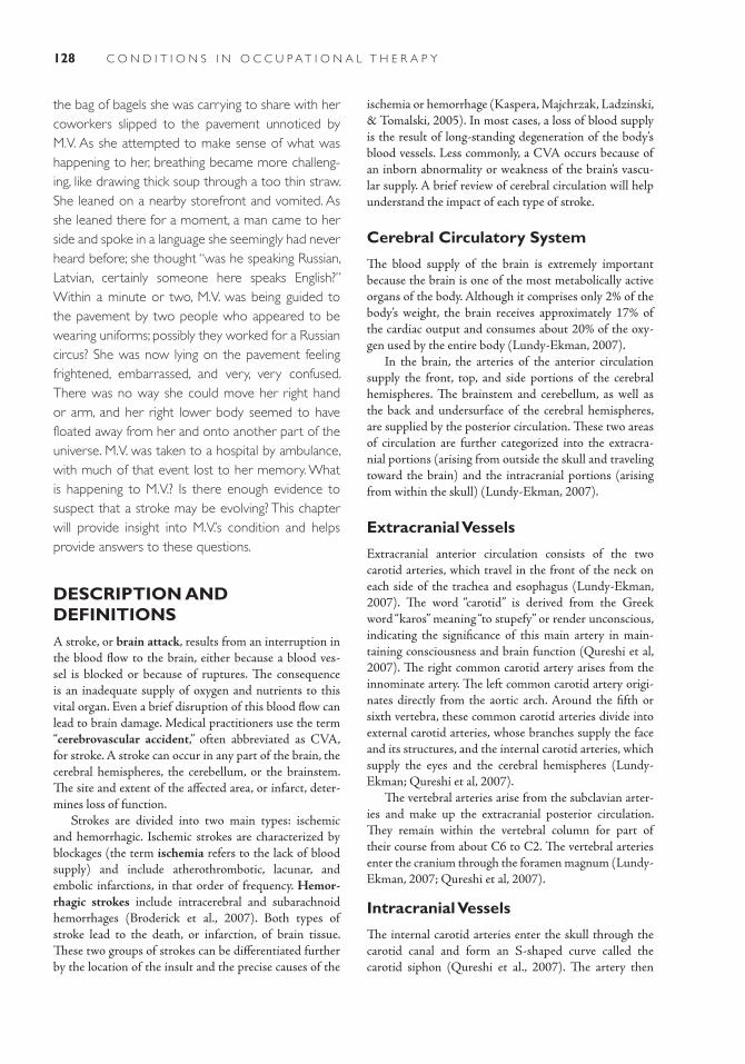
128 C O N D I T I O N S I N O C C U P A T I O N A L T H E R A P Y
ischemia or hemorrhage (Kaspera, Majchrzak, Ladzinski, & Tomalski, 2005). In most cases, a loss of blood supply is the result of long-standing degeneration of the body’s blood vessels. Less commonly, a CVA occurs because of an inborn abnormality or weakness of the brain’s vascu-lar supply . A brief review of cerebral circulation will help understand the impact of each type of stroke.
Cerebral Circulatory System
The blood supply of the brain is extremely important because the brain is one of the most metabolically active organs of the body. Although it comprises only 2% of the body’s weight, the brain receives approximately 17% of the cardiac output and consumes about 20% of the oxy-gen used by the entire body (Lundy-Ekman, 2007).
In the brain, the arteries of the anterior circulation supply the front, top, and side portions of the cerebral hemispheres. The brainstem and cerebellum, as well as the back and undersurface of the cerebral hemispheres, are supplied by the posterior circulation. These two areas of circulation are further categorized into the extracra-nial portions (arising from outside the skull and traveling toward the brain) and the intracranial portions (arising from within the skull) (Lundy-Ekman, 2007).
Extracranial Vessels
Extracranial anterior circulation consists of the two carotid arteries, which travel in the front of the neck on each side of the trachea and esophagus (Lundy-Ekman, 2007). The word “carotid” is derived from the Greek word “karos” meaning “to stupefy” or render unconscious, indicating the significance of this main artery in main-taining consciousness and brain function (Qureshi et al, 2007). The right common carotid artery arises from the innominate artery. The left common carotid artery origi-nates directly from the aortic arch. Around the fifth or sixth vertebra, these common carotid arteries divide into external carotid arteries, whose branches supply the face and its structures, and the internal carotid arteries, which supply the eyes and the cerebral hemispheres (Lundy-Ekman; Qureshi et al, 2007).
The vertebral arteries arise from the subclavian arter-ies and make up the extracranial posterior circulation. They remain within the vertebral column for part of their course from about C6 to C2. The vertebral arteries enter the cranium through the foramen magnum (Lundy-Ekman, 2007; Qureshi et al, 2007).
Intracranial Vessels
The internal carotid arteries enter the skull through the carotid canal and form an S-shaped curve called the carotid siphon (Qureshi et al., 2007). The artery then
the bag of bagels she was carrying to share with her coworkers slipped to the pavement unnoticed by M.V. As she attempted to make sense of what was happening to her, breathing became more challeng-ing, like drawing thick soup through a too thin straw. She leaned on a nearby storefront and vomited. As she leaned there for a moment, a man came to her side and spoke in a language she seemingly had never heard before; she thought “was he speaking Russian, Latvian, certainly someone here speaks English?” Within a minute or two, M.V. was being guided to the pavement by two people who appeared to be wearing uniforms; possibly they worked for a Russian circus? She was now lying on the pavement feeling frightened, embarrassed, and very, very confused. There was no way she could move her right hand or arm, and her right lower body seemed to have floated away from her and onto another part of the universe. M.V. was taken to a hospital by ambulance, with much of that event lost to her memory. What is happening to M.V.? Is there enough evidence to suspect that a stroke may be evolving? This chapter will provide insight into M.V.’s condition and helps provide answers to these questions.
DESCRIPTION AND DEFINITIONSA stroke, or brain attack, results from an interruption in the blood flow to the brain, either because a blood ves-sel is blocked or because of ruptures. The consequence is an inadequate supply of oxygen and nutrients to this vital organ. Even a brief disruption of this blood flow can lead to brain damage. Medical practitioners use the term “cerebrovascular accident,” often abbreviated as CVA, for stroke. A stroke can occur in any part of the brain, the cerebral hemispheres, the cerebellum, or the brainstem. The site and extent of the affected area, or infarct, deter-mines loss of function.
Strokes are divided into two main types: ischemic and hemorrhagic. Ischemic strokes are characterized by blockages (the term ischemia refers to the lack of blood supply) and include atherothrombotic, lacunar, and embolic infarctions, in that order of frequency. Hemor-rhagic strokes include intracerebral and subarachnoid hemorrhages (Broderick et al., 2007). Both types of stroke lead to the death, or infarction, of brain tissue. These two groups of strokes can be differentiated further by the location of the insult and the precise causes of the
129Chapter 9 : Cerebrovascu lar Acc ident
posterior cerebral arteries, from which it continues to the starting point in reverse order (Zazulia). When one major vessel supplying the brain is slowly occluded, either within the circle of Willis or proximal to it, the normally small communicating arteries may slowly enlarge to com-pensate for the occlusion (Lundy-Ekman, 2007). This system is imperfect, however, and it often fails to prevent strokes. In many individuals, the same atherosclerotic processes that caused a stroke also may damage commu-nicating arteries. In addition, only about one-fourth of strokes are caused by a blockage of the major neck vessels (Qureshi et al., 2007). For approximately one-third of the population, the communicating artery may be insuf-ficient or even absent (Zazulia). Such anomalies are more common in those who have strokes than in the general population and may be linked to increased risk of stroke in persons who also have atherosclerosis (van Kooij, Hendrikse, Benders, de Vries, & Groenendaal, 2010).
ETIOLOGY
Ischemic Stroke
This is the most common type of stroke accounting for 80% of cases (Fulgham et al., 2004). Cerebral infarction, or brain tissue death, results when circulation to an area of the brain is obstructed, with the result being ischemia. Ischemic strokes are classified as thrombotic, embolic, or lacunar strokes. The damaged area has two components: the tissues that have died as a result of blood supply loss and the peripheral area in which there may be tempo-rary dysfunction as a result of edema. Edematous brain tissue sometimes recovers slowly and gradually, result-ing in a reappearance of function after a period of 4 to 5 months (Fulgham et al.). In the past, prognoses for func-tional recovery have been limited to this time frame. However, recent research offers promising evidence that recovery is possible months and even years post stroke
enters the subarachnoid space by piercing the dura mater. It gives rise to the ophthalmic arteries, which supply the eyes; the posterior communicating arteries, which join with the posterior circulation; and the anterior cerebral arteries, which supply the orbital and medial surfaces of the frontal lobes and part of the basal frontal lobe white matter and caudate nucleus. The internal carotid artery also gives off the middle cerebral arteries, which supply almost the entire lateral surface of the frontal, parietal, and temporal lobes as well as the underlying white mat-ter and basal ganglia (Lundy-Ekman, 2007). The middle cerebral artery is the largest of the terminal branches of the internal carotid artery and is the direct continuation of this vessel.
The vertebral arteries enter the cranium within the posterior fossa and travel along the side of the medulla, where they give off their longest branch, the posterior inferior cerebellar artery. This artery supplies the lateral medulla and the back of the undersurface of the cerebel-lum (Zazulia, 2002). The two vertebral arteries then join at the junction between the medulla and pons to form the single midline basilar artery (Zazulia). The basilar artery gives off penetrating arteries to the base of the pons and two vessels (the anterior inferior and superior cerebellar arteries) that supply the upper and anterior undersur-faces of the cerebellum (Zazulia). At the level of the mid-brain, the basilar artery bifurcates into the two posterior cerebral arteries (Zazulia). As they circle the brainstem, these two arteries give off penetrating branches to the midbrain and thalamus and then divide into branches that supply the occipital lobes as well as the medial and undersurfaces of the temporal lobes (Zazulia). One of the branches of the posterior cerebral artery, the cal-carine artery, is of special significance because it is the main supplier of blood for the visual area of the cortex (Lundy-Ekman, 2007).
Communicating Arteries
The right and left carotid vessels connect with each other when they enter the brain, each sending out a small lateral branch that meets in the space between them. These are the anterior communicating arteries. They also branch backward to join with the right and left posterior cerebral arteries, called the posterior communicating arteries. This communicating vascular interchange is known as the cir-cle of Willis, which is pictured in Figure 9.1. It protects the brain should one of the four major supplying arteries com-ing up through the neck be blocked (Zazulia, 2002). This important anatomical feature is named for Dr. Thomas Willis and was first described in the mid-17th century (Zazulia). Starting from the midline anteriorly, the cir-cle consists of the anterior communicating, anterior cer-ebral, internal carotid, posterior communicating, and
Anteriorcerebral
Anterior communicating
Internalcarotid
Basilar
Posteriorcerebral
Posteriorcommunicating
Figure 9.1 Circle of Willis.
130 C O N D I T I O N S I N O C C U P A T I O N A L T H E R A P Y
(Hankey et al., 2007). Explanations focus on the brain’s ability to reroute neural pathways, a phenomenon known as neural plasticity ( Johansson, 2000; Takatsuru et al., 2009).
The actual physiologic events that follow an ischemic stroke occur in characteristic steps. First, the membrane surrounding each affected neuron leaks potassium (a mineral necessary for producing electrical impulses) and adenosine triphosphate (ATP, an energy-producing biochemical found in the body). Fluid quickly accu-mulates between the blood vessel and neuron, mak-ing it difficult for oxygen and nutrients to pass from the bloodstream into the damaged neuron. The initial injury produces a vicious circle in which more cellular injury results. Irreversible cell death will occur in 5 to 10 minutes if oxygen and nutrients are unable to reach them from the bloodstream or in a slightly longer period if blood flow is only partially interrupted. These dead cells form a zone of infarction that will not regenerate (Zazulia, 2002).
Downstream from the infarct zone is a zone of injury (penumbra) (Gonzalez, 2006; Bose A, Henkes H, and Alfke, K. (2008). This area may be served by collateral blood vessels and is capable of returning to normal func-tioning. A third area that reacts differently to the stroke process may also exist. In this area of hyperemia, the blood vessels are congested and swollen and also may have the potential for recovery.
The presence of these two zones, the regions of penumbra and hyperemia, may serve to minimize the total area of infarction. However, they do present problems for treatment because interventions that benefit one region may not help the other (Gonzalez, 2006; Bose, 2008).
Thrombosis
Cerebral thrombosis occurs when a blood clot forms in one of the arteries supplying the brain, causing vascular obstruction at the point of its formation. The size and location of the infarct depends on which vessel is occluded and the amount of collateral circulation. Thrombosis occurs most frequently in blood vessels that have already been damaged by atherosclerosis (Ho, Huang, Khor, & Lin, 2008).
Atherosclerosis is a gradual degenerative disease of the blood vessel walls. It is a pathologic process rather than a normal effect of human aging (Ho et al.). Rough, irregular fatty deposits form within the intima and inner media of the arteries and often lead to the generation of a thrombus or blood clot. This is the most common cause of stroke, with stenosis or narrowing of the blood vessels, resulting in much fewer cases (Ho et al., 2008). Large-vessel atherosclerosis accounts for 60% of ischemic stroke (University of Medicine and Dentistry of New Jersey, 2010). Because the body’s blood vessels have a
significant reserve capacity, ischemic strokes do not usu-ally occur until the vessel is two-thirds blocked (Univer-sity of Medicine and Dentistry of New Jersey, 2010). The impact of atherosclerosis on the vascular system is con-siderable; it is also a major risk factor for heart disease.
To understand the process that produces a mass of degenerated, thickened material (plaque) called athero-mas, imagine a glue bottle that has been allowed to col-lect the residuals of dried glue. The more clogged the cap of the bottle becomes, the more difficult it is for the glue to flow through. Squeezing the glue through the opening can push already dried glue more firmly against the open-ing. The opening will become smaller and smaller until it closes completely or bursts from the increased pressure.
In the cerebral circulation, atherosclerosis and throm-bus formation are most likely to occur in areas where blood vessels turn or divide, such as the origins of the internal carotid artery and the middle cerebral artery and the junction of the vertebral and basilar arteries (van Kooij et al., 2010).
Cerebral thrombosis often causes stuttering or pro-gressive symptoms that occur over several hours or days. Onset during sleep is common. Often a patient notices mild arm numbness at night and then awakens the next morning with paralysis. Transient ischemic attacks (TIAs) precede actual infarction about half the time (van Kooij et al., 2010).
Lacunar Strokes/Penetrating Artery Disease
Lacunar strokes are small infarcts, usually lying in the deep brain structures, such as the basal ganglia, thalamus, pons, internal capsule, and deep white matter (Porter & Kaplan, 2004). Approximately 25% of ischemic strokes are the result of damage to these deep structures (Porter & Kaplan). Within a few months of onset of a lacunar stroke, a small cavity (“lacune” in French) is left (Porter & Kaplan).
Lacunar strokes result from an occlusion of small branches of larger cerebral arteries—middle cerebral, posterior cerebral, basilar, and, to a lesser extent, anterior cerebral and vertebral arteries ( Jackson & Sudlow). Lacunar infarcts range in size from 2 to 15 mm (Porter & Kaplan, 2004; Jackson & Sudlow, 2005). Because of their small size, minimal neurologic symptoms are often present, and many such strokes go undetected. Recent findings indicate, however, that long-term prognosis is not good for lacunar strokes, and the recovery rate is similar to that for other types of stroke ( Jackson & Sudlow). Typically, lacunar strokes produce purely motor deficits (weakness or ataxia), purely sensory deficits, or a combination of sensory
131Chapter 9 : Cerebrovascu lar Acc ident
congenital. There are two types of hemorrhagic strokes. An intracerebral hemorrhage refers to bleeding directly into brain substance, whereas a subarachnoid hemor-rhage is bleeding occurring within the brain’s surrounding membranes and cerebrospinal fluid (CSF) (Feigin, Lawes, Bennet, Barker-Collo, & Parag, 2009). These two types of hemorrhage differ in incidence, etiology, clinical signs, and treatment.
Intracerebral Hemorrhages
Intracerebral hemorrhage results in bleeding directly into the brain and accounts for a high percentage of deaths because of stroke (Feigin et al., 2009). It may occur in any part of the brain and is most commonly linked to hyper-tension. Other causes include blood vessel abnormalities, such as arteriovenous malformations (AVMs) or aneu-rysms, or trauma (Locksley, 2010). Release of blood into brain tissue and surrounding edema will then disrupt the function of that particular brain region (Locksley, 2010). Blood irritates the brain tissue and causes swelling, or it may form a mass called a hematoma. In either case the increased pressure on brain tissues can rapidly destroy them. Factors that increase the risk of intracerebral hem-orrhage include blood and bleeding disorders such as hemophilia, sickle cell anemia, and leukemia; use of anti-coagulants; and liver disease (Porter & Kaplan, 2004).
Clinical signs of intracerebral hemorrhage are usu-ally focal; that is, unlike cerebral infarcts, hemorrhagic bleeds do not follow the anatomic distribution of blood vessels but move spherically through the tissue planes (Locksley, 2010). Typically they develop suddenly, often during activity (Amarenco et al., 2009). While extremely small hemorrhages may go undetected, a large hematoma causes headache, vomiting, convulsions, and decreased levels of alertness (Ohwaki et al., 2004).
Stupor and coma are common signs of very large hem-orrhages and indicate a poor prognosis, especially among individuals over 85 (Ohwaki et al., 2004). Nevertheless, recovery is possible.
Cerebral injury caused by intracerebral bleeding is a result of the damaging effect that the abnormal presence of blood has on the neurons. In addition to the irritant of the blood, abnormal pressure on neurons distorts their normal architecture. It also prevents oxygen and nutrients from passing to the cells from the blood stream. Eventu-ally, bleeding will stop and a hard clot will form. During a period of months, the clot slowly recedes, breaks down, and is absorbed by the body’s white blood cells (Zhao, Grotta, Gonzales, & Aronowski, 2009). If not damaged by increased pressure, the tissues irritated by the blood may heal, leading to a positive outcome, perhaps even full recovery (Zhao et al., 2009).
and motor deficits ( Jackson & Sudlow). Symptoms do not usually include aphasia, changes in cognition or personality, loss of consciousness, homonymous hemi-anopsia, or seizures ( Jackson & Sudlow). The most consistently identified risk factor for lacunar infarction is hypertension, and treatment is aimed at controlling it ( Jackson & Sudlow).
Embolism
Embolism occurs when a clot that has formed else-where (thrombus) breaks off (embolus), travels up the blood stream until it reaches an artery too small to pass through, and blocks the artery (Montaner et al., 2008). At this point, the effects of the embolus are similar to those produced by thrombosis. Embolic materials that travel to the arteries of the brain can originate from many sources, including the aortic arch and arteries arising from it, the extracranial carotid and vertebral arteries, and thrombi in the heart. Cardiac source emboli occur in approximately 20% of ischemic strokes and are referred to as cardio-genic (Cho et al., 2009). Many cardiac abnormalities can give rise to a cerebral embolism, including atrial fibrilla-tion, coronary artery disease, valvular heart disease, and arrhythmias. Cardiac surgery is also a cause (Cho et al., 2009). The middle cerebral artery is by far the most com-mon destination of cardiac emboli, followed by the poste-rior cerebral artery (Cho et al., 2009).
In contrast to thrombotic strokes, embolic strokes typically occur during daytime activity (Montaner, 2008). The embolism can be precipitated by a sudden movement, or even a sneeze, which raises blood pressure and dislodges the clot. Clinical symptoms are usually maximal at onset, but in some cases the neurologic symptoms improve or stabilize somewhat, then worsen as the embolus moves and blocks a more distal artery. A history of TIAs is rare. Seizures may be associated with embolic strokes.
Hemorrhagic Stroke
Approximately 20% of strokes are hemorrhagic (Amarenco, Bogousslavsky, Caplan, Donnan, & Henner-ici, 2009). Hemorrhagic strokes are caused by a rupture in a blood vessel or an aneurysm, with resultant bleeding into or around cerebral tissue. An aneurysm is a bulging or outpouching of a wall of an artery as a result of weak-ness in the vessel wall; it is prone to rupture at any time (Mayo Clinic Staff, 2009). While fatality rates for hemor-rhagic strokes are higher than for ischemic strokes, recent findings indicate that patients often make a better recov-ery after a hemorrhagic stroke (Amarenco et al., 2009). Hemorrhagic strokes are more common in young people than ischemic strokes as the vessel wall anomaly is often

132 C O N D I T I O N S I N O C C U P A T I O N A L T H E R A P Y
Townend, Tinson, Kwan, & Sharpe, 2010). Impact on family and caregivers is also enormous. If adequate social services and support are not available, caregivers assume the burden for functional tasks that the patient is unable to perform (McCullagh, Brigstocke, Donaldson, & Kalra, 2005).
Despite these grim statistics, there is some good news. With the exception of subarachnoid hemorrhage, between 1950 and 1990 all types of strokes have shown a significantly decreased incidence (Chiuve et al., 2008). This may be partly the result of increased control of risk factors such as hypertension, diabetes mellitus, and heart disease (Chiuve et al.). Individuals who have had a stroke now live almost twice as long; interestingly, one study even reported that long-term survivors appeared to be less depressed than the comparison group (Patel, McKevitt, Lawrence, Rudd, & Wolfe, 2007). Fewer people now die of stroke, notwithstanding the continuing debate as to whether this decline is the result of the occurrence of fewer strokes or better medical treatment (Carandang et al., 2006). However, it should be noted that the absolute number of strokes occurring in the United States is rising, probably a result of the aging of the population (Caran-dang et al.). Moreover, dramatic increases in obesity, among children as well as adults, offer grave cause for concern, given the close links determined between obesity and vascular disease (Lawlor & Leon, 2005).
Stroke in young children occurs in about 2.5 cases per 100,000 children per year as compared to 100 cases per 100,000 in the adult population (Lawlor & Leon, 2005). It is more frequent in children younger than 2 years of age. The effects of stroke among children are similar to those described in adults. Although the etiology of pedi-atric stroke is often unknown, the most common known cause is a congenital defect affecting the structure of the heart (Lawlor & Leon). Sickle-cell disease, which affects blood-clotting mechanisms, and genetic disorders are the next most commonly known causes. Forty-five percent of pediatric stroke patients experience neurologic defects throughout their lives; however, one-third of children with strokes display no symptoms afterward (Steinlin, Roellin, & Schroth, 2004).
RISK FACTORSA number of risk factors are associated with the likeli-hood of stroke and resemble those for heart disease. This is not surprising, considering that atherosclerosis is an underlying cause for both conditions. While some of these risk factors are inborn or otherwise unavoid-able, many are related to lifestyle and behavioral choices. Adopting healthy eating and exercise patterns early in life is especially important for individuals who are genetically
Subarachnoid Hemorrhages
Subarachnoid hemorrhages account for about 5% to 10% of all strokes and are slightly more common in women (Ohwaki et al.). About 95% are caused by the leakage of blood from aneurysms (Ohwaki et al.). A combina-tion of congenital and degenerative factors, usually at the points of origin or bifurcations of arteries, can precipitate formation of an aneurysm (Ohwaki et al., 2004). Blood may break through the weak point of the aneurysm at any time and, because of the force of arterial pressure, spread quickly into the CSF surrounding the brain. A subarach-noid hemorrhage also may be caused by bleeding from an AVM, which is an abnormal collection of vessels near the surface of the brain. Other less common causes of sub-arachnoid hemorrhages are hemophilia, excessive anti-coagulation therapy, and trauma to the skull and brain (Amarenco et al., 2009). The extravasated, or escaped, blood irritates the meninges, and intracranial pressure is increased owing to extra fluid in the closed cranial cav-ity. This can lead to headache, vomiting, and an altered state of consciousness. Sleepiness, stupor, agitation, restlessness, and actual coma are various manifestations of reduced consciousness. Headaches are usually severe and are described as the worst in the patient’s life (Stary, 2004). Because the bleeding takes place around the brain and not in the actual brain substance, motor, sensory, or visual abnormalities on one side of the body usually are not seen. Computed tomography (CT) scans (see later in this chapter) are the most reliable tool for detecting sub-arachnoid hemorrhage (Stary). If results are inconclusive, lumbar puncture with analysis of the CSF is the most reliable method of diagnosing subarachnoid hemorrhage (Stary).
INCIDENCE AND PREVALENCEStroke is the third leading cause of death in the United States, surpassed only by heart disease and cancer (Chi-uve et al., 2008). Stroke, the most common cause of disa-bility, is the most widespread diagnosis among clients seen by occupational therapists for the treatment of physically disabled adults (Legg et al., 2003). An estimated 600,000 to 730,000 people in the United States suffer an episode each year, and about 4.5 million stroke survivors are alive today (Xie et al., 2006). Approximately 50% to 70% of people regain functional independence after a stroke; however, 15% to 30% of those who survive an ischemic or hemorrhagic stroke suffer some permanent disability (Xie et al., 2007). Of the two main types, hemorrhagic strokes occur much less frequently (20% of all strokes) than ischemic strokes, which account for about 80% of all strokes (Xie et al., 2007). Depression affects approxi-mately one-third of stroke survivors (Hackett et al., 2005;
133Chapter 9 : Cerebrovascu lar Acc ident
pressure checks. Hypertension can be controlled through drug therapy, stress reduction, dietary con-trol, and regular exercise—treatments that all require patient compliance for maximum effectiveness.
■ Smoking—smoking doubles the risk of stroke (Bhat et al., 2008). Quitting smoking reduces the likeli-hood of stroke, and there is some evidence to suggest that 5 years after quitting smoking, individuals lower their risk of having a stroke to nearly that of people of who have never smoked (Zazulia, 2002).
■ TIAs—a TIA is considered an important risk factor for an impending stroke. Approximately 30% of all patients who have had a TIA are at risk of having a stroke within 2 years. This risk is greatest in the first month, so it is important to seek medical intervention quickly (Fulgham et al., 2004).
■ Geographic location—the highest death rates from strokes in the United States are in North and South Carolina, Georgia, northern Florida, Alabama, Mis-sissippi, and Tennessee. Specific pockets of high death rates in Texas, Oklahoma, and all of the Hawaiian Islands have been noted (Voeks et al., 2008). This geographical strip, often termed the “stroke belt,” is the source of numerous studies on environmental, cultural, or other geographically determined risk factors. A per-son who grows up in a high-risk area and then moves to a lower-risk area as an adult continues to carry the greater likelihood of having a stroke. This has led to the speculation that the causes may be diet related, cul-tural, or possibly even related to water supply or alti-tude (Glymour, Avendaño, & Berkman, 2007).
■ Diabetes mellitus—this disease is more common in stroke patients than in a normal population of similar age (Lees & Walters, 2005). Having diabetes increases the risk of stroke by two to three times, as diabetics have a tendency to form blood clots (Lees & Walters). Men are more vulnerable than women (Lees & Walters).
■ Oral contraceptives—women who have taken birth control pills, especially those with a high estrogen con-tent, have an increased risk for stroke as they become older. Smoking while taking the pill further increases the risk (Slooter et al., 2005).
■ Hyperlipidemia—including elevated triglycerides and “bad” cholesterol has long been suspected to be a risk factor for stroke (Milionis et al., 2005). Its role in CVA has been confirmed by several recent studies (Milionis et al.). Cholesterol levels should be moni-tored, and adults are encouraged to know their choles-terol counts. Dietary changes are effective in lowering cholesterol levels in some individuals; a class of drugs known as statins is also effective but may have side effects (Trubelja, Vaughan, & Coplan, 2005).
at risk, although everyone can benefit. Regular visits to a family physician are important.
■ Ethnicity—although all minority groups are at risk, stroke death rates are much higher for African Ameri-cans than for whites: Data from 2005 indicated that out of 100,000 people there were 87 deaths from stroke for black males and 78 for black females, com-pared to 59 for white males and 58 for white females (Bravata et al., 2005). Recent data also indicate that blacks living in the South have a greater risk of dying from stroke than those who live in the northern states (Voeks et al., 2008).
■ Age—risk for stroke increases with age, especially after age 65. People with high blood pressure and who exhibit the other risk factors listed in this chapter are increas-ingly vulnerable as they age. Younger people are not immune, either: Approximately 28% of strokes occur in individuals younger than age 65 (Bhat et al., 2008).
■ Heredity—a family history of stroke, particularly on the father’s side, increases one’s risk (Bhat et al., 2008). Genetic factors account for 7% to 20% of cases of subarachnoid hemorrhage, and some researchers recommend that people with more than one close rel-ative who suffered a hemorrhagic stroke be screened for aneurysms (Locksley, 2010).
■ Obesity—being overweight is a known risk factor for hypertension and diabetes mellitus and also is associated with stroke. Weight that is centered in the abdomen (so-called apple shape) has a particularly high association with stroke as well as heart disease, while individuals whose weight is distributed around the hips are less at risk (Suk et al., 2003). A sedentary lifestyle is associated with rising levels of obesity and has been implicated in occurrence of strokes as well as hypertension (Bhat et al., 2008).
■ Hypertension—called the “silent disease,” hyperten-sion has long been acknowledged as the most signifi-cant controllable risk factor for both stroke and heart attack (Bhat et al., 2008). Hypertension occurs in 25% to 40% of the U.S. population (Bhat et al., 2008). Symptoms of hypertension are not clearly identifi-able: an occasional headache, dizziness, or light-head-edness, which may indicate hypertension, can easily be attributed to other factors. Chronically elevated blood pressure exerts pressure on cerebral vessels, often resulting in lacunar infarctions or intracerebral hemorrhage ( Jackson & Sudlow, 2005; Amarenco et al., 2009). Hypertension also has been implicated in the atherosclerotic process because it drives fatty sub-stances into the arterial walls, making them brittle, narrowed, and hardened (Zazulia, 2002). The impact of hypertension increases with age (Bhat et al., 2008). For these reasons it is crucial to have regular blood
134 C O N D I T I O N S I N O C C U P A T I O N A L T H E R A P Y
polycythemia, can be controlled by medical interven-tion. Factors that cannot be changed are age, sex, race, family history, diabetes mellitus, and a prior stroke. The potential benefits of all medical and surgical interven-tions currently available for cerebrovascular disease pale in comparison to what can be achieved through risk factor control (Chiuve et al., 2008). Understanding and aware-ness of these risk factors for stroke is an important first step in reducing the likelihood of having stroke.
SIGNS AND SYMPTOMS
Neurologic Effects of Stroke
An occlusion that causes a serious stroke can occur any-where in the extracranial or intracranial system, but the most common site is in the distribution of the middle cer-ebral artery and its branches in the cerebrum. The major-ity of cerebral strokes occur in one or the other cerebral hemisphere, but not both (Wintermark et al., 2002). It is important to note that even in individuals with the same neurologic deficit, the impact of disability is different, depending on the individual’s life situation.
Stroke Warning Signs
To educate the public, the American Heart Association and National Stroke Association distribute pamphlets listing the warning signs of an impending serious stroke (American Heart Association [AHA], 2010). Many hos-pitals and community clinics also distribute this informa-tion. These include
■ Sudden numbness or weakness of the face, arm, or leg, especially on one side of the body
■ Sudden confusion, trouble speaking or understanding ■ Sudden trouble seeing in one or both eyes ■ Sudden trouble walking, dizziness, loss of balance, or
coordination ■ Sudden severe headache with no known cause
Some general medical symptoms related to type of stroke were discussed under Etiology. Signs and symptoms also depend on the size and location of the injury, and neu-rologists can often predict location by the symptoms the individual displays. However, it is important to remember that a stroke is complex, and each individual may experi-ence a unique constellation of symptoms. Relying on ste-reotypical models of stroke leads to generalized and often inappropriate therapy.
In addition to the above list, it is important to be aware that symptoms resulting from a partial reduction or tem-porary change in the blood flow to the brain are extremely important warning signs for stroke (Fogle et al., 2008). Several of these conditions are discussed below.
■ Asymptomatic carotid bruits—bruit is an abnormal sound or murmur heard when a stethoscope is placed over the carotid artery. This slushing noise indicates turbulent blood, often caused by a significant degree of stenosis. Carotid bruit clearly indicates increased stroke risk. Complete occlusion of the carotid artery some-times follows, resulting in stroke (Bhat et al., 2008).
■ Prior stroke—risk of stroke for a person who has already suffered a stroke is increased four to eight times (Fang et al., 2005).
■ Heart disease—diseased heart (whether it be chronic disease, acute heart attacks, or prosthetic heart valves) increases the risk of stroke. Independent of hyperten-sion, people with heart disease have more than twice the risk of stroke than people with normally function-ing hearts (Ohira et al., 2006). Atrial fibrillation, a con-dition in which the heart produces an irregular rhythm, is also a known risk factor for stroke (Fang et al., 2005).
■ Infections and inflammation— inflammation occur-ring with various infections, often mild, has been associated with stroke. A study in 2005 indicated that periodontal (gum) disease is associated with a 20% high risk for ischemic stroke and heart disease and that the risk may be even greater in adults under the age of 65 (Beck & Offenbacher, 2005). Research is under way to determine whether other infections that produce arterial inflammation can lead to stroke or heart disease. For example, chronic infection with Chlamydia pneumoniae, which causes mild pneumonia in adults, has been linked with higher risk for stroke (Elkind et al., 2006b).
■ Alcohol and drug abuse—numerous studies indicate that moderate consumption of alcohol decreases the risk of stroke and heart disease; however, excessive consumption and total abstinence are associated with higher risk (Elkind et al., 2006a). Cocaine and meth-amphetamine abuse are major factors in the incidence of stroke in young adults (Westover, McBride, & Haley, 2007). Use of anabolic steroids for bodybuilding is also associated with increased risk of stroke (Santamarina, Besocke, Romano, Loli, & Gonorazky, 2008).
■ Sleep apnea—it is a common disorder in which the throat becomes obstructed during sleep, interfering with normal breathing and sleep. Sleep apnea is wors-ened by obesity and may contribute to the narrowing of the carotid artery. Sleep apnea is currently thought to increase the risk of stroke three- to sixfold (Arzt, Young, Finn, Skatrud, & Bradley, 2005).
Of these many risk factors, several can be controlled by changes in lifestyle, including elevated blood cholesterol and lipids, cigarette smoking, use of oral contraceptives, excessive drinking of alcohol, and obesity. Some, such as hypertension, heart disease, TIAs, carotid bruits, and
135Chapter 9 : Cerebrovascu lar Acc ident
is exercised. Usually, movement of the arm produces light-headedness, numbness, and weakness. Other neu-rologic symptoms also may be present. In this syndrome, blood is “stolen” from the brain and instead is delivered to the exercised arm. It is a warning sign that advanced atherosclerosis may be present in the arteries through-out the body, including the cerebral arteries (Bicknell, Subramanian, & Wolfe, 2004).
DIAGNOSISThe diagnosis of stroke requires knowledge of the inci-dence of the different types of stroke and awareness of the presence of the risk factors mentioned above. Symptoms must be carefully noted from the patient or, if the patient is too ill, frightened, or confused, from the family (Hand, Kwan, Lindley, Dennis, & Wardlaw, 2006). Neurologists, neurosurgeons, and some internists are the specialists usu-ally involved in this acute diagnostic phase of treatment (Hand et al., 2006). A number of diagnostic techniques are utilized to distinguish stroke from other potential causes of observed symptoms and to assist in determining the location of lesions (Kidwell & Warach, 2003).
The physical examination of the patient with a sus-pected stroke or TIA includes a search for possible cardiac sources of emboli by listening to the heart and arteries of the neck. Also useful in the determination of cardiac-source emboli are electrocardiography, echocar-diography, and monitoring for arrhythmias. In addition to cardiac testing, various neuroimaging techniques (see below) and analysis of blood and CSF may be performed (Feldmann et al., 2007). A neurologic examination assists in determining the neurologic disability and usually includes evaluation of higher cortical function (memory and language), level of alertness, reflexes, visual and ocu-lomotor system, behavior, and gait (Feldmann et al.).
Other diagnostic methods include noninvasive stud-ies of blood vessels and invasive techniques requiring injection of dye into the arterial system, for example.
COURSE AND PROGNOSISStrokes result in anoxic damage to nervous tissue that causes various neurologic deficits, depending on where the blood supply was lost. If neuronal cell death occurs, it is considered irreparable and permanent, as no way has yet been found to regenerate nerve cells (Yuan, Lipinski, & Degterev, 2009). However, the nervous system has a high level of plasticity, especially during early development, and individual differences in neural connections and learned behaviors play a major role in functional recovery. No two brains can be expected to be structurally or function-ally identical (Carmichael, 2006). Spontaneous recovery
Transient Ischemic Attacks
TIAs result from a temporary blockage of the blood sup-ply to the brain. The symptoms occur rapidly and last for <24 hours. Seventy-five percent of TIAs last <5 minutes. The specific signs and symptoms depend on the portion of the brain affected but may include fleeting blindness in one eye, hemiparesis, hemiplegia, aphasia, dizziness, double vision, and staggering. Carotid artery disease and vertebral basilar artery disease may lead to TIAs (Mat-tace-Raso et al., 2006). The main distinction between TIAs and stroke is the short duration of the symptoms and the lack of permanent neurologic damage. People who have had TIAs are 9 1/2 times more likely to have a stroke than those of the same age and sex who have never had a TIA (Mattace-Raso et al., 2006). Without preventive treatment, a third of those who suffer TIAs will go on to have a stroke within 5 years (Mattace-Raso et al., 2006). Thus, it is crucial to detect the cause of a TIA and begin appropriate intervention promptly (Fogle et al., 2008).
Small Strokes (RINDs)
In some cases, the symptoms of a TIA may last longer than 24 hours. If they last a day or more and then com-pletely resolve or if they leave only minor neurologic deficits, they are called small strokes or lengthy TIAs ( Jackson & Sudlow, 2005). Often the remaining neuro-logic deficits are barely noticeable. Like TIAs, however, these small strokes are important warning signs that a more serious CVA may occur (Fogle et al., 2008). A small stroke that completely resolves is called a reversible ischemic neurologic deficit (RIND). An episode that lasts more than 72 hours and leaves some minor neuro-logic impairments is called a partially reversible ischemic neurologic deficit (PRIND). The mechanism of injury in RIND and PRIND is the same as that for a stroke or TIA. Like ischemic strokes, RINDs typically occur in the morning; since blood pressure is low during sleep, sud-den increases in blood pressure upon arising may cause problems (Metoki et al., 2006).
Many small strokes are not reported to a medical prac-titioner, which makes the exact frequency of occurrence of these strokes difficult to determine. It is important to recognize the symptoms of a small stroke so that it can be treated early, reducing the risk of more permanent injury.
Subclavian Steal Syndrome
This is a rare condition caused by a narrowing of the subclavian artery that runs under the clavicle. Symptoms occur when the arm on the side of the narrowed vessel
136 C O N D I T I O N S I N O C C U P A T I O N A L T H E R A P Y
A CVA in the region of the middle cerebral artery in the left cerebral hemisphere may produce the following symptoms:
1. Loss of voluntary movement and coordination on the right side of the face, trunk, and extremities.
2. Impaired sensation, including temperature discrimi-nation, pain, and proprioception on the right side (hemianesthesia).
3. Language deficits, called aphasia, in which the patient may be unable to speak or understand speech, writing, or gestures. The breakdown of language function is complex; the many types of aphasia will be discussed later in this chapter.
4. Problems with articulation of speech because of dis-turbances in muscle control of the lips, mouth, tongue, and vocal cords (dysarthria).
5. Blind spots in the visual field, usually on the right side.6. Slow and cautious personality.7. Memory deficits for recent or past events (Boyd &
Winstein, 2003).
Right-Sided Cerebral Injuries: Middle Cerebral Artery
The right cerebral hemisphere controls most of the func-tions on the left side of the body and also is responsible for spatial sensation, perception, and judgment. Injury to the middle cerebral artery of the right cerebral hemisphere may produce a combination of the following deficits:
1. Weakness (hemiparesis) or paralysis (hemiplegia) on the left side of the body (face, arm, trunk, and leg)
2. Impairment of sensation (touch, pain, temperature, and proprioception) on the left side of the body
3. Spatial and perceptual deficits4. Unilateral neglect, in which the patient neglects the
left side of the body or the left side of the environment5. Dressing apraxia, in which the patient is unable to
relate the articles of clothes to the body (Walker, Walker, & Sunderland, 2003)
6. Defective vision in the left halves of visual fields or left homonymous hemianopsia in which there is defective vision in each eye (the temporal half of the left eye and the nasal half of the right eye)
7. Impulsive behavior, quick and imprecise movements, and errors of judgment (Boyd & Winstein, 2003)
Anterior Cerebral Artery Stroke
The territory of the anterior cerebral artery is rarely infarcted because of the side-to-side communication pro-vided by the anterior communicating artery in the circle
may occur as edema subsides or viable neurons reactivate. Recovery also may occur with physiologic reorganization of neural connections or developmental strategies. Any injury brings different factors into play, affecting axonal and dendritic sprouting or collateral rearrangement, syn-aptic formation, the excitability of neurons, “substitution of parallel channels,” and “mobilization of redundant capacity” (Huang, Shen, & Duong, 2010). Recovery from neurologic deficits thus depends on the etiology and size of the infarct (Carmichael). Approximately 90% of neu-rologic recovery occurs within 3 months, with the rest occurring over a more extended time (Rossini, Calautti, Pauri, & Baron, 2003). It should be noted that recovery from hemorrhagic strokes proceeds more slowly, however (Rossini et al., 2003).
Accuracy in the prediction of function or rate of return in a given stroke patient is difficult because of individual variability of anatomy and extent of brain damage as well as differences in types of CVA, learning ability, premorbid personality and intelligence, and motivation (Rossini et al., 2003). Generally, the prognosis for recovery of func-tion is greater in young clients, possibly because the young brain is more plastic or because the young are generally in better physical condition.
Secondary complications are important to recovery and rehabilitation and may actually be more disabling than the stroke itself (McLean, 2004). These complica-tions, are discussed in this chapter and include depres-sion, seizures, infection, bowel/bladder incontinence, thromboembolism, shoulder subluxation, painful shoul-der, shoulder-hand syndrome, abnormal muscle tone, and associated reactions and movements.
Individuals with good sensation, minimal spasticity, some selective motor control, and no fixed contractures seem to make the greatest improvements in functional abilities. If an individual has no concept of the affected side and cannot localize stimuli to the affected side or if he or she has fecal or urinary incontinence, the outlook for inde-pendence is generally poor. Some individuals may continue to have strokes, complicating recovery (McLean, 2004).
Left-Sided Cerebral Injuries: Middle Cerebral Artery
The left cerebral hemisphere controls most functions on the right side of the body because of the decussa-tion of motor fibers (decussation of the pyramids) in the medulla. These fibers that cross, or decussate to the oppo-site side, form the lateral corticospinal tract. The rest of the fibers descend ipsilaterally, forming the anterior corti-cospinal tract (Wintermark et al., 2002). The proportion of crossing fibers varies from person to person, averaging about 85% (Wintermark et al.).
137Chapter 9 : Cerebrovascu lar Acc ident
area may produce contralateral pain and temperature loss, ipsilateral Horner’s syndrome (sinking of the eye-ball, ptosis of the upper eyelid, and a dry, cool face on the affected side), ataxia, and facial sensory loss. Ischemia to ipsilateral cranial nerve fibers VIII, IX, and X results in palatal paralysis, hoarseness, dysphagia, and vertigo (Wal-ton & Buono, 2003). There is no significant weakness in this syndrome (Dietrich et al.).
Brainstem strokes often result in coma because of damage to the centers involved with alertness and wake-fulness (reticular system) (Parvizi & Damasio, 2003). A hemorrhage into the brainstem area is rare, quickly accompanied by loss of consciousness, and usually fatal. Among patients who survive brainstem stroke, however, recovery is often good (Parvizi & Damasio).
OTHER COMPLICATIONS OF STROKESecondary conditions may occur in addition to these deficits. These are important manifestations of the patient’s recovery and rehabilitation and may actually be more disabling than the stroke itself (McLean, 2004). It is necessary to be aware of these complications so that they may be prevented.
Seizures
Brain scars that result from stroke may irritate the cor-tex and cause a spontaneous discharge of nerve impulses that may generalize to a full grand mal convulsion (Vespa et al., 2003). Seizures develop in up to 10% of stroke patients and are more common with embolic than throm-botic infarcts (Vespa et al., 2003). Anticonvulsant drugs are sometimes used in patients with early seizures, but their use is controversial (Camilo & Goldstein, 2004).
Infection
Alteration of swallowing function, aspiration, hypoven-tilation, and immobility in the stroke patient often lead to pneumonia (Prass, Braun, Dirnagl, Meisel, & Meisel, 2006). Changes in bladder function may lead to bladder distention and urinary tract infection (McLean, 2004). Impaired sensation and inadequate position changes may result in pressure sores (decubitus) and consequent infec-tion of these areas (Turhan, Atalay, & Atabek, 2006).
Thromboembolism
Immobility of the legs and prolonged bed rest often lead to thrombosis of dependent leg veins (MacDougall,
of Willis (Boyd & Winstein, 2003). Symptoms of an anterior cerebral artery stroke include
1. Paralysis of the lower extremity, usually more severe than that of the upper extremity, contralateral to the occluded vessel
2. Loss of sensation in the contralateral toes, foot, and leg3. Loss of conscious control of bowel or bladder4. Balance problems in sitting, standing, and walking5. Lack of spontaneity of emotion, whispered speech, or
loss of all communication6. Memory impairment or loss (Boyd & Winstein, 2003)
Vertebrobasilar Stroke
The vertebrobasilar system of arteries supplies blood primarily to the posterior portions of the brain, includ-ing the brainstem, cerebellum, thalamus, and parts of the occipital and temporal lobes. This posterior circulation is not divided into right and left halves, as in the anterior cir-culation (Coward et al., 2007). An occlusion here might produce
1. A variety of visual disturbances, including impaired coordination of the eyes
2. Impaired temperature sensation 3. Impaired ability to read and/or name objects 4. Vertigo and dizziness 5. Disturbances in balance when standing or walking
(ataxia) 6. Paralysis of the face, limbs, or tongue 7. Clumsy movements of the hands 8. Difficulty judging distance when trying to coordi-
nate limb movements (dysmetria) 9. Drooling and difficulty swallowing (dysphagia)10. Localized numbness11. Loss of memory (Boyd & Winstein, 2003)12. Drop attacks in which there is a sudden loss of
motor and postural control resulting in collapse, but the individual remains conscious (Parry et al., 2009)
TIAs in this area are common in the elderly. The verte-bral arteries travel up to the brainstem through a bony channel in the cervical vertebrae. In older adults, osteo-arthritis may develop in the cervical bones, causing nar-rowing of the cervical canal, especially when the head is extended or rotated (Parry et al., 2009).
Wallenberg’s Syndrome
Wallenberg’s syndrome is a classic brainstem stroke that also is referred to as lateral medullary syndrome (Dietrich et al., 2005). It occurs as the result of an occlu-sion of a vertebral or cerebellar artery. Strokes in this
138 C O N D I T I O N S I N O C C U P A T I O N A L T H E R A P Y
brainstem, thalamus, and spinal cord. It also provides better anatomic definition of the injury, but it does not distinguish between hemorrhage, tumor, and infarction as well as a CT scan. (Fiebach et al.).
Positron Emission Tomography
Positron emission tomography (PET) scan is being used experimentally. This scan shows how the brain uses oxy-gen, glucose, and other nutrients and has increased under-standing of the effects of stroke on the brain (De Reuck et al., 2006). Because these imaging techniques are not commonly available, PET scans are seldom used in the management of acute stroke (De Reuck et al.).
NONINVASIVE STUDY OF BLOOD VESSELSNoninvasive procedures used to evaluate both extracranial and intracranial blood flow include duplex ultrasonography as well as color-flow and transcranial Doppler ultrasound. These techniques can localize and determine the approxi-mate size of the lesions within the arteries (Aaslid, Huber, & Nornes, 2010).
Duplex ultrasonography is useful in detecting the presence and severity of the disease in the common and internal carotid arteries and in the subclavian and verte-bral arteries in the neck. This scan can reliably differenti-ate between minor plaque disease, stenosis, and occlusive lesions. It is an excellent method of monitoring the pro-gression or regression of atherosclerotic disease in the neck (Nederkoorn, van der Graaf, & Hunink, 2003).
Color-flow Doppler ultrasound is used because it is effective in showing lesions of the carotid and vertebral arteries (Aaslid et al., 2010).
Transcranial Doppler ultrasound gives information about pressure and flow in the intracranial arteries. This procedure is useful in monitoring changes in the arterial flow later in the course of the patient’s disease (Aaslid et al., 2010).
INVASIVE TECHNIQUESCerebral angiography involves radiography of the vascular system of the brain after injecting a dye or other contrast medium into the arterial blood system. Computer-generated images are then produced that can show the entire visible length of cerebral arter-ies as well as the nature, location, and extent of patho-logical changes. This technique is now safer than before (<1% incidence of mortality and serious morbidity);
Feliu, Boccuzzi, & Lin, 2006). In deep vein thrombosis (DVT), local pain and tenderness may develop in the calf, with some swelling and a slight increase in tem-perature. If the thrombosis is confined to the calf, it may not be serious. However, if the thrombosis spreads up toward the groin to involve the veins in the pelvis, there is a very real possibility of a clot breaking off into the blood stream. The clot will then travel through the right side of the heart and enter the lungs through the pul-monary arteries, resulting in sudden collapse and death owing to obstruction of the pulmonary arteries (Mac-Dougall et al., 2006). Early mobilization of the patient is of utmost importance in preventing DVT and subse-quent pulmonary embolism.
Medications and surgery may make a difference in the prognosis of an individual at risk for, or having had, a CVA.
NEUROIMAGING TECHNIQUESCT and magnetic resonance imaging (MRI) are invalu-able noninvasive tools that depict pathological changes in the brain in patients with stroke (Feldmann et al., 2007). One or the other of these is almost always used at some point for every patient with a suspected stroke. CT and MRI are also capable of showing zones of edema and the shifting of intracranial material (Feldmann et al.). Negative results of these tests may indicate that the ischemia is reversible (Feldmann et al.).
Computed Tomography
CT is a type of radiographic examination that is widely used for analysis of cerebral injury (Chalela et al., 2009). CT scans are employed to differentiate between a hem-orrhagic stroke and an ischemic stroke (Chalela et al.). While they are useful in clarifying the location and the mechanism and severity of stroke, they are not particu-larly sensitive to subtle ischemic changes (Chalela et al.). It is most useful in the diagnosis of stroke caused by hem-orrhage; CT findings may even be normal in patients with recent infarction. It is often not diagnostic in patients with TIAs and is not useful for imaging brainstem infarcts (Chalela et al.).
Magnetic Resonance Imaging
MRI is more sensitive than a CT scan and does not expose the patient to radiation. It provides detailed pictures of the brain by using a magnetic field. MRI is particularly helpful in revealing AVMs (Fiebach et al., 2004). It is superior to CT in imaging the cerebellum,
139Chapter 9 : Cerebrovascu lar Acc ident
(Mant et al.). It also benefits patients who have had a mild stroke but is not as effective in those with moderate or severe strokes (Sacco et al., 2006). Aspirin is clearly not suitable for patients with cerebral hemorrhage or those at risk of bleeding. It is less successful when used by women as compared with men. Aspirin is relatively safe and inexpensive. Recently published studies have recom-mended a lower dose of aspirin to reduce harmful side effects (Sacco et al.).
Anticoagulants
These drugs inhibit clotting by interfering with the activ-ity of chemicals in the liquid portion of blood that are essential for the coagulation process (Mant et al., 2007). Short-term (2 to 3 weeks) heparin therapy is prescribed for patients with complete blockages of large arteries, as heparin is effective in preventing the formation of emboli (Diener et al., 2006). For longer-term treatment (1 to 3 months), the drug warfarin may be used to pre-vent blockages in areas that cannot be treated by surgery (Mant et al.).
Thrombolytics
Thrombolytic therapy (t-PA), used for dissolution of an occluding thrombus, is frequently applied in the acute treatment of myocardial infarction as well as in the treatment of stroke. It is effective before exten-sive brain infarction has occurred, so it is appropri-ate only in stroke patients whose arterial damage has been identified early (Thomalla et al., 2006). The potential benefit must be weighed against reperfu-sion damage, a bleeding tendency, and the possibility of reocclusion (Amarenco, G., Easton, J., Sacco, R., & Teal, P., 2008). The Food and Drug Administra-tion has approved the use of t-PA for the treatment of acute stroke within 3 hours of onset (Gurm et al., 2008). One study reported that patients treated with t-PA were 30% to 50% more likely to have minimal or no disability 3 months after the onset of symptoms (Thomalla et al.).
Surgical Interventions
In some cases, surgical treatment may be the best choice for the patient. The neurosurgeon must carefully consider many factors before surgery is performed, including the patient’s overall health and life expectancy. Carotid endar-terectomy is among the most commonly performed vas-cular surgeries in the United States (Gurm et al., 2008).
however, it is recommended when noninvasive tech-niques have failed to yield a conclusive diagnosis or when surgery is being planned or considered (Kaufmann et al., 2007).
Analysis of CSF is helpful in diagnosing subarach-noid hemorrhage. In a lumbar puncture, the subarach-noid space (usually between the third and fourth lumbar vertebrae) is tapped and CSF is withdrawn. Analysis of the pigments in the spinal fluid also can help in estimat-ing the age of the hemorrhage and detecting rebleeding (Bykowski, Latour, & Warach, 2004).
OTHER TECHNIQUESOther diagnostic techniques used for stroke include elec-troencephalography, single-photon emission tomography, and special cardiac and coagulation tests that are useful in detecting unusual heart and blood disorders that can bring on a stroke (Petrella, Coleman, & Doraiswamy, 2003). After this diagnostic phase of evaluation, a neu-rologist will evaluate the brain-damaged person’s ability to function. Rehabilitation often will begin at that point to return the patient to the highest possible level of inde-pendent functioning (Duncan et al., 2005).
MEDICAL/SURGICAL MANAGEMENTAt present, the treatment of acute stroke is limited to management of the results of the primary event and pre-ventive measures against further injury or occurrence (Zhao et al., 2009).
Much is still unclear about the effectiveness of the routine use of agents to reverse the cause or decrease the effects of stroke (Zhao et al., 2009).
Before the stroke can be treated, it must be accurately identified as either cerebral infarction or cerebral hem-orrhage, since interventions that are beneficial with one type of stroke may be potentially dangerous to the other (Zhao et al., 2009).
Therefore, careful and exact diagnosis must be made first. Drug management of CVA is constantly evolving. Common categories of drugs used to minimize the dam-age of cerebral infarction are described in the following sections.
Antiplatelet Therapy
Aspirin is often prescribed to patients when the vascular lesion is not severely stenotic (Mant et al., 2007). Aspi-rin has been shown to reduce the risk of further stroke in patients who had suffered TIAs by 15% to 58%
140 C O N D I T I O N S I N O C C U P A T I O N A L T H E R A P Y
Corticosteroid therapy can cause a significant reduc-tion in interstitial cerebral edema, reducing swelling and improving outcomes after stroke (Thomas et al., 2007). However, a review of the literature indicates that there is no clear basis for evaluating the effect of corticoster-oid treatment for people with acute ischemic strokes (Veltkamp et al., 2005).
IMPACT ON OCCUPATIONAL PERFORMANCEThe impact of stroke on an individual is unique and may affect any number of client factors. Deficits in motor, process, and communication skills as well as impaired sensory functions and problems with perceptual pro-cessing may be compounded by secondary complica-tions such as infection or depression. Together, these profoundly affect an individual’s performance in all areas of occupation: activities of daily living (both basic and instrumental), work, play, leisure, and social participation (Langhorne, 2007).
Sensory Functions
Sensory functions can be affected at the very basic level of awareness and at the point of processing and modu-lation of sensory input. Loss of protective tactile func-tions, such as diminished awareness of temperature and pain, is a common concern for those with a CVA. Loss of these functions poses a safety risk. Individuals with proprioceptive dysfunction may show asymmetrical pos-ture, have difficulty maintaining balance, appear to for-get affected body parts, be unable to describe position or movement of limbs, and be susceptible to joint dam-age (Davis & Donnan, 2004). Individuals with a loss of tactile sensation may demonstrate a lack of awareness of body parts simply because they forget what they cannot feel. They are also vulnerable to damage of affected body parts, particularly skin breakdown. Moreover, diminished tactile function hinders resumption of motor activities. Depending on the location of the infarct, individuals may experience diminished vestibular function, which will limit mobility efficiency and safety. Impaired balance may cause difficulties in assuming and maintaining a vertical posture and in automatic adjustments to changes of posi-tion and antigravity movement. As a result, individuals demonstrate an asymmetrical posture at rest, leaning or falling to the hemiplegic side during mobility, or fail to use normal protective reactions when falling (Ward, Brown, Thompson, & Frackowiak, 2006).
Defects in visual field functions may impair reading, even in the absence of language dysfunction. For exam-ple, patients with right cerebral lesions may find reading
During the procedure, the diseased vessel is opened, the clot is removed, and an artificial graft is put in place (Por-ter & Kaplan, 2004). Carotid endarterectomy is a treat-ment option for patients with more than 50% stenosis of the carotid artery ipsilateral to the affected hemisphere (Porter & Kaplan; Bhat, 2008).
Subarachnoid hemorrhages are often caused by rup-tured aneurysms or AVMs. Surgical clipping or lesion removal is the most effective treatment of these anoma-lies. If the patient survives the initial bleeding, the goal of surgery is to correct the problem before bleeding recurs. In intracerebral hemorrhage, small hematomas usually resolve spontaneously (Zhao et al., 2009).
Large hematomas, however, often produce death. Some lesions may expand, causing gradually increasing neurologic signs. These expanding lesions can be drained surgically if they are near the surface of the brain, espe-cially in the cerebral or cerebellar white matter. Generally, hemorrhages are evacuated only if they are large and life threatening or when surgery is necessary to treat an aneu-rysm, tumor, or AVM (Zhao et al., 2009).
Superficial temporal artery bypass is a new, more deli-cate surgical therapy for preventing future strokes (Char-bel, Meglio, & Amin-Hanjani, 2005). The procedure begins with craniotomy to expose the brain; then a scalp artery is connected to an intracranial artery microsurgi-cally. This operation is extremely challenging to perform and is thus performed less frequently than carotid endar-terectomy. Surgeons who do the procedure, however, are enthusiastic about the results and claim that it revascu-larizes the brain better than endarterectomy (Pinar & Govsa, 2006).
Treatment of Secondary Effects
Specific pathophysiologic sequelae, or outcomes, fol-low the occurrence of any type of stroke. Treating these secondary effects is crucial to medical and functional recovery. Two of these are cerebral edema and ischemia. Oxygen therapy to reduce hypoxia, vasodilation to improve blood flow through ischemic areas, therapeutic hypertension, and hemodilution therapy are some of the treatments used for ischemia. Hemodilution results in a significant rise in cerebral blood flow and increased oxy-gen transfer (Vilea & Newell, 2008).
Edema often complicates ischemic strokes and must be controlled, because most deaths during the first week after a massive stroke are caused by extensive cerebral edema and increased intracranial pressure (Treggiari, Walder, Suter, & Romand, 2003). This pressure can displace the cerebrum downward and interfere with the functioning of the midbrain and lower brainstem, which control such basic vital functions as respiration and heart action (Treggiari et al., 2003).
141Chapter 9 : Cerebrovascu lar Acc ident
Motor Functions
Sensory loss seldom occurs in isolation but typically accompanies the loss of motor functions. Motor dys-function because of stroke usually results in changes in muscle tone that render normal movement impossible. It has been commonly said that hypotonicity (flaccid hemi-plegia) gives way to hypertonicity (spastic hemiplegia); occasionally there is progress into a final stage, in which normal movement patterns re-emerge (Kleinman et al., 2007; Formisano et al., 2005).
The muscle tone of individuals with an intact cen-tral nervous system operates within a range that permits effective voluntary movement. Normal muscle tone is high enough to stabilize and maintain a person through an activity, while at the same time low enough to allow ease of movement. This variability of tone allows mobility to be superimposed on stability (Formisano et al., 2005).
Abnormal tone may be termed either low or high. Hypotonus, or flaccidity, is felt as too little resistance or floppiness. When released, the extremity will drop. At the other extreme, when there is too much resist-ance, hypertonus, or spasticity, is felt. Spasticity is the result of hyperactive reflexes and loss of moderating or inhibiting influences from higher brain centers (Diser-ens, 2006; Sommerfeld et al., 2004). Spasticity may be aggravated by pain, emotional upset, or efforts to hurry. Spasticity is never isolated to one muscle group but is always a part of what is known as either an extensor or flexor synergy, that is, a grouping of stereotypical movements. These movement patterns usually consist of a flexion pattern in the arm (scapular retraction and depression, shoulder adduction and internal rotation, elbow flexion, forearm pronation, wrist flexion, finger and thumb flexion and adduction) and an extension pat-tern in the leg (pelvis rotated back and internal rotation, knee extension, foot plantar flexion and inversion, toe flexion and adduction).
Abnormal tone is not limited to the extremities, however, and is manifested in the head and trunk. The head is usually flexed toward the hemiplegic side and rotated so that the face is toward the unaffected side. The trunk is rotated back on the hemiplegic side with side flexion of the hemiplegic side (Lim, Koh, & Paik, 2008; Dipietro et al., 2007). These typi-cal patterns of spasticity interfere with the normal, smooth, efficient, and coordinated movement necessary for locomo-tion in and manipulation of the environment. If untreated, spasticity may lead to contractures.
Addressing the return of motor function is an impor-tant part of rehabilitation, and a number of theories and treatment methods have been developed. In recent years, one approach that is being studied for its effectiveness is known as constraint-induced therapy, which is outlined in Table 9-1.
difficult or impossible, because visuospatial deficits hin-der tracking or following the line of print across the page. Patients with homonymous hemianopsia on either side are unable to respond to people, objects, or the environ-ment on the affected side. They may bump into objects or be startled by their sudden appearance. Individuals expe-riencing visual inattention have difficulty scanning and shifting their gaze, particularly toward the affected side (Tyson, Hanley, Chillala, Selley, & Tallis, 2008).
Perceptual deficits may be difficult to understand for patient and family alike, but their impact on the patient’s ability to resume independent function may be profound. Depending on the location of the lesion, or infarct, such deficits include visual agnosia and visu-ospatial agnosia—difficulty in understanding the rela-tionship between objects and between self and objects (Zhang, Kedar, Lynn, Newman, & Biousse, 2006). Indi-viduals affected in this manner are unable to find their way in a familiar environment; they cannot trace a route on a map, pick out objects from a cluttered environment, copy drawings or simple construction, and may have difficulty in functional (spatial) tasks, such as dress-ing and reading a newspaper. Agnosia for sounds may also occur, so that the individual cannot understand or confuses nonverbal sounds (Lampinen, 2003; Vignolo, 2006). Another important loss is astereognosis, which affects functional use of the affected hand whenever vision is occluded: Tasks such as finding keys or coins in a pocket or a glass on a bedside table when it is dark may be difficult (Murray, Camen, Gonzalez, Bovet, & Clarke, 2006).
The location of the infarct determines which func-tions are lost and which remain intact. For example, somatagnosia, in which an individual has no awareness of his or her own body and its condition, is commonly seen in right parietal lobe lesions. Deficits that result from right cerebral injuries often cause unilateral per-ceptual problems of the left body side and space, such as unilateral inattention. Lesions in the left cerebral hemi-sphere, however, cause bilateral problems, such as right/left discrimination (Connell, 2008). Impairment of the left parietal lobe results in apraxia, whereby individuals are unable to adjust the movement of their own body parts. Yet impairment of the right parietal lobe causes an inability to adjust the position of external objects. Frontal lobe lesions may result in apraxia, in which sequencing of movement becomes difficult. Apraxic individuals may be unable to carry out a verbal request (for even a simple task such as combing the hair), although often they can perform such tasks automatically (Connell). They may perseverate, that is, persist in purposeless movement, or they may be unable to complete a required sequence of acts, copy gestures, drawings, or carry out simple spatial constructional tasks (Beis et al., 2004).
142 C O N D I T I O N S I N O C C U P A T I O N A L T H E R A P Y
TABLE 9.1 An Overview of Constraint-Induced Movement Therapy
Upper-limb hemiparesis following stroke can make bathing, dressing, and feeding a challenge for patient and caregiver. Between 30% and 66% of stroke survivors have limited use of their affected arm (van der Lee et al., 1999), making the impairment one of the most commonly treated by rehabilitation clinicians (Page et al., 2001).
Forced use of the hemiplegic upper extremity and the subsequent emergence of constraint-induced movement therapy (CI therapy) have received considerable attention as a means of helping some stroke patients avoid or overcome learned nonuse and regain upper limb function.
Forced use refers to the restriction of a patient’s stronger limb to encourage focused and frequent use of the impaired limb during daily activities.
The theory guiding forced use and CI therapy is based upon the principle of preventing or overcoming learned nonuse through intensive use of the affected limb.
Current health care trends have placed limitations on lengths of hospital and rehabilitation stays as well as curtailing the amount of therapy a stroke survivor can receive (Aycock et al., 2004).
Intensive training with CI therapy involves practicing functional task activities using repetitive task practice and shaping.
Overcoming learned nonuse and the improvements following CI therapy are thought to result from use-dependent cortical organization.
To be eligible for CI therapy, stroke patients must have adequate balance and remain safe while wearing the limb restraint. They also must be able to demonstrate active wrist extension of at least 10° from neutral, abduction/extension of the thumb of 10°, and movement in at least two additional digits in the affected extremity (Wolf et al., 2002).
The hemiplegic shoulder is also a common concern. Typical problems include shoulder subluxation, pain, and immobility. Because of the unstable nature of the gleno-humeral joint, the anatomy of the shoulder is particularly vulnerable to problems. Normally this lack of stability is partly compensated for by a strong surrounding muscula-ture. However, subluxation is inevitable once the surround-ing musculature of the shoulder, especially the so-called rotator cuff muscles, has been damaged (Ward, 2006; Paci, Nannetti, & Rinaldi, 2005). A closer look at the biome-chanics of the glenohumeral joint makes this clear.
Two-thirds of the humeral head is not covered by the glenoid fossa. In the normal orientation of the scapula, the glenoid fossa slopes upward. This orientation plays an important role in preventing downward dislocation of the humerus, as the humeral head would have to move laterally to move downward (Ward, 2006; Paci et al., 2005). When the arm is adducted, the superior part of the capsule and the coracohumeral ligament are taut, which prevents lateral movement of the humeral head and guards against downward displacement. The rotator cuff muscles play a crucial role: The supraspinatus mus-cle reinforces the horizontal tension of the capsule, while
the infraspinatus and the posterior portion of the deltoid, because of their horizontal fibers, also play an important role in preventing subluxation. When the humerus is adducted sideways or flexed forward, the superior capsule becomes lax, eliminating support, and joint stability must then be provided by muscle contraction. Thus the integ-rity of the joint depends almost exclusively on the rotator cuff muscles (Paci et al., 2007).
After a stroke, changes in muscle tone and move-ment, the position of the scapula, and joint capsule sta-bility allow the pull of gravity to draw the head of the humerus out of the glenoid fossa of the scapula, resulting in shoulder subluxation (Paci et al., 2007). Patients with hemiplegia have lost voluntary movement in muscles such as the supraspinatus, infraspinatus, and posterior fibers of the deltoid. In addition, the muscles that support the scapula in its normal alignment are affected, which leads to a change in the angulation of the glenoid fossa.
Another typical complication is “the painful shoulder.” This condition may either develop quickly after a stroke or at a much later stage. It presents with flaccid or spastic muscle tone and with or without subluxation. In hemiple-gia, the normal, coordinated, and timed movement of the
143Chapter 9 : Cerebrovascu lar Acc ident
not always sensitive to the wide range of mental functions and process skills that may permit effective occupational performance: initiation, recognition, attention, orienta-tion, sequencing, categorization, concept formation, spa-tial operations, problem solving, and learning abilities (Nys et al., 2007). Moreover, basic visual deficits also have an impact on cognitive performance. For example, visual attending and scanning deficits lead to a decrease in the efficiency required for cognitive performance (Pendlebury et al., 2010; Hoffmann et al., 2010).
Emotional Functions
Stroke patients may experience a number of psychological changes, including depression, irritability, low tolerance for stressful situations, fear and anxiety, anger, frustration, swearing, emotional lability, and catastrophic reactions. Significant depression has been recorded in 30% to 50% of stroke survivors (Riggs et al., 2007; Berg et al., 2009). These changes often are a major cause of concern to rela-tives and the individual. While depression is often viewed as a natural and understandable consequence of reduced function caused by stroke, proper treatment can result in observable improvement. Depression is more frequent and severe with lesions in the left hemisphere, as com-pared with right hemisphere or brainstem strokes (Wil-liams et al., 2007). Both organic and psychological factors are probably involved in poststroke depression (Williams et al., 2007).
Emotional lability, sudden and extreme shifts of mood, may be the result of a release of inhibition. The individual may switch from laughing to crying for no apparent reason. Excessive crying is the most common problem and is frequently the result of organic emotional lability rather than depression or sadness over perceived losses. Organic emotional lability is characterized by lit-tle or no obvious relation between the start of emotional expression and what is happening around the person (Provinciali et al., 2008).
Catastrophic reactions are outbursts in which frustra-tion, anger, and depression are combined. When individ-uals cannot perform tasks that used to be very easy, they may be unable to inhibit emotional expression and may begin sobbing, expressing a sense of hopelessness (Carota & Bogousslavsky, 2008; Thompson & Ryan, 2009). Outbursts and emotional difficulties are to be expected after stroke. Relatives and families should be told that a tendency to cry easily or get upset will improve with time. Families and therapists need to develop a positive, understanding attitude if the individual is to overcome psychological sequelae. The psychosocial impact of stroke on patients and families can be lessened with increased social support and access to services once the patient has been discharged to home and community (Ferro, Caeiro, & Santos, 2009).
scapula and humerus (scapulohumeral rhythm) has been disturbed by abnormal and unbalanced muscle tone. The typical hemiplegic postural components of depression and retraction of the scapula and internal rotation of the humerus are especially important to the mechanism of pain. Fear of pain during passive movement of the arm will further increase abnormal flexor tone, which can become a vicious circle (Dromerick, Edwards, & Kumar, 2008).
A chronically painful shoulder can lead to shoulder-hand syndrome, also known as reflex sympathetic dystro-phy (RSD). This complex condition produces severe pain, edema of the hand, and limitations in range of motion on the involved side (Salisbury, Choy, & Nitz, 2003). These complications not only interfere with movement, they may have profound emotional consequences.
Another motor dysfunction caused by stroke is the presence of associated reactions. Associated reactions in hemiplegia are abnormal reflex movements of the affected side that duplicate the synergy patterns of the arm and leg (Poduri, 1993). These movements may be observed when the patient moves with effort, is trying to maintain bal-ance, or is afraid of falling. A flexor pattern of involuntary movement in the arm is often observed when the individ-ual yawns, coughs, or sneezes. Associated reactions also are seen when new activities, such as running or putting on socks, are attempted after a stroke. They are stereo-typed reactions and may occur even if no active movement is present in the limb. The limb returns to its normal posi-tion only after cessation of the stimulus and usually does so gradually (Bhakta, O’Connor, & Cozens, 2008).
Unlike associated reactions, associated movements accompany voluntary movements and are normal, automatic postural adjustments. They reinforce precise movements of other parts of the body or occur when a great amount of strength is required. They are not pathological and can be stopped at will. Associated movements often can be observed in the unaffected extremities of stroke patients who are trying new activi-ties (Bhakta et al., 2008).
Other motor dysfunctions include orofacial weakness, which may cause difficulties in expression, speech (dysar-thria), mastication, and swallowing (dysphagia) (Honaga et al., 2007; Mackenzie & Lowit, 2007). Additionally, bladder or bowel incontinence may result from a commu-nication disorder or from disruption of normal routine and diet, lack of awareness of body function, or emotional disorder (Daniels et al., 2009; Kovindha, Wyndaele, & Madersbacher, 2010).
Mental Functions
Severe strokes often result in cognitive deficits that affect global and specific mental functions (Hallevi et al., 2009). Milder strokes may have a more subtle impact on mental function, however. Commonly used psychometrics are
144 C O N D I T I O N S I N O C C U P A T I O N A L T H E R A P Y
Case IllustrationsCase 1Initial PresentationMarilyn Purdue is a 72-year-old woman who has been a widow for several years since the sudden death of her second husband while they were vacationing on Sanibel Island in Florida. Marilyn has been treated con-sistently for the past 20 years for hypertension and hypercholesterolemia. She was told by a physician several years ago to watch her sugar, but she never thought she officially had diabetes. She had a stent placed in her right anterior descending artery in 2005 due to reported 90% blockage in one location. Mari-lyn has never done much in the way of organized or formal exercise; she has always chosen to keep active by tending her garden, doing mild yard work, and going out for coffee at least three times each week with her girlfriends. One of the gadgets that her two adult children were successful in convincing her to have is the “Life Line” emergency monitoring system. The “I’ve fallen, and I can’t get up” button. Today is the first time she has ever even thought of using the button. It is 3:10 a.m., and Marilyn has gotten out of her bed to urinate, not an unusual event for her at all. On the journey back to bed from her bathroom she suddenly loses her sense of balance and stability and begins to list and lean to her right side. She makes it back to the edge of the bed, but misjudging the distance she is from the edge, she proceeds to slide from the bed edge to the carpeted floor. She thinks to herself that if her kids saw her they would say she had been drinking, but she has not touched even so much as a glass of wine since her husband died. Attempt-ing to rise from the floor, Marilyn now senses that something more serious may be occurring, as she is unable to get herself off the floor and onto the bed. It is as though the entire right side of her body, her “strong side,” is no longer actively supporting her in any way. She is sweating profusely and has become aware of her stomach feeling upset. She feels nau-seous and will vomit if this feeling does not pass. She experiences no discernable pain, no headache, and no jaw pain radiating into her left arm. Good she thinks, “At least I am not having a damn heart attack.” But the sensation in her right side is persistent, the feeling of disconnection to her environment pervasive. Mari-lyn makes the most important decision of her entire life in the next moment; she activates the emergency medical monitor button and within a minute’s time she hears a voice. “Is it the operator?” She makes an utterance, which coming from her mouth sounds to
her like a motor boat engine attempting to turn over, slow, sluggish, wet, and heavy. No response from the operator. Again the motorboat attempts to start and again the same gurgle with no clear acknowledgement from the dispatcher at the other end. What is going on? Fortunately the dispatcher has Marilyn’s medical history accessible to him and realizes that she may be having a stroke. Her inability to articulate in a mean-ingful way to get her needs met causes the dispatcher to immediately activate the emergency medical ser-vices system. Within 3 or 4 minutes, the police and paramedics are at her door; they gain entrance to her ranch-style home through a back door that Marilyn inadvertently left unlocked earlier in the evening.
Hospital CourseAfter stabilization in the emergency department, Mari-lyn was admitted to a monitored bed in the intensive care unit (ICU). An intravenous solution of 5% dex-trose in 0.45% normal saline was started. Laboratory tests were all normal except for a blood glucose level of 315 mg/dL and occasional unifocal premature ven-tricular contractions. CT scan of the brain, taken the day of admission, was positive for bleeding into the left temproparietal lobe. Nursing began regular turning and positioning and range of motion exercises. The physician ordered physical, occupational, and speech therapy con-sultations on the third day of hospital admission. On day 6 of her hospital admission, Marilyn continued to dem-onstrate dysphagia to all consistencies of food and fluid, and a videofluroscopy study was ordered. A nasogastric tube remained in place for feeding and for the removal of gastric secretions. By day 8 Marilyn was medically cleared for transfer to the rehabilitation unit of the facil-ity. It was determined by her treatment team including medicine, nursing, neurology, and physical medicine and rehabilitation that she would be well enough to benefit from the intensive therapy program provided.
RehabilitationAt the end of her first week of stay in the rehabilita-tion unit, Marilyn began to demonstrate some active movement in her right hand and leg. None of the movement was yet considered purposeful or isolated, but it was active and somewhat volitional. Her bed mobility skills were improving, and she was able to roll from side to side with the use of the side rails, to relieve pressure on her back and buttocks when lying on the bed. She was able to wash her face with setup, and she was attempting more tasks each day
145Chapter 9 : Cerebrovascu lar Acc ident
using her nondominant left hand. The speech thera-pist was concerned about Marilyn’s swallowing and the fact that she was at high risk for aspiration. She lost 4 lb since admission, and if not for the IV fluids being administered, she would likely have become dehydrated. The speech therapist recommended a second videofluoroscopic examination to observe Marilyn’s functional swallow while ingesting a variety of consistencies of food and liquid. The outcome of the “cookie swallow” assessment should facilitate a decision regarding maintenance of the feeding tube. Physical therapy decided to hold program until Marilyn’s nutritional status stabilized and her physi-cal energy level improved. The rehabilitation stroke team was in close communication on a daily basis about this patient’s status, to be able to grade the intensity of interventions as indicated. Occupational therapy continued on a twice per day basis, providing bedside passive range of motion, active assisted range of motion, and core strengthening exercises includ-ing rolling in bed and bridging in bed activities. Addi-tionally, Marilyn indicated that she enjoyed listening to Country and Western music and that the noises of the ICU were distracting and disturbing her ability to rest. The OTR communicated with Marilyn’s sister and had the sister bring in Marilyn’s IPod on her next visit to the unit.
Marilyn’s physical strength continued to improve over the next 2 weeks, and she began receiving all therapies bid for the remainder of her 4-week stay on the rehabilitation unit. She was subsequently dis-charged to her sister’s home in a town approximately 20 miles from her own home in order to continue her recovery process. Upon discharge, Marilyn was able to stand independently from an armchair and ambulate with contact guard assist short distances. Although she regained minimal active movement in the right upper extremity, she was becoming adept with using her left hand for most of her self-care activities. Her speech abilities and cognitive-linguistic skills continue to progress steadily. Occupational therapist and speech therapist continue to see Marilyn two times a week as part of her home care program. A home health aide also comes to her sister’s home to assist with bathing Marilyn two times a week. A skilled nurse continues to come to the home one time a week to tend to a stage 3 decubitus ulcer that unfortunately developed on her sacrum during her stay in the acute care setting.
Marilyn intends to eventually return to her own home, realizing that she may continue to require and benefit from the assistance of formal care providers, as well as some minor home adaptations and assis-
tive equipment in the home to maximize her safety and function.
Case 2Initial PresentationMrs. Stella Rojo is 66 years old, from Puerto Rico, and a retired housekeeper. She was brought into the emergency room (ER) by paramedics after complain-ing of a severe headache that began earlier in the day while she was cleaning the windows in her small apartment. She was on a step stool and reportedly fell to the ground and was unable to move her left arm or leg. She was able to pull herself to a coffee table in her living room and reach the telephone to dial 911. Initially in the ER she seemed confused about her sur-roundings, and it was reported that in the ambulance she had a generalized tonic-clonic seizure.
Hospital CourseMrs. Rojo was intubated, and IV phenytoin sodium (Dilantin) was started for the seizures. An emergency CT scan of the head revealed an intracerebral hemor-rhage in the internal capsule and thalamus. There was no evidence of intracranial pressure. Her blood pres-sure on admission to the ER was 190/110 mm Hg but decreased within the first hour to 170/95 mm Hg. She was admitted to the neuro-ICU of the facility. On day 2 she was extubated and began to open her eyes but still seemed confused.
RehabilitationMrs. Rojo was much more alert by day 5. Her blood pressure was now 160/90 mm Hg. She started occu-pational therapy. When feeding, she demonstrated difficulty locating food items on the left side of her meal tray, and she called out for the nurses to find her drink container for her. When the nurses would tell her that it was on the left side of her tray she seemed to become irritated and think that they were belit-tling her. When visitors came to call on her Mrs. Rojo tended to become teary and even broke down into sudden crying spells. Her physician recommended transfer from the hospital to a skilled nursing facility.
At the time of discharge from the hospital the phy-sician believed that Mrs. Rojo should be sent to a nurs-ing facility because she was not thought to be a good candidate for inpatient rehabilitation. The rehabilita-tion therapy team in the hospital reached this decision because of her apparent cognitive deficits and severe left-sided neglect. Mrs. Rojo’s daughter, Yvonne, was her patient advocate and durable power of attorney, and she also agreed with the team’s decision.
146 C O N D I T I O N S I N O C C U P A T I O N A L T H E R A P Y
The rehabilitation therapy team in the nursing facility received orders from the medical director for evaluation and treatment as indicated. All therapies initiated their respective assessments within 24 hours of her admission to the facility. These included occupa-tional therapy, physical therapy, and speech-language pathology. Mrs. Rojo was assessed to be in need of skilled rehabilitative care and admitted to the reha-bilitation wing of the facility. Her treatment would be covered by her Medicare part A. Her family and her physician were informed that her Medicare coverage under part A would continue as long as Mrs. Rojo continued to need skilled care and demonstrated functional progress and the care was reasonable and medically necessary.
Even though all these criteria were met, there were still limits to the length of care coverage, and these were also discussed with the client’s health care advocate.
Mrs. Rojo made consistent functional gains in balance, mobility, self-care dressing, grooming, toilet-ing, and feeding skills. After approximately 4 weeks of combined therapies she reached her maximum potential in functional performance and was recom-mended for discharge from the therapy programs. Prior to discharge from occupational therapy, a home safety assessment was done, equipment and adapta-tion recommendations were shared with the family, and preparatory plans were set in motion.
Thirty-six days postadmission to the nursing facility, Mrs. Rojo was discharged to her daughter’s home. Home care services were arranged by the discharge planner and social services and included occupational therapy, physical therapy, and speech therapy. Home safety equipment was to include a wheelchair for trips to appointments, a quad-cane, overtoilet commode, transfer tub bench, wall safety grab bars, and a shower sprayer and flexible hose.
R E C O M M E N D E D L E A R N I N G R E S O U R C E S
American Heart Associationwww.americanheart.orgAmerican Stroke Associationwww.strokeassociation.orgChildren’s Hemiplegia and Stroke Associationwww.chasa.orgFamily Caregiver Alliancewww.caregiver.orgNational Institute of Neurological Disorders and
Strokewww.ninds.nih.govNational Stroke Associationwww.stroke.orgPediatric Stroke Networkwww.pediatricstrokenetwork.com
Survivor and Family ReferencesGardner, R. (2008). Take brave steps for stroke survivors
and families. Concord, MA: Infinity Publishing Co. ISBN 0-7414-4678-2.
Marler, J. R. (2005). Stroke for dummies. Hoboken, NJ: Wiley Publishing. ISBN-13:978-0- 7645-7201-2.
Personal StoriesBerger, P. E., & Mensh, S. (2002). How to conquer the world
with one hand and an attitude (2nd ed.). Merrifield, VA: Positive Power Publishing, 2002.
Bolte-Taylor, J. (2008). My stroke of insight: A brain scien-tist’s personal journey. New York, NY: Penguin Group. ISBN 978-0-670-02074-4.
Brady, D. (2002). When I learn … surviving a stroke with pride. Bloomington, IN: 1st Books Library.
Hutton, C., & Caplan, L. R. (2003). Striking back at stroke: A doctor-patient journal. New York, NY: Dana Press.
Robinson, R. (2005). Peeling the onion: Reversing the rav-ages of stroke. Key West, FL: SORA Publishing.
Simon, S. (2001). A stroke of genius: Messages of hope and healing. Cedars Group, 2001.
Aaslid, R., Huber, P., & Nornes, H. (2010). Evalu-ation of cerebrovascular spasm with transcranial Doppler ultrasound. Journal of Neurosurgery, 55(2), 112–123.
Amarenco, G., Easton, J., Sacco, R., & Teal, P. (2008). Antithrombotic and thrombolytic therapy for ischemic stroke. American College of Chest Physicians Evidence-Based Clinical Practice Guidelines. Chest, 133(6), 1–5.
R E F E R E N C E S
147Chapter 9 : Cerebrovascu lar Acc ident
Amarenco, P., Bogousslavsky, J., Caplan, L., Donnan, G., & Hennerici, M. (2009). Classification of stroke sub-types. Cerebrovascular Disease, 27, 493–501.
American Heart Association. (2010). Retrieved August 17, 2010, from http://www.heart.org/HEARTORG/General/911—Warnings-Signs-of-a-Heart-Attack_UCM_305346_SubHomePage.jsp
Arzt, M., Young, T., Finn, L., Skatrud, J., & Bradley, T. (2005). Association of sleep disordered breathing and the occurrence of stroke. American Journal of Respira-tory and Critical Care Medicine, 172, 1447–1451.
Aycock, D. M., Blanton, S., Clark, P. C., et al. (2004). What is constraint-induced therapy? Rehabilitation Nursing, 29(4), 114–116.
Beck, J., & Offenbacher, S. (2005). Systemic effects of periodontitis: Epidemiology of periodontal disease and cardiovascular disease. Journal of Periodontology, 76, 11, 2029–2030.
Beis, J., Keller, C., Morin, N., Bartolomeo, P., Bernati, T., Chokron, S., et al. (2004). Right spatial neglect after left hemisphere stroke. Neurology, 63, 1600–1605.
Berg, A., Lönnqvist, J., Palomäki, H., & Kaste, M. (2009). Assessment of depression after stroke. Stroke, 40, 523–529.
Bhakta, B., O’Connor, R., & Cozens, J. (2008). Associ-ated reactions after stroke: A randomized controlled trial of the effect of botulinum toxin type a. Journal of Rehabilitation Medicine, 40, 36–40.
Bhat, M., Cole, J., Sorkin, J., Wozniak, M., Malarcher, A., Wozniak, A. (2008). Dose-response relationship between cigarette smoking and risk of ischemic stroke in young women. Stroke, 39, 2439–2443.
Bicknell, C. D., Subramanian, A., & Wolfe, J. (2004). Coronary subclavian steal syndrome. European Journal of Vascular and Endovascular Surgery, 27(2), 220–221.
Bose, A., Henkes, H., Alfke, K. (2008). The Penumbra System. Catheter Cardiovascular Intervention, 72, 705–709.
Boyd, L., & Winstein, C. (2003). Impact of explicit infor-mation on implicit motor-sequence learning follow-ing middle cerebral artery stroke. Physical Therapy, 83(11), 976–989.
Bravata, D., Wells, C., Gulanski, B., Kernan, W., Brass, L., Long, J., & Concato, J. (2005). Racial disparities in stroke risk factors: The impact of socioeconomic status. Stroke, 36, 1507–1511.
Broderick, J., Connolly, S., Feldmann, E., Hanley, D., Kase, C., Krieger, D., et al. (2007). Guidelines for the man-agement of spontaneous intracerebral hemorrhage in adults: 2007 update: A guideline from the American Heart Association/American Stroke Association stroke council, high blood pressure research council, and the quality of care and outcomes in research inter-disciplinary working group. Circulation, 116, 391–413.
Bykowski, J., Latour, L., & Warach, S. (2004). More accurate identification of reversible ischemic injury in human stroke by cerebrospinal fluid suppressed diffusion-weighted imaging. Stroke, 35. Retrieved from http://stroke.ahajournals.org/cgi/content/ abstract/35/5/1100
Camilo, O., & Goldstein, L. (2004). Seizures and epilepsy after ischemic stroke. Stroke, 35, 1769–1775.
Carandang, R., Seshadri, S., Beiser, A., Kelly-Hayes, M., Kase, C., Kannel, W., & Carmichael, S. (2006). Cellular and molecular mechanisms of neural repair after stroke: Making waves. Annals of Neurology, 59(5), 735–742.
Carota, A., & Bogousslavsky, J. (2008). Stroke related psychi-atric disorders. Handbook of clinical neurology, stroke part II: Clinical manifestations and pathogenesis, 623–651. Retrieved from: http://www.sciencedirect.com
Chalela, J., Kidwell, C., Nentwich, L., Luby, M., Butman, J., Demchuk, A., et al. (2009). Magnetic resonance imaging and computed tomography in emergency assessment of patients with suspected acute stroke: A prospective comparison. Lancet, 369(9558), 293–298.
Charbel, F., Meglio, G., & Amin-Hanjani, S. (2005). Superficial temporal artery to middle cerebral artery bypass. Neurosurgery, 56(1), 186–190.
Chiuve, S., Rexrode, K., Spiegelman, D., Logroscino, G., Manson, J., & Rimm, E. (2008). Primary prevention of stroke by healthy lifestyle. Circulation, 118, 947–954.
Cho, H. J., Cho, H. Y., Kim, Y., Nam, H., Han, S., Heo, J. (2009).Transesophogeal echocardiography in patients with acute stroke with sinus rhythm and no cardiac disease history. Journal of Neurosurgery and Psychiatry, 81, 412–415.
Connell, L. (2008). Somatosensory impairment after stroke: Frequency of different deficits and their recov-ery. Clinical Rehabilitation, 22(8), 758–776.
Coward, L., McCabe, D., Ederle, J., Featherstone, R., Clifton, A., & Brown, M. (2007). Long-term outcome after angioplasty and stenting for symptomatic verte-bral artery stenosis compared with medical treatment in the carotid and vertebral artery transluminal angio-plasty study (CAVATAS): A randomized trial. Stroke, 38(5), 1526–1530.
Daniels, S., Schroeder, M., DeGeorge, P., Corey, D., Foundas, A., & Rosenbek, J. (2009). Defining and measuring dysphagia following stroke. American Journal of Speech-Language Pathology, 18, 74–81.
Davis, S., & Donnan, G. (2004). Steroids for stroke: Another potential therapy discarded prematurely? Stroke, 35, 230–231.
De Reuck, J., Claeys, I., Martens, S., Vanwalleghem, P., Van Maele, G., Phlypo, R., & Hallez, H. (2006). Computed tomographic changes of the brain and clinical outcome of patients with seizures and epilepsy
148 C O N D I T I O N S I N O C C U P A T I O N A L T H E R A P Y
Fiebach, J., Schellinger, P., Gass, A., Kucinski, T., Siebler, M., Villringer, A., et al. (2004). Stroke magnetic reso-nance imaging is accurate in hyperacute intracerebral hemorrhage: A multicenter study on the validity of stroke imaging, Stroke, 35, 502–506.
Fogle, C., Oser, C., Troutman, T., McNamara, M., Williamson, A., Keller, M., et al. (2008). Public edu-cation strategies to increase awareness of stroke warn-ing signs and the need to call 911. Journal of Public Health Management & Practice, 14(3), 17–22.
Formisano, R., Pantano, P., Buzzi, M., Vinicola, V., Penta, F., Barbanti, P., & Lenzi, G. (2005). Late motor recovery is influenced by muscle tone changes after stroke. Archives of Physical Medicine and Rehabilitation, 86(2), 308–311.
Fulgham, J. R., Ingall, T. J., Stead, L. G., Cloft, H. J., Wijdicks, E. F. M., & Flemming, K. D. (2004). Management of acute ischemic stroke. Mayo Clinic Proceedings, 79(11), 1459–1469.
Glymour, M., Avendaño, M., & Berkman, L. (2007). Is the ‘stroke belt’ worn from childhood? Risk of first stroke and state of residence in childhood and adult-hood. Stroke, 38, 2415–2421.
Gonzalez, R. (2006). Imaging-guided acute ischemic stroke therapy: From “time is brain” to “physiology is brain.” American Journal of Neuroradiology, 27(4), 728–735.
Hackett, M., Yapa, C., Parag, V., & Anderson, C. (2005). Frequency of depression after stroke: A system-atic review of observational studies. Stroke, 36(10), 2296–2301.
Hallevi, H., Albright, K., Martin-Schild, S., Barreto, A., Morales, M., Bornstein, N. et al. (2009). Recovery after ischemic stroke: Criteria for good outcome by level of disability at day 7. Cerebrovascular Disease, 28, 341–348.
Hand, P., Kwan, J., Lindley, R., Dennis, M., & Wardlaw, J. (2006). Distinguishing between stroke and mimic at the bedside: The brain attack study. Stroke, 37, 769–775.
Hankey, G. J., Spiesser, J., Hakimi, Z., Bego, G., Carita, P., & Gabriel, S. (2007). Rate, degree, and predictors of recovery from disability following ischemic stroke. Neurology, 68, 1583–1587.
Ho, B. L., Huang, P., Khor, G. T., & Lin, R. T. (2008). The simultaneous thrombosis of cerebral artery and venous sinus. Acta Neurologica Taiwan, 17, 112–116.
Hoffmann, T., Bennett, S., Koh, C., & McKenna, K. (2010). A systematic review of cognitive interventions to improve functional ability in people who have cog-nitive impairment following stroke. Topics in Stroke Rehabilitation, 17(2), 398–412.
Honaga, K., Masakado, Y., Oki, T., Hirabara, Y., Fujiwara, T., Ota, T., et al. (2007). Associated reaction and spas-ticity among patients with stroke. American Journal of Physical Medicine & Rehabilitation, 86(8), 656–661.
after an ischemic hemispheric stroke. European Journal of Neurology, 13(4), 402–407.
Diener, H., Ringelstein, E., von Kummer, R., Landgraf, H., Koppenhagen, K., Harenberg, J., et al. (2006). Prophylaxis of thrombotic and embolic events in acute ischemic stroke with the low-molecular-weight heparin certoparin. Stroke, 37, 139–144.
Dietrich, M., Benses, S., Stephan, T., Brandt, T., Schwaiger, M., & Bartenstein, P. (2005). Medial vestibular nucleus lesions in wallenberg’s syndrome cause decreased activ-ity of the contralateral vestibular cortex. Annals of the New York Academy of Sciences, 1039, 368–383.
Dipietro, L., Krebs, H., Fasoli, S., Volpe, B., Stein, J., Bever, C., & Hogan, N. (2007). Changing motor synergies in chronic stroke. Journal of Neurophysiology, 98, 757–768.
Diserens, K.,Vuadens, P., Michel, P., Reichhart, M., Her-rmann, F., Arnold, P., Bogousslavsky, J., & Ghika, J. (2006). Acute autonomic dysfunction contralateral to acute strokes: a prospective study of 100 consecutive cases. European Journal of Neurology, 13(11), 1245–1250.
Dromerick, A., Edwards, D., & Kumar, A. (2008). Hemiplegic shoulder pain syndrome: Frequency and characteristics during inpatient stroke rehabilitation. Archives of Physical Medicine and Rehabilitation, 89(8), 1589–1593.
Duncan, P., Zorowitz, R., Bates, B., Choi, J., Glasberg, J., Graham, G., et al. (2005). Management of adult stroke rehabilitation care: A clinical practice guideline. Stroke, 36. Retrieved from http://stroke.ahajournals.org/cgi/content/full/strokeaha;36/9/e100
Elkind, M., Sciacca, R., Boden-Albala, B., Rundek, T., Paik, M., & Sacco, R. (2006a). Moderate alcohol consumption reduces risk of ischemic stroke: The northern Manhattan study. Stroke, 37, 13–19.
Elkind, M., Tondella, M., Feikin, D., Fields, B., Homma, S., & Di Tullio, M. (2006b). Seropositivity to Chla-mydia pneumoniae is associated with risk of first ischemic stroke. Stroke, 37, 790–795.
Fang, M., Singer, D., Chang, Y., Hylek, E., Henault, L., & Jensvold, N. (2005).Gender differences in the risk of ischemic stroke and peripheral embolism in atrial fibrillation. Circulation, 112, 1687–1691.
Feigin, V., Lawes, C., Bennett, D., Barker-Collo, S., & Parag, V. (2009). Worldwide stroke incidence and early case fatality reported in 56 population-based studies: A systematic review. The Lancet Neurology, 8(4), 355–369.
Feldmann, E., Wilterdink, J., Kosinski, A., Lynn, M., Chimowitz, M., Sarafin, J., et al. (2007). The stroke outcomes and neuroimaging of intracranial athero-sclerosis (SONIA) trial. Neurology, 68, 2099–2106.
Ferro, J., Caeiro, L., & Santos, C. (2009). Poststroke emo-tional and behavior impairment: A narrative review. Cerebrovascular Disease, 27(Suppl. 1), 197–203.
149Chapter 9 : Cerebrovascu lar Acc ident
Lundy-Ekman, L. (2007). Neuroscience: Fundamentals for rehabilitation (3rd ed.). Philadelphia: WB Saunders.
MacDougall, D., Feliu, A., Boccuzzi, S., & Lin, J. (2006). Economic burden of deep-vein thrombosis, pul-monary embolism, and post-thrombotic syndrome. American Journal of Health-System Pharmacy, 63(20), 5–15.
Mackenzie, C., & Lowit, A. (2007). Behavioural inter-vention effects in dysarthria following stroke: Com-munication effectiveness, intelligibility and dysarthria impact. International Journal of Language and Commu-nication Disorders, 42(2), 131–152.
Mayo Clinic Staff. (2009). Brain aneurysm overview. Retrieved August 16, 2010, from http://www.mayo-clinic.com/health/brain-aneurysm/DS00582.
Mant, J., Hobbs, F., Fletcher, K., Roalfe, A., Fitzmaurice, D., Lip, G., & Murray, E. (2007). Warfarin versus aspi-rin for stroke prevention in an elderly community pop-ulation with atrial fibrillation (the Birmingham Atrial Fibrillation Treatment of the Aged Study, BAFTA): A randomized controlled trial. Lancet, 370, 493–503.
Mattace-Raso, F., Van der Cammen, T., Hofman, A., van Popele, N., Bos, M., Schalekamp, M., et al. (2006). Arterial stiffness and risk of coronary heart disease and stroke. The Rotterdam Study Circulation, 113, 657–663.
McCullagh, E., Brigstocke, G., Donaldson, N., & Kalra, L. (2005). Determinants of caregiving burden and quality of life in caregivers of stroke patients. Stroke, 36, 2181–2186.
McLean, D. (2004). Medical complications experienced by a cohort of stroke survivors during inpatient, tertiary-level stroke rehabilitation. Archives of Physical Medicine and Rehabilitation, 85(3), 466–469.
Metoki, H., Ohkubo, T., Kikuya, M., Asayama, K., Obara, T., Hashimoto, J., et al. (2006). Prognostic significance for stroke of a morning pressor surge and a nocturnal blood pressure decline: The Ohasama study. Hypertension, 47(2), 149–154.
Montaner, J., Perea-Gainza, M., Delgado, P., Ribó, M., Chacón, P., Rosell, A., et al. (2008). Etiologic diagno-sis of ischemic stroke subtypes with plasma biomark-ers. Stroke, 39, 2280–2287.
Murray, M., Camen, C., Gonzalez, A., Bovet, P., & Clarke, S. (2006). Rapid brain discrimination of sounds of objects. The Journal of Neuroscience, 26(4), 1293–1302.
Nederkoorn, P., van der Graaf, Y., & Hunink, M. (2003). Ultrasound and magnetic resonance angiography compared with digital subtraction angiography in carotid artery stenosis. Stroke, 34:1324–1331.
Nys, G., van Zandvoort, M., de Kort, P., Jansen, B., de Haan, E., & Kappelle, L. (2007). Cognitive disorders in acute stroke: Prevalence and clinical determinants. Cerebrovascular Disease, 23, 408–416.
Jackson, C., & Sudlow, C. (2005). Are lacunar strokes really different? A systematic review of differences in risk factor profiles between lacunar and nonlacunar infarcts. Stroke, 36, 891–901.
Johansson, B. (2000). Brain plasticity and stroke rehabili-tation: The willis lecture. Stroke, 31, 223–230.
Kaspera, W., Majchrzak, H., Ladzinski, P., & Tomalski, W. (2005). Color doppler sonographic evaluation of collateral circulation in patients with cerebral aneu-rysms and the occlusion of the brachiocephalic vessels. Neurosurgery, 57(6), 1117–1126.
Kaufmann, T., Huston, J., Mandrekar, J., Schleck, C., Thielen, K., & Kallmes, D. (2007). Complications of diagnostic cerebral angiography: Evaluation of 19826 consecutive patients. Radiology, 243. Retrieved from http://radiology.rsna.org/content/243/3/812. abstract
Kidwell, C., & Warach, S. (2003). Acute ischemic cere-brovascular syndrome: Diagnostic criteria. Stroke, 34, 2995–2998.
Kleinman, J., Newhart, M., Davis, C., Heidler-Gary, J., Gottesman, R., & Hillis, A. (2007). Right hemispa-tial neglect: Frequency and characterization follow-ing acute left hemisphere stroke. Brain and Cognition 64(1), 50–59.
Kovindha, A., Wyndaele, J., & Madersbacher, H. (2010). Prevalence of incontinence during rehabili-tation in patients following a stroke. Current Bladder Dysfunction Report.
Langhorne, P. (2007). Occupational therapy for patients with problems in personal activities of daily living after stroke: Systematic review of randomized trials. British Medical Journal, 335, 922.
Lawlor, D., & Leon, D. (2005). Association of body mass index and obesity measured in early childhood with risk of coronary heart disease and stroke in middle age. Circulation, 111, 1891–1896.
Lees, K., & Walters, M. (2005). Acute stroke and diabetes. Cerebrovascular Disease, 20(1), 9–14.
Legg, L., Drummond, A., Leonardi-Bee, J., Gladman JRF, Corr, S., Donkervoort, M., et al. (2003). Interaction with the physical environment in everyday occupa-tion after stroke: A phenomenological study of per-sons with visuospatial agnosia. Scandinavian Journal of Occupational Therapy, 10(4), 147–146.
Lim, J., Koh, J., & Paik, N. (2008). Intramuscular botu-linum toxin-a reduces hemiplegic shoulder pain. Stroke, 39, 126–131. Retrieved from http://stroke. ahajournals.org/cgi/content/abstract/39/1/126 accessed 8/21/10
Locksley, H. (2010). Natural history of subarachnoid hemorrhage, intracranial aneurysms and arterio-venous malformations: Based on 6368 cases in the cooperative study. Journal of Neurosurgery, 112, 2.
150 C O N D I T I O N S I N O C C U P A T I O N A L T H E R A P Y
guidelines for screening of extracranial carotid artery disease: A statement for healthcare professionals from the multidisciplinary practice guidelines committee of the American society of neuroimaging; cosponsored by the society of vascular and interventional neurol-ogy. Journal of Neuroimaging, 17(1), 19–47.
Page, S. J., Sisto, S. A., Levine, P., et al. (2001). Modified constraint induced therapy: A randomized feasibility and efficacy study. Journal of Rehabilitation Research and Development, 38, 583–590.
Parry, W., Reeve, L., Shaw, F., Davison, J., Norton, M., Frearson, R., et al. (2009). The Newcastle protocols 2008: An update on head-up tilt table testing and the management of vasovagal syncope and related disor-ders. Heart, 95, 416–420.
Riggs, R., Andrews, K., Roberts, P., & Gilewski, M. (2007). Visual deficit interventions in adult stroke and brain injury: A systematic review. American Journal of Physical Medicine & Rehabilitation, 86(10), 853–860.
Rossini, P., Calautti, C., Pauri, F., & Baron, J. (2003). Post-stroke plastic reorganization in the adult brain. The Lancet Neurology, 2(8), 493–508.
Sacco, R., Prabhakaran, S., Thompson, J., Murphy, A., Sciacca, R., Levin, B., & Mohr, J. (2006). Compari-son of Warfarin versus Aspirin for the prevention of recurrent stroke or death: Subgroup analyses from the warfarin-aspirin recurrent stroke study. Cerebrovascu-lar Disease, 22, 4–12.
Salisbury, S., Choy, N., & Nitz, J. (2003). Shoulder pain, range of motion, and functional motor skills after acute tetraplegia. Archives of Physical Medicine and Rehabilitation, 84, 1480–1485.
Santamarina, R., Besocke, A., Romano, L., Loli, P., & Gonorazky, S. (2008). Ischemic stroke related to anabolic abuse. Clinical Neuropharmacology 31, 2.
Slooter, A., Rosedaalo, F.,Tanis, B., Kemmeren, J., Vandergraaf, Y., & Algra, A. (2005). Prothrombotic conditions, oral contraceptives, and the risk of isch-emic stroke. Journal of Thrombosis and Haemostasis, 3(6), 1213–1217.
Sommerfeld, D., Eek, E., Svensson, A., Holmqvist, L., & von Arbin, M. (2004). Spasticity after stroke: Its occurrence and association with motor impairments and activity limitations. Stroke, 35. Retrieved from http://stroke.ahajournals.org/cgi/content/ abstract/ 35/1/134 accessed 8/21/10
Stary, H. (2004). Atlas of atherosclerosis: Progression and regression (2nd ed.) (p. 21:2). The Parthenon Publish-ing Group.
Steinlin, M., Roellin, K., & Schroth, G. (2004). Long-term follow-up after stroke in childhood. European Journal of Pediatrics,163, 245–250.
Suk, S., Sacco, R., Boden-Albala, B., Cheun, J., Pittman, J., Elkind, M., & Paik, M. (2003). Abdominal obesity
Ohira, T., Shahar, E., Chambless, L., Rosamond, W., Mosley, T., & Folsom, A. (2006). Risk factors for isch-emic stroke subtypes: The atherosclerosis risk in com-munities study. Stroke, 37, 2493–2498.
Ohwaki, K., Yano, E., Nagashima, H., Hirata, M., Nakagomi, T., & Tamura, A. (2004). Blood pressure management in acute intracerebral hemorrhage rela-tionship between elevated blood pressure and hema-toma enlargement. Stroke, 35, 1364–1367.
Paci, M., Nannetti, L., & Rinaldi, L. (2005). Glenohu-meral subluxation in hemiplegia: An overview. Jour-nal of Rehabilitation Research & Development, 42(4), 557–568.
Paci, M., Nannetti, L., Taiti, P., Baccini, M., Pasquini, J., & Rinaldi, L. (2007). Shoulder subluxation after stroke: Relationships with pain and motor recovery. Physiotherapy Research International, 12, 95–104.
Parvizi, J., & Damasio, A. (2003). Neuroanatomical correlates of brainstem coma. Brain, 126(7), 1524–1536.
Patel, M., McKevitt, C., Lawrence, E., Rudd, A., & Wolfe, C. (2007). Clinical determinants of long-term qual-ity of life after stroke. Age and Aging, 36(3), 316–322.
Pendlebury, S., Cuthbertson, F., Welch, S., Mehta, Z., & Rothwell, P. (2010).Underestimation of cogni-tive impairment by Mini-Mental State examination versus the montreal cognitive assessment in patients with transient ischemic attack and stroke: Population-based study. Stroke, 41, 1290–1293.
Petrella, J., Coleman, R., & Doraiswamy, P. (2003). Neu-roimaging and early diagnosis of alzheimer disease: A look to the future. Radiology, 226. Retrieved from http://radiology.rsna.org/content/226/2/315. abstract
Pinar, Y., & Govsa, F. (2006). Anatomy of the superfi-cial temporal artery and its branches: Its importance for surgery. Surgical and Radiologic Anatomy, 28(3), 248–253.
Poduri, K. (1993). Shoulder pain in stroke patients and its effects on rehabilitation. Journal of Stroke and Cere-brovascular Diseases, 3(4), 261–266.
Porter, R., & Kaplan, J. (Eds.). (2004). Merck manual online. Merck Sharp & Dohme. Neurologic Disor-ders Stroke. Retrieved from http://www.merck.com/mmpe/sec16/ch211/ch211b.html
Prass, K., Braun, J., Dirnagl, U., Meisel, C., & Meisel, A. (2006). Stroke propagates bacterial aspiration to pneumonia in a model of cerebral ischemia. Stroke, 37, 2607–2612.
Provinciali, L., Paolucci, S., Torta, R., Toso, V., Gobbi, B., & Gandolfo, C. (2008). Depression after first-ever ischemic stroke: The prognostic role of neuroana-tomic subtypes in clinical practice. Cerebrovascular Disease, 26, 592–599. Accessed 8/12/10
Qureshi, A. I., Alexandrov, A. V., Tegeler, C. H., Hobson II, R. W., Baker, J. D., & Hopkins, L. N. (2007). MD
151Chapter 9 : Cerebrovascu lar Acc ident
van der Lee, J. H., Wagenaar, R. C., Lankhorst, G. J., et al. (1999). Forced use of the upper extremity in chronic stroke patients: Results from a single-blind random-ized clinical trial. Stroke, 30, 2369–2375.
Veltkamp, R., Siebing, D., Sun, L., Heiland, S., Bieber, K., Marti, H., et al. (2005). Hyperbaric oxygen reduces blood–brain barrier damage and edema after tran-sient focal cerebral ischemia. Stroke, 36, 1679–1683.
Vespa, P., O’Phelan, K., Shah, M., Mirabelli, J., Starkman, S., Kidwell, C., et al. (2003). Acute seizures after intracerebral hemorrhage: A factor in progressive midline shift and outcome. Stroke, 60, 1441–1446.
Vignolia, M. (2006). Agnosia and auditory agnosia: Dis-sociations in stroke patients. Annals of the New York Academy of Sciences, 99, 50–57.
Vilea, M., & Newell, D. (2008). Superficial temporal artery to middle cerebral artery bypass: Past, present, and future. Neurosurgery, 24(6), 566–579.
Voeks, J., McClure, L., Go, R., Prineas, R., Cushman, M., Kissela, B., & Roseman, J. (2008). Regional differences in diabetes as a possible contributor to the geographic dis-parity in stroke mortality: The reasons for geographic and racial differences in stroke study. Stroke, 39, 1675–1680.
Vuadens, P., Michel, P., Reichhart, M., Herrmann, F., Arnold, P., Bogousslavsky, J., & Ghika, J. (2006). Acute autonomic dysfunction contralateral to acute strokes: A prospective study of 100 consecutive cases. European Journal of Neurology,13(11), 1245–125.
Walker, C., Walker, M., & Sunderland, A. (2003). Dress-ing after a stroke: A survey of current occupational therapy practice. The British Journal of Occupational Therapy, 66(1), 281–285.
Walton, K., & Buono, L. (2003). Horner syndrome. Current Opinion in Ophthalmology, 14(6), 357–363.
Ward, N. (2006). The neural substrates of motor recov-ery after focal damage to the central nervous system. Archives of Physical Medicine and Rehabilitation, 87(12), 30–35. Recovered from http://www.archives-pmr.org/article/S0003-9993(06)01282-2/abstract-article-footnote-1#article-footnote-1, http://www.archives-pmr.org/article/S0003-9993(06)01282-2/abstract-article-footnote-2#article-footnote-2
Ward, N., Brown, M., Thompson, A., & Frackowiak, R. (2006). Longitudinal changes in cerebral response to proprioceptive input in individual patients after stroke: An fMRI study. Neurorehabitation Neural Repair, 20(3), 398–405.
Westover, A., McBride, S., & Haley, R. (2007). Stroke in young adults who abuse amphetamines or cocaine: A population-based study of hospitalized patients. Archives of General Psychiatry, 64(4), 495–502.
Williams, L., Kroenke, K., Bakas, T., Plue, L., Brizendine, E., Tu, W., & Hendrie, H. (2007). Care management of post stroke depression. Stroke, 38, 998–1003.
and risk of ischemic stroke: The northern Manhattan stroke study. Stroke, 34, 1586–1592.
Takatsuru, D., Fukumoto, M., Yoshitomo, T., Nemoto, H., Tsukada, R., & Nabekurak, L. (2009). Neuro-nal circuit remodeling in the contralateral cortical hemisphere during functional recovery from cerebral infarction. Journal of Neuroscience, 29(32), 10081–10086.
Thomala, G., Schwark, C., Sobesky, J., Bluhmki, E., Fiebach, J., Fiehler, J., et al. (2006). Outcome and symptomatic bleeding complications of intravenous thrombolysis within 6 hours in MRI-selected stroke patients. Stroke, 37, 852–858.
Thomas, K., Gerlach, S., Jorn, H., Larson, J., Brott, T., & Files, J. (2007). Advances in the care of patients with intracerebral hemorrhage. Mayo Clinic Proceedings, 82(8), 987–990.
Thompson, H., & Ryan, A. (2009). The impact of stroke consequences on spousal relationships from the per-spective of the person with stroke. Journal of Clinical Nursing, 18(12), 1803–1811.
Townend, E., Tinson, D., Kwan, J., & Sharpe, M. (2010). Feeling sad and useless’: An investigation into per-sonal acceptance of disability and its association with depression following stroke. Clinical Rehabilitation, 24(6), 555–564.
Treggiari, M., Walder, B., Suter, P., & Romand, J. (2003). A systematic review of the prevention of delayed ischemic neurological deficits with hypertension, hypervolemia, and hemodilution therapy following subarachnoid hemorrhage. Journal of Neurosurgery, 98(5), 978–984.
Trubelja, N., Vaughan, C., & Coplan, N. (2005). The role of statins in preventing stroke. Preventive Cardiology, 8(2), 98–101.
Turhan, N., Atalay, A., & Atabek, H. (2006). Impact of stroke etiology, lesion location and aging on post-stroke urinary incontinence as a predictor of func-tional recovery. International Journal of Rehabilitation Research, 29(4), 353–358.
Tyson, S., Hanley, M., Chillala, J., Selley, A., & Tallis, R. (2008). Sensory loss in hospital admitted people with stroke: characteristics, associated factors, and rela-tionships with function. Neurorehabilitation & Neural Repair, 22(2), 166–172.
University of Medicine and Dentistry of New Jersey (2010). Types of Stroke. Retrieved May 20, 2011 from http://www.theuniversityhospital.com/stroke/types.htm
van Kooij, B., Hendrikse, J., Benders, M., de Vries, L., & Groenendaal, F. (2010). Anatomy of the circle of willis and blood flow in the brain-feeding vascula-ture in prematurely born infants. Neonatology, 97, 235–241.

152 C O N D I T I O N S I N O C C U P A T I O N A L T H E R A P Y
Yuan, J., Lipinski, M., & Degterev, A. (2009). Diversity in the mechanisms of neuronal cell death. Neuron, 40(2), 401–413.
Zazulia, A. R. (2002). Stroke. In V. S. Ramachan-dran (Ed.), Encyclopedia of the human brain Vol. 4 (pp. 475–492). Boston, MA: Academic Press.
Zhang, X., Kedar, S., Lynn, M., Newman, N., & Biousse, V. (2006). Natural history of homonymous hemiano-pia. Neurology, 66, 901–905.
Zhao, X., Grotta, J., Gonzales, N., & Aronowski, M. (2009). Resolution as a therapeutic target: the role of microglia/microphages. Stroke, 40, 592–594.
Wintermark, M., Reichhart, M., Cuisenaire, O., Maeder, P., Thiran, J., Schnyder, P., et al. (2002). Comparison of admission perfusion computed tomography and qualitative diffusion and perfusion weighted magnetic resonance imaging in acute stroke patients. Stroke, 33(8), 2025–2032.
Wolf, S. L., Blanton, S., Baer, H., et al. (2002). Repetitive task practice: A critical review of constraint-induced movement therapy in stroke. Neurologist, 8, 325–338.
Xie, J., Wu, E., Zheng, Z., Croft, J., Greenlund, K., Mensah, G., & Labarthe, D. (2006). Impact of stroke on health-related quality of Life in the noninstitu-tionalized population in the United States. Stroke, 37, 2567–2572.
153
Cardiopulmonary Disorders
■ C ar l a C h a s e
10
Nadine looked down at her feet and saw that they were really swollen today. She also felt more short of breath than she did a couple of days ago but assumed it was because she had been working hard to move some of her belongings into her daugh-
ter’s home as she was going to help take care of her grandchildren for a few weeks. A couple of months ago, her doctor had told her that she had signs of congestive heart failure (CHF), and although she thought the name of the condition sounded serious, she was told she could control it. She had no time to think about it at the moment though, as she was focused on helping her daughter.
Over the next few days, her shortness of breath and fatigue worsened—and so did her swollen ankles. One night Nadine felt like she couldn’t breathe if she lay flat in her bed, so she decided to sleep in her recliner. The next morning she decided to go see her doctor. While being weighed prior to her session, Nadine noticed that she had gained 4 pounds since last week, but also noted that she had been eating less. During their meeting, her doctor explained that her heart was not pump-ing hard enough to circulate her blood normally and that fluid was building up in her lungs and other parts of her body.
Occupational therapists often work with clients who have either a primary or secondary diagnosis that involves the cardiopulmonary system. This chapter reviews introductory anatomy and physiology information about the lungs and heart followed by descriptions of common condi-tions that negatively impact these important structures. Hypertension
(HTN) and coronary artery disease (CAD) are presented together as these conditions are commonly involved in cardiopulmonary diagnoses. Major conditions of the lungs included are pneumonia and chronic obstructive pulmonary disease (COPD). Major conditions of the heart included are CHF and myocardial infarction (MI).
K E Y T E R M S
AlveoliAngioplasty
AtherosclerosisCarotid endarterectomy
Chronic bronchitisChronic obstructive pulmonary
diseaseCilia
Congestive heart failureCoronary artery bypass grafting
Coronary artery diseaseDiastolicDyspnea
EmphysemaEpiglottis
HypertensionMetabolic equivalent
Myocardial infarctionPneumonia
RalesSystolic
Tachycardia
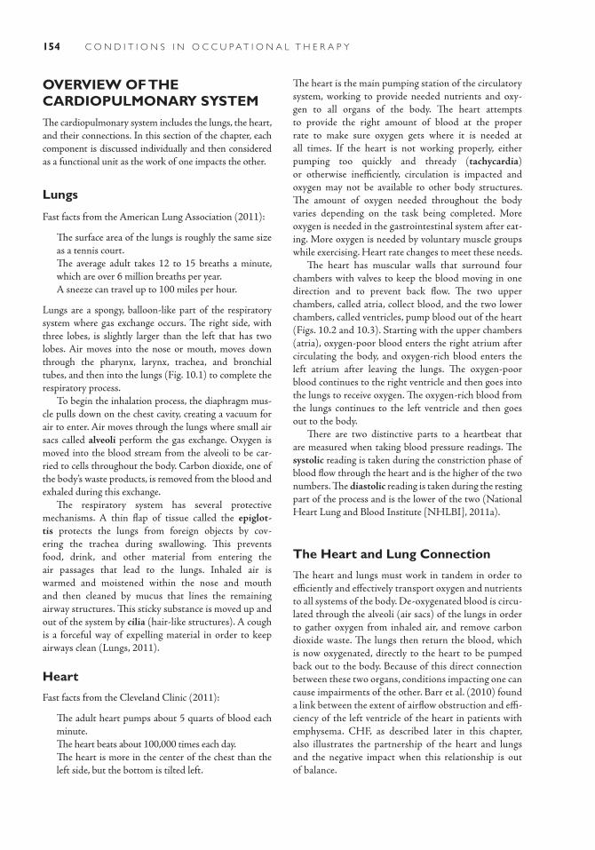
154 C O N D I T I O N S I N O C C U P A T I O N A L T H E R A P Y
OVERVIEW OF THE CARDIOPULMONARY SYSTEMThe cardiopulmonary system includes the lungs, the heart, and their connections. In this section of the chapter, each component is discussed individually and then considered as a functional unit as the work of one impacts the other.
Lungs
Fast facts from the American Lung Association (2011):
■ The surface area of the lungs is roughly the same size as a tennis court.
■ The average adult takes 12 to 15 breaths a minute, which are over 6 million breaths per year.
■ A sneeze can travel up to 100 miles per hour.
Lungs are a spongy, balloon-like part of the respiratory system where gas exchange occurs. The right side, with three lobes, is slightly larger than the left that has two lobes. Air moves into the nose or mouth, moves down through the pharynx, larynx, trachea, and bronchial tubes, and then into the lungs (Fig. 10.1) to complete the respiratory process.
To begin the inhalation process, the diaphragm mus-cle pulls down on the chest cavity, creating a vacuum for air to enter. Air moves through the lungs where small air sacs called alveoli perform the gas exchange. Oxygen is moved into the blood stream from the alveoli to be car-ried to cells throughout the body. Carbon dioxide, one of the body’s waste products, is removed from the blood and exhaled during this exchange.
The respiratory system has several protective mechanisms. A thin flap of tissue called the epiglot-tis protects the lungs from foreign objects by cov-ering the trachea during swallowing. This prevents food, drink, and other material from entering the air passages that lead to the lungs. Inhaled air is warmed and moistened within the nose and mouth and then cleaned by mucus that lines the remaining airway structures. This sticky substance is moved up and out of the system by cilia (hair-like structures). A cough is a forceful way of expelling material in order to keep airways clean (Lungs, 2011).
Heart
Fast facts from the Cleveland Clinic (2011):
■ The adult heart pumps about 5 quarts of blood each minute.
■ The heart beats about 100,000 times each day. ■ The heart is more in the center of the chest than the
left side, but the bottom is tilted left.
The heart is the main pumping station of the circulatory system, working to provide needed nutrients and oxy-gen to all organs of the body. The heart attempts to provide the right amount of blood at the proper rate to make sure oxygen gets where it is needed at all times. If the heart is not working properly, either pumping too quickly and thready (tachycardia) or otherwise inefficiently, circulation is impacted and oxygen may not be available to other body structures. The amount of oxygen needed throughout the body varies depending on the task being completed. More oxygen is needed in the gastrointestinal system after eat-ing. More oxygen is needed by voluntary muscle groups while exercising. Heart rate changes to meet these needs.
The heart has muscular walls that surround four chambers with valves to keep the blood moving in one direction and to prevent back flow. The two upper chambers, called atria, collect blood, and the two lower chambers, called ventricles, pump blood out of the heart (Figs. 10.2 and 10.3). Starting with the upper chambers (atria), oxygen-poor blood enters the right atrium after circulating the body, and oxygen-rich blood enters the left atrium after leaving the lungs. The oxygen-poor blood continues to the right ventricle and then goes into the lungs to receive oxygen. The oxygen-rich blood from the lungs continues to the left ventricle and then goes out to the body.
There are two distinctive parts to a heartbeat that are measured when taking blood pressure readings. The systolic reading is taken during the constriction phase of blood flow through the heart and is the higher of the two numbers. The diastolic reading is taken during the resting part of the process and is the lower of the two (National Heart Lung and Blood Institute [NHLBI], 2011a).
The Heart and Lung Connection
The heart and lungs must work in tandem in order to efficiently and effectively transport oxygen and nutrients to all systems of the body. De-oxygenated blood is circu-lated through the alveoli (air sacs) of the lungs in order to gather oxygen from inhaled air, and remove carbon dioxide waste. The lungs then return the blood, which is now oxygenated, directly to the heart to be pumped back out to the body. Because of this direct connection between these two organs, conditions impacting one can cause impairments of the other. Barr et al. (2010) found a link between the extent of airflow obstruction and effi-ciency of the left ventricle of the heart in patients with emphysema. CHF, as described later in this chapter, also illustrates the partnership of the heart and lungs and the negative impact when this relationship is out of balance.
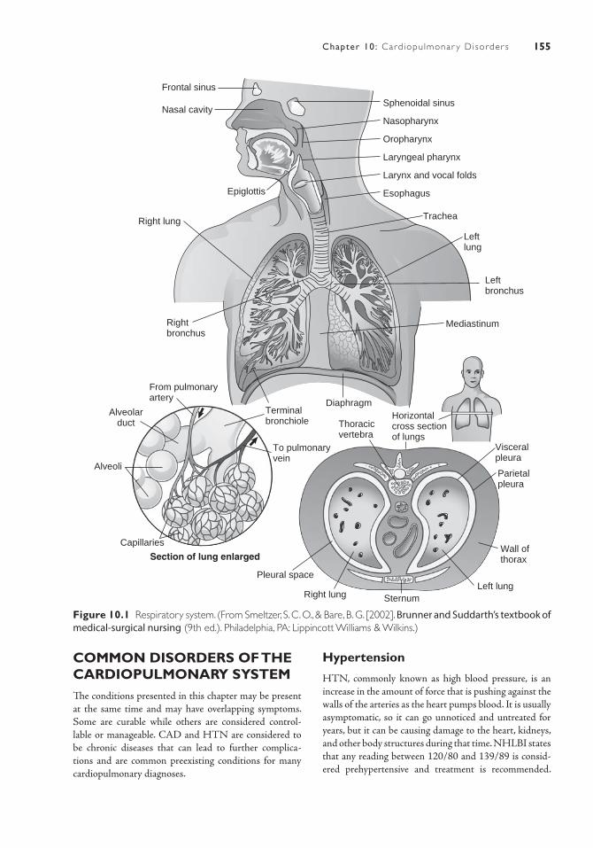
155Chapter 10: Card iopulmonar y Disorder s
COMMON DISORDERS OF THE CARDIOPULMONARY SYSTEMThe conditions presented in this chapter may be present at the same time and may have overlapping symptoms. Some are curable while others are considered control-lable or manageable. CAD and HTN are considered to be chronic diseases that can lead to further complica-tions and are common preexisting conditions for many cardiopulmonary diagnoses.
Hypertension
HTN, commonly known as high blood pressure, is an increase in the amount of force that is pushing against the walls of the arteries as the heart pumps blood. It is usually asymptomatic, so it can go unnoticed and untreated for years, but it can be causing damage to the heart, kidneys, and other body structures during that time. NHLBI states that any reading between 120/80 and 139/89 is consid-ered prehypertensive and treatment is recommended.
Frontal sinus
Nasal cavity
Epiglottis
Right lung
Rightbronchus
From pulmonaryartery
Alveolarduct
Alveoli
Capillaries
Section of lung enlarged
Pleural space
Right lung SternumLeft lung
Wall ofthorax
Parietalpleura
Visceralpleura
Horizontalcross sectionof lungs
Thoracicvertebra
DiaphragmTerminalbronchiole
Mediastinum
Sphenoidal sinus
Nasopharynx
Oropharynx
Laryngeal pharynx
Larynx and vocal folds
Esophagus
Trachea
Leftlung
To pulmonaryvein
Leftbronchus
Figure 10.1 Respiratory system. (From Smeltzer, S. C. O., & Bare, B. G. [2002]. Brunner and Suddarth’s textbook of medical-surgical nursing (9th ed.). Philadelphia, PA: Lippincott Williams & Wilkins.)
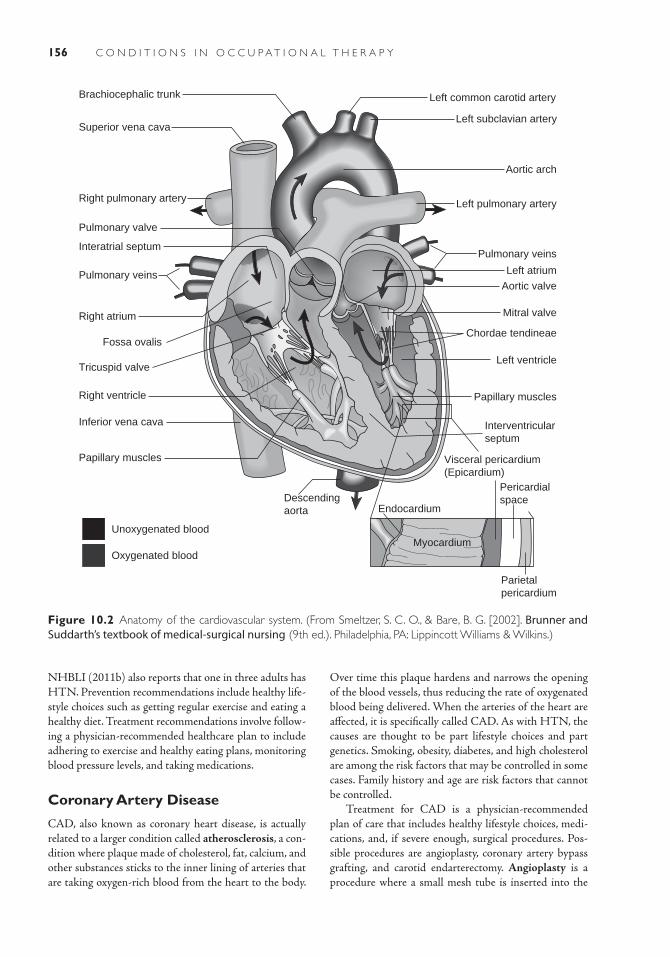
156 C O N D I T I O N S I N O C C U P A T I O N A L T H E R A P Y
NHBLI (2011b) also reports that one in three adults has HTN. Prevention recommendations include healthy life-style choices such as getting regular exercise and eating a healthy diet. Treatment recommendations involve follow-ing a physician- recommended healthcare plan to include adhering to exercise and healthy eating plans, monitoring blood pressure levels, and taking medications.
Coronary Artery Disease
CAD, also known as coronary heart disease, is actually related to a larger condition called atherosclerosis, a con-dition where plaque made of cholesterol, fat, calcium, and other substances sticks to the inner lining of arteries that are taking oxygen-rich blood from the heart to the body.
Superior vena cava
Brachiocephalic trunk
Right pulmonary artery
Pulmonary veins
Pulmonary valve
Interatrial septum
Right atrium
Tricuspid valve
Inferior vena cava
Right ventricle
Fossa ovalis
Aortic arch
Descendingaorta
Left pulmonary artery
Left atrium
Pulmonary veins
Aortic valve
Papillary muscles
Papillary muscles
Unoxygenated blood
Oxygenated blood
Left ventricle
Interventricularseptum
Mitral valve
Chordae tendineae
Endocardium
Myocardium
Visceral pericardium(Epicardium)
Parietalpericardium
Pericardialspace
Left subclavian artery
Left common carotid artery
Figure 10.2 Anatomy of the cardiovascular system. (From Smeltzer, S. C. O., & Bare, B. G. [2002]. Brunner and Suddarth’s textbook of medical-surgical nursing (9th ed.). Philadelphia, PA: Lippincott Williams & Wilkins.)
Over time this plaque hardens and narrows the opening of the blood vessels, thus reducing the rate of oxygenated blood being delivered. When the arteries of the heart are affected, it is specifically called CAD. As with HTN, the causes are thought to be part lifestyle choices and part genetics. Smoking, obesity, diabetes, and high cholesterol are among the risk factors that may be controlled in some cases. Family history and age are risk factors that cannot be controlled.
Treatment for CAD is a physician-recommended plan of care that includes healthy lifestyle choices, medi-cations, and, if severe enough, surgical procedures. Pos-sible procedures are angioplasty, coronary artery bypass grafting, and carotid endarterectomy. Angioplasty is a procedure where a small mesh tube is inserted into the
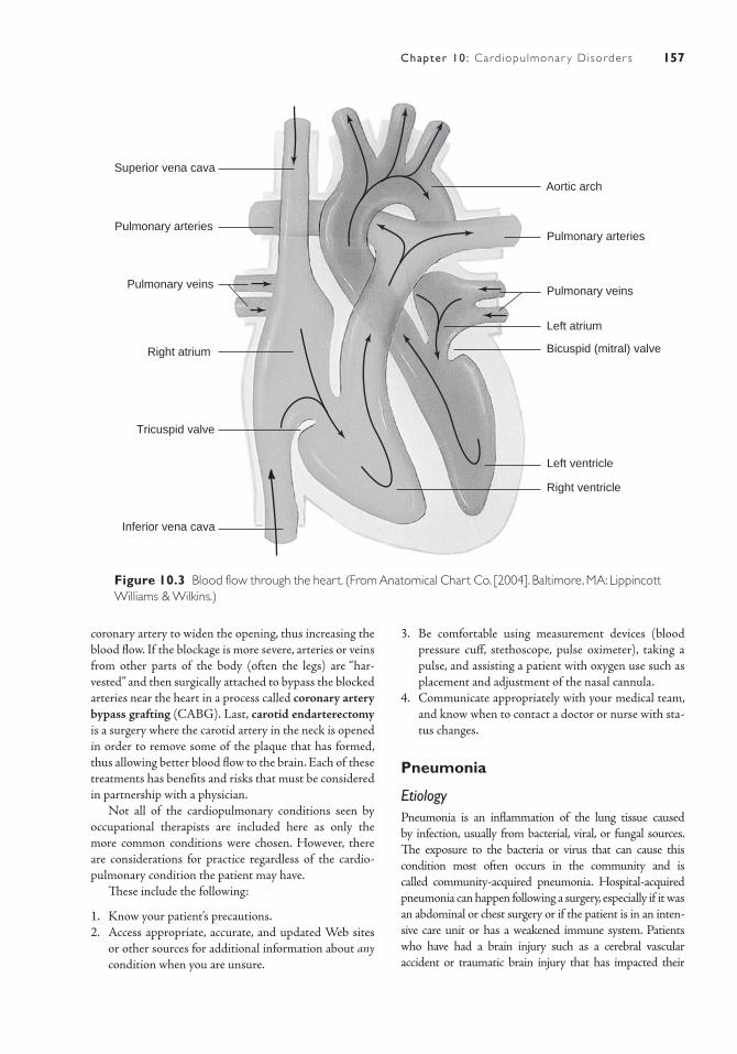
157Chapter 10: Card iopulmonar y Disorder s
coronary artery to widen the opening, thus increasing the blood flow. If the blockage is more severe, arteries or veins from other parts of the body (often the legs) are “har-vested” and then surgically attached to bypass the blocked arteries near the heart in a process called coronary artery bypass grafting (CABG). Last, carotid endarterectomy is a surgery where the carotid artery in the neck is opened in order to remove some of the plaque that has formed, thus allowing better blood flow to the brain. Each of these treatments has benefits and risks that must be considered in partnership with a physician.
Not all of the cardiopulmonary conditions seen by occupational therapists are included here as only the more common conditions were chosen. However, there are considerations for practice regardless of the cardio-pulmonary condition the patient may have.
These include the following:
1. Know your patient’s precautions.2. Access appropriate, accurate, and updated Web sites
or other sources for additional information about any condition when you are unsure.
Bicuspid (mitral) valve
Superior vena cava
Left ventricle
Right ventricle
Left atrium
Pulmonary veins
Pulmonary arteries
Aortic arch
Pulmonary arteries
Pulmonary veins
Right atrium
Tricuspid valve
Inferior vena cava
Figure 10.3 Blood flow through the heart. (From Anatomical Chart Co. [2004]. Baltimore, MA: Lippincott Williams & Wilkins.)
3. Be comfortable using measurement devices (blood pressure cuff, stethoscope, pulse oximeter), taking a pulse, and assisting a patient with oxygen use such as placement and adjustment of the nasal cannula.
4. Communicate appropriately with your medical team, and know when to contact a doctor or nurse with sta-tus changes.
Pneumonia
EtiologyPneumonia is an inflammation of the lung tissue caused by infection, usually from bacterial, viral, or fungal sources. The exposure to the bacteria or virus that can cause this condition most often occurs in the community and is called community-acquired pneumonia. Hospital-acquired pneumonia can happen following a surgery, especially if it was an abdominal or chest surgery or if the patient is in an inten-sive care unit or has a weakened immune system. Patients who have had a brain injury such as a cerebral vascular accident or traumatic brain injury that has impacted their

158 C O N D I T I O N S I N O C C U P A T I O N A L T H E R A P Y
swallowing sequence or gag reflex may be at risk for aspiration pneumonia, and it can occur when food or some other foreign substance enters the lungs and causes an infection.
Incidence and PrevalencePneumonia is most common in those over 65 years old and those who have a chronic condition or impaired immune system; however, it can also occur in younger, healthier people if the opportunity arises. According to the Center for Disease Control and Prevention (CDC) (2009), of the 60,000 cases of deaths from influenza and pneumonia in 2004, 85% involved those over 65 years of age. Age is a risk factor in that the older the person with this diagnosis, the greater the chance of hospitalization and/or death from pneumonia (Mayo Clinic, 2010a). Pneumonia as the cause of death increases drastically after the age of 85, with a jump from 37 deaths out of 100,000 for those 65 to 74 years of age to 137 per 100,000 deaths for those 75 to 84 and finally to 571.2 out of 100,000 for those 85 and older (Mayo Clinic, 2010a).
Signs and SymptomsThe early signs and symptoms of pneumonia are often confused with those of the flu and may start with a cough and a fever. Additional symptoms can include shortness of breath (dyspnea), sweating, chills, chest pain or tight-ness, headache, and fatigue.
Diagnosing pneumonia may involve the physician lis-tening to the chest during breathing for crackling sounds (rales) or rumbling sounds caused by increased mucus. A chest x-ray may be necessary to further assess lung func-tion. Blood and mucus tests may also be necessary to iden-tify the infection-causing organism.
Course and PrognosisThe first signs may appear as if it is the onset of the flu, with coughing and a slight fever. The severity of the symp-toms can vary widely depending on the infecting organ-ism and the overall health and age of the patient. If caught early or if it is a mild case, hospitalization can be avoided. If hospitalization is deemed necessary, it typically lasts 3 to 4 days. Pneumonia can be serious for older adults, especially if they are over 85 years old or have additional health complications. If left untreated, pneumonia can lead to severe respiratory distress and death, as organs are no longer getting the oxygen they need and begin to fail (NHLBI, 2011b).
Medical/Surgical ManagementVaccines are important in the prevention of pneumo-nia. Getting an annual flu shot helps, as flu can turn into pneumonia. Pneumonia vaccines are helpful for older adults and those with a challenged immune system.
As the severity of symptoms varies, so do the treat-ments. The standard plan, however, includes antibiotics,
rest, and fluids. Oxygen may be needed temporarily until the symptoms subside. Since coughing is an important mecha-nism for removing excess mucus from the airways, it should not be stopped totally, but can be quieted at times to allow for rest and sleep. Those who are more medically challenged may need to be hospitalized for close observation and may receive intravenous antibiotics and fluids as well as oxygen.
Chronic Obstructive Pulmonary Disease
EtiologyChronic obstructive pulmonary disease (COPD), a pro-gressive lung condition that makes it increasingly difficult to breathe, is an overarching term that includes the more specific conditions of emphysema and chronic bronchitis. Rather than resembling an inflated balloon with tension and pressure for proper air exchange, the lungs may become more floppy and somewhat deflated, may have damaged alveoli walls or thickened mucosal linings and therefore cannot support good airflow. In emphysema, the alveoli walls may become deflated or damaged, which reduces the amount of gas exchange that can occur. In chronic bron-chitis, the bronchial tubes become inflamed and thickened making it difficult to breathe. It is not uncommon for a per-son to have both conditions, and thus the term COPD may be more appropriate. The usual cause of COPD is the long-term inhalation of irritants. By far the most common source of irritants is cigarette smoke but can also include air pollu-tion, dust, or chemicals (American Lung Association, 2011).
Incidence and PrevalenceOver 12 million people are living with COPD, and it is the fourth leading cause of death in the United States (CDC, 2009). Because the condition worsens slowly and chronic exposure to an irritant may have occurred over several years, symptoms usually are not seen in people <40 years of age. Incidence is higher in areas of high pol-lution and from workers in high-risk areas (Mayo Clinic, 2010b).
Signs and SymptomsSymptoms of COPD include persistent cough that produces a large amount of mucus (also called smoker’s cough), wheezing or whistling while inhaling, chest tightness, and dyspnea with exertion. As the condition becomes more severe, swelling in the ankles and feet may be noticeable, and lips and fingernails may be bluish due to decreased oxygen levels.
In addition to listening to the lungs with a stethoscope and taking a thorough history related to smoking or other exposure to irritants, the physician may request a series of tests and labs to confirm the COPD diagnosis. Lung func-tion tests such as spirometry, the main test of lung function
159Chapter 10: Card iopulmonar y Disorder s
for those with COPD, measures how much air is forcefully exhaled and how quickly.
Course and PrognosisCOPD is not curable and slowly worsens over time. Breathing becomes more difficult and less oxygen gets to the body. The heart may become enlarged from the strain and blood pressure increases. Eventually, lack of oxygen to body structures leads to cognitive deficits and organ failure, which then leads to death. The rate of decline can be modi-fied and quality of life maintained longer by quitting smok-ing, organizing and simplifying daily life tasks, avoiding irritants, managing symptoms, getting annual flu shots, and preparing for emergencies by having medical information and medications conveniently located (NHBLI, 2011b).
Medical/Surgical ManagementMedical management of COPD includes use of oxygen, inhaled medications, and oral medications such as ster-oids and antibiotics, which are all designed to ease breath-ing. Qualified patients with serious symptoms may have lung reduction surgery or lung transplants.
Congestive Heart Failure
EtiologyCHF, also known simply as heart failure, is a chronic, non-curable condition where the heart does not beat strongly enough to maintain adequate blood flow to all systems in the body, causing organs to be oxygen-deprived and the body to retain fluid. Because of the close connection to the lungs, when blood flow weakens, fluid can build up in the tissues around the lungs causing difficulties with breathing. CHF can be caused by infections or conditions such as a heart attack that damage the heart muscle, but the most common causes are CAD and HTN (American Heart Association, 2009).
Incidence and PrevalenceCurrently, approximately 5.8 million people in the United States have CHF with about 670,000 people diagnosed each year (CDC, 2009). About 300,000 deaths each year are caused by CHF.
Signs and SymptomsSymptoms may be subtle at first but will increase in inten-sity as the condition progresses. Shortness of breath, dif-ficulty breathing while lying down, weight gain from fluid retention, and swelling of the feet and legs are common symptoms. Other symptoms that may be noticed include decreased urine output, general fatigue, decreased ability to focus, and occasional nausea and vomiting.
A physical examination by the physician provides information in order to make this diagnosis. Signs noted may be neck arteries that are distended, swollen liver
noted during palpation, fluid sounds in the lungs, and swelling of the feet and legs. More formal diagnostic pro-cedures include a chest x-ray, heart CT scan, echocardio-gram, MRI, and various blood and urine analyses.
Course and PrognosisMuch like COPD, CHF is a condition that progresses slowly over time depriving the body of needed oxygen and eventually leading to death. Shortness of breath is one of the first signs of this condition and may be mistaken for a nor-mal part of the aging early in the process. Decreasing endur-ance and increasing difficulty in breathing worsens as the condition progresses (American Heart Association, 2009).
As with all cardiopulmonary conditions presented in this chapter, eating right by taking smaller portions and avoiding salt, exercising regularly, avoiding smoking, and following a medical management plan can decrease the rate of progres-sion and help minimize the symptoms as long as possible. A systematic review of research articles leads authors to report that although length of life was not significantly impacted, men with mild to moderate CHF who participated in regu-lar exercise had a better quality of life and had better overall fitness than those who did not (Davies et al., 2010).
Medical/Surgical ManagementAlong with healthy lifestyle choices, medications and surgeries are also used to treat CHF and help minimize symptoms as they present. Medications may include angiotensin- converting enzyme (ACE) inhibitors that expand blood vessels and ease the heart’s workload, diuret-ics that help the body get rid of fluid, and dioxin that helps the heart contract properly. Surgical procedures can pro-vide some relief of symptoms. Insertion of a pacemaker to help maintain a consistent heartbeat, angioplasty to hold the arteries open, CABG and heart valve repair, or replace-ment to improve circulation are some of the more invasive treatment methods that are utilized when treating CHF.
Myocardial Infarction
EtiologyMI, more commonly known as a heart attack, occurs when a blood vessel to the heart is blocked, causing heart muscle tissue to be without oxygen and resulting in damage or death to that tissue. CAD, in which the artery walls have plaque formations that impair blood flow, diabetes, and high blood pressure all are known causes of MI. Other risk factors for this condition are increasing age, smoking, and family history.
PrevalenceAccording to the American Heart Association’s 2009 statis-tics report, someone in the United States has a heart attack every 34 seconds, with approximately 16% of those dying from that event.
160 C O N D I T I O N S I N O C C U P A T I O N A L T H E R A P Y
Signs and SymptomsThe major symptoms of a MI, particularly for men, are chest pain and tightness, feelings of indigestion, dizziness, shortness of breath, and excessive sweating. Women may be less likely than men to have the hallmark chest tight-ness symptom and tend to report shortness of breath, weakness, and fatigue (McSweeney et al., 2003).
A MI is a medical emergency that requires immediate attention. Once at the hospital, a physical exam will be completed, and tests such as a coronary angiography, CT scan, electrocardiogram, echocardiography, or MRI may be completed to make the final diagnosis.
Course and PrognosisThe prognosis for recovery depends on the amount of cardiac tissue that was damaged during the time of the blockage and the length of time it took to receive medi-cal attention. If caught and treated early, full recovery and return to all activities of daily living (ADLs) may be pos-sible. Sudden cardiac death can occur early in the process. During the early hospitalization stage of recovery, time in the intensive care unit may be necessary to monitor for irregular heart rhythms.
Cardiac rehabilitation that includes exercise, education, and counseling may be necessary to help patients ease back into their daily routine following strict precautions and pro-gressing at a slow and steady pace. To help guide physical reha-bilitation and determine safe recommended levels of activity, a metabolic equivalent (MET) chart is used. A MET is the amount of energy that an activity requires, with more seden-tary tasks such as watching TV or doing computer work using 1.5 to 2 METS ranging up to some competitive sports requir-ing 11 or more METs. The physician and the team determines the maximum MET level for the patient until healing begins, and this will guide how much exercise can be completed or how much help the person needs with daily activities.
Medical/Surgical ManagementIn addition to the immediate emergency response that includes oxygen and stabilizing medications, surgeries to reestablish blood flow to the damaged area and additional medications are often necessary. One or more CABG pro-cedures or angioplasty may be needed. Beta-blockers may be given to relieve chest pain and to treat heartbeat irregu-larities, ACE inhibitors reduce the workload and stress on the heart, and anticoagulants may help prevent blood clots from forming (PubMed Health, 2010).
TABLE 10.1 Common Symptoms and Their Impact on Occupational Performance
Dyspnea Clients may need frequent breaks and may need chairs placed in various positions around their house to provide a place to sit.
Fatigue Clients may need to have tasks broken down into smaller parts and to do them over a longer period of time. Work simplification and energy conservation techniques will be needed.
Depression This is a multilayered symptom as many emotional issues, stressors, and life reviews are common with cardiopulmonary conditions. Encouraging participation, supporting successes, and being empathetic are important therapeutic skills for helping clients figure out ways to do what they need to do in their occupational areas.
Difficulty focusing Visual reminders and verbal cues may be needed. Processing time may be lengthened, so a slower pace may be helpful when problem-solving more complex tasks.
Anxiety Clients may have more fear when attempting tasks. More time, encouragement, and pleasant distractions can help them move forward with the small day-to-day tasks. This can, however, become a larger issue that keeps clients from doing what they need to do.
Light-headedness Allow clients to sit whenever possible to complete a task. Have seats close, and encourage them to stand near a stable counter or table, or use a grab bar if standing is needed.
161Chapter 10: Card iopulmonar y Disorder s
IMPACT ON OCCUPATIONAL PERFORMANCEThe primary focus of the field of occupational therapy (OT) is clients’ occupations and how they perform them. The OT Practice Framework, 2nd ed. (American Occupa-tional Therapy Association, 2008), has divided occupations into the following eight categories in an attempt to support a thorough analysis of our client’s needs and to help organ-ize our critical thinking process:
1. Activities of daily living (ADL)2. Instrumental activities of daily living (IADL)3. Work4. Play5. Leisure6. Education7. Sleep/rest8. Social participation
Considering each of these areas of occupations and then further exploring the specific tasks within each area
provides a good base for determining the best treatment for the client. After determining the occupational needs of the client, it is important to then determine the effects of his or her symptoms on occupational performance.
The symptoms listed in Table 10.1 are common to many of the conditions presented in this chapter.
Precautions
Precautions are determined by the diagnosis and general cli-ent health. Some are formally dictated and documented by the physician, whereas others are determined by anyone on the healthcare team that recognizes a safety or health con-cern. They will change as the client’s health status changes. Not only is it important for the therapist to be familiar with all of the client’s precautions in order to keep him or her safe during the treatment session, it is also important to consider how these precautions impact the client’s ability to safely complete all areas of occupations (Table 10.2).
OT practitioners play a key role in helping people with cardiopulmonary diseases. Conditions such as CHF
TABLE 10.2 Common Precautions and Their Impact on Occupational Performance
No heavy lifting Any task that would require lifting would need to be modified or completed by someone else. Lifting precautions also limit wheelchair self-propulsion as this, too, puts too much strain on the upper body.
Heart rate ≤ 110 beats/minute Clients will need to be taught to take their own heart rate and be reminded to take it if more exertion is attempted. Written cues may be needed for reminders.
No activities at a MET rate higher than 6
Clients will be limited in the tasks they can complete until this precaution is lifted. In some cases, the activities that are of a higher MET value may not be permitted for many weeks.
Oxygen on at all times (liters determined by physician)
Clients will need to be connected to an oxygen source at all times. That usually involves a tank and a long hose that must be manipulated to ensure safety with mobility.
Maintain low-salt diet Clients may need to modify cooking habits and seasoning habits and to explore alternatives.
Record daily weight This is important for those with CHF. Clients may need a scale with large numbers and a system set up for recording their daily weight. Scale may need to be in a place where clients can hold on to a stable object while stepping up and down.
Follow recommended oral and inhaled medication schedule
Visual reminders and environmental set up may be needed. Having a dedicated site for the clients to go to when doing their breathing treatments can help.
162 C O N D I T I O N S I N O C C U P A T I O N A L T H E R A P Y
and COPD are chronic conditions that require new self- management techniques to maintain health, and these new medical needs have to be integrated into daily routines. According to AOTA, OT practitioners work
collaboratively with clients to identify needs and design strategies for independent health management, and help-ing clients take charge of their health care can improve their quality of life (as cited in Bondoc and Siebert, 2009).
Case IllustrationsCase Study 1Wanda, an 87-year-old woman, was admitted to the intensive care unit in a hospital through the emer-gency room 2 days ago. Prior to being brought to the hospital by ambulance, she had been coughing and feeling ill for several days but had refused to go see her doctor. When she finally agreed to get medical help, she reported that she was short of breath and felt a weight on her chest. She was also weak and malnourished. Wanda was diagnosed with pneumonia. Antibiotics were started, a saline drip was inserted, and, due to her severe weakness, a catheter was inserted to limit transfers to the toilet. The occupational therapist evaluated her, according to doctor’s orders, after she was transferred to a regular room.
Social HistoryWanda’s son moved into her single-story home with her following the death of her husband 10 years ago. She is particularly proud of her gardens and of the fact that she just retired a few years ago from seam-stress work for an upholstery shop. Since retirement, she reports enjoying visits to the library to get stacks of romance novels and cooking for her family. She is no longer driving and reports feeling frustrated by the loss of independence but states that she is too ner-vous to drive now.
Current Activity LevelWanda is currently much debilitated as she transitions from the intensive care unit to the regular hospital wing to continue her recovery. She is so weak that she is unable to move from supine to sit independently, but follows directions well and appears cognitively intact. Prior to coming into the hospital with pneu-monia, she was doing her basic care and some light cooking independently using a wheeled walker. Her son reports that she appeared to be slowing down a bit and taking more rest breaks recently.
Case Study 2William is a 63-year-old man who was diagnosed with emphysema 8 years ago. He had provided care for his mother as she struggled to breathe and had
to be connected to her oxygen tank “like an umbili-cal cord” throughout the house until she had died of emphysema 2 years ago. He now has his own oxygen tank and takes breathing treatments throughout the day, but still he reports feeling little relief from the struggle. His activity level has steadily declined, and he has lost his appetite. The steroid medication he is taking to help his breathing has caused his face to look puffy and almost unrecognizable in the mirror. The homecare agency has received a refer-ral to evaluate and treat William as he was recently discharged from the hospital following recovery from pneumonia.
Social HistoryWilliam is divorced, and his only daughter has moved to another state and is no longer visiting or calling. He has numerous siblings and nieces and nephews that visit. However, they are more likely to ask for help than to provide it. He lives alone, with his dog, in a single-story home in a subdivision outside of town. He still drives, but becomes anxious when traveling more than 10 or 15 minutes from his home, which makes going to his doctor appointments 30 minutes away very stressful. He has many friends who involve him in social events and give him rides as often as he is willing, but he finds that his breathing becomes more labored after being out for more than a cou-ple of hours. He admits to feelings of anxiety and depression.
Current Activity LevelSince his discharge from the hospital, William appears weak overall but is attempting to build his endurance as much as he is able. He is very active in online social media and spends hours each day online posting on social sites or playing games. He also reports having the television on “almost 24 hours a day.” He is able to cook and do light housekeeping but finds that it is getting more and more difficult to keep it up as he has in the past. He also reports that taking a shower standing in the tub “takes a lot out of him.” He appears eager to continue to maintain his independence and to take suggestions on best to do so.
163Chapter 10: Card iopulmonar y Disorder s
R E F E R E N C E S
American Heart Association. (2009). Heart disease and stroke statistics 2009 update. Circulation. Retrieved from http://www.heart.org/HEARTORG/ Conditions/HeartAttack/HeartAttackToolsResources/Patient-Information-Sheets-Heart-Attack_UCM_303950_Article.jsp
American Lung Association. (2011). How lungs work. Retrieved from http://www.lungusa.org/your-lungs/how-lungs-work
American Occupational Therapy Association. (2008). Occupational therapy practice framework: Domain and process (2nd ed.). American Journal of Occupa-tional Therapy, 62, 625–683.
Barr, R. G., Bluemke, D. A., Ahmed, F. S., Carr, J. J., Enright, P. L., Hoffman, E. A., et al. (2010). Percent emphysema, airflow obstruction, and impaired left ventricular filling. New England Journal of Medicine, 362, 217–227.
Bondoc, S., & Siebert, C. (2009). The role of occupational therapy in chronic disease management. American Occupational Therapy Association Fact Sheet, Retrieved from http://www.aota.org
Center for Disease Control and Prevention. (2009). FastStats A to Z. Retrieved from www.cdc.gov/nchs/fastats
Cleveland Clinic. (2011). Your heart and blood vessels. Retrieved from http://my.clevelandclinic.org/heart/heartworks/heartfacts.aspx
Davies, E. J., Moxham, T., Rees, K., Singh, S., Coats, A. J. S., Ebrahim, S., et al. (2010). Exercise training for systolic heart failure: Cochrane systematic review and meta-analysis. European Journal of Heart Failure, 12, 706–715.
Lungs. (2011). In Encyclopædia Britannica. Retrieved from http://www.britannica.com/EBchecked/topic/ 351473/lung
Mayo Clinic Health Information. (2010a). Pneumonia. Retrieved from http://www.mayoclinic.com/health/pneumonia/DS00135
Mayo Clinic Health Information. (2010b). Chronic obstructive pulmonary disease. Retrieved from http://www.mayoclinic.com/health/copd/DS00916
McSweeney, J. C., Cody, M., O’Sullivan, P., Elberson, K., Moser, D. K., & Garvin, B. J. (2003). Women’s early warning symptoms of acute myocardial infarc-tion. Circulation, 108, 2619–2623.
National Heart Lung and Blood Institute (NHLBI). (2011a). How the heart works. Retrieved from http://www.nhlbi.nih.gov/health/dci/Diseases/hhw_ anatomy.html
National Heart Lung and Blood Institute (NHLBI). (2011b). Diseases and conditions index. Retrieved from http://www.nhlbi.nih.gov/health/dci/Diseases/Hbp/HBP_WhatIs.html
PubMed Health. (2010). Heart attack. Retrieved from http://www.ncbi.nlm.nih.gov/pubmedhealth/PMH0001246/

165
Diabetes
■ Jo ann e E st e s
11
Serena is a 12-year-old girl who lives in an urban Hispanic neighbor-hood of a large Midwestern city. She spends most of her time not in school watching television and playing video games with a cou-ple of neighborhood friends or by herself. Serena has never been
interested in playing sports, so her parents stopped encouraging her to do so. Serena’s parents both hold full-time jobs, so she is often home alone and responsible for taking care of herself. Her meals and snacks consist of convenience and processed foods, and she also often eats at fast food res-taurants. Serena is 5′5′ tall, weighs 200 pounds, and has a body mass index (BMI) of 33.3 kg/m2, placing her in a category of obese children/adolescents. Lately, her mother notices that Serena seems more tired than usual, drinks a lot of soda pop, and uses the bathroom frequently. Serena’s mother takes her to the pediatrician where blood work results showed a random plasma glucose level of 225 mg/dL. She is diagnosed as having type 2 diabetes.
K E Y T E R M S
Autonomic neuropathyDiabetic foot
Diabetic ketoacidosisHemoglobin A1c
HyperglycemiaHypertensionHypoglycemia
KetonuriaNephropathy
Peripheral artery diseasePeripheral neuropathy
PolydipsiaPolyphagia
PolyuriaRetinopathy
Type 1 diabetesType 2 diabetes
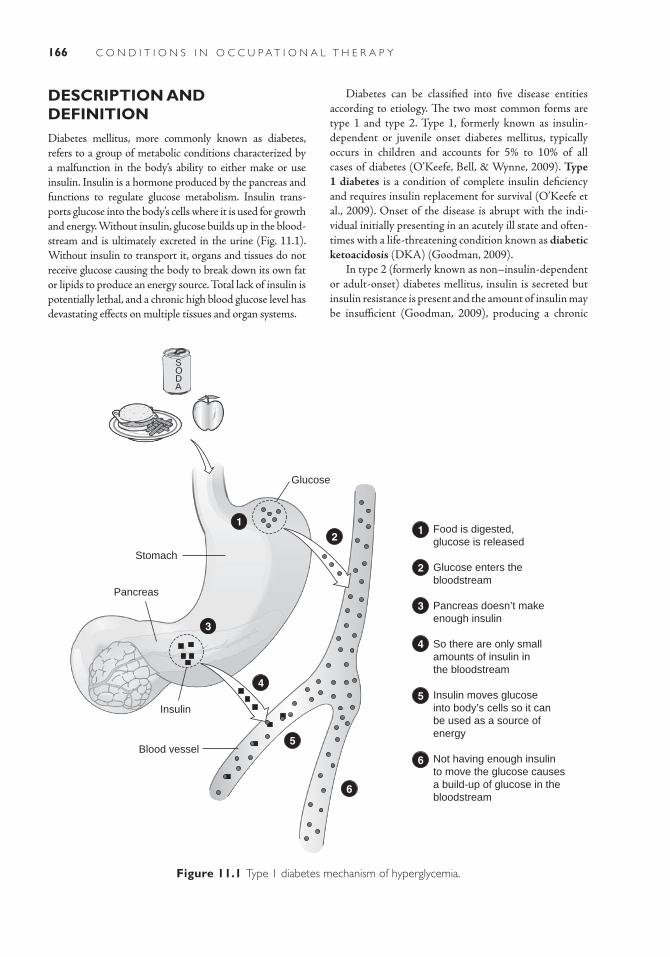
166 C O N D I T I O N S I N O C C U P A T I O N A L T H E R A P Y
DESCRIPTION AND DEFINITIONDiabetes mellitus, more commonly known as diabetes, refers to a group of metabolic conditions characterized by a malfunction in the body’s ability to either make or use insulin. Insulin is a hormone produced by the pancreas and functions to regulate glucose metabolism. Insulin trans-ports glucose into the body’s cells where it is used for growth and energy. Without insulin, glucose builds up in the blood-stream and is ultimately excreted in the urine (Fig. 11.1). Without insulin to transport it, organs and tissues do not receive glucose causing the body to break down its own fat or lipids to produce an energy source. Total lack of insulin is potentially lethal, and a chronic high blood glucose level has devastating effects on multiple tissues and organ systems.
Food is digested,glucose is released
Glucose enters thebloodstream
Pancreas doesn’t makeenough insulin
So there are only smallamounts of insulin inthe bloodstream
Insulin moves glucoseinto body’s cells so it canbe used as a source ofenergy
Not having enough insulinto move the glucose causesa build-up of glucose in thebloodstream
Glucose
Insulin
Blood vessel
Stomach
Pancreas
1
3
4
2
5
6
1
2
3
4
5
6
SODA
Figure 11.1 Type 1 diabetes mechanism of hyperglycemia.
Diabetes can be classified into five disease entities according to etiology. The two most common forms are type 1 and type 2. Type 1, formerly known as insulin-dependent or juvenile onset diabetes mellitus, typically occurs in children and accounts for 5% to 10% of all cases of diabetes (O’Keefe, Bell, & Wynne, 2009). Type 1 diabetes is a condition of complete insulin deficiency and requires insulin replacement for survival (O’Keefe et al., 2009). Onset of the disease is abrupt with the indi-vidual initially presenting in an acutely ill state and often-times with a life-threatening condition known as diabetic ketoacidosis (DKA) (Goodman, 2009).
In type 2 (formerly known as non– insulin-dependent or adult-onset) diabetes mellitus, insulin is secreted but insulin resistance is present and the amount of insulin may be insufficient (Goodman, 2009), producing a chronic
167Chapter 11: Diabetes
Type 2
Genetic predisposition is stronger for type 2 diabetes than for type 1 as concurrence of the disease in monozygotic twins approaches 100% (Adeghate, Schattner, & Dunn, 2006; O’Keefe et al., 2009). Other factors include aging (Hseuh & Bryer-Ash, 2009), physical inactivity (Kriska et al., 2001), chronic stress (Adeghate et al., 2006), and being obese. Obesity is a strong contributing factor as 80% of people with type 2 diabetes are obese with the remaining 20% having a high percentage of abdominal body fat (Goodman, 2009). Finally, ethnicity is a risk factor in that African Americans, Hispanics, Native Americans, Asians, and Pacific Islanders are more likely to develop dia-betes (O’Keefe et al., 2009;Pavkov et al, 2007).
INCIDENCE AND PREVALENCE
Children and Adolescence Incidence
The 2007 National Diabetes Fact Sheet provides statistics based on 2002 to 2003 data (Centers for Disease Con-trol and Prevention [CDC], 2008a). According to this report, 15,000 youth (rate of 19 per 1,000) were newly diagnosed with type 1 diabetes, and 3,700 youth (rate of 5.3 per 100,000) with type 2 diabetes per year. Non-Hispanic white youth had the highest rate of newly diag-nosed cases of type 1 diabetes (20 per 100,000), with type 2 being extremely rare in this group of youth under age 10. The highest rates of newly diagnosed type 2 cases occurred in American Indians (approximately 35 per 100,000); African Americans (approximately 20 per 100,000); Asian/Pacific Islanders (approximately 20 per 100,000); and Hispanics (approximately 10 per 100,000). For these groups, the incidence of type 2 diabetes was greater than that of type 1.
Adult Incidence
In 2007, about 1.6 million new cases of diabetes were diagnosed in people aged 20 or older in the United States, with the majority of cases in the 40 to 59 age group (CDC, 2008a). The age-adjusted incidence of diabetes was simi-lar for men (8.4 per 1,000) and women (7.8 per 1,000) aged 18 to 79 in 2008 (CDC, 2008b). Incidence for Black, Native-American, Hispanic, and Asian Americans is 1.5 to 2 times that of Caucasian Americans (Goodman, 2009).
Prevalence of Diabetes
Nearly 24 million Americans or approximately 8% of the population now have diabetes (CDC, 2008a). This number tripled from 1980 to 2007 (CDC, 2008b). Prev-alence is associated with race and ethnicity. For type 1
state of hyperglycemia (Surampudi, John-Kalarickal, & Fonseca, 2009). Type 2 accounts for 90% to 95% of all cases of diabetes and typically occurs with increasing age and in people who are obese (O’Keefe et al., 2009). A marked increase in prevalence of obesity in children and adolescents both in the United States and globally has resulted in increases in type 2 diabetes for these age groups (Lipton et al., 2005; Pinhas-Hamiel & Zeitler, 2004). Onset is gradual, and the disease may go unde-tected for years (O’Keefe et al.).
The third classification is gestational diabetes, a tran-sient form of diabetes mellitus that occurs during preg-nancy and typically requires no further treatment after delivery ( Jovanovic, 2010). However, 35% of women with gestational diabetes will develop type 2 diabetes within 5 to 10 years (O’Keefe et al., 2009). Diabetes resulting from other causes forms the fourth classification. These include pathological conditions that damage the pancreas (e.g., chronic pancreatitis, cystic fibrosis) and certain genetic syndromes (e.g., Down’s syndrome, Klinefelter’s syndrome, Turner’s syndrome, Freiderich’s ataxia) (American Dia-betes Association [ADA], 2007). Prediabetes is the fifth classification and occurs in individuals who have impaired glucose tolerance (IGT) or impaired fasting glucose (IFG). Approximately 25% of people with prediabetes eventually develop type 2 diabetes; however, some interventions (e.g., diet/exercise, medications, bariatric surgery) have been shown to slow the progression (O’Keefe et al.).
ETIOLOGY
Type 1
The exact cause of type 1 diabetes mellitus is not well understood. (Adeghate, Shattner, & Dunn, 2006). The disease process is precipitated by an autoimmune response whereby antibodies are produced that destroys pancre-atic insulin-producing cells known as beta cells (Sherwin, 1996). It has a strong genetic predisposition with environ-mental factors thought to trigger the autoimmune process that destroys the beta cells (Daneman, 2006). Environ-mental factors can be divided into three categories: viral infections, infant diet, and exposure to toxins (Atkinson & Eisenbarth, 2001). While exposure to certain viruses (e.g., Enterovirus, Coxsackie B, Mumps) is thought to be a trigger (Daneman, 2006), research about their role is contradictory (Eisenbarth & McCulloch, 2010). Infant diet, specifically ingestion of cow’s milk, early introduction of gluten-based cereals, and a short duration of breastfeeding are thought to be inciting factors (Akerblom, Vaarala, Hyoty, Ilonen, & Knip, 2002; Laron, 2002). Toxins in the form of ingested nitrates and nitrites in food (e.g., processed meat products) and water may also be a triggering factor (Akerblom et al., 2002; Devendra, Liu, & Eisenbarth, 2004).
168 C O N D I T I O N S I N O C C U P A T I O N A L T H E R A P Y
COURSE AND PROGNOSISAfter initial diagnosis of type 1 diabetes, a temporary remission period usually occurs for 3 to 6 months. Dur-ing this time, overall control of the disease is easier and insulin needs are less. Insulin production gradually decreases until levels are insignificant and a state of total diabetes is reached (Dambro, 1995). Life expectancy is lower overall than for persons who do not have diabetes. One study showed a 7.5-year reduction for adult men and 8.5-year reduction for adult women with diabetes as compared to their nondiabetic counterparts (Franco, Steyerberg, Hu, Mackenbach, & Nusselder, 2007). Longevity and quality of life are currently better than in the past owing to improvements in insulin delivery regimens, medication treatment for hyperlipidemia, and lifestyle modifications (Miao, Brismar, Nyren, Ugarph-Morawski, & Ye, 2005). In 1993, the National Institute of Health completed a 9-year study called the “Diabetes Control and Complications Trial.” Intensive insulin therapy was shown to slow the development of retinopathy and neuropathy (Sherwin, 1996). Prior to this landmark study it was unclear whether tight glucose control was beneficial.
People with diabetes experience periods of hypo-glycemia or hyperglycemia. Hypoglycemia or insulin shock is a condition of too much insulin or oral hypogly-cemic medication and not enough glucose in the blood stream (Dambro, 1995). Symptoms are vague: fatigue, headache, drowsiness, lassitude, tremulousness, shallow breathing, and nausea (Sherwin, 1996). It may produce seizures, accidental injury, catecholamine response, or arrhythmia or cardiac ischemia in patients with underly-ing cardiac disease (Sherwin). The patient needs to ingest some form of sugar, such as orange juice, cola, candy, or jelly, if able to swallow. On an emotional level, this could become a great fear of the patient’s leading to less-than-optimal blood sugar control (Sherwin).
Hyperglycemia is a condition of too little insulin caus-ing abnormally high blood glucose levels. Signs are thirst, heartburn, fast and deep breathing, excessive urination, headache, nausea, abdominal pain, blurred vision, and constipation (Dambro, 1995). If untreated, the patient is at risk for entering into a diabetic coma. Mortality from hyperglycemia increases with age and is usually caused by the presence of a comorbid condition (myocardial infarction, cerebral vascular accident, sepsis). Treatment depends on insulin to reverse metabolic abnormalities and on detection and successful treatment of the comor-bid condition.
Hyperglycemia has a direct toxic effect on body tis-sues (Hsueh & Bryer-Ash, 2009). Few diseases have the same potential for damaging as many organ systems and producing impairments as diabetes (Fig. 11.2).
diabetes, African Americans and people of Asian descent have the lowest prevalence whereas Caucasians and peo-ple of Finnish, Scandinavian, Scottish, and Sardinian descent have the highest (Sherwin, 1996). Prevalence of type 2 diabetes is highest among American Indians and Alaskan natives (16.5%), followed by African Americans (11.8%), Hispanics (10.4%), Asian Americans (7.5%), and Caucasians (6.6%) (National Institute of Diabetes and Digestive and Kidney Diseases, 2008). Type 1 dia-betes occurs equally among males and females (Dambro, 1995), with type 2 prevalence for males (9.3%) slightly higher than for females (7.2%) (Crawford et al., 2010).
SIGNS AND SYMPTOMSClassic symptoms of diabetes are polydipsia (increased thirst), polyuria (frequent urination), polyphagia (increased hunger), blurred vision, weakness, fatigue, and dizziness (Goodman, 2009). Additional symptoms of type 1 are weight loss, muscle cramps, irritability, emotional lability, headaches, anxiety attacks, abdomi-nal pain and discomfort, diarrhea or constipation, and altered school and work behaviors. The individual may also present with ketonuria (Goodman). Ketones are waste products from the body’s breakdown of fat for energy, high concentrations of which leads to DKA (ADA, 2007). Symptoms of DKA include fruity odor breath, nausea and vomiting, confusion, difficulty breath-ing, and dry or flushed skin (ADA, 2007).
People with type 2 diabetes experience some of the same symptoms as type 1, including polyuria, polydipsia, polyphagia, unusual weight loss, extreme weakness and fatigue, and irritability. Additionally, frequent skin, gum, or bladder infections; cuts or bruises that are slow to heal; and numbness or tingling in the hands or feet are symp-toms of type 2 diabetes (ADA, 2007). Symptoms related to hyperglycemia and complications can also occur (e.g., nephropathy, neuropathy, and retinopathy). These com-plications may be present upon diagnosis if preceded by long periods of hyperglycemia (Powers, 2001).
The clinical sign of diabetes is hyperglycemia or high plasma concentration of glucose measured following an 8-hour period of fasting. Normal fasting blood glucose level is <100 mg/dL. The range of fasting blood glucose level for IFG/IGT is 100 to 125 mg/dL (ADA, 2010). Diabetes is diagnosed if fasting blood glucose level is ≥126 mg/dL or random blood glucose level is >200 mg/dL with accompanying symptoms (ADA, 2010). Hemoglobin A1c (HbA1c) levels also diagnose the pres-ence of diabetes. HbA1c level is the average concentra-tion of glucose in the blood over a 6-week to 3-month time period, with levels ≥6.5% diagnostic of diabetes (ADA, 2010).

169Chapter 11: Diabetes
Atherosclerosis
Retinopathy glaucoma cataracts
Gum disease
Cardiovascular disease
Hypertension
Nephropathy
Peripheral neuropathy
Peripheral neuropathy
HeartStomach
KidneyBladder
SkinSexaual organs
Cerebral vascularaccident
Peripheral artery disease
Autonomicneuropathy
Diabetic foot
Figure 11.2 Impact of potential diabetes complications on body functions.
Damage occurs in macrovascular and microvascular structures as well as in autonomic and peripheral nerves. These complications are acute and chronic and impact the quality and duration of life for people with diabetes.
Macrovascular Complications
HypertensionEssential hypertension (HTN) is defined as blood pres-sure >140/90 mm Hg (Bloomgarden, 2001). Although it varies across different racial and ethnic groups, HTN affects up to 70% of people with diabetes and is twice as
common in people with diabetes as compared to those without (Klein, Klein, Lee, Cruickshanks, & Moss, 1996). In people with diabetes, incidence of HTN increases with longer duration of disease, older age, higher blood glucose levels, and being male (Klein et al., 1996). For people with type 1 diabetes, diabetic nephropathy is the most common cause of HTN (Lago, Singhy, & Nesto, 2007). Those with type 2 diabetes commonly develop HTN associated with central obesity (Lago et al., 2007). HTN combined with diabetes produces significant risk for cer-ebrovascular disease, retinopathy, and sexual dysfunction (Najarian et al., 2006). Each 10 mm Hg reduction in sys-tolic blood pressure has been shown to reduce the risk of
170 C O N D I T I O N S I N O C C U P A T I O N A L T H E R A P Y
such that after 20 years most people with type 1 and 60% of those with type 2 will have some retin-opathy (Mohamed et al., 2007). It was estimated that in 2005 to 2008, 28.5% of people with diabetes over age 40 years had diabetic retinopathy (Zhang et al., 2010). For this same group, 38.8% of non-Hispanic black individu-als with diabetes had retinopathy as compared to 34.0% of Mexican American individuals and 26.4% of non-His-panic white individuals (Zhang et al.). Similarly, males (38.1%) were more likely to have diabetic retinopathy than females (27.1%) (Zhang et al.). Vision loss can occur due to macular edema; rupture of new blood vessels that grow on the posterior surface of the vitreous leading to hemorrhage; pulling of new blood vessels on the retina leading to detachment; or formation of microvascular networks on the iris causing glaucoma (O’Keefe et al., 2009). Besides duration of disease, risk factors include hyperglecemia, HTN, use of insulin (Zhang et al.), hyperlipidemia (van Leiden et al., 2002), renal disease, heavy alcohol consumption, and smoking (Hseuh & Bryer-Ash, 2009). Tight glycemic control has been shown to reduce the incidence and progression of diabetic retin-opathy (Mohamed et al.).
Diabetic NephropathyDiabetic nephropathy is the leading cause of End-Stage Renal Disease (ESRD), affecting up to 35% of people with type 1 diabetes (Hseuh & Bryer-Ash, 2009) and 25% to 40% (i.e., cumulative incidence) for those with type 2 who have had diabetes for at least 25 years (Parv-ing, Hovind, Rossing, & Andersen, 2001). Thickening of glomerular basement membrane leads to the destruction of filtration structures and eventually to chronic renal failure (Goodman, 2009). Only 20% of people with dia-betic nephropathy progress to hemodialysis treatment or renal transplantation as CVD more commonly leads to mortality before those interventions are needed (O’Keefe et al., 2009). Key risk factors for developing diabetic nephropathy include presence of obesity, hyperglycemia, and HTN; duration of diabetes; age of diabetes diag-nosis (i.e., > age 50); gender (i.e., males > females); and race (i.e., Native Americans, Asians, Hispanics, and Afri-can Americans are at higher risk) (Hseuh & Bryer-Ash, 2009). Cigarette smoking also accelerates the rate of renal function loss (Hseuh & Bryer-Ash, 2009). Tight glycemic control and early treatment of HTN have been shown to slow progression of renal disease (Parving et al., 2001).
Neurologic Complications
Peripheral NeuropathyDiabetic peripheral neuropathy (DPN) is the most common complication for patients with diabetes (Duby, Campbell, Setter, White, & Rasmussen, 2004) and the
any complication by 12%, of death related to diabetes by 15%, of myocardial infarction by 11%, and of microvascu-lar complications by 13% (Adler et al., 2000).
Cardiovascular DiseaseCardiovascular disease (CVD) is responsible for 75% of deaths for people with diabetes (O’Keefe et al., 2009). Risk of mortality due to CVD for women with diabetes (11- to 13-fold) is much higher than for men with dia-betes (three- to fourfold) as compared with the general population ( Juutilainen, Lehto, Ronnemaa, Pyorala, & Laakso, 2008). Predisposition to circulatory disturbances (e.g., accelerated atherosclerosis, autonomic neuropathy) produces a two- to fourfold risk of coronary artery dis-ease and subsequent angina, acute myocardial infarction, and sudden cardiac death as compared to people without diabetes (Skyler et al., 2009; O’Keefe et al.). Active smok-ing doubles the risk of CVD in people with diabetes (O’Keefe et al.). The ADA (2010) recommends low-dose aspirin therapy (75 to 162 mg/day) for type 1 and type 2 patients with diabetes who are at risk for CVD.
Cerebrovascular DiseaseSimilarly, accelerated atherosclerosis leads to a 1.5 to 4 times greater risk of stroke as compared to people with-out diabetes (Hsueh & Bryer-Ash, 2009). Diabetes also doubles the risk of stroke recurrence, and patients have increased mortality following a stroke (O’Keefe et al., 2009). People with diabetes who have had a stroke are more likely to be discharged to a chronic care facility than to home (O’Keefe et al.).
Peripheral Artery DiseasePeripheral artery disease (PAD) occurs at an earlier age and at a rate of two to four times higher in people with diabetes (Hseuh & Bryer-Ash, 2009; O’Keefe et al., 2009). Severity of PAD increases with duration of diabetes (Beckman, Creager, & Libby, 2002). PAD pre-disposes people with diabetes to impaired wound healing (Falanga, 2005), tissue hypoxia, and decreased mobiliza-tion of white blood cells to infected tissues (Goodman, 2009), all of which contribute to diabetes being the lead-ing cause of nontraumatic lower extremity amputation (O’Keefe et al.).
Microvascular Complications
Diabetic RetinopathyRetinopathy is a key indicator of microvascular compli-cations and as such of the impact of diabetes. Diabetic retinopathy is the leading cause of blindness in adults ages 18 to 74 (Mohamed, Gillies, & Wong, 2007). Dura-tion of diabetes increases prevalence of retinopathy
171Chapter 11: Diabetes
Autonomic neuropathy results in dry, cracked skin making it more prone to ulceration (O’Keefe et al., 2009) and allowing easier access for infection entry (Weintraub & Sexton, 2010). Sensory polyneuropathy diminishes pain and temperature perceptions causing lesions to go unnoticed (Weintraub & Sexton). PAD impairs blood supply needed for healing, and hypergly-cemia reduces host defenses (O’Keefe et al.). Trauma to the foot combined with these risk factors results in lesions that can be slow to heal and subject to second-ary infection (Weintraub & Sexton). Infections may extend to deeper soft tissues, bones, joints, and systemic circulation (Weintraub & Sexton). Mild infection is treated with antimicrobial therapy, with surgical inter-vention in the form of debridement, revascularization, and/or amputation indicated for more severe infection (Weintraub & Sexton). Diabetic foot ulcers are respon-sible for 50% to 75% of all nontraumatic amputations in the United States (Hseuh & Bryer-Ash, 2009). Mor-tality due to lower extremity amputation decreases with greater attention to glycemic control, blood pressure, and lipid management (Young, McCardle, Randall, & Barclay, 2008).
Periodontal DiseasePatients with type 1 and type 2 diabetes are more prone to gingivitis and periodontitis (Papapanouz, 1996). Research has shown a positive relationship between poor glycemic control and more severe periodontal disease in patients with type 2 diabetes (Campus, Salem, Uzzau, Baldoni, & Tonolo, 2005; Tsi, Hayes, & Taylor, 2002). It has been suggested that increased incidence of peri-odontal disease in this population is due to inhibition of cellular mechanisms that destroy bacteria in the mouth or to impaired functioning of reparative (i.e., wound heal-ing) cells (Mealey, 2006).
MEDICAL/SURGICAL MANAGEMENT
Type 1
There is no known cure for diabetes, and until insulin was characterized and manufactured in 1921, diabetes was a death sentence. Medical management consists of glycemic control, insulin replacement, prevention of complica-tions, and attention to lifestyle. The ADA recommends intense and ongoing patient/family education about the disease process and management along with prevention and screening for microvascular and macrovascular com-plications (ADA, 2010). Current standards for glycemic control for children vary with less stringent controls for younger children (i.e., <age 6 or 7) (ADA, 2010). Blood
most common form of neuropathy in the Western world (Vinik, Park, Stansberry, & Pittinger, 2000). Prevalence estimates indicate that a range from 10% to 90% of people with diabetes have DPN (Hseuh & Bryer-Ash, 2009), with no difference based on the type of diabetes (Vinik et al., 2000). Most common symptoms include burning pain; stabbing, pricking, or tingling sensation; pathologic skin sensitivity; or deep aching pain (Boulton et al., 2000). Pain is worse at night, and symptoms are more common in lower extremities than in upper (Boul-ton et al., 2005). Proximal motor neuropathy (diabetic amyotrophy) is seen more in elderly people with dia-betes, begins with pain in thighs and hips, and is fol-lowed by weakness of lower extremity proximal muscles (Vinik et al.).
Autonomic NeuropathyDiabetic autonomic neuropathy (DAN) affects mul-tiple systems in the body (Hsueh & Bryer-Ash, 2009), produces a wide spectrum of symptoms (Boulton et al., 2005), and can occur within 1 year of being diagnosed with type 2 diabetes (Hsueh & Bryer-Ash). Cardiac autonomic neuropathy occurs in 22% of patients with type 2 diabetes and can lead to sudden death, congestive heart failure, and silent myocardial infarction (Hseuh & Bryer-Ash). Symptoms include exercise intolerance, resting tachycardia (>100 bpm), and orthostasis (fall in systolic blood pressure >20 mm Hg upon standing) (Boulton et al., 2005). Autonomic neuropathy of the gas-trointestinal system produces symptoms in 75% of people with diabetes (Hsueh & Bryer-Ash). Diabetes can cause an increase or decrease in gastric motility. The latter leads to delayed emptying and gastric retention (gastroparesis) (Duby et al., 2004). Patients may experience cramping, bloating, heartburn, loss of appetite, nausea, vomiting, constipation, and diarrhea (Boulton et al.; Duby et al., 2004; Hseuh & Bryer-Ash).
Genitourinary involvement produces bladder dys-function in the form of decreased sensation that results in decreased voiding frequency, urinary retention, and sub-sequent increased risk of urinary tract infections (Hsueh & Bryer-Ash, 2009). Sexual dysfunction occurs in up to 50% of men over the age of 50 in the form of erectile dysfunction (Duby et al., 2004), the most common and typically the first sign of DAN for this group (Hseuh & Bryer-Ash).
Other Complications
Diabetic FootSeveral sequelae of diabetes combine to make the feet vulnerable to ulceration. Diabetic foot ulcers are caused by repetitive stress on the skin with body weight and activity level increasing the pressure (Goodman, 2009).
172 C O N D I T I O N S I N O C C U P A T I O N A L T H E R A P Y
control for people with type 1 diabetes (Goodman, 2009). However, regular exercise provides the same benefits to children and adolescents with diabetes as it does to the general population.
Type 2
Diet modification, exercise, glycemic control, and preven-tion and/or treatment of complications are the cornerstones for type 2 intervention (ADA, 2010). Medical nutritional therapy in the form of controlling the amount and types of fats, carbohydrates, and proteins is recommended for weight reduction and prevention of atherosclerotic vascular disease (O’Keefe et al., 2009). A Mediterranean-style diet (i.e., consumption of monosaturated and omega-3 fatty acids, fresh fruits and vegetables, high fiber, and vegetable protein) has been shown to reduce insulin resistance in people with type 2 diabetes (Ryan et al., 2000).
The health-promoting benefits of increased physi-cal activity and regular exercise are well documented. The ADA recommends a minimum of 150 minutes of mod-erately intense physical activity (e.g., gardening, raking) and/or exercise per week (ADA, 2010). For patients with type 2 diabetes, regular exercise reduces insulin resistance, promotes cardiovascular health, and prevents/improves obesity, anxiety, and depression (Castaneda et al., 2002; O’Keefe et al., 2009).
The target for glycemic control is HbA1c < 7 (ADA, 2010). When diet and exercise are ineffective in control-ling blood glucose level, oral glucose-lowering agents are indicated. Several classes of these medications are used and individualized for each patient. Metformin (e.g., Glu-cophage) is typically the first line of intervention and is used to decrease liver output of glucose (Nathan). Glime-piride (e.g., Amaryl) and Repaglinide (e.g., Prandin) stimulate insulin secretion, and Pioglitazone (e.g., Actos) increases sensitivity of skeletal muscle tissues to insulin (Agency for Health Care Research and Quality, 2007; Nathan et al., 2006). Individuals with type 2 diabetes may initially respond to oral hypoglycemics but then not respond well after years of this therapy. This could be the result of decreased compliance with diet and exercise pro-grams, progression of pancreatic failure to produce insu-lin, complications from comorbid medical conditions or medications, or development of tolerance to medications. Insulin therapy is indicated at this point.
Self-Monitoring of Blood Glucose Levels
Self-monitoring of blood glucose levels (SMBG) is crucial for both type 1 and type 2 diabetes.
glucose levels near normal are seldom attained after the initial remission period (ADA, 2010). Risk of hypogly-cemia is a major limiting factor in glycemic control for individuals with type 1 diabetes (Atkinson & Eisenbarth, 2001; Bolli, 1999).
Insulin replacement uses combinations of rapid-acting (e.g., Humalog), short-acting (e.g., Humulin R), intermediate-acting (e.g., Humulin N), and long-acting insulin (e.g., Lantus) that can be delivered by syringe, sub-cutaneous insulin infusion (insulin pump), or inhalation (ADA, 2010; Plotnick, Clark, Brancati, & Erlinger, 2003; Skyler et al., 2005). Insulin schedule depends on the individual’s age, compliance level, and severity of disease, with ADA recommendations of three to four injections per day (ADA, 2010). One regimen used to achieve tight (i.e., near normal) glycemic control is a basal/bolus insu-lin regimen (Skyler et al., 2005). This regimen attempts to mimic normal physiologic secretion of insulin by combin-ing a long-acting insulin analog with a rapid-acting insu-lin analog that is given before meals and snacks. Basal/bolus therapy instituted at the time of diagnosis has been shown to result in better glycemic control as compared to a more conventional regimen (Adhikari, Adams-Huet, Wang, Marks, & White, 2009). Adherence to a basal/bolus injection regimen can be difficult for children to accept (Zgibor et al., 2000). Studies have shown deliv-ery via insulin pumps (Plotnick et al., 2003) and inhaled insulin (Skyler et al., 2005) to be safe and effective alter-natives to injected insulin.
In severe cases for people with type 1 diabetes and renal failure, surgical intervention in the form of a simul-taneous pancreas-kidney (SPK) transplant is performed. Eligibility criteria include <55 years old, minimal car-diovascular risk, absence of amputations, history of adherence to medical regimes, willingness to adhere to posttransplant management, BMI <32, and presence of two or more potential type 1 diabetes end-organ com-plications (Becker et al., 2001). SPK transplant has been shown to improve HTN (Elliott et al., 2001), reduce cor-onary atherosclerosis ( Jukema et al., 2002), and improve survival as compared to patients on hemodialysis (Witc-zak, Jenssen, Endresen, Roislien, & Hartmann, 2007).
Attention to lifestyle factors of diet and exercise are important to the management of type 1 diabetes. Nutri-tional requirements for children and adolescents with type 1 diabetes are the same as for those without the disease as there is no research addressing specific needs related to living with diabetes (ADA, 2010). Long delays between meals should be avoided, as not eating in a pre-dictable pattern according to insulin regimen may cause hypoglycemia. Frequent, small snacks at the time of peak insulin action should also be taken to avoid hypoglyce-mia. Exercise has not been shown to influence glycemic
173Chapter 11: Diabetes
Sensory functions are also impacted. Diabetic retinopathy affects vision and may lead to blindness. Neuropathies result in diminished touch, temperature, and pressure sensations; a variety of painful sensa-tions; and motor involvement in the form of muscle weakness and altered gait patterns in older adults (Brach, Talkowski, Strotmeyer, & Newman, 2008). Autonomic neuropathies lead to cracked and dry skin and to cardiac, gastrointestinal, and genitourinary dysfunction.
Occupational Performance
Performance in all areas of occupation may be affected by complications of diabetes. Common motor skill involvement includes impaired walking (leading to increased risk in falls), manipulation, and grip strength secondary to neuropathies. These impairments result in difficulty performing activities of daily living tasks such as dressing, grooming, and functional mobility. Effec-tive personal hygiene and grooming care is critical in preventing diabetic foot complications. Sexual activity may also be impaired by fatigue, comorbid conditions, decreased sensation, and emotional issues (anxiety, fear, guilt, anger, or shame) (Tilton, 1997). Furthermore, sexual dysfunction leads to decreased quality of life for people with diabetes (DeBarardis, Franciosi, & Belfiglio, 2002; Enzlin et al., 2002).
Function in IADL tasks is also affected by diabetes. Meal preparation must meet dietary guidelines. Tasks that require cognitive functioning (e.g., managing medica-tion routines, financial management) may be difficult due to fluctuations in blood glucose levels. Neuropathies can impact ability to use communication devices (e.g., writing) or perform home management tasks (e.g., meal prepara-tion). Community mobility and shopping are also more difficult in the presence of neuropathies and gait abnor-malities. Effective health management and maintenance related to SMBG levels and managing insulin regimen is vital to well-being and control of potential complications. Likewise, people with diabetes must demonstrate ability to respond to emergencies related to hyperglycemia or hypoglycemia.
Type 2 diabetes is associated with sleep disturbances, even in people who are not obese or have no other com-plications (Trento et al., 2008). Disturbances occur in the form of more movement in bed and less-efficient sleep and appear to be associated with glycemic control (Trento et al., 2008). Finally, social participation may be impacted by decreased mobility and fatigue (Schmader, 2002).
Self-monitoring requires that individuals with diabe-tes take active control of their own health and well-being, allows for more rapid treatment adjustments, and reinforces dietary guidelines. Portable glucose meters are available that take blood for sampling, give a digital readout of glucose levels, and have a computer-ized memory for record keeping. People with type 1 diabetes should monitor glucose levels three to four times per day (ADA, 2010). People with type 2 who are insulin dependent should monitor before break-fast, dinner, and bedtime, with the goal of monitoring being to avoid hypoglycemia. People with type 2 who are non–insulin dependent should learn to do SMBG for urgent situations.
IMPACT OF CONDITION ON CLIENT FACTORS
Body Functions
Diabetes impacts specific mental functions in several ways due to both acute and chronic complications. Acutely, insulin and blood glucose levels influence the level of arousal. Symptoms of hypoglycemia include drowsiness and fatigue. Variations in blood glucose levels can also cause mood swings and irritability.
Chronic complications impact those with both type 1 and type 2 diabetes. Individuals with type 1 diabe-tes have intact learning and memory but slower men-tal speed and a lessening of mental flexibility (Brands, Biessels, deHaan, Kappelle, & Kessles, 2005). Type 2 diabetes has been shown to be a risk factor for Alzhei-mer’s disease (Peila, Rodriguez, & Launer, 2002) and, especially when combined with HTN, for general cog-nitive decline in the elderly (Hassing et al., 2004). Ado-lescents (Kanner, Hamrin, & Grey, 2003) and young adults (Hislop, Fegan, Schlaeppi, Duck, & Yeap, 2008) with type 1 diabetes may be affected by depression. Anxiety is associated with type 2 diabetes (Li et al., 2008), while research about depression in this group is mixed. One study showed that type 2 diabetes does not increase the risk of depression (Brown, Majumdar, Newman, & Johnson, 2006), while another showed that younger females in this group may be at higher risk for being depressed (Lee et al., 2009). For elderly patients with type 2 diabetes, lower health-related quality of life and higher impairments in instrumen-tal activities of daily living (IADL) have been shown to be predictors of depression (Pawaskar, Anderson, & Balkrishnan, 2007).
174 C O N D I T I O N S I N O C C U P A T I O N A L T H E R A P Y
Case IllustrationsCase 1Andrew is a 68-year-old Caucasian male diagnosed as having type 2 diabetes at age 52. His medical his-tory includes a long-standing history of obesity with a BMI of 38 kg/m2. He was diagnosed with HTN and hyperlipidemia (i.e., high cholesterol level) at age 52 and ESRD at age 62 that requires a three times per week regimen of hemodialysis. Andrew has decreased vision due to retinopathy and neuropathies in all four extremities.
Andrew’s daily medication routine is extensive. He takes 5 mg Glipizide (oral hypoglycemic); 20 mg Lipitor (cholesterol-lowering medication); Catapres-TTS-3 change weekly (once per week blood pressure patch); one Nephrocap (multivitamin); 325 mg of fer-rous sulfate twice a day for anemia; three 667 PhosLo tablets three times a day with meals (calcium binds phosphorus with foods eaten to prevent phosphorus from going into the body; normal kidneys eliminate extra phosphorus in body; secondary hyperparathy-roidism can occur); Kayexalate 1T in diet soda pop by mouth daily (binds potassium so it can be elimi-nated with bowel movement); and 100 mg of Colace for constipation.
Andrew has been married to Jane for 48 years. They have six children and 15 grandchildren, all liv-ing within a 60-mile radius of Andrew and Jane. He retired 8 years ago from his job as a foreman in an auto factory. His hobbies include watching sports on television, attending his grandchildren’s activities, and participating in family get-togethers.
Andrew is right-hand dominant and receives dial-ysis through his left upper extremity (LUE). He has pain and numbness in his LUE because of an isch-emic neuropathy that is the result of decreased blood flow because blood is diverted to his dialysis access site. He has full bilateral upper extremity active range of motion but decreased peripheral sensation (tac-tile, pain, and temperature). A moderate decrease in bilateral grip and pinch strength has been noted, with his left side weaker than his right. Andrew recently stepped on a tack, did not feel it, and subsequently developed an infection that is not responding to antimicrobial medication. His physician has discussed the likelihood of a below the knee amputation. Dialy-sis nursing staff notes a chronic flat affect that may be an indication of mild depression. Andrew com-municates little information to his physician during hemodialysis visits.
Transportation services bring him to and from dialysis, as his visual impairment prevents driving. He ambulates independently with guidance owing to his decreased vision. Andrew is independent in all trans-fers, feeding, dressing, bathing, and hygiene although his wife is noticing that it takes him longer to do these tasks and he tends to drop things. His laboratory results show poor compliance with fluid restriction (1,800 mL fluid/day) and noncompliance with dietary restrictions. Jane contacted his physician with concerns about his lack of interest in leaving their house, par-ticipating in family activities, and sexual activity. Jane seems supportive but notes recent marital tension.
Andrew is moderately disabled by the physical and emotional ramifications of diabetes, retinopathy, neu-ropathies, and renal failure. This affects his roles of hus-band, father, and grandfather. His physical environment must be adapted to accommodate his disabilities. Socially, he is becoming withdrawn and disinterested in participating in previously enjoyed activities. The context of his occupational performance further reflects the impact that diabetes has on decreasing his quality of life.
Case 2Lisa is a 13-year-old girl who attends eighth grade at a local public school. She is an only child and lives with both parents. Lisa is an extremely bright and ath-letic child. She has several good friends and typically enjoys spending time with them, hanging out at the mall, having sleepovers, and attending school activities.
Lisa was in good health until 8 months ago when her parents found her in bed, lethargic, confused, weak, and complaining of being very thirsty and cold, and vomiting. They also noticed that her breath was “sweet-smelling.” During ambulance transportation to the local emergency room, Lisa became unresponsive and had to be resuscitated. She was subsequently diagnosed as being in a state of DKA and having type 1 diabetes. Initially, management of her condition was easy. Her parents monitored blood glucose levels four times per day, and little insulin was needed for control. After this “honeymoon period” ended, Lisa’s blood glucose levels became more difficult to manage.
Lisa says she is embarrassed and does not want her friends to know that she has diabetes. Consequently, she often does not follow dietary restrictions. She becomes irritable and combative with her parents regarding SMBG and insulin injections. She avoids
175Chapter 11: Diabetes
socializing with friends, and her teachers report a significant decline in her academic performance. Lisa refuses to do homework and spends most of her time alone in her bedroom. She often says that she does not care what would happen to her if she does not follow dietary and medication requirements.
Sensorimotor and cognitive functioning is cur-rently intact except for the level of arousal. As her glucose levels fluctuate, Lisa becomes drowsy and fatigued. This often occurs during school, where she lays her head on her desk and sleeps. These fluctua-tions subsequently impair her ability to learn.
At this point, psychosocial skills and psychologi-cal functioning are challenged. Lisa is having difficulty adjusting to the fact that she is different from her friends and often refuses to follow dietary restrictions in their presence. She quit playing soccer, no longer has
sleepovers with friends, and her mother has noticed increased arguing between Lisa and her friends.
Lisa’s mother also is having difficulty coping with her daughter’s diagnosis because her own father died of complications from diabetes when she was 14. She is distressed that Lisa is not enjoying her friends and school like a typical 13-year-old girl would. She is also concerned that Lisa’s physical, social, emo-tional, and intellectual development will lag because of her diabetes. Lisa’s father is spending more time at work than he used to. When he is home, he and Lisa often argue about her lack of interest in taking care of herself. These arguments escalate into screaming between Lisa and her father and end with Lisa run-ning into her bedroom and slamming the door. Both parents are concerned about the increased tension in their marriage and home.
Adeghate, E., Schattner, P., & Dunn, E. (2006). An update on the etiology and epidemiology of diabe-tes mellitus. Annals of New York Academy of Science, 1084, 1–29. doi: 10.1196/annals.1372.029.
Adhikari, S., Adams-Huet, B., Wang, Y. A., Marks, J. F., & White, P. C. (2009). Institution of basal-bolus ther-apy at diagnosis for children with type 1 diabetes mellitus. Pediatrics, 123, e673–e678. doi:10.1542/peds.2008–3027.
Adler, A. I., Stratton, I. M., Andrew, H., Neil, W., Yudkin, J. S., Matthews, D. R., et al. (2000). Association of systolic blood pressure with macrovascular and micro-vascular complications of type 2 diabetes (UKPDS 36): Prospective observational study. British Medical Journal, 321, 412–419.
Agency for Healthcare Research and Quality. (2007). Com-paring oral medications for adults with type 2 diabetes (AHRQ Publication number 07(08)-EHC010-3). Retrieved from http://effectivehealthcare.ahrq.gov/ehc/products/6/29/OralHypo_Clin_07.02.08.pdf
Akerblom, H. K., Vaarala, O., Hyoty, H., Ilonen, J., & Knip, M. (2002). Environmental factors in the etiology of type 1 diabetes. American Journal of Medical Genetics, 115, 18–29. doi:10.1002/amjg.10340.
American Diabetes Association (ADA). (2007). Diagno-sis and classification of diabetes mellitus. Diabetes Care, 30(Suppl. 1), S42–S47. doi:10.2337/ dc07-S042.
American Diabetes Association (ADA). (2010). Stan-dards of medical care in diabetes—2010. Diabetes Care, 33(Suppl. 1), S11–S61. doi:10.2337/ dc10-S011.
Atkinson, M. A., & Eisenbarth, G. S. (2001). Type 1 dia-betes: New perspectives on disease pathogenesis and treatment. Lancet, 358, 221–229.
Becker, B. N., Odorico, J. S., Becker, Y. T., Groshek, M., Werwinski, C., Pirsch, J. D., & Sollinger, H. W. (2001). Simultaneous pancreas-kidney and pancreas transplantation. Journal of the American Society of Nephrology, 12, 2517–2527.
Beckman, J. A., Creager, M. A., & Libby, P. (2002). Diabe-tes and atherosclerosis: Epidemiology, pathophysiol-ogy, and management. JAMA, 287(19), 2570–2581. doi:10.1001/jama.287.19.2570.
Bloomgarden, Z. T. (2001). Diabetes and hypertension. Diabetes Care, 24(9), 1679–1684.
Bolli, G. B. (1999). How to ameliorate the problem of hy-poglycemia in intensive as well as nonintensive treat-ment of type 1 diabetes. Diabetes Care, 22(Suppl. 2), B43–B52.
Boulton, A. J. M., Vinik, A. I., Arezzo, J. C., Bril, V., Feldman, E. L., Freeman, R., et al. (2005). Dia-betic neuropathies: A position statement by the American Diabetes Association. Diabetes Care, 28(4), 956–962.
Brach, J. S., Talkowski, J. B., Strotmeyer, E. S., & New-man, A. B. (2008). Diabetes mellitus and gait dys-function: Possible explanatory factors. Physical Ther-apy, 88(11), 1365–1374.
Brands, A. M. A., Biessels, G. J., deHaan, E. H. F., Kappelle, L. J., & Kessles, R. P. C. (2005). The ef-fects of type 1 diabetes on cognitive performance: A meta-analysis. Diabetes Care, 28(3), 726–735.
R E F E R E N C E S
176 C O N D I T I O N S I N O C C U P A T I O N A L T H E R A P Y
after kidney/pancreas transplantation. Circulation, 104, 563–569. doi:10.1161/hc3001.093434.
Enzlin, P., Mathieu, C., Vanden Bruel, A., Bosteels, J., Vanderschueren, D., & Kemyttenaere, K. (2002). Sexual dysfunction in women with type 1 diabetes. Diabetes Care, 25(4), 672–677.
Falanga, V. (2005). Wound healing and its impairment in the diabetic foot. Lancet, 366, 1736–1743.
Franco, O. H., Steyerberg, E. W., Hu, F. B., Mackenbach, J., & Nusselder, W. (2007). Associations of diabetes mellitus with total life expectancy and life expectancy with and without cardiovascular disease. Archives of Internal Medicine, 167, 1145–1151.
Goodman, C. C. (2009). The endocrine and metabolic systems. In C. C. Goodman & K. S. Fuller (Eds.), Pathology: Implications for the physical therapist (3rd ed.) (pp. 453–518). Philadelphia, PA: Saunders.
Hassing, L. B., Hofer, S. M., Nilsson, S. E., Berg, S., Pedersen, N. L., McClearn, G., & Johansson, B. (2004). Comorbid type 2 diabetes mellitus and hyper-tension exacerbates cognitive decline: Evidence from a longitudinal study. Age and Ageing, 33, 355–361. doi:10.1093/ageing/afh100.
Hislop, A. L., Fegan, P. G., Schlaeppi, M. J., Duck, M., & Yeap, B. B. (2008). Prevalence and associations of psychological distress in young adults with type 1 diabetes. Diabetic Medicine, 25, 91–96. doi: 10.1111/j.1464–5491.2007.02310.x.
Hsueh, W., & Bryer-Ash, M. (2009). Contemporary diagnosis and management of type 2 diabetes (3rd ed.). Newtown, PA: Handbooks in Health Care Company.
Jovanovic, L. (2010). Patient information: Gestational dia-betes mellitus. Up To Date. Retrieved from http://www.uptodate.com/online/content/topic.do?topicKey= pregnan/8876&selectedTitle=4%7E56&source= search_result
Jukema, J. W., Smets, Y. F. C., van der Pijl, J. W., Zwinderman, A. H., Vliegen, H. W., Ringers, J., et al. (2002). Impact of simultaneous pancreas and kidney transplantation on progression of coronary athero-sclerosis in patients with end-stage renal failure due to type 1 diabetes. Diabetes Care, 25(5), 906–911.
Juutilainen, A., Lehto, S., Ronnemaa, T., Pyorala, K., & Laakso, M. (2008). Similarity of the impact of type 1 and type 2 diabetes on cardiovascular mortality in middle-age subjects. Diabetes Care, 31(4), 714–719. doi:10.2337/dc07–2124.
Kanner, S., Hamrin, V., & Grey, M. (2003). Depression in adolescents with diabetes. Journal of Child and Ado-lescent Psychiatric Nursing, 16(1), 15–24.
Klein, R., Klein, B. E., Lee, K. E., Cruickshanks, K. J., & Moss, S. E. (1996). The incidence of hypertension in insulin-dependent diabetes. Archives of Internal Medicine, 156(6), 622–627.
Brown, L. C., Majumdar, S. R., Newman, S. C., & Johnson, J. A. (2006). Type 2 diabetes does not in-crease the risk of depression. CMAJ, 175(1), 42–46. doi:10.1503/cmaj.051429.
Campus, G., Salem, A., Uzzau, S., Baldoni, E., & Tonolo, G. (2005). Diabetes and periodontal disease: A case-control study. Journal of Periodontology, 76(3), 418–425. doi:10.1902/jop.2005.76.3.418.
Castaneda, C., Layne, J. E., Munoz-Orians, L., Gordon, P. L., Walsmith, J., Foldvari, M., et al. (2002). A ran-domized controlled trial of resistance exercise training to improve glycemic control in older adults with type 2 diabetes. Diabetes Care, 25(12), 2335–2341.
Centers for Disease Control and Prevention. (2008a). National Diabetes Fact Sheet: General information and national estimates on diabetes in the United States, 2007. Atlanta, GA: U.S. Department of Health and Human Services, Centers for Disease Control and Prevention. Retrieved from http://www.cdc.gov/diabetes/pubs/pdf/ndfs_2007.pdf
Centers for Disease Control and Prevention. (2008b). Diabetes data and trends. Atlanta, GA: U.S. Depart-ment of Health and Human Services, Centers for Disease Control and Prevention. Retrieved from http://apps.nccd.cdc.gov/DDTSTRS/default.aspx
Crawford, A. G., Cote, C., Couto, J., Daskiran, M., Gunnarsson, C., Haas, K., et al. (2010). Prevalence of obesity, type II diabetes mellitus, hyperlipidemia, and hypertension in the United States: Findings from the GE Centricity Electronic Medical Record database. Population Health Management, 13(3), 151–161. doi:10.1089/pop.2009.0039.
Dambro, M. (1995). Griffith’s five minute clinical consult. Baltimore, MD: Williams & Wilkins.
Daneman, D. (2006). Type 1 diabetes. Lancet, 367, 847–858.
DeBarardis, G., Franciosi, M., & Belfiglio, M. (2002). Erectile dysfunction and quality of life in type 2 dia-betic patients. Diabetes Care, 25, 284–291.
Devendra, D., Liu, E., & Eisenbarth, G. S. (2004). Type 1 diabetes: Recent developments. British Medical Jour-nal, 328, 750–754.
Duby, J. J., Campbell, R. K., Setter, S. M., White, J. R., & Rasmussen, K. A. (2004). Diabetic neuropathy: An intensive review. American Journal of Health-System Pharmacy, 61, 160–176.
Eisenbarth, G. S., & McCulloch, D. K. (2010). Pathogen-esis of type 1 diabetes mellitus. Up To Date. Retrieved from http://www.uptodate.com/online/content/topic.do?topicKey=diabetes/5832&selectedTitle=4%7E150&source=search_result
Elliott, M. D., Kapoor, A., Parker, M. A., Kaufman, D. B., Bonow, R. O., & Gheorghiade, M. (2001). Improve-ment in hypertension in patients with diabetes mellitus
177Chapter 11: Diabetes
fact sheet. Bethesda, MD: U.S. Department of Health and Human Services, National Institutes of Health. Retrieved from http://diabetes.niddk.nih.gov/dm/pubs/statistics/index.htm
O’Keefe, J. H., Bell, D. S., & Wynne, K. L. (2009). Diabetes essentials (4th ed.). Sudbury, MA: Jones and Bartlett Publishers.
Papapanouz, P. N. (1996). Periodontal disease: Epidemi-ology. Annals of Periodontology, 1(1), 1–36.
Parving, H., Hovind, P., Rossing, K., & Andersen, S. (2001). Evolving strategies for renoprotection: Dia-betic nephropathy. Current Opinion in Nephrology and Hypertension, 10, 515–522.
Pavkov, M. E., Hanson, R. L., Knowler, W. C., Bennett, P. H., Krakoff, J., & Nelson, R. G. (2007). Changing patterns of type 2 diabetes incidence among Pima Indians. Diabetes Care, 30(7), 1758–1763.
Pawaskar, M. D., Anderson, R. T., & Balkrishnan, R. (2007). Self-reported predictors of depressive symp-tomatology in an elderly population with type 2 diabetes mellitus: A prospective cohort study. Health and Quality of Life Outcomes, 5, 50–57. doi: 10.1186/1477-7525-5-50.
Peila, R., Rodriguez, B. L., & Launer, L. J. (2002). Type 2 diabetes, APOE gene, and the risk for dementia and related pathologies: The Honolulu-Asia Aging Study. Diabetes, 51, 1256–1262.
Pinhas-Hamiel, O., & Zeitler, P. (2004). The global spread of type 2 diabetes mellitus in children and adolescents. Journal of Pediatrics, 146, 693–700. doi:10.1016/j.peds.2004.12.042.
Plotnick, L. P., Clark, L. M., Brancati, F. L., & Erlinger, T. (2003). Safety and effectiveness of insulin pump ther-apy in children and adolescents with type 1 diabetes. Diabetes Care, 26(4), 1142–1146.
Powers, A. C. (2001). Diabetes mellitus. In E. Braunwald, A. S. Fauci, & D. L. Kasper (Eds.), Harrison’s prin-ciples of internal medicine (pp. 2109–2137). New York: McGraw-Hill.
Ryan, M., McInerney, D., Owens, D., Collins, P., Johnson, A., & Tomkin, G. H. (2000). Diabetes and the Mediterranean diet: A beneficial effect of oleic acid on insulin sensitivity, adipocyte glucose transport and endothelium-dependent vasoreactivity. QJM: An International Journal of Medicine, 93, 85–91.
Schmader, K. E. (2002). Epidemiology and impact on qual-ity of life of postherpetic neuralgia and painful diabetic neuropathy. The Clinical Journal of Pain, 18, 350–353.
Sherwin, R. (1996). Endocrine and reproductive diseases. In J. Bennett & F. Plumb (Eds.), Cecil textbook of medi-cine, Vol. 2 (20th ed.) (pp. 1258–1277). Philadelphia, PA: WB Saunders.
Skyler, J. S., Bergenstal, R., Bonow, R. O., Buse, J., Deedwania, P., Gale, E. A. M., et al. (2009). Intensive
Kriska, A. M., Pereira, M. A., Hanson, R. L., de Courten, M. P., Zimmet, P. Z., Alberti, K. G., et al. (2001). Association of physical activity and serum insulin concentrations in two populations at high risk for type 2 diabetes but differing BMI. Diabetes Care, 24(7), 1175–1180.
Lago, R. M., Singhy, P. P., & Nesto, R. W. (2007). Diabe-tes and hypertension. Nature Clinical Practice, 3(10), 667. doi:10.1038/ncpendme0638.
Laron, Z. (2002). Interplay between heredity and environment in the recent explosion of type 2 child-hood diabetes mellitus. American Journal of Medical Genetics, 115, 4–7. doi:10.1002/ajmg.10338.
Lee, H., Chapa, D., Kao, C., Jones, D., Kapustin, J., Smith, J., et al. (2009). Depression, quality of life, and glycemic control in individuals with type 2 dia-betes. Journal of the American Academy of Nurse Practitioners 21, 214–224. doi:10.1111/j. 1745–7599.2009.00396x.
Li, C., Barker, L., Ford, E. S., Zhang, X., Strine, T. W., & Mokdad, A. H. (2008). Diabetes and anxiety in US adults: Findings from the 2006 behavioral risk factor surveillance system. Diabetic Medicine, 25, 878–881. doi:10.1111/j.1464–5491.2008.02477x.
Lipton, R. B., Drumm, M., Burnet, D., Rich, B., Cooper, A., Baumann, E., & Hagopian, W. (2005). Obesity at the onset of diabetes in an ethnically diverse popula-tion of children: What does it mean for epidemiolo-gists and clinicians? Pediatrics, 115(5), e553–e560. doi:10.1542/peds.2004–1448.
Mealey, B. L. (2006). Periodontal disease and diabetes: A two-way street. Journal of the American Dental Association, 137, 26S–31S.
Miao, J., Brismar, K., Nyren, O., Ugarph-Morawski, A., & Ye, W. (2005). Elevated hip fracture risk in type 1 diabetic patients. Diabetes Care, 28(12), 2850–2855.
Mohamed, Q., Gillies, M. G., & Wong, T. Y. (2007). Management of diabetic retinopathy: A system-atic review. JAMA, 298(8), 902–916. doi:10.1001/jama,298.8.902.
Najarian, R. M., Sullivan, L. M., Kannel, W. B., Wilson, P. W. F., D’Agostino, R. B., & Wolf, P. A. (2006). Metabolic syndrome compared with type 2 diabetes mellitus as a risk factor for stroke: The Framingham offspring study. Archives of Internal Medicine, 166, 106–111.
Nathan, D. M., Buse, J. B., Davidson, M. B., Heine, R. J., Holman, R. R., Sherwin, R., Zinman, B. (2006). Management of hyperglycemia in type 2 diabetes: A consensus algorithm for the initiation and adjust-ment of therapy. Diabetes Care, 29(8), 1963–1972. doi:10.2337/dc06-9912.
National Institute of Diabetes and Digestive and Kidney Diseases. (2008). National Diabetes Statistics, 2007
178 C O N D I T I O N S I N O C C U P A T I O N A L T H E R A P Y
lipids, and obesity are associated with retinopathy: The Hoon study. Diabetes Care, 25(8), 1320–1325.
Vinik, A. I., Park, T. S., Stansberry, K. B., & Pittenger, G. L. (2000). Diabetic neuropathies. Diabetologia, 43, 957–973.
Weintraub, A. C., & Sexton, D. J. (2010). Overview of diabetic infections of the lower extremities. Up to Date. Retrieved from http://www.uptodate.com/on-line/content/topic.do?topicKey=skin_inf/13375&selectedTitle=2%7E150&source=search_result
Witczak, B. J., Jenssen, T., Endresen, K., Roislien, J., & Hartmann, A. (2007). Risk factors for mortality in diabetic nephropathy patients accepted for transplantation. Transplantation, 84(3), 356–361. doi:10.1097/01.tp.0000276935.31584.4c.
Young, M. J., McCardle, J. E., Randall, L. E., & Barclay, J. I. (2008). Improved survival of diabetic foot ulcer patients 1995–2008. Diabetes Care, 31(11), 2143–2147. doi:10.2337/dc08-1242.
Zgibor, J. C., Songer, T. J., Kelsey, S. F., Weissfeld, J., Drash, A. L., Becker, D., & Orchard, T. J. (2000). Dia-betes Care, 23, 472–476.
Zhang, X., Saaddine, J. B., Chou, C., Cotch, M. F., Cheng, Y. J., Geiss, L. S., et al. (2010). Prevalence of diabetic retinopathy in the United States, 2005–2008. JAMA, 304(6), 649–656. doi:10.1001/jama.2010.1111.
glycemic control and the prevention of cardiovascular events: Implications of the ACCORD, ADVANCE, and VA diabetes trials. Diabetes Care, 32(1), 187–192. doi:10.2337/dc08-9026.
Skyler, J. S., Weinstock, R. S., Raskin, P., Yale, J., Barrett, E., Gerich, J. E., & Getrstein, H. C. (2005). Use of inhaled insulin in a basal/bolus insulin regimen in type 1 diabetic subjects. Diabetes Care, 28(7), 1630–1635.
Surampudi, P. N., John-Kalarickal, J., & Fonseca, V. A. (2009). Emerging concepts in the pathophysiology of type 2 diabetes mellitus. Mount Sinai Journal of Medi-cine, 76, 216–226. doi:10.1002/msj.20113.
Tilton, M. (1997). Diabetes and amputation. In M. Sipski & C. Alexander (Eds.), Sexual function in people with disability and chronic illness: A health professional’s guide (pp. 279–302). Gaithersbirg, MD: Aspen, Inc.
Trento, M., Broglio, F., Riganti, F., Basile, M., Borgo, E., Kucich, C., et al. (2008). Sleep abnormalities in type 2 di-abetes may be associated with glycemic control. Acta Dia-betolgia, 45, 225–229. doi:10.1007/x00592- 008-0047-6.
Tsi, C., Hayes, C., & Taylor, G. W. (2002). Glycemic con-trol of type 2 diabetes and severe periodontal disease in the US adult population. Community Denistry and Oral Epidemiology, 30, 182–192.
Van Leiden, H. A., Dekker, J. M., Moll, A. C., Nijpels, G., Heine, R. J., Bouter, L. M., et al. (2002). Blood pressure,
179
Acquired Brain Injury
■ Ge r r y E . C o nt i
12
It had been a hot summer day, and his construction job required every ounce of his strength and endurance. He had missed lunch and was roofing in the heat as the final part of his day, the tar radiating heat upward while the sun beat down on his back. At last, it was over. J.D.
picked up Kathy, his girlfriend of 4 years, and headed for the beach. Five hours and many beers later, they sped home on familiar secondary roads. John negotiated the first part of an S-curve fine, but his reflexes were too slow to manage the second curve when a rabbit ran into the road. Overcompensating, he lost control of the car, slamming it up against a tree. In an instant, his life changed. Kathy was killed. J.D., age 20, survived. At the hospital, he was diagnosed with a moderate traumatic brain injury.
Acquired brain injury (ABI) is defined by the World Health Organiza-tion as “damage to the brain, which occurs after birth and is not related to a congenital or a degenerative disease. These impairments may be tem-porary or permanent and cause partial or functional disability or psycho-social maladjustment” (Bullock, Chestnut, & Clifton, 1995). ABI includes the conditions of traumatic brain injury (TBI), brain tumors, and stroke. This chapter will address TBI and brain tumors; stroke is discussed in a separate chapter.
K E Y T E R M S
Anterograde amnesiaAspiration
Benign tumorComa
Contrecoup injuryDecerebrate rigidityDecorticate rigidityExecutive functions
Heterotopic ossificationHydrocephalus
Malignant tumorMetastasis
Mild acquired brain injuryModerate acquired brain injury
Retrograde amnesiaSevere acquired brain injury
180 C O N D I T I O N S I N O C C U P A T I O N A L T H E R A P Y
Cochrane Collection suggests. Rather, early and structured education is recommended, with recommendations for symptom amelioration as needed (Turner-Stokes, Nair, Sedki, Disler, & Wade, 2005). Therefore, issues addressed in this chapter will pertain to moderate and severe TBI and to the 15% of people with mild TBI who experience primary deficits.
Etiology
The onset of TBI is sudden, following a single-incident neurologic insult, and results in both primary and sec-ondary brain damage. Primary brain damage may be focal (localized) or diffuse and is created by acceleration, decel-eration, and rotation or by the intrusion into the brain of a penetrating object. Focal lesions are limited in scope and are associated with direct impact of short duration such as occurs with a bullet. Diffuse lesions occur throughout multiple brain areas and may result from shrapnel, motor vehicle accidents, or sports. Diffuse axonal injuries (DAIs) occur as a result of collisions with the head at a velocity of approximately 15 miles per hour or greater (Meythaler, Peduzzi, & Eleftheriou, 2001). DAI therefore can occur with high-speed running collisions with others, as in football, soccer, or hockey, as well as with higher-speed motor vehicle accidents. Motor vehicle accidents typically result in both coup and contrecoup injuries. With these injuries, direct damage is incurred as the cerebrum rotates on the more stable brainstem while accelerating from the force of impact. The cerebrum strikes the skull (coup), and then accelerates in the opposite direction to strike the skull at an opposite location (contrecoup). This continues until the force of impact has been absorbed (Adams, Gra-ham, & Murray, 1982; Strich, 1970).
The mechanism for DAI is stretching and shearing of brain cell axons and is associated with immediate coma fol-lowing brain injury. Injury to the tracts leading from the hypothalamus and/or pituitary results in medical compli-cations of hyperventilation, hormonal changes, electrolyte disturbances of salt and water, altered temperature regula-tion, and dysfunctional control of hunger (Meythaler et al., 2001). Structures sensitive to diffuse axonal injury (DAI) include the parasagittal white matter of the cerebral hemi-sphere, including the sensory and motor cortices and the frontal lobe, corpus callosum, brainstem, and the cerebel-lum (Adams et al., 1982; Dawodu, 2005; Meythaler et al., 2001; Strich, 1970). Common cognitive deficits associ-ated with these structures include difficulty remembering new information, decreased ability to process information, and limited executive functions (Meythaler et al.). A com-mon motor deficit is difficulty with bilateral integration due to callosal damage.
Secondary damage occurs shortly after impact and is mainly a result of limited oxygenation of the brain or
TRAUMATIC BRAIN INJURY
Description and Definition
TBI, while it has been present throughout history, became a specialized area of rehabilitation only in the latter half of the 20th century. In 1973, only three major U.S. hospi-tals provided specialized care in rehabilitation for people with head injury, and it was not until 1995 that the first Guidelines for the Management of Severe Head Injury were established (Bullock et al., 1995). In the late 1990s, the term for the condition changed from head injury to traumatic brain injury, with the additional designation of open—cranium removed and dermis covering only the brain—or closed injury. Due to the short time that TBI has been studied, evidence-based knowledge is greatest concerning the medical assessment and management of TBI itself and its secondary sequelae, with less evidence available for the efficacy of interventions.
TBI involves a complex matrix of physical, cognitive, communicative, and neurobehavioral deficits that may have a lifetime effect on a person’s ability to participate in work, leisure, and social occupations. The combination of these changes makes the total disability far greater than any single deficit ( Jennett & Bond, 1975).
The extent of disability is typically identified within 48 hours of medical evaluation and is based on the length of amnesia and/or coma, using the Glasgow Coma Scale (GCS), where a greater score indicates more extensive brain damage. The following definitions will be used throughout this chapter.
Mild TBI: clinically identified as a loss of conscious-ness, or amnesia, for <10 minutes, GCS rating of 13 to 15, no skull fracture on physical exami-nation, and a nonfocal neurologic examination (Bazarian, McClung, & Shah, 2005).
Moderate TBI: hospitalization of at least 48 hours, an initial GCS rating of 9 to 12 or more (Dawodu, 2005).
Severe TBI: loss of consciousness and/or postac-quired amnesia >24 hours, and a GCS rating of 1 to 8 (Dawodu, 2005).
Mild TBI represents 80% of all brain injury, and about 85% of these people will recover without intervention over a 3-month period (Greenwald & Rigg, 2009). Per-sons with mild TBI typically report symptoms such as headaches, dizziness, fatigue, visual disturbance, and memory and executive-thinking difficulties during the first week following injury. However, for some individu-als, these difficulties persist from 3 months to a lifetime, causing significant distress and disruption of daily activi-ties (Ponsford, Willmott, & Rothwell, 2000).
Intensive assessment and intervention is not ben-eficial for people with mild TBI, a meta-analysis from the
181Chapter 12: Acquired Br a in In jur y
uncertain whether this incidence is due to issues of ethnicity or to lack of medical and rehabilitation outcomes (Ragnarsson, 2006).
The three leading causes of TBI are falls, motor vehi-cle accidents, and violence. Falls are the most common cause of TBI, but motor vehicle accidents are the most common cause of severe TBI. Children below the age of 5 and elderly adults over 85 years are the most commonly seen groups with brain injury in hospital emergency departments (Gordon et al., 2006). According to a major statewide study of 2003 to 2006, males accounted for approximately 61% of all motor vehicle accidents, both with and without the presence of alcohol or substance use (Rochette, Conner, & Smith, 2009). In addition, young adults between 16 and 30 years were also at increased risk for motor vehicle accidents. Among children involved in vehicular crashes resulting in moderate to severe injury, half were unrestrained at the time of the accident (Depart-ment of Transportation, National Highway Traffic Safety Administration [NHTSA], 2006). In the United States, survivors of violence, the second major cause of TBI, are more likely to be men, single, unemployed, and from a minority background (Bogner, Corrigan, & Mysiw, 2001; Bushnik, Hanks, Kreutzer, & Rosenthal, 2003). A his-tory of previous arrests in survivors of violence also has been shown (Bushnik et al., 2003).
Severity of injury is related to cause. Surviving vehicle crash victims tend to be injured more severely than survi-vors of either falls or violence, and to have additional inju-ries such as long bone fractures and plexopathies; falls are more often associated with mild injury. Intoxication, which is present in one-third to one-half of individuals at the time of injury (Corrigan, 1995), is significantly negatively corre-lated with outcome. Persons who were intoxicated when injured tend to be hospitalized longer and have greater severity of injury, greater incidence of death, and a lower cognitive status at the time of discharge, as well as greater periods of postacquired amnesia (Bogner et al., 2001; Cor-rigan, 1995; Cummings, Rivara, Olson, & Smith, 2006; Cunningham, Maio, Hill, & Zink, 2002). People injured by violence tend to have more severe injuries and poorer community reintegration (Gordon et al., 2006).
The costs of TBI, both individually and for society, are staggering. In 1996, approximately $5.4 billion were spent on TBI hospitalizations (Schootman, Buchman, & Lewis, 2003). Between 1997 and 1999, the individual cost of hos-pitalization varied from $15,860 for falls and $20,522 for motor vehicle accidents (McGarry et al., 2002). A study completed in Missouri between 2001 and 2005 also found that $1.1 billion for lost productivity accrued due to TBI-related deaths in the state. Severity of injury also affects cost. Examining a multiregional database, McGarry et al. (2002) found that costs of individual hospitalization aver-aged $8,189 for moderate and $14,603 for severe TBI.
DAI. Factors causing secondary damage include increased intracranial pressure, ischemia or cerebral hypoxia, and intracranial hemorrhage. Increased intracranial pres-sure results in swelling, which cannot be accommodated within the rigid structure of the skull. Swelling, in turn, may lead to herniation of brain tissue ( Jennett, 1990). Ischemia occurs when blood vessels can no longer pro-vide sufficient blood to the brain. Intracranial hemorrhage is an additional source of hypoxia, leading to cell death within minutes after injury.
Medical complications are frequent and impair recov-ery following TBI. Following a high-impact TBI, such as occurs with a motor vehicle accident, victims may have sin-gle or multiple seizures (Frey, 2003), hydrocephalus (Bey-erl & Black, 1984), extremity injuries including lacerations and fractures (Rimel & Jane, 1983), and/or cardiovascu-lar complications (Bontke, 1989). Respiratory function is often impaired, requiring nasal intubation or tracheostomy, and pneumonia may occur. Neurogenic bowel and bladder disorders require catheterization and close monitoring to avoid the development of decubiti.
Incidence and Prevalence
The great incidence of TBI around the world has led the World Health Organization to refer to TBI as a “silent epidemic” (Binder, Corrigan, & Langlois, 2005). An esti-mated 1.4 million Americans sustain a TBI each year; of these 235,000 require hospitalization and 50,000 die (Langlois, Rutland-Brown, & Thomas, 2006). Included in the numbers of hospitalized people are about 60,000 children and adolescents (Ragnarsson, 2006). Of this 1.4 million people, approximately 70,000 to 90,000 live with a significant TBI-related disability (National Institutes of Health, 1998). The incidence of disability increases with the severity of brain injury. Permanent disability occurs in about 10% of those with mild TBI, 66% of those with moderate TBI, and 100% of those with severe TBI ( Jallo & Narayan, 2000). Additionally, age, gender, and ethnicity may affect the incidence rate. At greatest risk for injury are young men between the ages of 15 and 24, who are twice as likely as women of the same age to sustain a head injury (Centers for Disease Control and Prevention [CDC], 2001). However, it is not known whether gender affects the severity of TBI and the outcome from TBI (Ragnars-son, 2006). Age groups that show an increased incidence of ABI include adults older than 75 years and children below the age of 5 years. A review of current evidence suggests that injury in older adults may result in greater impairment and more limited recovery (Ragnarsson, 2006). Inner-city environments have higher incidence rates (Bruns, 2003), with persons of African American ethnicity having the highest death rates from TBI of all races (CDC, 2001; Whitman & Coonley- Hoganson, 1984). It is currently

182 C O N D I T I O N S I N O C C U P A T I O N A L T H E R A P Y
2006; Hendricks, Heeren, & Vos, 2010). People expe-riencing dysautonomia have been shown to have longer rehabilitation lengths of stay, longer periods of post-traumatic amnesia, and lower Glascow Outcome Scale scores (Baguley, Nicholls, & Felmingham, 1999; Gordon et al., 2006).
Deep Venous ThrombosisIt has been estimated that up to 20% of people with TBI have deep venous thrombosis (DVT) on admission to the hospital (Carlile et al., 2010). DVT results from pro-longed immobilization. DVT can give rise to pulmonary emboli, which is the most common preventable cause of hospital death in TBI (Anderson, Wheeler, & Goldberg, 1991; Gordon et al., 2006).
Depressed Level of ConsciousnessBrain injury involves associated cerebral and brainstem depression or destruction that, in turn, affects the person’s level of consciousness. Mild brain injury may result in a relatively short loss of consciousness. Coma, defined as an alteration of consciousness associated with decreased arousal and awareness of all stimuli (Plum and Posner’s diagnosis of stupor and coma, 2007), is typically present following moderate to severe brain injury.
Either diffuse cerebral hypoxia or extensive cortical damage, with minimal to no impairment of the brainstem, may result in a vegetative state. In this state, the individu-al’s eyes may be open and follow a moving object, and the limbs may move but without apparent purpose. However, there is no response to pain or simple verbal requests, and there is no evidence of cortical function related to volun-tary movement (Bazarian et al., 2005). Persons in such a vegetative state may live briefly or for years.
Motor DeficitsDamage to the brainstem between the vestibular nuclei and the red nucleus produces decerebrate rigidity, defined as an extensor posture of all limbs and/or the trunk. When the brainstem is intact despite severe corti-cal damage, decorticate rigidity is present, with flexion of the upper and extension of the lower limbs. Abnormal reflexes complicate movement patterns. During deep coma, brainstem reflexes may result in grimacing to nox-ious stimuli, which may be accompanied by a change in postural tone in the extremities.
These deficits decline as coma lightens, and motor disturbances reflecting neural damage become apparent. These deficits may include quadriparesis, hemiplegia, or monoplegia, with or without fluctuating muscle tone or spasticity as well as disorders of coordination.
Spasticity is characterized by velocity-dependent increase in muscle tone resulting from hyperexcitability
The average lifetime cost of health care for each person with severe TBI is between $600,000 and $1,875,000 ( Johnstone, Mount, & Schopp, 2003). As only 20% to 50% of TBI survivors are employed (“NIH Consensus Devel-opment Panel on Rehabilitation of persons with traumatic brain injury. Rehabilitation of persons with Traumatic Brain Injury,” 1999), an additional $1 billion annually may be incurred due to lost wages, lost income taxes, and increased public assistance ( Johnstone et al., 2003).
Signs and Symptoms
People with TBI experience a wide range of deficits, depending on the location and severity of their injuries.
Medical Complications
SeizuresSeizures are a frequent complication of moderate or severe TBI (Ragnarsson, 2006) and are typically clas-sified as immediate, delayed, or postraumatic, depend-ing on whether they occur within 24 hours, 1 week, or after 1 week, respectively (Elvidge, 1939). Risk factors for seizures include a depressed skull fracture and intracra-nial hematoma ( Jennett, 1975). Additional risk factors include the type and site of the lesion, loss of conscious-ness, amnesia for more than 1 day, and being older than 65 years.
The risk of seizure varies for different groups, how-ever. For example, seizure rates for adults of up to 53% have been identified after military injuries, while a 4% sei-zure incidence may occur with a small intracranial injury (Frey, 2003). Children tend to be more prone to early sei-zures than adults (Asikainen, Kaste, & Sarna, 1999; Jen-nett, 1975). The overall incidence of seizures in children is <10%, with the greatest risk for those under the age of 7 (Strich, 1970). The risk of a first seizure for people of any age continues to be elevated for more than 10 years after brain injury (Annegers, Hauser, & Coan, 1999).
HydrocephalusPosttraumatic hydrocephalus is the most common neu-rosurgical complication of people with TBI, with rates of incidence varying from approximately 2% to 45% (Gor-don et al., 2006). Those with more severe injuries and those who have undergone decompressive craniotomies are at more risk to develop posttraumatic hydrocephalus.
DysautonomiaDysautonomia is frequently seen after severe TBI and is characterized by hypertension, tachycardia, increased body temperature and blood pressure, profuse sweating and decerebrate, or decorticate posturing (Gordon et al.,
183Chapter 12: Acquired Br a in In jur y
may infrequently occur, including unilateral neglect or inattention. However, visual rather than perceptual defi-cits are more common in the person with TBI.
Loss of the sense of smell (cranial nerve I) occurs in up to 40% of the brain-injured population, as a result of damage to the olfactory nerve (Costanzo & Becker, 1990). It is often the only cranial nerve damaged in mild brain injury (Burnett et al., 2003; Costanzo & Becker, 1990). Anosmia, or the absence of smell, is especially common following frontal or occipital blows, as nerve endings cross through the thin and easily fractured cribriform plate of the ethmoid bone in the nose. Recovery of smell is not universal and is often incomplete. A study of persons with TBI by Costanzo and Becker found that only 33% of TBI victims improved in smell function. If recovery occurs, it typically occurs between 6 and 12 months postinjury (Burnett et al.; Costanzo & Becker).
The external ear is the most commonly damaged sen-sory organ following trauma, which may impact hearing (Sakai & Mateer, 1984). Eighty to ninety percent of indi-viduals with TBI who receive a longitudinal fracture of the temporal bone will experience a conductive hearing loss as a result of damage to the vestibulocochlear nerve (VIII). Positional vertigo also has been found in about 50% of people with temporal bone fractures as well as in approxi-mately 20% of people with severe brain injury without a skull fracture (Keane & Baloh, 1992).
As oral feeding is attempted, damage to the glos-sopharyngeal (IX) and vagus (X) nerves in the medulla may become apparent. Dysfunction results in an absent or depressed gag reflex and decreased movement of the pal-ate and uvula. This limited oral-motor movement makes swallowing hazardous and may necessitate continued use of nasogastric or gastrostomy feeding tubes. In a study of swallowing disorders in brain-injured patients, Lazarus and Logemann (1987) found that 81% of patients had a delayed or absent swallowing reflex, 50% demonstrated limited tongue control, and 33% had slowed peristalsis. Aspiration, or pathologic inhalation of food or mucus into the respiratory tract, was found in one-third of all persons with TBI. The presence of aspiration is highly correlated with the development of pneumonia, which may be life threatening. In later stages of recovery, there may be hypo-tonia of the oral musculature, resulting in drooling, limited lip closure and tongue control, pocketing of food in the cheek, and a delayed swallow trigger (Logemann, Pepe, & Mackey, 1994; Mackay, Morgan, & Bernstein, 1999).
Course and Prognosis
Response to and recovery from TBI tends to be highly individual, due to the variety of neuropathologic effects that may be present as well as individual factors of age, gender, and preinjury history.
of the stretch reflex (Kandel, Schwartz, & Jessell, 2000). Spasticity is common in adults after moderate and severe TBI, interfering with limb mobility and performance capa-bilities (Burnett, Watanabe, & Greenwald, 2003; Gordon et al., 2006), and 65% of children also have spasticity follow-ing TBI (Dumas, Haley, Carey, Ludlow, & Rabin, 2003). With immobility, heterotopic ossification may form at synovial joints surrounded by spastic musculature, particu-larly the hips, knees, and elbows (Gordon et al., 2006).
Coordination deficits include tremor and ataxia. Tremor types include cerebellar, resting, essential, and physiologic. Cerebellar tremors are associated with ataxia, hypotonia, and balance disorders. They tend to occur in trunk and proximal muscles with intentional movement, at a frequency of approximately 4 to 6 per second. Rest-ing tremors are correlated with striatal damage and involve pill-rolling movement at rest, occurring at a similar rate. Essential tremors affect more distal musculature, occur at a frequency of 8 to 12 per second, and increase with anxiety and maintained positions. Physiologic tremor, commonly seen with aging, occurs at the same rate and is exacerbated by fatigue and stress. Postacquired ataxia is a result of dam-age to the sensory, equilibrium, or cerebellar systems and has been shown in 20% to 30% of persons sustaining DAI (Weintrab & Opal, 1989).
Cranial Nerve DysfunctionAs the cranial nerves originate from the brainstem, TBI typically results in damage to both the sensory and motor functions of these nerves. Lower levels of coma may per-mit only assessment of cranial nerves III, VI, and VII (Keane & Baloh, 1992). Pupillary reflexes are important early indicators of brain damage. The absence of a pupil-lary reflex in response to light by an unconscious patient is an indication of damage to the midbrain, from which the oculomotor nerve (III) originates. A fixed dilated pupil, indicative of pressure on the oculomotor nerve, is frequently seen following moderate to severe ABI (Kandel et al., 2000).
As coma lightens, significant visual deficits typically become apparent, because of damage to the oculomotor (III), trochlear (IV), and abducens (VI) nerves. These deficits include binocular, oculomotor, accommodative, refractive, visual field, and eyelid movement dysfunc-tion, as well as nystagmus, ptosis, and diplopia (Burnett et al., 2003; Freed & Hellerstein, 1997). Indeed, the composite signs of diplopia, blurred vision, visual-field loss, decreased oculomotor skills, and seeing movement in the stable external environment has been termed post-trauma vision syndrome (Hellerstein, Freed, & Maples, 1995). Double vision has been called the hallmark of visual deficits for persons with TBI and often results in the individual closing one eye for greater clarity. In addition to sensory deficits, visual perceptual disorders
184 C O N D I T I O N S I N O C C U P A T I O N A L T H E R A P Y
incoherent verbalization. As this behavior may be out of character for the person, it can upset family and friends and provide challenges for the treatment team. No short-term memory is present. Attention is severely limited and is frequently driven by visual stimuli. In the absence of motor deficits, sitting, standing, reaching, and ambulating are possible but do not occur purposefully or consistently on request (Hagan, 1997).
Level V is the confused, inappropriate, nonagi-tated level; more consistent motor response to requests becomes possible. Agitated and exaggerated behavior may still occur, especially in response to external stimuli. An inability to maintain selective attention is present, and frequent redirection is needed for any task completion. Simple social and automatic communication is possible but only for short periods of time. Memory is severely impaired, and initiation is often limited. While the per-son may be physically able to complete simple self-care and feeding, verbal supervision is needed to accomplish tasks. The use of selected formal or standardized assess-ments may become possible at level V.
In stages VI through VIII, the injured person becomes increasingly more aware of his or her person, the external environment, and other persons and is able to intentionally plan movement sequences. Responses to request become consistently more appropriate, and the supervision level decreases for previously learned tasks. New academic learning is generally impaired until level VIII (Hagan, 1997).
Other factors such as memory loss, age, and intracra-nial pressure are also associated with outcome. Postinjury amnesia of <1 day suggests a mild injury, whereas amne-sia lasting more than 1 day is indicative of a more severe injury. A younger age at injury improves both the chance of survival and overall outcome in adults. In children, higher death rates are associated with younger ages, and mortality below the age of 1 is great, with abuse common as the primary cause (Craig, Campbell, Richards, Ven-tureyra, & Hutchison, 2004).
Cognitive, Behavioral, and Psychological DeficitsCognitive and behavioral problems are among the most common, difficult, and long-lasting consequences of all levels of TBI in both adults and children. Limited mem-ory, especially, is typically present from coma through the person’s life span. Retrograde amnesia and anterograde amnesia inhibit learning and cognitive rehabilitation. Retrograde amnesia, or memory loss prior to the accident, may gradually but incompletely improve. Anterograde amnesia, defined as the inability to learn new long-term declarative information, is typically the last to improve.
Other effects of cognitive dysfunction are apparent in delayed and inconsistent cognitive function, impair-ment in routine activities of daily living (ADL), difficulty
Significant functional, emotional, behavioral, and social difficulties remain for many years following injury. Useful factors in determining a person’s prognosis are the trauma score, the GCS, presence of certain biomarkers such as S-100-B, and the presence or absence of hypoxia. In addition, consideration of neuroimaging studies and electrodiagnostic findings, length of coma, and duration of posttraumatic amnesia help determine general psycho-social and functional outcomes (Gordon et al., 2006).
The GCS was the first scale developed to predict both mortality and outcome for the comatose patient and remains the best-known and widely accepted scale of coma (Kornbluth & Bhardwaj, 2010). The Disability Rating Scale (DRS) (Fig. 12.1) has expanded on this information to provide a quantitative assessment of the disability of patients with severe brain injury. The DRS includes eight categories, including assessments of the cognitive components of self-care activities, the general level of functioning/dependence on others, and psycho-social skill/employability (Rappaport, Hall, & Hopkins, 1982). The DRS has demonstrated high interrater and test-retest reliability as well as concurrent and predictive validity (Hall, Cope, & Rappaport, 1985; Hall, Hamil-ton, & Gordon, 1993).
The Levels of Cognitive Functioning Scale (LCFS) (Hagan, 1997) (Fig. 12.2) is used in many rehabilita-tion programs. This scale classifies the admitted patient into one of eight levels of cognitive functioning and has been shown to have good interrater and test-retest reli-ability (Gouvier, Blanton, & LaPorte, 1987). Limitations for the scale are that it does not adequately reflect small changes in recovery, may not accurately place a patient with characteristics of two or more categories, and is less accurate at higher levels (Gouvier et al., 1987).
Level I of the LCFS is a period of dense unrespon-siveness to all external stimuli. In level II, an inconsistent, nonpurposeful, and often delayed response to external stimuli is seen. Responses may be gross body movements, vocalizations, or physiologic changes such as sweating. Visual tracking of large objects is present, but the eyes may not appear focused. In level III, the level of localized response, there is an inconsistent but specific response to a strong stimulus such as pain or a bright object. An inconsistent response to simple verbal commands may be present. The person may respond to discomfort by pulling at nasogastric or catheter tubing. At level III, observations of deficits of vision and/or visual perception, somatosen-sation, and movement may be present.
Level IV is a highly variable stage, which may last for shorter or longer periods of time for the individual per-son. In level IV, the confused-agitated level, the person is confused and agitated, primarily responsive to internal stimuli and unable to cooperate with treatment. Behav-ior may be aggressive, explosive, and nonpurposeful, with

185Chapter 12: Acquired Br a in In jur y
TBI NATIONAL DATABASE COLLECTION FORM
Patient Name: Date of Rating: _______________
Name of Person Completing Form: _________________________________________
DISABILITY RATING SCALE:Disability Rating Scale ratings to be completed within 72 hours after Rehab. Admission. And within 72 hours before Rehab. Discharge.
A. EYE OPENING:
(0) Spontaneous 0-SPONTANEOUS: eyes open with sleep/wake rhythms indicating active arousal mechanisms, does not assumeawareness.1-TO SPEECH AND/OR SENSORY STIMULATION: a response to any verbal approach, whether spoken orshouted, not necessarily the command to open the eyes. Also, response to touch, mild pressure.2-TO PAIN: tested by a painful stimulus.3-NONE: no eye opening even to painful stimulation.
(1) To Speech
(2) To Pain
(3) None
B. COMMUNICATION ABILITY:
(0) Oriented 0-ORIENTED: implies awareness of self and the environment. Patient able to tell you a) who he is; b) where he is;c) why he is there; d) year; e) season; f) month; g) day; h) time of day. 1-CONFUSED: attention can be held and patient responds to questions but responses are delayed and/or indicatevarying degrees of disorientation and confusion. 2-INAPPROPRIATE: intelligible articulation but speech is used only in an exclamatory or random way (such asshouting and swearing); no sustained communication exchange is possible.3-INCOMPREHENSIBLE: moaning, groaning or sounds without recognizable words, no consistent communicationsigns.4-NONE: no sounds or communications signs from patient.
(1) Confused
(2) Inappropriate
(3) Incomprehensible
(4) None
C. MOTOR RESPONSE:0-OBEYING: obeying command to move finger on best side. If no response or not suitable try another commandsuch as “move lips,” “blink eyes,” etc. Do not include grasp or other reflex responses. 1-LOCALIZING: a painful stimulus at more than one site causes limb to move (even slightly) in an attempt to removeit. It is a deliberate motor act to move away from or remove the source of noxious stimulation. If there is doubt as towhether withdrawal or localization has occurred after 3 or 4 painful stimulations,rate as localization. 2-WITHDRAWING: any generalized movement away from a noxious stimulus that is more than a simple reflexresponse 3-FLEXING: painful stimulation results in either flexion at the elbow, rapid withdrawal with abduction of the shoulderor a slow withdrawal with adduction of the shoulder. If there is confusion between flexing and withdrawing, then usepinprick on hands. 4-EXTENDING: painful stimulation results in extension of the limb.5-NONE: no response can be elicited. Usually associated with hypotonia. Exclude spinal transection as anexplanation of lack of response; be satisfied that an adequate stimulus has been applied.
(0) Obeying
(1) Localizing
(2) Withdrawing
(3) Flexing
(4) Extending
(5) None
D.FEEDING (COGNITIVE ABILITY ONLY)
(0.0) Complete
(1.0) Partial
(2.0) Minimal
(3.0) None
Does the patient show awareness of how and when to perform this activity? Ignore motor disabilities that interferewith carrying out this function. (This is rated under Level of Functioning described below.) 0-COMPLETE: continuously shows awareness that he knows how to feed and can convey unambiguousinformation that he knows when this activity should occur. 1-PARTIAL: intermittently shows awareness that he knows how to feed and/or can intermittently convey reasonablyclearly information that he knows when the activity should occur.2-MINIMAL: shows questionable or infrequent awareness that he knows in a primitive way how to feed and/orshows infrequently by certain signs, sounds, or activities that he is vaguely aware when the activity should occur. 3-NONE: shows virtually no awareness at anytime that he knows how to feed and cannot convey information bysigns, sounds, or activity that he knows when the activity should occur.
E.TOILETING (COGNITIVE ABILITY ONLY)
Does the patient show awareness of how and when to perform this activity? Ignore motor disabilities that interfere with carrying out this function. (This is rated under Level of Functioning described below.) Rate best response fortoileting based on bowel and bladder behavior 0-COMPLETE: continuously shows awareness that he knows how to toilet and can convey unambiguousinformation that he knows when this activity should occur. 1-PARTIAL: intermittently shows awareness that he knows how to toilet and/or can intermittently conveyreasonably clearly information that he knows when the activity should occur.2-MINIMAL: shows questionable or infrequent awareness that he knows in a primitive way how to toilet and/or shows infrequently by certain signs, sounds, or activities that he is vaguely aware when the activity should occur.3-NONE: shows virtually no awareness at anytime that he knows how to toilet and cannot convey information bysigns, sounds, or activity that he knows when the activity should occur.
(0.0) Complete
(1.0) Partial
(2.0) Minimal
(3.0) None
Figure 12.1 Disability Rating Scale (DRS). (From Wright, J. (2000). The Disability Rating Scale. The Center for Outcome Measurement in Brain Injury. http://www.tbims.org/combi/drs. Accessed August 31, 2010.)
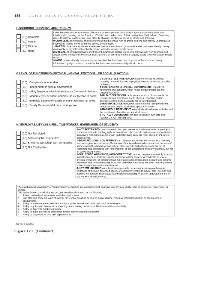
186 C O N D I T I O N S I N O C C U P A T I O N A L T H E R A P Y
F.GROOMING (COGNITIVE ABILITY ONLY)
Does the patient show awareness of how and when to perform this activity? Ignore motor disabilities thatinterfere with carrying out this function. (This is rated under Level of Functioning described below.) Grooming refers to bathing, washing, brushing of teeth, shaving, combing or brushing of hair and dressing. 0-COMPLETE: continuously shows awareness that he knows how to groom self and can convey unambiguousinformation that he knows when this activity should occur.1-PARTIAL: intermittently shows awareness that he knows how to groom self and/or can intermittently conveyreasonably clearly information that he knows when the activity should occur.2-MINIMAL: shows questionable or infrequent awareness that he knows in a primitive way how to groom selfand/or shows infrequently by certain signs, sounds, or activities that he is vaguely aware when the activity shouldoccur. 3-NONE: shows virtually no awareness at any time that he knows how to groom self and cannot conveyinformation by signs, sounds, or activity that he knows when the activity should occur.
(0.0) Complete
(1.0) Partial
(2.0) Minimal
(3.0) None
G.LEVEL OF FUNCTIONING (PHYSICAL, MENTAL, EMOTIONAL OR SOCIAL FUNCTION)
0-COMPLETELY INDEPENDENT: able to live as he wishes,requiring no restriction due to physical, mental, emotional or socialproblems.1-INDEPENDENT IN SPECIAL ENVIRONMENT: capable of functioning independently when needed requirements are met(mechanical aids) 2-MILDLY DEPENDENT: able to care for most of own needs but requires limited assistance due to physical, cognitive and/oremotional problems (e.g., needs non-resident helper). 3-MODERATELY DEPENDENT: able to care for self partially butneeds another person at all times. (person in home) 4-MARKEDLY DEPENDENT: needs help with all major activities andthe assistance of another person at all times. 5-TOTALLY DEPENDENT: not able to assist in own care andrequires 24-hour nursing care.
(0.0) Completely Independent
(1.0) Independent in special environment
(2.0) Mildly Dependent-Limited assistance (non-resid - helper)
(3.0) Moderately Dependent-moderate assist (person in home)
(4.0) markedly Dependent-assist all major activities, all times
(5.0) Totally Dependent-24 hour nursing care.
H."EMPLOYABILITY"(AS A FULL TIME WORKER, HOMEMAKER, OR STUDENT)
0-NOT RESTRICTED: can compete in the open market for a relatively wide range of jobs commensurate with existing skills; or can initiate, plan execute and assume responsibilitiesassociated with homemaking; or can understand and carry out most age relevant schoolassignments. 1-SELECTED JOBS, COMPETITIVE: can compete in a limited job market for a relativelynarrow range of jobs because of limitations of the type described above and/or because of some physical limitations; or can initiate, plan, execute and assume many but not all responsibilities associated with homemaking; or can understand and carry out many but notall school assignments. 2-SHELTERED WORKSHOP, NON-COMPETITIVE: cannot compete successfully in a jobmarket because of limitations described above and/or because of moderate or severe physical limitations; or cannot without major assistance initiate, plan, execute and assume responsibilities for homemaking; or cannot understand and carry out even relatively simple school assignments without assistance.3-NOT EMPLOYABLE: completely unemployable because of extreme psychosocial limitations of the type described above, or completely unable to initiate, plan, execute and assume any responsibilities associated with homemaking; or cannot understand or carryout any school assignments.
(0.0) Not Restricted
(1.0) Selected jobs, competitive
(2.0) Sheltered workshop, Non-competitive
(3.0) Not Employable
The psychosocial adaptability or “employability” item takes into account overall cognitive and physical ability to be an employee, homemaker orstudent. This determination should take into account considerations such as the following:1. Able to understand, remember and follow instructions.2. Can plan and carry out tasks at least at the level of an office clerk or in simple routine, repetitive industrial situation or can do school
assignments. 3. Ability to remain oriented, relevant and appropriate in work and other psychosocial situations.4. Ability to get to and from work or shopping centers using private or public transportation effectively. 5. Ability to deal with number concepts.6. Ability to make purchases and handle simple money exchange problems7. Ability to keep track of time and appointments
Revised 03/2010
Figure 12.1 (Continued )

187Chapter 12: Acquired Br a in In jur y
RANCHO LOS AMIGOS SCALEAKA level of cognitive functioning scale (LCFS)
___(1) Level I - No response.
Patient does not respond to external stimuli and appears asleep.
___(2) Level II - Generalized response.
Patient reacts to external stimuli in nonspecific, inconsistent, and nonpurposeful manner with stereotypic and limited responses.
___(3) Level III - Localized response.
Patient responds specifically and inconsistently with delays to stimuli, but may follow simple commands for motor action.
___(4) Level IV - Confused, agitated response.
Patient exhibits bizarre, nonpurposeful, incoherent or inappropriate behaviors, has no short- term recall, attention is short and nonselective. ___(5) Level V - Confused, inappropriate, nonagitated response.
Patient gives random, fragmented, and nonpurposeful responses to complex or unstructured stimuli - simple commands are followed consistently, memory and selective attention are impaired, and new information is not retained.
___(6) Level VI - Confused, appropriate, response.
Patient gives context appropriate, goal-directed responses, dependent upon external input for direction. There is carry-over for relearned, but not for new tasks, and recent memory problems persist.
___(7) Level VII - Automatic, appropriate response.
Patient behaves appropriately in familiar settings, performs daily routines automatically, and shows carry-over for new learning at lower than normal rates. Patient initiates social interactions, but judgment remains impaired.
___(8) Level VIII - Purposeful, appropriate response.
Patient oriented and responds to the environment but abstract reasoning abilities are decreased relative to premorbid levels.
Figure 12.2 Rancho Los Amigos AKA Level of Cognitive Functioning Scale (LCFS). (From Sander, A. (2002). The Level of Cognitive Functioning Scale. The Center for Outcome Measurement in Brain Injury. http://www.tbims.org/combi/lcfs. Accessed August 31, 2010.)
learning new motor routines, and adapting to new or cognitively demanding situations (Cicerone, Dahlberg, & Kalmar, 2000). Motor learning, or the ability to relearn previously well-known or learn anew adaptive motor skills, is often functional during the rehabilitation process despite memory loss.
As coma subsides, cognitive deficits become apparent. These may include difficulties with sustained attention, concentration, memory, comprehension, reasoning, self-monitoring and impulse control, other-awareness, and executive function. Executive functions involve the abil-ity to formulate context-appropriate goals and to initiate, plan and organize, sequence, and adapt behavior based on anticipated or actual consequences of actions (Cicerone et al., 2000; Hawley, 2004).
Neurobehavioral deficits occur as a result of cognitive deficits interacting with brain dysfunction. These deficits are typically seen whether the TBI is mild, moderate, or severe (Cicerone et al., 2000; Hawley, 2004) and include impulsivity, perseveration, irritability, poor control of temper, aggression, disinhibition, and apathy (Noggle & Pierson, 2010; Ylvisaker et al., 2007). Limited self-awareness or a lack of insight may slow rehabilitation progress as well as the ability to participate successfully in academic, vocational, and/or social roles.
Depression and loss of self-esteem may be particularly prevalent in children with TBI as they age. An increas-ing awareness of their deficits coupled with decreased academic achievement (Fay et al., 1994) may lead to depression in children.
188 C O N D I T I O N S I N O C C U P A T I O N A L T H E R A P Y
injury is hydrocephalus, which is a serious complication for up to 75% of individuals with TBI. Fractures are common as well, as 82% of those with TBI have one or more extracranial fractures, with 10% of these cervical spinal cord injuries. In the latter case, immediate medical management is needed for both a brain injury and a high-level spinal cord injury (Hanscom, 1987).
Intensive-care medical management is constant. An indwelling urinary catheter is placed and closely monitored. About one-third of those hospitalized with TBI aspirate food into their lungs, resulting in aspiration pneumonia. These persons usually have a delayed or absent swallow-ing reflex (Lazarus & Logemann, 1987; Logemann et al., 1994; Mackay et al., 1999). A nasogastric tube is posi-tioned and used for high caloric feeding for people with swallowing dysfunction. Close attention to skin integrity is essential, and the person’s total body position is changed frequently. Suctioning of the endotracheal tube and vigor-ous respiratory therapy are implemented to prevent addi-tional pulmonary problems (Costanzo & Becker, 1990).
Ongoing management of common medical complica-tions occurs in the acute phase. Prophylactic medication for seizures is provided typically for only the first 7 days following injury and then discontinued unless the per-son has recurring seizures. As a result of rigid abnormal posturing and other motor disturbances, many persons with TBI develop contractures of the neck, trunk, and/or extremities. The longer the duration of coma, the greater is the potential for the development of contractures, het-erotopic ossification, and DVTs. Treatment to prevent these complications commonly includes range of motion in the acute phase. Medical intervention for spasticity in the acute phase includes physical and pharmacologic interventions. Acute physical interventions include range of motion and splinting. Commonly prescribed medica-tions include baclofen, tizanidine, dantrolene, and botuli-num toxin (Greenwald & Rigg, 2009).
Rehabilitation interventions in the acute phase may begin as soon as neurologic stability is achieved. The focus of acute rehabilitation is to prevent joint deformity and to provide graded and specific sensory stimulation, with the assumption that selective sensory input may speed or improve neurologic recovery.
Medical Intervention in RehabilitationAdmission to an inpatient rehabilitation unit is needed for people with moderate to severe TBI. Criteria for transfer to the rehabilitation phase include medical stability, poten-tial for improvement, and tolerance for therapy. Inpatient rehabilitation requires tolerance for at least 3 hours of two or more therapies 5 to 7 days per week; subacute rehabili-tation requires the ability to participate for 0.5 to 2 hours each day. Intensive rehabilitation is usually directed by a physiatrist, a physician specializing in rehabilitation
Onset of an Axis 1 or Axis 2 psychiatric disorder may occur following a TBI. In one large sample of 722 out-patients with TBI, major depressive disorder was found in 42% of the sample (Kreutzer, Seel, & Gourley, 2001); another large study of 666 people found the incidence to be 27% (Seel, Kreutzer, & Rosenthal, 2003). Neither time since injury nor severity of injury is correlated with depression (Gordon et al., 2006). With depression comes the potential for suicide. Suicide rates for people with TBI vary between 2.7 and 4.1 times that of the general population when matched for age and sex (Engberg & Teasdale, 2004; Teasdale & Engberg, 2001). A diagnosis of TBI and evidence of aggression and hostility are pre-dictive of suicide attempts (Gordon et al., 2006).
Posttraumatic stress disorder is also seen in people with TBI. Studies have found that symptoms of post-traumatic stress disorder were related to the person’s level of insight, but not severity of injury, years of educa-tion, intelligence, or memory impairment (Gordon et al., 2006). Additional Axis I diagnoses correlated with TBI include substance abuse and aggressive behavior and agi-tation. Aggressive behavior has been shown to be three times greater in people with TBI compared to people with multiple trauma (Baguley et al., 1999). Axis II diagnoses associated with TBI include borderline, avoidant, para-noid, obsessive-compulsive, and narcissistic personality. The onset of an Axis II disorder is independent of sever-ity of injury, age at injury, and time since injury (Gordon et al.).
Medical/Surgical Management
Medical Intervention in the Acute PhasePhysicians from neurology, neurosurgery, nternal medi-cine, or orthopaedics may direct overall medical man-agement in the acute phase. The focus of acute medical management is preservation of life, management of sec-ondary complications, and the prevention of secondary damage. Maintaining an effective airway and circulatory function are critical life-preserving steps immediately after injury. An endotracheal tube is typically placed to support breathing. After arrival at the hospital, diagnos-tic tests are begun to identify the location and severity of all injuries. The patient typically receives a computer-ized axial tomography scan. If this reveals an intracranial hematoma, immediate surgical decompression is per-formed. Constant monitoring of consciousness occurs, as the duration and depth of coma are significant indicators of both mortality and morbidity (Carlile et al., 2010).
Diagnosis and management of secondary diagnoses also occurs upon arrival at the hospital. More than 50% of persons with severe head injury have associated inju-ries. A common secondary complication from the brain
189Chapter 12: Acquired Br a in In jur y
Effect of TBI on ProductivityMost persons with TBI have major difficulties return-ing to a productive life after injury whether the injury is mild or severe. Ruffolo, Friedland, and Dawson (1999) reported that while 42% of people with mild brain injury returned to work, only 12% returned to their premorbid level of employment. Cognitive and behavioral issues were cited for this decreased function.
For those with moderate to severe injuries, the abil-ity to return to work has been inconsistently correlated with self-awareness. Sherer, Bergloff, and Levin (1998), in a multicenter TBI Model System study, found that lim-ited self-awareness accounted for a substantial proportion (0.31) of the variance in positive vocational outcomes, while Coetzer and du Toit (2002) found no comparable correlation in their study of 40 people with TBI of varying levels of severity. In the absence of definitive findings to date, the difficulty of people with TBI to be aware of the effect of their statements and physical actions on others should be considered in planning for return to productiv-ity. Indeed, psychosocial skills that affect social integra-tion show a stronger correlation with successful return to work than either cognitive or sensorimotor skills, or any combination of the three factors (Conti, 1992).
Bushnik et al. (2003) found the unemployment rate for persons with TBI because of violence to be 70%, significantly greater than the rate of approximately 50% for those with TBI from all other causes. An additional factor independently predicting poorer productivity out-comes was preinjury substance abuse (Sherer, Bergloff, & High, 1999).
Neither gender nor race has been consistently linked to productivity outcomes. A review by van Reekum (2001) found conflicting evidence that females had poorer out-comes, partly based on varying definitions of productiv-ity. A major study of race (Sherer, Nick, & Sander, 2003) and productivity found that African Americans and oth-ers from minority backgrounds had less productive out-comes than whites, but race alone accounted for little of the variance in productivity. Race and productivity had confounding associations with preinjury productivity, educational level, and cause of injury.
The DRS has been found to predict employment. In a study of 145 persons with TBI, Cope, Cole, and Hall (1999) found that 62% of those with scores of 1 to 3 on rehabilitation admission were employed or in school at 1 year after discharge, while 39% of those with scores of 4 to 6 and only 11% of those with a DRS score of 7 to 20 were similarly employed. For those returning to work, supported part-time employment has been shown to be a viable and cost-effective option (Cope et al., 1999). However, limited employment opportunities with typically lower wages and decreased work hours often result in the need for public assistance ( Johnstone et al., 2003; Wehman et al., 2003).
medicine. Goals of the rehabilitation program are to max-imize the person’s function, minimize additional physical or psychosocial impairments, and prevent complications (Greenwald & Rigg, 2009). Along with the primary phy-sician, core rehabilitation members include specialists in occupational therapy, physical therapy, speech/language pathology, nursing, neuropsychology, and social work. In 2005, a major review of multidisciplinary rehabilitation of adults with TBI found “strong evidence of benefit from formal intervention,” with earlier functional gains from more intensive programs (Turner-Stokes et al., 2005).
The long-term rehabilitation goals in occupational therapy are to reestablish occupational performance skill, sensorimotor integration and control, and the integration of perceptual, cognitive, and communication skills with daily tasks. Where remediation is not possible or when maximal neurologic recovery is assumed to be complete, the use of compensation strategies may be appropriate. As basic goals are accomplished, discharge from the hospital may occur, with more advanced skills learned on an outpatient basis. Outpatient occupational therapy goals include instrumental ADL, further community reintegration, and work reentry.
Impact of TBI on Client Factors
All areas identified in the Occupational Therapy Practice Framework are affected with TBI. The deeply comatose person, with cognitive levels I through III, shows depressed function in all function. With further recovery, improvement in performance skills and pat-terns, as well as in client factors, may occur and enable the performance of preferred or required occupations. In a study of 1,170 records from the Traumatic Brain Injury Model Systems database, Bushnik et al. (2003) found that individuals with TBI as a result of a vehicular accident were initially admitted with significantly lower functional independence measure motor scores than those who were admitted because of violence, falls, or other causes. However, at discharge from rehabilitation, no significant differences were found among patients in the four etiology groups for most psychosocial and functional outcome measures. In fact, 85% to 96% of all patients demonstrated sufficient basic ADL skills to live in a private residence upon discharge.
Despite basic ADL skills, independence in commu-nity living is difficult for many people after TBI. A study of 175 survivors of moderate to severe TBI 2 years after acute rehabilitation found that over 75% of these survi-vors reported ongoing cognitive, emotional, and behavio-ral problems. Of those with remaining problems, between 60% and 75% reported difficulty with memory, fatigue, word-finding difficulties, irritability, decreased speed of thinking, and impaired concentration (Ponsford, Olver, & Curran, 1995).
190 C O N D I T I O N S I N O C C U P A T I O N A L T H E R A P Y
brain. There are four general categories of malignant brain tumors: gliomas, meningiomas, germ cell tumors, and sellar region tumors (Bondy et al., 2008; Fisher et al., 2006). Benign tumors, on the other hand, are not cancerous, and they do not invade other body tis-sues or spread to other body parts. Benign tumors may become life threatening as they cause increasing deficits with cell growth, because they press upon nearby struc-tures and tissues (Fisher, Schwartzbaum, Wrensch, & Wiemels, 2007). This chapter will discuss malignant primary brain tumors. However, the signs and symp-toms of, and diagnosis, and treatment for a secondary brain tumor are similar.
Etiology
Chemical changes in brain cells lead to the formation of brain tumors. However, why these changes occur is not fully understood. Most brain tumors develop for no apparent reason and are not associated with anything the person did or did not do.
A few risk factors are known. A small number of genetic factors are associated with brain tumor risk; how-ever, in a study of 500 patients with tumors, <1% had a known hereditary genetic syndrome (Wrensch, Lee, & Miike, 1997).
Gender appears to be associated with the risk for certain types of tumors. Men are more susceptible to glioma and germ cell tumors, while women are twice as likely as men to have meningiomas. In addition, rela-tively consistent research results show that being pre-menopausal confers a greater meningioma risk than being postmenopausal (Fisher et al., 2007).
High levels of ionizing radiation are strongly associ-ated with tumor development, as was found following the atomic bombing of Hiroshima in World War II (Fisher et al., 2007). Types of tumors associated with radiation include glioma, schwannoma, and pituitary tumors. In the past infants and children have been treated with radiation in the treatment of tinea capitis and skin hemangioma and have shown increased risk for nerve sheath tumors, meningioma, and pituitary adenoma. Taken as a whole, however, exposure to high levels of radiation is rare.
Many studies suggest that the risk for glioma is reduced as a result of allergies and immune-related condi-tions. This may arise from the anti-inflammatory effects of cytokines present in allergic and autoimmune diseases (Fisher et al., 2007).
Inconsistent results have been found for exposure to other environmental factors. Early studies of cell phone use and glioma risk have been generally inconclusive. However, long-term studies are needed to determine definitively any risk factor between cell phone use and glioma. Inconsistent, minimal or no evidence is present to
Factors Affecting DrivingPhysical disabilities, as well as cognitive, visual, or percep-tual, and self-awareness deficits can significantly impair driving function. A number of studies have attempted to identify factors that predict those persons with TBI who may successfully return to driving, with inconclu-sive results (Coleman, Rapport, & Ergh, 2002; Novack et al., 2010; Pietrapiana et al., 2005; Rapport, Coleman, & Hanks, 2008). Severity of injury and duration of coma have not shown clear predictive value (Coleman et al., 2002; Rapport, Hanks, & Bryer, 2006). One study found that persons more likely to return to driving included those discharged from rehabilitation with independence or modified independence in scores on the Functional Independence Measure (Fisk, Schneider, & Novack, 1998). Neuropsychologic testing may provide insight into driving potential, but these findings may be limited by the person’s level of self-awareness (Novack et al., 2010).
Nevertheless, about 50% of people with moderate to severe TBI have resumed driving within 5 years, with most driving within the first year of injury (Novack et al., 2010; Rapport et al., 2006). Tamietto et al. (2006) note, however, that as many as two-thirds of these may resume driving without any formal examination. In a study com-paring nondrivers to drivers with TBI, Rapport found that nondrivers who wanted to drive rated themselves as physically and cognitively fit to drive, despite cognitive skills that were significantly worse than drivers with TBI (Rapport et al., 2006). In discussing this finding, Rapport et al. stated that
even nondrivers rate their current driving abilities as better than average (which) may reflect unawareness of deficit, denial, resistance to role change or accurate self-perception.
TUMORS OF THE CENTRAL NERVOUS SYSTEM
Descriptions and Definitions
Brain tumors have increased in the past few decades. Fortunately, this appears to be due to enhanced neu-roimaging techniques and improved medical treatment options (Fisher, Schwartzbaum, Wrensch, & Berger, 2006), rather than a rise in natural incidence. Tumors are classified as primary or secondary, and malignant or benign. The site of origin for the tumor is consid-ered its primary site, even when the tumor has spread to other parts of body or brain. Malignant tumors are composed of abnormal cells that multiply rapidly, with the ability to invade, or metastasize, into other tissues. Brain tumors, conversely, rarely metastasize beyond the
191Chapter 12: Acquired Br a in In jur y
Signs and Symptoms
Signs and symptoms of brain tumors include fatigue, sleep disturbance, pain, mood disorders, and cognitive dysfunction. Fatigue may be the single most significant problem with brain tumors, resulting in increased day-time sleep and decreased or interrupted nighttime sleep. Nightly sleep disturbance is reported by up to 50% of people with brain tumors (Liu, Page, Solheim, Fox, & Chang, 2009). One study of people with recurrent malig-nant gliomas found that as many as 94% reported severe fatigue (Osoba, Brada, Prados, & Yung, 2000).
Headache is the most common type of pain experienced, with up to 50% of people with gliomas reporting severe pain. Mood disorders accompanying brain tumors include, not surprisingly, anxiety and depression. Depression partic-ularly has been linked to survival, and yet one study found that only 60% of people who reported this to their physician received antidepressants (Litofsky et al., 2004). Problems with memory and executive functions are found in almost half of all people with glioma tumors (Liu et al., 2009).
Diagnosis, Course, and Prognosis
Diagnosis begins with the onset of unexplained symp-toms. As tumor cells multiply in the brain, they cre-ate pressure on and irritate normal brain tissue. As a result, two-thirds of those with primary brain tumors report symptoms as just described. Amazingly, about one-third of all people with brain tumors have no symptoms initially.
The presence of symptoms is typically assessed by a neurologist, with the use of such diagnostic procedures as magnetic resonance imaging (MRI), computer-ized tomography (CT), positron emission tomography (PET), or biopsy. The MRI uses an extremely strong magnet and radio waves to produce brain images. It may or may not be used with angiography, in which a dye is inserted into the bloodstream to differentiate between healthy and tumor tissues. People receiving an MRI may not have pacemakers or metal implants or be allergic to the dye used in angiography.
CT scans involve multiple x-rays of the brain from different angles. From this, a computerized three-dimen-sional model of the brain can be displayed. Iodine is com-monly used as a contrast agent to enhance the image. As a result, this may not be the best diagnostic procedure for people with allergies, diabetes, or a heart, kidney, or thy-roid condition.
PET scans are typically an ancillary diagnostic tool to either the MRI or PET scan. Finally, a biopsy of the suspected brain tissue may be made. Following the biopsy surgery, histologic analysis of brain cell tissue occurs (Family Caregiver Alliance, 2010).
suggest any relationship with increased brain tumor risk and head trauma, certain dietary supplements, alcohol consumption, tobacco smoking, and exposure to electro-magnetic fields (Fisher et al., 2006, 2007). Inconclusive evidence is also present for many occupational risk factors.
Incidence and Prevalence
About 22,070 persons were diagnosed with primary malig-nant brain tumors in 2009, including 12,060 men and 10,060 women (CBTRUS, 2010). An estimated 2,300 new cases of primary brain tumors were expected to occur in children (American Cancer Society, 2010a). Incidence dif-fers according to gender, age, race and ethnicity, and geog-raphy. Adult men tend to have higher rates than women of primary malignant brain tumors while nonmalignant men-ingioma is more common in women (Bondy et al., 2008).
Age affects the survival rate for both the youngest and oldest people. Childhood cancer is rare; <1% of all new cancer diagnoses occur in children. Nevertheless, it is the second leading cause of death (after accidents) in children between 1 and 14 years (American Cancer Society, 2010a). It is also the third leading cause of death in young adults ages 20 to 39 ( Jemal, Siegel, Xu, & Ward, 2010). In gen-eral, and for most tumors, the 5-year survival rate increases with age (Bondy et al., 2008) through the 50s and early 60s.
Studies of race and ethnicity are confounded by issues related to low socioeconomic status. African Americans are more likely to develop tumors than any other racial or eth-nic group, and death rates from cancer are 34% higher for African American men and 17% higher for African Ameri-can women than their Caucasian counterparts. Hispanic people tend to have lower incidence rates for cancer. These figures must be placed in context with concerns of poverty. According to the U.S. Census Bureau, one in four African Americans and Hispanic people lived below the poverty line in 2008. By comparison, only one in 10 non-Hispanic Caucasian people lived below the poverty line. Further-more, one in five African Americans and one in three His-panic persons were uninsured and therefore less able to receive needed treatment. Finally, discrimination may play a role in the provision of health care. According to the Ameri-can Cancer Society (American Cancer Society, 2010a),
racial and ethnic minorities tend to receive lower qual-ity health care than whites even when insurance status, income, age, and severity of conditions are comparable.
Cancer rates tend to be higher in more developed coun-tries, partly due to greater technology resources for evalu-ation and treatment. In the United States, the lowest average annual rate of all central nervous system (CNS) tumors in 2004 was reported in Virginia, and the highest rate was found in Colorado (Fisher et al., 2007).
192 C O N D I T I O N S I N O C C U P A T I O N A L T H E R A P Y
of stereotactic radiosurgery include using a moving linear accelerator, the Gamma Knife, or accelerator-delivered proton and helium ions. Stereotactic radiosurgery typi-cally uses just one treatment session to deliver the full radiation dose (American Cancer Society, 2010d).
Common side effects of radiation therapy, requiring medical management, include fatigue, fever/chills, and a sore or dry mouth. Because radiation therapy to the brain may increase the chance of tooth cavities, ongoing dental consultations are advised. Hair loss also is a common side effect (American Cancer Society, 2010c). While this is not a dangerous side effect, it is often distressing to the person.
Newer approaches to chemotherapy are also available. Traditionally, chemotherapy drugs have had difficulty crossing the blood-brain barrier. Research is currently under way to modify these drugs to successfully cross the barrier, so that they can reach more easily the blood-stream in the brain. Some targeted drug treatments have been developed that focus on specific abnormalities within cancer cells. These treatments are very new and are still undergoing rigorous study in clinical trials. One targeted drug used to treat brain tumors is bevacizumab (Avastin). This drug, given intravenously, stops the formation of new blood cells, which cuts off the blood supply to a tumor and results in the death of the tumor cells (Mayo Clinic, 2010).
The schedule for chemotherapy is dependent on a number of factors and may be provided daily, weekly, or even monthly. After each treatment cycle, a break is provided to allow the body time to rebuild healthy new cells and recuperate from the strong chemicals used. Common side effects of chemotherapy include fatigue, nausea, vomiting, sore mouth, diarrhea or constipation, loss of appetite, pain or difficulty swallowing, swelling in hands or feet, itching or rash, shortness of breath, cough, muscle or joint pain, or numbness in hands or feet (American Cancer Society, 2010b).
In addition to chemotherapy, the medications most commonly prescribed for brain tumors are steroids and antiepileptic drugs. Steroids are used to reduce brain edema and ameliorate the person’s symptoms. They may be prescribed at diagnosis or before or after surgery. Common steroids include dexamethasone, Decadron, prednisone, or methylprednisone. Short-term side effects include insomnia, weight gain and increased appetite, mood swings, and irritability. Side effects from long-term use include cataracts, osteoporosis, muscle weakness, and diabetes (National Brain Tumor Society, 2010).
Antiepileptic medications may be used as a precau-tionary measure or in response to seizure activity. Com-mon anticonvulsant drugs include Dilantin, Tegretol, Depakote, Keppra, Neurontin, Topomax, Phenobarbi-tal, and Lamictal. Side effects from these medications include fatigue, weakness, nausea and vomiting, and incoordination.
In most cancer, an important next step is to deter-mine the stage of the tumor. However, brain tumors rarely spread to other organs, although they may spread within the brain. Therefore there is no formal staging system for the prognosis of brain tumor. Instead the oncologist will use the following information to deter-mine the outlook: the type of tumor, the grade of the tumor (how quickly it can be expected to spread), the person’s age, the person’s functional status (whether symptoms are present or not), the size and location of the tumor, the feasibility of surgical removal of the tumor, and whether there is evidence of spread to other parts of the brain and/or spinal cord (American Can-cer Society, 2010a).
The course following diagnosis involves immediate medical management, described below, followed by ongo-ing medical checkups for years.
In general, survival from any tumor is lowest in the oldest age groups. When types of tumors are examined, the relative survival probability of those with glioblasto-mas is the lowest, with a 37.7% 2-year survival rate and a 30.2% 5-year survival rate. The survival rates for benign meningioma are much less well known, but appear to be significantly better. One estimate is that the 5-year sur-vival rate for meningioma may be 81% for people ages 21 to 64 years and 56% for those 65 or older at diagnosis (McCarthy, Davis, & Freels, 1998).
Medical/Surgical Management
Management of brain tumors may involve surgery, radia-tion therapy, and/or chemotherapy. Surgery is now more sophisticated than in the past. It can now be guided at least partially by the use of MRI, to determine locations of important brain areas and their distance from a tumor. Surgery also may be directly aided by image guidance, which provides better visualization of the brain area (American Cancer Society, 2010a).
Radiation therapy uses x-rays, gamma rays, electron beams, or protons to destroy cancer cells. Because radia-tion can be localized, it affects only the body part being treated. It may be used alone or in combination with surgery or chemotherapy, where some drugs may actually make cancer cells more sensitive to radiation. Radiation may be given externally, using a linear accelerator, or inter-nally, where a radioactive source in an implant is surgically placed near the tumor (American Cancer Society, 2010d).
Significant improvements have occurred to radia-tion therapy recently, allowing smaller doses of radiation and more precise placement of the radiation. Stereotac-tic radiosurgery is not really surgery, but the delivery of precise radiation to a brain site is guided by MRI or CT scans. Nearby tissue is affected as little as possible. Types
193Chapter 12: Acquired Br a in In jur y
Mental functions are likely to decline, as well as movement-related skills, vision, and/or communica-tion, depending on the site of the tumor. Pain is likely to increase. Fatigue and side effect from medical treat-ment may make participation in all daily activities and preferred occupations difficult. While ADL may or may not remain intact for a considerable amount of time, instrumental activities such as work and leisure pur-suits are likely to suffer fairly quickly from both physical dysfunction and cognitive deficits. Rest may be dis-rupted and daytime sleep ineffective in decreasing per-ceptions of fatigue. In a study comparing people with malignant glioma to age- and gender-matched people with lung cancer, greater problems with vision, motor function, communication, headaches, and seizures were identified by the people with glioma (Klein, Taphoorn, & Heimans, 2001).
Both TBI and brain tumors result in physical, cog-nitive, and social-emotional deficits affecting all areas of occupation. For the occupational therapist, assessment and intervention is challenging, at times frustrating, and ultimately rewarding.
Complementary and alternative therapies may also be used, not to replace medical treatment, but to lessen symptoms. Complementary therapies may include stress management, relaxation and imagery training, medita-tion, acupuncture, herbal medicine, and massage.
Education and support is critical throughout the course of treatment. Support groups may be helpful to both the person with cancer and his or her caregivers. Additionally, individual or family counseling may provide additional support and individual-specific information.
Impact of a CNS Tumor on Client Factors
Cancer of any type, located anywhere in the body, is ter-rifying. While rates of survival have improved for some types of cancer, most people consider a tumor to be deadly. Values and beliefs may be in conflict. For example, a highly valued commitment to honesty may conflict with the desire not to cause others distress. A basic belief in fairness may be shaken by the timing of the diagnosis. Spiritual beliefs may be overturned, or spirituality may grow.
Case 1J.D., age 20, survived an automobile accident with a moderate TBI. After 2 weeks each in intensive care and an acute medical unit, he is to begin intensive rehabilitation. His occupational therapist cannot get reliable information from him, so she relies on a medical record review for his medical history and a discussion with his mother to identify his previous occupations.
His mother does not seem to be very aware of his activities. She states that he has had three jobs as a garage mechanic in the last 2 years. She says he seemed to get tired of routine and did not get along well with his bosses. He moved into her two- bedroom apartment about 6 months ago, so he could start saving money for a new car. He does not par-ticipate in any home-care tasks but does help with the rent. Leisure activities included fixing up his old car and “hot-rodding” around. J.D.’s mother does not care for many of his friends, but becomes tearful when asked about his relationship with Kathy. She says they were planning to be married in the fall.
The medical record review reveals that J.D. sus-tained a right tibia-fibula fracture with the TBI. After 3 days of general unresponsiveness, he began to obey simple commands (LCFS level III). In a few days, he became agitated and confused. A few words could be understood, including swear words. He persis-
tently pulled out both his urinary catheter and his nasogastric tube.
The agitation has lessened somewhat, but he continues to be intermittently disoriented, as when he calls his occupational therapist “Kathy.” There is a 1- to 2-second delay before any requested move-ment. His gaze appears divergent, and he performs best when closing one eye. When fatigued or per-haps frustrated, he is irritable and the therapist has used protective measures she was taught to both protect herself and J.D. from injury as he jabs his fist out toward her.
Motor deficits include poor sitting balance and right hemiplegia, with moderate to severe spasticity. The left upper extremity is within functional limits in range of motion and strength. Transfers require moderate assistance.
J.D. is independent in eating but requires assis-tance for all other tasks. He requires moderate assis-tance and verbal cuing for showering using a shower chair, donning and removing a T-shirt, and transfer-ring to and from the bed and wheelchair. Wheelchair mobility is slow, but he can wheel himself from one area to another with verbal cuing for the route. J.D. is unable to read, and perseveration is apparent when writing his name. J.D. has come a long way, but there is still a longer way to go to achieve maximal possible independence.
Case Illustration
194 C O N D I T I O N S I N O C C U P A T I O N A L T H E R A P Y
Case 2As a part of routine fourth-grade vision screening, the school nurse noticed that Jesus appeared to have some vision problems that were not present last year. Jesus said things seemed fuzzy. She sent a note home with Jesus to his mother, but never received a response. Six weeks later, his teacher sent Jesus to see the school nurse again, this time because of ongoing complaints of a headache. While with the nurse, Jesus vomited, and said that he had been rou-tinely vomiting for the last few weeks. He had no fever. Alarmed, the school nurse drove Jesus home to the trailer in which he and his mother, brother, and two sisters lived.
She spoke to the mother and stressed the need for immediate evaluation by a physician. Maria, the mother, stated she had no physician and no insurance and that the only medical care she and her family received was at the free clinic, open only on Tuesday afternoons and evenings. The school nurse wrote out information for the doctor there, and Maria promised to take Jesus to the next clinic.
The physician at the clinic examined Jesus and immediately set Jesus up for an evaluation by a pediatric oncologist. Social workers at the clinic assisted in finding transportation and assured Maria that medical care would be provided despite her
inability to pay. The visit to the pediatric oncologist provided a diagnosis of glioma. This message was sent to the social worker at the clinic, where there was a telephone. The social worker drove to Maria’s to give Maria this information and to plan how to achieve the medical care Jesus needed. School was put on the back burner. Jesus entered a regional children’s cancer center. Maria arranged for the other children to stay with an aunt and stays nearby the center.
It was a confusing and fun and sad time for Jesus. He missed his friends and his family, but he enjoyed the activities of the center. He did not like how he felt after radiation therapy, and it was hard to under-stand that something that made him feel so bad could make him better. He was glad that his mother is around, and, for the first time in years, spent time sitting on her lap.
Jesus came home, to a great family celebration. Maria ended the celebration after an hour, seeing that Jesus was tired. But Jesus was happy to be home. He did not care that he had no hair, because he was beginning to feel better, even though he tired easily. He missed his friends at school and hoped to go back soon, but he knew he still could not see well. Maria feared for the future of Jesus and her family and worried about bills that would certainly come.
R E C O M M E N D E D L E A R N I N G R E S O U R C E S
Consumer and Professional ResourcesBrain Injury Association of America, Inc.1608 Spring Hill Rd.Suite 110Vienna, VA 22182Brain Trauma Foundation415 Madison Ave.14th floorNew York, NY [email protected]://braintrauma.orgTel: 212-772-0608Fax: 212-772-0357Centers for Disease Control and Prevention1600 Clifton Rd.Atlanta, GA 30333Public InquiriesTel: (800) 311-3435www.cdc.gov
Family Caregiver Alliance/National Center on Care-giving
180 Montgomery StreetSuite 1100San Francisco, CA [email protected]: 800-445-8106Fax: 415-434-3508National Rehabilitation Information Center (NARIC)8201 Corporate DriveSuite 600Landover, MD [email protected]: 301-459-5900Fax: 301-562-2401
195Chapter 12: Acquired Br a in In jur y
Umphred, D. A. (2001). Neurological rehabilita-tion (4th ed.). St. Louis, MO: Mosby, Inc..
S U G G E S T E D R E A D I N G S
Adams, J. H., Graham, D. I., & Murray, L. S. (1982). Dif-fuse axonal injury due to non-missile head injury in humans. An analysis of 45 cases. Annals of Neurology, 12, 557–563.
American Cancer Society. (2010a). Cancer facts and fig-ures 2010. Retrieved from http://www.cancer.org/acs/groups/content/@epidemiologysurveilance/documents/document/acspc-026238.pdf
American Cancer Society (Producer). (2010b, August 30, 2010). Chemotherapy side effects worksheet. Retrieved from http://www.cancer.org/acs/groups/content/@nho/documents/document/acsq-009502.pdf
American Cancer Society (Producer). (2010c, August 30, 2010). External radiation side effects worksheet. Retrieved from http://www.cancer.org/acs/groups/content/@nho/documents/document/acsq-009503.pdf
American Cancer Society. (2010d). Understanding radiation therapy: A guide for patients and families. Retrieved from http://www.cancer.org/Treatment/ TreatmentsandSideEffects/TreatmentTypes/Radiation/UnderstandingRadiationTherapy-AGuideforPatientsandFamilies/understanding-radi-ation-therapy-intro
Anderson, F. A., Jr., Wheeler, H. B., & Goldberg, R. J. (1991). A population-based perspective of the hospital incidence and case-fatality rates of deep vein thrombo-sis and pulmonary embolism. The Worcester DVT Study. Archives of Internal Medicine, 151, 933–938.
Annegers, J. F., Hauser, W. A., & Coan, S. P. (1998) A popu-lation-based study of seizures after traumatic brain inju-ries, New England Journal of Medicine, 338(1), 20-24.
Asikainen, H., Kaste, M., & Sarna, S. (1999). Early and late posttraumatic seizures in traumatic brain injury rehabilitation patients: Brain injury factors causing late seizures and influence of seizures on long-term outcome. Epilepsia, 40(5), 584–589.
Baguley, I. J., Nicholls, J. L., & Felmingham, K. L. (1999). Dysautonomia after traumatic brain injury: A forgot-ten syndrome? Journal of Neurology, Neurosurgery, and Psychiatry, 67, 39–43.
Bazarian, J. J., McClung, J., & Shah, M. N. (2005). Mild acquired brain injury in the United States, 1998–2000. Brain Injury, 19, 85–91.
Beyerl, B., & Black, P. M. (1984). Postacquired hydrocephalus. Neurosurgery, 15, 257.
Binder, S., Corrigan, J. D., & Langlois, J. A. (2005). The public health approach to traumatic brain injury: An overview of CDC’s research and programs. Journal of Head Trauma Rehabilitation, 20, 189–195.
Bogner, J. A., Corrigan, J. D., & Mysiw, J. (2001). A comparison of substance abuse and violence in the prediction of long-term rehabilitation outcomes after acquired brain injury. Archives of Physical Medicine and Rehabilitation, 82, 571–577.
Bondy, M. L., Scheurer, M. E., Malmer, B., Barnholtz-Sloan, J. S., Davis, F. G., Il’yasova, D., et al. (2008). Brain tumor epidemiology: Consensus from the Brain Tumor Epide-miology Consortium. Cancer, 113(7 Suppl.), 1953–1968.
Bontke, C. F. (1989). Medical complications related to acquired brain injury. In L. J. Horn & D. N. Cope (Eds.), Physical medicine and rehabilitation: State of the art reviews. Traumatic brain injury (pp. 43–58). Philadelphia, PA: Hanley & Belfus.
Bruns, J. J. (2003). The epidemiology of acquired brain injury: A review. Epilepsia, 44, 2–10.
Bullock, R., Chestnut, R. M., & Clifton, G. (1995). Guidelines for the management of severe head injury: A joint initiative of the Brain Trauma Foundation, the American Associa-tion of Neurological Surgeons, the Joint Section on Neu-rotrauma and Critical Care: Brain Trauma Foundation.
Burnett, D. M., Watanabe, T. K., & Greenwald, B. D. (2003). Congenital and acquired brain injury. 2. Brain injury rehabilitation: Medical management. Archives of Physical Medicine and Rehabilitation, 84(Suppl 1), S8–S11.
Bushnik, T., Hanks, R. A., & Kreutzer, J., & Rosenthal, M. (2003). Etiology of brain injury: Characterization of different outcomes up to 1 year postinjury. Archives of Physical Medicine and Rehabilitation, 84, 255–262.
Carlile, M., Nicewander, D., Yablon, S. A., Brown, A., Brunner, R., Burke, D., et al. (2010). Prophylaxis for venous thromboembolism during rehabilitation for traumatic brain injury: A multicenter observational study. The Journal of Trauma, Injury, Infection, and Critical Care, 68, 916–923.
CBTRUS. (2010). Primary brain and central nervous system tumors diagnosed in the United States in 2004–2006.
R E F E R E N C E S
196 C O N D I T I O N S I N O C C U P A T I O N A L T H E R A P Y
Elvidge, A. R. (1939). Remarks on post-traumatic convulsive state. Transactions of the American Neuro-logical Association, 65, 125–129.
Engberg, A. W., & Teasdale, T. W. (2004). Psychosocial out-come following traumatic injury in adults: A long-term population-based follow-up. Brain Injury, 18, 533–545.
Family Caregiver Alliance. (2010). Fact sheet: Brain tumor. Retrieved October 2, 2010, from http://www. caregiver.org/caregiver/jsp/content_node.jsp?nodeid=568#
Fay, G. C., Jaffe, K. M., Polissar, M. L., Liao, S., Rivara, J. B., & Martin, K. M. (1994). Outcome of pediatric trau-matic brain injury at 3 years: A cohort study. Archives of Physical Medicine and Rehabilitation, 75, 733–741.
Fisher, J. L., Schwartzbaum, J. A., Wrensch, M., & Berger, M. S. (2006). Evaluation of epidemiological evidence for primary adult brain tumor risk factors using evidence-based medicine. Progressive Neurological Surgery, 19, 54–79.
Fisher, J. L., Schwartzbaum, J. A., Wrensch, M., & Wiemels, J. L. (2007). Epidemiology of brain tumors. Neurologic Clinics, 25, 867–890.
Fisk, G., Schneider, J., & Novack, T. (1998). Driving fol-lowing Traumatic Brain Injury: Prevalence, exposure, advice, and evaluations. Brain Injury, 12, 683–695.
Freed, S., & Hellerstein, L. F. (1997). Visual electrodiag-nostic findings in mild traumatic brain injury. Brain Injury, 11, 25–36.
Frey, L. C. (2003). Epidemiology of posttraumatic epilepsy: A critical review. Epilepsia, 44(Suppl. 10), 11–17.
Gordon, W. A., Zafonte, R., Cicerone, K., Cantor, J., Brown, M., Lombard, L., et al. (2006). Traumatic brain injury rehabilitation. State of the science. American Journal of Physical Medicine and Rehabilita-tion, 85, 343–382.
Gouvier, W. D., Blanton, P. D., & LaPorte, K. K. (1987). Reliability and validity of the Disability Rating Scale and the Levels of Cognitive Functioning Scale in monitoring recovery from severe head injury. Archives of Physical Medicine and Rehabilitation, 68, 94–97.
Greenwald, B. D., & Rigg, J. L. (2009). Neuroreha-bilitation in traumatic brain injury: Does it make a difference? Mount Sinai Journal of Medicine, 76, 182–189.
Hagan, C. (1997). Levels of cognitive functioning. Retrieved from http://www. neuroskillls.com/rancho.shtml
Hall, K. M., Cope, D. N., & Rappaport, M. (1985). Glasgow Outcome Scale and Disability Rating Scale: Comparative usefulness in following recovery in traumatic head injury. Archives of Physical Medicine and Rehabilitation, 66, 35–37.
Hall, K. M., Hamilton, B., & Gordon, W. A. (1993). Char-acteristics and comparisons of functional assessment indices: Disability Rating Scale, Functional Indepen-dence Measure, and Functional Assessment Measure. Journal of Head Trauma Rehabilitation, 8, 60–74.
Retrieved from http://www.cbtrus.org/2010- NPCR-SEER/CBTRUS-WEBREPORT- Final-3-2-10.pdf
Centers for Disease Control and Prevention (CDC). (2001). Acquired brain injury in the United States: A report to congress. Retrieved July 4, 2006, from http://www.cdc.gov/ncipc/TBI/TBI–congress/ index.htm
Cicerone, K., Dahlberg, C., & Kalmar, K. (2000). Evidence-based cognitive rehabilitation: Recom-mendations for clinical practice. Archives of Physical Medicine and Rehabilitation, 81, 1596–1615.
Coetzer, B. R., & Du Toit, P. L. (2002). Impaired aware-ness following brain injury and its relationship to placement and employment outcomes. Journal of Cog-nitive Rehabilitation, 20, 20–24.
Coleman, R. D., Rapport, L. J., & Ergh, T. C. (2002). Predictors of driving outcome after traumatic brain injury. Archives of Physical Medicine and Rehabilitation, 83, 1415–1422.
Conti, G. E. (1992). Factors affecting return to work for per-sons with Traumatic Brain Injury. Unpublished thesis. Eastern Michigan University.
Cope, D. N., Cole, J. R., & Hall, K. M. (1999). Brain injury: Analysis of outcome in a post-acute rehabilitation sys-tem. Part I: General analysis. Brain Injury, 5, 111–125.
Corrigan, J. D. (1995). Substance abuse as a mediating factor in outcome from acquired brain injury. Archives of Physical Medicine and Rehabilitation, 76, 302–309.
Costanzo, R. M., & Becker, D. P. (1990). Smell and taste dis-orders in head injury and neurosurgery patients. In R. L. Meiselman & R. S. Rivlin (Eds.), Clinical measurement of taste and smell (pp. 565–578). New York, NY: Macmillan.
Craig, G. N., Campbell, S. M. K., Richards, P. M. P., Ventureyra, E., & Hutchison, J. S. (2004). Medical and cognitive outcome in children with traumatic brain injury. The Canadian Journal of Neurological Sciences, 31, 213–219.
Cummings, P., Rivara, F. P., Olson, E. M., & Smith, K. M. (2006). Changes in traffic crash mortality rates attributed to use of alcohol, or lack of a seat belt, air bag, motorcycle helmet, or bicycle helment. Injury Prevention, 12, 148–154.
Cunningham, R. M., Maio, R. F., Hill, E. M., & Zink, B. J. (2002). The effects of alcohol on head injury in the motor vehicle crash victim. Alcohol, 37, 236–240.
Dawodu, S. T. (2005). Acquired brain injury: Definition, epidemiology, pathophysiology. eMedicine. Retrieved July 4, 2006.
Department of Transportation, National Highway Traf-fic Safety Administration (NHTSA). (2006). Traffic safety facts 2006. Retrieved July 4, 2006, from http://www.nhtsa.gov/portal/site/
Dumas, H. M., Haley, S. M., Carey, T. M., Ludlow, L. H., & Rabin, J. P. (2003). Lower extremity spasticity as an early marker of ambulatory recovery following traumatic brain injury. Childs Nervous System, 19, 114–118.
197Chapter 12: Acquired Br a in In jur y
Litofsky, N. S., Farace, E., Anderson, F., Jr., Meyers, C. A., Huang, W., & Laws, E. R., Jr. (2004). De-pression in patients with high-grade glioma: Results of the Glioma Outcome Project. Neurosurgery, 54, 358–367.
Liu, R., Page, M., Solheim, K., Fox, S., & Chang, S. M. (2009). Quality of life in adults with brain tumors: Current knowledge and future directions. Neuro- Oncology, 11, 330–339.
Logemann, J. A., Pepe, J., & Mackey, L. E. (1994). Disor-ders of nutrition and swallowing: Intervention strat-egies in the trauma center. Journal of Head Trauma Rehabilitation, 9, 43–56.
Mackay, L. E., Morgan, A. S., & Bernstein, B. A. (1999). Swallowing disorders in severe brain injury: Risk fac-tors affecting return to oral intake. Archives of Physical Medicine and Rehabilitation, 80, 365–371.
Mayo Clinic. (2010). Brain tumor. Treatments and drugs. Retrieved October 2, 2010, from http://www.mayoclinic.com/health/brain-tumor/DS00281/DSECTION=treatments-and-drugs
McCarthy, B. J., Davis, F. G., & Freels, S. (1998). Factors associated with survival in patients with meningioma. Journal of Neurosurgery, 88(5), 831–839.
McGarry, L. J., Thompson, D., Millham, F. H., Cowell, L., Snyder, P. J., & Lenderking, W. R. (2002). Outcomes and costs of acute treatment of treatment of traumatic brain injury. Journal of Trauma, 53, 1152–1159.
Meythaler, J. M., Peduzzi, J. D., & Eleftheriou, E. (2001). Current concepts: Diffuse axonal injury—Associated acquired brain injury. Archives of Physical Medicine and Rehabilitation, 82, 1461–1471.
National Brain Tumor Society. (2010). Treatment FAQ. Retrieved October 2, 2010: http://www.caregiver.org/caregiver/jsp/content_node.jsp?nodeid=568#
National Institutes of Health. (1998). Rehabilitation of persons with acquired brain injury. NIH Consensus Statement, 16, 1–41.
NIH Consensus Development Panel on Rehabilitation of persons with traumatic brain injury. Rehabilitation of persons with Traumatic Brain Injury. (1999). Journal of the American Medical Association, 282, 974–983.
Noggle, C. A., & Pierson, E. E. (2010). Psychosocial and behavioral functioning following pediatric TBI: Pre-sentation, assessment, and intervention. Applied Neu-ropsychology, 17(2), 110–115.
Novack, T. A., Labbe, D., Grote, M., Carlson, N., Sherer, M., Arango-Lasprilla, J. C., et al. (2010). Return to driving within 5 years of moderate-severe traumatic brain injury. Brain Injury, 24(3), 464–471.
Osoba, D., Brada, M., Prados, M. D., & Yung, W. K. (2000). Effect of disease burden on health-related quality of life in patients with malignant gliomas. Neuro-Oncology, 2, 221–228.
Hanscom, D. A. (1987). Acute management of the multiply injured head trauma patient. Journal of Head Trauma Rehabilitation, 2, 1–12.
Hawley, C. A. (2004). Behavior and school performance after head injury. Brain Injury, 18, 645–659.
Hellerstein, L. F., Freed, S., & Maples, W. C. (1995). Vision profile of patients with mild brain injuries. Journal of the American Optometric Association, 66, 634–639.
Hendricks, H. T., Heeren, A. H., & Vos, P. E. (2010). Dysautonomia after severe traumatic brain injury. European Journal of Neurology, 17, 1172–1177.
Jallo, J. I., & Narayan, R. K. (2000). Craniocerebral trau-ma. In W. G. Bradley, R. B. Daroff, & G. M. Fenichel (Eds.), Neurology in clinical practice (pp. 1055–1087). Boston: Butterworth-Heinemann.
Jemal, A., Siegel, R., Xu, J., & Ward, E. (2010). Cancer statistics 2010. CA: A Cancer Journal for Clinicians, 60, 277–300.
Jennett, B. (1975). Epilepsy after non-missile injuries (2nd ed.). Chicago, IL: Year Book Medical Publishers.
Jennett, B. (1990). Scale and scope of the problem. In M. Rosenthal, E. R. Griffith, & M. Bond (Eds.), Rehabilitation of the adult and child with traumatic brain injury (pp. 59–74). Philadelphia, PA: F. A. Davis.
Jennett, B., & Bond, M. (1975). Assessment or outcome after severe brain damage. Lancet, 1, 480–484.
Johnstone, B., Mount, D., & Schopp, L. H. (2003). Financial and vocational outcomes 1 year after acquired brain injury. Archives of Physical Medicine and Rehabilitation, 84, 238–241.
Kandel, E., Schwartz, J., & Jessell, T. (2000). Principles of neural science. New York, NY: McGraw-Hill.
Keane, J. R., & Baloh, R. W. (1992). Posttraumatic cranial neuropathies. The Neurology of Trauma, 10(4), 849–867.
Klein, M., Taphoorn, M. J., & Heimans, J. J. (2001). Neu-robehavioral status and health-related quality of life in newly diagnosed high-grade glioma patients. Journal of Clinical Oncology, 19, 4037–4047.
Kornbluth, J., & Bhardwaj, A. (2010). Evaluation of coma: A critical appraisal of popular scoring systems. Neurocritical Care. doi:10.1007?s12028-010-9409-3
Kreutzer, J. S., Seel, R. T., & Gourley, E. (2001). The prevalence and symptom rates of depression after traumatic brain injury: A comprehensive examina-tion. Brain Injury, 15, 563–576.
Langlois, J. A., Rutland-Brown, W., & Thomas, K. E. (2006). Traumatic brain injury in the United States: Emergency department visits, hospitalizations, and death. Atlanta, GA : Centers for Disease Control and Prevention, National Center for Injury Prevention and Control.
Lazarus, C., & Logemann, J. (1987). Swallowing disor-ders in closed head trauma patients. Archives of Physi-cal Medicine and Rehabilitation, 68, 79–84.
198 C O N D I T I O N S I N O C C U P A T I O N A L T H E R A P Y
Sherer, M., Bergloff, P., & High, W., Jr. (1999). Contribution of functional ratings to prediction of longterm employment outcome after traumatic brain injury. Brain Injury, 13, 973–981.
Sherer, M., Bergloff, P., & Levin, E. (1998). Impaired awareness and employment outcome after traumatic brain injury. Journal of Head Trauma Rehabilitation, 13, 52–61.
Sherer, M., Nick, T. G., & Sander, A. M. (2003). Race and productivity outcome after traumatic brain injury: Influence of confounding factors. Journal of Head Trauma Rehabilitation, 18, 408–424.
Strich, S. J. (1970). Lesions in the cerebral hemispheres after blunt head injury. Journal of Clinical Pathology, 23, 154.
Tamietto, M., Torrini, G., Adenzato, M., Pietrapiana, P., Rago, R., & Perino, C. (2006). To drive or not to drive (after TBI)? A review of the literature and its implica-tions for rehabilitation and future research. NeuroRe-habilitation, 21, 81–92.
Teasdale, T. W., & Engberg, A. W. (2001). Suicide after traumatic brain injury: A population study. Journal of Neurology, Neurosurgery, and Psychiatry, 71, 436–440.
Turner-Stokes, L., Nair, A., Sedki, I., Disler, P. B., & Wade, D. T. (2005). Multi-disciplinary rehabilitation for ac-quired brain injury in adults of working age. Cochrane Database of Systematic Reviews (3), CD004170.
van Reekum, R. (2001). Review: Women have worse out-comes than men after traumatic brain injury. Evidence Based Mental Health, 4, 58.
Wehman, P., Kregel, J., Keyser-Marcus, L., Sherron- Targett, P., Campbell, L., West, M., & Cifu, D.X. (2003). Supported employment for persons with ac-quired brain injury: a preliminary investigation of long-term follow-up costs and program efficiency. Archives of Physical Medicine and Rehabilitation, 84, 192–196.
Weintrab, A. H., & Opal, C. A. (1989). Motor and sensory dysfunction in the brain-injured adult. In L. J. Horn & D. N. Cope (Eds.), Physical medicine and rehabilitation: State of the art reviews. Traumatic brain injury (pp. 59–84). Philadelphia: Hanley & Belfus.
Whitman, S., & Coonley-Hoganson, R. T. (1984). Com-parative head trauma experiences in two socioeconomi-cally different area communities: Chicago—a population study. American Journal of Epidemiology, 119, 570–580.
Wrensch, M., Lee, M., & Miike, R. (1997). Familial and family history of cancer and nervous system conditions among adults with glioma and controls. American Journal of Epidemiology, 145(7), 581–593.
Ylvisaker, M., Turkstra, L., Coehlo, C., Yorkston, K., Kennedy, M., & Sohlberg, M. M. (2007). Behavioral interventions for children and adults with behav-ior disorders after TBI: A systematic review of the evidence. Brain Injury, 21(8), 769–805.
Pietrapiana, P., Tamietto, M., Torrini, G., Mezzanato, T., Rago, R., & Perino, C. (2005). Role of premorbid fac-tors in predicting safe return to driving after severe TBI. Brain Injury, 19, 197–211.
Plum and Posner’s diagnosis of stupor and coma. (2007). (4th ed.). New York, NY: Oxford University Press.
Ponsford, J., Olver, J. H., & Curran, C. (1995). A profile of outcome: 2 years after traumatic brain injury. Brain Injury, 9, 1–10.
Ponsford, J., Willmott, C., & Rothwell, A. (2000). Factors influencing outcome following mild traumatic brain injury in adults. Journal of the International Neuropsy-chological Society, 6, 568–579.
Ragnarsson, K. T. (2006). Traumatic brain injury research since the 1998 NIH Consensus Conference. Accomplishments and unmet goals. Journal of Head Trauma Rehabilitation, 21(5), 379–387.
Rappaport, M., Hall, K. M., & Hopkins, H. K. (1982). Disability rating scale for severe head trauma: Coma to community. Archives of Physical Medicine and Reha-bilitation, 63, 118–123.
Rapport, L. J., Coleman, B. R., & Hanks, R. A. (2008). Driving and community integration after traumatic brain injury. Archives of Physical Medicine and Rehabilitation, 89, 922–930.
Rapport, L. J., Hanks, R. A., & Bryer, R. C. (2006). Barriers to driving and community integration after traumatic brain injury. Journal of Head Trauma Rehabilitation, 21(1), 34–44.
Rimel, R. M., & Jane, J. A. (1983). Characteristics of the head injured patient. In M. Rosenthal & E. R. Griffith (Eds.), Rehabilitation of the head injured adult. Philadelphia, PA: F.A. Davis.
Rochette, L. M., Conner, K. A., & Smith, G. A. (2009). The contribution of traumatic brain injury to the medical and economic outcomes motor vehicle- related injuries in Ohio. Journal of Safety Research, 40, 353–358.
Ruffolo, C. F., Friedland, J. F., & Dawson, D. R. (1999). Mild traumatic brain injury from motor vehicle acci-dents: Factors associated with return to work. Archives of Physical Medicine and Rehabilitation, 80, 392–398.
Sakai, C. S., & Mateer, C. A. (1984). Otological and audiological sequelae of closed head trauma. Seminar of Hearing, 5, 157–173.
Schootman, M., Buchman, T. G., & Lewis, L. M. (2003). National estimates of hospitalization charges for the acute care of traumatic brain injuries. Brain Injury, 17, 983–990.
Seel, R. T., Kreutzer, J. S., & Rosenthal, M. (2003). Depression after traumatic brain injury: A National Institute on Disability and Rehabilitation Research Model Systems multicenter investigation. Archives of Physical Medicine and Rehabilitation, 84, 177–184.

199
Burns
■ Eli z ab e t h L . P h i l l ips
13
Sam, age 15, is scheduled for a burn dressing change at the out-patient burn clinic. Sam suffered 46% mixed partial- and full- thickness burns to his face, neck, chest, and bilateral arms 4 months ago when he added gasoline to a brush fire. The resulting
explosion set Sam’s clothes on fire. Sam panicked and started running. A neighbor who heard the explosion from inside his house tackled Sam and smothered the flames with a blanket. Sam spent 2 months as an inpatient on the burn unit where he received extensive grafting to his face, neck, arms, and chest. He was discharged 2 months ago but continues to have scattered small open areas. Sam arrives at the clinic, accompanied by his mother, and immediately requests assistance to remove his coat. When he is asked to try to remove his coat himself, he is able to complete the task but is noted to have extremely limited range of motion (ROM) and severe bilateral axillary contractures. It is noted that both his clothing and
temporary compression garment have been cut apart at the arms and shoulder and duct-taped together. When asked why this was done he replies that it is easier to remove his clothes this way. His mother states, “I told him he shouldn’t do that.”
DESCRIPTION AND DEFINITIONS
Anatomy and Physiology of the Skin
An understanding of burn injury must begin with a review of the anatomy and physiology of the skin. The skin is the largest organ of the body. The thin nonvascular outer layer, called the epidermis, consists of layers of epithelial cells. Beneath the epidermis is the thicker dermis, which makes up the bulk of the skin. Housed within the dermis are hair follicles, blood vessels, sweat glands, nerve ending, and sebaceous glands, which play an integral part in the functions of the skin (Fig. 13.1). The functions of the skin include (McGrath & Uitto, 2010):
■ Protection against infection ■ Prevention of loss of body fluid ■ Control of body temperature
K E Y T E R M S
AllograftAutograft
Burn scar contractureCollagen
Compression garmentDebridement
EscharFluid resuscitation
Full-thickness burnHypertrophic scar
Partial-thickness burnSuperficial burn
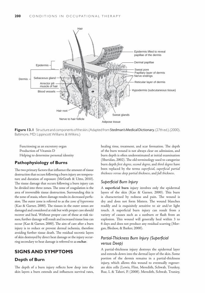
200 C O N D I T I O N S I N O C C U P A T I O N A L T H E R A P Y
■ Functioning as an excretory organ ■ Production of Vitamin D ■ Helping to determine personal identity
Pathophysiology of Burns
The two primary factors that influence the amount of tissue destruction that occurs following a burn injury are tempera-ture and duration of exposure (McGrath & Uitto, 2010). The tissue damage that occurs following a burn injury can be divided into three zones. The zone of coagulation is the area of irreversible tissue destruction. Surrounding this is the zone of stasis, where damage results in decreased perfu-sion. The outer zone is referred to as the zone of hyperemia (Kao & Garner, 2000). The tissues in the outer zones are damaged and considered at risk but with proper care should recover and heal. Without proper care of these at-risk tis-sues, further damage will result and increased tissue loss can occur (Kao & Garner, 2000). The aim of care after a burn injury is to reduce or prevent dermal ischemia, therefore avoiding further tissue death. The residual necrotic layers of skin destroyed by direct heat damage or the injury occur-ring secondary to heat damage is referred to as eschar.
SIGNS AND SYMPTOMS
Depth of Burn
The depth of a burn injury reflects how deep into the skin layers a burn extends and influences survival rates,
Figure 13.1 Structure and components of the skin. (Adapted from Stedman’s Medical Dictionary. (27th ed.). (2000). Baltimore, MD: Lippincott Williams & Wilkins.)
Epidermis lifted to reveal papillae of the dermis
Dermal papillae
Nerve endings
Sweat glands
Nerve to hair follicle
Hypodermis (subcutaneous tissue)
Dermis
Arrector pili muscle of hair
Blood vessels
Sebaceous gland
Hair
Epidermis
Adipose tissue
Hair root
Sweat pore
Reticular layer of dermis
Papillary layer of dermis
healing time, treatment, and scar formation. The depth of the burn wound is not always clear on admission, and burn depth is often underestimated at initial examination (Sheridan, 2002). The old terminology used to categorize burn depth first degree, second degree, and third degree have been replaced by the terms superficial, superficial partial thickness versus deep partial thickness, and full thickness.
Superficial Burn InjuryA superficial burn injury involves only the epidermal layers of the skin (Kao & Garner, 2000). This burn is characterized by redness and pain. The wound is dry and does not form blisters. The wound blanches readily and is exquisitely sensitive to air and/or light touch. A superficial burn injury can result from a variety of causes such as a sunburn or flash from an explosion. This wound will generally heal within 3 to 6 days and does not produce any residual scarring (Mor-gan, Bledsoe, & Barker, 2000).
Partial-Thickness Burn Injury (Superficial versus Deep)A partial-thickness injury destroys the epidermal layer and extends down into the dermal layer of the skin. Some portion of the dermis remains in a partial-thickness injury, which allows this wound to eventually regener-ate skin cells (Lewis, Flint, Meredith, Schwab, Trunkey, Rue, L & Taheri, P. (2008). Meredith, Schwab, Truniey,
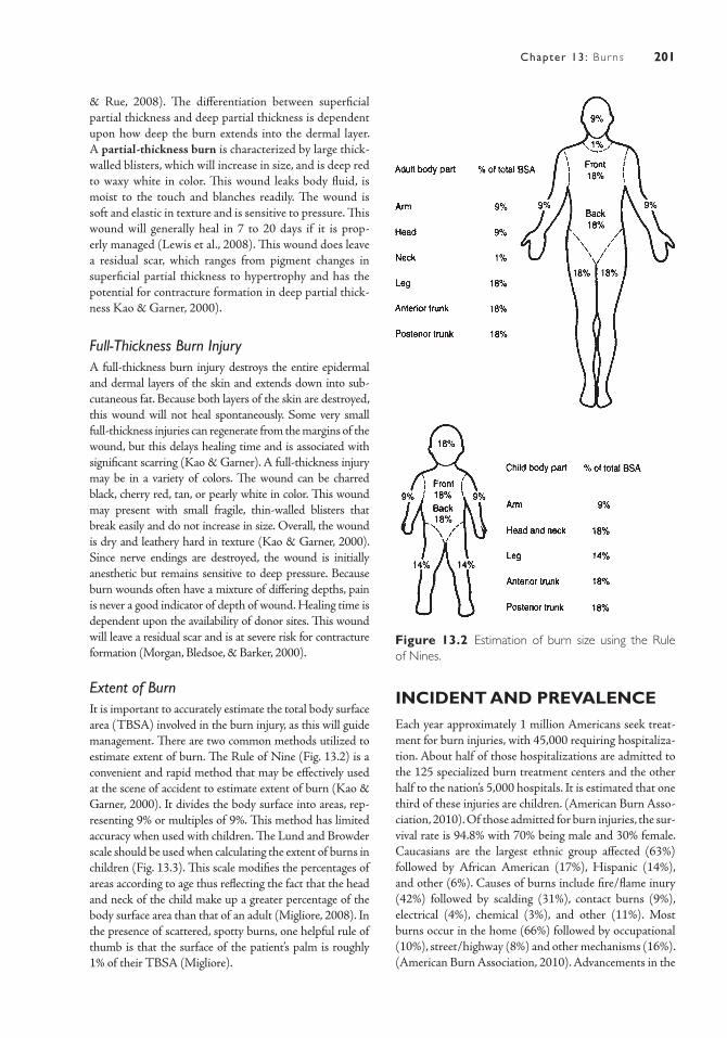
201Chapter 13: Burns
& Rue, 2008). The differentiation between superficial partial thickness and deep partial thickness is dependent upon how deep the burn extends into the dermal layer. A partial-thickness burn is characterized by large thick-walled blisters, which will increase in size, and is deep red to waxy white in color. This wound leaks body fluid, is moist to the touch and blanches readily. The wound is soft and elastic in texture and is sensitive to pressure. This wound will generally heal in 7 to 20 days if it is prop-erly managed (Lewis et al., 2008). This wound does leave a residual scar, which ranges from pigment changes in superficial partial thickness to hypertrophy and has the potential for contracture formation in deep partial thick-ness Kao & Garner, 2000).
Full-Thickness Burn InjuryA full-thickness burn injury destroys the entire epidermal and dermal layers of the skin and extends down into sub-cutaneous fat. Because both layers of the skin are destroyed, this wound will not heal spontaneously. Some very small full-thickness injuries can regenerate from the margins of the wound, but this delays healing time and is associated with significant scarring (Kao & Garner). A full-thickness injury may be in a variety of colors. The wound can be charred black, cherry red, tan, or pearly white in color. This wound may present with small fragile, thin-walled blisters that break easily and do not increase in size. Overall, the wound is dry and leathery hard in texture (Kao & Garner, 2000). Since nerve endings are destroyed, the wound is initially anesthetic but remains sensitive to deep pressure. Because burn wounds often have a mixture of differing depths, pain is never a good indicator of depth of wound. Healing time is dependent upon the availability of donor sites. This wound will leave a residual scar and is at severe risk for contracture formation (Morgan, Bledsoe, & Barker, 2000).
Extent of BurnIt is important to accurately estimate the total body surface area (TBSA) involved in the burn injury, as this will guide management. There are two common methods utilized to estimate extent of burn. The Rule of Nine (Fig. 13.2) is a convenient and rapid method that may be effectively used at the scene of accident to estimate extent of burn (Kao & Garner, 2000). It divides the body surface into areas, rep-resenting 9% or multiples of 9%. This method has limited accuracy when used with children. The Lund and Browder scale should be used when calculating the extent of burns in children (Fig. 13.3). This scale modifies the percentages of areas according to age thus reflecting the fact that the head and neck of the child make up a greater percentage of the body surface area than that of an adult (Migliore, 2008). In the presence of scattered, spotty burns, one helpful rule of thumb is that the surface of the patient’s palm is roughly 1% of their TBSA (Migliore).
INCIDENT AND PREVALENCEEach year approximately 1 million Americans seek treat-ment for burn injuries, with 45,000 requiring hospitaliza-tion. About half of those hospitalizations are admitted to the 125 specialized burn treatment centers and the other half to the nation’s 5,000 hospitals. It is estimated that one third of these injuries are children. (American Burn Asso-ciation, 2010). Of those admitted for burn injuries, the sur-vival rate is 94.8% with 70% being male and 30% female. Caucasians are the largest ethnic group affected (63%) followed by African American (17%), Hispanic (14%), and other (6%). Causes of burns include fire/flame inury (42%) followed by scalding (31%), contact burns (9%), electrical (4%), chemical (3%), and other (11%). Most burns occur in the home (66%) followed by occupational (10%), street/highway (8%) and other mechanisms (16%). (American Burn Association, 2010). Advancements in the
Figure 13.2 Estimation of burn size using the Rule of Nines.
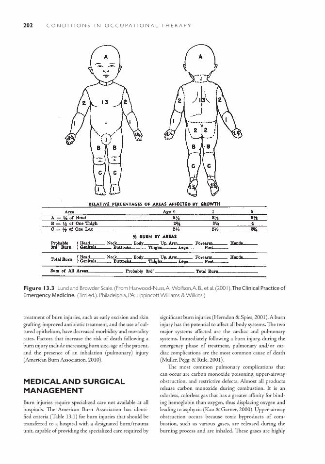
202 C O N D I T I O N S I N O C C U P A T I O N A L T H E R A P Y
treatment of burn injuries, such as early excision and skin grafting, improved antibiotic treatment, and the use of cul-tured epithelium, have decreased morbidity and mortality rates. Factors that increase the risk of death following a burn injury include increasing burn size, age of the patient, and the presence of an inhalation (pulmonary) injury (American Burn Association, 2010).
MEDICAL AND SURGICAL MANAGEMENTBurn injuries require specialized care not available at all hospitals. The American Burn Association has identi-fied criteria (Table 13.1) for burn injuries that should be transferred to a hospital with a designated burn/trauma unit, capable of providing the specialized care required by
Figure 13.3 Lund and Browder Scale. (From Harwood-Nuss, A., Wolfson, A. B., et al. (2001). The Clinical Practice of Emergency Medicine. (3rd ed.). Philadelphia, PA: Lippincott Williams & Wilkins.)
significant burn injuries (Herndon & Spies, 2001). A burn injury has the potential to affect all body systems. The two major systems affected are the cardiac and pulmonary systems. Immediately following a burn injury, during the emergency phase of treatment, pulmonary and/or car-diac complications are the most common cause of death (Muller, Pegg, & Rule, 2001).
The most common pulmonary complications that can occur are carbon monoxide poisoning, upper-airway obstruction, and restrictive defects. Almost all products release carbon monoxide during combustion. It is an odorless, colorless gas that has a greater affinity for bind-ing hemoglobin than oxygen, thus displacing oxygen and leading to asphyxia (Kao & Garner, 2000). Upper-airway obstruction occurs because toxic byproducts of com-bustion, such as various gases, are released during the burning process and are inhaled. These gases are highly
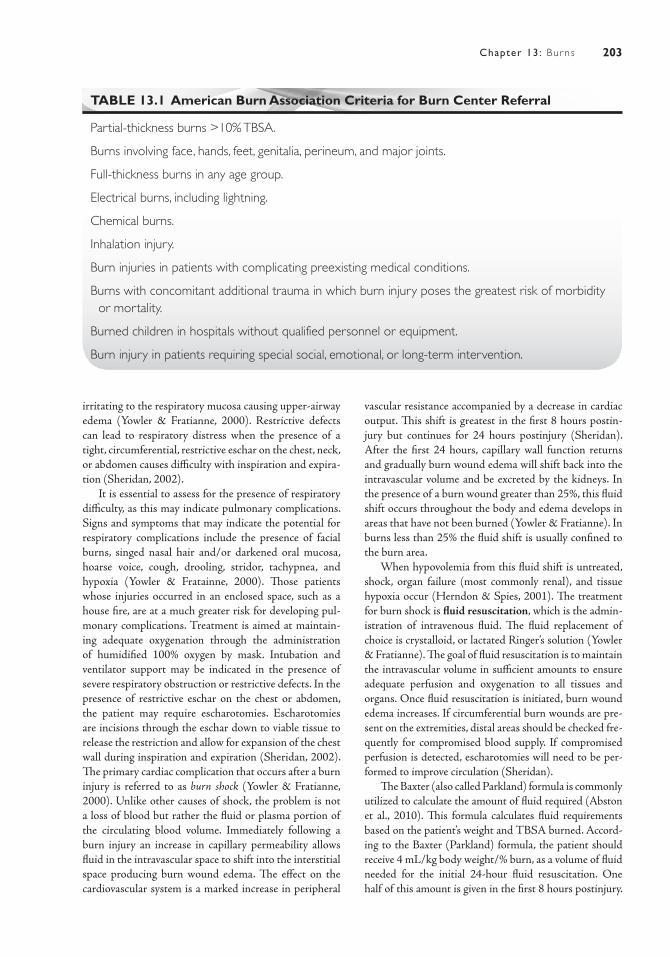
203Chapter 13: Burns
irritating to the respiratory mucosa causing upper-airway edema (Yowler & Fratianne, 2000). Restrictive defects can lead to respiratory distress when the presence of a tight, circumferential, restrictive eschar on the chest, neck, or abdomen causes difficulty with inspiration and expira-tion (Sheridan, 2002).
It is essential to assess for the presence of respiratory difficulty, as this may indicate pulmonary complications. Signs and symptoms that may indicate the potential for respiratory complications include the presence of facial burns, singed nasal hair and/or darkened oral mucosa, hoarse voice, cough, drooling, stridor, tachypnea, and hypoxia (Yowler & Fratainne, 2000). Those patients whose injuries occurred in an enclosed space, such as a house fire, are at a much greater risk for developing pul-monary complications. Treatment is aimed at maintain-ing adequate oxygenation through the administration of humidified 100% oxygen by mask. Intubation and ventilator support may be indicated in the presence of severe respiratory obstruction or restrictive defects. In the presence of restrictive eschar on the chest or abdomen, the patient may require escharotomies. Escharotomies are incisions through the eschar down to viable tissue to release the restriction and allow for expansion of the chest wall during inspiration and expiration (Sheridan, 2002). The primary cardiac complication that occurs after a burn injury is referred to as burn shock (Yowler & Fratianne, 2000). Unlike other causes of shock, the problem is not a loss of blood but rather the fluid or plasma portion of the circulating blood volume. Immediately following a burn injury an increase in capillary permeability allows fluid in the intravascular space to shift into the interstitial space producing burn wound edema. The effect on the cardiovascular system is a marked increase in peripheral
TABLE 13.1 American Burn Association Criteria for Burn Center Referral
Partial-thickness burns >10% TBSA.
Burns involving face, hands, feet, genitalia, perineum, and major joints.
Full-thickness burns in any age group.
Electrical burns, including lightning.
Chemical burns.
Inhalation injury.
Burn injuries in patients with complicating preexisting medical conditions.
Burns with concomitant additional trauma in which burn injury poses the greatest risk of morbidity or mortality.
Burned children in hospitals without qualified personnel or equipment.
Burn injury in patients requiring special social, emotional, or long-term intervention.
vascular resistance accompanied by a decrease in cardiac output. This shift is greatest in the first 8 hours postin-jury but continues for 24 hours postinjury (Sheridan). After the first 24 hours, capillary wall function returns and gradually burn wound edema will shift back into the intravascular volume and be excreted by the kidneys. In the presence of a burn wound greater than 25%, this fluid shift occurs throughout the body and edema develops in areas that have not been burned (Yowler & Fratianne). In burns less than 25% the fluid shift is usually confined to the burn area.
When hypovolemia from this fluid shift is untreated, shock, organ failure (most commonly renal), and tissue hypoxia occur ( Herndon & Spies, 2001). The treatment for burn shock is fluid resuscitation, which is the admin-istration of intravenous fluid. The fluid replacement of choice is crystalloid, or lactated Ringer’s solution (Yowler & Fratianne). The goal of fluid resuscitation is to maintain the intravascular volume in sufficient amounts to ensure adequate perfusion and oxygenation to all tissues and organs. Once fluid resuscitation is initiated, burn wound edema increases. If circumferential burn wounds are pre-sent on the extremities, distal areas should be checked fre-quently for compromised blood supply. If compromised perfusion is detected, escharotomies will need to be per-formed to improve circulation (Sheridan).
The Baxter (also called Parkland) formula is commonly utilized to calculate the amount of fluid required (Abston et al., 2010). This formula calculates fluid requirements based on the patient’s weight and TBSA burned. Accord-ing to the Baxter (Parkland) formula, the patient should receive 4 mL/kg body weight/% burn, as a volume of fluid needed for the initial 24-hour fluid resuscitation. One half of this amount is given in the first 8 hours postinjury.
204 C O N D I T I O N S I N O C C U P A T I O N A L T H E R A P Y
2003). The use of alternative therapies such as guided imagery, relaxation techniques, and music along with pharmaceuticals may further decrease the pain response during burn wound dressing changes.
Hydrotherapy (tub bath) is the optimal place for burn wound cleansing as the jets help loosen nonviable tissue and facilitate ROM exercises. If a patient’s condition is unstable, burn wound dressing changes are done in bed. A mild soap, soft washcloth, tweezers, and scissors are utilized to aid in debridement. To decrease the potential for hypothermia, time in the bath is limited and the temperature of the room should be kept at 82.4°F to 87.8°F (28°C to 31°C) (Hern-don & Spies, 2001). Once the wound has been cleansed, an antimicrobial agent and outer bandages are applied. The burn dressings act as a barrier to the environment, decrease temperature loss through the wound and promote comfort (Konigova, Matouskova, & Broz, 2000). Dressing changes are usually only done once a day. If outer bandages become saturated with drainage, however, it is necessary to replace them with dry outer bandages to prevent the wicking of bacteria down to the surface of the wound.
Topical antimicrobial agents are used to delay and/or minimize burn wound colonization (Murphy, Lee, & Herndon, 2003). The topical antimicrobial agent of choice is silver sulfadiazine 1% (Silvadene). Silvadene is a broad-spectrum antimicrobial agent effective against gram-positive and gram-negative bacteria with some anti-fungal activity. Silvadene does not penetrate burn eschar. It controls bacterial growth only on the surface of the wound, has few side effects, and is usually well tolerated by the patient. When there is a need to penetrate the eschar, such as on the ears to prevent chondritis, 10% mafenide (Sulfamylon) is used (Palmieri & Greenhalgh, 2002). Sulfamylon is effective against a broad range of micro-organisms but does not treat yeasts. This drug may pro-duce metabolic acidosis and application is painful, so the use of Sulfamylon is limited to small areas (Murphy, Lee, & Herndon). The use of intravenous antibiotic therapy is reserved for documented wound infections and is not given prophylactically (Palmeiri & Greenhalgh).
In full-thickness injuries, the risk of bacterial entrance and fluid/heat loss through the wound continues until the wound is closed either temporarily or permanently through the application of synthetic dressings (Biobrane; Integra) or biologic coverage (Konigova, Matouskova, & Broz, 2000). Grafting priorities are influenced by the location and size of the burn. If the patient requires long-term intravenous fluid administration, skin grafting may be needed on the chest to allow for insertion of a central line. Hands, because of their functional importance, are given grafting priority. Faces and ears have a dense cross section of dermal appendages and are given additional time to ascertain if healing will occur without surgical intervention (Kao & Garner, 2000).
The remaining half is given over the next 16 hours. Ade-quate assessment of the effectiveness of fluid resuscitation is important, as excessive fluids can cause increased burn wound edema and compromised local blood supply. Inade-quate fluid resuscitation can lead to hypovolemia and renal failure. Monitoring hourly urine output is a reliable guide to evaluate the effectiveness of fluid resuscitation. The use of the Baxter (Parkland) formula as a guide to predict fluid requirements has been proven effective but continu-ous monitoring of the patient is needed to insure adequate treatment of burn shock (Abston et al., 2010; Cartotto et al., 2002; Inoue, Okabayashi, & Ohtani, 2002).
A large burn injury triggers a significant and pro-longed stress response in the body and initiates the release of stress hormones such as catecholamines, prostaglandins, glucagon, and cortisol (Murphy, Lee, & Herndon, 2003). The release of these hormones initi-ates and mediates a hypermetabolic response in the body. This hypermetabolic state will result in increased energy catabolism, skeletal muscle catabolism, immune deficien-cies, peripheral lipolysis, and reduced bone mineraliza-tion and growth. Nutritional support is needed to meet the resulting increase in basal energy expenditure. Most patients cannot consume enough calories through eat-ing and require nutritional support via the enteral route (nasogastric feedings). Early initiation of nutritional sup-port via enteral feedings will help reduce the risk of bac-terial translocation from the gut and the development of sepsis (Kao & Garner, 2000).
Burn Wound Management
Eschar facilitates bacterial access and acts as the common denominator for burn sepsis. After the emergency phase of burn treatment, the patient enters the acute phase. In the acute phase of treatment, the most common cause of death is sepsis. Until the burn wound is healed, the patient remains vulnerable to invasive infection. Debride-ment is the cleansing and removal of nonadherent and nonviable tissue (Palmieri & Greenhalgh, 2002). Daily cleansing and debridement of the burn wound is neces-sary to decrease the potential for burn wound sepsis, to facilitate healing, and to prepare the wound for grafting if this procedure is needed to achieve wound closure.
Debridement is a painful procedure, and it is impor-tant to make sure the patient has been premedicated with analgesics and sedative medication prior to starting the dressing change. Commonly used analgesics include morphine, fentanyl, or codeine. A common drug given to sedate the patient is ketamine. Anxiolytics, such as diaz-epam or midazolam, are drugs given to control anxiety. Anxiety influences pain perception. The use of anxiolyt-ics is beneficial in reducing anticipatory anxiety regard-ing future dressing changes (Murphy, Lee, & Herndon,
205Chapter 13: Burns
immobilized in a functional position and remains in that position until the first dressing change. If grafts are placed on the chest or back, the bandages are sutured to the body to decrease the risk of shearing when repositioning the patient. Extremities are elevated to prevent/minimize edema formation. The timing of the first dressing change is controversial, and each physician will have his or her own preference. ROM to the grafted area is avoided until the graft is stable which is usually about 4 to 5 days after surgery.
Healing begins immediately after the burn injury so it is important to consider and initiate rehabilitation activities upon admission. While the scope of this book is not focused on specific rehabilitation approaches and methods, an overview is provided here with the expecta-tion that the reader will be studying specific procedures in a course designed for that purpose. Rehabilitation needs are influenced by the phase of treatment the patient is experiencing. During the emergency phase and fluid resuscitation, edema formation is to be expected and may be profound. The emphasis should be placed on pres-ervation of joint function and maintaining appropriate positioning of the affected area ( Jordan, Daher, & Wasil, 2000). ROM exercises help to reduce edema and main-tain joint mobility. ROM exercises should be preformed during the dressing change and throughout the day when the patient is awake (Celis, et al., 2003). Timing ROM exercises to occur when the patient has been medicated for pain will help patients tolerate the discomfort associ-ated with this activity (Sheridan, 2002).
Splints are constructed to prevent the formation of contracture deformities and should be worn when the patient is asleep or resting ( Jordan, Daher, & Wasil, 2000). In the acute phase of treatment, reconditioning exercises are started and ROM exercises and splinting are continued. Once the patient’s condition is stable, ambula-tion is initiated and patients are encouraged to participate in activities of daily living (ADL). Assistive devices may be needed to help patients participate in ADL.
When burn wounds have been grafted/healed, the patient enters the rehabilitation phase of treatment. The focus of rehabilitation includes reconditioning, ROM, scar revision, contracture release, and reconstruction. Healed burn scars are initially fragile, dry, itchy, and sus-ceptible to sunburns (Endorf & Arbabi, 2008). Destruc-tion of sebaceous glands in partial- and full-thickness injuries cause dry skin and itching. It is important that patients receive proper education regarding how to care for their burn scars. Use of an unscented soap and appli-cation of a moisturizing lotion several times a day will help lubricate the skin and reduce itching. Antihista-mines (such as Benadryl) can be taken orally to control itching and promote comfort. Application of a sunscreen is important if burn scars are exposed to the sun.
If the patient does not have available donor sites, the burn wound can be excised down to viable tissue and temporarily closed through the application of a synthetic dressing such as Biobrane or a biologic coverage such as an allograft. An allograft (also referred to as a homograft or cadaver skin) is donor skin taken from another person. The body, through cell-mediated immunity, will reject an allograft usually within 10 to 14 days after application (eMedicine, 2006; Kao & Garner, 2000). These tempo-rary means of wound closure provide the time needed to achieve a permanent method for closing the wound.
The only way to achieve permanent wound closure in large full-thickness burn injuries is through surgical intervention and the application of either an autograft or cultured epithelium. An autograft is the surgical transplan-tation of the patient’s own skin from one area to another. A split-thickness skin autograft is the standard method of achieving wound closure (Shakespeare, 2001). Donor skin is taken from areas of unburned skin. The harvested skin is 0.008 to 0.012 inches in thickness. This leaves a wound, referred to as a donor site, that takes about 7 to 10 days to heal. Once healed, donor sites can be reharvested but will take longer to heal. Burn eschar is excised down to viable tissue, and bleeding is controlled prior to placement of the skin graft. A split-thickness skin autograft can be applied as a mesh graft or a sheet graft. A mesh graft is a graft that has small holes placed evenly throughout the graft, which allows it to be expanded (eMedicine, 2006). Use of a mesh graft allows more area to be covered than the actual size of the donor skin taken. Unfortunately this results in an unsightly “cobblestone” appearance in the scar (eMedicine). Because of this, mesh grafts are avoided in areas where there are concerns about the appearance of the scar, such as on the face or hands. Split-thickness grafts can be applied as a sheet graft in areas with aesthetic concerns. A sheet graft is a graft in which the donor skin has been laid intact over the area to be grafted. While sheet grafts are preferred because of cosmetic reasons, limited availability of donor sites in large burn injuries limits their use.
In large burns with limited availability of donor sites, cultured epithelium can be used to achieve wound clo-sure. A biopsy of unburned skin is taken and sent to a laboratory that can grow cultured epithelium. It takes 3 to 4 weeks for cultured epithelium to be available for graft-ing. The resulting grafts are extremely fragile and sensi-tive to infection. Because of the expense of this form of treatment and the fragile nature of the graft, the use of cultured epithelium is controversial and is reserved for patients with massive burn injury (Kao & Garner, 2000).
Immediately after grafting, with either an autograft or cultured epithelium, the graft is fragile and susceptible to loss. Factors that can cause graft failure include shear-ing/motion, hematoma formation, and infection (Kao & Garner, 2000). To prevent loss of graft, the grafted area is

206 C O N D I T I O N S I N O C C U P A T I O N A L T H E R A P Y
the ability to perform ADLs. Mature burn scars do not have the capacity to stretch that is found in normal skin. Children with large full-thickness injuries may develop burn scar contractures years after their initial burn injury as they literally grow out of their scar.
Immature burn scars have a greater capacity to stretch but are prone to the development of contracture deformi-ties. Prevention of burn scar contracture through exercise, positioning, and splinting is important until the burn scar is mature. It is essential for practitioners, patients, and family members to realize that position of comfort often results in contracture formation. A prime example of this is the use of a pillow during sleep; while more comfort-able, this action may facilitate the development of neck contractures. Simple acts such as having the bed flat when sleeping or avoiding the use of straws when drinking cause the patient to have to assume a position that pro-vides a stress tension on the scar and help to prevent con-tracture deformities. ROM exercises help reduce the risk of contracture formation. An individualized exercise plan should be developed that meets the needs of the patient. It is important to involve both the patient and his or her family members in the development and execution of this plan to increase the likelihood that it will be followed. Incorporating exercises into activities that a patient likes to do may increase the likelihood that the patient will adhere to an exercise regimen. Engaging a child in play that involves large and/or fine motor groups is a great way for a child to perform ROM exercises. The use of splints at night will aid in maintaining the stretch achieved dur-ing the day through ROM exercises. Even with a patient’s best efforts, contracture deformities may develop that will require surgical intervention to release.
IMPACT ON OCCUPATIONAL PERFORMANCEA burn injury is a devastating injury that has far-reach-ing consequences that continue even after the wound is healed. The impact of a burn injury on an individual’s occupational performance is influenced by the size, loca-tion, and depth of the burn injury. A burn injury may impact an individual’s ability to perform basic ADLs, work activities, or play/leisure activities. Burn injuries that have the greatest potential to impact occupational performance include deep partial-thickness or full-thick-ness burns, burns involving major joints, and larger burn injuries. It is critical to follow up with the patient on a routine basis after discharge to identify and initiate early intervention if significant hypertrophic scar formation and contracture deformities are detected that are impact-ing the patient’s occupational performance.
It is important to recognize and assist patients and families in dealing with the psychological and psychosocial
Burn scars are prone to hypertrophic scar forma-tion. Hypertrophic scars are scars in which the tissues are enlarged above the surrounding skin and typically present as red, raised, and rigid (Puzey, 2001). Hypertrophic scars are differentiated from keloid scar formation by the fact that they remain within the boundary of the origi-nal wound and will eventually fade in color, flatten, and become more pliable as they mature. Hypertrophic scar formation is more common in children, people of color, and over areas involved in stretch or motion (Kao & Gar-ner, 2000). Hypertrophic scar formation is frequently seen in deep dermal wounds that heal spontaneously but take a longer time to heal (Sheridan, 2002). The exact mechanism of hypertrophic scar formation is unclear. Excessive deposition of tissue collagen appears to play a significant role, leading to a thickening of the scar (Atiyeh, Ioannovich, & Al-Amm, 2002). Collagen is a basic struc-tural fibrous protein found in all tissue. There is no single effective method to prevent hypertrophic scar formation.
Methods that may help control hypertrophic scar formation include the use of compression garments, scar massage, topical silicone, steroid injection, and surgery (Sheridan, 2002). Compression garments are the pre-ferred conservative method to treat hypertrophic scars and have been in use since the early 1970s. Compression garments are thought to reduce oxygen flow to the scar thereby decreasing collagen production (Engrav et al., 2010). Shortly after the burn wound is healed, measure-ments are taken and compression garments are custom-made for each patient. Compression garments are fit for all areas of partial- and full-thickness injuries and apply a pressure of 25 mm Hg to the scar. The garments need to be worn 23 hours a day; they should be removed only for bathing and the application of a moisturizing lotion.
It is important to verify that compression garments fit properly as excessive pressure may contribute to skin breakdown. Children who are growing and adults who have a significant weight gain after discharge from the hospital may need to be refitted for new compression gar-ments. Compression garments will need to be worn until burn scars are mature which can take from 1 to 2 years depending upon the individual. Mature burn scars are softer, more pliable, and are no longer hyperemic in color; their color is similar to the surrounding unburned skin. Once burn scars are mature, nothing can be gained by the continued use of compression garments.
Wound healing involves three processes: epitheli-alization, connective tissue deposition, and contraction. Wound contraction is an active process generated by fibro-blasts and myofibroblasts and is one of the most powerful mechanical forces in the body. Burn scar contracture is the shortening and tightening of the burn scar. Burn scar contracture deformities are most problematic over large joints. They can severely limit ROM and interfere with
207Chapter 13: Burns
another burn survivor and their families will help prepare them for the challenges ahead and assist them to deal with their emotions. This preparation will enable the family to provide the patient both the support and independence they will need to achieve the best outcome.
Additionally, children can benefit from school reentry and summer burn camp programs. School reentry pro-grams prepare classmates on what to expect and help in the transition back to school. Summer camp for burn chil-dren can also help to improve self-esteem and allow them to realize that they can overcome the difficulties they face.
impact of a major burn injury. Burn support groups can be helpful in assisting patients and families in dealing with the lifelong disfigurement and dysfunction that may result from a major burn injury. Having patients meet with a burn survivor can help them realize what challenges they may face and ways to overcome these challenges. Family members may experience many emotions such as fear, guilt, or sadness. If these emotions are not addressed, family members may feel the need to “take care of their loved one” and to do things for the patient that they need to do for themselves. Having family members meet with
Case IllustrationsCase 1L.S., a 5-year-old, is playing outside when her father arrives home. He is having difficulty with his car, which is overheating. Unaware that L.S. has come over to watch him work on the car he removes the radiator cap. Hot radiator fluid and steam strikes L.S. and she sustains a 32% mixed partial- and full-thickness burn injury to her face, neck, chest, and scattered area on bilateral arms.
During the emergency phase she required fluid resuscitation and intubation to maintain her airway during the initial fluid shift and resulting swelling. She was extubated on postburn day 3 and has had no further problems with her airway. Three weeks have passed and L.S. has been to surgery twice. During the first surgery, mesh grafts were placed on her chest and arms. During the last surgical procedure, a sheet graft was placed on her neck. Her grafts are intact and heal-ing well, but scattered open areas remain at the mar-gins of the grafts. L.S. has donor sites on her bilateral thighs and buttock, which are healing without difficulty.
Neuromuscular components of ROM, strength, and soft-tissue integrity have all been affected by the burn injury L.S. sustained. She has participated in ROM exercises since she was extubated. ROM exercises were halted for a few days, after the grafting proce-dures, to facilitate adherence of the graft. Currently, L.S.
has some restriction of ROM to bilateral axillary areas and has been fitted with an airplane splint to be worn when sleeping. This splint will maintain a 90-degree abduction to the axillary area. While she currently does not demonstrate any restriction of neck ROM, she is at great risk for developing contractures in this area. Her parents have been instructed to not allow L.S. to sleep with a pillow and to avoid the use of straws when drinking to promote adequate stretch on the neck. L.S. has been fitted for her compression garments, which should arrive in the next 2 weeks.
ADLs and play or leisure activities are the occu-pational performance areas that have been affected. Currently, L.S. has few limitations on ROM to most joints, but because of the discomfort experienced during ROM she is hesitant to move. Her parents have been instructed to apply lotion twice daily to facilitate stretching of the scar during ROM activities. Both parents express a great deal of difficulty with the discomfort that L.S. has experienced during her treat-ment and are hesitant to make her do things that may cause discomfort. L.S.’s father expresses a lot of guilt surrounding the circumstances that caused the burn injury. Arrangements are made for parents to meet with another burn survivor and the survivor’s family prior to discharge.
R E C O M M E N D E D L E A R N I N G R E S O U R C E S
American Burn Association625 North Michigan Ave, Suite 2550Chicago, IL 60611Tel: 312-642-9260Toll free: 800-548-2876Fax: 312-642-9130www.ameriburn.org
Shriners Burn Institutes2900 Rocky Point DrTampa, FL 33607-1460Tel: 813-281-0300www.shrinershq.org/Hospitals/BurnInst/The Phoenix Society for Burn Survivors, Inc.1835 R W Berends Dr SW
208 C O N D I T I O N S I N O C C U P A T I O N A L T H E R A P Y
Grand Rapids, MI 49519-4955Tel: 800-888-2876Fax: 616-458-2831www.phoenix-society.orgBurn Summer Camps for Kids(Information available at Kids Camps)2500 N. Military Trail Suite 450
Boca Raton, FL 33431Tel: 877-242-9330Fax: 866-665-2904www.kidscamps.com/special_needs/burn.htmlTotal Burn Carewww.Totalburncare.com Associated online support at:
www.totalburncare.com
Abston S., Blakeney P., Desai M., Edgar P., Heggers, J., Herndon, D., Hildreth M., Nichols R.J. (2010). Burn care: Resident orientation manual. Galvesoton Shri-ners Burn Hospital and University of Texas Medical Branch Blocker Burn Unit.
American Burn Association. (2010). National Burn Repository: report of data from 2000–2009.
Atiyehm, B., Ioannovich, J., Al-Amm, C., El-Musa K., & Dham, R. (2002). Improving scar quality: A prospec-tive clinical study. Aesthetic Plastic Surgery, 26, 470–476.
Cartotto, R., Innes, M., Musgrave, M., Gomez, M., & Coope, A. (2002). How well does the Parkland formula estimate actual fluid resuscitation volumes. Journal of Burn Care Rehabilitation, 23, 258–265.
Celis, M., Suman, O., Huang, T., Yen P., & Herndon, D. (2003). Effect of a supervised exercise and physiotherapy program on surgical interventions in children with ther-mal injury. Journal of Burn Care Rehabilitation, 24, 57–61.
Endorf, F., & Arbabi, S. (2008). Burn Injury. In L. Flint, J. Meredith, C. Schwab, D. Trunkey & L. Rue, (Eds.) Trauma: Contemporary Principles and Therapy. Lip-pincott Williams & Wilkins: Phildadelphia.
eMedicine “Skin, Grafts,” (February 2006) [Online]. Available http://www.emedicine.com/plastic/ topic 392.htm [Retreived January 13, 2011]
Engrav, L., Heimbach, D., Rivara, F., Moore, M., Wang, J., Carrougher, G., Costa, B., Numhom, S., Calderon, J., Gi-bran, N. (2010). 12-year within-wound study of custom pressure garment efficacy. Burns 36(7): 975–983, 2010.
Engrav, L. H., Colescott P. L., Kemalyan, N., et al. (2001). A biopsy of the use of the Baxter formula to resuscitate burns or do we do it like Charlie did it. Journal of Burn Care Rehabilliltation, 21, 91–95.
Flint, L., Meredith, W., Schwab, W., Trunkey, D., Rue, L & Taheri, P.(Eds). (2008). Trauma: Contempo-rary principles and therapy.Baltimore: Lippincott, Williams & Wilkins.
Herndon, D. & Spies, M. (2001). Modern burn care. Seminars in Pediatric Surgery, 10, 28–31.
R E F E R E N C E S
Inoue, T., Okabayashi, K., & Ohtani, M. (2002). Circu-lating blood volume in burn resuscitation. Hiroshima Journal of Medical Science, 51, 7–13.
Jordan, R., Daher, J., & Wasil, K. (2000). Splints and scar management for acute and reconstructive burn care. Clinical Plastic Surgery, 27, 71–85.
Kao, C., & Garner, W. (2000). Acute burns. Plastic Recon-struction Surgery, 101, 2482–2492.
Konigova, R., Matouskova, E., & Broz, L. (2000). Burn wound coverage and burn wound closure. Acta Chirurgiae Plasticae, 42, 64–68.
McGrath, J. & Uitto, J. (2010). Anatomy and organiza-tion of human skin. In T. Burns, C. Breathnac, N. Cox, & C. Griffiths (Eds.), Rooks textbook of dermatol-ogy (8th ed., Vol. 1). Oxford, UK: Wiley-Blackwell.
Migliore, S. (2008). Rehabilitation of the child with burns. In J. S. Tacklin (Ed.) Pediatric physical therapy (4th ed.). Baltimore, MD: Lippincott Williams & Wilkins.
Morgan, E., Bledsoe, S., & Barker, J. (2000). Ambulatory management of burns. American Family Physician, 62, 2015–2026.
Muller, M., Pegg, S., & Rule, M. (2001). Determinants of death following burn injury. British Journal of Surgery, 88, 583–587.
Murphy, K., Lee, J., & Herndon, D. (2003). Current phar-macotherapy for the treatment of severe burns. Expert Opinion on Pharmacotherapy, 4, 369–384.
Palmieri, T., & Greenhalgh, D. (2002). Topical treatment of pediatric patients with burns. American Journal of Clinical Dermatology, 3, 529–534.
Puzey, G. (2001). The use of pressure garments on hypertrophic scars. Journal of Tissue Viability, 12, 11–15.
Shakespeare, P. (2001). Burn wound healing and skin substitutes. Burns, 27, 517–522.
Sheridan, R. (2002). Burns. Critical Care Medicine, 30, S500–S514.
Yowler, C., & Fratianne, R. (2000). Current status of burn resuscitation. Clinical Plastic Surgery, 27, 1–10.

209
Progressive Neurological Disorders
■ D i an e K . D ire t t e
14
Shortly after the birth of her second child, Joan started getting the sensation of pins and needles in her hands and feet. Within a couple of months, she noticed some numbness and weakness in her arms and legs. She was having difficulty walking for long
distances and began to worry that she might drop her 2-year-old son or even her newborn. When the children napped in the afternoon, she found herself slumped on the couch for a much-needed rest. Convinced that this was just part of her postpartum recovery, Joan did not inform her doctor of this difficulty. However, when she found herself struggling 1 day to focus on the words of her son’s bedtime story, she decided to seek medical advice.
Over time, Joan found out she had a progressive neurologic disorder (PND) called multiple sclerosis (MS). PNDs are a group of diseases that affect various areas of the central nervous system (CNS), are chronic in
nature, and cause a deterioration of function over time. This chapter discusses three of the most common PNDs: MS, Parkinson’s disease (PD), and amyotrophic lateral sclerosis (ALS).
K E Y T E R M S
AtrophyBrainstem
CorticobulbarCorticospinalDemylination
DopamineElectroencephalogram
Lou Gehrig’s DiseaseLower Motor Neuron
NeurodegenerativeSubstantial Nigra
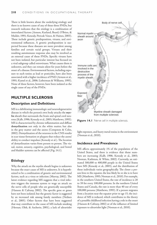
210 C O N D I T I O N S I N O C C U P A T I O N A L T H E R A P Y
There is little known about the underlying etiology and there is no known cause of any of these three PNDs, but research indicates that the etiology is a combination of interrelated factors (Armon, Kurland, Beard, O’Brien, & Mulder, 1991; Kenealy, Pericak-Vance, & Haines, 2003). These include genetic predisposition, viruses, and envi-ronmental influences. A genetic predisposition is sus-pected because these diseases are more prevalent among families and certain racial groups. Viruses and their resulting autoimmune response also may be involved as an external cause of these PNDs. Specific viruses have not been isolated, but particular interest has focused on a viral subgroup called retroviruses. What causes them is unknown, and they can remain silent for years before the onset of a disease. Environmental factors, including expo-sure to such toxins as lead or pesticides, have also been associated with a higher incidence of PND (Armon et al., 1991; Kamel et al., 2006; Lieberman & Williams, 1993). None of these factors, however, have been isolated as the single cause of any of the PNDs.
MULTIPLE SCLEROSIS
Description and Definitions
MS is a debilitating immunologic and neurodegenerative disease in which the person’s own body attacks the mye-lin sheath that surrounds the brain and spinal cord neu-rons (Kalb, 1996; Kenealy et al., 2003; Matthews, 1993). MS is characterized by chronic inflammation and diffuse demyelination not only in the white matter, but also in the grey matter and the axons (Compston & Coles, 2002). Demyelination of the neurons in the CNS results in scar tissue formation or plaques that reduce the axons’ ability to conduct impulses (Kenealy et al.). The location of demyelination varies from person to person. The vis-ual, motor, sensory, cognitive, psychological, and bowel and bladder systems can be affected (Fig. 14.1).
Etiology
Why the attack on the myelin sheath begins is unknown because the exact cause of MS is unknown. It is hypoth-esized to be a combination of genetic and environmental factors, such as a virus or infection (Murray, 2002). The latest evidence regarding MS suggests that a viral infec-tion triggers the immune system to wage an attack on the nerve cells of people who are genetically susceptible (Hanson & Cafruny, 2002). The specific gene or genes have not been isolated, but the genetic factor is suggested by an increased risk among family members (Neilsen et al., 2005). Other factors that have been suggested that may contribute to the cause of MS include smoking (Hernan, Olek, & Ascherio, 2001), a lack of ultraviolet
Body of nerve cell
AxonNormal myelinsheath aroundaxon
Immune cells areinvolved in thedestructiveprocess of themyelin sheath
Exposedfiber
Myeline sheath damagedfrom multiple sclerosis
Nerve
cel
l affe
cted
by
MS
Normal nerve cell
Figure 14.1 Nerve cell in multiple sclerosis.
light exposure, and heavy metal toxins in the environment (Noonan et al., 2010).
Incidence and Prevalence
MS affects approximately 1% of the population of the United States, and there is evidence that those num-bers are increasing (Kalb, 1996; Kenealy et al., 2003; Noonan, Kathman, & White, 2002). Currently, an esti-mated 300,000 to 400,000 people in the United States have MS (Kenealy et al., 2003), and the distribution of these individuals varies geographically. The closer a per-son lives to the equator, the less likely he or she is to have MS (Matthews, 1993; Noonan et al., 2010). For example, in the southern United States, the rate of incidence is 20 to 39 for every 100,000 persons. In the northern United States and Canada, the rate is more than 40 out of every 100,000 persons (Matthews, 1993). If a person migrates from a location near the equator prior to age 15, the inci-dence of MS is reduced, which contributes to the theory of a possible childhood infection having a role in the onset (Hanson & Cafruny, 2002) or of the influence of limited exposure to ultraviolet light (Noonan et al., 2010).
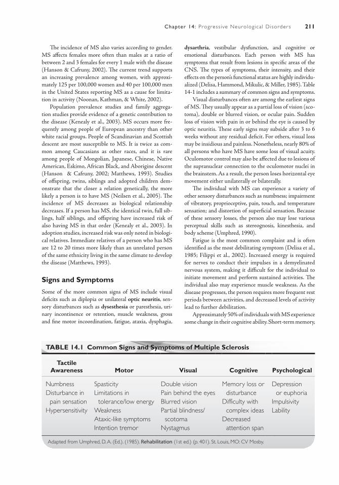
211Chapter 14: Progress ive Neurolog ica l Disorder s
dysarthria, vestibular dysfunction, and cognitive or emotional disturbances. Each person with MS has symptoms that result from lesions in specific areas of the CNS. The types of symptoms, their intensity, and their effects on the person’s functional status are highly individu-alized (Delisa, Hammond, Mikulic, & Miller, 1985). Table 14-1 includes a summary of common signs and symptoms.
Visual disturbances often are among the earliest signs of MS. They usually appear as a partial loss of vision (sco-toma), double or blurred vision, or ocular pain. Sudden loss of vision with pain in or behind the eye is caused by optic neuritis. These early signs may subside after 3 to 6 weeks without any residual deficit. For others, visual loss may be insidious and painless. Nonetheless, nearly 80% of all persons who have MS have some loss of visual acuity. Oculomotor control may also be affected due to lesions of the supranuclear connection to the oculomotor nuclei in the brainstem. As a result, the person loses horizontal eye movement either unilaterally or bilaterally.
The individual with MS can experience a variety of other sensory disturbances such as numbness; impairment of vibratory, proprioceptive, pain, touch, and temperature sensation; and distortion of superficial sensation. Because of these sensory losses, the person also may lose various perceptual skills such as stereognosis, kinesthesia, and body scheme (Umphred, 1990).
Fatigue is the most common complaint and is often identified as the most debilitating symptom (Delisa et al., 1985; Filippi et al., 2002). Increased energy is required for nerves to conduct their impulses in a demyelinated nervous system, making it difficult for the individual to initiate movement and perform sustained activities. The individual also may experience muscle weakness. As the disease progresses, the person requires more frequent rest periods between activities, and decreased levels of activity lead to further debilitation.
Approximately 50% of individuals with MS experience some change in their cognitive ability. Short-term memory,
The incidence of MS also varies according to gender. MS affects females more often than males at a ratio of between 2 and 3 females for every 1 male with the disease (Hanson & Cafruny, 2002). The current trend supports an increasing prevalence among women, with approxi-mately 125 per 100,000 women and 40 per 100,000 men in the United States reporting MS as a cause for limita-tion in activity (Noonan, Kathman, & White, 2002).
Population prevalence studies and family aggrega-tion studies provide evidence of a genetic contribution to the disease (Kenealy et al., 2003). MS occurs more fre-quently among people of European ancestry than other white racial groups. People of Scandinavian and Scottish descent are most susceptible to MS. It is twice as com-mon among Caucasians as other races, and it is rare among people of Mongolian, Japanese, Chinese, Native American, Eskimo, African Black, and Aborigine descent (Hanson & Cafruny, 2002; Matthews, 1993). Studies of offspring, twins, siblings and adopted children dem-onstrate that the closer a relation genetically, the more likely a person is to have MS (Neilsen et al., 2005). The incidence of MS decreases as biological relationship decreases. If a person has MS, the identical twin, full sib-lings, half siblings, and offspring have increased risk of also having MS in that order (Kenealy et al., 2003). In adoption studies, increased risk was only noted in biologi-cal relatives. Immediate relatives of a person who has MS are 12 to 20 times more likely than an unrelated person of the same ethnicity living in the same climate to develop the disease (Matthews, 1993).
Signs and Symptoms
Some of the more common signs of MS include visual deficits such as diplopia or unilateral optic neuritis, sen-sory disturbances such as dysesthesia or paresthesia, uri-nary incontinence or retention, muscle weakness, gross and fine motor incoordination, fatigue, ataxia, dysphagia,
TABLE 14.1 Common Signs and Symptoms of Multiple Sclerosis
Tactile Awareness Motor Visual Cognitive Psychological
NumbnessDisturbance in
pain sensationHypersensitivity
SpasticityLimitations in
tolerance/low energyWeaknessAtaxic-like symptomsIntention tremor
Double visionPain behind the eyesBlurred visionPartial blindness/
scotomaNystagmus
Memory loss or disturbance
Difficulty with complex ideas
Decreased attention span
Depression or euphoria
ImpulsivityLability
Adapted from Umphred, D. A. (Ed.). (1985). Rehabilitation (1st ed.) (p. 401). St. Louis, MO: CV Mosby.
212 C O N D I T I O N S I N O C C U P A T I O N A L T H E R A P Y
Diagnosis
The diagnosis of MS involves excluding other conditions that share the symptomatology of MS such as B12 defi-ciency, AIDS, rheumatoid arthritis, lupus, Sjogrens syn-drome, and Lyme disease (Kenealy et al., 2003). To make a definitive diagnosis of MS, the physician examines the person’s medical history, symptoms reported by the per-son, and signs detected by various tests (Kalb, 1996). These tests include magnetic resonance imaging (MRI) to detect plaques or lesions in two distinct areas of the CNS, neurologic examination, evoked potentials (visual, brainstem auditory, and somatosensory), and spinal tap to assess cerebral spinal fluid proteins.
The results of these diagnostic procedures help con-tribute to a diagnosis of MS, but they do not determine the diagnosis independently. Many other conditions can elicit positive results. The physician and the individual must work together to rule out other causes before a diag-nosis can be made. A definitive diagnosis of MS is made when the person has episodes of exacerbation and remis-sion and slow or step-by-step progression over 6 months. There also must be evidence of lesions in more than one site in the white matter (as determined by MRI) and no other neurologic explanation for the clinical picture (Umphred, 1990).
Medical/Surgical Management
For each of the PNDs discussed in this chapter, surgical intervention is not part of the routine care given. Several medications are used to alleviate the myriad signs caused by each of these diseases. The medications most often prescribed to treat the signs of MS include antispasmod-ics, muscle relaxants, and anticonvulsants. Some current medications include methylprednisolone for functional skills and Prokarin, amantadine, pemoline, and 4-amino-pyridene for fatigue (Bobholz & Rao, 2003; Lyros et al., 2010). Acetylocholinesterase inhibitors such as done-pexil, rivastigmine, and galantamine, which were devel-oped for the treatment of Alzheimer’s disease, have shown some promise for alleviating cognitive symptoms such as attention, information processing, and memory/learning (Bobholz & Rao, 2003; Lyros et al., 2010). Immu-nomodulating drugs such as Interferon A-1b, Interferon B-1b, glatiramer acetate, mitoxantrone, and natalizumab show some potential to provide a reduction in lesions for relapsing-remitting MS via anti-inflammatory effects (Noseworthy, 2003; Lyros et al., 2010). Possible future treatments include antiviral medications, vaccinations, transplantation of schwann cells, cell lines or stem cells, and/or gene therapy (Noseworthy, 2003).
Nonpharmacologic interventions that have been found to alleviate some symptoms of MS include cognitive
attention, processing speed, visuospatial abilities, verbal fluency, and executive functions have all been identified as deficit areas related to MS (Bobholz & Rao, 2003; Lyros, Messinis, Papageorgiou, & Papathanosopoulos, 2010). As with all aspects of MS, there is considerable variability among individuals with the disease depend-ing on the area of the brain affected (Bobholz & Rao, 2003). An emotional component to this disease results in some individuals having bouts of depression, euphoria, or lability caused by lesions in the frontal lobes of the brain (Feinstein, 2006).
Course and Prognosis
MS is the most common PND found in young adults. MS is usually diagnosed between the ages of 20 and 40 years (Matthews, 1993). It is rarely seen in children or diagnosed in adults older than age 50. No two people with MS follow the same course, and each person experi-ences variation in symptoms over time (Kalb, 1996). The clinical course of this disease can be roughly organized into four types or patterns including benign, relapsing-remitting- nonprogressive, relapsing-remitting-progres-sive, and progressive.
The first type of MS is benign, in which the person experiences one or two episodes of neurologic deficits with no residual impairments. This person’s chance of remaining symptom free increases with each nonsymptomatic year. The next pattern of progression is relapsing-remitting-nonprogressive. In this pattern the person returns to the previous level of function after each exacerbation. With the third type, relapsing-remitting-progressive, however, the person has some residual impairment with each remis-sion. The course of this type is unpredictable with varied patterns of exacerbation and remission. Finally, there is the progressive pattern, which involves a steady decline in func-tion without remissions and exacerbations. Individuals with MS may shift from one pattern to another, with no reliable predictors of these shifts (Delisa et al., 1985). Approximately 80% of people have one of the relapsing-remitting forms of MS in which they experience neuro-logic symptoms followed by complete or partial recovery (Hanson & Cafruny, 2002). Approximately 15% experi-ence the progressive pattern of decline in function, without periods of remission of symptoms (Noonan et al., 2010).
Overall, about 60% of individuals with MS remain fully functional up to 10 years after their first exacerba-tion, and about 30% remain functional 30 years after their first attack (Andreoli, Carpenter, Plum, & Smith, 1986). In spite of the seriousness of this disease, it does not signif-icantly decrease the person’s life expectancy. A few people, however, do become severely disabled and die prematurely because of recurring infections or complications resulting from inactivity (Andreoli et al., 1986; Umphred, 1990).
213Chapter 14: Progress ive Neurolog ica l Disorder s
with increased risk of PD (Logroscino, Gao, Chen, Wing, & Ascherio, 2008), increased dairy consumption was associated with increased risk of PD in men (Chen et al., 2007), and higher total blood cholesterol levels was associated with decreased risk of PD in women (de Lau, Koudstaal, Hofman, & Breteler, 2006).
Incidence and Prevalence
PD is the second most common neurodegenerative dis-ease (Zhang, Dawson, & Dawson, 2000). Worldwide the prevalence of PD is estimated at 1% of the popula-tion above the age of 65, with estimates as high as 1 million cases in the United States (Kamel et al., 2007). These cases appear to be evenly distributed throughout the world, based on available diagnoses. Diagnostic data, however, vary from one medical care system to another.
PD has been reported to affect males slightly more than females, but recent studies report an equal distribu-tion among the genders (Duvoisin & Sage, 1996). PD occurs in all races worldwide (Duvoisin & Sage, 1996). However, in the United States, there is a lower incidence among African Americans compared to Caucasians (Lieberman & Williams, 1993).
Signs and Symptoms
The major symptoms of PD are resting tremor, muscle rigidity, bradykinesia, and postural instability (Alba-nese, 2003; Duvoisin & Sage, 1996) (Table 14-2). The most obvious and familiar of these symptoms is tremor, which is usually noted initially in the hand on one side and sometimes in the foot. In the hand, the movement is frequently described as “pill-rolling.” The tremors are usu-ally variable. They disappear when the person is asleep or calmly resting, and they increase under stress or intense mental activity (Duvoisin & Sage, 1996). Infrequent eye blinking is often an early sign of PD followed by progres-sive loss of facial expression.
Secondary symptoms of PD include gait disturbances referred to as a “festinating gait” (short stepped or shuf-fling with reduced arm swing), dexterity and coordination difficulties, involuntary immobilization, micrographia (small handwriting), cognitive impairments (visuospatial, memory, and frontal lobe functions), sensory loss, muf-fled speech, frequent swallowing, poor balance, oculomo-tor impairments, reduced perception of and expression of emotions, sleep disturbances, reduced bowel and bladder function, painful cramping, sexual dysfunction, low blood pressure, seborrhea, depression or anxiety, and fatigue (Breitenstein, Van Lancker, Daum, & Waters, 2001; Carbon & Marie, 2003; Chen, Schernhammer, Schwar-zschild, & Ascherio, 2006; Hutton & Dippel, 1989;
rehabilitation, occupational therapy, psychotherapy, and early nursing education (Bobholz & Rao, 2003; Lyros et al., 2010; Wassem & Dudley, 2003). Low impact exer-cise with a gradual increase in intensity, duration, and fre-quency was also found to be effective for reducing fatigue in adults with MS (Neill, Belan, & Reid, 2006).
PARKINSON’S DISEASE
Description and Definitions
PD is a PND identified by depigmentation of the sub-stantia nigra and the presence of Lewy bodies (Hutton & Dippel, 1989; Zhang, Dawson, & Dawson, 2000). The substantia nigra, which is located in the basil ganglia, pro-duces dopamine, a neurotransmitter, and transports it to the striatum (Fig. 14.2). The decrease in dopamine leads to deficits in the speed and quality of motor movements, postural stability, cognitive skills, and affective expression (DeLong, 2000; Duvoisin & Sage, 1996; Lieberman & Williams, 1993). Neuronal degeneration progresses beyond the substantia nigra and the brainstem and can affect other neurotransmitter systems (Zhang et al., 2000). There are variations in the clinical and pathologi-cal presentation of PD, and there is overlap with other parkinsonian disorders (Albanese, 2003). The differentia-tion of PD from other parkinsonian disorders does not have definitive, validated tests.
Etiology
The underlying cause of PD is unknown although there is some evidence that implicates both genetic and environmental factors (Kamel et al., 2007). Two types of PD include familial PD in which there is a genetic association and the more common type called sporadic PD. Several genetic factors, such as linkages to chro-mosomes 2P and 4P, have been identified in familial PD (Zhang et al., 2000). As many as 50% of people with familial PD have an affected relative. The involve-ment of genetic factors in sporadic PD has not been fully established (Kamel et al., 2007).
The environmental factors that have been associated with PD are dietary intake and exposure to environ-mental elements. Several studies have found that living in rural areas, drinking well water, farming, and expo-sure to certain pesticides increase a person’s risk for PD (Kamel et al., 2007; McCormack et al., 2002; Priyadar-shi, Khuder, Schaub, & Priyadarshi, 2001). Diet may not be a primary cause of PD, but many studies have found an association between diet and the risk for PD, suggest-ing a role in the susceptibility to the disease (Gao et al., 2008). Some examples include exposure to iron sources and intake of too much iron supplement were associated
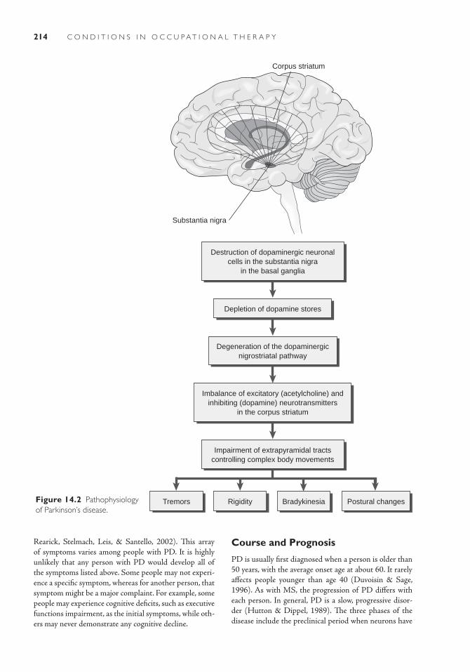
214 C O N D I T I O N S I N O C C U P A T I O N A L T H E R A P Y
Corpus striatum
Substantia nigra
Destruction of dopaminergic neuronalcells in the substantia nigra
in the basal ganglia
Imbalance of excitatory (acetylcholine) andinhibiting (dopamine) neurotransmitters
in the corpus striatum
Degeneration of the dopaminergicnigrostriatal pathway
Impairment of extrapyramidal tractscontrolling complex body movements
Rigidity
Depletion of dopamine stores
Tremors Bradykinesia Postural changesFigure 14.2 Pathophysiology of Parkinson’s disease.
Rearick, Stelmach, Leis, & Santello, 2002). This array of symptoms varies among people with PD. It is highly unlikely that any person with PD would develop all of the symptoms listed above. Some people may not experi-ence a specific symptom, whereas for another person, that symptom might be a major complaint. For example, some people may experience cognitive deficits, such as executive functions impairment, as the initial symptoms, while oth-ers may never demonstrate any cognitive decline.
Course and Prognosis
PD is usually first diagnosed when a person is older than 50 years, with the average onset age at about 60. It rarely affects people younger than age 40 (Duvoisin & Sage, 1996). As with MS, the progression of PD differs with each person. In general, PD is a slow, progressive disor-der (Hutton & Dippel, 1989). The three phases of the disease include the preclinical period when neurons have
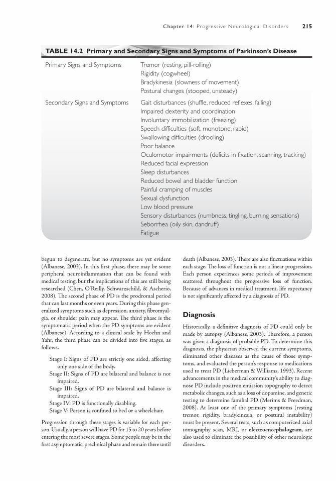
215Chapter 14: Progress ive Neurolog ica l Disorder s
TABLE 14.2 Primary and Secondary Signs and Symptoms of Parkinson’s Disease
Primary Signs and Symptoms Tremor (resting, pill-rolling)Rigidity (cogwheel)Bradykinesia (slowness of movement)Postural changes (stooped, unsteady)
Secondary Signs and Symptoms Gait disturbances (shuffle, reduced reflexes, falling)Impaired dexterity and coordinationInvoluntary immobilization (freezing)Speech difficulties (soft, monotone, rapid)Swallowing difficulties (drooling)Poor balanceOculomotor impairments (deficits in fixation, scanning, tracking)Reduced facial expressionSleep disturbancesReduced bowel and bladder functionPainful cramping of musclesSexual dysfunctionLow blood pressureSensory disturbances (numbness, tingling, burning sensations)Seborrhea (oily skin, dandruff)Fatigue
begun to degenerate, but no symptoms are yet evident (Albanese, 2003). In this first phase, there may be some peripheral neuroinflammation that can be found with medical testing, but the implications of this are still being researched (Chen, O’Reilly, Schwarzschild, & Ascherio, 2008). The second phase of PD is the prodromal period that can last months or even years. During this phase gen-eralized symptoms such as depression, anxiety, fibromyal-gia, or shoulder pain may appear. The third phase is the symptomatic period when the PD symptoms are evident (Albanese). According to a clinical scale by Hoehn and Yahr, the third phase can be divided into five stages, as follows.
Stage I: Signs of PD are strictly one sided, affecting only one side of the body.
Stage II: Signs of PD are bilateral and balance is not impaired.
Stage III: Signs of PD are bilateral and balance is impaired.
Stage IV: PD is functionally disabling.Stage V: Person is confined to bed or a wheelchair.
Progression through these stages is variable for each per-son. Usually, a person will have PD for 15 to 20 years before entering the most severe stages. Some people may be in the first asymptomatic, preclinical phase and remain there until
death (Albanese, 2003). There are also fluctuations within each stage. The loss of function is not a linear progression. Each person experiences some periods of improvement scattered throughout the progressive loss of function. Because of advances in medical treatment, life expectancy is not significantly affected by a diagnosis of PD.
Diagnosis
Historically, a definitive diagnosis of PD could only be made by autopsy (Albanese, 2003). Therefore, a person was given a diagnosis of probable PD. To determine this diagnosis, the physician observed the current symptoms, eliminated other diseases as the cause of those symp-toms, and evaluated the person’s response to medications used to treat PD (Lieberman & Williams, 1993). Recent advancements in the medical community’s ability to diag-nose PD include positron emission topography to detect metabolic changes, such as a loss of dopamine, and genetic testing to determine familial PD (Merims & Freedman, 2008). At least one of the primary symptoms (resting tremor, rigidity, bradykinesia, or postural instability) must be present. Several tests, such as computerized axial tomography scan, MRI, or electroencephalogram, are also used to eliminate the possibility of other neurologic disorders.
216 C O N D I T I O N S I N O C C U P A T I O N A L T H E R A P Y
nuclei of the brainstem, and the anterior horn cells of the spinal cord (Beresford; Przedborski, Mitsumoto, & Rowland, 2003; Weydt, Weiss, Moller, & Carter, 2002). Degeneration of motor neurons leads to progressive atro-phy of muscles, usually beginning with the loss of strength in the small muscles of the hands or feet (Beresford; Maloney, Burks, & Ringel, 1985). When the motor neurons are affected, the reflexes can become hyperactive. Progres-sive loss of muscle movement, spasticity, difficulty speak-ing and swallowing, loss of emotional control, and reduced body temperature regulation are common (Beresford, 1995; Maloney et al., 1985). The actual mechanism of neurode-generation remains a mystery. There is speculation that cytostolic and mitochondrial pathway dysfunction results in the buildup of proteins that lead to motor neuron death (Shaw, Al-Chalabi, & Neigh, 2001). There is also evidence of an inflammatory component in the mechanisms of the disease process (Weydt et al., 2002) (Fig. 14.3).
Etiology
There is no known cause of ALS, but it is speculated that several disorders with several causes lead to this motor neuron disease (Weydt et al., 2002). The two types of ALS, familial and sporadic, may have different causes. Only 5% to 10% of cases are familial (Zoccolella, Santamato, & Lamberti, 2009). Recent advances have discovered a genetic mutation as one cause for familial ALS (Shaw et al., 2001). There is also evidence of genetic causes using familial patterns of susceptibility. Family his-tory of a first- or second-degree relative with ALS is a sig-nificant risk factor for the disease.
In most cases, however, ALS occurs sporadically and is presumed to be acquired. There is no known cause of spo-radic ALS, but there is some speculation regarding viral, ret-roviral, and environmental causes (Shaw et al., 2001). Recent research has identified several risk factors associated with spo-radic ALS, including occupational exposure such as chemi-cals or electromagnetic fields (Weisskopf et al., 2005), lead exposure (Kamel et al., 2003), cigarette smoking (Nelson, McGuire, Longstreth, & Matkin, 2000; Weisskopf et al., 2004), and dietary consumption with increased fat and glu-tamate intake associated with increased risk and fiber intake with reduced risk (Nelson, Matkin, Longstreth, & McGuire, 2000). Some of these risk factors may also be involved in the familial type of ALS with an environmental exposure com-bined with genetic susceptibility (Kamel et al., 2003).
Incidence and Prevalence
The prevalence rate of people with ALS is estimated to be 4 to 7 out of every 100,000 people uniformly world-wide (Przedborski et al., 2003; Weydt et al., 2002), with
Medical/Surgical Management
The medications usually prescribed to treat the symp-toms of PD include dopamine replacement medications, acetylcholine inhibitors, and antiviral compounds. Some of the dopamine replacement medications include carbi-dopa/levodopa, pergolide, bromocriptine, ropinirole, and pramipexole. Some possible side effects of these medica-tions include nausea, loss of appetite, dyskinesias, dry mouth, sleepiness, confusion, hallucinations, and neuro-toxicity.
Many surgical procedures have also been developed to treat the symptoms of PD. These procedures include thalamotomy, pallidotomy, and deep brain stimulation (neurostimulation) (DeLong, 2000; Merims & Freed-man, 2008). Thalamotomy is a surgical procedure in which heat via an electrode or gamma-knife radiosurgery is used to destroy part of the thalamus. The thalamus is an area of the brain involved in movement. A thalam-otomy can reduce tremors associated with PD and may sustain the improvement for over 10 years. A pallidotomy is a surgical procedure in which heat via an electrode or gamma-knife radiosurgery is used to destroy part of the globus pallidus. The globus pallidus is also an area of the brain involved in movement. A pallidotomy can reduce tremors, shuffling gait, flat affect, rigidity, and slowness of movement. These symptoms may be dramatically reduced following this procedure, and the effects may last for at least 5 years. Deep brain stimulation (neurostimulation) is the implantation of a type of “brain pacemaker” that delivers electrical impulses to the subthalamic nucleus, the internal globus pallidus, or the thalamus to reduce tremors associated with PD. The potential side effects of this procedure include depression, slurred speech, tingling in the head and hands, and problems with balance. The generator usually needs to be replaced every 3 to 5 years.
Other medical management techniques that are being explored are gene therapy to replace enzymes involved in dopamine synthesis or to enhance the survival of dopamine neurons (Bohn, 2000), the use of growth factors pumped directly into dopamine-deficient areas of the brain (Mayor, 2002), and stem cell transplantation to generate dopa-mine-producing cells in the substantia nigra (Merims & Freedman, 2008).
AMYOTROPHIC LATERAL SCLEROSIS
Description and Definitions
ALS, also known as Lou Gehrig’s disease, is a fatal, progres-sive, degenerative motor neuron disease in which scars form on the neurons in the corticospinal pathways, the motor
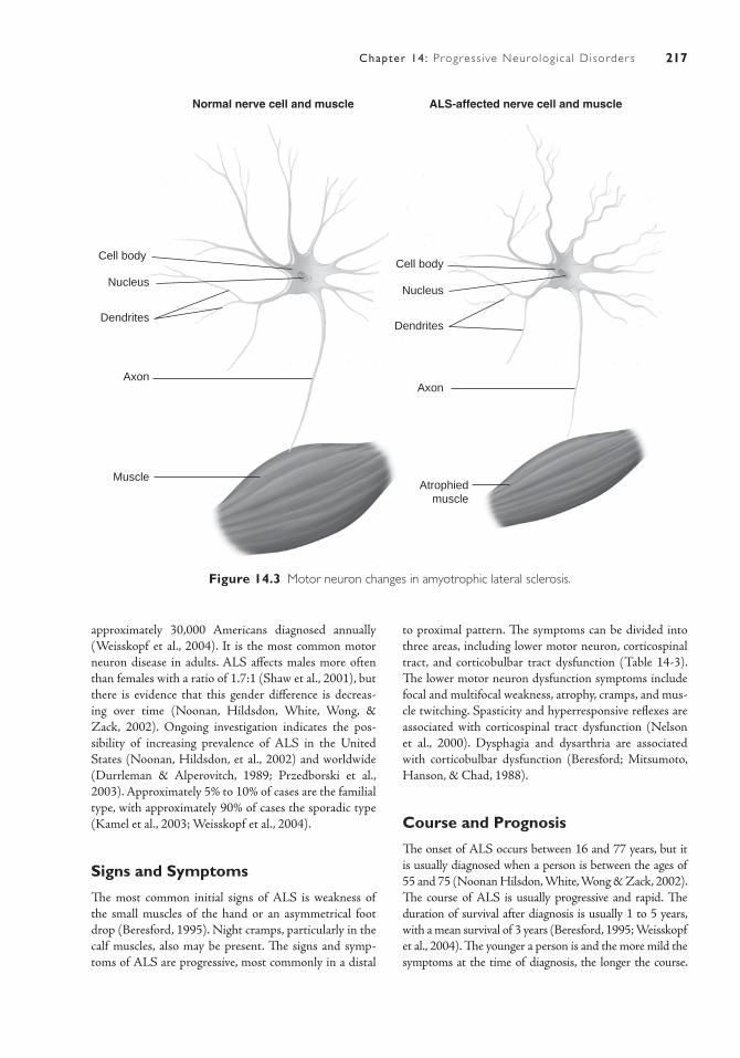
217Chapter 14: Progress ive Neurolog ica l Disorder s
Normal nerve cell and muscle ALS-affected nerve cell and muscle
Cell body
Nucleus
Dendrites
Axon
Atrophiedmuscle
Cell body
Nucleus
Dendrites
Axon
Muscle
Figure 14.3 Motor neuron changes in amyotrophic lateral sclerosis.
approximately 30,000 Americans diagnosed annually (Weisskopf et al., 2004). It is the most common motor neuron disease in adults. ALS affects males more often than females with a ratio of 1.7:1 (Shaw et al., 2001), but there is evidence that this gender difference is decreas-ing over time (Noonan, Hildsdon, White, Wong, & Zack, 2002). Ongoing investigation indicates the pos-sibility of increasing prevalence of ALS in the United States (Noonan, Hildsdon, et al., 2002) and worldwide (Durrleman & Alperovitch, 1989; Przedborski et al., 2003). Approximately 5% to 10% of cases are the familial type, with approximately 90% of cases the sporadic type (Kamel et al., 2003; Weisskopf et al., 2004).
Signs and Symptoms
The most common initial signs of ALS is weakness of the small muscles of the hand or an asymmetrical foot drop (Beresford, 1995). Night cramps, particularly in the calf muscles, also may be present. The signs and symp-toms of ALS are progressive, most commonly in a distal
to proximal pattern. The symptoms can be divided into three areas, including lower motor neuron, corticospinal tract, and corticobulbar tract dysfunction (Table 14-3). The lower motor neuron dysfunction symptoms include focal and multifocal weakness, atrophy, cramps, and mus-cle twitching. Spasticity and hyperresponsive reflexes are associated with corticospinal tract dysfunction (Nelson et al., 2000). Dysphagia and dysarthria are associated with corticobulbar dysfunction (Beresford; Mitsumoto, Hanson, & Chad, 1988).
Course and Prognosis
The onset of ALS occurs between 16 and 77 years, but it is usually diagnosed when a person is between the ages of 55 and 75 (Noonan Hilsdon, White, Wong & Zack, 2002). The course of ALS is usually progressive and rapid. The duration of survival after diagnosis is usually 1 to 5 years, with a mean survival of 3 years (Beresford, 1995; Weisskopf et al., 2004). The younger a person is and the more mild the symptoms at the time of diagnosis, the longer the course.
218 C O N D I T I O N S I N O C C U P A T I O N A L T H E R A P Y
TABLE 14.3 Signs and Symptoms of Amyotrophic Lateral Sclerosis
Lower Motor Neuron Corticospinal Tract Corticobulbar Tract
Focal and multifocal weakness
SpasticityHyperreactive reflexes
Dysphagia (difficulty swallowing)
Atrophy (progressive, distal to proximal)
Dysphagia (difficulty swallowing)
Dysarthria (impaired quality of speech production)
Muscle crampingMuscle twitching
Dysarthria (impaired quality of speech production)
There is some evidence of a “resistance in ALS,” in which a person may demonstrate improvements and live longer than 10 years. This is seen in approximately 10% to 16% of people with ALS (Mitsumoto et al., 1988). Death is usually from respiratory failure (Weydt et al., 2002).
Diagnosis
As with the other PNDs, the physician pieces together clinical symptoms, electromyogram (EMG) results, and tests to exclude other causes of the clinical presentation to make a definite diagnosis of ALS (Beresford, 1995; Mitsumoto et al., 1988). The EMG findings will include motor denervation and fasciculation (twitching) with intact sensory responses. A CT scan or MRI of the CNS may be used to rule out other causes of the symptoms. Blood tests are usually normal. Cerebrospinal fluid is often normal but may show raised protein levels.
Medical/Surgical Management
The medical treatments that are currently available do lit-tle to alter the fatal course of ALS (Weydt et al., 2002). The medications prescribed to treat the symptoms of ALS include antispasmodic medications, nonsteroidal anti-inflammatory medications, and antibiotics. There is evidence that Riluzole helps people stay in the milder stages for a longer time, and it is the only medication that increases survival, although for only a modest amount of time (Zoccolella et al., 2009). Experimental medications that have been in clinical trials include celecoxib (Cel-ebrex) and minocycline, which are anti-inflammatory drugs, and topiramate, gabapentin, and riluzole (Rilutek), which are antiglutamatergic drugs (Weydt et al.).
Gastrostomy and noninvasive positive-pressure venti-lation have been shown to increase both quality and pos-sibly length of life (Przedborski et al., 2003). Low-dose radiation and botulinum toxin injections into the salivary glands are sometimes used to treat drooling.
There is some evidence that attending a clinic staffed by a multidisciplinary team (including neurologists; special-ist nurses; physical, occupational, and speech therapists; a pulmonologist; a nutritionist; a psychologist; and a social worker) is effective for improving quality and length of life (Traynor, Alexander, Corr, Frost, & Hardiman, 2003). Pal-liative care may also be very important for people with ALS and their families. It has been estimated that 62.4% die in a hospice-supported environment (Przedborski et al., 2003).
IMPACT OF CONDITIONS ON CLIENT FACTORS AND OCCUPATIONAL PERFORMANCEEach of these PNDs is progressive and can affect all cli-ent factors, performance patterns, performance skills, and areas of occupation. The extent of this effect depends on the stage and severity of the disease. In each case, a person may have any combination of the deficits listed.
Activities of Daily Living
Self-care skills are affected by changes in the person’s senso-rimotor skills. Changes are usually noted in gross and fine motor coordination, postural control, muscle tone, endur-ance, and sensation (except in ALS). Loss of independence in bathing, dressing, personal device care, and toilet hygiene may occur. Toileting can become problematic for persons with MS or PD because of the loss of bladder and bowel control. The individual may experience any combination of the complications noted earlier in this chapter.
Eating may be difficult, either because the person loses the strength and coordination to self-feed or because of chewing or swallowing difficulties (dysphagia). The lat-ter is caused by weakness or incoordination of the phar-yngeal musculature, which also can make it difficult for an individual to ingest oral medications.
219Chapter 14: Progress ive Neurolog ica l Disorder s
Functional mobility is another critical concern. Neuromuscular and motor problems make ambulation difficult or impossible, either independently or with assis-tive devices, even in an electrically propelled wheelchair. Acquiring alternate methods of mobility requires the abil-ity to adapt. The person must be able to change motor pat-terns, requiring concurrent new and varied perceptual and cognitive strategies. At the same time, the individual is chal-lenged psychologically to make the necessary adjustments to new and different types of mobility. As the person’s function decreases, issues of home and work accessibility must be considered, and the necessary adaptations must be made to maintain performance in activities of daily living.
Instrumental Activities of Daily Living
Deficits in neuromusculoskeletal, movement-related func-tions and motor skills due to PNDs usually lead to reduced ability to perform all instrumental activities of daily living. In addition to neuromusculoskeletal losses, reduced sen-sory, cognitive, and perceptual functions may also interfere with independence in this area. In the early stages of these diseases adaptations need to be made to afford the person the opportunity to maintain function for as long as pos-sible. Most of these tasks, however, eventually need to be delegated to other members of the household, creating further issues of dependence and loss of role function.
A normal activity for many persons, including those with PND, is the care of others, including a spouse or sig-nificant other; children; or older, dependent adults. The individual with a PND may have increasing difficulty fulfilling this role. In fact, he or she may have to rely on these care receivers to provide support and care, creating a major role reversal. These changes in responsibilities can be very stressful for all concerned; they challenge every-one’s ability to maintain the integrity of relationships.
Education
Because of the age of onset for these PNDs, many peo-ple will not be involved in formal educational activities. Those who are may gradually experience a reduced abil-ity to physically participate due to decline in underlying neuromusculoskeletal and movement-related functions and motor skills. Deficits in cognitive and perceptual functions may further limit learning ability. Adaptations such as computer-based courses for formal and informal education may be a viable option.
Work
All client factors have the potential to affect work activi-ties. Work is a crucial area of occupational performance and, for many adults, is an important part of self- identity.
As motor skills decline, the ability to perform specific work tasks also declines. “Invisible symptoms” such as fatigue, weak or blurred vision, and difficulties with bladder control often confound the issue. Coworkers may not understand why someone who does not look ill cannot work. Again, this affects the person psychologi-cally, with changes in societal roles and self- concept. This is particularly true for an individual whose job requires a high degree of physical stamina and skill. For example, assembly workers or truck drivers may lose their jobs fairly early in the course of these diseases. An individual who has been the breadwinner of the family and whose identity is closely tied to physical strength and endur-ance may have serious adjustment problems. Cognitive deficits also may make it difficult for the person to func-tion and continue to find satisfaction in work.
Play and Leisure
Many leisure activities can be affected by the changes that result from PNDs. Alternative leisure activities must be explored as more and more performance deficits occur. A balance between work and play should be maintained as long as possible. However, if the person can no longer engage in usual work and daily living activities, it is even more critical to have meaningful and fulfilling leisure pur-suits. These activities will grow in importance as a means of self-actualization and satisfaction.
Social Participation
Communication, mobility, sexual dysfunction, and eat-ing problems may all affect the person’s normal socializa-tion with individuals or groups. Dysarthria or imperfect articulation is caused by a lack of control of the tongue and other oral muscles essential to speech. This problem can affect the person’s ability to communicate thoughts, needs, and desires and can limit social interaction. The individual may lose upper extremity function, making it difficult to compensate for speaking problems with written communication
Sexual dysfunction may also be present. Because of depression and diminished self-concept, the person may no longer feel attractive, which causes problems in sex-ual expression. Also, loss of specific motor and sensory function can affect physical performance.
Because of the unpredictable nature of the course of each PND, potential dependency issues are ongoing problems. This may lead to secondary psychosocial issues caused by these lifestyle changes. PNDs require an ini-tial social-psychological adjustment as well as continual readjustment because of erratic progression of symptoms. A person who was active and outgoing may have a dimin-ished self-concept because of the inability to engage in
220 C O N D I T I O N S I N O C C U P A T I O N A L T H E R A P Y
may avoid going out in public. Mothers may be unable to care for their children. Some may come to see themselves as no longer useful or attractive to others. Marriages may break up under the strain of living with PNDs. Occasionally, individuals with PNDs threaten suicide. An individual with a PND must think seriously about current role expectations and how these might be threat-ened by the PND.
activities that were once of interest and value. The result is a variety of role changes in the family or society.
Role expectations, which exist in every social situa-tion, are ways of behaving or reacting that fit with one’s self-image and the expectations of others. These include attitudes, activities, and patterns of decision making, expressing feelings, and meeting the needs of significant others. Some individuals with loss of bladder control
Case IllustrationsCase 1: Multiple SclerosisM. A. is a 28-year-old woman who was diagnosed with MS 5 years before this hospital admission. She was admitted because she noticed a progressive deterio-ration of function during the past 6 months. The main problems she identifies are an increase in fatigue, dif-ficulty with bowel and bladder function, and several falls. She complains of feeling moody and forgetting information.
She is married and has two children, an 11- year-old girl and a 5-year-old boy. She is self-employed as a paralegal, which requires her to spend many hours typing on the computer. Her husband works full-time and has been very supportive. At the time of admis-sion, he seemed overwhelmed.
M. A. tries to do her morning self-care but is find-ing it more difficult and frustrating. Getting dressed is particularly fatiguing, and she admits that at times she goes to bed fully dressed to avoid having to get dressed in the morning. She has been using a manual wheelchair off and on for the past 2 years.
Her daughter is currently helping with the laundry, cooking, and simple cleaning. M. A. states that she has problems doing household tasks because she must hold onto something stable before reaching for, or lift-ing, an object.
She currently enjoys no leisure activities. At one time, she liked to knit, but it has become too frustrat-ing to be pleasurable.
She complains of bladder urgency but often cannot void. She also has a mild dysarthria, spastic-ity of the lower extremities, weakness of the upper extremities (able to move against gravity withstanding minimal resistance), poor sitting balance, poor fine and gross motor coordination, blurred vision, and loss of stereognosis and light touch.
When the occupational therapist spoke with her, it became apparent that she did not comprehend the nature and course of MS. She is feeling frustrated and depressed about her recent decline of function.
She is currently taking Tylenol, Senokot, Metamucil, Colace, heparin, and multivitamins and is using Dulcolax suppositories.
Case 2: Parkinson’s DiseaseC. R. is a 72-year-old man with stage III PD. He has recently experienced a severe loss of balance and functional mobility. He reports difficulty moving quickly and gracefully. He also complains of poor handwriting, problems sleeping, and numbness in both hands.
C. R. is a single, retired accountant who lives inde-pendently in a two-story home. His bedroom and bathroom are on the second floor. There are four steps to enter the home. He has no children. He has one sister who lives within walking distance of his home. She, however, is suffering from arthritis and has difficulty offering much assistance.
C. R. has been caring for himself thus far, but his sister reports she is concerned about his safety, especially with activities such as cooking and bathing. She reports that he has fallen on several occasions recently and spends most of his time sitting in his chair in his living room.
His sister brings him meals as often as possible but is unable to bring meals in the morning or during bad weather. Her children live out of state and are only able to offer assistance to him during occasional visits when they try to do some of the major household chores such as painting, repairs, and cleaning.
C. R. was once interested in music and art. He played the piano and painted with watercolors. He reports that he has not participated in these leisure activities for “a long time.” He also played tennis on a regular basis at the local club. He is still a member of the club but has not been there in more than a year.
C. R. is reportedly self-conscious of his illness and, therefore, does not like to go out in public very much. His sister, who has always been very close to her brother, expresses concern about his “ depression
221Chapter 14: Progress ive Neurolog ica l Disorder s
and lack of motivation.” She states that she has tried on several occasions to get him to go to local musi-cal concerts or museums, but she feels he is just too depressed.
He is currently taking Sinemet CR and Artane. His sister, however, reports that he does not consis-tently take his medications because they “make him feel sick.”
Case 3: Amyotrophic Lateral SclerosisT. M. is a 48-year-old man who has recently been diag-nosed with ALS. Six months before being referred to O.T. services, he began to experience some weakness in his hands and he began dropping objects, such as tools. He reports loss of strength in his arms and legs. The weakness in his legs has become so severe that he now uses a borrowed wheelchair part of the time. He complains of difficulty sleeping due to cramps in his legs. He also reports that he has lost almost 20 lb in the last 6 or 8 months.
T. M. is married and has two sons, ages 6 and 4. He independently owns and operates a lawnmower shop at which he sells and repairs lawnmowers. He reports significant difficulty performing the repair parts of his job because of weakness in his hands and arms. Most
of the repairs have become backed up in the shop, and he is considering hiring assistance or sending the work to another shop.
His wife is employed full-time as a legal secretary. She has helped with the business as much as possible by completing some of the bookkeeping after hours. She is very busy caring for their boys and trying to maintain the household. She appears to be very sup-portive of T. M. but also seems very burdened by her responsibilities.
T. M. has many interests in sports and outdoor activities. He has been racing in “Iron Man” triathlons and marathons for the last several years. He was a high school track and cross country star. He enjoys biking and camping. Every summer, he and his family take a 2-week trip to a remote location where they hike and camp.
He is also supportive of his sons’ sports events and enjoys teaching them various sports. Last year, he was a soccer coach for his oldest boy’s team.
The course and prognosis of ALS has been explained to T. M. and his wife, and they have reportedly been discussing future plans. They are, however, “hoping for a miracle.” At this time, he is taking pain relievers to reduce the pain from the cramping in his legs.
R E C O M M E N D E D L E A R N I N G R E S O U R C E S
Multiple SclerosisNational Multiple Sclerosis Societyhttp://www.nmss.org/The Multiple Sclerosis Association of Americahttp://www.msaa.com/The Multiple Sclerosis Foundationhttp://www.msfacts.org/
Parkinson’s DiseaseNational Parkinson Foundationhttp://parkinson.org/Parkinson’s Disease Foundation, Inc.
http://pdf.org/The American Parkinson Disease Association, Inc.http://www.apdaparkinson.com/
Amyotrophic Lateral SclerosisThe ALS Associationhttp://www.alsa.org/World Federation of Neurology Amyotrophic Lateral
Sclerosishttp://www.wfnals.org/
Albanese, A. (2003). Diagnostic criteria for Parkinson’s disease. Neurological Sciences, 24, S23–S26.
Andreoli, T. E., Carpenter, C. C. J., Plum, F., & Smith, L. H. (1986). Cecil essentials of medicine. Philadelphia, PA: WB Saunders.
Armon, C., Kurland, L. T., Beard, C. M., O’Brien, P. C., & Mulder, D. W. (1991). Psychologic and adaptational difficulties anteceding amyotrophic lateral sclerosis: Rochester, Minnesota, 1925–1987. Neuroepidemiology, 10(3), 132–137.
R E F E R E N C E S
222 C O N D I T I O N S I N O C C U P A T I O N A L T H E R A P Y
Beresford, S. (1995). Motor neurone disease (amyotrophic lateral sclerosis). London, UK: Chapman & Hall.
Bobholz, J. A., & Rao, S. M. (2003). Cognitive dysfunc-tion in multiple sclerosis: A review of recent develop-ments. Current Opinions in Neurology, 16, 283–288.
Bohn, M. C. (2000). Parkinson’s Disease: A neurodegen-erative disease particularly amenable to gene therapy. Molecular Therapy, 1(6), 494–496.
Breitenstein, C., Van Lancker, D., Daum, I., & Waters, C. H. (2001). Impaired perception of vocal emotions in Par-kinson’s Disease: Influence of speech time processing and executive functioning. Brain and Cognition, 45, 277–314.
Carbon, M., & Marie, R. (2003). Functional imaging of cognition in Parkinson’s disease. Current Opinions in Neurology, 16, 475–480.
Chen, H., O’Reilly, E., McCullough, M. L., Rodriguez, C., Schwarzschild, M. A., Calle, E. E., & Ascherio, A. (2007). Consumption of dairy products and risk of Parkinson’s Disease. American Journal of Epidemiol-ogy, 165(9), 998–1006.
Chen, H., O’Reilly, E., Schwarzschild, M. A., & Ascherio, A. (2008). Peripheral inflammatory biomarkers and risk of Parkinson’s Disease. American Journal of Epidemiology, 167(1), 90–95.
Chen, H., Schernhammer, E., Schwarzschild, M. A., & Ascherio, A. (2006). A prospective study of night shift work, sleep duration and risk of Parkinson’s Disease. American Journal of Epidemiology, 163(8), 726–730.
Compston, A., & Coles, A. (2002). Multiple sclerosis. Lancet, 359, 1221–1231.
de Lau, L. M. L., Koudstaal, P. J., Hofman, A., & Breteler, M. B. (2006). Serum cholesterol levels and the risk of Parkinson’s Disease. American Journal of Epidemiol-ogy, 164(10), 998–1002.
Delisa, J. A., Hammond, M. C., Mikulic, M. A., & Miller, R. M. (1985). Multiple sclerosis: Part 1. Common physical disabilities and rehabilitation. American Family Physician, 32(4), 157–163.
Delisa, J. A., Miller, R. M., Mikulic, M. A., & Hammond, M. C. (1985). Multiple sclerosis: Part 2. Common functional problems and rehabilitation. American Family Physician, 32(5), 127–132.
DeLong, M. (2000). Parkinson’s Disease. Neurobiology of Disease, 7, 559–560.
Durrleman, S., & Alperovitch, A. (1989). Increas-ing trend of ALS in France and elsewhere: Are the changes real? Neurology, 39, 768–773.
Duvoisin, R. C., & Sage, J. (1996). Parkinson’s Disease: A guide for patient and family. Philadelphia, PA: Lippincott-Raven.
Feinstein, A. (2006). Mood disorders in multiple sclerosis and the effects on cognition. Journal of the Neurological Sciences, 245, 63–66.
Filippi, M., Rocca, M. A., Colombo, B., Falini, A., Codella, M., Scotti, G., & Comi, G. (2002). Functional mag-netic resonance imaging correlates of fatigue in mul-tiple sclerosis. NeuroImage, 15, 559–567.
Gao, X., Chen, H., Choi, H. K., Curhan, G., Schwarzschild, M. A., & Ascherio, A. (2008). Diet, urate, and Parkinson’s Disease in men. American Jour-nal of Epidemiology, 167(7), 831–838.
Hanson, L. J., & Cafruny, W. A. (2002). Current concepts in multiple sclerosis: Part 1. South Dakota Journal of Medicine, 55(10), 433–436.
Hernan, M. A., Olek, M. J., & Ascherio, A. (2001). Cigarette smoking and incidence of multiple sclerosis. American Journal of Epidemiology, 154(1), 69–74.
Hutton, T., & Dippel, R. L. (1989). Caring for the Parkin-son patient. New York, NY: Prometheus Books.
Kalb, R. C. (1996). Multiple sclerosis: The questions you have, the answers you need. New York, NY: Demos Vermande.
Kamel, F., Tanner, C. M., Umbach, D. M., Hoppin, J. A., Alavanja, M. C. R., Blair, A.,... & Sandler, D. P. (2007). Pesticide exposure and self-reported Parkinson’s Dis-ease in the agricultural health study. American Journal of Epidemiology, 165, 364–374.
Kamel, F., Umbach, D. M., Lehman, T. A., Park, L. P., Munsat, T. L., Shefner, J. M., et al. (2003). Amyo-trophic lateral sclerosis, lead and genetic susceptibil-ity: Polymorphisms in the aminolevulinic acid dehy-dratase and vitamin D receptor genes. Environmental Health Perspectives, 111(10), 1335–1339.
Kenealy, S. J., Pericak-Vance, M. S., & Haines, J. L. (2003). The genetic epidemiology of multiple sclero-sis. Journal of Neuroimmunology, 143, 7–12.
Lieberman, A. N., & Williams, F. L. (1993). Parkinson’s disease. New York, NY: Simon and Schuster.
Logroscino, G., Gao, X., Chen, H., Wing, A., & Ascherio, A. (2008). Dietary iron intake and risk of Parkinson’s Disease. American Journal of Epidemiology, 168(12), 1381–1388.
Lyros, E., Messinis, L., Papageorgiou, S. G., & Papathanosopoulos, P. (2010). Cognitive dysfunction in multiple sclerosis: The effects of pharmacological interventions. International Review of Psychiatry, 22(1), 35–42.
Maloney, F.P., Burks, J.S., & Ringel, S.P. (1985). Interdis-ciplinary Rehabilitation of Multiple Sclerosis and Neuro-muscular Disorders, New York, NY: Lippincott.
Matthews, B. (1993). Multiple sclerosis: The facts (3rd ed.). Oxford, UK: Oxford University Press.
Mayor, S. (2002). New treatment improves symptoms of Parkinson’s Disease. British Medical Journal, 324(7344), 997.
McCormack, A. L., Thiruchelvam, M., Mannig-Bog, A. B., Thiffault, C., Langston, J. W., Cory-Slechta, D. A., & Di Monte, D. A. (2002). Environmental risk factors
223Chapter 14: Progress ive Neurolog ica l Disorder s
Priyadarshi, A., Khuder, S. A., Schaub, E. A., & Priyadarshi, S. S. (2001). Environmental risk factors and Parkinson’s Disease: A metaanalysis. Environmen-tal Research Section A, 86, 122–127.
Przedborski, S., Mitsumoto, H., & Rowland, L. P. (2003). Recent advances in amyotrophic lateral sclerosis research. Current Neurology and Neuroscience Reports, 3, 70–77.
Rearick, M. P., Stelmach, G. E., Leis, B., & Santello, M. (2002). Coordination and control of forces during multifingered grasping in Parkinson’s Disease. Experi-mental Neurology, 177, 428–442.
Shaw, C. E., Al-Chalabi, A., & Neigh, N. (2001). Prog-ress in pathogenesis of amyotrophic lateral sclerosis. Current Neurology and Neuroscience Reports, 1, 69–76.
Traynor, B. J., Alexander, M., Corr, B., Frost, E., & Hardiman, O. (2003). Effect of a multidisciplinary amyotrophic lateral sclerosis (ALS) clinic on ALS survival: A population based study, 1996–2000. Journal of Neurology, Neurosurgery & Psychiatry, 74, 1258–1261.
Umphred, D. A. (1990). Neurological rehabilitation (2nd ed.). St Louis, MO: CV Mosby.
Wassem, R., & Dudley, W. (2003). Symptom manage-ment and adjustment of patients with multiple sclero-sis: A 4-year longitudinal intervention study. Clinical Nursing Research, 12(1), 102–117.
Weisskopf, M. G., McCullough, M. L., Calle, E. E., Thun, M. J., Cudkowicz, M., & Ascherio, A. (2004). Pro-spective study of cigarette smoking and amyotrophic lateral sclerosis. American Journal of Epidemiology, 160(1), 26–33.
Weisskopf, M. G., McCullough, M. L., Morozova, N., Calle, E. E., Thun, M. J., & Ascherio, A. (2005). Pro-spective study of occupation and amyotrophic lateral sclerosis mortality. American Journal of Epidemiology, 162(12), 1146–1152.
Weydt, P., Weiss, M. D., Moller, T., & Carter, G. T. (2002). Neuro-inflammation as a therapeutic target in amyotrophic lateral sclerosis. Current Opinion in Investigative Drugs, 3(12), 1720–1724.
Zhang, Y., Dawson, V. L., & Dawson, T. M. (2000). Oxi-dative stress and genetics in the pathogenesis of Par-kinson’s Disease. Neurobiology of Disease, 7, 240–250.
Zoccolella, S., Santamato, A., & Lamberti, P. (2009). Current and emerging treatments for amyotrophic lateral sclerosis. Neuropsychiatric Disease and Treat-ment, 5, 577–595.
and Parkinson’s Disease: Selective degeneration of nigral dopaminergic neurons caused by the herbicide paraquat. Neurobiology of Disease, 10, 119–127.
Merims, D., & Freedman, M. (2008). Cognitive and behavioural impairment in Parkinson’s Disease. International review of Psychiatry, 20(4), 364–373.
Mitsumoto, H., Hanson, M. R., & Chad, D. A. (1988). Amyotrophic lateral sclerosis: Recent advances in pathogenesis and therapeutic trials. Archives of Neurology, 45, 189–202.
Murray, J. (2002). Infection as a cause of multiple sclero-sis. British Medical Journal, 325(7373), 1128.
Neill, J., Belan, I., & Reid, K. (2006). Effectiveness of non-pharmacological interventions for fatigue in adults with multiple sclerosis, rheumatoid arthritis, or systemic lupus erythematosus: A systemic review. Inte-grative Literature Reviews and Meta-analyses, 617–635.
Neilsen, N. M., Westergaard, T., Rostgaard, K., Frisck, M., Hjalgrim, H., Wohlfahrt, J., Koch-Henriksen, N., & Melbye, M. (2005). Familial risk of multiple sclerosis: A nationwide cohort study. American Jour-nal of Epidemiology, 162(8), 774–778.
Nelson, L. M., Matkin, C., Longstreth, Jr., W. T., & McGuire, V. (2000). Population-based case-control study of amyotrophic lateral sclerosis in western Washington state. II. Diet. American Journal of Epide-miology, 151(2), 164–173.
Nelson, L. M., McGuire, V., Longstreth, Jr., W. T., & Mat-kin, C. (2000). Population-based case-control study of amyotrophic lateral sclerosis in western Washington state. I. Cigarette smoking and alcohol consumption. American Journal of Epidemiology, 151(2), 156–163.
Noonan, C. W., Hilsdon, R., White, M. C., Wong, L. -Y., & Zack, M. (2002). Continuing trend of increased motor neuron disease mortality in the United States. Epidemiology, 13, S202.
Noonan, C. W., Kathman, S. J., & White, M. C. (2002). Prevalence estimates for MS in the United States and evidence for an increasing trend for women. Neuro-logy, 58, 136–138.
Noonan, C. W., Williamson, D. M., Henry, J. P., Indian, R., Lynch, S. G., Neuberger, J. S., et al. (2010). The prevalence of multiple sclerosis in 3 US communi-ties. Preventing Chronic Disease, 7(1). Retrieved from http://www.cdc.gov/pcd/Issues/2010
Noseworthy, J. H. (2003). Management of multiple scle-rosis: Current trials and future options. Current Opin-ion in Neurology, 16, 289–297.


225
Rheumatic Diseases
■ D av i d P. O rc h ani an
15
Vincent O’Malley is a pleasant, energetic 11-year-old sixth-grade student at West Middle School. He enjoys riding his minibike in the fields around his neighborhood, playing street hockey, and playing video games with his Xbox 360. He has a variety of
friends in school and is never one to be without something to do on most days. There are two other children in his family, a younger brother, Jimmy, age 8, and an older sister Carrie, age 16. His mother is a parapro-fessional working in an elementary school with special needs children. His dad is a regional manager for Wal-Mart, and he works long hours, sometimes up to 50 to 60 hours per week. Approximately 4 months ago, Vincent was diagnosed with juvenile rheumatoid arthritis (JRA) of the pauciarticular type (four or fewer joints involved). The diagnosis was quite a while in coming from their pediatrician, and was made after a series of laboratory tests, radiographs, and physical examinations. Vin-cent’s mother remembers him complaining of mild, intermittent pain and stiffness in his lower back and hips for almost 3 months prior to the diagnosis. Performance areas affected include play, self-care, and educa-tion. Involved neuromuscular components include hip and trunk range of motion (ROM), lower extremity muscle strength, endurance, and at times postural control. His hip musculature is stiff at the end of range in the early morning and after sitting for periods of time >1 hour. Muscle strength is measured as 3/5 for hip extension and flexion and 3+/5 for trunk extension. On days when he is experiencing significant “stiffness” in his trunk and hips, his postural control is compromised and he cannot react quickly during play. His musculoskeletal endurance diminishes in the
afternoon, particularly after a busy day at school. He has limited his street hockey play to weekends when he tends to have more energy. Vincent’s parents feel it best to share as much information about his condition with his teachers each school year as is practical in an effort to optimize Vincent’s learning experience. His teachers
K E Y T E R M S
AnemiaAnkylosis
AntibodiesApophyseal
ArthritisBoutonniere deformity
BursitisCarpal tunnel syndrome
De Quervain’s tenosynovitisDiarthroses
Epstein-Barr virusInflammation
Juvenile rheumatoid arthritisOsteoarthritis
PannusRheumatoid arthritis
Rheumatoid factorRheumatologist
Sjögren’s syndromeSwan-neck deformities
SynovialTenosynovitis
226 C O N D I T I O N S I N O C C U P A T I O N A L T H E R A P Y
have appreciated this input and guidance and have a clear and compassionate perspective on what they may be confronted with on a day-to-day basis in their classrooms. One accommodation that they all have agreed to is to allow Vincent the opportunity to stand and walk around the classroom periodi-cally in order to relieve the stiffness that might be developing from extended static positioning at his desk. Regarding self-care, Vincent prefers showers to baths. This is actually better for him as there is only a 4-inch step over into the shower stall, and there is also a small bench built into the shower wall in case he needs to sit and rest. The bench seat is positioned approximately 17 inches above the floor, and there is a wall safety grab bar positioned on a 45-degree diagonal to the plane of the seat rest. He uses a squeeze bottle for shampoo and enjoys liquid soap, also administered from a lightweight squeeze bottle. His dad just installed a small shelf in the stall shower to hold these items. Dressing, grooming, feeding, and use of the toilet do not pre-sent any problems at this time for Vincent. He tends to avoid wearing tight clothing and generally wears T-shirts and loose-fitting cargo jeans or shorts on most days. He has just been fitted with braces for his teeth, and he is a little concerned about being able to maintain the high level of oral hygiene that his dentist is stressing. Vincent has never been one to floss his teeth, but now he must, and his parents will assist him in finding the right tools and tech-nique to accomplish this task. Vincent is blessed with a very understanding and supportive family. His parents have chosen to not treat him as a “special child.” He is required to participate in the routine activities of the family, including some yard work and other household chores. Generally, Vincent has been adjusting well to the limitations imposed by this dis-ease, although there are some occasions when he is severely limited by stiffness and painful movement, and these occasions upset and anger him. He is at an age and stage of life where he is trying many new things and experiencing many opportunities in school and leisure. His friends know of his con-dition, but they have been accepting as a whole, and Vincent is fortunate that his positive attitude and warm family atmosphere help him to attract and retain friendships with his peers.
DESCRIPTION AND DEFINITIONAugustin-Jacob Landre-Beauvais is given credit for the earliest description of rheumatoid arthritis (RA) in his thesis of 1800. It was not until 1858 that A.B. Garrod coined the actual term rheumatoid arthritis, and it was in 1941 that the American Rheumatism Association (ARA) adopted the terminology. The appearance and distribution of lesions in ancient skeletons suggests that RA may have existed in North America 3,000 years ago (Rothchild & Woods, 1990). More than 100 different forms of arthri-tis are documented. Arthritis is divided into eight major categories, with RA included under the synovitis category. Though less common than other forms of the disease, such as osteoarthritis, it is considered to be more seri-ous. Annual excess health care costs of RA patients were $8.4 billion, and costs of other RA consequences were $10.9 billion (Centers for Disease Control and Preven-tion, 2003). These costs translate to a total annual cost of $19.3 billion. From a stakeholder perspective, 33% of the total cost was allocated to employers, 28% to patients, 20% to the government, and 19% to caregivers. Adding intangible costs of quality-of-life deterioration ($10.3 billion) and premature mortality ($9.6 billion), total annual societal costs of RA (direct, indirect, and intan-gible) increased to $39.2 billion (Birnbaum et al., 2010). In one study done in the United States in 2009, of 10,298 patients, 8,916 had RA alone (86.6%), 608 had RA + car-diovascular disease (CVD) (5.9%), 716 had RA + depres-sion (7.0%), and 58 had RA + CVD + depression (0.5%). All patients with CVD and/or depression incurred sig-nificantly higher follow-up costs compared with patients with RA alone. Adjusted annual mean health care costs were highest for RA + CVD (US$14,145), followed by RA + CVD + depression ($13,513), RA + depression ($12,225), and RA alone ($11,404). Although patients with CVD and/or depression had a greater rate of RA-related hospitalization, adjusted RA-related health care costs did not reflect any statistically significant dif-ferences as compared to the RA-alone cohort ( Joyce, Smith, Khandker, Melin, & Singh, 2009). A significant proportion (13.4%) of patients with prevalent RA have comorbid CVD and/or depression. The presence of these conditions significantly affects annual health care costs as well as specific RA-related utilization patterns.
ETIOLOGYIt is essential to have a full awareness of the joint anat-omy and related structures (Fig. 15.1) as a preamble to discussing the etiology, signs and symptoms, and course of RA. The word “arthritis” derives from the Greek words “athron” (meaning joint) and “itis” (mean-ing inflammation or infection). Therefore, the word is defined as “inflammation or infection of the joint”
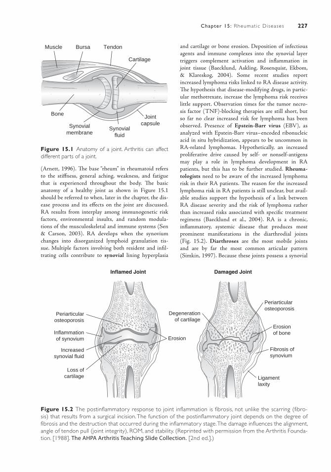
227Chapter 15: Rheumat ic Diseases
(Arnett, 1996). The base “rheum” in rheumatoid refers to the stiffness, general aching, weakness, and fatigue that is experienced throughout the body. The basic anatomy of a healthy joint as shown in Figure 15.1 should be referred to when, later in the chapter, the dis-ease process and its effects on the joint are discussed. RA results from interplay among immunogenetic risk factors, environmental insults, and random modula-tions of the musculoskeletal and immune systems (Sen & Carson, 2003). RA develops when the synovium changes into disorganized lymphoid granulation tis-sue. Multiple factors involving both resident and infil-trating cells contribute to synovial lining hyperplasia
and cartilage or bone erosion. Deposition of infectious agents and immune complexes into the synovial layer triggers complement activation and inflammation in joint tissue (Baecklund, Askling, Rosenquist, Ekbom, & Klareskog, 2004). Some recent studies report increased lymphoma risks linked to RA disease activity. The hypothesis that disease-modifying drugs, in partic-ular methotrexate, increase the lymphoma risk receives little support. Observation times for the tumor necro-sis factor (TNF)-blocking therapies are still short, but so far no clear increased risk for lymphoma has been observed. Presence of Epstein-Barr virus (EBV), as analyzed with Epstein-Barr virus–encoded ribonucleic acid in situ hybridization, appears to be uncommon in RA-related lymphomas. Hypothetically, an increased proliferative drive caused by self- or nonself-antigens may play a role in lymphoma development in RA patients, but this has to be further studied. Rheuma-tologists need to be aware of the increased lymphoma risk in their RA patients. The reason for the increased lymphoma risk in RA patients is still unclear, but avail-able studies support the hypothesis of a link between RA disease severity and the risk of lymphoma rather than increased risks associated with specific treatment regimens (Baecklund et al., 2004). RA is a chronic, inflammatory, systemic disease that produces most prominent manifestations in the diarthrodial joints (Fig. 15.2). Diarthroses are the most mobile joints and are by far the most common articular pattern (Simkin, 1997). Because these joints possess a synovial
Muscle Bursa Tendon
Cartilage
JointcapsuleSynovial
membraneSynovial
fluid
Bone
Figure 15.1 Anatomy of a joint. Arthritis can affect different parts of a joint.
Erosion
Erosionof bone
Ligamentlaxity
Fibrosis ofsynovium
Periarticularosteoporosis
Periarticularosteoporosis
Inflammationof synovium
Degenerationof cartilage
Increasedsynovial fluid
Loss ofcartilage
Inflamed Joint Damaged Joint
Figure 15.2 The postinflammatory response to joint inflammation is fibrosis, not unlike the scarring (fibro-sis) that results from a surgical incision. The function of the postinflammatory joint depends on the degree of fibrosis and the destruction that occurred during the inflammatory stage. The damage influences the alignment, angle of tendon pull (joint integrity), ROM, and stability. (Reprinted with permission from the Arthritis Founda-tion. [1988]. The AHPA Arthritis Teaching Slide Collection. [2nd ed.].)
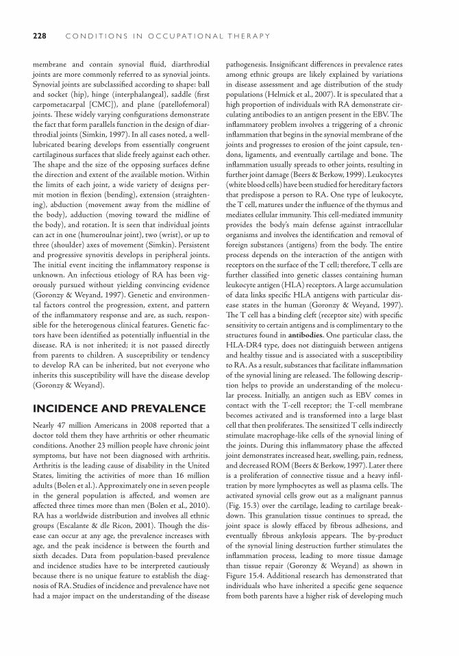
228 C O N D I T I O N S I N O C C U P A T I O N A L T H E R A P Y
membrane and contain synovial fluid, diarthrodial joints are more commonly referred to as synovial joints. Synovial joints are subclassified according to shape: ball and socket (hip), hinge (interphalangeal), saddle (first carpometacarpal [CMC]), and plane (patellofemoral) joints. These widely varying configurations demonstrate the fact that form parallels function in the design of diar-throdial joints (Simkin, 1997). In all cases noted, a well-lubricated bearing develops from essentially congruent cartilaginous surfaces that slide freely against each other. The shape and the size of the opposing surfaces define the direction and extent of the available motion. Within the limits of each joint, a wide variety of designs per-mit motion in flexion (bending), extension (straighten-ing), abduction (movement away from the midline of the body), adduction (moving toward the midline of the body), and rotation. It is seen that individual joints can act in one (humeroulnar joint), two (wrist), or up to three (shoulder) axes of movement (Simkin). Persistent and progressive synovitis develops in peripheral joints. The initial event inciting the inflammatory response is unknown. An infectious etiology of RA has been vig-orously pursued without yielding convincing evidence (Goronzy & Weyand, 1997). Genetic and environmen-tal factors control the progression, extent, and pattern of the inflammatory response and are, as such, respon-sible for the heterogenous clinical features. Genetic fac-tors have been identified as potentially influential in the disease. RA is not inherited; it is not passed directly from parents to children. A susceptibility or tendency to develop RA can be inherited, but not everyone who inherits this susceptibility will have the disease develop (Goronzy & Weyand).
INCIDENCE AND PREVALENCENearly 47 million Americans in 2008 reported that a doctor told them they have arthritis or other rheumatic conditions. Another 23 million people have chronic joint symptoms, but have not been diagnosed with arthritis. Arthritis is the leading cause of disability in the United States, limiting the activities of more than 16 million adults (Bolen et al.). Approximately one in seven people in the general population is affected, and women are affected three times more than men (Bolen et al., 2010). RA has a worldwide distribution and involves all ethnic groups (Escalante & dle Ricon, 2001). Though the dis-ease can occur at any age, the prevalence increases with age, and the peak incidence is between the fourth and sixth decades. Data from population-based prevalence and incidence studies have to be interpreted cautiously because there is no unique feature to establish the diag-nosis of RA. Studies of incidence and prevalence have not had a major impact on the understanding of the disease
pathogenesis. Insignificant differences in prevalence rates among ethnic groups are likely explained by variations in disease assessment and age distribution of the study populations (Helmick et al., 2007). It is speculated that a high proportion of individuals with RA demonstrate cir-culating antibodies to an antigen present in the EBV. The inflammatory problem involves a triggering of a chronic inflammation that begins in the synovial membrane of the joints and progresses to erosion of the joint capsule, ten-dons, ligaments, and eventually cartilage and bone. The inflammation usually spreads to other joints, resulting in further joint damage (Beers & Berkow, 1999). Leukocytes (white blood cells) have been studied for hereditary factors that predispose a person to RA. One type of leukocyte, the T cell, matures under the influence of the thymus and mediates cellular immunity. This cell-mediated immunity provides the body’s main defense against intracellular organisms and involves the identification and removal of foreign substances (antigens) from the body. The entire process depends on the interaction of the antigen with receptors on the surface of the T cell; therefore, T cells are further classified into genetic classes containing human leukocyte antigen (HLA) receptors. A large accumulation of data links specific HLA antigens with particular dis-ease states in the human (Goronzy & Weyand, 1997). The T cell has a binding cleft (receptor site) with specific sensitivity to certain antigens and is complimentary to the structures found in antibodies. One particular class, the HLA-DR4 type, does not distinguish between antigens and healthy tissue and is associated with a susceptibility to RA. As a result, substances that facilitate inflammation of the synovial lining are released. The following descrip-tion helps to provide an understanding of the molecu-lar process. Initially, an antigen such as EBV comes in contact with the T-cell receptor; the T-cell membrane becomes activated and is transformed into a large blast cell that then proliferates. The sensitized T cells indirectly stimulate macrophage-like cells of the synovial lining of the joints. During this inflammatory phase the affected joint demonstrates increased heat, swelling, pain, redness, and decreased ROM (Beers & Berkow, 1997). Later there is a proliferation of connective tissue and a heavy infil-tration by more lymphocytes as well as plasma cells. The activated synovial cells grow out as a malignant pannus (Fig. 15.3) over the cartilage, leading to cartilage break-down. This granulation tissue continues to spread, the joint space is slowly effaced by fibrous adhesions, and eventually fibrous ankylosis appears. The by-product of the synovial lining destruction further stimulates the inflammation process, leading to more tissue damage than tissue repair (Goronzy & Weyand) as shown in Figure 15.4. Additional research has demonstrated that individuals who have inherited a specific gene sequence from both parents have a higher risk of developing much
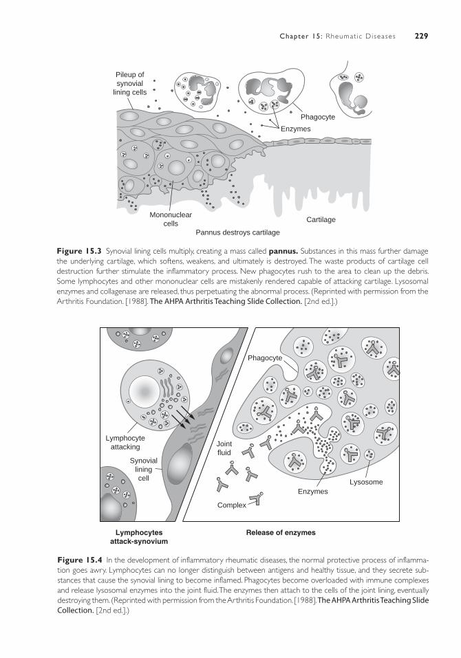
229Chapter 15: Rheumat ic Diseases
Lymphocyteattacking
Synovialliningcell
Phagocyte
Jointfluid
Complex
Lymphocytesattack-synovium
Release of enzymes
EnzymesLysosome
Figure 15.4 In the development of inflammatory rheumatic diseases, the normal protective process of inflamma-tion goes awry. Lymphocytes can no longer distinguish between antigens and healthy tissue, and they secrete sub-stances that cause the synovial lining to become inflamed. Phagocytes become overloaded with immune complexes and release lysosomal enzymes into the joint fluid. The enzymes then attach to the cells of the joint lining, eventually destroying them. (Reprinted with permission from the Arthritis Foundation. [1988]. The AHPA Arthritis Teaching Slide Collection. [2nd ed.].)
Pileup ofsynovial
lining cells
Phagocyte
Enzymes
CartilageMononuclear
cellsPannus destroys cartilage
Figure 15.3 Synovial lining cells multiply, creating a mass called pannus. Substances in this mass further damage the underlying cartilage, which softens, weakens, and ultimately is destroyed. The waste products of cartilage cell destruction further stimulate the inflammatory process. New phagocytes rush to the area to clean up the debris. Some lymphocytes and other mononuclear cells are mistakenly rendered capable of attacking cartilage. Lysosomal enzymes and collagenase are released, thus perpetuating the abnormal process. (Reprinted with permission from the Arthritis Foundation. [1988]. The AHPA Arthritis Teaching Slide Collection. [2nd ed.].)
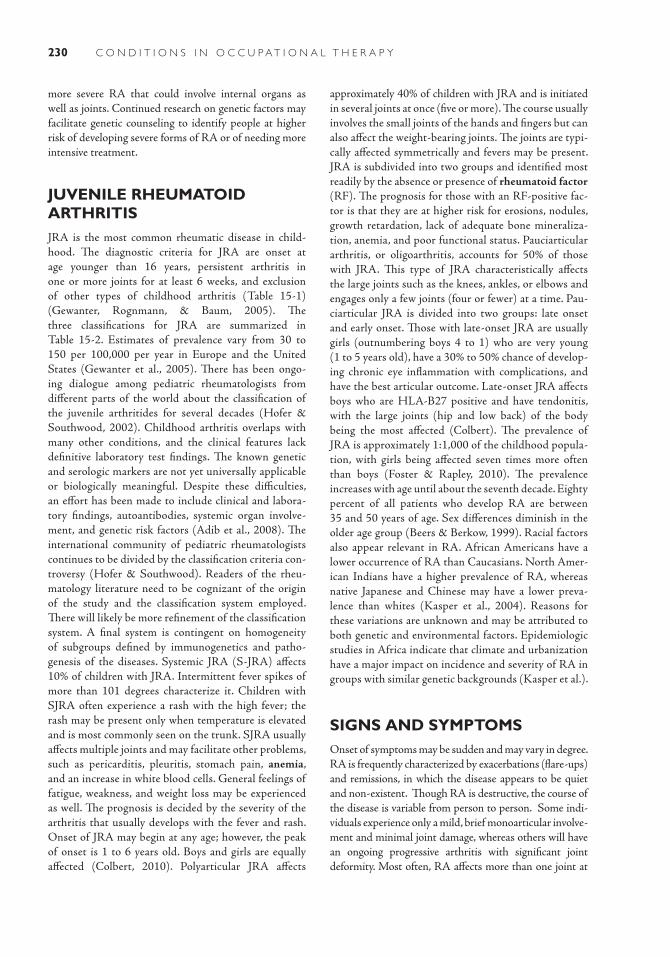
230 C O N D I T I O N S I N O C C U P A T I O N A L T H E R A P Y
more severe RA that could involve internal organs as well as joints. Continued research on genetic factors may facilitate genetic counseling to identify people at higher risk of developing severe forms of RA or of needing more intensive treatment.
JUVENILE RHEUMATOID ARTHRITISJRA is the most common rheumatic disease in child-hood. The diagnostic criteria for JRA are onset at age younger than 16 years, persistent arthritis in one or more joints for at least 6 weeks, and exclusion of other types of childhood arthritis (Table 15-1) (Gewanter, Rognmann, & Baum, 2005). The three classifications for JRA are summarized in Table 15-2. Estimates of prevalence vary from 30 to 150 per 100,000 per year in Europe and the United States (Gewanter et al., 2005). There has been ongo-ing dialogue among pediatric rheumatologists from different parts of the world about the classification of the juvenile arthritides for several decades (Hofer & Southwood, 2002). Childhood arthritis overlaps with many other conditions, and the clinical features lack definitive laboratory test findings. The known genetic and serologic markers are not yet universally applicable or biologically meaningful. Despite these difficulties, an effort has been made to include clinical and labora-tory findings, autoantibodies, systemic organ involve-ment, and genetic risk factors (Adib et al., 2008). The international community of pediatric rheumatologists continues to be divided by the classification criteria con-troversy (Hofer & Southwood). Readers of the rheu-matology literature need to be cognizant of the origin of the study and the classification system employed. There will likely be more refinement of the classification system. A final system is contingent on homogeneity of subgroups defined by immunogenetics and patho-genesis of the diseases. Systemic JRA (S-JRA) affects 10% of children with JRA. Intermittent fever spikes of more than 101 degrees characterize it. Children with SJRA often experience a rash with the high fever; the rash may be present only when temperature is elevated and is most commonly seen on the trunk. SJRA usually affects multiple joints and may facilitate other problems, such as pericarditis, pleuritis, stomach pain, anemia, and an increase in white blood cells. General feelings of fatigue, weakness, and weight loss may be experienced as well. The prognosis is decided by the severity of the arthritis that usually develops with the fever and rash. Onset of JRA may begin at any age; however, the peak of onset is 1 to 6 years old. Boys and girls are equally affected (Colbert, 2010). Polyarticular JRA affects
approximately 40% of children with JRA and is initiated in several joints at once (five or more). The course usually involves the small joints of the hands and fingers but can also affect the weight-bearing joints. The joints are typi-cally affected symmetrically and fevers may be present. JRA is subdivided into two groups and identified most readily by the absence or presence of rheumatoid factor (RF). The prognosis for those with an RF-positive fac-tor is that they are at higher risk for erosions, nodules, growth retardation, lack of adequate bone mineraliza-tion, anemia, and poor functional status. Pauciarticular arthritis, or oligoarthritis, accounts for 50% of those with JRA. This type of JRA characteristically affects the large joints such as the knees, ankles, or elbows and engages only a few joints (four or fewer) at a time. Pau-ciarticular JRA is divided into two groups: late onset and early onset. Those with late-onset JRA are usually girls (outnumbering boys 4 to 1) who are very young (1 to 5 years old), have a 30% to 50% chance of develop-ing chronic eye inflammation with complications, and have the best articular outcome. Late-onset JRA affects boys who are HLA-B27 positive and have tendonitis, with the large joints (hip and low back) of the body being the most affected (Colbert). The prevalence of JRA is approximately 1:1,000 of the childhood popula-tion, with girls being affected seven times more often than boys (Foster & Rapley, 2010). The prevalence increases with age until about the seventh decade. Eighty percent of all patients who develop RA are between 35 and 50 years of age. Sex differences diminish in the older age group (Beers & Berkow, 1999). Racial factors also appear relevant in RA. African Americans have a lower occurrence of RA than Caucasians. North Amer-ican Indians have a higher prevalence of RA, whereas native Japanese and Chinese may have a lower preva-lence than whites (Kasper et al., 2004). Reasons for these variations are unknown and may be attributed to both genetic and environmental factors. Epidemiologic studies in Africa indicate that climate and urbanization have a major impact on incidence and severity of RA in groups with similar genetic backgrounds (Kasper et al.).
SIGNS AND SYMPTOMSOnset of symptoms may be sudden and may vary in degree. RA is frequently characterized by exacerbations (flare-ups) and remissions, in which the disease appears to be quiet and non-existent. Though RA is destructive, the course of the disease is variable from person to person. Some indi-viduals experience only a mild, brief monoarticular involve-ment and minimal joint damage, whereas others will have an ongoing progressive arthritis with significant joint deformity. Most often, RA affects more than one joint at
231Chapter 15: Rheumat ic Diseases
TABLE 15.1 Criteria for the Diagnosis of Juvenile Rheumatoid Arthritis
I. GeneralThe JRA Criteria subcommittee in 1982 reviewed the1977 Criteria (86) and recommended that JRA be the name for the principal form of chronic arthritic disease in children and that this gen-eral class should be classified into three onset subtypes: systemic, polyarticular, and pauciarticular. The onset subtypes may be further subclassified into subsets as indicated below. The following classification enumerates the requirements for the diagnosis of JRA and the three clinical onset subtypes and lists subsets of each subtype that may be useful in further classification.
II. General criteria for the diagnosis of juvenile rheumatoid arthritis: A. Persistent arthritis of at least 6 weeks duration in one or more joints B. Exclusion of other causes of arthritis (see list of exclusions)
III. JRA onset subtypes: The onset subtype is determined by manifestations during the first 6 months of disease and remains the principal classification, although manifestations more closely resembling another subtype may appear later.
A. Systemic onset JRA: This subtype is defined as JRA with persistent intermittent fever (daily intermittent temperatures to 103°F or higher) with or without rheumatoid rash or other organ involvement. Typical fever and rash will be considered probable systemic onset JRA if not associ-ated with arthritis. Before a definite diagnosis can be made, arthritis, as defined, must be present.
B. Pauciarticular onset JRA: This subtype is defined as JRA with arthritis in four or fewer joints dur-ing the first 6 months of disease. Patients with systemic onset JRA are excluded from this onset subtype.
C. Polyarticular JRA: This subtype is defined as JRA with arthritis in five or more joints during the first 6 months of disease. Patients with systemic JRA onset are excluded from this subtype arthritis or Still’s disease. Hence reports of studies of JCA or JA cannot be directly compared with one another nor to reports of JRA or Still’s disease.
D. The onset subtypes may include the following subsets: 1. Systemic onset a. Polyarthritis b. Oligoarthritis 2. Oligoarthritis (pauciarticular onset) a. Antinuclear antibody (ANA) positive-chronic uveitis b. RF positive c. Seronegative, B27 positive d. Not otherwise classified 3. Polyarthritis a. RF positive b. Not otherwise classified
(Continued)
232 C O N D I T I O N S I N O C C U P A T I O N A L T H E R A P Y
TABLE 15.1 Criteria for the Diagnosis of Juvenile Rheumatoid Arthritis (Continued)IV. Exclusions A. Other rheumatic diseases 1. Rheumatic fever 2. Systemic lupus erythematosus 3. Ankylosing spondylitis 4. Polymyositis or dermatomyositis 5. Vasculitic syndromes 6. Scleroderma 7. Psoriatic arthritis 8. Reiter’s syndrome 9. Sjögren’s syndrome
10. Mixed connective tissue disease 11. Behçet’s syndrome B. Infectious arthritis C. Inflammatory bowel disease D. Neoplastic disease including leukemia E. Nonrheumatic conditions of bones and joints F. Hematologic diseases G. Psychogenic arthralgia H. Miscellaneous 1. Sarcoidosis 2. Hypertrophic osteoarthropathy 3. Villonodular synovitis 4. Chronic active hepatitis 5. Familial Mediterranean feverV. Other proposed terminology for juvenile chronic arthritis, JCA and JA are new diagnostic terms
currently in use in some places for the arthritides of childhood.
once. In two-thirds of patients, an exacerbation is initiated by feelings of fatigue, generalized weakness, weight loss, malaise, and vague musculoskeletal symptoms until syno-vitis becomes more obvious. Although joint involvement is generally symmetrical, some patients may experience an asymmetrical pattern (Kasper et al., 2004). An explora-tion of joint involvement can be divided into two sections: stages of inflammatory joint disease as experienced overall and specific manifestations to particular joints.
Articular and Periarticular Involvement
Table 15-3 presents the stages of the inflammatory pro-cess: (a) acute, (b) subacute, (c) chronic-active, and (d) chronic-inactive. As previously mentioned, onset may be sudden, with inflammation occurring in many joints at
once. In the acute and subacute phases, fatigue may be extensive enough to cause disability from disuse of joint motion and loss of strength before joint changes actually occur. Various degrees of general soreness and aching are experienced. These are usually followed by progres-sive, localized symptoms of pain, inflammation, warmth, and tenderness in a joint or multiple joints. Symmetrical involvement of small hand joints, feet, wrists, elbows, and ankles is typical, though initial manifestations may occur in any joint. Pain originates primarily from the joint cap-sule, which is heavily supplied with pain fibers and is highly sensitive to stretching and distension. Joint swelling results from accumulation of synovial fluid, hypertrophy of the synovium, and thickening of the joint capsule. Synovial thickening, the most specific physical finding, eventually occurs in most active joints. Various degrees of general-ized stiffness occur, including the gel phenomenon, which is the inability to move joints after prolonged rest. Morning
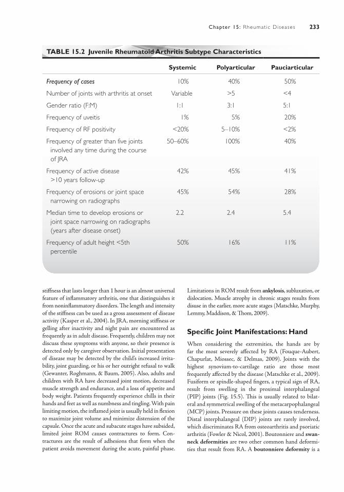
233Chapter 15: Rheumat ic Diseases
stiffness that lasts longer than 1 hour is an almost universal feature of inflammatory arthritis, one that distinguishes it from noninflammatory disorders. The length and intensity of the stiffness can be used as a gross assessment of disease activity (Kasper et al., 2004). In JRA, morning stiffness or gelling after inactivity and night pain are encountered as frequently as in adult disease. Frequently, children may not discuss these symptoms with anyone, so their presence is detected only by caregiver observation. Initial presentation of disease may be detected by the child’s increased irrita-bility, joint guarding, or his or her outright refusal to walk (Gewanter, Roghmann, & Baum, 2005). Also, adults and children with RA have decreased joint motion, decreased muscle strength and endurance, and a loss of appetite and body weight. Patients frequently experience chills in their hands and feet as well as numbness and tingling. With pain limiting motion, the inflamed joint is usually held in flexion to maximize joint volume and minimize distension of the capsule. Once the acute and subacute stages have subsided, limited joint ROM causes contractures to form. Con-tractures are the result of adhesions that form when the patient avoids movement during the acute, painful phase.
Limitations in ROM result from ankylosis, subluxation, or dislocation. Muscle atrophy in chronic stages results from disuse in the earlier, more acute stages (Matschke, Murphy, Lemmy, Maddison, & Thom, 2009).
Specific Joint Manifestations: Hand
When considering the extremities, the hands are by far the most severely affected by RA (Fouque-Aubert, Chapurlat, Miossec, & Delmas, 2009). Joints with the highest synovium-to-cartilage ratio are those most frequently affected by the disease (Matschke et al., 2009). Fusiform or spindle-shaped fingers, a typical sign of RA, result from swelling in the proximal interphalangeal (PIP) joints (Fig. 15.5). This is usually related to bilat-eral and symmetrical swelling of the metacarpophalangeal (MCP) joints. Pressure on these joints causes tenderness. Distal interphalangeal (DIP) joints are rarely involved, which discriminates RA from osteoarthritis and psoriatic arthritis (Fowler & Nicol, 2001). Boutonniere and swan-neck deformities are two other common hand deformi-ties that result from RA. A boutonniere deformity is a
TABLE 15.2 Juvenile Rheumatoid Arthritis Subtype Characteristics
Systemic Polyarticular Pauciarticular
Frequency of cases 10% 40% 50%
Number of joints with arthritis at onset Variable >5 <4
Gender ratio (F:M) 1:1 3:1 5:1
Frequency of uveitis 1% 5% 20%
Frequency of RF positivity <20% 5–10% <2%
Frequency of greater than five joints involved any time during the course of JRA
50–60% 100% 40%
Frequency of active disease >10 years follow-up
42% 45% 41%
Frequency of erosions or joint space narrowing on radiographs
45% 54% 28%
Median time to develop erosions or joint space narrowing on radiographs (years after disease onset)
2.2 2.4 5.4
Frequency of adult height <5th percentile
50% 16% 11%
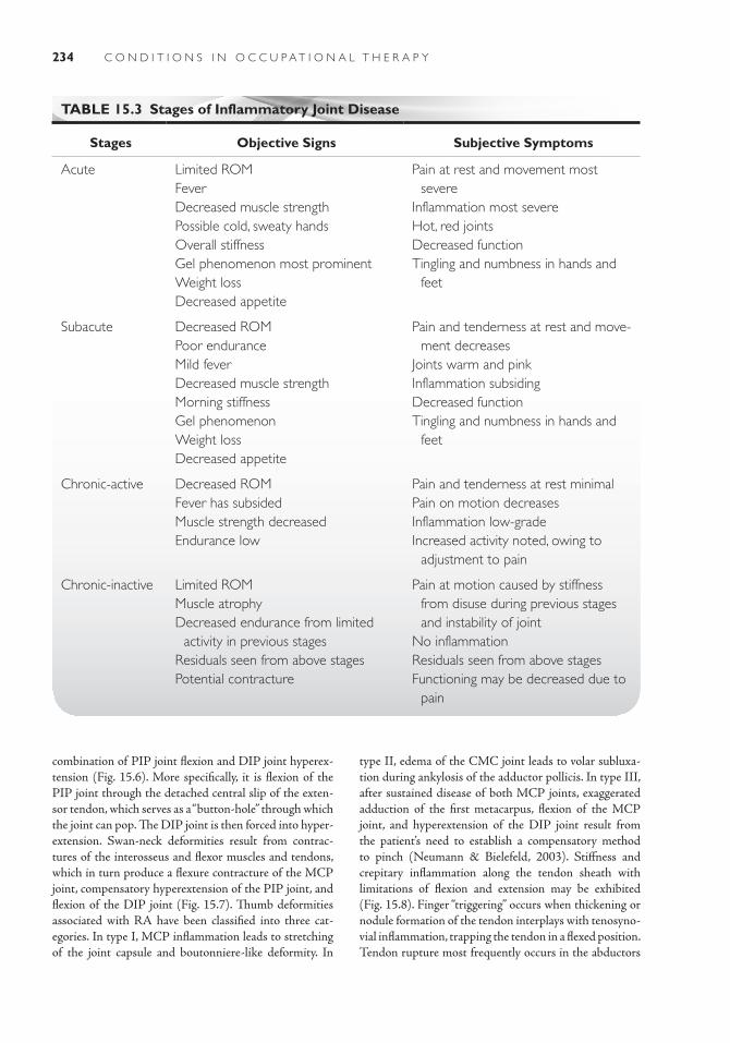
234 C O N D I T I O N S I N O C C U P A T I O N A L T H E R A P Y
combination of PIP joint flexion and DIP joint hyperex-tension (Fig. 15.6). More specifically, it is flexion of the PIP joint through the detached central slip of the exten-sor tendon, which serves as a “button-hole” through which the joint can pop. The DIP joint is then forced into hyper-extension. Swan-neck deformities result from contrac-tures of the interosseus and flexor muscles and tendons, which in turn produce a flexure contracture of the MCP joint, compensatory hyperextension of the PIP joint, and flexion of the DIP joint (Fig. 15.7). Thumb deformities associated with RA have been classified into three cat-egories. In type I, MCP inflammation leads to stretching of the joint capsule and boutonniere-like deformity. In
type II, edema of the CMC joint leads to volar subluxa-tion during ankylosis of the adductor pollicis. In type III, after sustained disease of both MCP joints, exaggerated adduction of the first metacarpus, flexion of the MCP joint, and hyperextension of the DIP joint result from the patient’s need to establish a compensatory method to pinch (Neumann & Bielefeld, 2003). Stiffness and crepitary inflammation along the tendon sheath with limitations of flexion and extension may be exhibited (Fig. 15.8). Finger “triggering” occurs when thickening or nodule formation of the tendon interplays with tenosyno-vial inflammation, trapping the tendon in a flexed position. Tendon rupture most frequently occurs in the abductors
TABLE 15.3 Stages of Inflammatory Joint Disease
Stages Objective Signs Subjective Symptoms
Acute Limited ROMFeverDecreased muscle strengthPossible cold, sweaty handsOverall stiffnessGel phenomenon most prominentWeight lossDecreased appetite
Pain at rest and movement most severe
Inflammation most severeHot, red jointsDecreased functionTingling and numbness in hands and
feet
Subacute Decreased ROMPoor enduranceMild feverDecreased muscle strengthMorning stiffnessGel phenomenonWeight lossDecreased appetite
Pain and tenderness at rest and move-ment decreases
Joints warm and pinkInflammation subsidingDecreased functionTingling and numbness in hands and
feet
Chronic-active Decreased ROMFever has subsidedMuscle strength decreasedEndurance low
Pain and tenderness at rest minimalPain on motion decreasesInflammation low-gradeIncreased activity noted, owing to
adjustment to pain
Chronic-inactive Limited ROMMuscle atrophyDecreased endurance from limited
activity in previous stagesResiduals seen from above stagesPotential contracture
Pain at motion caused by stiffness from disuse during previous stages and instability of joint
No inflammationResiduals seen from above stagesFunctioning may be decreased due to
pain
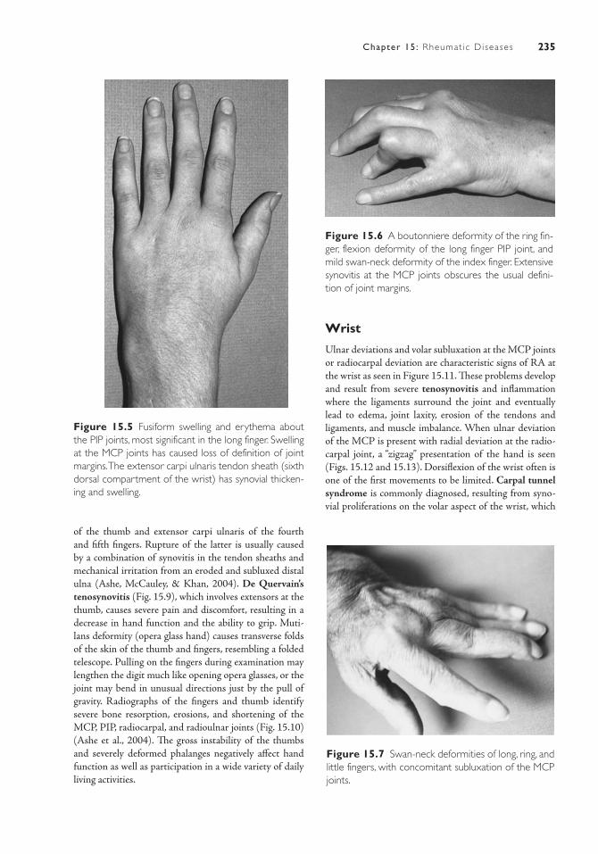
235Chapter 15: Rheumat ic Diseases
of the thumb and extensor carpi ulnaris of the fourth and fifth fingers. Rupture of the latter is usually caused by a combination of synovitis in the tendon sheaths and mechanical irritation from an eroded and subluxed distal ulna (Ashe, McCauley, & Khan, 2004). De Quervain’s tenosynovitis (Fig. 15.9), which involves extensors at the thumb, causes severe pain and discomfort, resulting in a decrease in hand function and the ability to grip. Muti-lans deformity (opera glass hand) causes transverse folds of the skin of the thumb and fingers, resembling a folded telescope. Pulling on the fingers during examination may lengthen the digit much like opening opera glasses, or the joint may bend in unusual directions just by the pull of gravity. Radiographs of the fingers and thumb identify severe bone resorption, erosions, and shortening of the MCP, PIP, radiocarpal, and radioulnar joints (Fig. 15.10) (Ashe et al., 2004). The gross instability of the thumbs and severely deformed phalanges negatively affect hand function as well as participation in a wide variety of daily living activities.
Figure 15.6 A boutonniere deformity of the ring fin-ger, flexion deformity of the long finger PIP joint, and mild swan-neck deformity of the index finger. Extensive synovitis at the MCP joints obscures the usual defini-tion of joint margins.
Wrist
Ulnar deviations and volar subluxation at the MCP joints or radiocarpal deviation are characteristic signs of RA at the wrist as seen in Figure 15.11. These problems develop and result from severe tenosynovitis and inflammation where the ligaments surround the joint and eventually lead to edema, joint laxity, erosion of the tendons and ligaments, and muscle imbalance. When ulnar deviation of the MCP is present with radial deviation at the radio-carpal joint, a “zigzag” presentation of the hand is seen (Figs. 15.12 and 15.13). Dorsiflexion of the wrist often is one of the first movements to be limited. Carpal tunnel syndrome is commonly diagnosed, resulting from syno-vial proliferations on the volar aspect of the wrist, which
Figure 15.5 Fusiform swelling and erythema about the PIP joints, most significant in the long finger. Swelling at the MCP joints has caused loss of definition of joint margins. The extensor carpi ulnaris tendon sheath (sixth dorsal compartment of the wrist) has synovial thicken-ing and swelling.
Figure 15.7 Swan-neck deformities of long, ring, and little fingers, with concomitant subluxation of the MCP joints.
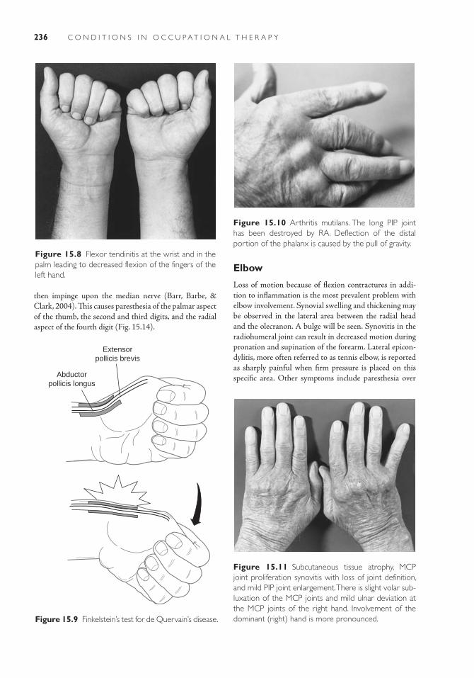
236 C O N D I T I O N S I N O C C U P A T I O N A L T H E R A P Y
Figure 15.8 Flexor tendinitis at the wrist and in the palm leading to decreased flexion of the fingers of the left hand.
Elbow
Loss of motion because of flexion contractures in addi-tion to inflammation is the most prevalent problem with elbow involvement. Synovial swelling and thickening may be observed in the lateral area between the radial head and the olecranon. A bulge will be seen. Synovitis in the radiohumeral joint can result in decreased motion during pronation and supination of the forearm. Lateral epicon-dylitis, more often referred to as tennis elbow, is reported as sharply painful when firm pressure is placed on this specific area. Other symptoms include paresthesia over Abductor
pollicis longus
Extensorpollicis brevis
Figure 15.9 Finkelstein’s test for de Quervain’s disease.
Figure 15.10 Arthritis mutilans. The long PIP joint has been destroyed by RA. Deflection of the distal portion of the phalanx is caused by the pull of gravity.
Figure 15.11 Subcutaneous tissue atrophy, MCP joint proliferation synovitis with loss of joint definition, and mild PIP joint enlargement. There is slight volar sub-luxation of the MCP joints and mild ulnar deviation at the MCP joints of the right hand. Involvement of the dominant (right) hand is more pronounced.
then impinge upon the median nerve (Barr, Barbe, & Clark, 2004). This causes paresthesia of the palmar aspect of the thumb, the second and third digits, and the radial aspect of the fourth digit (Fig. 15.14).
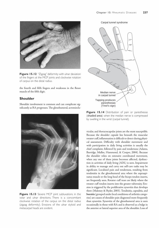
237Chapter 15: Rheumat ic Diseases
vicular, and thoracoscapular joints are the most susceptible. Because the shoulder capsule lies beneath the muscular rotator cuff, inflammation is difficult to detect during physi-cal assessment. Difficulty with shoulder movement and with participation in daily living activities is usually the chief complaint, followed by pain and tenderness (Adams, Burridge, Mullee, Hammond, & Cooper, 2004). Because the shoulder relies on extensive coordinated movement, when any one of these joints becomes affected, dysfunc-tion in activities of daily living (ADL) is seen. Impairment in ability to manage and carry out self-care tasks may be significant. Localized pain and tenderness, resulting from tendonitis in the glenohumeral area where the supraspi-natus muscle or the long head of the biceps tendon inserts, are frequently seen. Rotator cuff tears are likely where the rotator cuff tendon inserts into the greater tuberosity. Ero-sion is triggered by the proliferative synovitis that develops there (Maloney & Ryder, 2003). Tendinitis, capsulitis, and bursitis (grouped under local conditions of arthritis catego-ries) are causes of shoulder pain diagnosed more frequently than synovitis. Synovitis of the glenohumeral area is seen occasionally in those with RA and is observed as a bulge in the anterior or lateral superior area of the shoulder. Loss of
Figure 15.13 Severe MCP joint subluxations in the volar and ulnar directions. There is a concomitant clockwise rotation of the carpus on the distal radius (zigzag deformity). Erosions of the ulnar styloid and metacarpal heads are evident.
the fourth and fifth fingers and weakness in the flexor muscle of the fifth digit.
Shoulder
Shoulder involvement is common and can complicate sig-nificantly as RA progresses. The glenohumeral, acromiocla-
Figure 15.12 “Zigzag” deformity with ulnar deviation of the fingers at the MCP joints and clockwise rotation of carpus on the distal radius.
Carpal tunnel syndrome
Median nervein carpal tunnel
Tapping producesparesthesias(Tinel’s sign)
Figure 15.14 Distribution of pain or paresthesias (shaded area) when the median nerve is compressed by swelling in the wrist (carpal tunnel).

238 C O N D I T I O N S I N O C C U P A T I O N A L T H E R A P Y
motion is a complication of shoulder synovitis, which is seen in progressed cases, and is known as frozen shoulder.
Head, Neck, and Cervical Spine
The cervical spine is often involved in RA (Mink, Gordon, & Deutsch, 2003). Involvement of C1 and C2 may pro-duce life-threatening conditions. Neck pain on motion and occipital headaches are common symptoms of cervical spine involvement and occur in those individuals who have had RA longer than 10 years (Niere & Jerak, 2004). Patients with severe deformities in their hands, as in mutilans deformity, are very likely to have had significant amounts of corticosteroids for RA management (Crowther, 2001). During radiologic examination of this area in advanced cases of RA, the lower cervical and odontoid processes often appear eroded, as do the cervical apophyseal and intervertebral joints. The first to fourth cervical joints are those most commonly affected by inflammation and pain (Figs. 15.15 and 15.16). Involvement of the upper cervi-cal spine in advanced cases leads to subluxation, whereas lower cervical spine involvement produces symptoms of
cord-root compression. For example, with a C5 root com-pression, problems are (a) sensation on the radial aspect of the forearm, (b) muscle weakness and abduction of the shoulder and flexion of the elbow, and (c) decreased biceps jerk reflex. Subluxation also can cause twisting and compression of the vertebral arteries, which can lead to vertebrobasilar artery insufficiency. This may be facilitated by syncope on downward gaze. Flexion and extension of the cervical spine are usually less affected. The temporo-mandibular joints (TMJs) have varied involvement in RA, ranging from 1% to 60% by some estimates. Women are affected three times more often than men. Both TMJs are usually involved (Melchiorre et al., 2010). Involvement of this synovial joint results in the inability to open the mouth fully because of side-to-side gliding and protrusion. After persistent inflammation, normal approximation of the upper and lower teeth may be affected. Hoarseness occurs in up to 30% of patients presenting with RA. This stems from inflamed cricoarytenoid joints, which rotate with the vocal cords as they abduct and adduct to vary pitch and tone of the voice (Berjawi et al., 2010).
Spinousprocesssharpening
Apophysealerosion
Apophysealfusion
Atlanto-axialsubluxation
Subaxialsubluxation
Spondylodiscitis
Figure 15.15 Neck abnormalities in RA. The neck is usually involved in adult and juvenile RA. The most com-mon disorder is subluxation of the atlantoaxial joint, which occurs particularly on flexion of the neck. C1 moves forward on C2, and the odontoid process can actually cause pressure on the spinal cord posteriorly. Other findings include erosions at the apophyseal joints, fusion of the apophyseal joint, which occurs particularly in JRA, and subluxation at other levels. Disk involvement also may occur, and erosive changes and resorption can cause sharpening of the spinous process. (Reprinted with permission from the Arthritis Foundation. [1988]. The AHPA Arthritis Teaching Slide Collection. [2nd ed.].)
Figure 15.16 A lateral radiograph of a patient with RA who is experiencing severe upper extremity neu-rologic decline caused by C2–C3 and C3 and C4 ante-rior subluxations. The odontoid is not visible because of severe erosion.
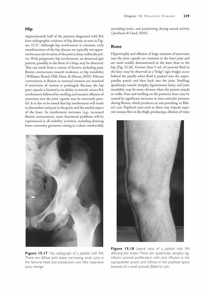
239Chapter 15: Rheumat ic Diseases
Hip
Approximately half of the patients diagnosed with RA have radiographic evidence of hip disease as seen in Fig-ure 15.17. Although hip involvement is common, early manifestations of the hip disease are typically not appar-ent because the location of the joint is deep within the pel-vis. With progressive hip involvement, an abnormal gait pattern, possibly in the form of a limp, may be observed. This can result from a variety of factors, including pain, flexion contractures, muscle weakness, or hip instability (Williams, Brand, Hill, Hunt, & Moran, 2010). Fibrous contractures in flexion or external rotation are standard if restriction of motion is prolonged. Because the hip joint capsule is limited in its ability to stretch, severe RA involvement followed by swelling and massive effusion of synovium into the joint capsule may be extremely pain-ful. It is also to be noted that hip involvement will result in discomfort and pain in the groin and the medial aspect of the knee. As involvement increases (e.g., increased flexion contractures), more functional problems will be experienced in all mobility activities, including donning lower extremity garments, sitting in a chair comfortably,
ascending stairs, and positioning during sexual activity ( Josefsson & Gard, 2010).
Knee
Hypertrophy and effusion of large amounts of synovium into the joint capsule are common in the knee joint and are more readily demonstrated in the knee than in the hip (Fig. 15.18). Greater than 5 mL of synovial fluid in the knee may be observed as a “bulge” sign; bulges occur behind the patella when fluid is pushed into the supra-patellar pouch and then back into the joint. Swelling, quadriceps muscle atrophy, ligamentous laxity, and joint instability may be more obvious when the patient stands or walks. Pain and swelling on the posterior knee may be caused by significant increases in intra- articular pressure during flexion, which produces an out-pouching, or Bak-er’s cyst. Popliteal cysts such as these may impede supe-rior venous flow in the thigh, producing a dilation of veins
Figure 15.17 Hip radiograph of a patient with RA. There are diffuse joint space narrowing, small cysts in the femoral head and acetabulum, and little reparative bony change.
Figure 15.18 Lateral view of a patient with RA affecting the knees. There are quadriceps atrophy, sig-nificant synovial proliferation with joint effusion in the suprapatellar pouch, and fullness in the popliteal space because of a small synovial (Baker’s) cyst.
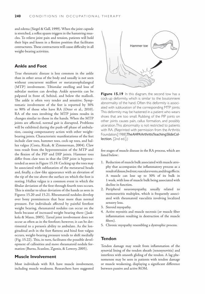
240 C O N D I T I O N S I N O C C U P A T I O N A L T H E R A P Y
and edema (Siegel & Gall, 1999). When the joint capsule is stretched, a reflex spasm triggers in the hamstring mus-cles. To relieve joint pain and tension, patients will hold their hips and knees in a flexion position that facilitates contractures. These contractures will cause difficulty in all weight-bearing activities.
Ankle and Foot
True rheumatic disease is less common in the ankle than in other areas of the body and usually is not seen without concurrent midfoot or metatarsophalangeal (MTP) involvement. Tibiotalar swelling and loss of subtalar motion can develop. Ankle synovitis can be palpated in front of, behind, and below the malleoli. The ankle is often very tender and sensitive. Symp-tomatic involvement of the feet is reported by 30% to 90% of those who have RA (Otter et al., 2010). RA of the toes involving the MTP joints results in changes similar to those in the hands. When the MTP joints are affected, normal gait is disrupted. Problems will be exhibited during the push-off phase of ambula-tion, causing compensatory action with other weight-bearing joints. Characteristic manifestations of the feet include claw toes, hammer toes, cock-up toes, and hal-lux valgus (Costa, Rizak, & Zimmerman, 2004). Claw toes result from the hyperextension of the MTP and the flexion of the PIP and DIP joints. Hammer toes differ from claw toes in that the DIP joint is hyperex-tended as seen in Figure 15.19. Cocking up the toes may be associated with subluxation of the metatarsal heads and, finally, a claw-like appearance with an elevation of the tip of the toe above the surface on which the foot is resting. Hallux valgus is a common event in which the fibular deviation of the first through fourth toes occurs. This is similar to ulnar deviation of the hands as seen in Figures 15.20 and 15.21. Rheumatoid nodules develop over bony prominences that bear more than normal pressure. For individuals affected by painful forefoot weight bearing, rheumatoid nodules can occur on the heels because of increased weight bearing there ( Jaak-kola & Mann, 2005). Tarsal joint involvement does not occur as often as in the forefoot; however, it can be det-rimental to a person’s ability to ambulate. As the lon-gitudinal arch in the foot flattens and hind foot valgus occurs, weight-bearing pressure tends to shift medially (Fig. 15.22). This, in turn, facilitates the possible devel-opment of callosities and more rheumatoid nodule for-mation (Burns, Scanlon, Zgonis, & Lowery, 2005).
Muscle Involvement
Most individuals with RA have muscle involvement, including muscle weakness. Researchers have suggested
five stages of muscle disease in the RA process, which are listed below:
1. Reduction of muscle bulk associated with muscle atro-phy that accompanies the inflammatory process as a result of disuse, bed rest, vascular events, and drug effects. A muscle can lose up to 30% of its bulk in 1 week, with loss of muscle bulk being associated with decline in function.
2. Peripheral neuromyopathy, usually related to mononeuritis multiplex, which is frequently associ-ated with rheumatoid vasculitis involving localized sensory loss.
3. Steroid myopathy.4. Active myositis and muscle necrosis (or muscle fiber
inflammation resulting in destruction of the muscle fibers).
5. Chronic myopathy resembling a dystrophic process.
Tendon
Tendon damage may result from inflammation of the synovial lining of the tendon sheath (tenosynovitis) and interferes with smooth gliding of the tendon. A lag phe-nomenon may be seen in patients with tendon damage or muscle weakening, displaying a significant difference between passive and active ROM.
Hammer toe
Figure 15.19 In this diagram, the second tow has a cock-up deformity, which is similar to the boutonniere abnormality of the hand. Often this deformity is associ-ated with subluxation of the corresponding MTP joints. This deformity may be hastened in a patient who wears shoes that are too small. Rubbing of the PIP joints on other joints causes pain, callus formation, and possibly ulceration. This abnormality is not restricted to patients with RA. (Reprinted with permission from the Arthritis Foundation. [1988]. The AHPA Arthritis Teaching Slide Col-lection. [2nd ed.].)

241Chapter 15: Rheumat ic Diseases
Extra-Articular Systemic Manifestations
As has been mentioned previously in this chapter, RA affects the joint and is systemic. The number and sever-ity of extra-articular features vary with the duration and extent of the disease and tend to occur in individuals with higher levels of RF in their blood (Hochberg, Johnston, & John, 2008). The following are additional manifestations that may exist in those with RF.
Rheumatoid or Subcutaneous NodulesRheumatoid nodule formation is one of the most com-mon extra-articular manifestations of RA and occurs in up to 50% of individuals at some point during the course of the disease (Hochberg et al., 2008). Periarticular struc-tures; extensor surfaces; and areas subject to pressure such as the olecranon (Fig. 15.23), the proximal ulna, the Achilles tendon, the occiput, and the sacrum are primary sites for those growths. Most can develop insidiously and regress at any time.
Hallux valgus
Figure 15.20 An anatomical and clinical diagram of hallux vagus. Pes planus and ligamentous laxity lead to lateral deviation of the great toe with a resultant hal-lux vagus. This deformity can be hastened by the wear-ing of narrow-toed shoes. Rubbing of the bunion on the shoe surface produced pain, and the lateral devia-tion of the great toe may impinge on other digits of the foot. This abnormality is not restricted to patients with RA. (Reprinted with permission from the Arthri-tis Foundation. [1988]. The AHPA Arthritis Teaching Slide Collection. [2nd ed.].)
Figure 15.21 Mild hallux valgus, with dorsal sub-luxation of the MTP joints and resultant “hammer toe” deformities of second through fifth toes. Midfoot insta-bility has led to eversion, with concomitant flattening of the feet.
Figure 15.22 Significant ankle and midfoot synovi-tis. Loss of definition of the arch and eversion at the subtalar joint also are evident.
Pulmonary Manifestations
Pleuritis, interstitial fibrosis, pulmonary nodules, pneumo-nitis, and other forms of pulmonary obstructive disease occur more frequently in those with RA than in the popu-lation at large (Kim, Collard, & King, 2009). Evidence of pleuritis is usually found at autopsy, because the disease is generally asymptomatic during life. In a few cases, upper-airway obstruction from cricoarytenoid arthritis or laryngeal nodules may develop. Some researchers believe that small-airway dysfunction is related to factors other than RA.
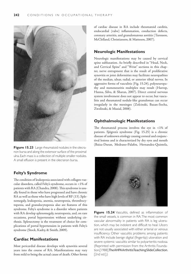
242 C O N D I T I O N S I N O C C U P A T I O N A L T H E R A P Y
of cardiac disease in RA include rheumatoid carditis, endocardial (valve) inflammation, conduction defects, coronary arteritis, and granulomatous aortitis (Turesson, McClelland, Christianson, & Matteson, 2007).
Neurologic Manifestations
Neurologic manifestations may be caused by cervical spine subluxation. As briefly described in “Head, Neck, and Cervical Spine” and “Wrist” sections in this chap-ter, nerve entrapment that is the result of proliferative synovitis or joint deformities may facilitate neuropathies of the median, ulnar, radial, or anterior tibial nerves. In aggressive forms of vasculitis (Fig. 15.24), polyneuropa-thy and mononeuritis multiplex may result (Harrop, Hanna, Silva, & Sharan, 2007). Direct central nervous system involvement does not appear to occur, but vascu-litis and rheumatoid nodule-like granulomas can occur irregularly in the meninges (Zolcinski, Bazan-Socha, Zwolinski, & Musial, 2008).
Ophthalmologic Manifestations
The rheumatoid process involves the eye in <1% of patients. Sjögren’s syndrome (Fig. 15.25) is a chronic disease of unknown etiology caus ing corneal and conjunc-tival lesions and is characterized by dry eyes and mouth (Baiza-Duran, Medrano-Palafox, Hernandez-Quintela,
Felty’s Syndrome
The condition of leukopenia associated with collagen-vas-cular disorders, called Felty’s syndrome, occurs in <1% of patients with RA (Chandra, 2008). This syndrome is usu-ally found in those who have progressed and have chronic RA as well as those who have high levels of RF (13). Sple-nomegaly, leukopenia, anemia, neutropenia, thrombocy-topenia, and granulocytopenia also are features of this syndrome. Felty’s syndrome is a disorder where patients with RA develop splenomegaly, neutropenia, and, on rare occasions, portal hypertension without underlying cir-rhosis. Splenectomy is the treatment of choice for com-plications of portal hypertension in patients with Felty’s syndrome (Stock, Kadry, & Smith, 2009).
Cardiac Manifestations
Most pericardial disease develops with synovitis several years into the course of RA. Manifestations may vary from mild to being the actual cause of death. Other forms
Figure 15.23 Large rheumatoid nodules in the olecra-non bursa and along the extensor surface of the proximal ulna. Each mass is a collection of multiple smaller nodules. A small effusion is present in the olecranon bursa.
Figure 15.24 Vasculitis, defined as inflammation of the small vessels, is common in RA. The most common vascular abnormality in patients with RA is leg ulcera-tion, which may be indolent and difficult to heal. Ulcers are not usually associated with either arterial or venous insufficiency. Other vasculitis problems among patients with RA include benign digital (fingertip) ulceration and severe systemic vasculitis similar to polyarteritis nodosa. (Reprinted with permission from the Arthritis Founda-tion. [1988]. The AHPA Arthritis Teaching Slide Collection. [2nd ed.].)
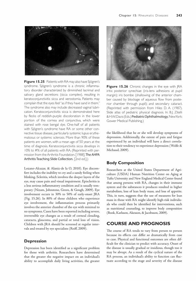
243Chapter 15: Rheumat ic Diseases
Lozano-Alcazar, & Alaniz-de la O, 2010). Eye discom-fort includes the inability to cry and a sandy feeling when blinking. Scleritis, which involves the deeper layers of the eye, may cause pain and visual impairment. Episcleritis is a less serious inflammatory condition and is usually tem-porary (Nizam, Johnstone, Green, & Gough, 2009). Eye involvement occurs in 30% to 50% of early-onset JRA (Fig. 15.26). In 80% of those children who experience eye involvement, the inflammation process primarily involves the anterior chamber of the eye with minimal to no symptoms. Cases have been reported including severe, irreversible eye changes as a result of corneal clouding, cataracts, glaucoma, and partial or total loss of vision. Children with JRA should be screened at regular inter-vals and treated by eye specialists (Reiff, 2009).
Depression
Depression has been identified as a significant problem for those with arthritis. Researchers have determined that the greater the negative impact on an individual’s ability to accomplish daily living activities, the greater
the likelihood that he or she will develop symptoms of depression. Additionally, the extent of pain and fatigue experienced by an individual will have a direct correla-tion to their tendency to experience depression (Wolfe & Michaud, 2009).
Body Composition
Researchers at the United States Department of Agri-culture (USDA) Human Nutrition Center on Aging at Tufts University and New England Medical Center found that among persons with RA, changes in their immune system and the substances it produces resulted in higher metabolism, loss of lean body mass, and loss of appetite. This, in turn, suggests that the use of measures for lean mass in those with RA might identify high-risk individu-als who could then be identified for interventions, such as nutritional counseling, to improve body composition (Book, Karlsson, Akesson, & Jacobsson, 2009).
COURSE AND PROGNOSISThe course of RA tends to vary from person to person because its effects can differ so dramatically from case to case. Physical and functional outcomes are often dif-ficult for the clinician to predict with accuracy. Onset of the disease is usually gradual or insidious, though too it may be abrupt. As a result of the cyclical nature of the RA process, an individual’s ability to function can fluc-tuate according to the stage and severity of the disease
Figure 15.25 Patients with RA may also have Sjögren’s syndrome. Sjögren’s syndrome is a chronic inflamma-tory disorder characterized by diminished lacrimal and salivary gland secretions (sicca complex), resulting in keratoconjunctivitis sicca and xerostomia. Patients may complain that the eyes feel “as if they have sand in them.” The syndrome also may include decreased vaginal lubri-cation. Keratoconjunctivitis sicca is demonstrated here by flecks of reddish-purple discoloration in the lower portion of the cornea and conjunctiva, which were stained with rose bengal dye. One-half of all patients with Sjögren’s syndrome have RA or some other con-nective tissue disease, particularly systemic lupus erythe-matosus or systemic sclerosis. More than 90% of these patients are women, with a mean age of 50 years at the time of diagnosis. Keratoconjunctivitis sicca develops in 10% to #% of all patients with RA. (Reprinted with per-mission from the Arthritis Foundation. [1988]. The AHPA Arthritis Teaching Slide Collection. [2nd ed.].)
Figure 15.26 Chronic changes in the eye with JRA iritis: posterior synechiae (iris-lens adhesions at pupil margin); iris bombe (shallowing of the anterior cham-ber caused by blockage of aqueous flow from poste-rior chamber through pupil); and secondary cataract. (Reprinted with permission from Hiles D. A. (1987). Slide atlas of pediatric physical diagnosis. In: B.J. Zitelli & H. W. Davis (Eds.) Pediatric Ophthalmology. New York: Gower Medical Publishing.)

244 C O N D I T I O N S I N O C C U P A T I O N A L T H E R A P Y
(Adams et al., 2004). Approximately 20% of patients will improve spontaneously and may even achieve remis-sion, especially in the first year of the disease. However, chronic disease progression and functional deterioration occur in the majority of all cases (Goronzy et al., 2004). Long-term studies have determined that patients with RA have greater probability of restrictions in daily activi-ties, restrictions in activity days, and 10 times the work disability rate as the general population (Allaire et al., 2009). In children with JRA, 70% to 90% make a satis-factory recovery from their disease without serious dis-ability. A small percentage will develop recurrence as adults (Gewanter et al., 2005). Researchers have found that some patients do experience spontaneous remission. Features that appear to have prognostic importance are (a) number and length of remissions, (b) levels of RF, (c) presence of subcutaneous nodules, (d) extent of bone ero-sion seen radiographically at the initial evaluation, and (e) sustained disease activity for more than 1 year. Addition-ally, it has been learned that in patients younger than age 50, women tend to have a worse prognosis with regard to persistence and severity (Table 15-4) (Iikuni et al., 2009). Classification and prognosis of RA also can be assessed by functional analysis as outlined in Table 15-5. The functional capacity of an individual declines as the disease becomes more prevalent. Research has established both an immediate and a long-term relationship between arthritis and limitations in function (Verschueren, Esselens, & Westhovens, 2009). Self-reported func-tional status and the physical demands of work are the best predictors of disability. Lack of autonomy over the work schedule, the pace of the work, and the nature of the job all increase the likelihood of becoming disabled (Kessler et al., 2008). Early mortality has been linked by
researchers to functional disability (Sokka, Abelson, & Pincus, 2008). Although there is no cure for RA, treat-ment methods continue to improve. Data suggest that individuals currently admitted to the hospital for RA are likely to have a decreased number of contractures and less fusion of peripheral joints at admission than did a similar cohort of patients 20 years ago (Strauss, Alfonso, Baid-wan, & Cesare, 2008). Upon completion of a thorough evaluation by a physician, early diagnosis can assist in the development of a treatment approach to reduce joint pain, impede the disease process, and decrease joint deformity. Early classification of the disease facilitates earlier inter-vention and ideally a slowing of the disease process and progress. Emotional and financial supports for treatment contribute to the prognostic outcome and performance of children and adults. Research has demonstrated a high incidence of depression, decreased self-esteem, and social withdrawal. In those individuals with a progressed case of RA, the type of treatment prescribed plays a key part in life expectancy as well as ultimate quality of life.
DIAGNOSISIndividuals with joint disease delay seeking medical care on average for 2 to 4 years (Kumar et al., 2007). It is much more difficult to establish a diagnosis of RA in the early development of the disease than in the more progressed, later stages. Several visits to a physician for evaluation and testing may be required before a defini-tive diagnosis can be confirmed. The American College of Rheumatology (ACR), formerly the American Rheuma-tism Association (ARA), first developed diagnostic criteria for the classification of RA in 1958, with revisions in 1987 (Tables 15-6 and 15-7). Originally, these criteria were
TABLE 15.4 Criteria for Remission in Rheumatoid Arthritis
Five or more of the following requirements must be fulfilled for at least 2 consecutive months:
1. Duration of morning stiffness not exceeding # minutes
2. No fatigue
3. No joint pain (by history)
4. No joint tenderness or pain on motion
5. No soft tissue swelling in joints of tendon sheaths
6. Erythrocyte sedimentation rate (Westergren method) < 30 mm/hour for a female or 20 mm/hour for a male
These criteria are intended to describe either spontaneous remission or drug-induced disease suppression, which stimulates spontaneous remission. To be considered for this designation, a patient must have met the ARA criteria for definite or classic rheumatoid arthritis at some time in the past. No alternative explanation may be involved to account for the failure to meet a particular requirement. For instance, in the presence of knee pain that might be related to degenerative arthritis, a point for “no joint pain” may not be awarded.
245Chapter 15: Rheumat ic Diseases
TABLE 15.5 Classification of Functional Capacity in Rheumatoid Arthritis
Class I Complete functional capacity with the ability to carry on all usual duties without handicaps
Class II Functional capacity adequate to conduct normal activities despite handicap of discomfort or limited mobility of one or more joints
Class III Functional capacity adequate to perform only a few or none of the duties of usual occu-pation or self-care
Class IV Largely or wholly incapacitated, with patient bedridden or confined to wheelchair, permit-ting little or no self-care
guidelines for the classification of disease syndromes to allow correct diagnosis in individuals taking part in clinical research investigations. The criteria have also been used as guidelines for the specific diagnosis of individuals in gen-eral (Symmons, 2007). The ACR continues to monitor the criteria for accuracy and validity. When diagnosis is still in doubt, further biopsies should be completed, if feasible, on subcutaneous nodules to differentiate them from gouty
tophi, amyloid, and other types of nodules. Even though RA shares many characteristics of other collagen diseases, particularly systemic lupus erythematosus, the latter can usually be identified by the characteristic skin lesions on the light-exposed areas, temporal frontal hair loss, oral and nasal mucosal lesions, and joint fluid with a white blood count seen as “overlap syndrome” (Amezcua-Guerra, 2009). Diagnosis of JRA may be a long, emotionally
TABLE 15.6 Classification of Progression of Rheumatoid Arthritis
Stage I—early
1. No destructive changes on roentgenographic examination
2. Roentgenologic evidence of osteoporosis may be present
Stage II—moderate
1. Roentgenologic evidence of osteoporosis with or without slight subchondral bone destruction; slight cartilage destruction may be present
2. No joint deformities, although limitation of joint mobility may be present
3. Extensive muscle atrophy
4. Extra-articular soft tissue lesions, such as nodules and tenosynovitis, may be present
Stage III—severe
1. Roentgenologic evidence of cartilage and bone destruction in addition to osteoporosis
2. Joint deformity such as subluxation, ulnar deviation, or hyperextension, without fibrous or bony ankylosis
3. Extensive muscle atrophy
4. Extra-articular soft tissue lesions, such as nodules and tenosynovitis, may be present
Stage IV—terminal
1. Fibrous or bone ankylosis
2. Same criteria of stage III
246 C O N D I T I O N S I N O C C U P A T I O N A L T H E R A P Y
TABLE 15.7 1987 ARA Revised Criteria for the Classification of Rheumatoid Arthritis
1. Morning stiffness Morning stiffness in and around the joints lasting at least 1 hour before maximal improvement.
2. Arthritis of three or more joint areas At least three joint areas at the same time have had soft tissue swelling or fluid observed by a physician. The 14 possible areas are right or left PIP, MCP, wrist, elbow, knee, ankle, and MTP joints.
3. Arthritis of hand joints At least one area swollen in a wrist, MCP, or PIP joint.
4. Symmetric arthritis Simultaneous involvement of the same joint areas on both sides of the body (bilateral involvement of PIPs MCPs, or MTPs is acceptable without absolute symmetry).
5. Rheumatoid nodules Subcutaneous nodules over bony prominences or exten-sor surfaces or in juxta-articular regions, observed by a physician.
6. Serum RF Demonstration of abnormal amounts of serum RF by any method for which the result has been positive in 5% of normal subjects.
7. Radiographic changes Radiographic changes typical of RA on posteroanterior hand and wrist radiographs, which must include erosions or unequivocal bony decalcification localized in the involved joints.
charged process for a family. The diagnosis depends on symptoms experienced in the first 6 months of the illness. The primary steps include many of those taken to diagnose RA in an adult. These include the following:
■ A comprehensive health history to help determine the length of time symptoms have been present.
■ A physical examination to look for joint inflammation, rashes, nodules, and eye problems.
■ Laboratory tests to rule out other diseases. ■ Radiograph examinations of joints to identify other
possible conditions. ■ Tests of fluids from joints and tissues to evaluate for
infections and/or inflammation.
MEDICAL/SURGICAL MANAGEMENTThe goals of medical management of RA and JRA are (a) relief of pain and stiffness, (b) reduction of inflammation, (c) preservation of muscle strength and joint function, (d) mini-mizing medication side effects, (e) maintenance of as much
of a normal lifestyle as possible, and, for children, (f ) promo-tion of normal growth and development. Another objective is to attempt to resolve or modify disease progress through early, aggressive drug therapies advocated because of the diagnosis, prognosis, and reversible damage in the articular cartilage within the first 2 years of the disease (Finckh et al., 2009). Because the etiology of RA is unknown and the mech-anisms of therapeutic interventions are uncertain, therapy remains essentially experimental. None of the interventions are curative; therefore, all must be viewed as palliative, aimed at relieving the signs and symptoms of the disease. A com-prehensive therapeutic management program for those with JRA and RA includes (a) physical management (includ-ing occupational and physical therapies), (b) psychosocial care (including self-image, pain management work, school participation, leisure, family functioning, family education, and financial considerations), (c) nutritional aspects, (d) pharmacologic management, and (e) other medical aspects including management of comorbidities. An interdisci-plinary approach that focuses on physical, functional, and psychosocial issues is common with RA patients. Physical and occupational therapy interventions, including exercise
247Chapter 15: Rheumat ic Diseases
and rest strategies, and physical agent modalities, including ultrasound, heat, gentle ROM, and splinting to allow proper alignment of deformed joints, have all assisted with manag-ing symptoms. Patient and family education centering on self-help and self-management of RA have demonstrated positive outcomes. Review of medical and patient education outcomes research shows that medications offer 20% to 50% improvement in arthritis symptoms for most patients and that patient education interventions, such as the various self-help courses available through the Arthritis Foundation, can reduce symptoms an additional #% to 30% (Lorig, Ritter, Laurent, & Plant, 2008).
Drug Therapy
A variety of different drugs are used in the medical man-agement of RA. Five different types of drugs are impor-tant: analgesic drugs; nonsteroidal anti-inflammatory drugs (NSAIDs), including salicylates; corticosteroids; disease-modifying antirheumatic drugs (DMARDs), which are considered second-line drugs; and cytotox-ics, which are considered third-line drugs. Treatment is focused on anti-inflammatory and immunosuppressive effects to both prevent destruction of the joint and give pain control. However, as mentioned earlier in this chap-ter, a wide variety of treatment intervention options are available to the clinician and should not necessarily be lim-ited to drug therapy alone. During the 1980s, a “pyramid approach” to medical management began with the most conservative methods and then assumed a more aggres-sive approach as the disease progressed (van Vollenhoven, 2009). This thought process began to slowly change when methotrexate, a cytotoxic drug, demonstrated that it could control RA with minimal toxicity compared with other second-line DMARDs. In the 1990s, the pyramid approach was used less frequently. Instead, more aggres-sive therapy consisting of immunosuppressives (includ-ing cytotoxic drugs) were implemented as the first part of treatment and combined with DMARDs to obtain disease control before severe joint destruction occurred. Salicylates (e.g., aspirin) or the newer NSAIDs are the primary, and less toxic, drugs available for RA treatment. These provide relief from pain, reduce inflammation, and, though most require a physician’s order, are relatively inexpensive (van Vollenhoven, 2009).
Surgery
Surgery plays a role in the management of RA in patients with severely damaged joints. Hip and knee arthroplasty and total joint replacements have offered the most posi-tive outcomes in terms of pain relief, improved function, and maximization of mobility. The goals of surgery are (a) pain reduction, (b) correction of deformity, and (c)
functional improvement. As with any major surgery, there are inherent risks that must be thoughtfully weighed. The patient must be prepared for surgery and demonstrate a willingness to be an active participant in the rehabilitation process (Grotte et al., 2010).
IMPACT ON OCCUPATIONAL PERFORMANCEEffects on occupational performance for individuals with RA are usually disruptive, but not life threatening. A person with RA will experience varying degrees of improvement, depending on the progression of the dis-ease, anatomical structures involved, systemic problems experienced, comorbidities, financial support available, and overall psychological outlook. Trombley calls iden-tifying occupational performance problems as the first step in assessment a more “top-down” approach (as opposed to the “bottom-up” approach of evaluating per-formance components like ROM and muscle strength). Taking the top-down approach also gives the clinician a better understanding of the context within which the person functions and the priorities he or she believes are paramount (Trombley, 1995). It is important to focus on the context in which the individual must per-form daily activities, as this will assist in determining the feasibility and appropriateness of intervention. Christiansen states that hand strength and dexterity limitations have an impact on all ADL and instrumen-tal activities of daily living (IADL), carried out within a variety of contexts (Christiansen & Matuska, 2004). Considering that every object in our environment must be grasped, gripped, manipulated, moved, smoothed, or pressed, one can understand how RA or JRA can cast a wide shadow over a variety of leisure, work, and self-care activities. A variety of tools are available to assess performance deficits and assets as well as performance components. For the occupational therapy professional one area of concentrated focus may be on hand and upper extremity function. A selection of tests of hand and upper limb function for individuals with rheumatic diseases follows:
■ Arthritis Hand Function Test (AHFT) (Adams, Mullee, Burridge, Hammond, & Cooper, 2010)
■ Cochin Rheumatoid Hand Disability Scale (Rannou et al., 2007).
■ Disabilities of the Arm, Shoulder, and Hand (DASH) Questionnaire (Raven et al., 2008)
■ Michigan Hand Outcomes Questionnaire (MHQ) (Walgee, Burns, & Chung, 2010)
■ Sequential Occupational Dexterity Assessment (SODA) (Rallon & Chen, 2008)
248 C O N D I T I O N S I N O C C U P A T I O N A L T H E R A P Y
As with all assessments, formal and informal, it is important to focus on the context in which the individual must per-form daily activities, as this will determine the feasibility and appropriateness of the occupational therapy intervention.
IMPACT ON CLIENT FACTORS
Sensory Functions and Pain
The processing of sensory information may be affected in the patient with RA. Tactile, proprioceptive, visual, and auditory abilities specifically seem to be those most vulner-able. Nerve impingement from the loss of soft tissue integ-rity can affect both tactile and proprioceptive processing throughout both proximal and peripheral joints. People with RA have an increased risk for eye problems caused by associated conditions or medication side effects. Con-sequent visual impairment may increase the risk of falls or difficulties self-administering medications (Piper et al., 2007). Auditory changes can occur in the individual with RA because of the inflammation of the inner ear bony joints (Murdin, Patel, Walmsley, & Yeoh, 2008).
Neuromusculoskeletal and Movement-Related Functions
By far the most significant deficit in the neuromuscu-lar component is the decrease in both passive and active ROM in the major joints. Subsequent to the loss of ROM, an individual can likely experience loss of muscle mass and strength. Joint deformities are common, especially in the position of flexion. The inflammation of RA causes joint swelling. Joint stiffness, especially in the morning, is a commonly reported complaint (Fendler & Braun, 2009). The cumulative effects of immobility, stiffness, and pain result in generalized fatigue that exacerbates the mobility impairment. Patients may present with decreased power during formal functional strength testing. The clinician should be aware that a patient might demonstrate strength in a “good” range in the pain-free portion of joint range and a significantly lower score in painful areas of joint range. Strength and function may likewise vary during those times of the day when joint stiffness is decreased. The most significant motor impairment will be demonstrated as depressed activity tolerance because of decreased neu-romuscular status in the individual presenting with RA. Fatigue should be assessed over a 24-hour period or longer in order to gain a full understanding of the client’s patterns of behavior and activity participation. Energy conserva-tion principles involve prioritizing activities; planning and pacing activities over the day, week, or month; and regu-lar physical activity as a mainstay for sustaining endur-ance for all daily life activities (Cordery & Rocchi, 1998).
It is beneficial for the clinician to measure heart rate, respiratory rate, and blood pressure during functional activities. Although formal evaluation of passive and active ROM and strength is useful as baseline data, if joint pain and poor tolerance to activity are apparent, the clinician must consider incorporating a functional ROM assess-ment in order to determine availability of motion for performing functional activities that are relevant and nec-essary for the client. Documenting the time of day during which the assessment takes place can assist in determining if there are cycles or periods when stiffness and diminished functionality are more pronounced. Joint instability can be a significant deterrent to initiation and completion of many daily self-care activities. Researchers have suggested that practice, along with application of joint-protection principles, improves functional ability (Oldfield & Felson, 2008). Orthotic devices and splints can help stabilize joints, address underlying biomechanics of motion, reduce pain during activity, and improve hand strength and func-tion (Gomes-Carreira, Jones, & Natour, 2010).
Mental Functions
RA does not have a direct effect on cognitive function-ing. It has been shown in the literature that difficulty with attention span, short-term memory, sequencing, and problem solving as well as other process skills may be caused and/or exacerbated by depression. It is not unu-sual for an individual with RA to have disease exacerba-tion as a result of major psychological stressors (Hanly, Omisade, Su, Farewell, & Fisk, 2010). Breakdown in cop-ing mechanisms may lead to feelings of hopelessness and a sense of helplessness. For some individuals a diminish-ment of self- concept and the ability to self-manage one’s life may occur. Depression and anxiety are other potential conditions that the individual with either RA or JRA may encounter at some time during their life with their condi-tion. It has been shown that nearly 50% of patients with RA report dysfunction in the areas of social interaction, communication with others, and other emotional behav-iors (Bai et al., 2009). The individual with RA or JRA frequently has to implement changes in various aspects of his or her life. Changes in medications, exercise routines, and self-care activities can create anxiety and a sense of loss of control for the individual. Some individuals while seeking to maintain autonomy will disregard the advice and guidance of their caregivers. The caregiver must allow the individual the prerogative to make decisions regarding his or her own care and to encourage autonomy and mas-tery of one’s self as well as one’s environment. Through mutual respect, the caregiver and the individual dealing with RA or JRA can maximize function while facilitating mastery and self-directedness.
249Chapter 15: Rheumat ic Diseases
CONCLUSIONDuring the past several decades, the health care envi-ronment has been stressed in regard to reimbursement and funding issues related to assessment and treatment of those with RA, JRA, and all of the other forms of arthritis (Lundkvist, Kastang, & Kobelt, 2008). Joint problems are limiting the functional capacity of 43 mil-lion Americans, and according to some estimates they are more costly than cancer or diabetes. The numbers are going up steadily with each passing year. By 2025, the total is expected to top 60 million people affected. As obesity in the United States continues to grow and reach epidemic proportions, more and more individu-als will suffer the effects of unrelenting wear and tear on their weight-bearing joints due to arthritis. The active generation of baby boomers now entering their retire-ment years will likewise be experiencing joint maladies directly attributable to the continuation of active and sometimes competitive athletics into their fifth and sixth decades. All indicators point health care providers and caregivers in the direction of needing to acknowledge the total life impact that these rheumatoid arthritic
conditions can and do have on people and their ability to carry on routine daily activities. Key professionals in the care of this growing population of individuals need-ing care will continue to be the primary care physician, the family practice physician, physician assistants, and nurse practitioners. Physical and occupational therapists will continue to be essential team members, as will ath-letic trainers, rehabilitation counselors, and educators. Many people will seek and receive assessment, diagno-sis, and treatment from medical specialists in the field of rheumatology. Research has shown clearly that posi-tive outcomes have been achieved with the initiation of early, aggressive intervention. Patient-centered treatment including self-help, self-management, and education will continue to be advocated by the array of health care pro-viders. The clinician’s role in partnering with each client is to assist in the development of realistic goals, to pro-vide encouragement, and to offer a positive environment for the exploration of the various options available now. It will be the continuing responsibility of all health care professionals involved in the assessment and treatment of all types of arthritis to remain informed and aware of advances in the field.
Case IllustrationsCase 1Pete is a 44-year-old landscaper and lawn irrigation system installer living in New Jersey. He is married, has two children in high school, and continues to remain active in his company’s softball league on weekends. His wife works part-time as a registered nurse. For the past few months Pete has experienced pain and stiffness in the small joints of his hands and stiffness in both elbows and knees. His wife also has noticed that his fingers look swollen and are warm to the touch. Pete tends to discount what he considers “minor aches and pains” and attributes his discomfort to “growing pains” and the fact that his hands are in cold water frequently when he is working. Typically, by his first coffee break at 10:# in the morning, Pete has forgotten his early morning pain and stiffness and is his usual self. After several more months of repeated complaints, Pete’s wife arranged for an evaluation with their family practice physician. After a series of tests and some time, he was diagnosed with RA.
Case 2Mora Pelladona is a 34-year-old single mother of two children; she lives in a rent-subsidized one-bedroom apartment in Staten Island, New York. Her 10-year-old
son is in the fourth grade, and an 8-year-old daughter born with spina bifida attends special education class in a school across town from their home. Mora was diagnosed with RA approximately 4 years ago and has been managing her home and her self-care as well as the responsibilities inherent in caring for her young children. Recently, Mora has been experienc-ing exacerbation of her symptoms, flare-ups including increased joint swelling and pain. Mora prides herself on being independent and able to multitask in order to accomplish many activities in her life. In her rare free time she enjoys scrap booking and playing poker with her friends in the apartment complex. Since the onset of her most recent exacerbation, Mora has seen a significant change in her lifestyle and ability to maintain some of the routine activities necessary for balanced family life. Moderate involvement of motor and neuromuscular components affects all occupa-tional performance areas. ROM and muscle strength is affected in all movement in her elbows, wrists, and hands. There is a slight, but noticeable ulnar drift at the MP joints bilaterally. Prehension skills are functional for routine activities of daily living, but Mora has noticed that she is having increased difficulty opening contain-ers such as the bottle of shampoo and the dead bolt on the front door. Gross grasp is weakening, and Mora

250 C O N D I T I O N S I N O C C U P A T I O N A L T H E R A P Y
has begun purchasing milk for the children in half-gal-lon containers rather than the cost-saving larger gal-lon size. Mora is experiencing stiffness and pain in the small joints of her fingers. This limits her when she attempts to prepare some dishes for the family dinner. She is also less interested in playing cards when she is feeling this way; she says that it is harder to keep her “poker face” when she is in such discomfort. Recently Mora’s sister has been helping out with grocery shop-ping and clothing shopping for the two children. Her sister is also willing and able to assist by taking Mora’s daughter to various clinic visits when necessary. Mora can operate her own car, but her endurance can be drastically affected when the RA is in a flare-up. Mora is hesitant to apply for a handicap-parking permit, but parking and walking distances into and out of stores is getting nearly impossible to do, particularly in the damp and cold winters experienced in the New York metropolitan area. Maintaining her self-esteem and morale is becoming more and more difficult as her disease persists. Previously Mora was able to rally
herself and work through her pain and the limitations this wrought on her life. Lately she has been staying in bed in the morning after the children leave for school, getting up only to answer the telephone or to make a cup of coffee for breakfast. There are some days when she does not change out of her pajamas, leaving the bedroom only to use the toilet. She can actually feel herself pulling away socially and emotionally from her sphere of friends and at times from her loving family. She feels angry and guilty about no longer being the “super person” she once thought she was. Mora never was one to look to support groups for answers to her life situation. She vowed to manage the needs of her children, especially her daughter’s many physical and medical needs, without leaning on others. Now she is worried that she is losing ground, and many days she feels like she is “sinking into wet concrete.” Her sister is planning to attend a local support group for family members of those with RA. The group meets monthly in the basement of a church in the city, and it is a first step toward helping Mora and her kids.
R E C O M M E N D E D L E A R N I N G R E S O U R C E S
OrganizationsConsumer and Professional ResourcesHealth OrganizationsAmerican Juvenile Arthritis Organization (AJAO)Tel: 404-872-7100, 1-800-568-4045www.arthritis.orgArthritis Foundation (AF)Tel: 404-872-7100, 800-568-4045www.arthritis.orgThe Arthritis Society (Canada)393 University Avenue, Suite 1700Toronto, Ontario M5G 1E6CanadaTel: 416-979-7228;Fax: [email protected] European League Against Rheumatism (EULAR)EULAR Executive SecretariatSeestrasse 2408802-KilchbergSwitzerlandTel: [email protected]
Higher Education and Training for People with Handi-caps (HEATH)
George Washington UniversityHEATH Resource Center2121 K Street NW, Suite 220Washington, D.C. 20037Tel: 800-544-3284www.heath.gwu.eduNational Chronic Pain Outreach Association (NCPOA)P.O. Box 274Millboro, VA 24460Tel: 540-862-9437; Fax: 540-862-9485www.chronic pain.orgSupport GroupsContact the Arthritis Foundation for local groups across
the United States of AmericaProfessional OrganizationsAmerican College of Rheumatology (ACR)/Arthritis
Health Professionals Association (AHPA)1800 Century PlaceSuite 250Atlanta, GA 30345Tel: 404-633-3777; Fax: 404-633-1870
251Chapter 15: Rheumat ic Diseases
[email protected] Occupational Therapy Association (AOTA)4720 Montgomery LaneP.O. Box 31220Bethesda, MD 20824–1220Tel: 301-652-7711, 800-377-8555 (TDD), 1-800-SAY-
AOTAwww.aota.orgThe American Orthopedic Society for Sports Medicine6300 N. River Road Suite 500Rosemont, IL 60018Tel: 847-292-4900www.sportsmed.orgAmerican Podiatric Medical Association, Inc.9312 Old Georgetown RoadBethesda, MD 20814–1621Tel: 1-800-FOOTCAREwww.apma.orgAmerican Physical Therapy Association (APTA)111 N. Fairfax StreetAlexandria, VA 22314–1488Tel: 1-800-999-2782, 703-683-6748 (TDD)www.apta.orgJuvenile Arthritis Informationwww.rheumatology.org/patient/jra.htm
Research Centers and InstitutesArthritis CenterBoston UniversityConte Building71 E. Newton StreetBoston, MA 02118Tel: 617-534-5#4Arthritis CenterUniversity of Missouri-ColumbiaMA427 Health Sciences Center1 Hospital DriveColumbia, MO 65212Tel: 314-882-8738Missouri Arthritis Rehabilitation Research and Training
CenterWalter Williams HallRoom 13Columbia, MO 65211
Tel: [email protected] Arthritis and Musculoskeletal and Skin Dis-
easesInformation Clearinghouse (NAMSIC)9000 Rockville PikePO Box AMSBethesda, MD 20892Tel: 301-495-4484www.niams.nih.gov/University of Connecticut Health CenterSchool of MedicineDivision of Rheumatic Diseases263 Farmington AvenueFarmington, CT 06030–1310Tel: 860-679-3605www.penguin.uchc.edu/rheumAdaptive AidsThese companies have catalogs that contain resources
for those with arthritis.AliMed Rehabilitation Products, Inc.297 High StreetDedham, MA 02026–9135Tel: 1-800–-255-2160www.alimed.comAmigo Mobility International, Inc.6692 Dixie HighwayBridgeport, MI 48722–9725Tel: 1-800-248-9131info @myamigo.comDeRoyal/LMB, Inc.P.O. Box 1181San Luis Obispo, CA 93406Tel: 1–800-541-3992Independent Living Aids, Inc.27 East MallPlainview, NY 11803Tel: 1-800-537–-2118Invacare Corporation899 Cleveland StreetElyria, OH
252 C O N D I T I O N S I N O C C U P A T I O N A L T H E R A P Y
R E F E R E N C E S
Adams, J., Burridge, J., Mullee, M., Hammond, A., & Cooper, C. (2004). Correlation between upper limb functional ability and structural hand impairment in an early rheumatoid population. Clinical Rehabilita-tion, 18(4), 405–413.
Adams, J., Mullee, M., Burridge, J., Hammond, A., & Cooper, C. (2010). Responsiveness of self-report and therapist-rated upper extremity structural impairment and functional outcome measures in early rheumatoid arthritis. Arthritis Care & Research, 62(2), 274–278.
Adib, N., Hyrich, K., Thornton, J., Lunt, M., Davidson, J., et al. (2008). Association between duration of symp-toms and severity of disease at first presentation to paediatric rheumatology: Results from the Childhood Arthritis Prospective Study. Rheumatology, 47(7), 991–995.
Allaire, S., Wolfe, F., Niu, J., LaValley, M. P., Zhana, B., & Reisine, S. (2009). Current risk factors for work dis-ability associated with rheumatoid arthritis: Recent data from a US national cohort. Arthritis Care & Research, 61(3), 321–328.
Amezcua-Guerra, L. M. (2009). Overlap between sys-temic lupus erythematosus and rheumatoid arthritis: Is it real or just and illusion. The Journal of Rheumatol-ogy, 36(1), 4–6.
Arnett, F. (1996). Rheumatoid arthritis. In J. C. Bennett & F. Plum (Eds.), Cecil’s textbook of medicine (Vol. 2) (20th ed.). Philadelphia, PA: WB Saunders Co.
Ashe, M. C., McCauley, T., & Khan, K. M. (2004). Ten-denopathies in the upper extremity: A paradigm shift. Journal of Hand Therapy, 17(3), 329–334.
Baecklund, E., Askling, J., Rosenquist, R., Ekbom, A., & Klareskog, L. (2004). Rheumatoid arthritis and malignant lymphomas. Current Opinion in Rheumatol-ogy, 16(3), 254–261.
Bai, M., Tomenson, B., Creed, F., Mantis, D., Tsifetaki, N., Voulgan, P. V., et al. (2009). The role of psychological distress and personality variables in the disablement process in rheumatoid arthritis. Scandivanian Journal of Rheumatology, 38(6), 419–430.
Baiza-Duran, L., Medrano-Palafox, J., Hernandez- Quintela, E., Lozano-Alcazar, J., & Alaniz-de la O, J. F. (2010). A comparative clinical trial of the efficacy of two different aqueous solutions of cyclosporine for the treatment of moderate-to-severe dry eye syndrome. British Journal of Opthalmology, 94(10), 1312–1315.
Barr, A. E., Barbe, M. F., & Clark, B. D. (2004). Work-related musculoskeletal disorders of the hand and wrist: Epidemiology, pathophysiology, and senso-rimotor changes. Journal of Orthopedic Sports Physical Therapy, 34(10), 610–627.
Beers, M. H., & Berkow, R. (Eds.) (1999). Rheumatoid arthritis. In The Merck Manual of Diagnosis and Ther-apy (17th ed.). New York, NY: John Wiley & Sons.
Berjawi, G., Uthman, I., Mahfoud, L., Husseini, S. T., Nassar, J., Kotobi, A., et al. (2010). Cricothyroid joint abnormalities in patients with rheumatoid arthritis. Journal of Voice, 24(6), 732–737.
Birnbaum, H., Pike, C., Kaufman, R., Maynchenko, Y., Kidolezi, Y., & Cifaldi, M. (2010). Societal cost of rheumatoid arthritis patients in the U.S. Current Medical Research and Opinion, 26(1), 77–90.
Bolen, J., Schieb, L., Hootman, J. M., Helmick, C. G., Theis, K., Murphy, L. B, et al. (2010). Differences in the prevalence and impact of arthritis among racial/ethnic groups in the United States, National Health Interview Survey, 2002, 2003, and 2006. Preventing Chronic Disease, 7(3), A64.
Book, C., Karlsson, M. K., Akesson, K., & Jacobsson, L. T. H. (2009). Early rheumatoid arthritis and body composition. British Society of Rheumatology, 48(9), 1128–1132.
Burns, P. R., Scanlon, R. L., Zgonis, T., & Lowery, C. (2005). Pathologic conditions of the heel: Tumors and arthritides. Clinical Podiatric Medicine and Sur-gery, 22(1), 115–136.
Centers for Disease Control and Prevention. (2003). Direct and indirect costs of arthritis and other rheu-matic conditions in the United States. (1997). Mor-bidity and Mortality Weekly Report, 52, 1124–1127.
Chandra, P. A. (2008). Rituximab is useful in the treat-ment of Felty’s Syndrome. American Journal of Thera-peutics, 15(4), 321–322.
Christiansen, C. Matuska, K. (2004). Ways of living: Adap-tive strategies for special needs. Bethesda: AOTA Press
Colbert, R. A. (2010). Classification of juvenile spondy-loarthritis: Enthesitis-related arthritis and beyond. Nature Reviews Rheumatology, 6, 477–485.
Cordery, J. & Rocchi (1998). Joint protection and fatigue management. In J. Melvin & G. Jensen (Eds.), Rheu-matologic Rehabilitation Series, Vol. 1: Assessment and Management. Bethesda, MD; American Occupa-tional Therapy Association.
Costa, M., Rizak, T., & Zimmerman, B. (2004). Rheu-matologic conditions of the foot. Journal of the Ameri-can Podiatric Medical Association, 94(2), 177–186.
Crowther, C. L. (2001). The effects of corticosteroids on the musculoskeletal system. Orthopedic Nursing, 20(6), 33–39.
Escalante, A., & dle Ricon, I. (2001). Epidemiology and the impact of rheumatic disorders in the United States. Current Opinions in Rheumatology, 13, 104–110.
253Chapter 15: Rheumat ic Diseases
Fendler, C., & Braun, J. (2009). Clinical measures in rheu-matoid arthritis and ankylosing spondylitis. Clinical & Experimental Rheumatology, 27(55), S80–S82.
Finckh, A., Bansback, N., Marra, C., Anis, A. H., Michaud, K., Lubin, S., et al. (2009). Treatment of very early rheumatoid arthritis with symptomatic therapy, disease-modifying antirheumatic drugs, or biologic agents. Annals of Internal Medicine, 151(9), 612–621.
Foster, H., & Rapley, T. (2010). Access to pediatirc rheu-matology care-a major challenge to improving out-come in juvenile idiopathic arthritis. The Journal of Rheumatology, 37(11), 2199–2202.
Fouque-Aubert, A., Chapurlat, R., Miossec, P., & Delmas, P. D. (2009). A comparative review of the different techniques to assess hand bone damage in rheumatoid arthritis. Joint Bone Spine, 77(3), 212–217.
Fowler, N. K., & Nicol, A. C. (2001). Functional and bio-mechanical assessment of the normal and rheumatoid hand. Biomechanics, 16(8),660–666.
Gewanter, H. L., Rohnmann, K. J., & Baum, J. (2005). The prevalence of juvenile arthritis. Arthritis & Rheu-matism, 26(5), 599–603.
Gomes-Carreira, A. C., Jones, A., & Natour, J. (2010). Assessment of the effectiveness of a functional splint for osteoarthritis of the trapeziometacarpal joint of the dominant hand: A randomized controlled study. Journal of Rehabilitation, 42, 469–474.
Goronzy, J. J., Matteson, E. L., Fulbright, J. W., Warrington, K. J., Chang-Miller, A., & Hunder, G. G., et al. (2004). Prognostic markers of radiographic progression in early rheumatoid arthritis. Arthritis & Rheumatism, 50(1), 43–54.
Goronzy, J. J., & Weyand, C. M. (1997). Rheumatoid arthritis: Epidemiology, pathology, and pathogenesis. In H. J. Klippel, C. M. Weyand, & R. L. Wortman (Eds.), Primer on the rheumatic diseases (11th ed.) Atlanta: Arthritis Foundation.
Grotte, M., Garratt, A. M., Klokkerud, M., Lochting, I., Uhlig, T., & Hagen, K. B. (2010). What’s in team rehabilitation care after arthroplasty for osteoarthritis? Results from a multi-center, longitudinal study assess-ing structure, process, and outcome. Journal of the Amer-ican Physical Therapy Association, 90(1), 121–131.
Hanly, J. G., Omisade, A., Su, L., Farewell, V., & Fisk, D. (2010). Assessment of cognitive function in systemic lupus erythematosus, rheumatoid arthritis, and mul-tiple sclerosis by computerized neuropsychological tests. Arthritis & Rheumatism, 62(5), 1478–1486.
Harrop, J. S., Hanna, A., Silva, M., & Sharan, A. (2007). Neurological manifestations of cervical spondylosis: An overview of signs, symptoms, and pathophysiol-ogy. Neurosurgery, 60(1), 14–20.
Helmick, C. G., Felson, D. T., Lawrence, R. C., Gabriel, S., Hirsch, R., et al. (2007). Estimates of the prevalence
of arthritis and other rheumatic conditions in the United States: Part I. Arthritis & Rheumatism, 58(1), 15–25.
Hochberg, M. C., Johnston, S. S., & John, A. K., (2008). The incidence and prevalence of extra-articular and systemic manifestations in a cohort of newly-diagnosed patients with rheumatoid arthritis between 1999 and 2006. Cur-rent Medical Research and Opinion, 24(2), 469–480.
Hofer, M., & Southwood, T. R. (2002). Classification of childhood arthritis. Clinical Rheumatology, 16(3), 379–396.
Jaakkola, J. I., & Mann, R. A. (2005). Managing foot and ankle problems in RA. Journal of Musculoskeletal Medicine, 22(1), 30–32.
Josefsson, K. A., & Gard, G. (2010). Women’s experiences of sexual health when living with rheumatoid arthri-tis-an explorative qualitative study. Musculoskeletal Disorders, 11, 240–248.
Joyce, A. T., Smith, P., Khandker, R., Melin, J. M., & Singh, A. (2009). Hidden cost of rheumatoid arthritis (RA): Estimating cost of co-morbid cardiovascular disease and depression among patients with RA. Jour-nal of Rheumatology 36(4), 743–752.
Kasper, D. L., Braunwald, E., Fauci, A., Longo, D., Jameson, J. L., & Fauci, A. S. (Eds.) (2004). Harrison’s principles of internal medicine (16th ed.). New York: McGraw-Hill.
Kessler, R., Maclean, J. R., Petukhova, M., Sarawate, C., Short, L., Li, T. T., & Stang, P. E. (2008). The effects of rheumatoid arthritis on labor force participation, work performance, and healthcare costs in two work-place samples. Journal of occupational & Environmental Medicine, 50(1), 88–98.
Kim, E. J., Collard, H. R., & King, T. E. (2009). Rheuma-toid arthritis-associated interstitial lung disease: The relevance of histopathologic and radiographic pattern. Chest, 136(5), 1397–1405.
Kumar, K., Daley, E., Carruthers, D. M., Situnayake, D., Gordon, C., Grindulis, K., et al. (2007). Delay in pre-sentation to primary care physicians is the main rea-son why patients with rheumatoid arthritis are seen late by rheumatologists. British Journal of Rheumatol-ogy, 49(9), 1438–1440.
Iikuni, N., Sato, E., Hoshi, M., Inoue, E., Taniguchi, A., Hara, M., et al. (2009). The influence of sex on patients with rheumatoid arthritis in a large obser-vational cohort. The Journal of Rheumatology, 36(3), 508–511.
Lorig, K. R., Ritter, P. L., Laurent, D. D., & Plant, K. (2008). The internet-based arthritis self- management program: A one-year randomized trial for patients with arthritis or fibromyalgia. Arthritis Care & Research, 59(7), 1009–1017.
Lundkvist, J., Kastang, F., & Kobelt, G. (2008). The burden of rheumatoid arthritis and access to

254 C O N D I T I O N S I N O C C U P A T I O N A L T H E R A P Y
treatment: Health burden and costs. The European Journal of Health Economics, 8(2), 49–60.
Maloney, M. D., & Ryder, S. (2003). Multiple overlapping causes and symptoms can complicate the differential diagnosis and management of shoulder disorders. Journal of Musculoskeletal Medicine, 20(9), 412–418.
Matschke, V., Murphy, P., Lemmy, A. B., Maddi-son, P. J., & Thom, J. M. (2009). Muscle quality, architecture, and activation in cachetic patients with rheumatoid arthritis. The Journal of Rheumatology, 37(2), 282–284.
Melchiorre, D., Falcini, F., Kaloudi, O., Bandinelli, F., Naccia, F., & Cerinica, M. M. (2010). Sonographic evaluation of the temporomandibular joints in juvenile idiopathic arthritis. Journal of Ultrasound, 13(1), 34–37.
Mink, J. H., Gordon, R. E., & Deutsch, A. L. (2003). The cervical spine: Radiologist’s perspective. Physical Medicine and Rehabilitation Clinic of North America, 14(3), 493–548.
Murdin, L., Patel., Walmsley, J., & Yeoh, L. H. (2008). Hearing difficulties are common in patients with rheumatoid arthritis. Clinical Rheumatology, 27(5), 637–640.
Neumann, D. A., & Bielefeld, T. (2003). The carpometa-carpal joint of the thumb: Stability, deformity, and therapeutic intervention. Journal of Orthopedic Sports Physical Therapy, 33(7), 386–399.
Niere, K., & Jerak, A. (2004). Measurement of headache frequency, intensity and duration: comparison of patient report by questionnaire and headache diary. Physiotherapy Research International, 9(4), 149–156.
Nizam, S., Johnstone, A., Green, M., & Gough, A. (2009). Necrotising scleritis and connective tissue disease—Three cases and review. Clinical Rheumatol-ogy, 28(3), 339–341.
Oldfield, V., & Felson, D. (2008). Exercise therapy and orthotic devices in rheumatoid arthritis: Evidence-based review. Current Opinion in Rheumatology, 20(3), 353–359.
Otter, S. J., Lucas, K., Springett, K., Moore, A., Davies, K., Cheek, L. et al. (2010). Foot pain in rheumatoid arthritis prevalence, risk factors and management: An epidemiological study. Clinical Rheumatology, 29(3), 255–271.
Piper, H., Douglas, K., Treharne, G. J., Milton, D. L., Haider, S., & Kitas, G. D. (2007). Prevalence and predictors of ocular manifestations of RA: Is there a need for routine screening? Musculoskeletal Care, 5(2), 102–117.
Rallon, C. R., & Chen, C. C. (2008). Relationship between performance-based and self-reported assess-ment of hand function. The American Journal of Occu-pational Therapy, 62(5), 574–579.
Rannou, F., Poiraudeau, S., Berezne, A., Baubet, T., Le-guern, V., Cabane, J., et al. (2007). Assessing dis-ability and quality of life in systemic sclerosis: Con-struct validities of the Cochin Hand Function Scale, Health Assessment Questionnaire (HAQ), Systemic Sclerosis HAQ, and medical Outcomes Study 36-item short form survey. Arthritis Care & Research, 57(1), 94–102.
Raven, E., Haverkamp, D., Sierevelt, I. N., van Montfoort, D. O., Poll, R. G., Blankevoort, L., & Tak, P. P. (2008). Construct validity and reliability of the Disability of Arm, Shoulder and Hand Questionaire for upper extremity complaints in rheumatoid arthritis. The Journal of Rheumatology, 35(12), 2334–2338.
Reiff, A. (2009). Ocular complications of childhood rheumatic diseases: Nonuveitic inflammatory eye diseases. Current Rheumatology Reports, 11(3), 226–232.
Rothchild, B. M., & Woods, R. J. (1990). Symmetri-cal erosive disease in Archaic Indians: The origin of rheumatoid arthritis in the New World. Seminars in Arthritis and Rheumatism, 19, 278–284.
Sen, M., & Carson, D. A. (2003). RA: Learning from its origins and development. Journal of Medical Microbiol-ogy, 20(11), 497–499, 503–506.
Siegel, L. B., & Gall, E. P. (1999). A systematic approach to the physical examination in RA: Part 2: The hip, lower extremities, and extra-articular systems. Journal of Musculoskeletal Rehabilitation, 16(7), 392–394.
Simkin, P. A. (1997). The musculoskeletal system: Joints. In H. J. Klippel, C. M. Weyand, & R. L. Wortman (Eds.). Primer on the rheumatic diseases (11th ed.). Atlanta, GA: Arthritis Foundation.
Sokka, T., Abelson, B., & Pincus, T. (2008). Mortal-ity in rheumatoid arthritis: 2008 update. Clinical Experimental Rheumatology, 26(51), S35–S61.
Stock, H., Kadry, Z., & Smith, J. P. (2009). Surgical man-agement of portal hypertension in Felty’s syndrome: A case report and literature review. Journal of Hepatol-ogy, 50(4), 831–835.
Strauss, E. J., Alfonso, D., Baidwan, G., & Cesare, P. E. (2008). Orthopedic manifestations and management of psoriatic arthritis. American Journal of Orthopedics, 37(3), 138–147.
Symmons, D. P. M. (2007). Classification criteria for rheumatoid arthritis—Time to abandon rheuma-toid factor? British Journal of Rheumatology, 46(5), 725–726.
Trombley, C. C. (1995). Theoretical foundations for prac-tice. In C. A. Trombley, (Ed.), Occupational therapy for physical dysfunction (4th ed.) (pp. 15–27).
Turesson, C., McClelland, R. L., Christianson, T. J. H., & Matteson, E. L. (2007). Severe extra-articular disease manifestations are associated with an increased risk of
255Chapter 15: Rheumat ic Diseases
first ever cardiovascular events in patients with rheuma-toid arthritis. Annals of Rheumatic Diseases, 66, 70–75.
van Vollenhoven, R. F. (2009). Treatment of rheumatoid arthritis: State of the art 2009. Nature Reviews Rheu-matology, 5, 531–541.
Verschueren, P., Esselens, G., & Westhovens, R. (2009). Predictors of remission, normalized physical function, and changes in the working situation during follow-up of patients with early rheumatoid arthritis: An obser-vational study. Scandinavian Journal of Rheumatology, 38(3), 166–172.
Walgee, J. F., Burns, P., & Chung, K. C. (2010). Validity of the Michigan Hand Outcomes Questionnaire for rheumatoid arthritis: A multi-center study. Plastic and Reconstructive Surgery, 125(6), 46.
Williams, S. B., Brand, C. A., Hill, K. D., Hunt, S. B., & Moran, H. (2010). Feasibility and outcomes of a home-based exercise program on improving bal-ance vand gait stability in women with lower-limb osteoarthritis or rheumatoid arthritis: A pilot study. Archives of Physical Medicine and Rehabilitation, 91(1), 106–114.
Wolfe, F., & Michaud, K. (2009). Predicting depression in rheumatoid arthritis: The signal importance of pain extent and fatigue, and co-morbidity. Arthritis Care & Research, 61(5), 667–673.
Zolcinski, M., Bazan-Socha, S., Zwolinski, G., & Musial, J. (2008). Central nervous system involvement as a major manifestation of rheumatoid arthritis. Rheuma-tology International, 28(3), 281–283.

257
Spinal Cord Injury
■ L aura V. Mi l l e r
16
… of the many forms of disability which can beset mankind, a severe injury or disease of the spinal cord undoubtedly constitutes one of the most devastating calamities in human life. (Guttmann, 1976)
—Sir Ludwig Guttmann, pioneer in 20th-century management of spinal cord injury
The future lies in our own hands, and if a challenge should enter our life, it is important to remember we have tremendous strength, courage, and ability to overcome any obstacle.
—Douglas Heir, Esq., Attorney-at-Law (Personal communication, December 1994)
The full impact of the preceding quotes may not strike the reader unless the whole story is known. The latter author, Doug Heir, sustained a spi-nal cord injury (SCI) at age 18. He dove into a pool to save a boy who appeared to be drowning. The boy was only playing, but Doug’s injury resulted in tetraplegia. Decades later, Doug has become known for being
many things, among them an author, the U.S. ambassador to the Soviet Union, the cover athlete for Wheaties cereal, an associate legal editor of the National Trial Lawyer, and a gold medalist in the 1988 Olympics in Seoul, South Korea—an impressive list of accomplishments for someone who sustained “one of the most devastating calamities in human life!”
The goals of the health care team should include empowering clients to take charge of their futures. To accomplish this, the health professional must understand the complexities of the diagnosis. This chapter explores the ramifications of spinal cord injuries, beginning with a brief overview of the central nervous system (CNS) and surrounding structures.
K E Y T E R M S
Autonomic dysreflexia (hyperreflexia)
CatheterizationCauda equina
Credé’s maneuverDecubitus ulcers
Heterotopic ossificationPeristalsisReflex arcScoliosis
Spinal shock
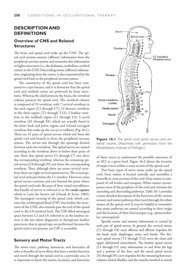
258 C O N D I T I O N S I N O C C U P A T I O N A L T H E R A P Y
DESCRIPTION AND DEFINITIONS
Overview of CNS and Related Structures
The brain and spinal cord make up the CNS. The spi-nal cord receives sensory (afferent) information from the peripheral nervous system and transmits this information to higher structures (i.e., the thalamus, cerebellum, cerebral cortex) in the CNS. Descending motor (efferent) informa-tion, originating from the cortex, is also transmitted by the spinal cord back to the peripheral nervous system.
The consistency of the spinal cord has been com-pared to a ripe banana, and it is fortunate that the spinal cord and cerebral cortex are protected by bony struc-tures. Whereas the skull protects the brain, the vertebral column protects the spinal cord. The vertebral column is composed of 33 vertebrae, with 7 cervical vertebrae in the neck region (C1 through C7); 12 thoracic vertebrae in the chest region (T1 through T12); 5 lumbar verte-brae in the midback region (L1 through L5); 5 sacral vertebrae (S1 through S5), which are actually fused in the lower back and pelvic region; and 4 fused coccygeal vertebrae that make up the coccyx or tailbone (Fig. 16.1). There are 31 pairs of spinal nerves, which exit from the spinal cord and branch to form the peripheral nervous system. The nerves exit through the openings formed between each two vertebrae. The spinal nerves are named according to the vertebrae above or below their point of exit. Note that spinal nerves C1 through C7 exit above the corresponding vertebrae, whereas the remaining spi-nal nerves (C8 through S5) exit below the corresponding vertebrae. Thus, although there are seven cervical verte-brae, there are eight cervical spinal nerves. The actual spi-nal cord ends just below the L1 vertebra. However, some spinal nerves continue and exit beyond the point where the spinal cord ends. Because of their visual resemblance, this bundle of nerves is referred to as the cauda equina, which is Latin for horse’s tail (Hanak & Scott, 1983). The meningeal covering of the spinal cord, which con-tains the cerebrospinal fluid (CSF) that bathes the struc-tures of the CNS, also extends past the end of the spinal cord to the L4 vertebral level. The CSF-filled meningeal space between L2 and L4, referred to as the lumbar cis-tern, is the site where diagnostic or therapeutic lumbar punctures, that is, spinal taps, are performed, because the spinal cord is not present, yet CSF is accessible.
Sensory and Motor Tracts
The terms tract, pathway, lemniscus, and fasciculus all refer to bundles of nerve fibers that have a similar function and travel through the spinal cord in a particular area. It is important to know the names, locations, and functions
of these tracts to understand the possible outcomes of an SCI at a given level. Figure 16.2 shows the location of major tracts within a cross section of the spinal cord.
Two basic types of nerve tissue make up the spinal cord. Gray matter is located centrally and resembles a butterfly in cross sections of the cord. Gray matter is com-posed of cell bodies and synapses. White matter encom-passes most of the periphery of the cord and contains the ascending and descending pathways. Table 16-1 provides a more detailed description of the functions of the various sensory and motor pathways that travel through the white matter of the spinal cord. It may be helpful to remember that many pathways are named according to their origin and the location of their final synapse (e.g., spinocerebel-lar, corticospinal).
Specific motor and sensory information is carried by each pair of spinal nerves. In general, the cervical nerves (C1 through C8) carry afferent and efferent impulses for the head, neck, diaphragm, arms, and hands. The tho-racic spinal nerves (T1 through T12) serve the chest and upper abdominal musculature. The lumbar spinal nerves (L1 through L5) carry information to and from the legs and a portion of the foot, and the sacral spinal nerves (S1 through S5) carry impulses for the remaining foot mus-culature, bowel, bladder, and the muscles involved in sexual
Figure 16.1 The spinal cord, spinal nerves, and ver-tebral column. (Reprinted with permission from the Rehabilitation Institute of Michigan.)
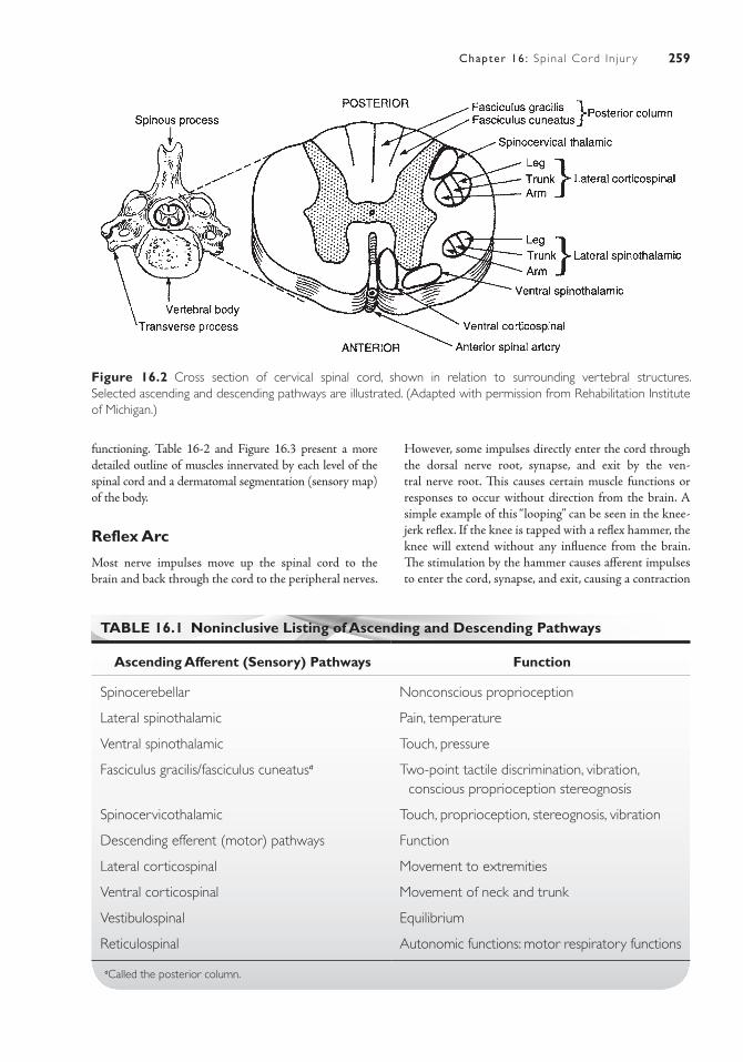
259Chapter 16: Sp ina l Cord In jur y
functioning. Table 16-2 and Figure 16.3 present a more detailed outline of muscles innervated by each level of the spinal cord and a dermatomal segmentation (sensory map) of the body.
Reflex Arc
Most nerve impulses move up the spinal cord to the brain and back through the cord to the peripheral nerves.
However, some impulses directly enter the cord through the dorsal nerve root, synapse, and exit by the ven-tral nerve root. This causes certain muscle functions or responses to occur without direction from the brain. A simple example of this “looping” can be seen in the knee-jerk reflex. If the knee is tapped with a reflex hammer, the knee will extend without any influence from the brain. The stimulation by the hammer causes afferent impulses to enter the cord, synapse, and exit, causing a contraction
Figure 16.2 Cross section of cervical spinal cord, shown in relation to surrounding vertebral structures. Selected ascending and descending pathways are illustrated. (Adapted with permission from Rehabilitation Institute of Michigan.)
TABLE 16.1 Noninclusive Listing of Ascending and Descending Pathways
Ascending Afferent (Sensory) Pathways Function
Spinocerebellar Nonconscious proprioception
Lateral spinothalamic Pain, temperature
Ventral spinothalamic Touch, pressure
Fasciculus gracilis/fasciculus cuneatusa Two-point tactile discrimination, vibration, conscious proprioception stereognosis
Spinocervicothalamic Touch, proprioception, stereognosis, vibration
Descending efferent (motor) pathways Function
Lateral corticospinal Movement to extremities
Ventral corticospinal Movement of neck and trunk
Vestibulospinal Equilibrium
Reticulospinal Autonomic functions: motor respiratory functions
aCalled the posterior column.
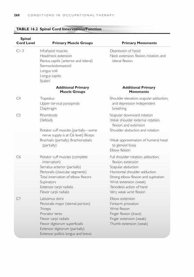
260 C O N D I T I O N S I N O C C U P A T I O N A L T H E R A P Y
TABLE 16.2 Spinal Cord Innervations/Function
Spinal Cord Level Primary Muscle Groups Primary Movements
C1-3 Infrahyoid musclesHead/neck extensionRectus capitis (anterior and lateral)SternocleidomastoidLongus colliLongus capitisScaleni
Depression of hyoidNeck extension, flexion, rotation, and
lateral flexion
Additional Primary Muscle Groups
Additional Primary Movements
C4 TrapeziusUpper cervical paraspinalsDiaphragm
Shoulder elevation, scapular adduction, and depression Independent breathing
C5 RhomboidsDeltoids
Rotator cuff muscles (partially—some nerve supply is at C6 level) Biceps
Brachialis (partially) Brachioradialis (partially)
Scapular downward rotationWeak shoulder external rotation,
flexion, and extensionShoulder abduction and rotation
Weak approximation of humeral head to glenoid fossa
Elbow flexion
C6 Rotator cuff muscles (complete innervation)
Serratus anterior (partially)Pectoralis (clavicular segments)Total innervation of elbow flexorsSupinatorsExtensor carpi radialisFlexor carpi radialis
Full shoulder rotation, adduction, flexion, extension
Scapular abductionHorizontal shoulder adductionStrong elbow flexion and supinationWrist extension (weak)Tenodesis action of handVery weak wrist flexion
C7 Latissimus dorsiPectoralis major (sternal portion)TricepsPronator teresFlexor carpi radialisFlexor digitorum superficialisExtensor digitorum (partially)Extensor pollicis longus and brevis
Elbow extensionForearm pronationWrist flexionFinger flexion (trace)Finger extension (weak)Thumb extension (weak)
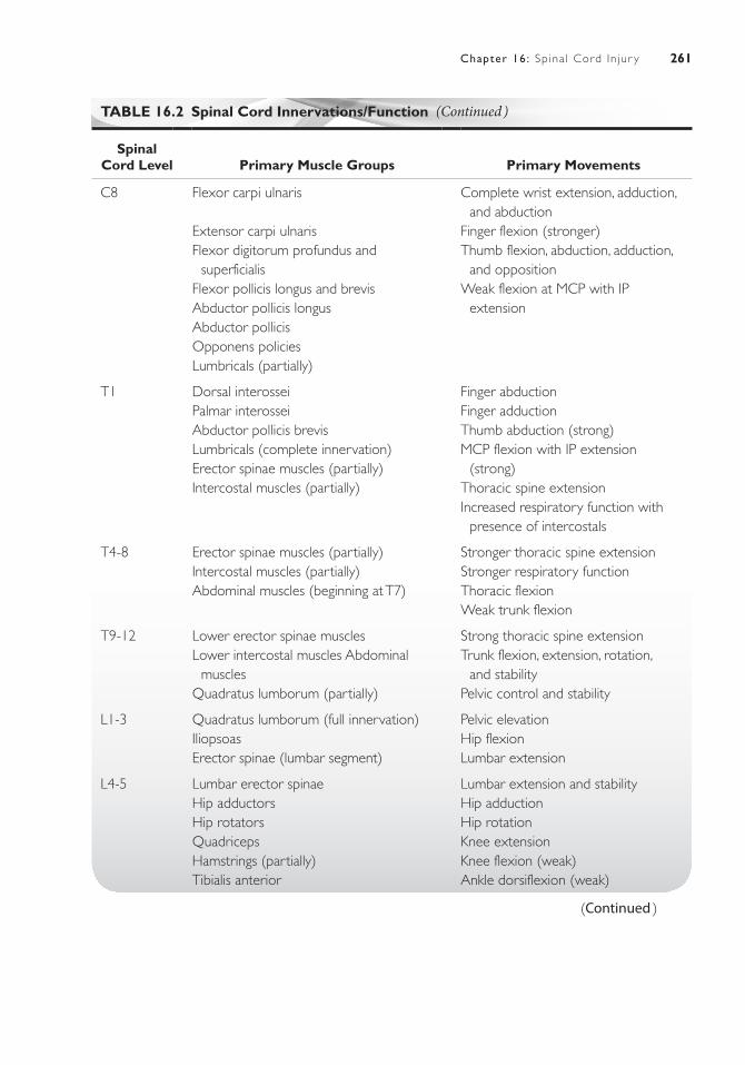
261Chapter 16: Sp ina l Cord In jur y
TABLE 16.2 Spinal Cord Innervations/Function
Spinal Cord Level Primary Muscle Groups Primary Movements
C8 Flexor carpi ulnaris
Extensor carpi ulnarisFlexor digitorum profundus and
superficialisFlexor pollicis longus and brevisAbductor pollicis longusAbductor pollicisOpponens policiesLumbricals (partially)
Complete wrist extension, adduction, and abduction
Finger flexion (stronger)Thumb flexion, abduction, adduction,
and oppositionWeak flexion at MCP with IP
extension
T1 Dorsal interosseiPalmar interosseiAbductor pollicis brevisLumbricals (complete innervation)Erector spinae muscles (partially)Intercostal muscles (partially)
Finger abductionFinger adductionThumb abduction (strong)MCP flexion with IP extension
(strong)Thoracic spine extensionIncreased respiratory function with
presence of intercostals
T4-8 Erector spinae muscles (partially)Intercostal muscles (partially)Abdominal muscles (beginning at T7)
Stronger thoracic spine extensionStronger respiratory functionThoracic flexionWeak trunk flexion
T9-12 Lower erector spinae musclesLower intercostal muscles Abdominal
musclesQuadratus lumborum (partially)
Strong thoracic spine extensionTrunk flexion, extension, rotation,
and stabilityPelvic control and stability
L1-3 Quadratus lumborum (full innervation)IliopsoasErector spinae (lumbar segment)
Pelvic elevationHip flexionLumbar extension
L4-5 Lumbar erector spinaeHip adductorsHip rotatorsQuadricepsHamstrings (partially)Tibialis anterior
Lumbar extension and stabilityHip adductionHip rotationKnee extensionKnee flexion (weak)Ankle dorsiflexion (weak)
(Continued )
(Continued )

262 C O N D I T I O N S I N O C C U P A T I O N A L T H E R A P Y
of the muscle fibers (Fig. 16.4). This activity is called a reflex arc. In persons with an intact spinal cord, afferent nerve impulses also travel to the brain almost instantane-ously. This allows an awareness, or “feeling,” of the initial stimulation (knee tap) and subsequent response (knee jerk). This concept is important to an understanding of SCIs. It explains why some individuals with SCI continue to have reflexes, but do not have voluntary control of their muscles. It also explains why others have no reflexes at all below the level of their injury. This is discussed in much greater detail in the section on classification of injuries.
ETIOLOGYHistorically, many demographic sources attempted to count the number of persons who have sustained SCIs. Since 1973, the National Spinal Cord Injury Database has been in exist-ence, making strides in collecting comprehensive data on a national level. In 1985, the Centers for Disease Control and Prevention began promoting surveillance mecha-nisms at state and national levels for the collection and reporting of these data. Prior to this time, data related to etiology and incidence of SCI in the United States were inconsistently collected and lacked uniformity; advance-ments are continuing to be made in this area.
The leading cause of SCI in the United States is motor vehicle accidents, followed by falls and acts of vio-lence (Fig. 16.5). Sports-related injuries account for most of the remaining SCIs, with diving being historically the most common (and preventable) cause (Table 16-3).
Analyzing the etiology of SCIs helps target pre-vention programs. Public awareness of the effects of using substances while operating a vehicle is certainly
heightened. Tougher penalties for driving under the influ-ence of cognitive altering substances have been enacted, and many states have adopted seat belt, child restraint, and “distracted” driving legislation. All of these efforts have the potential to reduce the leading cause of SCI. Grant monies have even been awarded to hospital-based programs that evaluate the home environments of senior citizens for safety. Their recommendations may reduce the risk of falls—a major cause of SCI in the elderly. An innovative effort sponsored by the University of Michigan Health System involves airing public service announce-ments on the prevention of diving injuries before the “coming attractions trailers” at popular movies for teens during the summer months.
Although much of the literature focuses on trauma, there are many nontraumatic causes of spinal cord dam-age (http://www.spinalcord.uab.edu). Developmental conditions such as spina bifida, scoliosis, and spinal cord agenesis may yield many of the same clinical signs as traumatic SCI. Acquired conditions, such as bacterial or viral infections, benign or malignant growths, embolisms, thromboses, hemorrhages, and even radiation or vaccina-tions, can also lead to damage of spinal cord tissue.
INCIDENCE AND PREVALENCEIncidence rates for SCI in the United States are estimated at 40 cases/million population/year, excluding those who die at the scene of an accident (http://www.spinalcord.uab.edu). This translates to about 12,000 new cases of SCI every year. The statistics indicate that over 80% of persons who sustain spinal cord injuries are male; notably, the mean age at the time of injury has increased from 28.7 years in the 1970s to the
TABLE 16.2 Spinal Cord Innervations/Function
Spinal Cord Level Primary Muscle Groups Primary Movements
S1-2 Hip extensorsHip abductorsHamstrings (complete innervation)Plantar flexorsInvertors of ankleEvertors of ankle
Hip extensionHip abduction and stabilityKnee flexionAnkle plantar : flexionAnkle inversion and stabilityAnkle eversion and stability
S2-5 BladderLower bowelGenital innervations
Genitourinary functionsBowel functions
MCP, metacarpophalangeal; IP, interphalangeal.
(Continued )
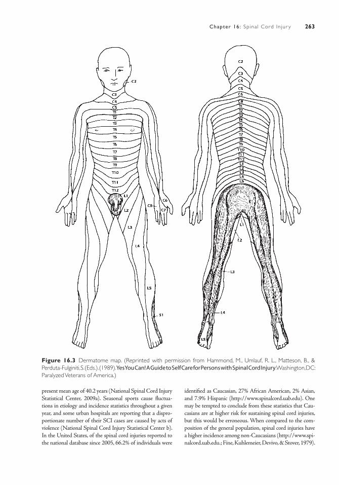
263Chapter 16: Sp ina l Cord In jur y
Figure 16.3 Dermatome map. (Reprinted with permission from Hammond, M., Umlauf, R. L., Matteson, B., & Perduta-Fulginiti, S. (Eds.). (1989). Yes You Can! A Guide to Self Care for Persons with Spinal Cord Injury. Washington, DC: Paralyzed Veterans of America.)
present mean age of 40.2 years (National Spinal Cord Injury Statistical Center, 2009a). Seasonal sports cause fluctua-tions in etiology and incidence statistics throughout a given year, and some urban hospitals are reporting that a dispro-portionate number of their SCI cases are caused by acts of violence (National Spinal Cord Injury Statistical Center b). In the United States, of the spinal cord injuries reported to the national database since 2005, 66.2% of individuals were
identified as Caucasian, 27% African American, 2% Asian, and 7.9% Hispanic (http://www.spinalcord.uab.edu). One may be tempted to conclude from these statistics that Cau-casians are at higher risk for sustaining spinal cord injuries, but this would be erroneous. When compared to the com-position of the general population, spinal cord injuries have a higher incidence among non-Caucasians (http://www.spi-nalcord.uab.edu.; Fine, Kuhlemeier, Devivo, & Stover, 1979).
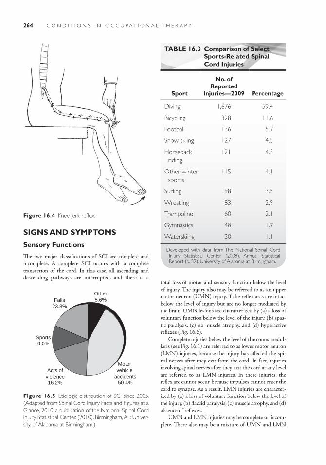
264 C O N D I T I O N S I N O C C U P A T I O N A L T H E R A P Y
Figure 16.4 Knee-jerk reflex.
Falls23.8%
Other5.6%
Motor vehicle
accidents50.4%
Acts ofviolence16.2%
Sports9.0%
Figure 16.5 Etiologic distribution of SCI since 2005. (Adapted from Spinal Cord Injury Facts and Figures at a Glance, 2010, a publication of the National Spinal Cord Injury Statistical Center. (2010). Birmingham, AL: Univer-sity of Alabama at Birmingham.)
SIGNS AND SYMPTOMS
Sensory Functions
The two major classifications of SCI are complete and incomplete. A complete SCI occurs with a complete transection of the cord. In this case, all ascending and descending pathways are interrupted, and there is a
TABLE 16.3 Comparison of Select Sports-Related Spinal Cord Injuries
Sport
No. of Reported
Injuries—2009 Percentage
Diving 1,676 59.4
Bicycling 328 11.6
Football 136 5.7
Snow skiing 127 4.5
Horseback riding
121 4.3
Other winter sports
115 4.1
Surfing 98 3.5
Wrestling 83 2.9
Trampoline 60 2.1
Gymnastics 48 1.7
Waterskiing 30 1.1
Developed with data from The National Spinal Cord Injury Statistical Center. (2008). Annual Statistical Report (p. 32). University of Alabama at Birmingham.
total loss of motor and sensory function below the level of injury. The injury also may be referred to as an upper motor neuron (UMN) injury, if the reflex arcs are intact below the level of injury but are no longer mediated by the brain. UMN lesions are characterized by (a) a loss of voluntary function below the level of the injury, (b) spas-tic paralysis, (c) no muscle atrophy, and (d) hyperactive reflexes (Fig. 16.6).
Complete injuries below the level of the conus medul-laris (see Fig. 16.1) are referred to as lower motor neuron (LMN) injuries, because the injury has affected the spi-nal nerves after they exit from the cord. In fact, injuries involving spinal nerves after they exit the cord at any level are referred to as LMN injuries. In these injuries, the reflex arc cannot occur, because impulses cannot enter the cord to synapse. As a result, LMN injuries are character-ized by (a) a loss of voluntary function below the level of the injury, (b) flaccid paralysis, (c) muscle atrophy, and (d) absence of reflexes.
UMN and LMN injuries may be complete or incom-plete. There also may be a mixture of UMN and LMN
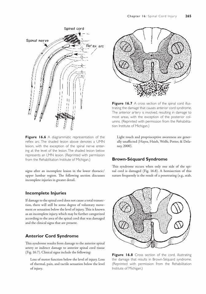
265Chapter 16: Sp ina l Cord In jur y
signs after an incomplete lesion in the lower thoracic/upper lumbar region. The following section discusses incomplete injuries in greater detail.
Incomplete Injuries
If damage to the spinal cord does not cause a total transec-tion, there will still be some degree of voluntary move-ment or sensation below the level of injury. This is known as an incomplete injury, which may be further categorized according to the area of the spinal cord that was damaged and the clinical signs that are present.
Anterior Cord Syndrome
This syndrome results from damage to the anterior spinal artery or indirect damage to anterior spinal cord tissue (Fig. 16.7). Clinical signs include the following:
■ Loss of motor function below the level of injury. Loss of thermal, pain, and tactile sensation below the level of injury.
Figure 16.6 A diagrammatic representation of the reflex arc. The shaded lesion above denotes a UMN lesion, with the exception of the spinal nerve enter-ing at the level of the lesion. The shaded lesion below represents an LMN lesion. (Reprinted with permission from the Rehabilitation Institute of Michigan.)
Figure 16.7 A cross section of the spinal cord illus-trating the damage that causes anterior cord syndrome. The anterior artery is involved, resulting in damage to most areas, with the exception of the posterior col-umns. (Reprinted with permission from the Rehabilita-tion Institute of Michigan.)
■ Light touch and proprioceptive awareness are gener-ally unaffected (Hayes, Hsieh, Wolfe, Potter, & Dela-ney, 2000).
Brown-Séquard Syndrome
This syndrome occurs when only one side of the spi-nal cord is damaged (Fig. 16.8). A hemisection of this nature frequently is the result of a penetrating (e.g., stab,
Figure 16.8 Cross section of the cord, illustrating the damage that results in Brown-Séquard syndrome. (Reprinted with permission from the Rehabilitation Institute of Michigan.)
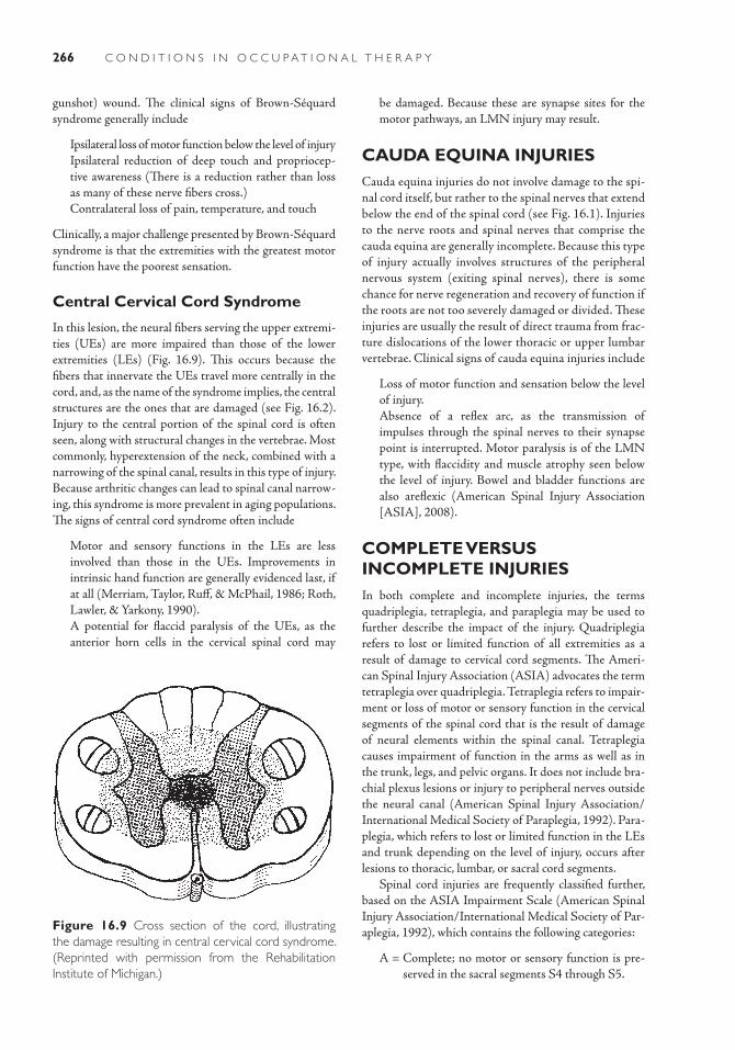
266 C O N D I T I O N S I N O C C U P A T I O N A L T H E R A P Y
gunshot) wound. The clinical signs of Brown-Séquard syndrome generally include
■ Ipsilateral loss of motor function below the level of injury ■ Ipsilateral reduction of deep touch and propriocep-
tive awareness (There is a reduction rather than loss as many of these nerve fibers cross.)
■ Contralateral loss of pain, temperature, and touch
Clinically, a major challenge presented by Brown-Séquard syndrome is that the extremities with the greatest motor function have the poorest sensation.
Central Cervical Cord Syndrome
In this lesion, the neural fibers serving the upper extremi-ties (UEs) are more impaired than those of the lower extremities (LEs) (Fig. 16.9). This occurs because the fibers that innervate the UEs travel more centrally in the cord, and, as the name of the syndrome implies, the central structures are the ones that are damaged (see Fig. 16.2). Injury to the central portion of the spinal cord is often seen, along with structural changes in the vertebrae. Most commonly, hyperextension of the neck, combined with a narrowing of the spinal canal, results in this type of injury. Because arthritic changes can lead to spinal canal narrow-ing, this syndrome is more prevalent in aging populations. The signs of central cord syndrome often include
■ Motor and sensory functions in the LEs are less involved than those in the UEs. Improvements in intrinsic hand function are generally evidenced last, if at all (Merriam, Taylor, Ruff, & McPhail, 1986; Roth, Lawler, & Yarkony, 1990).
■ A potential for flaccid paralysis of the UEs, as the anterior horn cells in the cervical spinal cord may
Figure 16.9 Cross section of the cord, illustrating the damage resulting in central cervical cord syndrome. (Reprinted with permission from the Rehabilitation Institute of Michigan.)
be damaged. Because these are synapse sites for the motor pathways, an LMN injury may result.
CAUDA EQUINA INJURIESCauda equina injuries do not involve damage to the spi-nal cord itself, but rather to the spinal nerves that extend below the end of the spinal cord (see Fig. 16.1). Injuries to the nerve roots and spinal nerves that comprise the cauda equina are generally incomplete. Because this type of injury actually involves structures of the peripheral nervous system (exiting spinal nerves), there is some chance for nerve regeneration and recovery of function if the roots are not too severely damaged or divided. These injuries are usually the result of direct trauma from frac-ture dislocations of the lower thoracic or upper lumbar vertebrae. Clinical signs of cauda equina injuries include
■ Loss of motor function and sensation below the level of injury.
■ Absence of a reflex arc, as the transmission of impulses through the spinal nerves to their synapse point is interrupted. Motor paralysis is of the LMN type, with flaccidity and muscle atrophy seen below the level of injury. Bowel and bladder functions are also areflexic (American Spinal Injury Association [ASIA], 2008).
COMPLETE VERSUS INCOMPLETE INJURIESIn both complete and incomplete injuries, the terms quadriplegia, tetraplegia, and paraplegia may be used to further describe the impact of the injury. Quadriplegia refers to lost or limited function of all extremities as a result of damage to cervical cord segments. The Ameri-can Spinal Injury Association (ASIA) advocates the term tetraplegia over quadriplegia. Tetraplegia refers to impair-ment or loss of motor or sensory function in the cervical segments of the spinal cord that is the result of damage of neural elements within the spinal canal. Tetraplegia causes impairment of function in the arms as well as in the trunk, legs, and pelvic organs. It does not include bra-chial plexus lesions or injury to peripheral nerves outside the neural canal (American Spinal Injury Association/International Medical Society of Paraplegia, 1992). Para-plegia, which refers to lost or limited function in the LEs and trunk depending on the level of injury, occurs after lesions to thoracic, lumbar, or sacral cord segments.
Spinal cord injuries are frequently classified further, based on the ASIA Impairment Scale (American Spinal Injury Association/International Medical Society of Par-aplegia, 1992), which contains the following categories:
A = Complete; no motor or sensory function is pre-served in the sacral segments S4 through S5.

267Chapter 16: Sp ina l Cord In jur y
B = Incomplete; sensory but not motor function is preserved below the neurologic level and extends through the sacral segments S4 through S5.
C = Incomplete; motor function is preserved below the neurologic level, and the majority of key muscles below the neurologic level have a muscle grade <3.
D = Incomplete; motor function is preserved below the neurologic level, and the majority of key muscles below the neurologic level have a muscle grade of ≥3.
E = Normal; motor and sensory function is normal.
A manual muscle test is performed to assess the strength of muscles and aid in the determination of the extent and nature of injury. Usually, strength is graded on a six-point scale (Aids to Investigation of Peripheral Nerve Injuries, 1943; Brunnstrom & Dennen, 1931; Hislop, Montgomery, & Connelly, 1995; Lovett, 1917).
0 = Total paralysis 1 = Palpable or visible contraction 2 = Active movement, full range of motion (ROM)
with gravity eliminated 3 = Active movement, full ROM against gravity 4 = Active movement, full ROM against moderate
resistance 5 = Normal active movement, full ROM against full
resistance NT = Not testable
COURSE AND PROGNOSISA prognosis implies that one can forecast or predict the outcome and chances for recovery from a particular disease or traumatic injury. That is somewhat challenging for SCIs. Although some aspects of SCI are highly predictable (e.g., specific muscle functions impaired with a complete lesion at C5), other aspects are much more vague.
Part of the ambiguity lies in the definitions of the term “recovery.” One definition is to get back, or regain, which tends to be the client’s focus. Another definition for recov-ery stresses compensation, which is often the thrust of the health care professional working with SCI. While the physi-ologic prognosis of SCI will be discussed here, the clinician should always be aware, and acknowledge the validity, of the client’s perspective on recovery. To “regain” and to “com-pensate for” are dramatically different frames of reference. When discussing prognosis with a person who has survived an SCI, the clinician should always be truthful, but must also be acutely aware of the impact of what the client is hearing. Perhaps the most crucial indicators of an individual’s func-tional outcome are personal characteristics such as motiva-tion, use of support systems, and coping mechanisms. The clinician must be skilled at fostering these strengths while at the same time providing accurate information.
POSTTRAUMATIC COMPLICATIONS
Spinal Shock
The period of altered reflex activity immediately after a traumatic SCI is known as spinal shock. As a result of injury, spinal c ord segments below the level of the lesion are deprived of excitatory input from higher CNS cent-ers. What is observed clinically during this phase is a flac-cid paralysis of muscles below the level of injury and an absence of reflexes (Yashon, 1986). The bladder is also flaccid, requiring catheterization, and there is no volun-tary control of the bowel. Depending on the level of the injury, the person with an SCI may require a ventilator because of lost or temporarily interrupted innervation to the diaphragm, intercostals, and abdominal muscles.
Spinal shock generally lasts from 1 week to 3 months after injury. Once spinal shock subsides, the areas of the spinal cord above the level of the lesion operate as they did premorbidly. Below the level of the lesion, reflexes will resume if the reflex arc is intact. This is an important con-cept to understand. Unlike a plant, which may die entirely if its stem is cut in half, the spinal cord is still alive and functional above and below the level of injury. The prob-lem is one of communication; the brain cannot receive sensory information beyond the lesion site and cannot volitionally control motor function below that point.
After spinal shock subsides, there is often an increase in spasticity, especially in the flexor muscle groups. The reflex arc “fires” and the brain is unable to interfere. After this phase, there may be a period of 6 to 12 months after injury when an increase in the spasticity of the extensor groups is common. Usually, after 1 year postinjury, the wide fluctuations in tone will cease.
An array of complications can greatly affect the prog-nosis of a person who has sustained an SCI. Some of the more common medical complications are addressed in the next section.
Respiratory Complications
Persons with spinal cord injuries at or below the level of T12 generally have a normal respiratory status. Injuries above that level, however, compromise the respiratory system to some degree. The abdominal musculature is innervated by segments T7 through T12, the intercostal muscles are served by segments T1 through T12, and the diaphragm is innervated by C4. Persons with com-plete injuries above C4 usually need a respirator. Some may be candidates for a phrenic nerve stimulator if the nerve shows the ability to conduct an impulse. Generally, persons with complete injuries at C4 and below do not use respirators, but respiratory complications may per-sist. Breathing may be shallow, and the ability to cough
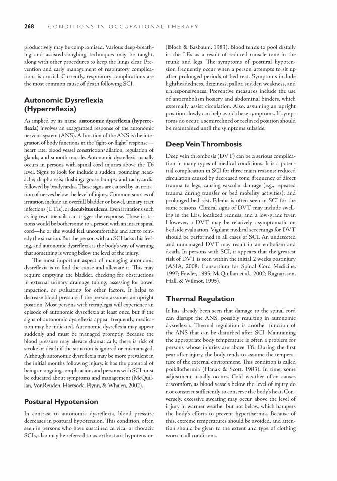
268 C O N D I T I O N S I N O C C U P A T I O N A L T H E R A P Y
productively may be compromised. Various deep-breath-ing and assisted-coughing techniques may be taught, along with other procedures to keep the lungs clear. Pre-vention and early management of respiratory complica-tions is crucial. Currently, respiratory complications are the most common cause of death following SCI.
Autonomic Dysreflexia (Hyperreflexia)
As implied by its name, autonomic dysreflexia (hyperre-flexia) involves an exaggerated response of the autonomic nervous system (ANS). A function of the ANS is the inte-gration of body functions in the “fight-or-flight” response—heart rate, blood vessel constriction/dilation, regulation of glands, and smooth muscle. Autonomic dysreflexia usually occurs in persons with spinal cord injuries above the T6 level. Signs to look for include a sudden, pounding head-ache; diaphoresis; flushing; goose bumps; and tachycardia followed by bradycardia. These signs are caused by an irrita-tion of nerves below the level of injury. Common sources of irritation include an overfull bladder or bowel, urinary tract infections (UTIs), or decubitus ulcers. Even irritations such as ingrown toenails can trigger the response. These irrita-tions would be bothersome to a person with an intact spinal cord—he or she would feel uncomfortable and act to rem-edy the situation. But the person with an SCI lacks this feel-ing, and autonomic dysreflexia is the body’s way of warning that something is wrong below the level of the injury.
The most important aspect of managing autonomic dysreflexia is to find the cause and alleviate it. This may require emptying the bladder, checking for obstructions in external urinary drainage tubing, assessing for bowel impaction, or evaluating for other factors. It helps to decrease blood pressure if the person assumes an upright position. Most persons with tetraplegia will experience an episode of autonomic dysreflexia at least once, but if the signs of autonomic dysreflexia appear frequently, medica-tion may be indicated. Autonomic dysreflexia may appear suddenly and must be managed promptly. Because the blood pressure may elevate dramatically, there is risk of stroke or death if the situation is ignored or mismanaged. Although autonomic dysreflexia may be more prevalent in the initial months following injury, it has the potential of being an ongoing complication, and persons with SCI must be educated about symptoms and management (McQuil-lan, VonReuden, Hartsock, Flynn, & Whalen, 2002).
Postural Hypotension
In contrast to autonomic dysreflexia, blood pressure decreases in postural hypotension. This condition, often seen in persons who have sustained cervical or thoracic SCIs, also may be referred to as orthostatic hypotension
(Bloch & Basbaum, 1983). Blood tends to pool distally in the LEs as a result of reduced muscle tone in the trunk and legs. The symptoms of postural hypoten-sion frequently occur when a person attempts to sit up after prolonged periods of bed rest. Symptoms include lightheadedness, dizziness, pallor, sudden weakness, and unresponsiveness. Preventive measures include the use of antiembolism hosiery and abdominal binders, which externally assist circulation. Also, assuming an upright position slowly can help avoid these symptoms. If symp-toms do occur, a semireclined or reclined position should be maintained until the symptoms subside.
Deep Vein Thrombosis
Deep vein thrombosis (DVT) can be a serious complica-tion in many types of medical conditions. It is a poten-tial complication in SCI for three main reasons: reduced circulation caused by decreased tone; frequency of direct trauma to legs, causing vascular damage (e.g., repeated trauma during transfer or bed mobility activities); and prolonged bed rest. Edema is often seen in SCI for the same reasons. Clinical signs of DVT may include swell-ing in the LEs, localized redness, and a low-grade fever. However, a DVT may be relatively asymptomatic on bedside evaluation. Vigilant medical screenings for DVT should be performed in all cases of SCI. An undetected and unmanaged DVT may result in an embolism and death. In persons with SCI, it appears that the greatest risk of DVT is seen within the initial 2 weeks postinjury (ASIA, 2008; Consortium for Spinal Cord Medicine, 1997; Fowler, 1995; McQuillan et al., 2002; Ragnarsson, Hall, & Wilmot, 1995).
Thermal Regulation
It has already been seen that damage to the spinal cord can disrupt the ANS, possibly resulting in autonomic dysreflexia. Thermal regulation is another function of the ANS that can be disturbed after SCI. Maintaining the appropriate body temperature is often a problem for persons whose injuries are above T6. During the first year after injury, the body tends to assume the tempera-ture of the external environment. This condition is called poikilothermia (Hanak & Scott, 1983). In time, some adjustment usually occurs. Cold weather often causes discomfort, as blood vessels below the level of injury do not constrict sufficiently to conserve the body’s heat. Con-versely, excessive sweating may occur above the level of injury in warmer weather but not below, which hampers the body’s efforts to prevent hyperthermia. Because of this, extreme temperatures should be avoided, and atten-tion should be given to the extent and type of clothing worn in all conditions.
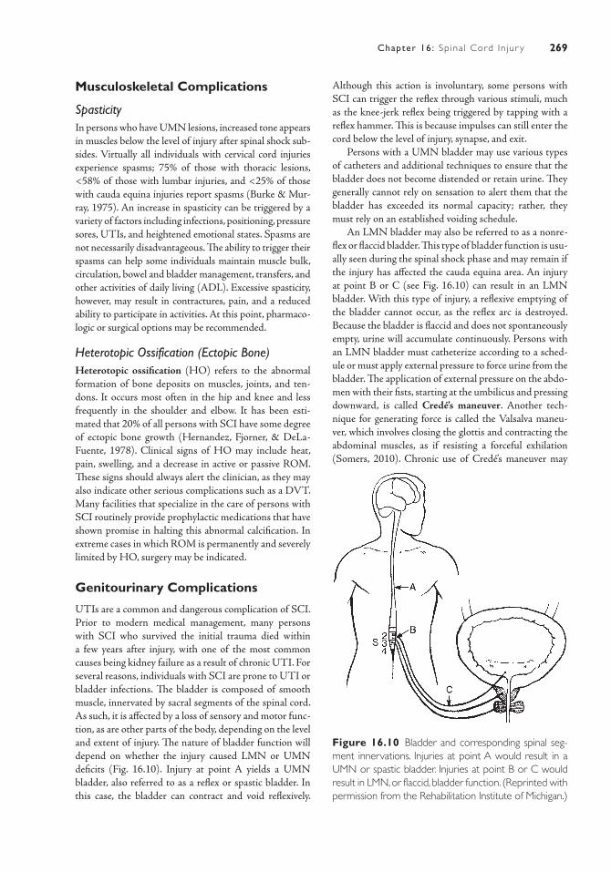
269Chapter 16: Sp ina l Cord In jur y
Musculoskeletal Complications
SpasticityIn persons who have UMN lesions, increased tone appears in muscles below the level of injury after spinal shock sub-sides. Virtually all individuals with cervical cord injuries experience spasms; 75% of those with thoracic lesions, <58% of those with lumbar injuries, and <25% of those with cauda equina injuries report spasms (Burke & Mur-ray, 1975). An increase in spasticity can be triggered by a variety of factors including infections, positioning, pressure sores, UTIs, and heightened emotional states. Spasms are not necessarily disadvantageous. The ability to trigger their spasms can help some individuals maintain muscle bulk, circulation, bowel and bladder management, transfers, and other activities of daily living (ADL). Excessive spasticity, however, may result in contractures, pain, and a reduced ability to participate in activities. At this point, pharmaco-logic or surgical options may be recommended.
Heterotopic Ossification (Ectopic Bone)Heterotopic ossification (HO) refers to the abnormal formation of bone deposits on muscles, joints, and ten-dons. It occurs most often in the hip and knee and less frequently in the shoulder and elbow. It has been esti-mated that 20% of all persons with SCI have some degree of ectopic bone growth (Hernandez, Fjorner, & DeLa-Fuente, 1978). Clinical signs of HO may include heat, pain, swelling, and a decrease in active or passive ROM. These signs should always alert the clinician, as they may also indicate other serious complications such as a DVT. Many facilities that specialize in the care of persons with SCI routinely provide prophylactic medications that have shown promise in halting this abnormal calcification. In extreme cases in which ROM is permanently and severely limited by HO, surgery may be indicated.
Genitourinary Complications
UTIs are a common and dangerous complication of SCI. Prior to modern medical management, many persons with SCI who survived the initial trauma died within a few years after injury, with one of the most common causes being kidney failure as a result of chronic UTI. For several reasons, individuals with SCI are prone to UTI or bladder infections. The bladder is composed of smooth muscle, innervated by sacral segments of the spinal cord. As such, it is affected by a loss of sensory and motor func-tion, as are other parts of the body, depending on the level and extent of injury. The nature of bladder function will depend on whether the injury caused LMN or UMN deficits (Fig. 16.10). Injury at point A yields a UMN bladder, also referred to as a reflex or spastic bladder. In this case, the bladder can contract and void reflexively.
Although this action is involuntary, some persons with SCI can trigger the reflex through various stimuli, much as the knee-jerk reflex being triggered by tapping with a reflex hammer. This is because impulses can still enter the cord below the level of injury, synapse, and exit.
Persons with a UMN bladder may use various types of catheters and additional techniques to ensure that the bladder does not become distended or retain urine. They generally cannot rely on sensation to alert them that the bladder has exceeded its normal capacity; rather, they must rely on an established voiding schedule.
An LMN bladder may also be referred to as a nonre-flex or flaccid bladder. This type of bladder function is usu-ally seen during the spinal shock phase and may remain if the injury has affected the cauda equina area. An injury at point B or C (see Fig. 16.10) can result in an LMN bladder. With this type of injury, a reflexive emptying of the bladder cannot occur, as the reflex arc is destroyed. Because the bladder is flaccid and does not spontaneously empty, urine will accumulate continuously. Persons with an LMN bladder must catheterize according to a sched-ule or must apply external pressure to force urine from the bladder. The application of external pressure on the abdo-men with their fists, starting at the umbilicus and pressing downward, is called Credé’s maneuver. Another tech-nique for generating force is called the Valsalva maneu-ver, which involves closing the glottis and contracting the abdominal muscles, as if resisting a forceful exhilation (Somers, 2010). Chronic use of Credé’s maneuver may
Figure 16.10 Bladder and corresponding spinal seg-ment innervations. Injuries at point A would result in a UMN or spastic bladder. Injuries at point B or C would result in LMN, or flaccid, bladder function. (Reprinted with permission from the Rehabilitation Institute of Michigan.)
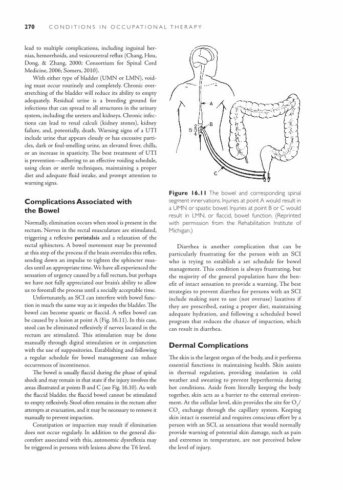
270 C O N D I T I O N S I N O C C U P A T I O N A L T H E R A P Y
lead to multiple complications, including inguinal her-nias, hemorrhoids, and vesicouretral reflux (Chang, Hou, Dong, & Zhang, 2000; Consortium for Spinal Cord Medicine, 2006; Somers, 2010).
With either type of bladder (UMN or LMN), void-ing must occur routinely and completely. Chronic over-stretching of the bladder will reduce its ability to empty adequately. Residual urine is a breeding ground for infections that can spread to all structures in the urinary system, including the ureters and kidneys. Chronic infec-tions can lead to renal calculi (kidney stones), kidney failure, and, potentially, death. Warning signs of a UTI include urine that appears cloudy or has excessive parti-cles, dark or foul-smelling urine, an elevated fever, chills, or an increase in spasticity. The best treatment of UTI is prevention—adhering to an effective voiding schedule, using clean or sterile techniques, maintaining a proper diet and adequate fluid intake, and prompt attention to warning signs.
Complications Associated with the Bowel
Normally, elimination occurs when stool is present in the rectum. Nerves in the rectal musculature are stimulated, triggering a reflexive peristalsis and a relaxation of the rectal sphincters. A bowel movement may be prevented at this step of the process if the brain overrides this reflex, sending down an impulse to tighten the sphincter mus-cles until an appropriate time. We have all experienced the sensation of urgency caused by a full rectum, but perhaps we have not fully appreciated our brain’s ability to allow us to forestall the process until a socially acceptable time.
Unfortunately, an SCI can interfere with bowel func-tion in much the same way as it impedes the bladder. The bowel can become spastic or flaccid. A reflex bowel can be caused by a lesion at point A (Fig. 16.11). In this case, stool can be eliminated reflexively if nerves located in the rectum are stimulated. This stimulation may be done manually through digital stimulation or in conjunction with the use of suppositories. Establishing and following a regular schedule for bowel management can reduce occurrences of incontinence.
The bowel is usually flaccid during the phase of spinal shock and may remain in that state if the injury involves the areas illustrated at points B and C (see Fig. 16.10). As with the flaccid bladder, the flaccid bowel cannot be stimulated to empty reflexively. Stool often remains in the rectum after attempts at evacuation, and it may be necessary to remove it manually to prevent impaction.
Constipation or impaction may result if elimination does not occur regularly. In addition to the general dis-comfort associated with this, autonomic dysreflexia may be triggered in persons with lesions above the T6 level.
Figure 16.11 The bowel and corresponding spinal segment innervations. Injuries at point A would result in a UMN or spastic bowel. Injuries at point B or C would result in LMN, or flaccid, bowel function. (Reprinted with permission from the Rehabilitation Institute of Michigan.)
Diarrhea is another complication that can be particularly frustrating for the person with an SCI who is trying to establish a set schedule for bowel management. This condition is always frustrating, but the majority of the general population have the ben-efit of intact sensation to provide a warning. The best strategies to prevent diarrhea for persons with an SCI include making sure to use (not overuse) laxatives if they are prescribed, eating a proper diet, maintaining adequate hydration, and following a scheduled bowel program that reduces the chance of impaction, which can result in diarrhea.
Dermal Complications
The skin is the largest organ of the body, and it performs essential functions in maintaining health. Skin assists in thermal regulation, providing insulation in cold weather and sweating to prevent hyperthermia during hot conditions. Aside from literally keeping the body together, skin acts as a barrier to the external environ-ment. At the cellular level, skin provides the site for O2/CO2 exchange through the capillary system. Keeping skin intact is essential and requires conscious effort by a person with an SCI, as sensations that would normally provide warning of potential skin damage, such as pain and extremes in temperature, are not perceived below the level of injury.

271Chapter 16: Sp ina l Cord In jur y
Damage to the skin as a result of pressure sores, or decubitus ulcers, is a major reason for hospital admis-sions in SCI populations. The mechanism of injury is continued pressure due to lack of movement. Circula-tion is impaired because of the pressure, and capillary exchange is impeded. This can result rapidly in tissue necrosis. The severity of pressure sores can be classified into four stages:
Stage I. Clinical signs are reddened or darkened skin. Damage is limited to more superficial (epidermal and dermal) layers. At this stage, tissue breakdown can be halted merely by removing pressure until the skin returns to its normal color.
Stage II. The skin now appears reddened and open. A blister or scab is present. The scab is not a sign of healing; rather, the tissue beneath it is necrotic. This involves the epidermal and dermal layers as well as deeper adipose tissue. Wound dressings may be involved at this stage, and it is imperative that pressure be kept off the site.
Stage III. The skin breakdown is deeper, and the wound is now draining. Muscle may be visible through the open wound. An ulcer is developing in the necrotic tissue. In addition to wound dress-ings, surgical intervention may be indicated if more conservative treatment is unsuccessful.
Stage IV. All structures, from the superficial levels to the bone, are destroyed. Infection and bone decay occur. Surgical intervention is likely, and the person with a decubitus ulcer at this stage often must spend weeks after a skin graft, with pressure totally removed from the involved site (Cassell, 1986).
Pressure sores are preventable with diligent attention and preventative strategies. A person with SCI can use, or instruct another person to assist with, a variety of pressure relief methods. Also, visual skin inspections should be performed at least twice daily, taking par-ticular note of areas most prone to breakdown. These would include areas where bony prominences (e.g., the sacrum, ischium, calcaneus, and scapula) can add to pressure. Proper nutrition also should be needed, as healthy skin is less apt to break down and is more responsive to healing in early stages of pressure sore development.
Other dermal complications, such as burns or frost-bite, are prevented by attentiveness and common sense. Even commonly encountered things, such as space heaters or exposed plumbing under a sink, can cause severe burns to persons with an SCI without their immediate knowledge. It is important to be aware of the environment and to rely on other, intact senses to avoid injury.
MEDICAL/SURGICAL MANAGEMENTMedical management of an SCI should begin immediately at the onset of injury. As typically injuries occur outside a hospital setting (roadways, lakes, sporting events), often first responders are not medical personnel. Any person with a suspected SCI should be immobilized with a backboard and neck brace before transport (Cooper, 2006). Diagnos-tic evaluation of the vertebral column and spinal cord must be completed to determine the next steps of intervention. Injuries may require surgical or nonsurgical intervention to decompress, realign, and stabilize the spine (McQuillan et al., 2002). Surgical procedures such as laminectomies may be performed to remove the cause of pressure on the cord (such as a bony fragment or bullet) (Hanak & Scott, 1993). Spinal fusion may also be required to achieve sta-bility. External alignment devices may be used following spinal surgery or may be sufficient substitutes for surgery (Hanak &Scott, 1993). In thoracic injuries, a thoraco-lum-bar-sacral-orthosis or clamshell brace is often used. This device is a “total-contact” molded two-piece brace, applied to the anterior and posterior of an individual to limit trunk movement (Beets, Faisant, Houghton, & Schlich, 1987). Multiple devices also exist for external stabilization of cer-vical injuries. These orthoses typically restrict the chin and the posterior aspect of the skull, restricting cervical motion (Somers, 2010). Among the most restrictive cervical devices is the “halo” orthosis. This is comprised of a metal ring affixed directly to the skull with screws (called “pins”) at occiptal and temporal points and steel bars mounted to a body vest (Somers, 2010; Hanak & Scott, 1993). The determination of the need and/or type of orthosis required demands careful assessment of vertebral stability and the nature of movement the device is able to restrict.
Concurrent with spinal decompression, realignment, and stabilization, prompt pharmacologic management is vital (McQuillan et al., 2002). In the most acute stages fol-lowing injury, recent research headlines have reported that the use of steroids—specifically, methylprednisolone—may improve neurologic outcomes of patients with SCI if administered within the first 8 hours after injury (Bracken, Holeord, & Shepard, 1990). Additionally, select experimental drugs administered soon after injury, such as GM-1 ganglioside (Sygen) and 4-AP, were hoped to aid in the preservation of damaged nerves and insulating them from the toxic level of chemicals the body releases during trauma and possibly reduce cell death (http://www.spi-nalcord.org/news). Study outcomes to date relating to the benefit of these drugs are mixed, and some concerns have been raised about potential adverse effects versus limited functional gains with some of these protocols. Another very recent acute experimental intervention that has been tried to mitigate spinal cord damage and hopefully
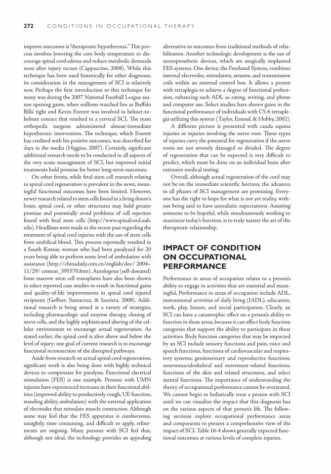
272 C O N D I T I O N S I N O C C U P A T I O N A L T H E R A P Y
improve outcomes is “therapeutic hypothermia.” This pro-cess involves lowering the core body temperature to dis-courage spinal cord edema and reduce metabolic demands soon after injury occurs (Cappuccino, 2008). While this technique has been used historically for other diagnoses, its consideration in the management of SCI is relatively new. Perhaps the first introduction to this technique for many was during the 2007 National Football League sea-son opening game, when millions watched live as Buffalo Bills’ tight end Kevin Everett was involved in helmet-to-helmet contact that resulted in a cervical SCI. The team orthopedic surgeon administered almost-immediate hypothermic intervention. The technique, which Everett has credited with his positive outcomes, was described for days in the media (Higgins, 2007). Certainly, significant additional research needs to be conducted in all aspects of the very acute management of SCI, but improved initial treatments hold promise for better long-term outcomes.
On other fronts, while fetal stem cell research relating to spinal cord regeneration is prevalent in the news, mean-ingful functional outcomes have been limited. However, newer research related to stem cells found in a living donor’s brain, spinal cord, or other structures may hold greater promise and potentially avoid problems of cell rejection found with fetal stem cells (http://www.spinalcord.uab.edu). Headlines were made in the recent past regarding the treatment of spinal cord injuries with the use of stem cells from umbilical blood. This process reportedly resulted in a South Korean woman who had been paralyzed for 20 years being able to perform some level of ambulation with assistance (http://chinadaily.com.cn/english/doc/ 2004–11/29/ content_395570.htm). Autologous (self-donated) bone marrow stem cell transplants have also been shown in select reported case studies to result in functional gains and quality-of-life improvements in spinal cord injured recipients (Geffner, Santacruz, & Izurieta, 2008). Addi-tional research is being aimed at a variety of strategies, including pharmacologic and enzyme therapy, cloning of nerve cells, and the highly sophisticated altering of the cel-lular environment to encourage actual regeneration. As stated earlier, the spinal cord is alive above and below the level of injury; one goal of current research is to encourage functional reconnection of the disrupted pathways.
Aside from research on actual spinal cord regeneration, significant work is also being done with highly technical devices to compensate for paralysis. Functional electrical stimulation (FES) is one example. Persons with UMN injuries have experienced increases in their functional abil-ities (improved ability to productively cough, UE function, standing ability, ambulation) with the external application of electrodes that stimulate muscle contraction. Although some may feel that the FES apparatus is cumbersome, unsightly, time consuming, and difficult to apply, refine-ments are ongoing. Many persons with SCI feel that, although not ideal, the technology provides an appealing
alternative to outcomes from traditional methods of reha-bilitation. Another technologic development is the use of neuroprosthetic devices, which are surgically implanted FES systems. One device, the Freehand System, combines internal electrodes, stimulators, sensors, and transmission coils within an external control box. It allows a person with tetraplegia to achieve a degree of functional prehen-sion, enhancing such ADL as eating, writing, and phone and computer use. Select studies have shown gains in the functional performance of individuals with C5-6 tetraple-gia utilizing this system (Taylor, Esnouf, & Hobby, 2002).
A different picture is presented with cauda equina injuries or injuries involving the nerve root. These types of injuries carry the potential for regeneration if the nerve roots are not severely damaged or divided. The degree of regeneration that can be expected is very difficult to predict, which must be done on an individual basis after extensive medical testing.
Overall, although actual regeneration of the cord may not be on the immediate scientific horizon, the advances in all phases of SCI management are promising. Every-one has the right to hope for what is not yet reality, with-out being said to have unrealistic expectations. Assisting someone to be hopeful, while simultaneously working to maximize today’s function, is to truly master the art of the therapeutic relationship.
IMPACT OF CONDITION ON OCCUPATIONAL PERFORMANCEPerformance in areas of occupation relates to a person’s ability to engage in activities that are essential and mean-ingful. Performance in areas of occupation include ADL, instrumental activities of daily living (IADL), education, work, play, leisure, and social participation. Clearly, an SCI can have a catastrophic effect on a person’s ability to function in these areas, because it can affect body function categories that support the ability to participate in these activities. Body function categories that may be impacted by an SCI include sensory functions and pain, voice and speech functions, functions of cardiovascular and respira-tory systems, genitourinary and reproductive functions, neuromusculoskeletal and movement-related functions, functions of the skin and related structures, and select mental functions. The importance of understanding the theory of occupational performance cannot be overstated. We cannot begin to holistically treat a person with SCI until we can visualize the impact that this diagnosis has on the various aspects of that person’s life. The follow-ing sections explore occupational performance areas and components to present a comprehensive view of the impact of SCI. Table 16-4 shows generally expected func-tional outcomes at various levels of complete injuries.
273Chapter 16: Sp ina l Cord In jur y
TABLE 16.4 Expected Functional Outcomes of Various Levels of Complete Injury (Noninclusive)
Last Spinal Cord Level Intact (Spared) Expected Functional Outcome
C1-3 Requires 24-h availability of caregiver.Generally ventilator-dependent; some individuals at C3 may be
successfully weaned from a ventilator.Requires maximal assistance of another for pressure relief or requires
an adapted switch and reclining chair.May propel power chair independently with adapted switches
(pneumatic, chin, head, mouthstick); requires maximal setup.Maximal assistance needed for transfers, positioning, bed mobility,
dressing, feeding, hygiene, grooming, and bowel/bladder care.Dependent with driving.
C4 Requires 24-h availability of caregiver.Generally able to be weaned from a ventilator ; continued difficulty
with productive coughing and deep breathing.Pressure relief, wheelchair propulsion, transfers, bed mobility, dressing,
hygiene, bowel/bladder care, and driving status comparable to C1-3 level.Adaptive feeding and grooming devices are available; they require
setup and are very time-consuming and exhaustive for a person at this level and generally do not result in task independence.
C5 May require 24-h availability of caregiver.Decreased respiratory endurance, but not using ventilator.A strong person with a C5 injury may be independent in pressure
relief by leaning side to side; a weaker person may require maximal assistance.
Independent on level surfaces with a power chair and occasionally wrist/forearm supports; a manual wheelchair with rim adaptations may be used by a strong person for short distances, but is typically not a reasonable mobility strategy.
Moderate to maximal assistance is required for all transfers, and generally a sliding board or mechanical lift is used.
Moderate assistance is required for bed mobility.A strong person with a C5 injury may assist with some dressing,
hygiene, and grooming activities with the aid of adapted equipment. Feeding is generally possible with the use of adapted utensils and setup.
Driving is feasible at this level (in the absence of additional complica-tions such as extensive UE tone/contractures); a person may be able to drive with specially adapted steering, braking, and acceleration hand controls, and due to transfer limitations would drive directly from the wheelchair.
(Continued )
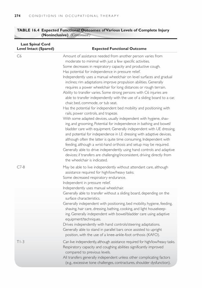
274 C O N D I T I O N S I N O C C U P A T I O N A L T H E R A P Y
TABLE 16.4 Expected Functional Outcomes of Various Levels of Complete Injury (Noninclusive)
Last Spinal Cord Level Intact (Spared) Expected Functional Outcome
C6 Amount of assistance needed from another person varies from moderate to minimal with just a few specific activities.
Some decreases in respiratory capacity and productive cough.Has potential for independence in pressure relief.Independently uses a manual wheelchair on level surfaces and gradual
inclines; rim adaptations improve propulsion abilities. Generally requires a power wheelchair for long distances or rough terrain.
Ability to transfer varies. Some strong persons with C6 injuries are able to transfer independently with the use of a sliding board to a car, chair, bed, commode, or tub seat.
Has the potential for independent bed mobility and positioning with rails, power controls, and trapeze.
With some adapted devices, usually independent with hygiene, shav-ing, and grooming. Potential for independence in bathing and bowel/ bladder care with equipment. Generally independent with UE dressing, and potential for independence in LE dressing with adaptive devices, although often the latter is quite time consuming. Independent with feeding, although a wrist-hand orthosis and setup may be required.
Generally able to drive independently using hand controls and adaptive devices; if transfers are challenging/inconsistent, driving directly from the wheelchair is indicated.
C7-8 May be able to live independently without attendant care, although assistance required for high/low/heavy tasks.
Some decreased respiratory endurance.Independent in pressure relief.Independently uses manual wheelchair.Generally able to transfer without a sliding board, depending on the
surface characteristics.Generally independent with positioning, bed mobility, hygiene, feeding,
shaving, hair care, dressing, bathing, cooking, and light housekeep-ing. Generally independent with bowel/bladder care using adaptive equipment/techniques.
Drives independently with hand controls/steering adaptations.Generally able to stand in parallel bars once assisted to upright
position, with the use of a knee-ankle-foot orthosis (KAFO).
T1-3 Can live independently, although assistance required for high/low/heavy tasks.Respiratory capacity and coughing abilities significantly improved
compared to previous levels.All transfers generally independent unless other complicating factors
(e.g., excessive tone challenges, contractures, shoulder dysfunction).
(Continued )
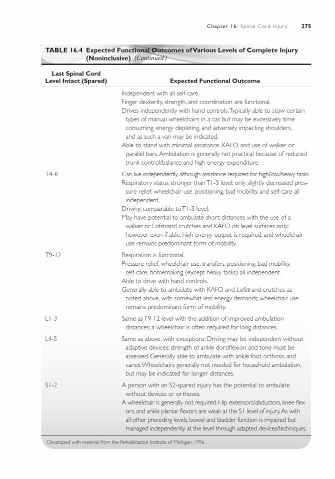
275Chapter 16: Sp ina l Cord In jur y
TABLE 16.4 Expected Functional Outcomes of Various Levels of Complete Injury (Noninclusive)
Last Spinal Cord Level Intact (Spared) Expected Functional Outcome
Independent with all self-care.Finger dexterity, strength, and coordination are functional.Drives independently with hand controls. Typically able to stow certain
types of manual wheelchairs in a car, but may be excessively time consuming, energy depleting, and adversely impacting shoulders, and as such a van may be indicated.
Able to stand with minimal assistance, KAFO, and use of walker or parallel bars. Ambulation is generally not practical because of reduced trunk control/balance and high energy expenditure.
T4-8 Can live independently, although assistance required for high/low/heavy tasks.Respiratory status stronger than T1-3 level; only slightly decreased pres-
sure relief, wheelchair use, positioning, bad mobility, and self-care all independent.
Driving: comparable to T1-3 level.May have potential to ambulate short distances with the use of a
walker or Lofstrand crutches and KAFO on level surfaces only; however even if able, high energy output is required, and wheelchair use remains predominant form of mobility.
T9-12 Respiration is functional.Pressure relief, wheelchair use, transfers, positioning, bad mobility,
self-care, homemaking (except heavy tasks) all independent.Able to drive with hand controls.Generally able to ambulate with KAFO and Lofstrand crutches as
noted above, with somewhat less energy demands; wheelchair use remains predominant form of mobility.
L1-3 Same as T9-12 level with the addition of improved ambulation distances; a wheelchair is often required for long distances.
L4-5 Same as above, with exceptions: Driving may be independent without adaptive devices; strength of ankle dorsiflexion and tone must be assessed. Generally able to ambulate with ankle foot orthosis and canes. Wheelchairs generally not needed for household ambulation, but may be indicated for longer distances.
S1-2 A person with an S2-spared injury has the potential to ambulate without devices or orthoses.
A wheelchair is generally not required. Hip extensors/abductors, knee flex-ors, and ankle plantar flexors are weak at the S1 level of injury. As with all other preceding levels, bowel and bladder function is impaired but managed independently at the level through adapted devices/techniques.
Developed with material from the Rehabilitation Institute of Michigan, 1996.
(Continued )

276 C O N D I T I O N S I N O C C U P A T I O N A L T H E R A P Y
Instrumental Activities of Daily Living
Grooming, Oral Hygiene, Eating, Bathing, DressingFor a person with tetraplegia, grooming, oral hygiene, and eating may be extremely laborious. The use of extensive adaptive devices or reliance on a caregiver may be necessary because of deficits in sensory and neuromuscular performance components. Persons with paraplegia are generally independent in these tasks, but they must often think ahead, making sure that items are available and sufficient time is allocated. Bathing and dressing present major challenges for persons with quadriplegia. With higher-level injuries (C1 through C4), total assistance is required. At lower levels of injury, with varying amounts of adaptive equipment and assistance from others in some task components, the person with quadriplegia can be a more active par-ticipant. It is extremely important for the person’s own goals to be acknowledged. A small but revealing study documented that individuals with a complete C6 injury typically required from 20 to 60 minutes to dress with the use of adaptations, yet none of the 10 patients in the study reported dressing themselves routinely after dis-charge from the hospital (Weingarden & Martin, 1989). They sought assistance from others because of feelings that the task was too time consuming and exhausting. The question becomes, “Is this functional?” If a person is attempting reentry into the work force or return to school, is it “functional” to spend an entire hour, as well as all the physical energy, in just getting dressed? Or is it a sign of greater autonomy to delegate some tasks to a caregiver to allow participation in activities that are more meaningful?
Secondary conditions aside, individuals with para-plegia are usually independent in bathing and dressing. Some even have the strength to transfer to the bottom of a tub without assistive devices.
ToiletingManaging altered bowel and bladder function is a chal-lenge for virtually everyone with SCI. The two aspects to this challenge are the actual physiologic management and the various techniques and equipment used in the toileting process.
Medications may be required for physiologic manage-ment. Stool softeners are used, as well as suppositories to assist in evacuation. A goal of effective bowel management is to eliminate or reduce reliance on medications. For per-sons with injuries at C7 or below, independence in toilet-ing can usually be achieved with an array of equipment that may include suppository inserters, digital stimulators (devices that trigger reflexes to relax the rectal sphincter
in UMN injuries), catheterization devices, leg separators, mirrors, and adapted commode chairs that allow access to the perianal area for bowel-training procedures.
Aside from medications and equipment, additional strategies include maintaining a specific schedule for elimination, eating a healthy diet that promotes regu-larity, assuming positions that facilitate elimination, and using Credé’s method. If toileting appears to be tiring, then it has been portrayed correctly! But many persons with SCI become adept at its management. It may be a very different picture, though, when a person attempts these procedures in a community environ-ment (i.e., rest rooms at work or school), due to acces-sibility, availability of equipment, and time limitations. In order to promote greater freedom and independence, some individuals opt to explore alternatives—such as undergoing surgical urinary diversion. Surgeries such as the Mitrofanoff procedure create a permanent stoma in the abdomen, which can allow an individual to cathe-terize through their navel. This presents benefits of eas-ier anatomical access and significantly less adjustment of clothing versus traditional catheterizing. While not every individual is a candidate, and the procedure is cer-tainly not without risk and potential long-term compli-cation, studies have revealed that many who undertake it have been highly satisfied with the degree of free-dom and independence it has allowed them (Merenda, Duffy, & Betz, 2007).
The person with a higher-level cervical injury faces greater challenges. Although the bowel- and bladder-management concepts are the same, neuromuscular deficits limit performance of the tasks, even with adaptive equipment. Generally, persons with injuries above the C6 level require a caregiver to assist with or perform functions such as transfers to adaptive com-mode chairs, suppository insertion, digital stimula-tion, catheterization, and general perianal care. The person with an SCI must indicate who the caregiver will be, particularly for tasks that are socially sensitive. Even though family members may be willing to assist, it is perfectly justifiable for the person to request someone else as a caregiver. Some persons may have no reservations about who assists them, whereas oth-ers may feel strongly that it would negatively affect established roles. Whatever the case, whenever feasi-ble, the preferences of the person with an SCI should be the deciding factor in selecting caregivers for vari-ous tasks.
Personal Device Care
The extent of personal and adaptive devices required by an individual with an SCI can generally be predicted by
277Chapter 16: Sp ina l Cord In jur y
the level of injury (see Table 16-4). Additional compli-cations, such as reduced ROM resulting from contrac-tures, may require the use of more extensive devices than are typically seen at a particular level. Usually, personal device care is performed by others for those with inju-ries at C4 and above. Those with injuries at the C5 level and below have progressively more ability to assist in personal device care and use, but this fluctuates greatly depending on the individual’s endurance, motivation, resources, and priorities.
Health Maintenance
Fostering a healthy lifestyle is critical, but often challeng-ing, after an SCI. Most persons with an SCI are advised to follow lifelong exercise programs to preserve and enhance ROM and strength as well as to promote good cardiovascular fitness and weight management. Many individuals report weight gains in the months after injury, as their energy demands are greatly altered in great part by use of a wheelchair for mobility. Attention also must be given to proper nutrition for weight man-agement, impact on skin integrity, and bowel and blad-der management. As mentioned earlier, routine attention to skin condition is crucial to avoiding dermal compli-cations. A health maintenance routine may require the assistance of others, depending on the level of injury, for such activities as setting up weights and equipment, per-forming passive ROM, pressure relief, and aiding in skin inspection.
Socialization, Functional Communication, and Emergency Response
With a singular diagnosis of SCI, no cognitive deficits are inherent that would preclude a person from socializing in an appropriate contextual manner. What is challenging, though, are the variety of barriers that may inhibit sociali-zation. Architectural, environmental, and transportation barriers; reduced endurance; and increased reliance on others may discourage or actually prevent someone from traveling to the places where they socialized before their injury. Psychological barriers also may prevent reintegra-tion into a premorbid social support system, and these “barriers” are as real as physical ones.
In all but the highest of injuries, verbal communication is functional. Persons with injuries at C7 or below are generally independent with a variety of forms of commu-nication (e.g., writing, keyboard use, phone use) without the use of adaptive equipment. Above this level, however, adaptive devices are progressively required.
The need to request assistance in emergencies is pos-sible at virtually all levels of injury, depending on the environment and adaptive equipment available. For per-sons with higher-level injuries, adaptive phone devices, emergency call systems, and environmental control units make contact with emergency agencies feasible. At the level of C4 and above, however, the availability of a car-egiver 24 hours a day remains the most appropriate safety option. Individuals with injuries below this level may use a phone independently with possible adaptations but may be limited in other emergency responses, such as exiting a dwelling or attending to the physical needs of another injured person.
Functional Mobility
Persons with complete injuries at the thoracic levels of injury and above usually rely on wheelchairs for household and community mobility. Those with injuries at C6 or above may require additional assistance in manual wheel-chair management; they may utilize a manual wheelchair with power-assist wheels or a power wheelchair. Indi-viduals with higher-level quadriplegia may use adapted orthotic devices to aid in controlling a power wheelchair or alternative interfaces such as “puff and sip” or chin controls to maneuver the wheelchair in the absence of sufficient UE function. Even at lower levels of injury, while an indi-vidual may be able to navigate a manual wheelchair under certain conditions, power wheelchairs may be considered due to community access needs (e.g., the need to navigate a large college campus routinely). Persons with lower-level thoracic injuries may be able to ambulate short distances with ambulation devices such as walkers or Lofstrand (forearm) crutches and LE orthotic devices; however, the practicality of this in household and community settings must be evaluated. It is important to consider the need to avoid repetitive motion disorders in the UEs, which can become problematic if overused during ambulation activi-ties. Persons with sacral injuries often can ambulate com-munity distances without orthoses or devices.
Sometimes it is not so much the ability or inability of the person with an SCI but the inaccessibility of the environment that limits functional mobility. Whereas the home environment can be modified through creative thoughts, planning, and finances, the community envi-ronment is much harder to change. Although the Ameri-cans with Disabilities Act celebrated its 20th anniversary of enactment on July 26, 2010, most would agree that compliance with and enforcement of barrier-free envi-ronments are less than ideal. Many physically challenged persons continue to fight discriminatory situations (e.g., inaccessible public transportation, restaurants, offices, classrooms). A lighter side of the inaccessibility issue
278 C O N D I T I O N S I N O C C U P A T I O N A L T H E R A P Y
was recalled by Ed Roberts, founder of the first Center for Independent Living. He reminisced that early protes-tors in the disability rights movement were released from police custody because jail cells were inaccessible (Price, 1990)! In the vast majority of situations, however, an inaccessible environment is frustrating, demeaning, and personally violating. The health care team can do its part by helping physically, emotionally, or cognitively chal-lenged persons to be aware of their rights, as well as by providing them with information on advocacy groups.
Functional mobility includes driving as a considera-tion for many. A strong C5-injured person may be able to drive with specially adapted low-effort steering, brak-ing, and acceleration hand controls. Usually, those with injuries above the C5 level cannot drive because of physi-cal and respiratory limitations. Persons with C6 injuries and below may be able to drive independently using hand controls and adaptive steering devices. A van with a power lift or ramp may be recommended for someone who has difficulty transferring from their wheelchair or inde-pendently stowing their wheelchair in a vehicle. Often, finances more than need may dictate the type of trans-portation used, and clinicians can be invaluable in helping clients identify what financial and community resources may be available to help them meet their mobility needs.
Sexual Expression
As with bowel and bladder function, most persons with SCI experience alterations in their ability to perform sexually as compared with their premorbid status. The nerves that innervate the genital area (both motor and sensory components) originate at the sacral spinal cord levels, so in the great majority of SCI cases, sexual func-tion is affected.
For the most part, a person can participate in a vari-ety of sexual activities despite SCI, although the level of sensation and motor response will vary, depending on the extent and level of injury. Medications, surgical implants, and sexual enhancement devices may be recommended by specialists, but this is highly individualized.
Advances in the field of fertility have improved the chances for a spinal cord–injured male to father a child. Although fertility rates for men with SCI are estimated at <10%, techniques such as electrostimulation to induce ejaculation in paraplegics have proven successful in many cases (Perkash, Martin, & Warner, 1990). Men may be well advised to delay a decision about surgical penile implants until at least 1 year after injury. This allows time to evaluate the full impact of the SCI on sexual function and to determine the extent, if any, of sensory or motor return in an incomplete injury. The reproductive capa-
bilities of women are generally unaltered by an SCI, and women and their partners should be made aware that the potential for pregnancy exists (Sipski, 1991).
Addressing sexual function should be an integral part of each person’s treatment plan, but a distinction should be made between purely physiologic sexual perfor-mance—arousal, orgasm, and ejaculation—and sexuality, which is the totality of a person’s attractiveness, personal-ity, and self-perception as a sexual being. Sexuality does not have to rely on physiology. Whereas performance may be hampered as a result of an SCI, one’s sexuality can be quite healthy and intact. The health care provider must make accurate information available and identify sources of more detailed information or expertise. It is entirely at the discretion of the person with an SCI to explore, or not explore, options available to them.
Home Maintenance
Tasks such as household maintenance, meal preparation, shopping, cleaning, clothing care, and safety procedures are included within the general category of home main-tenance. Usually, persons with a C6-level injury or above require assistance with all of these activities and need some personal attendant care. Persons with injuries at C7 or lower can often live independently (without attendant care) as they are able to perform their basic ADL tasks in accessible environments. However, they typically require assistance with heavier home-maintenance tasks or activi-ties requiring access to high or low areas.
Care of Others
An SCI can certainly make caring for others difficult, particularly if the injured person had been the primary caregiver for a child, spouse, or parent. Although a major goal of those with SCI is mastering personal self-care, a concurrent goal may be the introduction of activities (e.g., diapering, bathing a child) that can allow them to assume some premorbid roles. Often, many of these pre-vious responsibilities must be delegated to others. In these cases, it is ideal if the person with SCI retains the respon-sibility for the verbal direction of care.
Education
Persons with SCI can generally resume educational activ-ities, even while still inpatients in rehabilitation facilities. Many specialty hospitals retain the services of teachers from local school districts—or encourage the involve-ment of a student’s premorbid teachers—and educational
279Chapter 16: Sp ina l Cord In jur y
instruction is often scheduled along with other thera-pies for elementary, secondary, and high school students, encouraging smoother reintegration after discharge. The length of hospital admission following an SCI has decreased dramatically in the nearly three decades since more reliable statistics have been available; currently, the average length of combined acute/inpatient rehabilita-tion hospitalization following an SCI is 49 days (http://fscip.org//facts.htm). Certainly, a variety of factors can impact the length of hospitalization (e.g., level of injury, secondary diagnoses, complications, availability of spe-cialty hospital resources), but the overall trend toward reduced inpatient hospitalization allows for individuals to reintegrate back into their premorbid roles and sup-port systems much more rapidly than in the past. There are certainly advantages, but some inherent concerns, for children and adolescents returning more quickly to their previous school environments after the significant life changes brought on by an SCI. The ongoing support of the health care team can be critical in promoting success-ful reintegration.
College students would most likely be able to resume their studies following an SCI. Depending on their level of injury; however, persons may reevaluate their course of study to prepare for a more feasible career.
Adaptive writing devices, page turners, recording devices, and computers have made returning to the class-room less intimidating. Laws have also improved the accessibility of public buildings. It is challenging, though, for a student with an SCI to manage bowel and bladder schedules, adjustment of clothing, eating devices, and so forth, but with planning and the assistance of others if needed, many individuals have successfully returned to the classroom.
Vocational
It is appropriate for a person with any level of SCI to begin to formulate vocational options, even as an inpa-tient, with the members of the health care team. Indi-vidual situations are so variable—premorbid occupation, level of injury, educational level, other vocational inter-ests, family support, cognitive abilities, motivation, finan-cial resources—making it impossible to say whether a person with a certain level of SCI can or cannot be gain-fully employed. However, legislation mandates that work sites be accessible within reason. Also, many employers recognize the importance of a trained employee and will
make additional accommodations to return a valued per-son to the workplace.
As we move from an industrial to an information soci-ety, the job market will continue to change, with positions requiring less physical labor than in the past. Job require-ments will increasingly demand analytical thought, prob-lem solving, and creativity, all of which are certainly intact after an SCI!
Leisure Activities
For most people, leisure pursuits are an integral part of a meaningful life. Although an SCI can alter the way in which one participates in leisure activities, it does not have to change the intensity of participation.
Sports, both individual and team, are excellent lei-sure pursuits that are growing in popularity for persons with SCI. Virtually any sport can be undertaken, from basketball to tennis to archery. Adaptive equipment and modified regulations help make some sports more feasible for persons with an SCI, and these modifi-cations should not be viewed as detracting from the competitiveness of the sport. Consider that athletes in the general population use adaptive equipment all the time. How long would a catcher last in a baseball game without a mitt or a mask? Adaptive equipment need not detract from the legitimacy of the contest. It is hearten-ing to see that, even internationally, the wheelchair ath-lete is recognized for excellence, with designated events in the Olympic games. Persons with an SCI who want to participate have numerous avenues open to them.
Aside from sports, opportunities for social activities from square dancing to traveling abound. Travel agencies and tour groups have recognized the market created by the wheelchair traveler and have responded. Most hospi-tals with specialized SCI rehabilitation units have well-established programs that help persons get involved with special interest groups. Often, during the acute phase of SCI, persons cannot envision themselves participating again in the things they enjoy. The health care profes-sional must be available for these individuals to encourage renewed interest in favorite leisure activities.
Several resources related to leisure activities, as well as other issues, have been discussed in this chapter. The reader is encouraged to consult the references and sug-gested readings at the end of this chapter for more infor-mation. Additionally, Table 16-4 gives an overview of specific expected outcomes for each level of SCI.
280 C O N D I T I O N S I N O C C U P A T I O N A L T H E R A P Y
Case IllustrationsCase 1M.L. is a 24-year-old woman who sustained an SCI during a motor vehicle accident while on her honey-moon. Her husband was thrown from the vehicle but received only minor injuries. M.L. was transported by emergency medical services to the local emergency department. She was diagnosed with a C5 through C6 vertebral subluxation and a C6 crush injury, resulting in a complete (ASIA-A) C6 SCI. She also sustained a left clavicular fracture. She received nasal O
2 for respi-ratory support. Once stabilized, she was transferred to the specialized trauma center near her home. A halo vest was applied, which stabilized her cervi-cal spine. No operative procedures were indicated at that time. After 22 days, her endurance improved so that she could tolerate sitting upright in a chair for up to 1 hour. She was transferred to the nearby rehabilitation facility’s SCI unit. During her 12 weeks there, the only complications she experienced were two episodes of autonomic hyperreflexia (apparently secondary to hard stool in the lower rectum) and a mild UTI. Spasticity developed in her wrists, elbows, and LEs.
Before her injury, M.L. was a recently graduated college student with a liberal arts degree. She and her new husband had recently signed a 1-year lease on an upstairs apartment close to the university, as she was anticipating pursuing graduate studies. She had been totally independent in all of her ADL and home-man-agement activities. Her leisure pursuits included rec-reational team sports (particularly softball) and more sedentary activities like reading and gourmet cooking. She was also involved with her family, particularly two sisters and her parents, who live close by.
Many body functions are significantly affected by M.L.’s injury. Her sensorimotor deficits are consistent with those anticipated for a C6 complete quadriplegia.
M.L. is challenged by the psychosocial/psychologi-cal issues facing her as a result of her SCI. She feels that her role has changed significantly, especially in her relationship with her husband. He has been willing to assist her in those activities in which she is physically limited; however, his assistance in some activities—particularly bowel management—has been difficult for her to accept. This has been a source of frustration for her, and she has discussed with him the possibil-ity of hiring an attendant on a limited basis to assist with specific activities. He resists this idea, stating that it is his desire and duty to care for her. He took a temporary leave of absence from the family-owned
landscaping company where he is employed when she was discharged 2 weeks ago. There is no definite timetable for his return. M.L. also is concerned about her ability to express herself sexually, as well as her potential to have children in the future. She attended classroom sessions on these topics in the rehabilita-tion facility, but she did not seek any individual coun-seling. M.L. states that she probably was not ready to hear anything specific then, but she now wishes she had someone to whom she could ask questions.
M.L. has not begun to consider her work activi-ties or return to school. Even before her accident, she had been undecided about a career path. She has expressed interest, however, in exploring possible alternatives.
M.L. has stated that eventually she might like to get involved in team sports again. In the 2 weeks since her discharge, though, her main leisure pursuits have been reading and watching television.
Case 2H. B. is a 6-year-old boy who sustained an incomplete (Brown-Séquard) SCI, as the result of being struck by a stray bullet during a drive-by shooting in early August. He did not lose consciousness during transport to the emergency department. It was determined radiologi-cally that the bullet, which entered from the right near the base of his neck/upper back, was lodged in the C7/C8 cervical spinal canal, and surgery was performed to remove the bullet soon after admission. H.B. is now 2 weeks postinjury and exhibits a loss of voluntary movement, reduced touch, and proprioceptive sensa-tion on the right below the level of C7. He exhibits reduced pain, reduced thermal, and some reduction in touch awareness on the left below the level of C7. The levels of C7 and above appear intact for both motor and sensory function. H.B. has had an uncomplicated hospital admission at this time. It is anticipated that he will remain in the acute children’s specialty hospital for at least another 2 weeks and then transfer to the inpatient pediatric rehabilitation unit.
At the time of his injury, H.B. was living with his maternal grandmother, who is also his legal guard-ian. H.B. had been cocaine-positive at birth, and his biologic mother had relinquished her parental rights shortly after he was born. H.B.’s biologic father is unknown. H.B.’s grandmother is also caring for another of H.B.’s siblings, an 8-year-old sister. H.B.’s grandmother reports that prior to his injury, he did not appear to have any overt behavioral/cognitive
281Chapter 16: Sp ina l Cord In jur y
challenges and had successfully completed kindergar-ten. She related that he was a “normal, active boy,” and enjoyed playing any type of outdoor games as well as watching television. She states he did not have much exposure to computers or video games, as the family did not own these items. She states he had been look-ing forward to starting first grade, which is scheduled to begin in his district next week.
H.B. has been very cooperative with all of the health care providers working with him; he appears to enjoy the attention he is receiving from the staff as well as the visitors from the neighborhood, his grandmother’s church, and police department. His grandmother has expressed many concerns to the health care professionals working with H.B. Her overriding concern is fear that she will be unable to provide sufficient care—both from a physical and
financial perspective—for H.B. following his inpatient rehabilitation stay. She related that the family resi-dence is an older, rented duplex, with seven steps leading up to the front porch. She stated the inte-rior of the residence is not conducive to any type of mobility devices, as the hallways and doorways are narrow, and all the bedrooms are on the second story of the unit. She also states concern about how he will be treated when he eventually starts school; although his older sister is in the same elementary school, H.B.’s grandmother states she has had very limited contact with teachers/administrators at the school and has no idea what resources are available to help him. She expressed frustration that H.B.’s shooter has not been apprehended and shared feelings of hopelessness and helplessness in her ability to protect H.B. and his sister from harm in the future.
R E C O M M E N D E D L E A R N I N G R E S O U R C E S
Americans with Disabilities Act, http://www.ada.gov. Accessed June 17, 2010.
General Texts/Resources Relating to SCIAmerican Spinal Injury Association/International Medi-
cal Society of Paraplegia. (1992). International stan-dards for neurological and functional classification of spinal cord injury. Chicago: American Spinal Injury Association.
Berczeller, P. H., & Bezkor, M. F. (1986). Medical compli-cations of quadriplegia. Chicago: Yearbook Medical Publishers.
Buchanan, L. E. & Nawoczenski, D. A. (1987). Spinal cord injury: Concepts and management. Baltimore, MA: Williams & Wilkins.
Field-Fote, E. C. (2009). Spinal cord injury rehabilitation. Philadelphia: F.A. Davis Co.
National Institute of Neurological Disorders and Stroke. Retrieved from http://www.ninds.gov. Accessed July 22, 2005.
National Spinal Cord Injury Statistical Center (NSCISC). 619 19th Street South, SRC 515, Birmingham, Ala-bama, 35249–7330. Retrieved from http://www.spi-nalcord.uab.edu. Accessed August 12, 2010.
Ozer, M. N. (1988). The management of persons with spinal cord injury. New York: Demos Publications.
Somers, M. F. (2010). Spinal cord injury: Functional rehabilitation (3rd ed.). Upper Saddle River, NJ: Pearson.
Treischmann, R. (1988). Spinal cord injuries: Psychologi-cal, social and vocational rehabilitation (2nd ed.). New York: Demos Publications.
Whiteneck, G., Lammertse, D., …Manley, S. (1989). The Management of High Quadriplegia. New York: Demos Publications.
Yashon, D. (1986). Spinal injury (2nd ed.). East Norwalk, CT: Appleton-Century-Crofts.
Patient/Family ResourcesAlpert, M., & Wisnia, S. (2008). Spinal cord injury and the
family: A new guide. Boston, MA: Harvard University Press.
Christopher Reeve Foundation P.O. Box 277, FDR Sta-tion, New York, NY 10150–0277. Retrieved from www.apacure.com
Corbet, B. (1980). Options: Spinal cord injury and the future. Denver: A.B. Hirschfield.
Hammond, M., Umlauf, R. L., Matteson, B., & Perduta-Fulginiti, S. (Eds.). (1989). Yes you can! A guide to self care for persons with spinal cord injury. Washington, DC: Paralyzed Veterans of America.
Phillips, L., Ozer, A., … Axelson, P. (1987). Spinal cord injury: A guide for patient and family. New York: Raven Press.
Spinal Cord Injury Information Network. Retrieved from www.spinalcord.uab.edu. Accessed July 29, 2010.
The Miami Project to Cure Paralysis. University of Miami School of Medicine. P.O. Box 0#960, Mail Locator R-48, Miami, FL 33101. Retrieved from www.miamiproject.miami.edu. Accessed August 14, 2010.
282 C O N D I T I O N S I N O C C U P A T I O N A L T H E R A P Y
General NeuroanatomyBarr, M. L. (1979). The human nervous system: An ana-
tomic viewpoint. (3rd ed.). New York, NY: Harper & Row.
Liebman, M. (1986). Neuroanatomy made easy and understandable. (3rd ed.). Rockville, MD: Aspen Pub-lishers.
Aids to Investigation of Peripheral Nerve Injuries. (1943). Medical research council war memorandum (2nd ed.). Revised. London: HMSO.
American Spinal Injury Association (ASIA). (2000, reprinted 2008). American spinal injury association in-ternational standards for neurological classification of spi-nal cord injury. Atlanta, GA: American Spinal Injury Association.
American Spinal Injury Association/International Medical Society of Paraplegia. (1992). International standards for neurological and functional classification of spinal cord injury. Chicago, IL: American Spinal Injury Association.
Beets, C. L., Faisant, T., Houghton, V., & Schlich, C. M. (1987).The anterior shell orthosis: An alterna-tive TLSO. Clinical Prosthetics and Orthotics, 11(2), 95–100.
Bloch, R. F., & Basbaum, M. (1983). Management of spinal cord injuries (pp. 53–154). Baltimore, MD: Williams & Wilkins.
Bracken, M., Holeord, T., & Shepard, M. J. (1990). A randomized, controlled trial of methylpredniso-lone or naloxone in the treatment of acute spinal cord injury. New England Journal of Medicine, 322(20), 1405–1411.
Brunnstrom, F., & Dennen, M. (1931). Round table on muscle testing. Annual Conference of American Physical Therapy Association, Federation of Crippled and Disabled, Inc., New York, 1–12.
Burke, D. C., & Murray, D. D. (1975). Handbook of spinal cord medicine (67). New York, NY: Raven Press.
Cappuccino, A. (2008). Moderate hypothermia as treat-ment for spinal cord injury. Orthopedics, March, 2008. Retrieved from http://www.orthosupersite.com/view.aspx?rid=26905. Accessed May 18, 2010.
Cassell, B. L. (1986). Treating pressure sores stage by stage. RN, 49(36).
Chang, S. M., Hou, C. L., Dong, D. Q., & Zhang, H. (2000). Urologic status of 74 spinal cord injury patients from the 1976 Tangshan Earthquake, and
Managed for over 20 years using the Credé Maneuver. Spinal Cord, 38(9), 552–554.
Consortium for spinal cord medicine: Bladder management for adults with spinal cord injury: A clinical practice guideline for health-care providers. (2006). Washington, DC: Paralyzed Veterans of America.
Consortium for spinal cord medicine: Prevention of throm-boembolism in spinal cord injury. (1997). Washington, DC: Paralyzed Veterans of America.
Cooper, G. (2006). Essential physical medicine and reha-bilitation. Totowa, NJ: Humana Press.
Fine, P. R., Kuhlemeier, K. V., Devivo, M. J., & Stover, L. (1979). Spinal cord injury: An epidemiological per-spective. Paraplegia, 17, 237–250.
Fowler, S. B. (1995).Deep vein thrombosis and pulmo-nary embolism in neuroscience patients. Journal of Neuroscience in Nursing 27, 224–228.
Geffner, L. F., Santacruz, P., & Izurieta, M. (2008). Administration of autologous bone marrow stem cells into spinal cord injury patients via multiple routes is safe and improves their quality of life: Comprehensive case studies. Cell Transplantation, 17, 1277–1293.
Guttmann, L. (1976). Spinal cord injuries. Oxford, England: Blackwell Scientific.
Hanak, M., & Scott, A. (1983). Spinal cord injury: An il-lustrated guide for health care professionals. New York: Springer Publishing.
Hayes, K. C., Hsieh, J. T., Wolfe, D. L., Potter, P. J., & Delaney, G. A. (2000). Classifying incomplete spi-nal cord injury syndromes: algorithms based on the international standards for neurological and func-tional classification of spinal cord injury patients. Archives of Physical Medicine and Rehabilitation, 81, 644–652.
Hernandez, A. M., Fjorner, J. V., & DeLaFuente, T. (1978). The para-articular ossifications in our para-plegics and tetraplegics: A survey of 704 patients. Paraplegia, 272–275.
Higgins, M. (2007, September 12). Doctor’s say Bills’ Everett will walk again. The New York Times. Retrieved
R E F E R E N C E S

283Chapter 16: Sp ina l Cord In jur y
from http://www.nytimes.com/2007/09/12/sports/football/12everett.html?_r=1. Accessed April 30, 2010.
Hislop, H., Montgomery, J., & Connelly, D. (Eds.). (1995). Muscle testing: Techniques of manual examina-tion (6th ed.). Philadelphia: WB Saunders Co.
Lovett, R. (1917). The treatment of infantile paralysis (2nd ed.) (136). Philadelphia: P. Blakiston’s Son.
McQuillan, K., VonRueden, K., Hartsock, R., Flynn, & Whalen, E. (2002). Trauma nursing from resuscita-tion through rehabilitation (3rd ed.). Philadelphia: WB Saunders Co.
Merenda, L., Duffy, T., & Betz, R. (2007).Outcomes of urinary diversion in children with SCI. Journal of Spi-nal Cord Medicine, 30(Suppl 1), 41–47.
Merriam, W. F., Taylor, T. K., Ruff, S. J., & McPhail, M. J. (1986). A reappraisal of acute traumatic central cord syn-drome. Journal of Bone and Joint Surgery, 68, 708–713.
National Spinal Cord Injury Statistical Center. (2009a). 2009 Annual Statistical Report. Birmingham, AL: Uni-versity of Alabama at Birmingham.
National Spinal Cord Injury Statistical Center. (2009b). 2009 Facts and Figures at a Glance. Birmingham, AL: University of Alabama at Birmingham. Retrieved from http://fscip.org//facts.htm. Accessed August 14, 2010.
Perkash, I., Martin, D., & Warner, H. (1990). Electroe-jaculation in spinal cord injury patients: Simplified new equipment and techniques. Journal of Urolology, 143, 305–307.
Price, D. (1990, March 18). Building lives with no barriers (pp. 17–23A). Detroit News Washington Bureau.
Ragnarsson, K., Hall, K., & Wilmot, C. (1995). Man-agement of pulmonary, cardiovascular and metabolic conditions after spinal cord injury. In S. L. Stover, J. A. DeLisa, & G. G. Whiteneck (Eds.). Spinal cord injury: Clinical outcomes from the model systems. (pp. 79–99). Gaithersburg, MD: Aspen.
Roth, E., Lawler, M., & Yarkony, G. (1990). Trau-matic central cord syndrome: Clinical features and
functional outcomes. Archives of Physical Medicine and Rehabilitation, 71, 18–23.
Sipski, M. L. (1991). The impact of spinal cord injury on female sexuality, menstruation and pregnancy: A review of the literature. Journal of the American Paraplegic Society, 14, 122–126.
Somers, M. F. (2010). Spinal cord injury functional reha-bilitation (3rd ed.). Upper Saddle River, NJ: Pearson.
Spinal cord injury facts and figures at a glance—February 2010. (2010). Spinal cord injury information network. Retrieved from http://www.spinalcord.uab.edu. Accessed August 13, 2010.
Spinal cord injury treatment and cure research fact sheet. (2007). The National Spinal Cord Injury Association, July 2007. Retrieved from http://www.spinalcord.org/news. Accessed June 19, 2010.
Stem Cell Therapy Brings Paralyzed to Feet. (2004). Retrieved from http://chinadaily.com.cn/english/doc/ 2004–11/29/ content_395570.htm. Accessed December 21, 2004.
Taylor, P., Esnouf, J., & Hobby, J. (2002).The functional impact of the freehand system on tetraplegic hand function: Clinical results. Spinal Cord, 40, 560–566.
University of Alabama. Research in Spinal Cord Injury. (1998). SCI info sheet#18. Spinal Cord Injury Infor-mation Network. RRTC in Secondary Complications of SCI at the University of Alabama at Birmingham, Department of PM&R, December, 1998. Retrieved from http://www.spinalcord.uab.edu. Accessed November 5, 2004.
Weingarden, S., & Martin, C. (1989). Independent dress-ing after spinal cord injury: A functional time evalua-tion. Archives of Physical Medicine and Rehabilitation, 70, 518–519.
Yashon, D. (1986). Spinal injury (2nd ed.) (pp. 35–36). East Norwalk, CT: Appleton-Century-Crofts.

285
Orthopedics
■ He at h e r Java h e r i an -D ys in g e r
■ Sh aro n Pav l o v i c h
17
Mary is a 68-year-old widow who lives alone in a small two-story home. Though obese and managing diabetes, she is very active. She volunteers at her church and the community hospital. She loves to garden, walk, play cards with her friends, and do cross-
word puzzles. At home she cooks simple meals and does light housework as the pain in her hip and hands makes it too difficult to carry out the activ-ities that she did in the past. Mary was an avid square dancer, but with the hip pain and loss of her husband, she gave up dancing, a valued occupation.
After much encouragement from her friends and children, Mary finally went to the doctor and told him about the severity of her pain. After a few tests he diagnosed her with arthritis. She started anti-inflammatory medication to manage her pain and discomfort. Over the past year, how-ever, her right hip became more and more painful. She had difficulty getting out of bed in the morning and had to ask her children to help with the housework and gardening. She cried on the phone as she told her daugh-ter, “I can’t do anything anymore.” She missed being out in her garden and tending to the plants as it gave her peace and helped her feel connected to the memories of her husband. She also missed socializing, walking, and playing cards with her friends. When they came over to check on her, they encouraged her to talk to her doctor again.
After seeing her doctor, who confirmed that she had severe arthri-tis in her hip and degeneration, Mary chose to have an elective total hip replacement (THR). Plain radiographs obtained from the right hip revealed severe arthritic changes. Her physician thoroughly discussed the benefits and risks of the surgery with her. She was seen for preassessment train-ing, which involved meeting with several members of the orthopedic total
K E Y T E R M S
Arthroplasty
Closed fracture
Colles’ fracture
Comminuted fracture
Complex regional pain syndrome (CRPS)
Compound fracture
Delayed union
Distal radius fracture
Ecchymosis
Greenstick fracture
Heterotopic ossification
Humeral fracture
Malunion
Nonunion
Open fracture
Open reduction internal fixation (ORIF)
Osteoarthritis
Osteopenia
Osteoporosis
Pathologic fracture
Remodeling
Smith’s fracture
Volkmann’s deformity
286 C O N D I T I O N S I N O C C U P A T I O N A L T H E R A P Y
joint team. During this time she met with both an occupational and a physical therapist. The occupa-tional therapist asked her several questions about her postoperative goals, the design of her home, her family support, and the activities in which she liked to engage. The occupational therapist explained that she would receive occupational therapy after the surgery to help her regain independence in daily activities while safely following her hip precautions. The occu-pational therapist told her that it would take about 6 weeks to recover and that she should be walking with her friends, meeting them for card night, work-ing in the garden with possible modifications, and, most importantly, enjoying life again.
DESCRIPTION AND DEFINITIONOrthopedic conditions involve injury and disease of bones, joints, and their related structures, which include ligaments, tendons, and muscles. The severity of the injury determines the extent of involvement of those sup-porting structures. Orthopedic conditions may be caused through a variety of circumstances and diseases includ-ing traumatic injury, motor vehicular accident (MVA), or arthritis. Injuries during sports such as snowboarding, leisure activities including hiking, and unexpected falls that happen while going about one’s day contribute to many of the conditions we see in occupational therapy. Orthopedic conditions may also be caused by rheumatic diseases such as osteoarthritis and osteoporosis. As a result of these diseases, people may need a joint replace-ment or arthroplasty to promote participation in daily activities and to help improve their quality of life with severe arthritis.
One of the most common orthopedic conditions is a fracture, which is a break in the continuity of the bone. When one considers the forces involved in an injury such as the impact to the wrist as an older woman tries to protect herself from falling or from the force of a door crushing against a finger, it is easy to understand that the ligaments and tendons surrounding the involved joint or bone are often injured as well. Though this is common in orthopedic conditions, the focus of this chapter is on conditions related directly to bones and joints.
ETIOLOGY
Fractures
Fractures are caused by a trauma or disease of the bone or joint. There are two critical factors involved in the
determination of a fracture: (1) the amount of force applied to the bone and (2) the strength of the bone (Kunkler, 2002). Forces can be high energy such as those experi-enced in a motor vehicle accident (MVA) or low energy such as those experienced in a fall or through chronic stress as seen in long distance running. Consequently, these are called stress fractures. The second determining factor is bone strength, which may be normal or weakened from pathologic conditions such as tumors or osteoarthritis and osteoporosis. In addition, the age of a person and the size of the bone further influence the bones’ ability to with-stand a force. These factors negatively affect bone density, elasticity, and supportive structures, thus making the bone more vulnerable to fracture. For example, an older adult who has decreased bone density from osteoporosis has a weak bone that may be unable to sustain normal forces experienced during daily activities. Thus, the bone may fracture while the person simply bends over or gets out of bed. This type of fracture is called a pathologic fracture. Osteoarthritis and osteoporosis are common conditions that contribute to a pathologic fracture.
Osteoarthritis
Osteoarthritis, also referred to as degenerative joint dis-ease, is a noninflammatory joint disease that results in deterioration of articular cartilage and the formation of new bone or osteophytes on the joint surface. These changes often result in pain, joint edema, and impaired participation in life activities. Osteoarthritis is the most common joint disease in the upper extremity. Osteoar-thritis of the knee tends to cause the most disability as it impacts one’s ability to walk, go up and downstairs, and engage in leisure and work activities. As a result of osteo-arthritis, many individuals seek elective surgery for hip and knee replacements to return to a more active and pain-free lifestyle. In the United States, approximately 773,000 total hip and knee replacements are performed annually, with this number expected to increase with the aging of the population (National Institute of Arthritis and Musculo-skeletal and Skin Diseases, 2009). The technology of joint replacements continues to evolve, making them essential in promoting participation in daily activities and improving quality of life in people with severe arthritis.
Other Orthopedic Conditions
Osteoporosis is a disease characterized by low bone density and deterioration of bone. It is common in post-menopausal women due to the cessation of estrogen pro-duction. The National Osteoporosis Foundation (2010) estimates that over 10 million people in the United States have osteoporosis. A person with osteoporosis may frac-ture a bone through normal movement such as turning
287Chapter 17: Or thopedics
to reach an item of clothing when getting dressed in the morning. The most common sites for these fractures according to data gathered in 2005 are the vertebrae, the wrist, the hip, and pelvis, with approximately 547,000 vertebral fractures, 397,000 wrist fractures, and 297,000 hip fractures (National Osteoporosis Foundation, 2010).
Osteopenia is a reversible weakening of the bone that may be diagnosed through a bone density scan just as osteoporosis. It is estimated that nearly 34 million people have low bone mass (National Osteoporosis Foundation, 2010). As osteopenia is a reversible condition, it is impor-tant to take advantage of bone density screenings offered at health fairs and follow up with your physician. Meas-ures to help decrease the risk of osteopenia and halt fur-ther progression into osteoporosis include a balanced diet, supplements, weight-bearing exercise, and bone den-sity screenings.
Heterotopic ossification (HO) is an orthopedic con-dition resulting in abnormal bone formation in extraskel-etal soft tissues (Moore & Cho). This condition is often associated with traumatic injuries including severe burns, spinal cord injuries, and head injuries as well as other bone-forming diseases such as ankylosing spondylitis (Moore & Cho, 2010). The exact cause and pathophysiol-ogy of HO is still unclear. The massive trauma sustained by the body creates a whole body response, and in some instances, fibroblasts inappropriately start forming bone (Matityahu, et al, 2006). This results in excessive bone growth near joints causing stiffening and loss of move-ment. This painful and debilitating condition is still not clearly understood, but it can have a significant impact on a person’s functional abilities.
INCIDENCE AND PREVALENCE
Fractures
In 2007, the National Center for Injury Prevention (NCIP) reported unintentional falls as the leading cause of nonfatal injuries in the United States across all age groups. The most common fractures that result from falls are those of the humerus, wrist, pelvis, and hip. Falls are the leading cause of fractures among older adults (Bell, Talbot-Stern, & Hennessy, 2000). In 2001, more than 1.6 million older adults were treated for fall-related injuries, which included fractures and other conditions. The 2000 census reported that there are 360,000 to 480,000 fall-related fractures in older adults per year involving the hip, wrist, humerus, vertebrae, and pelvis, with the most seri-ous being a hip fracture (National Center for Injury Pre-vention and Control, 2004). Older adult women sustain approximately 80% of all hip fractures (Stevens & Olson, 2000). Population studies projected significant increases in rates of hip fractures, with the rising number of older
adults. Fortunately, the hip fracture rate decreased by 25% from 1996 to 2004 (CDC, 2009a). Hip fractures, how-ever, continue to be a significant condition contributing to high costs in health care and changes in quality of life (Cummings, Rubin, & Black, 1990; CDC, 2009a).
Hip fractures are one of the primary causes of dis-ability and mortality in the older adults. Statistics sug-gest that one in five people dies within the first year of sustaining a hip fracture (Gillespie, 2002). Some peo-ple will not return to their premorbid functional level and may rely on adaptive equipment or modifications to their daily activities (Crotty et al., 2010; Hall, Wil-liams, Senior, Goldswain, & Criddle, 2000; Koval, Sko-vron, Aharonof, & Zuckerman, 1998; Magaziner et al., 2000; Marottoli, Berkman, & Cooney, 1993). A rand-omized trial by Hagsten, Svensson, and Gardulf (2004), however, found that occupational therapy intervention increased a patient’s ability to perform activities of daily living (ADL) sooner than those who did not receive occupational therapy. Thus, patients receiving occupa-tional therapy were more likely to return to an inde-pendent living situation and require less postoperative care. This charges occupational therapy practitioners with the role of promoting life participation and well-being among individuals who are recovering from frac-tures as well as other orthopedic conditions.
Osteoarthritis
Arthritis is one of the leading causes of disability imped-ing people’s capacity to engage in meaningful and neces-sary daily occupations due to pain, weakness, and loss of range of motion (ROM) (Healthy People 2010, 2010). According to the CDC (2009b), 51.2 million people have arthritis. In the United States alone, it is estimated that 27 million people have osteoarthritis (National Institute of Arthritis and Musculoskeletal and Skin Diseases, 2002). Osteoarthritis typically affects the knee, distal interphalangeal joints, proximal interphalangeal joints, and the first carpal metacarpal joint.
Other Orthopedic Conditions
According to the National Osteoporosis Foundation, 10 million people have osteoporosis, of which 80% are women (National Osteoporosis Foundation, 2010). Early signs of osteoporosis are seen in younger women in the form of osteopenia. This trend emphasizes the impor-tance of prevention and lifestyle management as once the disease progresses beyond osteopenia to osteoporosis, bone regeneration is no longer reversible. The significantly weakened bone contributes to an increased risk of frac-tures seen in older adults (Greenspan et al., 1994) and impacts participation in meaningful activities and quality of life (Kotz, Deleger, Cohen, Kamigaki, & Kurata, 2004).
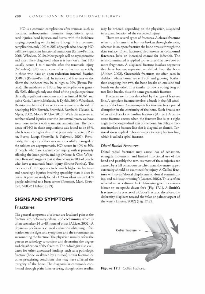
288 C O N D I T I O N S I N O C C U P A T I O N A L T H E R A P Y
HO is a common complication after traumas such as fractures, arthroplasties, traumatic amputations, spinal cord injuries, head injuries, and burns, with the incidence varying depending on the injury. Though it is a common complication, only 10% to 20% of people who develop HO will have significant functional limitations (Bruno-Petrina, 2008; Wheeless, 2010). Most people will be asymptomatic and most likely diagnosed when it is seen on a film. HO usually occurs 1 to 4 months after the traumatic injury (Wheeless). HO may occur after a fracture especially in those who have an open reduction internal fixation (ORIF) (Bruno-Petrina). In injuries and fractures to the elbow, the incidence may be as high as 90% (Bruno-Pet-rina). The incidence of HO in hip arthroplasties is gener-ally 50%, although only one-third of the people experience clinically significant symptoms such as limited ROM and pain (Kocic, Lazovic, Mitkovic, & Djokic, 2010; Wheeless). Revisions to hip and knee replacements increase the risk of developing HO (Barrack, Brumfield, Rorabeck, Cleland, & Myers, 2002; Moore & Cho, 2010). With the increase in combat-related injuries over the last several years, we have seen more soldiers with traumatic amputations. The inci-dence of HO in these amputations was found to be 63%, which is much higher than that previously expected (Pot-ter, Burns, Lacap, Granville, & Gajewski, 2007). Fortu-nately, the majority of the cases are successfully managed or the soldiers are asymptomatic. HO occurs in 40% to 50% of people who have a spinal cord injury, with it primarily affecting the knee, pelvis, and hip (Moore & Cho; Whee-less). Research suggests that it also occurs in 20% of people who have a traumatic brain injury (Bruno-Petrina). The incidence of HO appears to be much higher in fractures and neurologic injuries involving spasticity than it does in burns. A previous study found a 1.2% incident rate in 1,478 people admitted to a burn center (Peterson, Mani, Craw-ford, Neff, & Hiebert, 1989).
SIGNS AND SYMPTOMS
Fractures
The general symptoms of a break are localized pain at the fracture site, deformity, edema, and ecchymosis, which is often seen after 24 to 48 hours of onset (Altizer, 2002). A physician performs a clinical evaluation obtaining infor-mation on the signs and symptoms and the circumstances surrounding the fracture. The physician usually refers the person to radiology to confirm and determine the degree and classification of the fracture. The radiologist also eval-uates for other associated findings such as a pathologic fracture (bone weakened by a tumor), stress fracture, or other preexisting conditions that may have affected the integrity of the bone. The diagnosis is commonly con-firmed through plain films or x-ray, though other studies
may be ordered depending on the physician, suspected injury, and location of the suspected injury.
There are several types of fractures. A closed fracture refers to a fracture that has not broken through the skin, whereas in an open fracture the bone breaks through the skin surface. Open fractures, also known as compound fractures, have an increased chance for infection. The term comminuted is applied to fractures that have two or more fragments. A displaced fracture involves segments that have become separated or shifted from the bone (Altizer, 2002). Greenstick fractures are often seen in children whose bones are still soft and growing. Rather than snapping into two, the bone breaks on one side and bends on the other. It is similar to how a young twig or tree limb breaks, thus the name greenstick fracture.
Fractures are further described by the type of fracture line. A complete fracture involves a break in the full conti-nuity of the bone. An incomplete fracture involves a partial disruption in the continuity of the bone. Such breaks are often called cracks or hairline fractures (Altizer). A trans-verse fracture occurs when the fracture line is at a right angle to the longitudinal axis of the bone. An oblique frac-ture involves a fracture line that is diagonal or slanted. Tor-sional stress applied to bone causes a twisting fracture line, which is called a spiral fracture.
Distal Radial FracturesDistal radial fractures may cause loss of sensation, strength, movement, and limited functional use of the hand and possibly the arm. As most of these injuries are caused by a fall on an outstretched arm, the entire upper extremity should be examined for injury. A Colles’ frac-ture will reveal “dorsal displacement, dorsal comminut-ing, and radius shortening” (Laseter, 2002). This is often referred to as a dinner fork deformity given its resem-blance to an upside down fork (Fig. 17.1). A Smith’s fracture is the reverse of a Colles’ fracture; therefore, the deformity displaces toward the volar or palmar aspect of the wrist (Laseter, 2002) (Fig. 17.2).
Colles’ fracture
Figure 17.1 Colles’ fracture.
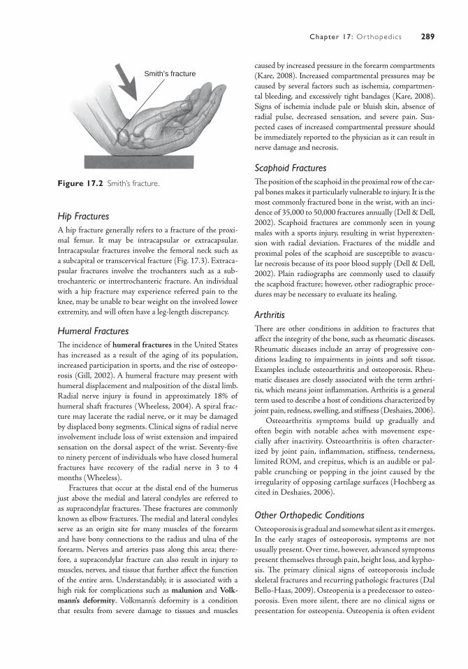
289Chapter 17: Or thopedics
Hip FracturesA hip fracture generally refers to a fracture of the proxi-mal femur. It may be intracapsular or extracapsular. Intracapsular fractures involve the femoral neck such as a subcapital or transcervical fracture (Fig. 17.3). Extraca-psular fractures involve the trochanters such as a sub-trochanteric or intertrochanteric fracture. An individual with a hip fracture may experience referred pain to the knee, may be unable to bear weight on the involved lower extremity, and will often have a leg-length discrepancy.
Humeral FracturesThe incidence of humeral fractures in the United States has increased as a result of the aging of its population, increased participation in sports, and the rise of osteopo-rosis (Gill, 2002). A humeral fracture may present with humeral displacement and malposition of the distal limb. Radial nerve injury is found in approximately 18% of humeral shaft fractures (Wheeless, 2004). A spiral frac-ture may lacerate the radial nerve, or it may be damaged by displaced bony segments. Clinical signs of radial nerve involvement include loss of wrist extension and impaired sensation on the dorsal aspect of the wrist. Seventy-five to ninety percent of individuals who have closed humeral fractures have recovery of the radial nerve in 3 to 4 months (Wheeless).
Fractures that occur at the distal end of the humerus just above the medial and lateral condyles are referred to as supracondylar fractures. These fractures are commonly known as elbow fractures. The medial and lateral condyles serve as an origin site for many muscles of the forearm and have bony connections to the radius and ulna of the forearm. Nerves and arteries pass along this area; there-fore, a supracondylar fracture can also result in injury to muscles, nerves, and tissue that further affect the function of the entire arm. Understandably, it is associated with a high risk for complications such as malunion and Volk-mann’s deformity. Volkmann’s deformity is a condition that results from severe damage to tissues and muscles
caused by increased pressure in the forearm compartments (Kare, 2008). Increased compartmental pressures may be caused by several factors such as ischemia, compartmen-tal bleeding, and excessively tight bandages (Kare, 2008). Signs of ischemia include pale or bluish skin, absence of radial pulse, decreased sensation, and severe pain. Sus-pected cases of increased compartmental pressure should be immediately reported to the physician as it can result in nerve damage and necrosis.
Scaphoid FracturesThe position of the scaphoid in the proximal row of the car-pal bones makes it particularly vulnerable to injury. It is the most commonly fractured bone in the wrist, with an inci-dence of 35,000 to 50,000 fractures annually (Dell & Dell, 2002). Scaphoid fractures are commonly seen in young males with a sports injury, resulting in wrist hyperexten-sion with radial deviation. Fractures of the middle and proximal poles of the scaphoid are susceptible to avascu-lar necrosis because of its poor blood supply (Dell & Dell, 2002). Plain radiographs are commonly used to classify the scaphoid fracture; however, other radiographic proce-dures may be necessary to evaluate its healing.
ArthritisThere are other conditions in addition to fractures that affect the integrity of the bone, such as rheumatic diseases. Rheumatic diseases include an array of progressive con-ditions leading to impairments in joints and soft tissue. Examples include osteoarthritis and osteoporosis. Rheu-matic diseases are closely associated with the term arthri-tis, which means joint inflammation. Arthritis is a general term used to describe a host of conditions characterized by joint pain, redness, swelling, and stiffness (Deshaies, 2006).
Osteoarthritis symptoms build up gradually and often begin with notable aches with movement espe-cially after inactivity. Osteoarthritis is often character-ized by joint pain, inflammation, stiffness, tenderness, limited ROM, and crepitus, which is an audible or pal-pable crunching or popping in the joint caused by the irregularity of opposing cartilage surfaces (Hochberg as cited in Deshaies, 2006).
Other Orthopedic ConditionsOsteoporosis is gradual and somewhat silent as it emerges. In the early stages of osteoporosis, symptoms are not usually present. Over time, however, advanced symptoms present themselves through pain, height loss, and kypho-sis. The primary clinical signs of osteoporosis include skeletal fractures and recurring pathologic fractures (Dal Bello-Haas, 2009). Osteopenia is a predecessor to osteo-porosis. Even more silent, there are no clinical signs or presentation for osteopenia. Osteopenia is often evident
Smith’s fracture
Figure 17.2 Smith’s fracture.
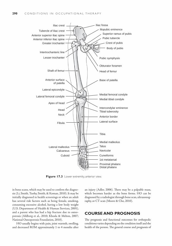
290 C O N D I T I O N S I N O C C U P A T I O N A L T H E R A P Y
Iliac crest Iliac fossa
Tubercle of iliac crest
Anterior superior iliac spineAnterior inferior iliac spine
Greater trochanter
Lesser trochanter
Intertrochanteric line
Shaft of femur
Anterior surfaceof patella
Lateral epicondyle
Lateral femoral condyle
Apex of head
Head
Neck
Fibula
Lateral malleolusCalcaneus
Cuboid
Iliopubic eminence
Superior ramus of pubis
Pubic tubercle
Crest of pubis
Body of pubis
Pubic symphysis
Obturator foramen
Head of femur
Base of patella
Medial femoral condyle
Medial tibial condyle
Intercondylar eminenceTibial tuberosity
Anterior border
Lateral surface
Tibia
Medial malleolus
Talus
Navicular
Cuneiforms
1st metatarsal
Distal phalanxProximal phalanx
Figure 17.3 Lower extremity, anterior view.
in bone scans, which may be used to confirm the diagno-sis (Li, Smith, Tuohy, Smith, & Koman, 2010). It may be initially diagnosed in health screenings or when an adult has several risk factors such as being female, smoking, consuming excessive alcohol, having a low body weight (U.S. Department of Health & Human Services, 2005), and a parent who has had a hip fracture due to osteo-porosis (Ahlborg et al., 2010; Khosla & Melton, 2007; National Osteoporosis Foundation, 2010).
HO usually begins with pain, joint warmth, swelling, and decreased ROM approximately 1 to 4 months after
an injury (Adler, 2006). There may be a palpable mass, which becomes harder as the bone forms. HO can be diagnosed by a radiologist through bone scan, ultrasonog-raphy, or CT scan (Moore & Cho, 2010).
COURSE AND PROGNOSISThe prognosis and functional outcomes for orthopedic conditions varies depending on the condition itself and the health of the person. The general course and prognosis of
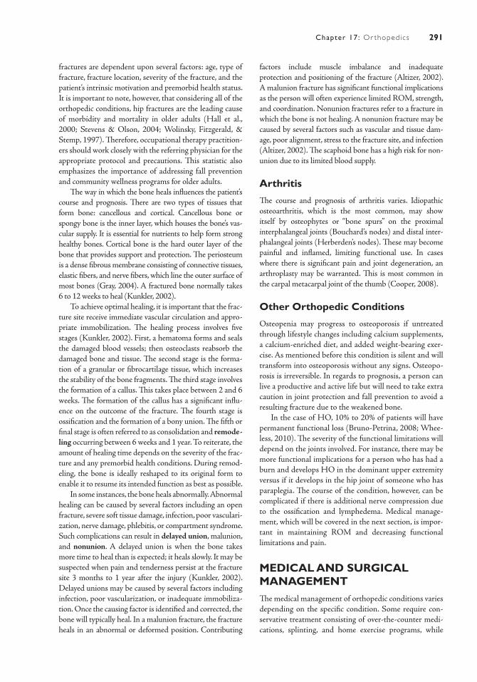
291Chapter 17: Or thopedics
fractures are dependent upon several factors: age, type of fracture, fracture location, severity of the fracture, and the patient’s intrinsic motivation and premorbid health status. It is important to note, however, that considering all of the orthopedic conditions, hip fractures are the leading cause of morbidity and mortality in older adults (Hall et al., 2000; Stevens & Olson, 2004; Wolinsky, Fitzgerald, & Stemp, 1997). Therefore, occupational therapy practition-ers should work closely with the referring physician for the appropriate protocol and precautions. This statistic also emphasizes the importance of addressing fall prevention and community wellness programs for older adults.
The way in which the bone heals influences the patient’s course and prognosis. There are two types of tissues that form bone: cancellous and cortical. Cancellous bone or spongy bone is the inner layer, which houses the bone’s vas-cular supply. It is essential for nutrients to help form strong healthy bones. Cortical bone is the hard outer layer of the bone that provides support and protection. The periosteum is a dense fibrous membrane consisting of connective tissues, elastic fibers, and nerve fibers, which line the outer surface of most bones (Gray, 2004). A fractured bone normally takes 6 to 12 weeks to heal (Kunkler, 2002).
To achieve optimal healing, it is important that the frac-ture site receive immediate vascular circulation and appro-priate immobilization. The healing process involves five stages (Kunkler, 2002). First, a hematoma forms and seals the damaged blood vessels; then osteoclasts reabsorb the damaged bone and tissue. The second stage is the forma-tion of a granular or fibrocartilage tissue, which increases the stability of the bone fragments. The third stage involves the formation of a callus. This takes place between 2 and 6 weeks. The formation of the callus has a significant influ-ence on the outcome of the fracture. The fourth stage is ossification and the formation of a bony union. The fifth or final stage is often referred to as consolidation and remode-ling occurring between 6 weeks and 1 year. To reiterate, the amount of healing time depends on the severity of the frac-ture and any premorbid health conditions. During remod-eling, the bone is ideally reshaped to its original form to enable it to resume its intended function as best as possible.
In some instances, the bone heals abnormally. Abnormal healing can be caused by several factors including an open fracture, severe soft tissue damage, infection, poor vasculari-zation, nerve damage, phlebitis, or compartment syndrome. Such complications can result in delayed union, malunion, and nonunion. A delayed union is when the bone takes more time to heal than is expected; it heals slowly. It may be suspected when pain and tenderness persist at the fracture site 3 months to 1 year after the injury (Kunkler, 2002). Delayed unions may be caused by several factors including infection, poor vascularization, or inadequate immobiliza-tion. Once the causing factor is identified and corrected, the bone will typically heal. In a malunion fracture, the fracture heals in an abnormal or deformed position. Contributing
factors include muscle imbalance and inadequate protection and positioning of the fracture (Altizer, 2002). A malunion fracture has significant functional implications as the person will often experience limited ROM, strength, and coordination. Nonunion fractures refer to a fracture in which the bone is not healing. A nonunion fracture may be caused by several factors such as vascular and tissue dam-age, poor alignment, stress to the fracture site, and infection (Altizer, 2002). The scaphoid bone has a high risk for non-union due to its limited blood supply.
Arthritis
The course and prognosis of arthritis varies. Idiopathic osteoarthritis, which is the most common, may show itself by osteophytes or “bone spurs” on the proximal interphalangeal joints (Bouchard’s nodes) and distal inter-phalangeal joints (Herberden’s nodes). These may become painful and inflamed, limiting functional use. In cases where there is significant pain and joint degeneration, an arthroplasty may be warranted. This is most common in the carpal metacarpal joint of the thumb (Cooper, 2008).
Other Orthopedic Conditions
Osteopenia may progress to osteoporosis if untreated through lifestyle changes including calcium supplements, a calcium-enriched diet, and added weight-bearing exer-cise. As mentioned before this condition is silent and will transform into osteoporosis without any signs. Osteopo-rosis is irreversible. In regards to prognosis, a person can live a productive and active life but will need to take extra caution in joint protection and fall prevention to avoid a resulting fracture due to the weakened bone.
In the case of HO, 10% to 20% of patients will have permanent functional loss (Bruno-Petrina, 2008; Whee-less, 2010). The severity of the functional limitations will depend on the joints involved. For instance, there may be more functional implications for a person who has had a burn and develops HO in the dominant upper extremity versus if it develops in the hip joint of someone who has paraplegia. The course of the condition, however, can be complicated if there is additional nerve compression due to the ossification and lymphedema. Medical manage-ment, which will be covered in the next section, is impor-tant in maintaining ROM and decreasing functional limitations and pain.
MEDICAL AND SURGICAL MANAGEMENTThe medical management of orthopedic conditions varies depending on the specific condition. Some require con-servative treatment consisting of over-the-counter medi-cations, splinting, and home exercise programs, while
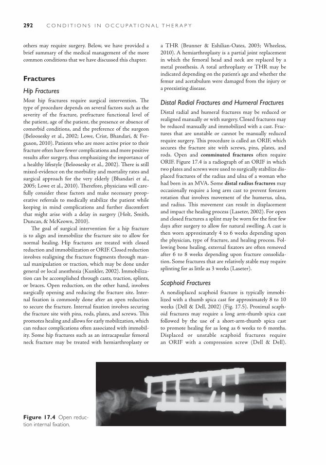
292 C O N D I T I O N S I N O C C U P A T I O N A L T H E R A P Y
others may require surgery. Below, we have provided a brief summary of the medical management of the more common conditions that we have discussed this chapter.
Fractures
Hip FracturesMost hip fractures require surgical intervention. The type of procedure depends on several factors such as the severity of the fracture, prefracture functional level of the patient, age of the patient, the presence or absence of comorbid conditions, and the preference of the surgeon (Beloosesky et al., 2002; Lowe, Crist, Bhandari, & Fer-guson, 2010). Patients who are more active prior to their fracture often have fewer complications and more positive results after surgery, thus emphasizing the importance of a healthy lifestyle (Beloosesky et al., 2002). There is still mixed- evidence on the morbidity and mortality rates and surgical approach for the very elderly (Bhandari et al., 2005; Lowe et al., 2010). Therefore, physicians will care-fully consider these factors and make necessary preop-erative referrals to medically stabilize the patient while keeping in mind complications and further discomfort that might arise with a delay in surgery (Holt, Smith, Duncan, & McKeown, 2010).
The goal of surgical intervention for a hip fracture is to align and immobilize the fracture site to allow for normal healing. Hip fractures are treated with closed reduction and immobilization or ORIF. Closed reduction involves realigning the fracture fragments through man-ual manipulation or traction, which may be done under general or local anesthesia (Kunkler, 2002). Immobiliza-tion can be accomplished through casts, traction, splints, or braces. Open reduction, on the other hand, involves surgically opening and reducing the fracture site. Inter-nal fixation is commonly done after an open reduction to secure the fracture. Internal fixation involves securing the fracture site with pins, rods, plates, and screws. This promotes healing and allows for early mobilization, which can reduce complications often associated with immobil-ity. Some hip fractures such as an intracapsular femoral neck fracture may be treated with hemiarthroplasty or
a THR (Brunner & Eshilian-Oates, 2003; Wheeless, 2010). A hemiarthroplasty is a partial joint replacement in which the femoral head and neck are replaced by a metal prosthesis. A total arthroplasty or THR may be indicated depending on the patient’s age and whether the femur and acetabulum were damaged from the injury or a preexisting disease.
Distal Radial Fractures and Humeral FracturesDistal radial and humeral fractures may be reduced or realigned manually or with surgery. Closed fractures may be reduced manually and immobilized with a cast. Frac-tures that are unstable or cannot be manually reduced require surgery. This procedure is called an ORIF, which secures the fracture site with screws, pins, plates, and rods. Open and comminuted fractures often require ORIF. Figure 17.4 is a radiograph of an ORIF in which two plates and screws were used to surgically stabilize dis-placed fractures of the radius and ulna of a woman who had been in an MVA. Some distal radius fractures may occasionally require a long arm cast to prevent forearm rotation that involves movement of the humerus, ulna, and radius. This movement can result in displacement and impact the healing process (Laseter, 2002). For open and closed fractures a splint may be worn for the first few days after surgery to allow for natural swelling. A cast is then worn approximately 4 to 6 weeks depending upon the physician, type of fracture, and healing process. Fol-lowing bone healing, external fixators are often removed after 6 to 8 weeks depending upon fracture consolida-tion. Some fractures that are relatively stable may require splinting for as little as 3 weeks (Laseter).
Scaphoid FracturesA nondisplaced scaphoid fracture is typically immobi-lized with a thumb spica cast for approximately 8 to 10 weeks (Dell & Dell, 2002) (Fig. 17.5). Proximal scaph-oid fractures may require a long arm-thumb spica cast followed by the use of a short-arm-thumb spica cast to promote healing for as long as 6 weeks to 6 months. Displaced or unstable scaphoid fractures require an ORIF with a compression screw (Dell & Dell).
Figure 17.4 Open reduc-tion internal fixation.
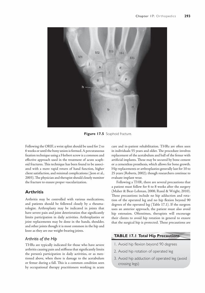
293Chapter 17: Or thopedics
a
b
k
c
g
i
o
d
p
j
n
e
f
h
M1
l
M2 M3 M4
M5
Figure 17.5 Scaphoid fracture.
Following the ORIF, a wrist splint should be used for 2 to 6 weeks or until the bony union is formed. A percutaneous fixation technique using a Herbert screw is a common and effective approach used in the treatment of acute scaph-oid fractures. This technique has been found to be associ-ated with a more rapid return of hand function, higher client satisfaction, and minimal complications ( Jeon et al., 2003). The physician and therapist should closely monitor the fracture to ensure proper vascularization.
Arthritis
Arthritis may be controlled with various medications, and patients should be followed closely by a rheuma-tologist. Arthroplasty may be indicated in joints that have severe pain and joint deterioration that significantly limits participation in daily activities. Arthroplasties or joint replacements may be done in the hands, shoulder, and other joints though it is most common in the hip and knee as they are our weight-bearing joints.
Arthritis of the HipTHRs are typically indicated for those who have severe arthritis causing pain and stiffness that significantly limits the person’s participation in daily activities, or as men-tioned above, when there is damage to the acetabulum or femur during a fall. This is a common condition seen by occupational therapy practitioners working in acute
care and in-patient rehabilitation. THRs are often seen in individuals 55 years and older. The procedure involves replacement of the acetabulum and ball of the femur with artificial implants. These may be secured by bone cement or a cementless prosthesis, which allows for bone growth. Hip replacements or arthroplasties generally last for 10 to 25 years (Roberts, 2002); though researchers continue to evaluate implant wear.
Following a THR, there are several precautions that a patient must follow for 6 to 8 weeks after the surgery (Maher & Bear-Lehman, 2008; Rasul & Wright, 2010). These precautions include no hip adduction and rota-tion of the operated leg and no hip flexion beyond 90 degrees of the operated leg (Table 17.1). If the surgeon uses an anterior approach, the patient must also avoid hip extension. Oftentimes, therapists will encourage their clients to avoid hip rotation in general to ensure that the surgical hip is protected. These precautions are
TABLE 17.1 Total Hip Precautions
1. Avoid hip flexion beyond 90 degrees
2. Avoid hip rotation of operated leg
3. Avoid hip adduction of operated leg (avoid crossing legs)
294 C O N D I T I O N S I N O C C U P A T I O N A L T H E R A P Y
currently the standard of care; however, recent research suggests that an anterolateral surgical approach rather than strict adherence to traditional hip precautions was more likely to be associated with a low rate of disloca-tion (Peak et al., 2005). Depending on the severity of the condition, the surgical approach and procedure, and the orthopedic surgeon, there may be weight-bearing pre-cautions for patients who undergo a THRs and ORIFs. Common weight-bearing statuses include non–weight bearing, touchdown or toe-touch weight bearing, partial weight bearing, weight bearing as tolerated, and full weight bearing (Table 17.2).
Hip precautions have functional implications for the person in a variety of daily activities. Occupational therapy practitioners must educate clients on the hip pre-cautions and teach them adaptations for sitting, dressing, bathing, reaching for items, and driving among others. In regards to driving, patients are to avoid driving for 4 to 6 weeks following surgery (Ganz, Levin, Peterson, & Rana-wat, 2003). At this time, most patients have reached their preoperative driving reaction time and continue to show improvements in reaction time for up to 1 year.
Arthritis of the KneeThe first line of management of arthritis of the knee is conservative treatment, which often begins with over-the-counter anti-inflammatory medications and heat and ice. If pain and functional limitations progress, the patient’s primary physician or rheumatologist may prescribe medi-cations to effectively control the arthritis. When con-servative treatment does not work, the patient may be a candidate for a partial or total knee replacement (TKR).
This is also referred to as a knee arthroplasty. A TKR involves resurfacing the entire knee joint with metal and plastic prosthetic components. The procedure typically involves a femoral component, a tibial plate, and a patellar button (Roberts, 2002) and may be done unilaterally or bilaterally.
After surgery, it is important to begin the rehabili-tation process (Bade, Kohrt, & Stevens-Lapsley, 2010; Rasul & Wright, 2010). Treatment involves pain medi-cation and early mobilization to strengthen the quadri-ceps and prevent loss of ROM. A continuous passive motion machine may be prescribed while in the hospital or at home to help improve circulation and movement at the knee joint depending on the patient’s postopera-tive health and functional abilities. Initial rehabilitation goals include independence in bed mobility, knee flexion to approximately 90 degrees, knee extension, and to walk with crutches or a walker (American Academy of Ortho-paedic Surgeons, 1995–2009).
To maximize the results of the surgery and functional outcomes, the patient needs education on several precau-tions such as avoiding putting a pillow under the knee while in bed, resting both feet on the floor when sitting to increase knee ROM, and wearing an immobilizer as instructed by the physician to protect the knee joint until the muscle is strong enough to support it (Erickson & Perkins, 1994; Rasul & Wright, 2010). The immobilizer may be required when walking, while in bed, or at all times. Kneeling should be avoided. Other considerations include not driving, running, and jumping until cleared by the physician. Generally, patients are able to return to most of their normal daily activities after 3 to 6 weeks
TABLE 17.2 Weight-Bearing Status
Weight-Bearing Status Percentage and Description
Non–weight bearing 0%. No weight bearing on the operated leg; it should not touch the floor.
Touchdown or toe-touch weight bearing 10%–15%. Foot of operated leg may rest on floor.
Partial weight bearing 30%–50%. Patient may put 30%–50% of weight through the operated leg.
Weight bearing as tolerated Patient may put as much weight through operated leg as tolerated without unnecessary pain or discomfort.
Full weight bearing 75%–100%. Patient bears full weight on the operated leg.
Modified from Goldstein, T. S. (1999). Geriatric orthopedics: Rehabilitative management of common problems (2nd ed.). Gaithersburg, MD: Aspen.
295Chapter 17: Or thopedics
though high-impact activities may need to be modified. Driving may be resumed around 4 to 6 weeks as long as the patient is able to bend his or her knee enough to get in and out of the car and have appropriate muscle strength and control to operate the pedals. Patients need to follow up with their physician to monitor for complications and ensure continued progress.
IMPACT ON OCCUPATIONAL PERFORMANCEOccupational therapy practitioners use a holistic approach to examine specific client factors residing within the person to understand their impact on occu-pational performance (American Occupational Therapy Association [AOTA], 2008). Systems of values, beliefs and spirituality; body functions, and body structures are client factors that are often affected by illness or disability (AOTA, 2008). Client factors are an important consider-ation when working with a person who has an orthopedic condition to help them engage in daily occupations such as dressing, working, managing the home, and leisure pursuits. We will discuss client factors that are commonly affected by an orthopedic condition.
During the initial evaluation it is important to carefully assess the nature and circumstances surround-ing the onset of the orthopedic condition. For example, children and adults are likely to sustain fractures dur-ing play and sports activities, whereas older adults may experience a fracture as a result of a fall or osteoporosis. In some circumstances, however, a fracture may also be a result of child abuse or domestic violence. The nature of the circumstance surrounding the injury or condition may impact secondary injuries and psychosocial involve-ment. It is important therefore to conduct a thorough his-tory of the fall and a radiologic analysis of the fracture (American Academy of Pediatrics, 2000) and, for chil-dren, to ascertain their developmental level.
Orthopedic conditions may compromise participation in several areas of occupation including ADLs, instru-mental activities of daily living, sleep, education, work, play, leisure, and social participation (AOTA, 2008). The degree of impairment depends upon the severity of the injury, location of the injury, and the client’s premorbid health status. Several client factors may be compromised by orthopedic conditions (Table 17.3). The main client factors that will affect one’s participation in occupation are neuromusculoskeletal and movement-related func-tions and structures and sensory and pain functions and
TABLE 17.3 Client Factors Commonly Involved in Orthopedic Conditions
1. Values, beliefs, and spirituality
2. Body functions(a) Mental functions(b) Sensory functions and pain
(i) Vestibular function (balance)(ii) Pain(iii) Protective functions of the skin
(c) Neuromusculoskeletal and movement-related functions(i) Joint mobility(ii) Joint stability(iii) Muscle power(iv) Muscle tone(v) Muscle endurance
(d) Skin and related structure functions(i) Repair function of the skin
3. Body structures(a) Structures related to movement(b) Skin and related structures
From American Occupational Therapy Association (AOTA). (2008). Occupational therapy practice framework: Domain and process (2nd ed.). American Journal of Occupational Therapy, 62, 625–683.
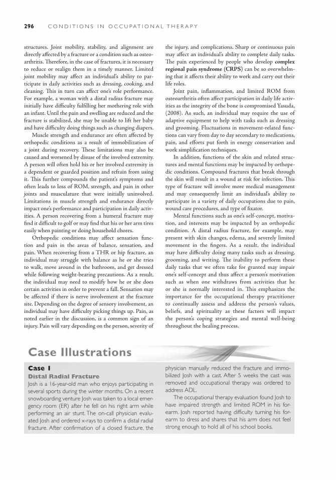
296 C O N D I T I O N S I N O C C U P A T I O N A L T H E R A P Y
structures. Joint mobility, stability, and alignment are directly affected by a fracture or a condition such as osteo-arthritis. Therefore, in the case of fractures, it is necessary to reduce or realign them in a timely manner. Limited joint mobility may affect an individual’s ability to par-ticipate in daily activities such as dressing, cooking, and cleaning. This in turn can affect one’s role performance. For example, a woman with a distal radius fracture may initially have difficulty fulfilling her mothering role with an infant. Until the pain and swelling are reduced and the fracture is stabilized, she may be unable to lift her baby and have difficulty doing things such as changing diapers.
Muscle strength and endurance are often affected by orthopedic conditions as a result of immobilization of a joint during reco very. These limitations may also be caused and worsened by disuse of the involved extremity. A person will often hold his or her involved extremity in a dependent or guarded position and refrain from using it. This further compounds the patient’s symptoms and often leads to loss of ROM, strength, and pain in other joints and musculature that were initially uninvolved. Limitations in muscle strength and endurance directly impact one’s performance and participation in daily activ-ities. A person recovering from a humeral fracture may find it difficult to golf or may find that his or her arm tires easily when painting or doing household chores.
Orthopedic conditions may affect sensation func-tion and pain in the areas of balance, sensation, and pain. When recovering from a THR or hip fracture, an individual may struggle with balance as he or she tries to walk, move around in the bathroom, and get dressed while following weight-bearing precautions. As a result, the individual may need to modify how he or she does certain activities in order to prevent a fall. Sensation may be affected if there is nerve involvement at the fracture site. Depending on the degree of sensory involvement, an individual may have difficulty picking things up. Pain, as noted earlier in the discussion, is a common sign of an injury. Pain will vary depending on the person, severity of
the injury, and complications. Sharp or continuous pain may affect an individual’s ability to complete daily tasks. The pain experienced by people who develop complex regional pain syndrome (CRPS) can be so overwhelm-ing that it affects their ability to work and carry out their life roles.
Joint pain, inflammation, and limited ROM from osteoarthritis often affect participation in daily life activ-ities as the integrity of the bone is compromised Yasuda, (2008). As such, an individual may require the use of adaptive equipment to help with tasks such as dressing and grooming. Fluctuations in movement-related func-tions can vary from day to day secondary to medications, pain, and efforts put forth in energy conservation and work simplification techniques.
In addition, functions of the skin and related struc-tures and mental functions may be impacted by orthope-dic conditions. Compound fractures that break through the skin will result in a wound at risk for infection. This type of fracture will involve more medical management and may consequently limit an individual’s ability to participate in a variety of daily occupations due to pain, wound care procedures, and type of fixator.
Mental functions such as one’s self-concept, motiva-tion, and interests may be impacted by an orthopedic condition. A distal radius fracture, for example, may present with skin changes, edema, and severely limited movement in the fingers. As a result, the individual may have difficulty doing many tasks such as dressing, grooming, and writing. The inability to perform these daily tasks that we often take for granted may impair one’s self-concept and thus affect a person’s motivation such as when one withdraws from activities that he or she is normally interested in. This emphasizes the importance for the occupational therapy practitioner to continually assess and address the person’s values, beliefs, and spirituality as these factors will impact the person’s coping strategies and mental well-being throughout the healing process.
Case IllustrationsCase 1Distal Radial FractureJosh is a 16-year-old man who enjoys participating in several sports during the winter months. On a recent snowboarding venture Josh was taken to a local emer-gency room (ER) after he fell on his right arm while performing an air stunt. The on-call physician evalu-ated Josh and ordered x-rays to confirm a distal radial fracture. After confirmation of a closed fracture, the
physician manually reduced the fracture and immo-bilized Josh with a cast. After 5 weeks the cast was removed and occupational therapy was ordered to address ADL.
The occupational therapy evaluation found Josh to have impaired strength and limited ROM in his for-earm. Josh reported having difficulty turning his for-earm to dress and shares that his arm does not feel strong enough to hold all of his school books.
297Chapter 17: Or thopedics
Case 2Supracondylar Fracture of the HumerusAna is a 57-year-old woman who fractured her humerus in an MVA. She was taken to the ER where the physician conducted a clinical assessment and ordered x-rays to confirm the suspected fracture. The radiologist report confirmed that there was a closed transverse fracture of the middle third of the right humeral shaft. The physician was able to reduce the fracture without surgery. He immobilized the arm in a sling. Four weeks later, Ana still had significant pain and tenderness in her upper arm. Radiologic tests revealed that the bone was forming a malunion. The physician determined that it was necessary to surgi-
cally reduce the fracture and stabilize it with internal fixation. Radiologic studies showed signs of healing 6 weeks after the second reduction as well as signs of osteopenia. The physician wrote a prescription for occupational therapy.
The occupational therapist evaluated Ana. He noted that she held her arm in a dependent position and was very hesitant to move it or let him examine it. She reported significant pain and had atrophy of her upper arm and forearm muscles. Her skin appeared shiny in comparison to her left arm. Ana had limited movement and reported that her husband had to help her with everything. She had been unable to return to work as an office manager because the pain was just too great.
R E C O M M E N D E D L E A R N I N G R E S O U R C E S
BooksBonder, B. R., & Dal Bello-Haas, V. (2009). Functional
performance in older adults. Philadelphia, PA: F.A. Davis.
Mackin, E. J., Callahan, A. D., Osterman, L., Skirven, T. M., & Schneider, L. (2002). Rehabilitation of the hand and upper extremity (5th ed.). St. Louis, MO: Mosby.
Radomski, M. V., & Trombly-Latham, C. A. (2007). Occu-pational therapy for physical dysfunction (6th ed.). Philadelphia, PA: Lippincott Williams & Wilkins.
JournalsAmerican Journal of Occupational Therapywww.aota.orgAmerican Journal of Orthopedicshttp://www.amjorthopedics.com/Journal of American Geriatrics Societyhttp://www.wiley.com/bw/journal.asp?ref=0002-
8614&site=1
Orthopaedic Nursinghttp://journals.lww.com/or thopaedicnursing/pages/
default.aspx
Web sitesAmerican Occupational Therapy Association. Evidence-
Based Practice & Researchhttp://www.aota.org/Educate/Research.aspxCenters for Disease Control and Preventionhttp://www.cdc.govHealthy People 2020http://www.healthypeople.gov/hp2020/Lewis, W. H. (Ed.). Gray’s anatomy (12th ed.). New York,
NY: Bartleby.com. Retrieved from http://www.bar-tleby.com/107
United States Department of Health & Human Serviceshttp://www.hhs.gov/Wheeless, C. R. (2010). Wheeless’ textbook of orth-
paedics. http://www.wheelessonline.com
Adler, C. (2006). Spinal cord injury. In M. B. Early (Ed.), Physical dysfunction practice skills for the occupational therapy assistant (2nd ed., pp. 528–550). St. Louis, MO: Mosby.
Ahlborg, H., Rosengren, B., Jarvinen, T., Rogmark, C., Nilsson, J., Sernbo, I., & Karlsson, M. (2010). Preva-lence of osteoporosis and incidence of hip fracture in women—Secular trends over 30 years. BMC Muscu-loskeletal Disorders, 11(1).
Altizer, L. (2002). Fractures. Orthopaedic Nursing, 21, 51–59.American Academy of Pediatrics. (2000). Diagnostic
imaging of child abuse. Pediatrics, 105, 1345–1348.American Occupational Therapy Association (AOTA).
(2008). Occupational therapy practice framework: Domain and process (2nd ed.). American Journal of Occupational Therapy, 62, 625–683.
Bade, M. J., Kohrt, W. M., & Stevens-Lapsley, J. E. (2010). Outcomes before and after total knee arthroplasty in
R E F E R E N C E S
298 C O N D I T I O N S I N O C C U P A T I O N A L T H E R A P Y
health adults. Journal of Orthopaedic Sports Physical Therapy, 40(9), 559–567. http://www.jospt.org/
Bhandari, M., Devereaux, P., Tornetta, P., Swiontkowski, M., Berry D., Haidukewych, G.,... Guyatt, G. (2005). Operative management of displaced femoral neck frac-tures in elderly patients. An international survey. Journal of Bone and Joint Surgery (America), 87(9), 2122–2130.
Barrack, R., Brumfield, C., Rorabeck, C., Cleland, D., & Myers, L. (2002). Heterotopic ossification after revi-sion total knee arthroplasty. Clinical Orthopaedics & Related Research, 404, 208–213. DOI: 10.1097/01.blo.0000030497.43495.3f.
Beloosesky, Y., Grinblat, J., Epelboym, B., Weiss, A., Grosman, B., & Hendel, D. (2002). Functional gain of hip fracture patients in different cognitive and func-tional groups. Clinical Rehabilitation, 16, 321–328.
Bell, A. J., Talbot-Stern, J. K., & Hennessy, A. (2000). Characteristics and outcomes of older patients pre-senting to the emergency department after a fall: A retrospective analysis. eMedical Journal of Australia, 173, 179–182. Retrieved from www.mja.com.au. The Journal of Bone and Joint Surgery (American) 2007;89:476–486. doi:10.2106/JBJS.F.00412
Brunner, L. C., & Eshilian-Oates, L. (2003). Hip fractures in adults. American Family Physician, 67, 537–542.
Bruno-Petrina, A. (2008). Posttraumatic heterotopic ossification. Emedicine. Retrieved from http:// emedicine.medscape.com/article/326242-overview
Centers for Disease Control and Prevention (CDC). (2009a). Arthritis: Morbidity. Summary Health statis-tics for US Adults: National Health Interview Survey, 2008. Retrieved from www.cdc.gov/nchs/fstats/ arthrits.htm on March 31, 2010.
Centers for Disease Control and Prevention (CDC). (2009b). Injury prevention and control: Home and recre-ational safety: Hip fractures among older adults. Retrieved August 26, 2010, from http://www.cdc.gov/Homean-dRecreationalSafety/Falls/adulthipfx.html
Cooper, C. (2008). Hand impairments. In M. V. Radomski & C. A. Trombly Latham (Eds.), Occupational therapy for physical dysfunction (6th ed.). Philadelphia, PA: Lippincott Williams & Wilkins.
Crotty, M., Unroe, K., Cameron, I. D., Miller, M., Ramirez, G., & Couzner, L. (2010). Rehabilitation interventions for improving physical and psycho-social functioning after hip fracture in older people. Cochrane Database Systematic Review, 1, CD007624.
Cummings, S. R., Rubin, S. M., & Black, D. (1990). The future of hip fractures in the United States: Numbers, costs, and potential effects of postmenopausal estrogen. Clinical Orthopaedics & Related Research, 252, 163–166.
Dal Bello-Haas, V. (2009). Neuromusculoskeletal and movement function. In B. R. Bonder & V. Dal
Bello-Haas (Eds.), Functional performance in older adults (pp. 130–176). Philadelphia, PA: F.A. Davis.
Dell, P. C., & Dell, R. B. (2002). Management of carpal fractures and dislocations. In Mackin, E. J., Callahan, A. D., Osterman, L., Skirven, T. M., & Schneider, L. (Eds.). Rehabilitation of the hand and upper extremity (5th ed.). St. Louis, MO: Mosby.
Deshaies, L. (2006). Arthritis. In H. M. Pendleton & W. S. Krohn (Eds.), Occupational therapy practice skills for physical dysfunction (pp. 950–982). St. Louis, MO: Mosby.
Erickson, B., & Perkins, M. (1994). Interdisciplinary team approach in the rehabilitation of hip and knee arthroplasties. American Journal of Occupational Ther-apy, 48(5), 439–445.
Ganz, S. B., Levin, A., Peterson, M. G., & Ranawat, C. S. (2003). Improvement in driving reaction time after total hip arthroplasty. Clinical Orthopaedics, 413, 192–200.
Gill, M. (2002). Managing distal and humeral shaft frac-tures. Journal of the National Association of Orthopedic Technologists, 4, 1–4.
Gillespie, W. (2002). Hip fractures. Clinical Evidence, 8, 1126–1148.
Gray, H. (2004). Bone. In W. H. Lewis (Ed.), Gray’s anatomy (12th ed.). New York, NY: Bartleby.com. Retrieved from http://www.bartleby.com/107
Greenspan, W., Myers, E., Maitland, L., Kido, T., Krasnow, M., & Hayes, W. (1994). Trochanteric bone mineral density is associated with type of hip fracture in the elderly. Bone and Mineral, 9, 1889–1894.
Hagsten, B., Svensson, O., & Gardulf, A. (2004). Early individualized postoperative occupational therapy training in 100 patients improves ADL after hip fracture. Acta Orthopaedica, 75, 177–183.
Hall, S. E., Williams, J. A., & Senior, J.A., Goldswain, P. R., & Criddle, R. A. (2000). Hip fracture outcomes: Quality of life and functional status in older adults liv-ing in the community. Australian New Zealand Journal of Medicine, 30, 327–332.
Healthy People 2010. (2010). Arthritis, osteoporosis, and chronic back conditions. Retrieved July 26, 2010, from www.healthypeople.gov/document/html/volume1/02arthritis.htm
Holt, G., Smith, R., Duncan, K., & McKeown, D. (2010). Does delay to theatre for medical reasons affect the peri-operative mortality in patients with a fracture of the hip? Journal of Bone and Joint Surgery (British), 92(8), 835–841.
Kare, J. A. (2008). Volkmann contracture. Emedicine. http://www.emedicine.com/orthoped/topic 578.htm
Jeon, I.,Oh, C., Park, B., Ihn, J. Kim P.(2003). Minimal invasive percutaneous Herbert screw fixation in acute unstable scaphoid fracture. Hand Surgery, 8,213–218.
299Chapter 17: Or thopedics
Khosla, S., & Melton, J. (2007). Osteopenia. The New England Journal of Medicine, 356(22), 2293–2300. www.nejm.org
Kocic, M., Lazovic, M., Mitkovic, M., & Djokic, B. (2010). Clinical significance of the heterotopic ossi-fication after total hip arthroplasty. Orthopedics, doi: 10.3928/01477447-20091124-13.
Kotz, K., Deleger, S., Cohen, R., Kamigaki, A., & Kurata, J. (2004). Osteoporsis and health-related quality-of-life outcomes in the Alameda County study population. Preventing Chronic Disease: Public health Research, Prac-tice, and Policy, 1(1), 1–9. http://www.cdc.gov/pcd/issues/2004/jan/03_0005.htm
Koval, K. J., Skovron, M. L., Aharonof, G., & Zuckerman, J. D. (1998). Predictors of functional recovery after hip fractures in the elderly. Current Orthopaedic Practice, 348. http://journals.lww.com/c- orthopaedicpractice/pages/default.aspx
Kunkler, C. E. (2002). Fractures. In A. B. Maher, S. W. Salmond & T. A. Pellino (Eds.). Orthopedic nursing. Philadelphia: Saunders.
Jagmin, M. G. (2002). Assessment and management of im-mobility. In A. B. Maher, S. W. Salmond, & T. A. Pellino (Eds.). Orthopedic nursing. Philadelphia, PA: Saunders.
Laseter, G. F. (2002). Therapist’s management of dis-tal radius fractures. In E. J. Mackin, A. D. Callahan, L. Osterman, T. M. Skirven & L. Schneider (Eds.). Rehabilitation of the hand and upper extremity (5th ed.). St. Louis, MO: Mosby.
Li, Z., Smith, B. P., Tuohy, C., Smith, T. L., & Koman, L. A. (2010). Complex regional pain syndrome after hand surgery. Hand Clinics, 26(2), 281–289. doi:10.1016/j.hcl.2009.11.001.
Lowe, J., Crist, B., Bhandari, M. & Ferguson, T. (2010). Optimal treatment of femoral neck fractures according to patient’s physiologic age: an evidence-based review. Orthopedic Clinics of North America, 41, 2, 157-66.
Magaziner, J., Hawkes, W., Hebel, J. R., Zimmerman, S. I., Fox, K. M., Dolan, M., et al. (2000). Recovery from hip fracture in eight areas of function. Journal of Gerontology: Medical Sciences, 55(9), M498–M507. doi: 10.1093/gerona/55.9.M498.
Maher, C., & Bear-Lehman, J. (2008). Orthopaedic con-ditions. In M. V. Radomski & C. A. Trombly Latham (Eds.). Occupational therapy for physical dysfunction (6th ed.). Philadelphia, PA: Lippincott Williams & Wilkins.
Marottoli, R. A., Berkman, L. F., & Cooney, L. M. (1993). Decline in physical function following hip fracture. Journal of American Geriatrics Society, 40(9), 861–866.
Matityahu, A., Bruck, N., & Miclau, T. (2006). Heterotopic ossification and acetabular fractures.
Current Opinions in Orthopaedics, 17(1), 34–37. doi: 10.1097/01.bco.0000192520.48411.fa.
Moore, D. S., & Cho, G. (2010). Heterotopic ossifica-tion. eMedicine. Retrieved from http://emedicine. medscape.com/article/390416-overview
National Center for Injury Prevention and Control. Office of Statistics and Programming, NCIP, CDC. (2007). Ten leading causes of nonfatal injury, United States. Retrieved from http://www.cdc.gov/ncipc/wisqars/nonfatal/quickpicks/quickpicks_2007/allinj.htm
National Institute of Arthritis and Musculoskeletal and Skin Diseases. (2002, Revised 2006). Osteoarthritis. NIH Publication No. 06-4617. Retrieved from http://www.niams.nih.gov/Health_Info/ Osteoarthritis/ default.asp
National Institutes of Arthritis and Musculoskeletal and Skin Diseases. (April 2009). Arthritis. NIH Publica-tion No. 09-5149.
National Osteoporosis Foundation. (2010). Fast facts on osteoporosis. Retrieved August 16, 2010, from http://www.nof.org/osteoporosis/diseasefacts.htm
Peak, E., Parvizi, J., Ciminiello, M., Purtill, J. J., Sharkey, P. F., Hozack, W. J., et al. (2005). The role of patient re-strictions in reducing the prevalence of early dislocation following total hip arthroplasty. A randomized, pro-spective study. Journal of Bone and Joint Surgery Ameri-can Volume, 87(2), 247–253. http://www.ejbjs.org/
Peterson, S., Mani, M., Crawford, C., Neff, J., & Hiebert, J. (1989). Postburn heterotopic ossification: Insights for management decision making. Journal of Trauma-Injury Infection & Critical Care, 29(3), 365–369. http://journals.lww.com/jtrauma/pages/default.aspx
Potter, B., Burns, T., Lacap, A., Granville, R., & Gajewski, D. (2007). Heterotopic ossification following trau-matic and combat-related amputations prevalence, risk factors, and preliminary results of excision. Jour-nal of Bone and Joint Surgery (American), 89, 476–486. doi:10.2106/JBJS.F.00412.
Rasul, A. T., & Wright, J. (2010). Total joint replacement re-habilitation, eMedicine. Retrieved September 17, 2010, from http://emedicine.medscape.com/article/320061-overview
Roberts, D. (2002). Degenerative disorders. In A. B. Maher, S. W. Salmond & T. A. Pellino (Eds), Orthopedic nursing. Philadelphia, PA: Saunders.
Stevens, J. A., & Olson, S. (2000). Reducing falls and resulting hip fractures among older women. National Cen-ter for Injury Prevention and Control. Retrieved from http://www.cdc.gov/mmwr/preview/ mmwrhtml/rr4902a2.htm
U.S. Department of Health & Human Services. (2005). Osteonecrosis, osteoporosis, and osteopenia. Retrieved September 16, 2010, from http://www.aidsinfo.

300 C O N D I T I O N S I N O C C U P A T I O N A L T H E R A P Y
nih.gov/ContentFiles/OsteonecrosisOsteoporosis-Osteopenia_FS_en.pdf
Wheeless, C. R. (2010). Fractures of the humerus. Whee-less’ textbook of orthpaedics. Retrieved from http://www.wheelessonline.com
Wolinsky, F. D., Fitzgerald, J. F., &Stump, T. E. (1997). The effects of hip fractures on mortality, hospital-
ization, and functional status: A prospective study. American Journal of Public Health, 87, 398–403.
Yasuda, Y. L. (2008). Chapter 44: Rheumatoid arthritis, osteoarthritis, and fibromyalgia. In M. V. Radomski & C. A. Trombly-Latham (Eds.), Occupational therapy for physical dysfunction (6th ed.). Philadelphia, PA: Wolters Kluwer, Lippincott Williams & Wilkins.
301
Low Vision Disorders
■ D i an e K . D ire t t e
18
George was leading a leisurely retirement and enjoying the time he had to kick back and do all the things he couldn’t do while he was working. He was finally able to spend time fishing at his cot-tage and taking care of his grandkids when his daughter needed
his help. George knew his vision wasn’t what it used to be, but he started using reading glasses and felt like he was getting by. One day while driv-ing home from the store, he stopped at a four-way intersection and then drove straight into the side of a car that had stopped before him. Luckily no one was hurt, but after that his daughter insisted that he should see an ophthalmologist who diagnosed him with a low vision disorder called age-related macular degeneration (AMD). George was devastated. He feared he would become blind and lose his ability to live alone.
Low vision disorders are progressive diseases that lead to chronic loss of sight and limit everyday function. Low vision disorders are one of the most common causes of disability in the United States with an estimated 3 million people 40 years and older affected (Rosenberg & Sperazza, 2008). It is estimated that 80% of the population of people with low vision is over 65 years old (Massof, 2002). Because of the aging of the population, the number of people with low vision disorders is expected to continue to increase with projected estimations of 5.5 million people in the United
States by the year 2020 (Rosenberg & Sperazza). In the United States, the three most common causes of low vision for people who are over 40 years old are macular degeneration, glaucoma, and cataracts (Rosenberg & Sperazza). Because of the preva-lence of these diagnoses, most occupational therapists will work with people who have one of these disorders as either a primary or a secondary diagnosis.
Low vision is the third most common cause of impaired function in people who are older than 70 years (Rossenberg & Sperazza, 2008). Low vision disorders not only limit a person’s ability to function independently but also increase the risk of depres-sion, social isolation, fall injuries, and a general decline in overall health (Massoff, 2008;
K E Y T E R M S
Age-related Macular DegenerationAngle-closure Glaucoma
Aqueous HumorCataracts
Central ScotomasCentral Vision
Contrast SensitivityGlare Sensitivity
Intraocular PressureNeovascular Glaucoma
MaculaMetamorphopsia
MyopiaOpacifications
Open-angle GlaucomaPeripheral Vision
RetinaTrabecular Meshwork
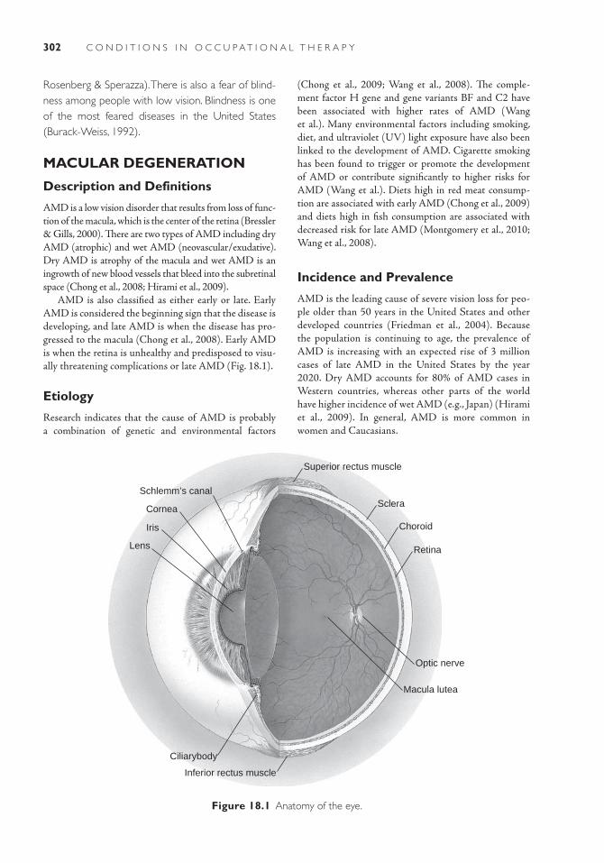
302 C O N D I T I O N S I N O C C U P A T I O N A L T H E R A P Y
Rosenberg & Sperazza). There is also a fear of blind-ness among people with low vision. Blindness is one of the most feared diseases in the United States (Burack-Weiss, 1992).
MACULAR DEGENERATION
Description and Definitions
AMD is a low vision disorder that results from loss of func-tion of the macula, which is the center of the retina (Bressler & Gills, 2000). There are two types of AMD including dry AMD (atrophic) and wet AMD (neovascular/exudative). Dry AMD is atrophy of the macula and wet AMD is an ingrowth of new blood vessels that bleed into the subretinal space (Chong et al., 2008; Hirami et al., 2009).
AMD is also classified as either early or late. Early AMD is considered the beginning sign that the disease is developing, and late AMD is when the disease has pro-gressed to the macula (Chong et al., 2008). Early AMD is when the retina is unhealthy and predisposed to visu-ally threatening complications or late AMD (Fig. 18.1).
Etiology
Research indicates that the cause of AMD is probably a combination of genetic and environmental factors
(Chong et al., 2009; Wang et al., 2008). The comple-ment factor H gene and gene variants BF and C2 have been associated with higher rates of AMD (Wang et al.). Many environmental factors including smoking, diet, and ultraviolet (UV) light exposure have also been linked to the development of AMD. Cigarette smoking has been found to trigger or promote the development of AMD or contribute significantly to higher risks for AMD (Wang et al.). Diets high in red meat consump-tion are associated with early AMD (Chong et al., 2009) and diets high in fish consumption are associated with decreased risk for late AMD (Montgomery et al., 2010; Wang et al., 2008).
Incidence and Prevalence
AMD is the leading cause of severe vision loss for peo-ple older than 50 years in the United States and other developed countries (Friedman et al., 2004). Because the population is continuing to age, the prevalence of AMD is increasing with an expected rise of 3 million cases of late AMD in the United States by the year 2020. Dry AMD accounts for 80% of AMD cases in Western countries, whereas other parts of the world have higher incidence of wet AMD (e.g., Japan) (Hirami et al., 2009). In general, AMD is more common in women and Caucasians.
Superior rectus muscle
Sclera
Choroid
Retina
Optic nerve
Macula lutea
Inferior rectus muscle
Ciliarybody
Lens
Iris
Cornea
Schlemm’s canal
Figure 18.1 Anatomy of the eye.
303Chapter 18: Low Vis ion Disorder s
Signs and Symptoms
The primary symptom of wet AMD is acute loss of cen-tral vision that becomes permanent as the retina atro-phies and is replaced by fibrous tissue. Dry AMD can also result in the loss of central vision, but the progression is much slower taking years to develop. Initially, a person with AMD may by asymptomatic. Over time, loss of vis-ual acuity for discrimination of details, metamorphopsia (distortion of objects), central scotomas, increased glare sensitivity, decreased contrast sensitivity, and decreased color vision can develop (Rosenberg & Sperazza, 2008).
Although the loss of vision can be profound and greatly impair function, people with AMD frequently do not notice the symptoms in the early stages. The visual loss does not result in black or white spots, but in spotty loss that is filled in by the surrounding vision that is still intact (Table 18-1).
Course and Prognosis
The course of AMD varies from person to person. Dry AMD does not always advance into wet AMD, but wet AMD always is preceded by dry AMD. The timeline for this process, however, is variable. Some people have dry AMD for years before it develops into wet AMD, and oth-ers develop wet AMD right away (Mogk & Mogk, 2003).
The prognosis for loss of central vision due to AMD also varies. For some people loss of central vision will proceed rapidly and others will have AMD for years and still maintain a functional level of central vision (Mogk & Mogk, 2003). Regardless of the speed of
progression, AMD does not cause total blindness. AMD results in loss of central vision, but it does not affect peripheral vision. Therefore, the person will retain peripheral vision even if he or she has late AMD.
Diagnosis
The signs of AMD are detected through examination by an ophthalmoscope. In early AMD, the formation of drusen, small yellow deposits in the center of the retina, can be seen when the pupil is dilated (Bressler & Gills, 2000). In wet AMD, abnormal new blood vessels in the choroidal layer of the eye that nourish the outer retina grow and proliferate with fibrous tissue within the drusen material. The bleeding accumulates within and beneath the retina and the retina atrophies or becomes replaced with fibrous tissue. In dry AMD, the pigmented layer of the retina slowly atrophies.
Medical/Surgical Management
Although there are no treatments to cure or reverse the vision loss due to AMD, there are some treatments aimed at preserving the central vision for as long as pos-sible. Current treatments of AMD include medications and laser treatment. Photodynamic therapy and intra-vitreal injections of antivascular endothelial growth factor and corticosteroids have shown promise for some people with AMD. In addition, high-dose regimens of zinc and antioxidants (vitamin C, vitamin E, and
TABLE 18.1 Signs and Symptoms of Low Vision Disorders
Macular Degeneration Glaucoma Cataracts
Acute loss of central vision Slow loss of peripheral vision Decreased visual acuity
Loss of visual acuity Decreased ability to see in dim light
Cloudy, blurry, or foggy vision
Metamorphopsia (distortion of objects)
Decreased contrast sensitivity Decreased contrast sensitivity
Central scotomas Poor adaptation to changes in lighting
Increased glare sensitivity
Increased glare sensitivity Increased glare sensitivity Near-sightedness (myopia)
Decreased contrast sensitivity Blurred vision Decreased color perception
Decreased color vision Decreased depth perceptionOcular painEventual loss of central vision
Especially to blue hues
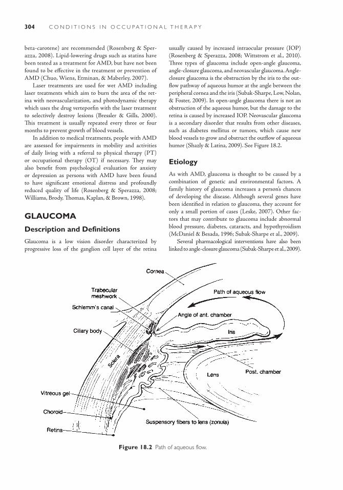
304 C O N D I T I O N S I N O C C U P A T I O N A L T H E R A P Y
beta-carotene) are recommended (Rosenberg & Sper-azza, 2008). Lipid- lowering drugs such as statins have been tested as a treatment for AMD, but have not been found to be effective in the treatment or prevention of AMD (Chuo, Wiens, Etminan, & Maberley, 2007).
Laser treatments are used for wet AMD including laser treatments which aim to burn the area of the ret-ina with neovascularization, and photodynamic therapy which uses the drug verteporfin with the laser treatment to selectively destroy lesions (Bressler & Gills, 2000). This treatment is usually repeated every three or four months to prevent growth of blood vessels.
In addition to medical treatments, people with AMD are assessed for impairments in mobility and activities of daily living with a referral to physical therapy (PT) or occupational therapy (OT) if necessary. They may also benefit from psychological evaluation for anxiety or depression as persons with AMD have been found to have significant emotional distress and profoundly reduced quality of life (Rosenberg & Sperazza, 2008; Williams, Brody, Thomas, Kaplan, & Brown, 1998).
GLAUCOMA
Description and Definitions
Glaucoma is a low vision disorder characterized by progressive loss of the ganglion cell layer of the retina
usually caused by increased intraocular pressure (IOP) (Rosenberg & Sperazza, 2008; Wittstrom et al., 2010). Three types of glaucoma include open-angle glaucoma, angle-closure glaucoma, and neovascular glaucoma. Angle-closure glaucoma is the obstruction by the iris to the out-flow pathway of aqueous humor at the angle between the peripheral cornea and the iris (Subak-Sharpe, Low, Nolan, & Foster, 2009). In open-angle glaucoma there is not an obstruction of the aqueous humor, but the damage to the retina is caused by increased IOP. Neovascular glaucoma is a secondary disorder that results from other diseases, such as diabetes mellitus or tumors, which cause new blood vessels to grow and obstruct the outflow of aqueous humor (Shazly & Latina, 2009). See Figure 18.2.
Etiology
As with AMD, glaucoma is thought to be caused by a combination of genetic and environmental factors. A family history of glaucoma increases a person’s chances of developing the disease. Although several genes have been identified in relation to glaucoma, they account for only a small portion of cases (Leske, 2007). Other fac-tors that may contribute to glaucoma include abnormal blood pressure, diabetes, cataracts, and hypothyroidism (McDaniel & Besada, 1996; Subak-Sharpe et al., 2009).
Several pharmacological interventions have also been linked to angle-closure glaucoma (Subak-Sharpe et al., 2009).
Figure 18.2 Path of aqueous flow.
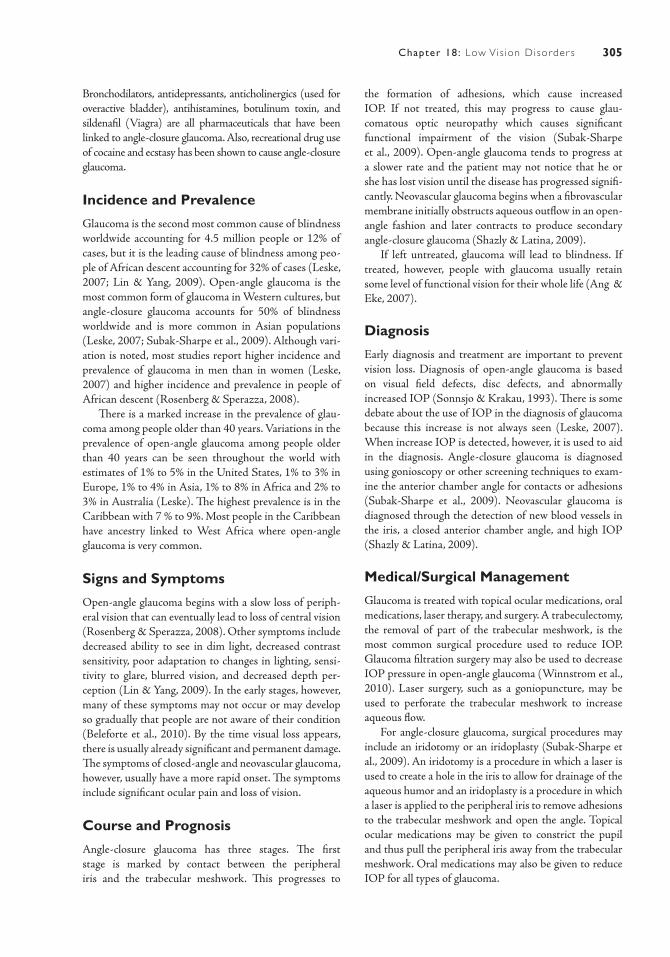
305Chapter 18: Low Vis ion Disorder s
Bronchodilators, antidepressants, anticholinergics (used for overactive bladder), antihistamines, botulinum toxin, and sildenafil (Viagra) are all pharmaceuticals that have been linked to angle- closure glaucoma. Also, recreational drug use of cocaine and ecstasy has been shown to cause angle-closure glaucoma.
Incidence and Prevalence
Glaucoma is the second most common cause of blindness worldwide accounting for 4.5 million people or 12% of cases, but it is the leading cause of blindness among peo-ple of African descent accounting for 32% of cases (Leske, 2007; Lin & Yang, 2009). Open-angle glaucoma is the most common form of glaucoma in Western cultures, but angle-closure glaucoma accounts for 50% of blindness worldwide and is more common in Asian populations (Leske, 2007; Subak-Sharpe et al., 2009). Although vari-ation is noted, most studies report higher incidence and prevalence of glaucoma in men than in women (Leske, 2007) and higher incidence and prevalence in people of African descent (Rosenberg & Sperazza, 2008).
There is a marked increase in the prevalence of glau-coma among people older than 40 years. Variations in the prevalence of open-angle glaucoma among people older than 40 years can be seen throughout the world with estimates of 1% to 5% in the United States, 1% to 3% in Europe, 1% to 4% in Asia, 1% to 8% in Africa and 2% to 3% in Australia (Leske). The highest prevalence is in the Caribbean with 7 % to 9%. Most people in the Caribbean have ancestry linked to West Africa where open-angle glaucoma is very common.
Signs and Symptoms
Open-angle glaucoma begins with a slow loss of periph-eral vision that can eventually lead to loss of central vision (Rosenberg & Sperazza, 2008). Other symptoms include decreased ability to see in dim light, decreased contrast sensitivity, poor adaptation to changes in lighting, sensi-tivity to glare, blurred vision, and decreased depth per-ception (Lin & Yang, 2009). In the early stages, however, many of these symptoms may not occur or may develop so gradually that people are not aware of their condition (Beleforte et al., 2010). By the time visual loss appears, there is usually already significant and permanent damage. The symptoms of closed-angle and neovascular glaucoma, however, usually have a more rapid onset. The symptoms include significant ocular pain and loss of vision.
Course and Prognosis
Angle-closure glaucoma has three stages. The first stage is marked by contact between the peripheral iris and the trabecular meshwork. This progresses to
the formation of adhesions, which cause increased IOP. If not treated, this may progress to cause glau-comatous optic neuropathy which causes significant functional impairment of the vision (Subak-Sharpe et al., 2009). Open-angle glaucoma tends to progress at a slower rate and the patient may not notice that he or she has lost vision until the disease has progressed signifi-cantly. Neovascular glaucoma begins when a fibrovascular membrane initially obstructs aqueous outflow in an open-angle fashion and later contracts to produce secondary angle-closure glaucoma (Shazly & Latina, 2009).
If left untreated, glaucoma will lead to blindness. If treated, however, people with glaucoma usually retain some level of functional vision for their whole life (Ang & Eke, 2007).
Diagnosis
Early diagnosis and treatment are important to prevent vision loss. Diagnosis of open-angle glaucoma is based on visual field defects, disc defects, and abnormally increased IOP (Sonnsjo & Krakau, 1993). There is some debate about the use of IOP in the diagnosis of glaucoma because this increase is not always seen (Leske, 2007). When increase IOP is detected, however, it is used to aid in the diagnosis. Angle-closure glaucoma is diagnosed using gonioscopy or other screening techniques to exam-ine the anterior chamber angle for contacts or adhesions (Subak-Sharpe et al., 2009). Neovascular glaucoma is diagnosed through the detection of new blood vessels in the iris, a closed anterior chamber angle, and high IOP (Shazly & Latina, 2009).
Medical/Surgical Management
Glaucoma is treated with topical ocular medications, oral medications, laser therapy, and surgery. A trabeculectomy, the removal of part of the trabecular meshwork, is the most common surgical procedure used to reduce IOP. Glaucoma filtration surgery may also be used to decrease IOP pressure in open-angle glaucoma (Winnstrom et al., 2010). Laser surgery, such as a goniopuncture, may be used to perforate the trabecular meshwork to increase aqueous flow.
For angle-closure glaucoma, surgical procedures may include an iridotomy or an iridoplasty (Subak-Sharpe et al., 2009). An iridotomy is a procedure in which a laser is used to create a hole in the iris to allow for drainage of the aqueous humor and an iridoplasty is a procedure in which a laser is applied to the peripheral iris to remove adhesions to the trabecular meshwork and open the angle. Topical ocular medications may be given to constrict the pupil and thus pull the peripheral iris away from the trabecular meshwork. Oral medications may also be given to reduce IOP for all types of glaucoma.

306 C O N D I T I O N S I N O C C U P A T I O N A L T H E R A P Y
CATARACTS
Description and Definitions
Cataracts are opacifications of the crystalline lens of the eye, which result in a decreased amount of light reach-ing the retina. The incoming light is scattered and visual acuity is decreased. While there are some variant cataract conditions such as congenital cataracts due to rubella, the focus of this chapter will be on the most common type, age-related cataracts.
Etiology
As with other low vision disorders, the cause of cataracts is a combination of factors. Risk factors for the develop-ment of cataracts include aging, cigarette smoking, ocular UVB and radiation exposure, drug use, systemic diseases (e.g., diabetes), and dietary nutritional intake with increas-ing age being the strongest risk factor by far (Rosenberg & Sperazza, 2008). Some of the drugs associated with increased cataract risk are corticosteroids, antipsychotics, chemotherapy agents, cholesterol-lowering medications, and tranquilizers. Lutein (found in dark green leafy veg-etables and eggs), vitamin C, and vitamin E intake have shown some associations with reduced risks for the devel-opment of cataracts (Lyle, Mares-Perlman, Klein, Klein, & Greger, 1999). Iodine deficiency and injuries to the lens are also risk factors for cataracts.
Incidence and Prevalence
Cataracts are the third leading cause of blindness in the United States, accounting for about 9% of all cases (Sper-duto, 1994). They are more prevalent among females than males and are more prevalent among people of African decent (Rosenberg & Sperazza, 2008; Sperduto, 1994). Cataracts also vary in prevalence among different age groups with approximately 4% of people ages 52 to 64 years, 28% of people ages 65 to 74 years, and 46% of people ages 75 to 85 years.
As the population ages, the number of people world-wide who will be blind due to cataracts is expected to increase dramatically over the next few decades (Sper-duto, 1994). The number of people who are blind due to cataracts could reach close to 40 million by the year 2025. As the ozone layer is depleted, there is an increase in UV radiation and this is expected to also increase the incidence of c ataracts worldwide.
Signs and Symptoms
Decreased visual acuity, decreased contrast sensitivity, glare disability, near-sightedness (myopia), and decreased
color perception, especially to blue hues, are all common symptoms of cataracts (Rosenberg & Sperazza, 2008). Complaints of cloudy, blurry, or foggy vision are often noted. Some people may experience temporary improve-ment in near acuity as the shape of the lens is changed by the cataract.
Course and Prognosis
When a person is young, the lens is transparent and incoming light has no difficulty reaching the retina. Over time, however, the lens becomes less transparent as a cloudy, opaque cataract develops in the lens. One eye is usually affected earlier than the other, but eventually, both eyes are usually involved. If left untreated, cataracts may lead to blindness or develop into other eye diseases such as AMD or glaucoma (Rosenberg & Sperazza, 2008).
Diagnosis
A diagnosis of cataracts is done using pupil dilation, acu-ity charts, and tonometry. Tonometry is done to examine ocular fluid pressure inside the eye. Some cataracts can be detected with a visual examination that reveals a cloudy lens, but pupil dilation is done to further examine the back of the eye.
Medical/Surgical Management
Initially, the cataracts may be managed by prescription lenses, tobacco cessation, and UV protection, but the only medical treatment for cataracts is the surgical removal of the lens. When the cataract has advanced to a stage that inter-feres with a person’s ability to function, surgery is usually recommended (Sperduto, 1994).
IMPACT OF CONDITIONS ON CLIENT FACTORS AND OCCUPATIONAL PERFORMANCEVisual function is the client factor that is impaired in low vision disorders. The extent of the impact on occupational performance depends on the type of low vision disorder, the stage of progression of that disorder, and the treat-ment that has been received. Loss of central vision is the main concern for people with AMD and cataracts, and loss of peripheral vision is the main concern for people with glaucoma. For most people, cataracts will be surgi-cally removed when they interfere with the person’s abil-ity to function. There are standardized assessments, such
307Chapter 18: Low Vis ion Disorder s
as the Impact of Vision Impairment Scale and the Visual Functioning Questionnaire, which may be useful to help therapists ascertain the impact of a person’s visual loss on occupational performance (Lamoureux et al., 2008; Magacho et al., 2004).
Activities of Daily Living
Because of the loss of central vision, the self-care skills that require visual acuity are most affected by AMD and cataracts. These include skills such as makeup applica-tion, dental care, nail care, and shaving. Management of medications may be difficult due to difficulty reading labels and identifying various medications. Eating may also be visually challenging in advanced stages of the disorders.
Safety during functional mobility is a concern for all low vision disorders. The loss of central vision in AMD and cataracts and the loss of peripheral vision in glaucoma can impact a person’s ability to safely navigate both within and outside the home. Within the home, entryways and stairs are especially difficult particularly at night. Adapta-tions, such as increased lighting, railings, and visual con-trasts may need to be made to increase safety. Outside the home, unfamiliar places may pose challenges for both locating points of interest and negotiating varying surfaces.
Instrumental Activities of Daily Living
Driving is impacted by all low vision disorders. Because driving represents independence for most people, it can also be the most difficult occupation to address. In addi-tion, driving is necessary for most other IADLs such as shopping, going to the post office, and making appoint-ments.
Home management, including cleaning, doing laun-dry, making home repairs, and maintaining a yard, can all be impacted by low vision disorders. Being able to perform meal preparation accurately and safely are also concerns as loss of visual acuity will interfere with see-ing food items, utensils (i.e., knives, measuring spoons, measuring cups), appliance settings, and recipes.
Money management for reading bills, paying bills, keeping records, and identifying and handling money can all be impacted by a loss of central vision. Large-print electronic systems may be useful for assisting with money management.
Education
Because all of these low vision disorders are likely to be age related, most people with these disorders will have
completed their formal education. If participation in education is still essential, functional mobility and read-ing may interfere with successful completion of education- related occupations.
Work
The age of retirement has progressively increased and therefore the person may still be involved in work occu-pations when low vision disorders progress to the point that they interfere with function. Depending on the area of work in which the person participates, visual deficits in central and peripheral vision can impact the person’s ability to successfully complete work tasks. Work-related activities such as reading, working on the computer, using the telephone, working on an assembly line, and driving can all be impaired by visual deficits.
Play and Leisure
Many leisure activities can be affected by low vision disorders. Many people with low vision disorders are retired and fill their time with leisure activities. In addition, these leisure activities may be part of the per-son’s identity providing life purpose and satisfaction. Painting, knitting, sewing, gardening, playing games, completing puzzles, watching television, reading, and participating in sports are just some of the many lei-sure activities that may be difficult for a person with a low vision disorder.
Social Participation
Low vision disorders can impact social function in many ways. People with AMD frequently complain about difficulty recognizing faces, which may lead to feelings of humiliation and embarrassment is social situations. People with low vision disorders may also have difficulty negotiating crowded places such as concerts, restaurants, and ceremonies. In restaurants, they may have difficulty reading the menu in addition to navigating the physical layout. They may become distressed if they collide with people or objects. All of these factors may lead the person into a life with more isolation. The combination of the loss of independence and anxiety about the ability to function in social situ-ations can often lead to depression. When the person becomes depressed, he or she is less likely to pursue social contact, and a cycle of isolation and depression can be worsened.
308 C O N D I T I O N S I N O C C U P A T I O N A L T H E R A P Y
Case IllustrationsCase 1: Macular DegenerationMary is a 72-year-old female who has been referred to OT due to vision loss. Mary was diagnosed with wet AMD two years ago and is having difficulty func-tioning in her home. The recent death of her husband, who was able to help her in the home, has left her trying to find ways to manage on her own. She has two children. Her daughter lives about one hour away with her three children and her son lives about two hours away in a large urban area. Her children are reportedly concerned that she is no longer safe to live independently.
Mary worked for many years as a legal assistant until she retired at age 62. Since then, she has been enjoying traveling with her husband, spending time with her grandchildren, and participating in various leisure activities, such as knitting, reading, and garden-ing. She was once an avid tennis player, but had to stop playing due to her vision loss.
Mary now lives alone in a four-bedroom, split-level home and would like to remain there for as long as possible. She is having difficulty accepting the fact that driving is dangerous for her and she tries to get by using a GPS. She also becomes embarrassed when she doesn’t recognize people, so she has started to avoid social situations where she is expected to inter-act with people who don’t know about her vision loss.
She has undergone three laser treatments since she was diagnosed, but because they don’t improve her vision, she has difficulty understanding why she needs more treatments. She is also taking high doses of zinc, vitamin C, vitamin E, and beta-carotene, but she states that sometimes she is not sure if she has taken the right pills.
Mary has been referred to OT for evaluation and treatment. Her primary goal is to maintain her current lifestyle as long as possible.
Case 2: GlaucomaBob is known amongst his friends as Bob the Builder. He retired from his job as a site manager 10 years ago, but has been busier than ever helping his friends with home improvement projects. Bob’s wife states “he is so busy helping their friends, that he never has time to get to their own home improvement needs.” Bob and his wife, Sharon, live in a four-bedroom century-old farmhouse that they have been fixing up since they moved in 10 years ago. Their home is on a 40-acre lot, but they rent out the land to a local farmer.
Bob and Sharon have been married for 15 years. They don’t have children of their own, but both have
children from previous marriages. All of their four children live within an hour of them and they have seven grandchildren among them.
Last year, Bob went to his eye doctor for a routine check up. It had been about five years since his last checkup. His kids were teasing him about his outdated glasses and he decided to get a new style. Bob hadn’t noticed any significant changes in his vision. He knew he was having some difficulty seeing when it was dim lighting, but he thought this was just due to his eyes get-ting older. He was very surprised when the eye doctor told him that he suspected that he had glaucoma.
The eye doctor referred him to a specialist who confirmed the diagnosis. The ophthalmologist explained that he had what is called open-angle glau-coma and showed him diagrams of how much periph-eral vision he had already lost. Bob and his family were surprised. Bob felt healthy and never really noticed the significant loss of vision. His mother had glaucoma, but he didn’t connect that to the possibility that he would one day develop it, as well.
Bob uses eye drops and recently underwent surgery to reduce the pressure in his eye. He has been referred to OT to see if adaptation can be made to his environment and lifestyle to help him maintain as much function as possible in the coming years.
Case 3: CataractsElizabeth, who is known as Beth to her family and friends, has been dealing with increasingly limited vision due to cataracts. Beth is 68 years old and has lived with her partner, Sue, for the last 26 years in a high-rise condo in an urban area. Beth has a daugh-ter from a previous relationship and she lives within walking distance to her mother’s home. Beth is retired from her career as an architect, but Sue still works as a teacher and has an hour commute to and from work.
Since her retirement, Beth spends most of her time participating in leisure activities. She loves to paint, swim, and work out in the building’s gym facilities. She is also an avid reader, but has been frustrated with her visual deficits when trying to read. Her daughter owns and operates a local coffee shop, so she frequently stops by there to hang out or help out her daughter as needed.
Beth has been very aware of her increasingly limited vision over the past several years. She describes her vision as cloudy, blurry, and sensitive to “bad lighting.” She started wearing glasses a few years ago, but now her vision loss is starting to interfere with her ability to function around the home and within her community.
309Chapter 18: Low Vis ion Disorder s
Beth’s eye doctor discussed routine eye surgery to remove the cataracts from both eyes. Once the eye doctor discovered that Beth had scarring on her ret-ina in the right eye due to an old injury, however, she decided that the surgery would only benefit the left eye. Beth, therefore, will continue to have a significant
loss of vision in her right eye even after the surgery is done.
Beth was referred to OT for an evaluation and intervention to assist her in making any adaptation necessary to increase her ability to function indepen-dently in her home and her community.
R E C O M M E N D E D L E A R N I N G R E S O U R C E S
American Academy of Ophthalmologyhttp://www.aao.orgAmerican Council of the Blindhttp://www.acb.orgAmerican Foundation for the Blindhttp://www.afb.orgMacular DegenerationMedlinePluswww.nlm.nih.gov/medlineplus/maculardegeneration.htmlMayoClinic.comwww.mayoclinic.com/health/macular-degeneration/
DS00284Macular Degeneration Foundationwww.eyesight.org/Glaucoma
Glaucoma Research Foundationwww.glaucoma.org/MayoClinicwww.mayoclinic.com/health/glaucoma/DS00283National Eye Institutewww.nei.nih.govCataractsMedlinePluswww.nlm.nih.gov/medlineplus/cataract.htmlNational Eye Institutewww.nei.nih.govMayoClinicwww.mayoclinic.com/health/cataracts/DS00050
R E F E R E N C E S
Ang, G. S., & Eke, T. (2007). Lifetime visual prognosis for patients with open-angle glaucoma. Eye, 21, 604–608.
Beleforte, N. A., Moreno, M. C., de Zavalia, N., Sande, P. H., Chianelli, M. S., Keller Sarmiento, M. I., et al. (2010). Melatonin: A novel neuroprotectant for the treatment of glaucoma. Journal of Pineal Research, 48, 353–364.
Bressler, N. M., & Gills, J. P. (2000). Age related macu-lar degeneration: New hope for a common problem comes from photodynamic therapy. British Medical Journal, 321, 1425–1427.
Burack-Weiss, A. (1992). Psychological aspects of aging and vision loss. In E. Faye & S. Stuen (Eds.), The aging eye and low vision: A study guide for physicians (pp. 29–34). New York, NY: Lighthouse.
Chong, E. W. -T., Simpson, J. A., Robman, L. D., Hodge, A. M., Aung, K. Z., English, D., R., et al. (2008). Red meat and chicken consumption and its associa-tion with age-related macular degeneration. American Journal of Epidemiology, 169(7), 867–876.
Chuo, J. Y., Wiens, M., Etminan, M., & Maberley, D. A. L. (2007). Use of lipid-lowering agents for the
prevention of age-related macular degeneration: A meta-analysis of observational studies. Ophthalmic Epidemiology, 14(6), 367–374.
Friedman, D. S., O’Colmain, B. J., Munoz, B., Tomany, S. C., McCarty, C., de Jong, P. T., et al. (2004). Prevalence of age-related macular degeneration in the United States. Archives of Ophthalmology, 122(4), 564–572.
Hirami, Y., Mandai, M., Takahashi, M., Teramukai, S., Tada, H., & Yoshimura, N. (2009). Association of clinical characteristics with disease subtypes, initial visual acuity, and visual prognosis in neovascular age-related macular degeneration. Japanese Journal of Opthalamology, 53, 396–407.
Lamoureux, E. L., Pallant, J. F., Pesudova, K., Tennant, A., Rees, G., O’Connor, P. M., et al. (2008). Assess-ing participation in daily living and the effectiveness of rehabilitation in age related macular degeneration patients using the impact of vision impairment scale. Ophthalmic Epidemiology, 15, 105–113.
Leske, M. C. (2007). Open-angle glaucoma- an epidemio-logical overview. Ophthalmic Epidemiology, 14, 166–172.
310 C O N D I T I O N S I N O C C U P A T I O N A L T H E R A P Y
Lin, J., & Yang, M. (2009). Correlation of visual function with health-related quality of life in glaucoma patients. Journal of Evaluation of Clinical Practice, 16, 134–140.
Lyle, B. J., Mares-Perlman, J. A., Klein, B. E. K., Klein, R., & Greger, J. L. (1999). Antioxidant intake and rick of incident age-related nuclear cataracts in the Beaver Dam eye study. American Journal of Epidemiology, 149(9), 801–809.
Magacho, L., Lima, F. E., Nery, A. C. S., Sagawa, A., Magacho, B., & Avila, M. P. (2004). Quality of life in glaucoma patients: Regression analysis and correla-tion with possible modifiers. Ophthalmic Epidemiol-ogy, 11(4), 263–270.
Massof, R. W. (2002). A model of the prevalence and incidence of low vision and blindness among adults in the U.S. Optometry and Vision Science, 79, 31–38.
McDaniel, D., & Besada, E. (1996). Hypothyroidism—A possible etiology of open-angle glaucoma. Journal of the American Optometric Association, 67(2), 109–114.
Mogk, L. G., & Mogk, M. (2003). Macular degeneration: The complete guide to saving and maximizing your sight. New York, NY: Random House.
Montgomery, M. P., Kamel, F., Pericak-Vance, M. A., Haines, J. L., Postel, E. A., Agarwal, A., et al. (2010). Overall diet quality and age-related macular degen-eration. Ophthalmic Epidemiology, 17(1), 58–65.
Rosenberg, E. A., & Sperazza, L. C. (2008). The visually impaired patient. American Family Physician, 77(10), 1431–1438.
Shazly, T. A., & Latina, M. A. (2009). Neovascular glaucoma: Etiology, diagnosis and prognosis. Seminars in Ophthalmology, 24, 113–121.
Sonnsjo, B., & Krakau, C. E. T. (1993). Arguments for vascular glaucoma etiology. Acta Ophthalmologica, 71, 433–444.
Sperduto, R. D. (1994). Age-related cataracts: Scope of problem and prospects for prevention. Preventive Medicine, 23, 735–739.
Subak-Sharpe, I., Low, S., Nolan, W., & Foster, P. J. (2009). Pharmacological and environmental factors in primary angle-closure glaucoma. British Medical Bulletin, 93, 125–143.
Wang, J. J., Rochtchina, E., Smith, W., Klein, R., Klein, B. E. K., Joshi, T., et al. (2008). Combined effects of complement factor H genotypes, fish consumption, and inflammatory markers on long-term risk for age-related macular degeneration in a cohort. American Journal of Epidemiology, 169(5), 633–641.
Williams, R. A., Brody, B. L., Thomas, R. G., Kaplan, R. M., & Brown, S. I. (1998). The psychosocial impact of macular degeneration. Archives of Ophthalmologie, 116, 514–520.
Wittstrom, E., Schatz, P., Lovestram-Adrian, M., Ponjavic, V., Bergstrom, A., & Andreasson, S. (2010). Improved retinal function after trabecu-lectomy in glaucoma patients. Graefes Archive for Clinical and Experimental Ophthalmology, 248, 485–495.
311
Infectious Diseases
■ Kar in J. O p a c i c h
19
An occupational therapist is working for a migrant health center clinic. Most of the clients work on farms either planting and har-vesting or caring for livestock. Among the largest employers is a thoroughbred breeding farm that employs 15 men and women
to clean stalls, feed, and attend to the mares and foals. Over the last week, three workers from this farm have been treated for severe gastrointestinal distress. The pathogen was determined to be Salmonella, and it was discov-ered that one of the mares and her newborn foal have also been diagnosed with Salmonella.1 These three workers have been sharing a dormitory room and two had been directly responsible for cleaning the infected mare’s stall or handling the mare and her foal. The workers have no health insurance and no other means of financial support. They are all receiving treatment through the migrant health clinic, but they have to return to work since they have no health benefits and they do not get paid unless they work.
Cumulative discoveries and advancements in science have revealed the underlying mechanisms of infectious diseases that threaten the life and well-being of individuals and global populations. Infectious diseases may cause minor acute discomfort to chronic conditions to life-ending systemic dam-age. Vivid descriptions of infectious diseases have appeared in literature from ancient Greece to modern times. Hippocrates (400 bc), in a collection of ancient accounts entitled, Of the Epidemics, described the courses of individ-ual sickness in painstaking detail trying to identify commonalities, patterns, and sources of illness affecting the whole community. Although much has been learned about causality since the days of Hippocrates, c. 400 bc, even recent history is replete with accounts of superstitions and misunderstanding about infectious diseases, none more poignant than the maltreatment of people
with Hansen’s disease (leprosy) in Hawaii in the 20th century (Gugelyk & Bloombaum, 1Three to six percent of the mare population have been found to be silent salmonella carriers who can transmit the infection to their foals in utero (Ward, Alinovi, Couetil, & Wu, 2005).
K E Y T E R M S
AntibodyAntigen
Burden of diseaseDisease surveillance
Disease trajectoryEpidemiology
Health disparityHerd immunity
HIVImmune response
IncidenceInfectious diseases
Inoculum doseLatent tuberculosis
MorbidityMortality
MRSAPathogen
PrevalenceRisk factors
Tuberculosis (TB)Zoonotic infection
312 C O N D I T I O N S I N O C C U P A T I O N A L T H E R A P Y
1979). While hundreds of infectious diseases have been identified and demystified, new pathogens that compromise human, animal, and environmental health are always evolving. The Center for Disease Con-trol (CDC) now publishes a journal titled Emerging Effective Diseases (listed in Recommended Learning Resources section at the end of this chapter), which is entirely devoted to disseminating information about evolving or resurgent diseases such as HIV/AIDS, avian flu, West Nile virus, and tuberculosis (TB).
Each wave of illness has led to new knowledge and technology giving rise to the field of epide-miology, a scientific discipline devoted to studying the patterns of communicable diseases in order to predict, contain, and ultimately prevent the spread of disease. The famous account of Dr. John Snow, the scientist regarded as the father of modern epidemiol-ogy, chronicles the investigation that pinpointed the contaminated water pumps that spread cholera killing hundreds of people in London in 1854 (Snow, 1965). Recognizing the value of efficient disease tracking and information sharing, the U.S. Centers for Disease Control was authorized in 1946 to accumulate sta-tistics on incidence, prevalence, morbidity, and mortality associated with major communicable diseases. Nearly 70 distinct diseases have been listed for surveillance and mandatory reporting in 2011 and these can be found at the CDC website listed in the recommended learning resources at the end of this chapter. The data collected serve to identify populations most vulnerable to infection and informs the development of individual and population inter-ventions to curtail the spread of disease. National agencies, state departments of health, and city public health agencies may all collect and house health and communicable disease statistics. Geographic infor-mation systems (GIS) have improved the ability of these agencies to track, collect, and analyze data from broad geographic regions to areas as small as neigh-borhoods. Multilevel data are especially important for scientists to decipher disease trends, associations, and disparities on global, national, municipal, and in some cases even neighborhood levels.
This chapter will provide explanations by way of examples of infectious disease, TB, and HIV that affect occupational performance. Recognizing the
inextricable connection between individual well-being and community health, occupational therapy practitioners are apt to acknowledge the impor-tance of addressing transient disruptions and lasting effects of infectious diseases that impact occupa-tional performance. Using TB and HIV as metaphors, infectious disease is explored from both population and individual perspectives from which the reader will develop a basic understanding of causative organisms, routes of infection, manifestations of ill-ness, and medical interventions. Occupational ther-apy practitioners will find it necessary to take into account the context in which individuals experience their illnesses to fully address the occupational lives of their clients. The following exemplars can serve as templates for broadening our understanding of other infectious diseases encountered in occupa-tional therapy practice.
DESCRIPTION AND DEFINITIONS OF INFECTIOUS DISEASESInfectious diseases are characterized by a set of symptoms attributable to the introduction of a specific pathogen, a microorganism that compromises the health of an indi-vidual. Most pathogens fall into three general categories: bacteria, viruses, and fungi. Pathogens may be transmit-ted by direct contact (in utero, sexual activity, or nonsex-ual physical contact), aspiration (airborne), or ingestion (water or food). See Table 19.1 for examples of some of the infectious diseases of most interest to the Centers for Diseases Control (CDC), the associated pathogens, and routes of transmission. Tracking these diseases over time yields the evidence necessary to develop interventions and policies that protect individuals and populations from contracting and spreading infectious diseases. For instance, the preponderance of data pertaining to food-borne infections resulted in a new federal legislation, The Food and Drug Administration Food Safety Moderniza-tion Act (2010), imposing higher standards and penalties for the protection of consumers. Since food preparation and eating are fundamental human occupations, practi-tioners might consider how food safety is integrated into patient, family, and community habits and routines.
The scientific literature reveals that severity of ill-ness in any individual is contingent upon multiple fac-tors including the virility of the pathogen, the inherent disease trajectory, the premorbid health status of the
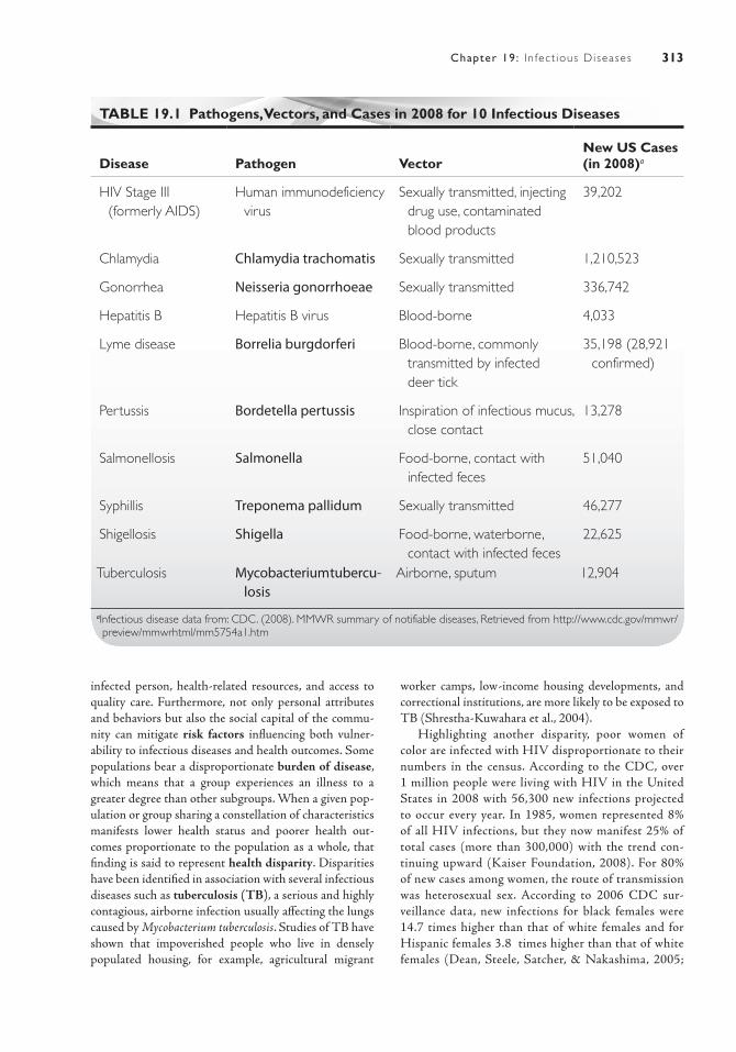
313Chapter 19: In fect ious Diseases
infected person, health-related resources, and access to quality care. Furthermore, not only personal attributes and behaviors but also the social capital of the commu-nity can mitigate risk factors influencing both vulner-ability to infectious diseases and health outcomes. Some populations bear a disproportionate burden of disease, which means that a group experiences an illness to a greater degree than other subgroups. When a given pop-ulation or group sharing a constellation of characteristics manifests lower health status and poorer health out-comes proportionate to the population as a whole, that finding is said to represent health disparity. Disparities have been identified in association with several infectious diseases such as tuberculosis (TB), a serious and highly contagious, airborne infection usually affecting the lungs caused by Mycobacterium tuberculosis. Studies of TB have shown that impoverished people who live in densely populated housing, for example, agricultural migrant
worker camps, low-income housing developments, and correctional institutions, are more likely to be exposed to TB (Shrestha-Kuwahara et al., 2004).
Highlighting another disparity, poor women of color are infected with HIV disproportionate to their numbers in the census. According to the CDC, over 1 million people were living with HIV in the United States in 2008 with 56,300 new infections projected to occur every year. In 1985, women represented 8% of all HIV infections, but they now manifest 25% of total cases (more than 300,000) with the trend con-tinuing upward (Kaiser Foundation, 2008). For 80% of new cases among women, the route of transmission was heterosexual sex. According to 2006 CDC sur-veillance data, new infections for black females were 14.7 times higher than that of white females and for Hispanic females 3.8 times higher than that of white females (Dean, Steele, Satcher, & Nakashima, 2005;
TABLE 19.1 Pathogens, Vectors, and Cases in 2008 for 10 Infectious Diseases
Disease Pathogen VectorNew US Cases (in 2008)a
HIV Stage III (formerly AIDS)
Human immunodeficiency virus
Sexually transmitted, injecting drug use, contaminated blood products
39,202
Chlamydia Chlamydia trachomatis Sexually transmitted 1,210,523
Gonorrhea Neisseria gonorrhoeae Sexually transmitted 336,742
Hepatitis B Hepatitis B virus Blood-borne 4,033
Lyme disease Borrelia burgdorferi Blood-borne, commonly transmitted by infected deer tick
35,198 (28,921 confirmed)
Pertussis Bordetella pertussis Inspiration of infectious mucus, close contact
13,278
Salmonellosis Salmonella Food-borne, contact with infected feces
51,040
Syphillis Treponema pallidum Sexually transmitted 46,277
Shigellosis Shigella Food-borne, waterborne, contact with infected feces
22,625
Tuberculosis Mycobacterium tubercu-losis
Airborne, sputum 12,904
aInfectious disease data from: CDC. (2008). MMWR summary of notifiable diseases, Retrieved from http://www.cdc.gov/mmwr/preview/mmwrhtml/mm5754a1.htm
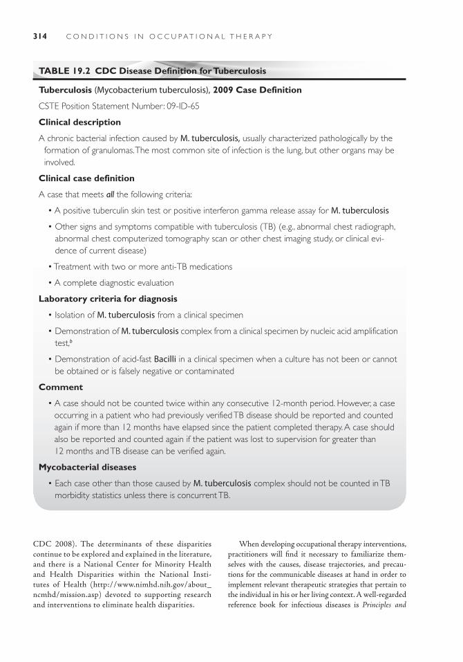
314 C O N D I T I O N S I N O C C U P A T I O N A L T H E R A P Y
CDC 2008). The determinants of these disparities continue to be explored and explained in the literature, and there is a National Center for Minority Health and Health Disparities within the National Insti-tutes of Health (http://www.nimhd.nih.gov/about_ncmhd/mission.asp) devoted to supporting research and interventions to eliminate health disparities.
When developing occupational therapy interventions, practitioners will find it necessary to familiarize them-selves with the causes, disease trajectories, and precau-tions for the communicable diseases at hand in order to implement relevant therapeutic strategies that pertain to the individual in his or her living context. A well-regarded reference book for infectious diseases is Principles and
TABLE 19.2 CDC Disease Definition for Tuberculosis
Tuberculosis (Mycobacterium tuberculosis), 2009 Case Definition
CSTE Position Statement Number: 09-ID-65
Clinical description
A chronic bacterial infection caused by M. tuberculosis, usually characterized pathologically by the formation of granulomas. The most common site of infection is the lung, but other organs may be involved.
Clinical case definition
A case that meets all the following criteria:
• A positive tuberculin skin test or positive interferon gamma release assay for M. tuberculosis
• Other signs and symptoms compatible with tuberculosis (TB) (e.g., abnormal chest radiograph, abnormal chest computerized tomography scan or other chest imaging study, or clinical evi-dence of current disease)
• Treatment with two or more anti-TB medications
• A complete diagnostic evaluation
Laboratory criteria for diagnosis
• Isolation of M. tuberculosis from a clinical specimen
• Demonstration of M. tuberculosis complex from a clinical specimen by nucleic acid amplification test,b
• Demonstration of acid-fast Bacilli in a clinical specimen when a culture has not been or cannot be obtained or is falsely negative or contaminated
Comment
• A case should not be counted twice within any consecutive 12-month period. However, a case occurring in a patient who had previously verified TB disease should be reported and counted again if more than 12 months have elapsed since the patient completed therapy. A case should also be reported and counted again if the patient was lost to supervision for greater than 12 months and TB disease can be verified again.
Mycobacterial diseases
• Each case other than those caused by M. tuberculosis complex should not be counted in TB morbidity statistics unless there is concurrent TB.
315Chapter 19: In fect ious Diseases
Practice of Infectious Diseases, 7th edition (Mandell, Doug-las, & Bennett, 2010). Therapists may also find Emerging Infectious Diseases: A Guide to Diseases, Causative Agents, and Surveillance (Beltz, 2011) to be useful for reference.
In 1997 the CDC established uniform criteria for reporting cases of infectious disease. Each communicable disease has its own specific criteria (definition) that is peri-odically revisited and redefined as knowledge accumulates.
COURSE AND PROGNOSIS OF INFECTIOUS DISEASEEach disease is characterized by an intricate biologic mechanism designed to perpetuate itself, and any indi-vidual’s immune response or immunity, which reflects one’s defense or resistance against infection and plays a role in the expression of illness. Some pathogens are very hardy and can live on inanimate surfaces for long periods, for example, Staphylococcus, while others, for example, human immunodeficiency virus (HIV), are quite fragile and generally require blood-to-blood contact to survive. The amount of pathogen necessary to produce infection is called the inoculum dose, and this dosage varies depending on the virility of the pathogen itself and on the efficiency of one’s immune system to recognize, isolate, and destroy the foreign intruder. Some infectious pathogens are frag-ile and require ideal conditions to flourish. In contrast, the pathogen associated with hantavirus, a disease con-tracted from dried droppings or secretions from infected rodents, can survive for days under normal conditions, and a tiny inoculum dose can create biologic havoc in humans through hanta pulmonary syndrome (CDC, 2005a). HIV is an example of a pathogen that requires a small inocu-lum dose, but the virus itself is rather fragile. It is most efficiently transmitted by blood-to-blood contact through small lesions during sexual intercourse or by injection drug use or accidental needle sticks (Blanchard & Arai, 2010). Some infectious agents are quite hardy and can linger on inanimate surfaces until they come into contact with a hos-pitable host. Staphylococcus bacteria are fairly common and can cause illness from skin rashes to respiratory ailments. When staph (short for Staphylococcus) comes into contact with more vulnerable hosts, like the wounds of postsurgi-cal patients, it can be far more dangerous especially when the strain of staph is resistant to customary antibiotics, for example, methicillin-resistant Staphylococcus aureus or MRSA (Enright et al., 2002).
When a pathogen enters the body, the immune sys-tem attempts to interfere with its reproduction and exacerbation. This typically entails a chain of microbio-logic events: recognition of the pathogen as foreign, iso-lation of the pathogen, and destruction of the pathogen. Microorganisms called antigens, uniquely configured
proteins attached to the pathogen, stimulate the body’s production of antibodies, again typically proteins, that recognize and attach to the disease-producing entity marking it for destruction by phagocytes. The immune system is challenged to conduct this molecular activ-ity efficiently enough to limit the replication of the cells that threaten the integrity of the body and produce the symptoms we recognize as illness. Some pathogens, for example, retroviruses like HIV, are especially equipped to insinuate themselves into the cellular reproductive mech-anism commandeering the RNA to reproduce viral cells and ultimately turning the immune system into a factory for reproducing itself.
Pharmacologic science has rendered antibiotic, anti-viral, antiretroviral, and antifungal drugs to assist the immune system in combating disease-producing patho-gens, thus minimizing effects of infections that may have been historically fatal. George Washington is widely believed to have died at age 67 from a sore throat, specifi-cally epiglottitis, attributable to Haemophilus influenzae type B, a bacterial infection now curable with antibiotics and for which children are routinely inoculated (Ander-son, 2005). Practitioners are advised to consult medi-cal reference books that address both the microbiologic mechanisms and the symptoms specific to any individu-al’s infectious illness. Despite progress, not all infectious diseases are curable even with drug intervention, but by and large, their exacerbation can be managed to yield a much better quality of life for the infected individual.
Some infectious diseases are not only curable but also preventable by vaccination. Using minute amounts of live or killed pathogen, vaccinations trigger immune responses that either prevent the illness or minimize its effect in the vaccinated individual. Children are now rou-tinely immunized for measles, mumps, pertussis, diph-theria, polio, and other potentially dangerous diseases. When critical masses of people (or other animal species) are rendered immune to a communicable disease, the rampant spread of infection is curtailed, a phenomenon known as herd immunity. All vaccinations are accompa-nied with risks, and some cultural groups are leery about immunization altogether; however, historical data regard-ing vaccination campaigns, for example, poliomyelitis, have shown to be highly successful in minimizing what were once devastating levels of mortality and morbidity (Graham, Ledgerwood, & Nabel, 2009).
TUBERCULOSIS (TB)
Definition and Description of TB
Mycobacterium tuberculosis is the pathogen that is present in the sputum of actively infected people, other animals, and birds. Among humans, the droplets that are emitted
316 C O N D I T I O N S I N O C C U P A T I O N A L T H E R A P Y
when coughing during the infectious stage can transmit the disease to others. TB is a zoonotic infection, which means that it can cross species. For instance, research has demonstrated that bovine TB may be contracted through the milk of infected cows which is thought to account for much of the TB in Southeast Asia (DeKantor, LoBue, & Thoen, 2010). TB most often affects the lungs by caus-ing tubular inflammation and eventual scar tissue that impedes respiration. Untreated, TB can cause protracted illness and painful deterioration involving all organ and skeletal systems until eventual death.
Incidence and Prevalence of TB
TB is on the list of notifiable diseases that must be reported to the CDC. Although the prevalence of TB has declined steadily in the United States from a rate of 52.6 per 100,000 in 1952 to 3.8 per 100,000 in 2009 or 11,545 active cases, it is still among the most prevalent diseases globally (World Health Organization, 2010). The great-est proportion of active TB infections worldwide occur in Southeast Asia and Africa. TB is an opportunistic infec-tion affecting society’s most vulnerable people, those who are already ill, weak, impoverished, or marginalized from mainstream society (Shrestha-Kuwahara et al., 2004). In the United States, 29% of reported TB in 2009 occurred among people of Hispanic or Latino ethnicity, 26% among Black or African Americans, and 26% among Asians. Cit-ies with the highest number of reported cases of active TB in 2009 were New York City, Los Angeles, Houston, and Chicago (CDC, 2009).
Signs and Symptoms of TB
People who contract TB may experience an array of symptoms including fever, weight loss, and night sweats. Because TB usually affects the respiratory system, symp-toms commonly include coughing, chest pain, and bloody expectorant. Like other infections, TB triggers an immune response, and initial symptoms may abate. Without appro-priate treatment, TB can exist in a dormant state known as latent tuberculosis until an individual’s immune system is compromised for another reason, for example, HIV or hepatitis. Although TB in its latent form is not transmit-ted to others, if left untreated it poses a lifetime risk to the infected individual.
Medical/Surgical Management of TB
A simple skin test is commonly used to screen for TB. It entails administration of a droplet of Mantoux tuber-culin intradermally by syringe, and it reveals exposure
to M. tuberculosis through the appearance of a localized inflammatory response. The next level of detection entails chest radiograph to reveal the presence of tubules in the lungs. The standard for determining the presence and spe-cific configuration of the latent tuberculosis infection is the QuantiFERON-TB Gold blood test (CDC, 2005b). Once diagnosed, an infected person is engaged in a strict daily pharmaceutical regimen, and the medications com-monly administered are isoniazid, rifampin, ethambutol, and pyrazinamide. Treatment entails taking at least two medications religiously for a period of 6 to 12 months (Lobue, Enarson, & Thoen, 2010). These drugs are sometimes associated with unpleasant side effects con-tributing to noncompliance. Because strict adherence to the drug protocol is critical for eradicating the infection, people may be directly observed taking the daily medi-cation. Poor adherence can result in multidrug-resistant disease and persistent infection (Horsburgh et al., 2010).In the United States, TB screening leads to early medi-cal intervention and helps to prevent the long-term and devastating multisystem destruction that can result if not treated appropriately. It is understood that adherence to the prescribed medication regimen is of utmost impor-tance, because nonadherence can lead to persistent infec-tion in turn resulting in wasting and painful damage to all organ systems and the skeletal system. Therapists working in third-world countries or with immigrants from these countries may see more advanced disease symptoms. Access to treatment and adherence may be more compli-cated for social, political, and economic reasons. For those who are interested in learning about the global picture of infectious diseases, Dworkin’s book, Outbreak Investiga-tions Around the World: Case Studies in Infectious Disease Field Epidemiology (2010) may be enlightening.
Social and Behavioral Considerations of TB
It is important to understand the underlying social and economic conditions that render certain populations more vulnerable to TB and some other infectious dis-eases. Risk factors including poor nutrition, crowded housing, and poor sanitation have also been recognized as contributing to the spread of TB. These conditions have been and continue to be associated with poverty in both industrialized countries and developing nations. Population density in settings such as dilapidated hous-ing structures, migrant worker camps, and prisons where people live in close proximity increases the likelihood of transmission in the presence of infection. Unwilling-ness to admit illness for economic reasons or lack of access to medical care may delay health-seeking behavior. To reiterate, TB, like many other infectious diseases, is
317Chapter 19: In fect ious Diseases
opportunistic, and people who may be poorly nourished or who are experiencing other health challenges may not have robust immune systems to ward off infection.
Lifestyles, cultural interpretations, and mistrust of health care providers can impose barriers to detection and treatment as well (Shrestha-Kuwahara et al., 2004). Fear, stigmatization, and poor health literacy among disparity populations must be overcome to improve individual and population health. Readers who are interested in learning more about TB may benefit from CDC’s online course, Interactive Core Curriculum on Tuberculosis: What the Cli-nician Should Know, accessible free of charge at http://www.cdc.gov/tb/webcourses/CoreCurr/index.htm.
The TB case at the end of the chapter relays some specific facts in terms of prevalence, disease trajectory, and medical management, but the process of information gathering can be applied to any infectious disease. Occupa-tional therapy practitioners can access timely information from the CDC and other credible Web sites and references to understand how the infection can affect individual and community occupations.
HUMAN IMMUNODEFICIENCY VIRUS (HIV)
Definition and Description of HIV
The origin of what has become known as HIV appears to have been the slaughter of chimpanzees infected with a virus that crossed species, mutated, and spread through human behavior. Retrospective studies indicate that HIV entered the United States sometime in the 1970s, and that the male homosexual population initially expe-rienced the greatest burden of disease (Curran, 2003; Armstrong, Calabrese, & Taege, 2005). Most common routes of transmission include unprotected sex, receiving contaminated blood products, injection of illicit drugs, and perinatal transmission from mother to child.
Incidence and Prevalence of HIV
As of 2010, the CDC estimates that over 1 mil-lion people in the United States are HIV infected, further hypothesizing that one in five of these peo-ple remain unaware of their status. In terms of risk behaviors, approximately 12% of these cases are attributed to injection drug use. Although men who have sex with men still represent the largest cohort of people infected with HIV in the United States, the proportion of heterosexual infection has risen to 31% with women representing 27% of new cases (CDC, 2008b, 2010). The aforementioned distribu-tion of disease is unique to the United States and var-ies globally where HIV is largely transmitted through
unprotected heterosexual sex (UNAIDS, 2010). The number of infants diagnosed with HIV in the United States, has declined due to perinatal screening and intervention (Panel on Treatment of HIV-Infected Pregnant Women and Prevention of Perinatal Trans-mission, 2010).
Signs and Symptoms of HIV
Flu-like symptoms are reported and frequently over-looked by people initially infected with HIV. Definitive diagnosis of HIV infection entails a blood test. One of two commonly used blood tests, ELISA, is a sensi-tive screening test that detects and confirms exposure to HIV through the presence of antibodies. A more specific test, the Western Blot, determines the presence of actual virus. As in the above scenario, HIV may be diagnosed secondary to another presenting problem, like another STD or pneumonia (Divisions of HIV/AIDS Prevention, 2010). HIV is a retrovirus, meaning that it reverses the usual pattern of replication by using the host cell’s DNA to manufacture viral RNA that can then be incorporated into DNA and insinuated into healthy cells. Once the virus enters the bloodstream, it assaults the immune system proliferating until the CD4 and T helper cells can no longer defend the person from illness. Unless this process is halted, HIV infection can pro-gress to autoimmune syndrome now called HIV Stage 3 (AIDS) and usually occurs over a period of several years. The official criteria for a diagnosis of HIV Stage 3 (AIDS) include a T cell count below 200 and the pres-ence of at least one opportunistic illness (CDC, 2008a). Commonly reported symptoms of AIDS include weight loss, fevers and sweats, weakness, persistent rashes or infections, opportunistic infections (e.g., candidiasis, TB), memory impairment, depression, irritability, cogni-tive deterioration, and others.
Course and Prognosis of HIV
Improved interventions have resulted in longer life for many people with HIV. For those with access to resources, years of quality life may be extended for 15 or more years beyond diagnosis. Consequently, HIV is now considered a chronic rather than a fatal disease. Among the treatment options, highly active antiret-roviral therapy (HAART) protocols include multiple drugs with different actions to stop the proliferation of HIV at different points in the reproductive cycle of the virus (May et al., 2007). However, not all people with HIV are responsive to these protocols, and the drugs may be associated with unpleasant side effects such as nausea, diarrhea, mood disorders, or fevers that can
318 C O N D I T I O N S I N O C C U P A T I O N A L T H E R A P Y
affect quality of life. In the case scenario, the experience of drug-related symptoms may be an issue for Joseph. Should his HIV infection continue to progress rap-idly to Stage 3, Joseph may experience musculoskeletal wasting, cognitive impairment, neuropathies, and neu-rologic symptoms of greater magnitude. He may also suffer from rare cancers and opportunistic infections due to the impairment of the immune system to rec-ognize and destroy pathogens and aberrant organisms.
Social and Behavioral Considerations of HIV
People with HIV are at risk for stigmatization and iso-lation. Beyond providing information and education to the HIV+ person, health practitioners may be involved in educating partners, families, and communities about the disease. In Case 2, Joseph’s fiancée is 3 months pregnant. Ethically and legally, she must be informed of her HIV risk and the potential risk to her unborn fetus. Screening for HIV is important because perina-tal treatment can prevent the baby from seroconversion and undo suffering (Birkhead et al., 2010).
Joseph will have little chance of living well with HIV unless he ceases to use illicit drugs. Sharing needles is also the likely source of his HIV infection, and even if he con-tinues to use drugs, Joseph must alter these practices to avoid putting others at risk of infection. If Joseph remains committed to sobriety, there are clearly additional issues that must be addressed for him to be restored to a state of occupational coherence. He may need psychological counseling and peer support to deal with both his mili-tary experiences and his HIV diagnosis. All health prac-titioners need to be prepared to counsel and assist people with HIV who may be suffering from isolation or stigma-tization (Duffy, 2005).
IMPACT ON CLIENT FACTORS AND OCCUPATIONAL PERFORMANCE“Participation in activities and occupations that are meaningful to the client involves emotional, psychosocial, cognitive, and physical aspects of performance. This par-ticipation provides a means to enhance health, well-being and life satisfaction” (American Occupational Therapy Association, 2004, p. 673).
The cases challenge the occupational practitioner to think broadly about the impact of an infectious disease on individuals in a community context. Focusing first on the two children who have been identified with active TB, school and home occupations and issues for others
in the community will also be considered. This approach is consistent with an ecologic approach to both infectious disease and human occupation.
Disease investigation will clarify the source of infec-tion for the two school-age children. Whatever the source, the children must be treated to prevent spread-ing TB. It is likely that the children will be confined at home until acute symptoms have abetted, but all school personnel including the occupational therapy practitioner will be expected to comply with screening and observing for further indications of the spread of disease (Lazar, Sosa, & Lobato, 2010). Because of preconceived notions about TB or fear of stigmatization, some people may be reluctant to be tested or to seek appropriate care. Astute occupational therapy practitioners will be sensitive to the needs of the community and will interact with cultural humility.
Both for individuals and for the community, adher-ence to prevention measures will be necessary. The occu-pational therapy practitioner should reinforce hygienic behaviors for all school children and especially children participating in therapy, for example, covering the mouth when coughing or sneezing, disposing tissues, and wash-ing hands routinely. Students should be discouraged from sharing food and utensils with simple explanations of how germs are passed from one person to another. All toys and surfaces where sputum might transmit bacte-ria should be cleaned with an appropriate disinfectant between clients. Children in group treatments should be closely monitored for attention to hygiene and for signs of illness.
It is of utmost importance for the entire health and education team to collaborate to provide relevant infor-mation about TB and to prevent stigmatizing the infected children and their families. The school nurse is likely to be the person who will observe the children taking their daily medication, at least during the school week, to assure adherence. Discretion will be important to keep the children from suffering negative social consequences, so their daily school routines should be planned with this in mind. Age-appropriate explanations and health education should be anticipated, and all staff should be attentive to issues of self-esteem and potential bullying. Communication with the families of schoolchildren and particularly the families of the children with TB is of critical importance, and the family liaison, whoever that might be, must be prepared to ascertain cultural beliefs and interpretations of TB.
The occupational therapy practitioner can be help-ful in addressing daily routines and habits both at school and at home. Infected individuals are likely to be fatigued and to experience malaise, but those symptoms should abet with treatment. Because the children may experience unpleasant side effects of the medications, they may need

319Chapter 19: In fect ious Diseases
encouragement and rewards for persevering. Every effort should be made to empower affected children to partici-pate willingly in their own care. Once the acute illness is addressed and the children are determined to be noninfec-tious, there is no reason to exclude them. If during their usual school activities they appear to have lost strength and endurance or manifest other enduring symptoms of illness, the occupational therapist may be involved in resolving these.
In her book, An Occupational Perspective of Health, Ann Wilcock (2006) builds connections among epi-genetic inheritance, biologic drives for survival, social determinants of health, and human occupational needs. Theoretical perspectives that acknowledge health as a state produced by the interaction of multiple systems are generally categorized as ecologic models (Smedley & Syme, 2000). Rather than a narrow, causative view of disease more commonly associated with bench sciences and medicine, Wilcock embraces an ecology of health with meaningful occupation at its core. Wherever the intervention occurs along the illness trajectory, occupa-tional therapy aimed at meaningful doing entails analy-sis with information gleaned from several disciplines, for example, biology, medicine, sociology, anthropology, geography, public health.
Unlike the early years of the epidemic, people who expected to die have found themselves reprieved and fac-ing the challenge of reconstructing their lives after HIV infection. Along with accommodating the demands of the disease, this may entail developing new occupational roles and behaviors (Kielhofner et al., 2004). Since his discharge from the armed services, it is likely that Joseph’s role exploration and development of life skills during emerging adulthood were derailed. Once sober, he may need to reassess his interests and capacities to establish a progenerative life plan. Given his recent dysfunctional
behavior, he is likely to need assistance building a trusting intimate relationship as well as preparing to be a parent. Joseph is likely to need support to restore or establish healthy friendships and will need to contemplate with whom, when, and how he will share his HIV status.
For the remainder of his life, Joseph will need to mini-mize the potential of exposing others to infection as well as protecting himself from dangerous illness. First and foremost, Joseph will need to commit to practicing safe sex. Especially in his present state of health, Joseph is vul-nerable to opportunistic infections including pneumonia and TB. Joseph will need to establish additional routines and habits like adhering to his drug treatment program, his medication regime, and ongoing medical monitoring. Many people with HIV find that psychosocial therapy, nutritional counseling, vocational counseling, and alter-native therapies are helpful.
If Joseph is to thrive, he will need to resume role explo-ration and a means of supporting himself and potentially a family. Given his aspiration to work in construction, Joseph may need assistance in assessing the demands and tasks entailed in the work. Joseph will need to rebuild strength and endurance since he is currently debilitated. Periods of fatigue and weakness may be associated with both the disease and the treatment which may require a flexible work schedule, and realistically, Joseph may need to explore less physically demanding and safer roles within or outside the construction industry.
The following two cases illustrate a process of informa-tion gathering focusing on how infectious diseases express themselves in individuals and impact communities. Using the cases as prototypes for building insight into the biol-ogy and epidemiology for other communicable diseases, occupational therapy practitioners can further explore the impact of these and other infections on client factors in order to address occupational performance.
Case IllustrationsCase 1An itinerant occupational therapist sees children at three different elementary schools. One of these, Central Ele-mentary, serves a high number of immigrant families. Most of these families originate from rural areas where they are engaged in agricultural work. The population in the school district as well as school enrollment has increased rapidly over the past 5 years. Housing in the surrounding commu-nity is older construction largely designed for single-family occupancy, but it is now common for extended families to live together to share expenses and responsibilities.
Upon arriving at Central Elementary on this particular day, the occupational therapist is informed that two chil-dren seen by the school nurse for respiratory infections last week were diagnosed with active TB. TB is on the list of mandatory reportable diseases, so the state Depart-ment of Health (DPH) has been notified, and treatment has been initiated for the two children. Nurses and tech-nicians from the state DPH are at the school on this day conducting TB screening tests for students and families and providing educational materials. All school person-nel are undergoing screening as well. Results are to be
320 C O N D I T I O N S I N O C C U P A T I O N A L T H E R A P Y
interpreted 3 days from now, and public health nurses will be back on site to examine the skin tests and to draw blood from those whose tests are positive. The occupa-tional therapist was treating both children for significant developmental delays affecting school performance.
Case 2Joseph enlisted in the army immediately upon graduation from high school. After returning from a 2-year tour of duty in the army, Joseph had diffi-culty readjusting to civilian life. He had hoped to get a job in the construction industry, but the housing downturn resulted in many fewer jobs. Joseph found himself with few marketable job skills and fewer opportunities. He self-managed his temper and his anxiety with opioids and used drugs intravenously
for a period of nearly a year. Joseph had lost consid-erable weight, lost his job stocking auto parts, and lost many of his friends by the time his family and fiancée convinced him to enter a drug treatment program.Joseph was hospitalized for detoxification before entering the Live Free Center that includes occupa-tional therapy among its services. During that initial hospitalization, it was discovered that he was also HIV+ and that his T cell count was 300. Joseph was referred to an HIV program where it was decided that he be placed on a protocol for HAART. During his second week in the drug treatment center, Joseph’s fiancée informed him that she is 3 months pregnant. Joseph is consumed by guilt, depressed, and worried that he may have infected his girlfriend and child.
SUMMARYGiven the wide variability in cause, presentation, and sequelae of infectious diseases, occupational therapy prac-titioners can expect to encounter these illnesses in a range of settings whatever the context of their clients’ lives. In order to develop responsible occupational interventions, practitioners will need to approach each situation asking a set of questions. These questions should yield information about critical epidemiology, physical manifestations of
infectious disease, and medical interventions. Consistent with the tenets of occupational therapy, practitioners must also consider the social and behavioral parameters associated with the specific infectious diseases including the impact on the individual, the family, and the commu-nity in which the person receiving therapy conducts his or her daily life. Adding to this information the unique client factors, the practitioner can most successfully collaborate with the individual to develop a plan for achieving mean-ingful goals for occupational performance.
R E C O M M E N D E D L E A R N I N G R E S O U R C E S
Center for Disease Control Centers for Disease Con-trol and Prevention (CDC)
Department of Health and Human Services600 Clifton Rd.Atlanta, GA 30333Tel: [email protected] Effective DiseasesCenter for Disease Control Centers for Disease Con-
trol and Prevention (CDC)Department of Health and Human Services600 Clifton Rd.Atlanta, GA 30333
Tel: 800-233-4636http://www.cdc.gov/ncidod/EID/index.htmInstitute of Medicine (IOM)2100 C. Street, NWWashington, DCTel: 202-334-2000www.iom.eduWorld Health Organization (WHO)Avenue Appia 201211 Geneva 27SwitzerlandTel: + 41 22 791 21 11www.who.int/en
321Chapter 19: In fect ious Diseases
American Occupational Therapy Association. (2004). Occupational therapy scope of practice. American Journal of Occupational Therapy, 58, 673–677.
Anderson, Virginia (2005, February 22). Healthy living: Bye, George. The Atlanta Journal—Constitution.
Armstrong, W., Calabrese, L., & Taege, A. J. (2005). HIV update 2005: Origins, issues, prospects, and complica-tions. Cleveland Clinic Journal of Medicine, 72(1), 73–78.
Beltz, L. A. (2011). Emerging infectious diseases: A guide to diseases, causative agents, and surveillance. Jossey-Bass and APHA Press.
Birkhead, G. S., Pulver, W. P., Warren, B. L., Klein, S. J., Parker, M. M., Caggana, M., & Smith, L. (APA rules indicate that you all names up to 7 authors). (2010, January 1). Progress in prevention of mother-to-child transmission of HIV in New York State: 1988–2008. Journal of Public Health Management and Practice, 16(6), 481–491.
Blanchard, J. F., & Arai, S. O. (2010). Emergent proper-ties and structural patterns in sexually transmitted infection and HIV research. Sexually Transmitted Infections, 86, iii4–iii9.
Broome, C. (2009). Effective global responses Broome C. (1998), Effective global response to emerging infectious diseases. Emerging Infectious Diseases, 4(3):358–9.
CDC. (2005a). All about hantaviruses: Technical informa-tion. Retrieved January 31, 2011, from http://www.cdc.gov/ncidod/diseases/hanta/hps/noframes/phys/printtechsection.htm
CDC. (2005b). Guidelines for the investigation of con-tacts of persons with infectious tuberculosis and guidelines for using the QuantiFERON-TB Gold test for detecting Mycobacterium tuberculosis infection, United States. MMWR, 54(RR-15), 49–55.
CDC. (2008a). Case definitions for infectious conditions un-der public health surveillance. Human immunodeficiency virus infection. Retrieved February 4, 2011, from http://www.cdc.gov/osels/ph_surveillance/nndss/ casedef/aids2008.htm
CDC. (2008b). Subpopulation estimates from the HIV incidence surveillance system—United States, 2006. MMWR, 57(36), 985–989.
CDC. (2009). Reported Tuberculosis in the United States 2009, Division of Tuberculosis Elimination. Retreived January 25, 2011, from http://www.cdc.gov/tb/sta-tistics/reports/2009/pdf/report2009.pdf
CDC. (2010b). HIV in the United States, July 2010, Fact Sheet, National Center for HIV/AIDS, Hepatitis, STD, and TB Prevention, Division of HIV/AIDS Pre-vention. Retrieved February 25, 2011, from http://www.cdc.gov/hiv/resources/factsheets/us.htm
Curran, J. W. (2003). Reflections on AIDS, 1981–2031. American Journal of Preventive Medicine, 24(3), 281–284.
Dean, H., Steele, C., Satcher, A., & Nakashima, A. (2005). HIV/AIDS among minority races and ethnicities in the United States, 1999–2003. Journal of the National Medical Association, 97(Suppl. 7), 5S–12S.
DeKantor, I. N., LoBue, P.A., & Thoen, C. O. (2010). Human tuberculosis caused by Mycobacterium bovis in the United States, Latin America, and the Car-ribean. International Journal of Tuberculosis and Lung Disease, 14(11), 1369–1373.
Divisions of HIV/AIDS Prevention, National Center for HIV/AIDS, Viral Hepatitis, STD, and TB Pre-vention. (2010). Basic information about HIV and AIDS. Retrieved March 4, 2011, from http://www.cdc.gov/hiv/topics/basic/index.htm
Duffy, L. (2005). Suffering, shame, and silence: The stigma of HIV/AIDS. Journal of the Association of Nurses in AIDS Care, 16(1), 13–20.
Dworkin, Mark. (2010). Outbreak investigations around the world: Case studies in infectious disease field epidemiol-ogy. Sudbury, MA: Jones & Bartlett Publishers, LLC.
Enright, M. C., Robinson, D. A., Randle, G., Feil, E. J., Grundmann, H., & Spratt, B. G. (2002). The evolutionary history of methicillin-resistant Staphy-lococcus aureus (MRSA). PNAS, 99(11), 7687–7692.
Food and Drug Administration. (2011, January 4). FDA Food Safety Modernization Act, Public Law 111–353. Retrieved February 25, 2011, from http://www.gpo.gov/fdsys/pkg/PLAW-111publ353/pdf/PLAW-111publ353.pdf
Graham, B. S., Ledgerwood, J. E., & Nabel, G. J. (2009). Vaccine development in the twenty-first century: Changing paradigms for elusive viruses. Clinical Phar-macology and Therapeutics, 86(3), 234–236.
Gugelyk, T., & Bloombaum, M. (1979). Ma ´i Ho ´oka´ awake: The separating sickness. Bangkok: Anoi Press.
Hippocrates. (400 B.C.). Of the Epidemics. Retrieved on February 25, 2011, from http://www.greektexts.com/library/Hippocrates/Of_The_Epidemics/eng/index.html
Horsburgh, C. R., Goldberg, S., Bethel, J., Chen, S., Colson, P. W., Hirsch-Moverman, Y., et al. (2010). Latent tuberculosis infection treatment acceptance and completion in the United States and Canada. Chest, 137(2), 401–409.
Kaiser Family Foundation. (2008, May). Women and HIV/AIDS in the United States, HIV/AIDS Policy Fact Sheet. Retrieved on February 25, 2011, from http://www.kff.org/hivaids/upload/6092-04.pdf
R E F E R E N C E S
322 C O N D I T I O N S I N O C C U P A T I O N A L T H E R A P Y
Kielhofner, G., Braveman, Finlayson, M., Paul-Ware, A., Goldbaum, L., & Goldstein, K. (2004). Outcomes of a vocational program for persons with AIDS. Amer-ican Journal of Occupational Therapy, 58(1), 64–72.
Lazar, C. M., Sosa, L., & Lobato, M. N. (2010). Prac-tices and policies of providers testing school-aged children for tuberculosis, Connecticut, 2008. Journal of Community Health, 35, 495–499.
LoBue, P. A., Enarson, D. A., & Thoen, T. C. (2010). Tu-berculosis in humans and its epidemiology, diagnosis and treatment in the United States [Serialized article. Tuberculosis: a re-emerging disease in animals and humans. Number 2 in the series]. International Journal of Tuberculosis and Lung Disease, 14(10), 1226–1232.
Mandell, G. L., Douglas, & Bennett, J. E. (2010). Princi-ples and practice of infectious diseases (7th ed.) [Expert Consult Premium Edition—Enhanced Online Fea-tures and Print By Gerald L. Mandell, John E. Ben-nett, & Raphael Dolin]. Philadelphia, PA: Churchill Livingstone/Elsevier.
May, M., Sterne, J. A., Sabin, C., Costagliola, D., Justice, A. C., Thiébaut, R., et al. (2007). Prognosis of HIV-1-infected patients up to 5 years after initiation of HAART: collaborative analysis of prospective studies. AIDS, 21(9), 1185–1197.
Panel on Treatment of HIV-Infected Pregnant Women and Prevention of Perinatal Transmission. (2010, May 24). Recommendations for use of antiret-roviral drugs in pregnant HIV-1-infected women for maternal health and interventions to reduce perinatal
HIV transmission in the United States. pp. 1–117. Available at http://aidsinfo.nih.gov/Content-Files/PerinatalGL.pdf. Retrieved March 4, 2011.
Shrestha-Kuwahara, R., Wilce, M., Joseph, H. A., Carey, J. W., Plank, R., & Sumartojo, E. (2004). Tuberculo-sis research and control. In Carol R. Ember & Melvin Ember (Eds). Encyclopedia of medical anthropology: Health and illness in the world’s cultures (pp. 28–542). New York: Kluwer Academic.
Smedley B. D., & Syme, S. L. (Eds.), (2000). Institute of Medicine. Promoting health: Strategies from social and behavioral research. Washington, DC: National Acad-emies Press.
Snow, J. (1965). On the mode of communication of chol-era (2nd ed.). New York: Hafner Publishing Co. (Reprinted from Snow on Cholera, 1854)
UNAIDS. (2010). UNAIDS Report on the global AIDS epidemic 2010. Retrieved March 7, 2011, from http://www.unaids.org/globalreport/Global_report.htm
Ward, M. P., Alinovi, A. A., Couetil, L. L., & Wu, C. C. (2005). Evaluation of a PCR to detect Salmonella in fecal samples of horses admitted to a veterinary teach-ing hospital. Journal of Veterinary Diagnostic Investiga-tion, 17, 18–123.
Wilcock, A. (2006). An occupational perspective of health (2nd ed.). Thorofare, NJ: Slack, Inc.
World Health Orgnaization. (2010). Global Tuberculosis Control 2010. Retrieved on February 25, 2011, from http://whqlibdoc.who.int/publications/2010/9789241564069_eng.pdf
323
Developmental Trauma Disorder
■ Ben Atch i son ■ Brandon Morkut
20
According to his foster mother, 6-year-old Travis was removed from his birth home 1 year and 3 months before the date of this report, based on evidence that his biological father isolated Travis for hours in a small closet in his home. In one of many incidents
of maltreatment, Travis describes his father “swinging me around the room and hitting me against the wall.” His foster mother also reports that Travis’ biological mother attempted to “sell him for drugs” but it is not certain that this in fact occurred. Travis’ father is reportedly intellectually disabled, with an IQ of 70 and is living with his parents. His parental rights have been ter-minated and he has had no contact with Travis. His mother died a year ago from cardiac complications due to extensive use of crack cocaine. Travis is described by his foster mother as having significant learning difficulties—he is in kindergarten with a 1:1 aid to support him. He has had episodes of “destructive often,” going on “rampages” in the classroom. She also notes that he is “hypervigilant,” seeming to wait for the “next bad thing to hap-pen” and will stay very close to her at all times when they are together. This
is Travis’ third permanent foster home since he was removed 1 year ago. A permanent, adoptive home is being sought by the Department of Human Resources.
DESCRIPTION AND DEFINITIONIt was not until the 19th century that children were granted the same legal status as domesti-cated animals with regard to protection against cruelty and/or neglect. Until that time child abuse was not only acceptable but also was, essentially, legally sanctioned. It wasn’t until 1962 that the term battered child syndrome became part of the medical vocabulary and not until 1976 that all of the states in the United States had adopted laws mandating the reporting of suspected child abuse.
K E Y T E R M S
Affective and physiologic dysregulation
AlexithymiaAttentional and behavioral
dysregulationDevelopmental trauma disorder
DissociationHypervigilanceParentification
ResiliencySelf- and relational dysregulation
SomatizationTransgenerational
Trauma
324 C O N D I T I O N S I N O C C U P A T I O N A L T H E R A P Y
Traumatic stress occurs when children and adolescents are exposed to traumatic events or traumatic situations that overwhelm their ability to cope (National Children’s Traumatic Stress Network [NCTSN], 2011). Trauma, from the Greek word meaning wound, was originally used in medicine for a serious physical injury but it is more widely used to refer to emotional shock following a stress-ful event or to an experience that is deeply distressing. The American Heritage Dictionary defines trauma as a serious injury or shock to the body, as from violence or an accident which is what comes to mind when thinking of “trauma care” or a “trauma unit.” This chapter is focused on the sec-ond component of this definition, which is “an emotional wound or shock that creates substantial, lasting damage to the psychological development of a person, often leading to neurosis, and an event or situation that causes great dis-tress and disruption” (American Heritage, 2011).
The construct of child trauma has historically been thought of in the context of isolated, single episodes of exposure to violence, abuse, and neglect rather than a series of chronic, pervasive incidents that occur in rela-tional situations. Developmental trauma disorder (DTD) describes clinically significant patterns of behav-ior or syndromes that occur in an individual as a result of persistent and chronic exposure to violent episodes including physical, sexual, and emotional abuse resulting in functional impairment in one or more important areas. These patterns of behavior and or syndromes are observed in maladaptive psychobiological responses and capacities that are not culturally exclusive. (van der Kolk, 2005).
As of the date of the publication of this chapter, the diagnosis of DTD has been submitted and is being considered for official inclusion in two major diagnostic reference sources. These are the Diagnostic and Statistical Manual of Mental Disorders (DSM-IV) (American Psy-chiatric Association, 2000) which is most significant in the United States, and the International Statistical Clas-sification of Diseases and Related Health Problems (World Health Organization, 2007). Clinicians, including occu-pational therapists, who treat children with a history of chronic trauma, have increasingly called for a diagnosis that more adequately addresses the multiple domains of concern that results from developmentally adverse inter-personal trauma. The criteria that have been proposed for DTD have been gathered by extensive clinical data pro-vided by clinicians within major national organizations including the National Child Traumatic Stress Network, American Psychiatric Association, American Psycho-logical Association, other mental health profession’s lead organizations, and from advocacy organizations such as the National Alliance for Mental Illness.
The diagnosis of DTD considers the impact of chronic, persistent exposure to trauma rather than acute, episodic events such as the destruction of the Financial
Trade Center in New York City on September 11, 2001 or the impact of exposure to the many natural disasters that have occurred in this decade. Currently, posttrau-matic stress disorder (PTSD) is the only trauma-related diagnosis in the DSM-IV and it is used for both children and adults. It was created and published in the DSM-III in 1980 in response to the large number of Vietnam veterans returning home with psychiatric problems. The three main symptom clusters developed to describe PTSD including reexperience, avoidance, and hypera-rousal, are virtually unchanged in the current DSM-IV (van der Kolk, 2005). For the most part, traumatized chil-dren diagnosed as having PTSD are essentially measured against criteria developed around the experience of male combat veterans over 30 year ago.
In summary, the definition of DTD as stated in the proposal for the DSM-V inclusion is as follows:
The child or adolescent has experienced or witnessed multi-ple or prolonged adverse events over a period of at least one year beginning in childhood or early adolescence including: (1) Direct experience or witnessing of repeated and severe episodes of interpersonal violence, and (2) Significant dis-ruptions of protective caregiving as the result of repeated changes in the primary caregiver; repeated separation from the primary caregiver; or exposure to severe and persistent emotional abuse. (van der Kolk et al., 2009, p. 6)
ETIOLOGYDTD is related to events occurring within the context of the external environment and the type of child-caregiver relationship including repetitive, unpredictable, uncon-trollable, and violent environments in conjunction with inconsistent protective caregiving. The abuse, whether it is physical, sexual, or emotional, and neglect a child experiences from a caregiver are often transgenerational, that is, the caregiver’s actions are based on his or her own previous parenting experiences (Perry, 2001). The most deadly form of child trauma is caused by neglect, as death can occur by accidents due to absence of supervision or abandonment and from failure to seek medical attention provided in cases of injury, illness, or a medical condition. Fatal injuries from trauma are caused by varied actions including severe head trauma such as violent shaking of an infant or small child; forceful punching of the fist to the abdomen, chest, or head; scalding; intentional drown-ing; suffocation; poisoning; and starvation.
Several risk factors related to DTD have been identi-fied (Sedlak, et al, 2010):
■ Over 75% of inflicted abuse is a result of parental action. Parental risk factors include young or single

325Chapter 20: Deve lopmenta l Tr auma Disorder
parents, those who did not graduate from high school, and those who either were abused themselves as chil-dren or endured a severely dysfunctional home life.
■ Adults with psychiatric disorders such as depression and bipolar disorder (BPD) are more likely to abuse children.
■ A common theme when interviewing abusive indi-viduals is their unrealistic expectations of infant or child development. Often they expect maturation of developmental milestones significantly beyond the age of the child. This is especially true for toilet training expectations.
■ Commonly, the child is incapable of providing what many parents anticipate to be unconditional love. The normally self- centered nature of childhood behavior clashes with the abusive adult’s expectations and does so with disastrous results.
■ The perpetrator’s childhood: Approximately 80% of offenders were themselves abused as children.
■ Substance abuse: Children in alcohol- abusing fami-lies are nearly four times more likely to be mistreated, almost five times more likely to be physically neglected, and 10 times more likely to be emotionally neglected than children in non–alcohol-abusing families. In fact, 85% of cases in which parents’ rights are terminated are a result of an inability of the parents to resolve sub-stance addiction.
■ Family support systems: The disintegration of the nuclear family and its inherent support systems is believed to be associated with child abuse.
■ Social forces: Experts debate whether a presumed reduction in religious/moral values coupled with an increase in the depiction of violence by the entertain-ment and informational media may increase child abuse.
■ Children at higher risk for abuse include infants who are felt to be “overly fussy,” as well as children with con-genital anomalies, chronic/recurrent conditions, and children with chronic diseases as well as children with learning disabilities, speech/language disorders, and intellectual disability.
■ Specific “trigger” events that occur just before many fatal parental assaults on infants and young children include an infant’s inconsolable crying and feeding dif-ficulties, a toddler’s failed toilet training, and exagger-ated parental perceptions of acts of “disobedience” by the child.
■ Family income strongly correlates with incidence rates. Children from families with annual incomes below $15,000 per year are more than 25 times more likely than children from families with annual income above $30,000 to be harmed or endangered by abuse or neglect. Poverty clearly predisposes to child abuse. However, it must be recognized that all data available
can only be based on reported cases. It is very likely that trauma exposure exists among other classes as well but those families are often protected by posi-tion and wealth and thus their cases do not necessarily become part of a community’s child protective services (CPS) system.
■ According to the statistics, the majority of perpetra-tors of child mistreatment (77%) are parents and another 11% are other relatives of the victim. People who are in other caretaking relationships to the victim (for example, child-care providers, foster parents, and facility staff ) account for only 2% of the offenders.
■ About 10% of all perpetrators are classified as non-caretakers or unknown. In many states, child abusers by definition must be in a caretaking role.
■ An estimated 81% of all offenders are under age 40. Overall, approximately 61% of perpetrators are female, although the gender of the abuser differs by the type of mistreatment. Neglect and medical neglect are most often attributed to female caretakers, while sexual abuse is most often associated with male offenders.
■ Younger children are at risk: 67% of abused children are less than 1 year old; 80% are less than 3 years.
■ Repeated abuse has been shown to occur more than 50% of the time; repeatedly abused children have a 10% chance of sustaining a lethal event.
■ Adopted and foster children are at higher risk.
INCIDENCE AND PREVALENCEChild abuse is a worldwide problem affecting children from birth to 18 years of age. The U.S. Department of Health and Human Services Fourth National Incidence Study (NIS-4) of Child Abuse and Neglect (NIS-4) was published in 2010 (Sedlak et al., 2010). The Endanger-ment Standard Database includes both those cases where harm has been demonstrated and cases in which harm is not clearly observed but there is reason to believe that the possibility of maltreatment endangered the children; the data indicate that nearly 3 million children (an estimated 2,905,800) experienced maltreatment during the 2005–2006 study year which translates to one child in every 25 in the United States. While approximately one-third, or 835,000 children, were abused, an estimated 2,251,600 children, or 77%, were neglected. The majority of abused children (57%, or 476,600 children) were physically abused, more than one-third (36%, or 302,600 chil-dren) were emotionally abused, and less than one-fourth (22%, or 180,500 children) were sexually abused. More than one-half of the neglected children were physically neglected (53%, or 1,192,200 children) and a similar per-centage were emotionally neglected (52%, or 1,173,800),
326 C O N D I T I O N S I N O C C U P A T I O N A L T H E R A P Y
whereas 16% (an estimated 360,500) were educationally neglected. Further, 1,460 children (four children per day) died in 2005 as a result of inflicted trauma with more than 77% of these deaths in children less than 4 years of age. A parent or primary caretaker was identified as the perpetra-tor in 83% of the cases reported, and almost half of those were reported as abused or neglected again within a 5-year period. This study also found that children younger than 3 years of age were most likely to be victims of recurring maltreatment and that when the perpetrator of abuse or neglect was exclusively the mother, there was a much higher risk of repeated abuse. Studies have shown a consistent pat-tern regarding the abuse and neglect inflicted on children of different genders. Approximately 75% of sexual abuse is inflicted upon girls. Girls also are more likely to suffer from sexual abuse, emotional abuse, and neglect. Boys, on the other hand, are more likely to experience physical trauma (other than sexual abuse). When focusing solely on cause of death, studies indicate fathers are more likely to kill their child via physical abuse, while mothers cause fatalities by reason of willful neglect (for example, starvation).
In a survey of clinicians within the National Chil-dren’s Trauma Stress Network NCTSN (2003) inter-personal victimization uniformly emerged as the most prevalent form of trauma exposure, typically occurring in the home. Each of the following types of trauma expo-sure was reported for approximately one-half of the children the clinicians treated, indicating multitraumatic experiences:
■ Psychological maltreatment, that is, verbal abuse, emotional abuse, or emotional neglect
■ Dependence on an impaired caregiver, that is, parental mental illness or substance abuse
■ Domestic violence ■ Sexual maltreatment/assault ■ Neglect (i.e., physical, medical, or educational neglect) ■ Physical maltreatment/assault
Forms of trauma exposure not involving interpersonal victimization were significantly less common, as fewer than 1 in 10 children included in the NCTSN survey had been exposed to serious accidents, medical illness, or dis-aster. The survey further revealed that a large percentage of trauma-exposed children exhibit several forms of post-traumatic symptoms and signs not captured by standard PTSD, depression, or anxiety disorder diagnoses.
Interestingly, 50% or more of the cases in the sur-vey were reported to exhibit significant disturbances in the following domains: affect regulation, attention and concentration, negative self-image, impulse control, and aggression or risk taking. In addition, approximately one-third of the sample exhibited significant problems with somatization, which occurs when mental and emotional stresses become physical complaints in the absence of an
explained diagnosis as well as attachment dysfunction. The one-third of this sample also included conduct dis-order, increased sexual interest, activity or avoidance, and dissociation, which in its most severe form is essentially an “out-of-body experience” in which the person detaches from an event that is overwhelmingly stressful.
Complicating the collection of data to provide incidence and prevalence data is the general underre-porting of child abuse. Very young children are incapable of verbally communicating the harm inflicted on them. Other factors such as fear, guilt, or confusion about the abuser’s erratic behaviors may also hinder younger chil-dren from informing on their abuser. While reports of alleged child abuse are not always substantiated during the investigation process, most authorities nevertheless believe that a large underreporting bias is inherent in the data. The NIS-4 also reported that three times as many children are maltreated as are reported to CPS agencies (Sedlak et al., 2010).
SIGNS AND SYMPTOMSThere are three major categories in which the signs and symptoms of DTD cluster. Those include affective and physiologic dysregulation, attentional and behavioral dysregulation, and self- and relational dysregulation (van der Kolk et al., 2009). Based on extensive evidence to date and the continuing efforts of trauma researchers to legitimize the need for a more comprehensive diagno-sis for trauma, the proposed criteria for the inclusion of DTD in the DSM-V are described in this section, which illuminates the chronic and pervasive nature of trauma and the complexity of its impact on one’s life (van der Kolk, et al., 2009).
Affective and Physiologic Dysregulation
Behaviors include difficulty with arousal regulation and modulation and toleration and recovery from extreme affective states such as fear, anger, and shame that emerge from chronic trauma exposure, manifested by prolonged and extreme emotional tantrums on one side of the spectrum to catatonic-like immobilization. Disorders of bodily functions may occur including disturbances in sleeping, eating, and elimination (Egger, Costello, Erkanli, & Angold, 1999; Glod, Teicher, Hartman, & Harakal, 1997; Noll, Trickett, Susman, & Putnam, 2006). Sensory processing disorders such as overreactivity or underreac-tivity to touch and sounds has been reported along with diminished awareness of and or dissociation of sensa-tions (Atchison, 2007; Richardson, Henry, Black-Pond, & Sloane, 2008). Signs of disorganized behaviors during

327Chapter 20: Deve lopmenta l Tr auma Disorder
routine transitions are often mistakenly identified among children with DTD as attention deficit disorder (ADD) or attention deficit hyperactivity disorder (ADHD), when in fact the behaviors are manifestations of neural dysfunction directly related to chronic trauma exposure (Alessandri, 1991; Perry, 2001). Alexithymia, or the impaired capacity to describe emotions or bodily states, is a persistent sign of affective dysregulation. This includes difficulty describing internal states and communicat-ing basic needs such as hunger or elimination, as well as expressing emotions, wishes, and desires. (Sayar, Kose, Grabe, & Topbas, 2005; Way, Yelsma, Van Meter, & Black-Pond, 2007).
Attentional and Behavioral Dysregulation
Signs and symptoms include impairment in those com-petencies related to sustained attention, learning, or coping with stress. An inability to sustain goal-directed behavior may include a lack of curiosity, difficulties with planning or completing tasks, or avolition. The capac-ity to focus on and complete tasks as well as capacity to plan and anticipate affects nearly half of those exposed to chronic trauma. In addition, children and adolescents with a chronic history are twice as likely as other trauma-exposed children to have impairments in their ability to organize behavior to achieve rewards in the environment (Nolin & Ethier, 2007).
A persistent sense of threat or impaired capacity to perceive threat may be observed as the child may misread cues related to safety and danger. The child demonstrates an impaired capacity or misperception of risk, combined with poor impulse control and difficulty understanding consequences. When experiencing a sense of threat, mal-adaptive attempts at self-soothing may manifest by sub-stance abuse and rhythmical maladaptive actions such as head banging, body rocking, and compulsive masturba-tion (Ford et al., 2000). Self-harm among chronically traumatized children and adolescents has been described in the literature. These include dangerous actions such as setting fires; sexual promiscuity; and actions of self-harm such as cutting, picking, and burning one’s skin; and other self-mutilation actions (Brown, Houck, Hadley, & Lescano, 2005).
Self- and Relational Dysfunction
One of the most significant signs of the developmental impact of trauma is maladaptive behaviors related to dis-ruption of attachment. The type of relationship between a child and a caregiver influences how a child responds to present and future emotional and physical experiences.
A normal child-caregiver relationship develops into a secure attachment in about 55% to 65% of the general population (NCTSN, 2003). In a secure attachment, the caregiver responds to stressful situations by providing the child a safe environment and a sense of protection, which in turn influences the child’s ability to appropriately regulate affect and behavior. Over 80% of children who experience chronic trauma demonstrate insecure attach-ment patterns (NCTSN, 2003). Repeated exposure to unpredictable and uncontrollable stressful environments in conjunction with erratic, hostile, rejecting, or abusive caregiving, are precursors to an insecure child-caregiver attachment (Cook et al., 2007). Insecure attachment and associated signs can be subcategorized into three typolo-gies: avoidant, ambivalent, and disorganized.
Avoidant attachment develops as a result of persis-tent caregiver rejection and failure to provide basic emo-tional and physical support to their child. These children often become skilled at distrusting their emotions in conjunction with avoiding establishment of meaning-ful relationships with peers and adults. Children with ambivalent attachment experience parents who demon-strate predictable patterns of detachment and neglect to excessive intrusiveness. The first sense that an important adult is rejecting or overly engaging facilitates the child’s maladaptive coping response which is an act of separat-ing from others (Cook, Blaustein, Spinazzola, & van der Kolk, 2003). Disorganized attachment develops when children are traumatized by repeated exposure to uncon-trollable and unpredictable stress, for example, physical or sexual abuse, in conjunction with consistent absence of a caregiver who is nurturing, reliable, and protective. These children who demonstrate difficulty organizing an adaptive response to daily experiences due to child-hood trauma display significant challenges regulating their emotions, problem-solving through the use of lan-guage, managing stress, and developing empathy (Cook et al., 2007). It has been hypothesized that disorganized attachment interferes with efficient neurodevelopmental connections between the right and left hemispheres of the orbital prefrontal cortex, which is responsible for reg-ulating emotions, conscious decision making, and social behavior (Cook et al., 2003). Other brain structures asso-ciated with the development of attachment that can be affected by complex trauma include the amygdala, that is, threat detection, hippocampus, that is, new memory and learning, hypothalamic-pituitary-adrenal (HPA) axis, that is, response to perceived threat, and neurotrans-mitters, that is, oxytocin, dopamine, norepinephrine, and others (Henry, Sloane, & Black-Pond, 2007). Attachment dysfunction leads to intense preoccupation with safety of the caregiver or other loved ones or difficulty tolerating reunion with them after separation.
328 C O N D I T I O N S I N O C C U P A T I O N A L T H E R A P Y
Other signs that emerge in self- and relational dysfunction include the following:
■ Persistent negative sense of self, including self-loath-ing, helplessness, worthlessness, ineffectiveness, or defectiveness
■ Extreme and persistent distrust, defiance, or lack of reciprocal behavior in close relationships with adults or peers
■ Reactive physical or verbal aggression toward peers, caregivers, or other adults
■ Inappropriate (excessive or promiscuous) attempts to get intimate contact(including but not limited to sexual or physical intimacy) or excessive reliance on peers or adults for safety and reassurance
■ Impaired capacity to regulate empathic arousal as evidenced by lack of empathy for, or intolerance of, expressions of distress of others, or excessive respon-siveness to the distress of others
COURSE AND PROGNOSISThe course of DTD is marked by observable changes in behavior across multiple domains with prognosis directly related to the extent of exposure, the application of appropriate interventions, and resiliency factors. Signs and symptoms and associated impairments have been shown to be characterized by progressive deterioration with episodic signs of symptom severity in childhood and adolescence, as well as persistent challenges across the lifespan in many cases. The multiple consequences that emerge as a result of chronic trauma and impact on the course are detailed in a seminal white paper by the NCTSN Complex Trauma Task Force titled Complex Trauma in Children and Adolescents (Cook et al., 2003). Two major considerations for course and prognosis are directly related to the extent of neurobiologic damage and resiliency factors.
Neurobiologic
The impact of chronic trauma on brain development has been reported in the literature with the primary benefit of these studies being a better understanding of the scope of the problems associated with trauma exposure as well as informing clinicians about the intervention approaches necessary to improve the prognosis for healing and recov-ery. Ito, Teicher, Glod, and Harper (1993) reported that children exposed to chronic trauma had left hemisphere EEG abnormalities in anterior, temporal, and parietal areas. Taylor, S., Eisenberger, N., Saxbe, D., Lehmna, B, & Lieberman, M., (2006) found decreased amygdala activa-tion among children who experienced detached emotional
engagement from parents. The amygdala is necessary to perceive such emotions as fear and to respond to those feelings in a protective manner. In a task requiring identifi-cation of emotions, Taylor also found a significant positive correlation between the activation of the amygdala and right ventrolateral prefrontal, a finding which indicates reduced inhibition of the amygdala.
Curtis and Cicchetti (2007) found that maltreated children categorized as nonresilient had decreased left hemisphere activation when compared to resilient mal-treated children, and decreased left parietal activity compared to nonmaltreated children. Neuroendocrine changes have been documented in the aftermath of child-hood interpersonal trauma.
Bevans, Cerbone, and Overstreet (2008) found that exposure to childhood trauma was related to alterations in diurnal cortisol variation. Cortisol, a steroid hormone, is released in response to stress and acts to restore homeo-stasis. However, prolonged cortisol secretion results in significant physiologic changes including immunologic and neurologic changes.
Cicchetti and Rogosch (2001) found that maltreated children with internalizing problems and coexisting internalizing and externalizing problems had elevated cortisol compared to nonmaltreated children. Neuroim-aging studies have indicated reduced growth of the hip-pocampus and limbic abnormalities as well as diminished growth in the left hemisphere and compromised function of the corpus callosum, the structure which allows for efficient interhemispheric connectivity (Teicher, 2000). Henry et al. (2007) summarized brain structures affected as a result of chronic trauma, which are illustrated in Table 20-1.
Recognition of the brain-behavior connection by becoming aware of the increasing evidence linking trauma and neurodevelopmental function is critical to occupational therapists working with this population so that appropriate intervention is provided to improve the prognosis for healing and engagement and participation in occupations.
Resiliency Factors
The concept of resilience is central to understanding the course and prognosis of DTD. Human resiliency refers to the ability of an individual to recover from adverse or trau-matic events in a manner that is adaptive and nonpatho-logic. While the risk of pathologic responses are indeed great among children who have experienced trauma, there is the potential to gain competence across a variety of domains if provided with the necessary intervention, including those that address both internal and external factors (Masten & Coatsworth, 1998). Resilience is most threatened by the loss of organic and relational protective
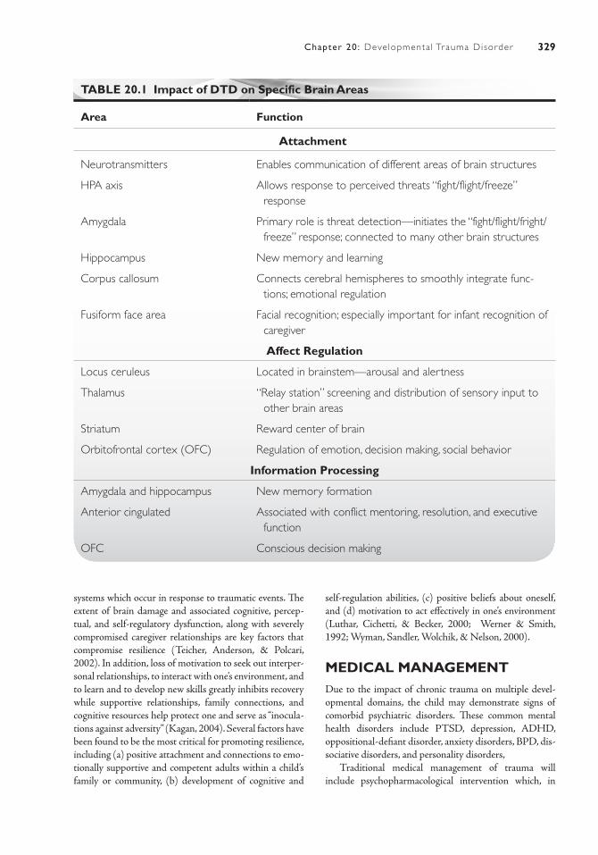
329Chapter 20: Deve lopmenta l Tr auma Disorder
TABLE 20.1 Impact of DTD on Specific Brain Areas
Area Function
Attachment
Neurotransmitters Enables communication of different areas of brain structures
HPA axis Allows response to perceived threats “fight/flight/freeze” response
Amygdala Primary role is threat detection—initiates the “fight/flight/fright/freeze” response; connected to many other brain structures
Hippocampus New memory and learning
Corpus callosum Connects cerebral hemispheres to smoothly integrate func-tions; emotional regulation
Fusiform face area Facial recognition; especially important for infant recognition of caregiver
Affect Regulation
Locus ceruleus Located in brainstem—arousal and alertness
Thalamus “Relay station” screening and distribution of sensory input to other brain areas
Striatum Reward center of brain
Orbitofrontal cortex (OFC) Regulation of emotion, decision making, social behavior
Information Processing
Amygdala and hippocampus New memory formation
Anterior cingulated Associated with conflict mentoring, resolution, and executive function
OFC Conscious decision making
systems which occur in response to traumatic events. The extent of brain damage and associated cognitive, percep-tual, and self-regulatory dysfunction, along with severely compromised caregiver relationships are key factors that compromise resilience (Teicher, Anderson, & Polcari, 2002). In addition, loss of motivation to seek out interper-sonal relationships, to interact with one’s environment, and to learn and to develop new skills greatly inhibits recovery while supportive relationships, family connections, and cognitive resources help protect one and serve as “inocula-tions against adversity” (Kagan, 2004). Several factors have been found to be the most critical for promoting resilience, including (a) positive attachment and connections to emo-tionally supportive and competent adults within a child’s family or community, (b) development of cognitive and
self- regulation abilities, (c) positive beliefs about oneself, and (d) motivation to act effectively in one’s environment (Luthar, Cichetti, & Becker, 2000; Werner & Smith, 1992; Wyman, Sandler, Wolchik, & Nelson, 2000).
MEDICAL MANAGEMENTDue to the impact of chronic trauma on multiple devel-opmental domains, the child may demonstrate signs of comorbid psychiatric disorders. These common mental health disorders include PTSD, depression, ADHD, oppositional- defiant disorder, anxiety disorders, BPD, dis-sociative disorders, and personality disorders,
Traditional medical management of trauma will include psychopharmacological intervention which, in
330 C O N D I T I O N S I N O C C U P A T I O N A L T H E R A P Y
best practice, is prescribed as an adjunct to psychosocial treatment modalities and not as a free-standing approach. Medication should only be used in conjunction with trauma-specific treatment and not in the absence of it.
In a survey sample representing 1,699 children, (Pynoos et al., 2008) ages infancy through 18, who were exposed to chronic trauma, it was determined that most trauma inter-vention occurs in an outpatient setting with the frequency of intervention modalities identified as follows:
■ Weekly psychotherapy (78%) ■ Self-management/coaching (62%) ■ Family therapy (56%) ■ Play therapy (55%) ■ Expressive therapies (41%) ■ Pharmacotherapy (27%) ■ Community outreach (25%)
The majority of those responding to this survey included psychiatrists, social workers, and psychologists and are representative of traditional models. Traditional models of intervention are typically provided by either a social worker or psychologist, however there are existing mod-els of assessment and intervention that are grounded in interdisciplinary and transdisciplinary approaches that include several professional disciplines on a team including occupational therapy (Hyter, Atchison, Henry, Sloane, & Black-Pond, 2001; Koomar, 2009).
IMPACT ON CLIENT FACTORS AND OCCUPATIONAL PERFORMANCEAs a result of DTD, there are significant disturbances across many of the client factors and performance skills described in the occupational therapy practice frame-work (AOTA, 2002). As described in the section on signs and symptoms, client factors include dysfunction in global and specific mental functions, sensory functions and pain, some functions of the cardiovascular and res-piratory systems, immunologic and digestive systems, and speech functions, particularly that related to fluency. Evi-dence of compromised performance skills has been deter-mined including those related to motor and praxis skills, sensory-perceptual skills, emotional regulation skills, cognitive skills, and communication and social skills that impact all areas of occupation (Richardson et al., 2008; Atchison, 2007; Pynoos et al., 2008).
Personal Activities of Daily Living
BathingAs a result of physiologic manifestations of stress, regu-lation of bodily functions is compromised leading to
difficulties in personal activities of daily living (PADLs). Stress responses are especially impacted by children with a history of sexual abuse while being bathed as well as being physically abused by immersion in hot or cold water.
ToiletingEncopresis, or the act of passing feces in inappropriate places such as in clothing or other places, is known to occur more frequently among children who have experi-enced sexual abuse, although it is not a specific indicator of child abuse. Enuresis, or the repeated voiding of urine in the clothing and in inappropriate places, often accom-panies encopresis among traumatized children. Whether intentional or not, it is essentially an expression of the child’s only mechanism of control in the midst of com-plete submission to the perpetrators of abuse.
Feeding and EatingThere are many relational factors between a primary caregiver and a child with regard to nutritional intake, beginning in the initial attachment process in infancy. A negative nurturing relationship with an infant includ-ing lack of attunement, irritability, depression, and other maternal problems will result in significant maladaptive, disorganized responses by the infant including difficulty with oral mechanisms such as suckling, sucking, swal-lowing and breathing rhythm, food refusal, and overac-tive response to certain smells and tastes. Toddlers will often present with severe dental decay, or “bottle rot,” a condition resulting from being bottle-fed with high con-centrates of sugar while dental development is taking place and at times is a major source of nutrition. Children exposed to chronic trauma are at a higher risk for devel-oping an eating disorder as they grow older. In a home where physical or sexual abuse is taking place, the child may turn to an eating disorder to gain a sense of control. Similar to the psychodynamics associated with encopresis and enuresis—the lack of control in terms of what is hap-pening to their bodies—they are unable to control their food intake or their weight. Children who are compulsive eaters or those who hoard and hide food are usually using food to help them deal with feelings of anger, sadness, hurt, loneliness, abandonment, fear, and pain.
Personal Hygiene and GroomingDTD often results in children who lack the attention needed to provide proper hygiene and grooming, which often comes to the attention of school personnel and may facilitate a referral to CPS. This may be the result of incompetent parenting who lack the intellectual require-ments to facilitate a child’s awareness of healthy grooming or willful neglect of one, but not all, children among a set of siblings.
331Chapter 20: Deve lopmenta l Tr auma Disorder
Sexual ActivityReactions to chronic sexual abuse naturally include signifi-cant dysfunction in sexual activity including age-inappro-priate sexual activity, the most frequent being sexualized behavior including a preoccupation with sexual organs of self, parents, and others expressed in drawings and in lan-guage. Children with a history of molestation are seven times more likely to become drug/alcohol dependent (Pynoos et al., 2008). In a sexual abuse effects study of 938 adolescents admitted to residential, therapeutic communi-ties for the treatment of substance abuse and related disor-ders, 64% of the girls and 24% of the boys reported histories of sexual abuse (Hawke, Jainchill, & DeLeon, 2000).
Instrumental Activities of Daily Living
DTD has a significant impact on instrumental activities of daily living (IADL). A common occurrence among a group of siblings in an abusive and neglectful environ-ment is for one child, usually the oldest, to take on the role of a parent, referred to as parentification as there is an absence of an adult careprovider willing or able to pro-vide care for others. Oftentimes, neglected children are exposed to dangerous in home situations such as fire haz-ards, firearms, insect and animal infestation, from parents operating methamphetamine labs. Parents might become physically or mentally unable to care for a child. Other times, alcohol or drug abuse may seriously impair judg-ment and the ability to keep a child safe.
Rest and Sleep
Exposure to trauma often results in significant difficulty with rest and sleep patterns as the child often exhib-its hypervigilance and overactivity. Multiple exposures to violence and trauma result in autonomic and endo-crine hyperarousal which are observed overreactions to stimuli. This may include being easily startled, crav-ing high-risk, stimulating, or dangerous activity—all of which impair the balance of play, work, and rest. The NCTSN clinician survey (Pynoos et al., 2008) found that 73% of children with DTD experienced sleep dis-turbances, a finding that has been supported by addi-tional studies (Egger et al., 1999; Glod et al., 1997; Noll et al., 2006). While not a common occurrence, victims of chronic abuse may experience “sleep terrors” which are also called “night terrors.” In a typical episode, an individual will sit up in bed and begin to scream or shout which may include kicking and thrashing. The child may say or shout nonsensible exclamations with an intense fearful expres-sion with eyes wide open and heart racing. In addition, the child may sweat, breathe heavily, and be very tense.
Education
In a study that included a sample of 9,336 children receiv-ing trauma intervention across the United States, 41% had academic problems including behavior problems in school/daycare (Pynoos et al., 2008).Academic func-tioning is a significant area of developmental competence beginning with preschool to higher education. In addi-tion to intellectual abilities, success is significantly tied to a child’s ability to regulate internal events or experience and to effectively interact with peers and teachers. By pre-school, trauma-exposed children demonstrate problems in both of these areas as demonstrated by poor frustration tolerance, a higher incidence of anger and noncompliance, and signicantly higher dependency on others for support ( Egeland, Sroufe, & Erikson, 1983; Vondra, Barnett, & Chichetti, 1990). In elementary school, they are less persistent on and more likely to avoid challenging tasks and are overly reliant on teachers’ guidance and feedback (Shonk & Cicchetti, 2001). By middle school and high school, they are more likely to be considered having a lack of motivation, learning below average, and there is a higher incidence of disciplinary referrals and suspensions (Eck-enrode, Laird, & Doris, 1993). Developmental delays and emotional/behavioral dysregulation, learning disabilities, and intellectual impairment that cannot be accounted for by neurologic or other factors that are experienced by chil-dren exposed to trauma can profoundly affect their school performance (Atchison, 2007). A cycle often is created whereby a student’s lack of success in school reduces his or her self-esteem and increases a lethargy often perceived as laziness or willful disobedience by adults. The student’s poor self-concept then further exacerbates limited school functioning. If this cycle continues, the student may face significant challenges as he or she plans for the future. A variety of school intervention programs, with a focus on creating trauma-informed or trauma-sensitive classrooms, have been utilized and are described in the literature as a means of addressing these students’ needs (Atch-ison, 2011; Katoaka, et al, 2003 Macy, Macy, Gross, & Brighton, 2003; Stein et al., 2003). The key components of a trauma sensitive classroom that emerge from descrip-tions of intervention programs for children exposed to chronic trauma include
■ Establishing and maintaining safety ■ Relational engagement ■ Acquiring affective regulation skills ■ Participation in and enhancement of their own learning ■ Assistance in making meaning of students’ experi-
ences
Unfortunately, there exists a paucity of awareness in most school systems regarding the need for classroom-based intervention that addresses the child as the victim of
332 C O N D I T I O N S I N O C C U P A T I O N A L T H E R A P Y
chronic abuse. The child, because of the many challenges he or she has in response to trauma experiences, acts out and behaves in ways that are interpreted as willful and thus being labeled as oppositional-defiant or other diagnoses that lay blame solely on the child, leading to continuous conflicts with school personnel. The lack of appropriate intervention leads to academic underperformance, non-attendance, disciplinary problems, a high drop-out rate, failure to complete diplomas which ultimately leads to poor success in vocational pursuits.
Work
Persons with DTD typically show disinterest in work, with ill-defined employment interests and poor employ-ment-seeking skills or vocational interests. The lack of antecedent behaviors which are necessary for job perfor-mance skills leads to the inability to acquire and main-tain employment. Persistent conflict with coworkers or supervisors is also a significant barrier to successful work performance.
Play and Leisure
The lack of acquisition in the components of interpersonal competence, poor self-concept, difficulty with social com-munication, sensory processing disorders, and intellectual impairment have a significant impact on play exploration, constructive play, and symbolic play. Typical early child-hood behavior among those with DTD will often reflect traumatic events which may include sexual acting out or violent play with dolls, seemingly disorganized and non-purposeful interaction with items that may indicate a reenactment of events. Persistent themes may be noted as the child is essentially reliving the event in an attempt to
control or gain mastery over fears that continue to create fear or that overwhelm the child. The child may be easily triggered by environmental stimuli, including other chil-dren and adults, which result in rage and physical aggres-sion during play sessions with other children or alone. At later ages, the pursuit of leisure interests, which rely on intrinsic motivation and engagement in self-selected activities are typically not healthy or productive as would be other children with a firm foundation built from suc-cessful play experiences and development of competence in play.
Social Participation
In a sample of 9,336 children with a history of trauma who were receiving intervention across the United States, 48% were reported to have had difficulty with social engagement within the home and in the commu-nity (Pynoos et al., 2008). Within peer groups, there is a significant degree of isolation, deviant affiliations, persistent physical or emotional conflict, avoidance/pas-sivity, involvement in violence or unsafe acts, and age-inappropriate affiliations or style of interaction. Family interaction is marked by interpersonal conflict, avoid-ance/passivity, running away, detachment and surrogate replacements, attempts to physically or emotionally hurt family members, and nonfulfillment of responsibilities within the family. Many individuals with a history of DTD are at high risk for dysfunctional social partici-pation within their communities including a high inci-dence of arrests and recidivism, detention, convictions, incarceration, violation of probation or other court orders, increasingly severe offenses, crimes against other persons, and disregard or contempt for the law or for conventional moral standards (Pynoos et al., 2008).
Case IllustrationsCase 1Derrick is a 3-year, 4-month-old boy who has been liv-ing in his current foster care home since he was 1 year 6 months old. While Derrick was living with his bio-logical father, he struck Derrick’s legs and buttocks “to get him to sit still” during, diapering. The foster mother reported that Derrick’s biological mother and father placed him in his room each day at 6:00 p.m., leaving him there for the rest of the evening, into the morn-ing. Both biological parents were reportedly intoxi-cated often, with consistent physical altercations with
one another in the presence of Derrick and his brother Jason. Jason is four years old and is living in a different foster home while Derrick’s biological mother has been diagnosed with BPD and has a history of severe physi-cal abuse by her own biological mother prior to being adopted as a child. Reportedly, she was drinking heav-ily and using crack cocaine during the first and second trimesters of her pregnancy. Derrick’s biological parents were recently released from prison and are pursuing custody. During the weekly 2-hour supervised visitation with them and his brother, Derrick tends to gravitate
333Chapter 20: Deve lopmenta l Tr auma Disorder
toward his case worker rather than actively engaging with his brother or his parents and shows little reac-tion when the session has ended and his parents and brother leave the room.
Derrick’s foster mother is concerned about his difficulty with regulation of his mood. He is described as having severe tantrums in which he will scream in response to loud noises and voices and unexpected movements by others. He is described as “being upset about 70% of the time” requiring significant time and effort to calm him.
The foster mother reports that Derrick is in Head Start and is having difficulty with typical devel-opmental tasks expected for a child his age. A recent assessment by an occupational therapist placed him at 16- to 19-month developmental age range across all domains including gross and fine motor, percep-tual, cognitive, language, and self care. Observations of his play skills, based on the Knox Play Scale, cor-relate with this range across all four domains. He is not yet independent in toileting, and is reported to be aggressive with his peers, hitting or kicking others if he wants a toy or a snack that another child has. His foster mother states that if he doesn’t like a toy or food he is given, he will throw it and fall down crying and kicking his feet on the floor.
Case 2Kayla is a 14-year-old who is currently living with her adoptive mother and her live-in female partner, who is concerned about her daughter’s difficulty in school as well as her hypersexuality as noted by inappropri-ate and indiscriminate self-stimulation and persistent sexual themes expressed during conversations about boys at school such as “He’s hot … I would do him” and “Look at that guy … I’d like to make out with him.” She reports that Kayla was removed from her biological mother at age six and lived in five differ-ent foster homes until she was adopted at age 9. The
identity of her biological father is unknown. Removal from her home was followed by termination of her mother’s parental rights due to substantiated sexual abuse of Kayla by a live-in boyfriend, who was subse-quently imprisoned for 16 to 20 years on charges of first-degree sexual misconduct. In addition to active substance abuse, including use of methamphetamine and alcohol while in a drug treatment program, Kayla’s biological mother was charged with having failed to protect her daughter from repeated sexual abuse that occurred in the home while she was present. In addi-tion to charges of sexual abuse, it was determined that Kayla was frequently slapped by her mother and in one incident, she pushed Kayla to the floor where she put her foot on the side of her face, leaving an imprint.Kayla is repeating the eighth grade and is struggling aca-demically in all subjects except art, which she enjoys. A recent assessment by a trauma assessment team indi-cated that she has a composite IQ score of 78 (verbal = 75, nonverbal = 76) and scored low in the areas across developmental domains including fine motor, visual processing, receptive language, memory, attention, and higher level cognitive functioning. In addition to her aca-demic concerns, her teacher reports that Kayla is resis-tive to completion of assignments in class, is inattentive, and often sent to the principal’s office for behavioral problems such as swearing at other students, asking to copy homework, taking pencils and pens from others, and threatening to “slap you silly” to a student who told her to stop bothering her during class. A referral for special education evaluation has been made to deter-mine if Kayla qualifies for support services. She is also reported to have significant aversion to certain smells and tastes, often threatening to “throw up” if an odor she perceives as offensive is detected.
Kayla’s adoptive mother reports that Kayla is “gothic” and resists attempts to “tone down her dress attire.” She fears that Kayla may be “running with a fast crowd” and is concerned that she may be sexually active.
R E C O M M E N D E D L E A R N I N G R E S O U R C E S
American Professional Society on the Abuse of Chil-dren (APSAC)
350 Poplar AvenueCHO 3B-3406Elmhurst, IL 60126Tel: 877- 402-7722www.apsac.org
The Annie E. Casey Foundation701 St. Paul StreetBaltimore, MD 21202The Child Trauma Academy5161 San Felipe, Suite 320Houston, TX 77056Tel: 866-943-9779
334 C O N D I T I O N S I N O C C U P A T I O N A L T H E R A P Y
www.childtrauma.orgThe Doris Duke Charitable Foundation650 Fifth Avenue, 19th FloorNew York, NY 10019Tel: 212-974-7000www.ddcf.orgNational Children’s Traumatic Stress NetworkCenter for Mental Health ServicesSubstance Abuse and Mental Health Services Admin-
istration
Department of Health and Human Services5600 Fishers LaneParklawn Building, Room 17C-26Rockville, MD 20857www.nctsn.orgThe National Institute for Trauma and Loss in Children42855 Garfield RoadSuite 111Clinton Township, MI 48038www.starrtraining.org/contact-tlc
R E F E R E N C E S
Alessandri, S. (1991). Play and social behavior in mal-treated preschoolers. Development and Psychopathol-ogy, 3(2), 191–205.
American Heritage Dictionary (4th ed.). (2006). Boston, MA: Houghton Mifflin Company.
American Occupational Therapy Association. (2002). Occupational therapy practice framework: Domain and process. American Journal of Occupational Thera-py, 56, 609–639.
American Psychiatric Association. (2000). Diagnostic and statistical manual of mental disorders (Rev. 4th ed). Washington, DC: American Psychiatric Association.
Atchison, B. (2007). Sensory modulation disorders among children with a history of trauma: A frame of reference for speech pathologists. Language, Speech, and Hearing Services in Schools, 38, 109–116.
Atchison, B. (2011). Creating trauma-informed class-room: the school intervention project. National Child Traumatic Stress Network Webinar Series. Re-trieved February 12, 2011, from http://learn.nctsn.org/course/category.php?id=3
Bevans, K., Cerbone, A., & Overstreet, S. (2008). Rela-tions between recurrent trauma exposure and recent life stress and salivary cortisol among children. Devel-opment and Psychopathology, 20(1), 257–272.
Brown, L. K., Houck, C. D., Hadley, W. S., & Lescano, C. M. (2005). Self-Cutting and sexual risk among adoles-cents in intensive psychiatric treatment. Psychiatric Services, 56(2), 216–218.
Cicchetti, D. & Rogosch, F. (2001). The impact of child mal-treatment and psychopathology on neuroendocrine func-tioning. Development and Psychopathology, 13:783–804.
Cook, A., Blaustein, M., Spinazzola, J., & van der Kolk, B. (Eds.) (2003). Complex trauma in children and ado-lescents. National Child Traumatic Stress Network. http://www.NCTSNet.org
Cook, A., Spinazzola, J., Ford, J., Lanktree, C., Blaustein, M., & Sprague, C. (2007). Complex Ttrauma in Chil-dren and Aadolescents. Focal Point, 21(1), 4–8.
Curtis, W. J., & Cicchetti, D. (2007). Emotion and resilience: A multilevel investigation of hemispheric electroencephalogram asymmetry and emotion regulation in maltreated and nonmaltreated chil-dren. Development and Psychopathology, 19(3), 811–840.
Eckenrode, J., Laird, M., & Doris, J. (1993). School per-formance and disciplinary problems among abused and neglected children. Developmental Psychology, 29, 53–62.
Egeland, B., Sroufe, A., & Erickson, M. (1983). The developmental consequence of different patterns of maltreatment. Child Abuse & Neglect, 7, 459–469.
Egger, H. L., Costello, E. J., Erkanli, A., & Angold, A. (1999). Somatic complaints and psychopathology in children and adolescents: Stomach aches, musculo-skeletal pains, and headaches. Journal of the Ameri-can Academy of Child & Adolescent Psychiatry, 38(7), 852–860.
Ford, J. D., Racusin, R., Ellis, C., Daviss, W. B., Reiser, J., Fleischer, A., et al. (2000). Child maltreatment, other trauma exposure, and posttraumatic symptom-atology among children with Oppositional Defiant and Attention Deficit Hyperactivity Disorders. Child Maltreatment, 5, 205–217.
Glod, C. A., Teicher, M. H., Hartman, C. R., & Harakal, T. (1997). Increased nocturnal activity and impaired sleep maintenance in abused children. Journal of the American Academy of Child & Adolescent Psychiatry, 36(9), 1236–1243.
Hawke, J., Jainchill, N., & DeLeon, G. (2000). School professionals’ attributions of blame for child sexual abuse. Journal of Child and Adolescent Substance Abuse, 9(3), 35–47.
Henry, J., Sloane, M., & Black-Pond, C. (2007). Neu-robiology and neurodevelopmental impact of child-hood traumatic stress and prenatal alcohol exposure. Language, Speech, and Hearing Services in Schools, 38, 99–108.

335Chapter 20: Deve lopmenta l Tr auma Disorder
Hyter, Y., Atchison, B. Henry, J., Sloane, M. & Black-Pond, C. (2001). A response to traumatized children: Devel-oping a best practice model. Occupational Therapy in Health Care, 15(3), 113–140.
Ito, Y., Teicher, M. H., Glod, C. A., & Harper, D. (1993). Increased prevalence of electrophysiological abnor-malities in children with psychological, physical, and sexual abuse. Journal of Neuropsychiatry & Clinical Neurosciences, 5(4), 401–408.
Kagan, R. (2004). Rebuilding attachment with trauma-tized children: Healing from losses, violence, abuse and neglect. Philadelphia, PA: Haworth Press.
Kataoka, S. H., Stein, B. D., Jaycox, L. H., Wong, M., Escudero, P., Tu, W., Zaragoza, C., & Fink, A. (2003) A School-Based Mental Health Program for Trauma-tized Latino Immigrant Children. Journal of American Child & Adolescent Psychiatry, 42(3), 311–318.
Koomar, J. A. (2009, December). Trauma- and attach-ment-informed sensory integration assessment and intervention. Sensory Integration Special Interest Section Quarterly, 32(4), 1–4.
Luthar, S. S., Cicchetti, D., & Becker, B. (2000). The con-struct of resilience: A critical evaluation and guidelines for future work. Child Development, 71, 543–562.
Macy, R. D., Macy, D. J., Gross, S. I., & Brighton, P. (2003). Healing in familiar settings: Support for chil-dren and youth in the classroom and community. New Directions for Youth Development, 98, 51–79.
Masten, A., & Coatsworth, J., (1998). The development of competence in favorable and unfavorable environ-ments: Lessons from research of successful children. American Psychologist, 53, 205–220.
National Children’s Traumatic Stress Network. What is Trauma? Retrieved January 15, 2011, from http://www.nctsn.org/content/defining-trauma-and- child-traumatic-stress
Nolin, P., & Ethier, L. (2007). Using neuropsychological profiles to classify neglected children with or without physical abuse. Child Abuse & Neglect, 31(6), 631–643.
Noll, J. G., Trickett, P. K., Susman, E. J., & Putnam, F. W. (2006). Sleep Disturbances and Childhood Sexual Abuse. Journal of Pediatric Psychology, 31(5), 469–480.
Perry, B. D. (2001). Bonding and attachment in maltreated children: Consequences of emotional neglect in childhood. The Child Trauma Academy. Retrieved October 11, 2010, from: http://www.childtrauma.org/images/stories/Articles/attcar4_03_v2_r.pdf
Pynoos, R., Fairbank, J. A., Briggs-King, E. C., Steinberg, A., Layne, C., Stolbach, B., et al. (2008). Trauma exposure, adverse experiences, and diverse symptom profiles in a national sample of traumatized children. Paper presented at the 24th Annual Meeting of the International Society for Traumat-ic Stress Studies, Chicago, IL, November 15, 2008.
Richardson, M., Henry, J., Black-Pond, C., & Sloane, M. (2008). Multiple types of maltreatment: Behavioral and developmental impact on children in the child wel-fare system. Journal of Child & Adolescent Trauma, 1, 1–14.
Sayar, Kose, Grabe, H., & Topbas. (2005). Alexithymia and dissociative tendencies in an adolescent sample from Eastern Turkey. Psychiatry and clinical neurosci-ences, 59(2), 127–134.
Sedlak, A. J., Mettenburg, J., Basena, M., Petta, I., McPher-son, K., Greene, A., et al. (2010). Fourth National In-cidence Study of Child Abuse and Neglect (NIS–4): Re-port to Congress, Executive Summary. Washington, DC: U.S. Department of Health and Human Services, Administration for Children and Families.
Shonk, S. M., & Cicchetti, D. (2001). Maltreatment, competency deficits, and risk for academic and behav-ioral maladjustment. Developmental Psychology, 37, 3–17.
Spinazzola, J., Ford, J. D., Zucker, M., van der Kolk, B. A., Silva, S., Smith, S. F., et al. (2005). Survey evaluates complex trauma exposure, outcome, and intervention among children and adolescents. Psychiatric Annals, 35(5), 433–439.
Stein, B. D., Jaycox, L. H., Kataoka, S. H., Wong, M., Tu, W., Elliott, M. N., et al. (2003). A mental health interven-tion for schoolchildren exposed to violence. Journal of American Medical Association, 290, 603–611.
Teicher, M. (2000). Wounds that won’t heal: The neuro-biology of child abuse. Cerebrum, 2 (4) 50–62.
Taylor, S., Eisenberger, N., Saxbe, D., Lehman, B, and Lieberman, M. (2006). Neural responses to emotion-al stimuli are associated with childhood family stress. Biological Psychiatry, 60: 296-301.
Teicher, M. H., Andersen, S. L., & Polcari, A. (2002). Developmental neurobiology of childhood stress and trauma. Psychiatric Clinics of North America, 25, Special Issue: Recent advances in the study of biological alterations in post-traumatic stress disorder, 397–426.
van der Kolk, B. A. (2005). Developmental trauma dis-order: toward a rational diagnosis for children with complex trauma histories. Psychiatric Annals, 35(5), 2–8.
van der Kolk, B. A., Pynoos, R., Cicchetti, M., Cloitre, M., D’Andrea, W., Ford, J. et al. (2009). Proposal to include a developmental trauma disorder diagnosis for children and adolescents in DSM-V. National Children’s Trau-matic Stress Network. Retrieved June 9, 2011, from http://www.nctsn.org/sites/default/files/assets/pdfs/ComplexTrauma_All.pdf
Vondra, J. I., Barnett, D., & Cicchetti, D. (1990). Self-concept, motivation, and competence among pre-schoolers from maltreating and comparison families. Child Abuse & Neglect, 14, 525–540.

336 C O N D I T I O N S I N O C C U P A T I O N A L T H E R A P Y
Way, I., Yelsma, P., Van Meter, A., & Black-Pond, C. (2007). Understanding alexithymia and language skills in children: Implications for assessment and intervention. Language, Speech and Hearing Services in Schools, 38, 128–139.
Werner A. A., & Smith A. E. (1992). High risk children from birth to adulthood. Ithaca, NY: Cornell University Press.
World Health Organization. (2007). International statisti-
cal classification of diseases and related health problems (ICD-10). World Health Organization: Geneva.
Wyman, P. A., Sandler, I., Wolchik, S., & Nelson, K. (2000). Resilience as cumulative competence promo-tion and stress protection: Theory and intervention. In D. Cicchetti & J. Rappaport (Eds.), The promotion of wellness in children and adolescents (pp. 133–184). Washington, DC: Child Welfare League of America.

337
Muscular Dystrophy
■ Je nni fe r L . Fo rg a c h ■ An dre a L . Wa sh in g t o n
21
J.B. is 6 years old. He has a 3-year-old brother and 1-year-old sister. J.B.’s mother is concerned because he does not seem to be “keep-ing up” with his peers. He gets tired quickly, has had an increased
number of falls, and has never learned how to jump. J.B. recently had to quit his soccer team due to increased fatigue. J.B.’s mother takes him to his pediatrician who notes that J.B. has enlarged calves, proximal muscle weakness, and uses Gower’s maneuver to rise off the floor. The pediatrician refers J.B. and his siblings for blood work, genetic testing, and to neurology/muscular dystrophy (MD) specialty clinic for further assessment.
J.B. arrives to the MD clinic. The neurologist meets with J.B. and his family. J.B.’s blood work shows elevated creatine kinase (CK) lev-els greater than 6,500 IU/L, while his 3-year-old brother’s are normal at around 150 IU/L. Genetic testing reveals that J.B. is positive for Duchenne muscular dystrophy. His younger brother tests negative and his 1-year-old sister is found to be a carrier. J.B. is referred to occupational and physical therapy and is recommended to follow up with the physician in 6 months. Although the above scenario illustrates one type of MD, MD is not strictly one disease.
MD is one of the most prolific neuromuscular disorders. It is actually a grouping of nine different disorders with more than 30 different subtypes. MD can broadly be defined as a group of hereditary diseases that weaken the muscles. They are characterized by progressive muscle weakness, defects in muscle protein, and/or the death of muscle cells and tissues. The progressive muscle weakness is caused by the lack, or absence, of the structural protein dystrophin. Dystrophin is a vital part of the intracellular protein complex that is responsible for maintaining the shape
and structure of the muscle fiber. In general terms, dystrophin acts as the “glue” to hold
K E Y T E R M S
Amyotrophic Lateral Sclerosis (ALS)
AtrophyAutosomal dominant
Autosomal recessiveCharcot Marie Tooth (CMT)
CompensationCongenital
ContractureCorticosteroid
Creatine kinaseDystrophin
Electromyography (EMG)Familial
Gower’s maneuverGower’s sign
Lumbar lordosisMuscle biopsy
Neuromuscular disorderProgressive
Spinal Muscular Atrophy (SMA)Valley sign
Winged scapulaX-linked

338 C O N D I T I O N S I N O C C U P A T I O N A L T H E R A P Y
the muscle together. Without this “glue,” the muscle breaks down causing the progressive weakness that is the hallmark characteristic of MD (Emery, 1994, 2001; http//:www.mda.org, 2010). It is the distribu-tion and the progression of the muscle weakness that is used to distinguish between the nine types (Fig. 21.1). The nine main types of MD that have been identified are (http//:www.mda.org, 2010) as follows:
■ Duchenne muscular dystrophy (DMD) ■ Becker muscular dystrophy (BMD) ■ Emery-Dreifuss muscular dystrophy (EDMD) ■ Limb-girdle muscular dystrophy (LGMD) ■ Facioscapulohumeral muscular dystrophy (FSHD) ■ Myotonic muscular dystrophy (MMD) ■ Oculopharyngeal muscular dystrophy (OPMD) ■ Distal muscular dystrophy (DD) ■ Congenital muscular dystrophy (CMD)
Although there are currently nine identified forms of the disease, this chapter highlights six types of MD most commonly seen by occupational therapists.
DESCRIPTION AND DEFINITION
Duchenne Muscular Dystrophy
Although DMD is genetically present at birth, symptoms of the disease may not present themselves until the child is 3 to 4 years of age. DMD affects only males and, as discussed later, is an x-linked recessive inherited condi-tion (Biggar, 2006). DMD is caused by an absence, or deficiency, of dystrophin. Initial symptoms include are delayed motor development, proximal weakness, and increased fatigue. These symptoms manifest themselves
as a waddling gait, enlarged calf muscles, increased falls, and failure to develop the ability to run or jump (Emery, 1994, 2001). Also present in 90% of males with DMD is the “valley sign.” This is a depressed area on the posterior axillary fold that can be seen when the patient abducts shoulders to 90 degrees, elbows flexed to 90 degrees, with bilateral hands pointing upward (Fig. 21.2).
The pattern of muscle weakness is always bilateral and symmetrical. It progresses from proximal to distal, and affects the lower extremities prior to upper extremi-ties. The rate of progression varies from person to person. The individual can alternately go through periods of both rapid progression and slowed or no progression. Most individuals with DMD will be wheelchair dependent by 12 years of age. DMD affects all voluntary skeletal mus-cles, as well as cardiac and pulmonary muscles (Emery, 2001). Of note, up to one-third of the individuals with DMD may have cognitive impairments. Current research indicates that this may also be related to the lack of dys-trophin (Ashraf & Wong, 2005; Emery, 1994, 2001).
As muscle wasting progresses, active range of motion (ROM) decreases, and the individual can be left with contractures at the elbows, hips, and knees, as well as severe spinal deformities. The joint contractures are often at a 90-degree angle later in the disease process, indicative of prolonged wheelchair use.
Becker Muscular Dystrophy
Like DMD, BMD is characterized by progressive muscle weakness, affecting only males, as an x-linked recessive inherited condition. BMD is also genetically present at birth, but onset of symptoms can vary widely from 2 to 40 years of age. Symptoms most often appear, however, between 6 and 18 years of age. Individuals with BMD often demonstrate delayed ambulation, difficulty climbing
Figure 21.1 Typical distribution of muscle weakness seen in different types of MD. Left to right: Duchenne/Becker, Emery-Dreifuss, limb-girdle, facioscapulohumeral, distal, oculopharyngeal.
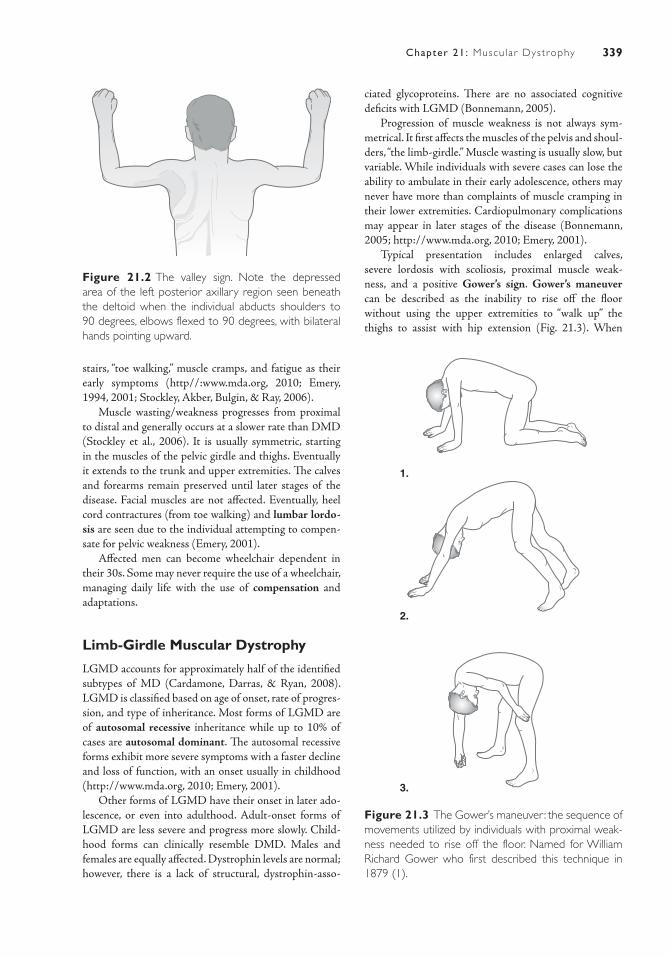
339Chapter 21: Muscu lar Dystrophy
stairs, “toe walking,” muscle cramps, and fatigue as their early symptoms (http//:www.mda.org, 2010; Emery, 1994, 2001; Stockley, Akber, Bulgin, & Ray, 2006).
Muscle wasting/weakness progresses from proximal to distal and generally occurs at a slower rate than DMD (Stockley et al., 2006). It is usually symmetric, starting in the muscles of the pelvic girdle and thighs. Eventually it extends to the trunk and upper extremities. The calves and forearms remain preserved until later stages of the disease. Facial muscles are not affected. Eventually, heel cord contractures (from toe walking) and lumbar lordo-sis are seen due to the individual attempting to compen-sate for pelvic weakness (Emery, 2001).
Affected men can become wheelchair dependent in their 30s. Some may never require the use of a wheelchair, managing daily life with the use of compensation and adaptations.
Limb-Girdle Muscular Dystrophy
LGMD accounts for approximately half of the identified subtypes of MD (Cardamone, Darras, & Ryan, 2008). LGMD is classified based on age of onset, rate of progres-sion, and type of inheritance. Most forms of LGMD are of autosomal recessive inheritance while up to 10% of cases are autosomal dominant. The autosomal recessive forms exhibit more severe symptoms with a faster decline and loss of function, with an onset usually in childhood (http://www.mda.org, 2010; Emery, 2001).
Other forms of LGMD have their onset in later ado-lescence, or even into adulthood. Adult-onset forms of LGMD are less severe and progress more slowly. Child-hood forms can clinically resemble DMD. Males and females are equally affected. Dystrophin levels are normal; however, there is a lack of structural, dystrophin-asso-
Figure 21.2 The valley sign. Note the depressed area of the left posterior axillary region seen beneath the deltoid when the individual abducts shoulders to 90 degrees, elbows flexed to 90 degrees, with bilateral hands pointing upward.
ciated glycoproteins. There are no associated cognitive deficits with LGMD (Bonnemann, 2005).
Progression of muscle weakness is not always sym-metrical. It first affects the muscles of the pelvis and shoul-ders, “the limb-girdle.” Muscle wasting is usually slow, but variable. While individuals with severe cases can lose the ability to ambulate in their early adolescence, others may never have more than complaints of muscle cramping in their lower extremities. Cardiopulmonary complications may appear in later stages of the disease (Bonnemann, 2005; http://www.mda.org, 2010; Emery, 2001).
Typical presentation includes enlarged calves, severe lordosis with scoliosis, proximal muscle weak-ness, and a positive Gower’s sign. Gower’s maneuver can be described as the inability to rise off the floor without using the upper extremities to “walk up” the thighs to assist with hip extension (Fig. 21.3). When
1.
2.
3.
Figure 21.3 The Gower’s maneuver: the sequence of movements utilized by individuals with proximal weak-ness needed to rise off the floor. Named for William Richard Gower who first described this technique in 1879 (1).
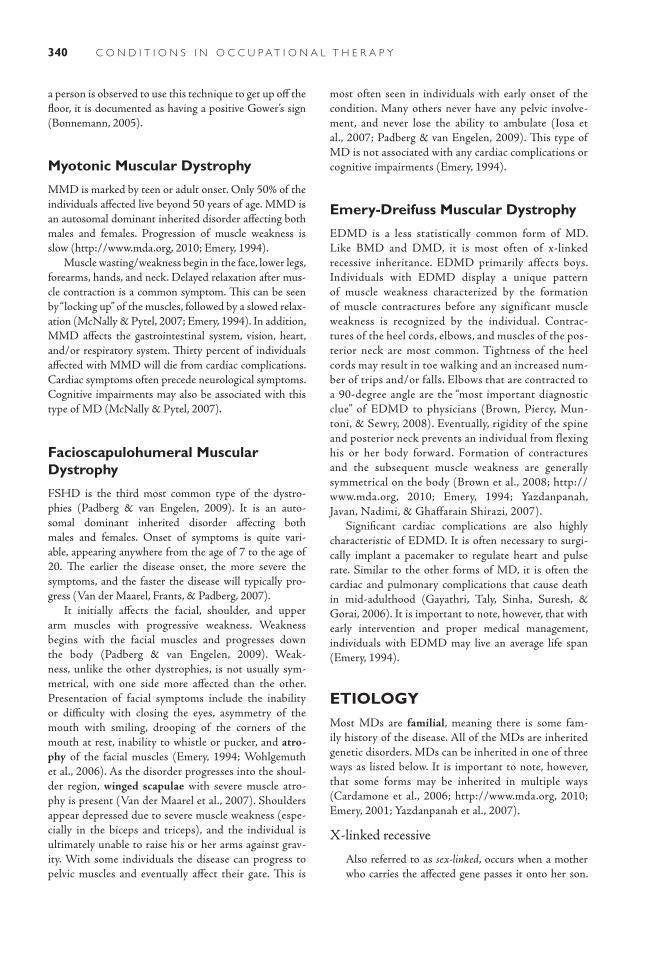
340 C O N D I T I O N S I N O C C U P A T I O N A L T H E R A P Y
a person is observed to use this technique to get up off the floor, it is documented as having a positive Gower’s sign (Bonnemann, 2005).
Myotonic Muscular Dystrophy
MMD is marked by teen or adult onset. Only 50% of the individuals affected live beyond 50 years of age. MMD is an autosomal dominant inherited disorder affecting both males and females. Progression of muscle weakness is slow (http://www.mda.org, 2010; Emery, 1994).
Muscle wasting/weakness begin in the face, lower legs, forearms, hands, and neck. Delayed relaxation after mus-cle contraction is a common symptom. This can be seen by “locking up” of the muscles, followed by a slowed relax-ation (McNally & Pytel, 2007; Emery, 1994). In addition, MMD affects the gastrointestinal system, vision, heart, and/or respiratory system. Thirty percent of individuals affected with MMD will die from cardiac complications. Cardiac symptoms often precede neurological symptoms. Cognitive impairments may also be associated with this type of MD (McNally & Pytel, 2007).
Facioscapulohumeral Muscular Dystrophy
FSHD is the third most common type of the dystro-phies (Padberg & van Engelen, 2009). It is an auto-somal dominant inherited disorder affecting both males and females. Onset of symptoms is quite vari-able, appearing anywhere from the age of 7 to the age of 20. The earlier the disease onset, the more severe the symptoms, and the faster the disease will typically pro-gress (Van der Maarel, Frants, & Padberg, 2007).
It initially affects the facial, shoulder, and upper arm muscles with progressive weakness. Weakness begins with the facial muscles and progresses down the body (Padberg & van Engelen, 2009). Weak-ness, unlike the other dystrophies, is not usually sym-metrical, with one side more affected than the other. Presentation of facial symptoms include the inability or difficulty with closing the eyes, asymmetry of the mouth with smiling, drooping of the corners of the mouth at rest, inability to whistle or pucker, and atro-phy of the facial muscles (Emery, 1994; Wohlgemuth et al., 2006). As the disorder progresses into the shoul-der region, winged scapulae with severe muscle atro-phy is present (Van der Maarel et al., 2007). Shoulders appear depressed due to severe muscle weakness (espe-cially in the biceps and triceps), and the individual is ultimately unable to raise his or her arms against grav-ity. With some individuals the disease can progress to pelvic muscles and eventually affect their gate. This is
most often seen in individuals with early onset of the condition. Many others never have any pelvic involve-ment, and never lose the ability to ambulate (Iosa et al., 2007; Padberg & van Engelen, 2009). This type of MD is not associated with any cardiac complications or cognitive impairments (Emery, 1994).
Emery-Dreifuss Muscular Dystrophy
EDMD is a less statistically common form of MD. Like BMD and DMD, it is most often of x-linked recessive inheritance. EDMD primarily affects boys. Individuals with EDMD display a unique pattern of muscle weakness characterized by the formation of muscle contractures before any significant muscle weakness is recognized by the individual. Contrac-tures of the heel cords, elbows, and muscles of the pos-terior neck are most common. Tightness of the heel cords may result in toe walking and an increased num-ber of trips and/or falls. Elbows that are contracted to a 90-degree angle are the “most important diagnostic clue” of EDMD to physicians (Brown, Piercy, Mun-toni, & Sewry, 2008). Eventually, rigidity of the spine and posterior neck prevents an individual from flexing his or her body forward. Formation of contractures and the subsequent muscle weakness are generally symmetrical on the body (Brown et al., 2008; http://www.mda.org, 2010; Emery, 1994; Yazdanpanah, Javan, Nadimi, & Ghaffarain Shirazi, 2007).
Significant cardiac complications are also highly characteristic of EDMD. It is often necessary to surgi-cally implant a pacemaker to regulate heart and pulse rate. Similar to the other forms of MD, it is often the cardiac and pulmonary complications that cause death in mid-adulthood (Gayathri, Taly, Sinha, Suresh, & Gorai, 2006). It is important to note, however, that with early intervention and proper medical management, individuals with EDMD may live an average life span (Emery, 1994).
ETIOLOGYMost MDs are familial, meaning there is some fam-ily history of the disease. All of the MDs are inherited genetic disorders. MDs can be inherited in one of three ways as listed below. It is important to note, however, that some forms may be inherited in multiple ways (Cardamone et al., 2006; http://www.mda.org, 2010; Emery, 2001; Yazdanpanah et al., 2007).
X-linked recessive
■ Also referred to as sex-linked, occurs when a mother who carries the affected gene passes it onto her son.
341Chapter 21: Muscu lar Dystrophy
Although the mother carries the affected gene on one of her X chromosomes, she may never show symp-toms since it is a recessive trait.
■ MDs inherited this way: Duchenne, Becker, Emery-Dreifuss
Autosomal recessive
■ Occurs when both parents carry and pass on the affected gene
■ MD inherited this way: Emery- Dreifuss, limb-girdle, oculopharyngeal, distal, congenital
Autosomal dominant
■ Occurs when a child inherits a normal gene from one parent and an affected gene from the other parent
■ MDs inherited this way: Emery-Dreifuss, limb-gir-dle, facioscapulohumeral, myotonic, oculopharyngeal, distal, congenital
INCIDENCE AND PREVALENCEIt is estimated that at least 1 in every 5,000 people in the population are currently affected with some form of MD. The actual prevalence of the disease is most likely higher than this as many forms of MD are often misdiagnosed or not yet identified. Therefore, specific current statistical information is difficult to obtain (Cardamone et al., 2008; Emery, 1994, 2001; Kohler et al., 2009; McNally & Pytel, 2007). Table 21.1 is an approximation of available statisti-cal figures in research literature today. It should be used only as a tool to compare the occurrence of one type of MD to another.
It is most important to recognize from Table 21.1 that DMD and BMD are by far the most common types and affect only males. Other forms, like LGMD and EDMD, have statistically unknown or unreliable data. This may be due to EDMD being less common than the other forms, and the many different subtypes of LGMD. MD
occurs worldwide affecting all races, ages, and genders. Depending on the region of the world, or country, a cer-tain form of MD may be very common or very rare.
SIGNS AND SYMPTOMSRefer to each of the individual descriptions of the dif-ferent types of MD for specific signs and symptoms as it relates to each of the various subtypes. As mentioned, decreased strength, muscle cramping, and decreased gross motor skills are the common link between all forms, with each form having its own unique presenta-tion. As previously defined, the Gower’s maneuver and the “valley sign” are two key clinical indicators that may be evident in any form of MD with proximal muscle weakness (Emery, 1994, 2001).
There are other progressive neuromuscular disorders that mimic MD and are often mistaken for MD in their early stages. Some of these include spinal muscular atro-phy (SMA) and its subtypes myasthenia gravis (MG), Pompe disease, Charcot Marie Tooth (CMT), mito-chondrial myopathy, and amyotrophic lateral sclerosis (ALS). These disorders exhibit the same symptoms at onset including muscle cramping, generalized progressive weakness, and joint stiffness and are often treated in neu-rological clinics alongside individuals with MD (http://www.mda.org, 2010).
DIAGNOSISMD is often first suspected by a primary care physician when a patient presents with complaints of muscle weak-ness, increased falls, muscle cramps or increased fatigue. It is at that point that clinical assessment becomes a vital part of the diagnostic process.
The patient is physically assessed by the physician. Based on the distribution of the weakness in conjunction
TABLE 21.1 Incidence and Prevalence of Specific Types of muscular dystrophy
Type of MD Incidence Prevalence
Duchenne MD 1 in 3,500 males 1 in 4,000 males
Becker MD 1 in 30,000 males 1 in 20,000 males
Facioscapulohumeral MD 1 in 22,000 males and females 1 in 50,000 males and females
Myotonic MD 1 in 8,000 males and females 1 in 20,000 males and females
Limb-girdle MD Statistically unknown 1 in 25,000 males and females
Emery-Dreifuss MD Statistically unknown 1 in 100,000 males and females
342 C O N D I T I O N S I N O C C U P A T I O N A L T H E R A P Y
with other aforementioned physical symptoms, MD may be suspected. At this point, the physician will begin to order more testing to confirm diagnosis.
The following three diagnostic tests are typically used to confirm or rule out an MD diagnosis (Emery, 1994, 2001; Stockley et al., 2006):
■ Blood work to perform genetic testing and reveal creatine kinase levels. Creatine kinase levels become elevated when there is muscle damage in the body; therefore, elevated levels will indicate that there is muscle wasting occurring. Normal CK levels average around 100 to 200 IU/L. The levels in DMD, in com-parison, can be elevated 50 to 100 times this amount (Biggar, 2006; Emery, 1994, 2001).
■ Electromyography (EMG) to evaluate the current electrical activity produced by the skeletal muscles. An EMG examination can be used to confirm the diagno-sis of dystrophy and distinguish this from other types of neuromuscular diseases. It cannot reveal what type of dystrophy a patient may have (Emery, 2001).
■ Muscle biopsy used to examine the cellular makeup of the muscle tissue, and look at the differences in muscle fibers, fiber size, fiber splitting, and fiber necrosis. Muscle biopsy can be used to distinguish the type of dystrophy, based on subtle differences in the muscle tissues (Emery, 1994).
These are not stand-alone tests; therefore, it is important to complete a thorough clinical and neurological exam to identify any patterns of muscle weakness, test reflex responses and coordination, and identify contracted muscles (Wren, Blumi, Tseng-Ong, & Gilsanz, 2007). If one child is diagnosed with MD, all siblings and parents should be tested for the condition as well.
COURSE AND PROGNOSISEach of the MDs follows its own unique course. Each type has a variable prognosis with most ultimately resulting in a shortened lifespan. Prognosis will depend not only on the primary diagnosis but also on what specific subtype of the disorder is present. For instance, LGMD has up to 15 different subtypes. Some of these subtypes have a normal life expectancy, while others have a shortened lifespan. The course and prognosis of each of the MDs examined in this chapter are discussed individually below. While the figures represent an average of current statisti-cal data, research and medical advances are beginning to extend life expectancies (http://www.mda.org, 2010).
DMD: Males affected with DMD have an average life expectancy of late teens to early 20s. The cause of death is usu-ally secondary to respiratory and/or cardiac complications (Biggar, 2006; Emery, 1994, 2001; Kohler et al., 2009).
BMD: Less severe than DMD, males affected with BMD demonstrate a much slower progression. Life expec-tancy varies greatly, but average is between middle and late adulthood, approximately 25 to 30 years after onset of the condition (Emery, 2001; Stockley et al., 2006).
EDMD: Disease progresses slowly, often with cardiac and respiratory complications. Individuals may live an average lifespan if proper cardiac management is received (Cardamone et al., 2008; Chen, 2008).
LGMD: Progresses slowly. Although symptoms may appear in childhood, they often do not appear until adoles-cence or even adulthood. If muscles deteriorate rapidly, life expectancy may be only into early 30s. Otherwise, an indi-vidual may live a normal lifespan. The course of LGMD is by far the most variable of the dystrophies (Bonnemann, 2005; http://www.mda.org, 2010; Emery, 1994, 2001).
FSHD: Symptoms appear around age 20. Disease progresses slowly with some periods of rapid decline. Course of disease may spread over many decades, with life expectancy being normal (Padberg & van Engelen, 2009; Van der Maarel et al., 2007).
MMD: Disease progresses slowly with course span-ning up to 50 to 60 years. Life expectancy may be typically into late adulthood (Cardamone et al., 2008).
MEDICAL/SURGICAL MANAGEMENTCurrently there is no cure or specific treatment that can reverse or stop the progression of any form of MD. Symptom management will have the greatest impact on enhancing a person’s quality of life and possibly increasing his or her life expectancy. The primary goal of treatment with MD is maintenance of independence for as long as possible (Emery, 2001).
Respiratory Maintenance
As mobility and muscle functioning decrease, respiratory maintenance becomes increasingly difficult. The individ-ual will have decreased ability to use the voluntary muscles of the thorax to fully inhale, or cough, causing inability to keep the lungs clear. The individual may require suction-ing, cough assist, or antibiotics to preserve lung function. They are high risk for pneumonia and other respiratory infections. Monitoring for signs/symptoms of aspiration is an important part of pneumonia prevention (Kohler et al., 2009; Thompson, 1999).
Skin Integrity
Skin integrity becomes a risk factor with decreased mobil-ity. When skin integrity is compromised, the individual is
343Chapter 21: Muscu lar Dystrophy
at increased risk for infection, further tissue damage, and loss of joint mobility. Careful skin inspection should be incorporated into one’s daily routine. Severe infections can lead to death if not properly managed.
Pain
Pain should be assessed in all individuals with MD. While most types of MD are not directly associated with pain, some physical symptoms may contribute to discomfort (Emery, 1994). Initial feelings of muscle cramping or spasms may occur. The pain cycle may initially worsen when a stretching or strengthening program is initiated. It is advantageous to complete these exercises on a regu-lar basis as the body will tolerate the stretches with less discomfort. In fact, it is common for an individual with MD to request joint stretching as a source of pain relief. As the disease progresses, pain levels should be continu-ally monitored and assessed as increased or localized pain could be indicative of further concerns. Adequate pain management is essential to quality of life as well as pro-moting maximum participation in daily tasks.
Nutrition
Good nutrition and hydration are essential to maintain-ing overall health and can aid in the maintenance of skin integrity. Nutrition can also assist in controlling obesity which can be detrimental to function and mobility. People with MD are at high risk for obesity, secondary to lack of mobility and loss of muscle function. Safe maintenance of nutrition becomes a priority as oral and pharyngeal mus-culature may also be weakened in some forms of MD. As a result, swallowing can become unsafe. Individuals should be closely monitored for signs of dysphagia so that diet can be modified or a feeding tube inserted (Carda-mone et al., 2008; Thompson, 1999).
Occupational and/or Physical Therapy
Therapy should be initiated as soon as diagnosis is con-firmed to train the patient and family in an extensive home exercise program. A baseline of activities of daily living (ADL) skills and mobility performance should also be obtained. It is important to initiate therapy prior to the formation of contractures or muscle tightness (Thompson, 1999). Therapy is an essential component in preventing or slowing loss of strength, ROM, and function. Another major role of therapy is to assess current equipment needs for home and community use. This role becomes increas-ingly important as the disease progresses and specific indi-vidual needs change. Evaluation and provision of splints and orthotics as needed to prevent joint contracture formation should further be considered at the time of the evaluation.
Drug Intervention
Corticosteroids may decrease muscle damage and assist with preserving respiratory function. Although they present possible benefits, they are often associated with side effects that must be closely monitored, including the tendency to gain weight. While corticosteroids are widely used, there continues to be controversy. Signifi-cant research is being conducted regarding whether the use of steroids has any effect on life expectancy. In addi-tion to corticosteroids, there are other medications that are often prescribed by a physician for treatment of MD. Immunosuppressant drugs can be used to delay muscle degeneration. Anticonvulsants can be used to control muscle activity. Antibiotics can be used to control res-piratory infections. Over-the-counter analgesics can be used for pain management and cramping (Biggar, 2006).
Surgical Management
Surgical management often includes corrective and/or preventative surgeries. Some of these may include, but are not limited to, contracture release, scoliosis repair, cardiac stability, respiratory assist, or spinal stabilization. Spinal deformities can physically reduce a person’s ability to breathe, and should be closely monitored. Pacemakers can be surgically implanted to control heart rhythms as needed. Tracheostomies may need to be performed if a person requires ventilator assistance or suctioning of secretions. Feeding tubes, either temporary or perma-nent, may need to be considered if dysphagia is a concern (Kohler et al., 2009; Thompson, 1999).
IMPACT OF CONDITIONS ON CLIENT FACTORSAll occupational performance areas and performance components may be affected by MD. These are most dependent on the type and the stage of the disease. As previously mentioned, each type of MD has its own unique progression and variable severity.
Activities of Daily Living
The individual with MD may initially be independent with his or her ADL skills; however, a decline in function is anticipated, especially with more severe forms. Main-taining the basic underlying skills of strength and ROM will keep the individual independent longer. As active movement becomes more difficult, the person will require assistance from a caregiver to complete basic tasks such as dressing, grooming, and bathing. Toileting may become a

344 C O N D I T I O N S I N O C C U P A T I O N A L T H E R A P Y
cumbersome task. This can be due to physical limitations making toilet transfers difficult; or due to loss of muscle control causing incontinence. Often with more severe forms of MD the individual is required to wear protective toileting garments.
Eventually, the use of adaptive equipment such as commode chairs, shower chairs, bathing aides, mechanical lifts, ADL equipment, and wheelchairs/mobility equip-ment may be necessary. These pieces of equipment will help increase independence and make caring for the individual easier and safer. Home modifications such as ramps, grab bars, and bedrails should be considered to promote independence and safety within an individual’s home environment.
In less severe forms, the individual may maintain the ability to complete the task himself or herself, but may require increased time to do so. Energy conservation, or task adaptation, should be a consideration for these indi-viduals (Kohler et al., 2009).
School
School is a major concern as the majority of MD cases are diagnosed in early childhood or early adolescence. Most forms of MD do not involve cognitive impairments and therefore, physical limitations ultimately become the barrier at school (Donders & Taneja, 2009; Thompson, 1999). Often the child with MD fatigues quickly, has dif-ficulty keeping up with his or her peers and may require physical assistance to get through the school day. A one-on-one aid may be necessary to assist with maneuvering their wheelchair, toileting, assisting with meals, and com-pleting basic educational tasks.
Increased absences due to illness may be common. This, combined with wheelchair/adaptive equipment use and the decreased physical ability to participate in the same activities as their peers, may contribute to social isolation.
Work
Older individuals may face many of the same barriers as children in school. Social isolation, environmental barri-ers, fatigue, an inability to keep up with work activities, and requiring physical assistance for some basic tasks may be common in the workplace.
Obtaining and maintaining employment and having the ability to change jobs are more difficult for these individuals due to physical limitations, decreased job
opportunities, and employer misperceptions. Some individuals with severe MD may lose, or never have, the physical ability to work. Others may maintain employ-ment with altered responsibilities/adaptations or with relatively little interruption at all.
Play/Leisure
A child’s primary responsibility is to play. Many indi-viduals diagnosed with MD are children and this area becomes a significant area of concern. It is important to encourage play, in any capacity, to ensure pleasure, learning, and development. A child with physical impair-ments may require modifications to their leisure activities to promote success and enjoyment.
For individuals of all ages, leisure activities such as computer and video game play, text messaging, and read-ing are commonly enjoyed as hand function is often preserved well enough to complete these tasks. It is ben-eficial to also incorporate and encourage gross motor movements into leisure activities (i.e., balloon volleyball, painting on an easel, etc.) as a means to preserve muscle strength and function (Thompson, 1999).
Psychosocial
MD affects everyone within the family, not solely the affected individual. The emotions, fears, and anxieties experienced by the individual are shared by the entire family. Parents often face feelings of guilt, helplessness, and fear over the anticipated loss of their child. Fear and anxiety are often a concern for the younger sibling as they watch the deterioration of their older sibling, with the anticipation that they will soon, too, face the same fate.
As the affected individual loses function, and the loss of independence becomes more evident, the individual often struggles with anger and frustration. Individuals who face severe functional loss often experience feelings of depression. Suicidal ideations may become a concern as the individual faces the loss of their perceived role (Bostram & Ahlstrom, 2005; Chen, 2008; Poysky & Kinnett, 2008).
ACKNOWLEDGMENTSWe would like to thank Lawrence C. Banko for his con-tributions. Lawrence was responsible for drawing all sketches/illustrations used in this chapter.
345Chapter 21: Muscu lar Dystrophy
Case IllustrationsCase 1A.F. is 32 years old. When she was 13 years old, she began to notice that her hips and lower back would fatigue quickly and that she was having minor trou-ble with her balance. Her primary care physician felt that it was “growing pains” and told her that he was sure it would pass. A year later, A.F. continued to have problems and her symptoms worsened. A.F. returned to her physician and, at that time, blood work and a muscle biopsy were conducted. A.F. was eventually diagnosed with LGMD.
On this day, A.F. presents to the MD clinic per recommendation of her primary care physician. The occupational therapist evaluates A.F. It was revealed that A.F. has had significant functional decline in the last year. She was previously walking with a walker for household distances. She is now using a borrowed wheelchair for all ambulation. A.F. can independently complete a stand-pivot transfer from her wheelchair to the examining table with increased time allotted. Manual muscle testing scores as follows: grossly 3/5 bilateral shoulder strength, 3+/5 strength at bilateral elbows, and 4/5 in the wrists and hands.
A.F. currently has a shower chair in the bathtub that she borrowed from her grandmother. However, A.F. further reports that she is no longer able to step over the side of the tub. She is now wearing a diaper due to her inability to get to the toilet quickly enough. She attempts to dress herself seated at the edge of the bed, but is unable to don her socks or shoes as a result of leg weakness and poor sitting balance. A.F. currently lives alone and does not have family close by to assist her. She has hired a maid to keep the house clean and assist with weekly laundry. She recently had to quit her job as a school teacher due to her physical decline. She is now significantly concerned about her finances.
She reports that she is feeling isolated at home. She does not identify any leisure interests other than watching television. She states that it is too much work to get out of the house, so she just stays home. A.F. is anxious about her recent decline, and is not knowledgeable about her condition or prognosis. She is not currently taking any medications except an over-the-counter analgesic, as needed.
Case 2H.M. is a 12-year-old male diagnosed with DMD. He is currently an inpatient at a children’s hospital. He presented to the ER with complaints of difficulty
breathing. He subsequently required intubation and eventually a tracheostomy.
Occupational therapy was consulted to evaluate H.M. and to provide recommendations to his family. A review of the medical record reveals that H.M. was diagnosed with MD at the age of 3. He is currently on prednisone, digitalis, a multivitamin, a diuretic, and an analgesic as needed. He is the oldest of four children, with two other brothers also diagnosed with DMD. He had spinal stabilization surgery approximately 1 year ago and releases of bilateral knee contractures about 6 months ago.
Upon entering H.M.’s hospital room, the occu-pational therapist completes a parent interview and receives the following information: H.M. is currently in the 7th grade in a POHI classroom. He lives in a two-story home, with first-floor bedroom and bathroom. They have a ramp outside to enter the home. H.M. has not received therapy in almost a year. Parents are concerned that H.M. has outgrown his current wheelchair and is not properly supported by his seating system. Parents also report that H.M. has been gaining weight, making all wheelchair and sur-face transfers increasingly difficult. Prior to admission, H.M. required variable assistance for all ADL, IADL, and transfers. There is no current home exercise pro-gram in place.
Upon interviewing H.M., he appears to be delayed cognitively, requiring simple questions and multiple cues to follow one-step commands. He reports no pain throughout his day. He spends his time watching television, playing video games, and texting his friends on his cell phone. H.M. states that he would like to be more independent within his school and home environments.
Evaluation findings as follows: H.M. presents with decreased upper extremity active ROM and strength, as he is unable to raise his arms over his head or fully straighten his elbows. He also presents with con-tractures of bilateral shoulders, elbows, and wrists. H.M. demonstrates the ability to isolate all fingers and thumb movements. He presents with decreased grip strength bilaterally. He currently demonstrates the ability to feed himself with a spoon, but he is observed to lower his head to the plate. H.M. is coughing and choking during meals with minimal chewing observed. He currently requires maximal assistance for all bath-ing/dressing tasks which is being completed in bed at this time. Bed mobility is impaired. He requires maximum assistance to transfer from supine to edge
346 C O N D I T I O N S I N O C C U P A T I O N A L T H E R A P Y
Ashraf, E. A., & Wong, B. L. (2005). The diagnosis of muscular dystrophy. The Pediatric Annals, 34, 525–528.
Biggar, W. D. (2006). Duchenne muscular dystrophy. Pediatrics in Review, 27, 83–88.
Bonnemann, C. G. (2005). Limb girdle muscular dystro-phy in childhood. The Pediatric Annals, 34, 569–577.
Bostrom, K., & Ahlstrom, G. (2005). Living with a hereditary disease: persons with muscular dystrophy and their next of kin. American Journal of Medical Genetics, 136A, 17–24.
Brown, S., Piercy, R., Muntoni, F., & Sewry, C. (2008). Investigating the pathology of Emery-Dreifuss mus-cular dystrophy. Biochemical Society, 36, 1335–1338.
Cardamone, M., Darras, B., & Ryan, M. (2008). Inher-ited myopathies and muscular dystrophies. Seminars in Neurology, 28, 250–259.
Chen, Jih-Yuan. (2008). Mediators affecting family func-tion in families of children with Duchenne muscu-lar dystrophy. Kaohsiung Journal of Medical Science, 24, 514–521.
Diseases. (2011). Welcome to MDA, helping Jerry’s kids. Retrieved January 5, 2010, from http://www.mda.org
Donders, J., & Taneja, C. (2009). Neurobehavioral char-acteristics of children with Duchenne muscular dys-trophy. Child Neuropsychology, 15, 295–304.
Emery, Alan E. H. (2001). The muscular dystrophies. New York, NY: Oxford University Press.
Emery, Alan E. H. (1994). Muscular dystrophy: the facts. New York, NY: Oxford University Press.
Gayathri, N., Taly, A. B., Sinha, S., Suresh, T. G., & Gorai, D. (2006). Emery Dreifuss muscular dystro-phy: a clinico-pathological study. Neurology India, 54, 197–199.
Iosa, M., Mazza, C., Frusciante, R., Zok, M., Aprile, I., Ricci, E., et al. (2007). Mobility assessment of patients with fascioscapulohumeral dystrophy. Journal of Clinical Biomechanics, 22, 1074–1082.
Kohler, M., Clarenbach, C. F., Bahler, C., Brack, T., Russi, E. W., & Bloch, K. E. (2009). Disability and survival in Duchenne muscular dystrophy. Journal of Neurol-ogy, Neurosurgery, and Psychology, 80, 320–325.
McNally, E. M., & Pytel, P. (2007). Muscle diseases: the muscular dystrophies. The Annual Review of Pathol-ogy: Mechanisms of Disease, 2, 87–109.
Padberg, G.W., & van Engelen, B. G. M. (2009). Fas-cioscapulohumeral muscular dystrophy. Current Opinion in Neurology, 22, 539–542.
Poysky, J., & Kinnett, K. (2008). Facilitating family adjustment to a diagnosis of Duchenne muscular dys-trophy. Neuromuscular Disorders, 19, 733–738.
Stockley, T., Akber, S., Bulgin, N., & Ray, P. (2006). Strategy for comprehensive molecular testing for Duchenne and Becker muscular dystrophies. Genetic Testing, 10, 229–243.
Thompson, C. E. (1999). Raising a child with a neuromus-cular disorder. A guide for parents, grandparents, friends, and professionals. New York, NY: Oxford University Press.
Van der Maarel, S. M., Frants, R. R., & Padberg, G. (2007). Facioscapulohumeral muscular dystrophy. Biochimica et Biophysica Acta, 1772, 186–194.
Wohlgemuth, M., de Swart, B. J. M., Kalf, J. G., Joosten, F. B. M., Van der Vliet, A. M., & Padberg, G. W. (2006). Dysphagia in fascioscapulo-humeral muscular dystrophy. Neurology, 66, 1926–1928.
Wren, T. A. L., Blumi, S., Tseng-Ong, L., & Gilsanz, V. (2007). Three-point technique of fat quantification of muscle tissue as a marker of disease progression in Duchenne muscular dystrophy: preliminary study. American Journal of Radiology, 190, W8–W12.
Yazdanpanah, P., Javan, A., Nadimi, B., & Ghaffarain Shirazi, H. R. (2007). Genetic pattern of 3 cases of Emery-Dreifuss muscular dystrophy in a family. East-ern Mediterranean Health Journal, 13, 201–205.
R E F E R E N C E S
of bed, and is unable to maintain sitting balance with-out support.
The inpatient occupational therapist made the following recommendations to H.M. and his family. Referrals were made to outpatient occupational and physical therapy following discharge. A thorough feeding evaluation is recommended with possible
feeding tube placement, as he is showing signs/symp-toms of aspiration. A new wheelchair and seating system evaluation is to be completed. H.M.’s parents were also provided an initial home exercise program for safe positioning, daily stretching, and mobiliza-tion. The social work department was contacted to address possible psychosocial and coping needs.
347
Glossary
Activity demands: The aspects of the task that influence the performance by the person. These demands include the objects used and their properties, space demands, social demands, sequencing and timing, required actions, required body functions, and required body structures.
Affect: Observable facial expressions of a feeling or emotion. Patients may be described as having a flat or blunt affect.
Affective and physiologic dysregulation: Behaviors include difficulty with arousal regula-tion and modulation and toleration and recov-ery from extreme affective states such as fear, anger, and shame that emerge from chronic trauma exposure, manifested by prolonged and extreme emotional tantrums on one side of the spectrum to catatonic-like immobilization.
Age-related macular degeneration (AMD): A low vision disorder that results from loss of function of the macula, which is the center of the retina. There are two types of AMD includ-ing dry AMD (atrophic) and wet AMD (neo-vascular/exudative). Dry AMD is atrophy of the macula and wet AMD is an ingrowth of new blood vessels that bleed into the subretinal space.
Agnosia: Inability to recognize the import of sen-sory impressions despite being able to recognize the elemental sensation of the stimulus. Lan-guage deficits must be absent for this diagnosis. Various agnosias correspond to several senses and may be distinguished as auditory, gustatory, olfactory, tactile, and visual. Specific sensory agnosias can occur when the connections are
disrupted between the primary cortical receptor region for a stimulus and the memory of prior stimuli. For example, the inability to recognize familiar objects by sight or touch.
Agoraphobia: “A mental disorder character-ized by an irrational fear of leaving the famil-iar setting of home, or venturing into the open; often associated with panic attacks.” Commonly referred to as fear of the marketplace.
Alexithymia: The impaired capacity to describe emotions or bodily states; is a persistent sign of affective dysregulation.
Algorithms: A step-by-step approach to treat-ment based upon controlled clinical trials.
Allograft: An allograft (also referred to as a homograft or cadaver skin) is donor skin taken from another person.
Alogia: Absence of speech because of mental illness
Altruism: The unselfish concern for the welfare of others. This concept is reflected in actions and attitudes of commitment, caring, dedica-tion, responsiveness, and understanding.
Alveoli: Small sacs in the lungs which perform the gas exchange. Oxygen is moved into the blood stream from the alveoli to be carried to cells throughout the body
Ambiguous hand preference: Absence of a dominant hand preference; the switching of hands within the same activity.
Aminobutyric acid (GABA): “A constituent of the central nervous system; quantitatively the principal inhibitory neurotransmitter.”
Amotivation: Lacking in motivation.
348 G L O S S A R Y
Amygdala: A structure that is part of the limbic system and is involved in the emotions of aggression and fear.
Amyloid plaques: Insoluble shards of amyloid that stick to dead and dying neurons.
Amyloid precursor protein (APP): A benign substance that lives in various parts of the body, including the brain, and whose role in cellular function is unknown.
Amyotrophic lateral sclerosis (ALS): A progressive neurological disorder characterized by degeneration of motor neurons located in the ventral horn of the spinal cord.
Analgesic: A pain medication.Anemia: Decrease in the number of red blood
cells (RBC) or less than the normal quantity of hemoglobin in the blood
Aneurysm: Localized abnormal dilatation of the wall of a blood vessel, usually an artery, or in the heart, resulting in the formation of a sac. Usual causes are the weakness of the vessel wall, resulting from atherosclerosis, or a congenital defect. However, any injury to the middle or muscular layer of the arterial wall can predis-pose the stretching of the vessel wall.
Angioplasty: is a procedure where a small mesh tube is inserted into the coronary artery to widen the opening, thus increasing the blood flow.
Angle-closure glaucoma: The obstruction by the iris to the outflow pathway of aqueous humor at the angle between the peripheral cor-nea and the iris aqueous humor.
Anhedonia: A lack of interest in previously plea-surable activities.
Anhedonia: Reduced ability or inability to expe-rience pleasure.
Ankylosis: the fusion of a joint, often in an abnormal position, usually resulting from destruction of articular cartilage and subchon-dral bone, as occurs in rheumatoid arthritis.
Anomia: Inability to name objects.Anterograde amnesia: The inability to learn
new long-term declarative information.Antibodies: A specialized immune protein pro-
duced because of the introduction of an antigen
into the body. The production of antibodies is a major function of the immune system.
Anticholinergic side effects: Most commonly dry mouth or constipation.
Antigen: A microorganism that is a uniquely configured protein that attaches to the patho-gen and stimulates the body’s production of antibodies.
Antipsychotic medications: Medications desig-ned to reduce positive and negative symptoms.
Anxiety: “Apprehension of danger, and dread accompanied by restlessness, tension, tachycar-dia, and dyspnea unattached to a clearly identi-fiable stimulus.”
Anxiolytic: A sedative or mild tranquilizer used to treat anxiety or alcohol withdrawal.
Apathy: An absence or suppression of emotion, feeling, or concern.
Aphasia: Difficulty with expressive and/or recep-tive language.
Apophyseal: Refers to the articulations between the articular facets of adjacent vertebrae, or facet joints. These joints are a frequent site of degen-erative joint disease, or osteoarthritis, spondy-litic and traumatic diseases.
Apraxia: Loss of skilled purposeful movement that cannot be attributed either to deficits in primary motor skills or problems in compre-hension. It can affect ideation and concept for-mation as well as programming and planning of movement.
Arthroplasty: Joint replacement.Arteriovenous: Referring both to an artery
and a vein. An arteriovenous malformation, or AVM, is a congenital malformation character-ized by an abnormal collection of blood vessels near the surface of the brain. AVM can lead to subarachnoid hemorrhage.
Asperger’s disorder: A neurobiologic disorder of unknown etiology in which cognitive and communication skills develop normally or nearly normally during the first few years of life. How-ever, social impairment is evident, repetitive and stereotypical behaviors are observed, and com-munication, though not delayed, is marked by unusual topics of interest and rigidity.
349G L O S S A R Y
Aspiration: Pathological inhalation of food or mucus into the respiratory tract.
Associated reactions: Involuntary movements or reflexive increases in tone on the affected side of individuals with hemiplegia. These move-ments replicate synergy patterns and often appear when the individual is trying new or stressful activities, for example, a resisted grasp by the noninvolved hand causes a grasp reaction in the involved hand.
Associality: A negative symptom characterized by decreased interest in socialization and main-tenance of relationships.
Astereognosis: Inability to recognize objects by touch (also called tactile agnosia; see Agnosia).
Asterixis: A hand-flapping tremor.Ataxia: Inability to coordinate muscle activity
during voluntary movement. In posterior col-umn damage of the spinal cord, incoordina-tion and a loss of proprioception is caused by misjudgment of limb position with balance problems. Cerebellar ataxia produces a reeling, wide-based gait.
Attentional and behavioral dysregulation: Signs and symptoms include impairment in those competencies related to sustained atten-tion, learning, or coping with stress. An inability to sustain goal-directed behavior may include a lack of curiosity, difficulties with planning or completing tasks, or avolition.
Athetoid (dyskinetic): Resembling athetosis or repetitive involuntary, slow, sinuous, writh-ing movements. Classification of cerebral palsy in which involuntary purposeless movement occurs when an individual attempts purpose-ful motion. The abnormal movements may not only occur in the limb being moved but also involve an “overflow” of activity to all the other limbs with an exaggeration of reflexes.
Atherosclerosis: A widespread form of arte-riosclerosis in which deposits of plaques (ath-eromas) containing cholesterol and other lipid material are formed within the inner layer (intima) of large- and medium-sized arteries.
Atheromas: An abnormal mass of fatty or lipid material with a fibrous covering, which forms
a discrete raised plaque within the inner layer (intima) of an artery.
Arthritis: Inflammation or infection of a joint.Atrophy: A wasting of tissue that results in
decreased muscle mass.Auditory processing: The process of the brain
recognizing and interpreting sounds in the envi-ronment. This process is responsible for the comprehension of language.
Autistic disorder: A neurobiologic disorder with an unknown etiology, characterized by sig-nificant delay or deviation in social interaction, communication, and repetitive or stereotyped behaviors.
Autograft: The surgical transplantation of the patient’s own skin from one area to another.
Autonomic dysreflexia (hyperreflexia): An uninhibited and exaggerated reflex of the autonomic nervous system to stimulation. The response occurs in about 85% of all patients who have spinal cord injury above the level of the 6th thoracic vertebra. It is potentially dan-gerous because of attendant vasoconstriction and immediate elevation of blood pressure, which in turn can bring about hemorrhagic retinal damage or cerebrovascular accident. Less serious effects include severe headache, changes in heart rate, sweating and flushing above the level of the spinal cord injury, and pallor and goose bumps below that level.
Autonomic neuropathy: A form of peripheral neuropathy. A group of symptoms that occur when there is damage to the nerves that manage every day body functions such as blood pres-sure, heart rate, bowel and bladder emptying, and digestion.
Autosomal dominant: A disease that occurs when a child inherits a normal gene from one parent and an affected gene from the other parent.
Autosomal recessive: A disease that occurs when both parents carry and pass on the affected gene.
Babinski’s reflex: Dorsiflexion (extension) of the great toe and outspreading of the outer toes when the sole of the foot is stroked. Normally
350 G L O S S A R Y
present in infants under the age of 6 months, in adults this reflex occurs in lesions of the pyra-midal tract.
Benign tumor: Noncancerous, which do not invade other body tissues or spread to other body parts. However, may become life threat-ening as they cause increasing deficits with cell growth, because they press upon nearby struc-tures and tissues.
Beta-amyloid plaques: An accumulation of amyloid precursor proteins that have not been correctly broken down in the body.
Boutonniere deformity: A common hand deformity secondary to the pathologic effects of rheumatoid arthritis resulting in a com-bination of PIP joint flexion and DIP joint hyperextension
Bradykinesia: Slowness of all voluntary move-ment and speech.
Brain attack: Synonym for stroke, or cerebro-vascular accident.
Brainstem: connects the cerebrum with the spinal cord and includes the midbrain, medulla oblon-gata, and the pons. Motor and sensory neurons travel via the brainstem which allows for com-munication between brain and spinal cord. The brainstem also controls life-supporting autonomic functions of the peripheral nervous system.
Bronchopulmonary dysplasia: A chronic lung disease of babies, which most commonly develops in the first 4 weeks after birth and most often affects babies born at least 4 weeks before term.
Burden of disease: A situation in which a group experiences an illness to a greater degree than other subgroups.
Burn scar contracture: Forms due to the shortening and tightening of the burn scar. Burn scar contracture deformities are the most problematic over large joints.
Bursitis: Inflammation of the fluid-filled sac (bursa) that lies between a tendon and skin, or between a tendon and bone.
Carotid endarterectomy: Excision of thick-ened atheromatous areas of the innermost layer of an artery (intima) to improve circulation.
Carpal tunnel syndrome: Resulting from compression on the volar aspect of the wrist, which then impinge upon the median nerve. This causes paresthesia of the palmar aspect of the thumb, the second and third digits, and the radial aspect of the fourth digit.
Casein: A protein found in cow’s milk. A theory in the field of autism suggests that children with autism do not digest this protein appro-priately, leading to a buildup of morphine-like substances in the body and causing social with-drawal and abnormal behaviors.
Cataracts: Opacifications of the crystalline lens of the eye, which result in a decreased amount of light reaching the retina.
Catatonia: Extreme psychomotor agitation or retardation.
Catatonic behavior: Loss of responsiveness to environmental cues. A person with catatonic behavior may assume rigid or bizarre postures and resist attempts made to move or reposi-tion him or her. Excessive, nonpurposeful motor activity may also be observed. In extreme cases, the individual appears to be completely unre-sponsive, as in a catatonic stupor.
Catheterization: Passage of a catheter into a body channel or cavity, especially introduction of a catheter via the urethra into the urinary bladder.
Cauda equina: The collection of dorsal and ventral nerve roots descending from the lower spinal cord and occupying the vertebral canal below the cord at the L1 region.
Central scotomas: Diminished vision or visual loss in the center of the visual field.
Central vision: Sharp, foveal vision used for reading, watching television, driving or any activity for which visual detail is of primary importance.
Cerebellum: A region of the brain that is found at the rear of the head and is above the brain-stem. It has been traditionally known for its role in the coordination of movement, but recent research suggests the cerebellum may also have a role in cognitive, sensory, language, and abstract-thinking skills.
351G L O S S A R Y
Cerebrovascular accident (CVA, stroke, brain attack): Disorder of the blood ves-sels serving the brain, resulting in disruption of blood flow to parts of the brain leading to neu-ronal death. Impact of CVA varies according to the cause of the disruption (blockages or hem-orrhages) and location of the insult, as well as a variety of premorbid and psychosocial conditions.
Cerebrospinal fluid assays: A diagnostic tool that measures biomarkers in cerebrospinal fluid.
Charcot-Marie-Tooth (CMT): An inherited neurological disorder that results in motor and sensory peripheral neuropathy.
Childhood disintegrative disorder: A rare neurobiologic disorder that develops in children who had appeared completely normal during their first few years of life. Following its onset, language, social interaction, and self-care skills are lost and autistic-like symptoms appear.
Chronic bronchitis: Inflammation of the bron-chial tubes resulting in a thickening of the walls, making it difficult to breathe.
Chronic obstructive pulmonary disease (COPD): A term that includes chronic bron-chitis, asthma, emphysema, and bronchiectasis. The condition is irreversible and characterized by progressive limitation in the flow of air in and out of the lungs.
Cilia: Hair-like structures in the lungs airways that help to move the mucus out and clean the airways.
Circumlocution: The use of pantomime, non-verbal communication, or word substitution to avoid revealing an inability to say or remember a word.
Client factors: The body functions and the body structures that reside within the person.
Clonus: Alternate involuntary muscular contrac-tion and relaxation in rapid succession.
Closed fracture: Refers to a fracture that has not broken through the skin.
Cogwheel rigidity: Stiff movement in one direction in small evenly spaced segments. The tension in muscle gives way in little jerks when the muscle is passively stretched.
Collagen: A basic structural fibrous protein found in all tissue. Excessive deposition of tis-sue collagen leads to a thickening of the burn scar.
Collateral circulation: Secondary circulation that continues to an area of the brain following obstruction of a primary blood vessel and may prevent major ischemia.
Colles’ fracture: Fracture resulting in dorsal displacement, dorsal comminuting, and radius shortening; often referred to as a dinner fork deformity given its resemblance to an upside down fork.
Coma: A nonsleep loss of consciousness asso-ciated with unresponsiveness to touch, pain, sound, or movement that lasts for an extended period.
Complex regional pain syndrome (CRPD): Also referred to as reflex sympathetic dystro-phy (RSD) is a chronic pain condition in which high levels of nerve impulses are sent to an affected site.
Comminuted fracture: A fracture in which a bone in broken, splintered, or crushed into a number of pieces.
Compensation: The use of one system or mus-cle group to complete the work not capable of being completed by the usual system.
Compound fracture: Also referred to as an open fracture as the fractured bone breaks through the skin surface and are more prone to infection.
Compression garment: Compression gar-ments are the preferred conservative method to treat hypertrophic scars and have been in use since the early 1970s. Compression garments are thought to reduce oxygen flow to the scar thereby decreasing collagen production.
Compulsion: “Uncontrollable impulses to per-form an act, often repetitively, as an unconscious mechanism to avoid unacceptable ideas and desires which, by themselves, arouse anxiety; the anxiety becomes fully manifest if perfor-mance of the compulsive act is prevented; may be associated with obsessive thoughts.”
352 G L O S S A R Y
Concrete thinking: Literal interpretation of words without understanding abstract meanings.
Congenital: A condition existing before birth, at birth, or that develops during the first month of life.
Congestive heart failure: The inability of the heart to function as a pump, which results in a decreased blood flow to the tissues and conges-tion in the pulmonary and circulatory systems.
Constructional apraxia: Apraxia caused by loss of visuospatial skills; for example, difficulty drawing a clock face.
Contracture: Abnormal shortening of muscle tissue that renders the muscle highly resistant to stretching, which can lead to permanent disabil-ity. In many cases, contractures can be prevented by range of motion (ROM) exercises (active or passive) and by adequate support of the joints to eliminate constant shortening or stretching of the muscles and surrounding tissue.
Context: The conditions that surround the per-son. Those conditions include cultural, physical, social, personal, spiritual, temporal, and virtual contexts.
Contrast sensitivity: The ability to visually discern the edges or luminance of an object to strike the skull at an opposite location (contre-coup).
Coronary artery bypass grafting (CABG): A process in which arteries or vein from other parts of the body (often the legs) are “harvested” and then surgically attached to bypass the blocked arteries near the heart.
Coronary artery disease: Tied very closely to atherosclerosis where plaque, made of choles-terol, fat, calcium, and other substances, sticks to the inner lining of arteries that are taking oxygen-rich blood from the heart to the body.
Cortical atrophy: Birth defect with a nonge-netic origin that can contribute to or cause ID due to shrinkage of the posterior part of the brain.
Corticobulbar: Refers to the pathway connect-ing the cerebral cortex to the brainstem. Bulbar refers to the nerves and tracts connected to the
medulla; muscles innervated include those of the tongue, pharynx, and larynx.
Corticospinal: Also referred to as the pyrami-dal tracts; are descending tracts which conduct motor impulses from the brain to the spinal cord.
Corticosteroids: A class of steroids that are produced in the adrenal cortex. Synthetic ver-sions are produced to closely resemble these ste-roids and are used for medicinal purposes.
Coup-contrecoup injury: Situation in which direct brain damage is incurred in traumatic event as the cerebrum rotates on the more stable brainstem while accelerating from the force of impact. The cerebrum strikes the skull (coup), and then accelerates in the opposite direction.
Craniostenosis: Birth defect with a nongenetic origin that can contribute to or cause ID due to premature fusion of the skull resulting in a reduction in space for the brain to grow and in increased intracranial pressure.
Creatine kinase: An enzyme expressed by vari-ous tissues and cells in the body.
Credé’s method: Use of manual pressure on the bladder to express urine, particularly in bladder training for individuals with paralysis. The hands are held flat against the abdomen, just below the umbilicus. A firm downward stroke toward the bladder is repeated six or seven times, followed by pressure from both hands placed directly over the bladder to manu-ally remove all urine.
Cyclothymia: Characterized by a chronic course of hypomania and mild depression, this disor-der presents as a mild form of bipolar disorder (BPD).
Cytomegalovirus: A herpesvirus that can result in a congenital infection.
Debridement: The cleansing and removal of nonadherent and nonviable tissue.
Decerebrate rigidity: An extensor posture of all limbs and/or the trunk.
Decorticate rigidity: Flexion of the upper, but extension of the lower, limbs.
353G L O S S A R Y
Decubitus ulcers: An ulcer caused by local interference with the circulation, usually occur-ring over a bony prominence at the sacrum, hip (trochanter), heel, shoulder, or elbow. It begins as a reddened area and can quickly involve deeper structures and become an ulcer, also called a bedsore or a pressure sore.
Decussation: An X-shaped crossing, especially of nerves or bands of nerve fibers, connecting parts on opposite sides of the brain or spinal cord.
Deep vein thrombosis (DVT): A thrombosis, commonly seen in the legs or pelvis, that results from phlebitis, vein injury, or prolonged bed rest.
Deformity: Distortion of any part or general dis-figurement of the body.
Delayed union: A delayed union is when the bone takes more time to heal than is expected; it heals slowly. It may be suspected when pain and tenderness persist at the fracture site 3 months to 1 year after the injury.
Delusions: Atypical and well-organized beliefs not explained by evidence or culture.
Delusions of grandeur: Self-inflated views usually present in conversation.
Delusions of persecution: Individuals have consistent and organized beliefs about other people intending to harm them.
Demyelination: Destruction, removal, or loss of the myelin sheath of a nerve or nerves.
De Quervain’s disease: Pathologic process that involves extensors at the thumb, causes severe pain and discomfort, resulting in a decrease in hand function and the ability to grip.
Developmental trauma disorder: Describes clinically significant patterns of behavior or syn-dromes that occur in an individual as a result of persistent and chronic exposure to violent epi-sodes including physical, sexual, and emotional abuse resulting in functional impairment in one or more important areas.
Diabetic foot: A person who has diabetes is at increased risk for slow-healing injuries to distal extremities. It is caused by decreased vascular-ization; the risk of an infection that does not heal is higher. One possible intervention is limb amputation.
Diabetic ketoacidosis: State of medical emer-gency; life-threatening condition caused by a state of relative insulin deficiency, characterized by hyperglycemia, ketonemia, metabolic aci-dosis, and electrolyte depletion. Often the first presenting sign of type 1 diabetes.
Diarthroses: are the most mobile joints and are by far the most common articular pattern. Because these joints possess a synovial mem-brane and contain synovial fluid, these joints are more commonly referred to as synovial joints.
Diastolic blood pressure: The pressure in the arteries when the heart is at rest.
Dignity: The importance of valuing the inher-ent worth and uniqueness of each person. This value is demonstrated by an attitude of empathy and respect for self and others.
Diplegia: Paralysis of like parts on either side of the body. In cerebral palsy, diplegia describes involve-ment of the lower extremities predominantly, with only mildly affected upper extremities.
Diffusion-tensor imaging: A diagnostic tool that uses MRI methods to produce images of the structure and organization of the connec-tions throughout the central nervous system.
Disease surveillance: An epidemiological prac-tice by which the spread of disease is monitored in order to establish patterns of progression.
Disease trajectory: Patterns of progres-sion and functional decline that result from a condition.
Disinhibition: Loss of inhibition, or the cultural and social sense of self restraint.
Disorganized behavior: Unpredictable, socially inappropriate behaviors that interfere with daily activities. Examples include agitated or angry outbursts with no known provocation; sexually acting out in public, that is, masturbation; and difficulties performing goal-directed tasks such as meal preparation or grooming.
Disorganized thinking: Thought processes that lack cohesive patterns. Speech, which can provide clues about disorganized thought pro-cesses, may include any of the following: loose associations (answers that begin to veer “off
354 G L O S S A R Y
track” of the original questions), tangential (unrelated comments or answers) or incoherent (a mixture of words often referred to as a “word salad”).
Dissociation: is essentially an “out-of-body expe-rience” in which the person detaches from an event that is overwhelmingly stressful.
Distal radius fracture: Common fracture of the radius. Due to its proximity to the wrist joint, often referred to as a wrist fracture.
Dopamine: A neurotransmitter that helps regu-late movement and emotional responses; a defi-ciency results in Parkinson’s disease.
Down syndrome: A chromosomal condition caused by the presence or all or part of an extra 21st chromosome.
Dysarthria: Imperfect articulation of speech caused by disturbances of muscular control resulting from central or peripheral nervous sys-tem damage.
Dysesthesia: Impairment of any sense, especially of the sense of touch; a painful, persistent sen-sation induced by a gentle touch of the skin.
Dysgraphia: Inability to write a sentence.Dysosmic aphasia: Difficulty naming objects.Dysphagia: Difficulty in swallowing, which may
range from mild discomfort to a seriously com-promised ability to control the muscles needed for chewing and swallowing. Dysphagia may seriously interfere with a patient’s nutritional status. Interventions, such as positioning the patient during feeding and use of foods of the proper consistency, are widely used.
Dysphoria: A mood disturbance resulting in a depressed mood.
Dyspnea: Difficult, labored, or painful breathing.Dysthymia: Unlike individuals with depressive
disorders, people with dysthymia report that they have always been depressed. Character-ized by insidious onset frequently beginning in childhood, a persistent or intermittent course, and ongoing feelings of depression over a period of at least 2 years.
Dystophin: A protein that connects the cyto-skeleton of a muscle fiber to the surrounding extracellular matrix through the cell membrane.
It is responsible for maintaining the shape and structure of the muscle fiber.
Ecchymosis: Skin discoloration caused by escape of blood into the tissues from ruptured blood vessels.
Echolalia: The automatic and meaningless rep-etition of another’s words or phrases. In autism, this repetition may be either immediate or delayed, and often does not appear appropriate to the conversation or activity.
Electroconvulsive therapy: The induction of a brief seizure by passing electric current through the brain. This treatment is used mostly for depression.
Electroencephalogram: The record obtained during the amplification, recording, and sub-sequent analysis of the electrical activity of the brain using an instrument called an electroencephalograph.
Electromyography (EMG): A diagnostic tool used for testing the electrical activity of muscles.
Embolism: Sudden blockage of an artery by a moving clot of foreign material (embolus) lodged in place by the blood current. Emboli usually lodge at divisions of an artery, where the blood vessel narrows, and are typically blood clots. However, they may also be formed from globules of fat, air bubbles, pieces of tissue, or clumps of bacteria.
Emotional lability: Inability to regulate emo-tions appropriately, typically resulting in laugh-ing or crying in unsuitable contexts.
Emphysema: The destruction of the walls of the bronchioles and alveoli, which results in abnor-mal and enlarged air spaces.
Epidemiology: A scientific discipline devoted to studying the patterns of communicable diseases.
Epiglottis: A thin flap of tissue in the esophagus that protects the lungs from foreign objects by covering the trachea during swallowing.
Epstein-Barr virus (EBV): A virus causing infection and is characterized by fatigue and general malaise. Infection with EBV is fairly common and is usually a transient and minor thing. However, in some individuals EBV can
355G L O S S A R Y
trigger chronic illness. It is a particular danger to people with compromised immune systems.
Equality: The value that all individuals are perceived as having the same fundamental human rights and opportunities. This value is demonstrated by an attitude of fairness and impartiality.
Equinovalgus: Deformity in which the foot is plantar flexed, everted, and abducted.
Equinovarus: Deformity in which the foot is planter flexed, inverted and adducted.
Eschar: The residual necrotic layers of skin destroyed by direct heat damage or the injury occurring secondary to heat damage.
Euphoria: A feeling or state of well-being or elation.
Euthymia: Normal range of mood.Executive functioning: A broad band of skills
that allow an individual to engage in indepen-dent, self-directed behavior. Includes volition, planning, purposeful action, and self-awareness. Executive functions involve the ability to for-mulate context-appropriate goals and to initi-ate, plan and organize, and sequence and adapt behavior based on anticipated or actual conse-quences of actions.
Extrapyramidal symptoms: Involuntary move-ment, changes in muscle tone, and abnormal posture.
Extrapyramidal syndrome: A side effect from medication that results in abnormal movements similar to Parkinson’s disease.
Familial: A disease for which there is a family history.
Fixed ideas: An individual holds an unchange-able idea.
Flaccidity: Abnormally low muscle tone, which is felt as too little resistance to movement. Also known as hypotonus.
Flight of ideas: Accelerated speech with rapid changes in subject. Associations are under-standable unlike the lack of connections that are associated with looseness of associations.
Fluid resuscitation: Medical practice of replen-ishing bodily fluid lost through sweating, bleed-ing, fluid shifts, or other pathologic processes.
Fragile X syndrome: is a genetic cause of ID that results from a mutation at what is known as the fragile site on the X chromosome.
Freedom: The value of allowing each individual the right to exercise choice and to demonstrate independence, initiative, and self-direction.
Frontal lobes: Region of the brain located at the front of the cerebrum. This region is responsible for social behavior, spontaneous production of language, initiation of motor activity, processing sensory stimuli and then planning reaction as a result of the input, abstract thinking, problem solving, and judgment.
Full thickness burn: Burn injury that destroys the entire epidermal and dermal layers of the skin and extends down into subcutaneous fat.
Gastrointestinal disorder: A disorder of the digestive system.
Gastroesophageal reflux: Caused by stom-ach acid coming up from the stomach into the esophagus. A typical symptom is heartburn.
Gaze shift: Sharing attention with another per-son by alternating gaze between an object and a person or following the gaze of.
Genes: A region of DNA that carries the genetic instructions for a cell.
Generalized anxiety disorder: Chronic, repeated episodes of anxiety or dread accompa-nied by autonomic changes.
Genotype: Genetic makeup of a living organism.Glare sensitivity: Visual problem in which the
person has difficulty discerning images in high light.
Gluten: A protein found in wheat products. A theory in the field of autism suggests that chil-dren with autism do not digest this protein appropriately, leading to a buildup of morphine-like substances in the body and causing social withdrawal and abnormal behaviors.
Gower’s maneuver: The inability to rise off the floor without using the upper extremities to “walk up” the thighs to assist with hip extension.
Gower’s sign: When a person is observed to use his or her upper extremities to “walk up” the thighs to assist with hip extension, it is a posi-tive Gower’s sign.
356 G L O S S A R Y
Grandiosity: An inflated sense of self-esteem or importance.
Graphesthesia: The ability by which outlines, numbers, words, or symbols traced or written on the skin are recognized.
Greenstick fracture: Often seen in children whose bones are still soft and growing. Rather than snapping into two, the bone breaks on one side and bends on the other. It is similar to how a young twig or tree limb breaks, thus the name greenstick fracture.
Hallucinations: Disorders of perception. Atypi-cal auditory, visual, and/or olfactory sensory perceptions.
Health disparity: A situation in which a given population or group sharing a constellation of characteristics manifests lower health status and poorer health outcomes proportionate to the population as a whole.
Herd immunity: A phenomenon in which critical masses of people (or other animal spe-cies) are rendered immune to a communicable disease and the rampant spread of infection is curtailed.
Hematoma: A localized collection of extrava-sated blood, usually clotted, in an organ, space, or tissue. Familiar, typically benign forms of hematoma include contusions (bruises) and black eyes. Hematomas can occur almost any-where in the body and are especially serious when they occur inside the skull, where they may produce pressure on the brain. The most common types affecting the brain are epidural (above the dura mater, between it and the skull) and subdural (beneath the dura mater, between the touch casing and the more delicate mem-branes covering the tissue of the brain, the pia-arachnoid).
Hemianopsia: Defective vision or blindness in one half of the visual field, usually referring to bilateral defects resulting from a single lesion, often as the result of a cerebrovascular accident (CVA). The individual is unable to perceive objects to the side of the visual midline. Visual loss is contralateral; that is, it is on the side opposite the brain lesion.
Hemiparesis: Paresis or weakness affecting one side of the body.
Hemiplegia: Paralysis of one side of the body, usually caused by a brain lesion, such as a tumor, or a cerebrovascular accident. Paralysis occurs on the side opposite the lesion, or infarct, because of decussation of most of the fibers in the motor tracts of the brain. For example, dam-age to the right hemisphere of the brain affects motor control of the left half of the body.
Hemoglobin A1c level: The average concen-tration of glucose in the blood over a 6-week to 3-month time period, with levels ≥6.5% diag-nostic of diabetes.
Hemorrhagic stroke: A type of stroke result-ing from blood that has escaped the normal vessels and entered brain tissue or the subarach-noid space. Intracerebral hemorrhage is a type of hemorrhagic stroke that occurs when blood leaking from a cerebral vessel directly enters the brain tissue.
Heterotopic ossification: The formation of bone in soft tissue and periarticular locations. Early clinical signs include warmth, swelling, pain, and decreased joint motion. Common joints for heterotopic ossification are the shoul-der, elbow, hip, and knee.
Hippocampus: A structure that is part of the limbic system and is involved in long-term memory functions.
Homonymous hemianopsia: Loss of one half of the visual field, on the same side, in both eyes.
Human immunodeficiency virus (HIV): A retrovirus that causes acquired immuno-deficiency syndrome (AIDS), a condition in humans in which progressive failure of the immune system allows life-threatening oppor-tunistic infections and cancers to thrive.
Humeral fracture: A fracture that results in humeral displacement and malposition of the distal limb
Hydrocephalus: The abnormal accumulation of cerebrospinal fluid (CSF) in the brain.
Hydrocephaly: Birth defect with a nongenetic origin that can contribute to or cause ID due
357G L O S S A R Y
to an abnormal accumulation of cerebrospi-nal fluid in the ventricles of the brain causing increased intracranial pressure and progressive enlargement of the skull.
Hyperflexia: Overflexion of a limb.Hyperglycemia: An abnormally high concen-
tration of glucose (115 to 139 mg/dL) in the circulating blood, especially with reference to a fasting level.
Hyperlexia: The ability to read words above age-level expectation. Usually, children with this ability have difficulty understanding the mean-ing of the words they read.
Hyperphenylalaninemia: An environmental factor in the etiology of ID that is the result of the maternal condition in which the presence of blood phenylalanine levels exceed the lim-its of the upper reference range (2 mg/dL).
Hyperorality: Excessive overeating.Hypersexuality: Disinhibition in regard to sex-
ual impulses.Hypertension: Persistent blood pressure mea-
surements above the normal systolic (140 mm Hg) or diastolic (90 mm Hg) pressures.
Hypertonicity (spasticity): Abnormal muscle tone felt as too much resistance to movement as a result of hyperactive reflexes and loss of inhib-iting influences from higher brain centers.
Hypertrophic scar: A cutaneous condition characterized by excess scar tissue, Hypertro-phic scars occur when the body overproduces collagen, which causes the scar to be raised above the surrounding skin; these scars take the form of a red raised lump on the skin.
Hypervigilance: Autonomic and endocrine hyperarousal which are observed overreactions to stimuli. This may include being easily star-tled, craving high-risk, stimulating, or danger-ous activity; all of which impair the balance of play, work, and rest.
Hypoglycemia: An abnormally small concentra-tion of glucose in the circulating blood, that is, 50 mg/dL in men and <45 mg/dL in women.
Hypomania: An episode of manic symptoms that are not severe enough to meet the criteria for mania listed in DSM IV-TR.
Hypotonicity: Reduced tone; noted by minimal resistance to passive movement.
Hypoxemia: Insufficient oxygenation of the blood.
Hypoxia: A pathological condition that results in insufficient oxygenation of the blood.
Ideas of reference: Individuals believe that others are talking about or referring to them.
Immune response: A person’s defense or resis-tance against infection.
Incentive spirometry: A machine that mea-sures the amount (volume) of air inhaled. This machine is used to validate chronic obstructive pulmonary disease (COPD) as a diagnosis.
Incidence: A measure of the risk of developing a new condition within a specified period of time. It is usually expressed as a proportion or a rate.
Infectious diseases: A group of conditions that are characterized by a set of symptoms attribut-able to the introduction of a specific pathogen.
Inferior olive: Region of the brainstem that transmits error signals to the cerebellum.
Inflammation: The initial response of the body to harmful stimuli and is achieved by the increased movement of plasma and leukocytes from the blood into the injured tissues. A cas-cade of biochemical events propagates and matures the inflammatory response, involving the local vascular and immune system.
Inoculum dose: The amount of pathogen neces-sary to produce an infection.
Intraocular pressure (IOP): The fluid pres-sure of the aqueous humor inside the eye.
Ischemia: Deficiency of blood resulting from the functional constriction or actual obstruc-tion of a blood vessel, often leading to the death (necrosis) of the surrounding tissue.
Joint attention: The ability to use eye contact and gestures in order to share experiences with others.
Justice: The value of upholding of such moral and legal principles as fairness, equity, truthfulness, and objectivity.
Juvenile rheumatoid arthritis: The most common rheumatic disease in childhood. The diagnostic criteria for JRA are onset at age
358 G L O S S A R Y
younger than 16 years, persistent arthritis in one or more joints for at least 6 weeks, and exclusion of other types of childhood arthritis.
Kernicterus: A form of hemolytic jaundice of the newborn. The basal ganglia and other areas of the brain and spinal cord are infiltrated with bilirubin, a yellow-pigmented substance pro-duced by the breakdown of hemoglobin. Devel-ops during the 2nd to 8th day of life.
Ketonuria: Condition in which ketone bodies are present. Ketones are waste products from the body’s breakdown of fat for energy.
Kinesthesia: The ability and sense by which position, weight, and movement are perceived.
Kyphosis: Abnormally increased convexity in the curvature of the thoracic spine viewed from the side, resulting from an acquired disease, an injury, or a congenital disorder or disease.
Lability: Rapidly shifting or changing emotions.Latent tuberculosis: TB that exists in a dor-
mant state.Limbic system: This group of subcortical struc-
tures is involved in various emotions and mem-ory functions.
Long-term memory: The ability to recall infor-mation for long periods of time without effort.
Lordosis: Forward curvature of the lumbar spine.Lou Gehrig’s disease: Common name for
Amyotrophic Lateral Sclerosis (ALS) which is a fatal, progressive, degenerative motor neuron disease in which scars form on the neurons in the corticospinal pathways, the motor nuclei of the brainstem, and the anterior horn cells of the spinal cord.
Lower motor neuron: Injury to the ventral horn of the spinal cord results in lower motor neuron damage; sign include focal and multi-focal weakness, atrophy, cramps, and muscle twitching as well as flaccid paralysis with loss of movement, tone, and reflex activity.
Macula: The visual structure at the center of the retina. It is responsible for the processing of the central vision.
Magnetic resonance imaging (MRI): A neu-roimaging tool that uses radiology to visualize detailed internal structures.
Magnetic resonance spectroscopy: A diag-nostic tool that measures the levels of different metabolites in body tissues.
Magnetic resonance volumetry: A diagnos-tic tool that measures the volume of the internal structures of the body.
Major depressive episode: A period of depressed or irritable mood lasting at least 2 weeks, resulting in severe impairments in functioning.
Malignant tumor: Composed of abnormal cells that multiply rapidly, with the ability to invade, or metastasize into other tissues.
Malunion: Refers to nonunion of a fractured bone.
Mania: An extremely elevated mood.Manic episode: A highly elevated or irritable
mood lasting at least 1 week, with or without psychotic symptoms such as delusions and hal-lucinations.
Melancholia: This term dates back to the 4th century and continues to be used in DSM IV-TR. Refers to the following signs: anhedo-nia, early morning awakening with depression worse in the morning, psychomotor agitation or retardation, weight loss, and feelings of guilt.
Mental disorders: DSM IV-TR characterizes a mental disorder as “a clinically significant behav-ioral or psychological syndrome or pattern that occurs in an individual and that is associated with present distress or disability or with sig-nificantly increased risk of suffering death, pain, disability, or an important loss of freedom.”
Metabolic equivalent (MET): A MET is the amount of energy that an activity requires, with more sedentary tasks such as watching TV or doing computer work using 1.5 to 2 METS ranging up to some competitive sports requiring 11 or more METs.
Metamorphopsia: The visual distortion of objects.
Metastasis: is the spread of a disease from one organ or part to another nonadjacent organ or part.
Mild acquired TBI: Clinically identified as a loss of consciousness for <10 minutes or amnesia, a Glasgow Coma Scale (GCS) rating of 13 to 15,

359G L O S S A R Y
no skull fracture on physical examination, and a nonfocal neurological examination.
Mixed episode: The presence of both manic and major depressive symptoms almost daily for at least 1 week, resulting in rapid mood cycling with or without psychotic symptoms.
Moderate acquired TBI: Hospitalization of at least 48 hours, an initial GCS rating of 9 to 12 or higher.
Modified constraint-induced therapy: A treatment for hemiplegia characterized by restricted movement of the intact extremity to elicit movement of the involved extremity.
Morbidity: A diseased state, disability or poor health due to any cause.
Mortality: The condition of being susceptible to death. A mortality rate is a measure of the num-ber of deaths in a given population.
MRSA: Methicillin-resistant Staphylococcus, a strain of staphylococcus that is resistant to cus-tomary antibiotics.
Muscle biopsy: A medical diagnostic tool used to examine the cellular makeup of the muscle tis-sue, and look at the differences in muscle fibers, fiber size, fiber splitting, and fiber necrosis.
Muscular dystrophy: A group of genetic con-ditions that results in progressive weakening of the muscles. Most types of MD are multi- system disorders that can affect the heart, gas-trointestinal and nervous systems, endocrine glands, skin, eyes, and brain.
Mutism: Inability to speak.Myelin: The lipid substance forming a sheath
around the axons of certain nerve fibers, occur-ring predominantly in the cranial and spinal nerves that compose the white matter of the brain and spinal cord. The myelin sheath is formed by a glial cell, either an oligodendrocyte (in the central nervous system) or Schwann cell (in the peripheral nervous system).
Myelogram: A graphic representation of the dif-ferential count of cells found in a stained repre-sentation of bone marrow.
Myocardial infarction (MI): is caused by a lack of blood supply to the heart muscle and tis-sue death occurs.
Myoclonus: Spasm of a muscle or group of muscles.
Myopia: A condition of the eye where the light that comes in does not directly focus on the retina.
Negative symptoms: Decreases or deficits in typical behaviors, seen in mental illness.
Nonunion: A fracture in which the bone is not healing. A nonunion fracture may be caused by several factors such as vascular and tissue dam-age, poor alignment, stress to the fracture site, and infection.
Neovascular glaucoma: A secondary disorder that results from other diseases, such as diabe-tes mellitus or tumors, which cause new blood vessels to grow and obstruct the outflow of aqueous humor.
Nephropathy: Any disease of the kidney.Neurobiologic: Of or relating to the biologic
study of the nervous system. The cause of autism is considered to be an abnormality in the structure or function of the brain.
Neurodegenerative: Refers to degeneration of nervous tissue.
Neurofibrillary tangles: Contaminated tau protein that becomes twisted and causes even-tual cell death.
Neurogenic bowel/bladder: Dysfunction resulting from congenital abnormality, injury, or disease process of the brain, spinal cord, or local nerve supply to the urinary bladder or rectum and their respective outlets. The dysfunction may manifest as partial or complete retention, incontinence, or frequency of elimination.
Neuroleptic: A medication having antipsychotic action, affecting sensorimotor, cognitive, and psychological functions.
Neuromuscular disorders: Diseases that affect the nerves that control voluntary muscles.
Neuron: A nerve cell that is specialized in trans-mitting and receiving electrical signals to com-municate information between different regions of the body.
Neuropathy: Pathology of the nervous system.Neurotransmitters: Chemical transmitters,
such as dopamine and glutamate, secreted by neurons that bind receptors on nearby cells.

360 G L O S S A R Y
Nonreflex neurogenic bladder or bowel: Also called autonomic bladder/bowel. A neu-rogenic bladder/bowel resulting from a lesion or injury in the sacral portion of the spinal cord that interrupts the reflex arc that controls the bladder/bowel. The lesion may be in the cauda equina, conus medullaris, sacral roots, or pelvic nerve. It is marked by loss of normal bladder/bowel sensations and reflex activity, inability to initiate urination/elimination normally, and stress incontinence.
Nystagmus: Involuntary, rapid, rhythmic move-ment (horizontal, vertical, rotatory, or mixed, i.e., two types) of the eyeball.
Obsession: “A recurrent and persistent idea, thought, or impulse to carry out an act that is ego-dystonic, that is experienced as senseless or repugnant, and that the individual cannot vol-untarily suppress.”
Obsessive-compulsive disorder: “A type of anxiety disorder whose essential feature is recurrent thoughts, impulses or images or compulsions (repetitive, purposeful, and inten-tional behaviors performed in response to an obsession) sufficiently severe to cause marked distress, be time consuming or interfere sig-nificantly with the individual’s normal routine, occupational functioning, or usual social activi-ties or relationships with others.”
Opacifications: Cloudy or opaque spots in the lens.
Open fracture: A fracture in which the bone breaks through the skin surface; also referred to as a compound fracture.
Open-angle glaucoma: A low vision disorder in which retinal damage occurs due to increased intraocular pressure.
Open reduction internal fixation (ORIF): A surgical procedure which involves the open-ing of and reducing the fracture site. Internal fixation is commonly done after an open reduc-tion to secure the fracture. Internal fixation involves securing the fracture site with pins, rods, plates, and screws.
Optic neuritis: Inflammation of the optic nerve, affecting the part of the nerve within the eyeball
(neuropapillitis) or the part behind the eyeball (retrobulbar neuritis), usually causing pain and partial blindness in one eye.
Orthostatic intolerance: An inability to toler-ate sitting upright or standing, related to a fall in blood pressure that causes dizziness, syncope, and blurred vision.
Osteoarthritis: Also referred to as degenerative joint disease, is a noninflammatory joint disease that results in deterioration of articular cartilage and the formation of new bone or osteophytes on the joint surface.
Osteopenia: Low bone mass; reversible weaken-ing of the bone that may be diagnosed through a bone density scan just as osteoporosis.
Osteoporosis: Disease characterized by low bone density and deterioration of bone. It is common in postmenopausal women due to the cessation of estrogen production.
Oxygen transport: The delivery of fully oxy-genated blood to peripheral tissues, the cellular uptake of oxygen, the utilization of oxygen from the blood, and the return of partially desatu-rated blood to the lungs.
Panic: “Extreme and unreasoning anxiety and fear, often accompanied by disturbed breath-ing, increased heart activity, vasomotor changes, sweating and a feeling of dread.”
Panic attack: “Sudden onset of intense appre-hension, fear, terror or impending doom accom-panied by increased autonomic nervous system activity and by various constitutional distur-bances, depersonalization, and derealization.”
Pannus: Activation of the synovial cells which accumulate and create pannus, a malignant mass over the cartilage, leading to cartilage breakdown. This granulation tissue continues to spread, the joint space is slowly effaced by fibrous adhesions, and eventually fibrous anky-losis appears.
Paranoia: Persecutory delusions.Pathologic fracture: A weakening of bone that
may result in it being unable to sustain normal forces experienced during daily activities. Thus, the bone may fracture while the person simply bends over or gets out of bed.
361G L O S S A R Y
Paraphasia: A language disorder in which a per-son hears and comprehends words but is unable to speak correctly.
Paratonia: Involuntary resistance to passive movement of the extremities.
Parentification: A common occurrence among a group of siblings in an abusive and neglect-ful environment for one child, usually the old-est, to take on the role of a parent, as there is an absence of an adult care provider willing or able to provide care for others.
Parkinsonism: Characterized by tremor, muscle rigidity, slow shuffling gait, and other classic symptoms of Parkinson’s disease.
Partial-thickness burn: Burn injury which involves part or all of the epidermis. Referred to as second-degree burns.
Pathogen: A microorganism that compromises the health of an individual. Most pathogens fall into three general categories: bacteria, viruses, and fungi.
Performance in areas of occupation: A broad category of human activity that are typi-cally part of daily life. The areas include activi-ties of daily living, instrumental activities of daily living, education, work, and play, leisure, and social participation.
Performance patterns: The habits, routines, and roles that a person adopts.
Performance skills: The features of what a person does during an activity. These skills are separated into the categories of motor skills, process skills, and communication/interaction skills.
Peripheral artery disease: Leads to impaired wound healing, tissue hypoxia, and decreased mobilization of white blood cells to infected tis-sues; occurs at an earlier age and at a rate of two to four times higher in people with diabetes.
Peripheral neuropathy: Most common symp-toms include burning pain; stabbing, pricking, or tingling sensation; pathologic skin sensitiv-ity; or deep aching pain; commonly diagnosed among people with diabetes.
Peripheral vision: Perception of images from the edges of the visual fields.
Peristalsis: The worm-like movement by which the alimentary canal or other tubular organs with both longitudinal and circular muscle fibers propel their contents, consisting of a wave of contraction passing along the tube.
Perseverative: Pertaining to the involuntary and pathologic repetition of verbal or motor response.
Personal episodic memory: Recall of time-related information about one’s self, such as where and if one ate breakfast.
Person-first language: The identification of a person as the first descriptor and the disease as a secondary descriptor.
Phenotype: Genetic markers that show in observable characteristics in organisms.
Phobia: “Any objectively unfounded morbid dread or fear that arouses a state of panic.”
Physiatrist: A physician specializing in rehabili-tation medicine.
PICA: A medical disorder characterized by an appetite for nonfood substances.
Pneumonia: An inflammation of lung tis-sue, where the alveoli in the affected areas fill with fluid. The condition is caused by bacteria, viruses, aspiration, or immobility.
Polydipsia: Excessive thirst that occurs frequently.Polyphagia: Excessive eating.Polyuria: Excessive excretion of urine.Positive symptoms: Observable behaviors of
psychosis such as hallucinations and delusions.Positron emission tomography: is a nuclear
medicine imaging technique that produces a three-dimensional image of the functional pro-cesses of the body.
Posttraumatic stress disorder: Development of characteristic symptoms following a psycho-logically traumatic event that is generally outside the range of usual human experience; symptoms include numbed responsiveness to environmen-tal stimuli, a variety of autonomic and cognitive dysfunctions, and dysphoria.
Premorbid functioning: The period of time before the onset of symptoms.
Prevalence: The total number of cases of the risk factor in the population divided by the
362 G L O S S A R Y
number of individuals in the population. It is used to estimate how common a condition is within a population over a certain period of time.
Primitive reflexes: Innate primary reactions found in newborns and indicative of severe brain damage if present beyond their usual time of disappearance. Adult patients with closed head injury or stroke may manifest these signs; absence on reevaluation is a sign of progress in recovery. Examples include placing reactions, Moro reflex, grasp reflex, rooting reflex, and sucking reflex.
Procedural memory: Recall of information on how to perform a task such as knowing how to write or ride a bike.
Prodromal phase: The period of time when symptoms begin to emerge, which can range from weeks or months to years before the full onset of symptoms.
Prudence: The ability to govern and discipline oneself through the use of reason. To be pru-dent is to value judiciousness, discretion, vigi-lance, moderation, care, and circumspection in the management of one’s affairs, to temper extremes, make judgments, and respond on the basis of intelligent reflection and rational thought.
Procedural learning: The ability to learn new motor, perceptual, or basic cognitive behaviors.
Progressive: Representing change. In the medi-cal fields, it usually means a continual decline in function.
Psychomotor agitation: Repetitive and non-productive motor activity. May present as pac-ing or fidgeting.
Psychomotor retardation: Movements, reac-tions, and speech that are slowed.
Psychosis: The inability to distinguish fantasy from reality; delusion or hallucinations that may be mood-congruent or mood- incongruent. Presence of psychotic features in major depres-sive disorders (MDDs) indicates a poor prognosis.
Pulse oximetry: Measures the oxygen concen-tration in arterial blood. The normal range is
95% to 100%. Measurements are taken to pre-vent hypoxia and to evaluate treatment.
Purkinje cells: Cells that form a layer near the surface of the cerebellum and convey signals away from the cerebellum.
Quadriplegia: Paralysis of all four limbs (also referred to as tetraplegia).
Rales: Crackling sounds or rumbling sound caused by increased mucus which the physician diagnoses by way of listening to the chest dur-ing breathing is.
Rapid-cycling: Four or more episodes of some type of mood disturbance within the past year episodes meet criteria for mania, hypomania, mixed, or depressed episodes as identified in DSM IV-TR.
Recent memory: The cognitive ability to tem-porarily store and manage information that is required to carry out cognitive tasks such as learning and reasoning. It is also referred to as short-term or working memory.
Reflex arc: A reflex, which is built in and does not need conscious thought to take effect, is the total of any particular automatic response mediated by the nervous system. A reflex arc is usually a simple reflex such as a knee jerk, which involves only two nerves and one synapse. Other arcs may involve an interneuron. When the sensory nerve ending is stimulated, a nerve impulse trav-els along a sensory (afferent) neuron to the spi-nal cord. An association neuron or interneuron then transfers the impulse to a motor (efferent) neuron, which carries the impulse to a muscle, which then contracts and moves a body part.
Relapse: Return of symptoms, resulting in rehos-pitalization for mental illness.
Remodeling: The final stage of bone healing in which the bone is ideally reshaped to its original form to enable it to resume its intended func-tion as best as possible.
Remote memory: The cognitive ability to recall information from the past. It is also referred to as long-term memory.
Resiliency: The ability of an individual to recover from adverse or traumatic events in a manner that is adaptive and nonpathologic.
363G L O S S A R Y
Retina: A structure of the central nervous system that consists of a multilayered, light-sensitive tissue that lines the rear inner surface of the eye.
Retinopathy: Noninflammatory degenerative disease of the retina.
Retrograde amnesia: Loss of memory of events that occurred prior to the injury.
Rett’s disorder: A rare neurobiologic disorder that primarily affects females and is caused by a deviation on the methyl-CpG-binding protein 2 (MECP2) gene. This disorder has a distinc-tive course since early development in children is typical, followed by the deceleration of head growth, loss of purposeful hand movements, and appearance of autistic-like symptoms.
Rheumatoid arthritis: Arthritis refers to joint inflammation; “rheum” in rheumatoid refers to the stiffness, general aching, weakness, and fatigue that is experienced throughout the body.
Rheumatoid factor (RF): An antibody that is measurable in the blood. Antibodies are normal proteins in our blood that are important parts of our immune system; however, rheumatoid factor is an antibody that is not usually pres-ent in the normal individual. The prognosis for those with an RF positive factor is that they are at higher risk for erosions, nodules, growth retardation, lack of adequate bone mineraliza-tion, anemia, and poor functional status.
Rheumatologist: A physician who specializes in treatment of rheumatic diseases.
Risk factors: A variable associated with an increased risk of disease or infection.
Rote memory: The process of memorizing and using language overheard from others rather than producing spontaneously generated language.
Self and relational dysfunction: One of the most significant signs of the developmental impact of trauma; is a disruption of attach-ment. The type of relationship between a child and a caregiver influences how a child responds to present and future emotional and physical experiences.
Spina bifida: Birth defect with a nongenetic ori-gin that can contribute to or cause ID due to
an incomplete closing of the embryonic neural tube.
Scoliosis: Lateral curvature of the vertebral col-umn. This deviation of the normally straight vertical line of the spine may or may not include rotation or deformity of the vertebrae.
Semantic memory: Ability to remember the name of an object.
Severe acquired brain injury: Loss of con-sciousness and/or posttraumatic amnesia for more than 24 hours, a GCS rating of 1 to 8.
Short-term memory: The ability to recall information after a short period of time.
Sjogren’s syndrome: is a chronic disease of unknown etiology causing corneal and conjunc-tival lesions and is characterized by dry eyes and mouth.
Smith’s fracture: Opposite of Colles’ fracture in that the displacement from the break is posi-tioned toward the volar or palmar aspect of the wrist rather than the dorsal aspect.
Social phobia: “A persistent pattern of signifi-cant fear of a social or performance situation, manifested by anxiety or panic on exposure to the situation or in anticipation of it, which the person realizes is unreasonable or excessive and interferes significantly with the person’s functioning.”
Somatization: occurs when mental and emo-tional stresses become physical complaints in the absence of an explained diagnosis.
Spasticity (hypertonicity): Abnormally high muscle tone, i.e., tone which is felt as too much resistance to movement, resulting from hyperac-tive reflexes or the loss of inhibiting influences from higher brain centers.
Sphygmomanometer: The instrument used to measure blood pressure.
Spinal muscular atrophy (SMA): A neuro-muscular disease characterized by degeneration of motor neurons resulting in progressive mus-cular atrophy and weakness.
Spinal shock: Result of an acute transverse lesion of the spinal cord that causes immediate flaccid paralysis and loss of all sensation and reflex activity (including autonomic functions)
364 G L O S S A R Y
below the level of injury. On return of reflex activity, there is increased spasticity of muscles and exaggerated tendon reflexes.
Stereotyped behavior: A pattern of behavior, such as hand flapping or nonproductive explo-ration of objects, that is often seen in autism.
Strabismus: Deviation of the eye in which the visual axes assume a position relative to each other different from that required by the physi-ological conditions; also called squint.
Stretch reflex: Reflex contraction of a muscle in response to passive longitudinal stretching.
Subdural hematoma: An accumulation of blood in the subdural space of the brain, usually caused by an injury.
Substantia nigra: Located in the basil gan-glia, this structure produces dopamine, a neu-rotransmitter, and transports it to the striatum.
Sundowning: A condition in which a person with cognitive impairment becomes confused or disoriented at the end of the day.
Superficial burn: refers to the depth of the burn; also referred to as a first-degree burn, occurs when the top layer of skin, called the epi-dermis, is burned.
Swan-neck deformity: A deformity which results from contractures of the interosseus and flexor muscles and tendons, which in turn produce a flexor contracture of the MCP joint, compensatory hyperextension of the PIP joint, and flexion of the DIP joint.
Synovial: Pertaining to, consisting of, or secreting synovia, the lubricating fluid of the joints, bur-sae, and tendon sheaths.
Systolic blood pressure: The amount of force used to pump blood out of the heart into the arterial circulation.
Tachycardia: Abnormal rapid heart rate which may indicate inefficient heart function, reducing circulation integrity and diminishing oxygen-ation of other body structures.
Tactile defensiveness: A type of sensory defensiveness in which a person overreacts or avoids touching certain textures.
Tangential thinking: Rapid shifting from one thought to a closely related thought.
Tangentiality: Pattern of communicating in which the person moves off the central point and, unlike circumstantiality, is unable to return to the point.
Tardive dyskinesia: A side effect from medica-tion that results in motor abnormalities such as writhing movements.
Tau: A type of protein.Tay-Sachs disease: An autosomal, recessive,
single gene disorder that can cause ID.Temporal lobes: This region of the brain is
located at the side of the cerebrum, below the frontal lobe. Its functions include auditory pro-cessing, comprehension, naming, verbal mem-ory, and high-level visual processing including object and facial recognition.
Tenosynovitis: Inflammation of the synovial lin-ing of the tendon sheath.
Teratogenic: Environmental factors that cause malformations of an embryo or fetus.
Theory of mind: The ability to under-stand another person’s thoughts, feelings, or intentions.
Thrombus: A blood clot comprising such blood factors as platelets and fibrin; frequently the cause of obstructions in the vascular system resulting in medical problems such as stroke.
Tissue plasminogen activator (tPA): Medi-cations used for dissolution of an occluding thrombus.
Topagnosia: Loss of ability to localize site of tac-tile sensations.
Topographic orientation: Orientation of the self within the environment.
Toxemia: An environmental factor in the etiology of ID that is the result of the maternal condi-tion in which high blood pressure and protein in the urine develop after the 20th week of pregnancy (also known as preeclampsia).
Trabecular meshwork: An area of tissue in the eye at the base of the cornea that is respon-sible for draining the aqueous humor from the eye via the anterior chamber.
Transgenerational: Caregiver’s actions are based on his or her own previous parenting experiences.
365G L O S S A R Y
Transient ischemic attacks (TIA): A sudden episode of temporary symptoms, typically due to diminished blood flow through the carotid arteries or sometimes related to impaired cir-culation through the vertebrobasilar vessels. TIA is a powerful warning sign of stroke, and symptoms can range from obvious loss of sensa-tion or motor function to more subtle signs. See Cerebrovascular accident.
Trauma: From the Greek word meaning wound, has been traditionally used in medicine to indicate a serious physical injury but it is more widely used to refer to emotional shock follow-ing a stressful event or to chronic experience that is deeply distressing.
Truth: Faithful to facts and reality. Truthfulness or veracity is demonstrated by being account-able, honest, forthright, accurate, and authentic in our attitudes and actions.
Tuberculosis (TB): A serious and highly con-tagious, airborne infection usually affecting the lungs. It is caused by mycobacterium tuberculosis.
Type 1 diabetes: A disease in which the pan-creas stops producing insulin as a result of auto-immune destruction of cells.
Type 2 diabetes: A disease where the body cells are insulin resistant.
Unipolar: Refers to the syndrome that consists only of major depressive episodes without manic episodes.
Unilateral spatial neglect: Inattention to visual stimuli presented on the individual’s
side contralateral to a cerebral lesion. It may occur independently of visual deficits or with hemianopsia.
Utilization behavior: Touching, grasping, or manipulating anything that is within sight.
Valley Sign: A diagnostic observation in which two bulges are visible on either side of a depres-sion on the back of the shoulder when a person is asked to abduct the shoulders to 90 degrees with 90-degree flexion of the elbows so that hands are pointing upward.
Vegetative signs: Physiologic signs that develop in the areas of sexual activity, sleep, appetite, and other biologic rhythms.
Visuospatial: Pertaining to the ability to com-prehend visual representations and their spatial relationships.
Volkmann’s deformity: An orthopedic defor-mity that results from severe damage to tissues and muscles caused by increased pressure in the forearm compartments.
Winged scapulae: A condition in which the shoulder blade protrudes from a person’s back in an abnormal position.
X-linked: A disease that occurs when a mother who carries the affected gene passes it onto her son. Although the mother carries the affected gene on one of her X chromosomes, she may never show symptoms since it is a recessive trait.
Zoonotic infection: A condition that can cross species.


367367
Index
AAAMR. See American Association on Mental RetardationACE (angiotensin-converting enzyme) inhibitors, 159Acetylcholine
Alzheimer’s disease, 109, 114mood disorder, 61
Acquired brain injurytraumatic. See Traumatic brain injurytumor of central nervous system
definition, 190diagnosis, course and prognosis, 191–192etiology, 190–191impact of, 193incidence and prevalence, 191medical/surgical management, 192–193signs and symptoms, 191
Activities of daily living (ADL)in late Alzheimer disease, 111muscular dystrophy, 343–344traumatic brain injury, 184, 189, 193
Activity demands, 3, 347Adaptive behavior, 48, 48tAffect, 60, 71, 347Affective and physiologic dysregulation, 326–327, 347Affective disorder
intellectual disability, 53schizophrenia, 72
AFO (Ankle-foot orthosis), 17Age-related macular degeneration (AMD), 347
case study, 308course and prognosis, 303–304definition, 302diagnosis, 304etiology, 302–303incidence and prevalence, 303medical/surgical management, 304signs and symptoms, 303, 303ttypes, 302
AgingAlzheimer’s disease, 109, 110cataract, 306central cord syndrome, 266cerebral thrombosis, 130humeral fracture, 289osteoarthritis, 286senile dementia, 100stroke risk and, 133type 2 diabetes, 167
Agnosia, 347Alzheimer’s disease, 113cerebrovascular accident, 141in dementia, 102stroke, 141
Agoraphobia, 81, 85, 87, 93, 347. See also Phobia
Ailurophobia, 86. See also PhobiaAkathesia, 74Alcohol abuse, stroke risk and, 134Alexithymia, 327, 347Algorithms
definition, 347for mental illness treatment, 89
Allograft, 205, 347Alogia, 72, 347Alprazolam, 90Altruism, 2, 347Alveoli, 154, 347Alzheimer’s disease
case study, 121–122course and prognosis, 112diagnostic criteria, 109etiology, 102
cardiovascular risk factors, 109inflammation, 109neuropathology, 102, 108neurotransmitter abnormalities, 109
genetic predisposition, 108–109impact on client factors
early stage, 115–116late stage, 117middle stage, 116–117
incidence and prevalence, 109–110medical management
behavioral and environmental management, 114–115
diagnosis, 113general treatment approach, 113–114pharmacotherapy, 114
signs and symptoms, 110–112early stage, 111late stage, 112middle stage, 111–112mild cognitive impairment, 110
Ambiguous hand preference, 41, 347American Association on Intellectual and Developmental
Disabilities (AAIDD), 48, 49, 51, 53
Page numbers followed by f or t refer to illustrations or tables, respectively.

368 I N D E X
American Association on Mental Retardation (AAMR)adaptive behavior definition, 49Adaptive behavior Scale, 54mental retardation classification system, 53
American Burn Association, criteria for burn center referral, 202, 203t
American Occupational Therapy Association (AOTA), 90American Psychiatric Association, 59, 80, 100, 102Aminobutyric acid. See g-aminobutyric acidAmnesia
anterograde, 184retrograde, 184, 363
Amotivation, 347Amphetamine, 134Amygdala, 26, 27f, 348Amyloid plaques, 348Amyloid precursor protein (APP), 102, 348Amyotrophic lateral sclerosis (ALS), 341, 348
course and prognosis, 217–218definition, 216, 217fdiagnosis, 218etiology, 216incidence and prevalence, 216–217medical/surgical management, 218motor neuron change, 11fsigns and symptoms, 217, 218t
Analgesicsabuse, in Alzheimer’s disease, 110definition, 348for wound debridement, 204
Anemia, 230, 348Aneurysm
definition, 348genetic factor, 133hemorrhagic stroke, 131subarachnoid hemorrhage and, 132
Angiography, cerebral, 138Angioplasty, 156, 348Angiotensin-converting enzyme (ACE) inhibitors, 159Angle-closure glaucoma, 348Anhedonia
definition, 348in major depressive disease, 60, 65schizophrenia, 72, 74
Animal phobias, 81, 86, 88. See also PhobiaAnkle–foot orthosis (AFO), 17Ankylosis, 228, 348Anomia
in Alzheimer’s disease, 111definition, 348
Anosmia, in traumatic brain injury, 183Anterior cerebral artery stroke, 136–167. See also StrokeAnterior cord syndrome, 265, 265f. See also Spinal cord injuryAnterograde amnesia, 184, 348Antibiotics, for autism, 37–38Antibodies, 315, 348Anticholinergic side effects, 348Anticoagulants
intracerebral hemorrhage, 131for stroke, 139
Anticonvulsantsanxiety disorder, 90multiple sclerosis, 212muscular dystrophy, 343
Antidepressantsanxiety disorder, 90
brain tumors, 191glaucoma, 305major depressive disorders, 63side effects, 63, 64t
Antifungals, for autism, 38Antigen, 348Antigens, 315Antimicrobial agent, for burn wound, 204Antiplatelet therapy, stroke, 139Antipsychotic medication, 73
anxiety disorder, 90cataracts, 306definition, 348schizophrenia, 73, 74side effects, 78t
Anxiety, 348Anxiety disorder
case study, 79–80, 94–96classification, 80–83course and prognosis, 87–89etiology, 83–84impact on occupational performance, 92–93incidence and prevalence, 85–86medical/surgical management, 89–92signs and symptoms, 86–87
Anxiolytics, 348Apathy
in Alzheimer disease, 117definition, 348frontotemporal dementia, 119in vascular dementia, 118
Aphasiain Alzheimer’s disease, 102definition, 348in dementia, 102in transient ischemic attacks, 135
Apolipoprotein E gene, 108Apophyseal joints, 238, 348APP (amyloid precursor protein), 102Apraxia
constructional, 101definition, 348in delirium, 102in stroke, 136, 141
Aripiprazole, 73Arteriovenous malformation, 348Arteriovenous malformations (AVMs), 131Arthritis, 226, 228, 349. See also Rheumatoid arthritisArthroplasty
arthritis, 293definition, 348hip fractures, 292knee, 294
Asperger’s disorder, 348. See also Autism spectrum disorderscourse and prognosis, 34–35incidence, 29signs and symptoms, 31
Aspirationdefinition, 349traumatic brain injury, 183, 188
Aspirin, in stroke, 139Assertive community treatment, 92Associality, 349Associated movements, 143Associated reactions, 136, 349Astereognosis

369I N D E X
in cerebral palsy, 14definition, 349stroke, 141
Asterixis, 101, 349Ataxia
in cerebral palsy, 13, 15definition, 349in lacunar stroke, 130motor deficits, 183
Atheromas, 130, 349Atherosclerosis
cerebral thrombosis, 130coronary artery disease, 156definition, 349diabetes, 170, 172stroke and, 129subclavian steal syndrome, 135
Athetoid (dyskinetic), 13, 349Atlantoaxial instability
Down syndrome, 54ATP (adenosine triphosphate), 130Atrophy, 349Attention
in schizophrenia, 72Attentional and behavioral dysregulation, 327, 349Auditory hallucinations
schizophrenia, 70Auditory processing, 29, 349Autism spectrum disorders
case studies, 23–24, 41–43characteristics, 24, 24fco-occurring conditions, 32–33course and prognosis, 34–35diagnosis, 33–34etiology
brain structure/function abnormalities, 25–27, 26f, 27fcombination of factors, 28–29environmental factors, 28genetics, 27–28physiologic abnormalities, 28
impact on client factorsbrain structure, 41digestive system, 40global impairment, 38gross and fine motor skills, 40–41mental functions, 38–39sensory functions and pain, 39–40urinary and reproductive functions, 40
incidence and prevelance, 29medical/surgical management
complementary and alternative medicine, 36–38, 37tconsideration, 35–36medical condition, 36pharmacologic therapies, 36
signs and symptoms, 29–31terminology, 25types, 24, 24f
Autistic disorder, 349. See also Autism spectrumcourse and prognosis, 34signs and symptoms, 30–31, 31f
Autograft, 205, 349Autonomic dysreflexia
definition, 349in spinal cord injury, 268
Autonomic dysreflexia (hyperreflexia), 349Autonomic neuropathy, 171, 349
Autosomal dominant single gene disorder, 50Autosomal recessive single gene disorder, 50Avoidant disorder, intellectual disability, 53Avolition, 60, 72
BBabinski’s reflex, 349–350Baclofen, 17, 188Baker’s cyst, 239, 239fBaxter formula, 203, 204b-amyloid plaques, 102, 350Basic activities of daily living
schizophrenia, 74Battered child syndrome, 323Baxter (Parkland) formula, 203b -Blockers, myocardial infarction, 160Becker muscular dystrophy (BMD), 338–339Behavioral theory, in anxiety disorders, 84Benign tumor, 190, 350Benzodiazepine, 90Bipolar disorder
case study, 59, 66course and prognosis, 63etiology, 60–61impact on occupational performance, 65incidence and prevalence, 61–62management
electroconvulsive therapy, 63–64pharmacology, 63, 64t
rapid-cycling, 60signs and symptoms, 62
Birth injury, 10Bladder function. See Urinary functionBlindness, in diabetics mellitus, 170Blood glucose level, self-monitoring, 172–173Blood injection injury phobia, 81. See also PhobiaBody composition, in rheumatoid arthritis, 243Body surface area, 201Bone healing, 291, 292Botulinum toxin
in amyotrophic lateral sclerosis, 218in cerebral palsy, 17in traumatic brain injury, 188
Boutonniere deformity, 233–235, 235f, 350Bowel function, in spinal cord injury, 270Bradykinesia, 120, 350
definition, 350in dementia with Lewy bodies, 120Parkinson’s disease, 213
Brain attack, 128, 350. See also StrokeBrain pacemaker, 216Brain structure/function
in Alzheimer’s disease, 102in autism spectrum disorders, 25–27, 26f, 27fcerebral circulatory system, 128communicating arteries, 129, 129fextracranial vessels, 128intracranial vessels, 128–129in Lewy body’dementia, 119limbic system, 26, 27fin vascular dementia, 117
Brainstem, 350Brainstem stroke, 137. See also StrokeBrief psychotic disorder, 75Bronchopulmonary dysplasia, 16, 350Brown–Séquard syndrome, 265–266, 265f

370 I N D E X
Bulge sign, 239Burden of disease, 313, 350Burn scar contracture, 206, 350Burn shock, 203Burn size, 201, 201fBurns
impact on occupational performance, 206–207incident and prevalence, 201–202medical and surgical management
american burn association criteria, 202, 203tBaxter formula, 203, 204burn shock, 203fluid resuscitation, 203, 204upper-airway obstruction, 202
pathophysiology, 200signs and symptoms, 200–201skin anatomy and physiology, 199, 200fwound management
allograft, 205autograft, 205burn scar contracture, 206collagen, 206debridement, 204hydrotherapy, 204hypertrophic scar, 206ROM, 204–206
Bursitis, 237, 350
CCABG (Coronary artery bypass grafting), 157Cancellous bone, 291Cannabis, schizophrenia, 71CAP (community-acquired pneumonia), 157Carbamazepine, 90Carbon monoxide poisoning, 202Cardiac rehabilitation, 160Cardiopulmonary disease
case study, 162chronic obstructive pulmonary disease. See Chronic
obstructive pulmonary diseasecoronary artery disease, 156–157heart, 154hypertension, 155–156impact on occupational performance, 160t, 161–162, 161tlungs, 154pneumonia, 157–158signs of distress, 158tuberculosis, 313
Cardiopulmonary rehabilitation, 160Cardiovascular disease (CVD)
congenital, in Down syndrome, 55congestive heart failure. See Congestive heart failurediabetes and, 170hypertension. See Hypertensionmyocardial infarction. See Myocardial infarctionrheumatoid arthritis and, 242risk factors
Alzheimer’s disease, 109in stroke, 10treatment, 226in vascular dementia, 109
Cardiovascular systemanatomy, 154, 156froute to blood flow, 154, 157f
Carotid arteries, 128Carotid bruit, stroke risk and, 134
Carotid endarterectomy, 139, 157, 350Carpal tunnel syndrome, 235, 350Case studies
age related macular degeneration, 309Alzheimer’s disease, 121–122anxiety disorder, 79–80, 94–96autism spectrum disorders, 23–24, 41–43bipolar disorder, 59, 66cardiopulmonary system, 162cataracts, 374cerebral palsy, 9–10, 18–20cerebrovascular accident, 127–128developmental trauma disorder, 323, 332–333Down syndrome, 47, 55–56generalized anxiety disorder, 94glaucoma, 374infectious diseases, 311–312, 319–320intellectual disability, 47low vision disorders, 301–302, 308–309major depressive disorder, 66muscular dystrophy, 337–338, 338f, 345–346obsessive–compulsive disorder, 94–95posttraumatic stress disorder, 95–96rheumatic arthritis, 249–250schizophrenia, 69–70, 76spinal cord injury, 280–281stroke, 127–128, 144–146
Casein, 28, 350Casting, 17Cataracts, 54, 350
case study, 308–309course and prognosis, 307definitions, 307diagnosis, 307–308etiology, 307incidence and prevalence, 307medical/surgical management, 308signs and symptoms, 307type, 307
Catatonia, 350Catatonic schizophrenia, 71, 350Catecholamines
in anxiety disorder, 83burn injury, 204diabetes, 168
Catheterization, 267, 350Cauda equina, 350
anatomy, 258injuries, 266
Celecoxib, 218Central cervical cord syndrome, 266, 266f.
See also Spinal cord injuryCentral nervous system
anatomy, 258, 258freflex arc, 259, 262, 264fsensory and motor tracts, 258, 258f, 259t–262t
Central scotomas, 303, 350Central vision, 303, 305–307, 350Cerebellar tremors, 183Cerebellum, 25, 350Cerebral angiography, 138Cerebral palsy
ataxia, 13, 15athetoid (dyskinetic), 13–15case studies, 9–10, 18–20characteristics, 10

371I N D E X
cognitive impairment, 15course and prognosis, 16definition, 10diagnosis, 16etiology, 10, 13t, 12gastrointestinal difficulties, 15–16impact on client factors, 18impact on occupational performance, 17–18incidence and prevalence, 12medical/surgical management, 16–17mental retardation and, 18oral motor, 15pulmonary defects, 16risk factors, 11t, 12seizure disorder and, 15signs and symptoms
atypical motor performance, 13atypical posture, 13delayed motor development, 13reflex abnormalities, 13tone abnormalities, 13
spasticcharacteristics, 13definition, 13–14diplegia, 14hemiplegia, 14quadriplegia, 14
types, 13–15visual and hearing impairments in, 15
Cerebrospinal fluid analysis, 351Alzheimer’s disease, 102amyotrophic lateral sclerosis, 218in stroke, 139
Cerebrovascular accident (CVA), 127–128, 350. See also StrokeCerebrum
anatomy, 26fcirculatory system, 128
Cervical spineinjury (See Spinal cord injury)in rheumatic arthritis, 238, 238f
Charcot–Marie–Tooth (CMT), 341, 351Chelation, 37Childbirth, mood disorders following, 61Childhood disintegrative disorder, 351. See also Autism
spectrum disorderscourse and prognosis, 35incidence, 29signs and symptoms, 32, 32t
Chlamydia pneumoniae, stroke risk and, 134Cholinesterase inhibitors, 114, 118Choreiform movement, 14Chromosomal aberration, intellectual disability, 50Chronic bronchitis, 158, 351Chronic obstructive pulmonary disease (COPD),
153, 351course and prognosis, 159etiology, 158incidence and prevalence, 158medical/surgical management, 159signs and symptoms, 158–159
Cilia, 154, 351Circle of Willis, 129, 129fCircumlocution, 111, 351Claw toe, 240Client factors, 3, 351
Alzheimer’s disease, 115–117
in autism spectrum disorders, 38–41in cerebral palsy, 18developmental trauma disorder, 330–332diabetes mellitus, 173Down syndrome, 54–55infectious diseases, 318–319intellectual disability, 54–55low vision disorders, 308–309muscular dystrophy, 343–344progressive neurological disorders, 218–220rheumatic arthritis, 248
Clomipramine, 90Clonus, 13, 351Closed fracture, 351Clozapine, 73Cognitive behavioral therapy (CBT)
anxiety disorder, 90obsessive–compulsive disorder, 91for panic disorder, 90–91in posttraumatic stress disorder, 84social phobia, 91
Cognitive disorders, 100. See also Delirium; DementiaCognitive impairment
Alzheimer’s disease, 109, 110in autism spectrum disorders, 39, 40cerebral palsy, 15delirium, 100duchenne muscular dystrophy, 338facioscapulohumeral muscular dystrophy, 340HIV infection, 318intellectual disability, 55in mood disorders, 60myotonic muscular dystrophy, 340in Parkinson’s disease, 213reversible dementia, 120schizophrenia, 73
Cognitive syndromes. See also specific disordersCogwheel rigidity, 215t
definition, 351in Parkinson’s disease, 215t
Collagen, 206, 351Collateral circulation, 130, 351Colles’ fracture, 288, 288f, 351Color-flow Doppler ultrasound, 138Coma, 180, 182–184, 187–190, 351. See also Traumatic
brain injuryComminuted fractures, 292, 351Communicating arteries, 129, 129fCommunity-acquired pneumonia (CAP), 153. See also
PneumoniaCompensation, 351Complementary and alternative medicine
for autism spectrum disordersantibiotics, 37–38antifungals, 38chelation, 37gluten-free, casein-free diet, 36–37secretin, 37supplements, 36
brain injury, 193potential side effects, 43t
Complex regional pain syndrome (CRPS), 351Compound fracture, 351Compression garment, 351Compulsions, 81–84, 86–88, 351. See also Obsessive-
compulsive disorder

372 I N D E X
Computed tomography (CT)Alzheimer’s disease, 113in cerebral palsy, 16stroke, 138subarachnoid hemorrhage, 132in traumatic brain injury, 191
Concrete thinking, 352Congenital, 352Congenital heart disease, in Down syndrome, 55Congenital hypothyroidism
Down syndrome, 55Congestive heart failure, 153, 352
course and prognosis, 159etiology, 159incidence and prevalence, 159medical/surgical management, 159signs and symptoms, 159
Constraint-induced therapy, 141Constructional apraxia, 352Context, 3, 4f, 352Contractures, 13–14, 338, 352
burn scar, 206, 350in cerebral palsy, 13definition, 352muscular dystrophy, 338TBI, 188
Contrast sensitivity, 303, 305, 306, 352Contrecoup injury, 180COPD. See Chronic obstructive pulmonary diseaseCoronary artery bypass grafting (CABG), 157, 352Coronary artery disease (CAD), 153, 352Cortical atrophy, 49, 352Cortical bone, 291Corticobulbar, 352Corticospinal, 352Corticosteroids, 343
AMD, 303cataracts, 306definition, 352muscular dystrophy, 343for RA management, 238
Coup injuries, 180, 352Coup-contrecoup injury, 352Cranial nerve dysfunction, 183Craniostenosis, 49, 352Creatine kinase (CK), 337, 342, 352Credé’s method, 269, 276CRPS (complex regional pain syndrome), 296CSII (continuous subcutaneous insulin infusion), 172CT. See Computed tomographyCultured epithelium, 205CVA (Cerebrovascular accident), 2, 127–128. See also StrokeCyclothymia, 60, 352Cytomegalovirus, 49, 352
DDAI (diffuse axonal injuries), 180, 181, 183. See also Traumatic
brain injuryDe Quervain’s tenosynovitis, 235, 236f, 353Debridement, 204, 352Decerebrate rigidity, 182, 352Decorticate rigidity, 182, 352Decubitus ulcers, 268, 353Decussation, 136, 353Deep vein thrombosis (DVT), 182
definition, 353
spinal cord injury, 268stroke, 138
Deformitydefinition, 353feet, 14in stroke, 14
Delayed union, 353Delirium
course and prognosis, 101diagnostic criteria, 100etiology, 100incidence and prevalence, 101medical management, 101signs and symptoms, 101
Delusional disorder, 75Delusions, 70Delusions of grandeur, 353Delusions of persecution, 353Dementia, 101–102, 103t–104t
Alzheimer’s disease. See Alzheimer’s diseasecase studies, 99frontotemporal, 119in Parkinson’s disease, 102reversible causes, 120vascular, 117–118
Dementia with Lewy Bodies (DLB)course and prognosis, 120etiology, 119incidence and prevalence, 119medical management, 120signs and symptoms, 119–120
Demyelination, 210, 353Depression. See also Major depressive disorder
after myocardial infarction, 212brain injury, 182dementia, 114, 120in early Alzheimer’s disease, 111impact on occupational performance, 160tpanic disorder, 87phobia, 88PTSD, 87in rheumatic arthritis, 243in stroke, 132, 143TBI, 188
Dermatome map, 263fDevelopmental trauma disorder (DTD), 353
case study, 323, 332–333course and prognosis
amygdala activation, 328brain-behavior connection, 328, 329tcortisol variation, 328resiliency factors, 328–329
definition, 323–324etiology, 324–325impact on client factors and occupational performance
activities of daily living, 330–331education, 331–332instrumental activities of daily living, 331play and leisure, 332rest and sleep, 331social participation, 332work, 332
incidence and prevalence, 325–326medical management, 329–330risk factors, 324–325signs and symptoms

373I N D E X
affective and physiologic dysregulation, 326–327attentional and behavioral dysregulation, 327self- and relational dysfunction, 327–328
Diabetes control and complications trial, 168Diabetes mellitus
classification, 166–167course and prognosis
diabetic foot, 171hypoglycemia, 168, 169fintensive insulin therapy, 168macrovascular complication, 169–170microvascular complication, 170neurologic complication, 170–171type 1, 168
definition, 166–167etiology, 167impact on client factor
body function, 173occupational performance, 173
incidence and prevalence, 167–168medical/surgical management
blood glucose level, self-monitoring, 172–173type 1, 171–172type 2, 172
signs and symptoms, 168stroke risk and, 133
Diabetic autonomic neuropathy (DAN), 171Diabetic coma, 168Diabetic foot, 171, 353Diabetic ketoacidosis (DKA), 166, 168, 353Diabetic nephropathy, 170Diabetic peripheral neuropathy (DPN), 170, 171Diabetic retinopathy, 170Diagnostic Adaptive Behavior Scale (DABS), 54Diagnostic and Statistical Manual of Mental Disorders
(DSM-IV-TR), Fourth Edition (DSM-IV)Alzheimer’s disease, 100, 109anxiety, 80dementia, 102, 118intellectual disability, 53
Diarthroses, 353Diastolic, 154Diastolic blood pressure, 353Diffuse axonal injuries (DAI), 180, 181, 183Diffusion-tensor imaging, 353
Alzheimer’s disease, 102Dignity, 2, 353Diplegia, 9, 353Disability rating scale (DRS), 184, 185, 185f, 189Disease surveillance, 353Disease trajectory, 353Disinhibition, 117, 353Disorganized behavior, 353
schizophrenia, 71Disorganized thinking, 353–354
schizophrenia, 71Disorganized type schizophrenia, 72Dissociation, 326, 354Distal radius fracture, 354Dopamine, 354Doppler ultrasound, in stroke, 138Down syndrome, 354
case study, 47, 55–56course and prognosis, 53etiology, 51impact on client factors, 54–55
signs and symptoms, 51, 53Down syndrome (DS)
Alzheimer’s disease and, 108–109Duchenne muscular dystrophy (DMD), 338, 339fDuplex ultrasonography, in stroke, 138Dysarthria, 218t, 219
in amyotrophic lateral sclerosis, 217in cerebral palsy, 14definition, 354multiple sclerosis, 211spastic quadriplegia, 14in stroke, 136, 143
Dysautonomia, 182Dysesthesia, 211, 354Dysgraphia, 354Dyskinetic, 13Dysnomic aphasia, 101Dysosmic aphasia, 354Dysphagia
AD, 112in amyotrophic lateral sclerosis, 217definition, 354muscular dystrophy, 343in stroke, 137
Dysphoria, 60, 72, 354Dyspnea, 158, 354Dysthymia, 354Dystrophin, 337, 354
EEcchymosis, 354Echolalia, 29, 354Ectopic bone. See Heterotopic ossificationEdema
burn wound, 203–204in ischemic stroke, 129
Elbowin rheumatoid arthritis, 236, 237
Electroconvulsive therapy (ECT), 63–64, 354schizophrenia, 74
Electroencephalogram (EEG), 215, 354in cerebral palsy, 16definition, 354in Parkinson’s disease, 215
ElectroencephalographyAlzheimer’s disease, 102
Electromyogram (EMG), in Parkinson’s disease, 218Electromyography (EMG), 342, 354Embolic stroke, 6. See also StrokeEmbolism, 131, 354Embolus, 131Emery-dreifuss muscular dystrophy
(EDMD), 340Emotional lability, 38, 118, 143, 354Emphysema, 158, 354Endarterectomy, 139End-stage renal disease (ESRD), 170Epidemiology, 354Epiglottis, 154, 354Epstein-Barr virus (EBV), 227, 354–355Equality, 2, 355Equinovalgus, 14, 355Equinovarus, 14, 355Eschar, 200, 203–205, 355Escharotomy, 203Essential tremors, 183

374 I N D E X
Ethnicitydiabetes risk and, 167multiple sclerosis and, 211in rheumatoid arthritis, 228stroke risk and, 133
Euphoria, 60, 355definition, 355in deliriummultiple sclerosis and, 211, 211t
Euthymia, 355Evidence-based practice, 3–4Executive function, 180
definition, 113, 118, 187in dementia, 102in schizophrenia, 72in traumatic brain injury, 180
Executive functioning, 355Exercise
in diabetes, 172range of motion, 204–206
Extent of burn, 201, 201f, 202fExtrapyramidal symptoms, 355
antipsychotics and, 73definition, 355in dementia with lewy bodies
Extrapyramidal syndrome, 73, 355Eye, anatomy of, 302, 302f
FFacioscapulohumeral muscular dystrophy
atrophy, fascial muscles, 340winged scapulae, 340
Falls, 286, 287Familial, 355Familial Parkinson’s disease (FPD), 213, 215Fasciculation, 218Fasting blood sugar, 168Fatigue
in diabetes, 173in multiple sclerosis, 211in rheumatoid arthritis, 248
Felty’s syndrome, 242Fetal rubella syndrome, 33Fixed ideas, 355Flaccidity, 141, 355. See also HypotonicityFlexor tendinitis, 234, 236fFlight of ideas, 60, 355
definition, 355in mood disorder, 60
Fluid resuscitation, 203, 204, 355Food preferences, in autism spectrum disorders, 40Foot
deformities, 13in rheumatoid arthritis, 240
Fracturesclassification, 288course and prognosis, 290–291distal radial, 288, 288f, 289fetiology, 286–287hip (see Hip, fractures)humeral, 289incidence and prevalence, 287–288pathologic, 286scaphoid, 289signs and symptoms, 289, 290fsupracondylar, 289
types, 288Fragile X syndrome, 49, 355
autism spectrum disorders and, 33genetic factors, 49–50
Free radicals, 109Freedom, 2, 355Freehand system, 272Freud, Sigmund, 10Frontal lobes, 26, 355Frontotemporal dementia
course and prognosis, 119etiology, 119incidence and prevalence, 119medical management, 119signs and symptoms, 119
Frozen shoulder, 238Full-thickness burn injury, 201, 355. See also BurnFunctional electrical stimulation (FES), 272
GGabapentin, 63, 218Gait disturbances, in Parkinson’s disease, 213Galantamine, 114, 212g-aminobutyric acid (GABA), 83, 347
in anxiety disorders, 83definition, 347
Gastroesophageal reflux, 355Gastrointestinal disorder, 355Gastrointestinal disorders
in autism spectrum disorders, 28definition, 355
Gastroparesis, in diabetes, 171Gaze shift, 29, 355GCS (Glasgow Coma Scale), 180, 184Gel phenomenon, 232Gene(s), 33. See also GeneticsGeneralized anxiety disorder, 355
case study, 94characreristics, 82–83course and prognosis, 89diagnosis, 82–83etiology, 83–84impact on occupational performance, 93incidence and prevalence, 85medical/surgical management, 92signs and symptoms, 87
Genes, 355Genetics
Alzheimer’s disease, 108–109in amyotrophic lateral sclerosis, 216in autism spectrum disorders, 27–28intellectual disability, 49–50, 50tMDD and BPD, 61in multiple sclerosis, 210in schizophrenia, 71single gene disorders, 50stroke risk and, 133
Genotype, 355Geographic location
diabetes risk and, 191multiple sclerosis and, 210stroke risk and, 133
Glare sensitivity, 303, 355Glasgow coma scale (GCS), 180, 184Glaucoma
case study, 309

375I N D E X
course and prognosis, 306definitions, 304–307diagnosis, 306etiology, 305incidence and prevalence, 305–306medical/surgical management, 306–307signs and symptoms, 306types, 304
Glenohumeral joint. See ShoulderGlucose monitors, 173Glutamate, in schizophrenia, 71Gluten, 28, 355Gower’s maneuver, 355Gower’s sign, 339, 339f, 355Grandiosity, 60, 356Graphesthesia, 14, 356Gray matter, 258Greenstick fracture, 356
HHaemophilus influenzae
intellectual disability, 51Hallucinations, 70–71, 356
in Alzheimer’s disease, 112, 114delirium, 100dementia, 102in psychoses, 70
Hallux valgus, 240, 241fHaloperidol, 101Hammer toe, 240f, 241fHand, in rheumatoid arthritis, 233–235, 235f, 236fHead
in rheumatic arthritis, 238, 288fHead iniury. See Traumatic brain injuryHeadache, in subarachnoid hemorrhage, 132Health disparity, 356Hearing impairments
in cerebral palsy, 15in Down syndrome, 54–55in traumatic brain injury, 183
Heart, 154Heart disease. See also Cardiovascular disease
stroke risk and, 134Heart failure. See Congestive heart failureHematoma, 131, 356Hemianopsia, 15, 131, 356. See also Homonymous hemianopsiaHemiparesis, 356
in transient ischemic attacks, 135Hemiplegia, 135, 356
definition, 356flaccid. See Hypotonicityshoulder, 142spastic, 14
Hemodialysis, 170, 172Hemoglobin A1c level, 168, 356Hemorrhagic stroke, 356
aneurysm, 131definition, 356etiology, 131heredity, 133incidence and prevalence, 132intracerebral hemorrhage, 7signs and symptoms, 131subarachnoid hemorrhage, 7–8
Hemorrhagic strokes, 2Heparin, for stroke, 139
Herd immunity, 315, 356Heredity
stroke risk and, 133Herpes simplex II, 51Heterotopic ossification (HO), 183, 287, 356Hip
anatomy, 290fractures
incidence and prevalence, 287management, 292signs and symptoms, 289, 290f
replacement, 289, 289fin rheumatoid arthritis, 239, 239f
Hippocampus, 356anatomy, 33fin autism spectrum disorders, 26, 27fdefinition, 356in schizophrenia, 71
Hoarseness, in rheumatoid arthritis, 238Homonymous, 15Homonymous hemianopsia, 356
in cerebral palsy, 15definition, 356in stroke, 131
Horner’s syndrome, 137Human immunodeficiency virus (HIV), 356
course and prognosis, 317–318definition, 317incidence and prevalence, 317signs and symptoms, 317social and behavioral considerations, 318
Humeral fractures, 289, 356Huntington’s disease, 129t
Alzheimer’s disease, 109dementia, 102, 105t
Hydrocephalus, 181, 182characteristics, 102, 120definition, 356in mental retardation, 48in traumatic brain injury, 181–182
Hydrocephaly, 49, 356–357. See also HydrocephalusHydrotherapy, 204
for burn wounds, 204Hyperemia, 5Hyperflexia, 112, 357Hyperglycemia, 204f, 167, 168, 357Hyperlexia, 39, 357Hyperlipidemia
stroke risk and, 133Hyperorality, 119, 357Hyperphenylalaninemia, 49, 357Hyperreflexia, 13. See also Autonomic dysreflexiaHypersexuality, 119, 357Hypertension (HTN), 153, 155–156, 169–170
definition, 357stroke risk and, 130, 132, 133
Hypertonicity (spasticity), 357in cerebral palsy, 9, 13definition, 357, 363in spinal cord injury, 269in stroke, 141
Hypertrophic scar, 206, 357Hypervigilance, 331, 357Hypoglycemia, 168, 169f, 172–173, 357Hypoglycemic drugs, for type 2 diabetes, 172Hypomania, 60, 357

376 I N D E X
Hypomanic episode, 60Hypothyroidism, in down syndrome, 55Hypotonia, 54Hypotonicity, 357
in cerebral palsy, 13in down syndrome, 55in stroke, 13
Hypoxemia, 10, 357Hypoxia, 51, 357
at birthmental retardation and, 51
in burn injuries, 203
IIADLs. See Instrumental activities of daily livingIdeas of reference, 357Immune response, 315, 357Impact on occupational performance
schizophrenia, 74–75Incentive spirometry, 357Incidence, 357Infections
Alzheimer’s disease, 115cerebral palsy and, 12delirium, 100dementia and, 103topportunistic, 316–319in stroke, 137stroke risk and, 134zoonotic, 316
Infectious diseases, 357burden of disease, 313case study, 311–312, 319–320course and prognosis, 315definitions, 312–315, 313tdisease trajectory, 312–313epidemiology, 312health disparity, 313impact on client factors and occupational performance,
318–319incidence, 312methicillin-resistant Staphylococcus aureus, 315morbidity and mortality, 312pathogens, 312, 379tprevalence, 312risk factors, 313tuberculosis. See Tuberculosis
Inferior olive, 25, 357Inflammation, 357
Alzheimer’s disease, 109in rheumatoid arthritis, 226, 227fstroke risk and, 134
Inoculum dose, 315, 357Instrumental activities of daily living (IADL), 173
developmental trauma disorder, 331Instrumental activities of daily living (IADLs). See also
Occupational performancein Alzheimer’s disease
early stage, 111middle stage, 116
anxiety, 90in diabetes, 173generalized anxiety disorder, 93by level of injury, 273–275tOCD, 93in Parkinson’s disease, 219
in progressive neurological disorders, 219PTSD, 93schizophrenia
education, 74–75leisure and social participation, 75work, 75
in spinal cord injurybathing and dressing, 266care of others, 278education, 278–279functional mobility, 277–278grooming, oral hygiene and eating, 276health maintenance, 277home maintenance, 278leisure activities, 279personal device care, 276–277sexual expression, 278socialization, functional communication
and emergency response, 277toileting, 266vocational, 279
Insulinfunction, 166in type 1 diabetes, 171–172in type 2 diabetes, 172
Insulin shock, 168. See also HypoglycemiaIntellectual disability (ID)
adaptive behavior, 48, 48tadult-onset degenerative disease, 48–49case study, 47course and prognosis, 53definition, 48diagnosis, 54etiology, 49
environmental influences, 50–51genetic causes, 49–50, 50t
impact on client factors, 54–55impact on occupational performance, 54–55incidence and prevalence, 51intellectual functioning, 48medical/surgical management, 54signs and symptoms, 51, 52t, 53
Intellectual functioning, 48Intelligence quotient (IQ), 48Intensive insulin therapy, 168Interferon B-1b, 215, 212Intracerebral hemorrhage, 125. See also Hemorrhagic strokeIntraocular pressure (IOP), 304, 357Ischemia, 2, 357Ischemic stroke. See also Stroke
embolism, 6etiology, 129–130incidence and prevalence, 8lacunar, 6signs and symptoms, 129–130thrombosis, 6–7
JJakob–Creutzfeldt disease, 102, 103tJoint(s). See also Orthopaedic conditions;
specific jointsanatomy, 227, 227fclassification, 228inflammation, 226, 227f
Joint attention, 35, 357Justice, 2, 357

377I N D E X
Juvenile rheumatoid arthritis, 357–358. See also Rheumatoid arthritis
classifications, 230diagnostic criteria, 230, 231t–232teye involvement, 243, 243fpauciarticular, 230, 231tpolyarticular, 230, 231tsigns and symptoms, 230, 232systemic, 230, 2231t–232t
KKeratoconjunctivitis sicca, 243fKernicterus, 15, 358Ketamine, 204Ketonuria, 168, 358Kinesthesia, 14, 358Knee
replacement, 294in rheumatic arthritis, 239– 240, 239f
Knee–jerk reflex, 264fKyphosis, 14, 358
LLability, emotional, 143, 354, 358Lacunar stroke, 117, 130–131. See also Ischemic strokeLamotrigine, 63Language
in Alzheimer’s diseaseearly stage, 111late stage, 111middle stage, 112
delay, in autism spectrum disorders, 39Latent tuberculosis, 316, 358Lateral epicondylitis, 236Lateral medullary syndromeLearning theory, in anxiety disorders, 84Left-sided cerebral injury, 12Lesch–Nyhan syndrome, 50Leukocytes, 228Levels of cognitive functioning scale (LCFS), 184, 187fLevodopa
for Parkinson’s disease, 216Lewy body dementia
course and prognosis, 120etiology, 119incidence and prevalence, 119management, 120signs and symptoms, 119–120
Limb-girdle muscular dystrophy (LGMD)autosomal dominant, 339, 339fautosomal recessive, 339, 339f
Limbic system, 26, 358Lithium, 63Little, William, 10Long-term memory, 111, 358Lordosis, 14, 358Lou Gehrig’s disease, 358Low birth weight, cerebral palsy and, 12Low vision disorders
case study, 301–302, 308–309cataracts, 307–308glaucoma, 304–307impact on client factors
activities of daily living, 308education, 308–309instrumental activities of daily living, 308
play and leisure, 309social participation, 309work-related activities, 309
macular degeneration (see Age-related macular degeneration)Lower motor neuron, 358Lower motor neuron (LMN) injury, 264, 265f. See also Spinal
cord injuryLumbar lordosis, 339Lund and Browder scale, 201, 202fLung transplantation, 154Lungs, 154Lymphoma, 227
MMacula, 358Mafenide, 204Magnetic resonance imaging (MRI), 358
Alzheimer’s disease, 102bipolar disorder, 61in cerebral palsy, 16major depressive disorder, 61in multiple sclerosis, 212in Parkinson’s disease, 215in stroke, 16in vascular dementia, 118
Magnetic resonance spectroscopy, 358Alzheimer’s disease, 102
Magnetic resonance volumetry, 358Alzheimer’s disease, 102
Major depressive disordercase study, 66course and prognosis, 62–63etiology, 60–61impact on occupational performance, 65incidence and prevalence, 61–62management
electroconvulsive therapy, 63–64pharmacology, 63, 64trepetitive transcranial magnetic stimulation, 65
signs and symptoms, 62Major depressive episode, 60, 60f, 358Malignant tumor, 190, 358Malunion, 358Mania, 358Manic episode, 60, 358MAOIs (monoamine oxidase inhibitors), 63, 64tMeasles-mumps-rubella (MMR) vaccine, 28Melancholia, 358Melatonin
mood disorder, 61Memantine, 114Memory function
in Alzheimer’s diseaseearly stage, 111late stage, 112middle stage, 111
in multiple sclerosis, 212Meningitis
autism spectrum disorders and, 33mental retardation and, 49
Mental disorders, 358Mental illness
intellectual disability, 53Mental retardation
cerebral palsy, 18Mesh graft, 205

378 I N D E X
Metabolic disorders, autism spectrum disorders and, 32Metabolic equivalent (MET), 160, 358Metamorphopsia, 303, 358Metastasis, 358Methicillin-resistant Staphylococcus aureus (MRSA), 315Methotrexate, 247Methylprednisolone, 212, 271Middle cerebral artery stroke. See StrokeMild acquired TBI, 358–359Mild cognitive impairment, 110Mild traumatic brain injury. See Traumatic brain injuryMini Mental State Examination (MMSE), 112Minocycline, 218Mixed episode, 60, 359Moderate acquired TBI, 359Modified constraint-induced therapy, 359Monoamine oxidase inhibitors (MAOIs), 63Mood disorders. See Bipolar disorder; Major depressive
disorderMood stabilizers, 64tMorbidity, 359Morning stiffness, 232–233Mortality, 359Motor deficits, 182–183Motor functions
in autism spectrum disorders and, 25in stroke, 13in traumatic brain injury, 182–183
MRI. See Magnetic resonance imagingMRSA, 359Multiple sclerosis (MS)
course and prognosis, 212definition, 210, 210fdiagnosis, 212etiology, 210incidence and prevalence, 210–211management, 212–213signs and symptoms, 211–212, 211t
Muscle biopsy, 342, 359Muscular dystrophy (MD), 53, 359
case study, 337–338, 338f, 345–346course and prognosis, 342definition, 338–340diagnosis, 341–342etiology, 340–341impact of conditions on client factors
activities of daily living, 343–344play and leisure, 344psychosocial, 344school, 344work, 344
incidence and prevalence, 341, 341tmedical management
drug intervention, 343nutrition, 343occupational and/or physical therapy, 343pain, 343respiratory maintenance, 342skin integrity, 342–343
signs and symptoms, 341surgical management, 343
Mutilans deformity, 235, 238Mutism, 359
in frontotemporal dementia, 118Myasthenia gravis (MG), 341Mycobacterium tuberculosis. See Tuberculosis
Myelin, 210, 210f, 359Myelogram, 359Myocardial infarction, 153
course and prognosis, 160definition, 359etiology, 159medical/surgical management, 160prevalence, 159signs and symptoms, 160
Myocardial infarction (MI), 359Myoclonus, 101, 359Myopia, 306, 359Myotonic muscular dystrophy (MMD), 340
NNational Institutes of Health (NIH)
Alzheimer’s disease diagnostic criteria, 113Diabetes Control and Complications Trial, 168
Natural environment phobias, 81. See also PhobiaNeck, in rheumatic arthritis, 238, 238fNegative symptoms, 359Neovascular glaucoma, 304, 305, 359Nephropathy, 168–170, 359Neurobiological, 24, 359Neurodegenerative, 359Neurofibrillary tangles, 102, 359Neurogenic bowel/bladder, 359Neuroleptic, 359Neuroleptic agents
in Alzheimer’s disease, 114definition, 359dementia with lewy bodies, 120doses and side effects, 114in schizophrenia, 73
Neuromuscular disorders, 337, 341, 359Neurons, 25, 359Neuropathy, 359Neurostimulation, 216Neurotransmitters, 109, 359Noncompliance, 60Nonreflex neurogenic bladder/bowel, 360Nonunion, 291, 359Nutritional support, in burn injuries, 204Nystagmus, 15, 211t, 360
OObesity
stroke risk and, 133in type 2 diabetes risk and, 167
Oblique fracture, 288Obsession, 81–84, 86–88, 360. See also Obsessive-compulsive
disorderObsessive-compulsive disorder (OCD), 360
case studies, 94–95characteristics, 86–87course and prognosis, 88definition, 360diagnosis, 81–82etiology, 83, 84impact on occupational performance, 93incidence and prevalence, 85medical/surgical management, 91signs and symptoms, 86–87
Occupation, 3Occupational development, 6Occupational dysfunction, 7

379I N D E X
Occupational performancein Alzheimer’s disease, 206in amyotrophic lateral sclerosis, 218in anxiety disorders, 92–93in bipolar disorders, 65in burn injuries, 206–207in cerebral palsy, 17–18in chronic obstructive pulmonary disease, 161in Colles’ fracture, 295in developmental trauma disorder, 330–332in diabetes mellitus, 173in Down syndrome, 54in generalized anxiety disorder, 93infectious diseases, 318–319in major depressive disorder, 65in posttraumatic stress disorder, 93in rheumatoid arthritis, 247–248in schizophrenia, 74–75in spinal cord injury, 272in stroke, 17–22in total hip replacement, 296
Occupational therapycore values, 2person-first language, 2–3philosophical assumptions, 2professiona l language, 3
Occupational Therapy Practice Framework: Domain and Process, 3, 5t
Olanzapinein Alzheimer’s disease, 114doses and side effects, 90
Opacifications, 360Open fracture, 288, 360Open reduction and internal fixation (ORIF), 288, 360Open-angle glaucoma, 360Opportunistic infection, 316–319Optic neuritis, 211, 360Oral contraceptives, stroke risk and, 133Organization and framework, 5–7Orthopaedic conditions. See also Joint(s)
case-studies, 285–286etiology, 286–287fractures. See Fracturesheterotopic ossification, 287impact on occupational performance, 295–296incidence and prevalence, 287–288osteoarthritis, 286osteopenia, 287osteoporosis, 286–287
Orthoses, in cerebral palsy, 17Orthostatic hypotension, 268Orthostatic intolerance, 360Osteoarthritis
definition, 360etiology, 286incidence and prevalence, 287
Osteopenia, 287, 360Osteoporosis, 286–287, 360Oxidative damage, 109Oxygen transport, 360
PPallidotomy, 216Panic, 360Panic attack, 81, 360Panic disorder
course and prognosis, 87–88diagnosis, 81impact on occupational performance, 92–93incidence and prevalence, 85medical/surgical management, 91signs and symptoms, 86
Pannus, 228, 229f, 360Paranoia, 70, 360Paranoid personality disorder, intellectual
disability, 53Paranoid schizophrenia, 72. See also SchizophreniaParaphasia, 111, 361Paraplegia, 266, 276Paratonia, 112, 361Parentification, 331, 361Parkinsonism, 361Parkinson’s disease (PD)
Alzheimer’s disease, 102, 109course and prognosis, 214–215definition, 213, 214fdementia, 102dementia with lewy bodies, 119, 120diagnosis, 215etiology, 213incidence and prevalence, 213medical/surgical management, 216pathophysiology, 216fsigns and symptoms, 213–214, 215ttypes, 213
Parkland ( Baxter) formula, 203Paroxetine, 90Partially reversible ischemic neurologic deficit
(PRIND), 10, 135Partial-thickness burn, 200–201, 361.
See also BurnsPathogen, 361Pathologic fracture, 286, 360Patient Outcomes Research Team (PORT), 89Pauciarticular juvenile rheumatoid arthritis, 230. See also
Juvenile rheumatoid arthritisPenetrating artery disease, 6. See also Lacunar strokePenumbra, 5, 130Performance in areas of occupation, 3, 361. See also
Occupational performancePerformance patterns, 3, 361Performance skills, 3, 361Perinatal factors, intellectual disability, 51Periodontal disease, as risk factor for stroke, 171Peripheral artery disease (PAD), 170, 361Peripheral neuropathy, 361Peripheral vision, 361Peristalsis, 270, 361Perseverate, 117Perseverative, 361Personal episodic memory, 111, 361Personality disorders, intellectual disability, 53Person-first language, 2–3, 361Pervasive developmental disorder-not otherwise specified, 35.
See also Autism spectrum disordersPervasive developmental disorders, 35. See also Autism
spectrum disordersPET. See Positron emission tomographyPhenobarbital, 192Phenotype, 361Phenylketonuria (PKU), 50Phenytoin, 145

380 I N D E X
Phobia, 361course and prognosis, 88definition, 81diagnosis, 81impact on occupational performance, 93incidence and prevalence, 85medical/surgical management, 91signs and symptoms, 86social (See Social phobia)
Physiatrist, 361Physiologic tremor, 183PICA, 56, 361Pick’s disease. See Frontotemporal dementiaPneumonia, 153, 361
course and prognosis, 158etiology, 157–158incidence and prevalence, 158medical/surgical management, 158signs and symptoms, 158
Poikilothermia, 268Polyarticular juvenile rheumatoid arthritis, 230. See also
Juvenile rheumatoid arthritisPolydipsia, 168, 361Polyphagia, 168, 361Polyuria, 168, 361Positive symptoms, 72, 361Positron emission tomography (PET), 361
Alzheimer’s disease, 102bipolar disorder, 61major depressive disorder, 61
Postnatal factors, intellectual disability, 51Posttrauma vision syndrome, 183Posttraumatic stress disorder (PTSD), 361
arousal, 82case study, 95–96course and prognosis, 88–89diagnosis, 82etiology, 83, 84impact on occupational performance, 93incidence and prevalence, 85–86medical/surgical management, 91–92signs and symptoms, 87traumatic event, 82
Postural hypotension, 268Potassium, 5Practice framework, occupational therapy, 3Pregnancy
mood disorders following, 33risk factors for cerebral palsy, 10–12, 11trisk factors for mental retardation, 49
Prematurity, cerebral palsy and, 12Premorbid, 72, 361Prenatal factors, intellectual disability, 50–51Pressure sores. See Decubitus ulcersPrevalence, 361Primitive reflexes, 9, 362Prior stroke, stroke risk and, 134Procedural learning, 362Procedural memory, 111, 362Prodromal phase, 72, 362Prodromal symptoms, 101Progressive, 362Progressive neurological disorders (PND)
amyotrophic lateral sclerosis (See Amyotrophic lateral sclerosis)
impact on client factors
activities of daily living, 218–219education, 219instrumental activities of daily living, 219play and leisure, 219social participation, 219–220work, 219
multiple sclerosis (See Multiple sclerosis)Parkinson’s disease (See Parkinson’s disease)
Progressive supranuclear palsy, 107t, 109Prudence, 2, 362Psychomotor agitation, 60, 362Psychomotor retardation, 60, 362Psychosis, 60, 362. See also SchizophreniaPsychosocial risk factors, mood disorder, 61Psychotic disorders, intellectual disability, 53PTSD. See Posttraumatic stress disorderPulmonary disease, in rheumatoid arthritis, 241Pulse oximetry, 362Pupillary reflexes, in traumatic brain injury, 183Purkinje cells, 25, 362Pyrophobia, 104. See also Phobia
QQuadriplegia, 362
definition, 266spastic, 14
Quetiapine, 73
RRadial fractures
case study, 296management, 292sign and symptoms, 288, 288f, 289f
Rales, 158, 362Rancho Los Amigos Scale, 187Range of motion (ROM), in burn injuries, 204–206Rapid cycling, 362Recent memory, 101, 362Reflex arc, 259, 262, 264f, 362Reflex sympathetic dystrophy, 143, 351Relapse
definition, 362in schizophrenia, 73
Remodeling, 291, 362Remote memory, 111, 362Renal disease and diabetes, 170Repetitive transcranial magnetic stimulation (rTMS), 8, 74Residual type schizophrenia, 72Resilience, 328–329Resiliency, 362Respiratory system, 154, 155fResting tremors, 183Retina, 363Retinopathy, 168–170, 363Retrograde amnesia, 184, 363Rett’s disorder, 363. See also Autism spectrum disorders
brain size in, 25genetic factors, 27incidence, 29motor skills in, 32signs and symptoms, 31–32
Reversible dementia, 120Reversible ischemic neurologic deficit (RIND), 10Rheumatoid arthritis, 363
anatomy, 227, 227fcase studies, 249–250

381I N D E X
course and prognosis, 243–244, 244t, 245tdiagnosis, 244–246, 245t, 246tetiology, 226–228, 227fhistory, 225–226impact on client factors
mental functions, 248neuromusculoskeletal and movement-related
functions, 248sensory functions and pain, 248
impact on occupational performance, 247–248incidence and prevalence, 228, 229f, 230medical/surgical management
drug therapy, 247goals of, 246surgery, 247
sign and symptomsankle and foot, 240, 240f, 241fbody composition, 243cardiac manifestations, 242depression, 243elbow, 236, 237Felty’s syndrome, 242hand, 233–235head, neck, and cervical spine, 238, 238fhip, 239, 239fknee, 239– 240, 239fmuscle involvement, 240neurologic manifestations, 242, 242fophthalmologic manifestations, 242–243, 243fpulmonary manifestations, 241rheumatoid nodules, 241, 242fshoulder, 237–238tendon, 240
Rheumatoid factor (RF), 230, 363Rheumatoid nodules, 240, 241, 242fRheumatologists, 227, 363Rhizotomy, 17Right-sided cerebral injury, 12Riluzole, 218RIND (reversible ischemic neurologic deficit), 10Risperidone
in Alzheimer’s disease, 114in autism spectrum disorders and, 36
Rivastigmine, 114ROM (range of motion), in burn injuries,
204–206Rotator cuff tears, 237Rote memory, 38, 363Rule of nines, 201, 201f
SScaphoid fractures, 289Schizoaffective disorder, 75Schizophrenia
case study, 69–70, 76course and prognosis, 72–73etiology, 71impact on occupational performance
basic activities of daily living, 74education, 74–75instrumental activities of daily living, 74leisure and social participation, 75work, 75
incidence and prevalence, 71medical/surgical management
electroconvulsive therapy, 74
pharmacologic treatment, 73–74repetitive transcranial magnetic stimulation, 74
signs and symptomsaffective symptoms, 72cognitive symptoms, 72negative symptoms, 72positive symptoms, 71–72
Schizophreniform disorder, 75Scoliosis, 14, 363Secretin, 37Seizures, 182
autism spectrum disorders, 29in cerebral palsy, 15intellectual disability, 55stroke, 13
Selective dorsal rhizotomy (SDR), 17Selective serotonin re-uptake inhibitors (SSRIs), 63, 114Self and relational dysfunction, 327–328, 363Self-monitoring of blood glucose level (SMBG), 172, 173Semantic memory, 111, 363Senile dementia, 100. See also DementiaSensory disturbances
in autism spectrum disorders, 29in multiple sclerosis, 211in stroke, 18
Serotonin and norepinephrine reuptake inhibitors (SNRIs), 63Serotonin, in mood disorder, 61Sertraline, 114Severe acquired brain injury, 363Sexual expression and function
in autism, 40in spinal cord injury, 278
Shared psychotic disorder, 75–76Sheet graft, 205, 207Short-term memory, 111, 363Shoulder
anatomy, 237problems in stroke, 136in rheumatic arthritis, 237–238
Silver sulfadiazine, 204Single gene disorders, 50, 50tSituational phobias, 81. See also PhobiaSjogren’s syndrome, 363Skin
burn injuries (See Burns)decubitus ulcers, 268in diabetes, 171in spinal cord injury, 270–271
Skin graft, 202Sleep apnea, stroke risk and, 9Sleep disturbances
in Alzheimer’s disease, 113in autism spectrum disorders, 29delirium, 101
Smith’s fracture, 288, 289f, 363Smoking
schizophrenia, 73stroke risk and, 133
Social phobia, 80, 363course and prognosis, 88diagnosis, 81etiology, 84impact on occupational performance, 93incidence and prevalence, 85medical/surgical management, 91signs and symptoms, 86

382 I N D E X
Somatization, 326, 363Somatognosia, 141Spasticity, 11, 13, 263. See also HypertonicitySphygmomanometer, 363Spina bifida, 49, 363Spinal cord
anatomy, 258, 258fascending pathways, 259tinjury. See Spinal cord injurynerves and functions, 260t–262t
Spinal cord injurycase study, 280–281cauda equina injuries, 266complete, 266–267complications
autonomic dysreflexia, 268bowel function, 270, 270fdeep vein thrombosis, 268dermal, 270–271genitourinary, 269–270, 269fmusculoskeletal, 269postural hypotension, 268respiratory, 267–268spinal shock, 267thermal regulation, 268
course and prognosis, 267etiology, 262, 264fimpact on occupational performance, 272, 273t–275tincidence and prevalence, 262–263incomplete, 265instrumental activities of daily living
bathing and dressing, 266care of others, 278education, 278–279functional mobility, 277–278grooming, oral hygiene and eating, 276health maintenance, 277home maintenance, 278leisure activities, 279personal device care, 276–277sexual expression, 278
lower motor neurons, 264, 265fmedical/surgical management, 271–272signs and symptoms
anterior cord syndrome, 265, 265fBrown–Séquard syndrome, 265–266, 265fcentral cervical cord syndrome, 266, 266fsensory functions, 264–265
upper motor neurons, 264, 265fSpinal muscular atrophy (SMA), 341, 363Spinal shock, 267, 363Spiral fracture, 288, 289Spirometry, 158Splinting, in cerebral palsy, 17Sporadic Parkinson’s disease (SPD), 213Sports, spinal cord injury in, 264tSSRIs (selective serotonin re-uptake inhibitors)
Alzheimer’s disease, 114frontotemporal dementia, 119
Static encephalopathy, 10. See also Cerebral palsyStem cell therapy, in spinal cord injury, 272Stereotyped behavior, 30, 40, 364Strabismus, 18, 364Stretch reflex, 13, 364
Stroke. See also Vascular dementiacase study, 127–128, 144–146children, 132complications
infection, 137seizures, 137thromboembolism, 137–138
course and prognosisanterior cerebral artery stroke, 136–137left-sided cerebral injury, 136right-sided cerebral injury, 136vertebrobasilar stroke, 137Wallenberg’s syndrome, 137
diagnosisinvasive study, 138–139neuroimaging techniques, 138noninvasive study, 138physical examination, 139
etiology, 129–132hemorrhagic (See Hemorrhagic stroke)impact on occupational performance
emotional functions, 143mental functions, 143motor functions, 141–143, 142tsensory functions, 140–141
incidence and prevalence, 132medical/surgical management
anticoagulants, 139antiplatelet therapy, 139surgical interventions, 139–140thrombolytics, 139treatment of secondary effects, 140
mortality and morbidity, 8risk factors, 132–134signs and symptoms
neurologic effects, 134small strokes, 135stroke warning signs, 134subclavian Steal syndrome, 135transient ischemic attacks, 135
Subarachnoid hemorrhage, 132. See also Hemorrhagic stroke
Subclavian steal syndrome, 135Subdural hematoma, 364Substance abuse
Alzheimer’s disease, 110dementia induced by, 102stroke risk and, 134
Substance-induced psychotic disorder, 76Substantia nigra, 364Suicide
in schizophrenia, 73in traumatic brain injury, 188
Sundowning, 101, 364Superficial burn, 200, 364. See also BurnsSuperficial versus deep. See Partial-thickness burnSupplements, nutritional, in autism spectrum
disorders, 36Supracondylar fractures, 289Sustained attention, 187, 327Swallowing dysfunction. See DysphagiaSwan-neck deformity, 233, 364Synovial, 364Synovial hyperplasia, 227

383I N D E X
Systemic juvenile rheumatoid arthritis (S-JRA), 230, 231t–232t. See also Juvenile rheumatoid arthritis
Systolic blood pressure, 154, 364
TT cell, 228Tachycardia, 154, 364Tactile defensiveness, 40, 364Tangential thinking, 364Tangentiality, 364Tardive dyskinesia, 73, 364Tau, 364tau protein, 108Tay-Sachs disease, 50, 364TBI. See Traumatic brain injuryTemporal artery bypass, 140Temporal lobes, 364Temporomandibular joint (TMJ), in rheumatoid arthritis, 238Tendon damage, in rheumatoid arthritis, 240Tenosynovitis, 364Teratogenic, 49, 364Tetraplegia, 266Thalamotomy, 216Theory of mind, 29, 364Thorazine
schizophrenia, 73Thromboembolism, 14. See also EmbolismThrombosis, cerebral, 130. See also Ischemic strokeThrombus, 5, 364TIA. See Transient ischemic attacksTinel’s sign, 237fTissue plasminogen activator (tPA), 16, 364Toilet training, in autism spectrum disorders, 40Tonic labyrinthine reflex (TLR), 14Topagnosia, 14, 364Topiramate, 63, 218Topographic orientation, 102, 364Total body surface area (TBSA), 201, 203Total hip replacement, 285Toxemia, 49, 364Toxins
as cause of Alzheimer’s disease, 109in progressive neurological disorders, 210
t-PA, 139Trabecular meshwork, 305, 364Transcranial Doppler ultrasound, in stroke, 138Transcranial magnetic stimulation (TMS), schizophrenia, 74Transgenerational, 364Transgenerational trauma, 324Transient ischemic attacks (TIA), 365
as predictor of stroke, 133signs and symptoms, 135stroke risk and, 133
Transverse fracture, 288Trauma, 324, 365
intellectual disability, 51mood disorder, 61schizophrenia, 71
Traumatic brain injury (TBI)AD, 110coma, 180course and prognosis
cognitive, behavioral and psychological deficit, 184, 187, 188
disability rating scale, 184, 185flevels of cognitive functioning scale, 184, 187f
etiology, 180–181glasgow coma scale, 180, 184impact of
ADL, 189factors affecting driving, 190productivity, 189
incidence and prevalence, 181–182medical intervention
acute phase, 188rehabilitation, 188–189
mild, 180moderate, 180severe, 180signs and symptoms
cranial nerve dysfunction, 183depressed level of consciousness, 182medical complication, 182motor deficits, 182–183
Tremorin Parkinson’s disease, 213in traumatic brain injury, 183types, 183
Tricyclic antidepressants, 63Triglycerides, stroke risk and, 133Trisomy, 50. See also Down syndromeTruth, 2, 365Tuberculosis (TB), 365
defintion, 315–316incidence and prevalence, 316medical/surgical management, 316sign and symptoms, 316social and behavioral considerations, 316–317
Tuberous sclerosis, autism spectrum disorders, 33Type 1 diabetes, 166–173, 166f, 365. See also Diabetes
mellitusType 2 diabetes, 167–173, 365. See also Diabetes mellitus
UUltrasonography
in stroke, 138Undifferentiated type schizophrenia, 72Unilateral neglect, 12Unilateral spatial neglect, 365Unipolar, 365Unipolar depression, 59Upper motor neurons, (UMN) injury, 264, 265fUrinary function
in autism spectrum disorders, 40Utilization behavior, 365
VValley sign, 338, 339f, 365Vascular dementia
course and prognosis, 118etiology, 117impact on occupational performance, 115incidence and prevalence, 118medical management, 118signs and symptoms, 118
Vasculitis, in rheumatoid arthritis, 242Vegetative signs, 365Vegetative state, 182

384 I N D E X
Verbal fluency, in schizophrenia, 212Vertebral arteries, 3Vertebrobasilar stroke, 13. See also StrokeVigilance, 2Vineland Adaptive Behavior Scales-Second Edition
(VAB-II), 54Visual hallucinations
schizophrenia, 70–71Visual impairments
cerebral palsy, 15in intellectual disability, 51in progressive neurological disorders, 211
Visuospatial deficits, 365in early Alzheimer’s disease, 111in middle-stage Alzheimer’s disease, 111–112in stroke, 141
Vitamin deficiencies, as cause of dementia, 102Volkmann’s deformity, 289, 365
WWallenberg’s syndrome, 137Warfarin, for stroke, 139White matter, 258
Winged scapulae, 365Withdrawal syndromes, delirium in, 100, 101Wound healing, 206Wrist
fractures, 287in rheumatoid arthritis, 235–236, 237f
XXenophobia, 86. See also PhobiaX-linked disorders, 50, 365
ZZig zag deformity, 235, 237fZiprasidone, 73Zone of hyperemia, 200Zoonotic infection, 316, 365