Clinically Driven Smart Implantable Electronic Devices Moving ...
A
ctsdemcrpc©
K
1
ncti(s[d
((v
0d
Sensors and Actuators A 132 (2006) 354–361
Concept study of an implantable microsystem for electrical resistance andtemperature measurements in dairy cows, suitable for estrus detection
Raul Morais a,∗, A. Valente a, J.C. Almeida b, Amelia M. Silva a, Salviano Soares a,M.J.C.S. Reis a, R. Valentim c, Jorge Azevedo b
a CETAV, Centro de Estudos Tecnologicos, do Ambiente e da Vida, UTAD University, Quinta de Prados, Apartado 1013, 5001-801 Vila Real, Portugalb CECAV, Centro de Ciencia Animal e Veterinaria, UTAD University, Quinta de Prados, Apartado 1013, 5001-801 Vila Real, Portugal
c IPB, Instituto Politecnico de Braganca, Campus de Santa Apolonia, Apartado 1138, 5301-854 Braganca, Portugal
Received 9 September 2005; received in revised form 13 March 2006; accepted 11 April 2006Available online 5 June 2006
bstract
In cattle breeding industry, where artificial insemination techniques are employed, the successful detection of estrus onset leads to considerableost-saving in herd management. One of the most reliable approaches is based on the determination of progesterone concentration in milk. However,hese methods rely on the biosensor concept where a biological substrate is used in a chemical-binding reaction to directly or indirectly produceome effect (electrical or light) that is used at the transducer level. These methods present several drawbacks concerning real-time measurementsue to the complexity of the reactions involved and reagent/waste handling. Another approach is to combine measurements of temperature andlectrical resistance of reproductive tissues to predict estrus. Using a low-power microsystem with wireless capabilities it is possible to take theseeasurements in situ and more frequently. The proposed microsystem comprises a second-order delta–sigma modulator for analog-to-digital
onversion and a class-E radio-frequency (RF) transmitter operating in the ISM-band of 433 MHz to transfer acquired data to a collar. Electricalesistance is measured by using a modified Wenner array and temperature by the on-chip temperature sensor. System (including battery and antenna)ackage is made of a tissue-compatible material to allow implantation in the cow’s vulvar muscle. Since estrus prediction is based on relative
hanges of the two correlated parameters, calibration is not necessary. Some preliminary results regarding the measuring concept are presented.2006 Elsevier B.V. All rights reserved.
eywords: Microsystem; Electrical resistivity sensor; Wireless interface; Estrus prediction
ibetvtes
. Introduction
Monitoring progesterone levels in milk is an effective methodot only for predicting ovulation, and thus time to artifi-ial insemination, but also for detecting pregnancy and fer-ility problems. Laboratorial measurements, based on radio-mmunoassay (RIA) or enzyme-linked immunosorbent assayELISA) methodology, have allowed estrus detection with 98%
pecificity, but these methods require time and specific skills1]. Several approaches have been developed by researchers toetermine progesterone in milk and blood [2–4]. Other stud-∗ Corresponding author. Tel.: +351 259 350343; fax: +351 259 350300.E-mail addresses: [email protected] (R. Morais); [email protected]
A. Valente); [email protected] (J.C. Almeida); [email protected]. Silva); [email protected] (S. Soares); [email protected] (M.J.C.S. Reis);[email protected] (R. Valentim); [email protected] (J. Azevedo).
o
tbitoif
924-4247/$ – see front matter © 2006 Elsevier B.V. All rights reserved.oi:10.1016/j.sna.2006.04.011
es have shown that electrical impedance in vaginal mucus maye used to pinpoint proper insemination time [5]. Decreasinglectrical resistance (ER) values were always associated withhe onset of estrus. Nevertheless it should be kept in mind thatulvar ER measurements vary among cows. A suitable solu-ion should monitor the relative changes within cows during thestrus cycle. Once the ER readings begin to decline, the cowhould be probed more frequently until the lowest reading isbtained.
Complementary to the ER variation during the estrus cycle, aemperature variation is also present and can be correlated. It haseen reported that 12 h before ovulation the vaginal temperaturencrease from 37.94 ± 0.33 to 39.00 ± 0.64 ◦C, as compared to
he pre-estrus value [6]. It was also reported that the predictionf estrus based on vaginal temperature was excellent when anncrease of at least 0.4 ◦C in vagina temperature was observedor 3 or more consecutive hours [7].
R. Morais et al. / Sensors and Actu
Ffi
ltbtstS
2
alafoecsscmtt
cF
cae
V
wcb
V
w
K
iarfr
ρ
Baiass
ig. 1. Perspective of the proposed microsystem package illustrating the modi-ed Wenner array used to measure electrical resistance and temperature.
The combination between these two parameters not only al-ows a greater specificity in prediction, but enables the concep-ion of a complete and autonomous microsystem for ER andody temperature measurements, suitable to be implanted inhe cow vulvar muscle. To achieve this purpose some key is-ues have been specially addressed: measurement methods, dataransfer, power consumption and package (or encapsulation).ystem conception and measurement methods are presented.
. System overview
The specificity of the proposed methodology dictates that sizend type of encapsulation is a major concern. For a reliable andong-term operation, the device must be small, bio-compatiblend power-efficient. Regarding energy consumption, data trans-er is accomplished by using a highly efficient RF-transmitterperating in class-E mode to transfer small data packets ev-ry hour to a receiver device located in the cow collar. Issuesoncerning data exchange with a collecting and/or processingtation are not discussed here. Sensor implementation for mea-uring ER is based on a four-point electrode arrangement which
onsists of outer-current injecting electrodes and inner-voltageeasurement electrodes with the same separation distance be-ween every electrode. This arrangement is called Wenner alphaype, which has been modified to allow measurements using four
gatb
Fig. 2. Block diagram of the
ators A 132 (2006) 354–361 355
onductive strips in the perimeter of the package, as depicted inig. 1.
In a Wenner structure, two electrodes (A and B) are used toreate a current that flows away from or towards each electrodecross the surface. The total potential difference between thelectrodes M and N is given by [8]:
MN = VM − VN = ρI
2π
[(1
AM− 1
MB
)−
(1
AN− 1
NB
)]
(1)
here ρ represents the surface resistivity (� m) and I is theurrent (A) that flows between electrodes A and B. Eq. (1) maye rearranged to yield:
MN = ρI
K(2)
here
≡ 2π
[(1
AM− 1
MB
)−
(1
AN− 1
NB
)]−1
(3)
s the “geometric factor” that will acquire a particular value forgiven electrode spacing. For the Wenner array, all of the sepa-
ations are equal to a constant value a and the Wenner geometricactor assumes the simple form K = 2πa. Thus, the apparentesistivity is
=(
VMN
I
)K (4)
y knowing the injected current value I, ER variations can beccomplished by measuring the potential difference VMN at thenner electrodes. Temperature variations are detected by usingprecision temperature sensor, thermally connected to a metal
trip outside the capsule (fifth strip in Fig. 1). Fig. 2 shows aimplified block diagram of the entire system.
The excitation block, basically a sine-step current source,
enerates an ac current that flows through outer electrodes And B. The measurement block is responsible for analog mul-iplexing and for analog-to-digital conversion. The core of thislock is a second-order delta–sigma (��) modulator, imple-proposed microsystem.

ctuators A 132 (2006) 354–361
of the tissue, related to the impedance distribution of the interior,which is measured using two adjacent electrodes. This voltagedrop, v(t), can be seen as the product of ZX times iext(t), asdepicted in Fig. 4. A shift in the phase angle between the injected
Fig. 3. Equivalent electric circuit for a living tissue.
ccc
pdfrt[
4
cooditap
t(stcsrc
4
itvt
t
Fig. 4. Principle of operation of an ac impedance measurement system.
urrent and the measured voltage drop defines reactance or aomplex impedance measurement including the dielectric non-onducting space attributed to cell membrane capacitance.
In clinical practice, and for most of the numerous experimentsublished in the literature [11], the BIA measurements are con-uced using a four-electrode, or tetrapolar, method with a fixedrequency of about 50 kHz. The reason for this value is that iteduces electrode polarization anomalies and it ideally cancelshe influence of the electrode–electrolyte interface impedance12].
.1. Excitation circuit
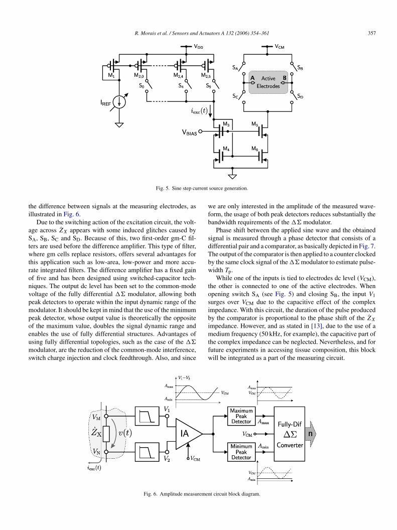
To efficiently measure ac impedance, an integrated sinusoidalurrent source is needed. Most sine current generators are basedn a direct digital synthesizer (DDS), which requires a read-nly memory (ROM) sine look-up table and a current-modeigital-to-analog converter (DAC). This solution requires a largentegration area since waveform generation is closely related tohe ROM size and the DAC resolution. Instead of using suchpproach a set of precisely scaled current mirrors can be used toroduce sine steps as depicted in Fig. 5.
Switches S0 to S5 are turned ON by an up/down countero produce a half-sine wave, where individual current mirrorsM2,0, . . . , M2,5) have been calculated to perform an accurateine current step. Excitation current iext(t) (equivalent to a rec-ified sine waveform) is then mirrored by a wide swing cascadeurrent mirror (M5, M6) and transformed to a full wave by thewitching network formed by switches SA, SB, SC and SD. Theeference current IREF is generated by a bandgap reference cir-uit and can be adjusted externally during prototype evaluation.
.2. Measurement circuit
Voltage drop at the inner electrodes of the array is character-zed by its amplitude (real part of the measured impedance) andhe phase shift between the injected current and the measured
356 R. Morais et al. / Sensors and A
mented in a fully differential topology using switched-capacitortechniques. The output of the �� modulator (bitstream) is thenapplied to a first-order digital filter to provide a 14-bit word. Theassociated control logic is responsible for controlling the analogmultiplexer and for digital word synchronization, regarding thetemperature sensor and potential difference samples. For datatransmission, digital samples are assembled in a frame contain-ing ID, preamble and checksum control fields. Prior to transmis-sion, data is encoded as a pulse-width modulated (PWM) signaland then transmitted, by means of amplitude shift-keying (ASK)modulation, through a power amplifier operating at 433.92 MHzISM band in class-E mode. The RF frequency is generated byan on-chip frequency synthesizer based on a 13.56 MHz crystal.
The temperature sensor, developed in a previous work [9], isa proportional to absolute temperature (PTAT) circuit with bipo-lar devices fabricated in the CMOS process. It uses a dynamicPTAT voltage generator with thirty two switching stages us-ing dynamic element matching and dynamic amplification tech-niques to achieve a high-performance temperature sensor withhigh accuracy (better than 0.1%) and high resolution (better than0.05 ◦C, typical 0.01 ◦C).
3. Electrical bio-impedance
One of the classic signs of estrus in cows is a swollen vulva.Such swelling is the result of changes in tissue hydration, whichcauses changes in ER [10]. Changes in cell density, fluid volume,and electrolyte content of bovine vulvar tissue can be accom-plished by considering the living tissue as an electrical circuit.The simplified circuit used to simulate electrical properties of abiological subject includes parallel resistance Re (extracellularspace resistance) to serially connected Ri (intracellular spaceresistance) with Cm (membrane capacitance), as depicted inFig. 3.
To determine tissue water and composition a bioelectricalimpedance analysis (BIA) technique is used. This technique isan assessment of changes in a biological object reflected by thecapacitive reactance, resistance, and other factors for an indica-tion of any alteration in the tissue composition.
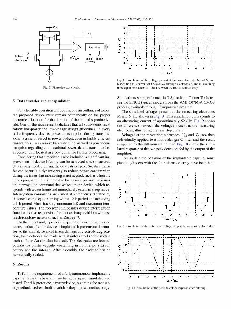
4. Measurement methods
The biological object impedance is measured by injectinga small alternating current, iext(t), of constant amplitude at afixed frequency through the tissue by using two electrodes. Thecurrent injection develops a voltage distribution on the periphery
oltage. Both values must be acquired to calculate the value ofhe complex impedance ZX.
The amplitude measurement is based on two peak detectorshat detect and hold the minimum and the maximum values of
R. Morais et al. / Sensors and Actuators A 132 (2006) 354–361 357
rrent
ti
aStwtronvpmpoeums
wfb
sdTbw
tosibi
Fig. 5. Sine step cu
he difference between signals at the measuring electrodes, asllustrated in Fig. 6.
Due to the switching action of the excitation circuit, the volt-ge across ZX appears with some induced glitches caused byA, SB, SC and SD. Because of this, two first-order gm-C fil-
ers are used before the difference amplifier. This type of filter,here gm cells replace resistors, offers several advantages for
his application such as low-area, low-power and more accu-ate integrated filters. The difference amplifier has a fixed gainf five and has been designed using switched-capacitor tech-iques. The output dc level has been set to the common-modeoltage of the fully differential �� modulator, allowing botheak detectors to operate within the input dynamic range of theodulator. It should be kept in mind that the use of the minimum
eak detector, whose output value is theoretically the oppositef the maximum value, doubles the signal dynamic range and
nables the use of fully differential structures. Advantages ofsing fully differential topologies, such as the case of the ��odulator, are the reduction of the common-mode interference,witch charge injection and clock feedthrough. Also, and since
mtfw
Fig. 6. Amplitude measuremen
source generation.
e are only interested in the amplitude of the measured wave-orm, the usage of both peak detectors reduces substantially theandwidth requirements of the �� modulator.
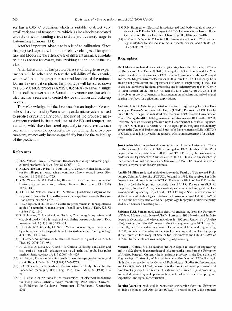
Phase shift between the applied sine wave and the obtainedignal is measured through a phase detector that consists of aifferential pair and a comparator, as basically depicted in Fig. 7.he output of the comparator is then applied to a counter clockedy the same clock signal of the �� modulator to estimate pulse-idth Tp.While one of the inputs is tied to electrodes dc level (VCM),
he other is connected to one of the active electrodes. Whenpening switch SA (see Fig. 5) and closing SB, the input V1urges over VCM due to the capacitive effect of the complexmpedance. With this circuit, the duration of the pulse producedy the comparator is proportional to the phase shift of the ZX
mpedance. However, and as stated in [13], due to the use of a
edium frequency (50 kHz, for example), the capacitive part ofhe complex impedance can be neglected. Nevertheless, and foruture experiments in accessing tissue composition, this blockill be integrated as a part of the measuring circuit.
t circuit block diagram.
358 R. Morais et al. / Sensors and Actuators A 132 (2006) 354–361
5
talfrstsa
pdfdcasItapfm
tftsobh
6
cti
Frt
Sip
Mate
iilated response of the two peak detectors fed by the output of theamplifier.
To simulate the behavior of the implantable capsule, someplastic cylinders with the four-electrode array have been built
Fig. 9. Simulation of the differential voltage drop at the measuring electrodes.
Fig. 7. Phase detector circuit.
. Data transfer and encapsulation
For a feasible operation and continuous surveillance of a cow,he proposed device must remain permanently on the propernatomical location for the duration of the animal’s productiveife. One of the requirements dictates that all subsystems mustollow low-power and low-voltage design guidelines. In everyadio-frequency device, power consumption during transmis-ions is a major parcel in power budget, even in highly efficientransmitters. To minimize this restriction, as well as power con-umption regarding computational power, data is transmitted toreceiver unit located in a cow collar for further processing.
Considering that a receiver is also included, a significant im-rovement in device lifetime can be achieved since measuredata is only needed during the cow estrus cycle. So, data trans-er can occur in a dynamic way to reduce power consumptionuring the times that monitoring is not needed, such as when theow is pregnant. This is controlled by the receiver unit that issuesn interrogation command that wakes up the device, which re-ponds with a data frame and immediately enters in sleep mode.nterrogation commands are issued at a frequency dictated byhe cow’s estrus cycle starting with a 12-h period and achieving1-h period when tracking minimum ER and maximum tem-
erature values. The receiver unit, besides device interrogationunction, is also responsible for data exchange within a wirelessesh-topology network, such as ZigBee™.On the other hand, a proper encapsulation must be addressed
o ensure that after the device is implanted it presents no discom-ort to the animal. To avoid tissue damage or electrode degrada-ion, the electrodes are made with stainless steel (noble metalsuch as Pt or Au can also be used). The electrodes are locatedutside the plastic capsule, containing in its interior a Li-ionattery and the antenna. After assembly, the package can beermetically sealed.
. Results
To fulfill the requirements of a fully autonomous implantableapsule, several subsystems are being designed, simulated andested. For this prototype, a macrodevice, regarding the measur-ng method, has been built to validate the proposed methodology.
ig. 8. Simulation of the voltage present at the inner electrodes M and N, cor-esponding to a current of 652�ARMS through electrodes A and B, assuminghree equal resistances of 100 � between the four-electrode array.
imulations were performed in T-Spice from Tanner Tools us-ng the SPICE typical models from the AMI C07M-A CMOSrocess, available through Europractice program.
The simulated voltages present at the measuring electrodesand N are shown in Fig. 8. This simulation corresponds to
n alternating current of approximately 52 kHz. Fig. 9 showshe difference between the voltages present at the measuringlectrodes, illustrating the sine step current.
Voltages at the measuring electrodes, VM and VN, are thenndividually applied to a first-order gm-C filter and the results applied to the difference amplifier. Fig. 10 shows the simu-
Fig. 10. Simulation of the peak detectors response after filtering.

R. Morais et al. / Sensors and Actuators A 132 (2006) 354–361 359
wmeIcctcpa
mwmtalo
c
Fi
aas(
Fig. 11. Several capsule prototypes.
ith different electrode separations, as depicted in Fig. 11. Theaterials used (PVC for the cylinder and stainless steel for the
lectrodes) were specifically chosen to ensure biocompatibility.n order to keep the electrodes in place, a small chamfer wasarved around the external side of the cylinder. All electricalonnections were made on the inside of the cylinder, which washen filled with a non-conductive glue. To simulate the electri-al conductivity of the vaginal mucus, each capsule was thenlaced in an aqueous solution of sodium chloride (0.08 mol L−1
t 23 ◦C).At this stage, capsule evaluation was performed by a
acrodevice, Fig. 12, where discrete electronic componentsere employed to generate a sine step current source. Thisacrodevice, beside the current source, filters and peak detec-
ors, has a 14-bit analog-to-digital converter, a microcontrollernd a RS232 driver to acquire and transfer collected data to a
aptop computer for posterior analysis. Fig. 13 shows the resultsf some measurements.Fig. 13 shows the injected sine wave corresponding to an acurrent of amplitude of approximately 660 �ARMS (933 �Apeak)
ig. 12. Experimental setup using a macrodevice to evaluate capsule behaviornside an aqueous solution.
awtt1
7
pss
bimmi(vmn
Fig. 13. Waveforms obtained using a macrodevice.
nd frequency of 52 kHz. The waveforms at both outputs of themplifier and peak detector are shown in Fig. 13 (minimum nothown). The peak detector output indicates a value of 921 mVgain of the amplifier was set to 10), which is consistent with100 � resistance measurement. In fact, for a specific capsuleith its particular cell constant value, the result is the inverse of
he measured conductance, which was 9.8 mS. The inverse ofhe conductance is the resistance leading to a measured value of02 �.
. Conclusions
The preliminary results suggests that the proposed im-lantable microsystem for vulvar ER and body temperature mea-urements can fulfill the requirements of a low-cost autonomousystem to help estrus prediction in herd management.
The major advantage of the presented solution is the capa-ility of continuous monitoring of two correlated parametersn estrus detection during a long period of time without hu-
an intervention. A prototype, comprising the excitation andeasurement circuits, as well as the RF transmitter, is being
mplemented in a standard CMOS 0.7 �m mixed-signal process
AMIS C07M-A) to validate the proposed methodology. Pre-ious implementations [14,9] have shown that the delta–sigmaodulator exhibits an effective 16.1-bit resolution (98.7 dB dy-amic range) with an 800 Hz bandwidth. The temperature sen-
3 Actu
sswL
ttrv
iwDtLum
stsvoro
R
B
Rodaaiois
AvtMPigot
Jodpti
AndctvaUs
Sod(PUaU
MaoEHab
60 R. Morais et al. / Sensors and
or has a 0.05 ◦C precision, which is suitable to detect verymall variations of temperature, which is also closely associatedith the onset of standing estrus and the pre-ovulatory surge inuteinizing hormone (LH).
Another important advantage is related to calibration. Sincehe proposed capsule will monitor relative changes of tempera-ure and ER during the estrus cycle of different animals, absoluteeadings are not necessary, thus avoiding calibration of the de-ice.
After fabrication of this prototype, a set of long-term exper-ments will be scheduled to test the reliability of the capsule,hich will be at the proper anatomical location of the animal.uring this evaluation phase, the prototype will be scaled down
o a 3.3 V CMOS process (AMIS C035M-A) to allow a singlei-ion cell as power source. Some improvements are also sched-led such as a receiver to control device shutdown and wake-upodes.To our knowledge, it’s the first time that an implantable cap-
ule with a circular strip Wenner array and a microsystem is usedo predict estrus in dairy cows. The key of the proposed mea-urement method is the correlation of the ER and temperatureariation, which have been used separately to predict estrus, eachne with a reasonable specificity. By combining these two pa-ameters, we not only increase specificity but also the reliabilityf the prediction.
eferences
[1] M.N. Velasco-Garcia, T. Mottram, Biosensor technology addressing agri-cultural problems, Biosyst. Eng. 84 (2003) 1–12.
[2] R.M. Pemberton, J.P. Hart, T.T. Mottram, An electrochemical immunosen-sor for milk progesterone using a continuous flow system, Biosens. Bio-electron. 16 (2001) 715–723.
[3] R.W. Claycomb, M.J. Delwiche, Biosensor for on-line measurement ofbovine progesterone during milking, Biosens. Bioelectron. 13 (1998)1173–1180.
[4] Y.F. Xu, M. Velasco-Garcia, T.T. Mottram, Quantitative analysis of theresponse of an electrochemical biosensor for progesterone in milk, Biosens.Bioelectron. 20 (2005) 2061–2070.
[5] R.L. Scipioni, R.H. Foote, An electronic probe versus milk progesteroneas aids for reprodutive management of small dairy herds, J. Dairy Sci. 82(1999) 1742–1745.
[6] R. Bobowiec, T. Studzinski, A. Babiarz, Thermoregulatory effects andelectrical conductivity in vagina of cow during oestrus cycle, Arch. Exp.Veterinarmed. 4 (44) (1990) 573–579.
[7] B.L. Kyle, A.D. Kennedy, J.A. Small, Measurement of vaginal temperatureby radiotelemetry for the prediction of estrus in beef cows, Theriogenology49 (1998) 1437–1449.
[8] R. Herman, An introduction to electrical resistivity in geophysics, Am. J.Phys. 69 (2001) 943–952.
[9] A. Valente, R. Morais, C. Couto, J.H. Correia, Modeling, simulation andtesting of a silicon soil moisture sensor based on the dual-probe heat-pulsemethod, Sens. Actuators A 115 (2004) 434–439.
[10] P.L. Senger, The estrus detection problem: new concepts, technologies, andpossibilities, J. Dairy Sci. 77 (1994) 2745–2753.
[11] D.A. Schoeller, R.F. Kushner, Determination of body fluids by theimpedance technique, IEEE Eng. Med. Biol. Mag. 8 (1998) 19–
21.[12] A. I. Cano, Contributions to the measurement of electrical impedancefor living tissue ischemia injury monitoring, PhD Thesis, Universi-tat Politecnica de Catalunya, Departament D’Enginyeria Electronica,2005.
at
Ro
ators A 132 (2006) 354–361
[13] R.N. Baumgarter, Electrical impedance and total body electrical conduc-tivity, in: A.F. Roche, S.B. Heymsfield, T.G. Lohman (Eds.), Human BodyComposition, Human Kinectics, Champaign, IL, 1996, pp. 79–107.
[14] R. Morais, A. Valente, C. Couto, J.H. Correia, A wireless RF CMOS mixed-signal interface for soil moisture measurements, Sensors and Actuators A115 (2004) 376–384.
iographies
aul Morais graduated in electrical engineering from the University of Tras-s-Montes and Alto Douro (UTAD), Portugal in 1993. He obtained the MScegree in industrial electronics in 1998 from the University of Minho, Portugalnd the PhD degree in microelectronics in 2004 from the UTAD. Presently, he isn assistant professor in the Department of Electrical Engineering, UTAD. Hes also a researcher in the signal processing and biotelemetry group at the Centerf Technological Studies for Environment and Life (CETAV) of UTAD, and hes involved in the development of instrumentation solutions and mixed-signalensing interfaces for agricultural applications.
ntonio Luıs G. Valente graduated in Electrical Engineering from the Uni-ersity of Tras-os-Montes and Alto Douro (UTAD), Portugal in 1994. He ob-ained the MSc degree in industrial electronics in 1999 from the University of
inho, Portugal and the PhD degree in microelectronics in 2004 from the UTAD.resently, he is an assistant professor in the Department of Electrical Engineer-
ng, UTAD. He is also a researcher in the signal processing and biotelemetryroup at the Center of Technological Studies for Environment and Life (CETAV)f UTAD and he is involved in the research of silicon microsensors for agricul-ure.
ose Carlos Almeida graduated in animal science from the University of Tras-s-Montes and Alto Douro (UTAD), Portugal in 1987. He obtained the PhDegree in animal reproduction in 2000 from UTAD. Presently, he is an assistantrofessor in Department of Animal Science, UTAD. He is also a researcher, athe Center of Animal and Veterinary Science (CECAV-UTAD), and his area ofnterest is reproduction in farm animals.
melia M. Silva graduated in biochemistry at the Faculty of Science and Tech-ology, Coimbra University (FCTUC), Portugal in 1992. She received her MScegree in cell biology from the FCTUC, Portugal in 1995 and her PhD in bio-hemistry (cellular biophysics speciality) from FCTUC, Portugal, in 2003. Athe present, Amelia M. Silva, is an assistant professor at the Biological and En-ironmental Engineering Department, UTAD, Portugal. She is also a researchert the Center of Technological Studies for Environment and Life (CETAV, atTAD) and has been involved on cell physiology, biophysics and biochemistry
tudies on hormone secreting cells.
alviano F.S.P. Soares graduated in electrical engineering from the Universityf Tras-os-Montes e Alto Douro (UTAD), Portugal in 1991. He obtained the MScegree in electronics and telecomunications in 1995 from University of AveiroUA), Portugal, and the PhD degree in electrical engineering in 2003 from UA.resently, he is an assistant professor in Department of Electrical Engineering,TAD, and also a researcher in the signal processing and biotelemetry group
t the Center of Technological Studies for Environment and Life (CETAV) ofTAD. His main interest area is digital signal processing.
anuel J. Cabral S. Reis received the PhD degree in electrical engineeringnd the MSc degree in electronics and telecommunications from the Universityf Aveiro, Portugal. Currently he is assistant professor in the Department ofngineering of University of Tras-os-Montes e Alto Douro (UTAD), Portugal.e is also a researcher at the Center of Technological Studies for Environment
nd Life (CETAV) of UTAD, where he is the director of signal processing andiotelemetry group. His research interests are in the area of signal processing,
nd include modelling and approximation, and problems such as sampling, in-erpolation, and signal reconstruction.amiro Valentim graduated in zootechnic engineering from the Universityf Tras-os-Montes and Alto Douro (UTAD), Portugal in 1989. He obtained

Actu
tnMsmoa
Jversity of Tras-os-Montes and Alto Douro (UTAD), Portugal in 1981. He ob-
R. Morais et al. / Sensors and
he MSc degree in animal science from IAMZ-CIHEAM (Mediterranean Agro-omic Institute of Zaragoza—member of the Internacional Centre for Advanced
editerranean Agronomic Studies) in 1994, Spain and the PhD degree in animalcience in 2004 from UTAD. Presently, he is an assistant professor in Depart-ent of Zootechnic, ESA-IPB (Agrarian Superior School, Politechnic Institute
f Braganca), Portugal. He is involved in the study of how environmental factorffects reproduction of local sheep and goat breeds.
taaa
ators A 132 (2006) 354–361 361
orge Manuel Teixeira de Azevedo graduated in animal science from the Uni-
ained the PhD degree in animal science in 1994 from UTAD. Presently, he isfull professor in Department of Animal Science, UTAD, Portugal. He is alsoresearcher, at the Center of Animal and Veterinary Science (CECAV-UTAD),nd his area of interest is animal production.
Copyright © 2022 FDOKUMEN