Major predictors of fibrous adherences in transvenous implantable cardioverter-defibrillator lead...
27
Author's Accepted Manuscript Major Predictors of Fibrous Adherences in Trans- venous ICD Lead Extraction Luca Segreti MD, Andrea Di Cori MD, Ezio Soldati MD, Giulio Zucchelli MD, PhD, Stefano Viani MD, Luca Paperini MD, Raffaele De Lucia MD, Giovanni Coluccia MD, Sergio Valsecchi PhD, Maria Grazia Bongiorni MD, FESC PII: S1547-5271(14)00865-0 DOI: http://dx.doi.org/10.1016/j.hrthm.2014.08.011 Reference: HRTHM5896 To appear in: Heart Rhythm Cite this article as: Luca Segreti MD, Andrea Di Cori MD, Ezio Soldati MD, Giulio Zucchelli MD, PhD, Stefano Viani MD, Luca Paperini MD, Raffaele De Lucia MD, Giovanni Coluccia MD, Sergio Valsecchi PhD, Maria Grazia Bongiorni MD, FESC, Major Predictors of Fibrous Adherences in Transvenous ICD Lead Extraction, Heart Rhythm, http://dx.doi.org/10.1016/j.hrthm.2014.08.011 This is a PDF file of an unedited manuscript that has been accepted for publication. As a service to our customers we are providing this early version of the manuscript. The manuscript will undergo copyediting, typesetting, and review of the resulting galley proof before it is published in its final citable form. Please note that during the production process errors may be discovered which could affect the content, and all legal disclaimers that apply to the journal pertain. www.elsevier.com/locate/buildenv
-
Upload
independent -
Category
Documents
-
view
0 -
download
0
Transcript of Major predictors of fibrous adherences in transvenous implantable cardioverter-defibrillator lead...

Author's Accepted Manuscript
Major Predictors of Fibrous Adherences in Trans-venous ICD Lead Extraction
Luca Segreti MD, Andrea Di Cori MD, Ezio SoldatiMD, Giulio Zucchelli MD, PhD, Stefano Viani MD,Luca Paperini MD, Raffaele De Lucia MD, GiovanniColuccia MD, Sergio Valsecchi PhD, Maria GraziaBongiorni MD, FESC
PII: S1547-5271(14)00865-0DOI: http://dx.doi.org/10.1016/j.hrthm.2014.08.011Reference: HRTHM5896
To appear in: Heart Rhythm
Cite this article as: Luca Segreti MD, Andrea Di Cori MD, Ezio Soldati MD, GiulioZucchelli MD, PhD, Stefano Viani MD, Luca Paperini MD, Raffaele De Lucia MD,Giovanni Coluccia MD, Sergio Valsecchi PhD, Maria Grazia Bongiorni MD, FESC,Major Predictors of Fibrous Adherences in Transvenous ICD Lead Extraction, HeartRhythm, http://dx.doi.org/10.1016/j.hrthm.2014.08.011
This is a PDF file of an unedited manuscript that has been accepted for publication. As aservice to our customers we are providing this early version of the manuscript. Themanuscript will undergo copyediting, typesetting, and review of the resulting galley proofbefore it is published in its final citable form. Please note that during the production processerrors may be discovered which could affect the content, and all legal disclaimers that applyto the journal pertain.
www.elsevier.com/locate/buildenv

Major Predictors of Fibrous Adherences in Transvenous ICD Lead Extraction
Luca Segreti,a MD; Andrea Di Cori,
a MD; Ezio Soldati,
a MD; Giulio Zucchelli,
a MD, PhD; Stefano
Viani,a MD; Luca Paperini,
a MD; Raffaele De Lucia,
a MD; Giovanni Coluccia,
a MD; Sergio
Valsecchi,b PhD; Maria Grazia Bongiorni,
a MD, FESC
a Second Cardiology Division, Cardiothoracic and Vascular Department, University Hospital of
Pisa, Pisa, Italy; b
Boston Scientific Italy, Milan, Italy
Short title: ICD Lead Extraction and Fibrous Adherences
Word count: 4231.
Funding Sources: None to declare.
Disclosure: Sergio Valsecchi is an employee of Boston Scientific. No other potential conflict of
interest to declare.
Correspondence to:
Luca Segreti, MD,
Second Cardiology Division
Cardiothoracic and Vascular Department
University Hospital of Pisa
via Paradisa, 2 - 56100 Pisa, Italy
Tel. 0039050993043| Fax. 0039050992362 | e-mail: [email protected]
2
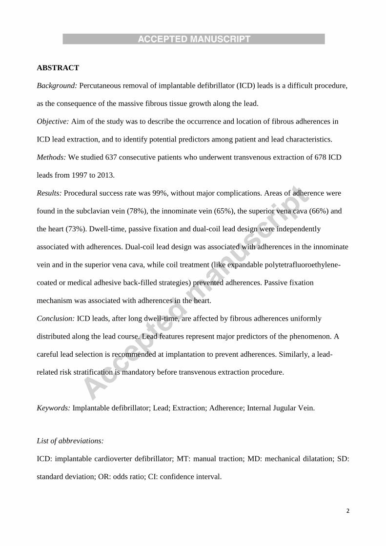
ABSTRACT
Background: Percutaneous removal of implantable defibrillator (ICD) leads is a difficult procedure,
as the consequence of the massive fibrous tissue growth along the lead.
Objective: Aim of the study was to describe the occurrence and location of fibrous adherences in
ICD lead extraction, and to identify potential predictors among patient and lead characteristics.
Methods: We studied 637 consecutive patients who underwent transvenous extraction of 678 ICD
leads from 1997 to 2013.
Results: Procedural success rate was 99%, without major complications. Areas of adherence were
found in the subclavian vein (78%), the innominate vein (65%), the superior vena cava (66%) and
the heart (73%). Dwell-time, passive fixation and dual-coil lead design were independently
associated with adherences. Dual-coil lead design was associated with adherences in the innominate
vein and in the superior vena cava, while coil treatment (like expandable polytetrafluoroethylene-
coated or medical adhesive back-filled strategies) prevented adherences. Passive fixation
mechanism was associated with adherences in the heart.
Conclusion: ICD leads, after long dwell-time, are affected by fibrous adherences uniformly
distributed along the lead course. Lead features represent major predictors of the phenomenon. A
careful lead selection is recommended at implantation to prevent adherences. Similarly, a lead-
related risk stratification is mandatory before transvenous extraction procedure.
Keywords: Implantable defibrillator; Lead; Extraction; Adherence; Internal Jugular Vein.
List of abbreviations:
ICD: implantable cardioverter defibrillator; MT: manual traction; MD: mechanical dilatation; SD:
standard deviation; OR: odds ratio; CI: confidence interval.

3
INTRODUCTION
In recent years, the growth of implantable defibrillator (ICD) implantations has led to an increase in
ICD removal procedures for lead failure or infection (1). Although safe and effective, transvenous
lead extraction continues to be a challenging procedure (2,3). Fibrous ingrowth, the main obstacle
to lead removal (4), may vary from patient to patient, affecting complexity, duration and outcomes
of the procedure. Some factors have been suggested to increase the fibrous reaction, but until today
no data have been systematically collected to describe rate, location and predictors of fibrous
adherences in patients with ICD leads.
Aim of the study was to analyze data collected in patients who underwent transvenous ICD lead
extraction in our center and to describe, from a procedural standpoint, the occurrence and the
location of fibrous adherences, and the potential predicting value of patient and lead characteristics.
METHODS
Patient Population and Procedure
We included and analyzed all consecutive patients who underwent a transvenous ICD lead
extraction in our center from January 1997 to December 2012. In accordance with the guiding
principles of the Declaration of Helsinki about studies involving humans, all the patients gave
informed consent to the procedure and to personal data collection and the study was approved by
the Institutional Committee on Human Research at our Institution.
The extraction procedure has been previously described (5,6). After device removal, the leads were
examined visually and by means of fluoroscopy in their intravascular segment; the proximal end
was then clipped and a standard stylet was inserted into the lead. Lead extraction was attempted by
means of gentle manual traction (MT).
If this proved unsuccessful, we crossed to mechanical dilatation (MD) with a single-sheath (i.e.
non-telescopic) technique. This involved inserting and advancing multisized dilators (Cook

4
Intravascular Inc., Leechburg, PA, USA) through the venous entry-site, as first choice, or through
the right internal jugular vein, if required (5,6).
Lead extraction time was defined as the time from the start of MT to complete lead removal.
Procedural outcomes (i.e. success and complications) were defined according to previously
published guidelines (7,8).
Definition of Fibrous Adherence Sites
As previously described (9-11), we adopted an indirect procedural model to assess the presence and
the location of fibrous sites. The presence of at least one site of fibrotic adherence was defined in
case of ineffective MT (i.e. necessity to cross to MD). The location of fibrotic tissue was defined by
an expert operator, who evaluated the difficulty in sheath advancement during MD. When the
sheath advancement required more than 1 minute of MD at one site, that site was defined as a local
area of fibrotic tissue. Regarding the number of sites, we identified 4 intravascular adherence sites
(subclavian vein, innominate vein, superior vena cava, cardiac chambers) on the basis of the
relationship between cardiac leads and anatomical landmarks, detectable with the postero-anterior
projection at fluoroscopy.
The following radiologic landmarks definitions were used (12-14):
- Subclavian venous district: from the first rib-clavicular joint to the ipsilateral sterno-
clavicular joint.
- Innominate venous district: from the left to the right sterno-clavicular joint.
- Superior Vena Cava district: from an horizontal plane passing through the upper margin of
the aortic arch to an horizontal plane passing through the upper margin of the right heart
contour (i.e. right lateral border of the right atrium).
- Cardiac Chambers District: from an horizontal plane passing through the upper margin of
the right heart contour to an horizontal plane passing through the upper margin of the left
emi-diaphragm. (Figure 1).

5
Statistical Analysis
Descriptive statistics are reported as means standard deviation (SD) for normally distributed
continuous variables, or medians with ranges in the case of skewed distribution. Categorical data
are expressed as percentages, and differences in proportions were compared by means of a Chi-
square analysis or Fisher’s exact test, as appropriate.
Univariate binary logistic regression analysis was utilized to evaluate the relationship between the
presence of adherence requiring mechanical dilatation and patient and lead characteristics. All
variables displaying statistical significance, i.e. p value < 0.05, underwent multivariate binary
logistic regression analysis. A p value <0.05 was considered significant for all tests. Statistical
analyses were performed by means of SYSTAT 12.0 (Systat software Inc., Chicago, USA)
software.
RESULTS
Patient population and extracted leads
From January 1997 to December 2013, 637 consecutive patients required transvenous removal of
678 ICD leads. The majority of ICD leads were passively fixed (69%), had 2 coils (78%), were
silicone-insulated and had a multi-lumen design. In the majority (74%) of our patients the clinical
indication for the removal procedure was infection.
In addition to ICD leads, 369 atrial, 221 coronary sinus and 89 ventricular pacing leads had to be
removed from our patients. The median number of cardiac leads to be removed from each patient
was 2, ranging from 1 to 7.
Patient characteristics are summarized in Table 1 and the characteristics of the leads extracted are
reported in Table 2.

6
Procedural Outcomes
MT was effective in 44 leads (6%). To remove the remaining 634 leads, MD was attempted through
the venous entry-site and was effective in 555 cases (82%). Seventy-eight of the 79 leads that could
not be successfully extracted through the venous entry-site approach were finally approached via
the internal jugular vein, successful extraction being achieved in 73 cases (94%). The final
procedural success rate was 99% (672 out of 678). The median extraction time was 8 min, ranging
from 1 to 210 min.
Six leads (0.9%) could not be transvenously removed: they were abandoned in situ with no
sequelae, or the patient was referred to the cardiac surgeon for open-chest extraction, according to
the indication.
There were no major complications associated with lead extraction. Minor complications were
reported in 25 patients (4%).
Fibrous Adherences Assessment: rate and location
Fibrous adherences were found in 593 patients (94%). The average number of adherences was
2.8±1.2 per patient. Areas of adherence were documented in all sites, with a statistically higher
prevalence in the subclavian vein and in the heart (p<0.001) [subclavian vein (78%), innominate
vein (65%), superior vena cava (66%) and heart (73%)] (Figure 2).
Univariate and multivariate analyses of clinical and lead-related factors were performed to identify
predictors of fibrous adherences at any site and at each specific intravascular site.
At multivariate logistic regression analysis, dwell time (odds ratio [OR] 1.10 , 95% confidence
interval [CI] 1.06 – 1.14; P <0.001), passive fixation (OR 3.25 , 95% CI 1.41 – 7.53; P = 0.006)
and dual coil lead design (OR 2.94, 95% CI 1.27 – 6.78; P = 0.011) were independent predictors of
fibrous adherence (Table 3).
Dual-coil design predicted adherences in the innominate vein (OR 3.93, 95% CI 2.47 –6.23; P
<0.001) and in the superior vena cava (OR 3.52, 95% CI 2.15 – 5.74; P <0.001), and the passive

7
fixation mechanism was associated with adherences in the heart (OR 1.72, 95% CI 1.11 – 2.65; P
=0.015). The coil treatment (expandable polytetrafluoroethylene-coated or medical adhesive back-
filled coils) was associated with a low rate of adherences in the superior vena cava (OR 1.75, 95%
CI 1.04 – 2.93; P = 0.035) and in the heart (OR 2.11, 95% CI 1.25 – 3.57; P = 0.005) (Figure 2, 3
and 4).
Results of Univariate and Multivariate analysis are shown in Table 3.
Location and rate of fibrous adherences are shown in Figure 2, 3 and 4.
DISCUSSION
A comprehensive knowledge of adherence location is the key point of a safe and effective
transvenous procedure. The aim of MD is dissecting all lead-related adherences from the venous
entry site to the tip and complications are commonly observed as the result of vascular or cardiac
damage during dilatation. In the past, adherence rate and location have been described for standard
pacing leads, and some predictors were identified (5,9,15).
This was the first large single-center experience systematically describing location, rate and
predictors of adherence in more than 600 patients with ICD leads. Our analysis confirmed the
peculiar post-implantation behavior of ICD leads. Compared to standard pacing leads, they show a
similar time-dependent fibrous ingrowth, but with a larger involvement of the lead course (16).
In agreement with previous experiences (17-21), we found that dwell-time was independently
associated with the use of mechanical dilatation. Moreover, some structural lead features seem to
promote the formation of adherence at specific locations. In this study, a major predictor of
adherence turned out to be the dual coil technology (22) (Table 3). The coils of ICD leads have been
shown to induce an extensive growth of scar tissue, which surrounds and entraps the lead, and
requires complex extraction procedures (23). Nonetheless, a dual coil design predicted a risk of
adherence at the level of innominate vein and superior vena cava (Figure 2) and only the presence
of a coil treatment (like expandable polytetrafluoroethylene-coated or medical adhesive back-filled

8
strategies) was protective (Table 3, Figure 5), confirming the findings of previous studies
(11,24,25).
As shown in a recent multicenter study (26), the presence of a superior vena cava coil is associated
with significantly higher complication rates, and transvenous extraction of dual-coil ICD leads is
2.6 times more difficult than that of single-coil leads and is more frequently associated with the use
of powered sheaths and with higher complication rates. A complication in these vascular districts
can lead to serious consequences that may not be mitigated by emergency surgery (27).
The second independent predictor of adherence related to the fixation mechanism. As previously
described (28), tines were often associated with MD. In particular, isodiametric active fixation leads
seemed to prevent adherences inside the ventricles, and were more easily and completely removed
by MT (Table 3, Figure 4).
All these peculiar lead features (non isodiametric structure, dual coil, lack of coil treatment, passive
fixation mechanism), with a typical model of response to injury, cause a progressive time-
dependent tissue ingrowth. This explains the extent of fibrosis (94% of patients with a mean of
2.8±1.2 adhesion sites per patient), specifically associated with older ICD lead models. Although
early experiences demonstrated the feasibility of transvenous ICD lead extraction (14, 29-36), these
leads have always been considered more difficult to extract, because of their larger size, aggressive
fibrosis around the coils (19), and their non-isodiametric structure (especially of older models) (37-
39). In the Pacing Lead Surveillance Study in Europe (PLESSE), transvenous ICD lead extraction
was associated with a 12% risk of major complications, while the risk of major complications
associated with pacing lead extraction was 1.7% (40). Additional multicenter experiences have
documented potential major complications, such as death (at a rate of 0.28-0.8%), emergency
cardiac surgery (1-2%) and cardiac perforations (1-4%) (18,32). Although some clinical factors ,
like BMI and renal insufficiency, were recently suggested to be predictors of all-cause mortality
within 30 days of transvenous extraction, (41) in our experience no significant correlation were

9
found with adherences rate and location, suggesting a potential unfavorable predictive role
independent from procedural standpoints, like adherences and related necessity of MD.
Identification of adherence predictors has major implications. It may improve and facilitate risk
stratification of transvenous ICD lead removal. In addition, it could promote prevention, stimulating
the production of new lead technologies to reduce the impact of post-implantation tissue ingrowth.
At the present time, the use of single-covered coil lead with active fixation constitutes the first
strategy to reduce lead related fibrosis.
Study limitations
As the present study is a retrospective review, all the potential limitations of retrospective analyses
should be taken into account when interpreting its results. Moreover, although large, the present
series represents a single-center experience. Thus, the present results cannot be generalized beyond
the specific technique adopted in our institution and described in this paper. Finally, fibrous
adherence sites were identified indirectly from a procedural perspective. Gross examination and
histopathology of the extracted leads was beyond the intentions of the present study.
CONCLUSION
Very few ICD leads can be successfully removed by simple MT, and dilatation is often required.
Fibrous ingrowth on these leads is extensive, and may encapsulate the entire intravascular path up
to cardiac chambers. Structural lead features seem associated with adherence formation, enhancing
the response to injury in a time dependent fashion. A careful lead selection is recommended at
implantation to prevent intravenous adherences. Similarly, a lead related risk stratification is
mandatory before a transvenous extraction procedure.

10
ACKNOWLEDGMENTS
The authors are indebted to their nurses Michela Favaro, Cristina Giannessi, Giampietro Ercoli,
Mascia Sarti, Susy Bardelli, and Patrizio Lazzerini for their excellent support in the management of
patients in the cath lab and operating room.

REFERENCES
1. Bongiorni MG, Blomström-Lundqvist C, Kennergren C, Dagres N, Pison L, Svendsen JH,
Auricchio A; Scientific Initiative Committee, European Heart Rhythm Association: Current
practice in transvenous lead extraction: a European Heart Rhythm Association EP Network
Survey. Europace 2012; 14:783-786.
2. Diemberger I, Mazzotti A, Biffi M, Massaro G, Martignani C, Ziacchi M, Reggiani ML,
Battistini P, Boriani G: From lead management to implanted patient management:
systematic review and meta-analysis of the last 15 years of experience in lead extraction.
Expert Rev Med Devices 2013; 10:551-573.
3. Maytin M, Epstein LM: The challenges of transvenous lead extraction. Heart 2011; 97:425-
434.
4. Epstein AE, Neal Kay G, Plumb VJ, Dailey SM, Anderson PG: Gross and Microscopic
Pathological Changes Associated With Nonthoracotomy Implantable Defibrillator Leads.
Circulation 1998; 98;1517-1524.
5. Bongiorni MG, Soldati E, Zucchelli G, Di Cori A, Segreti L, De Lucia R, Solarino G,
Balbarini A, Marzilli M, Mariani M: Transvenous removal of pacing and implantable
cardiac defibrillating leads using single sheath mechanical dilatation and multiple venous
approaches: high success rate and safety in more than 2000 leads. Eur Heart J 2008; 29:
2886-2893.
6. Bongiorni MG: Transvenous Lead Extraction: from Simple Traction to the Internal
Transjugular Approach, Bongiorni MG editor. Springer Verlag 2011: 1-180.
7. Deharo JC, Bongiorni MG, Rozkovec A, Bracke F, Defaye P, Fernandez-Lozano I, Golzio
PG, Hansky B, Kennergren C, Manolis AS, Mitkowski P, Platou ES; European Heart
Rhythm Association: Pathways for training and accreditation for transvenous lead
extraction: a European Heart Rhythm Association position paper. Europace 2012; 14:124-
134.

12
8. Wilkoff BL, Love CJ, Byrd CL, Bongiorni MG, Carrillo RG, Crossley GH 3rd, Epstein LM,
Friedman RA, Kennergren CE, Mitkowski P, Schaerf RH, Wazni OM: Transvenous lead
extraction: Heart Rhythm Society expert consensus on facilities, training, indications, and
patient management: this document was endorsed by the American Heart Association
(AHA). Heart Rhythm 2009; 6:1085-1104.
9. Smith HJ, Fearnot NE, Byrd CL, Wilkoff BL, Love CJ, Sellers TD: Five-years experience
with intravascular lead extraction. U.S. Lead Extraction Database. Pacing Clin
Electrophysiol 1994; 17:2016-2020.
10. Bongiorni MG, Zucchelli G, Soldati E, Arena G, Giannola G, Di Cori A, Lapira F, Bartoli
C, Segreti L, De Lucia R, Barsotti A: Usefulness of mechanical transvenous dilation and
location of areas of adherence in patients undergoing coronary sinus lead extraction.
Europace 2007; 9:69-73.
11. Di Cori A, Bongiorni MG, Zucchelli G, Segreti L, Viani S, Paperini L, Soldati E:
Transvenous extraction performance of expanded polytetrafluoroethylene covered ICD leads
in comparison to traditional ICD leads in humans. Pacing Clin Electrophysiol 2010;
33:1376-1381.
12. Kim MC, Kim KS, Choi YK, et al. An estimation of right- and left-sided central venous
catheter insertion depth using measurement of surface landmarks along the course of central
veins. AnesthAnalg 2011; 112: 1371–4)
13. Godoy MC, Leitman BS, de Groot PM, Vlahos I, Naidich DP. Chest radiography in the
ICU: Part 1, Evaluation of airway, enteric, and pleural tubes. AJR Am J Roentgenol.
2012;198(3):563-71.
14. Godoy MC, Leitman BS, de Groot PM, Vlahos I, Naidich DP. Chest radiography in the
ICU: Part 2, Evaluation of cardiovascular lines and other devices. AJR Am J Roentgenol.
2012;198(3):572-81.
15. Di Cori A, Bongiorni MG, Zucchelli G, Segreti L, Viani S, de Lucia R, Paperini L, Soldati
E: Large, single-center experience in transvenous coronary sinus lead extraction: procedural

13
outcomes and predictors for mechanical dilatation. Pacing Clin Electrophysiol 2012;
35:215-222
16. Saad EB, Saliba WI, Schweikert RA, Al-Khadra AS, Abdul-Karim A, Niebauer MJ, Wilkoff
BL: Nonthoracotomy implantable defibrillator lead extraction: results and comparison with
extraction of pacemaker leads. Pacing Clin Electrophysiol 2003; 26:1944-1950.
17. Bracke F, Meijer A, Van Gelder B: Extraction of pacemaker and implantable cardioverter
defibrillator leads: patient and lead characteristics in relation to the requirement of extraction
tools. Pacing Clin Electrophysiol 2002; 25:1037-1040.
18. Byrd CL, Wilkoff BL, Love CJ, Sellers TD, Reiser C: Clinical study of the laser sheath for
lead extraction: the total experience in the United States Pacing and clinical
electrophysiology. Pacing Clin Electrophysiol 2002; 25:804-808.
19. Maytin M, Love CJ, Fischer A, Carrillo RG, Garisto JD, Bongiorni MG, Segreti L, John
RM, Michaud GF, Albert CM, Epstein LM: Multicenter experience with extraction of the
Sprint Fidelis implantable cardioverter-defibrillator lead. J Am Coll Cardiol 2010; 56:646-
650.
20. Bongiorni MG, Segreti L, Di Cori A, Zucchelli G, Viani S, Paperini L, De Lucia R, Boem
A, Levorato D, Soldati E: Safety and efficacy of internal transjugular approach for
transvenous extraction of implantable cardioverter defibrillator leads. Europace 2014; doi:
10.1093/europace/euu004.
21. Maytin M, Wilkoff BL, Brunner M, et al: Multicenter Experience with Extraction of the
Riata™/Riata™ ST ICD Lead. Heart Rhythm. 2014; doi: 10.1016/j.hrthm.2014.05.014.
22. Gradaus R, Breithardt G, Böcker D. ICD leads: design and chronic dysfunctions. Pacing
Clin Electrophysiol 2003; 26:649-657.
23. Kennergren C, Bjurman C, Wiklund R, Gäbel J: A single-centre experience of over one
thousand lead extractions. Europace 2009; 11:612-617.

14
24. Wilkoff, B. L., Belott, P. H., Love, C. J., Scheiner, A., Westlund, R., Rippy, M., Krishnan,
M., Norlander BE, Steinhaus B, Emmanuel J, Zeller PJ: Improved extraction of ePTFE and
medical adhesive modified defibrillation leads from the coronary sinus and great cardiac
vein. Pacing Clin Electrophysiol 2005; 28:205-211.
25. Hackler JW, Sun Z, Lindsay BD, Wilkoff BL, Niebauer MJ, Tchou PJ, Chung MK:
Effectiveness of Implantable Cardioverter Defibrillator Lead Coil Treatments in Facilitating
Ease of Extraction. Heart Rhythm 2010; 7:890-897.
26. Epstein LM, Love CJ, Wilkoff BL, et al: Superior vena cava defibrillator coils make
transvenous lead extraction more challenging and riskier. J Am Coll Cardiol 2013; 61:987-
989.
27. Hauser RG, Katsiyiannis WT, Gornick CC, Almquist AK, Kallinen LM: Deaths and
cardiovascular injuries due to device-assisted implantable cardioverter-defibrillator and
pacemaker lead extraction. Europace 2010; 12:395-401.
28. Smith MC and Love CJ: Extraction of transvenous pacing and ICD leads. Pacing Clin
Electrophysiol 2008; 31:736-752.
29. Jordaens L, Van Belleghem Y, Herregods L: Removal of endocardial defibrillation leads.
Pacing Clin Electrophysiol 1996; 19:127-129.
30. Le Franc P, Klug D, Jarwe M, Lacroix D, Delfaut P, Kouakam C, Kacet S: Extraction of
endocardial implantable cardioverter-defibrillator leads. Am J Cardiol 1999; 84:187-191.
31. Vassilikos VP, Maounis TN, Chiladakis J, Cokkinos DV, Manolis AS: Percutaneous
extraction of transvenous defibrillator leads using the VascoExtor pacing lead removal
system. J Interv Card Electrophysiol 1999; 3:247-251.
32. Byrd CL, Wilkoff BL, Love CJ, et al: Intravascular extraction of problematic or infected
permanent pacemaker leads: 1994-1996. U.S. Extraction Database, MED Institute. Pacing
Clin Electrophysiol 1999; 22:1348-1357.

15
33. Wilkoff BL, Byrd CL, Love CJ, Hayes DL, Sellers TD, Schaerf R, Parsonnet V, Epstein
LM, Sorrentino RA, Reiser C: Pacemaker lead extraction with the laser sheath: results of the
pacing lead extraction with the excimer sheath (PLEXES) trial. J Am Coll Cardiol 1999;
33:1671-1676.
34. Kennergren, C: First European experience using excimer laser for the extraction of
permanent pacemaker leads. Pacing Clin Electrophysiol 1998; 21:268-270.
35. Epstein LM, Byrd CL, Wilkoff BL, Love CJ, Sellers TD, Hayes DL, Reiser C: Initial
experience with larger laser sheaths for the removal of transvenous pacemaker and
implantable defibrillator leads. Circulation 1999; 100:516-525.
36. Kantharia BK, Padder FA, Pennington JC 3rd, Wilbur SL, Samuels FL, Maquilan M,
Kutalek SP: Feasibility, safety, and determinants of extraction time of percutaneous
extraction of endocardial implantable cardioverter defibrillator leads by intravascular
countertraction method. Am J Cardiol 2000; 85:593-597.
37. Wiggers CJ: The muscular reactions of the mammalian ventricles to artificial surface
stimuli. Am J Physiol 1925; 73:346-378
38. Verma A, Wilkoff BL: Intravascular pacemaker and defibrillator lead extraction: A state-of-
the-art review. Heart Rhythm 2004; 1:739-745.
39. Haqqani HM, Mond HG: The Implantable Cardioverter-Defibrillator Lead: Principles,
Progress, and Promises. Pacing Clin Electrophysiol 2009; 32:1336-1353.
40. Kennergren C, Bucknall CA, Butter C, et al. Laser-assisted lead extraction: the European
experience. Europace 2007; 9:651-656.
41. Brunner MP, Cronin EM, Duarte VE, et al. Clinical predictors of adverse patient outcomes
in an experience of more than 5000 chronic endovascular pacemaker and defibrillator lead
extractions. Heart Rhythm. 2014;11(5):799-805.

16
CLINICAL PERSPECTIVES
Percutaneous removal of implantable defibrillator (ICD) leads is a difficult procedure, as the
consequence of the massive fibrous tissue growth along the lead. In this series of 678 ICD leads
extracted from 1997 to 2013 in our center, we analyzed the rate, location and predictors of fibrous
adherences. We found that very few ICD leads can be successfully removed by simple manual
traction, because of extensive fibrous ingrowth encapsulating the entire intravascular lead path up to
cardiac chambers. Dwell-time was independently associated with adhesions, together with lead
features, such as passive fixation or dual-coil lead design. Nonetheless, coil treatment (like
expandable polytetrafluoroethylene-coated or medical adhesive back-filled strategies) prevented
adherences. The present findings allow a risk stratification before transvenous extraction procedure.
Moreover, they strengthen the importance of a careful lead selection at implantation to prevent
intravenous adherences, and seem to suggest the adoption of ICD leads with specific characteristics
only after careful evaluation of the expected benefits and possible risks.

17
FIGURE LEGENDS
Figure 1
Overview of the anatomical landmarks in the postero-anterior fluoroscopic view, in a patient with a
dual chamber ICD. Red dotted lines illustrate the three horizontal planes passing through the upper
margin of the aortic arch (A), through the upper margin of the right heart contour (B) and through
the upper margin of the left emi-diaphragm (C). Continuous red lines illustrate the rib-clavicular
joints (a), the sterno-clavicular joints (b) and the aortic arch (c). Finally, the yellow lines show the
final obtained upper venous anatomy with the corresponding sites (subclavian, innominate, superior
vena cava, right cardiac chambers). For description see text.
Figure 2.
Location and rate of fibrous adherences in the overall population (IV = innominate vein, SV =
subclavian vein, SVC = superior vena cava).
Figure 3.
Overall Adherences rate and location with comparison between single and dual coil ICD lead
groups. Dual coil leads showed a statistically higher significant rate of adherences at the IV and
SVC sites (p<0.001) (IV = innominate vein, SV = subclavian vein, SVC = superior vena cava).
Figure 4.
Overall Adherences rate and location with comparison between active and passive fixation ICD
lead groups. Passive fixation leads showed a statistically higher significant rate of adherences at the
Heart site (p<0.001) (IV = innominate vein, SV = subclavian vein, SVC = superior vena cava).

18
Figure 5.
Overall Adherences rate and location with comparison between treated and untreated coil ICD lead
groups. Untreated coil leads showed a statistically significant higher rate of adherences at the SVC
site (p<0.001) (IV = innominate vein, SV = subclavian vein, SVC = superior vena cava).

19

20

21

22

23

24
Table 1. Patient Population.
Patients n = 637
Male gender, n (%) 539 (85)
Age, years 62±15
Body mass index, Kg/m2 25.9±3.9
Creatinine, mg/dl 1.3±0.6
Coronary artery disease, n (%) 312 (49)
Primary prevention ICD indication, n (%) 306 (48)
Previous cardiac surgery, n (%) 153 (24)
Time from implant, months 48±36
Implanted device, n (%)
- Single-chamber ICD
- Dual-chamber ICD
- Biventricular ICD
236 (37)
180 (28)
221 (35)
Cardiac leads per patient 2 [1 – 7]
Indication for removal, n (%)
- Local infection
- Sepsis
- Lead failure
- Other
315 (49)
158 (25)
154 (24)
10 (2)

25
Table 2. Extracted ICD leads.
ICD Lead Characteristic n = 678
Coil:
- Single
- Dual
- Unknown
135
531
12
Size:
- ≤ 7 Fr
- 8-9 Fr
- ≥ 10 Fr
- Unknown
114
380
152
32
Coil treatment:
- ePTFBE
- MABF
- No treatment
80
46
552
Fixation:
- Active
- Passive
- Unknown
214
468
4
Insulation:
- Silicone
- Polyurethane
- Optim
- Unknown
568
32
46
32
Design:
- Multi-lumen
- Coaxial
- Unknown
613
33
32
ePTFBE = expandable polytetrafluoroethylene-coated coil;
MABF = medical adhesive back-filled coil

26
Table 3. Univariate and multivariate analyses of factors predicting the presence of fibrous
adherences.
Univariate Analysis Multivariate Analysis
OR (95% CI) p OR (95% CI) p
Male gender 0.81 (0.36-1.80) 0.617 - -
Age 0.99 (0.97-1.01) 0.632 - -
Body mass index 1.02 (0.92-1.14) 0.636 - -
Creatinine 1.00 (0.49-2.01) 0.994 - -
Previous cardiac surgery 1.06 (0.41-2.70) 0.899 - -
Time from implant 1.10 (1.06-1.14) <0.001 1.10 (1.06-1.14) <0.001
Number of cardiac leads 0.80 (0.60-1.07) 0.139 - -
Removal for infection 1.24 (0.63- 2.43) 0.532 - -
Passive fixation 6.01 (3.07-11.74) <0.001 3.25 (1.41-7.53) 0.006
Lead size 1.55 (1.15-2.09) 0.001 1.15 (0.77-1.72) 0.480
Dual coil 4.50 (2.41-8.41) <0.001 2.94 (1.27-6.78) 0.011
No ePTFBE/MABF 5.80 (3.07-10.96) <0.001 1.82 (0.79-4.19) 0.156
Subclavian access 1.19 (0.55-2.54) 0.661 - -
Right-sided implantation 0.23 (0.10-0.51) 0.001 0.66 (0.22-1.98) 0.460
OR = Odds Ratio; CI = Confidence Interval; ePTFBE = expandable
polytetrafluoroethylene-coated coil; MABF = medical adhesive back-filled coil