Audit of implantable cardioverter-defibrillators in a single UK centre
Upload
khangminh22Category
view
0download
0
�����������������
Citation: Rohrer, U.; Manninger, M.;
Zirlik, A.; Scherr, D. Multiparameter
Monitoring with a Wearable
Cardioverter Defibrillator. Sensors
2022, 22, 22. https://doi.org/
10.3390/s22010022
Academic Editor: Toshiyo Tamura
Received: 1 November 2021
Accepted: 16 December 2021
Published: 21 December 2021
Publisher’s Note: MDPI stays neutral
with regard to jurisdictional claims in
published maps and institutional affil-
iations.
Copyright: © 2021 by the authors.
Licensee MDPI, Basel, Switzerland.
This article is an open access article
distributed under the terms and
conditions of the Creative Commons
Attribution (CC BY) license (https://
creativecommons.org/licenses/by/
4.0/).
sensors
Review
Multiparameter Monitoring with a WearableCardioverter DefibrillatorUrsula Rohrer , Martin Manninger , Andreas Zirlik and Daniel Scherr *
Division of Cardiology, Department of Medicine, Medical University of Graz, 8036 Graz, Austria;[email protected] (U.R.); [email protected] (M.M.);[email protected] (A.Z.)* Correspondence: [email protected]
Abstract: A wearable cardioverter-defibrillator (WCD) is a temporary treatment option for patientsat high risk for sudden cardiac death (SCD) and for patients who are temporarily not candidates foran implantable cardioverter defibrillator (ICD). In addition, the need for telemedical concepts in thedetection and treatment of heart failure (HF) and its arrhythmias is growing. The WCD has evolvedfrom a shock device detecting malignant ventricular arrhythmias (VA) and treating them with shocksto a heart-failure-monitoring device that captures physical activity and cardioacoustic biomarkersas surrogate parameters for HF to help the treating physician surveil and guide the HF therapy ofeach individual patient. In addition to its important role in preventing SCD, the WCD could becomean important tool in heart failure treatment by helping prevent HF events by detecting imminentdecompensation via remote monitoring and monitoring therapy success.
Keywords: defibrillator; sudden cardiac death; ventricular arrhythmia; telemedicine; monitoring;heart failure
1. Introduction
According to data from the World Health Organization, more than 17 million peopleper year die due to cardiovascular diseases (CVD), making this entity responsible for everythird death worldwide. Within these CVD-related deaths, around 25% is categorized assudden cardiac death (SCD) [1,2].
To prevent SCD, current guidelines recommend the optimization of cardiovascular(CV) risk factors in primary and secondary prevention. The second therapeutic principle isused to assess the individual risk of SCD in patients with cardiomyopathies by taking riskfactors and comorbidities into account [2–4].
The most effective long-term therapy used to prevent SCD in high-risk patients isan implantable cardioverter defibrillator (ICD) [5–7]. The benefit derived from an ICDstrongly depends on adequate risk assessment beforehand.
Thus, the implantation of an ICD should be reserved for patients with a permanenthigh risk for SCD rather than those with a potentially reversible SCD risk. The lattergroup of patients with a temporary risk for SCD may be candidates for a wearable car-dioverter defibrillator (WCD). The WCD is also a viable option for patients waiting for ICDimplantation or patients after ICD explantation, i.e., due to infections or endocarditis.
2. The Wearable Cardioverter Defibrillator (WCD)
Over twenty years ago, the wearable cardioverter defibrillator (WCD) was developedto find a potential solution to protecting patients with a temporary high SCD risk or with apermanent risk for SCD who are not eligible for immediate ICD implantation [8–10].
Sensors 2022, 22, 22. https://doi.org/10.3390/s22010022 https://www.mdpi.com/journal/sensors

Sensors 2022, 22, 22 2 of 18
2.1. Composition of the Device
A wearable cardioverter defibrillator consists of a monitor with a rechargeable batteryand a fabric vest provided with four ECG electrodes to monitor the patient’s heart rhythm aswell as defibrillation electrodes to deliver electrical shock therapy, if needed. Additionally,the WCD captures a variety of information that is transmitted to an online network. Thesedata include basic information such as wearing time, alarms, treatments, the patient’sactivity level, body position, and heart failure monitoring via cardioacoustic biomarkersanalyzed using a more complex algorithm that is further explained in Section 3.3.
2.1.1. The Fabric Vest
The device consists of several components, starting with a fabric garment that comesin different sizes and has an adjustable belt to fit every body size tightly and a back partwith shoulder straps to attach technical components such as the defibrillation pads, theelectrode belt, and the vibration box (see Figure 1).
Sensors 2022, 22, x FOR PEER REVIEW 2 of 18
2.1. Composition of the Device
A wearable cardioverter defibrillator consists of a monitor with a rechargeable bat-
tery and a fabric vest provided with four ECG electrodes to monitor the patient’s heart
rhythm as well as defibrillation electrodes to deliver electrical shock therapy, if needed.
Additionally, the WCD captures a variety of information that is transmitted to an online
network. These data include basic information such as wearing time, alarms, treatments,
the patient’s activity level, body position, and heart failure monitoring via cardioacoustic
biomarkers analyzed using a more complex algorithm that is further explained in Section
0.
2.1.1. The Fabric Vest
The device consists of several components, starting with a fabric garment that comes
in different sizes and has an adjustable belt to fit every body size tightly and a back part
with shoulder straps to attach technical components such as the defibrillation pads, the
electrode belt, and the vibration box (see Figure 1).
Figure 1. A WCD with its components: the fabric garment with an adjustable belt and shoulder
straps (1), self-gelling defibrillation pad with ten gel capsules (2), the electrode belt (3), the vibration
box (4), the heart sounds sensor included in the apical defibrillation pad (5), and the monitor with
the response buttons (6); © ZOLL CMS GmbH.
2.1.2. The Defibrillation Pads
The three defibrillation pads are in contact to the patient’s skin: one pad that includes
the heart sounds sensor (see Section 3.3—Monitoring Heart Failure) is located in an apical
position, and the other two pads are located in a posterior position on both sides of the
vertebra and fitted closely to the dorsal muscles.
The pads are dry and not adhesive and include ten gel capsules (see Figure 1). Shortly
before delivering a WCD shock, an acoustic and visual alarm via the monitor and a vibra-
tional alarm via the vibration box on the patient’s back inform about the imminent shock.
Then, blue contact gel from the gel capsules is automatically applied to enhance the trans-
mission of the electrical current. The energy of the biphasic direct current shock can be
individually adjusted from 75 to 150 J.
Figure 1. A WCD with its components: the fabric garment with an adjustable belt and shoulderstraps (1), self-gelling defibrillation pad with ten gel capsules (2), the electrode belt (3), the vibrationbox (4), the heart sounds sensor included in the apical defibrillation pad (5), and the monitor with theresponse buttons (6); © ZOLL CMS GmbH.
2.1.2. The Defibrillation Pads
The three defibrillation pads are in contact to the patient’s skin: one pad that includesthe heart sounds sensor (see Section 3.3—Monitoring Heart Failure) is located in an apicalposition, and the other two pads are located in a posterior position on both sides of thevertebra and fitted closely to the dorsal muscles.
The pads are dry and not adhesive and include ten gel capsules (see Figure 1). Shortlybefore delivering a WCD shock, an acoustic and visual alarm via the monitor and avibrational alarm via the vibration box on the patient’s back inform about the imminentshock. Then, blue contact gel from the gel capsules is automatically applied to enhance thetransmission of the electrical current. The energy of the biphasic direct current shock canbe individually adjusted from 75 to 150 J.
2.1.3. The ECG Electrodes
The WCD continuously monitors and analyzes the heart rhythm of a two-lead ECGwith four ECG electrodes appearing as an electrode belt attached to the fabric harness (seeFigure 2). The electrodes capture a surface ECG but does not include information aboutthe atrial and the ventricular activation separately; therefore, the arrhythmia detection

Sensors 2022, 22, 22 3 of 18
algorithm is kept simple [11]: the heart rate (HR) is continuously assessed automatically byapplying the Fourier transformation frequency plot on the QRS complexes detected fromtwo ECG channels that are provided from the front-to-back and right-to-left surface ECGleads. The preset heart rate zones are 150 bpm for ventricular tachycardia (VT) and 200 bpmfor ventricular fibrillation (VF) but can be adjusted individually by the treating physician.
Sensors 2022, 22, x FOR PEER REVIEW 3 of 18
2.1.3. The ECG Electrodes
The WCD continuously monitors and analyzes the heart rhythm of a two-lead ECG
with four ECG electrodes appearing as an electrode belt attached to the fabric harness (see
Figure 2). The electrodes capture a surface ECG but does not include information about
the atrial and the ventricular activation separately; therefore, the arrhythmia detection al-
gorithm is kept simple [11]: the heart rate (HR) is continuously assessed automatically by
applying the Fourier transformation frequency plot on the QRS complexes detected from
two ECG channels that are provided from the front-to-back and right-to-left surface ECG
leads. The preset heart rate zones are 150 bpm for ventricular tachycardia (VT) and 200
bpm for ventricular fibrillation (VF) but can be adjusted individually by the treating phy-
sician.
Figure 2. Upper picture: The fabric garment without the technical components (1). Lower picture:
the fabric garment with the defibrillation pads (2), the electrode belt (3), and the vibration box (4);
©ZOLL CMS GmbH.
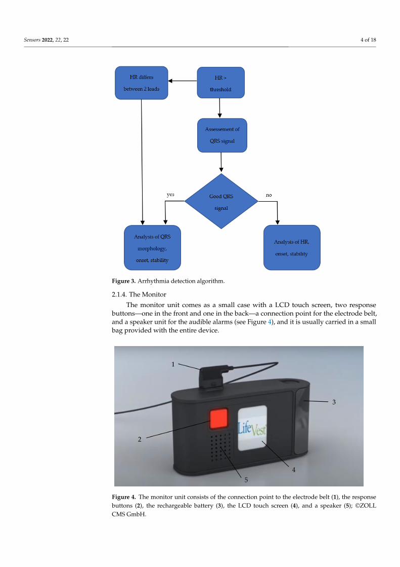
When the heart rate exceeds one of the pre-set thresholds, a QRS analysis is initiated,
including an analysis of stability, onset, and QRS morphology in a second step, to distin-
guish whether a ventricular arrhythmia (VA) is present. If the HR differs significantly be-
tween the two leads, the HR is weighed lower than the analysis of QRS morphology, on-
set, and stability. The QRS morphology is matched with a template ECG in the patient’s
normal rhythm. If only a part of the analysis is applicable due to the quality of the cap-
tured ECG, one part of the detection algorithm is weighed higher for the adjudication and
vice versa (see Figure 3) [12].
Figure 2. Upper picture: The fabric garment without the technical components (1). Lower picture:the fabric garment with the defibrillation pads (2), the electrode belt (3), and the vibration box (4);©ZOLL CMS GmbH.
When the heart rate exceeds one of the pre-set thresholds, a QRS analysis is initiated,including an analysis of stability, onset, and QRS morphology in a second step, to dis-tinguish whether a ventricular arrhythmia (VA) is present. If the HR differs significantlybetween the two leads, the HR is weighed lower than the analysis of QRS morphology,onset, and stability. The QRS morphology is matched with a template ECG in the patient’snormal rhythm. If only a part of the analysis is applicable due to the quality of the capturedECG, one part of the detection algorithm is weighed higher for the adjudication and viceversa (see Figure 3) [12].
In the case of a conscious and hemodynamically stable patient, the response but-tons that sit on the monitor unit can deactivate the imminent shock delivery to avoidinappropriate shocks [12].
The electrode belt also includes a three-axis accelerometer to collect information aboutthe individual daily step count as well as the patient’s body position. While the step countis counted in continuous numbers, the body position is categorized into upright (angle of60–90 degrees), reclined (angle of 30–60 degrees), or lying (0–30 degrees) positions [13].
Sensors 2022, 22, 22 4 of 18Sensors 2022, 22, x FOR PEER REVIEW 4 of 18
Figure 3. Arrhythmia detection algorithm.
In the case of a conscious and hemodynamically stable patient, the response buttons
that sit on the monitor unit can deactivate the imminent shock delivery to avoid inappro-
priate shocks [12].
The electrode belt also includes a three-axis accelerometer to collect information
about the individual daily step count as well as the patient’s body position. While the step
count is counted in continuous numbers, the body position is categorized into upright
(angle of 60–90 degrees), reclined (angle of 30–60 degrees), or lying (0–30 degrees) posi-
tions [13].
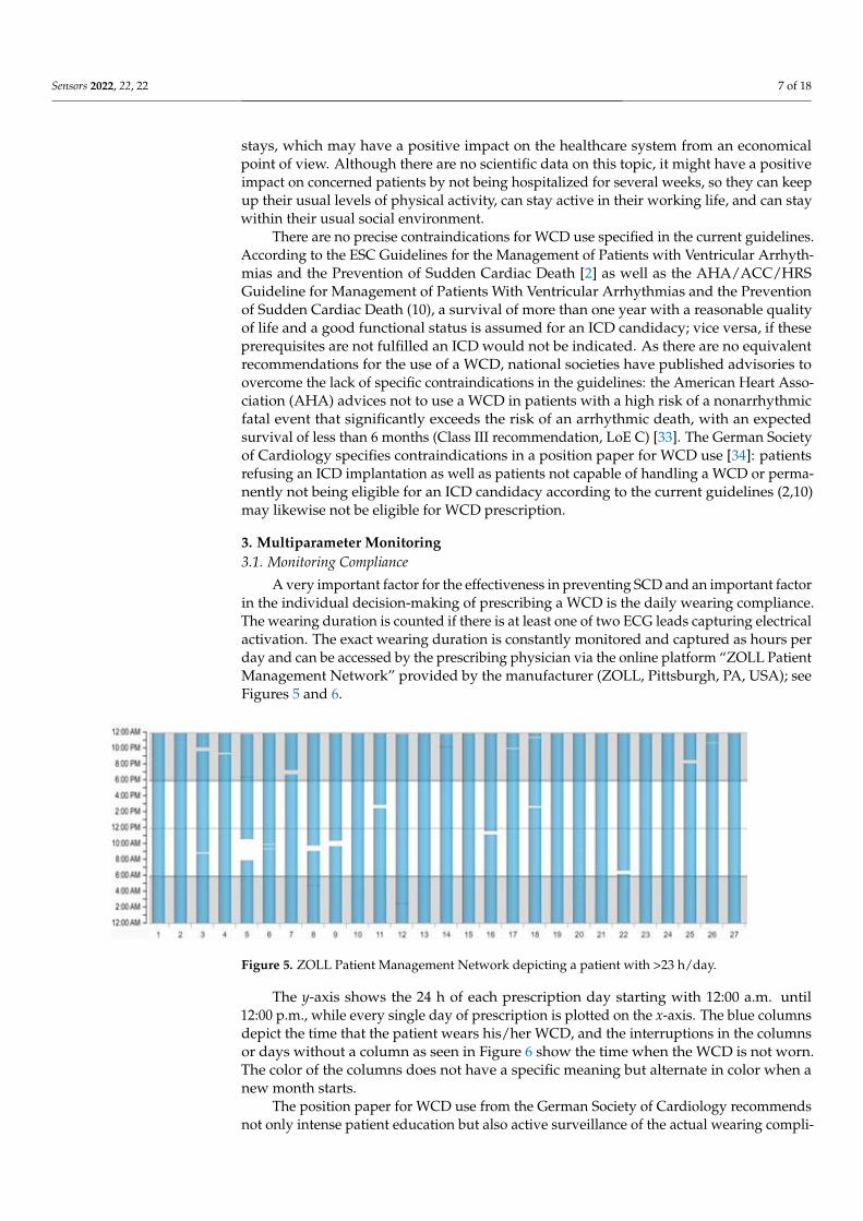
2.1.4. The Monitor
The monitor unit comes as a small case with a LCD touch screen, two response but-
tons—one in the front and one in the back—a connection point for the electrode belt, and
a speaker unit for the audible alarms (see figure 4), and it is usually carried in a small bag
provided with the entire device.
The enclosed processor is provided with a software to monitor the ECG and to ana-
lyze the captured information to detect and treat ventricular arrhythmias. The lithium-ion
batteries have an energy capacity for 24 h continuous operation and up to five shocks,
while every patient is provided with two sets of battery packs and a charging station. The
LCD monitor shows the current battery status and whether the signal quality of the ECG
electrodes is sufficient and has a control panel to send data from the monitor unit to the
network manually or to adjust options such as language or volume. The device has three
different alarms: physiological alarms that appear as a reaction to arrhythmias; technical
alarms when a technical problem is present; and informative alarms when the batteries
need to be exchanged soon or the signal quality of the ECG is insufficient because the ECG
electrodes are too loose, which is assessed by a microampere alternating current. When
Figure 3. Arrhythmia detection algorithm.
2.1.4. The Monitor
The monitor unit comes as a small case with a LCD touch screen, two responsebuttons—one in the front and one in the back—a connection point for the electrode belt,and a speaker unit for the audible alarms (see Figure 4), and it is usually carried in a smallbag provided with the entire device.
Sensors 2022, 22, x FOR PEER REVIEW 5 of 18
any kind of problem is detected, the monitor also provides further instructions and addi-
tional information about the triggered alarm [14,15].
Figure 4. The monitor unit consists of the connection point to the electrode belt (1), the response
buttons (2), the rechargeable battery (3), the LCD touch screen (4), and a speaker (5); ©ZOLL CMS
GmbH.
2.2. Clinical Application
The first data presented about clinical WCD use in patients with either symptomatic
heart failure with reduced ejection fraction (WEARIT study) [16] or patients at high risk
for SCD after acute myocardial infarction or bypass surgery (BIROAD Study) [16] were
gathered in 289 patients and attested to safety and efficacy of the WCD as a treatment
option for malignant arrhythmias in these patients. The WEARIT study included 177 pa-
tients (LVEF 30 ± 10%, 83% male) with symptomatic heart failure and a LVEF below 30%,
with a functional New York Heart Association (NYHA) Class III or IV in an ambulatory
setting. The BIROAD study included 112 patients (LVEF 19 ± 7%, 79% male) with recent
myocardial infarction meeting one of the following additional inclusion criteria: experi-
encing a VA within 48 h of the index event or even within 48 h after coronary artery bypass
grafting (CABG); a LVEF below 30%; survival from sudden cardiac arrest of syncope at
least after 48 h after CABG; and not eligible for ICD implantation, not able to be implanted
with an ICD within 4 months due to capacity reasons, or refusal to use an ICD. Compared
with the current available WCD, the device used for the WEARIT/BIROAD study used a
monophasic waveform with a maximum output of 285 joule and was not provided with
a telemonitoring functionality. During follow-up, eight WCD shocks were delivered in
6/289 (2%) patients with a 75% (6/8) shock efficacy. During follow-up, 12/289 (4.2%) were
found deceased up while the authors stated that the patients were either not wearing the
WCD correctly or not wearing it at all, with only one patient with a detected VA and an
ineffective shock due to reversal of the defibrillation pads. Six inappropriate shocks were
captured within 901 patient months (0.7% per month of patient use) [16].
Following this clinical non-randomized study, the prospective WEAR-IT II registry
was used to evaluate the newer generation of WCD with a lower weight and a bipolar
waveform with a maximum energy level of 150 J: the registry was used to investigate 2000
well-compliant patients (median age 62 years, IQR 16; median LVEF 25%, median wear-
time 22.5 h/day) at high SCD risk, with 805/2000 (40%) patients suffering from ischemic
cardiomyopathy, 927/2000 (46%) with non-ischemic cardiomyopathy (NICMP), and
286/2000 (14%) with a congenital or an inherited heart disease in a real-world setting. After
Figure 4. The monitor unit consists of the connection point to the electrode belt (1), the responsebuttons (2), the rechargeable battery (3), the LCD touch screen (4), and a speaker (5); ©ZOLLCMS GmbH.

Sensors 2022, 22, 22 5 of 18
The enclosed processor is provided with a software to monitor the ECG and to analyzethe captured information to detect and treat ventricular arrhythmias. The lithium-ionbatteries have an energy capacity for 24 h continuous operation and up to five shocks,while every patient is provided with two sets of battery packs and a charging station. TheLCD monitor shows the current battery status and whether the signal quality of the ECGelectrodes is sufficient and has a control panel to send data from the monitor unit to thenetwork manually or to adjust options such as language or volume. The device has threedifferent alarms: physiological alarms that appear as a reaction to arrhythmias; technicalalarms when a technical problem is present; and informative alarms when the batteriesneed to be exchanged soon or the signal quality of the ECG is insufficient because the ECGelectrodes are too loose, which is assessed by a microampere alternating current. When anykind of problem is detected, the monitor also provides further instructions and additionalinformation about the triggered alarm [14,15].
2.2. Clinical Application
The first data presented about clinical WCD use in patients with either symptomaticheart failure with reduced ejection fraction (WEARIT study) [16] or patients at high riskfor SCD after acute myocardial infarction or bypass surgery (BIROAD Study) [16] weregathered in 289 patients and attested to safety and efficacy of the WCD as a treatment optionfor malignant arrhythmias in these patients. The WEARIT study included 177 patients(LVEF 30 ± 10%, 83% male) with symptomatic heart failure and a LVEF below 30%, with afunctional New York Heart Association (NYHA) Class III or IV in an ambulatory setting.The BIROAD study included 112 patients (LVEF 19 ± 7%, 79% male) with recent myocardialinfarction meeting one of the following additional inclusion criteria: experiencing a VAwithin 48 h of the index event or even within 48 h after coronary artery bypass grafting(CABG); a LVEF below 30%; survival from sudden cardiac arrest of syncope at least after48 h after CABG; and not eligible for ICD implantation, not able to be implanted withan ICD within 4 months due to capacity reasons, or refusal to use an ICD. Comparedwith the current available WCD, the device used for the WEARIT/BIROAD study used amonophasic waveform with a maximum output of 285 joule and was not provided witha telemonitoring functionality. During follow-up, eight WCD shocks were delivered in6/289 (2%) patients with a 75% (6/8) shock efficacy. During follow-up, 12/289 (4.2%) werefound deceased up while the authors stated that the patients were either not wearing theWCD correctly or not wearing it at all, with only one patient with a detected VA and anineffective shock due to reversal of the defibrillation pads. Six inappropriate shocks werecaptured within 901 patient months (0.7% per month of patient use) [16].
Following this clinical non-randomized study, the prospective WEAR-IT II registrywas used to evaluate the newer generation of WCD with a lower weight and a bipolarwaveform with a maximum energy level of 150 J: the registry was used to investigate2000 well-compliant patients (median age 62 years, IQR 16; median LVEF 25%, medianwear-time 22.5 h/day) at high SCD risk, with 805/2000 (40%) patients suffering fromischemic cardiomyopathy, 927/2000 (46%) with non-ischemic cardiomyopathy (NICMP),and 286/2000 (14%) with a congenital or an inherited heart disease in a real-world setting.After a median WCD wear-time of 90 days, 41/2000 (2%) patients experienced a total of30 events of VA with shocks and a first shock efficacy of 100%, with 46% of all detectedsustained VTs not requiring electrical therapy and a very low inappropriate shock rate of0.5% (10/2000). After a median follow-up duration of three months, the group of ischemicand congenital cardiomyopathy (CMP) showed the highest rate of VA (3% in ischemicand congenital CMP vs. 1% in non-ischemic CMP, p = 0.02). At the end of follow-up,1160/2000 (58%) patients no longer had an indication for ICD implantation. The WEAR-ITII registry showed the efficacy and safety of the WCD in 2000 high-risk patients in differentclinical settings: good patient compliance was already observed when using the newer andtherefore lighter model of the WCD; the VA detection was accurate; and the bipolar shockswere effective, with a low rate of inappropriate shocks [17].

Sensors 2022, 22, 22 6 of 18
Valuable real-world data on WCD have been provided over the last 20 years by na-tionwide registries, such as the German Registry reporting their experience of 6043 patientsin 404 centers [18], the US registry with 3569 patients included [19], and the AustrianWCD registry capturing 448 patients from 56 centers over more than 10 years. Within theAustrian WCD registry, 19 episodes of shocked VT/VF were documented in 11/448 (2.5%)patients, with a first shock success rate of 84% and an all-over shock efficacy of 95%. Therate of inappropriate shocks was 0.4% (2/448 patients) [20].
Several studies focused on different clinical settings such as recent cardiac decom-pensation in advanced heart failure [21,22] or during the evaluation of a newly diagnosedCMP [23] as well the WCD use in smaller subgroups such as patients with congenital andinherited CMP [24] and patients suffering from peripartum CMP [25,26].
Several groups focused on patients after a recent acute myocardial infarction with orwithout revascularization [27,28] as these patients not only were a very vulnerable cohort ofpatients but also showed the highest rate of shocks when compared with other groups [29].
In a German Single Center study of 114 patients (follow-up of 52.0 days (25.0, 90.0),compliance 23.1 h (19.0, 23.8)) consisting of 31.6% of patients with ischemic CMP (ICMP),45.6% of patients with NICMP and 11.4% of patients with a previous ICD explantation dueto device infection, the cohort of patients with an ICMP experienced the highest rate of VA:9.6% of VA in the allover population compared with 16.7% in patients with ICMP, 3.8% ofpatients with NICMP, and 15.4% of patients with a previous device infection. The authorssuggested a high risk of VA in patients with ICMP, especially of patients with a recent acutemyocardial infarction [27].
Following these data, the randomized-controlled “VEST” trial (Vest Prevention ofEarly Sudden Death Trial) aimed to prove the benefit of WCD prescription on arrhythmicmortality in patients with severe left ventricular dysfunction (LVEF ≤ 35%) after acutemyocardial infarction, with the result still being discussed very controversially: the dataconcerning the primary outcome of arrhythmic mortality could not show a benefit of WCDprescription (1.6% in the device group vs. 2.4% in the control group—p = 0.18), whilethe secondary outcomes including non-arrhythmic mortality and death from any causediffered statistically significantly between the groups (3.1% in the device group versus 4.9%in the control group, p = 0.04) in the as-treated analysis. The authors stated that the resultsfor non-arrhythmic and total mortality were not corrected for multiple testing and wereinterpreted as a chance finding [30]. The discussion around low compliance and cross-overwithin the cohorts (WCD and optimal medical therapy versus optimal medical therapyalone) led to a post hoc analysis, with the authors stating a benefit on arrhythmic mortalityin patients treated per protocol [31]. The data are further discussed in Section 3.1.
Based on the current evidence at that timepoint, the 2015 ESC Guidelines for the Man-agement of Patients with Ventricular Arrhythmias and the Prevention of Sudden CardiacDeath (2) recommends the evaluation of a WCD in adult patients with poor left ventricular(LV) systolic function with a temporary risk of SCD who are not candidates for an ICD.The guideline sets a Class IIb Level of Evidence (LoE) C recommendation for patients thatneed to be bridged to heart transplantation or to a transvenous ICD implantation or inperipartum cardiomyopathy (PPCMP) and a Class IIa LoE C recommendation in patientsrecovering from an inflammatory heart disease with residual reduced ejection fraction.
The WCD use after acute myocardial infarction should be considered in selectedpatients with a high SCD risk due to incomplete revascularization, having a pre-existingLV dysfunction or arrhythmias more than 48 h after the index myocardial infarction (ClassIIb recommendation, LoE C). However, these guidelines do not incorporate data from theVEST trial [30], which were published 3 years after the guidelines were introduced [2].
The WCD not only can bridge the time to a definite ICD candidacy as recommendedby the guidelines but also may protect patients that suffer from ICD electrode infection ormaterial failure with the need for a lead extraction, avoiding reimplantation during thesame procedure, in order to minimize the risk of a new infection [32]. During the periodbetween explantation and re-implantation, the patients may be spared from long hospital
Sensors 2022, 22, 22 7 of 18
stays, which may have a positive impact on the healthcare system from an economicalpoint of view. Although there are no scientific data on this topic, it might have a positiveimpact on concerned patients by not being hospitalized for several weeks, so they can keepup their usual levels of physical activity, can stay active in their working life, and can staywithin their usual social environment.
There are no precise contraindications for WCD use specified in the current guidelines.According to the ESC Guidelines for the Management of Patients with Ventricular Arrhyth-mias and the Prevention of Sudden Cardiac Death [2] as well as the AHA/ACC/HRSGuideline for Management of Patients With Ventricular Arrhythmias and the Preventionof Sudden Cardiac Death (10), a survival of more than one year with a reasonable qualityof life and a good functional status is assumed for an ICD candidacy; vice versa, if theseprerequisites are not fulfilled an ICD would not be indicated. As there are no equivalentrecommendations for the use of a WCD, national societies have published advisories toovercome the lack of specific contraindications in the guidelines: the American Heart Asso-ciation (AHA) advices not to use a WCD in patients with a high risk of a nonarrhythmicfatal event that significantly exceeds the risk of an arrhythmic death, with an expectedsurvival of less than 6 months (Class III recommendation, LoE C) [33]. The German Societyof Cardiology specifies contraindications in a position paper for WCD use [34]: patientsrefusing an ICD implantation as well as patients not capable of handling a WCD or perma-nently not being eligible for an ICD candidacy according to the current guidelines (2,10)may likewise not be eligible for WCD prescription.
3. Multiparameter Monitoring3.1. Monitoring Compliance
A very important factor for the effectiveness in preventing SCD and an important factorin the individual decision-making of prescribing a WCD is the daily wearing compliance.The wearing duration is counted if there is at least one of two ECG leads capturing electricalactivation. The exact wearing duration is constantly monitored and captured as hours perday and can be accessed by the prescribing physician via the online platform “ZOLL PatientManagement Network” provided by the manufacturer (ZOLL, Pittsburgh, PA, USA); seeFigures 5 and 6.
Sensors 2022, 22, x FOR PEER REVIEW 8 of 18
Figure 5. ZOLL Patient Management Network depicting a patient with >23 h/day.
Figure 6. ZOLL Patient Management Network depicting a patient with 5.3 h/day wearing.
The y-axis shows the 24 h of each prescription day starting with 12:00 a.m. until 12:00
p.m., while every single day of prescription is plotted on the x-axis. The blue columns
depict the time that the patient wears his/her WCD, and the interruptions in the columns
or days without a column as seen in Figure 6 show the time when the WCD is not worn.
The color of the columns does not have a specific meaning but alternate in color when a
new month starts.
The position paper for WCD use from the German Society of Cardiology recom-
mends not only intense patient education but also active surveillance of the actual wearing
compliance. If the compliance is constantly lower than 20 h/day and does not improve
even after a follow-up training, the ongoing use of a WCD is contraindicated [34].
Real-world data show a good compliance with a median wearing duration of 21.3–
23.5 h in all patients and > 20 h/day in most patients in big nationwide registries
[17,20,35,36]. Factors such as a younger age of the patient have been identified to decrease
wearing time [18,36], while neither gender, BMI, nor the number of inappropriate alarms
showed an impact on the compliance [36].
In the clinical situation of patients after acute myocardial infarction with left ventric-
ular dysfunction, the VEST trial [30] showed negative results in its’ primary outcome ar-
rhythmic death; see Section 2.2. The results showed a below-average wearing compliance:
in the VEST trial device cohort, only 53% had an average wearing duration ≥ 22 h within
the planned 90 days of prescription, and 30% of patients stopped wearing the WCD within
one month of randomization; 43% stopped within two months; and altogether, 80%
stopped wearing the WCD earlier than the intended 90 days follow-up period, with 34%
of patients not having worn the WCD at any time, resulting in 9/25 patients who did not
wear their WCD being deceased during the follow-up as ventricular arrhythmias could
not be detected or treated [13,19].
In the per-protocol analysis of the VEST data, the authors suggested that the SCD risk
is decreased by the WCD in patients with a good wearing compliance (> 90%, 21.6 h) com-
pared with the whole cohort, which showed a worse compliance as explained above [31]
Figure 5. ZOLL Patient Management Network depicting a patient with >23 h/day.
The y-axis shows the 24 h of each prescription day starting with 12:00 a.m. until12:00 p.m., while every single day of prescription is plotted on the x-axis. The blue columnsdepict the time that the patient wears his/her WCD, and the interruptions in the columnsor days without a column as seen in Figure 6 show the time when the WCD is not worn.The color of the columns does not have a specific meaning but alternate in color when anew month starts.
The position paper for WCD use from the German Society of Cardiology recommendsnot only intense patient education but also active surveillance of the actual wearing compli-

Sensors 2022, 22, 22 8 of 18
ance. If the compliance is constantly lower than 20 h/day and does not improve even aftera follow-up training, the ongoing use of a WCD is contraindicated [34].
Sensors 2022, 22, x FOR PEER REVIEW 8 of 18
Figure 5. ZOLL Patient Management Network depicting a patient with >23 h/day.
Figure 6. ZOLL Patient Management Network depicting a patient with 5.3 h/day wearing.
The y-axis shows the 24 h of each prescription day starting with 12:00 a.m. until 12:00
p.m., while every single day of prescription is plotted on the x-axis. The blue columns
depict the time that the patient wears his/her WCD, and the interruptions in the columns
or days without a column as seen in Figure 6 show the time when the WCD is not worn.
The color of the columns does not have a specific meaning but alternate in color when a
new month starts.
The position paper for WCD use from the German Society of Cardiology recom-
mends not only intense patient education but also active surveillance of the actual wearing
compliance. If the compliance is constantly lower than 20 h/day and does not improve
even after a follow-up training, the ongoing use of a WCD is contraindicated [34].
Real-world data show a good compliance with a median wearing duration of 21.3–
23.5 h in all patients and > 20 h/day in most patients in big nationwide registries
[17,20,35,36]. Factors such as a younger age of the patient have been identified to decrease
wearing time [18,36], while neither gender, BMI, nor the number of inappropriate alarms
showed an impact on the compliance [36].
In the clinical situation of patients after acute myocardial infarction with left ventric-
ular dysfunction, the VEST trial [30] showed negative results in its’ primary outcome ar-
rhythmic death; see Section 2.2. The results showed a below-average wearing compliance:
in the VEST trial device cohort, only 53% had an average wearing duration ≥ 22 h within
the planned 90 days of prescription, and 30% of patients stopped wearing the WCD within
one month of randomization; 43% stopped within two months; and altogether, 80%
stopped wearing the WCD earlier than the intended 90 days follow-up period, with 34%
of patients not having worn the WCD at any time, resulting in 9/25 patients who did not
wear their WCD being deceased during the follow-up as ventricular arrhythmias could
not be detected or treated [13,19].
In the per-protocol analysis of the VEST data, the authors suggested that the SCD risk
is decreased by the WCD in patients with a good wearing compliance (> 90%, 21.6 h) com-
pared with the whole cohort, which showed a worse compliance as explained above [31]
Figure 6. ZOLL Patient Management Network depicting a patient with 5.3 h/day wearing.
Real-world data show a good compliance with a median wearing duration of 21.3–23.5 hin all patients and >20 h/day in most patients in big nationwide registries [17,20,35,36].Factors such as a younger age of the patient have been identified to decrease wearingtime [18,36], while neither gender, BMI, nor the number of inappropriate alarms showedan impact on the compliance [36].
In the clinical situation of patients after acute myocardial infarction with left ventriculardysfunction, the VEST trial [30] showed negative results in its’ primary outcome arrhythmicdeath; see Section 2.2. The results showed a below-average wearing compliance: in theVEST trial device cohort, only 53% had an average wearing duration ≥22 h within theplanned 90 days of prescription, and 30% of patients stopped wearing the WCD within onemonth of randomization; 43% stopped within two months; and altogether, 80% stoppedwearing the WCD earlier than the intended 90 days follow-up period, with 34% of patientsnot having worn the WCD at any time, resulting in 9/25 patients who did not wear theirWCD being deceased during the follow-up as ventricular arrhythmias could not be detectedor treated [13,19].
In the per-protocol analysis of the VEST data, the authors suggested that the SCD risk isdecreased by the WCD in patients with a good wearing compliance (>90%, 21.6 h) comparedwith the whole cohort, which showed a worse compliance as explained above [31].
Technological advancements (reduced size and weight of the WCD) as well as thor-ough education might enhance the patient’s compliance, which is essential for benefitingfrom WCD prescription.
3.2. Monitoring Arrhythmias3.2.1. Sensor Interferences
Some patients may already be implanted with a cardiac pacemaker for bradyarrhyth-mia before WCD prescription. As the QRS complex may be deformed and imitate leftbundle branch block, this can lead to misclassification of supraventricular arrhythmias.Additionally, paced QRS complexes can mislead the detection algorithm of the WCD intoidentifying SVTs as VTs, finally leading to inappropriate arrhythmia detection, triggeringa significant number of alarms and inappropriate shocks in rare cases when patients arenot able to abort the treatment [36]. In the case shown in Figure 7, a patient experiencedtotal AV-block after receiving a WCD shock for ventricular tachycardia and deactivatedinappropriate detections by pressing the response buttons depicted by the small picturesbelow the ECG stripe.

Sensors 2022, 22, 22 9 of 18
Sensors 2022, 22, x FOR PEER REVIEW 9 of 18
Technological advancements (reduced size and weight of the WCD) as well as thor-
ough education might enhance the patient’s compliance, which is essential for benefiting
from WCD prescription.
3.2. Monitoring Arrhythmias
3.2.1. Sensor Interferences
Some patients may already be implanted with a cardiac pacemaker for bradyarrhyth-
mia before WCD prescription. As the QRS complex may be deformed and imitate left
bundle branch block, this can lead to misclassification of supraventricular arrhythmias.
Additionally, paced QRS complexes can mislead the detection algorithm of the WCD into
identifying SVTs as VTs, finally leading to inappropriate arrhythmia detection, triggering
a significant number of alarms and inappropriate shocks in rare cases when patients are
not able to abort the treatment [36]. In the case shown in Figure 7, a patient experienced
total AV-block after receiving a WCD shock for ventricular tachycardia and deactivated
inappropriate detections by pressing the response buttons depicted by the small pictures
below the ECG stripe.
Figure 7. Pressing the response buttons while experiencing AVB III°.
The pacing spikes of unipolar pacing can result in oversensing and can result in de-
tected heart rates that do not correspond to an effective heart rate. When the ventricle is
paced, the t-wave voltage increases in the same way as the QRS complex and can enhance
oversensing as well. With a unipolar pacing program, up to 10% suffer from misled VA
detection and are in danger of an inappropriate shock when not manually withholding
imminent WCD therapy [37]. T-wave oversensing may also occur in patients with an in-
trinsic rhythm such as that shown in a patient in Figure 8.
Figure 8. T-wave oversensing leading to an inappropriate shock.
Clinical experience in this cohort of patients shows that bipolar ventricular pacing
may also lead to inappropriate WCD alarms [38].
The most important reason for interference in inadequate automatically triggered
alarms is artefacts for 95.6% in the Austrian WCD registry [39], mostly coming from
Figure 7. Pressing the response buttons while experiencing AVB III◦.
The pacing spikes of unipolar pacing can result in oversensing and can result indetected heart rates that do not correspond to an effective heart rate. When the ventricle ispaced, the t-wave voltage increases in the same way as the QRS complex and can enhanceoversensing as well. With a unipolar pacing program, up to 10% suffer from misled VAdetection and are in danger of an inappropriate shock when not manually withholdingimminent WCD therapy [37]. T-wave oversensing may also occur in patients with anintrinsic rhythm such as that shown in a patient in Figure 8.
Sensors 2022, 22, x FOR PEER REVIEW 9 of 18
Technological advancements (reduced size and weight of the WCD) as well as thor-
ough education might enhance the patient’s compliance, which is essential for benefiting
from WCD prescription.
3.2. Monitoring Arrhythmias
3.2.1. Sensor Interferences
Some patients may already be implanted with a cardiac pacemaker for bradyarrhyth-
mia before WCD prescription. As the QRS complex may be deformed and imitate left
bundle branch block, this can lead to misclassification of supraventricular arrhythmias.
Additionally, paced QRS complexes can mislead the detection algorithm of the WCD into
identifying SVTs as VTs, finally leading to inappropriate arrhythmia detection, triggering
a significant number of alarms and inappropriate shocks in rare cases when patients are
not able to abort the treatment [36]. In the case shown in Figure 7, a patient experienced
total AV-block after receiving a WCD shock for ventricular tachycardia and deactivated
inappropriate detections by pressing the response buttons depicted by the small pictures
below the ECG stripe.
Figure 7. Pressing the response buttons while experiencing AVB III°.
The pacing spikes of unipolar pacing can result in oversensing and can result in de-
tected heart rates that do not correspond to an effective heart rate. When the ventricle is
paced, the t-wave voltage increases in the same way as the QRS complex and can enhance
oversensing as well. With a unipolar pacing program, up to 10% suffer from misled VA
detection and are in danger of an inappropriate shock when not manually withholding
imminent WCD therapy [37]. T-wave oversensing may also occur in patients with an in-
trinsic rhythm such as that shown in a patient in Figure 8.
Figure 8. T-wave oversensing leading to an inappropriate shock.
Clinical experience in this cohort of patients shows that bipolar ventricular pacing
may also lead to inappropriate WCD alarms [38].
The most important reason for interference in inadequate automatically triggered
alarms is artefacts for 95.6% in the Austrian WCD registry [39], mostly coming from
Figure 8. T-wave oversensing leading to an inappropriate shock.
Clinical experience in this cohort of patients shows that bipolar ventricular pacingmay also lead to inappropriate WCD alarms [38].
The most important reason for interference in inadequate automatically triggeredalarms is artefacts for 95.6% in the Austrian WCD registry [39], mostly coming from patientmovements resulting in artefacts or having poor skin-to-electrode contact while having anunderlying sinus rhythm, such as that shown in Figure 9.
Sensors 2022, 22, x FOR PEER REVIEW 10 of 18
patient movements resulting in artefacts or having poor skin-to-electrode contact while
having an underlying sinus rhythm, such as that shown in Figure 9.
Figure 9. Automatically recorded ECG—artefacts with underlying sinus rhythm.
In an analysis of 106 patients (median 52 years (P25: 37 years; P75: 66 years), 31%
female), neither compliance, gender, having a previous cardiac implantable electronical
device (CIED) and/or experiencing active pacing, being diagnosed with other arrhyth-
mias, QRS duration, BMI, nor age were predictors of inappropriate alarms [36]. Data from
a German single center experience reported by Erath et al. observed that 57% of patients
have inappropriate alarms due to artefacts. The data suggested that skinny and more ac-
tive patients trigger a significantly higher number of alarms resulting from insufficient
skin contact of the ECG electrodes. Within the publication, the proposed solution was to
reprogram the VT zone from the preset 150 bpm to 180 bpm based on the MADIT-RIT
trial, which originally investigated reprogramming CIED to avoid inappropriate therapies
[40,41]
When patients are properly educated and are able to react adequately, inappropriate
shocks can usually be avoided by pressing the response buttons, resulting in a low inap-
propriate shock rate, ranging from 0.4% (26/6043) in the German WCD registry [18] to
0.5% (10/927) in the prospective WEARIT-II registry (17) and 0.8% (7/879) of patients in
our Austrian WCD registry [39], with up to 1.9% (2/106) of patients having inappropriate
treatments in real-world cohorts with a smaller number of patients [36].
3.2.2. The WCD as a Shock Box
The WCD provides a therapeutic option to prevent SCD: after adequate detection of
VA, patients have the option to react to an acoustic and vibrational alarm to either reject
the WCD shock by pressing the buttons or, if not reacting, to receive a shock from the
WCD. Appropriate shocks are meant to be delivered as a reaction for VAs (see figure 10),
and shocks for supraventricular arrhythmias, atrial fibrillation, atrial flutter, or normal
sinus rhythm with artefacts are considered inappropriate. That a shock is not always nec-
essary even though a VA is present was shown in the WEARIT-II registry: 22 patients
aborted the treatment in 90/120 (75%) sustained VT events opposed to 30/120 (25%) of
events requiring therapy due to hemodynamic instability (17)
As registry data and data from clinical trials show, the efficacy of WCD shocks is
usually very high (94 to 100%) (15, 17–19), but there might still be the need for mechanical
cardiopulmonary resuscitation in patients with VA: if a patient is in a ventricular storm,
the electrical therapies applied by the WCD might be not enough; additionally, the WCD
can also detect non-shockable rhythms such as asystole that also result in cardiac arrest.
The events are captured and sent to the online network, and consequences such as opti-
mization of medical therapy, planning of a catheter ablation, etc. can be drawn from the
transmitted information to a later timepoint.
Besides that, there is a German initiative to enhance the early transmission of events
from patients with potential life-threatening arrhythmias to optimize the rescue chain to
increase the likelihood of a survival after cardiac arrest. This initiative is working on es-
tablishing a telemedical link from the WCD directly to local emergency call centers and/or
to local healthcare practitioners so that advanced life support can be provided if the WCD
shock is not successful or the ECG shows a significant recording that does not require
Figure 9. Automatically recorded ECG—artefacts with underlying sinus rhythm.
In an analysis of 106 patients (median 52 years (P25: 37 years; P75: 66 years), 31% fe-male), neither compliance, gender, having a previous cardiac implantable electronicaldevice (CIED) and/or experiencing active pacing, being diagnosed with other arrhythmias,

Sensors 2022, 22, 22 10 of 18
QRS duration, BMI, nor age were predictors of inappropriate alarms [36]. Data from aGerman single center experience reported by Erath et al. observed that 57% of patientshave inappropriate alarms due to artefacts. The data suggested that skinny and more activepatients trigger a significantly higher number of alarms resulting from insufficient skin con-tact of the ECG electrodes. Within the publication, the proposed solution was to reprogramthe VT zone from the preset 150 bpm to 180 bpm based on the MADIT-RIT trial, whichoriginally investigated reprogramming CIED to avoid inappropriate therapies [40,41].
When patients are properly educated and are able to react adequately, inappropriateshocks can usually be avoided by pressing the response buttons, resulting in a low inap-propriate shock rate, ranging from 0.4% (26/6043) in the German WCD registry [18] to0.5% (10/927) in the prospective WEARIT-II registry (17) and 0.8% (7/879) of patients inour Austrian WCD registry [39], with up to 1.9% (2/106) of patients having inappropriatetreatments in real-world cohorts with a smaller number of patients [36].
3.2.2. The WCD as a Shock Box
The WCD provides a therapeutic option to prevent SCD: after adequate detection ofVA, patients have the option to react to an acoustic and vibrational alarm to either reject theWCD shock by pressing the buttons or, if not reacting, to receive a shock from the WCD.Appropriate shocks are meant to be delivered as a reaction for VAs (see Figure 10), andshocks for supraventricular arrhythmias, atrial fibrillation, atrial flutter, or normal sinusrhythm with artefacts are considered inappropriate. That a shock is not always necessaryeven though a VA is present was shown in the WEARIT-II registry: 22 patients abortedthe treatment in 90/120 (75%) sustained VT events opposed to 30/120 (25%) of eventsrequiring therapy due to hemodynamic instability (17).
Sensors 2022, 22, x FOR PEER REVIEW 11 of 18
defibrillation such as bradycardia or asystole but may be a fatal event as well and cannot
be cured with an electrical shock [42]
Figure 10. WCD shock for VF and VT.
3.2.3. Manually Triggered Alarms
In addition to automatically triggered alarms as described above, the patients can
trigger an ECG recording themselves when they feel palpitations or any symptoms that
may be associated with heart rhythm disorders.
In the Austrian WCD registry, 5492 manually recorded ECGs were triggered by
555/879 (63%) patients. Within these ECGs, only one ECG showed slow sustained V and
25 ECGs in nine patients showed non-sustained VTs. Twenty-six patients experienced
atrial fibrillation in 81 events, four patients recorded an ECG due to 5 bradycardic events,
2 ECGs in two patients showed premature ventricular beats (PVC), and 42 ECGs in nine-
teen patients showed supraventricular tachycardia (SVT). In 11 patients (42 ECGs), sinus
tachycardia was detected, and in 96.6% (5308/5492), the triggered ECGs showed normo-
cardic sinus rhythm [39]
Manually recorded ECGs can help detect non-VA arrhythmias such as new onset
atrial fibrillation or relevant bradycardia that is not detected by the WCD algorithm but
needs an experienced cardiologist to interpret ECG recordings in the clinical context.
3.3. Monitoring Heart Failure
Besides patients with inherited cardiomyopathies or channelopathies who often pre-
sent with arrhythmias without clinical evidence of systolic dysfunction, a high percentage
of patients prescribed with a WCD suffer from heart failure with reduced ejection fraction
(HFrEF). Besides arrhythmia monitoring, monitoring heart failure and signs for an immi-
nent decompensation could help reduce morbidity and mortality in this dominant cohort
Figure 10. WCD shock for VF and VT.
As registry data and data from clinical trials show, the efficacy of WCD shocks isusually very high (94 to 100%) (15, 17–19), but there might still be the need for mechanical

Sensors 2022, 22, 22 11 of 18
cardiopulmonary resuscitation in patients with VA: if a patient is in a ventricular storm, theelectrical therapies applied by the WCD might be not enough; additionally, the WCD canalso detect non-shockable rhythms such as asystole that also result in cardiac arrest. Theevents are captured and sent to the online network, and consequences such as optimizationof medical therapy, planning of a catheter ablation, etc. can be drawn from the transmittedinformation to a later timepoint.
Besides that, there is a German initiative to enhance the early transmission of eventsfrom patients with potential life-threatening arrhythmias to optimize the rescue chainto increase the likelihood of a survival after cardiac arrest. This initiative is working onestablishing a telemedical link from the WCD directly to local emergency call centersand/or to local healthcare practitioners so that advanced life support can be provided ifthe WCD shock is not successful or the ECG shows a significant recording that does notrequire defibrillation such as bradycardia or asystole but may be a fatal event as well andcannot be cured with an electrical shock [42].
3.2.3. Manually Triggered Alarms
In addition to automatically triggered alarms as described above, the patients cantrigger an ECG recording themselves when they feel palpitations or any symptoms thatmay be associated with heart rhythm disorders.
In the Austrian WCD registry, 5492 manually recorded ECGs were triggered by555/879 (63%) patients. Within these ECGs, only one ECG showed slow sustained Vand 25 ECGs in nine patients showed non-sustained VTs. Twenty-six patients experiencedatrial fibrillation in 81 events, four patients recorded an ECG due to 5 bradycardic events,2 ECGs in two patients showed premature ventricular beats (PVC), and 42 ECGs in nineteenpatients showed supraventricular tachycardia (SVT). In 11 patients (42 ECGs), sinus tachy-cardia was detected, and in 96.6% (5308/5492), the triggered ECGs showed normocardicsinus rhythm [39].
Manually recorded ECGs can help detect non-VA arrhythmias such as new onset atrialfibrillation or relevant bradycardia that is not detected by the WCD algorithm but needs anexperienced cardiologist to interpret ECG recordings in the clinical context.
3.3. Monitoring Heart Failure
Besides patients with inherited cardiomyopathies or channelopathies who oftenpresent with arrhythmias without clinical evidence of systolic dysfunction, a high per-centage of patients prescribed with a WCD suffer from heart failure with reduced ejectionfraction (HFrEF). Besides arrhythmia monitoring, monitoring heart failure and signs foran imminent decompensation could help reduce morbidity and mortality in this domi-nant cohort of patients at risk for SCD. The risk of re-hospitalization after an index eventof an admission for symptomatic heart failure is greatest within the first 30 days afterdischarge [43,44].
Preceding an imminent cardiac decompensation that requires an inpatient admissionto a hospital, patients themselves can notice early signs of an already manifest cardiacdecompensation [45]. Symptoms such as an increased burden of atrial arrhythmias, anincrease in resting heart rate or decreased physical activity as well as increased lungfluid levels can be detected by ICD algorithms. Remote monitoring with implantabledevices to anticipate a manifest decompensation before clinical symptoms are present hasshown improved clinical outcomes in studies with several hundred patients. In HFrEF, amonitoring approach that uses several parameters captured with an ICD is mentioned witha Class IIb recommendation, LoE B in symptomatic patients in the current guidelines forheart failure [46] In HFrEF and HF with preserved ejection fraction (HFpEF), measurementsof pulmonary arterial pressures with the CardioMems system may be considered (ClassIIb recommendation, LoE B) in the specific situation with a previous HF hospitalization toavoid re-hospitalization due to HF [47–50].

Sensors 2022, 22, 22 12 of 18
These implantable devices are mentioned in the current ESC heart failure guide-lines [50] and can measure surrogate parameters or direct invasive pressures depending onthe type of device and may help to monitor the course of the disease and to intervene byanticipating cardiac decompensations before hospitalization is needed.
The population provided with a WCD is at risk of acute heart failure as patients arenewly diagnosed with a CMP or are in an acute phase of a cardiac disease, and very often,the prescription of a WCD occurs in an inpatient setting. The idea to monitor so-calledcardiac acoustic biomarkers (CAB) via sensors on the harness of the WCD came up toidentify early evidence of cardiac decompensation in patients with HFrEF and specificallyin patients with a left ventricular ejection fraction (LVEF) ≤ 35% in the heart soundsregistry, the “HEAR-IT” study [51] A multiparameter monitoring algorithm was applied on671 patients (61 ± 13 years) prescribed with a WCD: the cardiohaemic vibrations measuredwith the defibrillation electrodes adjacent to the patient’s body surface incorporating athree-axis accelerometer and the simultaneously registered two lead surface ECGs. Fromthese sensors, the heart sounds are combined with the information for the ECG, and acombination of the electromechanical activation time (EMAT) and the third heart sound(S3) strength was measured over time; the trend of this combined parameter showed agood correlation to classify patients into groups being at either low or high risk for a heartfailure event.
The EMAT is a parameter measured from the onset of the Q wave on the surfaceECG to the peak of the first heart sound and serves as parameter for systolic function ifprolonged. The third heart sound is measured on a scale of 0–10 and is also a well-knownsign for heart failure in clinical auscultation and is a sign for increased intracardiac fillingpressures. The algorithm used a 10 s measurement every 5 min to measure the CABs andthe heart rate. These values were observed over time, and thresholds were defined. Patientswith CABs above the upper threshold were at very high risk for a heart failure eventwhile, if the CABs improved, they needed to fall below the lower cut-off to count to thelow-risk cohort. During follow-up, 81/671 (12%) had a heart failure event (cardiovasculardeath, arrhythmias and hospital admissions, and emergency department visits for HF). Theincrease in CABs above the upper threshold identified 69% of the events at least 2 weeksbefore and 90% of HF events at least 3 days before. The initial classification within thefirst 7 days after discharge from the index hospitalization in high- or low-risk HF eventsthrough CABs was more accurate in the prediction of an event compared with the NYHAclassification, while on the other hand, the algorithm combining CABs and HR had anegative predictive value of 94% for HF events. The authors proposed the integration ofCABs in clinical practice to help prevent HF events [51] Currently, CABs recorded by theWCD are not available for real-world patient care and were only studied in trials.
An additional feature of the newer WCD generation, called the “TRENDS” optionprovides a combined multiparameter monitoring approach for heart failure incorporatingthe heart rate and changes in average heart rate, the activity via a daily step count aswell as the body orientation/position detected via a three-axis accelerometer. All of thisinformation is available as daily data and was captured over time as trends in the onlineWCD network; see Figure 11.
The TRENDS data were captured and available also in clinical patients outside ofclinical trials, while the visibility within the online network needs to be activated foreach patient and each physician separately and is not routinely used for follow-ups. Re-cently, a retrospective analysis of TRENDS data patients from the PROLONG II trial [52]proposed a clinical application of the TRENDS data: 267 patients (31.9% female, meanLVEF 25.3 ± 8.5%) with a newly diagnosed CMP were observed over the time of WCDprescription (111.8 ± 74.5 days). The first and last seven days of usage were compared andshowed significant changes in heart rate, step count, and five-minute heart rate variabilityapproximate (HRV5), which is a surrogate for beat-to-beat heart rate variability and wascalculated from the data available from the online network. After multivariate analysis, thechange in HRV5 seemed to be an independent predictor for LVEF improvement defined

Sensors 2022, 22, 22 13 of 18
as an increase of ≥ 10%. The authors stated that HRV5 may be a potential indicator oftreatment response during the evaluation phase of newly diagnosed heart failure [53].
Sensors 2022, 22, x FOR PEER REVIEW 13 of 18
the heart rate and changes in average heart rate, the activity via a daily step count as well
as the body orientation/position detected via a three-axis accelerometer. All of this infor-
mation is available as daily data and was captured over time as trends in the online WCD
network; see Figure 11.
Figure 11. TRENDS data of a patient with an ongoing WCD prescription showing the daily step
count (1), the body position (2), the body angle while reclined or lying (3) and the body position
while reclined or lying (4).
The TRENDS data were captured and available also in clinical patients outside of
clinical trials, while the visibility within the online network needs to be activated for each
patient and each physician separately and is not routinely used for follow-ups. Recently,
a retrospective analysis of TRENDS data patients from the PROLONG II trial [52] pro-
posed a clinical application of the TRENDS data: 267 patients (31.9% female, mean LVEF
25.3 ± 8.5%) with a newly diagnosed CMP were observed over the time of WCD prescrip-
tion (111.8 ± 74.5 days). The first and last seven days of usage were compared and showed
significant changes in heart rate, step count, and five-minute heart rate variability approx-
imate (HRV5), which is a surrogate for beat-to-beat heart rate variability and was calcu-
lated from the data available from the online network. After multivariate analysis, the
change in HRV5 seemed to be an independent predictor for LVEF improvement defined
as an increase of ≥ 10%. The authors stated that HRV5 may be a potential indicator of
treatment response during the evaluation phase of newly diagnosed heart failure [53].
The physical activity (PA) has a prognostic role for HF events and cardiovascular
death in patients at a high risk as proven in a cohort of post-myocardial infarction patients
with patients self-reporting their physical activity [54] and in studies with device-meas-
ured physical activity in heart failure patients [55,56].
A retrospective, observational study analyzed the average daily step count and a
change within 4057 patients. These surrogate parameters of physical activity were meas-
ured by the WCD, and whether a decrease in PA is prognostic for a deterioration of heart
failure and ventricular arrhythmia events was evaluated. Patients with a lower step count,
Figure 11. TRENDS data of a patient with an ongoing WCD prescription showing the daily stepcount (1), the body position (2), the body angle while reclined or lying (3) and the body positionwhile reclined or lying (4).
The physical activity (PA) has a prognostic role for HF events and cardiovasculardeath in patients at a high risk as proven in a cohort of post-myocardial infarction patientswith patients self-reporting their physical activity [54] and in studies with device-measuredphysical activity in heart failure patients [55,56].
A retrospective, observational study analyzed the average daily step count and achange within 4057 patients. These surrogate parameters of physical activity were measuredby the WCD, and whether a decrease in PA is prognostic for a deterioration of heart failureand ventricular arrhythmia events was evaluated. Patients with a lower step count, witha cut-off of 3637 steps per day during the first week after WCD prescription, showed a4.3 times higher likelihood to receive a shock for VA compared with more active patients,especially in the first month after WCD prescription. Similar to the CABs [51], a low PAwithin the first seven days after hospital discharge from the index hospitalization wasassociated with a higher event rate [13]. In a study evaluating 4928 female patients, adecline in PA was seen 2 weeks before a WCD treatment for VA [57] In contrast, all studiesabout PA in patients after an index HF event attested to the feasibility of telemedicalsurveilled step count in patients with an overall good compliance as a possible tool forremote monitoring in heart failure [13,57,58].
While the daily step count is a surrogate for physical activity, the 6 min walk test(6MWT) is a well-established and objectifiable test for the functional status of a heartfailure patient. In times with a rise in importance of telemedical options, reliable andobjectifiable options are necessary. A randomized clinical trial confirmed the feasibility of a6MWT, analyzing WCD-guided 6MWT tests performed regularly at home in 197 patients(57 ± 12 years, LVEF 23 ± 7%) over a run of 8 weeks. There was no difference withinthese groups.

Sensors 2022, 22, 22 14 of 18
Currently, these data are not routinely used although they are partially available forthe treating physicians, as the clinical evidence not only to detect but also to intervene andprevent HF events through any of these noninvasive diagnostic options is still missing.
3.4. Monitor Therapy Success
In the acute phase of a newly diagnosed CMP, the establishment and up-titration ofpharmacological therapy is essential [50] Heart rate monitoring as permanently measuredby the WCD can be useful in patients with atrial fibrillation to assess a pharmacologicallyestablished rhythm or rate control or to assess target values in heart rate. Case reportsalready reported clinical application of the WCD as a monitoring device to supervise up-titration of HF medication and to control the heart rhythm as well as to consequently takeaction on repeated onset atrial fibrillation to avoid cardiac decompensation [59].
An upcoming study will investigate betablocker up-titration in heart failure patientsmonitored with a WCD. Beta blockers may have bradycardia as a side effect and do notunfold their full therapeutic effect when under-dosed. Consequently, the WCD is used asa monitoring tool during up-titration of beta blocker therapy. (“Optimizing Beta BlockerDosage in Women While Using the Wearable Cardioverter Defibrillator—OPT BB women”,clinicaltrials.gov identifier: NCT04504188).
Following the current ESC heart failure guidelines [50], the decision about implantingan ICD should be made after implementation of an optimal medical therapy (OMT) for threemonths while patients are at risk for SCD and seem to be unprotected. Monitoring therapysuccess and protecting patients from SCD in order to avoid untimely and unnecessaryICD implantations during up establishment and up-titration of heart failure therapy wastested in the PROLONG study [60], with 156 patients (54 ± 14 years, 35% female) with aLVEF 24 ± 7% being prescribed with a WCD after being newly diagnosed with HFrEF. Afollow-up was scheduled after 3 months to assess the LVEF and functional status of thepatients. Patient in the PROLONG study received a WCD for NICMP in 55%, 29% hadan ICMP, 12% had a PPCMP, and 4% of patients were diagnosed with acute myocarditis.After 3 months, the LVEF and functional status of the patients were reassessed and adecision on whether to prolong the evaluation was made. If the LVEF was between 30 and35% and the LVEF improved by ≥ 5% within the first three months or if the maximallytolerated guideline-directed medical therapy was not established optimally yet, the WCDprescription was prolonged for another 3 months and a reassessment was scheduled. While65/156 (42%) showed a LVEF above 35% after 3 months, another 26/156 (19%) improvedafter 6 months of therapy in total.
A total of 11/156 (7%) patients experienced 12 WCD shocks after a median prescriptionduration of 59 days (13;161 days), with ten shocks happening within the first three monthsafter WCD prescription and two shocks being observed between the third and the sixthmonths. The authors therefore conclude that malignant ventricular arrhythmias may occurthroughout the course of the 6 months and may enhance the high risk of SCD in thispopulation [60].
4. Conclusions
The WCD is an established treatment option in selected patient groups with a highrisk of SCD who are not eligible for an immediate ICD implantation. The prescriptionof a WCD may help to avoid unnecessary ICD implantation and can cover the period ofestablishing an optimal medical treatment.
Besides the potential benefit for patients and healthcare systems, the patient is thekey component to ensure therapy success with a WCD: the downside of a wearable devicecompared with an implanted device is that the WCD can only protect from malignantventricular arrhythmias when actively worn. Through the online network provided by themanufacturer, the treating physician can actively surveil the wearing duration day by dayand, similar to ICD telemedical approaches, receive alarms when compliance is insufficient.
Sensors 2022, 22, 22 15 of 18
Another potential downside from the vest device is the arrhythmia detection andtreatment algorithm, which produce a high number of inadequate alarms. The false alarmsoriginate primarily from motion artefacts and usually can be handled by the patientsafter thorough education about their device. Sensor interferences such as pacing artefactsfrom implanted pacemakers do not pose a big issue in clinical routine after years ofWCD use. As comparable in CIED programming the indication, the medical history andpotential sensor interferences need to be taken into account when prescribing a WCDand when programming individual thresholds. Nevertheless, data from big nation-wideregistries have shown a low number of inappropriate shocks following these inadequatealarms and the WCD has been proven to be safe and effective in detecting and treatingmalignant arrhythmias.
Additionally, functions to assess the functional status via surrogate parameters todetect a deterioration in heart failure can support healthcare providers in identifyingsubgroups with a high cardiovascular risk or even in differentiating between therapyresponders and non-responders early and potentially giving the possibility to interveneearly and to prevent imminent CV events, while the evidence for a clinical benefit fromrandomized controlled trials is still missing. Generally, a WCD produces a large amountof data through its multiparameter monitoring function, which needs to be thoroughlyreviewed. Features such as the TRENDS data and CABs that provide information aboutfunctional status and physical activity could be added as part to the treatment and follow-up of heart failure patients, while for now, this information is not yet integrated in routineclinical practice.
Another big downside of more than two decades of WCD use is that data fromrandomized-controlled clinical trials are still scarce, and the evidence is based mostly onregistry data [61].
As an outlook into the future, cardiovascular sensors such as the WCD not onlycan help in protecting from SCD but also in guiding medical therapy for heart failurepatients. The current pandemic situation helps to accelerate the establishment of technicalinfrastructure and the financial reimbursement of telemedicine via trained medical staff, andthese functions could potentially help to reduce ambulatory follow-ups and hospitalizationsin the future to protect patients and to lower the economic healthcare burden.
Author Contributions: Conceptualization, U.R., M.M., A.Z. and D.S.; methodology, U.R. and D.S.;formal analysis, U.R. and D.S.; resources, U.R., M.M., A.Z. and D.S.; writing—original draft prepara-tion, U.R. and D.S.; writing—review and editing, U.R., M.M., A.Z. and D.S.; supervision, A.Z. andD.S.; project administration, A.Z. and D.S. All authors have read and agreed to the published versionof the manuscript.
Funding: The Medical University of Graz received external funding for participation in the HEARITregistry by ZOLL Medical Corporation.
Institutional Review Board Statement: Not applicable.
Informed Consent Statement: Not applicable.
Conflicts of Interest: U.R., M.M., and D.S. were co-investigators in the following studies: LifeVestTrends Validation Protocol (TRENDS, ClinicalTrials.gov Identifier: NCT02149290); Heart SoundsRegistry (HEARIT-reg, ClinicalTrials.gov Identifier: NCT03203629).
References1. Mendis, S.; Puska, P.; Norrving, B.; World Health Organization; World Heart Federation; World Stroke Organization. Global
Atlas on Cardiovascular Disease Prevention and Control; Mendis, S., Puska, P., Norrving, B., World Health Organization, Eds.; WorldHealth Organization: Geneva, Switzerland, 2011.
2. Priori, S.G.; Blomstrom-Lundqvist, C.; Mazzanti, A.; Blom, N.; Borggrefe, M.; Camm, J.; Elliott, P.M.; Fitzsimons, D.; Hatala, R.;Hindricks, G.; et al. ESC Guidelines for the management of patients with ventricular arrhythmias and the prevention of suddencardiac death: The Task Force for the Management of Patients with Ventricular Arrhythmias and the Prevention of SuddenCardiac Death of the European Society of Cardiology (ESC). Endorsed by: Association for European Paediatric and CongenitalCardiology (AEPC). Eur. Heart J. 2015, 36, 2793–2867. [PubMed]

Sensors 2022, 22, 22 16 of 18
3. Al-Khatib, S.M.; Stevenson, W.G.; Ackerman, M.J.; Bryant, W.J.; Callans, D.J.; Curtis, A.B.; Deal, B.J.; Dickfeld, T.; Field, M.E.;Fonarow, G.C.; et al. AHA/ACC/HRS Guideline for Management of Patients with Ventricular Arrhythmias and the Preventionof Sudden Cardiac Death: Executive Summary: A Report of the American College of Cardiology/American Heart AssociationTask Force on Clinical Practice Guidelines and the Heart Rhythm Society. Circulation 2017, 72, e91–e220.
4. Dickstein, K.; Cohen-Solal, A.; Filippatos, G.; McMurray, J.J.; Ponikowski, P.; Poole-Wilson, P.A.; Stromberg, A.; van Veldhuisen,D.J.; Atar, D.; Hoes, A.W.; et al. ESC Guidelines for the diagnosis and treatment of acute and chronic heart failure 2008: The TaskForce for the Diagnosis and Treatment of Acute and Chronic Heart Failure 2008 of the European Society of Cardiology. Developedin collaboration with the Heart Failure Association of the ESC (HFA) and endorsed by the European Society of Intensive CareMedicine (ESICM). Eur. Heart J. 2008, 29, 2388–2442.
5. Antiarrhythmics versus Implantable Defibrillators (AVID) Investigators. A comparison of antiarrhythmic-drug therapy withimplantable defibrillators in patients resuscitated from near-fatal ventricular arrhythmias. N. Engl. J. Med. 1997, 337, 1576–1583.[CrossRef]
6. Connolly, S.J.; Gent, M.; Roberts, R.S.; Dorian, P.; Green, M.S.; Klein, G.J.; Mitchell, L.B.; Sheldon, R.S.; Roy, D. CanadianImplantable Defibrillator Study (CIDS): Study design and organization. CIDS Co-Investigators. Am. J. Cardiol. 1993, 72, 103F–108F.[CrossRef]
7. Connolly, S.J.; Hallstrom, A.P.; Cappato, R.; Schron, E.B.; Kuck, K.H.; Zipes, D.P.; Greene, H.L.; Boczor, S.; Domanski, M.;Follmann, D.; et al. Meta-analysis of the implantable cardioverter defibrillator secondary prevention trials. AVID, CASH andCIDS studies. Antiarrhythmics vs. Implantable Defibrillator study. Cardiac Arrest Study Hamburg. Canadian ImplantableDefibrillator Study. Eur. Heart J. 2000, 21, 2071–2078. [CrossRef] [PubMed]
8. Auricchio, A.; Klein, H.; Geller, C.J.; Reek, S.; Heilman, M.S.; Szymkiewicz, S.J. Clinical efficacy of the wearable cardioverter-defibrillator in acutely terminating episodes of ventricular fibrillation. Am. J. Cardiol. 1998, 81, 1253–1256. [CrossRef]
9. Adler, A.; Halkin, A.; Viskin, S. Wearable cardioverter-defibrillators. Circulation 2013, 127, 854–860. [CrossRef]10. Al-Khatib, S.M.; Stevenson, W.G.; Ackerman, M.J.; Bryant, W.J.; Callans, D.J.; Curtis, A.B.; Deal, B.J.; Dickfeld, T.; Field, M.E.;
Fonarow, G.C.; et al. 2017 AHA/ACC/HRS Guideline for Management of Patients with Ventricular Arrhythmias and thePrevention of Sudden Cardiac Death. Circulation 2018, 138, e272–e391.
11. Dillon, K.A.; Szymkiewicz, S.J.; Kaib, T.E. Evaluation of the effectiveness of a wearable cardioverter defibrillator detectionalgorithm. J. Electrocardiol. 2010, 43, 63–67. [CrossRef]
12. Reek, S.; Burri, H.; Roberts, P.R.; Perings, C.; Epstein, A.E.; Klein, H.U.; Committee, E.S.D.; Lip, G.; Gorenek, B.; Sticherling,C.; et al. The wearable cardioverter-defibrillator: Current technology and evolving indications. Europace 2017, 19, 335–345.[CrossRef] [PubMed]
13. Burch, A.E.; D’Souza, B.; Gimbel, J.R.; Rohrer, U.; Masuda, T.; Sears, S.; Scherr, D. Physical activity is reduced prior to ventriculararrhythmias in patients with a wearable cardioverter defibrillator. Clin. Cardiol. 2020, 43, 60–65. [CrossRef] [PubMed]
14. Agarwal, M.; Narcisse, D.; Khouzam, N.; Khouzam, R.N. Wearable Cardioverter Defibrillator “The Lifevest”: Device Design,Limitations, and Areas of Improvement. Curr. Probl. Cardiol. 2018, 43, 45–55. [CrossRef] [PubMed]
15. Francis, J.; Reek, S. Wearable cardioverter defibrillator: A life vest till the life boat (ICD) arrives. Indian Heart J. 2014, 66, 68–72.[CrossRef] [PubMed]
16. Feldman, A.M.; Klein, H.; Tchou, P.; Murali, S.; Hall, W.J.; Mancini, D.; Boehmer, J.; Harvey, M.; Heilman, M.S.; Szymkiewicz,S.J.; et al. Use of a wearable defibrillator in terminating tachyarrhythmias in patients at high risk for sudden death: Results of theWEARIT/BIROAD. Pacing Clin. Electrophysiol. 2004, 27, 4–9. [CrossRef]
17. Kutyifa, V.; Moss, A.J.; Klein, H.; Biton, Y.; McNitt, S.; MacKecknie, B.; Zareba, W.; Goldenberg, I. Use of the wearable cardioverterdefibrillator in high-risk cardiac patients: Data from the Prospective Registry of Patients Using the Wearable CardioverterDefibrillator (WEARIT-II Registry). Circulation 2015, 132, 1613–1619. [CrossRef] [PubMed]
18. Wassnig, N.K.; Gunther, M.; Quick, S.; Pfluecke, C.; Rottstadt, F.; Szymkiewicz, S.J.; Ringquist, S.; Strasser, R.H.; Speiser, U.Experience with the Wearable Cardioverter-Defibrillator in Patients at High Risk for Sudden Cardiac Death. Circulation 2016, 134,635–643. [CrossRef]
19. Chung, M.K.; Szymkiewicz, S.J.; Shao, M.; Zishiri, E.; Niebauer, M.J.; Lindsay, B.D.; Tchou, P.J. Aggregate national experience withthe wearable cardioverter-defibrillator: Event rates, compliance, and survival. J. Am. Coll. Cardiol. 2010, 56, 194–203. [CrossRef]
20. Odeneg, T.; Ebner, C.; Mörtl, D.; Keller, H.; Dirninger, A.; Stix, G.; Föger, B.; Grimm, G.; Steinwender, C.; Gebetsberger, F.; et al.Indications for and outcome in patients with the wearable cardioverter-defibrillator in a nurse-based training programme: Resultsof the Austrian WCD Registry. Eur. J. Cardiovasc. Nurs. 2019, 18, 75–83. [CrossRef]
21. Barsheshet, A.; Kutyifa, V.; Vamvouris, T.; Moss, A.J.; Biton, Y.; Chen, L.; Storozynsky, E.; Wan, C.; Szymkiewicz, S.J.; Goldenberg,I. Study of the wearable cardioverter defibrillator in advanced heart-failure patients (SWIFT). J. Cardiovasc. Electrophysiol. 2017, 28,778–784. [CrossRef]
22. Kao, A.C.; Krause, S.W.; Handa, R.; Karia, D.; Reyes, G. Bianco Wearable defibrillator use in heart failure (WIF): Results of aprospective registry. BMC Cardiovasc. Disord. 2012, 12, 123. [CrossRef]
23. Singh, M.; Wang, N.C.; Jain, S.; Voigt, A.H.; Saba, S.; Adelstein, E.C. Utility of the Wearable Cardioverter-Defibrillator in Patientswith Newly Diagnosed Cardiomyopathy: A Decade-Long Single-Center Experience. J. Am. Coll. Cardiol. 2015, 66, 2607–2613.[CrossRef]
Sensors 2022, 22, 22 17 of 18
24. Rao, M.; Goldenberg, I.; Moss, A.J.; Klein, H.; Huang, D.T.; Bianco, N.R.; Szymkiewicz, S.J.; Zareba, W.; Brenyo, A.; Buber, J.; et al.Wearable defibrillator in congenital structural heart disease and inherited arrhythmias. Am. J. Cardiol. 2011, 108, 1632–1638.[CrossRef]
25. Duncker, D.; Haghikia, A.; Konig, T.; Hohmann, S.; Gutleben, K.J.; Westenfeld, R.; Oswald, H.; Klein, H.; Bauersachs, J.;Hilfiker-Kleiner, D.; et al. Risk for ventricular fibrillation in peripartum cardiomyopathy with severely reduced left ventricularfunction-value of the wearable cardioverter/defibrillator. Eur. J. Heart Fail. 2014, 16, 1331–1336. [CrossRef] [PubMed]
26. Saltzberg, M.T.; Szymkiewicz, S.; Bianco, N.R. Characteristics and outcomes of peripartum versus nonperipartum cardiomyopathyin women using a wearable cardiac defibrillator. J. Card. Fail. 2012, 18, 21–27. [CrossRef]
27. Zishiri, E.T.; Williams, S.; Cronin, E.M.; Blackstone, E.H.; Ellis, S.G.; Roselli, E.E.; Smedira, N.G.; Gillinov, A.M.; Glad, J.A.; Tchou,P.J.; et al. Early risk of mortality after coronary artery revascularization in patients with left ventricular dysfunction and potentialrole of the wearable cardioverter defibrillator. Circ. Arrhythm. Electrophysiol. 2013, 6, 117–128. [CrossRef] [PubMed]
28. Beiert, T.; Malotki, R.; Kraemer, N.; Stöckigt, F.; Linhart, M.; Nickenig, G.; Schrickel, J.W.; Andrié, R.P. A real world wearablecardioverter defibrillator experience—Very high appropriate shock rate in ischemic cardiomyopathy patients at a Europeansingle-center. J. Electrocardiol. 2017, 50, 603–609. [CrossRef]
29. Epstein, A.E.; Abraham, W.T.; Bianco, N.R.; Kern, K.B.; Mirro, M.; Rao, S.V.; Rhee, E.K.; Solomon, S.D.; Szymkiewicz, S.J. Wearablecardioverter-defibrillator use in patients perceived to be at high risk early post-myocardial infarction. J. Am. Coll. Cardiol. 2013,62, 2000–2007. [CrossRef] [PubMed]
30. Olgin, J.E.; Pletcher, M.J.; Vittinghoff, E.; Wranicz, J.; Malik, R.; Morin, D.P.; Zweibel, S.; Buxton, A.E.; Elayi, C.S.; Chung, E.H.; et al.Wearable Cardioverter-Defibrillator after Myocardial Infarction. N. Engl. J. Med. 2018, 379, 1205–1215. [CrossRef]
31. Olgin, J.E.; Lee, B.K.; Vittinghoff, E.; Morin, D.P.; Zweibel, S.; Rashba, E.; Chung, E.H.; Borggrefe, M.; Hulley, S.; Lin, F.; et al.Impact of wearable cardioverter-defibrillator compliance on outcomes in the VEST trial: As-treated and per-protocol analyses. J.Cardiovasc. Electrophysiol. 2020, 31, 1009–1018. [CrossRef]
32. Mitacchione, G.; Schiavone, M.; Gasperetti, A.; Viecca, M.; Curnis, A.; Forleo, G.B. Neglected lead tip erosion: An unusual case ofS-ICD inappropriate shock. J. Cardiovasc. Electrophysiol. 2020, 31, 3322–3325. [CrossRef]
33. Piccini, J.P.; Allen, L.A.; Kudenchuk, P.J.; Page, R.L.; Patel, M.R.; Turakhia, M.P. Wearable Cardioverter-Defibrillator Therapyfor the Prevention of Sudden Cardiac Death: A Science Advisory from the American Heart Association. Circulation 2016, 133,1715–1727. [CrossRef] [PubMed]
34. Deneke, T.; Bosch, R.; Eckardt, L.; Nowak, B.; Schwab, J.O.; Sommer, P.; Veltmann, C.; Helms, T.M. Der tragbare Kar-dioverter/Defibrillator (WCD)—Indikationen und Einsatz. Kardiologe 2019, 13, 292–304. [CrossRef]
35. Klein, H.U.; Meltendorf, U.; Reek, S.; Smid, J.; Kuss, S.; Cygankiewicz, I.; Jons, C.; Szymkiewicz, S.; Buhtz, F.; Wollbrueck, A.; et al.Bridging a temporary high risk of sudden arrhythmic death. Experience with the wearable cardioverter defibrillator (WCD).Pacing Clin. Electrophysiol. 2010, 33, 353–367. [CrossRef] [PubMed]
36. Zylla, M.M.; Hillmann, H.A.K.; Proctor, T.; Kieser, M.; Scholz, E.; Zitron, E.; Katus, H.A.; Thomas, D. Use of the wearablecardioverter-defibrillator (WCD) and WCD-based remote rhythm monitoring in a real-life patient cohort. Heart Vessels 2018, 33,1390–1402. [CrossRef]
37. Schmitt, J.; Abaci, G.; Johnson, V.; Erkapic, D.; Gemein, C.; Chasan, R.; Weipert, K.A.Y.; Hamm, C.W.; Klein, H.U. Safety of theWearable Cardioverter Defibrillator (WCD) in Patients with Implanted Pacemakers. Pacing Clin. Electrophysiol. 2017, 40, 271–277.[CrossRef]
38. Manninger, M.; Odeneg, T.; Fruhwald, F.; Brussee, H.; Scherr, D. Oversensing of the wearable cardioverter defibrillator duringbipolar ventricular stimulation. Wien. Klin. Wochenschr. 2017, 129, 910–912. [CrossRef]
39. Rohrer UM, M.; Odeneg, T.; Ebner, C.; Moertl, D.; Dirninger, A.; Keller, H.; Stix, G.; Alber, H.; Steinwender, C.; Binder, R.; et al.Incidence and predictors of alarms in patients with wearable cardioverter defibrillator (WCD)—Results of the Austrian WCDRegistry. Eur. Heart J. 2019, 40, 1346. [CrossRef]
40. Erath, J.W.; Vamos, M.; Sirat, A.S.; Hohnloser, S.H. The wearable cardioverter-defibrillator in a real-world clinical setting:Experience in 102 consecutive patients. Clin. Res. Cardiol. 2017, 106, 300–306. [CrossRef]
41. Schuger, C.; Daubert, J.P.; Brown, M.W.; Cannom, D.; Estes, N.A., 3rd; Hall, W.; Kayser, T.; Klein, H.; Olshansky, B.; Power,K.A.; et al. Multicenter automatic defibrillator implantation trial: Reduce inappropriate therapy (MADIT-RIT): Background,rationale, and clinical protocol. Ann. Noninvasive Electrocardiol. 2012, 17, 176–185. [CrossRef]
42. Reiss, N.; Schmidt, T.; Hoffmann, J.D.; Cannom, D.; Estes, N.A., 3rd; Hall, W.J.; Kayser, T.; Klein, H.; Olshansky, B.; Power,K.A.; et al. Telemedical Concepts for Heart Failure Patients Treated with a Wearable Cardioverter Defibrillator. Stud. HealthTechnol. Inform. 2020, 271, 93–100.
43. Ross, J.S.; Chen, J.; Lin, Z.; Bueno, H.; Curtis, J.P.; Keenan, P.S.; Normand, S.L.; Schreiner, G.; Spertus, J.A.; Vidán, M.T. Recentnational trends in readmission rates after heart failure hospitalization. Circ. Heart Fail. 2010, 3, 97–103. [CrossRef] [PubMed]
44. Saito, M.; Negishi, K.; Marwick, T.H. Meta-Analysis of Risks for Short-Term Readmission in Patients with Heart Failure. Am. J.Cardiol. 2016, 117, 626–632. [CrossRef] [PubMed]
45. Zile, M.R.; Bennett, T.D.; St John Sutton, M.; Cho, Y.K.; Adamson, P.B.; Aaron, M.F.; Aranda, J.M., Jr.; Abraham, W.T.; Smart, F.W.;Stevenson, L.W.; et al. Transition from chronic compensated to acute decompensated heart failure: Pathophysiological insightsobtained from continuous monitoring of intracardiac pressures. Circulation 2008, 118, 1433–1441. [CrossRef] [PubMed]

Sensors 2022, 22, 22 18 of 18
46. Hindricks, G.; Taborsky, M.; Glikson, M.; Heinrich, U.; Schumacher, B.; Katz, A.; Brachmann, J.; Lewalter, T.; Goette, A.; Block,M.; et al. Implant-based multiparameter telemonitoring of patients with heart failure (IN-TIME): A randomised controlled trial.Lancet 2014, 384, 583–590. [CrossRef]
47. Abraham, W.T.; Adamson, P.B.; Bourge, R.C.; Aaron, M.F.; Costanzo, M.R.; Stevenson, L.W.; Strickland, W.; Neelagaru, S.; Raval,N.; Krueger, S.; et al. Wireless pulmonary artery haemodynamic monitoring in chronic heart failure: A randomised controlledtrial. Lancet 2011, 377, 658–666. [CrossRef]
48. Abraham, W.T.; Stevenson, L.W.; Bourge, R.C.; Lindenfeld, J.A.; Bauman, J.G.; Adamson, P.B. Sustained efficacy of pulmonaryartery pressure to guide adjustment of chronic heart failure therapy: Complete follow-up results from the CHAMPION ran-domised trial. Lancet 2016, 387, 453–461. [CrossRef]
49. Ponikowski, P.; Voors, A.A.; Anker, S.D.; Bueno, H.; Cleland, J.G.F.; Coats, A.J.S.; Falk, V.; Gonzalez-Juanatey, J.R.; Harjola, V.P.;Jankowska, E.A.; et al. 2016 ESC Guidelines for the diagnosis and treatment of acute and chronic heart failure: The Task Force forthe diagnosis and treatment of acute and chronic heart failure of the European Society of Cardiology (ESC)Developed with thespecial contribution of the Heart Failure Association (HFA) of the ESC. Eur. Heart. J. 2016, 37, 2129–2200.
50. McDonagh, T.A.; Metra, M.; Adamo, M.; Gardner, R.S.; Baumbach, A.; Böhm, M.; Burri, H.; Butler, J.; Celutkiene, J.; Chioncel,O.; et al. 2021 ESC Guidelines for the diagnosis and treatment of acute and chronic heart failure: Developed by the Task Forcefor the diagnosis and treatment of acute and chronic heart failure of the European Society of Cardiology (ESC) With the specialcontribution of the Heart Failure Association (HFA) of the ESC. Eur. Heart J. 2021, 42, 3599–3726.
51. Erath, J.W.; Wanczura, P.; Wranicz, J.; Linke, A.; Rohrer, U.; Scherr, D. Influence of decompensated heart failure on cardiac acousticbiomarkers: Impact on early readmissions. ESC Heart Fail. 2020, 7, 4198–4205. [CrossRef]
52. Mueller-Leisse, J.; Brunn, J.; Zormpas, C.; Hohmann, S.; Hillmann, H.A.K.; Eiringhaus, J.; Bauersachs, J.; Veltmann, C.; Duncker,D. Extended follow-up after wearable cardioverter-defibrillator period: The PROLONG-II study. ESC Heart Fail. 2021. [CrossRef][PubMed]
53. Hillmann, H.A.K.; Hohmann, S.; Mueller-Leisse, J.; Zormpas, C.; Eiringhaus, J.; Bauersachs, J.; Veltmann, C.; Duncker, D.Feasibility and First Results of Heart Failure Monitoring Using the Wearable Cardioverter–Defibrillator in Newly DiagnosedHeart Failure with Reduced Ejection Fraction. Sensors 2021, 21, 7798. [CrossRef]
54. Ekblom, O.; Ek, A.; Cider, Å.; Hambraeus, K.; Börjesson, M. Increased Physical Activity Post-Myocardial Infarction Is Related toReduced Mortality: Results from the SWEDEHEART Registry. J. Am. Heart Assoc. 2018, 7, e010108. [CrossRef]
55. Vegh, E.M.; Kandala, J.; Orencole, M.; Upadhyay, G.A.; Sharma, A.; Miller, A.; Merkely, B.; Parks, K.A.; Singh, J.P. Device-measuredphysical activity versus six-minute walk test as a predictor of reverse remodeling and outcome after cardiac resynchronizationtherapy for heart failure. Am. J. Cardiol. 2014, 113, 1523–1528. [CrossRef]
56. Conraads, V.M.; Spruit, M.A.; Braunschweig, F.; Cowie, M.R.; Tavazzi, L.; Borggrefe, M.; Hill, M.R.; Jacobs, S.; Gerritse, B.; vanVeldhuisen, D.J. Physical activity measured with implanted devices predicts patient outcome in chronic heart failure. Circ. HeartFail. 2014, 7, 279–287. [CrossRef]
57. Burch, A.E.; Erath, J.W.; Kutyifa, V.; Aßmus, B.; Bonderman, D.; Russo, A.M. Decline in physical activity in the weeks precedingsustained ventricular arrhythmia in women. Heart Rhythm. O2 2020, 1, 283–287. [CrossRef]
58. Tripp, C.; Burch, A.E.; Erath, J.W.; Hain, A.; Sears, S.F. Physical Activity in Adults with Wearable Cardioverter Defibrillators in thePost-Myocardial Infarction Period. J. Cardiopulm. Rehabil. Prev. 2020, 40, 164–166. [CrossRef]
59. Pulickal, T.; Helms, T.M.; Perings, C.A. Der Wearable Kardioverter-Defibrillator als Diagnostikum. HerzschrittmachertherapieElektrophysiologie 2021, 32, 264–268. [CrossRef]
60. Duncker, D.; König, T.; Hohmann, S.; Bauersachs, J.; Veltmann, C. Avoiding Untimely Implantable Cardioverter/DefibrillatorImplantation by Intensified Heart Failure Therapy Optimization Supported by the Wearable Cardioverter/Defibrillator-ThePROLONG Study. J. Am. Heart Assoc. 2017, 6, e004512. [CrossRef] [PubMed]
61. Cheung, C.C.; Olgin, J.E.; Lee, B.K. Wearable cardioverter-defibrillators: A review of evidence and indications. Trends Cardiovasc.Med. 2021, 31, 196–201. [CrossRef] [PubMed]
Copyright © 2022 FDOKUMEN